EP1692486B1 - Biocapteurs permettant de detecter des macromolecules et d'autres analytes - Google Patents
Biocapteurs permettant de detecter des macromolecules et d'autres analytes Download PDFInfo
- Publication number
- EP1692486B1 EP1692486B1 EP04813618.8A EP04813618A EP1692486B1 EP 1692486 B1 EP1692486 B1 EP 1692486B1 EP 04813618 A EP04813618 A EP 04813618A EP 1692486 B1 EP1692486 B1 EP 1692486B1
- Authority
- EP
- European Patent Office
- Prior art keywords
- aptamer
- epitope
- thrombin
- binding
- molecular
- Prior art date
- Legal status (The legal status is an assumption and is not a legal conclusion. Google has not performed a legal analysis and makes no representation as to the accuracy of the status listed.)
- Not-in-force
Links
Images


Classifications
-
- G—PHYSICS
- G01—MEASURING; TESTING
- G01N—INVESTIGATING OR ANALYSING MATERIALS BY DETERMINING THEIR CHEMICAL OR PHYSICAL PROPERTIES
- G01N21/00—Investigating or analysing materials by the use of optical means, i.e. using sub-millimetre waves, infrared, visible or ultraviolet light
- G01N21/62—Systems in which the material investigated is excited whereby it emits light or causes a change in wavelength of the incident light
- G01N21/63—Systems in which the material investigated is excited whereby it emits light or causes a change in wavelength of the incident light optically excited
- G01N21/64—Fluorescence; Phosphorescence
- G01N21/6428—Measuring fluorescence of fluorescent products of reactions or of fluorochrome labelled reactive substances, e.g. measuring quenching effects, using measuring "optrodes"
-
- A—HUMAN NECESSITIES
- A61—MEDICAL OR VETERINARY SCIENCE; HYGIENE
- A61P—SPECIFIC THERAPEUTIC ACTIVITY OF CHEMICAL COMPOUNDS OR MEDICINAL PREPARATIONS
- A61P43/00—Drugs for specific purposes, not provided for in groups A61P1/00-A61P41/00
-
- C—CHEMISTRY; METALLURGY
- C12—BIOCHEMISTRY; BEER; SPIRITS; WINE; VINEGAR; MICROBIOLOGY; ENZYMOLOGY; MUTATION OR GENETIC ENGINEERING
- C12N—MICROORGANISMS OR ENZYMES; COMPOSITIONS THEREOF; PROPAGATING, PRESERVING, OR MAINTAINING MICROORGANISMS; MUTATION OR GENETIC ENGINEERING; CULTURE MEDIA
- C12N15/00—Mutation or genetic engineering; DNA or RNA concerning genetic engineering, vectors, e.g. plasmids, or their isolation, preparation or purification; Use of hosts therefor
- C12N15/09—Recombinant DNA-technology
- C12N15/11—DNA or RNA fragments; Modified forms thereof; Non-coding nucleic acids having a biological activity
- C12N15/111—General methods applicable to biologically active non-coding nucleic acids
-
- C—CHEMISTRY; METALLURGY
- C12—BIOCHEMISTRY; BEER; SPIRITS; WINE; VINEGAR; MICROBIOLOGY; ENZYMOLOGY; MUTATION OR GENETIC ENGINEERING
- C12Q—MEASURING OR TESTING PROCESSES INVOLVING ENZYMES, NUCLEIC ACIDS OR MICROORGANISMS; COMPOSITIONS OR TEST PAPERS THEREFOR; PROCESSES OF PREPARING SUCH COMPOSITIONS; CONDITION-RESPONSIVE CONTROL IN MICROBIOLOGICAL OR ENZYMOLOGICAL PROCESSES
- C12Q1/00—Measuring or testing processes involving enzymes, nucleic acids or microorganisms; Compositions therefor; Processes of preparing such compositions
- C12Q1/68—Measuring or testing processes involving enzymes, nucleic acids or microorganisms; Compositions therefor; Processes of preparing such compositions involving nucleic acids
- C12Q1/6813—Hybridisation assays
- C12Q1/6816—Hybridisation assays characterised by the detection means
- C12Q1/6825—Nucleic acid detection involving sensors
-
- G—PHYSICS
- G01—MEASURING; TESTING
- G01N—INVESTIGATING OR ANALYSING MATERIALS BY DETERMINING THEIR CHEMICAL OR PHYSICAL PROPERTIES
- G01N33/00—Investigating or analysing materials by specific methods not covered by groups G01N1/00 - G01N31/00
- G01N33/48—Biological material, e.g. blood, urine; Haemocytometers
- G01N33/50—Chemical analysis of biological material, e.g. blood, urine; Testing involving biospecific ligand binding methods; Immunological testing
- G01N33/53—Immunoassay; Biospecific binding assay; Materials therefor
- G01N33/5308—Immunoassay; Biospecific binding assay; Materials therefor for analytes not provided for elsewhere, e.g. nucleic acids, uric acid, worms, mites
-
- G—PHYSICS
- G01—MEASURING; TESTING
- G01N—INVESTIGATING OR ANALYSING MATERIALS BY DETERMINING THEIR CHEMICAL OR PHYSICAL PROPERTIES
- G01N33/00—Investigating or analysing materials by specific methods not covered by groups G01N1/00 - G01N31/00
- G01N33/48—Biological material, e.g. blood, urine; Haemocytometers
- G01N33/50—Chemical analysis of biological material, e.g. blood, urine; Testing involving biospecific ligand binding methods; Immunological testing
- G01N33/53—Immunoassay; Biospecific binding assay; Materials therefor
- G01N33/536—Immunoassay; Biospecific binding assay; Materials therefor with immune complex formed in liquid phase
- G01N33/542—Immunoassay; Biospecific binding assay; Materials therefor with immune complex formed in liquid phase with steric inhibition or signal modification, e.g. fluorescent quenching
-
- C—CHEMISTRY; METALLURGY
- C12—BIOCHEMISTRY; BEER; SPIRITS; WINE; VINEGAR; MICROBIOLOGY; ENZYMOLOGY; MUTATION OR GENETIC ENGINEERING
- C12N—MICROORGANISMS OR ENZYMES; COMPOSITIONS THEREOF; PROPAGATING, PRESERVING, OR MAINTAINING MICROORGANISMS; MUTATION OR GENETIC ENGINEERING; CULTURE MEDIA
- C12N2310/00—Structure or type of the nucleic acid
- C12N2310/10—Type of nucleic acid
- C12N2310/16—Aptamers
-
- C—CHEMISTRY; METALLURGY
- C12—BIOCHEMISTRY; BEER; SPIRITS; WINE; VINEGAR; MICROBIOLOGY; ENZYMOLOGY; MUTATION OR GENETIC ENGINEERING
- C12N—MICROORGANISMS OR ENZYMES; COMPOSITIONS THEREOF; PROPAGATING, PRESERVING, OR MAINTAINING MICROORGANISMS; MUTATION OR GENETIC ENGINEERING; CULTURE MEDIA
- C12N2310/00—Structure or type of the nucleic acid
- C12N2310/30—Chemical structure
- C12N2310/35—Nature of the modification
- C12N2310/351—Conjugate
- C12N2310/3517—Marker; Tag
-
- C—CHEMISTRY; METALLURGY
- C12—BIOCHEMISTRY; BEER; SPIRITS; WINE; VINEGAR; MICROBIOLOGY; ENZYMOLOGY; MUTATION OR GENETIC ENGINEERING
- C12N—MICROORGANISMS OR ENZYMES; COMPOSITIONS THEREOF; PROPAGATING, PRESERVING, OR MAINTAINING MICROORGANISMS; MUTATION OR GENETIC ENGINEERING; CULTURE MEDIA
- C12N2320/00—Applications; Uses
- C12N2320/10—Applications; Uses in screening processes
-
- G—PHYSICS
- G01—MEASURING; TESTING
- G01N—INVESTIGATING OR ANALYSING MATERIALS BY DETERMINING THEIR CHEMICAL OR PHYSICAL PROPERTIES
- G01N21/00—Investigating or analysing materials by the use of optical means, i.e. using sub-millimetre waves, infrared, visible or ultraviolet light
- G01N21/62—Systems in which the material investigated is excited whereby it emits light or causes a change in wavelength of the incident light
- G01N21/63—Systems in which the material investigated is excited whereby it emits light or causes a change in wavelength of the incident light optically excited
- G01N21/64—Fluorescence; Phosphorescence
- G01N21/6428—Measuring fluorescence of fluorescent products of reactions or of fluorochrome labelled reactive substances, e.g. measuring quenching effects, using measuring "optrodes"
- G01N2021/6432—Quenching
-
- G—PHYSICS
- G01—MEASURING; TESTING
- G01N—INVESTIGATING OR ANALYSING MATERIALS BY DETERMINING THEIR CHEMICAL OR PHYSICAL PROPERTIES
- G01N21/00—Investigating or analysing materials by the use of optical means, i.e. using sub-millimetre waves, infrared, visible or ultraviolet light
- G01N21/62—Systems in which the material investigated is excited whereby it emits light or causes a change in wavelength of the incident light
- G01N21/63—Systems in which the material investigated is excited whereby it emits light or causes a change in wavelength of the incident light optically excited
- G01N21/64—Fluorescence; Phosphorescence
- G01N21/6428—Measuring fluorescence of fluorescent products of reactions or of fluorochrome labelled reactive substances, e.g. measuring quenching effects, using measuring "optrodes"
- G01N2021/6439—Measuring fluorescence of fluorescent products of reactions or of fluorochrome labelled reactive substances, e.g. measuring quenching effects, using measuring "optrodes" with indicators, stains, dyes, tags, labels, marks
- G01N2021/6441—Measuring fluorescence of fluorescent products of reactions or of fluorochrome labelled reactive substances, e.g. measuring quenching effects, using measuring "optrodes" with indicators, stains, dyes, tags, labels, marks with two or more labels
Definitions
- the invention relates to kits, molecular beacons, and methods for detecting any polypeptide, analyte, macromolecular complex, or combination thereof.
- the invention relates to biomedical research tools and diagnostic kits.
- This propensity is designed to be low such that in the absence of the protein only a small fraction of DNA half-sites will associate.
- the protein When the protein is present in the reaction mixture, it will bind only to the duplex containing fully functional binding site. This selective binding will drive association of DNA half-sites and this protein-dependent association can be used to generate a spectroscopic signal reporting the presence of the target protein.
- the term "molecular beacons" is used in the art to describe the above assay to emphasize that selective recognition and generation of the signal reporting the recognition occur in this assay simultaneously. Molecular beacons for DNA binding proteins have been developed for several proteins illustrating their general applicability ( Heyduk, T.; Heyduk, E. Nature Biotechnology 2002, 20, 171-176 ).
- aptamers recognizing target protein are the centerpiece of the great majority of protein detection assays so far.
- Development of in vitro methods for selecting aptamers recognizing target proteins from a population of random sequence nucleic acids provided the first real alternative to antibodies.
- One of the potentially important advantages of aptamers is that they are made of easy to propagate and synthesize oligonucleotides. Additionally, standard nucleic acid chemistry procedures can be used to engineer aptamers to contain reporter groups such as, for example, fluorescence probes.
- Aptamer beacons are oligonucleotides that have a binding region that can bind to a non-nucleotide target molecule, such as a protein, a steroid or an inorganic molecule.
- the aptamer beacons having binding regions configured to bind to different target molecules are said to be used in solution-based and solid, array-based systems.
- the aptamer beacons can be attached to solid supports, e.g. a different predetermined point in two-dimensional arrays.
- the inventor has successfully generalized the sensor design applied previously for detecting sequence-specific polynucleotide binding to proteins and other analytes that lack natural nucleic acid binding activity, to greatly expand the applicability of these sensors.
- natural DNA sequences recognizing sequence specific DNA binding proteins were replaced with nucleic acids that bind a particular target protein obtained by in vitro selection from a pool of random sequences.
- nucleic acid (DNA or RNA) aptamers capable of specific binding to proteins lacking natural DNA binding activity can be produced by in vitro SELEX (systematic evolution of ligands by exponential enrichment) procedure ( Tuerk, C.; Gold, L.
- SELEX involves selection of nucleic acid sequences binding a specific target from a pool of random DNA (or RNA) sequences by cycles of binding, washing out unbound sequences, and PCR amplification of target-bound sequences. Numerous examples of successful selection of aptamers specifically binding various proteins as well as other target molecules (Turek 1990, Polisky 1995 and Wilson 1999) provide a strong indication that producing aptamers to a large number of naturally occurring proteins is possible.
- compositions and methods which further enable the application of the proximity-based assay described in copending patent application 10/062,064 (which is incorporated herein by reference) to extend beyond nucleic acid binding factors, their ligands and coregulators to include any polypeptide (including prions or other misfolded proteins), analyte, small molecule ligand or macromolecular complex.
- the invention is directed to the use of a set of labeled aptamers that contain short (preferably about 5-7 nucleotides) complementary single stranded polynucleotide sequences at the distal tip of each aptamer (called"signaling oligos").
- Each aptamer of the set of aptamers binds to a specific and different epitope of a polypeptide or macromolecular complex, i. e. , a first aptamer of the set binds to a first epitope of the polypeptide or macromolecular complex, and a second aptamer of the set binds to a second epitope of the polypeptide or macromolecular complex.
- the first aptamer binds to the first epitope and the second aptamer binds to the second epitope such that the short complementary single stranded polynucleotide sequences at the distal tip of each aptamer can stably associate with each other.
- a label on the first aptamer is brought into proximity to a label on the second aptamer to produce a measurable signal.
- two or more novel nucleic acid half sites that bind to any polypeptide or macromolecular complex may be made and used in a proximity-based assay to detect any polypeptide or macromolecular complex.
- the set of aptamers may be joined by a flexible linker to form a bivalent aptamer construct.
- the present invention relates to a molecular beacon comprising a first molecular-recognition construct and a second molecular recognition construct: wherein
- bivalent aptamer constructs can be useful in myriad applications beyond the detection of molecules and macromolecular complexes.
- the bivalent aptamers may be used in much the same way as antibodies, e.g., to detect molecules and complexes, to purify molecules and complexes, to block epitopes and antigens, to facilitate immune responses (both innate and specific) in an organism, to treat diseases and to confer passive immunity to a subject.
- the preferred subject is a human, a farm animal or a companion animal.
- the bivalent aptamers can be used as therapeutic compositions to block molecular interactions in a cell or tissue, or to facilitate molecular interactions in a cell or tissue to effect a desired therapeutic outcome in a patient.
- Preferred patients include humans, farm animals and companion animals.
- aptamer constructs or bivalent aptamer constructs can be used in medical or veterinary diagnostics or in pharmaceutical screens to help identify potential safe and effective pharmaceutical products.
- aptamer refers to any polynucleotide, generally a RNA or a DNA that has a useful biological activity in terms of biochemical activity, molecular recognition or binding attributes.
- an aptamer has a molecular activity such as binding to a polypeptide at a specific epitope (region) of the polypeptide or having an enzymatic activity. It is generally accepted that an aptamer, which is specific in its binding to any polypeptide, may be synthesized and/or identified by in vitro evolution methods.
- natural cognate binding element sequence refers to a nucleotide sequence that serves as a binding site for a nucleic acid binding factor.
- the natural cognate binding element sequence is a naturally occurring sequence that is recognized by a naturally occurring nucleotide binding factor.
- molecular-recognition construct refers to a construct that contains an "epitope binding agent” and can serve as a "molecular beacon".
- a molecular-recognition construct also contains a "signaling oligo” and a "label”.
- a first molecular-recognition construct and a second molecular-recognition construct may be joined together by a "linker” to form a "bivalent molecular-recognition construct.”
- "Molecular beacon” refers to any chemical-based system for detecting or quantifying the presence of an analyte, polypeptide or other biomolecules, macromolecular complex comprising a biomolecules or a biological coregulator using a measurable read-out system as the detection method.
- An molecular-recognition construct or bivalent molecular-recognition construct is a particular type of molecular beacon with improved specificity and sensitivity.
- aptamer construct refers to a construct that contains an aptamer or a natural cognate binding element sequence and can serve as a "molecular beacon".
- an aptamer construct also contains a "signaling oligo" and a "label”.
- a first aptamer construct and a second aptamer construct may be joined together by a "linker” to form a "bivalent aptamer construct.”
- An aptamer construct is a subset of molecular-recognition construct.
- An aptamer construct or bivalent aptamer construct is also a particular type of molecular beacon with improved specificity and sensitivity.
- antibody generally means a polypeptide or protein that recognizes and can bind to an epitope of an antigen.
- An antibody as used herein, may be a complete antibody as understood in the art, i.e., consisting of two heavy chains and two light chains; or an antibody may be a fragment of a complete antibody, such as a Fab fragment or a peptide comprising a hypervariable region.
- epitope binding agent refers to any substance that is capable of binding to a specific epitope of an antigen, a polypeptide, protein or macromolecular complex.
- epitope binding agents include ligands and fragments of ligands, receptors and fragments of receptors, antibodies and fragments of antibodies, aptamers and other polynucleotides, coenzymes and other coregulators, and allosteric molecules and ions.
- Preferred epitope binding agents include aptamers, natural cognate binding element sequences, antibodies and fragments thereof.
- epitope refers generally to a particular region of an antigen, a hapten, a molecule, a polymer, a prion, a virion, a cell, a peptide, polypeptide, protein, or macromolecular complex.
- An epitope may consist of a small peptide derived from a larger polypeptide.
- An epitope may be a two or three dimensional surface or surface feature of a polypeptide, protein or macromolecular complex that comprises several non-contiguous peptide stretches or amino acid groups.
- signaling oligo means a short (generally 2 to 15 nucleotides, preferably 5 to 7 nucleotides in length) single stranded polynucleotide.
- a first signaling oligo sequence is complementary to the second signaling oligo.
- the first signaling oligo and the second signaling oligo can not form a stable association with each other through hydrogen bonding unless the first and second signaling oligos are brought into close proximity to each other through the mediation of a third party agent.
- linker refers to any polymer attached to an aptamer or aptamer construct. The attachment may be covalent or non-covalent. It is envisioned that the linker can be a polymer of amino acids or nucleotides. A preferred linker molecule is flexible and does not interfere with the binding of a nucleic acid binding factor to the set of nucleic acid components. A preferred linker molecule is comprised of 12 moieties of the Spacer 18 phosphoramidate (Glen Research, Sterling, VA). Another preferred linker molecule is poly dT.
- in vitro evolution generally means any method of selecting for an aptamer which binds to a biomolecule, particularly a peptide or polypeptide.
- in vitro evolution is also known as “in vitro selection”, “SELEX” or “systematic evolution of ligands by exponential enrichment.” Briefly, in vitro evolution involves screening a pool of random polynucleotides for a particular polynucleotide that binds to a biomolecule or has a particular activity that is selectable.
- the particular polynucleotide i.e, aptamer
- a round of aptamer amplification is employed to increase the representation of potentially useful aptamers.
- Successive rounds of selection and amplification are employed to exponentially increase the abundance of the particular and useful aptamer.
- aptamers that bind to distinct epitopes of any given polypeptide or macromolecular complex.
- Aptamers are selected against "substrates", which contain the epitope of interest.
- a "substrate” is any molecular entity that contains an epitope to which an aptamer can bind and that is useful in the selection of an aptamer.
- label refers to any substance attachable to a polynucleotide, polypeptide, aptamer, nucleic acid component, or other substrate material, in which the substance is detectable by a detection method.
- Non-limiting examples of labels applicable to this invention include but are not limited to luminescent molecules, chemiluminescent molecules, fluorochromes, fluorescent quenching agents, colored molecules, radioisotopes, scintillants, massive labels (for detection via mass changes), biotin, avidin, streptavidin, protein A, protein G, antibodies or fragments thereof, Grb2, polyhistidine, Ni 2+ , Flag tags, myc tags, heavy metals, enzymes, alkaline phosphatase, peroxidase, luciferase, electron donors/acceptors, acridinium esters, and colorimetric substrates.
- the skilled artisan would readily recognize other useful labels that are not mentioned above, which may be employed in the operation of the present invention.
- detection method means any method known in the art to detect a molecular interaction event.
- the phrase "detectable signal”, as used herein, is essentially equivalent to “detection method.”
- Detection methods include detecting changes in mass (e.g., plasmin resonance), changes in fluorescence (e.g., FRET, FCCS, fluorescence quenching or increasing fluorescence, fluorescence polarization), enzymatic activity (e.g., depletion of substrate or formation of a product, such as a detectable dye - NBT-BCIP system of alkaline phosphatase is an example), changes chemiluminescence or scintillation (e.g., scintillation proximity assay, luminescence resonance energy transfer, bioluminescence resonance energy transfer and the like).
- fluorescence e.g., FRET, FCCS, fluorescence quenching or increasing fluorescence, fluorescence polarization
- enzymatic activity e.g., depletion
- analyte refers generally to a ligand, chemical moiety, compound, ion, salt, metal, enzyme, secondary messenger of a cellular signal transduction pathway, drug, nanoparticle, environmental contaminant, toxin, fatty acid, steroid, hormone, carbohydrate, amino acid, peptide, polypeptide, protein or other amino acid polymer, microbe, virus or any other agent which is capable of binding to a polypeptide, protein or macromolecular complex in such a way as to create an epitope or alter the availability of an epitope for binding to an aptamer.
- Macromolecular complex refers to any composition of matter comprising a macromolecule.
- these are complexes of one or more macromolecules, such as polypeptides, lipids, carbohydrates, nucleic acids, natural or artificial polymers and the like, in association with each other. The association may involve covalent or non-covalent interactions between components of the macromolecular complex.
- Macromolecular complexes may be relatively simple, such as a ligand bound polypeptide, relatively complex, such as a lipid raft, or very complex, such as a cell surface, virus, bacteria, spore and the like.
- Macromolecular complexes may be biological or non-biological in nature.
- the invention is directed to a method of detecting a polypeptide in a sample comprising the steps of contacting a sample with a first molecular-recognition construct and a second molecular-recognition construct, then detecting the stable interaction of the first and second molecular-recognition constructs via a detection method.
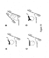
- a detection method Several useful molecular-recognition construct combination (sensor) variants are envisioned by the inventor, which are graphically depicted in figure 26 .
- Panel A depicts a sensor variant comprising two aptamers recognizing two distinct epitopes of a protein.
- Panel B depicts a sensor variant comprising a double stranded polynucleotide containing binding site for DNA binding protein and an aptamer recognizing a distinct epitope of the protein.
- Panel C depicts a sensor variant comprising an antibody and an aptamer recognizing distinct epitopes of the protein.
- Panel D depicts a sensor variant comprising a double stranded polynucleotide containing a binding site for a DNA binding protein and an antibody recognizing a distinct epitope of the protein.
- Panel E depicts a sensor variant comprising two antibodies recognizing two distinct epitopes of the protein.
- Panel F depicts a sensor variant comprising two double stranded polynucleotide fragments recognizing two distinct sites of the protein.
- Panel G depicts a sensor variant comprising two single stranded polynucleotide elements recognizing two distinct sequence elements of another single stranded polynucleotide.
- Panel H depicts a sensor variant that allows for the direct detection of formation of a protein-polynucleotide complex using a double stranded polynucleotide fragment (containing the binding site of the protein) labeled with a first signaling oligonucleotide and the protein labeled with a second signaling oligonucleotide.
- Panel I depicts a sensor variant that allows for the direct detection of the formation of a protein-protein complex using two corresponding proteins labeled with signaling oligonucleotides.
- the first and second molecular-recognition constructs are aptamer constructs, such that the first aptamer construct contains an aptamer or a naturally occurring nucleic acid sequence that recognizes an epitope on a polypeptide (i.e., the first epitope) and the second aptamer construct contains an aptamer or a naturally occurring nucleic acid sequence that recognizes a separate epitope (i.e., the second epitope) on the same polypeptide ( Figure 26 , panels A and B).
- the first aptamer construct and the second aptamer construct each contain a short single stranded oligonucleotide sequence (signaling oligo) such that the short single stranded oligonucleotide of the first aptamer construct (i.e., first signaling oligo) is complementary to the short single stranded oligonucleotide of the second aptamer construct (i.e., second signaling oligo).
- the signaling oligos should be short enough so that they can not form a stable interaction with each other in the absence of the polypeptide, which is capable of bringing the two aptamer constructs together.
- the signaling oligos are at least 5 nucleotides long, and no more that 7 nucleotides long.
- the first aptamer construct contains a first label and the second aptamer construct contains a second label, such that, in the presence of a polypeptide that contains the first epitope and the second epitope, the first and second labels interact to produce a detectable signal that signifies the presence or amount of polypeptide present in the sample.
- the first label is a fluorescence donor and the second label is a fluorescence recipient and the detection method is a detection of a change in fluorescence signal output.
- the first aptamer construct may be fixed to a surface
- the second aptamer construct may be fixed to a surface
- both may be fixed to a surface.
- surfaces can be microtitre plates, test tubes, beads, resins and other polymers and the like.
- the first aptamer construct and the second aptamer construct may be joined with each other by a flexible linker to form a bivalent aptamer.
- Preferred flexible linkers include Spacer 18 polymers and deoxythymidine (“dT”) polymers.
- the first and second aptamers may be used to detect macromolecular complexes in a sample.
- the first epitope is preferably on one polypeptide and the second epitope is on another polypeptide, such that when a macromolecular complex is formed, the one and another polypeptides are bought into proximity, resulting in the stable interaction of the first aptamer construct and the second aptamer construct to produce a detectable signal, as described above.
- the first and second aptamer constructs may be fixed to a surface or to each other via a flexible linker, as described above.
- the first and second aptamers may be used to detect analytes in a sample.
- a first or second epitope is created or made available to bind to a first or second aptamer construct.
- the first aptamer construct and the second aptamer construct are brought into stable proximity to produce a detectable signal, as described above.
- the first and second aptamer constructs may be fixed to a surface or to each other via a flexible linker, as described above.
- a method of making a set of aptamer constructs comprising a first and second aptamer construct, comprising the steps of (a) selecting a first aptamer against a first substrate, which comprises a first epitope, and selecting a second aptamer against a second substrate, which comprises a second epitope, wherein the first aptamer is capable of binding to the first epitope and the second aptamer is capable of binding to the second epitope, (b) attaching a first label to the first aptamer and attaching a second label to the second aptamer, (c) attaching a first signaling oligo to the first aptamer and attaching a second signaling oligo to the second aptamer, wherein the second signaling oligo is complementary to the first signaling oligo, and (d) such that (i) the first aptamer construct comprises the first aptamer, the first label and the first signaling oligo, and (i
- the first substrate may be a polypeptide and the second substrate may be the polypeptide bound to the first aptamer, wherein the first aptamer masks the first epitope, such that the first epitope is not available for the second aptamer to bind.
- the first aptamer may be replaced by a first aptamer construct that contains (i) the first aptamer and signaling oligo, or (ii) the first aptamer, signaling oligo and label, thereby producing a second substrate that allows for the selection of the optimum second aptamer or aptamer construct for signal detection.
- the first and second aptamer constructs may then be joined together by a flexible linker, as described above.
- the first substrate may be a peptide consisting essentially of the first epitope and the second substrate may be a peptide consisting essentially of the second epitope.
- the first and second aptamer constructs created by this method may be linked together by a flexible linker, as described above.
- a bivalent aptamer construct comprising a first aptamer, a first label, a first signaling oligo, a second aptamer, a second label, a second signaling oligo and a linker, wherein the first aptamer is capable of binding to a first epitope and the second aptamer is capable of binding to a second epitope.
- the first and second epitopes may be on the same polypeptide, on different polypeptides of a macromolecular complex, or present on a polypeptide that is bound to an analyte.
- the flexible linker is preferably a polymer that does not interfere with the function of the aptamers.
- Prefered flexible linkers include deoxythymidine polymer (poly dT) and Spacer 18 polymer. However, the skilled artisan in the practice of this invention may substitute any number of linkers.
- the bivalent aptamer construct may not have labels for detection. It is envisioned that these alternative bivalent aptamer constructs may be used much like antibodies to detect molecules, bind molecules, purify molecules (as in a column or pull-down type of procedure), block molecular interactions, facilitate or stabilize molecular interactions, or confer passive immunity to an organism. It is further envisioned that the bivalent aptamer construct can be used for therapeutic purposes. This invention is truly powerful in that it enables the skilled artisan to build any combination of aptamers that recognize any two or more disparate epitopes form any number of molecules into a bivalent, trivalent, or other multivalent aptamer construct to pull together those disparate molecules to test the effect or to produce a desired therapeutic outcome.
- a bivalent aptamer construct may be constructed to facilitate the binding of a ligand to its receptor in a situation wherein the natural binding kinetics of that ligand to the receptor is not favorable (e.g., insulin to insulin receptor in patients suffering diabetes.)
- kits comprising a first epitope binding agent, to which is attached a first label, and a second epitope binding agent, to which is attached a second label, wherein (a) when the first epitope binding agent and the second epitope binding agent label bind to a first epitope of a polypeptide and a second epitope of the polypeptide, respectively, (b) the first label and the second label interact to produce a detectable signal.
- the epitope binding agent is an aptamer construct, which comprises an aptamer, a label and a signaling oligo.
- the epitope binding agent may be an antibody or antibody fragment.
- the kit is useful in the detection of polypeptides, analytes or macromolecular complexes, and as such, may be used in research or medical/veterinary diagnostics applications.
- Disclosed is a method of diagnosing a disease comprising the steps of (a) obtaining a sample from a patient, (b) contacting the sample with a first epitope binding agent and a second epitope binding agent, and (c) detecting the presence of a polypeptide, analyte or macromolecular complex in the sample using a detection method, wherein the presence of the polypeptide, analyte or macromolecular complex in the sample indicates whether a disease is present in the patient.
- the first epitope binding agent is a first aptamer to which a first label and a first signaling oligo are attached
- the second epitope binding agent is a second aptamer to which a second label and a second signaling oligo, which is complementary to the first signaling oligo, are attached
- the detection method is a fluorescence detection method, wherein, (d) when the first aptamer binds to the polypeptide and the second aptamer binds to the polypeptide, (e) the first signaling oligo and the second signaling oligo associate with each other, and (f) the first label is brought into proximity to the second label such that a change in fluorescence occurs.
- Prefered samples include blood, urine, ascites, cells and tissue samples/biopsies.
- Prefered patients include humans, farm animals and companion animals.
- a method of screening a sample for useful reagents comprising the steps of (a) contacting a sample with a first epitope binding agent and a second epitope binding agent, and (b) detecting the presence of a useful reagent in the sample using a detection method.
- Preferred reagents include a polypeptide, which comprises a first epitope and a second epitope, an analyte that binds to a polypeptide (in which case the method further comprises the step of adding the polypeptide to the screening mixture) and a potential therapeutic composition.
- the first epitope binding agent is a first aptamer to which a first label and a first signaling oligo are attached
- the second epitope binding agent is a second aptamer to which a second label and a second signaling oligo, which is complementary to the first signaling oligo, are attached
- the detection method is a fluorescence detection method, wherein, (d) when the first aptamer binds to the polypeptide and the second aptamer binds to the polypeptide, (e) the first signaling oligo and the second signaling oligo associate with each other, and (f) the first label is brought into proximity to the second label such that a change in fluorescence
- EXAMPLE 1 GENERAL METHOD FOR PREPARING SPECIFIC APTAMER CONSTRUCTS
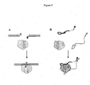
- a method for the rapid and sensitive detection in a sample of proteins, protein complexes, or analytes that bind to proteins is based on the protein-driven association of two nucleic acid constructs containing aptamers that recognize two distinct epitopes of a protein (a.k.a. "aptamer constructs") ( Fig. 1A ). These two aptamer constructs also contain short complementary signaling oliginucleotides attached to the aptamers through a flexible linker. Upon the simultaneous binding of the two aptamers to the target protein, the complementary oligonucleotides (a.k.a.
- signaling oligos are brought into relative proximity which promotes their association to form a stable duplex. Attaching fluorescence probes to the ends of the signaling oligos provides a means of detecting the protein-induced association of the two aptamer contructs ( Fig. 1A ). In the case of proteins which possess natural nucleic acid binding activity, one of the aptamers can be substituted with a nucleic acid sequence containing the natural binding site of the protein ( Fig. 1B ).
- aptamers directed to two distinct epitopes of a given protein is an essential step in developing the aptamer constructs depicted in Fig. 1 .
- Review of the available literature on aptamers reveals at least two possible approaches to achieve this goal.
- the first approach is to perform in vitro selection (a.k.a. in vitro evolution) of nucleic acid aptamers using different methods for the separation of protein-bound and protein-unbound nucleic acid aptamers.
- the rationale here is that in these different partitioning methods different regions of the protein is preferentially displayed resulting in aptamers directed to different regions of the protein surface.
- Aptamers selected to thrombin (infra) are an example of such an approach.
- the partitioning approach relies on the chance selection of distinct epitopes rather than on intelligent design.
- the second approach is to raise or select the aptamers using as substrates peptides that correspond to selected regions of the target protein molecule.
- aptamers capable of recognizing the intact protein from which the peptide used as a substrate for aptamer development was derived.
- this approach has been widely used to generate antibodies which recognize an intact protein.
- the general approach for preparing a set of aptamers directed to an epitope of the protein distinct from the binding site of the first aptamer can be also used for proteins that possess natural DNA binding activity. That is, co-aptamers, which bind the substrate protein at a site distinct from the natural DNA binding site, can be produced. Co-aptamers produced by this method are optimized for functioning in the molecular detection method depicted in Fig.1 .
- Fig. 2 illustrates the overall design of the methods described above. Selection of the co-aptamer is performed using substrate protein pre-bound to the first aptamer ( Fig. 2A ). Alternatively, selection of the co-aptamer is performed using protein pre-bound to it's natural nucleic acid binding site ( Fig. 2B ). A short (5-7 nt) single-stranded oligonucleotide, i.e., the signaling oligo ( Fig. 2 ), is attached to the first aptamer by a flexible linker. Random DNA (or RNA) to be used for selection of co-aptamers is flanked by uniform sequences for the purpose of PCR amplification.
- One of these uniform flanking sequences contains at its end a sequence that is complementary to the signaling oligo of the first aptamer, i.e., the other signaling oligo ( Fig. 2 ).
- the creation and selection of co-aptamers using such a random DNA (or RNA) construct is biased towards aptamers that are able to bind to the substrate protein at a site distinct from the epitope of the first aptamer, and are able to form a duplex between the signaling oligo of the first aptamer.
- the degree of the bias in the selection is adjusted by varying the length of the signaling oligo of the first aptamer and complementary signaling oligo of the second aptamer.
- EXAMPLE 2 METHODS AND APTAMERS FOR DETECTING THROMBIN
- the inventors of the instant invention have developed a methodology for detecting DNA binding proteins, as described in Heyduk, T. and Heyduk, E. Molecular beacons for detecting DNA binding proteins. Nature Biotechnology, 20, 171-176, 2002 , Heyduk, E., Knoll, E., and Heyduk, T, Molecular beacons for detecting DNA binding proteins: mechanism of action, Analyt. Biochem. 316, 1-10, 2003 , and copending patent applications number 09/928,385 , which issued as U.S. Pat. No. 6,544,746 , 10/062,064 , PCT/US02/24822 and PCT/US03/02157 , all of which are incorporated herein by reference.
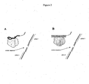
- This methodology is based on splitting the DNA binding site for a protein into two DNA "half-sites" ( Fig. 3A ).
- Each of the resulting "half-sites” contains a short complementary single-stranded region of the length designed to introduce some propensity for the two DNA "half-sites” to associate recreating the duplex containing the fully functional cognate protein binding site.
- This propensity is designed to be low such that in the absence of the protein only a small fraction of DNA half-sites will associate.
- the protein When the protein is present in the reaction mixture, it will bind only to the duplex containing a full and functional binding site. This selective binding drives the association of DNA half-sites and this protein-dependent association can be used to generate a spectroscopic or other signal reporting the presence of the target protein.
- molecular beacons is used in the scientific literature to describe this assay in order to emphasize the fact that the selective recognition and generation of the reporting signal occur simultaneously.
- Molecular beacons for DNA binding proteins have been developed for several proteins (Heyduk and Heyduk, 2002) illustrating their general applicability. Their physical mechanism of action has been established (Heyduk, Knoll and Heyduk, 2003) and they have been also used as a platform for the assay detecting the presence of ligands binding to DNA binding proteins ( Heyduk, E., Fei, Y., and Heyduk, T. Homogenous fluorescence assay for cAMP. Combinatorial Chemistry and High-throughput Screening 6, 183-194, 2003 ). While already very useful, this assay is limited to proteins which exhibit natural DNA binding activity.
- nucleic acid DNA or RNA
- nucleic acid aptamers capable of specific binding to proteins lacking natural DNA binding activity can be produced by in vitro selection methods ( Ellington, A.D., and Szostak, J.W. In vitro selection of RNA molecules that bind specific ligands. Nature 346, 818-822, 1990 ; Tuerk, C., and Gold, L. Systematic evolution of ligands by exponential enrichment: RNA ligands to bacteriophage T4 DNA polymerase. Science 249, 505-510, 1990 ; Gold, L., Polisky, B., Uhlenbeck, O. & Yarus, M. Diversity of Oligonucleotide Function. Ann. Rev. Biochem.
- Described in this example is the novel concept of nucleic acid-based molecular beacons for protein detection, which is not limited to proteins with natural DNA binding activity.
- the example of thrombin (infra) provides experimental validation for this invention.
- Fig. 3B illustrates the overall concept of molecular beacons recognizing any target protein. This design shares some general similarities with molecular beacons for DNA binding proteins described previously and supra ( Fig. 3A ). Instead of splitting the DNA duplex containing the natural binding site for a protein into the two "half-sites", two aptamers recognizing two different epitopes of the protein are used as functional equivalents of the "half-sites”. Short complementary oligonucleotides (signaling oligos) containing the fluorophore (first label) and the quencher (second label) are attached to the two aptamers via a flexible linker ( Fig. 3B ).
- the two aptamer constructs do not associate since the complementary signal oligos are too short to promote association.
- the preferential binding of the protein to the two aptamers should drive the association of the two aptamer constructs resulting in a fluorescence signal change due to bringing the first and second labels into a close proximity.
- Thrombin was selected as a model non-DNA-binding-protein system to provide experimental verification of the concept illustrated in Fig. 3B .
- Two laboratories have previously identified DNA aptamers which selectively recognized two distinct epitopes of the protein ( Bock, L.C., Griffin, L.C., Latham, J.A., Vermass, E.H., and Toole, J.J. Selection of single-stranded DNA molecules that bind and inhibit human thrombin, Nature 355, 564-566, 1992 ; and Tasset, D.M., Kubik, M.F., and Steiner, W. Oligonucleotide inhibitors of human thrombin that bind distinct epitopes, J. Mol. Biol.
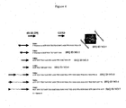
- aptamers were labeled with fluorescein (THR1 and THR2 in Fig. 4 ) to facilitate determination of the affinity of various constructs for thrombin.
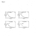
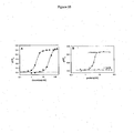
- Formation of a complex between thrombin and fluorescein-labeled 60-18 [29] aptamer (THR1) could be conveniently followed by fluorescence polarization ( Fig. 5A ) whereas binding of the fluorescein-labeled G15D aptamer (THR2) could be followed by changes in fluorescence intensity ( Fig. 5B ).
- Both aptamers bound thrombin in the nanomolar concentration range ( Fig. 5A and 5B ). Quantitative analysis of the binding in the case of THR2 ( Fig.
- Fig. 6 illustrates the manner in which these experiments were performed. Fluorescence spectra of HR2 were recorded in the presence and absence of thrombin ( Fig. 6A ). Thrombin produced ⁇ 50% increase in fluorescence of THR2. Unlabeled competitor aptamer constructs were then added (Figs. 6 B-D) . A small effect of thrombin on the fluorescence of THR2 in the presence of a competitor would be a hallmark of an efficient competitor. THR3 was not a competitor ( Fig. 6B ) in agreement with the data shown in Fig. 5 C and D .
- THR4 (an unlabeled variant of THR2) was able to compete as expected ( Fig. 6C ). However, THR7 (one of the bivalent aptamer constructs) was a much better competitor than THR4 ( Fig. 6D ). No fluorescence change of THR2 in the presence of thrombin was detected when THR7 was present in solution.
- Fig. 7 shows a summary of the competition experiments with all of the constructs shown in Fig. 4 .
- THR5 needs both thrombin epitopes for high-affinity binding
- THR3 one should observe diminished ability of THR5 to compete with THR2. This is exactly what has been observed in experiments illustrated in Fig. 8 .
- THR5 alone was a very effective competitor for THR2 (compare Fig. 8D with 8A).
- THR3 alone was not a competitor for THR2 (compare Fig. 8A and C ).
- THR5 in the presence of THR3 was a worse competitor than THR5 alone (compare Fig. 8B with 8C).
- high-affinity binding of the bivalent aptamer constructs to thrombin involves both first and second aptamer epitopes.
- the bivalent aptamer construct-thrombin complex was stable enough to survive electrophoresis in native polyacrylamide gel ( Fig. 9A ).
- the plot of the complex formed vs. the ratio of thrombin to THR7 did in fact show a 1:1 stoichiometry of the complex ( Fig. 9B ).
- Fig. 11 shows the results of simultaneous titration of the constructs shown in Fig. 10 with thrombin. Formation of aptamer construct-thrombin complexes was followed by EMSA. Each of the constructs bound thrombin with high affinity. However, it is clear that the construct with 7 nt poly dT linker had significantly lower affinity to thrombin compared to constructs with 17 and 27 nt linkers.
- the experimental data presented in Figs. 3-11 provided evidence that all necessary conditions for the signaling beacon shown in Fig. 3B to function were met in the case of thrombin and the two aptamers binding to two distinct region of thrombin.
- the beacon shown in Fig. 12A and B is a derivative of THR16/THR17 bivalent aptamer construct. Aptamers were connected using 17 nt long poly dT linker to 7 nt complementary oligonucleotides (signaling oligos) labeled at 5' and 3' with fluorescein and dabcyl, respectively.
- the beacon shown in Fig. 13 differs only in the nature of the linkers from the beacon shown in Fig. 12 . The remaining sequence is otherwise identical.
- Fig. 13 C shows that upon addition of thrombin to a mixture of THR20 and THR21, protein concentration-dependent quenching of fluorescence was observed whereas no change of fluorescence was detected when thrombin was added to THR21 alone. Response of the beacon to thrombin in the case of this particular beacon was much larger (a ⁇ 2-fold decrease in fluorescence).
- Fig. 14 The experiment illustrated in Fig. 14 was conducted to provide confirmation that indeed the fluorescein-labeled aptamer construct (THR21) was incorporated into a stable complex in the presence of THR20 and thrombin.
- Fig. 15 shows the results, which illustrates the sensitivity of thrombin detection ( Fig. 15A ) and specificity of thrombin detection ( Fig. 15B ). Because the binding of thrombin to bivalent aptamer constructs was extremely tight (pM K d 's), and since the assay appears to be limited only by the sensitivity of detection of fluorescein signal, the sensitivity of thrombin detection could be manipulated by changing the concentration of the aptamer constructs. This is illustrated in Fig.
- Fig. 16 shows the results of competition experiments, in which the ability of various aptamer constructs to dissociate the preformed thrombin-aptamer construct complex was tested.
- the data obtained showed that all bivalent aptamer constructs were by far much more efficient competitors than any of the individual epitope-specific aptamers, in agreement with similar experiments performed with fluorescein-labeled individual aptamer (supra, THR2; Fig. 6 ).
- beacon design described here can be also adopted to improve beacons for detecting proteins exhibiting natural DNA binding activity (Fig. 1 A) .
- one of the aptamers "half-sites" can be replaced with the DNA duplex (containing the protein binding site sequence) connected to signaling complementary oligonucleotide via flexible linker.
- thrombin was a gift from Dr. Ray Rezaie (St. Louis University).
- Factor Xa, prothrombin, ovalbumin, bovine serum albumin, SSB, trypsin and plasma were purchased from Sigma (St. Louis, MO).
- HeLa cellular extracts were from ProteinOne (College Park, MD).
- Texas Red-NHS and Sybr Green were from Molecular Probes (Eugene, OR), Cy5-NHS and Cy3-NHS were from Amersham Biosciences (Piscataway, NJ), and AMCA-sulfoNHS was from Pierce (Rockford, IL). All other reagents were commercially available analytical grade.
- Oligonucleotide constructs used throughout this work are listed in Table 1. Oligonucleotides were obtained from Keck Oligonucleotide Synthesis Facility at Yale University or from IDT (Coralville, IA). 5' fluorescein and 3' dabcyl were incorporated using appropriate phosphoramidates during oligonucleoide synthesis. All other fluorophores were incorporated into oligonucleotides by post-synthetic modification of oligonucleotides containing 5' amino or C6 amino-dT at appriopriate positions with NHS esters of the dyes.
- Oligonucleotides labeled with fluorescence probes were purified by reverse-phase HPLC as described previously ( Heyduk, E.; Heyduk, T. Anal. Biochem. 1997, 248, 216-227 ). Modification of oligonucleotides with europium chelate ((Eu 3+ )DTPA-AMCA) was performed by a two-step procedure described in Heyduk, E.; Heyduk, T.; Claus, P.; Wisniewski, J.R. J. Biol. Chem. 1997, 272,19763-19770 .
- Time-resolved fluorescence in the case of europium chelate - Cy5 labeled beacons was recorded on a laboratory-built instrumentation ( Heyduk, T.; Heyduk, E. Analytical Biochemistry 2001, 289, 60-67 ) which employed pulsed nitrogen laser as the excitation source. Emission was integrated for 100 ⁇ s with 30 ⁇ sec delay after laser pulse.
- Fluorescence intensity of THR2 in the presence and absence of the competitor was determined. Concentration of thrombin, THR2, and the competitor (when present) were 150 nM, 200 nM, and 200 nM, respectively. Under these conditions, binding of aptamers to thrombin was essentially stoichiometric. Previously described method ( Matlock, D.L.; Heyduk, T. Biochemistry 2000, 39, 12274-12283 ) was used to calculate the ratio of the dissociation constant for THR2 to that of the competitor under these experimental conditions.
- Thrombin aptamer binding by electrophoretic mobility shift analysis ESA
- Fig. 17B illustrates the overall concept of molecular beacons for proteins lacking natural sequence-specific DNA binding activity. This design shares some general similarities with molecular beacons for DNA binding proteins described previously by inventor ( Heyduk, T.; Heyduk, E. Nature Biotechnology 2002, 20, 171-176 ; Heyduk, E.; Knoll, E.; Heyduk, T. Analyt. Biochem. 2003, 316, 1-10 ; Knoll, E.; Heyduk, T. Analyt. Chem. 2004, 76, 1156-1164 ; Heyduk, E.; Fei, Y.; Heyduk, T. Combinatorial Chemistry and High-throughput Screening 2003, 6, 183-194 ), ( Fig. 17A ).
- two aptamers recognizing two nonoverlapping epitopes of the protein are used as functional equivalents of the "half-sites".
- Short complementary "signaling” oligonucleotides containing the fluorophore and the quencher are attached to the two aptamers via flexible linkers ( Fig. 17B ).
- the two-aptamer "half-sites" can not associate since the complementary oligonucleotides are too short to promote efficient annealing.
- Binding of the aptamer "half-sites" to the target protein brings the two “signaling” oligonucleotides to relative proximity increasing their local concentrations. This results in the annealing of the "signaling" oligonucleotides, which brings the fluorophore and the quencher to close proximity resulting in a change of fluorescence signal.
- thrombin is a proteolytic enzyme involved in blood clotting cascade and naturally does not bind to DNA or RNA.
- DNA aptamers which selectively recognized two distinct epitopes of the protein ( Bock, L.C.; Griffin, L.C.; Latham, J.A.; Vermass, E.H.; Toole, J.J. Nature 1992, 355, 564-566 , Tasset, D.M.; Kubik, M.F.; Steiher, W. J. Mol. Biol. 1997, 272, 688-698 ).
- THR1 fluorescein-labeled 60-18 [29] aptamer
- THR2 fluorescein-labeled G15D aptamer
- THR4 (unlabeled variant of THR2), as expected, was able to compete ( Fig. 18C ). Quantitative analysis of the competition in this case showed that THR4 bound thrombin 1.7 times better then THR2 indicating that labeling this aptanier with fluorescein had small (insignificant) negative effect on aptamer binding to thrombin. It is obvious that all of the bivalent aptamer constructs were by far better competitors than THR4 ( Fig. 18C ). THR7 appeared to be the best competitor, essentially completely blocking THR2 binding at 1:1 ratio.
- the ratio of thrombin to THR7 indicated 1:1 stoichiometry of the complex ( Fig. 18D ) consistent with the notion that both aptamers - components of THR7 - bind to their respective epitopes in THR7-thiombin complex.
- thrombin beacon illustrated in Fig. 19A .
- Thrombin aptamers were connected using 5 Spacer18 linkers to a 7 nucleotide (“nt") complementary oligonucleotides labeled at 5' and 3' with fluorescein and dabcyl, respectively.
- nt nucleotide
- Fig. 18C individual aptamers
- thrombin Addition of thrombin to a mixture of fluorochrome and quencher-labeled THR20 and THR21 resulted in protein concentration-dependent quenching of fluorescence intensity ( Fig. 19C ). Maximum quenching observed was ⁇ 40%. No fluorescence change was observed ( Fig. 19C ) upon addition of thrombin to THR21 in the absence of dabcyl-labeled partner (THR20) indicating that fluorescence quenching occurred due to protein-induced increased proximity of signaling oligonucleotides resulting in their annealing as illustrated in Fig. 19B . At nanomolar concentrations of the beacon components and thrombin ⁇ 15 min of incubation was sufficient to produce maximal response of the beacon.
- thrombin beacons analogous to one shown in Fig. 19 but in which 17 nt poly dT linkers were used in place of Spacer18 linkers. While thrombin-dependent quenching of fluorescence was observed, the quenching was ⁇ 2 times smaller than with the construct containing Spacer18 linkers. It is likely that poly dT linkers, while flexible, exhibited some residual rigidity ( Mills, J.B.; Vacano, E.; Hagerman, P.J. J. Mol. Biol. 1999, 285, 245-257 ), which perhaps might impede association of the signaling duplex when the two aptamers are bound to thrombin. When the beacon shown in Fig. 19 was titrated with trypsin, a proteolytic enzyme structurally similar to thrombin, no change of fluorescence intensity was observed. We concluded that a functional thrombin beacon according to the design illustrated in Fig. 17B was obtained.
- Fig. 20 shows fluorescence spectra of beacons (without and with thrombin addition) labeled with: fluorescein-Texas Red ( Fig. 20B ), fluorescein-Cy5 ( Fig, 20C ) and Cy3-Cy5 ( Fig. 20D ). In all cases functional beacons were obtained. In each case of the beacon with fluorescent donor and fluorescent acceptor, a large thrombin concentration-dependent increase of sensitized acceptor emission was observed ( Fig. 20 , insets and Fig. 21 A-D ). For comparison, Fig.
- Fig. 20A illustrates fluorescence quenching observed in the presence of thrombin in the case of fluorophore-quencher pair (fluorescein-dabcyl).
- Fig. 21E illustrates results obtained with europium chelate-Cy5 donor-acceptor pair which allowed the use of time-resolved FRET (TR-FRET) as a detection method ( Selvin, P.R.; Rana, T.M.; Hearst, J.E. J. Am. Chem. Soc. 1994, 116,6029-6030 ; Selvin, P.R.; Hearst, J.E. Proc. Natl. Acad. Sci USA 1994, 91, 10024-10028 ; Matthis, G. Clinic.
- TR-FRET time-resolved FRET
- Fig. 21F summarizes the performance of beacon variants with various combinations of donor and acceptor probes. The figure shows the fold of signal change in the presence of saturating concentrations of thrombin compared to background signal of the beacon observed, in the absence of the protein. This ratio varied from ⁇ 2 in the case of fluorescein-dabcyl pair to ⁇ 22 in the case of europium chelate-Cy5 pair.
- beacon variants with fluorescent donor and fluorescent acceptor are those with fluorescent donor and fluorescent acceptor.
- their response can be measured by a two-color determination of the ratio of acceptor to donor signals.
- ratiometric measurement provides more stable signal, which is more resistant to nonspecific effects due to light absorption, light scattering or fluorescence quenching caused by additives present in the sample.
- Increased signal-to-background ratio obtained with optimized donor-acceptor pairs resulted in an increased sensitivity of the beacon. This is illustrated in Fig. 22 which shows responses of three selected beacon variants to low concentrations of thrombin.
- fluorescein-dabcyl labeled beacon the lowest ( ⁇ 2 fold) signal change in the presence of saturating concentration of thrombin
- statistically significant signal change could only be detected at the highest thrombin concentration tested (1 nM).
- fluorescein-Texas Red labeled beacon ⁇ 5 fold signal change at saturating thrombin concentration
- statistically significant signal change could be detected at lower thrombin concentration (200 pM).
- fluorescein-Cy5 labeled beacon ⁇ 15 fold signal change at saturating thrombin concentration
- statistically significant signal change could be detected already at the lowest thrombin concentration tested (50 pM).
- Fig. 23 illustrates excellent reproducibility and stability of thrombin beacon signal. Beacon signal was measured at four thrombin concentrations in five independent measurements. Coefficients of variation were small at each protein concentration tested ( Fig. 23A ). Beacon signal was stable for at least 24 hours ( Fig. 23B ).
- thrombin beacon exhibited excellent discrimination between SSB and thrombin illustrating enhanced specificity of the beacon.
- thrombin beacon One difficultly we've encountered working with cellular extracts was the degradation of oligonucleotides - components of the assay - by nucleases present in cellular extracts.
- thrombin is a plasma protein
- the beacon could be used to detect the protein in plasma. All of the thrombin in plasma is present in a form of its precursor, prothrombin, which is converted to thrombin via proteolytic processing by factor Xa. Prothrombin was recognized by thrombin beacon albeit with much reduced (>20 fold) sensitivity compared to thrombin (not shown). This is well illustrated by experiment shown in Fig. 25C in which sensitized acceptor emission of the beacon in the presence of prothrombin was monitored as a function of time. At the point marked by the arrow Factor Xa was added to the mixture to initiate conversion of prothrombin to thrombin.
- aptamer-based molecular beacons described here is a generalization of the previously developed by us molecular beacons for detecting sequence-specific DNA blinding proteins ( Fig. 17 ).
- Experiments with thrombin as a model protein presented here provided a proof-of-principle evidence for the feasibility of this design. We believe this design will have several important advantages. Since the design of molecular beacons described here is not limited to any specific protein, it will be generally applicable to a large number of proteins. Signaling in the presence of the target protein by our beacon requires a cooperative recognition of two separate epitopes of the protein by two distinct aptamers. This will result in an enhanced specificity of the beacon and increased affinity (i.e. sensitivity of detection).
- aptamers - components of the beacon do not require any engineering of their structure to tailor their distribution of conformations to allow "switching" between different states in the presence of the protein. Such engineering could be dependent on a particular sequence (structure) of the aptamer and, such balancing of the energetics of alternative conformations of nucleic acids is not necessarily a trivial matter. Since the signaling elements ("signaling" oligonucleotides) in the instant beacon design are separate from its aptamer components, any aptamer sequence (and structure) should be compatible with our beacon design.
- any protein for which it will be possible to obtain two aptamers recognizing two distinct epitopes of the protein should be a good target for developing molecular beacons according to scheme in Fig. 17 .
- Antibodies recognizing distinct epitopes of the protein can be obtained relatively easily. Similarly, there are no reasons why aptamers recognizing distinct epitopes could not be developed for many target proteins and several examples are already available ( Jayasena, S.D. Clinical Chem. 1999, 45, 1628-1650 ). Several approaches towards achieving this goal would be possible. The first approach would be to perform in vitro selections (SELEX) using different methods for separation of protein-bound and unbound oligonucleotides. The rationale here is that in these different partitioning methods different regions of the protein could be preferentially displayed resulting in aptamers directed to different regions of protein surface.
- Aptamers selected to thrombin are an example of such approach (Bock, 1992; Tasset, 1997).
- the second approach could be to raise the aptamers to peptides corresponding to different regions of the target protein molecule.
- Experimental evidence exists to show that such strategy can be used to develop aptamers capable of recognizing the intact protein from which the peptide used as a target for aptamer development was derived We, X.; Ellington, A.D. Proc. Natl. Acad. Sci. USA 1996, 93, 7475-7480 ).
- Such approach is widely used to generate antibodies recognizing proteins.
- Two aptamers recognizing different epitopes of the protein can be also produced by a two-step sequential SELEX in which the second step involves selecting an aptamer in the presence of saturating concentration of the aptamer selected in the first step.
- thrombin as a model system
- we have developed a novel in vitro selection strategy to produce pairs of aptamers specifically designed to function in our molecular beacon design Heyduk, E., Kalucka, J., Kinnear, B., Knoll, E., and Heyduk, T., unpublished.
- FIG. 26G The sensor design depicted shown in Fig. 26G is demonstrated in Figure 28 .
- Panel A depicts the principle of the sensor function.
- Single stranded DNA which contains two distinct sequence elements that are complementary to elements in the sensor, to the mixture of two donor and acceptor labeled sensor components, there is a concomitant increase in sensitized acceptor fluorescence intensity ( Figure 28 , B, line with + sign).
- the sensor in this particular case contained Texas Red-labeled THR129 and THR32.
- a nonspecific single stranded DNA binding protein("SSB") at nanomolar concentrations produced a large signal (as measured by fluorescence polarization assay) with the single, fluorescein-labeled aptamer (THR1, Table 1).
- SSB produced the response in a concentration range very similar to the concentration of thrombin required to bind this aptamer.
- a single thrombin aptamer exhibited very poor discrimination between SSB and thrombin.
- Figure 30 summarizes a method for selecting aptamers useful in the practice of the invention.
- Panel A depicts the selection of a second aptamer in the presence of the protein bound to the first aptamer.
- a signaling oligo is at the 5'-end of the random-sequence containing construct and the complementary signaling oligo is attached to the first aptamer via a long flexible linker.
- Selection of co-aptamers using this type of random DNA (or RNA) construct will be biased towards aptamers which are capable of binding to the protein at a site distinct from the epitope of the first aptamer, and which will function in sensors depicted in Fig. 26A .
- FIG. B An alternative scenario is depicted in panel B, which describes the simultaneous selection of two aptamers binding two distinct epitopes of the protein.
- the bars depict short complementary sequences at the 5'-end and 3'-end of a random-sequence containing the aptamer constructs. Selection of aptamers using such random DNA (or RNA) constructs will be biased towards aptamers that are capable of binding to the protein simultaneously at two distinct epitopes of the protein, and which will function in sensors depicted in Fig. 26A .
- a second aptamer can be selected in the presence of the protein bound with a double stranded DNA ( Figure 30 , panel C).
- the bar depicts the short sequence (at the 5'-end of the random-sequence containing construct) complementary to the signaling oligonucleotide attached to the double stranded DNA via a long flexible linker.
- Selection of co-aptamers using such a random DNA (or RNA) construct will be biased towards aptamers that are capable of binding to the protein at a site distinct from the double stranded DNA binding site of the protein and which will function in sensors depicted in Fig. 26B .
- a second aptamer can be selected in the presence of the protein bound with an antibody at a distinct epitope of the protein ( Figure 30 , panel D).
- the bar depicts the short sequence (at the 5'-end of the random-sequence containing construct) complementary to the signaling oligonucleotide attached to the antibody via a long flexible linker. Selection of co-aptamers using such a random DNA (or RNA) construct will be biased towards aptamers which would be able to bind to the protein at a site distinct from the epitope of the antibody and which will function in sensors depicted in Fig. 26C .
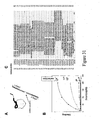
- the selection of an aptamer binding to the thrombin at the epitope distinct from the binding site of G15D aptamer was performed using SELEX procedure starting from a construct containing 33 nt random sequence (THR11) in the presence of the excess of G15D aptamer-containing construct (THR22) ( Figure 31 , panel A).
- Panel B depicts the thrombin binding activity of single stranded DNAs obtained after each indicated round of selection. Measurable thrombin binding activity appeared after 4 th selection and reached maximum after 12 th selection. Binding was measured in the presence of the excess of THR22. DNA obtained after 12 th selection was cloned and DNA obtained from the individual clones was sequenced.
- Panel C depicts the sequence alignment (using ClustalX) of the individual clones. Clones obtained from 4 independent selection experiments are shown. These selections were performed using the following pairs of aptamer constructs and random sequence-containing constructs: THR22 and THR 11; THR25 and THR 11; THR42 and THR11; THR43 and THR 11. Several families of highly conserved sequences are easily visible in panel C.
- a functional thrombin sensor comprising Texas Red-labeled THR27 and fluorescein-labeled THR35 or THR36, which contain sequences corresponding to that of clones 20-26 from Fig, 31C , is depicted in Figure 32 .
- THR35 and THR36 differ by the length of DNA sequence flanking the sequence of clones 20-26.
- the fluorescence image (sensitized acceptor emission) of wells of a microplate containing 20 nM (panel A) or 100 nM (panel B) of the indicated thrombin sensor and the indicated concentrations of thrombin are shown.
- a sensor comprising THR21 and THR127 is shown.
- Figure 33 summarizes of a simultaneous selection of two aptamers binding to thrombin at two distinct epitopes.
- Selection of aptamers was performed using the SELEX procedure starting from two constructs containing 30 nt random sequence (THR149 and THR50) (panel A).
- Thrombin binding activity of the mixture of single stranded DNA's obtained after each indicated round of selection is shown in panel B.
- Measurable thrombin binding activity appeared after 6 th selection and reached a maximum after the 14 th selection.
- DNA obtained after 14 th selection was cloned and the DNA obtained from the individual clones were sequenced.
- Panel C depicts the sequence alignment (using ClustalX) of the clones. Several families of highly conserved sequences are easily visible.
- Aptamer-based molecular beacons were developed for cAMP response element binding protein ("CRP"). Aptamers were selected to bind at sites distinct from the DNA binding site of the protein. Selection was performed using SELEX procedure starting from a construct containing 33 nucleotide random sequence (MIS12) in the presence of the excess of CRP binding site-containing construct (MIS10X3 hybridized with MIS11) ( Figure 34 , panel A). CRP binding activity of single stranded DNA that was obtained after indicated round of selection is depicted in Figure 34 , panel B. Measurable CRP binding activity appeared after 6 th selection and reached maximum after 12 th selection. Binding was measured in the presence of the excess of MIS10X3 hybridized with MIS11.
- DNA obtained after 12 th selection was cloned and DNA obtained from the individual clones were sequenced.
- the sequence alignment (using ClustalX) of the clones is depicted in panel C. conserveed core sequence of ⁇ 16 nucleotides could be identified.
Claims (11)
- Procédé de détection d'un polypeptide dans un échantillon comprenant les étapes suivantes :(a) la mise en contact d'un échantillon avec une première construction de reconnaissance moléculaire et une seconde construction de reconnaissance moléculaire, et(b) la détection d'une association de la première construction de reconnaissance moléculaire, de la seconde construction de reconnaissance moléculaire, et du polypeptide par un procédé de détection ; où(c) la première construction de reconnaissance moléculaire est capable de se lier à un premier épitope du polypeptide et la seconde construction de reconnaissance moléculaire est capable de se lier à un second épitope du polypeptide,(d) la première construction de reconnaissance moléculaire comprend (i) un premier agent de liaison d'épitope qui peut se lier au premier épitope, (ii) un premier oligo de signalisation et (iii) un premier marqueur, le premier agent de liaison d'épitope étant fixé de façon covalente au premier oligo de signalisation par l'intermédiaire d'un premier lieur flexible, et(e) la seconde construction de reconnaissance moléculaire comprend (iv) un second agent de liaison d'épitope qui peut se lier au second épitope, (v) un second oligo de signalisation, qui est complémentaire au premier oligo de signalisation mais qui forme une association stable avec le premier oligo de signalisation seulement lorsqu'il est placé en étroite proximité avec celui-ci par l'intermédiaire de la liaison au polypeptide, et (vi) un second marqueur, le second agent de liaison d'épitope étant fixé de façon covalente au second oligo de signalisation par l'intermédiaire d'un second lieur flexible,
dans lequel les premier et second marqueurs produisent un signal différent lorsque la première construction de reconnaissance moléculaire, la seconde construction de reconnaissance moléculaire et le polypeptide s'associent, plaçant de cette manière les premier et second marqueurs en étroite proximité, par rapport au signal qu'ils produisent lorsque la première construction de reconnaissance moléculaire, la seconde construction de reconnaissance moléculaire et le polypeptide ne sont pas associés, et en outre oùi) le premier agent de liaison d'épitope et le second agent de liaison d'épitope sont chacun un aptamère, ouii) le premier agent de liaison d'épitope est un polynucléotide double brin et le second agent de liaison d'épitope est un aptamère, ouiii) le premier agent de liaison d'épitope est un aptamère et le second agent de liaison d'épitope est un anticorps. - Procédé selon la revendication 1, dans lequel le premier agent de liaison d'épitope est un aptamère et le second agent de liaison d'épitope est un aptamère.
- Procédé selon la revendication 2 qui est utilisé pour détecter un analyte dans un échantillon, le procédé comprenant comme étape (a) la mise en contact d'un échantillon avec une première construction d'aptamère, une seconde construction d'aptamère et un polypeptide, où en présence de l'analyte, la première construction d'aptamère est capable de se lier à un premier épitope du polypeptide et la seconde construction d'aptamère est capable de se lier à un second épitope du polypeptide, si bien que ledit signal différent indique la présence de l'analyte.
- Procédé selon la revendication 3, dans lequel le polypeptide subit un changement conformationnel après liaison de l'analyte.
- Procédé selon la revendication 4, dans lequel l'analyte est un médicament et le polypeptide est capable de lier le médicament.
- Procédé selon la revendication 5, dans lequel l'analyte est une toxine trouvée dans l'environnement.
- Procédé selon la revendication 2, dans lequel le polypeptide est la thrombine ou une protéine de liaison de l'élément de réponse à l'AMPc (« CRP »).
- Procédé selon la revendication 7, dans lequel le premier aptamère se lie à un exosite du fibrinogène (60-18[29]) de la thrombine et le second aptamère se lie à un exosite de l'héparine G15D de la thrombine.
- Procédé selon l'une quelconque des revendications 1 à 8, dans lequel le procédé de détection est choisi dans le groupe constitué de la résonance des plasmons, le transfert d'énergie de fluorescence par résonance (« FRET »), la FCCS, l'extinction de fluorescence, la polarisation de fluorescence, la production d'un produit coloré, la chimioluminescence, la scintillation, la bioluminescence, et le transfert d'énergie de luminescence par résonance.
- Balise moléculaire comprenant une première construction de reconnaissance moléculaire et une seconde construction de reconnaissance moléculaire : où(a) la première construction de reconnaissance moléculaire est capable de se lier à un premier épitope d'un polypeptide et la seconde construction de reconnaissance moléculaire est capable de se lier à un second épitope du polypeptide,(b) la première construction de reconnaissance moléculaire comprend (i) un premier agent de liaison d'épitope qui peut se lier au premier épitope, (ii) un premier oligo de signalisation et (iii) un premier marqueur, le premier agent de liaison d'épitope étant fixé de façon covalente au premier oligo de signalisation par l'intermédiaire d'un premier lieur flexible, et(c) la seconde construction de reconnaissance moléculaire comprend (iv) un second agent de liaison d'épitope qui peut se lier au second épitope, (v) un second oligo de signalisation qui est complémentaire au premier oligo de signalisation mais qui forme une association stable avec le premier oligo de signalisation seulement lorsqu'il est placé en étroite proximité avec celui-ci par l'intermédiaire de la liaison au polypeptide, et (vi) un second marqueur, le second agent de liaison d'épitope étant fixé de façon covalente au second oligo de signalisation par l'intermédiaire d'un second lieur flexible,où les premier et second marqueurs produisent un signal différent lorsque la première construction de reconnaissance moléculaire, la seconde construction de reconnaissance moléculaire, et le polypeptide s'associent, plaçant de cette manière les premier et second marqueurs en étroite proximité, par rapport au signal qu'ils produisent lorsque la première construction de reconnaissance moléculaire, la seconde construction de reconnaissance moléculaire, et le polypeptide ne sont pas associés ; oùi) le premier agent de liaison d'épitope et le second réactif de liaison d'épitope sont chacun un aptamère, ouii) le premier agent de liaison d'épitope est un polynucléotide double brin et le second agent de liaison d'épitope est un aptamère, ouiii) le premier agent de liaison d'épitope est un aptamère et le second agent de liaison d'épitope est un anticorps.
- Balise moléculaire selon la revendication 10, où les premier et second oligo de signalisation sont chacun constitués d'au moins 5 nucléotides et de pas plus de 7 nucléotides.
Applications Claiming Priority (2)
| Application Number | Priority Date | Filing Date | Title |
|---|---|---|---|
| US52907603P | 2003-12-12 | 2003-12-12 | |
| PCT/US2004/041315 WO2005059509A2 (fr) | 2003-12-12 | 2004-12-10 | Biocapteurs permettant de detecter des macromolecules et d'autres analytes |
Publications (3)
| Publication Number | Publication Date |
|---|---|
| EP1692486A2 EP1692486A2 (fr) | 2006-08-23 |
| EP1692486A4 EP1692486A4 (fr) | 2008-05-21 |
| EP1692486B1 true EP1692486B1 (fr) | 2015-10-28 |
Family
ID=34699935
Family Applications (1)
| Application Number | Title | Priority Date | Filing Date |
|---|---|---|---|
| EP04813618.8A Not-in-force EP1692486B1 (fr) | 2003-12-12 | 2004-12-10 | Biocapteurs permettant de detecter des macromolecules et d'autres analytes |
Country Status (7)
| Country | Link |
|---|---|
| US (2) | US7939313B2 (fr) |
| EP (1) | EP1692486B1 (fr) |
| JP (2) | JP2007525661A (fr) |
| CN (1) | CN101208437B (fr) |
| CA (1) | CA2545006C (fr) |
| HK (1) | HK1120568A1 (fr) |
| WO (1) | WO2005059509A2 (fr) |
Families Citing this family (76)
| Publication number | Priority date | Publication date | Assignee | Title |
|---|---|---|---|---|
| JP2007525661A (ja) | 2003-12-12 | 2007-09-06 | セントルイス ユニバーシティー | 大分子その他の分析物の検出用生物センサー |
| EP1885891A2 (fr) * | 2005-05-26 | 2008-02-13 | Ensemble Discovery Corporation | Biodétection reposant sur la chimie à base de matrices d'acides nucléiques |
| US8956857B2 (en) | 2005-06-06 | 2015-02-17 | Mediomics, Llc | Three-component biosensors for detecting macromolecules and other analytes |
| US7795009B2 (en) | 2005-06-15 | 2010-09-14 | Saint Louis University | Three-component biosensors for detecting macromolecules and other analytes |
| US8945840B2 (en) | 2005-06-10 | 2015-02-03 | Saint Louis University | Methods for the selection of aptamers |
| US7811809B2 (en) | 2005-06-15 | 2010-10-12 | Saint Louis University | Molecular biosensors for use in competition assays |
| WO2007025049A2 (fr) * | 2005-08-26 | 2007-03-01 | Archemix Corp. | Aptameres se liant a la thrombine avec une forte affinite |
| CA2660129C (fr) | 2006-08-09 | 2015-11-10 | Saint Louis University | Biocapteurs moleculaires pour la detection de macromolecules et d'autres analytes |
| EP2111553B1 (fr) | 2007-01-24 | 2018-09-19 | Carnegie Mellon University | Biocapteurs optiques |
| DE102007063902B3 (de) * | 2007-08-31 | 2017-11-23 | Rheinische Friedrich-Wilhelms-Universität Bonn | Aptamere, die an ein an der Hämostase beteiligtes Zielmolekül binden |
| DE102007041476B4 (de) | 2007-08-31 | 2017-03-30 | Rheinische Friedrich-Wilhelms-Universität Bonn | Aptamere, die an ein an der Hämostase beteiligtes Zielmolekül binden |
| KR101623992B1 (ko) * | 2007-11-21 | 2016-05-24 | 에라스무스 유니버시티 메디컬 센터 로테르담 | 개선된 fret-프로브 및 이의 용도 |
| WO2009079212A2 (fr) | 2007-12-03 | 2009-06-25 | Carnegie Mellon University | Biocapteurs fluorogéniques de peptides liés |
| JP5210620B2 (ja) * | 2007-12-19 | 2013-06-12 | 独立行政法人科学技術振興機構 | ペプチドアプタマーライブラリーの作製方法および用途 |
| US20140128280A1 (en) * | 2008-04-23 | 2014-05-08 | Arizona Board of Regents, a body corporate of the State of Arizona, acting for and on behaif of Ariz | Synthetic Antibodies |
| EP2703816B1 (fr) * | 2008-11-21 | 2016-10-05 | Saint Louis University | Biocapteur pour la détection de multiples épitopes sur une cible |
| US8551768B2 (en) * | 2008-11-21 | 2013-10-08 | The Trustees Of Columbia University In The City Of New York | Split DNA enzyme for visual single nucleotide polymorphism typing |
| WO2011100561A1 (fr) | 2010-02-12 | 2011-08-18 | Saint Louis University | Biocapteurs moléculaires permettant l'amplification de signaux |
| US20190300945A1 (en) | 2010-04-05 | 2019-10-03 | Prognosys Biosciences, Inc. | Spatially Encoded Biological Assays |
| US10787701B2 (en) | 2010-04-05 | 2020-09-29 | Prognosys Biosciences, Inc. | Spatially encoded biological assays |
| AU2011237729B2 (en) | 2010-04-05 | 2014-04-03 | Prognosys Biosciences, Inc. | Spatially encoded biological assays |
| TWI497076B (zh) | 2010-06-25 | 2015-08-21 | Univ Nat Cheng Kung | 適合體及c-反應蛋白檢測方法 |
| WO2012085111A1 (fr) | 2010-12-23 | 2012-06-28 | F. Hoffmann-La Roche Ag | Complexe polypeptide-polynucléotide et son utilisation dans l'administration d'une fraction effectrice ciblée |
| EP2479914B1 (fr) * | 2011-01-21 | 2015-03-04 | Alcatel Lucent | Procédé et élément émetteur pour la transmission d'informations de canal pour l'adaptation de lien, procédé et élément émetteur pour la réception d'informations de canal |
| GB201106254D0 (en) | 2011-04-13 | 2011-05-25 | Frisen Jonas | Method and product |
| FR2977674B1 (fr) * | 2011-07-06 | 2015-08-14 | Cisbio Bioassays | Methode amelioree de detection et/ou de quantification d'un analyte present a la surface d'une cellule |
| ES2700792T3 (es) * | 2011-11-11 | 2019-02-19 | Eli N Glezer | Procedimientos de ensayo asistidos por coagente de unión |
| CN103160512B (zh) * | 2011-12-15 | 2014-09-24 | 国家纳米科学中心 | 一种核酸及药物载体和药物组合物及其制备方法 |
| WO2014102803A1 (fr) * | 2012-12-31 | 2014-07-03 | Yeda Research And Development Co. Ltd. | Capteur moléculaire et ses procédés d'utilisation |
| ES2942031T3 (es) * | 2013-03-11 | 2023-05-29 | Meso Scale Technologies Llc | Métodos mejorados para realizar ensayos multiplexados |
| US10114015B2 (en) | 2013-03-13 | 2018-10-30 | Meso Scale Technologies, Llc. | Assay methods |
| CN111662960B (zh) | 2013-06-25 | 2024-04-12 | 普罗格诺西斯生物科学公司 | 采用微流控装置的空间编码生物分析 |
| AU2014342528A1 (en) | 2013-10-28 | 2016-04-28 | Dots Technology Corp. | Allergen detection |
| CN103630517B (zh) * | 2013-11-15 | 2015-12-09 | 南京邮电大学 | 一种基于拆分适配体和水溶性共轭聚合物的凝血酶检测方法 |
| CN104020293A (zh) * | 2014-03-25 | 2014-09-03 | 李根喜 | 基于多肽的黑色素瘤生物标记物传感器 |
| US11639929B2 (en) | 2014-04-29 | 2023-05-02 | Yeda Research And Development Co. Ltd. | Universal histidine-tag binding compounds and methods of use thereof as fluorescent probes and sensors |
| WO2015171591A1 (fr) | 2014-05-05 | 2015-11-12 | Board Of Trustees Of The University Of Arkansas | Compositions et procédés d'inhibition de protéines antiapoptotiques bcl-2 comme agents anti-âge |
| JP6695280B2 (ja) | 2014-05-15 | 2020-05-20 | メソ スケール テクノロジーズ エルエルシー | 改善されたアッセイ方法 |
| CA2955972A1 (fr) | 2014-07-22 | 2016-01-28 | Bioventures, Llc. | Compositions et procedes de depletion selective de cellules senescentes |
| EP2980218A1 (fr) * | 2014-07-30 | 2016-02-03 | Rheinische Friedrich-Wilhelms-Universität Bonn | Complexe de thrombine aptamère à utiliser comme antidote à action directe sur les inhibiteurs de la thrombine |
| ES2891305T3 (es) | 2014-09-10 | 2022-01-27 | Univ Washington | Composiciones y métodos para el tratamiento de lesiones cutáneas precancerosas |
| US10274484B2 (en) | 2014-09-12 | 2019-04-30 | Mediomics Llc | Molecular biosensors with a modular design |
| BR112017005252B1 (pt) * | 2014-09-22 | 2023-12-26 | Japan Science And Technology Agency | Método para detecção de molécula alvo e kit para utilização no referido método |
| RU2717956C2 (ru) * | 2014-09-30 | 2020-03-27 | Конинклейке Филипс Н.В. | Способ обнаружения пространственной близости первого и второго эпитопов |
| WO2016109378A1 (fr) * | 2014-12-29 | 2016-07-07 | North Carolina State University | Diagnostic multiplexé pour reconnaître des concentrations de protéines et de peptides apparentés |
| CN104569430B (zh) * | 2015-01-04 | 2017-01-04 | 深圳市艾瑞生物科技有限公司 | 一种快速定量检测心脏型脂肪酸结合蛋白的均相荧光免疫试剂组及其制备方法 |
| EP4151748B1 (fr) | 2015-04-10 | 2023-12-20 | 10x Genomics Sweden AB | Analyse de plusieurs acides nucléiques spatialement différenciés de spécimens biologiques |
| CA2998112A1 (fr) * | 2015-09-08 | 2017-03-16 | BellBrook Labs | Dosage de criblage a haut rendement par aptameres fendus |
| US11938193B2 (en) | 2016-01-08 | 2024-03-26 | Washington University | Compositions comprising chemerin and methods of use thereof |
| CN105543231B (zh) * | 2016-01-11 | 2019-07-05 | 河南省农业科学院农业质量标准与检测技术研究所 | 伏马毒素b1核酸适配体链置换探针的筛选及应用 |
| JP6968430B2 (ja) | 2016-01-29 | 2021-11-17 | ワシントン・ユニバーシティWashington University | 緑内障におけるgdf15及びその使用方法 |
| KR102447884B1 (ko) | 2016-04-21 | 2022-09-27 | 바이오벤처스, 엘엘씨 | 항-세포자멸적 bcl-2 계열 단백질의 열화를 유도하는 화합물 및 이의 용도 |
| WO2017189846A1 (fr) | 2016-04-27 | 2017-11-02 | Thaddeus Stappenbeck | Méthodes de pronostic de la maladie de crohn, comportant la défensine 5 humaine (hd5) |
| US20200182889A1 (en) * | 2016-07-15 | 2020-06-11 | Ji Hoon Lee | Chemiluminescent biosensor for detecting coagulation factors |
| WO2018081388A1 (fr) | 2016-10-26 | 2018-05-03 | Washington University | Compositions comprenant de la desaminotyrosine et leurs utilisations pour améliorer la stimulation de l'interféron de type i |
| US11175296B2 (en) | 2016-10-26 | 2021-11-16 | Washington University | Methods of diagnosing and treating cancer comprising ME1 |
| CA3048992A1 (fr) | 2016-12-30 | 2018-07-05 | Quidel Corporation | Immunoessai medie par phages et procedes de determination de la sensibilite de bacteries a des agents antibiotiques ou probiotiques |
| WO2018158771A1 (fr) | 2017-02-28 | 2018-09-07 | Yissum Research Development Company Of The Hebrew University Of Jerusalem Ltd | Conjugués comprenant des aptamères du facteur de croissance de l'angiogenèse oculaire et leurs utilisations dans la détection et le traitement d'indications d'angiogenèse ophtalmologique |
| WO2019103131A1 (fr) * | 2017-11-24 | 2019-05-31 | 国立大学法人 東京大学 | Procédé de discrimination de zones de liaison d'aptamère, procédé de mesure de constante de dissociation, ou similaire, de complexe ternaire constitué d'une paire d'aptamères et d'une substance cible, et procédé de sélection de paire d'aptamères idéale |
| US11085935B2 (en) | 2018-05-03 | 2021-08-10 | Washington University | Methods of treating based on site-specific tau phosphorylation |
| CN108956991B (zh) * | 2018-07-25 | 2021-04-27 | 南通大学 | 一种荧光共振能量转移生物传感器及其应用 |
| CN109444103B (zh) * | 2018-12-18 | 2021-09-28 | 咸阳师范学院 | 一种pei功能化绿色荧光碳点制备方法及基于该碳点的凝血酶检测方法 |
| WO2020243579A1 (fr) | 2019-05-30 | 2020-12-03 | 10X Genomics, Inc. | Procédés de détection de l'hétérogénéité spatiale d'un échantillon biologique |
| EP3980553A4 (fr) * | 2019-06-05 | 2024-02-28 | Liv Process Inc | Aptamères contre clostridium difficile |
| US20220236257A1 (en) * | 2019-06-05 | 2022-07-28 | Yeda Research And Development Co. Ltd. | Artificial receptors, recombinant cells comprising thereof, methods for their preparation, and method of using thereof |
| WO2020252086A1 (fr) | 2019-06-10 | 2020-12-17 | Washington University | Aliments dirigés dans le microbiote pour réparer le microbiote intestinal d'un sujet |
| CN110672567B (zh) * | 2019-09-26 | 2022-01-11 | 南通大学 | 低分子量肝素金纳米材料及其在乙酰肝素酶检测中的应用 |
| WO2021097183A1 (fr) * | 2019-11-15 | 2021-05-20 | President And Fellows Of Harvard College | Dispositif et procédé de détection d'analytes |
| WO2021150578A1 (fr) * | 2020-01-20 | 2021-07-29 | Arizona Board Of Regents On Behalf Of The University Of Arizona | Biocapteurs de protéine f-actine marquée par fluorescence et procédés de recherche de médicament à haut débit |
| CN113624724A (zh) * | 2020-05-07 | 2021-11-09 | 廖世奇 | 一种适配体分子信标对靶分子的多元检测分析方法 |
| WO2021247568A1 (fr) | 2020-06-02 | 2021-12-09 | 10X Genomics, Inc. | Trancriptomique spatiale pour les récepteurs d'antigènes |
| AU2021283174A1 (en) | 2020-06-02 | 2023-01-05 | 10X Genomics, Inc. | Nucleic acid library methods |
| EP4162074B1 (fr) | 2020-06-08 | 2024-04-24 | 10X Genomics, Inc. | Méthodes de détermination de marge chirurgicale et méthodes d'utilisation associées |
| US11898146B2 (en) | 2020-12-03 | 2024-02-13 | Liv Process, Inc. | Aptamers against Clostridium difficile, compositions comprising aptamers against Clostridium difficile and methods of using the same |
| CN113702308B (zh) * | 2021-08-25 | 2024-01-30 | 青岛大学 | 用于大肠杆菌检测的适配体纳米比色生物传感器及应用 |
| NL2032916B1 (en) * | 2022-08-31 | 2024-03-15 | Univ Delft Tech | Single-molecule aptamer FRET for protein identification and structural analysis |
Family Cites Families (93)
| Publication number | Priority date | Publication date | Assignee | Title |
|---|---|---|---|---|
| US5744101A (en) * | 1989-06-07 | 1998-04-28 | Affymax Technologies N.V. | Photolabile nucleoside protecting groups |
| US5660985A (en) * | 1990-06-11 | 1997-08-26 | Nexstar Pharmaceuticals, Inc. | High affinity nucleic acid ligands containing modified nucleotides |
| US5270163A (en) * | 1990-06-11 | 1993-12-14 | University Research Corporation | Methods for identifying nucleic acid ligands |
| US5750342A (en) * | 1990-06-11 | 1998-05-12 | Nexstar Pharmaceuticals, Inc. | Nucleic acid ligands of tissue target |
| US5874218A (en) * | 1990-06-11 | 1999-02-23 | Nexstar Pharmaceuticals, Inc. | Method for detecting a target compound in a substance using a nucleic acid ligand |
| US5763566A (en) * | 1990-06-11 | 1998-06-09 | Nexstar Pharmaceuticals, Inc. | Systematic evolution of ligands by exponential enrichment: tissue SELEX |
| US5543293A (en) * | 1990-06-11 | 1996-08-06 | Nexstar Pharmaceuticals, Inc. | DNA ligands of thrombin |
| US5962219A (en) * | 1990-06-11 | 1999-10-05 | Nexstar Pharmaceuticals, Inc. | Systematic evolution of ligands by exponential enrichment: chemi-selex |
| US5864026A (en) * | 1990-06-11 | 1999-01-26 | Nexstar Pharmaceuticals, Inc. | Systematic evolution of ligands by exponential enrichment: tissue selex |
| EP1493825A3 (fr) * | 1990-06-11 | 2005-02-09 | Gilead Sciences, Inc. | Procédé pour la production de ligands constitués par des acides nucléiques |
| US6083696A (en) * | 1990-06-11 | 2000-07-04 | Nexstar Pharmaceuticals, Inc. | Systematic evolution of ligands exponential enrichment: blended selex |
| US5705337A (en) * | 1990-06-11 | 1998-01-06 | Nexstar Pharmaceuticals, Inc. | Systematic evolution of ligands by exponential enrichment: chemi-SELEX |
| US6011020A (en) * | 1990-06-11 | 2000-01-04 | Nexstar Pharmaceuticals, Inc. | Nucleic acid ligand complexes |
| US5683867A (en) * | 1990-06-11 | 1997-11-04 | Nexstar Pharmaceuticals, Inc. | Systematic evolution of ligands by exponential enrichment: blended SELEX |
| US5637459A (en) * | 1990-06-11 | 1997-06-10 | Nexstar Pharmaceuticals, Inc. | Systematic evolution of ligands by exponential enrichment: chimeric selex |
| US6465188B1 (en) * | 1990-06-11 | 2002-10-15 | Gilead Sciences, Inc. | Nucleic acid ligand complexes |
| US5853984A (en) * | 1990-06-11 | 1998-12-29 | Nexstar Pharmaceuticals, Inc. | Use of nucleic acid ligands in flow cytometry |
| US6030776A (en) * | 1990-06-11 | 2000-02-29 | Nexstar Pharmaceuticals, Inc. | Parallel SELEX |
| US5567588A (en) * | 1990-06-11 | 1996-10-22 | University Research Corporation | Systematic evolution of ligands by exponential enrichment: Solution SELEX |
| US5789157A (en) * | 1990-06-11 | 1998-08-04 | Nexstar Pharmaceuticals, Inc. | Systematic evolution of ligands by exponential enrichment: tissue selex |
| US5476766A (en) * | 1990-06-11 | 1995-12-19 | Nexstar Pharmaceuticals, Inc. | Ligands of thrombin |
| US5688935A (en) * | 1990-06-11 | 1997-11-18 | Nexstar Pharmaceuticals, Inc. | Nucleic acid ligands of tissue target |
| US5496938A (en) * | 1990-06-11 | 1996-03-05 | Nexstar Pharmaceuticals, Inc. | Nucleic acid ligands to HIV-RT and HIV-1 rev |
| US6147204A (en) * | 1990-06-11 | 2000-11-14 | Nexstar Pharmaceuticals, Inc. | Nucleic acid ligand complexes |
| US6127119A (en) * | 1990-06-11 | 2000-10-03 | Nexstar Pharmaceuticals, Inc. | Nucleic acid ligands of tissue target |
| US5712375A (en) * | 1990-06-11 | 1998-01-27 | Nexstar Pharmaceuticals, Inc. | Systematic evolution of ligands by exponential enrichment: tissue selex |
| US5641629A (en) * | 1990-06-11 | 1997-06-24 | Nexstar Pharmacueticals Inc | Spectroscopically detectable nucleic acid ligands |
| US5650275A (en) * | 1990-06-11 | 1997-07-22 | Nexstar Pharmacueticals Inc | Target detection method using spectroscopically detectable nucleic acid ligands |
| US5861254A (en) * | 1997-01-31 | 1999-01-19 | Nexstar Pharmaceuticals, Inc. | Flow cell SELEX |
| US6261774B1 (en) * | 1990-06-11 | 2001-07-17 | Gilead Sciences, Inc. | Truncation selex method |
| US5723289A (en) * | 1990-06-11 | 1998-03-03 | Nexstar Pharmaceuticals, Inc. | Parallel selex |
| US6001577A (en) * | 1998-06-08 | 1999-12-14 | Nexstar Pharmaceuticals, Inc. | Systematic evolution of ligands by exponential enrichment: photoselection of nucleic acid ligands and solution selex |
| US5840867A (en) * | 1991-02-21 | 1998-11-24 | Gilead Sciences, Inc. | Aptamer analogs specific for biomolecules |
| US5582981A (en) * | 1991-08-14 | 1996-12-10 | Gilead Sciences, Inc. | Method for identifying an oligonucleotide aptamer specific for a target |
| US5756291A (en) * | 1992-08-21 | 1998-05-26 | Gilead Sciences, Inc. | Aptamers specific for biomolecules and methods of making |
| US5998142A (en) * | 1993-09-08 | 1999-12-07 | Nexstar Pharmaceuticals, Inc. | Systematic evolution of ligands by exponential enrichment: chemi-SELEX |
| US6048698A (en) * | 1994-09-20 | 2000-04-11 | Nexstar Pharmaceuticals, Inc. | Parallel SELEX™ |
| US6114120A (en) * | 1995-05-03 | 2000-09-05 | Nexstar Pharmaceuticals, Inc. | Systematic evolution of ligands by exponential enrichment: tissue selex |
| US6013443A (en) * | 1995-05-03 | 2000-01-11 | Nexstar Pharmaceuticals, Inc. | Systematic evolution of ligands by exponential enrichment: tissue SELEX |
| US5582961A (en) * | 1995-06-06 | 1996-12-10 | Eastman Kodak Company | Photographic elements which achieve colorimetrically accurate recording |
| SE504798C2 (sv) * | 1995-06-16 | 1997-04-28 | Ulf Landegren | Immunanalys och testkit med två reagens som kan tvärbindas om de adsorberats till analyten |
| US6001570A (en) * | 1997-02-18 | 1999-12-14 | Invitro Diagnostics, Inc. | Compositions, methods, kits and apparatus for determining the presence or absence of target molecules |
| WO1999031276A1 (fr) * | 1997-12-15 | 1999-06-24 | Nexstar Pharmaceuticals, Inc. | Detection homogene d'une cible au moyen d'une interaction entre une balise ligand et un ligand d'acide nucleique |
| US5989823A (en) * | 1998-09-18 | 1999-11-23 | Nexstar Pharmaceuticals, Inc. | Homogeneous detection of a target through nucleic acid ligand-ligand beacon interaction |
| US6225058B1 (en) * | 1998-01-13 | 2001-05-01 | Invitro Diagnostics, Inc. | Compositions, methods, kits and apparatus for determining the presence or absence of target molecules |
| AU752220B2 (en) * | 1998-01-27 | 2002-09-12 | British Biocell International Limited | Modified nucleic acid probes and uses thereof |
| US6287772B1 (en) * | 1998-04-29 | 2001-09-11 | Boston Probes, Inc. | Methods, kits and compositions for detecting and quantitating target sequences |
| US6287765B1 (en) * | 1998-05-20 | 2001-09-11 | Molecular Machines, Inc. | Methods for detecting and identifying single molecules |
| AU5492499A (en) * | 1998-08-21 | 2000-03-14 | University Of Virginia Patent Foundation | Signal generating oligonucleotide-based biosensor |
| US6867289B1 (en) * | 1998-10-26 | 2005-03-15 | Board Of Regents, The University Of Texas Systems | Thio-modified aptamer synthetic methods and compositions |
| US6329145B1 (en) * | 1999-02-09 | 2001-12-11 | Gilead Science, Inc. | Determining non-nucleic acid molecule binding to target by competition with nucleic acid ligand |
| EP1183521B1 (fr) | 1999-05-14 | 2014-03-19 | Brandeis University | Detection au moyen d'aptamère |
| US6680377B1 (en) * | 1999-05-14 | 2004-01-20 | Brandeis University | Nucleic acid-based detection |
| US6506887B1 (en) * | 1999-07-29 | 2003-01-14 | Somalogic, Incorporated | Conditional-selex |
| US20030232388A1 (en) * | 1999-09-27 | 2003-12-18 | Kreimer David I. | Beads having identifiable Raman markers |
| US7306904B2 (en) * | 2000-02-18 | 2007-12-11 | Olink Ab | Methods and kits for proximity probing |
| AU2001257091A1 (en) * | 2000-04-18 | 2001-10-30 | Gilead Sciences, Inc. | Aptamer based two-site binding assay |
| US20020022224A1 (en) * | 2000-04-28 | 2002-02-21 | Hornby David P. | Method for isolating single-stranded DNA |
| US6511809B2 (en) * | 2000-06-13 | 2003-01-28 | E. I. Du Pont De Nemours And Company | Method for the detection of an analyte by means of a nucleic acid reporter |
| US6451588B1 (en) * | 2000-06-30 | 2002-09-17 | Pe Corporation (Ny) | Multipartite high-affinity nucleic acid probes |
| US20030207271A1 (en) * | 2000-06-30 | 2003-11-06 | Holwitt Eric A. | Methods and compositions for biological sensors |
| US6380377B1 (en) * | 2000-07-14 | 2002-04-30 | Applied Gene Technologies, Inc. | Nucleic acid hairpin probes and uses thereof |
| US7125660B2 (en) * | 2000-09-13 | 2006-10-24 | Archemix Corp. | Nucleic acid sensor molecules and methods of using same |
| CA2418724A1 (fr) * | 2000-09-13 | 2002-03-21 | Archemix Corporation | Biocapteur d'acide nucleique active par une cible et ses procedes d'utilisation |
| US6376190B1 (en) * | 2000-09-22 | 2002-04-23 | Somalogic, Inc. | Modified SELEX processes without purified protein |
| DE10048944A1 (de) * | 2000-10-03 | 2002-04-18 | Andreas Kage | Verfahren zur Selektion hochaffin an ein Target bindender Nukleinsäuren durch zweidimensionale Auftrennung |
| JP2004537257A (ja) * | 2000-10-06 | 2004-12-16 | ニューゲン テクノロジーズ, インコーポレイテッド | 核酸配列の検出および/または定量のための、方法およびプローブ |
| WO2002056024A2 (fr) * | 2001-01-12 | 2002-07-18 | Affibody Ab | Methodes de detection |
| AU2775402A (en) * | 2001-06-29 | 2003-01-02 | Medimolecular Pty Ltd | Nucleic acid ligands to complex targets |
| US6544746B2 (en) * | 2001-08-13 | 2003-04-08 | St. Louis University | Rapid and sensitive proximity-based assay for the detection and quantification of DNA binding proteins |
| US7172865B2 (en) | 2001-08-13 | 2007-02-06 | Saint Louis University | Rapid and sensitive assay for the detection and quantification of coregulators of nucleic acid binding factors |
| US6593091B2 (en) * | 2001-09-24 | 2003-07-15 | Beckman Coulter, Inc. | Oligonucleotide probes for detecting nucleic acids through changes in flourescence resonance energy transfer |
| US20030232383A1 (en) * | 2001-11-02 | 2003-12-18 | Sylvia Daunert | Novel reagentless sensing system for measuring carbohydrates based on the galactose/glucose binding protein |
| DE10161424A1 (de) * | 2001-12-13 | 2003-07-03 | Winkler & Duennebier Ag | Verfahren und Vorrichtung zum Erkennen von Produktfehlern bei der Herstellung von Briefhüllen, Versandtaschen und dergleichen |
| US6688081B2 (en) * | 2001-12-18 | 2004-02-10 | Schmalbach-Lubeca Ag | Method for reducing headspace gas |
| NZ534930A (en) | 2002-01-31 | 2006-07-28 | Univ Saint Louis | Methods of detecting and quantifying nucleic acid binding factors and coregulators thereof based upon proximity-based luminescence transfer |
| US20030224435A1 (en) * | 2002-05-16 | 2003-12-04 | Scott Seiwert | Detection of abused substances and their metabolites using nucleic acid sensor molecules |
| AU2003245676A1 (en) * | 2002-06-24 | 2004-01-06 | Cornell Research Foundation, Inc. | Exhaustive selection or rna aptamers against complex targets |
| US7282328B2 (en) * | 2002-09-20 | 2007-10-16 | New England Biolabs, Inc. | Helicase dependent amplification of nucleic acids |
| EP1581548A4 (fr) * | 2002-11-21 | 2008-04-23 | Archemix Corp | Agents thérapeutiques aptameres multivalents possédant des propriétés pharmacodynamiques ameliorées et des procédés de préparation et d'utilisation de ceux-ci |
| CA2522753C (fr) * | 2003-04-18 | 2014-06-10 | Becton, Dickinson And Company | Amplification immunologique |
| US20050106594A1 (en) * | 2003-08-22 | 2005-05-19 | Andrew Ellington | In vitro selection of aptamer beacons |
| WO2005074417A2 (fr) * | 2003-09-03 | 2005-08-18 | Salk Institute For Biological Studies | Dosages de detection d'antigenes multiples et reactifs correspondants |
| US7419835B2 (en) * | 2003-11-21 | 2008-09-02 | Palo Alto Research Center Incorporated | Screening for ligand binding at specific target sites |
| JP2007525661A (ja) | 2003-12-12 | 2007-09-06 | セントルイス ユニバーシティー | 大分子その他の分析物の検出用生物センサー |
| US20050221408A1 (en) * | 2004-03-19 | 2005-10-06 | U.S. Genomics, Inc. | Compositions and methods for detection of single molecules |
| EP1885891A2 (fr) | 2005-05-26 | 2008-02-13 | Ensemble Discovery Corporation | Biodétection reposant sur la chimie à base de matrices d'acides nucléiques |
| US7795009B2 (en) | 2005-06-15 | 2010-09-14 | Saint Louis University | Three-component biosensors for detecting macromolecules and other analytes |
| US8945840B2 (en) | 2005-06-10 | 2015-02-03 | Saint Louis University | Methods for the selection of aptamers |
| US7811809B2 (en) * | 2005-06-15 | 2010-10-12 | Saint Louis University | Molecular biosensors for use in competition assays |
| CA2660129C (fr) | 2006-08-09 | 2015-11-10 | Saint Louis University | Biocapteurs moleculaires pour la detection de macromolecules et d'autres analytes |
| WO2008038696A1 (fr) | 2006-09-27 | 2008-04-03 | National University Corporation Tokyo University Of Agriculture And Technology | Procédé de dosage d'une substance cible dans un échantillon, aptamère et son procédé de fabrication |
| EP2703816B1 (fr) | 2008-11-21 | 2016-10-05 | Saint Louis University | Biocapteur pour la détection de multiples épitopes sur une cible |
-
2004
- 2004-12-10 JP JP2006543991A patent/JP2007525661A/ja active Pending
- 2004-12-10 WO PCT/US2004/041315 patent/WO2005059509A2/fr active Application Filing
- 2004-12-10 CA CA2545006A patent/CA2545006C/fr not_active Expired - Fee Related
- 2004-12-10 CN CN2004800368747A patent/CN101208437B/zh not_active Expired - Fee Related
- 2004-12-10 EP EP04813618.8A patent/EP1692486B1/fr not_active Not-in-force
- 2004-12-11 US US10/539,107 patent/US7939313B2/en active Active
-
2008
- 2008-11-10 HK HK08112317.3A patent/HK1120568A1/xx not_active IP Right Cessation
-
2010
- 2010-12-06 US US12/961,135 patent/US8592202B2/en active Active
-
2011
- 2011-12-26 JP JP2011284014A patent/JP2012115269A/ja active Pending
Also Published As
| Publication number | Publication date |
|---|---|
| US20060110739A1 (en) | 2006-05-25 |
| US20110091893A1 (en) | 2011-04-21 |
| EP1692486A2 (fr) | 2006-08-23 |
| CN101208437A (zh) | 2008-06-25 |
| CN101208437B (zh) | 2012-07-04 |
| JP2007525661A (ja) | 2007-09-06 |
| WO2005059509A2 (fr) | 2005-06-30 |
| CA2545006A1 (fr) | 2005-06-30 |
| CA2545006C (fr) | 2013-09-17 |
| US8592202B2 (en) | 2013-11-26 |
| WO2005059509A3 (fr) | 2007-11-15 |
| JP2012115269A (ja) | 2012-06-21 |
| US7939313B2 (en) | 2011-05-10 |
| EP1692486A4 (fr) | 2008-05-21 |
| HK1120568A1 (en) | 2009-04-03 |
Similar Documents
| Publication | Publication Date | Title |
|---|---|---|
| EP1692486B1 (fr) | Biocapteurs permettant de detecter des macromolecules et d'autres analytes | |
| US7795009B2 (en) | Three-component biosensors for detecting macromolecules and other analytes | |
| US7811809B2 (en) | Molecular biosensors for use in competition assays | |
| US9618505B2 (en) | Biosensors for detecting macromolecules and other analytes | |
| RU2316599C2 (ru) | Передающие сигнал аптамеры, которые преобразуют сигнал распознавания молекулы в дифференциальный сигнал | |
| Tombelli et al. | Analytical applications of aptamers | |
| EP2046998B1 (fr) | Biocapteurs moléculaires pour la détection de macromolécules et d'autres analytes | |
| US20030228603A1 (en) | Compositions selective for caffeine or aspartame and methods of using same | |
| Nie et al. | A self-assemble aptamer fragment/target complex based high-throughput colorimetric aptasensor using enzyme linked aptamer assay | |
| US9914959B2 (en) | Multiplexed homogeneous oligonucleotide detection | |
| Feng et al. | Label-free optical bifunctional oligonucleotide probe for homogeneous amplification detection of disease markers | |
| JP2021535409A (ja) | 核酸に基づく検出方法 | |
| AU2006226844A1 (en) | Elucidation of high affinity, high specificity oligonucleotides and derivatized oligonucleotide sequences for target recognition | |
| JP2010104297A (ja) | siRNAによるRNA切断反応の検出方法 | |
| CN112424371A (zh) | 蛋白质-多核苷酸缀合物的检测测定 | |
| Vicens-Contreras | Molecular beacon aptamers: Fundamentals for fluorescence-based detection and for protein studies | |
| Xu et al. | Molecular recognition technology for detection of protein target by molecular beacon |
Legal Events
| Date | Code | Title | Description |
|---|---|---|---|
| PUAI | Public reference made under article 153(3) epc to a published international application that has entered the european phase |
Free format text: ORIGINAL CODE: 0009012 |
|
| 17P | Request for examination filed |
Effective date: 20060322 |
|
| AK | Designated contracting states |
Kind code of ref document: A2 Designated state(s): AT BE BG CH CY CZ DE DK EE ES FI FR GB GR HU IE IS IT LI LT LU MC NL PL PT RO SE SI SK TR |
|
| AX | Request for extension of the european patent |
Extension state: AL BA HR LV MK YU |
|
| DAX | Request for extension of the european patent (deleted) | ||
| PUAK | Availability of information related to the publication of the international search report |
Free format text: ORIGINAL CODE: 0009015 |
|
| RIC1 | Information provided on ipc code assigned before grant |
Ipc: C07H 21/04 20060101ALI20080304BHEP Ipc: A61K 31/07 20060101ALI20080304BHEP Ipc: A01N 43/04 20060101ALI20080304BHEP Ipc: C12Q 1/68 20060101AFI20080304BHEP |
|
| A4 | Supplementary search report drawn up and despatched |
Effective date: 20080423 |
|
| RIC1 | Information provided on ipc code assigned before grant |
Ipc: G01N 33/542 20060101ALI20080417BHEP Ipc: C12Q 1/68 20060101AFI20080304BHEP |
|
| 17Q | First examination report despatched |
Effective date: 20080701 |
|
| APBK | Appeal reference recorded |
Free format text: ORIGINAL CODE: EPIDOSNREFNE |
|
| APBN | Date of receipt of notice of appeal recorded |
Free format text: ORIGINAL CODE: EPIDOSNNOA2E |
|
| APBR | Date of receipt of statement of grounds of appeal recorded |
Free format text: ORIGINAL CODE: EPIDOSNNOA3E |
|
| APAF | Appeal reference modified |
Free format text: ORIGINAL CODE: EPIDOSCREFNE |
|
| APBT | Appeal procedure closed |
Free format text: ORIGINAL CODE: EPIDOSNNOA9E |
|
| GRAP | Despatch of communication of intention to grant a patent |
Free format text: ORIGINAL CODE: EPIDOSNIGR1 |
|
| INTG | Intention to grant announced |
Effective date: 20150105 |
|
| GRAP | Despatch of communication of intention to grant a patent |
Free format text: ORIGINAL CODE: EPIDOSNIGR1 |
|
| INTG | Intention to grant announced |
Effective date: 20150327 |
|
| GRAS | Grant fee paid |
Free format text: ORIGINAL CODE: EPIDOSNIGR3 |
|
| GRAA | (expected) grant |
Free format text: ORIGINAL CODE: 0009210 |
|
| AK | Designated contracting states |
Kind code of ref document: B1 Designated state(s): AT BE BG CH CY CZ DE DK EE ES FI FR GB GR HU IE IS IT LI LT LU MC NL PL PT RO SE SI SK TR |
|
| REG | Reference to a national code |
Ref country code: GB Ref legal event code: FG4D |
|
| REG | Reference to a national code |
Ref country code: CH Ref legal event code: EP |
|
| REG | Reference to a national code |
Ref country code: FR Ref legal event code: PLFP Year of fee payment: 12 |
|
| REG | Reference to a national code |
Ref country code: AT Ref legal event code: REF Ref document number: 757975 Country of ref document: AT Kind code of ref document: T Effective date: 20151115 |
|
| REG | Reference to a national code |
Ref country code: IE Ref legal event code: FG4D |
|
| REG | Reference to a national code |
Ref country code: CH Ref legal event code: NV Representative=s name: E. BLUM AND CO. AG PATENT- UND MARKENANWAELTE , CH |
|
| REG | Reference to a national code |
Ref country code: DE Ref legal event code: R096 Ref document number: 602004048145 Country of ref document: DE |
|
| REG | Reference to a national code |
Ref country code: LT Ref legal event code: MG4D |
|
| REG | Reference to a national code |
Ref country code: NL Ref legal event code: MP Effective date: 20151028 |
|
| REG | Reference to a national code |
Ref country code: AT Ref legal event code: MK05 Ref document number: 757975 Country of ref document: AT Kind code of ref document: T Effective date: 20151028 |
|
| PG25 | Lapsed in a contracting state [announced via postgrant information from national office to epo] |
Ref country code: NL Free format text: LAPSE BECAUSE OF FAILURE TO SUBMIT A TRANSLATION OF THE DESCRIPTION OR TO PAY THE FEE WITHIN THE PRESCRIBED TIME-LIMIT Effective date: 20151028 Ref country code: IS Free format text: LAPSE BECAUSE OF FAILURE TO SUBMIT A TRANSLATION OF THE DESCRIPTION OR TO PAY THE FEE WITHIN THE PRESCRIBED TIME-LIMIT Effective date: 20160228 Ref country code: IT Free format text: LAPSE BECAUSE OF FAILURE TO SUBMIT A TRANSLATION OF THE DESCRIPTION OR TO PAY THE FEE WITHIN THE PRESCRIBED TIME-LIMIT Effective date: 20151028 Ref country code: ES Free format text: LAPSE BECAUSE OF FAILURE TO SUBMIT A TRANSLATION OF THE DESCRIPTION OR TO PAY THE FEE WITHIN THE PRESCRIBED TIME-LIMIT Effective date: 20151028 Ref country code: LT Free format text: LAPSE BECAUSE OF FAILURE TO SUBMIT A TRANSLATION OF THE DESCRIPTION OR TO PAY THE FEE WITHIN THE PRESCRIBED TIME-LIMIT Effective date: 20151028 |
|
| PG25 | Lapsed in a contracting state [announced via postgrant information from national office to epo] |
Ref country code: FI Free format text: LAPSE BECAUSE OF FAILURE TO SUBMIT A TRANSLATION OF THE DESCRIPTION OR TO PAY THE FEE WITHIN THE PRESCRIBED TIME-LIMIT Effective date: 20151028 Ref country code: BE Free format text: LAPSE BECAUSE OF NON-PAYMENT OF DUE FEES Effective date: 20151231 Ref country code: GR Free format text: LAPSE BECAUSE OF FAILURE TO SUBMIT A TRANSLATION OF THE DESCRIPTION OR TO PAY THE FEE WITHIN THE PRESCRIBED TIME-LIMIT Effective date: 20160129 Ref country code: PT Free format text: LAPSE BECAUSE OF FAILURE TO SUBMIT A TRANSLATION OF THE DESCRIPTION OR TO PAY THE FEE WITHIN THE PRESCRIBED TIME-LIMIT Effective date: 20160229 Ref country code: PL Free format text: LAPSE BECAUSE OF FAILURE TO SUBMIT A TRANSLATION OF THE DESCRIPTION OR TO PAY THE FEE WITHIN THE PRESCRIBED TIME-LIMIT Effective date: 20151028 Ref country code: SE Free format text: LAPSE BECAUSE OF FAILURE TO SUBMIT A TRANSLATION OF THE DESCRIPTION OR TO PAY THE FEE WITHIN THE PRESCRIBED TIME-LIMIT Effective date: 20151028 Ref country code: AT Free format text: LAPSE BECAUSE OF FAILURE TO SUBMIT A TRANSLATION OF THE DESCRIPTION OR TO PAY THE FEE WITHIN THE PRESCRIBED TIME-LIMIT Effective date: 20151028 |
|
| PG25 | Lapsed in a contracting state [announced via postgrant information from national office to epo] |
Ref country code: CZ Free format text: LAPSE BECAUSE OF FAILURE TO SUBMIT A TRANSLATION OF THE DESCRIPTION OR TO PAY THE FEE WITHIN THE PRESCRIBED TIME-LIMIT Effective date: 20151028 Ref country code: MC Free format text: LAPSE BECAUSE OF FAILURE TO SUBMIT A TRANSLATION OF THE DESCRIPTION OR TO PAY THE FEE WITHIN THE PRESCRIBED TIME-LIMIT Effective date: 20151028 Ref country code: LU Free format text: LAPSE BECAUSE OF FAILURE TO SUBMIT A TRANSLATION OF THE DESCRIPTION OR TO PAY THE FEE WITHIN THE PRESCRIBED TIME-LIMIT Effective date: 20151210 |
|
| REG | Reference to a national code |
Ref country code: DE Ref legal event code: R097 Ref document number: 602004048145 Country of ref document: DE |
|
| PG25 | Lapsed in a contracting state [announced via postgrant information from national office to epo] |
Ref country code: DK Free format text: LAPSE BECAUSE OF FAILURE TO SUBMIT A TRANSLATION OF THE DESCRIPTION OR TO PAY THE FEE WITHIN THE PRESCRIBED TIME-LIMIT Effective date: 20151028 Ref country code: RO Free format text: LAPSE BECAUSE OF FAILURE TO SUBMIT A TRANSLATION OF THE DESCRIPTION OR TO PAY THE FEE WITHIN THE PRESCRIBED TIME-LIMIT Effective date: 20151028 Ref country code: EE Free format text: LAPSE BECAUSE OF FAILURE TO SUBMIT A TRANSLATION OF THE DESCRIPTION OR TO PAY THE FEE WITHIN THE PRESCRIBED TIME-LIMIT Effective date: 20151028 Ref country code: SK Free format text: LAPSE BECAUSE OF FAILURE TO SUBMIT A TRANSLATION OF THE DESCRIPTION OR TO PAY THE FEE WITHIN THE PRESCRIBED TIME-LIMIT Effective date: 20151028 |
|
| PLBE | No opposition filed within time limit |
Free format text: ORIGINAL CODE: 0009261 |
|
| STAA | Information on the status of an ep patent application or granted ep patent |
Free format text: STATUS: NO OPPOSITION FILED WITHIN TIME LIMIT |
|
| REG | Reference to a national code |
Ref country code: IE Ref legal event code: MM4A |
|
| 26N | No opposition filed |
Effective date: 20160729 |
|
| PG25 | Lapsed in a contracting state [announced via postgrant information from national office to epo] |
Ref country code: IE Free format text: LAPSE BECAUSE OF NON-PAYMENT OF DUE FEES Effective date: 20151210 |
|
| PG25 | Lapsed in a contracting state [announced via postgrant information from national office to epo] |
Ref country code: SI Free format text: LAPSE BECAUSE OF FAILURE TO SUBMIT A TRANSLATION OF THE DESCRIPTION OR TO PAY THE FEE WITHIN THE PRESCRIBED TIME-LIMIT Effective date: 20151028 |
|
| REG | Reference to a national code |
Ref country code: FR Ref legal event code: PLFP Year of fee payment: 13 |
|
| PG25 | Lapsed in a contracting state [announced via postgrant information from national office to epo] |
Ref country code: BE Free format text: LAPSE BECAUSE OF FAILURE TO SUBMIT A TRANSLATION OF THE DESCRIPTION OR TO PAY THE FEE WITHIN THE PRESCRIBED TIME-LIMIT Effective date: 20151028 |
|
| PG25 | Lapsed in a contracting state [announced via postgrant information from national office to epo] |
Ref country code: HU Free format text: LAPSE BECAUSE OF FAILURE TO SUBMIT A TRANSLATION OF THE DESCRIPTION OR TO PAY THE FEE WITHIN THE PRESCRIBED TIME-LIMIT; INVALID AB INITIO Effective date: 20041210 Ref country code: BG Free format text: LAPSE BECAUSE OF FAILURE TO SUBMIT A TRANSLATION OF THE DESCRIPTION OR TO PAY THE FEE WITHIN THE PRESCRIBED TIME-LIMIT Effective date: 20151028 |
|
| PG25 | Lapsed in a contracting state [announced via postgrant information from national office to epo] |
Ref country code: CY Free format text: LAPSE BECAUSE OF FAILURE TO SUBMIT A TRANSLATION OF THE DESCRIPTION OR TO PAY THE FEE WITHIN THE PRESCRIBED TIME-LIMIT Effective date: 20151028 |
|
| PG25 | Lapsed in a contracting state [announced via postgrant information from national office to epo] |
Ref country code: TR Free format text: LAPSE BECAUSE OF FAILURE TO SUBMIT A TRANSLATION OF THE DESCRIPTION OR TO PAY THE FEE WITHIN THE PRESCRIBED TIME-LIMIT Effective date: 20151028 |
|
| REG | Reference to a national code |
Ref country code: FR Ref legal event code: PLFP Year of fee payment: 14 |
|
| PGFP | Annual fee paid to national office [announced via postgrant information from national office to epo] |
Ref country code: DE Payment date: 20181127 Year of fee payment: 15 |
|
| PGFP | Annual fee paid to national office [announced via postgrant information from national office to epo] |
Ref country code: GB Payment date: 20181205 Year of fee payment: 15 Ref country code: FR Payment date: 20181122 Year of fee payment: 15 Ref country code: CH Payment date: 20181217 Year of fee payment: 15 |
|
| REG | Reference to a national code |
Ref country code: DE Ref legal event code: R119 Ref document number: 602004048145 Country of ref document: DE |
|
| REG | Reference to a national code |
Ref country code: CH Ref legal event code: PL |
|
| GBPC | Gb: european patent ceased through non-payment of renewal fee |
Effective date: 20191210 |
|
| PG25 | Lapsed in a contracting state [announced via postgrant information from national office to epo] |
Ref country code: FR Free format text: LAPSE BECAUSE OF NON-PAYMENT OF DUE FEES Effective date: 20191231 Ref country code: DE Free format text: LAPSE BECAUSE OF NON-PAYMENT OF DUE FEES Effective date: 20200701 Ref country code: GB Free format text: LAPSE BECAUSE OF NON-PAYMENT OF DUE FEES Effective date: 20191210 |
|
| PG25 | Lapsed in a contracting state [announced via postgrant information from national office to epo] |
Ref country code: CH Free format text: LAPSE BECAUSE OF NON-PAYMENT OF DUE FEES Effective date: 20191231 Ref country code: LI Free format text: LAPSE BECAUSE OF NON-PAYMENT OF DUE FEES Effective date: 20191231 |