EP1364657B1 - Remedies for myelocytic leukemia - Google Patents
Remedies for myelocytic leukemia Download PDFInfo
- Publication number
- EP1364657B1 EP1364657B1 EP02711350.5A EP02711350A EP1364657B1 EP 1364657 B1 EP1364657 B1 EP 1364657B1 EP 02711350 A EP02711350 A EP 02711350A EP 1364657 B1 EP1364657 B1 EP 1364657B1
- Authority
- EP
- European Patent Office
- Prior art keywords
- antibody
- interferon
- cells
- irf
- antigen
- Prior art date
- Legal status (The legal status is an assumption and is not a legal conclusion. Google has not performed a legal analysis and makes no representation as to the accuracy of the status listed.)
- Expired - Lifetime
Links
- 208000025113 myeloid leukemia Diseases 0.000 title claims description 17
- 102100037086 Bone marrow stromal antigen 2 Human genes 0.000 claims description 104
- 102000006992 Interferon-alpha Human genes 0.000 claims description 85
- 108010047761 Interferon-alpha Proteins 0.000 claims description 84
- 102000004344 Interferon regulatory factor 2 Human genes 0.000 claims description 78
- 108090000908 Interferon regulatory factor 2 Proteins 0.000 claims description 78
- 108010051118 Bone Marrow Stromal Antigen 2 Proteins 0.000 claims description 75
- 108090000623 proteins and genes Proteins 0.000 claims description 65
- 241000282414 Homo sapiens Species 0.000 claims description 61
- 230000000694 effects Effects 0.000 claims description 54
- 230000014509 gene expression Effects 0.000 claims description 41
- 108010074328 Interferon-gamma Proteins 0.000 claims description 40
- 229960003130 interferon gamma Drugs 0.000 claims description 38
- 102000004169 proteins and genes Human genes 0.000 claims description 28
- 125000003275 alpha amino acid group Chemical group 0.000 claims description 26
- 210000004408 hybridoma Anatomy 0.000 claims description 25
- 230000010056 antibody-dependent cellular cytotoxicity Effects 0.000 claims description 19
- 238000004519 manufacturing process Methods 0.000 claims description 19
- 239000003814 drug Substances 0.000 claims description 18
- 238000011282 treatment Methods 0.000 claims description 18
- 208000031261 Acute myeloid leukaemia Diseases 0.000 claims description 11
- 208000033776 Myeloid Acute Leukemia Diseases 0.000 claims description 11
- 208000032791 BCR-ABL1 positive chronic myelogenous leukemia Diseases 0.000 claims description 8
- 208000010833 Chronic myeloid leukaemia Diseases 0.000 claims description 8
- 208000033761 Myelogenous Chronic BCR-ABL Positive Leukemia Diseases 0.000 claims description 8
- 230000001472 cytotoxic effect Effects 0.000 claims description 5
- 102000008070 Interferon-gamma Human genes 0.000 claims 11
- 210000004027 cell Anatomy 0.000 description 182
- 206010035226 Plasma cell myeloma Diseases 0.000 description 62
- 238000000034 method Methods 0.000 description 61
- 201000000050 myeloid neoplasm Diseases 0.000 description 55
- 108020004414 DNA Proteins 0.000 description 51
- 239000013612 plasmid Substances 0.000 description 39
- 102100037850 Interferon gamma Human genes 0.000 description 29
- OHDXDNUPVVYWOV-UHFFFAOYSA-N n-methyl-1-(2-naphthalen-1-ylsulfanylphenyl)methanamine Chemical compound CNCC1=CC=CC=C1SC1=CC=CC2=CC=CC=C12 OHDXDNUPVVYWOV-UHFFFAOYSA-N 0.000 description 27
- 235000018102 proteins Nutrition 0.000 description 26
- 239000000523 sample Substances 0.000 description 24
- 230000000638 stimulation Effects 0.000 description 24
- 108091007433 antigens Proteins 0.000 description 23
- 102000036639 antigens Human genes 0.000 description 23
- 208000019420 lymphoid neoplasm Diseases 0.000 description 22
- 239000000427 antigen Substances 0.000 description 21
- 239000003795 chemical substances by application Substances 0.000 description 20
- 239000000284 extract Substances 0.000 description 20
- 239000000243 solution Substances 0.000 description 19
- 238000006243 chemical reaction Methods 0.000 description 18
- 201000005787 hematologic cancer Diseases 0.000 description 17
- 208000024200 hematopoietic and lymphoid system neoplasm Diseases 0.000 description 17
- 210000004881 tumor cell Anatomy 0.000 description 17
- 208000015914 Non-Hodgkin lymphomas Diseases 0.000 description 16
- 230000003013 cytotoxicity Effects 0.000 description 16
- 231100000135 cytotoxicity Toxicity 0.000 description 16
- 238000000684 flow cytometry Methods 0.000 description 16
- 241000588724 Escherichia coli Species 0.000 description 15
- 208000032839 leukemia Diseases 0.000 description 15
- 230000000527 lymphocytic effect Effects 0.000 description 15
- 238000011144 upstream manufacturing Methods 0.000 description 15
- 108700039691 Genetic Promoter Regions Proteins 0.000 description 14
- 206010025323 Lymphomas Diseases 0.000 description 14
- 238000002474 experimental method Methods 0.000 description 14
- 102000014150 Interferons Human genes 0.000 description 13
- 108010050904 Interferons Proteins 0.000 description 13
- 150000001413 amino acids Chemical class 0.000 description 13
- 230000035897 transcription Effects 0.000 description 13
- 238000013518 transcription Methods 0.000 description 13
- 239000013598 vector Substances 0.000 description 13
- FWMNVWWHGCHHJJ-SKKKGAJSSA-N 4-amino-1-[(2r)-6-amino-2-[[(2r)-2-[[(2r)-2-[[(2r)-2-amino-3-phenylpropanoyl]amino]-3-phenylpropanoyl]amino]-4-methylpentanoyl]amino]hexanoyl]piperidine-4-carboxylic acid Chemical compound C([C@H](C(=O)N[C@H](CC(C)C)C(=O)N[C@H](CCCCN)C(=O)N1CCC(N)(CC1)C(O)=O)NC(=O)[C@H](N)CC=1C=CC=CC=1)C1=CC=CC=C1 FWMNVWWHGCHHJJ-SKKKGAJSSA-N 0.000 description 12
- 241000124008 Mammalia Species 0.000 description 12
- 108700008625 Reporter Genes Proteins 0.000 description 12
- 230000007910 cell fusion Effects 0.000 description 12
- 208000019691 hematopoietic and lymphoid cell neoplasm Diseases 0.000 description 12
- 229940079322 interferon Drugs 0.000 description 12
- 239000002609 medium Substances 0.000 description 12
- 238000005516 engineering process Methods 0.000 description 11
- 239000003623 enhancer Substances 0.000 description 11
- 239000013604 expression vector Substances 0.000 description 11
- 241000283707 Capra Species 0.000 description 10
- 208000031422 Lymphocytic Chronic B-Cell Leukemia Diseases 0.000 description 10
- 206010028980 Neoplasm Diseases 0.000 description 10
- 239000012980 RPMI-1640 medium Substances 0.000 description 10
- LOKCTEFSRHRXRJ-UHFFFAOYSA-I dipotassium trisodium dihydrogen phosphate hydrogen phosphate dichloride Chemical compound P(=O)(O)(O)[O-].[K+].P(=O)(O)([O-])[O-].[Na+].[Na+].[Cl-].[K+].[Cl-].[Na+] LOKCTEFSRHRXRJ-UHFFFAOYSA-I 0.000 description 10
- 239000012634 fragment Substances 0.000 description 10
- 239000002953 phosphate buffered saline Substances 0.000 description 10
- 108091008146 restriction endonucleases Proteins 0.000 description 10
- 239000000126 substance Substances 0.000 description 10
- 208000024893 Acute lymphoblastic leukemia Diseases 0.000 description 9
- 208000014697 Acute lymphocytic leukaemia Diseases 0.000 description 9
- PEDCQBHIVMGVHV-UHFFFAOYSA-N Glycerine Chemical compound OCC(O)CO PEDCQBHIVMGVHV-UHFFFAOYSA-N 0.000 description 9
- 208000006664 Precursor Cell Lymphoblastic Leukemia-Lymphoma Diseases 0.000 description 9
- 230000004913 activation Effects 0.000 description 9
- 230000001965 increasing effect Effects 0.000 description 9
- 239000000203 mixture Substances 0.000 description 9
- 230000001105 regulatory effect Effects 0.000 description 9
- 230000001235 sensitizing effect Effects 0.000 description 9
- 229940124597 therapeutic agent Drugs 0.000 description 9
- 230000005026 transcription initiation Effects 0.000 description 9
- 108010047041 Complementarity Determining Regions Proteins 0.000 description 8
- 241001465754 Metazoa Species 0.000 description 8
- 241000699666 Mus <mouse, genus> Species 0.000 description 8
- FAPWRFPIFSIZLT-UHFFFAOYSA-M Sodium chloride Chemical compound [Na+].[Cl-] FAPWRFPIFSIZLT-UHFFFAOYSA-M 0.000 description 8
- 238000004458 analytical method Methods 0.000 description 8
- 239000013613 expression plasmid Substances 0.000 description 8
- 210000002865 immune cell Anatomy 0.000 description 8
- 238000005259 measurement Methods 0.000 description 8
- 239000008194 pharmaceutical composition Substances 0.000 description 8
- 208000010839 B-cell chronic lymphocytic leukemia Diseases 0.000 description 7
- 241000196324 Embryophyta Species 0.000 description 7
- 108060001084 Luciferase Proteins 0.000 description 7
- 239000005089 Luciferase Substances 0.000 description 7
- 208000034578 Multiple myelomas Diseases 0.000 description 7
- 241000283973 Oryctolagus cuniculus Species 0.000 description 7
- 239000004480 active ingredient Substances 0.000 description 7
- 210000001185 bone marrow Anatomy 0.000 description 7
- 238000012217 deletion Methods 0.000 description 7
- 230000037430 deletion Effects 0.000 description 7
- MHMNJMPURVTYEJ-UHFFFAOYSA-N fluorescein-5-isothiocyanate Chemical compound O1C(=O)C2=CC(N=C=S)=CC=C2C21C1=CC=C(O)C=C1OC1=CC(O)=CC=C21 MHMNJMPURVTYEJ-UHFFFAOYSA-N 0.000 description 7
- 239000007788 liquid Substances 0.000 description 7
- 210000004698 lymphocyte Anatomy 0.000 description 7
- 108020004999 messenger RNA Proteins 0.000 description 7
- 239000006228 supernatant Substances 0.000 description 7
- 238000005406 washing Methods 0.000 description 7
- 208000009746 Adult T-Cell Leukemia-Lymphoma Diseases 0.000 description 6
- 208000016683 Adult T-cell leukemia/lymphoma Diseases 0.000 description 6
- 108091003079 Bovine Serum Albumin Proteins 0.000 description 6
- 102000004289 Interferon regulatory factor 1 Human genes 0.000 description 6
- 108090000890 Interferon regulatory factor 1 Proteins 0.000 description 6
- TWRXJAOTZQYOKJ-UHFFFAOYSA-L Magnesium chloride Chemical compound [Mg+2].[Cl-].[Cl-] TWRXJAOTZQYOKJ-UHFFFAOYSA-L 0.000 description 6
- 239000002202 Polyethylene glycol Substances 0.000 description 6
- 108010000134 Vascular Cell Adhesion Molecule-1 Proteins 0.000 description 6
- 102100023543 Vascular cell adhesion protein 1 Human genes 0.000 description 6
- 201000006966 adult T-cell leukemia Diseases 0.000 description 6
- 208000032852 chronic lymphocytic leukemia Diseases 0.000 description 6
- 239000002299 complementary DNA Substances 0.000 description 6
- 230000012010 growth Effects 0.000 description 6
- 239000001963 growth medium Substances 0.000 description 6
- 210000003958 hematopoietic stem cell Anatomy 0.000 description 6
- 230000003053 immunization Effects 0.000 description 6
- 238000000338 in vitro Methods 0.000 description 6
- 230000001939 inductive effect Effects 0.000 description 6
- 229920001223 polyethylene glycol Polymers 0.000 description 6
- 238000000746 purification Methods 0.000 description 6
- 241000620209 Escherichia coli DH5[alpha] Species 0.000 description 5
- 102000043138 IRF family Human genes 0.000 description 5
- 108091054729 IRF family Proteins 0.000 description 5
- 230000001154 acute effect Effects 0.000 description 5
- 230000000259 anti-tumor effect Effects 0.000 description 5
- 239000000872 buffer Substances 0.000 description 5
- 239000003153 chemical reaction reagent Substances 0.000 description 5
- 238000001962 electrophoresis Methods 0.000 description 5
- 239000012091 fetal bovine serum Substances 0.000 description 5
- 230000004927 fusion Effects 0.000 description 5
- 238000001727 in vivo Methods 0.000 description 5
- 238000002360 preparation method Methods 0.000 description 5
- 108090000765 processed proteins & peptides Proteins 0.000 description 5
- 238000006467 substitution reaction Methods 0.000 description 5
- 238000012360 testing method Methods 0.000 description 5
- 229940126585 therapeutic drug Drugs 0.000 description 5
- 238000002560 therapeutic procedure Methods 0.000 description 5
- 108091032973 (ribonucleotides)n+m Proteins 0.000 description 4
- 206010000830 Acute leukaemia Diseases 0.000 description 4
- 241000255789 Bombyx mori Species 0.000 description 4
- CURLTUGMZLYLDI-UHFFFAOYSA-N Carbon dioxide Chemical compound O=C=O CURLTUGMZLYLDI-UHFFFAOYSA-N 0.000 description 4
- 108091035707 Consensus sequence Proteins 0.000 description 4
- 208000017604 Hodgkin disease Diseases 0.000 description 4
- 208000010747 Hodgkins lymphoma Diseases 0.000 description 4
- 108060003951 Immunoglobulin Proteins 0.000 description 4
- 241000254158 Lampyridae Species 0.000 description 4
- 208000028018 Lymphocytic leukaemia Diseases 0.000 description 4
- 241000829100 Macaca mulatta polyomavirus 1 Species 0.000 description 4
- PXIPVTKHYLBLMZ-UHFFFAOYSA-N Sodium azide Chemical compound [Na+].[N-]=[N+]=[N-] PXIPVTKHYLBLMZ-UHFFFAOYSA-N 0.000 description 4
- 102000040945 Transcription factor Human genes 0.000 description 4
- 108091023040 Transcription factor Proteins 0.000 description 4
- 210000004369 blood Anatomy 0.000 description 4
- 239000008280 blood Substances 0.000 description 4
- 238000012258 culturing Methods 0.000 description 4
- 201000010099 disease Diseases 0.000 description 4
- 208000037265 diseases, disorders, signs and symptoms Diseases 0.000 description 4
- 229940079593 drug Drugs 0.000 description 4
- 239000012636 effector Substances 0.000 description 4
- 239000012894 fetal calf serum Substances 0.000 description 4
- 238000001943 fluorescence-activated cell sorting Methods 0.000 description 4
- 230000006870 function Effects 0.000 description 4
- 239000007789 gas Substances 0.000 description 4
- 102000018358 immunoglobulin Human genes 0.000 description 4
- 230000002401 inhibitory effect Effects 0.000 description 4
- 208000003747 lymphoid leukemia Diseases 0.000 description 4
- 244000005700 microbiome Species 0.000 description 4
- 102000039446 nucleic acids Human genes 0.000 description 4
- 108020004707 nucleic acids Proteins 0.000 description 4
- 150000007523 nucleic acids Chemical class 0.000 description 4
- 210000003819 peripheral blood mononuclear cell Anatomy 0.000 description 4
- 239000008177 pharmaceutical agent Substances 0.000 description 4
- YBYRMVIVWMBXKQ-UHFFFAOYSA-N phenylmethanesulfonyl fluoride Chemical compound FS(=O)(=O)CC1=CC=CC=C1 YBYRMVIVWMBXKQ-UHFFFAOYSA-N 0.000 description 4
- 239000011541 reaction mixture Substances 0.000 description 4
- 238000012216 screening Methods 0.000 description 4
- 238000000926 separation method Methods 0.000 description 4
- 210000002966 serum Anatomy 0.000 description 4
- 238000002741 site-directed mutagenesis Methods 0.000 description 4
- 239000011780 sodium chloride Substances 0.000 description 4
- 230000004083 survival effect Effects 0.000 description 4
- 230000009261 transgenic effect Effects 0.000 description 4
- 230000003612 virological effect Effects 0.000 description 4
- 241000283690 Bos taurus Species 0.000 description 3
- 208000011691 Burkitt lymphomas Diseases 0.000 description 3
- 102000053602 DNA Human genes 0.000 description 3
- 241000238631 Hexapoda Species 0.000 description 3
- 241000282412 Homo Species 0.000 description 3
- 206010052178 Lymphocytic lymphoma Diseases 0.000 description 3
- 241000699670 Mus sp. Species 0.000 description 3
- 208000009052 Precursor T-Cell Lymphoblastic Leukemia-Lymphoma Diseases 0.000 description 3
- 108010076504 Protein Sorting Signals Proteins 0.000 description 3
- 239000007983 Tris buffer Substances 0.000 description 3
- 238000002835 absorbance Methods 0.000 description 3
- 239000002671 adjuvant Substances 0.000 description 3
- 238000001042 affinity chromatography Methods 0.000 description 3
- 125000000539 amino acid group Chemical group 0.000 description 3
- AVKUERGKIZMTKX-NJBDSQKTSA-N ampicillin Chemical compound C1([C@@H](N)C(=O)N[C@H]2[C@H]3SC([C@@H](N3C2=O)C(O)=O)(C)C)=CC=CC=C1 AVKUERGKIZMTKX-NJBDSQKTSA-N 0.000 description 3
- 229960000723 ampicillin Drugs 0.000 description 3
- 210000003719 b-lymphocyte Anatomy 0.000 description 3
- 210000000601 blood cell Anatomy 0.000 description 3
- 201000011510 cancer Diseases 0.000 description 3
- 229910002092 carbon dioxide Inorganic materials 0.000 description 3
- 238000005119 centrifugation Methods 0.000 description 3
- 238000004587 chromatography analysis Methods 0.000 description 3
- 238000010367 cloning Methods 0.000 description 3
- 230000004186 co-expression Effects 0.000 description 3
- 230000004069 differentiation Effects 0.000 description 3
- 229910052739 hydrogen Inorganic materials 0.000 description 3
- 238000002649 immunization Methods 0.000 description 3
- 230000006698 induction Effects 0.000 description 3
- 238000001990 intravenous administration Methods 0.000 description 3
- 230000001926 lymphatic effect Effects 0.000 description 3
- 229910001629 magnesium chloride Inorganic materials 0.000 description 3
- 210000004962 mammalian cell Anatomy 0.000 description 3
- 210000001161 mammalian embryo Anatomy 0.000 description 3
- 235000013336 milk Nutrition 0.000 description 3
- 239000008267 milk Substances 0.000 description 3
- 210000004080 milk Anatomy 0.000 description 3
- 239000002773 nucleotide Substances 0.000 description 3
- 125000003729 nucleotide group Chemical group 0.000 description 3
- 210000004180 plasmocyte Anatomy 0.000 description 3
- 102000004196 processed proteins & peptides Human genes 0.000 description 3
- 239000000700 radioactive tracer Substances 0.000 description 3
- 238000011160 research Methods 0.000 description 3
- 230000004044 response Effects 0.000 description 3
- 238000010186 staining Methods 0.000 description 3
- 210000000130 stem cell Anatomy 0.000 description 3
- 239000000758 substrate Substances 0.000 description 3
- 230000001225 therapeutic effect Effects 0.000 description 3
- LENZDBCJOHFCAS-UHFFFAOYSA-N tris Chemical compound OCC(N)(CO)CO LENZDBCJOHFCAS-UHFFFAOYSA-N 0.000 description 3
- 241000701161 unidentified adenovirus Species 0.000 description 3
- 206010000871 Acute monocytic leukaemia Diseases 0.000 description 2
- 208000036762 Acute promyelocytic leukaemia Diseases 0.000 description 2
- 206010067484 Adverse reaction Diseases 0.000 description 2
- 206010003445 Ascites Diseases 0.000 description 2
- IJGRMHOSHXDMSA-UHFFFAOYSA-N Atomic nitrogen Chemical compound N#N IJGRMHOSHXDMSA-UHFFFAOYSA-N 0.000 description 2
- 208000004736 B-Cell Leukemia Diseases 0.000 description 2
- 241000894006 Bacteria Species 0.000 description 2
- 241000701822 Bovine papillomavirus Species 0.000 description 2
- 241000699800 Cricetinae Species 0.000 description 2
- IAZDPXIOMUYVGZ-UHFFFAOYSA-N Dimethylsulphoxide Chemical compound CS(C)=O IAZDPXIOMUYVGZ-UHFFFAOYSA-N 0.000 description 2
- 238000003718 Dual-Luciferase Reporter Assay System Methods 0.000 description 2
- KCXVZYZYPLLWCC-UHFFFAOYSA-N EDTA Chemical compound OC(=O)CN(CC(O)=O)CCN(CC(O)=O)CC(O)=O KCXVZYZYPLLWCC-UHFFFAOYSA-N 0.000 description 2
- 238000002965 ELISA Methods 0.000 description 2
- DHMQDGOQFOQNFH-UHFFFAOYSA-N Glycine Chemical compound NCC(O)=O DHMQDGOQFOQNFH-UHFFFAOYSA-N 0.000 description 2
- ZRALSGWEFCBTJO-UHFFFAOYSA-N Guanidine Chemical compound NC(N)=N ZRALSGWEFCBTJO-UHFFFAOYSA-N 0.000 description 2
- 102100034523 Histone H4 Human genes 0.000 description 2
- 108010033040 Histones Proteins 0.000 description 2
- 102100034343 Integrase Human genes 0.000 description 2
- 108090000467 Interferon-beta Proteins 0.000 description 2
- 208000025205 Mantle-Cell Lymphoma Diseases 0.000 description 2
- 208000035489 Monocytic Acute Leukemia Diseases 0.000 description 2
- 241000699660 Mus musculus Species 0.000 description 2
- 201000003793 Myelodysplastic syndrome Diseases 0.000 description 2
- 229910020700 Na3VO4 Inorganic materials 0.000 description 2
- 244000061176 Nicotiana tabacum Species 0.000 description 2
- 235000002637 Nicotiana tabacum Nutrition 0.000 description 2
- 108091028043 Nucleic acid sequence Proteins 0.000 description 2
- 108700020796 Oncogene Proteins 0.000 description 2
- 108010033276 Peptide Fragments Proteins 0.000 description 2
- 102000007079 Peptide Fragments Human genes 0.000 description 2
- 208000007452 Plasmacytoma Diseases 0.000 description 2
- 241001505332 Polyomavirus sp. Species 0.000 description 2
- 208000033759 Prolymphocytic T-Cell Leukemia Diseases 0.000 description 2
- 208000033826 Promyelocytic Acute Leukemia Diseases 0.000 description 2
- 108010092799 RNA-directed DNA polymerase Proteins 0.000 description 2
- 208000000924 Right ventricular hypertrophy Diseases 0.000 description 2
- 241000235070 Saccharomyces Species 0.000 description 2
- IQFYYKKMVGJFEH-XLPZGREQSA-N Thymidine Chemical compound O=C1NC(=O)C(C)=CN1[C@@H]1O[C@H](CO)[C@@H](O)C1 IQFYYKKMVGJFEH-XLPZGREQSA-N 0.000 description 2
- 102000006601 Thymidine Kinase Human genes 0.000 description 2
- 108020004440 Thymidine kinase Proteins 0.000 description 2
- 239000002253 acid Substances 0.000 description 2
- 230000006838 adverse reaction Effects 0.000 description 2
- 230000003321 amplification Effects 0.000 description 2
- 210000004102 animal cell Anatomy 0.000 description 2
- 230000003042 antagnostic effect Effects 0.000 description 2
- 230000001580 bacterial effect Effects 0.000 description 2
- 230000000903 blocking effect Effects 0.000 description 2
- 238000010805 cDNA synthesis kit Methods 0.000 description 2
- 238000004113 cell culture Methods 0.000 description 2
- 230000022131 cell cycle Effects 0.000 description 2
- 238000002512 chemotherapy Methods 0.000 description 2
- 208000024207 chronic leukemia Diseases 0.000 description 2
- 238000003776 cleavage reaction Methods 0.000 description 2
- 230000005757 colony formation Effects 0.000 description 2
- 238000012790 confirmation Methods 0.000 description 2
- 238000010276 construction Methods 0.000 description 2
- 238000007796 conventional method Methods 0.000 description 2
- 239000012228 culture supernatant Substances 0.000 description 2
- 230000001419 dependent effect Effects 0.000 description 2
- 238000006471 dimerization reaction Methods 0.000 description 2
- 230000002708 enhancing effect Effects 0.000 description 2
- 210000003527 eukaryotic cell Anatomy 0.000 description 2
- 230000001605 fetal effect Effects 0.000 description 2
- 201000003444 follicular lymphoma Diseases 0.000 description 2
- 230000002538 fungal effect Effects 0.000 description 2
- 210000003714 granulocyte Anatomy 0.000 description 2
- 230000003394 haemopoietic effect Effects 0.000 description 2
- 230000002209 hydrophobic effect Effects 0.000 description 2
- FDGQSTZJBFJUBT-UHFFFAOYSA-N hypoxanthine Chemical compound O=C1NC=NC2=C1NC=N2 FDGQSTZJBFJUBT-UHFFFAOYSA-N 0.000 description 2
- 229940072221 immunoglobulins Drugs 0.000 description 2
- 101150066555 lacZ gene Proteins 0.000 description 2
- 210000000265 leukocyte Anatomy 0.000 description 2
- 238000003670 luciferase enzyme activity assay Methods 0.000 description 2
- 238000004020 luminiscence type Methods 0.000 description 2
- 210000001165 lymph node Anatomy 0.000 description 2
- 208000025036 lymphosarcoma Diseases 0.000 description 2
- 239000012139 lysis buffer Substances 0.000 description 2
- 230000035800 maturation Effects 0.000 description 2
- 229920000609 methyl cellulose Polymers 0.000 description 2
- 239000001923 methylcellulose Substances 0.000 description 2
- 210000000663 muscle cell Anatomy 0.000 description 2
- 230000035772 mutation Effects 0.000 description 2
- 229910052757 nitrogen Inorganic materials 0.000 description 2
- 238000003199 nucleic acid amplification method Methods 0.000 description 2
- 238000011580 nude mouse model Methods 0.000 description 2
- 210000005259 peripheral blood Anatomy 0.000 description 2
- 239000011886 peripheral blood Substances 0.000 description 2
- 230000002093 peripheral effect Effects 0.000 description 2
- 210000001322 periplasm Anatomy 0.000 description 2
- 229910052698 phosphorus Inorganic materials 0.000 description 2
- 239000002504 physiological saline solution Substances 0.000 description 2
- 229920000642 polymer Polymers 0.000 description 2
- 229920001184 polypeptide Polymers 0.000 description 2
- 229910052700 potassium Inorganic materials 0.000 description 2
- 230000003389 potentiating effect Effects 0.000 description 2
- 210000001236 prokaryotic cell Anatomy 0.000 description 2
- 238000003571 reporter gene assay Methods 0.000 description 2
- 230000007017 scission Effects 0.000 description 2
- 230000028327 secretion Effects 0.000 description 2
- 239000012090 serum-supplement Substances 0.000 description 2
- 238000007920 subcutaneous administration Methods 0.000 description 2
- 229910052717 sulfur Inorganic materials 0.000 description 2
- 230000005758 transcription activity Effects 0.000 description 2
- IHIXIJGXTJIKRB-UHFFFAOYSA-N trisodium vanadate Chemical compound [Na+].[Na+].[Na+].[O-][V]([O-])([O-])=O IHIXIJGXTJIKRB-UHFFFAOYSA-N 0.000 description 2
- 230000000381 tumorigenic effect Effects 0.000 description 2
- 241001430294 unidentified retrovirus Species 0.000 description 2
- XLYOFNOQVPJJNP-UHFFFAOYSA-N water Substances O XLYOFNOQVPJJNP-UHFFFAOYSA-N 0.000 description 2
- JKMHFZQWWAIEOD-UHFFFAOYSA-N 2-[4-(2-hydroxyethyl)piperazin-1-yl]ethanesulfonic acid Chemical compound OCC[NH+]1CCN(CCS([O-])(=O)=O)CC1 JKMHFZQWWAIEOD-UHFFFAOYSA-N 0.000 description 1
- QKNYBSVHEMOAJP-UHFFFAOYSA-N 2-amino-2-(hydroxymethyl)propane-1,3-diol;hydron;chloride Chemical compound Cl.OCC(N)(CO)CO QKNYBSVHEMOAJP-UHFFFAOYSA-N 0.000 description 1
- DGZSVBBLLGZHSF-UHFFFAOYSA-N 4,4-diethylpiperidine Chemical compound CCC1(CC)CCNCC1 DGZSVBBLLGZHSF-UHFFFAOYSA-N 0.000 description 1
- TVZGACDUOSZQKY-LBPRGKRZSA-N 4-aminofolic acid Chemical compound C1=NC2=NC(N)=NC(N)=C2N=C1CNC1=CC=C(C(=O)N[C@@H](CCC(O)=O)C(O)=O)C=C1 TVZGACDUOSZQKY-LBPRGKRZSA-N 0.000 description 1
- UZOVYGYOLBIAJR-UHFFFAOYSA-N 4-isocyanato-4'-methyldiphenylmethane Chemical compound C1=CC(C)=CC=C1CC1=CC=C(N=C=O)C=C1 UZOVYGYOLBIAJR-UHFFFAOYSA-N 0.000 description 1
- HRPVXLWXLXDGHG-UHFFFAOYSA-N Acrylamide Chemical compound NC(=O)C=C HRPVXLWXLXDGHG-UHFFFAOYSA-N 0.000 description 1
- 241000589155 Agrobacterium tumefaciens Species 0.000 description 1
- 102000002260 Alkaline Phosphatase Human genes 0.000 description 1
- 108020004774 Alkaline Phosphatase Proteins 0.000 description 1
- 108010039627 Aprotinin Proteins 0.000 description 1
- 241000228212 Aspergillus Species 0.000 description 1
- 241000228245 Aspergillus niger Species 0.000 description 1
- 101100136076 Aspergillus oryzae (strain ATCC 42149 / RIB 40) pel1 gene Proteins 0.000 description 1
- 208000025321 B-lymphoblastic leukemia/lymphoma Diseases 0.000 description 1
- 102100024222 B-lymphocyte antigen CD19 Human genes 0.000 description 1
- 238000011725 BALB/c mouse Methods 0.000 description 1
- 244000063299 Bacillus subtilis Species 0.000 description 1
- 235000014469 Bacillus subtilis Nutrition 0.000 description 1
- DWRXFEITVBNRMK-UHFFFAOYSA-N Beta-D-1-Arabinofuranosylthymine Natural products O=C1NC(=O)C(C)=CN1C1C(O)C(O)C(CO)O1 DWRXFEITVBNRMK-UHFFFAOYSA-N 0.000 description 1
- 208000005623 Carcinogenesis Diseases 0.000 description 1
- 102000011632 Caseins Human genes 0.000 description 1
- 108010076119 Caseins Proteins 0.000 description 1
- 241000725101 Clea Species 0.000 description 1
- 101150074155 DHFR gene Proteins 0.000 description 1
- 102000004594 DNA Polymerase I Human genes 0.000 description 1
- 108010017826 DNA Polymerase I Proteins 0.000 description 1
- 239000003298 DNA probe Substances 0.000 description 1
- 229920002307 Dextran Polymers 0.000 description 1
- 239000006144 Dulbecco’s modified Eagle's medium Substances 0.000 description 1
- 208000031637 Erythroblastic Acute Leukemia Diseases 0.000 description 1
- 208000036566 Erythroleukaemia Diseases 0.000 description 1
- 102000009109 Fc receptors Human genes 0.000 description 1
- 108010087819 Fc receptors Proteins 0.000 description 1
- 241000233866 Fungi Species 0.000 description 1
- 239000004471 Glycine Substances 0.000 description 1
- 239000007995 HEPES buffer Substances 0.000 description 1
- 101000980825 Homo sapiens B-lymphocyte antigen CD19 Proteins 0.000 description 1
- 241000701024 Human betaherpesvirus 5 Species 0.000 description 1
- 206010020649 Hyperkeratosis Diseases 0.000 description 1
- UGQMRVRMYYASKQ-UHFFFAOYSA-N Hypoxanthine nucleoside Natural products OC1C(O)C(CO)OC1N1C(NC=NC2=O)=C2N=C1 UGQMRVRMYYASKQ-UHFFFAOYSA-N 0.000 description 1
- 108010086140 Interferon alpha-beta Receptor Proteins 0.000 description 1
- 102000000588 Interleukin-2 Human genes 0.000 description 1
- 108010002350 Interleukin-2 Proteins 0.000 description 1
- GDBQQVLCIARPGH-UHFFFAOYSA-N Leupeptin Natural products CC(C)CC(NC(C)=O)C(=O)NC(CC(C)C)C(=O)NC(C=O)CCCN=C(N)N GDBQQVLCIARPGH-UHFFFAOYSA-N 0.000 description 1
- 241000711408 Murine respirovirus Species 0.000 description 1
- 208000033833 Myelomonocytic Chronic Leukemia Diseases 0.000 description 1
- CHJJGSNFBQVOTG-UHFFFAOYSA-N N-methyl-guanidine Natural products CNC(N)=N CHJJGSNFBQVOTG-UHFFFAOYSA-N 0.000 description 1
- 108091061960 Naked DNA Proteins 0.000 description 1
- 241000208125 Nicotiana Species 0.000 description 1
- 108091034117 Oligonucleotide Proteins 0.000 description 1
- 241000609499 Palicourea Species 0.000 description 1
- 229930040373 Paraformaldehyde Natural products 0.000 description 1
- 241001494479 Pecora Species 0.000 description 1
- 102000010292 Peptide Elongation Factor 1 Human genes 0.000 description 1
- 108010077524 Peptide Elongation Factor 1 Proteins 0.000 description 1
- 208000033766 Prolymphocytic Leukemia Diseases 0.000 description 1
- 241000700159 Rattus Species 0.000 description 1
- 108020004511 Recombinant DNA Proteins 0.000 description 1
- 108091027981 Response element Proteins 0.000 description 1
- 241000283984 Rodentia Species 0.000 description 1
- 108010044012 STAT1 Transcription Factor Proteins 0.000 description 1
- 102000004265 STAT2 Transcription Factor Human genes 0.000 description 1
- 108010081691 STAT2 Transcription Factor Proteins 0.000 description 1
- 108010017324 STAT3 Transcription Factor Proteins 0.000 description 1
- 240000004808 Saccharomyces cerevisiae Species 0.000 description 1
- 229920002684 Sepharose Polymers 0.000 description 1
- 102100029904 Signal transducer and activator of transcription 1-alpha/beta Human genes 0.000 description 1
- 102100024040 Signal transducer and activator of transcription 3 Human genes 0.000 description 1
- 241000282887 Suidae Species 0.000 description 1
- NINIDFKCEFEMDL-UHFFFAOYSA-N Sulfur Chemical compound [S] NINIDFKCEFEMDL-UHFFFAOYSA-N 0.000 description 1
- 102100036011 T-cell surface glycoprotein CD4 Human genes 0.000 description 1
- 108010022394 Threonine synthase Proteins 0.000 description 1
- 101710120037 Toxin CcdB Proteins 0.000 description 1
- 102000004357 Transferases Human genes 0.000 description 1
- 108090000992 Transferases Proteins 0.000 description 1
- 101100068489 Vicia faba AGPC gene Proteins 0.000 description 1
- 108010027570 Xanthine phosphoribosyltransferase Proteins 0.000 description 1
- 241000269370 Xenopus <genus> Species 0.000 description 1
- JLCPHMBAVCMARE-UHFFFAOYSA-N [3-[[3-[[3-[[3-[[3-[[3-[[3-[[3-[[3-[[3-[[3-[[5-(2-amino-6-oxo-1H-purin-9-yl)-3-[[3-[[3-[[3-[[3-[[3-[[5-(2-amino-6-oxo-1H-purin-9-yl)-3-[[5-(2-amino-6-oxo-1H-purin-9-yl)-3-hydroxyoxolan-2-yl]methoxy-hydroxyphosphoryl]oxyoxolan-2-yl]methoxy-hydroxyphosphoryl]oxy-5-(5-methyl-2,4-dioxopyrimidin-1-yl)oxolan-2-yl]methoxy-hydroxyphosphoryl]oxy-5-(6-aminopurin-9-yl)oxolan-2-yl]methoxy-hydroxyphosphoryl]oxy-5-(6-aminopurin-9-yl)oxolan-2-yl]methoxy-hydroxyphosphoryl]oxy-5-(6-aminopurin-9-yl)oxolan-2-yl]methoxy-hydroxyphosphoryl]oxy-5-(6-aminopurin-9-yl)oxolan-2-yl]methoxy-hydroxyphosphoryl]oxyoxolan-2-yl]methoxy-hydroxyphosphoryl]oxy-5-(5-methyl-2,4-dioxopyrimidin-1-yl)oxolan-2-yl]methoxy-hydroxyphosphoryl]oxy-5-(4-amino-2-oxopyrimidin-1-yl)oxolan-2-yl]methoxy-hydroxyphosphoryl]oxy-5-(5-methyl-2,4-dioxopyrimidin-1-yl)oxolan-2-yl]methoxy-hydroxyphosphoryl]oxy-5-(5-methyl-2,4-dioxopyrimidin-1-yl)oxolan-2-yl]methoxy-hydroxyphosphoryl]oxy-5-(6-aminopurin-9-yl)oxolan-2-yl]methoxy-hydroxyphosphoryl]oxy-5-(6-aminopurin-9-yl)oxolan-2-yl]methoxy-hydroxyphosphoryl]oxy-5-(4-amino-2-oxopyrimidin-1-yl)oxolan-2-yl]methoxy-hydroxyphosphoryl]oxy-5-(4-amino-2-oxopyrimidin-1-yl)oxolan-2-yl]methoxy-hydroxyphosphoryl]oxy-5-(4-amino-2-oxopyrimidin-1-yl)oxolan-2-yl]methoxy-hydroxyphosphoryl]oxy-5-(6-aminopurin-9-yl)oxolan-2-yl]methoxy-hydroxyphosphoryl]oxy-5-(4-amino-2-oxopyrimidin-1-yl)oxolan-2-yl]methyl [5-(6-aminopurin-9-yl)-2-(hydroxymethyl)oxolan-3-yl] hydrogen phosphate Polymers Cc1cn(C2CC(OP(O)(=O)OCC3OC(CC3OP(O)(=O)OCC3OC(CC3O)n3cnc4c3nc(N)[nH]c4=O)n3cnc4c3nc(N)[nH]c4=O)C(COP(O)(=O)OC3CC(OC3COP(O)(=O)OC3CC(OC3COP(O)(=O)OC3CC(OC3COP(O)(=O)OC3CC(OC3COP(O)(=O)OC3CC(OC3COP(O)(=O)OC3CC(OC3COP(O)(=O)OC3CC(OC3COP(O)(=O)OC3CC(OC3COP(O)(=O)OC3CC(OC3COP(O)(=O)OC3CC(OC3COP(O)(=O)OC3CC(OC3COP(O)(=O)OC3CC(OC3COP(O)(=O)OC3CC(OC3COP(O)(=O)OC3CC(OC3COP(O)(=O)OC3CC(OC3COP(O)(=O)OC3CC(OC3COP(O)(=O)OC3CC(OC3CO)n3cnc4c(N)ncnc34)n3ccc(N)nc3=O)n3cnc4c(N)ncnc34)n3ccc(N)nc3=O)n3ccc(N)nc3=O)n3ccc(N)nc3=O)n3cnc4c(N)ncnc34)n3cnc4c(N)ncnc34)n3cc(C)c(=O)[nH]c3=O)n3cc(C)c(=O)[nH]c3=O)n3ccc(N)nc3=O)n3cc(C)c(=O)[nH]c3=O)n3cnc4c3nc(N)[nH]c4=O)n3cnc4c(N)ncnc34)n3cnc4c(N)ncnc34)n3cnc4c(N)ncnc34)n3cnc4c(N)ncnc34)O2)c(=O)[nH]c1=O JLCPHMBAVCMARE-UHFFFAOYSA-N 0.000 description 1
- 210000000683 abdominal cavity Anatomy 0.000 description 1
- 230000005856 abnormality Effects 0.000 description 1
- 238000011481 absorbance measurement Methods 0.000 description 1
- 238000010521 absorption reaction Methods 0.000 description 1
- 238000009825 accumulation Methods 0.000 description 1
- 208000021841 acute erythroid leukemia Diseases 0.000 description 1
- 125000001931 aliphatic group Chemical group 0.000 description 1
- 150000001408 amides Chemical class 0.000 description 1
- 230000006229 amino acid addition Effects 0.000 description 1
- 229940126575 aminoglycoside Drugs 0.000 description 1
- 229960003896 aminopterin Drugs 0.000 description 1
- 208000007502 anemia Diseases 0.000 description 1
- 210000000628 antibody-producing cell Anatomy 0.000 description 1
- 239000002246 antineoplastic agent Substances 0.000 description 1
- 229960004405 aprotinin Drugs 0.000 description 1
- 125000003118 aryl group Chemical group 0.000 description 1
- 238000003556 assay Methods 0.000 description 1
- 210000003651 basophil Anatomy 0.000 description 1
- IQFYYKKMVGJFEH-UHFFFAOYSA-N beta-L-thymidine Natural products O=C1NC(=O)C(C)=CN1C1OC(CO)C(O)C1 IQFYYKKMVGJFEH-UHFFFAOYSA-N 0.000 description 1
- 239000012148 binding buffer Substances 0.000 description 1
- 230000004071 biological effect Effects 0.000 description 1
- 230000015572 biosynthetic process Effects 0.000 description 1
- 230000017531 blood circulation Effects 0.000 description 1
- 230000036765 blood level Effects 0.000 description 1
- 210000001124 body fluid Anatomy 0.000 description 1
- 239000010839 body fluid Substances 0.000 description 1
- 238000010322 bone marrow transplantation Methods 0.000 description 1
- 229940098773 bovine serum albumin Drugs 0.000 description 1
- 239000007853 buffer solution Substances 0.000 description 1
- 238000004364 calculation method Methods 0.000 description 1
- 230000036952 cancer formation Effects 0.000 description 1
- 229910052799 carbon Inorganic materials 0.000 description 1
- 239000001569 carbon dioxide Substances 0.000 description 1
- 231100000504 carcinogenesis Toxicity 0.000 description 1
- 230000010261 cell growth Effects 0.000 description 1
- 230000022534 cell killing Effects 0.000 description 1
- 230000001413 cellular effect Effects 0.000 description 1
- 238000007385 chemical modification Methods 0.000 description 1
- 210000004978 chinese hamster ovary cell Anatomy 0.000 description 1
- 230000001684 chronic effect Effects 0.000 description 1
- 201000010902 chronic myelomonocytic leukemia Diseases 0.000 description 1
- 238000002648 combination therapy Methods 0.000 description 1
- 230000000052 comparative effect Effects 0.000 description 1
- 239000000470 constituent Substances 0.000 description 1
- 230000000445 cytocidal effect Effects 0.000 description 1
- 229940127089 cytotoxic agent Drugs 0.000 description 1
- SUYVUBYJARFZHO-RRKCRQDMSA-N dATP Chemical compound C1=NC=2C(N)=NC=NC=2N1[C@H]1C[C@H](O)[C@@H](COP(O)(=O)OP(O)(=O)OP(O)(O)=O)O1 SUYVUBYJARFZHO-RRKCRQDMSA-N 0.000 description 1
- SUYVUBYJARFZHO-UHFFFAOYSA-N dATP Natural products C1=NC=2C(N)=NC=NC=2N1C1CC(O)C(COP(O)(=O)OP(O)(=O)OP(O)(O)=O)O1 SUYVUBYJARFZHO-UHFFFAOYSA-N 0.000 description 1
- 230000003247 decreasing effect Effects 0.000 description 1
- 230000008021 deposition Effects 0.000 description 1
- 238000001514 detection method Methods 0.000 description 1
- 238000011161 development Methods 0.000 description 1
- 238000003745 diagnosis Methods 0.000 description 1
- 238000000502 dialysis Methods 0.000 description 1
- 102000004419 dihydrofolate reductase Human genes 0.000 description 1
- 238000003113 dilution method Methods 0.000 description 1
- SWSQBOPZIKWTGO-UHFFFAOYSA-N dimethylaminoamidine Natural products CN(C)C(N)=N SWSQBOPZIKWTGO-UHFFFAOYSA-N 0.000 description 1
- 238000012137 double-staining Methods 0.000 description 1
- 210000003979 eosinophil Anatomy 0.000 description 1
- 210000003743 erythrocyte Anatomy 0.000 description 1
- 239000011536 extraction buffer Substances 0.000 description 1
- 230000002349 favourable effect Effects 0.000 description 1
- 238000001914 filtration Methods 0.000 description 1
- 238000007499 fusion processing Methods 0.000 description 1
- 238000002523 gelfiltration Methods 0.000 description 1
- 238000007429 general method Methods 0.000 description 1
- 239000011521 glass Substances 0.000 description 1
- 230000005484 gravity Effects 0.000 description 1
- 201000009277 hairy cell leukemia Diseases 0.000 description 1
- 239000005556 hormone Substances 0.000 description 1
- 229940088597 hormone Drugs 0.000 description 1
- 125000002887 hydroxy group Chemical group [H]O* 0.000 description 1
- 230000036737 immune function Effects 0.000 description 1
- 230000016784 immunoglobulin production Effects 0.000 description 1
- 239000000367 immunologic factor Substances 0.000 description 1
- 238000009169 immunotherapy Methods 0.000 description 1
- 238000011534 incubation Methods 0.000 description 1
- ZPNFWUPYTFPOJU-LPYSRVMUSA-N iniprol Chemical compound C([C@H]1C(=O)NCC(=O)NCC(=O)N[C@H]2CSSC[C@H]3C(=O)N[C@@H](CCCCN)C(=O)N[C@@H](C)C(=O)N[C@@H](CCCNC(N)=N)C(=O)N[C@H](C(N[C@H](C(=O)N[C@@H](CCCNC(N)=N)C(=O)N[C@@H](CC=4C=CC(O)=CC=4)C(=O)N[C@@H](CC=4C=CC=CC=4)C(=O)N[C@@H](CC=4C=CC(O)=CC=4)C(=O)N[C@@H](CC(N)=O)C(=O)N[C@@H](C)C(=O)N[C@@H](CCCCN)C(=O)N[C@@H](C)C(=O)NCC(=O)N[C@@H](CC(C)C)C(=O)N[C@@H](CSSC[C@H](NC(=O)[C@H](CC(O)=O)NC(=O)[C@H](CCC(O)=O)NC(=O)[C@H](C)NC(=O)[C@H](CO)NC(=O)[C@H](CCCCN)NC(=O)[C@H](CC=4C=CC=CC=4)NC(=O)[C@H](CC(N)=O)NC(=O)[C@H](CC(N)=O)NC(=O)[C@H](CCCNC(N)=N)NC(=O)[C@H](CCCCN)NC(=O)[C@H](C)NC(=O)[C@H](CCCNC(N)=N)NC2=O)C(=O)N[C@@H](CCSC)C(=O)N[C@@H](CCCNC(N)=N)C(=O)N[C@@H]([C@@H](C)O)C(=O)N[C@@H](CSSC[C@H](NC(=O)[C@H](CC=2C=CC=CC=2)NC(=O)[C@H](CC(O)=O)NC(=O)[C@H]2N(CCC2)C(=O)[C@@H](N)CCCNC(N)=N)C(=O)N[C@@H](CC(C)C)C(=O)N[C@@H](CCC(O)=O)C(=O)N2[C@@H](CCC2)C(=O)N2[C@@H](CCC2)C(=O)N[C@@H](CC=2C=CC(O)=CC=2)C(=O)N[C@@H]([C@@H](C)O)C(=O)NCC(=O)N2[C@@H](CCC2)C(=O)N3)C(=O)NCC(=O)NCC(=O)N[C@@H](C)C(O)=O)C(=O)N[C@@H](CCC(N)=O)C(=O)N[C@H](C(=O)N[C@@H](CC=2C=CC=CC=2)C(=O)N[C@H](C(=O)N1)C(C)C)[C@@H](C)O)[C@@H](C)CC)=O)[C@@H](C)CC)C1=CC=C(O)C=C1 ZPNFWUPYTFPOJU-LPYSRVMUSA-N 0.000 description 1
- 238000003780 insertion Methods 0.000 description 1
- 230000037431 insertion Effects 0.000 description 1
- 230000010354 integration Effects 0.000 description 1
- 108010085650 interferon gamma receptor Proteins 0.000 description 1
- 238000001361 intraarterial administration Methods 0.000 description 1
- 238000007918 intramuscular administration Methods 0.000 description 1
- 238000007912 intraperitoneal administration Methods 0.000 description 1
- 238000004255 ion exchange chromatography Methods 0.000 description 1
- 238000002955 isolation Methods 0.000 description 1
- 210000003734 kidney Anatomy 0.000 description 1
- 230000002147 killing effect Effects 0.000 description 1
- 238000011031 large-scale manufacturing process Methods 0.000 description 1
- 201000002364 leukopenia Diseases 0.000 description 1
- 231100001022 leukopenia Toxicity 0.000 description 1
- GDBQQVLCIARPGH-ULQDDVLXSA-N leupeptin Chemical compound CC(C)C[C@H](NC(C)=O)C(=O)N[C@@H](CC(C)C)C(=O)N[C@H](C=O)CCCN=C(N)N GDBQQVLCIARPGH-ULQDDVLXSA-N 0.000 description 1
- 108010052968 leupeptin Proteins 0.000 description 1
- 230000000670 limiting effect Effects 0.000 description 1
- 230000004807 localization Effects 0.000 description 1
- 210000003563 lymphoid tissue Anatomy 0.000 description 1
- 230000010534 mechanism of action Effects 0.000 description 1
- 239000007758 minimum essential medium Substances 0.000 description 1
- 239000003607 modifier Substances 0.000 description 1
- 210000001616 monocyte Anatomy 0.000 description 1
- 210000005087 mononuclear cell Anatomy 0.000 description 1
- 230000001613 neoplastic effect Effects 0.000 description 1
- 210000000440 neutrophil Anatomy 0.000 description 1
- 235000015097 nutrients Nutrition 0.000 description 1
- 210000000287 oocyte Anatomy 0.000 description 1
- 210000000056 organ Anatomy 0.000 description 1
- 230000002018 overexpression Effects 0.000 description 1
- 238000004091 panning Methods 0.000 description 1
- 229920002866 paraformaldehyde Polymers 0.000 description 1
- 101150040383 pel2 gene Proteins 0.000 description 1
- 101150050446 pelB gene Proteins 0.000 description 1
- 238000002823 phage display Methods 0.000 description 1
- 239000000546 pharmaceutical excipient Substances 0.000 description 1
- 239000008363 phosphate buffer Substances 0.000 description 1
- 208000017426 precursor B-cell acute lymphoblastic leukemia Diseases 0.000 description 1
- 239000000047 product Substances 0.000 description 1
- 230000002035 prolonged effect Effects 0.000 description 1
- 238000000163 radioactive labelling Methods 0.000 description 1
- 238000001959 radiotherapy Methods 0.000 description 1
- 230000009257 reactivity Effects 0.000 description 1
- 230000002829 reductive effect Effects 0.000 description 1
- 102000037983 regulatory factors Human genes 0.000 description 1
- 108091008025 regulatory factors Proteins 0.000 description 1
- 230000009711 regulatory function Effects 0.000 description 1
- 230000010076 replication Effects 0.000 description 1
- 230000000717 retained effect Effects 0.000 description 1
- 238000010839 reverse transcription Methods 0.000 description 1
- 238000005185 salting out Methods 0.000 description 1
- 238000012163 sequencing technique Methods 0.000 description 1
- 238000001228 spectrum Methods 0.000 description 1
- 210000004989 spleen cell Anatomy 0.000 description 1
- 239000003381 stabilizer Substances 0.000 description 1
- 239000011593 sulfur Substances 0.000 description 1
- 238000003786 synthesis reaction Methods 0.000 description 1
- 229940126622 therapeutic monoclonal antibody Drugs 0.000 description 1
- 206010043554 thrombocytopenia Diseases 0.000 description 1
- 229940104230 thymidine Drugs 0.000 description 1
- 210000001519 tissue Anatomy 0.000 description 1
- 238000012546 transfer Methods 0.000 description 1
- 230000009466 transformation Effects 0.000 description 1
- 238000000108 ultra-filtration Methods 0.000 description 1
- 241000701447 unidentified baculovirus Species 0.000 description 1
- 210000003501 vero cell Anatomy 0.000 description 1
- 235000021247 β-casein Nutrition 0.000 description 1
Images


Classifications
-
- A—HUMAN NECESSITIES
- A61—MEDICAL OR VETERINARY SCIENCE; HYGIENE
- A61K—PREPARATIONS FOR MEDICAL, DENTAL OR TOILETRY PURPOSES
- A61K39/00—Medicinal preparations containing antigens or antibodies
- A61K39/395—Antibodies; Immunoglobulins; Immune serum, e.g. antilymphocytic serum
- A61K39/39533—Antibodies; Immunoglobulins; Immune serum, e.g. antilymphocytic serum against materials from animals
- A61K39/3955—Antibodies; Immunoglobulins; Immune serum, e.g. antilymphocytic serum against materials from animals against proteinaceous materials, e.g. enzymes, hormones, lymphokines
-
- A—HUMAN NECESSITIES
- A61—MEDICAL OR VETERINARY SCIENCE; HYGIENE
- A61K—PREPARATIONS FOR MEDICAL, DENTAL OR TOILETRY PURPOSES
- A61K38/00—Medicinal preparations containing peptides
- A61K38/16—Peptides having more than 20 amino acids; Gastrins; Somatostatins; Melanotropins; Derivatives thereof
- A61K38/17—Peptides having more than 20 amino acids; Gastrins; Somatostatins; Melanotropins; Derivatives thereof from animals; from humans
- A61K38/1703—Peptides having more than 20 amino acids; Gastrins; Somatostatins; Melanotropins; Derivatives thereof from animals; from humans from vertebrates
- A61K38/1709—Peptides having more than 20 amino acids; Gastrins; Somatostatins; Melanotropins; Derivatives thereof from animals; from humans from vertebrates from mammals
-
- A—HUMAN NECESSITIES
- A61—MEDICAL OR VETERINARY SCIENCE; HYGIENE
- A61K—PREPARATIONS FOR MEDICAL, DENTAL OR TOILETRY PURPOSES
- A61K38/00—Medicinal preparations containing peptides
- A61K38/16—Peptides having more than 20 amino acids; Gastrins; Somatostatins; Melanotropins; Derivatives thereof
- A61K38/17—Peptides having more than 20 amino acids; Gastrins; Somatostatins; Melanotropins; Derivatives thereof from animals; from humans
- A61K38/19—Cytokines; Lymphokines; Interferons
- A61K38/21—Interferons [IFN]
- A61K38/212—IFN-alpha
-
- A—HUMAN NECESSITIES
- A61—MEDICAL OR VETERINARY SCIENCE; HYGIENE
- A61K—PREPARATIONS FOR MEDICAL, DENTAL OR TOILETRY PURPOSES
- A61K38/00—Medicinal preparations containing peptides
- A61K38/16—Peptides having more than 20 amino acids; Gastrins; Somatostatins; Melanotropins; Derivatives thereof
- A61K38/17—Peptides having more than 20 amino acids; Gastrins; Somatostatins; Melanotropins; Derivatives thereof from animals; from humans
- A61K38/19—Cytokines; Lymphokines; Interferons
- A61K38/21—Interferons [IFN]
- A61K38/217—IFN-gamma
-
- A—HUMAN NECESSITIES
- A61—MEDICAL OR VETERINARY SCIENCE; HYGIENE
- A61K—PREPARATIONS FOR MEDICAL, DENTAL OR TOILETRY PURPOSES
- A61K39/00—Medicinal preparations containing antigens or antibodies
- A61K39/395—Antibodies; Immunoglobulins; Immune serum, e.g. antilymphocytic serum
- A61K39/39533—Antibodies; Immunoglobulins; Immune serum, e.g. antilymphocytic serum against materials from animals
- A61K39/39558—Antibodies; Immunoglobulins; Immune serum, e.g. antilymphocytic serum against materials from animals against tumor tissues, cells, antigens
-
- A—HUMAN NECESSITIES
- A61—MEDICAL OR VETERINARY SCIENCE; HYGIENE
- A61P—SPECIFIC THERAPEUTIC ACTIVITY OF CHEMICAL COMPOUNDS OR MEDICINAL PREPARATIONS
- A61P19/00—Drugs for skeletal disorders
-
- A—HUMAN NECESSITIES
- A61—MEDICAL OR VETERINARY SCIENCE; HYGIENE
- A61P—SPECIFIC THERAPEUTIC ACTIVITY OF CHEMICAL COMPOUNDS OR MEDICINAL PREPARATIONS
- A61P35/00—Antineoplastic agents
-
- A—HUMAN NECESSITIES
- A61—MEDICAL OR VETERINARY SCIENCE; HYGIENE
- A61P—SPECIFIC THERAPEUTIC ACTIVITY OF CHEMICAL COMPOUNDS OR MEDICINAL PREPARATIONS
- A61P35/00—Antineoplastic agents
- A61P35/02—Antineoplastic agents specific for leukemia
-
- C—CHEMISTRY; METALLURGY
- C07—ORGANIC CHEMISTRY
- C07K—PEPTIDES
- C07K14/00—Peptides having more than 20 amino acids; Gastrins; Somatostatins; Melanotropins; Derivatives thereof
- C07K14/435—Peptides having more than 20 amino acids; Gastrins; Somatostatins; Melanotropins; Derivatives thereof from animals; from humans
- C07K14/52—Cytokines; Lymphokines; Interferons
- C07K14/555—Interferons [IFN]
-
- C—CHEMISTRY; METALLURGY
- C07—ORGANIC CHEMISTRY
- C07K—PEPTIDES
- C07K16/00—Immunoglobulins [IGs], e.g. monoclonal or polyclonal antibodies
- C07K16/18—Immunoglobulins [IGs], e.g. monoclonal or polyclonal antibodies against material from animals or humans
- C07K16/28—Immunoglobulins [IGs], e.g. monoclonal or polyclonal antibodies against material from animals or humans against receptors, cell surface antigens or cell surface determinants
- C07K16/30—Immunoglobulins [IGs], e.g. monoclonal or polyclonal antibodies against material from animals or humans against receptors, cell surface antigens or cell surface determinants from tumour cells
- C07K16/3061—Blood cells
-
- G—PHYSICS
- G01—MEASURING; TESTING
- G01N—INVESTIGATING OR ANALYSING MATERIALS BY DETERMINING THEIR CHEMICAL OR PHYSICAL PROPERTIES
- G01N33/00—Investigating or analysing materials by specific methods not covered by groups G01N1/00 - G01N31/00
- G01N33/48—Biological material, e.g. blood, urine; Haemocytometers
- G01N33/50—Chemical analysis of biological material, e.g. blood, urine; Testing involving biospecific ligand binding methods; Immunological testing
- G01N33/53—Immunoassay; Biospecific binding assay; Materials therefor
- G01N33/574—Immunoassay; Biospecific binding assay; Materials therefor for cancer
- G01N33/57484—Immunoassay; Biospecific binding assay; Materials therefor for cancer involving compounds serving as markers for tumor, cancer, neoplasia, e.g. cellular determinants, receptors, heat shock/stress proteins, A-protein, oligosaccharides, metabolites
-
- A—HUMAN NECESSITIES
- A61—MEDICAL OR VETERINARY SCIENCE; HYGIENE
- A61K—PREPARATIONS FOR MEDICAL, DENTAL OR TOILETRY PURPOSES
- A61K39/00—Medicinal preparations containing antigens or antibodies
- A61K2039/505—Medicinal preparations containing antigens or antibodies comprising antibodies
-
- C—CHEMISTRY; METALLURGY
- C07—ORGANIC CHEMISTRY
- C07K—PEPTIDES
- C07K2317/00—Immunoglobulins specific features
- C07K2317/20—Immunoglobulins specific features characterized by taxonomic origin
- C07K2317/24—Immunoglobulins specific features characterized by taxonomic origin containing regions, domains or residues from different species, e.g. chimeric, humanized or veneered
-
- C—CHEMISTRY; METALLURGY
- C07—ORGANIC CHEMISTRY
- C07K—PEPTIDES
- C07K2317/00—Immunoglobulins specific features
- C07K2317/70—Immunoglobulins specific features characterized by effect upon binding to a cell or to an antigen
- C07K2317/73—Inducing cell death, e.g. apoptosis, necrosis or inhibition of cell proliferation
- C07K2317/732—Antibody-dependent cellular cytotoxicity [ADCC]
Definitions
- the present disclosure relates to the use of interferon ⁇ , interferon ⁇ , and the IRF-2 protein as expression-enhancing agents for HM1.24 antigen in myelocytic leukemia.
- the present disclosure also relates to the use of interferon ⁇ , interferon ⁇ , and the IRF-2 protein as expression-enhancing agents for HM1.24 antigen in acute and chronic lymphocytic leukemia.
- the present disclosure relates to anti-HM1.24 antibody and interferon ⁇ or interferon ⁇ . for use in the manufacture of a medicament for the treatment of myelocytic leukemia.
- Leukocytes occurring in normal human peripheral blood comprise granulocytes, monocytes and lymphocytes, and granulocytes are further divided into neutrophils, eosinophils, and basophils.
- myelocytic stem cells and lymphatic stem cells differentiate from common undifferentiated hematopoietic stem cells, and from these stem cells, finally, each line of leukocytes differentiates.
- Blood cell-related cells including these hematopoietic stem cells are also referred to as hematopoietic cells.
- Tumors (hematopoietic tumors) of hematopoietic cells include leukemia, lymphoma, myeloma, and the like.
- Leukemia is a disease of cancerated hematopoietic cells, in which bone marrow is occupied by leukemic cells and thereby normal hematopoietic functions are inhibited, resulting in the decreased production of normal blood cells and in subsequent development of anemia, leukopenia and thrombocytopenia. Also, based on the origin of leukemic cells, leukemia is roughly divided into two types: myelocytic leukemia and lymphocytic leukemia, each of which is further divided into the acute form and the chronic form. Furthermore, as a subtype, mixed lineage leukemia having cellular traits of the two lineages, bone marrow lineage and lymphocyte lineage, is also known.
- Tumorigenesis takes place at the level of hematopoietic stem cells, wherein there are a case in which differentiation stops at a certain stage of differentiation and maturation and tumors are only formed in the cells upstream thereof, and a case in which the functions of differentiation and maturation are retained though it evades the biological regulatory functions and exhibits autonomous growth.
- the former includes acute leukemia, and the latter includes chronic leukemia and the myelodysplastic syndrome.
- acute leukemia is roughly divided into acute myelocytic leukemia (AML), acute monocytic leukemia (AMoL), acute erythroleukemia, megakaryobalastic leukemia, and acute lymphocytic leukemia (ALL).
- APL acute promyelocytic leukemia
- Acute leukemia and the myelodysplastic syndrome may be classified based on the French-American-British classification (FAB classification).
- FAB classification acute lymphocytic leukemia is divided into L1, L2, and L3, and for example Burkitt's lymphoma is classified into L3.
- Acute myelocytic leukemia is divided into MO, M1, M2, M3, M4, M5, M6, and M7 and, for example, erythrocyte abnormality is classified into M6 and megakaryobalastic leukemia is classified into M7.
- chronic leukemia is roughly divided into chronic myelocytic leukemia (CML) and chronic lymphocytic leukemia (CLL). Also, as a subtype of chronic myelocytic leukemia, chronic myelomonocytic leukemia is known, and as a subtype of chronic lymphocytic leukemia, prolymphocytic leukemia is known.
- Lymphoma is a generic term for tumors derived from cells constituting lymphatic tissues such as the lymph node, and is hematopoietic cell tumors caused mainly by canceration of lymphocytes.
- Malignant lymphoma is divided into Hodgkin's disease and non-Hodgkin lymphoma, both of which are cancerated lymphocytic cells and can be divided into the T lymphocytic and the B lymphocytic types.
- non-Hodgkin lymphoma there are known B lymphocytic tumors such as follicular lymphoma, mantle cell lymphoma, Burkitt's lymphoma, pre-B lymphoma and the like.
- B lymphocytic tumors there are known adult T cell leukemia (ATL) and peripheral non-ATL T-lymphoma (PNTL).
- ATL adult T cell leukemia
- PNTL peripheral non-ATL T-lymphoma
- diffuse lymphoma comprising two types (T lymphocytic and B lymphocytic) is included in non-Hodgkin lymphoma.
- hairy cell leukemia which is a B lymphocytic tumor is known.
- lymphocytic tumors Lymphocytic leukemia and lymphoma which are canceration of major constituent cells of lymphocytes are referred to as lymphocytic tumors, and are roughly divided into B lymphocytic tumors and T lymphocytic tumors.
- B lymphocytic tumors include acute B lymphocytic leukemia (B-ALL), chronic B lymphocytic leukemia (B-CLL), pre-B lymphoma, Burkitt's lymphoma, follicular lymphoma, mantle cell lymphoma, diffuse lymphoma and the like.
- T lymphocytic tumors include acute T lymphocytic leukemia (T-ALL), chronic T lymphocytic leukemia (T-CLL), adult T cell leukemia (ATL), peripheral non-ATL T-lymphoma (PNTL) and the like (Zukai Rinshogan series (Illustrated Clinical Cancer Series) No.
- Myeloma which is also referred to as plasmacytoma and multiple myeloma is a neoplastic disease characterized by the accumulation of monoclonal plasma cells in the bone marrow.
- Myeloma is a disease in which plasma cells, i.e. terminally differentiated B cells that produce and secrete immunoglobulins, are monoclonally increased mainly in the bone marrow, and thus in the serum of patients with this disease, monoclonal immunoglobulins or components thereof, L chain, H chain, etc., can be detected.
- chemotherapeutic agents etc. have been used so far, but no effective therapeutic agents have been found that lead to the complete remission and the extension of the survival of patients. Thus, there has been a long-awaited need for agents having therapeutic effects based on a new mechanism of action. For lymphoma and leukemia as well, though moderately effective chemotherapy has been developed, new agents have been waited for due to adverse reactions.
- anti-HM1.24 antibody has an in vitro cytotoxicity on a human myeloma cell line RPMI8226. It has also been shown that chimeric anti-HM1.24 antibody, or anti-HM1.24 antibody that is mouse anti-HM1.24 antibody rendered chimeric, and a humanized reshaped anti-HM1.24 antibody, specifically bind to myeloma cells and have cytotoxicity ( Blood (1999) 93, 3922-3920 ).
- HM1.24 antigen has been highly expressed not only in myeloma cells that are terminally differentiated B cells but also in lymphocytic tumors, and anti-HM1.24 antibody that recognizes HM1.24 antigen is useful as a therapeutic agent for lymphocytic tumors.
- HM1.24 antigen has been highly expressed in myeloma cells that are terminally differentiated B cells and in lymphocytic tumors, and anti-HM1.24 antibody that recognizes this antigen exhibits cell-killing activity in proportion to the number of HM1.24 antigens on the cell surface, and thus immunotherapy with anti-HM1.24 antibody is expected to provide an effective method of treating multiple myeloma and lymphocytic tumors.
- HM1.24 antigen which is an antigen against anti-HM1.24 antibody, expressed on the cell surface could be enhanced, the administration of a smaller amount of the antibody is expected to provide equivalent cytotoxicity, and it would become possible to decrease adverse reactions.
- HM1.24 antigen expressed on the cell surface could be enhanced, cytotoxicity or cytocidal effect through ADCC activity or CDC activity with anti-HM1.24 antibody is expected for hematopoietic tumor cells for which, generally, anti-HM1.24 antibody alone is not effective.
- interferon that was discovered as a substance having an activity of inhibiting viral growth, is currently known to be classified into four groups of ⁇ , ⁇ , ⁇ , and m in mammals, and to have a variety of biological activities ( Pestka, S., et al., Ann. Rev. Biochem. (1987) 56, 727-777 ; Langer, J. A., et al., Immunology Today (1988) 9, 393-400 ).
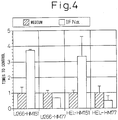
- interferon ⁇ and interferon ⁇ could have an effect of increasing the expressed amount of HM1.24 antigen in myeloma cells or cells of hematopoietic tumors such as lymphocytic tumors.
- interferon regulatory factor (IRF)-1 and 2 were identified as a transcription regulatory factor of the IFN- ⁇ gene.
- IRF-1 and 2 are generally known to bind to the same gene regulatory sequence, and act in an antagonistic manner in that IRF-1 acs as a transcription activation factor and IRF-2 as a transcription suppressing factor.
- the NIH3T3 cells in which IRF-2 was highly expressed has been demonstrated to exhibit enhanced cell saturation density, colony formation in the methylcellulose gel, and a tumorigenic property in nude mice, and IRF-2 has been shown to act as an oncogene.
- IRF-2 is required for the expression of histone H4 that acts in the control of cell cycle.
- IRF-2 has also been shown to increase the expression of vascular cell adhesion molecule-1 (VCAM-1) in muscle cells, and it has also been demonstrated that the acid region (182 to 218) of IRF-2 is involved in the activation of VCAM-1. Based on this, it is known that IRF-2 not only acts as a transcription regulatory factor but may act as a transcription activation factor.
- the IRF-2 protein binds to the promoter (HM1.24 promoter) of the HM1.24 antigen gene, and activates said promoter.
- lymphocytic tumors there can be mentioned various chemotherapies, X-ray therapy, bone marrow transplantation and the like, but no therapeutic methods are satisfactory, and thus epoch-making therapeutic drugs or methods that attains complete remission of lymphocytic tumors and prolong the patient's survival are awaited.
- therapeutic methods that are effective for leukemia other than lymphocytic leukemia i.e. myelocytic leukemia such as acute myelocytic leukemia and chronic lymphocytic leukemia are awaited.
- Therapeutic drugs and methods that attain complete remission of these myelomas, lymphocytic tumors, and myelocytic leukemia and that can prolong the patient's survival could provide therapeutic drugs and methods for hematopoietic tumors in general.
- the above should provide a means of treating certain hematopoietic tumors with anti-HM1.24 antibody by enhancing the amount expressed of HM1.24 antigen or by using a means of newly inducing HM1.24 antigen.
- WO0113940 discloses the use of interferon and IRF-2 as an expression enhancer for HM1.24 in myeloma.
- WO9918997 discloses biological response modifiers (e.g. interferon) in combination with an antibody that specifically binds to protein expressed in lymphocytic tumors for treating the same.
- biological response modifiers e.g. interferon
- the present invention relates to the use of interferon-alpha or interferon-gamma or interferon regulatory factor 2 (IRF-2) in the manufacture of a medicament that enhances or induces the expression of HM1.24 antigen having the amino acid sequence set forth in SEQ. ID NO:2 for the treatment of myelocytic leukemia, wherein the medicament is used in combination with an antibody that specifically binds to an protein having the amino acid sequence set forth in SEQ. ID NO:2 and has cytotoxic activity.
- IRF-2 interferon-alpha or interferon-gamma or interferon regulatory factor 2
- the present invention relates to interferon-alpha or interferon-gamma or interferon regulatory factor 2 (IRF-2) that enhances or induces the expression of HM1.24 antigen having the amino acid sequence set forth in SEQ. ID NO:2 for use in the treatment of myelocytic leukemia, wherein interferon-alpha or interferon-gamma or interferon regulatory factor 2 (IRF-2) is used in combination with an antibody that specifically binds to a protein having the amino acid sequence set forth in SEQ. ID NO: 2 and has cytotoxic activity.
- IRF-2 interferon-alpha or interferon-gamma or interferon regulatory factor 2
- HM1.24 antigen having the amino acid sequence set forth in SEQ ID NO: 2 in hematopoietic tumor cells, said agent comprising interferon ⁇ , interferon ⁇ , or the IRF-2 protein as an active ingredient.
- the above hematopoietic tumors are, for example, leukemia, lymphoma, myeloma etc.
- leukemia includes, for example, acute myelocytic leukemia, chronic myelocytic leukemia, acute lymphocytic leukemia, chronic lymphocytic leukemia etc.
- lymphoma includes, for example, Hodgkin's disease, T lymphocytic non-Hodgkin lymphoma, B lymphocytic non-Hodgkin lymphoma etc.
- the above myeloma includes multiple myeloma.
- the present inventors have also found that, by allowing an antigen that specifically binds to said HM1.24 antigen to bind to the hematopoietic tumor cells in which HM1.24 antigen has been expressed by the above expression-enhancing or expression-inducing agent of HM1.24 antigen, anti-tumor effect on said hematopoietic tumor cells can be enhanced.
- a therapeutic agent or a pharmaceutical composition for the treatment of hematopoietic tumors comprising, as an active ingredient, an antibody that specifically binds to a protein having the amino acid sequence set forth in SEQ ID NO: 2, wherein interferon ⁇ , interferon ⁇ , or the IRF-2 protein is used in combination.
- a therapeutic agent or a pharmaceutical composition for the treatment of hematopoietic tumors comprising, as an active ingredient, (1) interferon ⁇ , interferon ⁇ , or the IRF-2 protein, and (2) an antibody that specifically binds to a protein having the amino acid sequence set forth in SEQ ID NO: 2.
- a therapeutic agent or a pharmaceutical composition for the treatment of hematopoietic tumors comprising, as an active ingredient, interferon ⁇ , interferon ⁇ , or the IRF-2 protein, wherein an antibody that specifically binds to a protein having the amino acid sequence set forth in SEQ ID NO: 2 is used in combination.
- the above hematopoietic tumors are, for example, leukemia, lymphoma, or myeloma.
- leukemia includes, for example, acute myelocytic leukemia, chronic myelocytic leukemia, acute lymphocytic leukemia, chronic lymphocytic leukemia etc.
- lymphoma includes, for example, Hodgkin's disease, T lymphocytic non-Hodgkin lymphoma, B lymphocytic non-Hodgkin lymphoma etc.
- the above myeloma includes multiple myeloma.
- the above antibody is preferably an antibody having cytotoxicity, and the cytotoxicity is the ADCC activity.
- the antibody is preferably a monoclonal antibody, a chimeric antibody, a humanized antibody, or a human antibody.
- the monoclonal antibody is preferably anti-HM1.24 antibody produced by a hybridoma having the Deposit No. FERM BP-5233, and said chimeric antibody or humanized antibody is preferably a chimeric antibody or a humanized antibody of anti-HM1.24 antibody produced by a hybridoma having the Deposit No. FERM BP-5233.
- kit for the treatment of a patient having a hematopoietic tumor comprising:
- kits for the treatment of a patient having a hematopoietic tumor comprising:
- kits for the treatment of a patient having a hematopoietic tumor comprising:
- Also described herein is a method of screening an expression-enhancing agent of HM1.24 antigen, comprising the steps of:
- compositions for the treatment of hematopoietic tumors wherein said composition comprises the above expression-enhancing agent and said composition is used in combination with an antibody that specifically binds to a protein having the amino acid sequence set forth in SEQ ID NO: 2.
- Also described herein is a method of screening an expression-enhancing agent comprising the steps of:
- compositions for the treatment of hematopoietic tumors wherein said composition comprises the above expression-enhancing agent and said composition is used in combination with an antibody that specifically binds to a protein having the amino acid sequence set forth in SEQ ID NO: 2.
- Interferon was discovered as a substance having an activity of inhibiting viral growth and currently four types, ⁇ , ⁇ , ⁇ , and ⁇ , are known in mammals. In addition to the activity of inhibiting viral growth, they are known to exhibit an activity of inhibiting cell growth and of modulating immunological functions ( Interferon "Cytokine", Toshiaki Osawa etd., (1990) 115-133, Tokyo Kagaku Dojin Co., Ltd. ; Pestka, S., et al., Ann. Rev. Biochem. (1987) 56, 727-777 ; Langer, J. A., et al., Immunology Today (1988) 9, 393-400 ).
- Interferon- ⁇ and interferon- ⁇ may be mutants as long as they have an activity of increasing the amount expressed of HM1.24 antigen.
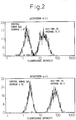
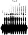
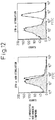
- myeloma cells are harvested from a myeloma cell line or a patient with myeloma and then subjected to flow cytometry for detection.
- mutants they may be interferon- ⁇ and interferon- ⁇ in which one or several, or a plurality of amino acid residues have been deleted, substituted, or substituted or inserted.
- substitution is preferred between amino acids whose properties are conserved.
- substitution is preferred between hydrophobic amino acids (A, I, L, M, F, P, W, Y, V), hydrophilic amino acids (R, D, N, C, E, Q, G, H, K, S, T), amino acids having aliphatic side chains (G, A, V, L, I, P), amino acids having hydroxyl group-containing side chains (S, T, Y), amino acids having sulfur-containing side chains (C, M), amino acids having carboxylic acid- and amide-containing side chains (D, N, E, Q), amino acids having base-containing side chains (R, K, H), and amino acids having aromatic group-containing side chains (H, F, Y, W).
- hydrophobic amino acids A, I, L, M, F, P, W, Y, V
- hydrophilic amino acids R, D, N, C, E, Q, G, H, K, S, T
- amino acids having aliphatic side chains G, A, V
- peptide fragments of interferon- ⁇ or interferon- ⁇ may be used.
- peptide fragments that have binding sites with interferon- ⁇ or interferon- ⁇ receptors are preferred.
- they are peptides comprised of 100 or more, more preferably 130 or more, still more preferably 150, and most preferably 160 or more contiguous amino acid residues.
- Interferon regulatory factors (IRF)-1 and -2 were identified as transcription regulatory factors of the IFN- ⁇ gene ( Taniguchi, T. et al., Nucleic Acids Res. (1989) 17, 8372 ; Taniguchi, T. et al., Cell (1989) 58, 729 ).
- IRF-1 and -2 are generally known to bind to the same gene regulatory sequence: IRF-1 and IRF-2 act in an antagonistic manner in that IRF-1 acs as a transcription activation factor, whereas IRF-2 as a transcription suppressing factor.
- the NIH3T3 cells in which IRF-2 is highly expressed has been demonstrated to exhibit enhanced cell saturation density, colony formation in the methylcellulose gel, and a tumorigenic property in nude mice, and IRF-2 acts as an oncogene.
- IRF-2 is required for the expression of histone H4 that acts for the control of cell cycle.
- IRF-2 is also shown to increase the expression of vascular cell adhesion molecule-1 (VCAM-1) in muscle cells, and it is becoming increasingly clear that the acid region (182 to 218) of IRF-2 is involved in the activation of VCAM-1. Based on this, it is known that IRF-2 not only acts as a transcription regulatory factor but as a transcription activation factor.
- the hybridoma that produces the antibody for use in the present invention can be basically constructed using a known technology as described below.
- the HM1.24 antigen protein or a HM1.24 antigen-expressing cell may be used as a sensitizing antigen and is immunized in the conventional method of immunization.
- the immune cells thus obtained are fused with known parent cells in the conventional cell fusion process, and then monoclonal antibody-producing cells are screened by the conventional screening method to construct the desired hybridoma.
- monoclonal antibody may be obtained in the following manner.
- a human multiple myeloma cell line KPMM2 Japanese Unexamined Patent Publication (Kokai) No. 7-236475
- KPC-32 Goto, T. et al., Jpn. J. Clin. Hematol. (1991) 32, 1400
- the sensitizing antigen it is also possible to use a protein having the amino acid sequence set forth in SEQ ID NO: 1 or a peptide or a polypeptide containing an epitope recognized by anti-HM1.24 antibody.
- the cDNA of the protein having the amino acid sequence set forth in SEQ ID NO 1 used as the sensitizing antigen may be inserted into the XbaI cleavage site of the pUC19 vector to prepare a plasmid pRS38-pUC19.
- coli having the plasmid pRS38-pUC19 has been internationally deposited under the provisions of the Budapest Treaty as Escherichia coli DH5 ⁇ (pRS38-pUC19) on October 5, 1993 with the Patent Microorganism Depository, the National Institute of Bioscience and Human Technology (Chuo Dai 6, 1-1, Higashi 1-chome, Tsukuba city, Ibaraki Pref., Japan) as FERM BP-4434 (Japanese Unexamined Patent Publication (Kokai) No. 7-196694 ).
- a peptide or a polypeptide that contains an epitope recognized by anti-HM1.24 antibody can be constructed by gene engineering technology.
- Mammals to be immunized with the sensitizing antigen are not specifically limited, and they are preferably selected in consideration of their compatibility with the parent cell for use in cell fusion. They generally include rodents such as mice, rats, and hamsters.
- Immunization of animals with a sensitizing antigen may be carried out using a known method.
- a general method for example, involves the intraperitoneal or subcutaneous administration of a sensitizing antigen to the mammal.
- a sensitizing antigen which has been diluted and suspended in an appropriate amount of phosphate buffered saline (PBS) or physiological saline etc. is mixed, as desired, with an appropriate amount of a common adjuvant, for example Freund's complete adjuvant. After being emulsified, it is preferably administered to the mammal for several times every 4 to 21 days. Alternatively, a suitable carrier may be used at the time of immunization of the sensitizing antigen.
- PBS phosphate buffered saline
- physiological saline etc. a common adjuvant
- a common adjuvant for example Freund's complete adjuvant.
- a suitable carrier may be used at the time of immunization of the sensitizing antigen.
- immune cells are harvested from the mammal and are subjected to cell fusion.
- immune cells to be subjected to cell fusion there may be specifically mentioned spleen cells.
- the mammalian myeloma cells as the other parent cells which are subjected to cell fusion with the above-mentioned immune cells preferably include various known cell lines such as P3X63Ag8.653 ( J. Immunol. (1979) 123: 1548-1550 ), P3X63Ag8U.1 ( Current Topics in Microbiology and Immunology (1978) 81: 1-7 ), NS-1 ( Kohler, G. and Milstein, C., Eur. J. Immunol. (1976) 6: 511-519 ), MPC-11 ( Margulies, D.H. et al., Cell (1976) 8: 405-415 ), SP2/0 ( Shulman, M.
- P3X63Ag8.653 J. Immunol. (1979) 123: 1548-1550
- P3X63Ag8U.1 Current Topics in Microbiology and Immunology (1978) 81: 1-7
- NS-1 Kohler, G. and Milstein, C
- Cell fusion between the above immune cells and the myeloma cells may be essentially conducted in accordance with a known method such as is described in Milstein et al. ( Kohler, G. and Milstein, C., Methods Enzymol. (1981) 73: 3-46 ) and the like.
- the above cell fusion is carried out in the conventional nutrient broth in the presence of, for example, a cell fusion accelerator.
- a cell fusion accelerator for example, polyethylene glycol (PEG), Sendai virus (HVJ) and the like may be used, and, in addition, an adjuvant such as dimethyl sulfoxide etc. may be added as desired to enhance the efficiency of the fusion.
- the preferred ratio of the immune cells and the myeloma cells to be used is, for example, 1 to 10 times more immune cells than the myeloma cells.
- culture media to be used for the above cell fusion include RPMI1640 medium and MEM culture medium suitable for the growth of the above myeloma cell lines, and the conventional culture medium used for this type of cell culture, and besides a serum supplement such as fetal calf serum (FCS) may be added.
- FCS fetal calf serum
- predetermined amounts of the above immune cells and the myeloma cells are mixed well in the above culture liquid, to which a PEG solution previously heated to about 37 °C, for example a PEG solution with a mean molecular weight of about 1000 to 6000, is added at a concentration of 30 to 60% (w/v), and mixed to obtain the desired fusion cells (hybridomas). Then by repeating the sequential addition of a suitable culture liquid and centrifugation to remove the supernatant, cell fusion agents etc. which are undesirable for the growth of the hybridoma can be removed.
- a PEG solution previously heated to about 37 °C for example a PEG solution with a mean molecular weight of about 1000 to 6000
- Said hybridoma is selected by culturing in a conventional selection medium, for example, the HAT culture medium (a culture liquid containing hypoxanthine, aminopterin, and thymidine). Culturing in said HAT culture medium is continued generally for a period of time sufficient to effect killing of the cells (non-fusion cells) other than the desired hybridoma, generally several days to several weeks. Then, the conventional limiting dilution method is conducted in which the hybridomas that produce the desired antibody are screened and monoclonally cloned.
- a conventional selection medium for example, the HAT culture medium (a culture liquid containing hypoxanthine, aminopterin, and thymidine). Culturing in said HAT culture medium is continued generally for a period of time sufficient to effect killing of the cells (non-fusion cells) other than the desired hybridoma, generally several days to several weeks. Then, the conventional limiting dilution method is conducted in which the hybridomas that produce the desired antibody are
- a transgenic animal having a repertoire of human antibody genes is immunized with HM1.24 antigen or HM1.24 antigen-expressing cells to obtain the desired antibody according to the above-mentioned method (see International Patent Publication WO 93/12227 , WO 92/03918 , WO 94/02602 , WO 94/25585 , WO 96/34096 , and WO 96/33735 ).
- the desired human antibody may be isolated by means of panning.
- the variable region of human antibody is expressed on the surface of a phage by the phage display method as a single chain antibody (scFv) to select a phage that binds to the HM1.24 antigen using a HM1.24 antigen-immobilized plate.
- scFv single chain antibody
- the gene encoding the variable region of the human antibody that binds to the HM1.24 antigen can be identified.
- anti-HM1.24 antibody can be prepared.
- the monoclonal antibody-producing hybridomas thus constructed can be subcultured in the conventional culture liquid, or can be stored for a prolonged period of time in liquid nitrogen.
- the hybridoma is cultured by the conventional method and the antibodies are obtained as the culture supernatant, or a method in which the hybridoma is administered to and grown in a mammal compatible with said hybridoma and the antibodies are obtained as the ascites, or other methods.
- the former method is suitable for obtaining high-purity antibodies, whereas the latter is suitable for a large scale production of antibodies.
- the anti-HM1.24 antibody-producing hybridoma can be constructed using the method of Goto, T. et al. (Blood (1994) 84: 1922-1930 ). It can be conducted by: a method in which the anti-HM1.24 antibody-producing hybridoma that was internationally deposited under the provisions of the Budapest Treaty as FERM BP-5233 on April 27, 1995 with the Patent Microorganism Depository, the National Institute of Bioscience and Human Technology (Chuo Dai 6, 1-1, Higashi 1-chome, Tsukuba city, Ibaraki Pref., Japan) is intraperitoneally injected to BALB/c mice (manufactured by CLEA Japan) to obtain the ascites, from which the anti-HM1.24 antibody is purified, or: a method in which said hybridoma is cultured in a suitable culture medium such as the RPMI1640 medium containing 10% bovine fetal serum and 5% BM-Condimed H1 (manufactured by Boehr
- a recombinant antibody which was produced by the recombinant gene technology, in which an antibody gene was cloned from the hybridoma and integrated into a suitable vector which was then introduced into a host, can be used in the present invention as monoclonal antibody (see, for example, Carl, A. K., Borrebaeck, and James, W. Larrick, THERAPEUTIC MONOCLONAL ANTIBODIES, published in the United Kingdom by MACMILLAN PUBLISHERS LTD. 1990 ).
- mRNA encoding the variable region (V) of the desired antibody is isolated from the hybridoma producing the antibody.
- the isolation of mRNA is conducted by preparing total RNA using, for example, a known method such as the guanidine ultracentrifuge method ( Chirgwin, J.M. et al., Biochemistry (1979) 18, 5294-5299 ), the AGPC method (Chmczynski, P. et al., (1987) 162, 156-159), and then mRNA is purified from the total RNA using the mRNA Purification kit (manufactured by Pharmacia) and the like.
- mRNA can be directly prepared using the QuickPrep mRNA Purification Kit (manufactured by Pharmacia).
- cDNA of the V region of the antibody may be synthesized using a reverse transcriptase from the mRNA thus obtained.
- cDNA may be synthesized using the AMV Reverse Transcriptase First-strand cDNA Synthesis Kit and the like.
- the 5'-Ampli FINDER RACE Kit manufactured by Clontech
- the 5'-RACE method Frohman, M.A. et al., Proc. Natl. Acad. Sci. U.S.A. (1988) 85, 8998-9002 ; Belyavsky, A. et al., Nucleic Acids Res.
- the desired DNA fragment is purified from the PCR product obtained and may be ligated to vector DNA. Moreover, a recombinant vector is constructed therefrom and then is introduced into E. coli etc., from which colonies are selected to prepare the desired recombinant vector.
- the base sequence of the desired DNA may be confirmed by a known method such as the dideoxy method.
- the DNA encoding the V region of the desired antibody may be ligated to DNA encoding the constant region (C region) of the desired antibody, which is then integrated into an expression vector.
- the DNA encoding the V region of the antibody may be integrated into an expression vector which already contains DNA encoding the C region of the antibody.
- the antibody gene is integrated as described below into an expression vector so as to be expressed under the control of the expression regulatory region, for example an enhancer and/or a promoter. Subsequently, the expression vector may be transformed into a host cell and the antibody can then be expressed therein.
- the expression regulatory region for example an enhancer and/or a promoter.
- artificially altered recombinant antibody such as chimeric antibody and humanized antibody can be used for the purpose of lowering heterologous antigenicity against humans.
- altered antibody can be produced using known methods.
- Chimeric antibody can be obtained by ligating the thus obtained DNA encoding the V region of antibody to DNA encoding the C region of human antibody, which is then integrated into an expression vector and introduced into a host for production of the antibody therein (see European Patent Application EP 125023 , and International Patent Publication WO 96/02576 ). Using this known method, chimeric antibody useful for the present invention can be obtained.
- E. coli having the plasmid that contains the L chain and the H chain of chimeric anti-HM1.24 antibody has been internationally deposited under the provisions of the Budapest Treaty as Escherichia coli DH5 ⁇ (pUC19-1.24L-g ⁇ ) and Escherichia coli DH5 ⁇ (pUC19-1.24H-g ⁇ 1), respectively, on August 29, 1996 with the Patent Microorganism Depository, the National Institute of Bioscience and Human Technology (Chuo Dai 6, 1-1, Higashi 1-chome, Tsukuba city, Ibaraki Pref., Japan) as FERM BP-5646 and FERM BP-5644, respectively (see Japanese Patent Application No. 8-264756 ).
- Humanized antibody which is also referred to as reshaped human antibody has been made by transplanting the complementarity determining region (CDR) of antibody of a mammal other than the human, for example mouse antibody, into the CDR of human antibody.
- CDR complementarity determining region
- the general recombinant DNA technology for preparation of such antibodies is also known (see European Patent Application EP 125023 and International Patent Publication WO 96/02576 ).
- a DNA sequence which was designed to ligate the CDR of mouse antibody with the framework region (FR) of human antibody is synthesized by the PCR method from several divided oligonucleotides having sections overlapping with one another at the ends thereof.
- the DNA thus obtained is ligated to the DNA encoding the C region of human antibody, and then is integrated into an expression vector, which is introduced into a host for antibody production (see European Patent Application EP 239400 and International Patent Publication WO 96/02576 ).
- the FR is selected for which the complementarity determining region forms a favorable antigen binding site.
- amino acids in the framework region of the antibody variable region may be substituted so that the complementarity determining region of reshaped human antibody may form an appropriate antigen binding site ( Sato, K. et al., Cancer Res. (1993) 53, 851-856 ).
- E. coli having the plasmid that contains the L chain and the H chain of humanized anti-HM1.24 antibody has been internationally deposited under the provisions of the Budapest Treaty as Escherichia coli DH5 ⁇ (pUC19-RVLa-AHM-g ⁇ ) and Escherichia coli DH5 ⁇ (pUC19-RVHr-AHM-g ⁇ 1), respectively, on August 29, 1996 with the Patent Microorganism Depository, the National Institute of Bioscience and Human Technology (Chuo Dai 6, 1-1, Higashi 1-chome, Tsukuba city, Ibaraki Pref., Japan) as FERM BP-5645 and FERM BP-5643, respectively (Patent Application No. 8-264756 ).
- the C region of human antibody is used and, as the C region of human antibody that exhibits cytotoxicity, human C ⁇ , for example C ⁇ 1, C ⁇ 2, C ⁇ 3, and C ⁇ 4, can be used.
- human C ⁇ for example C ⁇ 1, C ⁇ 2, C ⁇ 3, and C ⁇ 4
- antibody having C ⁇ 1 and C ⁇ 3 in particular has potent cytotoxicity, i.e. ADCC activity and CDC activity, and is used preferably in the present invention.
- Chimeric antibody consists of the variable region of antibody derived from a mammal other than the human and the C region derived from human antibody
- humanized antibody consists of the complementarity determining region of antibody derived from a mammal other than the human and the framework region (FR) and the C region of antibody derived from human antibody. Accordingly, antigenicity thereof in the human body has been reduced so that they are useful as the active ingredient of the therapeutic agents of the present invention.
- a preferred embodiment of the humanized antibody for use in the present invention includes humanized anti-HM1.24 antibody (see WO 98/14580 ).
- Antibody genes constructed as described above may be expressed and obtained in a known method. In the case of mammalian cells, expression may be accomplished using an expression vector containing a commonly used useful promoter, the antibody gene to be expressed, and DNA in which the poly A signal has been operably linked at 3' downstream thereof or a vector containing said DNA.
- a commonly used useful promoter the antibody gene to be expressed
- the promoter/enhancer include human cytomegalovirus immediate early promoter/enhancer.
- promoter/enhancer which can be used for the expression of antibody for use in the present invention
- viral promoters/enhancers such as retrovirus, polyoma virus, adenovirus, and simian virus 40 (SV40), and promoters/enhancers derived from mammalian cells such as human elongation factor 1 ⁇ (HEF1 ⁇ ).
- expression may be readily accomplished by the method of Mulligan et al. (Nature (1979) 277, 108 ) when SV40 promoter/enhancer is used, or by the method of Mizushima et al. (Nucleic Acids Res. (1990) 18, 5322 ) when HEF1 ⁇ promoter/enhancer is used.
- expression may be conducted by operably linking a commonly used useful promoter, a signal sequence for antibody secretion, and the antibody gene to be expressed, followed by expression thereof.
- a commonly used useful promoter for example, there can be mentioned lacZ promoter and araB promoter.
- lacZ promoter for example, lacZ promoter and araB promoter.
- the method of Ward et al. (Nature (1098) 341, 544-546 ; FASEB J. (1992) 6, 2422-2427 ) may be used when lacZ promoter is used, and the method of Better et al. (Science (1988) 240, 1041-1043 ) may be used when araB promoter is used.
- the pelB signal sequence Lei, S.P. et al., J. Bacteriol. (1987) 169, 4379 ) can be used. After separating the antibody produced in the periplasm, the structure of the antibody is appropriately refolded before use (see, for example, WO 96/30394 ).
- expression vectors can include as selectable markers the aminoglycoside transferase (APH) gene, the thymidine kinase (TK) gene, E. coli xanthine guaninephosphoribosyl transferase (Ecogpt) gene, the dihydrofolate reductase (dhfr) gene and the like.
- APH aminoglycoside transferase
- TK thymidine kinase
- Ecogpt E. coli xanthine guaninephosphoribosyl transferase
- dhfr dihydrofolate reductase
- the production system of antibody preparation comprises the in vitro and the in vivo production system.
- the in vitro production system there can be mentioned a production system which employs eukaryotic cells and the production system which employs prokaryotic cells.
- animal cells include (1) mammalian cells such as CHO cells, COS cells, myeloma cells, baby hamster kidney (BHK) cells, HeLa cells, and Vero cells, (2) amphibian cells such as Xenopus oocytes, or (3) insect cells such as sf9, sf21, and Tn5.
- Known plant cells include, for example, those derived from the genus Nicotiana, more specifically cells derived from Nicotiana tabacum, which is subjected to callus culture.
- Known fungal cells include yeasts such as the genus Saccharomyces, more specifically Saccharomyces cereviceae, or filamentous fungi such as the genus Aspergillus, more specifically Aspergillus niger.
- bacterial cells When the prokaryotic cells are used, there are the production systems which employ bacterial cells.
- Known bacterial cells include Escherichia coli (E. coli), and Bacillus subtilis.
- the antibody By introducing, via transformation, the gene of the desired antibody into these cells and culturing the transformed cells in vitro, the antibody can be obtained. Culturing is conducted in the known methods. For example, as the culture liquid, DMEM, MEM, RPMI1640, and IMDM can be used, and serum supplements such as fetal calf serum (FCS) may be used in combination.
- FCS fetal calf serum
- antibodies may be produced in vivo by implanting cells into which the antibody gene has been introduced into the abdominal cavity of an animal and the like.
- tabacco for example, can be used.
- Antibody genes are introduced into these animals or plants, and the antibodies are produced in such animals or plants, and recovered therefrom.
- an antibody gene is inserted into the middle of the gene encoding protein which is inherently produced in the milk such as goat ⁇ casein to prepare fusion genes.
- DNA fragments containing the fusion gene into which the antibody gene has been inserted are injected into a goat embryo, and the embryo is introduced into a female goat.
- the desired antibody is obtained from the milk produced by the transgenic goat born to the goat who received the embryo or offsprings thereof.
- hormones may be given to the transgenic goat as appropriate. ( Ebert, K.M. et al., Bio/Technology (1994) 12, 699-702 ).
- baculovirus into which the desired antibody gene has been inserted is infected to the silkworm, and the desired antibody can be obtained from the body fluid of the silkworm ( Susumu, M. et al., Nature (1985) 315, 592-594 ).
- the desired antibody gene is inserted into an expression vector for plants, for example pMON 530, and then the vector is introduced into a bacterium such as Agrobacterium tumefaciens. The bacterium is then infected to tabacco such as Nicotiana tabacum to obtain the desired antibody from the leaves of the tabacco ( Julian, K.-C. Ma et al., Eur. J. Immunol. (1994) 24, 131-138 ).
- DNA encoding the heavy chain (H chain) or the light chain (L chain) of antibody may be separately integrated into an expression vector and the hosts are transformed simultaneously, or DNA encoding the H chain and the L chain may be integrated into a single expression vector and the host is transformed therewith (see International Patent Publication WO 94-11523 ).
- the antibody produced as described above can be bound to various molecules such as polyethylene glycol (PEG) for use as a modified antibody.
- PEG polyethylene glycol
- Antibody as used herein includes these modified antibodies. In order to obtain these modified antibody, the antibody obtained may be chemically modified. These methods have already been established in the field of the art.
- Antibodies produced and expressed as described above can be separated from the inside or outside of the cell or from the host and then may be purified to homogeneity. Separation and purification of the antibody for use in the present invention may be accomplished by affinity chromatography.
- affinity chromatography As the column used for such affinity chromatography, there can be mentioned Protein A column and Protein G column. Examples of the column employing Protein A column are Hyper D, POROS, Sepharose F.F. and the like.
- Separation and purification of the antibody for use in the present invention may be accomplished by combining, as appropriate, chromatography other than the above-mentioned affinity chromatography, filtration, ultrafiltration, salting-out, dialysis and the like. Chromatography includes, for example, ion exchange chromatography, hydrophobic chromatography, gel-filtration and the like.
- the concentration of antibody obtained in the above method can be determined by the measurement of absorbance or by ELISA and the like.
- the antibody for use in the present invention or a sample containing the antibody is appropriately diluted with PBS(-) and then the absorbance is measured at 280 nm, followed by calculation using the absorption coefficient of 1.35 OD at 1 mg/ml.
- the ELISA method measurement is conducted as follows.
- Reactivity of the antibody of the present invention with lymphocytes may be examined by flow cytometry (FCM) analysis.
- FCM flow cytometry
- established cell lines or freshly isolated cells can be used.
- established cell lines there may be used myeloma-derived RPMI8226 (ATCC CCL 155), myeloma-derived U266 (ATCC TIB 196), myeloma-derived KPMM2, myeloma-derived KPC-32, and plasmacytoma-derived ARH-77 (ATCC CRL 1621), and the like.
- the cells that have not been stimulated and that are not expressing HM1.24 antigen or the cells that at least are expressing the antigen are determined using FCM analysis.
- the cells described in Examples and a test substance are incubated for 1-2 days, and then stained with mouse anti-human HM1.24 antibody as a primary antibody. The cells are washed and further stained with FITC-labeled anti-mouse IgG antibody as a secondary antibody. Finally, after washing the cells, the fluorescence intensity of FITC is measured by a flow cytometer.
- FCM analysis by direct staining may be used in which the cells are treated with a high concentration of immunoglobulin, and then stained, after blocking Fc receptors, with FITC-labeled anti-human HM1.24 antibody.
- HM1.24 antigen it is also possible to screen expression enhancers of HM1.24 antigen by the reporter gene assay using the HM1.24 promoter sequence.
- reporter gene luciferase can be used.
- a plasmid is constructed that contains the HM1.24 promoter sequence upstream of the reporter gene, after which it is transformed into the cells, and the cells obtained are cultured with a test substance for 1-2 days, and the cells recovered are subjected to FCM analysis to screen drugs that enhance the expression of HM1.24 antigen.
- the antibody for use in the present invention is one which has, for example, an ADCC activity as the cytotoxicity.
- the ADCC activity on hematopoietic tumor cells can be measured in the following manner.
- mononuclear cells (E) are isolated as the effector cells from human peripheral blood or bone marrow by the gravity centrifuge method.
- target cells T
- RPMI8226 ATCC CCL 155
- U266 ATCC TIB 196
- KPMM2 KPC-32
- ARH-77 ATCC CRL 1621
- HEL HEL
- the cytotoxicity (%) can be calculated as (A-C)/(B-C) X 100, in which A is radioactivity (cpm) liberated in the presence of the antibody, B is radioactivity (cpm) liberated by NP-40, and C is radioactivity (cpm) liberated by the medium alone containing no antibody.
- C ⁇ in particular C ⁇ 1 and C ⁇ 3 as the constant region (C region) of antibody in humans.
- a more potent ADCC activity or CDC activity can be induced by adding, altering, or modifying part of the amino acids in the C region of antibody.
- oligomer site-specific mutagenesis using a primer, the addition of a base sequence using a restriction enzyme cleavage site, and the use of a chemical modifier that creates a covalent bond.
- Described herein is a method of treating hematopoietic tumors by administering to the patient a pharmaceutical agent that enhances the amount expressed of HM1.24 antigen, and a pharmaceutical agent that induces the expression of HM1.24 antigen in the cell that normally is not expressing HM1.24 antigen on the cell surface and anti-HM1.24 antibody.
- the hematopoietic tumors are, for example, myeloma, preferably multiple myeloma, lymphocytic tumors such as lymphoma, preferably Hodgkin's disease or non-Hodgkin lymphoma, lymphocytic leukemia, preferably acute T lymphocytic leukemia, chronic T lymphocytic leukemia, acute B lymphocytic leukemia, chronic B lymphocytic leukemia, and myelocytic leukemia for example acute myelocytic leukemia and chronic myelocytic leukemia.
- acute leukemia belonging to L1-L3 and M0-M7 in the FAB Classification is also included.
- a pharmaceutical agent that enhances the amount expressed of HM1.24 antigen, and a pharmaceutical agent that induces the expression of HM1.24 antigen in the cell that normally is not expressing HM1.24 antigen on the cell surface and anti-HM1.24 antibody is preferably interferon- ⁇ , interferon- ⁇ , the IRF-2 protein, or a vector containing DNA encoding the IRF-2 protein, and preferably interferon- ⁇ and interferon- ⁇ .
- the dosage of interferon- ⁇ and interferon- ⁇ is, when administered to humans, is an amount that attains the highest blood level of 1-10000 I.U./ml (International Units), more preferably 5-1000 I.U./ml, even more preferably 5-500 I.U./ml, and most preferably 5-50 I.U./ml.
- Interferon and anti-HM1.24 antibody may be administered together or separately. In the latter case, preferably interferon is given first, followed by the administration of anti-HM1.24 antibody within 96 hours.
- the interval between the interferon administration and the anti-HM1.24 antibody administration is not limited as long as the amount expressed of HM1.24 antigen is being enhanced by the administration of interferon, but it is preferably within 96 hours, more preferably 72 hours, still more preferably 48 hours.
- Interferon and anti-HM1.24 antibody for a plurality of times depending on the clinical response of the patient is within the scope of the present invention.
- the route of administration is preferably given directly into the blood circulation, and intravenous administration or intraarterial administration is preferred. Continued administration is possible and intravenous drip may be used.
- the administration may also be subcutaneous or intramuscular administration.
- a therapeutic agent or a pharmaceutical composition for the treatment of hematopoietic tumors comprising interferon- ⁇ or interferon- ⁇ and anti-HM1.24 antibody.
- the therapeutic agent may contain a pharmaceutically acceptable vehicle that has been used for interferon and antibody preparations, such as physiological saline or 5% dextran, together with a common stabilizer or a excipient.
- kits for treating a patient with hematopoietic tumors comprising a pharmaceutical composition comprising anti-HM1.24 antibody as an active ingredient and an instruction manual that contains description of combined therapy with interferon- ⁇ or interferon- ⁇ .
- compositions comprising anti-HM1.24 antibody as an active ingredient for treating a patient with hematopoietic tumors, wherein said composition is used in combination with interferon- ⁇ or interferon- ⁇ .
- a gene encoding a substance that enhances or induces the expression of HM1.24 antigen is also described herein.
- the DNA sequences of interferon- ⁇ , interferon- ⁇ , IRF-2 are known and can be administered to patients by integration into the desired vector.
- they are inserted into adenovirus vector (for example pAdexLew) or retrovirus vector (for example pZIPneo), which are then administered to the living body.
- the method of administration may be ex vivo or in vivo.
- naked DNA may be administered.
- Examples 1-5 are to be regarded as comparative examples.
- Example 1 Enhancement of the amount of HM1.24 antige expressed in myeloma cells by interferon- ⁇
- a human myeloma cell line U266 (ATCC TIB 196) and myeloma cells derived from the bone marrow of a patient with multiple myeloma were cultured in a RPMI1640 medium (Sigma, St Louis, MO, USA) containing 10% fetal bovine serum (Whittaker Bioproducts, Inc., Walkersville, MD, USA) in a 5% carbon dioxide incubator at 37°C.
- the hybridoma that produces mouse anti-HM1.24 antibody has been internationally deposited as FERM BP-5233 (deposition date: April 27, 1995) with the National Institute of Bioscience and Human Technology, of 1-3, Higashi 1-chome, Tsukuba city, Ibaraki Pref.
- Myeloma cells (1 X 10 5 /ml) were cultured in the presence or absence of 1000 U/ml of the natural type interferon- ⁇ (Otsuka Pharmaceutical, Tokyo) for 48 hours, and changes in HM1.24 antigen (the base sequence encoding this is shown in SEQ ID NO: 1) were determined by flow cytometry.
- FITC-human IgG1 (1 mg/ml) or FITC-anti-HM1.24 antibody (1 mg/ml) was added to stain at 4 °C for 60 minutes.
- 20 ⁇ l of PE-anti-CD38 (Becton Dickinson, San Jose, CA, USA) was added for double staining to identify the myeloma cells.
- the cells were washed twice with PBC, and were stored in PBS containing 1% paraformaldehyde (Wako Pure Chemical Industries, Ltd., Osaka). Subsequently, the expression of HM1.24 antigen was analyzed using a flow cytometer (EPICS XL, Coulter, Hialeah, FL, USA).
- a myeloma cell line U266 ( Figure 1 ) and the patient's myeloma cells ( Figure 2 ) were expressing HM1.24 antigen at a condition of no stimulation, and the stimulation with interferon- ⁇ further increased the amount expressed of HM1.24 antigen.
- Interferon- ⁇ further enhanced the expression of HM1.24 antigen in the myeloma cell, and increased the number of anti-HM1.24 antibodies that bind to the myeloma cell. Since the therapeutic anti-tumor effect by anti-HM1.24 antibody is proportional to the number of antibodies that bound, treatment with anti-HM1.24 antibody after the administration of interferon- ⁇ is expected to provide a therapy that enhances the therapeutic effect by antibody and further enhances effectiveness.
- the reporter gene at the promoter region was analyzed.
- the gene (SEQ ID NO: 3) of the HM1.24 promoter region was obtained by PCR cloning.
- Genomic DNA was prepared from human peripheral blood mononuclear cells using the DNAzol reagent (GIBCO). With the genomic DNA obtained as the template, using primer HM2k (aaaggtaccagctgtctttctgtctgtctgtcc) (SEQ ID NO: 4) and BST2B (atagtcatacgaagtagatgccatccag) (SEQ ID NO: 5), PCR (94°C for 1 min, 55°C for 1 min, 72°C for 1 min, 30 cycles) was performed using TaKaRa Taq (Takara Shuzo, Ohtsu) in the Thermal Cycler 480 (Perkin-Elmer, CA, USA).
- HM-2k/GL3 The plasmid HM-2k/GL3 obtained was treated with restriction enzymes KpnI and XhoI, from which a deletion clone was constructed using the deletion kit (Takara Shuzo) for kilo-sequence to obtain a plasmid HM-493/GL3 containing from the transcription initiation point to - 493 bp upstream. Furthermore, HM-2k/GL3 was treated with restriction enzymes KpnI and Af1II, from which a deletion clone was constructed as described above, and HM-151/GL3 and HM-77/GL3 containing from the transcription initiation point to -151 bp or -77 bp upstream were obtained.
- the polyethyleneimine-Transferrinfection Kit (Tf PEI-Kit) (Bender MedSystems, Vienna, Austria) was used, and for the luciferase assay the Dual-Luciferase Reporter Assay System (Promega) was used.
- the cell line was cultured overnight in RPMI-1640 containing 50 ⁇ m Defferrioxamine and 10% FBS.
- a mixture of the reporter gene plasmid at a final concentration of 20 ⁇ g/ml, 0.4 ⁇ g/ml of pRL-SV40, and 1 ⁇ g/ml of Tf-PEI reagent was prepared and was incubated at room temperature for 20 minutes. 5 X 10 5 cells/ml of cells were added at three volumes of the Tf-PEI/plasmid mixture, and was incubated at 37°C for four hours, washed with the medium, and 100 ⁇ l per well at a concentration of 2 X 10 5 cells/ml was cultured in a 96-well flat-bottomed plate.
- IFN- ⁇ was added to a final concentration of 0, 10, 100, or 1000 U/ml, which was cultured at 37°C for two days. After the cells were washed in PBS(-), they were dissolved in 20 ⁇ l of the Passive Lysis Buffer, six ⁇ l of which was applied to the C96 White Polysorp Fluoronunc plate (Nunc). Using the Luminoskan (Labsystems), luminescence intensity was measured for Firefly and Renila at 30 ⁇ l of the substrate solution and a measurement time of 10 seconds. The measured values were corrected by Firefly/Renila, and the relative activity was determined with the control (medium) as one.
- the luciferase activity of the reporter was increased in a IFN- ⁇ concentration dependent manner for both of the upstream 2 kbp and 493 bp, confirming that the enhanced transcription activity of the promoter region causes the expression induction of the antigen ( Figure 3 ).
- the Electrophoresis Mobility Shift Assay with the HM1.24 promoter region as the probe was performed as follows to identify IRF-2 as the binding factor.
- the myeloma cells U266-B1 (ATCC-TIB196) were cultured in the RPMI-1640 medium (GIBCO-BRL) containing 10% FBS (HyClone) at 37°C in a 5% CO 2 incubator.
- IFN- ⁇ interferon- ⁇
- Pepro Tech EC interferon- ⁇
- IFN- ⁇ was added to the medium to a final concentration of 1000 U/ml, and the cells were recovered at 30 minutes, two hours, four hours, and eight hours after the addition.
- the cells were suspended into cold PBS (-), centrifuged at 1,000 rpm to discard the supernatant, and suspended in a 10 mM Tris, 10 mM NaCl, and 6 mM MgCl 2 solution.
- the cells were suspended into 10 mM Tris, 10 mM NaCl, 6 mM MgCl 2 , 1 mM DTT, 0.4 mM PMSF, 1 mM Na 3 VO 4 .
- the cells were homogenized on ice using a glass homogenizer, centrifuged at 6000 g for three minutes, and the supernatant was discarded.
- the cells were suspended into the extraction buffer (20% glycerol, 20 mM HEPES, 420 mM NaCl, 1.5 mM MgCl 2 , 0.2 mM EDTA, 0.2 mM PMSF, 1 mM DTT, 0.1 mM Na 3 VO 4 , 2 mg/ml aprotinin, and 5 mg/ml leupeptin), and then was allowed to stand in ice for 20 minutes. It was centrifuged at 12000 g for 10 minutes, and the supernatant was recovered.
- the extraction buffer (20% glycerol, 20 mM HEPES, 420 mM NaCl, 1.5 mM MgCl 2 , 0.2 mM EDTA, 0.2 mM PMSF, 1 mM DTT, 0.1 mM Na 3 VO 4 , 2 mg/ml aprotinin, and 5 mg/ml leupeptin
- ISRE2 was constructed that contains sequences (ttcccagaaa (SEQ ID NO: 10) and ggaaactgaaact (SEQ ID NO: 11)) having a homology with GAS (IFN- ⁇ activation site: the GAS consensus sequence is ttncnnnaa (SEQ ID NO: 8)) and ISRE (IFN- ⁇ stimulation response factor: the ISRE consensus sequence is ngaaanngaaact (SEQ ID NO: 9)), at the HM1.24 promoter region.
- GAS IFN- ⁇ activation site: the GAS consensus sequence is ttncnnnaa (SEQ ID NO: 8)
- ISRE IFN- ⁇ stimulation response factor: the ISRE consensus sequence is ngaaanngaaact (SEQ ID NO: 9)
- oligo DNA ISRE-F2 (aatttctgggaaactgaaactgaaacct (SEQ ID NO: 12)) and ISRE-R2 (aattaggttttcagtttcagtttcccaga (SEQ ID NO: 13)) were mixed and annealed to form a double stranded DNA probe ISRE2.
- oligo DNA adp-1 (catggcatctacttcgtatgactattgcagagtgcc (SEQ ID NO: 14)) and adp-2 (catgggcactctgcaatagtcatacgaagtagatgc (SEQ ID NO: 15)) were mixed and annealed to form an unrelated probe adp. Probes were labeled using the Band Shift Kit (Amersham Pharmacia Biotech) according to the standard protocol.
- reaction solution [(NEC-)] to which no extract was added
- a reaction solution [0h] to which an extract from the cell culture that was cultured without stimulation by interferon- ⁇ was added
- a reaction solution ⁇ 8h (+cold)] in which 50 ng of a nonlabeled ISRE2 probe was added in stead of the labeled probe to the extract of 8 hour-culture
- a reaction solution [8h (+cold unrelated)] in which 50 ng of an unrelated probe adp was added to the extract of 8 hour-culture were prepared, and were processed as described above to detect signals.
- the myeloma cells U266-B1 (ATCC-TIB196) were cultured for eight hours in the presence of 1000 U/ml of interferon- ⁇ to prepare an extract.
- the following procedure was performed according to the standard protocol of the Band Shift Kit (Amersham Pharmacia Biotech). Thus, 2 ⁇ g of antibody was added to 5 ⁇ g of the extract, and incubated at room temperature for 15 minutes to obtain an extract/antibody reaction solution.
- the reaction mixture was subjected to electrophoresis as described in the above (3) to detect signals.
- HM1.24 promoter activity by IRF-2 co-expression was determined by the reporter gene assay using the U266 cells, and it was revealed that IRF-2 actually has the transcription activation activity of the HM1.24 promoter.
- a myeloma cell line U266-B1 (ATCC TIB196) was used. The cells were cultured in the RPMI-1640 medium (GIBCO) (referred to hereinafter as the medium) containing 10% FBS (GIBCO BRL) in an 5% CO 2 incubator.
- GIBCO RPMI-1640 medium
- FBS GIBCO BRL
- the gene of the HM1.24 promoter region was obtained by PCR cloning. From human peripheral blood mononuclear cells, genomic DNA was prepared using the DNAzol reagent (GIBCO). With the genomic DNA obtained as the template, using primer HM2k (aaaggtaccagctgtctttctgtctgtctgtccc) (SEQ ID NO: 16) and BST2B (atagtcatacgaagtagatgccatccag) (SEQ ID NO: 17), PCR (94°C for 1 min, 55°C for 1 min, 72°C for 1 min, 30 cycles) was performed using TaKaRa Taq (Takara Shuzo, Ohtsu) in the Thermal Cycler 480 (Perkin-Elmer, CA, USA).
- HM-2k/GL3 The plasmid HM-2k/GL3 obtained was treated with restriction enzymes KpnI and XhoI, from which a deletion clone was constructed using the deletion kit (Takara Shuzo) for kilo-sequence to obtain a plasmid HM-491/GL3 containing up to -491 bp upstream of the transcription initiation point. Furthermore, HM-2k/GL3 was treated with restriction enzymes KpnI and Af1II, from which a deletion clone was constructed as described above, and HM-151/GL3 containing -151 bp upstream of the transcription initiation point was obtained.
- PCR (94°C for 1 min, 55°C for 1 min, 72°C for 1 min, 30 cycles) was performed using TaKaRa Taq (Takara Shuzo, Ohtsu) in the Thermal Cycler 480 (Perkin-Elmer, CA, USA).
- the fragment obtained was treated with restriction enzymes KpnI and BglII (Takara Shuzo), and was cloned into the KpnI-BglII site of a reporter gene plasmid pGL3-basic (Promega, WI, USA) using the ligation high (Toyobo) to transform competent E. coli JM109 (Nippongene).
- the transformed E. coli was cultured at 37°C in the LB medium containing 100 ⁇ g/ml ampicillin, and a plasmid was prepared using the QIAGEN plasmid maxi kit (QIAGEN, Hilden, Germany). A plasmid HM-125/GL3 was thus obtained that contains up to 125 bp upstream of the transcription initiation point.
- HM-2k/GL3 as the template, using primer HMP700 (aaaggtaccagagtttacctggtatcctgg) (SEQ ID NO: 20) and GL primer 2, PCR was performed in a similar procedure, and by introducing the fragment into the KphI-BglII site of pGL3-basic, HM-700/GL3 containing up to about 700 bp upstream of the transcription initiation point was obtained.
- HM-2k/GL3 as the template, using primer HMP700 and 11A' (cagaggattaattaggtaccgaaagagaggtgggcttt) (SEQ ID NO: 21), PCR (98°C for 15 seconds, 65°C for 2 seconds, 74°C for 30 seconds, 25 cycles) was performed using the KOD polymerase (Toyobo) in the Thermal Cycler 480 (Perkin-Elmer, CA, USA). The fragment obtained was inserted into the pCR4 Blunt-TOPO vector using the Zero Blunt TOPO PCR cloning kit for sequencing ver. B (Invitrogen).
- the plasmid obtained was treated with a restriction enzyme KpnI, and an about 550 bp fragment was recovered, which was introduced into the KpnI site of HM-125/GL3 using the ligation high.
- KpnI restriction enzyme
- dSIRE/GL3 lacking -25 to about -145 upstream of the transcription initiation point was obtained.
- the IRF-2 expression plasmid was constructed as follows. From the U266 cells for which eight hours have elapsed after stimulation with interferon- ⁇ (1000 U/ml), total RNA was extracted using the TRIzol reagent (GIBCO BRL). With RNA obtained by using the First-strand cDNA Synthesis kit (Pharmacia) as the template, using NotI-d(T) 18 , as the primer, a reverse transcription reaction was performed at 37°C for one hour.
- PCR With the PCR reaction as the template, using IRF2-F1 (agagggtaccatgccggtggaaaggatgcg) (SEQ ID NO: 24) and IRF2-R1 (agtcggtaccttaactgctcttgacgcggg) (SEQ ID NO: 25) as primers, PCR (94°C for 45 seconds, 60°C for 45 seconds, 72°C for 60 seconds, 30 cycles) was performed using the KOD polymerase (Toyobo).
- the fragment obtained was treated with a restriction enzyme KpnI, and then introduced into the KpnI site of an expression plasmid pTracer-CMV (Invitrogen) using the ligation high (Toyobo) to obtain a IRF-2 expression plasmid pIRF-2/Tracer.
- the polyethyleneimine-Transferrinfection Kit (Tf PEI-Kit) (Bender MedSystems, Vienna, Austria) was used, and for the luciferase assay the Dual-Luciferase Reporter Assay System (Promega) was used.
- the cell line was cultured overnight in RPMI-1640 containing 50 ⁇ M Defferrioxamine and 10% FBS.
- a mixture of the reporter gene plasmid at a final concentration of 20 ⁇ g/ml, 20 ⁇ g/ml of pIRF-2/Tracer or pTracer-CMV, 0.4 ⁇ g/ml of pRL-SV40, and 1 ⁇ g/ml of Tf-PEI reagent was prepared, and was incubated at room temperature for 20 minutes.
- Luminoskan Using the Luminoskan (Labsystems), luminescence intensity was measured for Firefly and Renila at 30 ⁇ l of the substrate at a measurement time of 10 seconds. The measured values were corrected by Firefly/Renila to obtain the correct relative activity.
- the HM1.24 promoter reporter plasmid and the IRF-2 expression plasmid were introduced into the U266 cells, and the reporter activity was determined ( Figure 9 ).
- the luciferase activity was increased in -700 and -151 containing the ISRE motif sequence that is a IRF-2 binding site by IRF-2 co-expression.
- no changes in luciferase activity were noted in dISRE/GL3 that lacks the ISRE sequence by IRF-2 co-expression. This result indicated that IRF-2 binds to the ISRE region of the HM1.24 promoter and enhances its transcription activity.
- the IRF-2 expression plasmid (pIRF-2/Tracer) or the control plasmid (pTracer/CMV) is introduced into the U266 cells in the method described above, then cultured for 1-2 days, from which the cells are recovered, and then stained with mouse anti-human HM1.24 antibody as a primary antibody. The cells are washed, and further stained with FITC-labeled anti-mouse IgG antibody as a secondary antibody. After washing the cells, the FITC fluorescence intensity of the cells was measured by a flow cytometer. It is confirmed that in the cells in which the IRF-2 expression plasmid was introduced there are more cells having a high FITC intensity compared to the cells in which the control plasmid was introduced.
- Example 6 Enhancement of the amount expressed of HM1.24 antigen by interferon- ⁇ in lymphatic tumor cells and expression induction of HM1.24 antigen in myelocytic leukemia cells
- a human myelocytic leukemia cell line HEL Japanese Cancer Research Resources, Tokyo, Japan
- tumor cells derived from the bone marrow and lymph node of patients with hematopoietic tumors were cultured in the method described in Example 1.
- Interferon- ⁇ used is the same as in Example 1, and the method of stimulation by interferon- ⁇ and the method of flow cytometry were performed in a similar manner to that described in Example 1.
- FITC-anti-HM1.24 antibody FITC-humanized anti-HM1.24 antibody (1 mg/ml) was used (see International Patent Publication WO 98/14580 , the versions of the light chain and the heavy chain variable region are RVLa and RVHs, respectively).
- PE-anti-CD33 antibody for the identification of myelocytic leukemia cells
- PE-anti CD19 antibody for the identification of B lymphocytic lymphoma
- PE-anti CD4 antibody all are from Pharmingen, San Diego, CA, USA
- HM1.24 antigen under no stimulation with IFN- ⁇ , very little or little expression of HM1.24 antigen on the cell surface of HEL and tumor cells derived from patients with acute myelocytic leukemia was observed, indicating that the expression of HM1.24 antigen under no stimulation with IFN- ⁇ is substantially not observed in these tumor cells.
- tumor cells derived from patients with acute lymphocytic leukemia B lymphocytic non-Hodgkin lymphoma, and T lymphocytic non-Hodgkin lymphoma
- the expression of HM1.24 antigen on the cell surface was observed even under no stimulation with IFN- ⁇ .
- IFN- ⁇ when stimulated with IFN- ⁇ , the expression of HM1.24 antigen was induced in HEL and tumor cells derived from patients with acute myelocytic leukemia.
- IFN- ⁇ could cause the expression of HM1.24 antigen.
- tumor cells derived from patients with acute lymphocytic leukemia, B lymphocytic non-Hodgkin lymphoma, and T lymphocytic non-Hodgkin lymphoma the amount expressed of HM1.24 antigen increased.
- the amount of HM1.24 antigen expressed by IFN- ⁇ stimulation was almost the same.
- HEL human myelocytic leukemia cell line
- peripheral blood mononuclear cells (5 x 10 5 ) from normal healthy subjects were added, and were allowed to stand at 37°C for 4 hours.
- the radioactivity of the culture supernatant was counted by a gamma counter, and cytotoxicity by ADCC was calculated by the following equation.
- the maximum value was measured by disrupting the cells by adding 1% NP40 to the target cells.
- the minimum value was measured by adding the culture liquid RPMI1640 alone ( Fig. 13 ).
- ADCC activity % measured value ⁇ minimum value / maximum value ⁇ minimum value
- HM1.24 antigen on the surface of tumor cells due to stimulation by IFN- ⁇ etc. leads to enhancement in cytotoxicity due to anti-HM1.24 antibody, i.e. enhancement in anti-tumor effect and, therefore, that the combined use of an expression-enhancing agent such as IFN- ⁇ of anti-HM1.24 antibody could attain an equivalent effect with the use of a smaller amount of anti-HM1.24 antibody. It also indicates that, even in hematopoietic tumor cells for which a marked effect cannot be expected with anti-HM1.24 antibody alone, the combined use of an expression-enhancing agent such as IFN- ⁇ and anti-HM1.24 antibody is likely to attain an anti-tumor effect.
Applications Claiming Priority (3)
| Application Number | Priority Date | Filing Date | Title |
|---|---|---|---|
| JP2001031492 | 2001-02-07 | ||
| JP2001031492 | 2001-02-07 | ||
| PCT/JP2002/000989 WO2002064159A1 (en) | 2001-02-07 | 2002-02-06 | Remedies for tumor in hematopoietic organs |
Publications (3)
| Publication Number | Publication Date |
|---|---|
| EP1364657A1 EP1364657A1 (en) | 2003-11-26 |
| EP1364657A4 EP1364657A4 (en) | 2006-03-29 |
| EP1364657B1 true EP1364657B1 (en) | 2016-12-28 |
Family
ID=18895569
Family Applications (1)
| Application Number | Title | Priority Date | Filing Date |
|---|---|---|---|
| EP02711350.5A Expired - Lifetime EP1364657B1 (en) | 2001-02-07 | 2002-02-06 | Remedies for myelocytic leukemia |
Country Status (4)
| Country | Link |
|---|---|
| US (2) | US7931897B2 (ja) |
| EP (1) | EP1364657B1 (ja) |
| JP (1) | JP3986439B2 (ja) |
| WO (1) | WO2002064159A1 (ja) |
Families Citing this family (12)
| Publication number | Priority date | Publication date | Assignee | Title |
|---|---|---|---|---|
| WO2001013940A1 (fr) * | 1999-08-23 | 2001-03-01 | Chugai Seiyaku Kabushiki Kaisha | Potentialisateurs d'expression de l'antigene hm1.24 |
| EP1364657B1 (en) * | 2001-02-07 | 2016-12-28 | Chugai Seiyaku Kabushiki Kaisha | Remedies for myelocytic leukemia |
| JP4522431B2 (ja) * | 2001-02-07 | 2010-08-11 | 中外製薬株式会社 | 造血器腫瘍の治療剤 |
| US20080219974A1 (en) * | 2002-03-01 | 2008-09-11 | Bernett Matthew J | Optimized antibodies that target hm1.24 |
| KR20050053749A (ko) * | 2002-10-08 | 2005-06-08 | 다우 아그로사이언시즈 엘엘씨 | 항바이러스제, 면역보강제 그리고 백신 촉진제의 생산과송달을 위해 수정된 재조합 세포들 |
| US8603481B2 (en) | 2003-10-10 | 2013-12-10 | Chugai Seiyaku Kabushiki Kaisha | Therapeutic agents for solid tumors |
| DE102005022319A1 (de) | 2005-05-10 | 2006-11-23 | Symbiotec Gesellschaft Zur Forschung Und Entwicklung Auf Dem Gebiet Der Biotechnologie Mbh | Verwendung von Histonen zu therapeutischen Zwecken |
| AU2007299843B2 (en) * | 2006-09-18 | 2012-03-08 | Xencor, Inc | Optimized antibodies that target HM1.24 |
| US8529896B2 (en) * | 2007-10-16 | 2013-09-10 | Sbi Biotech Co., Ltd. | Anti-BST2 antibody |
| EP3148580B1 (en) | 2014-05-29 | 2021-01-20 | MacroGenics, Inc. | Tri-specific binding molecules that specifically bind to multiple cancer antigens and methods of use thereof |
| CN115243714A (zh) * | 2020-03-06 | 2022-10-25 | 匹兹堡大学联邦系统高等教育 | 用于治疗癌症的表达irf调节剂的溶瘤病毒 |
| WO2023116782A1 (zh) * | 2021-12-21 | 2023-06-29 | 上海驯鹿生物技术有限公司 | 靶向gprc5d的全人源抗体和嵌合抗原受体(car)及其应用 |
Family Cites Families (42)
| Publication number | Priority date | Publication date | Assignee | Title |
|---|---|---|---|---|
| US4401756A (en) * | 1981-04-14 | 1983-08-30 | Immunex Corporation | Process for preparing human interleukin 2 |
| JPS58201994A (ja) | 1982-05-21 | 1983-11-25 | Hideaki Hagiwara | 抗原特異的ヒト免疫グロブリンの生産方法 |
| US4816567A (en) | 1983-04-08 | 1989-03-28 | Genentech, Inc. | Recombinant immunoglobin preparations |
| GB8607679D0 (en) | 1986-03-27 | 1986-04-30 | Winter G P | Recombinant dna product |
| US4894227A (en) | 1986-08-01 | 1990-01-16 | Cetus Corporation | Composition of immunotoxins with interleukin-2 |
| JPS6459878A (en) | 1987-08-31 | 1989-03-07 | Matsushita Electric Ind Co Ltd | Semiconductor laser protective circuit |
| US5530101A (en) * | 1988-12-28 | 1996-06-25 | Protein Design Labs, Inc. | Humanized immunoglobulins |
| GB9015198D0 (en) | 1990-07-10 | 1990-08-29 | Brien Caroline J O | Binding substance |
| WO1992020791A1 (en) | 1990-07-10 | 1992-11-26 | Cambridge Antibody Technology Limited | Methods for producing members of specific binding pairs |
| ES2108048T3 (es) | 1990-08-29 | 1997-12-16 | Genpharm Int | Produccion y utilizacion de animales inferiores transgenicos capaces de producir anticuerpos heterologos. |
| US5650150A (en) * | 1990-11-09 | 1997-07-22 | Gillies; Stephen D. | Recombinant antibody cytokine fusion proteins |
| WO1992019759A1 (en) | 1991-04-25 | 1992-11-12 | Chugai Seiyaku Kabushiki Kaisha | Reconstituted human antibody against human interleukin 6 receptor |
| DK0605522T3 (da) | 1991-09-23 | 2000-01-17 | Medical Res Council | Fremgangsmåde til fremstilling af humaniserede antistoffer |
| ES2227512T3 (es) | 1991-12-02 | 2005-04-01 | Medical Research Council | Produccion de anticuerpos contra auto-antigenos a partir de repertorios de segmentos de anticuerpos fijados en un fago. |
| CA2124967C (en) | 1991-12-17 | 2008-04-08 | Nils Lonberg | Transgenic non-human animals capable of producing heterologous antibodies |
| JP3507073B2 (ja) | 1992-03-24 | 2004-03-15 | ケンブリッジ アンティボディー テクノロジー リミティド | 特異的結合対の成員の製造方法 |
| CA2140638C (en) | 1992-07-24 | 2010-05-04 | Raju Kucherlapati | Generation of xenogeneic antibodies |
| MX9305855A (es) * | 1992-09-24 | 1995-01-31 | Tadatsugu Taniguchi | Factores 1 y 2 reguladores del interferon en el diagnostico de latumorigenicidad. |
| US5648267A (en) | 1992-11-13 | 1997-07-15 | Idec Pharmaceuticals Corporation | Impaired dominant selectable marker sequence and intronic insertion strategies for enhancement of expression of gene product and expression vector systems comprising same |
| US5328989A (en) * | 1993-03-05 | 1994-07-12 | Schering-Plough | Purification of human interleukin-10 from a cell culture medium |
| CA2161351C (en) | 1993-04-26 | 2010-12-21 | Nils Lonberg | Transgenic non-human animals capable of producing heterologous antibodies |
| GB9313509D0 (en) | 1993-06-30 | 1993-08-11 | Medical Res Council | Chemisynthetic libraries |
| US5830463A (en) * | 1993-07-07 | 1998-11-03 | University Technology Corporation | Yeast-based delivery vehicles |
| JP3412930B2 (ja) | 1993-10-15 | 2003-06-03 | 俊夫 平野 | プレb細胞増殖支持能を有する膜タンパクポリペプチド及びその遺伝子 |
| ZA948016B (en) | 1993-10-15 | 1995-05-31 | Toshio Hirano | A membrane protein polypeptide having pre-B cell crowth-supporting ability and a gene thereof |
| EP0731842A1 (en) | 1993-12-03 | 1996-09-18 | Medical Research Council | Recombinant binding proteins and peptides |
| JP3554355B2 (ja) | 1994-03-03 | 2004-08-18 | 中外製薬株式会社 | Il−6オートクライン増殖性ヒト骨髄腫細胞株 |
| EP0770628B9 (en) | 1994-07-13 | 2007-02-28 | Chugai Seiyaku Kabushiki Kaisha | Reconstituted human antibody against human interleukin-8 |
| AU3272695A (en) | 1994-08-12 | 1996-03-07 | Immunomedics Inc. | Immunoconjugates and humanized antibodies specific for b-cell lymphoma and leukemia cells |
| JP3651697B2 (ja) * | 1995-03-24 | 2005-05-25 | 株式会社アミノアップ化学 | 新規多糖体物質 |
| EP0817794A1 (en) | 1995-03-31 | 1998-01-14 | Jakob Bohr | Method for protein folding |
| AU700250B2 (en) * | 1995-04-02 | 1998-12-24 | Chugai Seiyaku Kabushiki Kaisha | Pharmaceutical composition for curing thrombocytopenia |
| CA2219361C (en) | 1995-04-27 | 2012-02-28 | Abgenix, Inc. | Human antibodies derived from immunized xenomice |
| EP0823941A4 (en) | 1995-04-28 | 2001-09-19 | Abgenix Inc | HUMAN ANTIBODIES DERIVED FROM IMMUNIZED XENO MOUSES |
| UA76934C2 (en) | 1996-10-04 | 2006-10-16 | Chugai Pharmaceutical Co Ltd | Reconstructed human anti-hm 1.24 antibody, coding dna, vector, host cell, method for production of reconstructed human antibody, pharmaceutical composition and drug for treating myeloma containing reconstructed human anti-hm 1.24 antibody |
| US6503510B2 (en) | 1997-02-12 | 2003-01-07 | Chugai Seiyaku Kabushiki Kaisha | Remedies for lymphocytic tumors |
| WO1998037913A1 (fr) | 1997-02-28 | 1998-09-03 | Chugai Seiyaku Kabushiki Kaisha | Inhibiteurs d'activation de lymphocytes |
| CN1277632A (zh) | 1997-10-03 | 2000-12-20 | 中外制药株式会社 | 天然人源化抗体 |
| CA2308007C (en) | 1997-10-14 | 2011-05-17 | Chugai Seiyaku Kabushiki Kaisha | Enhancer for antibody to lymphocytic tumors |
| WO2001013940A1 (fr) * | 1999-08-23 | 2001-03-01 | Chugai Seiyaku Kabushiki Kaisha | Potentialisateurs d'expression de l'antigene hm1.24 |
| WO2001097844A1 (en) | 2000-06-22 | 2001-12-27 | Idec Pharmaceuticals Corporation | Bispecific fusion protein and method of use for enhancing effector cell killing of target cells |
| EP1364657B1 (en) * | 2001-02-07 | 2016-12-28 | Chugai Seiyaku Kabushiki Kaisha | Remedies for myelocytic leukemia |
-
2002
- 2002-02-06 EP EP02711350.5A patent/EP1364657B1/en not_active Expired - Lifetime
- 2002-02-06 JP JP2002563952A patent/JP3986439B2/ja not_active Expired - Lifetime
- 2002-02-06 WO PCT/JP2002/000989 patent/WO2002064159A1/ja active Application Filing
- 2002-02-06 US US10/467,358 patent/US7931897B2/en not_active Expired - Lifetime
-
2011
- 2011-04-13 US US13/085,741 patent/US8834876B2/en not_active Expired - Fee Related
Non-Patent Citations (1)
| Title |
|---|
| CLYNES RAPHAEL A ET AL: "Inhibitory Fc receptors modulate in vivo cytoxicity against tumor targets", NATURE MEDICINE, NATURE PUBLISHING GROUP, NEW YORK, NY, US, vol. 6, no. 4, 1 April 2000 (2000-04-01), pages 443 - 446, XP002291039, ISSN: 1078-8956, DOI: 10.1038/74704 * |
Also Published As
| Publication number | Publication date |
|---|---|
| US20060078539A1 (en) | 2006-04-13 |
| JP3986439B2 (ja) | 2007-10-03 |
| JPWO2002064159A1 (ja) | 2004-06-10 |
| US20110223133A1 (en) | 2011-09-15 |
| US8834876B2 (en) | 2014-09-16 |
| WO2002064159A1 (en) | 2002-08-22 |
| EP1364657A1 (en) | 2003-11-26 |
| US7931897B2 (en) | 2011-04-26 |
| EP1364657A4 (en) | 2006-03-29 |
Similar Documents
| Publication | Publication Date | Title |
|---|---|---|
| US8834876B2 (en) | Therapeutic agent for hematopoietic tumors | |
| EP2050466B1 (en) | Diagnosis and treatment of cancer using anti-desmoglein-3 antibodies | |
| EP0997152B1 (en) | Antibodies as REMEDIES FOR LYMPHOCYTIC TUMORS (EXCEPT MYELOMA) | |
| US8287863B2 (en) | Method for treating myeloma utilizing an expression enhancer for HM1.24 antigen | |
| JP4346116B2 (ja) | リンパ球系腫瘍に対する抗体の作用増強剤 | |
| JP4522431B2 (ja) | 造血器腫瘍の治療剤 | |
| JP3886238B2 (ja) | リンパ球系腫瘍の治療剤 | |
| CZ285499A3 (cs) | Léčebné činidlo pro lymfatické nádory, protilátka, chimerní protilátka a pozměněná protilátka | |
| JP2003201299A (ja) | リンパ球系腫瘍の治療剤 | |
| MXPA99007358A (en) | Remedies for lymphocytic tumors |
Legal Events
| Date | Code | Title | Description |
|---|---|---|---|
| PUAI | Public reference made under article 153(3) epc to a published international application that has entered the european phase |
Free format text: ORIGINAL CODE: 0009012 |
|
| 17P | Request for examination filed |
Effective date: 20030808 |
|
| AK | Designated contracting states |
Kind code of ref document: A1 Designated state(s): AT BE CH CY DE DK ES FI FR GB GR IE IT LI LU MC NL PT SE TR |
|
| AX | Request for extension of the european patent |
Extension state: AL LT LV MK RO SI |
|
| A4 | Supplementary search report drawn up and despatched |
Effective date: 20060213 |
|
| 17Q | First examination report despatched |
Effective date: 20070810 |
|
| GRAP | Despatch of communication of intention to grant a patent |
Free format text: ORIGINAL CODE: EPIDOSNIGR1 |
|
| INTG | Intention to grant announced |
Effective date: 20160719 |
|
| RIN1 | Information on inventor provided before grant (corrected) |
Inventor name: WAKAHARA, YUJI Inventor name: KOSAKA, MASAAKI Inventor name: OZAKI, SHUJI |
|
| STAA | Information on the status of an ep patent application or granted ep patent |
Free format text: STATUS: GRANT OF PATENT IS INTENDED |
|
| GRAS | Grant fee paid |
Free format text: ORIGINAL CODE: EPIDOSNIGR3 |
|
| GRAA | (expected) grant |
Free format text: ORIGINAL CODE: 0009210 |
|
| STAA | Information on the status of an ep patent application or granted ep patent |
Free format text: STATUS: THE PATENT HAS BEEN GRANTED |
|
| AK | Designated contracting states |
Kind code of ref document: B1 Designated state(s): AT BE CH CY DE DK ES FI FR GB GR IE IT LI LU MC NL PT SE TR |
|
| REG | Reference to a national code |
Ref country code: GB Ref legal event code: FG4D |
|
| REG | Reference to a national code |
Ref country code: CH Ref legal event code: EP |
|
| REG | Reference to a national code |
Ref country code: AT Ref legal event code: REF Ref document number: 856738 Country of ref document: AT Kind code of ref document: T Effective date: 20170115 |
|
| REG | Reference to a national code |
Ref country code: IE Ref legal event code: FG4D |
|
| REG | Reference to a national code |
Ref country code: DE Ref legal event code: R096 Ref document number: 60248582 Country of ref document: DE |
|
| REG | Reference to a national code |
Ref country code: FR Ref legal event code: PLFP Year of fee payment: 16 |
|
| PG25 | Lapsed in a contracting state [announced via postgrant information from national office to epo] |
Ref country code: SE Free format text: LAPSE BECAUSE OF FAILURE TO SUBMIT A TRANSLATION OF THE DESCRIPTION OR TO PAY THE FEE WITHIN THE PRESCRIBED TIME-LIMIT Effective date: 20161228 Ref country code: GR Free format text: LAPSE BECAUSE OF FAILURE TO SUBMIT A TRANSLATION OF THE DESCRIPTION OR TO PAY THE FEE WITHIN THE PRESCRIBED TIME-LIMIT Effective date: 20170329 |
|
| REG | Reference to a national code |
Ref country code: NL Ref legal event code: MP Effective date: 20161228 |
|
| REG | Reference to a national code |
Ref country code: AT Ref legal event code: MK05 Ref document number: 856738 Country of ref document: AT Kind code of ref document: T Effective date: 20161228 |
|
| PG25 | Lapsed in a contracting state [announced via postgrant information from national office to epo] |
Ref country code: FI Free format text: LAPSE BECAUSE OF FAILURE TO SUBMIT A TRANSLATION OF THE DESCRIPTION OR TO PAY THE FEE WITHIN THE PRESCRIBED TIME-LIMIT Effective date: 20161228 Ref country code: BE Free format text: LAPSE BECAUSE OF NON-PAYMENT OF DUE FEES Effective date: 20170228 |
|
| PG25 | Lapsed in a contracting state [announced via postgrant information from national office to epo] |
Ref country code: NL Free format text: LAPSE BECAUSE OF FAILURE TO SUBMIT A TRANSLATION OF THE DESCRIPTION OR TO PAY THE FEE WITHIN THE PRESCRIBED TIME-LIMIT Effective date: 20161228 |
|
| PG25 | Lapsed in a contracting state [announced via postgrant information from national office to epo] |
Ref country code: ES Free format text: LAPSE BECAUSE OF FAILURE TO SUBMIT A TRANSLATION OF THE DESCRIPTION OR TO PAY THE FEE WITHIN THE PRESCRIBED TIME-LIMIT Effective date: 20161228 Ref country code: AT Free format text: LAPSE BECAUSE OF FAILURE TO SUBMIT A TRANSLATION OF THE DESCRIPTION OR TO PAY THE FEE WITHIN THE PRESCRIBED TIME-LIMIT Effective date: 20161228 Ref country code: PT Free format text: LAPSE BECAUSE OF FAILURE TO SUBMIT A TRANSLATION OF THE DESCRIPTION OR TO PAY THE FEE WITHIN THE PRESCRIBED TIME-LIMIT Effective date: 20170428 Ref country code: BE Free format text: LAPSE BECAUSE OF FAILURE TO SUBMIT A TRANSLATION OF THE DESCRIPTION OR TO PAY THE FEE WITHIN THE PRESCRIBED TIME-LIMIT Effective date: 20161228 |
|
| PG25 | Lapsed in a contracting state [announced via postgrant information from national office to epo] |
Ref country code: MC Free format text: LAPSE BECAUSE OF FAILURE TO SUBMIT A TRANSLATION OF THE DESCRIPTION OR TO PAY THE FEE WITHIN THE PRESCRIBED TIME-LIMIT Effective date: 20161228 |
|
| REG | Reference to a national code |
Ref country code: CH Ref legal event code: PL Ref country code: DE Ref legal event code: R097 Ref document number: 60248582 Country of ref document: DE |
|
| PG25 | Lapsed in a contracting state [announced via postgrant information from national office to epo] |
Ref country code: LI Free format text: LAPSE BECAUSE OF NON-PAYMENT OF DUE FEES Effective date: 20170228 Ref country code: IT Free format text: LAPSE BECAUSE OF FAILURE TO SUBMIT A TRANSLATION OF THE DESCRIPTION OR TO PAY THE FEE WITHIN THE PRESCRIBED TIME-LIMIT Effective date: 20161228 Ref country code: CH Free format text: LAPSE BECAUSE OF NON-PAYMENT OF DUE FEES Effective date: 20170228 |
|
| PLBE | No opposition filed within time limit |
Free format text: ORIGINAL CODE: 0009261 |
|
| STAA | Information on the status of an ep patent application or granted ep patent |
Free format text: STATUS: NO OPPOSITION FILED WITHIN TIME LIMIT |
|
| REG | Reference to a national code |
Ref country code: IE Ref legal event code: MM4A |
|
| PG25 | Lapsed in a contracting state [announced via postgrant information from national office to epo] |
Ref country code: DK Free format text: LAPSE BECAUSE OF FAILURE TO SUBMIT A TRANSLATION OF THE DESCRIPTION OR TO PAY THE FEE WITHIN THE PRESCRIBED TIME-LIMIT Effective date: 20161228 |
|
| 26N | No opposition filed |
Effective date: 20170929 |
|
| PG25 | Lapsed in a contracting state [announced via postgrant information from national office to epo] |
Ref country code: LU Free format text: LAPSE BECAUSE OF NON-PAYMENT OF DUE FEES Effective date: 20170206 |
|
| REG | Reference to a national code |
Ref country code: FR Ref legal event code: PLFP Year of fee payment: 17 |
|
| PG25 | Lapsed in a contracting state [announced via postgrant information from national office to epo] |
Ref country code: IE Free format text: LAPSE BECAUSE OF NON-PAYMENT OF DUE FEES Effective date: 20170206 |
|
| PG25 | Lapsed in a contracting state [announced via postgrant information from national office to epo] |
Ref country code: CY Free format text: LAPSE BECAUSE OF NON-PAYMENT OF DUE FEES Effective date: 20161228 |
|
| PG25 | Lapsed in a contracting state [announced via postgrant information from national office to epo] |
Ref country code: TR Free format text: LAPSE BECAUSE OF FAILURE TO SUBMIT A TRANSLATION OF THE DESCRIPTION OR TO PAY THE FEE WITHIN THE PRESCRIBED TIME-LIMIT Effective date: 20161228 |
|
| PGFP | Annual fee paid to national office [announced via postgrant information from national office to epo] |
Ref country code: GB Payment date: 20201229 Year of fee payment: 20 |
|
| PGFP | Annual fee paid to national office [announced via postgrant information from national office to epo] |
Ref country code: FR Payment date: 20210224 Year of fee payment: 20 |
|
| PGFP | Annual fee paid to national office [announced via postgrant information from national office to epo] |
Ref country code: DE Payment date: 20210226 Year of fee payment: 20 |
|
| REG | Reference to a national code |
Ref country code: GB Ref legal event code: PE20 Expiry date: 20220205 |
|
| PG25 | Lapsed in a contracting state [announced via postgrant information from national office to epo] |
Ref country code: GB Free format text: LAPSE BECAUSE OF EXPIRATION OF PROTECTION Effective date: 20220205 |