EP1298207A1 - Verfahren zur Herstellung von Protein-Bibliotheken und zur Selektion von Proteinen daraus - Google Patents
Verfahren zur Herstellung von Protein-Bibliotheken und zur Selektion von Proteinen daraus Download PDFInfo
- Publication number
- EP1298207A1 EP1298207A1 EP01123596A EP01123596A EP1298207A1 EP 1298207 A1 EP1298207 A1 EP 1298207A1 EP 01123596 A EP01123596 A EP 01123596A EP 01123596 A EP01123596 A EP 01123596A EP 1298207 A1 EP1298207 A1 EP 1298207A1
- Authority
- EP
- European Patent Office
- Prior art keywords
- cells
- antibody
- gene
- cell
- dna
- Prior art date
- Legal status (The legal status is an assumption and is not a legal conclusion. Google has not performed a legal analysis and makes no representation as to the accuracy of the status listed.)
- Granted
Links
Images








Classifications
-
- C—CHEMISTRY; METALLURGY
- C40—COMBINATORIAL TECHNOLOGY
- C40B—COMBINATORIAL CHEMISTRY; LIBRARIES, e.g. CHEMICAL LIBRARIES
- C40B40/00—Libraries per se, e.g. arrays, mixtures
- C40B40/02—Libraries contained in or displayed by microorganisms, e.g. bacteria or animal cells; Libraries contained in or displayed by vectors, e.g. plasmids; Libraries containing only microorganisms or vectors
-
- C—CHEMISTRY; METALLURGY
- C07—ORGANIC CHEMISTRY
- C07K—PEPTIDES
- C07K16/00—Immunoglobulins [IG], e.g. monoclonal or polyclonal antibodies
- C07K16/18—Immunoglobulins [IG], e.g. monoclonal or polyclonal antibodies against material from animals or humans
- C07K16/28—Immunoglobulins [IG], e.g. monoclonal or polyclonal antibodies against material from animals or humans against receptors, cell surface antigens or cell surface determinants
- C07K16/30—Immunoglobulins [IG], e.g. monoclonal or polyclonal antibodies against material from animals or humans against receptors, cell surface antigens or cell surface determinants from tumour cells
-
- C—CHEMISTRY; METALLURGY
- C07—ORGANIC CHEMISTRY
- C07K—PEPTIDES
- C07K16/00—Immunoglobulins [IG], e.g. monoclonal or polyclonal antibodies
- C07K16/40—Immunoglobulins [IG], e.g. monoclonal or polyclonal antibodies against enzymes
-
- C—CHEMISTRY; METALLURGY
- C07—ORGANIC CHEMISTRY
- C07K—PEPTIDES
- C07K16/00—Immunoglobulins [IG], e.g. monoclonal or polyclonal antibodies
- C07K16/46—Hybrid immunoglobulins
- C07K16/461—Igs containing Ig-regions, -domains or -residues form different species
-
- C—CHEMISTRY; METALLURGY
- C12—BIOCHEMISTRY; BEER; SPIRITS; WINE; VINEGAR; MICROBIOLOGY; ENZYMOLOGY; MUTATION OR GENETIC ENGINEERING
- C12N—MICROORGANISMS OR ENZYMES; COMPOSITIONS THEREOF; PROPAGATING, PRESERVING, OR MAINTAINING MICROORGANISMS; MUTATION OR GENETIC ENGINEERING; CULTURE MEDIA
- C12N15/00—Mutation or genetic engineering; DNA or RNA concerning genetic engineering, vectors, e.g. plasmids, or their isolation, preparation or purification; Use of hosts therefor
- C12N15/09—Recombinant DNA-technology
- C12N15/10—Processes for the isolation, preparation or purification of DNA or RNA
- C12N15/1034—Isolating an individual clone by screening libraries
- C12N15/1037—Screening libraries presented on the surface of microorganisms, e.g. phage display, E. coli display
-
- C—CHEMISTRY; METALLURGY
- C07—ORGANIC CHEMISTRY
- C07K—PEPTIDES
- C07K2317/00—Immunoglobulins specific features
- C07K2317/30—Immunoglobulins specific features characterized by aspects of specificity or valency
- C07K2317/31—Immunoglobulins specific features characterized by aspects of specificity or valency multispecific
-
- C—CHEMISTRY; METALLURGY
- C07—ORGANIC CHEMISTRY
- C07K—PEPTIDES
- C07K2317/00—Immunoglobulins specific features
- C07K2317/50—Immunoglobulins specific features characterized by immunoglobulin fragments
- C07K2317/55—Fab or Fab'
-
- C—CHEMISTRY; METALLURGY
- C07—ORGANIC CHEMISTRY
- C07K—PEPTIDES
- C07K2317/00—Immunoglobulins specific features
- C07K2317/60—Immunoglobulins specific features characterized by non-natural combinations of immunoglobulin fragments
- C07K2317/62—Immunoglobulins specific features characterized by non-natural combinations of immunoglobulin fragments comprising only variable region components
- C07K2317/622—Single chain antibody (scFv)
-
- C—CHEMISTRY; METALLURGY
- C07—ORGANIC CHEMISTRY
- C07K—PEPTIDES
- C07K2319/00—Fusion polypeptide
- C07K2319/01—Fusion polypeptide containing a localisation/targetting motif
- C07K2319/035—Fusion polypeptide containing a localisation/targetting motif containing a signal for targeting to the external surface of a cell, e.g. to the outer membrane of Gram negative bacteria, GPI- anchored eukaryote proteins
Definitions
- the present invention relates to a method for Production of a protein library with high diversity and for the selection of proteins therefrom, in particular human monoclonal antibodies with the desired specificity.
- Köhler and Milstein developed a method for antibody production, the hybridoma technique (Köhler and Milstein, 1975, Continuous cultures of fused cells secreting antibody of predefined specificity. Nature, 256, 495--497). It first requires the immunization of a laboratory animal (usually a mouse or rat). The spleen or lymph nodes are then removed and the B-lymphocytes (precursors of the antibody-producing cells) contained in them in large numbers. Each B-lymphocyte produces because of "its" individual gene rearrangement. Antibodies with only a single binding specificity. The descendants of this B lymphocyte, the B lymphocyte clones, only produce antibodies of this binding specificity.
- the B lymphocytes In order to be able to produce these in vitro , the B lymphocytes must be grown in cell culture. This is achieved by fusing them with myeloma cells, descendants of a plasma cell tumor.
- the resulting hybridoma cells have properties of both fusion partners: on the one hand they have the immortality of the cancer cells, on the other hand they produce the respective antibody of the B-lymphocyte partner.
- the descendants of a single hybridoma cell ie its clone
- produce antibodies with this defined specificity They are therefore also called monoclonal antibodies.
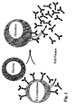
- the technique of hybridoma production is shown schematically in FIG. 1.
- One advantage of the monoclonal antibodies over the polyclonal antibodies is that they can be produced by the immortal cells in principle in an unlimited amount.
- mice immunized and then the mouse B lymphocytes with a Myeloma cell line fused.
- the hybridoma cells thus formed are then propagated separately as single cell clones and the Supernatant of each clone on antibody desired Specificity examined.
- the identified single clones must then immediately in a second selection round be subcloned as they are genetic at this time are unstable. This process is very time consuming, so that in the end a maximum of a few thousand hybridoma clones the desired specificity can be tested.
- the Construction and search of a hybridoma library is included this technology is only possible to a very limited extent. So that's it It is difficult to automate this process. This conventional technology does not allow the production of human antibodies.
- human hybridoma antibodies which are obtained from transgenic mice whose own immunoglobulin gene locus has been replaced by parts of the human immunoglobulin gene locus (Jakobovits, 1995, Production of fully human antibodies by transgenic mice. Curr Opin Biotechnol 6 , 561- 566; Lonberg and Huszar, 1995, Human antibodies from transgenic mice. Int Rev Immunol. 13 , 65-93; Kucherlapati et al. U.S. Patent 6114598: Generation of xenogeneic antibodies).
- the human antibody genes rearrange, go through the class switch and are somatically hypermuted. These transgenic mice thus produce human antibodies in mouse cells, which (in contrast to human hybridoma cells) lead to stable mouse hybridomas.
- HAMA antibodies are directed against the constant part of the antibodies, which is why the production of antibody chimeras has been started.
- a human antibody gene is first placed in a cloning vector (Wright et al., 1992, Genetically engineered Antibodies: Progress and Pröspects. Critical Rev Immunol. 12 , 125 168).
- the individual antibody domains form compact folding units that are connected to each other by a peptide strand. When entire antibody domains are exchanged, the chance of a disturbance of the antibody function is lowest.
- US Patent No. 5202238 describes a method by which mouse monoclonal antibodies can be humanized. At the center of the method is the so-called “homologous recombination”. Human sequences flanked by suitable genomic mouse sequences are recombined with precise location in the active antibody locus. The great advantage of this method is that the signals of the antibody locus optimized for good antibody production are largely retained (Yarnold and Fell, 1994, Chimerization of antitumor antibodies via homologous recombination conversion vectors. Cancer Research 54 , 506-512; Fell et al., 1989, Homologous recombination in hybridoma cells: heavy chain chimeric antibody produced by gene targeting. PNAS 86, 8507-8511).
- Recombinant antibodies lack the constant antibody part and thus the effector functions that are essential for many therapeutic approaches. Therefore, newly found “antibody search heads” are “grafted” onto eukaryotic expression vectors for many applications, ie the labor-intensive method described above is used. In addition to the experimental work associated with this, poor expression can occur since different codons are preferred in the bacterial system than in eukaryotic systems.
- Another disadvantage arises from the properties of the mostly used "single chain” antibodies (scFv): These are usually relatively unstable and aggregate easily. In addition, many variable domains are apparently attacked by E. coli proteases. The published complexities of the scFv antibody libraries (up to 10 11 ) should therefore be viewed with caution.
- This technique is based on the one above described hybridoma technique. The difference is however, used a stable myeloma cell line (and produced), which is an antibody binding protein (e.g. Protein G) anchored in large quantities to the cell surface (Breitling et al., 1999, selection of monoclonal antibodies. DE 199 00 635 A1. PCT registration under number PCT DE00 / 00079). This avoids a large part of the work caused by cloning and subcloning the monoclonal hybridomas arises. Instead, the desired antibody specificities in the FACS sorter or with magnetobeads from one Pool of hybridomas can be isolated since the ones produced Antibodies by binding to the antibody binding protein described be anchored to the cell surface.
- an antibody binding protein e.g. Protein G
- the method according to the invention is based on the fact that a large and very uniform number from cell to cell Number of antibodies covalently to the surface of a eukaryotic cell are bound and thus from cell to cell Cell comparable signals can be expected.
- specific antibodies along with the Cell from a multitude of cells is comparatively simple be selected. Through this selection a obtained monoclonal antibody.
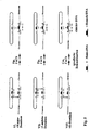
- the host of antibody presenters By remodeling a cell eukaryotic cell obtained. This is particularly the case by several homologues carried out one after the other (FIGS. 3, 4, 7 and 8) and specific recombination events (FIGS. 5, 6 and 9). Mouse hybridoma cells are particularly suitable for this ( Figure 3).
- FIGS. 3, 4, 7 and 8 By remodeling a cell eukaryotic cell obtained. This is particularly the case by several homologues carried out one after the other (FIGS. 3, 4, 7 and 8) and specific recombination events (FIGS. 5, 6 and 9).
- Mouse hybridoma cells are particularly suitable for this ( Figure 3).
- the undirected mutations can be combined, for example, using the method developed by Stemmer (1994, Nature 370, 389-391). However, it is also possible to simply replace the variable domain of an antibody chain with a large number of other variable domains in a preselected antibody-producing cell using the specific recombination signals (FIG. 5, 6, 9) and then to search for more affine variants.
- the method according to the invention shows several ways which enable somatic hypermutation followed by selection of more affine antibodies in vitro (FIGS. 10, 11).
- the method according to the invention has the following Advantages on: It allows the production of a highly complex Library e.g. as a source of monoclonal antibodies ( Figure 6, 9) as well as simple selection, especially high affinity human monoclonal antibody ( Figures 10, 11). simultaneously can the prospects of success of such a selection be enlarged. Due to the large and comparatively uniform number of antibodies of the same specificity presented can change the signal strength when looking for specific monoclonal antibodies can be increased significantly. The one with the Process according to the invention can be obtained in high yield high affinity human monoclonal antibodies e.g. used as tumor diagnostics and therapeutics. In addition, instead of antibodies, too other proteins, or individual protein domains, in particular Exons are presented on the surface of "their" cell , making it a particularly easy search for Binding partners for these presented protein fragments is made possible.
- B-lymphoblasts are usually added with a mixture of Polyethylene glycol (PEG) with DMSO with a myeloma cell line (e.g. Ag8.653) merged. Then through limited dilution propagates individual cell clones and after about 14 days whose cell culture supernatant is on the searched specific antibody reactivity examined.
- the resulting hybridoma cell acquires properties of both parental cells: the myeloma cell controls immortality a tumor cell line and some control signals for efficient production of antibodies during the B lymphoblast a specific antibody reactivity genomic brings along recombined antibody gene fragments.
- the resulting hybridoma cell is initially genetically unstable, so that in at least one further selection round one genetically more stable variant must be established.
- Hybridoma cells produce monoclonal, especially human Antibodies in good yield and quality.
- each of a loxP and a loxP511 point are flanked (e.g. loxP scFv1 loxP511 loxP scFv2 loxP511 ... loxP scFvN loxP511).
- loxP scFv1 loxP511 loxP scFv2 loxP511 ... loxP scFvN loxP511 flanked (e.g. loxP scFv1 loxP511 loxP scFv2 loxP511 ... loxP scFvN loxP511).
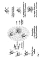
- the affinity of an antibody to its antigen is a measure of the ratio of antibody-bound antigens vs free antibodies and free antigens in solution (generally: receptor vs ligand).
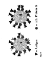
- receptor vs ligand This enables simultaneous affinity comparison of different surface-presented antibodies or different presented proteins (or the corresponding hybridoma cells) with fluorescence-labeled antigens (red triangles) at the same time as the selection using FACS.
- fluorescence-labeled antigens red triangles
- FACS fluorescence-labeled antigens
- the antibody presented by hybridoma cell B binds to the antigen in a significantly more affinity than the antibody presented by cell A.
- the measure of the affinity in this case is the quotient from red to green fluorescence of the cells sorted in the FACS.
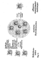
- Somatic hypermutation As in Figure 9 described, by specific recombination, the exons of the active variable vL2 domain against a bevy of exons exchanged, each a differently mutated vL2 domain code (see also Figure 5 and 6). This creates unproductive mutations (2d), weaker affine antibodies (2c and 2e) and, in rare cases, higher affinity antibodies (2b). As described in FIG. 10, these can be sorted in the FACS become.
- the vector pBS is shown loxP-IgG1, with the help of Flp the constant domains including the membrane domains M1 and M2 of an IgG1 e.g. be recombined into a pre-selected FRT cassette can.
- This vector also brings a loxP cassette with which Cre, e.g. variable domains can be recombined.
- Cloning vector was next the vector pBS FRTvKappa cut with AatII (FIG. 15A). Synthetic oligonucleotide sequences were set in these which the loxP and the loxP511 digit with an additional BamH1 site encoded.
- the resulting vector was created with BamH1 cut.
- the cyan and underlined figures PCR primers hIgG1-1 and hIgG1-2 were used for duplication of a chimeric genomic IgG1 gene. These primers brought in a BamH1 site in a subsequent PCR reaction With.
- Template DNA was the one described in FIG. 12B
- Vector pBS MhIgG1M alternatively pBS MhIgG1Mdelta350.
- the vector pBS loxP-IgG1 was created, or alternatively the vector pBS loxP-IgG1delta350. Restriction sites are underlined.
- mice and human sequences The boundary between mouse and human sequences is shown by a HindIII restriction site marked. Coding human Sequences as well as the mouse M1 and mouse M2 exons are marked with green Lowercase letters shown. The splice acceptor at the 5 'end The hinge exon is underlined in red.
- the vector pBS loxP-IgG1deltaCH1 obtained. Both vectors are identical, only the vector sequences pBS loxP-IgG1deltaCH1 lack the DNA sequences between the PfM1 and BamH1 sites. To do this, the vector pBS loxP-IgG1 cut with BamH1 and one using the PCR primer deltaCH1-1 and hIgG1-2 fragment cloned. The vector pBS loxP-IgG1 served as the DNA template.
- Restriction sites are underlined.
- the one in brackets set restriction sites PvuII or PvuII / MscI contain residuals resulting from the cloning Sequences.
- the starting point was the PvuII-cut figure 17A described vector pBS FRT KappaHEAPvuII.
- the red and PCR primers bla1 and bla2 shown in lower case were used to reproduce the coding DNA of beta-lactamase.
- the template DNA was the plasmid pBSIISK +. To cloning this PCR fragment resulted in the vector pBS FRT KappaHEAbla.
- this is the invention Method characterized in that in step (a) the Introduction of the recombination signals by homologous Recombination of transfected DNA with the respective gene loci takes place, the recombination signals of the transfected DNA from homologous areas to the respective gene loci of the cell are flanked and in step (b) at least one of the the homologous recombination of expanded cells is expanded, which change the specific recombination signals in has the gene loci.
- a core element of the present invention is the introduction of modified DNA sequences, in particular of recognition sites for recombinases into the genome of cell lines by means of homologous recombination (FIGS. 3, 4, 8).
- DNA sequences are exchanged in a defined gene locus. This is achieved by flanking the newly introduced DNA sequences of usually> 700 bp long to the defined gene locomotive homologous regions and in this form (mostly as linearized DNA; see Hasty et al., 1992, Mol. Ell. Biol. 12 , 2464-2474) can be transfected into the cells. In a few cases (approx. 1 out of 10 7 cells) the desired homologous recombination then takes place.
- the loxP sites (FRT sites have a very similar structure) consist of two 13Bp-long inverted repeats, which are separated by an 8Bp-long spacer. This spacer specifies the directionality of the recombined DNA flanked by loxP sites, ie the experimenter can use a (genomic) loxP or FRT site to predict the exact chromosomal gene locus and at the same time the orientation of the recombined DNA. Higher recombination frequencies are usually achieved by exchanging gene cassettes.
- Cassette exchange also takes place with genes between two inverted recognition sites of the same identity (eg loxP gene Pxol).
- the product of a specific recombination can have three forms in this case: 1.
- the gene between the loxP sites can be inverted.
- the gene between the loxP sites can be replaced by another purely recombined one, which is integrated 2. in the correct orientation and 3. in the wrong orientation between the recognition sites.
- the DNA sequences of suitable genes are also known, Gene fragments (especially vH, vLambda, vKappa and the associated J segments) and human loci (http://genome.ucsc.edu/goldenPath/hgTracks.html; www.gdb.org; www.gdb.org/hugo; www.ncbi.nlm.nih.gov; www.ncbi.nlm.nih.gov/LocusLink) or the mouse (www.informatics.jax.org), especially the active ones Antibody gene loci and the variety of antibody genes (Immunoglobulin Facts Book, Lefranc and Lefranc, 2001, Academic Press, ISBN 0-12-441351-X; http://imgt.cines.fr; Kabat database: http://immuno.bme.nwe.edu) and the Genloci of the T-cell receptor (T-Cell Receptor Facts Book, Le
- Another core element of the present invention is the production of a large number of different DNA sequences, in particular of as many different antibody genes as possible. Flanked in each case by the recognition sites for recombinases mentioned, these DNA sequences serve as donors for many different DNA sequences which, under the influence of recombinases, recombine in the gene loci mentioned with comparatively high efficiency with the acceptors mentioned for DNA sequences.
- the primers preferably hybridizing to genomic regions which are poorly conserved in evolution.
- the genomic DNA of a large number of B-lymphocytes serves as a template for the less than 1,000 PCR reactions required for this.
- the genomic DNA of these B-lymphocytes contains a large number of differently genomically recombined antibody genes (vH, vKappa, vLambda), which can be easily reproduced in their diversity by the procedure described.
- the variety of genomically recombined T cell receptors can be converted into a group of suitable DNA sequences by means of comparatively few PCR reactions.
- telomeres can either be in a given cloning vector be cloned that already contains these recognition sites, or a subsequent second PCR brings with it PCR primers used these recognition sites.
- the crowd of the different PCR products is then preferably cloned into a plasmid (e.g. pUC19 from Biolabs, pBluescript from Stratagene, pCR-TOPO, pCR-XL-TOPO, pCR-Vector, pZErO-1, pCR-Blunt, pSinRep5 from Invitrogen).
- a plasmid e.g. pUC19 from Biolabs, pBluescript from Stratagene, pCR-TOPO, pCR-XL-TOPO, pCR-Vector, pZErO-1, pCR-Blunt, pSinRep5 from Invitrogen.
- an origin of replication under selection pressure to one stable episomal replication
- the time can be easily extended within which it to a specific recombination with those mentioned Genloci can come.
- the Presence of the corresponding recombina set, or the corresponding expression vectors necessary.
- the Expression cassettes for the recombinases mentioned in the just included cloning vector are the Expression cassettes for the recombinases mentioned in the just included cloning vector.
- a host of different plasmids can also be generated in vitro in extremely high complexity (> 10 12 ) if necessary, avoiding duplicating them in bacteria beforehand (electroporation can currently routinely achieve complexities of approx. 10 9 independent bacterial clones per ⁇ g of DNA used ; see e.g. Clontech # C2023-1 330, the E.coli strain KC8> 10 9 cfu / ⁇ g pUC).
- the different PCR products described which are flanked by the recognition sites for recombinases mentioned, are preferably first circularized with ligase (donors for DNA sequences) and mixed with a cloning vector which contains the same recognition sites (acceptors for DNA sequences).
- transfection of the eukaryotic cells is also carried out using standard methods known to the person skilled in the art (Sambrook and Russell: Molecular Cloning, a laboratory manual, 3rd Edition, 2001, ISBN 0-87969-577-3; Chapter 16), such as, for example, electroporation (AGS / Hybaid or BioRad, manual of the electroporators BTX or BioRad GenePulser) or transfection eg with LipofectAMINE TM 2000 Reagent (Invitrogen # 1668-027), with DMRIE-C Reagent (Invitrogen # 10459-014), or LipofectAMINE TM Reagent (Invitrogen # 18324 -012), FuGENE 6 Transfection Reagent (Roche # 1815091), DOTAP Liposomal Transfection Reagent (Roche # 1811177), or DOSPER Liposomal Transfection Reagent (Roche # 1811169).
- electroporation AGS / Hybaid
- the percentage of successfully electroporated hybridomas is usually 30-40% of the cells used.
- the DNA sequences mentioned are preferably linearized. If a specific recombination of the transfected DNA sequences is desired, circular DNA sequences are preferably used, in particular episomally replicating vectors are suitable for this in hybridoma cells (see above).
- the proportion of successful specific recombination by cassette exchange within a eukaryotic cell is approximately 1% of the successfully electroporated cells (Feng et al., 1999, Site-specific chromosomal integration in mammalian cells: highly efficient CRE recombinase-mediated cassette exchange. J- Mol-Biol. 292, 779-785), so that approximately 10 7 different specific recombination events are obtained by using 3 ⁇ 10 9 cells.
- Another core element of the present invention is the isolation or enrichment of those cells (if possible after each process step) in which the desired recombination event has taken place (FIGS. 7, 8, 9).
- the invention makes this possible by detecting modified proteins on the surface of the living cells producing them. This is also carried out according to common methods known to those skilled in the art, such as the use of magnetic beads (Dynal, Oslo, Norway; Technical Handbook: Cell Separation and Protein Purification from Dynal; Current Protocols in Immunology, John Wiley & Sons, New York, ISBN 0- 471-52276-7) or a FACS sorter (Shapiro, HM Practical flow Cytometry, 3 edition, around 1995, Wiley-Liss New York, ISBN 0-471-30376-3;.
- the very rare (approx. 1 out of 10 7 cells) homologous recombination events can be identified, for example, with a FACSVantage SE upgraded to a turbo sorter, or a FACSDiva (Becton-Dickinson), or a MoFlow (cytomation), whereby when introducing specific recombination signals into defined Genloci can additionally be preselected based on a recombined resistance gene (e.g.
- the more frequent specific recombination events can also be enriched with standard methods by detecting modified proteins on the surface of the cells with a FACS sorter or magnetobeads (FIG. 9), as in the following Examples described.
- the modified proteins are preferably stained before sorting with a FACS with the aid of fluorescence-labeled mono- or polyclonal antibodies (or protein G or the like), which are commercially available from many companies (for example Jackson ImmunoResearch, West Grove, PA, USA or Dianova, Germany or Southern Biotechnology Associates, Birmingham, AL, USA or BIOZOL, Germany). Clear signals are obtained in particular by double staining, with two different fluorescences detecting the proteins (or their lack) encoded by the DNA sequences exchanged for one another. The methods required for this are known to the person skilled in the art and are described in a large number of detailed publications (for example Scheffold and Kern, 2000, Recent developments in flow cytometry. J Clin Immunol. 20 , 400-7.
- the absence of a signal can also be demonstrated if, for example, the vH domain of an IgG antibody presented on the surface of a hybridoma has been replaced by a G418 resistance gene by homologous recombination.
- staining with FITC-labeled goat anti-IgG antibody results in a further enrichment of homologous recombination events with the FACS: in this case, the cells with green fluorescence have not acquired their G418 resistance through homologous recombination and are therefore being acquired depleted in the FACS.
- epitope-specific monoclonal antibodies such as a Mycl-9E10 epitope-specific antibody (Evan et al., 1985, Mol. Cell. Biol. 5 , 3610-3616), with which, for example, altered vH domains can be detected can.
- the exchanged DNA sequences can also be detected directly in the FACS without further staining if, for example, the gene for an EGFP (enhanced green fluorescent protein; Clontech) is recombined and expressed.
- EGFP enhanced green fluorescent protein
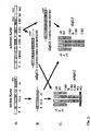
- the surface presentation, in particular of antibodies, is preferably achieved in the present invention by the differential splicing of the mRNA for the heavy antibody chain (or corresponding chimeric mRNAs) (FIG. 2; Immunology by Janeway and Travers, chapters 3-24, 4, edition 1999, Spectrum publishing house, ISBN 443062757).
- This offers the advantage that, in addition to a variant covalently bound to the membrane, large quantities of the form secreted into the culture medium are also available for the rapid and simple characterization of a cell clone or a cell library.
- the proportion of membrane-bound antibodies and thus the expected signal strength can be increased if the intron between the CH3 domain and the M1 domain of an IgG or IgA (or between the CH4 domain and the M1 domain of an IgM), in particular by 300 bp to 1,000 bp (Peterson and Perry, 1986, PNAS 83 , 8883-8887; Tsurushita and Korn, 1987, Molec. Cell. Biol. 7 , 2602-2605; Galli et al., 1987, Genes Dev 1 , 471-481; Seipelt and Peterson, 1995, Molecular Immunology 32 , 277-285; Figure 3, III.).
- membrane anchors such as, for example, the alpha T cell receptor (EMBL accession number X02883, the sequences of exon 3) or an MHC molecule, can also be used, as a result of which even more proteins can be presented on the surface.
- a non-covalent coupling of antibodies on the cell surface by membrane-standing protein G is also possible (Breitling et al., 1999, PCT DE00 / 00079), whereby this has the disadvantage of crosstalk: Not all antibodies are necessarily separated from the one presenting them Cell coded.
- the surface presentation is particularly useful when many (> 10 2 ) cells that are different from one another are to be searched for a specific protein activity by the expressed proteins.
- the present invention achieves this very analogously to the technique of recombinant antibodies, where this is done in particular by the presentation of the antibodies on a phage (Breitling et al., 1991, Gene 104 , 147-153) or on a bacterium (Fuchs et al., 1991 , Bio / Technology 9 , 1369-1372). As described there, much more complex protein libraries can be searched.
- the present invention has the additional advantage of a comparatively very high signal intensity due to the large number of proteins of the same type, in particular antibodies, which are presented in each case.
- a FACS sorter nowadays achieves sorting speeds of approx. 10 8 cells per hour (eg FACSVantage SE, see above), whereby an even more complex protein library can additionally be enriched beforehand by magnetobeads.
- the technique of Kern et al. FACS sorting required for this invention is described. (1998, Nature Medicine 4, 975-978) and Maecker et al. (2001, J Immunol Methods 255 , 27-40), who were able to identify epitope-specific T cells.
- the starting cell before the specific recombination no antibodies on the Surface presented, but instead a G418 resistance in the gene locus of the monospecific cell line is installed.
- the cells are enriched one, in particular human IgG chain or kappa or lambda chain present. These are the cells that are productive have performed specific recombination.
- the result of this procedure is a complex (> 10 6 different hybridoma specificities) hybridoma library (FIGS. 5, 6, 9), the individual members of which present large amounts of a human antibody on the surface (approx. 10 4 to 10 6 antibody molecules per Cell).
- the number of antibodies presented per cell is in a comparatively narrow range, since all cells originate from the same parental hybridoma.
- the present invention thus also relates to an antibody library which can be obtained by the process according to the invention. This preferably comprises a group of at least 100 different cells, more preferably a group of at least 1000 cells.
- a special embodiment of the invention The process is characterized in that the eukaryotic cells, mammalian cells, preferably neoplastic lymphocytes or precursors thereof; leukemia cells or are malignant lymphoma cells or hybridoma cells.
- vH genes vLambda genes and / or vKappa genes human genes.
- a method is particularly preferred, wherein the Gene loci are the antibody loci and the active vH gene, vLambda gene or vKappa gene included.
- the method according to the invention also includes a method for Production of a library of T cell receptor-producing Cells, the different DNA sequences different T-alpha receptor genes or T-beta receptor genes included, as well as a method for producing a Library of exon-expressing cells, the different DNA sequences different genomic Contain exons that encode splice signals.
- the gene loci which are T cell receptor loci and the active T alpha receptor gene or contain the active T-beta receptor gene.
- the antibodies are monoclonal human antibodies, the covalently to the surface of the cell expressing it are bound.
- Method according to the invention for producing a library of antibody-producing eukaryotic cells is that Intron towards the 5 'end in front of the M1 exon of an IgG, IgM, IgA, IgD or IgE gene shortened by more than 50 base pairs.
- the method according to the invention is used after the transfection a variety of different DNA sequences from the this creates a multitude of different cells an enrichment of the cells carried out on the Cell surface to the surface of each one expressing bound cells present expressing cells, so that a cell population with the highest possible protein diversity arises.
- the above invention also relates to a library of Protein-producing, preferably antibody-producing, eukaryotic cells, which according to The inventive method is available.
- the present invention also relates to a method for Isolation of a monoclonal antibody with a desired specificity (see Figure 9) using the method is characterized in that one according to the Antibody bank produced according to the method of the invention incubated with the appropriate antigen and then isolates or enriches the cell line attached to its Surface that bound the antigen.
- the method of the invention prepared cells with the antibody library biotinylated and then labeled with streptavidin-FITC Antigen stained (or directly FITC-labeled antigen) and the strongest staining events selected.
- the antibody library also with a mixture of optional differently labeled antigens are stained and again the strongest staining events are selected.
- the individual cells in cell culture are expanded and produce the desired, especially human monoclonal antibodies.
- the antigen specific Selection only one round of selection pardoned because of the very high number of antibodies presented gives very clear signals.
- Antibody library should be due to the recombination of the vH and vL domains also have antibody specificities against human Antigens are represented
- this is Method according to the invention further characterized in that high affinity antibodies are generated.
- more affine monoclonal antibodies can be obtained using FACS be selected.
- FACS FACS be selected.
- the individual cells are expanded in cell culture and each produce one human monoclonal antibodies are comparatively higher Affinity for the antigen used for selection. This is a very simple method to get high affinity to find monoclonal antibodies.
- the above is Process characterized in that the insulation high affinity antibodies the introduction of mutations within preceded by variable antibody genes. This can e.g. by performing a somatic hypermutation or a gene conversion of the antibody genes, which like described is followed by the selection of changed, especially more affinity presented on the cell surface Antibodies ( Figure 11).
- a "chain shuffling" and one subsequent selection of high affinity monoclonal antibodies be performed.
- a large number of undirected mutations can be introduced into a template DNA, in particular into preselected variable domains, by an error-prone PCR (Stemmer, 1994, Nature 370, 389-391). Then, as described, as large a number of these mutated variable antibody genes as possible are recombined into the antibody locus by means of specific recombination signals, and then the cells are selected, for example in the FACS, which present an antibody of higher affinity on the surface.
- the undirected mutations can also be combined using the method developed by Stemmer (gene conversion / somatic hypermutation).
- Another way according to the invention for generating undirected mutations is by means of eukaryotic expression vectors which express the genes RAD54, RecQ4 and / or the gene DNA PolX mu in lymphoid cells, in particular in combination with anti-sense RNA against XRCC2, XRCC3 or RAD51B ( Kitao et al., 1998, Cloning of two new human helicase genes of the RecQ family: biological significance of multiple species in higher eukaryotes.
- the gene products RAD54, RecQ4, and DNA PolX mu are part of the mutator complex responsible for the introduction of somatic hypermutations into the active antibody genes, while the suppression of the expression of XRCC2, XRCC3 or RAD51B by increased single strand breaks apparently initiates the somatic hypermutations.
- approximately 1.5 kb DNA sequences downstream of the active vH, vKappa or vLambda promoter are mutated undirected, regardless of which DNA sequences are present there.
- somatic hypermutation one can proceed, for example, by selecting a family of antigen-specific hybridoma cells with FITC-labeled antigen derived therefrom from the antibody library according to the invention, as described above. This group of selected hybridoma cells is expanded in cell culture and then a mixture of the expression vectors described above is electroporated into these cells. The resulting cluster of hybridoma cells is expanded in cell culture. Then, as described above, high-affinity monoclonal human antibodies are selected using a FACS sorter.
- An embodiment of the above is particularly preferred Process in which the cell contains a large number of DNA sequences is transfected, each by specific Recombination signals are flanked and being the multitude of DNA sequences using "error-prone" -PCR.
- the present invention also relates to methods for the humanization of already existing mouse hybridomas by means of homologous recombination (FIGS. 3, 7).
- any given mouse IgG1 hybridoma cell can be converted into a human IgG1 hybridoma cell with a given DNA vector that is always the same (FIGS. 3, 7), or with other DNA vectors into human IgG2, IgG3, IgG4, IgA1, IgA2, IgE or IgM.
- Still other DNA vectors enable the conversion of a mouse IgM hybridoma etc. This particularly simple and advantageous method does not require the use of an annoying resistance marker (see also Baker et al., 1994, J.
- the intron is towards the 5 'end before the M1 exon of the active IgG, IgM, IgA, IgD or IgE gene by more than Shortened 50 base pairs.
- Procedures are attached to the humanized constant domains fused additional protein coding DNA sequences, preferably the additional protein encoding codes DNA sequences one linker sequence and one Single chain antibody, the C-terminal to the constant domain the light antibody chain is fused.
- the process extends to the homologous areas of transfected DNA sequences to the respective antibody gene loci the hybridoma cell over at least 400 base pairs,
- the present invention also relates to a vector that has a or more of the DNA sequences described above contains, as well as this vector containing host cells.
- the hybridoma cell line HEA125 served as an example of a monoclonal antibody to be humanized.
- This mouse hybridoma cell line produces a mouse IgG1 antibody together with a mouse kappa chain.
- This monoclonal antibody recognizes the human tumor-associated antigen Ep-CAM with a comparatively high affinity and specificity (Moldenhauer et al., 1987, Br J Cancer 56 , 714-722).
- the HEA125 cell line was grown and expanded.
- RPMI 1640 Gibco BRL # 31870-025
- FCS 1mM pyruvate
- the other culture conditions plastic containers of Falcon 25 cm 3 ; 37 ° C warming cabinet; 5-7.5% CO 2 fumigation, maximum 10 6 cells per ml, etc. are known to the person skilled in the art.
- the expanded cells were washed twice with ice-cold Dulbeccos PBS (DPBS) and about 10 7 cells per 400 ⁇ l were stained with FITC-labeled goat anti-mouse IgG antibodies (Dianova). Propidium iodide (1 ⁇ g / ml) was used as counterstain to identify dead cells.
- DPBS Dulbeccos PBS
- Figure 12 the resulting chimeric DNA sequences mapped Sequence was checked.
- the primers used for the Cloning in pBSIISK + are described in Figure 12.
- Figure 12A is the chimeric DNA sequence of the vector pBS-MhKappaM mapped to humanize the constant Kappa chain from HEA125 was used.
- Figure 12B is the chimeric DNA sequence of the vector pBS MhIgG1M mapped to humanize the constant IgG1 CH1, CH2 and CH3 domains from HEA125 was used.
- the chimeric mouse kappa humanization vector pBS MhKappaM described in Example 1c and FIG. 12A was linearized with the restriction enzyme BglI and the expanded subpopulation of HEA125 cells described in Example 1b was added to each 10 ⁇ g linearized plasmid DNA. These cells were transfected with the optimized electroporation conditions described in Example 1d. After 2-4 days in culture (described in Example 1a), the cells were washed twice with ice-cold DPBS and about 10 8 cells per 4 ml with FITC-labeled goat anti-mouse kappa antibody and at the same time with PE-conjugated goat anti - Human Kappa antibody stained (Southern Biotechnology Associates). Propidium iodide was used as counterstain to identify dead cells. After washing again with ice-cold DPBS, the cells were sorted using a FACS sorter, about 10 8 cells being sorted in 2 hours.
- the chimeric mouse IgG1 humanization vector pBS MhIgG1M described in Example 1c and FIG. 12B was linearized with the restriction enzyme SspI and cells of the partially humanized subclone HEA125-hKappa described in Example 1e were added to each 10 ⁇ g linearized plasmid DNA.
- Cell culture, FACS, staining of the cells are identical to those described in Example 1e, with the difference that a PE-conjugated goat anti-human IgG antibody was used instead of the PE-conjugated goat anti-human kappa antibody.
- IgG1 producing mouse hybridoma cell the constant domains humanized to human IgG1.
- Mouse hybridoma Cells that are IgG2a, IgG2b, IgG3, IgA, IgE or IgM can produce with other chimeric DNA sequences be humanized in a completely analogous way.
- Whichever Humanization vector can be used the individual Antibody classes are also converted or changed become, for example, a mouse IgM in humans IgG1, IgG2, IgG3, IgG4, IgA1, IgA2 or IgE converted becomes. The same applies to the conversion of a constant Mouse kappa domain in a constant human lambda domain.
- the vector pBS MhIgGlMdelta350 was used which has the deletion of approx. 350 bp in the intron between the CH3 and the M1 domain described in FIG. 12B, but is otherwise identical to that in Example 1f and Figure 12B is the vector pBS MhIgG1M described.
- the procedure was the same as described in Example 1f.
- approximately 1-2 from approximately 10 8 cells with a homologous recombination were found in the region of the antibody gene locus of the heavy chain.
- the cell line HEA125-mhIgG1hKappa propagates that compared to that in Example 1f described cell line HEA125-hIgG1hKappa Because of the shortened intron between the CH3 domain and the M1 domain of IgG1 significantly more membrane bound has humanized antibodies (see also FIG. 7, Hybridoma cell 2H).
- FIG. 13B As a template for the genomic mouse gene sequences served mouse genomic DNA Hybridoma cell line HEA125. As a template for that Resistance gene PGKneo served the vector ploxPfrtPGKneofrtloxP (Erich Greiner, dkfz, department Molecular biology of cell I). In Figure 13B, the resulting vector pBS MvHG418M mapped, its Sequence was then checked.
- the cell line HEA125-hIgG1hKappa obtained in Example 2 was linearized with that described in Figure 13B Vector pBS MvHG418M transfected, wherein the in Example 1d described electroporation conditions applied were.
- the DNA transfected cells first with a pre-selection G418 subjected (depending on the batch 400-800 ⁇ g G418 per ml for 14 days; Cell culture conditions otherwise as in example 1).
- the expanded G418-resistant cells were then as described in Example 1b with FITC-labeled Goat anti-human IgG antibody stained and im FACS sorted. The results were as above described about 2,000 individual cells sorted, none Antibody-specific staining (i.e., if possible low green fluorescence). These single cells were under the culture conditions described in Example 1a Expanded 2-3 weeks.
- Example 3b From the clones obtained in Example 3b, the genomic DNA isolated, each as a template for a PCR served (Roche Diagnostics; Expand Long Template PCR System; see also for the PCR conditions). The one for that PCR primers vHG418-3 and vHG418-4 used are shown in FIG 13B.
- the respective PCR bands were in one 0.8% TAE agarose gel separated by size and with the expected result compared. As a result, 8 of the 2,000 clones examined a PCR band of the expected Size of approx. 1.85 kb.
- genomic DNA was the result of the clone HEA125-mhIgGlhKappa-loxPG418 with genomically coded specific Recombination signals (loxP and loxP511) in the area of the active vH gene locus further propagated.
- Example 3b Cells of the approximately 2,000 clones described in Example 3b were pooled, alternatively the expanded clone HEA125-mhIgG1hKappa-loxPG418 described in Example 3c was used.
- the vector pBS loxPvHmyc shown in FIG. 15B was electroporated into these cells.
- This vector encodes the genomically recombined vH gene of HEA125, which is flanked by an loxP and an loxP511 site.
- a myc tag is built into the CDR3 region of the vH domain.
- the Cre expression vector pMC-Cre was electroporated into the cells (conditions for this see Example 1d).
- Example 1a After 2-4 days in culture (described in Example 1a), the cells were washed twice with ice-cold DPBS and about 10 8 cells per ml were stained with FITC-conjugated goat anti-human IgG antibodies (Dianova). Alternatively, staining was done with FITC-labeled 1-9E10 anti-myc antibody. Propidium iodide was used as counterstain to identify dead cells. After washing again with ice-cold DPBS, single cells with strong FITC fluorescence are sorted using a FACS sorter.
- Example 1a A total of 20 sorted single cells were expanded for 2-3 weeks under the culture conditions described in Example 1a. The individual clones were then tested for G418 resistance as described in Example 3b. As a result, 17 of the 20 clones tested were sensitive to G418.
- the G418-sensitive cell line HEA125-mhloxPmyc was expanded further. Cells of this clone were washed twice with ice-cold DPBS and about 10 8 cells per ml with FITC-labeled anti-myc antibody (Myc1-9E10 epitope; Evan et al., 1985, Mol. Cell. Biol. 5 , 3610- 3616) colored. Propidium iodide was used as counterstain to identify dead cells. The FACS showed a strong green fluorescence of the cell line HEA125-mhloxPmyc.
- the resulting G418-sensitive cell line HEA125-mhloxPmyc produces a monoclonal hybridoma IgG1 antibody of a defined specificity (in the example the vH domain of HEA125 with an additional c-myc tag in the CDR3 of the vH domain) with humanized constant domains.
- the vH exon is flanked by 2 different loxP sites within the vH gene locus.
- a large part of the antibodies produced (in the order of 10 4 to 10 6 antibody molecules) is covalently bound to a membrane anchor on the surface of the hybridoma cells.
- Example 3d The cell line HEA125-mhloxPmyc obtained in Example 3d was linearized with that described in Example 4a Vector pBS MKappaG418M as described in Example 3b electroporated, then a G418 selection as described performed and cells with no expressed kappa chain sorted in FACS. When coloring was in Difference to example 3b, however, with FITC-labeled Goat anti-human stained kappa antibody. Thereby as a result, as described in Example 3b, approximately 2,000 Sorted single cells that have no kappa-specific staining (i.e. the lowest possible green fluorescence). These single cells were among those in Example 1a described culture conditions expanded 2-3 weeks.
- Example 4b The clones described in Example 4b were used as described the genomic DNA isolated, a PCR performed and the expected size of the PCR band checked.
- the PCR primers KG418-3 and KG418-4 are described in Figure 13A. Pointed as a result 14 of the 2,000 clones examined contained a PCR band from expected size of approx. 1.9 kb. After Control sequencing of the genomic DNA (or the PCR band described) was obtained as a result of the clone HEA125-mhloxPmycFRTG418 with genomically encoded specific recombination signals (FRT0 and FRT3) in the Area of the active vKappa gene locus propagated further.
- FRT0 and FRT3 genomically encoded specific recombination signals
- the resulting cell line HEA125-mhloxPmycFRTG418 produced a monoclonal hybridoma IgG1 antibody specificity (in the example the vH domain of HEA125 with an additional c-myc tag in CDR3 of the vH domain) with humanized constant domains. additionally the vH exon is within the vH gene locus of 2 flanked by different loxP positions. Instead of vKappa domain is a G418 resistance gene from 2 different FRT positions are flanked.
- Example 4c Cells of the approximately 2,000 clones described in Example 4b were pooled, alternatively the expanded clone HEA125-mhloxPmycFRTG418 described in Example 4c was used.
- the vector pBS FRTvKappa shown in FIG. 15A was electroporated into these cells.
- This vector codes for the genomically recombined vKappa gene of HEA125 (without leader exon), which is flanked by an FRT0 and an FRT3 site.
- the Flp expression vector pOG44 was electroporated into the cells (conditions for this see Example 1d).
- Example 1a After 2-4 days in culture (described in Example 1a), the cells were washed twice with ice-cold DPBS and about 10 8 cells per ml were stained with PE-conjugated goat anti-human kappa antibody (Dianova). Propidium iodide was used as counterstain to identify dead cells. After washing again with ice-cold DPBS, single cells with strong PE fluorescence were sorted using a FACS sorter.
- the resulting cell line HEA125-mhRek produces one monoclonal hybridoma IgG1 antibody of a defined Specificity with humanized constant domains.
- the vH domain of HEA125 encodes an additional one c-myc-tag in the CDR3 of the vH domain, that with the monoclonal Antibody 1-9E10 can be detected in FACS.
- the active vH exon is within the vH gene locus flanked by 2 different loxP points.
- the active vKappa exon within the vKappa gene locus is flanked by 2 different FRT positions.
- a large Some of the antibodies produced are covalently attached a membrane anchor tied to the surface of the Hybridoma cells.
- genomic sequences of the human vLambda gene locus are known (one-megabase sequence analysis of the human immunoglobulin lambda gene locus "; Genome Res. 7: 250-261 (1997); Accession Numbers: D87000; D87007; D87009; D87010; D87014; D87015 ; D87016; D87017; D87018; D87021; D87022; D87023; D87024; X51755; X51754; see also Immunoglobulin Facts Book, Lefranc and Lefranc, where 33 functional vLambda genes are described.
- restriction sites BssHII, MluI, NotI, SalI, XhoI and EagI are particularly suitable for cloning the variety of human vLambda genes.
- VLambda gene-specific PCR primers were produced, each hybridizing to the intron between the leader exon and the respective vLambda exon (FIG. 14A).
- the comparable area of the active vKappa gene locus of HEA125 is about the SwaI interface in the intron between the leader exon and vKappa exon.
- the primers hybridize to regions which are each approximately 130-170 bp upstream from the 5 'end of the respective vLambda exon.
- the hybridization temperature of the primers was calculated to be about 65 ° C. in each case.
- the 4 active human J Lambda gene segments are separated from the neighboring constant cLambda exons by an intron. Approximately 89 bp each (in the area of the BsaI interface at HEA125) downstream of the 3 'ends of the J lambda segments, a total of 4 J lambda gene segment-specific PCR primers were produced, each with a calculated hybridization temperature of approximately 65 ° C ( Figure 14A).
- PCR bands were sized as described selected and purified, resulting in 96 PCR bands templates, each flanked by FRTO and FRT3 sequences. A further 96 PCR bands were obtained, each flanked by MluI and NotI restriction sites.
- the 96 different PCR bands flanked by FRT sites flanked in Example 5d described with the aid of T4 DNA ligase were incubated together with Flp recombinase and with the vector pBS FRT clone, the resulting recombination products were cut with the restriction enzyme SalI and how just described in E. coli transfected and then a highly complex mixture of vector DNA (pBS FRTklon-vLambda) isolated.
- the highly complex mixtures of human vLambda vector DNA (pBS FRT clone vLambda) described in Example 5e were electroporated together with the Flp expression vector pOG44 into the cells of the expanded clone HEA125-mhloxPmycFRTG418 (see Example 4c), the cells after 3 days with FITC-labeled goat anti-human kappa antibody stained and isolated as described using FACS sorter cells. As a result, about 0.7% of the sorted cells (or about 10 6 cells) were obtained and combined with one another, which had a clear green fluorescence in the FACS.
- This vLambda cell library was expanded in cell culture.
- the result of this procedure is a complex (> 10 6 different hybridoma specificities) hybridoma library (FIG. 9), the individual members of which present large amounts of different human vLambda chains each fused to a constant human kappa domain on the surface.
- a highly complex mixture such as the described pBS FRTklon-vLambda can be obtained.
- pBSIISK + a vector which replicates episomally in eukaryotic cells is used (based, for example, on pCEP4, pREP7, pREP10, pEBVHis or pREP4 from Invitrogen).
- the recombinase Flp is encoded by an adjustable expression cassette which is stably integrated in the ear.
- Systems of this type are offered by the company Invitrogen (GeneSwitch TM system K1060-01; T-Rex TM system K1020-01).
- the genomic sequences of the human vKappa gene locus are known (Immunoglobulin Facts Book, Lefranc and Lefranc, 2001, Academic Press, ISBN 0-12-441351-X).
- Computer analysis of 34 known functional human vKappa genes revealed no BglI, BssHII, BstBI, ClaI, EagI, HindIII, MluI, NotI, NruI, PvuI, SacII, SfiI, SnaBI, SpeI and no StuI restriction sites within a range of approx. 200- 300 bp upstream from the leader exon of the examined genes to the respective genomic recombination signal at the end of the CDR3.
- the region of the J segments was examined, calculated from the 5 'end of the active J1 kappa gene segment to approximately 200 bp downstream from the 3' end of the active J5 kappa gene segment (see acession number J00242).
- the restriction sites BssHII, EagI, HindIII, MluI, NotI, SfiI and SpeI are particularly suitable for cloning the variety of human vKappa genes.
- the SalI restriction site cuts only once outside the IGKV1D-43 gene.
- VKappa gene-specific PCR primers were produced each to the intron between the leader exon and the hybridize each vKappa exon (FIG. 14B).
- the comparable area of the active vKappa gene locus from HEA125 is about the SwaI interface in the intron between leader exon and vKappa exon.
- the primer hybridize to areas each around 130-170bp upstream from the beginning of the respective vKappa exon lie.
- the hybridization temperature of the primers was up calculated at approx. 65 ° C each.
- the 5 active human J Kappa gene segments in contrast to the J Lambda gene segments, are clustered, which means that the same constant cKappa exon is always used, which is separated from the clustered J Kappa segments by an intron.
- Approx. 89 bp each the comparable range for HEA125 for the BsaI interface
- a total of 5 J kappa gene segment-specific PCR primers were calculated downstream from the 3 'ends of the different J kappa segments, with a calculated hybridization temperature of approx. 65 ° C ( Figure 14B).
- Example 5d for vLambda the diversity of the genomically recombined human vKappa genes was obtained by combining the 5 different J Kappa gene segment primers with 34 different functional vKappa gene-specific primers, ie 170 different PCR Reactions carried out.
- the PCR bands were selected according to their size and purified so that the result was 170 PCR bands (approx. 600 bp), each flanked by FRT0 and FRT3 sequences.
- a further 170 PCR bands were obtained (approx. 550 bp), each flanked by MluI and NotI restriction sites.
- the 170 different circularized PCR bands flanked by FRT sites described in Example 6d were incubated together with Flp recombinase with the vector pBS FRTvKappa, the resulting recombination products were cut with the restriction enzyme SalI and, as described in Example 5e, in E. coli transfected and then a highly complex mixture of vector DNA (pBS FRTklon-vKappa) isolated.
- Example 5f for vLambda those in Example. 6e described highly complex mixtures of. human vKappa vector DNA (pBS FRT clone vKappa) electroporated together with the Flp expression vector pOG44 into the cells of the expanded cell line HEA125-mhloxPmycFRTG418; stained with FITC-labeled goat anti-human kappa antibodies and isolated as described using FACS sorter cells. As a result, about 0.8% of the sorted cells (or about 10 6 cells) were obtained and combined with one another, which had a clear green fluorescence in the FACS. This vKappa cell library was expanded in cell culture.
- restriction sites BssHII, MluI, NheI, NotI, SalI, SfiI and XhoI are particularly suitable for cloning the variety of human vH genes.
- the restriction sites BssHII, MluI, NotI and SalI (with the exception of the IGKV1D-43 gene) also do not occur in the vLambda and vKappa gene regions examined.
- VH gene-specific PCR primers were produced each about 182 bps upstream from the 5 'end of each Hybridize vH leader exons to the respective vH genes ( Figure 14C).
- the hybridization temperature of the primers was calculated at approx. 65 ° C in each case.
- the total of 6 active human J H gene segments are clustered like the J Kappa gene segments, which means that the same constant CH exons are always used (however depending on a class switch that may have been carried out).
- the CH1 exon is separated from the clustered J H gene segments by an intron.
- Approx. 83 bp each (the comparable range for HEA125 is for the Bsu36I interface) downstream of the 3 'end of the respective J H gene segments, a total of 6 J H gene segment-specific PCR primers with a calculated hybridization temperature of approximately 65 ° C. were produced (Figure 14C).
- Example 7d After digestion of the isolated vector DNA pBS loxPklon (Qiagen Plasmid Purification Kit) described in FIG. 15D with the restriction enzymes MluI and NotI, the correspondingly digested 264 different PCR bands described in Example 7d were ligated in, the resulting ligation products were transfected in E. coli and then as in Example 5e described a highly complex mixture (> 10 6 different transformants) of vector DNA isolated (pBS loxPklon-vH).
- the 264 different PCR bands flanked by loxP sites described in Example 7d were incubated together with Cre recombinase with the vector pBS loxPklon, the resulting recombination products were cut with the restriction enzyme SalI and, as just described, transfected into E. coli and then incorporated highly complex mixture of vector DNA (pBS loxPklon-vH) isolated.
- Example 7e described highly complex mixtures of human vH vector DNA (pBS loxPklon-vH) along with the Cre expression vector pMC-Cre in the cells electroporated.
- the ones in Example 5f were used for this and 6f described vLambda and vKappa cell libraries.
- the cells were stained with PE-conjugated goat anti-human IgG antibody and at the same time with FITC-labeled monoclonal anti mycl-9E10 antibody. As described, cells were isolated using a FACS sorter. As a result, about 0.8% of the sorted cells (or about 10 7 cells) were obtained and combined with one another, which in the FACS had a strong red fluorescence with at the same time as little green fluorescence as possible.
- This antibody cell library was expanded in cell culture and aliquots of it frozen in liquid nitrogen.
- the result of this procedure is a complex (> 10 7 different hybridoma specificities) antibody library (FIG. 9), the individual members of which present large amounts of different human IgG1 antibodies in each case, fused to constant human domains on the surface.
- the hybridoma cells secrete the greater part of the antibodies produced into the surrounding medium.
- the individual process steps were largely based on the known prior art.
- the vLambda domains of this antibody library are fused to the constant kappa domain in the example.
- example 1e was therefore modified in such a way that, instead of the vector pBS MhKappaM shown in FIG. 12A, a vector was used which encodes a constant human-lambda domain after the homologous recombination.
- Example 8 Selection of monoclonal antibodies using PACS / Magnetobeads
- Example 7f The antibody library described in Example 7f was cultured as described in Example 1a.
- the expanded cells were washed twice with ice-cold DPBS and about 10 7 cells per 400 ⁇ l were stained with FITC-conjugated BSA.
- Propidium iodide (1 ⁇ g / ml) was used as counterstain to identify dead cells.
- the cells were sorted using a FACS sorter. As a result, approximately 15 per 10 7 hybridoma cells were found which had a comparatively strong green fluorescence.
- Example 1a The sorted individual cells were expanded as described in Example 1a (possibly together with feeder cells) and the supernatant with the secreted antibodies contained therein was examined for antigen specificity in a Western blot.
- the supernatant from 2 clones reacted with BSA (MW approx. 68kd), 5 further clones had a FITC-BSA specific staining (MG approx. 72kd).
- Example 7f In parallel, about 10 8 cells of the antibody library described in Example 7f were washed 2x with ice-cold DPBS and then incubated in 5 ml DPBS buffer for 5 minutes with BSA or magnetobeads coated with ovalbumin. Unbound cells were washed away using a magnet and the remaining magnetobead-bound cells were cultured as described in Example 1a. As a result, the supernatant of the cells enriched with BSA magnetobeads reacted in the Western blot with BSA (MW approx. 68 kd) but not with ovalbumin, while the supernatant of the cells enriched with ovalbumin magnetobeads had no staining.
- Anti-FITC antibodies were cloned each cleaned with the help of Protein G Sepharose and some of them conjugated with horseradish peroxidase (Anti-FITC-POX antibody). The concentration of Various purified antibodies were made up to 1mg / ml each set in PBS.
- An ELISA plate was coated with FITC-BSA (each 0.1 ⁇ g in 100 ⁇ l PBS), blocked (with 1% milk powder in 200 ⁇ l PBS) and the 5 different peroxidase-conjugated antibodies (each 1: 2000 diluted in 100 ⁇ l PBS-Tween20) competes with increasing amounts of the non-peroxidase-conjugated antibodies.
- the non-peroxidase-conjugated antibodies were preincubated with the coated FITC antigen for 10 minutes each. After intermediate washing steps, residual bound peroxidase with the substrate OPD / H 2 O 2 was detected,
- This example shows a very simple method to get high to find affine monoclonal antibodies. It enables an easy to carry out normalization of the number of Antibodies presented an "on line” affinity comparison the antibody specificities found (FIG. 10).
- Example 9 In the expanded cell line anti-FITC5 described in Example 9 became the variety of vH genes described in Example 7e as described in Example 7f using a Cre expression vector recombined. The resulting anti-FITC antibody Library was described as in Example 9 with FITC-BSA and at the same time with PE-conjugated protein G. colored.
- This example shows a very simple method to get one To produce a host of hybridomas from which comparatively high affinity monoclonal antibodies can be isolated.
- Example 11 Execution of a somatic hypermutation to Obtaining high affinity antibodies
- the cDNA sequences of the genes RAD 54, RecQ4, polX mu, RAD51B, XRCC2 and XRCC3 are known.
- the cDNA sequences were the genes RAD54, RecQ4 and polX must each be under the Control of an RSV promoter in the eukaryotic Expression vector pREP4 (Invitrogen) cloned and the correct sequence checked.
- the Expression vectors pREP4-RAD54, pREP4-RecQ4 and pREP4-polXmu and the anti-sense RNA expression vectors pREP4-RAD51B, pREP4-XRCC2 and pREP4-XRCC3 obtained.
- Circular DNA of the vectors pREP4-XRCC2, pREp4-RAD54, pREP4-RecQ4 and pREP4-polXmu was mixed in a ratio of 1: 1: 1: 1 and among the optimized ones described in Example 1d Electroporation conditions in that described in Example 9 expanded cell line anti-FITC5 electroporated. Subsequently the cells were as described in Example 1a for 2-3 days taken in culture. Optionally, cells that make up the episomal replicating pREP4 vectors, then selected with hygromycin. Then the Cells as described in Example 9 with FITC-BSA and stained simultaneously with PE-conjugated ProteinG.
- This example shows another very simple method to make a bevy of hybridomas from which comparatively high affinity monoclonal antibodies isolated can be.
- Example 12 Generation of a bispecific antibody
- the cDNA of the mouse hybridoma cell line HEA125 was used as a template for the gene sequences, as well as an expression vector for the scFv antibody 215 (Kontermann et al., 1995, Characterization of the epitope recognized by a monoclonal antibody directed against the largest subunit of Drosophila RNA polymerase II. Biol Chem. Hoppe-Seyler 376 , 473-481).
- the region of the linker sequence between cKappa (HEA) and the scFv (215) antibody was generated by a synthetic overhanging PCR oligonucleotide.
- the vector pBS FRT KappaHEAscFv215 was obtained.
- FIG. 17A shows the resulting chimeric DNA sequence, the sequence of which was subsequently checked.
- the vector pBS FRT KappaHEAscFv215 described was electroporated into the cell line HEA125-mhloxPmycFRTG418.
- the Flp expression vector pOG44 was electroporated into the cells (conditions for this see Example 1d). After 2-4 days in culture (conditions for this see Example 1a), the cells were washed twice with ice-cold DPBS and about 10 8 cells per ml were stained with PE-conjugated goat anti-mouse kappa antibody (Southern Biotechnology Associates). Propidium iodide was used as counterstain to identify dead cells.
- the cell line HEA125-mhloxPmycFRTscFv215 obtained as a result produces a bispecific monoclonal hybridoma IgG1 antibodies of a defined Specificity (in the example the vH domain of HEA125 with a additional c-myc tag in the CDR3 of the vH domain) humanized IgG1 constant domains.
- a defined Specificity in the example the vH domain of HEA125 with a additional c-myc tag in the CDR3 of the vH domain
- humanized IgG1 constant domains In addition to that is the vH exon within the vH gene locus of 2 flanked by different loxP positions.
- vKappa domain of HEA125 is encoded, which with the Mouse cKappa domain, a linker sequence and the scFv215 Antibody is fused.
- Example 12 the cell line became HEA125-mhloxPmycFRTG418 electroporated with chimeric DNA, with PE conjugated Goat anti mouse kappa antibody stained single cells with comparatively strong PE fluorescence sorted, single clones expanded and the genomic Sequence checked.
- example 12 was instead of the vector pBS FRT described KappaHEAscFv215 the vector pBS FRT KappaHEAbla used ( Figure 17B).
- the starting clone described resulted HEA125-mhloxPmycFRTG418 approx. 0.1% cells with comparatively strong PE fluorescence.
- the cell line HEA125-mhloxPmycFRTbla obtained as a result produces a monoclonal hybridoma IgGl antibody a defined specificity (in the example the vH domain from HEA125 with an additional c-myc tag in CDR3 der vH domain) with humanized IgG1 constant domains.
- the vH exon is within the vH gene locus flanked by 2 different loxP points.
- the vKappa domain is encoded by HEA125, which with the mouse cKappa domain, a linker sequence and the Gene for the beta-lactamase is fused.
- Example 12 the cell line became HEA125-mhloxPmycFRTG418 electroporated with chimeric DNA, with PE conjugated Goat anti human kappa antibody stained single cells with comparatively strong PE fluorescence sorted, single clones expanded and the genomic Sequence checked.
- example 12 was instead of the vector pBS FRT described KappaHEAscFv215 the vector pBS FRT Kappa215 is used ( Figure 18A).
- the starting clone described resulted HEA125-mhloxPmycFRTG418 approx. 0.1% cells with comparatively strong PE fluorescence.
- the cell line HEA125-mhloxPmycFRT215 obtained as a result produces a monoclonal hybridoma IgG1 antibody a defined specificity (in the example the vH domain from HEA125 with an additional c-myc tag in CDR3 der vH domain) with humanized constant domains.
- the vH exon is within the vH gene locus flanked by 2 different loxP points.
- the vKappa domain is derived from antibody 215 coded.
- An analogous procedure gives one changed vH domain.
- the vector pBS loxP-FdHEA described in Example 15a was electroporated together with the Cre expression vector pMC-Cre as described in Example 1d into the cell line HEA125-mhRek described in Example 4d and then approx. By limited dilution of the approx. 10 7 cells used. 2,000 clones propagated separately.
- An ELISA plate was coated with 100 ⁇ l of the respective cell culture supernatant of the 2,000 clones described, blocked (with 1% milk powder in 200 ⁇ l PBS) and then with peroxidase-conjugated 1-9E10 anti-myc antibody or goat anti-mouse coupled with peroxidase IgG antibody (Dianova) stained (diluted 1: 2000 in 100 ⁇ l PBS).
- the resulting cell line HEA125 Fab secretes monoclonal Fab antibody fragment of a defined Specificity (in the example the vH and vKappa domains of HEA125) with humanized constant kappa domain.
- the vH-CH1 exon is within the vH gene locus flanked by 2 different loxP points.
- the vKappa exon is from 2 different FRT positions flanked.
- Example 4 The gene loci of the human alpha and beta or gamma and delta T cell receptors are known (see: T cell Receptor Facts Book, Lefranc and Lefranc, 2001, Academic Press, ISBN 0-12-441352-8). Starting from the cell line HEA125-mhRek (Example 4) was completely analogous to that in Example 1e first described the procedure constant kappa domain against a constant domain of alpha-T cell receptor exchanged. Only the coding DNA sequences of the N-terminal 121 amino acids the constant domain (and the linker to the membrane domain) of the alpha T cell receptor followed by one Stop codon, i.e. without membrane anchors or without the C-terminal ones 20 amino acids.
- Example 1f the constant CH1 domain of the IgGl against the N-terminal 150 amino acids of the constant domain of betal T cell receptor exchanged (fused to Hinge exon of an IgG1).
- the splice donor of this chimeric Exons came from the hinge region of an IgG1.
- T-beta-T cell receptors are analogous to Examples 6 and 7 with gene segment-specific primers and with the help of Cre and Flp. This creates a T cell receptor library, being the respective beta chains fused to the hinge, CH2 and CH3 domain of an IgG1 are. A significant portion of these fusion proteins will due to the membrane-permanent splice variant of the IgG1 portion presented on the surface of the cells.
- the selection of specific binders is carried out analogously to Example 8 or 9.
- the cells of the T cell receptor library are stained with PE-labeled protein G, while the cells of the Jurkat lymphoma cell line are stained with FITC-labeled anti-CD5 antibody. 10 7 each of the cells stained in this way are washed twice with DPBS, mixed together and incubated in 1 ml of RPMI medium on ice for 30 min.
- the FACS Sorter then sorts duplicates that have a green-red double fluorescence. The selection is more affine (or less affine). Binder takes place analogously to Examples 10 and 11.
- the gene loci of the active human alpha and beta T cell receptors are known (T-Cell Receptor Facts Book, Lefranc and Lefranc, 2001, Academic Press, ISBN 0-12-441352-8).
- Starting from the Jurkat human T cell line were initially analogous to Examples 3 and 4 variable domains of the T-alpha and the T-beta cell receptor flanked with FRT or loxP points.
- Subsequently was analogous to Examples 6 and 7 and the cloning subsequent specific recombination of the diversity of variable domains of the T-alpha and T-beta cell receptors carried out.
- gene segment-specific primers were used or the specific recombinases Cre and Flp used.
- T cell receptor library with membrane-permanent T-cell receptors. Both chains of the T cell receptor are due to their natural Membrane anchor anchored in the cell membrane.
- the selection specific binder is carried out analogously to Example 8 or 9.
- the selection of more affine (or less affine) binders is carried out analogously to Examples 10 and 11.
- the linearized vector pBS was inserted into the Jurkat cell line MvHG418MdeltaPGK ( Figure 13B) electroporated and as in Example 3b described G418-resistant cells selected. The dilution of the Obtained cell clone Jurkat G418.
- the vector pBS loxP-IgG1 described in FIG. 16 was electroporated into the cell line Jurkat G418 together with the Flp expression vector pOG44, as described in Example 1d and then approximately 500 clones were propagated separately by limited dilution of the approximately 10 7 cells used. Of these 500 clones, 4 clones were G418 sensitive.
- the sequencing of genomic DNA with the help of FRT-specific PCR primers resulted in the cell line Jurkat loxP-IgG1.
- the vector pBS loxPvH (FIG. 15B) was then electroporated into the cells of the expanded Jurkat loxp-IgG1 cell line described in Example 18b together with the Cre expression vector pMC-Cre, the cells after 3 days with FITC-labeled goat anti-human. IgG1 antibodies stained and isolated as described using FACS sorter cells. As a result, 425 were sorted from approx. 10 6 cells and 10 of them were propagated separately, which showed a clear green fluorescence in the FACS. After sequencing PCR-amplified genomic DNA, the result of this procedure was the Jurkat loxPvHEAIgG1 cell line. This cell line encodes the vH domain of HEA125 fused to the constant human CH1, CH2 and CH3 domains. Part of this heavy antibody chain is presented on the surface of the Jurkat loxPvHEAIgg1 cell line.
- a simple selection of the desired recombination events is made possible both for the integration of the resistance gene into the recombinase cassette (see example 18a, G418 selection) and for the secretion of the resistance gene by Flp or Cre (see example 18b).
- An example of this is the fusion of the neophosphoryltransferase II gene to the gene of the herpes simplex thymidine kinase.
- Gancyclovir is then used to select cells that have lost the integrated chimeric gene (Syntex # 115561), which is converted into a cell poison in the presence of the thymidine kinase (TK) (Masour et al., 1988, Nature 336, 348-352 ). Before this, cell lines can be produced very easily by selection with Gancyclovir, the endogenous TC of which no longer works.
- TK thymidine kinase
- the vector pBS loxP-IgGdeltaCH1 (FIG. 16) was electroporated into the cells of the expanded Jurkat G418 cell line described in Example 18a together with the Flp expression vector pOG44, as described in Example 1d, and then by limited dilution of the approx. 10 7 cells used approx. 500 clones propagated separately. Of these 500 clones, 5 clones were G418 sensitive. Sequencing of genomic DNA using FRT-specific PCR primers resulted in the Jurkat loxP-IgG1deltaCH1 cell line. This cell line encodes the constant hinge, CH2, CH3, M1 and M2 domains of an IgGl antibody under the control of an endogenous Jurkat promoter. There is a loxP exchange cassette in the area of the variable domains. The splice acceptor at the 5 'end of the hinge exon is shown in FIG. 16 together with the open reading frame.
- Human lymphocytes were purified using a Ficoll gradient using standard methods and the genomic DNA was obtained therefrom (see, for example, Sambrook and Russell: Molecular Cloning, a laboratory manual, 3rd Edition, 2001, ISBN 0-87969-577-3). This DNA was cut with AatII, NotI, MluI or SalI and selected for size in a 0.8% TAE agarose gel (1-5 kb). The size-selected DNA was then cloned into the vector pBS loxPklon described in FIG. 15D and then a highly complex mixture (> 10 6 different transformants) of vector DNA was isolated (Qiagen Plasmid Purification Kit).
- Example 19b The highly complex mixture of vector DNA described in Example 19b was electroporated together with the Cre expression vector pMC-Cre into the cells of the expanded clone Jurkat loxP-IgG1deltaCH1, the cells after 3 days with FITC-labeled goat anti-human IgG Antibody stained and isolated as described using FACS sorter cells. As a result, 125 from approx. 10 8 cells were sorted and combined, which showed a clear green fluorescence in the FACS. This exon trap library was expanded in cell culture.
- Example 20 Finding differences in 2 complexes Mixtures / Search for tumor-associated antigens
- Approx. 10 8 normal unlabeled human T-lymphocytes purified using a Ficoll gradient were mixed with approx. 10 7 cells of the antibody library described in Example 7 in 5 ml DPBS buffer and preincubated on ice for 10 min.
- the cells of the antibody library described in Example 7 were previously stained with PE-labeled protein G as described.
- approximately 10 6 cells of the Jurkat lymphoma cell line were stained with FITC-labeled anti-CD5 antibody and added.
- the Jurkat cells were immediately additionally irradiated with 400 rad. After another 20 minutes on ice, doublets were sorted in the FACS sorter, which had a green-red double fluorescence. As a result, 5 individual cells were sorted and expanded.
- the cell culture supernatant from one of these 5 cell lines gave a specific signal on Jurkat cells in the FACS compared to the staining of lymphocytes obtained from the blood.
- the antibodies secreted into the medium were detected by FITC-labeled goat anti-human IgG antibodies (Dianova).
Landscapes
- Chemical & Material Sciences (AREA)
- Health & Medical Sciences (AREA)
- Life Sciences & Earth Sciences (AREA)
- Organic Chemistry (AREA)
- Genetics & Genomics (AREA)
- Molecular Biology (AREA)
- Biochemistry (AREA)
- Immunology (AREA)
- General Health & Medical Sciences (AREA)
- Biophysics (AREA)
- Medicinal Chemistry (AREA)
- Engineering & Computer Science (AREA)
- Proteomics, Peptides & Aminoacids (AREA)
- General Engineering & Computer Science (AREA)
- Biotechnology (AREA)
- Zoology (AREA)
- Wood Science & Technology (AREA)
- Bioinformatics & Cheminformatics (AREA)
- Microbiology (AREA)
- Biomedical Technology (AREA)
- Plant Pathology (AREA)
- Chemical Kinetics & Catalysis (AREA)
- General Chemical & Material Sciences (AREA)
- Physics & Mathematics (AREA)
- Crystallography & Structural Chemistry (AREA)
- Virology (AREA)
- Cell Biology (AREA)
- Peptides Or Proteins (AREA)
- Preparation Of Compounds By Using Micro-Organisms (AREA)
- Micro-Organisms Or Cultivation Processes Thereof (AREA)
Abstract
Description
- Die einfache Selektion spezifischer Reaktivitäten aus einer sehr grossen Zahl unterschiedlicher Antikörper (allgemeiner: Proteine), möglichst in Verbindung mit einer hohen Signalstärke während des Selektionsvorgangs (z.B. indem jede Zelle viele gleichartige Antikörper präsentiert);
- Die vergleichsweise einfache Änderung der exprimierten Gene, wie z.B. die Fusion der leichten Antikörperkette mit einem Einzelkettenantikörper (bispezifische Antikörper) oder die Fusion mit einem weiteren Proteinanteil (siehe auch: Bispecific Antibodies von Michael W. Fanger. Springer Verlag, 1995, ISBN 3-540-58885-X);
- Die Produktion grosser Mengen der selektionierten Antikörper (Proteine) in guter Qualität für diagnostische oder therapeutische Zwecke, insbesondere durch eine ins Kulturmedium abgegebene Variante und
- dadurch die einfache Verifizierung und Charakterisierung der selektionierten Zelllinie oder Sub-Bibliothek.
- durch eine Fehler-anfällige-PCR eine Vielzahl von ungerichteten Mutationen, insbesondere in bereits vorselektionierte variable Antikörpergene eingeführt;
- eine Vielzahl dieser mutierten variablen Antikörpergene mittels spezifischer Rekombinationssignale in den Antikörperlokus einrekombiniert (Figur 5, 6); und
- anschliessend die Zellen z.B. im FACS selektioniert, die einen Antikörper höherer Affinität auf der Oberfläche präsentieren (Figur 10).
- mit vH, CH1, H, CH2, CH3-Domäne (sekretierte Form; sIgG1) bzw.
- mit vH, CH1, H, CH2, CH3, M1 und M2-Domäne (Membranständige Form; mIgG1).
Blau kursiv unterstrichen sind 5 verschiedene Primer dargestellt (Primer delta1 bis delta5), mit deren Hilfe zwischen ca. 350 Bp und ca. 900 Bp-lange DNA-Sequenzen zwischen der nächstgelegenen HindIII-Stelle und den jeweiligen Primern deletiert werden können. Dazu wird eine PCR mit dem Primer MG4 und den jeweiligen Primern delta1 bis delta5 durchgeführt. Diese besitzen einen zusätzlichen nicht dargestellten HindIII-Überhang. Damit können die 5 PCR-Banden in HindIII (Teilverdau) und EagI geschnittenen Vektor pBS MhIgG1M einligiert werden. Die dadurch erhaltenen Deletionen im Intron zwischen CH3 und M1-Domäne führen zu einer verstärkten Oberflächenexpression. Der Vektor pBS MhIgG1Mdelta350 ist identisch mit dem Vektor pBS MhIgG1M, es fehlen darin jedoch die Sequenzen zwischen den Primern MG3 (inclusive der MG3-Primer-Sequenzen) und delta1 (exclusive der deltal-Primer-Sequenzen).
- 4 JLambda-Segmente x 24 vLambda-Gene = 96 vLambda-spezifische PCRs;
- 5 JKappa-Segmente x 34 vKappa-Gene = 170 vKappa-spezifische PCRs; und
- 6 JH-Segmente x 44 vH-Gene = 264 vH-spezifische PCRs,
- A. humane vLambda-Primer und JLambda-Primer
- B. humane vKappa-Primer und JKappa-Primer
- C. humane vH-Primer und JH-Primer
Die Sequenz des aktiven HEA125-vKappa-Genlocus ergibt sich aus den Figuren 15A (aktives genomisch rekombiniertes vKappa-Exon) und 13A (5'- und 3'-flankierende Bereiche), wobei in den Figuren zwei zusätzliche im HEA125-Genom nicht vorhandene FRT-Stellen dargestellt sind,
- die vKappa-Domäne von HEA125 (grüne Kleinbuchstaben);
- fusioniert an die cKappa-Maus-Domäne von HEA125 (blaue Grossbuchstaben);
- fusioniert an eine Linker-Sequenz (kursiv Magenta);
- fusioniert an die vH-Domäne des scFv-Antikörpers 215 (cyane Kleinbuchstaben);
- fusioniert an einen GlySer-Linker (kursiv Magenta);
- fusioniert an die vKappa-Domäne des scFv-Antikörpers 215 (grüne Kleinbuschstaben); und
- schliesslich fusioniert an ein myc-tag und ein His-tag (jeweils kursiv Magenta).
- die vKappa-Domäne von HEA125 (grüne Kleinbuchstaben);
- fusioniert an die cKappa-Maus-Domäne von HEA125 (blaue Grossbuchstaben);
- fusioniert an eine Linker-Sequenz (kursiv Magenta)und
- fusioniert an die codierende Sequenz der beta-Lactamase (grüne Kleinbuschstaben).
- Ag8
- Myelomzelllinie X63AG8.653
- Antikörper-
- entsprechenden Antikörper-Gene und -Proteine
- Bibliothek
- siehe Antikörper-Bank
- bla
- beta-Lactamase, Gen dafür
- Bp
- Basenpaare
- Antigen
- spezifisch einen Antikörper bindender Ligand; im Sinne dieser Erfindung
auch
ein Ligand, der spezifisch an einen Rezeptor bindet - Antikörper
- spezifisch ein Antigen bindendes Protein; im Sinne dieser Erfindung auch allgemeiner als Protein zu verstehen, das spezifisch einen Liganden bindet
- cH
- konstante Domänen der schweren Antikörperkette, Gen dafür
- CH1
- erste konstante Domäne eines Immunglobulins (IgG, IgA, IgE, IgD, IgM), Exon dafür
- CH2
- zweite konstante Domäne eines Immunglobulins (IgG, IgA, IgE, IgD, IgM), Exon dafür
- CH3
- dritte konstante Domäne eines Immunglobulins (IgG, IgA, IgE, IgD, IgM), Exon dafür
- CH4
- vierte konstante Domäne eines Immunglobulins (IgE, IgM), Exon dafür
- cL
- konstante Domäne der leichten Antikörperkette, Gen dafür
- D
- D-Segment der vH-Domäne
- DMSO
- Dimethylsulfoxid
- downstream
- ausgehend vom 3'-Ende einer DNA-Sequehz in 3'-Richtung
- e
- Enhancer
- Fab
- Antikörperteil bestehend aus leichter Kette, vH- und CH1-Domäne, Gen dafür
- FACS
- Fluorescence Activated Cell Sorting
- FRT
- Flp Recognition Targets
- FRT0
- GAAGTTCCTATTCTCTAGAAAGTATAGGAACTTC
- FRT3
- GAAGTTCCTATTCTTCAAATAGTATAGGAACTTC
- G418
- siehe Neo
- H
- Hinge, Hinge-Exon
- His-tag
- eine Abfolge von 5 oder 6 Histidinen geeignet zur Affinitätsreinigung per NiChelat; DNA-Sequenzen dafür
- JH
- Joining-Segment der vH-Domäne, Gensegment dafür
- JLambda
- Joining-Segment der vLambda-Domäne, Gensegment dafür
- JKappa
- Joining-Segment der vKappa-Domäne, Gensegment dafür
- L
- Leader, Signalpeptid, Exon dafür; die Leader-Exons der Antikörpergene codieren nur den N-terminalen Teil des Leaderpeptids
- loxP
- DNA-Sequenzen, die von der Rekombinase Cre erkannt werden
- lox66-Stelle
- TACCGTTCGTATAATGTATGCTATACGAAGTTAT
- lox71-Stelle
- ATAACTTCGTATAATGTATGCTATACGAACGGTA
- loxP-Stelle
- ATAACTTCGTATAATGTATGCTATACGAAGTTAT
- loxP511-Stelle
- ATAACTTCGTATAATGTATACTATACGAAGTTAT
- loxG-Stelle
- ATAACTTCGTATAGCATACATTATACGAAGTTGC
- M
- Membran-Domäne eines Immunglobulins (IgA1, IgA2), Exon dafür
- M1
- erste Membran-Domäne eines Immunglobulins (IgG, IgE, IgD, IgM), Exon dafür
- M2
- zweite Membran-Domäne eines Immunglobulins (IgG, IgE, IgD, IgM), Exon dafür
- mIgG1
- Membran-ständiges IgG1; mRNA dafür
- myc-tag
- Peptidsequenz, die von dem monoklonalen Antikörper 1-9E10 erkannt
wird;
DNA-Sequenzen dafür - Myelom
- Fusionspartner zur Herstellung eines Hybridoms; Abkömmling einer Plasmacytom-Zelllinie
- Neo
- Das Neophosphoryltransferase II Gen vermittelt eine Resistenz gegen Neomycin bzw. gegen G418, Gen dafür
- P
- Promotor
- pBS185
- Cre-Expressionsvektor von Life Technologies (Gibco-BRL) #10347-011
- PEG
- Polyethylenglykoll
- pGH-1
- Neo-Gen und HSV-tk Gen flankiert von einer loxP-Stelle; Gu und Rajewsky, 1993; Cell 73, 1155-1164
- PGK
- Phosphoglycerin Kinase
- PGKneo
- Neo (G418)-Resistenzgen unter der Kontrolle des PGK-Promotors
- pMC-Cre
- Cre-Expressionsvektor; Gu und Rajewsky, 1993, Cell 73, 1155-1164; siehe auch Transgenic Animal Web: www.med.umich.edu/tamc/mta.html
- pIC-Cre
- Cre-Expressionsvektor; Gu und Rajewsky, 1993, Cell 73, 1155-1164
- pOG44
- Flp-Expressionsvektor von Invitrogen; Bestellnummer V6005-20 oder aus dem Kit Flp-In™ pcDNA5/FRT Core Bestellnummer K6010-02
- polX mu
- EMBL Datenbank accession number AJ251804 (Maus), AJ131891 (Homo Sapiens); komplette mRNA
- pOPE101-215
- EMBL Datenbank accession number ASY14585; synthetisches Gen eines scFv(215)-Antikörpers; siehe auch Kontermann et al., 1995, Biol. Chem. Hoppe-Seyler 376, 473-481
- pREP4
- Invitrogen V004-50; in Mammalliazellen episomal replizierendes Plasmid, durch Hygromycin selektionierbar
- pSH47
- EMBL Datenbank accession numberAF298782; Cre Expressionsvektor für Hefe
- pSVlacZT
- Cre-Rekombinationssubstrat-Vektor zum Nachweis von Cre-Aktivität; Torres und Kühn Laboratory Protocols for Conditional Gene Targeting, 1997, Oxford University Press, ISBN 0-19-963677-X
- Protein G
- Antikörper-Bindeprotein zum Nachweis der konstanten Antikörperdomänen
- RAD 54
- EMBL Datenbank accession number BC001965; komplette mRNA
- RAD51B
- EMBL Datenbank accession number U84138; komplette mRNA
- RecQ4
- EMBL Datenbank accession number AB006532; komplette mRNA
- scFv, scFvn
- single chain Fv-Antikörper
- sIgG1
- sekretiertes IgG1; mRNA dafür
- Stop
- Stop-Codon
- upstream
- ausgehend vom 5'-Ende einer DNA-Sequenz in 5'-Richtung
- vH, vH1, vHn
- variable Domäne der schweren Antikörperkette
- vL, vL1, vLn
- variable Domäne der leichten Antikörperkette
- vLambda
- variable Domäne der leichten Lambda-Antikörperkette
- vKappa
- variable Domäne der leichten Kappa-Antikörperkette
- XRCC2
- EMBL Datenbank accession number AF035587; komplette mRNA
- XRCC3
- EMBL Datenbank accession number AF035586; komplette mRNA
- dass in einen oder zwei aktive Genloci, insbesondere in die exprimierten vH- und vL-Gene einer B-Zelliinie spezifische Rekombinationssignale eingeführt werden (d.h., es werden Akzeptoren für DNA-Sequenzen generiert; Figur 3, 4, 6), insbesondere durch homologe Rekombination (Figur 3, 4);
- dass die resultierende Zelllinie expandiert wird;
- dass mit vergleichsweise wenigen Gensegment-spezifischen PCR-Primern, insbesondere mit vH, vKappa und vLambda-spezifischen Primern und mit JH, Jkappa und JLambda-Segment spezifischen Gegenprimern eine Vielzahl von unterschiedlichen Genfragmenten vervielfältigt wird (Figur 14);
- dass die Vielzahl der vervielfältigten Genfragmente jeweils mit spezifischen Rekombinationssignalen flankiert werden (d.h., es werden Donor-DNA-Sequenzen generiert; Figur 5, 6);
- dass die genannte Vielzahl der vervielfältigten Genfragmente in die genannte Zelllinie transfiziert wird und dort unter Einfluss einer spezifischen Rekombinase, insbesondere von Cre oder Flp eine Vielzahl von spezifischen Rekombinationsereignissen (Figur 5, 6) innerhalb der genannten aktiven Genloci stattfinden und
- dass auf Grund der spezifischen Rekombinationsereignisse viele unterschiedliche Zellen entstehen, die jeweils unterschiedliche, gegenüber der Ausgangszelle veränderte Proteine, insbesondere Antikörper auf der Oberfläche der jeweiligen Zelloberfläche präsentieren (Figur 9).
- dass die genannten Gene, insbesondere Antikörpergene, insbesondere ungerichtet mutiert oder verändert werden, oder gegen eine Schar ähnlicher Gene oder Genfragmente ausgetauscht werden;
- dass die genannten mutierten Gene ein Protein codieren, das auf der Oberfläche der sie codierenden Zelle präsentiert wird und
- dass diese Oberflächenpräsentation zur Selektion der veränderten, die Mutation(en) enthaltenden Zelle ausgenützt wird.
- dass die genannten Hybridomzellen durch eine homologe Rekombination innerhalb des aktiven Antikörperlocus verändert werden, ohne dass dabei ein störender Resistenzmarker eingesetzt wird;
- dass die genannte homologe Rekombination zu einem veränderten Genprodukt, insbesondere zu einem humanisierten Antikörper führt;
- dass das genannte veränderte Genprodukt, insbesondere der humanisierte Antikörper auf der Oberfläche der genannten durch homologe Rekombination veränderten Hybridomzelle präsentiert wird und
- dass diese Oberflächenpräsentation zur Selektion der veränderten Zelle ausgenützt wird.
- das Cre-lox-System (Rekombinase Cre, spezifische Erkennungsstellen loxP; siehe u.a. Creator™ Kit von Clontech; US-Patent Nr. 4959317; Griffiths et al., 1994, EMBO Journal 13, 3245-3260);
- das Flp-System (Rekombinase Flp, spezifische Erkennungsstellen FRT; O'Gorman et al. 1991, Science 251, 1351-1355; Flp-In™ pcDNA5/FRT Complete Kit von Invitrogen #K60101-01) und
- das Gateway-System (Lambda-Integrase Int und Integration Host Factor IHF, spezifische Erkennungsstellen attB x attP und attL x attR; siehe u.a Gateway™ von GibcoBRL /Invitrogen),
- U266, die eine Lambda-Kette und ein IgE produziert (Ikeyama et al., 1986, Purification and characterization of IgE produced by human myeloma cell line, U266. Mol Immunol 23, 159-167);
- IM-9 (Lesniak und Roth, 1976, Regulation of receptor concentration by homologous hormone. Effect of human growth hormone on its receptor in IM-9 lymphocytes. J Biol Chem 251, 3720-3729) und
- die T-Zelllinie Jurkat (Gillis und Watson, 1980, Biochemical and biological characterization of lymphocyte regulatory molecules. V. Identification of an interleukin 2-producing human leukemia T cell line. J Exp Med 152, 1709-1719) oder
- die Hühnchen B-Zelllinie DT40 (Buerstedde und Takeda, 1991, Cell 67, 179-188)
- Die vorstehend beschriebene, möglichst komplexe Schar von vL-Gen-Plasmiden wird in möglichst viele Zellen (ca, 3x 10⁹ Zellen) einer monospezifischen Zelllinie elektroporiert. Gleichzeitig wird in die Zellen z.B. ein Flp-Expressionsvektor elektroporiert;
- Der Anteil der erfolgreich elektroporierten Hybridome liegt dabei i.d.R. bei 30-40% der eingesetzten Zellen. Damit entsteht eine Schar von Hybridom-Zellen, die in einer Frequenz von etwa 1 aus 300 unterschiedliche vL-Domänen auf der Oberfläche präsentieren, so dass eine Hybridom-Bibliothek einer Komplexität von ca. 107 unterschiedlicher Hybridome (Gesamtmenge 3x 10⁹ Zellen), d.h. unterschiedlicher Antikörper, entsteht;
- Diese Schar von Hyridomzellen wird optional expandiert und danach wie beschrieben mit zwei unterschiedlichen Färbereagenzien eingefärbt;
- Anschliessend werden im FACS-Sorter oder mit Magnetobeads möglichst viele Zellen sortiert. Abgereichert werden dabei die Zellen der monospezifischen Zelllinie sowie unproduktiv rekombinierte Zellen, angereichert werden die Zellen, die eine humane Kappa- oder Lambda-Kette präsentieren;
- Die vorstehend beschriebene, möglichst komplexe Schar von vH-Gen-Plasmiden wird in möglichst viele Zellen der gerade beschriebenen Schar von Hybridom-Zellen elektroporiert. Gleichzeitig wird in die Zellen ein Cre-Expressionsvektor elektroporiert;
- Damit entsteht eine Schar von Hybridom-Zellen, die in einer Frequenz von etwa 1 aus 300 unterschiedliche vH-Domänen auf der Oberfläche präsentieren;
- Diese Schar von Hybridom-Zellen wird erneut wie beschrieben mit 2 unterschiedlichen Färbereagenzien eingefärbt und
- Anschliessend werden im FACS-Sorter oder mit Magnetobeads möglichst viele Zellen sortiert. Abgereichert werden dabei die Zellen der für den vH-Genlocus monospezifischen Zelllinie sowie unproduktiv rekombinierte Zellen, angereichert werden die Zellen, die eine humane IgG-Kette präsentieren (Figur 9).
- Die Zellen wurden in RPMI-Medium + 10% FCS kultiviert;
- anschliessend 2x in eiskaltem DPBS gewaschen;
- in ca. 107 Zellen pro 400µl DPBS-Puffer aufgenommen;
- zu 400µl Zellen je 10µg supercoiled Plasmid DNA pEGFP N3 MCS (Clontech) zugegeben;
- in einer 4 mm breiten Elektroporationsküvette gemischt;
- die Küvette mit den Zellen 10 min auf Eis inkubiert;
- anschliessend mit dem BTX-Elektroporator (AGS) 1 oder 2 Strompulse von je 2ms Dauer bei 500V gegeben;
- erneut 10 min auf Eis inkubiert;
- je 107 Zellen in 10ml RPMI + 10% FCS + 20% konditioniertes Medium dazu in 50cm3 Flaschen kultiviert;
- 2 Tage später 2x in eiskaltem DPBS gewaschen und
- im FACS die durch EGFP hervorgerufene grüne Fluoreszenz gemessen.
- Der in Beispiel 3c beschriebene Ausgangsklon HEA125-mhIgGlhKappa-loxPG418 ca. 1% Zellen mit vergleichsweise starker FITC-Fluoreszenz;
- Der in Beispiel 3b beschriebene Pool von Zellen ca. 15 Zellen mit vergleichsweise starker FITC-Fluoreszenz pro 108 sortierter Zellen.
- Der in Beispiel 4c beschriebene Ausgangsklon HEA125-mhloxPmycFRTG418 ca. 1% Zellen mit vergleichsweise starker PE-Fluoreszenz;
- Der in Beispiel 4b beschriebene Pool von Zellen ca. 20 Zellen mit vergleichsweise starker PE-Fluoreszenz pro 108 sortierter Zellen.
- 5' attataACGCGT... (hier folgen jeweils die Sequenzen der in Beispiel 5b beschriebenen vLambda-Gen-spezifischen PCR-Primer (in das PCR-Produkt wird dadurch eine zur Klonierung benötigte MluI-Restriktionsstelle eingeführt);
- 5' ttcGAAGTTCCTATTCTCTAGAAAGTATAGGAACTTC... (hier folgen jeweils die Sequenzen der in Beispiel 5b beschriebenen vLambda-Gen-spezifischen PCR-Primer (in das PCR-Produkt wird dadurch eine für eine alternative Klonierungsstrategie benötigte FRTO-Stelle eingeführt);
- 5' attata GCGGCCGC... (hier folgen jeweils die Sequenzen der in Beispiel 5c beschriebenen JLambda-Gensegment - spezifischen PCR-Primer (in das PCR-Produkt wird dadurch eine zur Klonierung benötigte NotI-Restriktionsstelle eingeführt) und
- 5' ttcGAAGTTCCTATACTATTTGAAGAATAGGAACTTC... (hier folgen jeweils die Sequenzen der in Beispiel 5c beschriebenen JLambda-Gensegment-spezifischen PCR-Primer (in das PCR-Produkt wird dadurch eine für eine alternative Klonierungsstrategie benötigte FRT3-Stelle eingeführt).

Nach Verdau der isolierten Vektor DNA (Qiagen Plasmid Purification Kit) mit den Restriktionsenzymen MluI und NotI wurden die entsprechend verdauten in Beispiel 5d beschriebenen 96 verschiedenen PCR-Banden einligiert, die resultierenden Ligationsprodukte in E.coli transfiziert und anschliessend ein hochkomplexes Gemisch (>106 verschiedene Transformanden) von Vektor-DNA (pBS FRTklonvLambda) isoliert (Qiagen Plasmid Purification Kit). Alternativ dazu wurden die von FRT-Stellen flankierten in Beispiel 5d beschriebenen 96 verschiedenen mit Hilfe von T4-DNA-Ligase zirkularisierten PCR-Banden zusammen mit Flp-Rekombinase und mit dem Vektor pBS FRTklon inkubiert, die resultierenden Rekombinationsprodukte mit dem Restriktionsenzym SalI geschnitten und wie gerade beschrieben in E.coli transfiziert und anschliessend ein hochkomplexes Gemisch von Vektor-DNA (pBS FRTklon-vLambda) isoliert.
Nicht dargestellt ist eine bevorzugte Ausführung mit der ein hochkomplexes Gemisch wie das beschriebene pBS FRTklon-vLambda erhalten werden kann. Dabei wird an Stelle des Klonierungsvektors pBSIISK+ ein in eukaryontischen Zellen episomal replizierender Vektor verwendet (basierend z.B. auf pCEP4, pREP7, pREP10, pEBVHis oder pREP4 von Invitrogen). Die Rekombinase Flp wird dabei von einer regulierbaren Expressionskassette codiert, die stabil ohromosomal integriert ist. Systeme dieser Art werden u.a. von der Firma Invitrogen angeboten (GeneSwitch™ System K1060-01; T-Rex™ System K1020-01).
Alternativ dazu wurden die von FRT-Stellen flankierten in Beispiel 6d beschriebenen 170 verschiedenen jeweils zirkularisierten PCR-Banden zusammen mit Flp-Rekombinase mit dem Vektor pBS FRTvKappa inkubiert, die resultierenden Rekombinationsprodukte mit dem Restriktionsenzym SalI geschnitten und wie in Beispiel 5e beschrieben in E.coli transfiziert und anschliessend ein hochkomplexes Gemisch von Vektor-DNA (pBS FRTklon-vKappa) isoliert.
Wie in Beispiel 5d beschrieben dienten diese insgesamt 264 PCR-Banden einer Grösse von ca. 790 Bp jeweils als Template für eine weitere PCR mit den beschriebenen Bedingungen, wobei diesmal anstelle der in Beispiel 7b beschriebenen Primer, PCR-Primer verwendet wurden, deren Sequenzen an ihrem 5'-Ende um folgende Basen verlängert sind:
- 5' attata ACGCGT... (hier folgen jeweils die Sequenzen der in Beispiel 7b beschriebenen vH-Gen-spezifischen PCR-Primer (in das PCR-Produkt wird dadurch eine zur Klonierung benötigte MluI-Restriktionsstelle eingeführt);
- 5' ttcATAACTTCGTATAATGTATGCTATACGAAGTTAT... (hier folgen jeweils die Sequenzen der in Beispiel 7b beschriebenen vH-Gen-spezifischen PCR-Primer (in das PCR-Produkt wird dadurch eine für eine alternative Klonierungsstrategie benötigte loxP-Stelle eingeführt);
- 5' attata GCGGCCGC... (hier folgen jeweils die Sequenzen der in Beispiel 7c beschriebenen JH-Gensegment - spezifischen PCR-Primer (in das PCR-Produkt wird dadurch eine zur Klonierung benötigte NotI-Restriktionsstelle eingeführt) und;
- 5' cctATAACTTCGTATAATGTATACTATACGAAGTTAT... (hier folgen jeweils die Sequenzen der in Beispiel 7c beschriebenen JH-Gensegment-spezifischen PCR-Primer (in das PCR-Produkt wird dadurch eine für eine alternative Klonierungsstrategie benötigte loxP511-Stelle eingeführt).
Alternativ dazu wurden die von loxP-Stellen flankierten in Beispiel 7d beschriebenen 264 verschiedenen PCR-Banden zusammen mit Cre-Rekombinase mit dem Vektor pBS loxPklon inkubiert, die resultierenden Rekombinationsprodukte mit dem Restriktionsenzym SalI geschnitten und wie gerade beschrieben in E.coli transfiziert und anschliessend ein hochkomplexes Gemisch von Vektor-DNA (pBS loxPklon-vH) isoliert.
Die vLambda-Domänen dieser Antikörper-Bibliothek sind im Beispiel an die konstante Kappa-Domäne fusioniert. In einer nicht dargestellten weiteren bevorzugten Ausführungsform wurde deswegen das Beispiel 1e dahingehend geändert, dass an Stelle des in Figur 12A dargestellten Vektors pBS MhKappaM ein Vektor verwendet wurde, der nach der homologen Rekombination eine konstante Mensch-Lambda-Domäne codiert.
- anti-FITCl-POX-Antikörper bei 1:1000 verdünntem anti-FITC2-Antikörper;
- anti-FITC2- POX-Antikörper bei 1:500 verdünntem anti-FITC2-Antikörper;
- anti-FITC3- POX-Antikörper bei 1:2000 verdünntem anti-FITC2-Antikörper;
- anti-FITC4- POX-Antikörper bei 1:2000 verdünntem anti-FITC2-Antikörper;
- anti-FITC5- POX-Antikörper bei 1:5000 verdünntem anti-FITC2-Antikörper.
- Kann eine konstante Maus-Kappa-Domäne auch gegen eine konstante Mensch-Lambda-Domäne ausgetauscht werden (Beispiel 1e);
- Ergibt jeweils einer aus insgesamt 9 verschiedenen Exon-Trap-Vektoren mit verschobenen Splice-Donor oder Splice-Akzeptor-Stellen ein Genprodukt im richtigen Leseraster,
- Ist im Prinzip jeder aktive Genlocus geeignet, um ausgehend von einer Zelllinie durch spezifische Rekombination eine Vielfalt unterschiedlicher Zellen bzw. damit assoziierter Genprodukte zu erzeugen (Beispiele 3,4, 17, 18, 19);
- Können auch gleiche spezifische Rekombinationssignale die variablen Sequenzen flankieren (Beispiele 3, 4, 12, 13, 14, 15, 16, 17, 18, 19);
- Oder gleiche invertierte Rekombinationssignale die variablen Sequenzen flankieren;
- Oder mehrere (viele) hintereinandergeschaltete variablen Sequenzen, die jeweils von Rekombinationssignalen flankiert sind, einrekombiniert werden (Figur 6);
- Kann an Stelle eines Rekombinasen-Expressionsvektors auch das Rekombinasenprotein selbst verwendet werden;
- Kann dieses Rekombinasenprotein an eine Tet-Repressor-Domäne fusioniert werden, um dadurch die Rekombinationseffizienz von Plasmiden mit Tet-Operator-Stellen zu erhöhen;
- Kann eine Zelllinie hergestellt werden, die die benötigten spezifischen Rekombinasen, insbesondere induzierbar produziert (Beispiele 3d, 4d), wobei z.B. die Cre-Aktivität mit dem Vektor pSVlacZT sehr einfach überprüft werden kann;
- Kann die Vielfalt der Antikörpergene (Beispiele 5, 6, 7, 18) auch durch die chemische Synthese vieler unterschiedlicher CDRs innerhalb eines oder weniger vorgegebener Antikörpergen-Frameworks erfolgen;
- Oder durch die Vervielfältigung von cDNA oder von scFv-Antikörperbibliotheken (Beispiel 18) durch PCR;
- Kann die Vielfalt der einrekombinierten variablen Antikörpergene durch negative Selektion mit Gancyclovir vorselektioniert werden (Beispiel 18);
- Können an Stelle einer G418-Resistenz oder eines myc-Epitops (Beispiele 3, 4, 18) auch andere selektionierbare Gensegmente eingebaut werden (z.B. EGFP);
- Ist die Anwendung sowohl einer positiv (z.B. G418) wie negativ (z.B. Gancyclovir) selektionierbaren Rekombinationskassette möglich (Beispiel 18);
- Können Bibliotheken (Beispiele 7, 16, 17, 18, 19) mit Arrays jeder Art kombiniert werden um ein massives paralleles Screening zu ermöglichen;
- Können sehr einfach vielfältige Formen von bispezifischen, bifunktionellen oder allgemein veränderten Antikörpern hergestellt werden (Beispiele 12, 13, 14, 15, 18);
- Kann die ins Kulturmedium sekretierte Spleissvariante eines Antikörpers (allgemein: eines entsprechenden Fusionsproteins; Beispiele 12, 13, 16, 18, 19) zur schnellen und einfachen Charakterisierung einer selektionierten Zelllinie eingesetzt werden(Beispiel 9), oder auch einer Sub-Bibliothek (Beispiel 8), oder auch als Charakterisierung einer erstellten Bibliothek dienen;
- Kann die sekretierte Spleissvariante, insbesondere eines humanen Antikörpers u.U. direkt als pharmakologisch wirksame Substanz eingesetzt werden;
- Können T-Zellbibliotheken (Beispiel 16, 17) mit Zell-Bibliotheken MHC-gebundener Antigene kombiniert werden, um T-Zell-spezifische Epitope zu suchen.
Claims (36)
- Verfahren zur Herstellung einer Bibliothek von Protein-produzierenden eukaryontischen Zellen, dadurch gekennzeichnet, dasswobei eine Vielzahl von Zellen entsteht, die jeweils unterschiedliche Proteine exprimieren, die jeweils von den in die Genloci integrierten unterschiedlichen DNA-Sequenzen codiert werden, und wobei die exprimierten Proteine an die Oberfläche der sie jeweils exprimierenden Zellen gebunden sind.(a)in einen oder zwei chromosomale Genloci der Zellen zunächst spezifische Rekombinationssignale eingeführt werden;(b) mindestens eine der dadurch veränderten Zellen expandiert wird, die als Veränderung die spezifischen Rekombinationssignale in den Genloci aufweist;(c) in die expandierten Zellen eine Vielzahl von unterschiedlichen DNA-Sequenzen transfiziert werden, die jeweils von spezifischen Rekombinationssignalen flankiert sind; und(d) die Vielzahl von unterschiedlichen DNA-Sequenzen in die Genloci der expandierten Zellen auf Grund der spezifischen Rekombinationssignale und der dafür spezifischen Rekombinase integrieren,
- Verfahren nach Anspruch 1, wobei in Schritt (a) die Einführung der Rekombinationssignale durch homologe Rekombination transfizierter DNA mit den jeweiligen Genloci stattfindet, die Rekombinationssignale der transfizierten DNA von homologen Bereichen zu den jeweiligen Genloci der Zelle flankiert werden und in Schritt (b) mindestens eine der durch die homologe Rekombination veränderten Zellen expandiert wird, die als Veränderung die spezifischen Rekombinationssignale in den Genloci aufweist.
- Verfahren nach Anspruch 1 oder 2 zur Herstellung einer Bibliothek von Antikörper-produzierenden eukaryontischen Zellen, dadurch gekennzeichnet, dasswobei eine Vielzahl von Zellen entsteht, die jeweils unterschiedliche Antikörper exprimieren, die jeweils von den in die Genloci integrierten unterschiedlichen DNA-Sequenzen codiert werden und wobei die exprimierten Antikörper an die Oberfläche der sie jeweils exprimierenden Zellen gebunden sind.(a)in einen oder zwei chromosomale Genloci der Zellen zunächst spezifische Rekombinationssignale eingeführt werden;(b) mindestens eine der dadurch veränderten Zellen expandiert wird, die als Veränderung die spezifischen Rekombinationssignale in den Genloci aufweist;(c) in die expandierten Zellen eine Vielzahl von unterschiedlichen DNA-Sequenzen transfiziert werden, die jeweils unterschiedliche vH-Gene, vLambda- oder vKappa-Gene enthalten, die jeweils von spezifischen Rekombinationssignalen flankiert sind; und(d) die Vielzahl von unterschiedlichen DNA-Sequenzen in die Genloci der expandierten Zellen auf Grund der spezifischen. Rekombinationssignale und der dafür spezifischen Rekombinase integrieren,
- Verfahren nach einem der Ansprüche 1 bis 3, wobei die Rekombinationssignale loxP-, FRT- oder att-Signale sind.
- Verfahren nach einem der Ansprüche 1 bis 4, wobei die eukaryontischen Zellen Säugerzellen oder Hybridomzellen sind.
- Verfahren nach Anspruch 5, wobei die Säugerzellen neoplastische Lymphozyten oder Vorläufer davon, Leukämiezellen oder maligne Lymphomzellen sind.
- Verfahren nach einem der Ansprüche 3 bis 6, wobei die vH-Gene, vLambda-Gene und/oder vKappa-Gene humane Gene sind.
- Verfahren nach einem der Ansprüche 1, 2 und 4 bis 7 zur Herstellung einer Bibliothek von T-Zellrezeptor-produzierenden Zellen, wobei die unterschiedlichen DNA-Sequenzen unterschiedliche T-alpha Rezeptor-Gene oder T-beta Rezeptor-Gene enthalten.
- Verfahren nach einem der Ansprüche 1 bis 7 zur Herstellung einer Bibliothek von Exon-exprimierenden Zellen, wobei die unterschiedlichen DNA-Sequenzen unterschiedliche genomische Exons enthalten, die Splice-Signale codieren.
- Verfahren nach einem der Ansprüche 3 bis 7, wobei die Genloci die Antikörperloci sind und das aktive vH-Gen, vLambda-Gen oder vKappa-Gen enthalten.
- Verfahren nach Anspruch 8, wobei die Genloci die T-Zellrezeptorloci sind und das aktive T-alpha Rezeptor-Gen oder das aktive T-beta Rezeptor-Gen enthalten.
- Verfahren nach einem der Ansprüche 3 bis 7, 1o und 11, wobei die Antikörper monoklonale humane Antikörper sind, die kovalent an die Oberfläche der sie exprimierenden Zelle gebunden sind.
- Verfahren nach Anspruch 12, wobei die von der jeweiligen Zelle exprimierten monoklonalen humanen Antikörper durch das differenzielle Spleissen der konstanten Domänen eines IgG, IgM, IgA, IgD oder IgE kovalent an die Oberfläche der sie exprimierenden Zelle gebunden sind.
- Verfahren nach einem der Ansprüche 1 bis 13, wobei pro expandierter einzelner Zelle mehr als 102 verschiedene Zellen erhalten werden, die jeweils unterschiedliche Proteine exprimieren.
- Verfahren nach einem der Ansprüche 3 bis 14, wobei die homologen Bereiche der transfizierten DNA zu den jeweiligen Genloci der Zelle, die die spezifischen Rekombinationssignale flankieren, sich über mindestens 400 Basenpaare erstrecken.
- Verfahren nach einem der Ansprüche 11 bis 15, wobei das Intron in Richtung 5'-Ende vor dem M1-Exon eines IgG-, IgM-, IgA-, IgD- oder IgE-Gens um mehr als 50 Basenpaare verkürzt ist.
- Verfahren nach einem der Ansprüche 1 bis 16, wobei nach der Transfektion mit einer Vielzahl von unterschiedlichen DNA-Sequenzen aus der dadurch entstehenden Vielzahl von unterschiedlichen Zellen eine Anreicherung derjenigen Zellen durchgeführt wird, die auf der Zelloberfläche die an die Oberfläche der sie jeweils exprimierenden Zellen gebundenen Proteine präsentieren, so dass eine Zellpopulation mit möglichst hoher Protein-Diversität entsteht.
- Verfahren nach einem der Ansprüche 1 bis 17, wobei bei den Transfektionsschritten mit der Vielzahl von unterschiedlichen DNA-Sequenzen in die Genloci, die jeweils flankiert von Erkennungsstellen für eine Rekombinase sind, ausserdem DNA-Sequenzen mit exprimierbaren, die entsprechenden Rekombinase codierenden DNA-Sequenzen transfiziert und/oder aktiviert werden.
- Bibliothek von Protein-produzierenden eukaryontischen Zellen erhältlich gemäß einem Verfahren nach einem der Ansprüche 1 bis 18.
- Bibliothek nach Anspruch 19, die eine Bibliothek von Antikörper-produzierenden eukaryontischen Zellen ist.
- Verfahren zur Isolierung eines monoklonalen Antikörpers mit einer gewünschten Spezifität, dadurch gekennzeichnet, dass man eine Bibliothek nach Anspruch 20 mit einem Antigen inkubiert und im Anschluss daran die Zelle isoliert, die an ihrer Oberfläche das Antigen gebunden hat.
- Verfahren nach Anspruch 21, weiter dadurch gekennzeichnet, dass dabei hochaffine Antikörper isoliert werden.
- Verfahren nach Anspruch 22, wobei der Isolierung hochaffiner Antikörper die Einführung von Mutationen innerhalb der variablen Antikörpergene vorausgeht.
- Verfahren nach Anspruch 24, wobei die Zelle mit einem Expressionsvektor transfiziert wird, der eine RAD54, polX mu und/oder RecQ4 codierende DNA-Sequenz exprimiert.
- Verfahren nach Anspruch 23 oder 24, wobei die Zelle mit einem Expressionsvektor transfiziert wird, der antisense-RNA exprimiert, die zu den Genen RAD51B, XRCC2 oder XRCC3 komplementär ist.
- Verfahren nach einem der Ansprüche 23 bis 25, wobei die Zelle mit einer Vielzahl von DNA-Sequenzen transfiziert wird, die jeweils von spezifischen Rekombinationssignalen flankiert sind und wobei die Vielzahl von DNA-Sequenzen mittels "error-prone"-PCR gewonnen wurde.
- Verfahren zur Humanisierung einer Hybridomzelle, dadurch gekennzeichnet, dasswobei die exprimierten Antikörper auf Grund der kovalenten Kopplung der membranständigen Spleissvariante der schweren Antikörperkette an die Oberfläche der sie jeweils exprimierenden Zellen gebunden sind, was den Nachweis und die Selektion derjenigen Zellen erlaubt, die humanisierte konstante Antikörperdomänen präsentieren.(a)eine eine oder mehrere humane konstante IgG-, IgM-, IgA-, IgD- oder IgE-Domänen codierende DNA-Sequenz in die Hybridomzelllinie transfiziert wird;(b) die DNA-Sequenz der humanen konstanten IgG-, IgM-, IgA-, IgD- oder IgE-Domänen von DNA-Sequenzen flankiert wird, die zu den chromosomalen Genbereichen homolog sind, die die konstanten Domänen des aktiven, die schwere Kette des Antikörpers codierenden Genlocus der genannten Hybridomzelle flankieren;(c) eine oder wenige Zellen expandiert werden, die auf Grund einer homologen Rekombination eine IgG-, IgM-, IgA-, IgD- oder IgE-schwere Kette mit einem humanisierten konstanten Anteil exprimieren;(d) eine eine humane konstante Kappa- oder Lambda-Domäne codierende DNA-Sequenz in die Hybridomzelllinie transfiziert wird, wobei diese DNA-Sequenz von DNA-Sequenzen flankiert wird, die zu den chromosomalen Genbereichen homolog sind, die die konstante Kappa- oder Lambda-Domäne des aktiven Kappaoder Lambda-Genlocus der Hybridomzelle flankieren;(e) anschliessend eine oder wenige Zellen expandiert werden, die auf Grund einer homologen Rekombination eine Kappa- oder Lambda-Kette mit einem humanisierten konstanten Anteil exprimieren,
- Verfahren nach Anspruch 27, wobei zunächst die konstanten Domänen der leichten Antikörperkette humanisiert werden und danach die konstanten Domänen der schweren Antikörperkette.
- Verfahren nach Anspruch 27, wobei nur die konstanten Domänen der schweren Antikörperkette oder nur die konstanten Domänen der leichten Antikörperkette humanisiert werden.
- Verfahren nach einem der Ansprüche 27 bis 29, wobei das Intron in Richtung 5'-Ende vor dem M1-Exon des aktiven IgG-, IgM-, IgA-, IgD- oder IgE-Gens um mehr als 50 Basenpaare verkürzt ist.
- Verfahren nach einem der Ansprüche 27 bis 30, wobei an die humanisierten konstanten Domänen zusätzliche Protein-codierende DNA-Sequenzen fusioniert sind.
- Verfahren nach Anspruch 31, wobei die zusätzlichen Protein-codierenden DNA-Sequenzen eine Linkersequenz und einen Einzelkettenantikörper codieren, der C-terminal an die konstante Domäne der leichten Antikörperkette fusioniert ist.
- Verfahren nach einem der Ansprüche 27 bis 32, wobei die homologen Bereiche der transfizierten DNA-Sequenzen zu den jeweiligen Antikörper-Genloci der Hybridom-Zelle sich über mindestens 400 Basenpaare erstrecken.
- Verfahren nach einem der Ansprüche 27 bis 33, wobei bei den homologen Rekombinationen keine Resistenzmarker in die Zellen eingeführt werden, die zur Selektion homologer Rekombinationsereignisse eingesetzt werden.
- Vektor, eine oder mehrere DNA-Sequenzen wie in einem der Ansprüche 1 bis 34 definiert enthaltend.
- Wirtszelle, einen Vektor gemäß Anspruch 35 enthaltend.
Priority Applications (13)
| Application Number | Priority Date | Filing Date | Title |
|---|---|---|---|
| AT01123596T ATE476503T1 (de) | 2001-10-01 | 2001-10-01 | Verfahren zur herstellung von protein- bibliotheken und zur selektion von proteinen daraus |
| DE50115580T DE50115580D1 (de) | 2001-10-01 | 2001-10-01 | Verfahren zur Herstellung von Protein-Bibliotheken und zur Selektion von Proteinen daraus |
| ES01123596T ES2350178T3 (es) | 2001-10-01 | 2001-10-01 | Procedimiento de producción de bibliotecas de proteínas y de selección de proteínas a partir de las mismas. |
| EP01123596.7A EP1298207B2 (de) | 2001-10-01 | 2001-10-01 | Verfahren zur Herstellung von Protein-Bibliotheken und zur Selektion von Proteinen daraus |
| DE50115587T DE50115587D1 (de) | 2001-10-01 | 2001-10-01 | Verfahren zur Herstellung von Protein-Bibliotheken und zur Selektion von Proteinen daraus |
| PT01123596T PT1298207E (pt) | 2001-10-01 | 2001-10-01 | Processo para a produção de bibliotecas de proteínas e para a selecção de proteínas a partir da mesma |
| ES06002567T ES2350248T3 (es) | 2001-10-01 | 2001-10-01 | Procedimiento de producción de bibliotecas de proteínas y de selección de proteínas a partir de las mismas. |
| AT06002567T ATE476504T1 (de) | 2001-10-01 | 2001-10-01 | Verfahren zur herstellung von protein- bibliotheken und zur selektion von proteinen daraus |
| EP06002567A EP1652920B1 (de) | 2001-10-01 | 2001-10-01 | Verfahren zur Herstellung von Protein-Bibliotheken und zur Selektion von Proteinen daraus |
| PT06002567T PT1652920E (pt) | 2001-10-01 | 2001-10-01 | Processo para a produção de bibliotecas de proteínas e para a selecção de proteínas a partir da mesma |
| JP2003532674A JP4553584B2 (ja) | 2001-10-01 | 2002-09-27 | タンパク質ライブラリーの作製方法およびそこからタンパク質を選択する方法 |
| PCT/EP2002/010852 WO2003029458A2 (de) | 2001-10-01 | 2002-09-27 | Verfahren zur herstellung von protein-bibliotheken und zur selektion von proteinen daraus |
| US10/491,653 US8771960B2 (en) | 2001-10-01 | 2002-09-27 | Method for producing protein libraries and for selecting proteins from said libraries |
Applications Claiming Priority (1)
| Application Number | Priority Date | Filing Date | Title |
|---|---|---|---|
| EP01123596.7A EP1298207B2 (de) | 2001-10-01 | 2001-10-01 | Verfahren zur Herstellung von Protein-Bibliotheken und zur Selektion von Proteinen daraus |
Related Child Applications (3)
| Application Number | Title | Priority Date | Filing Date |
|---|---|---|---|
| EP06002567A Division-Into EP1652920B1 (de) | 2001-10-01 | 2001-10-01 | Verfahren zur Herstellung von Protein-Bibliotheken und zur Selektion von Proteinen daraus |
| EP06002567A Division EP1652920B1 (de) | 2001-10-01 | 2001-10-01 | Verfahren zur Herstellung von Protein-Bibliotheken und zur Selektion von Proteinen daraus |
| EP06002567.3 Division-Into | 2006-02-08 |
Publications (3)
| Publication Number | Publication Date |
|---|---|
| EP1298207A1 true EP1298207A1 (de) | 2003-04-02 |
| EP1298207B1 EP1298207B1 (de) | 2010-08-04 |
| EP1298207B2 EP1298207B2 (de) | 2015-12-30 |
Family
ID=8178824
Family Applications (2)
| Application Number | Title | Priority Date | Filing Date |
|---|---|---|---|
| EP01123596.7A Expired - Lifetime EP1298207B2 (de) | 2001-10-01 | 2001-10-01 | Verfahren zur Herstellung von Protein-Bibliotheken und zur Selektion von Proteinen daraus |
| EP06002567A Expired - Lifetime EP1652920B1 (de) | 2001-10-01 | 2001-10-01 | Verfahren zur Herstellung von Protein-Bibliotheken und zur Selektion von Proteinen daraus |
Family Applications After (1)
| Application Number | Title | Priority Date | Filing Date |
|---|---|---|---|
| EP06002567A Expired - Lifetime EP1652920B1 (de) | 2001-10-01 | 2001-10-01 | Verfahren zur Herstellung von Protein-Bibliotheken und zur Selektion von Proteinen daraus |
Country Status (8)
| Country | Link |
|---|---|
| US (1) | US8771960B2 (de) |
| EP (2) | EP1298207B2 (de) |
| JP (1) | JP4553584B2 (de) |
| AT (2) | ATE476503T1 (de) |
| DE (2) | DE50115580D1 (de) |
| ES (2) | ES2350248T3 (de) |
| PT (2) | PT1652920E (de) |
| WO (1) | WO2003029458A2 (de) |
Cited By (8)
| Publication number | Priority date | Publication date | Assignee | Title |
|---|---|---|---|---|
| EP1699825A2 (de) * | 2003-12-22 | 2006-09-13 | Amgen, Inc. | Verfahren zur identifizierung funktioneller antikörper |
| CN104582476A (zh) * | 2012-06-05 | 2015-04-29 | 瑞泽恩制药公司 | 使用共同轻链制备完全人双特异性抗体的方法 |
| EP2831316A4 (de) * | 2012-03-31 | 2016-03-30 | Abmart Shanghai Co Ltd | Peptid- und antikörperbibliotheken und verwendungen davon |
| CN105884887A (zh) * | 2011-04-25 | 2016-08-24 | 瑞泽恩制药公司 | 表达具有共同轻链的抗体的非人动物 |
| US9796788B2 (en) | 2010-02-08 | 2017-10-24 | Regeneron Pharmaceuticals, Inc. | Mice expressing a limited immunoglobulin light chain repertoire |
| US9969814B2 (en) | 2010-02-08 | 2018-05-15 | Regeneron Pharmaceuticals, Inc. | Methods for making fully human bispecific antibodies using a common light chain |
| US10143186B2 (en) | 2010-02-08 | 2018-12-04 | Regeneron Pharmaceuticals, Inc. | Common light chain mouse |
| US12628801B2 (en) | 2022-05-02 | 2026-05-19 | Regeneron Pharmaceuticals, Inc. | Humanized universal light chain mice |
Families Citing this family (24)
| Publication number | Priority date | Publication date | Assignee | Title |
|---|---|---|---|---|
| US20070224635A1 (en) * | 2004-01-30 | 2007-09-27 | Maxygen Holdings, Ltd. | Regulated Stop Codon Readthrough |
| WO2005077125A2 (en) * | 2004-02-11 | 2005-08-25 | Applera Corporation | Methods and compositions for detecting nucleic acids |
| JP4482693B2 (ja) * | 2005-08-29 | 2010-06-16 | 国立大学法人 岡山大学 | 抗体産生細胞の特異的選択方法 |
| US7732195B2 (en) * | 2006-11-01 | 2010-06-08 | Facet Biotech Corporation | Tethered vectors for cell surface immunoglobulin display |
| KR20100089816A (ko) * | 2007-08-21 | 2010-08-12 | 모르포시스 아게 | 이황화 결합을 형성하기 위한 개선된 방법 |
| WO2009059235A2 (en) * | 2007-11-01 | 2009-05-07 | Facet Biotech Corporation | Immunoglobulin display vectors |
| US20100317546A1 (en) * | 2008-01-11 | 2010-12-16 | Morphosys Ag | Display Vectors and Methods and Uses Thereof |
| EP2080770A1 (de) * | 2008-01-21 | 2009-07-22 | MorphoSys AG | Proteinöse Bindemoleküle mit Reinigungstags |
| US20110136236A1 (en) * | 2008-04-21 | 2011-06-09 | Unitargeting Research As | Genetically modified eukaryotic cells |
| AU2009286956B2 (en) | 2008-08-28 | 2012-12-06 | Novartis Ag | Cell surface display of polypeptide isoforms by stop codon readthrough |
| ES2592314T3 (es) | 2008-08-29 | 2016-11-29 | Merz Pharma Gmbh & Co. Kgaa | Neurotoxinas clostridiales con persistencia modificada |
| CN102282266A (zh) * | 2008-11-21 | 2011-12-14 | 德根生物科技有限公司 | 高复杂度哺乳动物展示文库及其筛选方法 |
| EP2393927B1 (de) | 2009-02-09 | 2014-12-31 | MorphoSys AG | Herstellung von oligoklonalen mischungen aus immunoglobulinen in einzelnen zellen |
| US9233173B2 (en) * | 2009-04-08 | 2016-01-12 | Deutsches Krebsforschungszentrum | Amatoxin-armed therapeutic cell surface binding components designed for tumour therapy |
| JP2011182645A (ja) * | 2010-03-04 | 2011-09-22 | Kyushu Univ | 動物細胞ディスプレイ法およびそれを利用した完全ヒトモノクローナル抗体の体外成熟法 |
| PT3865581T (pt) | 2011-08-05 | 2024-10-04 | Regeneron Pharma | Murganhos da cadeia leve universal humanizada |
| CN106255410B (zh) | 2014-03-21 | 2020-01-10 | 瑞泽恩制药公司 | 产生单结构域结合蛋白的非人动物 |
| GB201407852D0 (en) | 2014-05-02 | 2014-06-18 | Iontas Ltd | Preparation of libraries od protein variants expressed in eukaryotic cells and use for selecting binding molecules |
| US11111314B2 (en) | 2015-03-19 | 2021-09-07 | Regeneron Pharmaceuticals, Inc. | Non-human animals that select for light chain variable regions that bind antigen |
| US11623950B2 (en) * | 2015-07-09 | 2023-04-11 | Institut National De La Sante Et De La Recherche Medicale (Inserm) | Lentiviral vector expressing membrane-anchored or secreted antibody |
| KR102559149B1 (ko) | 2018-10-26 | 2023-07-24 | 에프. 호프만-라 로슈 아게 | 재조합효소 매개 카세트 교환을 이용한 다중특이적 항체 스크리닝 방법 |
| US12156517B1 (en) * | 2021-02-26 | 2024-12-03 | The Jackson Laboratory | Humanized mouse models of the FcRn recycling pathway |
| JP2025170455A (ja) * | 2022-10-05 | 2025-11-19 | 株式会社Logomix | 細胞ライブラリおよびその製造方法 |
| JP7212982B1 (ja) | 2022-10-05 | 2023-01-26 | 株式会社Logomix | 細胞ライブラリおよびその製造方法 |
Citations (8)
| Publication number | Priority date | Publication date | Assignee | Title |
|---|---|---|---|---|
| US5202238A (en) * | 1987-10-27 | 1993-04-13 | Oncogen | Production of chimeric antibodies by homologous recombination |
| US5871907A (en) * | 1991-05-15 | 1999-02-16 | Medical Research Council | Methods for producing members of specific binding pairs |
| US6010884A (en) * | 1992-12-04 | 2000-01-04 | Medical Research Council | Recombinant binding proteins and peptides |
| WO2000005355A1 (en) * | 1998-07-24 | 2000-02-03 | Baylor College Of Medicine | Rapid subcloning using site-specific recombination |
| WO2000031236A2 (en) * | 1998-11-20 | 2000-06-02 | Lexicon Genetics Incorporated | Vectors for gene mutagenesis and gene discovery by gene trapping |
| US6091001A (en) * | 1995-03-29 | 2000-07-18 | Abgenix, Inc. | Production of antibodies using Cre-mediated site-specific recombination |
| WO2000076310A1 (en) * | 1999-06-10 | 2000-12-21 | Abgenix, Inc. | Transgenic animals for producing specific isotypes of human antibodies via non-cognate switch regions |
| US6284536B1 (en) * | 1998-04-20 | 2001-09-04 | The Regents Of The University Of California | Modified immunoglobin molecules and methods for use thereof |
Family Cites Families (10)
| Publication number | Priority date | Publication date | Assignee | Title |
|---|---|---|---|---|
| CA1293460C (en) | 1985-10-07 | 1991-12-24 | Brian Lee Sauer | Site-specific recombination of dna in yeast |
| JP3507073B2 (ja) † | 1992-03-24 | 2004-03-15 | ケンブリッジ アンティボディー テクノロジー リミティド | 特異的結合対の成員の製造方法 |
| EP0732404A4 (de) * | 1993-12-03 | 1997-07-30 | Asahi Chemical Ind | Vektor zur expression und detektion |
| US6130364A (en) * | 1995-03-29 | 2000-10-10 | Abgenix, Inc. | Production of antibodies using Cre-mediated site-specific recombination |
| US6300065B1 (en) * | 1996-05-31 | 2001-10-09 | Board Of Trustees Of The University Of Illinois | Yeast cell surface display of proteins and uses thereof |
| US5928914A (en) | 1996-06-14 | 1999-07-27 | Albert Einstein College Of Medicine Of Yeshiva University, A Division Of Yeshiva University | Methods and compositions for transforming cells |
| US5981907A (en) * | 1997-10-09 | 1999-11-09 | Ut Automotive Dearborn, Inc. | Rear wiper monitoring theft deterrent circuit |
| ZA989497B (en) † | 1997-10-20 | 2000-04-19 | Roche Diagnostics Gmbh | Positive-negative selection in homologous recombination. |
| US6406863B1 (en) * | 2000-06-23 | 2002-06-18 | Genetastix Corporation | High throughput generation and screening of fully human antibody repertoire in yeast |
| CA2550933A1 (en) * | 2003-12-22 | 2005-07-14 | Amgen Inc. | Methods for identifying functional antibodies |
-
2001
- 2001-10-01 AT AT01123596T patent/ATE476503T1/de active
- 2001-10-01 EP EP01123596.7A patent/EP1298207B2/de not_active Expired - Lifetime
- 2001-10-01 ES ES06002567T patent/ES2350248T3/es not_active Expired - Lifetime
- 2001-10-01 EP EP06002567A patent/EP1652920B1/de not_active Expired - Lifetime
- 2001-10-01 PT PT06002567T patent/PT1652920E/pt unknown
- 2001-10-01 ES ES01123596T patent/ES2350178T3/es not_active Expired - Lifetime
- 2001-10-01 AT AT06002567T patent/ATE476504T1/de active
- 2001-10-01 PT PT01123596T patent/PT1298207E/pt unknown
- 2001-10-01 DE DE50115580T patent/DE50115580D1/de not_active Expired - Lifetime
- 2001-10-01 DE DE50115587T patent/DE50115587D1/de not_active Expired - Lifetime
-
2002
- 2002-09-27 WO PCT/EP2002/010852 patent/WO2003029458A2/de not_active Ceased
- 2002-09-27 US US10/491,653 patent/US8771960B2/en not_active Expired - Fee Related
- 2002-09-27 JP JP2003532674A patent/JP4553584B2/ja not_active Expired - Fee Related
Patent Citations (8)
| Publication number | Priority date | Publication date | Assignee | Title |
|---|---|---|---|---|
| US5202238A (en) * | 1987-10-27 | 1993-04-13 | Oncogen | Production of chimeric antibodies by homologous recombination |
| US5871907A (en) * | 1991-05-15 | 1999-02-16 | Medical Research Council | Methods for producing members of specific binding pairs |
| US6010884A (en) * | 1992-12-04 | 2000-01-04 | Medical Research Council | Recombinant binding proteins and peptides |
| US6091001A (en) * | 1995-03-29 | 2000-07-18 | Abgenix, Inc. | Production of antibodies using Cre-mediated site-specific recombination |
| US6284536B1 (en) * | 1998-04-20 | 2001-09-04 | The Regents Of The University Of California | Modified immunoglobin molecules and methods for use thereof |
| WO2000005355A1 (en) * | 1998-07-24 | 2000-02-03 | Baylor College Of Medicine | Rapid subcloning using site-specific recombination |
| WO2000031236A2 (en) * | 1998-11-20 | 2000-06-02 | Lexicon Genetics Incorporated | Vectors for gene mutagenesis and gene discovery by gene trapping |
| WO2000076310A1 (en) * | 1999-06-10 | 2000-12-21 | Abgenix, Inc. | Transgenic animals for producing specific isotypes of human antibodies via non-cognate switch regions |
Non-Patent Citations (2)
| Title |
|---|
| MOLDENHAUER ET AL., BR J CANCER, vol. 56, 1987, pages 714 - 722 |
| NATURE, vol. 256, pages 495 - 497 |
Cited By (15)
| Publication number | Priority date | Publication date | Assignee | Title |
|---|---|---|---|---|
| EP1699825A2 (de) * | 2003-12-22 | 2006-09-13 | Amgen, Inc. | Verfahren zur identifizierung funktioneller antikörper |
| US7884054B2 (en) * | 2003-12-22 | 2011-02-08 | Amgen Inc. | Methods for identifying functional antibodies |
| US10167344B2 (en) | 2010-02-08 | 2019-01-01 | Regeneron Pharmaceuticals, Inc. | Mice expressing a limited immunoglobulin light chain repertoire |
| US9796788B2 (en) | 2010-02-08 | 2017-10-24 | Regeneron Pharmaceuticals, Inc. | Mice expressing a limited immunoglobulin light chain repertoire |
| US9969814B2 (en) | 2010-02-08 | 2018-05-15 | Regeneron Pharmaceuticals, Inc. | Methods for making fully human bispecific antibodies using a common light chain |
| US10143186B2 (en) | 2010-02-08 | 2018-12-04 | Regeneron Pharmaceuticals, Inc. | Common light chain mouse |
| US10412940B2 (en) | 2010-02-08 | 2019-09-17 | Regeneron Pharmaceuticals, Inc. | Mice expressing a limited immunoglobulin light chain repertoire |
| US10986820B2 (en) | 2010-02-08 | 2021-04-27 | Regeneron Pharmaceuticals, Inc. | Common light chain mouse |
| US11026407B2 (en) | 2010-02-08 | 2021-06-08 | Regeneran Pharmaceuticals, Inc. | Mice expressing a limited immunoglobulin light chain repertoire |
| US12389888B2 (en) | 2010-02-08 | 2025-08-19 | Regeneron Pharmaceuticals, Inc. | Mice expressing a limited immunoglobulin light chain repertoire |
| CN105884887A (zh) * | 2011-04-25 | 2016-08-24 | 瑞泽恩制药公司 | 表达具有共同轻链的抗体的非人动物 |
| EP2831316A4 (de) * | 2012-03-31 | 2016-03-30 | Abmart Shanghai Co Ltd | Peptid- und antikörperbibliotheken und verwendungen davon |
| CN104582476B (zh) * | 2012-06-05 | 2017-03-08 | 瑞泽恩制药公司 | 使用共同轻链制备完全人双特异性抗体的方法 |
| CN104582476A (zh) * | 2012-06-05 | 2015-04-29 | 瑞泽恩制药公司 | 使用共同轻链制备完全人双特异性抗体的方法 |
| US12628801B2 (en) | 2022-05-02 | 2026-05-19 | Regeneron Pharmaceuticals, Inc. | Humanized universal light chain mice |
Also Published As
| Publication number | Publication date |
|---|---|
| EP1652920B1 (de) | 2010-08-04 |
| ATE476503T1 (de) | 2010-08-15 |
| EP1298207B2 (de) | 2015-12-30 |
| WO2003029458A3 (de) | 2003-12-11 |
| JP2005503826A (ja) | 2005-02-10 |
| EP1652920A1 (de) | 2006-05-03 |
| DE50115580D1 (de) | 2010-09-16 |
| ES2350178T3 (es) | 2011-01-19 |
| US20050059082A1 (en) | 2005-03-17 |
| PT1298207E (pt) | 2010-11-08 |
| US8771960B2 (en) | 2014-07-08 |
| PT1652920E (pt) | 2010-11-08 |
| ATE476504T1 (de) | 2010-08-15 |
| ES2350248T3 (es) | 2011-01-20 |
| JP4553584B2 (ja) | 2010-09-29 |
| DE50115587D1 (de) | 2010-09-16 |
| EP1298207B1 (de) | 2010-08-04 |
| WO2003029458A2 (de) | 2003-04-10 |
Similar Documents
| Publication | Publication Date | Title |
|---|---|---|
| EP1298207B1 (de) | Verfahren zur Herstellung von Protein-Bibliotheken und zur Selektion von Proteinen daraus | |
| US10492476B2 (en) | Non-human mammals for the production of chimeric antibodies | |
| JP6479024B2 (ja) | 抗体親和性の最適化のための高スループットマウスモデル | |
| RU2731084C2 (ru) | Продуцирующие антитела млекопитающие, не являющиеся человеком | |
| CA2124967C (en) | Transgenic non-human animals capable of producing heterologous antibodies | |
| KR101553244B1 (ko) | 중쇄 항체를 만드는 마우스 | |
| AU2009248834B2 (en) | Method of generating single VL domain antibodies in transgenic animals | |
| DE60106469T2 (de) | Verfahren zur Herstellung von genetisch veränderten Lymphozyten Vorläuferzellen von Wirbeltieren und deren Gebrauch zur Produktion von heterologen Bindeproteinen | |
| KR20100128296A (ko) | 항원- 또는 리간드-특이적 결합 단백질의 확인 | |
| EP1345495B1 (de) | Verfahren zur herstellung von genetisch modifizierten antikörperproduzierenden zelllinien mit verbesserten antikörpereigenschaften | |
| Ma et al. | Transgenic animals for the generation of human antibodies | |
| DE69633238T2 (de) | Methode zur erhöhung der enzymdiversität | |
| US20040157788A1 (en) | High affinity antibodies | |
| Wurm | 1.4 Aspects of Gene Transfer and Gene Amplification | |
| AU2015200339A1 (en) | Method of generating single VL domain antibodies in transgenic animals | |
| HK1149280B (en) | Identification of antigen- or ligand-specific binding proteins |
Legal Events
| Date | Code | Title | Description |
|---|---|---|---|
| PUAI | Public reference made under article 153(3) epc to a published international application that has entered the european phase |
Free format text: ORIGINAL CODE: 0009012 |
|
| AK | Designated contracting states |
Kind code of ref document: A1 Designated state(s): AT BE CH CY DE DK ES FI FR GB GR IE IT LI LU MC NL PT SE TR |
|
| AX | Request for extension of the european patent |
Extension state: AL LT LV MK RO SI |
|
| RIN1 | Information on inventor provided before grant (corrected) |
Inventor name: KUEHLWEIN, THORSTEN Inventor name: BREITLING, FRANK Inventor name: MOLDENHAUER, GERHARD Inventor name: LUETTGAU, SANDRA Inventor name: POUSTKA, ANNEMARIE |
|
| 17P | Request for examination filed |
Effective date: 20030924 |
|
| AKX | Designation fees paid |
Designated state(s): AT BE CH CY DE DK ES FI FR GB GR IE IT LI LU MC NL PT SE TR |
|
| GRAP | Despatch of communication of intention to grant a patent |
Free format text: ORIGINAL CODE: EPIDOSNIGR1 |
|
| GRAS | Grant fee paid |
Free format text: ORIGINAL CODE: EPIDOSNIGR3 |
|
| GRAA | (expected) grant |
Free format text: ORIGINAL CODE: 0009210 |
|
| AK | Designated contracting states |
Kind code of ref document: B1 Designated state(s): AT BE CH CY DE DK ES FI FR GB GR IE IT LI LU MC NL PT SE TR |
|
| REG | Reference to a national code |
Ref country code: GB Ref legal event code: FG4D Free format text: NOT ENGLISH |
|
| REG | Reference to a national code |
Ref country code: CH Ref legal event code: EP |
|
| REG | Reference to a national code |
Ref country code: IE Ref legal event code: FG4D Free format text: LANGUAGE OF EP DOCUMENT: GERMAN |
|
| REF | Corresponds to: |
Ref document number: 50115580 Country of ref document: DE Date of ref document: 20100916 Kind code of ref document: P |
|
| REG | Reference to a national code |
Ref country code: CH Ref legal event code: NV Representative=s name: ING. MARCO ZARDI C/O M. ZARDI & CO. S.A. |
|
| REG | Reference to a national code |
Ref country code: PT Ref legal event code: SC4A Free format text: AVAILABILITY OF NATIONAL TRANSLATION Effective date: 20101028 |
|
| REG | Reference to a national code |
Ref country code: NL Ref legal event code: T3 |
|
| REG | Reference to a national code |
Ref country code: ES Ref legal event code: FG2A Effective date: 20110107 |
|
| PG25 | Lapsed in a contracting state [announced via postgrant information from national office to epo] |
Ref country code: FI Free format text: LAPSE BECAUSE OF FAILURE TO SUBMIT A TRANSLATION OF THE DESCRIPTION OR TO PAY THE FEE WITHIN THE PRESCRIBED TIME-LIMIT Effective date: 20100804 |
|
| PG25 | Lapsed in a contracting state [announced via postgrant information from national office to epo] |
Ref country code: CY Free format text: LAPSE BECAUSE OF FAILURE TO SUBMIT A TRANSLATION OF THE DESCRIPTION OR TO PAY THE FEE WITHIN THE PRESCRIBED TIME-LIMIT Effective date: 20100804 |
|
| REG | Reference to a national code |
Ref country code: IE Ref legal event code: FD4D |
|
| PG25 | Lapsed in a contracting state [announced via postgrant information from national office to epo] |
Ref country code: SE Free format text: LAPSE BECAUSE OF FAILURE TO SUBMIT A TRANSLATION OF THE DESCRIPTION OR TO PAY THE FEE WITHIN THE PRESCRIBED TIME-LIMIT Effective date: 20100804 Ref country code: GR Free format text: LAPSE BECAUSE OF FAILURE TO SUBMIT A TRANSLATION OF THE DESCRIPTION OR TO PAY THE FEE WITHIN THE PRESCRIBED TIME-LIMIT Effective date: 20101105 |
|
| PG25 | Lapsed in a contracting state [announced via postgrant information from national office to epo] |
Ref country code: DK Free format text: LAPSE BECAUSE OF FAILURE TO SUBMIT A TRANSLATION OF THE DESCRIPTION OR TO PAY THE FEE WITHIN THE PRESCRIBED TIME-LIMIT Effective date: 20100804 Ref country code: IE Free format text: LAPSE BECAUSE OF FAILURE TO SUBMIT A TRANSLATION OF THE DESCRIPTION OR TO PAY THE FEE WITHIN THE PRESCRIBED TIME-LIMIT Effective date: 20100804 |
|
| BERE | Be: lapsed |
Owner name: DEUTSCHES KREBSFORSCHUNGSZENTRUM STIFTUNG DES OFF Effective date: 20101031 |
|
| PLBI | Opposition filed |
Free format text: ORIGINAL CODE: 0009260 |
|
| PG25 | Lapsed in a contracting state [announced via postgrant information from national office to epo] |
Ref country code: MC Free format text: LAPSE BECAUSE OF NON-PAYMENT OF DUE FEES Effective date: 20101031 |
|
| PLAX | Notice of opposition and request to file observation + time limit sent |
Free format text: ORIGINAL CODE: EPIDOSNOBS2 |
|
| 26 | Opposition filed |
Opponent name: HARDS & FRANKE Effective date: 20110504 |
|
| REG | Reference to a national code |
Ref country code: DE Ref legal event code: R026 Ref document number: 50115580 Country of ref document: DE Effective date: 20110504 |
|
| PG25 | Lapsed in a contracting state [announced via postgrant information from national office to epo] |
Ref country code: BE Free format text: LAPSE BECAUSE OF NON-PAYMENT OF DUE FEES Effective date: 20101031 |
|
| PLAF | Information modified related to communication of a notice of opposition and request to file observations + time limit |
Free format text: ORIGINAL CODE: EPIDOSCOBS2 |
|
| PLBB | Reply of patent proprietor to notice(s) of opposition received |
Free format text: ORIGINAL CODE: EPIDOSNOBS3 |
|
| PG25 | Lapsed in a contracting state [announced via postgrant information from national office to epo] |
Ref country code: LU Free format text: LAPSE BECAUSE OF NON-PAYMENT OF DUE FEES Effective date: 20101001 |
|
| PG25 | Lapsed in a contracting state [announced via postgrant information from national office to epo] |
Ref country code: TR Free format text: LAPSE BECAUSE OF FAILURE TO SUBMIT A TRANSLATION OF THE DESCRIPTION OR TO PAY THE FEE WITHIN THE PRESCRIBED TIME-LIMIT Effective date: 20100804 |
|
| PLAB | Opposition data, opponent's data or that of the opponent's representative modified |
Free format text: ORIGINAL CODE: 0009299OPPO |
|
| R26 | Opposition filed (corrected) |
Opponent name: FRANKE & PARTNER PATENT- UND RECHTSANWALTSKANZLEI Effective date: 20110504 |
|
| PGFP | Annual fee paid to national office [announced via postgrant information from national office to epo] |
Ref country code: PT Payment date: 20130401 Year of fee payment: 13 |
|
| PGFP | Annual fee paid to national office [announced via postgrant information from national office to epo] |
Ref country code: CH Payment date: 20131023 Year of fee payment: 13 Ref country code: AT Payment date: 20131021 Year of fee payment: 13 |
|
| PGFP | Annual fee paid to national office [announced via postgrant information from national office to epo] |
Ref country code: NL Payment date: 20131021 Year of fee payment: 13 Ref country code: ES Payment date: 20131022 Year of fee payment: 13 |
|
| REG | Reference to a national code |
Ref country code: PT Ref legal event code: MM4A Free format text: LAPSE DUE TO NON-PAYMENT OF FEES Effective date: 20150401 |
|
| REG | Reference to a national code |
Ref country code: NL Ref legal event code: V1 Effective date: 20150501 |
|
| REG | Reference to a national code |
Ref country code: CH Ref legal event code: PL |
|
| REG | Reference to a national code |
Ref country code: AT Ref legal event code: MM01 Ref document number: 476503 Country of ref document: AT Kind code of ref document: T Effective date: 20141001 |
|
| PG25 | Lapsed in a contracting state [announced via postgrant information from national office to epo] |
Ref country code: CH Free format text: LAPSE BECAUSE OF NON-PAYMENT OF DUE FEES Effective date: 20141031 Ref country code: LI Free format text: LAPSE BECAUSE OF NON-PAYMENT OF DUE FEES Effective date: 20141031 Ref country code: PT Free format text: LAPSE BECAUSE OF NON-PAYMENT OF DUE FEES Effective date: 20150401 |
|
| PG25 | Lapsed in a contracting state [announced via postgrant information from national office to epo] |
Ref country code: AT Free format text: LAPSE BECAUSE OF NON-PAYMENT OF DUE FEES Effective date: 20141001 Ref country code: NL Free format text: LAPSE BECAUSE OF NON-PAYMENT OF DUE FEES Effective date: 20150501 |
|
| REG | Reference to a national code |
Ref country code: FR Ref legal event code: PLFP Year of fee payment: 15 |
|
| PUAH | Patent maintained in amended form |
Free format text: ORIGINAL CODE: 0009272 |
|
| REG | Reference to a national code |
Ref country code: ES Ref legal event code: FD2A Effective date: 20151127 |
|
| STAA | Information on the status of an ep patent application or granted ep patent |
Free format text: STATUS: PATENT MAINTAINED AS AMENDED |
|
| 27A | Patent maintained in amended form |
Effective date: 20151230 |
|
| AK | Designated contracting states |
Kind code of ref document: B2 Designated state(s): AT BE CH CY DE DK ES FI FR GB GR IE IT LI LU MC NL PT SE TR |
|
| REG | Reference to a national code |
Ref country code: DE Ref legal event code: R102 Ref document number: 50115580 Country of ref document: DE |
|
| PGFP | Annual fee paid to national office [announced via postgrant information from national office to epo] |
Ref country code: DE Payment date: 20151022 Year of fee payment: 15 Ref country code: IT Payment date: 20151026 Year of fee payment: 15 Ref country code: GB Payment date: 20151026 Year of fee payment: 15 |
|
| PG25 | Lapsed in a contracting state [announced via postgrant information from national office to epo] |
Ref country code: ES Free format text: LAPSE BECAUSE OF NON-PAYMENT OF DUE FEES Effective date: 20141002 |
|
| PGFP | Annual fee paid to national office [announced via postgrant information from national office to epo] |
Ref country code: FR Payment date: 20151026 Year of fee payment: 15 |
|
| REG | Reference to a national code |
Ref country code: DE Ref legal event code: R119 Ref document number: 50115580 Country of ref document: DE |
|
| GBPC | Gb: european patent ceased through non-payment of renewal fee |
Effective date: 20161001 |
|
| REG | Reference to a national code |
Ref country code: FR Ref legal event code: ST Effective date: 20170630 |
|
| PG25 | Lapsed in a contracting state [announced via postgrant information from national office to epo] |
Ref country code: DE Free format text: LAPSE BECAUSE OF NON-PAYMENT OF DUE FEES Effective date: 20170503 Ref country code: FR Free format text: LAPSE BECAUSE OF NON-PAYMENT OF DUE FEES Effective date: 20161102 Ref country code: GB Free format text: LAPSE BECAUSE OF NON-PAYMENT OF DUE FEES Effective date: 20161001 |
|
| PG25 | Lapsed in a contracting state [announced via postgrant information from national office to epo] |
Ref country code: IT Free format text: LAPSE BECAUSE OF NON-PAYMENT OF DUE FEES Effective date: 20161001 |