EP1272668B1 - Methodes, compositions et necessaires correspondants servant au depistage et a la surveillance du cancer du sein - Google Patents
Methodes, compositions et necessaires correspondants servant au depistage et a la surveillance du cancer du sein Download PDFInfo
- Publication number
- EP1272668B1 EP1272668B1 EP01926549A EP01926549A EP1272668B1 EP 1272668 B1 EP1272668 B1 EP 1272668B1 EP 01926549 A EP01926549 A EP 01926549A EP 01926549 A EP01926549 A EP 01926549A EP 1272668 B1 EP1272668 B1 EP 1272668B1
- Authority
- EP
- European Patent Office
- Prior art keywords
- seq
- polynucleotide
- oligonucleotide
- biological sample
- group
- Prior art date
- Legal status (The legal status is an assumption and is not a legal conclusion. Google has not performed a legal analysis and makes no representation as to the accuracy of the status listed.)
- Expired - Lifetime
Links
Images


Classifications
-
- C—CHEMISTRY; METALLURGY
- C12—BIOCHEMISTRY; BEER; SPIRITS; WINE; VINEGAR; MICROBIOLOGY; ENZYMOLOGY; MUTATION OR GENETIC ENGINEERING
- C12Q—MEASURING OR TESTING PROCESSES INVOLVING ENZYMES, NUCLEIC ACIDS OR MICROORGANISMS; COMPOSITIONS OR TEST PAPERS THEREFOR; PROCESSES OF PREPARING SUCH COMPOSITIONS; CONDITION-RESPONSIVE CONTROL IN MICROBIOLOGICAL OR ENZYMOLOGICAL PROCESSES
- C12Q1/00—Measuring or testing processes involving enzymes, nucleic acids or microorganisms; Compositions therefor; Processes of preparing such compositions
- C12Q1/68—Measuring or testing processes involving enzymes, nucleic acids or microorganisms; Compositions therefor; Processes of preparing such compositions involving nucleic acids
- C12Q1/6844—Nucleic acid amplification reactions
-
- C—CHEMISTRY; METALLURGY
- C12—BIOCHEMISTRY; BEER; SPIRITS; WINE; VINEGAR; MICROBIOLOGY; ENZYMOLOGY; MUTATION OR GENETIC ENGINEERING
- C12Q—MEASURING OR TESTING PROCESSES INVOLVING ENZYMES, NUCLEIC ACIDS OR MICROORGANISMS; COMPOSITIONS OR TEST PAPERS THEREFOR; PROCESSES OF PREPARING SUCH COMPOSITIONS; CONDITION-RESPONSIVE CONTROL IN MICROBIOLOGICAL OR ENZYMOLOGICAL PROCESSES
- C12Q1/00—Measuring or testing processes involving enzymes, nucleic acids or microorganisms; Compositions therefor; Processes of preparing such compositions
- C12Q1/68—Measuring or testing processes involving enzymes, nucleic acids or microorganisms; Compositions therefor; Processes of preparing such compositions involving nucleic acids
- C12Q1/6809—Methods for determination or identification of nucleic acids involving differential detection
-
- C—CHEMISTRY; METALLURGY
- C12—BIOCHEMISTRY; BEER; SPIRITS; WINE; VINEGAR; MICROBIOLOGY; ENZYMOLOGY; MUTATION OR GENETIC ENGINEERING
- C12Q—MEASURING OR TESTING PROCESSES INVOLVING ENZYMES, NUCLEIC ACIDS OR MICROORGANISMS; COMPOSITIONS OR TEST PAPERS THEREFOR; PROCESSES OF PREPARING SUCH COMPOSITIONS; CONDITION-RESPONSIVE CONTROL IN MICROBIOLOGICAL OR ENZYMOLOGICAL PROCESSES
- C12Q1/00—Measuring or testing processes involving enzymes, nucleic acids or microorganisms; Compositions therefor; Processes of preparing such compositions
- C12Q1/68—Measuring or testing processes involving enzymes, nucleic acids or microorganisms; Compositions therefor; Processes of preparing such compositions involving nucleic acids
- C12Q1/6844—Nucleic acid amplification reactions
- C12Q1/6851—Quantitative amplification
-
- C—CHEMISTRY; METALLURGY
- C12—BIOCHEMISTRY; BEER; SPIRITS; WINE; VINEGAR; MICROBIOLOGY; ENZYMOLOGY; MUTATION OR GENETIC ENGINEERING
- C12Q—MEASURING OR TESTING PROCESSES INVOLVING ENZYMES, NUCLEIC ACIDS OR MICROORGANISMS; COMPOSITIONS OR TEST PAPERS THEREFOR; PROCESSES OF PREPARING SUCH COMPOSITIONS; CONDITION-RESPONSIVE CONTROL IN MICROBIOLOGICAL OR ENZYMOLOGICAL PROCESSES
- C12Q1/00—Measuring or testing processes involving enzymes, nucleic acids or microorganisms; Compositions therefor; Processes of preparing such compositions
- C12Q1/68—Measuring or testing processes involving enzymes, nucleic acids or microorganisms; Compositions therefor; Processes of preparing such compositions involving nucleic acids
- C12Q1/6876—Nucleic acid products used in the analysis of nucleic acids, e.g. primers or probes
- C12Q1/6883—Nucleic acid products used in the analysis of nucleic acids, e.g. primers or probes for diseases caused by alterations of genetic material
- C12Q1/6886—Nucleic acid products used in the analysis of nucleic acids, e.g. primers or probes for diseases caused by alterations of genetic material for cancer
-
- C—CHEMISTRY; METALLURGY
- C12—BIOCHEMISTRY; BEER; SPIRITS; WINE; VINEGAR; MICROBIOLOGY; ENZYMOLOGY; MUTATION OR GENETIC ENGINEERING
- C12Q—MEASURING OR TESTING PROCESSES INVOLVING ENZYMES, NUCLEIC ACIDS OR MICROORGANISMS; COMPOSITIONS OR TEST PAPERS THEREFOR; PROCESSES OF PREPARING SUCH COMPOSITIONS; CONDITION-RESPONSIVE CONTROL IN MICROBIOLOGICAL OR ENZYMOLOGICAL PROCESSES
- C12Q2600/00—Oligonucleotides characterized by their use
- C12Q2600/16—Primer sets for multiplex assays
Definitions
- the present invention relates generally to the field of cancer diagnostics. More specifically, the present invention relates to methods, compositions and kits for the detection of cancer that employ oligonucleotide hybridization and/or amplification to simultaneously detect two or more tissue-specific polynucleotides in a biological sample suspected of containing cancer cells.
- Cancer remains a significant health problem throughout the world.
- the failure of conventional cancer treatment regimens can commonly be attributed, in part, to delayed disease diagnosis.
- significant advances have been made in the area of cancer diagnosis, there still remains a need for improved detection methodologies that permit early, reliable and sensitive determination of the presence of cancer cells.
- breast cancer is second only to lung cancer in mortality among women in the U.S., affecting more than 180,000 women each year and resulting in approximately 40,000-50,000 deaths annually. For women in North America, the lifetime odds of getting breast cancer are one in eight.
- Management of the disease currently relies on a combination of early diagnosis (through routine breast screening procedures) and aggressive treatment, which may include one or more of a variety of treatments such as surgery, radiotherapy, chemotherapy and hormone therapy.
- aggressive treatment which may include one or more of a variety of treatments such as surgery, radiotherapy, chemotherapy and hormone therapy.
- the course of treatment for a particular breast cancer is often selected based on a variety of prognostic parameters, including analysis of specific tumor markers. See, e.g., Porter-Jordan et al., Breast Cancer 8 :73-100 (1994).
- the use of established markers often leads, however, to a result that is difficult to interpret; and the high mortality observed in breast cancer patients indicates that improvements are needed in the diagnosis of the disease.
- mammaglobin has been identified as one of the most breast-specific genes discovered to date, being expressed in approximately 70-80% of breast cancers. Because of its highly tissue-specific distribution, detection of mammaglobin gene expression has been used to identify micrometastatic lesions in lymph node tissues and, more recently, to detect circulating breast cancer cells in peripheral blood of breast cancer patients with known primary and metastatic lesions.
- Mammaglobin is a homologue of a rabbit uteroglobin and the rat steroid binding protein subunit C3 and is a low molecular weight protein that is highly glycosylated.
- Watson et al. Cancer Res . 56 :860-5 (1996); Watson et al., Cancer Res . 59 :3028-3031 (1999); Watson et al., Oncogene 16 :817-24 (1998).
- mammaglobin has been reported to be breast specific and overexpression has been described in breast tumor biopsies (23%), primary and metastatic breast tumors ( ⁇ 75%) with reports of the detection of mammaglobin mRNA expression in 91% of lymph nodes from metastatic breast cancer patients. Leygue et al., J. Pathol . 189 :28-33 (1999) and Min et al., Cancer Res. 58 :4581-4584 (1998).
- mammaglobin gene expression is not a universal feature of breast cancer, the detection of this gene alone may be insufficient to permit the reliable detection of all breast cancers. Accordingly, what is needed in the art is a methodology that employs the detection of two or more breast cancer specific genes in order to improve the sensitivity and reliability of detection of micrometastases, for example, in lymph nodes and bone marrow and/or for recognition of anchorage-independent cells in the peripheral circulation.
- the present invention achieves these and other related objectives by providing methods that are useful for the identification of tissue-specific polynucleotides, in particular tumor-specific polynucleotides, as well as methods, compositions and kits for the detection and monitoring of cancer cells in a patient afflicted with the disease.
- a method for detecting the presence of a breast cancer cell comprising the steps of:
- said first polynucleotide is selected from the group consisting of polynucleotides depicted in SEQ ID NO:73, SEQ ID NO:74 and SEQ ID NO:76, or the complement thereof; and wherein said second polynucleotide comprises the polynucleotide set forth in SEQ ID NO: 75, or the complement thereof.
- the method further comprises:
- the method further comprises:
- a method for detecting the presence of a breast cancer cell comprising the steps of:
- said first polynucleotide is selected from the group consisting of polynucleotides depicted in SEQ ID NO:73, SEQ ID NO:74 and SEQ ID NO:76; and wherein said second polynucleotide comprises the polynucleotide set forth in SEQ ID NO: 75.
- the method further comprises contacting said biological sample with a third oligonucleotide pair said third pair comprising a forward primer and a reverse primer that hybridize to a third polynucleotide and the complement thereof, respectively, wherein said third polynucleotide is selected from the group consisting of polynucleotides depicted in SEQ ID NO:5, SEQ ID NO:6 and SEQ ID NO:7; wherein step (d) further comprises amplifying said third polynucleotide; and wherein step (e) further comprises detecting said third polynucleotide.
- the method of further comprises contacting said biological sample with a fourth oligonucleotide pair said fourth pair comprising a forward primer and a reverse primer that hybridize to a fourth polynucleotide and the complement thereof, respectively, wherein said fourth polynucleotide is selected from the group consisting of polynucleotides depicted in SEQ ID NO:13, SEQ ID NO:15, SEQ ID NO:17, SEQ ID NO:19, SEQ ID NO:20, SEQ ID NO:21, SEQ ID NO:22, SEQ ID NO:23 and SEQ ID NO:24; wherein step (d) further comprises amplifying said fourth polynucleotide; and wherein step (e) further comprises detecting said fourth polynucleotide.
- said oligonucleotides are selected from the group consisting of oligonucleotides depicted in SEQ ID N0s:33-38, 48-51, and 53-62.
- step (e) comprises a step selected from the group consisting of detecting a radiolabel and detecting a fluorophore.
- said oligonucleotides are intron spanning oligonucleotides.
- said intron spanning oligonucleotides are selected from the group consisting of oligonucleotides depicted in SEQ ID NOs:36-44 and 48-62.
- detection of said amplified first, second, third, or fourth polynucleotide comprises contacting said amplified first, second, third or fourth polynucleotide with a labeled oligonucleotide probe that hybridizes, under moderately stringent conditions, to said first, second, third, or fourth polynucleotide.
- said labeled oligonucleotide probe comprises a detectable moiety selected from the group consisting of a radiolabel and a fluorophore.
- the said step of amplifying is achieved by a polynucleotide amplification methodology selected from the group consisting of reverse transcription polymerase chain reaction (RT-PCR), inverse PCR, RACE, ligase chain reaction (LCR), Qbeta Replicase, isothermal amplification, strand displacement amplification (SDA), rolling chain reaction (RCR), cyclic probe reaction (CPR), transcription-based amplification systems (TAS), nucleic acid sequence based amplification (NASBA) and 3SR.
- RT-PCR reverse transcription polymerase chain reaction
- RACE inverse PCR
- LCR ligase chain reaction
- Qbeta Replicase isothermal amplification
- SDA strand displacement amplification
- RCR rolling chain reaction
- CPR transcription-based amplification systems
- TAS nucleic acid sequence based amplification
- NASBA nucleic acid sequence based amplification
- the methods of the present invention further comprise a step of enriching said cancer cell from said biological sample prior to hybridizing said oligonucleotide primer(s).
- said step of enriching said cancer cell from said biological sample is achieved by a methodology selected from the group consisting of cell capture and cell depletion.
- cell capture is achieved by immunocapture, said immunocapture comprising the steps of:
- said antibody is a monoclonal antibody.
- the said antibody is conjugated to magnetic beads or is formulated in a tetrameric antibody complex.
- cell depletion is achieved by a method comprising the steps of:
- said biological sample is selected from the group consisting of blood, serum, lymph node, bone marrow, sputum, urine and tumor biopsy sample.
- said biological sample is selected from the group consisting of blood, a lymph node and bone marrow.
- said lymph node is a sentinel lymph node.
- said step of detection comprises a step of fractionation.
- composition for detecting a breast cancer cell in a biological sample of a patient comprising:
- the composition further comprises a third and a fourth oligonucleotide wherein said third and said fourth oligonucleotide hybridize to a third polynucleotide or the complement thereof and a fourth polynucleotide or the complement thereof, respectively; wherein said third polynucleotide is selected from the group consisting of polynucleotides depicted in SEQ ID NO:5, SEQ ID NO:6 and SEQ ID NO:7; and wherein said fourth polynucleotide is selected from the group consisting of polynucleotides depicted in SEQ ID NO:13, SEQ ID NO:15, SEQ ID NO:17, SEQ ID NO:19, SEQ ID NO:20, SEQ ID NO:21, SEQ ID NO:22, SEQ ID NO:23 and SEQ ID NO:24.
- said third polynucleotide is selected from the group consisting of polynucleotides depicted in SEQ ID NO:5, SEQ ID NO:
- said oligonucleotides are selected from the group consisting of oligonucleotides as disclosed in SEQ ID NOs: 33-44, 48-62, and 72.
- composition for detecting a breast cancer cell in a biological sample of a patient comprising:
- the composition further comprises a third oligonucleotide pair and a fourth oligonucleotide pair wherein said third oligonucleotide pair and said fourth oligonucleotide pair hybridize to a third polynucleotide or the complement thereof and to a fourth polynucleotide or the complement thereof, respectively; wherein said third polynucleotide is selected from the group consisting of polynucleotides depicted in SEQ ID NO:5, SEQ ID NO:6 and SEQ ID NO:7; and wherein said fourth polynucleotide is selected from the group consisting of polynucleotides depicted in SEQ ID NO:13, SEQ ID NO:15, SEQ ID NO:17, SEQ ID NO:19, SEQ ID NO:20, SEQ ID NO:21, SEQ ID NO:22, SEQ ID NO:23 and SEQ ID NO:24.
- said third polynucleotide is selected from the group consisting of poly
- said oligonucleotides are selected from the group consisting of oligonucleotides as disclosed in SEQ ID NOs: 33-44, 48-62, and 72.
- kits for the detection of breast cancer including the compositions of the present invention.
- the present disclosure provides methods for identifying one or more tissue-specific polynucleotides which methods comprise the steps of: (a) performing a genetic subtraction to identify a pool of polynucleotides from a tissue of interest; (b) performing a DNA microarray analysis to identify a first subset of said pool of polynucleotides of interest wherein each member polynucleotide of said first subset is at least two-fold over-expressed in said tissue of interest as compared to a control tissue; and (c) performing a quantitative polymerase chain reaction analysis on polynucleotides within said first subset to identify a second subset of polynucleotides that are at least two-fold over-expressed as compared to the control tissue.
- Preferred genetic subtractions are selected from the group consisting of differential display and cDNA subtraction and are described in further detail herein below.
- the present disclosure provide methods of identifying a subset of polynucleotides showing concordant and/or complementary tissue-specific expression profiles in a tissue of interest.
- Such methods comprise the steps of, (a) performing an expression analysis selected from the group consisting of DNA microarray and quantitative PCR to identify a first polynucleotides that is at least two-fold over-expressed in a tissue of interest as compared to a control tissue; and (b) performing an expression analysis selected from the group consisting of DNA microarray and quantitative PCR to identify a first polynucleotides that is at least two-fold over-expressed in a tissue of interest as compared to a control tissue.
- Such methods comprise the steps of: (a) obtaining a biological sample from the patient; (b) contacting biological sample with a first oligonucleotide pair wherein the members of the first oligonucleotide pair hybridize, under moderately stringent conditions, to a first polynucleotide and the complement thereof, respectively; (c) contacting the biological sample with a second oligonucleotide pair wherein the members of the second oligonucleotide pair hybridize, under moderately stringent conditions, to a second polynucleotide and the complement thereof, respectively and wherein the first polynucleotide is unrelated in nucleotide sequence to the second polynucleotide; (d) amplifying the first polynucleotide and the second polynucleotide; and (e) detecting the amplified first polynucleotide and the amplified second
- detection of the amplified first and/or second polynucleotides may be preceded by a fractionation step such as, for example, gel electrophoresis.
- detection of the amplified first and/or second polynucleotides may be achieved by hybridization of a labeled oligonucleotide probe that hybridizes specifically, under moderately stringent conditions, to the first or second polynucleotide.
- Oligonucleotide labeling may be achieved by incorporating a radiolabeled nucleotide or by incorporating a fluorescent label.
- cells of a specific tissue type may be enriched from the biological sample prior to the steps of detection. Enrichment may be achieved by a methodology selected from the group consisting of cell capture and cell depletion.
- Exemplary cell capture methods include immunocapture and comprise the steps of: (a) adsorbing an antibody to a tissue-specific cell surface to cells said biological sample; (b) separating the antibody adsorbed tissue-specific cells from the remainder of the biological sample.
- Exemplary cell depletion may be achieved by cross-linking red cells and white cells followed by a subsequent fractionation step to remove the cross-linked cells.
- Alternative embodiments of the present disclosure provide methods for determining the presence or absence of a cancer in a patient, comprising the steps of: (a) contacting a biological sample obtained from the patient with an oligonucleotide that hybridizes to a polynucleotide that encodes a breast tumor protein; (b) detecting in the sample a level of a polynucleotide (such as, for example, mRNA) that hybridizes to the oligonucleotide; and (c) comparing the level of polynucleotide that hybridizes to the oligonucleotide with a predetermined cut-off value, and therefrom determining the presence or absence of a cancer in the patient
- the amount of mRNA is detected via polymerase chain reaction using, for example, at least one oligonucleotide primer that hybridizes to a polynucleotide encoding a polypeptide as recited above, or a complement of such a polynu
- the amount of mRNA is detected using a hybridization technique, employing an oligonucleotide probe that hybridizes to a polynucleotide that encodes a polypeptide as recited above, or a complement of such a polynucleotide.
- methods for monitoring the progression of a cancer in a patient, comprising the steps of: (a) contacting a biological sample obtained from a patient with an oligonucleotide that hybridizes to a polynucleotide that encodes a breast tumor protein; (b) detecting in the sample an amount of a polynucleotide that hybridizes to the oligonucleotide; (c) repeating steps (a) and (b) using a biological sample obtained from the patient at a subsequent point in time; and (d) comparing the amount of polynucleotide detected in step (c) with the amount detected in step (b) and therefrom monitoring the progression of the cancer in the patient.
- Certain embodiments of the present disclosure provide that the step of amplifying said first polynucleotide and said second polynucleotide is achieved by the polymerase chain reaction (PCR).
- PCR polymerase chain reaction
- the cancer cell to be detected may be selected from the group consisting of prostate cancer, breast cancer, colon cancer, ovarian cancer, lung cancer head & neck cancer, lymphoma, leukemia, melanoma, liver cancer, gastric cancer, kidney cancer, bladder cancer, pancreatic cancer and endometrial cancer.
- the biological, sample is selected from the group consisting of blood, a lymph node and bone marrow.
- the lymph node may be a sentinel lymph node.
- the first polynucleotide is selected from the group consisting of mammaglobin, lipophilin B, GABA ⁇ (B899P), B726P, B511S, B533S, B305D and B311D.
- the second polynucleotide is selected from the group consisting of mammaglobin, lipophilin B, GABA ⁇ (B899P), B726P, B511S, B533S, B305D and B311D.
- Alternate embodiments of the present invention provide methods for detecting the presence or absence of a cancer in a patient, comprising the steps of: (a) contacting a biological sample obtained from a patient with a first oligonucleotide that hybridizes to a polynucleotide selected from the group consisting of mammaglobin and lipophilin B; (b) contacting the biological sample with a second oligonucleotide that hybridizes to a polynucleotide sequence selected from the group consisting of GABA ⁇ (B899P); (c) detecting in the sample an amount of a polynucleotide that hybridizes to at least one of the oligonucleotides; and (d) comparing the amount of polynucleotide that hybridizes to the oligonucleotide to a predetermined cut-off value, and therefrom determining the presence or absence of a cancer in the patient.
- oligonucleotides may be selected from those disclosed herein such as those presented in SEQ ID Nos:33-72.
- the amount of polynucleotide that hybridizes to the oligonucleotide is determined using a polymerase chain reaction.
- the amount of polynucleotide that hybridizes to the oligonucleotide may be determined using a hybridization assay.
- Still other embodiments of the present invention provide methods for determining the presence or absence of a cancer cell in a patient, comprising the steps of: (a) contacting a biological sample obtained from a patient with a first oligonucleotide that hybridizes to a polynucleotide selected from the group consisting of a polynucleotide depicted in SEQ ID NO:73 and SEQ ID NO:74 or complement thereof; (b) contacting the biological sample with a second oligonucleotide that hybridizes to a polynucleotide depicted in SEQ ID NO:75 or complement thereof; (c) contacting the biological sample with a third oligonucleotide that hybridizes to a polynucleotide selected from the group consisting of a polynucleotide depicted in SEQ ID NO:1, SEQ ID NO:3, SEQ ID NO:5, SEQ ID NO:6 and SEQ ID NO:7 or complement thereof; (d) contacting the biological
- Other related embodiments of the present invention provide methods for determining the presence or absence of a cancer cell in a patient, comprising the steps of: (a) contacting a biological sample obtained from a patient with a first oligonucleotide and a second oligonucleotide wherein said first and second oligonucleotides hybridize under moderately stringent conditions to a first and a second polynucleotide selected from the group selected from the group consisting of SEQ ID NO:73, SEQ ID NO:74, SEQ ID NO:75, SEQ ID NO:1, SEQ ID NO:3, SEQ ID NO:5, SEQ ID NO:6, SEQ ID NO:7, SEQ ID NO:11, SEQ ID NO:13, SEQ ID NO:15, SEQ ID NO:17, SEQ ID NO:19, SEQ ID NO:20, SEQ ID NO:21, SEQ ID NO:22, SEQ ID NO:23, SEQ ID NO:24, SEQ ID NO:30, SEQ ID NO:32
- Other related embodiments of the present disclosure provide methods for determining the presence or absence of a cancer cell in a patient, comprising the steps of: (a) contacting a biological sample obtained from a patient with a first oligonucleotide and a second oligonucleotide wherein said first and second oligonucleotides hybridize under moderately stringent conditions to a first and a second polynucleotide are both tissue-specific polynucleotides of the cancer to be detected and wherein said first polynucleotide is unrelated structurally, to said second polynucleotide; (b) detecting in the sample said first and said second hybridized oligonucleotides; and (c) comparing the amount of polynucleotide that hybridizes to the oligonucleotide to a predetermined cut-off value, wherein the presence of a hybridized first oligonucleotide or a hybridized second oligonucleotide in excess of
- compositions useful in the methods disclosed herein comprise two or more oligonucleotide primer pairs each one of which specifically hybridizes to a distinct polynucleotide.
- oligonucleotide primers suitable for compositions of the present invention are disclosed herein by SEQ ID NOs: 33-71.
- Exemplary polynucleotides suitable for compositions of the present invention are disclosed in SEQ ID NO:73, SEQ ID NO:74, SEQ ID NO:75, SEQ ID NO:1, SEQ ID NO:3, SEQ ID NO:5, SEQ ID NO:6, SEQ ID NO:7, SEQ ID NO:11, SEQ ID NO:13, SEQ ID NO:15, SEQ ID NO:17, SEQ ID NO:19, SEQ ID NO:20, SEQ ID NO:21, SEQ, ID NO:22, SEQ ID NO:23, SEQ ID NO:24, SEQ ID NO:30, SEQ ID NO:32, and SEQ ID NO:76.
- kits that are suitable for performing the detection methods of the present invention.
- Exemplary kits comprise oligonucleotide primer pairs each one of which specifically hybridizes to a distinct polynucleotide.
- kits according to the present invention may also comprise a nucleic acid polymerase and suitable buffer.
- Exemplary oligonucleotide primers suitable for kits of the present invention are disclosed herein by SEQ ID NOs: 33-71.
- Exemplary polynucleotides suitable for kits of the present invention are disclosed in SEQ ID NO:73, SEQ ID NO:74, SEQ ID NO:75, SEQ ID NO:1, SEQ ID NO:3, SEQ ID NO:5, SEQ ID NO:6, SEQ ID NO:7, SEQ ID NO:11, SEQ ID NO:13, SEQ ID NO:15, SEQ ID NO:17, SEQ ID NO:19, SEQ ID NO:20, SEQ ID NO:21, SEQ ID NO:22, SEQ ID NO:23, SEQ ID NO:24, SEQ ID NO:30, SEQ ID NO:32, and SEQ ID NO:76.
- the present invention is directed generally to methods that are suitable for the identification of tissue-specific polynucleotides as well as to methods, compositions and kits that are suitable for the diagnosis and monitoring of breast cancer.
- tissue-specific polynucleotide(s) from a patient's biological sample the over-expression of which polynucleotides indicates the presence of a cancer cell within the patient's biological sample. Accordingly; the present invention also provides methods that are suitable for the identification of tissue-specific polynucleotides.
- tissue-specific polynucleotides or “tumor-specific polynucleotides” are meant to include all polynucleotides that are at least two-fold over-expressed as compared to one or more control tissues.
- over-expression of a given polynucleotide may be assessed, for example, by microarray and/or quantitative real-time polymerase chain reaction (Real-time PCR TM ) methodologies.
- Exemplary methods for detecting tissue-specific polynucleotides may comprise the steps of: (a) performing a genetic subtraction to identify a pool of polynucleotides from a tissue of interest; (b) performing a DNA microarray analysis to identify a first subset of said pool of polynucleotides of interest wherein each member polynucleotide of said first subset is at least two-fold over-expressed in said tissue of interest as compared to a control tissue; and (c) performing a quantitative polymerase chain reaction analysis on polynucleotides within said first subset to identify a second subset of polynucleotides that are at least two-fold over-expressed as compared to said control tissue.
- polynucleotide refers generally to either DNA or RNA molecules.
- Polynucleotides may be naturally occurring as normally found in a biological sample such as blood, serum, lymph node, bone marrow, sputum, urine and tumor biopsy samples.
- polynucleotides may be derived synthetically by, for example, a nucleic acid polymerization reaction.
- polynucleotides may be single-stranded (coding or antisense) or double-stranded, and may be DNA (genomic, cDNA or synthetic) or RNA molecules.
- RNA molecules include HnRNA molecules, which contain introns and correspond to a DNA molecule in a one-to-one manner, and mRNA molecules, which do not contain introns. Additional coding or non-coding sequences may, but need not, be present within a polynucleotide of the present invention, and a polynucleotide may, but need not, be linked to other molecules and/or support materials.
- Polynucleotides may comprise a native sequence (i.e. an endogenous sequence that encodes a tumor protein, such as a breast tumor protein, or a portion thereof) or may comprise a variant, or a biological or antigenic functional equivalent of such a sequence.
- Polynucleotide variants may contain one or more substitutions, additions, deletions and/or insertions, as further described below.
- variants also encompasses homologous genes of xenogenic origin.
- two sequences are said to be “identical” if the sequence of nucleotides or amino acids in the two sequences is the same when aligned for maximum correspondence, as described below. Comparisons between two sequences are typically performed by comparing the sequences over a comparison window to identify and compare local regions of sequence similarity.
- a “comparison window” as used herein refers to a segment of at least about 20 contiguous positions, usually 30 to about 75, 40 to about 50, in which a sequence may be compared to a reference sequence of the same number of contiguous positions after the two sequences are optimally aligned.
- Optimal alignment of sequences for comparison may be conducted using the Megalign program in the Lasergene suite of bioinformatics software (DNASTAR, Inc., Madison, WI), using default parameters.
- This program embodies several alignment schemes described in the following references: Dayhoff, M.O. (1978) A model of evolutionary change in proteins - Matrices for detecting distant relationships. In Dayhoff, M.O. (ed.) Atlas of Protein Sequence and Structure, National Biomedical Research Foundation, Washington DC Vol. 5, Suppl. 3, pp. 345-358; Hein J. (1990) Unified Approach to Alignment and Phylogenes pp. 626-645 Methods in Enzymology vol.
- optimal alignment of sequences for comparison may be conducted by the local identity algorithm of Smith and Waterman (1981) Add APL. Math 2 :482, by the identity alignment algorithm of Needleman and Wunsch (1970) J. Mol. Biol. 48:443, by the search for similarity methods of Pearson and Lipman (1988) Proc. Natl. Acad Sci. USA 85: 2444, by computerized implementations of these algorithms (GAP, BESTFIT, BLAST, FASTA, and TFASTA in the Wisconsin Genetics Software Package, Genetics Computer Group (GCG), 575 Science Dr., Madison, WI), or by inspection.
- BLAST and BLAST 2.0 are described in Altschul et al. (1977) Nucl. Acids Res. 25:3389-3402 and Altschul et al . (1990) J. Mol. Biol . 215:403-410, respectively.
- BLAST and BLAST 2.0 can be used, for example with the parameters described herein, to determine percent sequence identity for the polynucleotides and polypeptides of the invention.
- Software for performing BLAST analyses is publicly available through the National Center for Biotechnology Information.
- cumulative scores can be calculated using, for nucleotide sequences, the parameters M (reward score for a pair of matching residues; always >0) and N (penalty score for mismatching residues; always ⁇ 0).
- M forward score for a pair of matching residues; always >0
- N penalty score for mismatching residues; always ⁇ 0
- a scoring matrix can be used to calculate the cumulative score. Extension of the word hits in each direction are halted when: the cumulative alignment score falls off by the quantity X from its maximum achieved value; the cumulative score goes to zero or below, due to the accumulation of one or more negative-scoring residue alignments; or the end of either sequence is reached.
- the BLAST algorithm parameters W, T and X determine the sensitivity and speed of the alignment.
- the "percentage of sequence identity” is determined by comparing two optimally aligned sequences over a window of comparison of at least 20 positions, wherein the portion of the polynucleotide or polypeptide sequence in the comparison window may comprise additions or deletions ( i.e ., gaps) of 20 percent or less, usually 5 to 15 percent, or 10 to 12 percent, as compared to the reference sequences (which does not comprise additions or deletions) for optimal alignment of the two sequences.
- the percentage is calculated by determining the number of positions at which the identical nucleic acid bases or amino acid residue occurs in both sequences to yield the number of matched positions, dividing the number of matched positions by the total number of positions in the reference sequence ( i . e ., the window size) and multiplying the results by 100 to yield the percentage of sequence identity.
- the present invention encompasses polynucleotide and polypeptide sequences having substantial identity to the sequences disclosed herein, for example those comprising at least 50% sequence identity, preferably at least 55%, 60%, 65%, 70%, 75%, 80%, 85%, 90%, 95%, 96%, 97%, 98%, or 99% or higher, sequence identity compared to a polynucleotide or polypeptide sequence of this invention using the methods described herein, (e.g. , BLAST analysis using standard parameters, as described below).
- BLAST analysis using standard parameters, as described below.
- the present invention provides isolated polynucleotides and polypeptides comprising various lengths of contiguous stretches of sequence identical to or complementary to one or more of the sequences disclosed herein.
- polynucleotides are provided by this invention that comprise at least about 15, 20, 30, 40, 50, 75, 100, 150, 200, 300, 400, 500 or 1000 or more contiguous nucleotides of one or more of the sequences disclosed herein as well as all intermediate lengths there between.
- intermediate lengths means any length between the quoted values, such as 16, 17, 18, 19, etc .; 21, 22, 23, etc.; 30, 31, 32, etc.; 50, 51, 52, 53, etc.; 100, 101, 102, 103, etc.; 150, 151, 152, 153, etc .; including all integers through 200-500; 500-1,000, and the like.
- polynucleotides of the present invention may be combined with other DNA sequences, such as promoters, polyadenylation signals, additional restriction enzyme sites, multiple cloning sites, other coding segments, and the like, such that their overall length may vary considerably. It is therefore contemplated that a nucleic acid fragment of almost any length may be employed, with the total length preferably being limited by the ease of preparation and use in the intended recombinant DNA protocol.
- illustrative DNA segments with total lengths of about 10,000, about 5000, about 3000, about 2,000, about 1,000, about 500, about 200, about 100, about 50 base pairs in length, and the like, (including all intermediate lengths) are contemplated to be useful in many implementations of this invention.
- the present invention is directed to polynucleotides that are capable of hybridizing under moderately stringent conditions to a polynucleotide sequence provided herein, or a fragment thereof, or a complementary sequence thereof.
- Hybridization techniques are well known in the art of molecular biology.
- suitable moderately stringent conditions for testing the hybridization of a polynucleotide of this invention with other polynucleotides include prewashing in a solution of 5 X SSC, 0.5% SDS, 1.0 mM EDTA (pH 8.0); hybridizing at 50°C-65°C, 5 X SSC, overnight; followed by washing twice at 65°C for 20 minutes with each of 2X, 0.5X and 0.2X SSC containing 0.1% SDS.
- nucleotide sequences that encode a polypeptide as described herein. Some of these polynucleotides bear minimal homology to the nucleotide sequence of any native gene. Nonetheless, polynucleotides that vary due to differences in codon usage are specifically contemplated by the present invention. Further, alleles of the genes comprising the polynucleotide sequences provided herein are within the scope of the present invention. Alleles are endogenous genes that are altered as a result of one or more mutations, such as deletions, additions and/or substitutions of nucleotides. The resulting mRNA and protein may, but need not, have an altered structure or function. Alleles may be identified using standard techniques (such as hybridization, amplification and/or database sequence comparison).
- Polynucleotides that are suitable for detection according to the methods of the present invention may be identified, as described in more detail below, by screening a microarray of cDNAs for tissue and/or tumor-associated expression (e . g ., expression that is at least two-fold greater in a tumor than in normal tissue, as determined using a representative assay provided herein). Such screens may be performed, for example, using a Synteni microarray (Palo Alto, CA) according to the manufacturer's instructions (and essentially as described by Schena et al., Proc. Natl. Acad Sci. USA 93 :10614-10619 (1996) and Heller et al., Proc. Natl. Acad. Sci. USA 94 :2150-2155 (1997)).
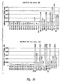
- Microarray is an effective method for evaluating large numbers of genes but due to its limited sensitivity it may not accurately determine the absolute tissue distribution of low abundance genes or may underestimate the degree of overexpression of more abundant genes due to signal saturation. For those genes showing overexpression by microarray expression profiling, further analysis was performed using quantitative RT-PCR based on Taqman TM probe detection, which comprises a greater dynamic range of sensitivity. Several different panels of normal and tumor tissues, distant metastases and cell lines were used for this purpose.
- Suitable polynucleotides according to the present invention may be further characterized or, alternatively, originally identified by employing a quantitative PCR methodology such as, for example, the Real-time PCR methodology.
- tissue and/or tumor samples such as, e.g. , metastatic tumor samples, may be tested along side the corresponding normal tissue sample and/or a panel of unrelated normal tissue samples.
- Real-time PCR is a technique that evaluates the level of PCR product accumulation during amplification. This technique permits quantitative evaluation of mRNA levels in multiple samples. Briefly, mRNA is extracted from tumor and normal tissue and cDNA is prepared using standard techniques.
- Real-time PCR may, for example, be performed either on the ABI 7700 Prism or on a GeneAmp® 5700 sequence detection system (PE Biosystems, Foster City, CA).
- the 7700 system uses a forward and a reverse primer in combination with a specific probe with a 5' fluorescent reporter dye at one end and a 3' quencher dye at the other end (TaqmanTM).
- TaqmanTM a forward and a reverse primer in combination with a specific probe with a 5' fluorescent reporter dye at one end and a 3' quencher dye at the other end
- the Real-time PCR is performed using Taq DNA polymerase with 5'-3' nuclease activity, the probe is cleaved and begins to fluoresce allowing the reaction to be monitored by the increase in fluorescence (Real-time).
- the 5700 system uses SYBR® green, a fluorescent dye, that only binds to double stranded DNA, and the same forward and reverse primers as the 7700 instrument.
- Matching primers and fluorescent probes may be designed according to the primer express program (PE Biosystems, Foster City, CA). Optimal concentrations of primers and probes are initially determined by those of ordinary skill in the art. Control (e.g ., ⁇ -actin) primers and probes may be obtained commercially from, for example, Perkin Elmer/Applied Biosystems (Foster City, CA).
- a standard curve is generated using a plasmid containing the gene of interest.
- Standard curves are generated using the Ct values determined in the real-time PCR, which are related to the initial cDNA concentration used in the assay. Standard dilutions ranging from 10-10 6 copies of the gene of interest are generally sufficient.
- a standard curve is generated for the control sequence. This permits standardization of initial RNA content of a tissue sample to the amount of control for comparison purposes.
- the present invention provides the illustrative breast tissue- and/or tumor-specific polynucleotides mammaglobin, lipophilin B, GABA ⁇ (B899P), B726P, B511S, B533S, B305D and B311D having sequences set forth in SEQ ID NO: 1, 3, 5-7, 11, 13, 15, 17, 19-24, 30, 32, and 73-76 illustrative polypeptides encoded thereby having amino acid sequences set forth in SEQ ID NO: 2, 4, 8-10, 12, 14, 16, 18, 25-29 and 31 and 77 that may be suitably employed in the detection of cancer, more specifically, breast cancer.
- the methods disclosed herein will also permit the identification of additional and/or alternative polynucleotides that are suitable for the detection of a wide range of cancers including, but not limited to, prostate cancer, breast cancer, colon cancer, ovarian cancer, lung cancer head & neck cancer, lymphoma, leukemia, melanoma, liver cancer, gastric cancer, kidney cancer, bladder cancer, pancreatic cancer and endometrial cancer.
- a cancer cell may be detected in a patient based on the presence of one or more polynucleotides within cells of a biological sample (for example, blood, lymph nodes, bone marrow, sera, sputum, urine and/or tumor biopsies) obtained from the patient.
- a biological sample for example, blood, lymph nodes, bone marrow, sera, sputum, urine and/or tumor biopsies
- polynucleotides may be used as markers to indicate the presence or absence of a cancer such as, e.g. , breast cancer.
- the present invention achieves these and other related objectives by providing a methodology for the simultaneous detection of more than one polynucleotide, the presence of which is diagnostic of the presence of cancer cells in a biological sample.
- Each of the various cancer detection methodologies disclosed herein have in common a step of hybridizing one or more oligonucleotide primers and/or probes, the hybridization of which is demonstrative of the presence of a tumor- and/or tissue-specific polynucleotide.
- intron-spanning oligonucleotides that are inoperative against polynucleotide of genomic DNA and, thus, these oligonucleotides are effective in substantially reducing and/or eliminating the detection of genomic DNA in the biological sample.
- the presence of a cancer cell in a patient may be determined by employing the following steps: (a) obtaining a biological sample from said patient; (b) contacting said biological sample with a first oligonucleotide that hybridizes to a first polynucleotide said first polynucleotide selected from the group consisting of polynucleotides depicted in SEQ ID NO:73 and SEQ ID NO:74; (c) contacting said biological sample with a second oligonucleotide that hybridizes to a second polynucleotide selected from the group consisting of SEQ ID NO: 1, 3, 5-7, 11, 13, 15, 17, 19-24, 30, 32, and 75; (d) detecting in said sample an amount of a polynucleotide that hybridizes to at least one of the oligonucleotides; and (e) comparing the amount of the polynucleotide that hybridizes to said oligonucleotide to
- Alternative embodiments of the present invention provide methods wherein the presence of a cancer cell in a patient is determined by employing the steps of: (a) obtaining a biological sample from said patient; (b) contacting said biological sample with a first oligonucleotide that hybridizes to a first polynucleotide said first polynucleotide depicted in SEQ ID NO:76; (c) contacting said biological sample with a second oligonucleotide that hybridizes to a second polynucleotide selected from the group consisting of SEQ ID NO: 1, 3, 5-7, 11, 13, 15, 17, 19-24, 30, 32, and 75; (d) detecting in said sample an amount of a polynucleotide that hybridizes to at least one of the oligonucleotides; and (e) comparing the amount of the polynucleotide that hybridizes to said oligonucleotide to a predetermined cut-off value, and therefrom determining the presence or absence of
- inventions provide methods for determining the presence or absence of a cancer in a patient. Such methods comprise the steps of: (a) obtaining a biological sample from said patient; (b) contacting said biological sample obtained from a patient with a first oligonucleotide that hybridizes to a polynucleotide sequence selected from the group consisting of polynucleotides depicted in SEQ ID NO:73, SEQ ID NO:74 and SEQ ID NO:76; (c) contacting said biological sample with a second oligonucleotide that hybridizes to a polynucleotide as depicted in SEQ ID NO:75; (d) contacting said biological sample with a third oligonucleotide that hybridizes to a polynucleotide selected from the group consisting of polynucleotides depicted in SEQ ID NO:5, SEQ ID NO:6 and SEQ ID NO:7; (e) contacting said biological sample with a fourth oligonucle
- oligonucleotide primers and probes should comprise an oligonucleotide sequence that has at least about 60%, preferably at least about 75% and more preferably at least about 90%, identity to a portion of a polynucleotide encoding a breast tumor protein that is at least 10 nucleotides, and preferably at least 20 nucleotides, in length.
- oligonucleotide primers hybridize to a polynucleotide encoding a polypeptide described herein under moderately stringent conditions, as defined above.
- Oligonucleotide primers which may be usefully employed in the diagnostic methods described herein preferably are at least 10-40 nucleotides in length.
- the oligonucleotide primers comprise at least 10 contiguous nucleotides, more preferably at least 15 contiguous nucleotides, of a DNA molecule having a sequence recited in SEQ ID NO: 1, 3, 5-7, 11, 13, 15, 17, 19-24, 30, 32 and 73-76.
- Techniques for both PCR based assays and hybridization assays are well known in the art (see, for example, Mullis et al., Cold Spring Harbor Symp. Quant. Biol., 51 :263, 1987; Erlich ed., PCR Technology, Stockton Press, NY, 1989).
- the present invention also provides amplification-based methods for detecting the presence of a cancer cell in a patient.
- Exemplary methods comprise the steps of (a) obtaining a biological sample from a patient; (b) contacting the biological sample with a first oligonucleotide pair the first pair comprising a first oligonucleotide and a second oligonucleotide wherein the first oligonucleotide and the second oligonucleotide hybridize to a first polynucleotide and the complement thereof, respectively; (c) contacting the biological sample with a second oligonucleotide pair the second pair comprising a third oligonucleotide and a fourth oligonucleotide wherein the third and the fourth oligonucleotide hybridize to a second polynucleotide and the complement thereof, respectively, and wherein the first polynucleotide is unrelated in nucleotide sequence to the second polynucle
- Methods according to the present invention are suitable for identifying polynucleotides obtained from a wide variety of biological sample such as, for example, blood, serum, lymph node, bone marrow, sputum, urine and tumor biopsy sample.
- biological sample such as, for example, blood, serum, lymph node, bone marrow, sputum, urine and tumor biopsy sample.
- the biological sample is either blood, a lymph node or bone marrow.
- the lymph node may be a sentinel lymph node.
- cancers include, but are not limited to, prostate cancer, breast cancer, colon cancer, ovarian cancer, lung cancer head & neck cancer, lymphoma, leukemia, melanoma, liver cancer, gastric cancer, kidney cancer, bladder cancer, pancreatic cancer and endometrial cancer.
- Certain exemplary embodiments of the present invention provide methods wherein the polynucleotides to be detected are selected from the group consisting of mammaglobin, lipophilin B, GABA ⁇ (B899P), B726P, B511S, B533S, B305D and B311D.
- polynucleotides to be detected may be selected from the group consisting of those depicted in SEQ ID NO:73, SEQ ID NO:74, SEQ ID NO:75, SEQ ID NO:1, SEQ ID NO:3, SEQ ID NO:5, SEQ ID NO:6, SEQ ID NO:7, SEQ ID NO:11, SEQ ID NO:13, SEQ ID NO:15, SEQ ID NO:17, SEQ ID NO:19, SEQ ID NO:20, SEQ ID NO:21, SEQ ID NO:22, SEQ ID NO:23, SEQ ID NO:24, SEQ ID NO:30, SEQ ID NO:32, and SEQ ID NO:76.
- Suitable exemplary oligonucleotide probes and/or primers that may be used according to the methods of the present invention are disclosed herein by SEQ ID N0s:33-35 and 63-72.
- the oligonucleotides may be intron spanning oligonucleotides. Exemplary intron spanning oligonucleotides suitable for the detection of various polynucleotides disclosed herein are depicted in SEQ ID NOs:36-62.
- the artisan may prefer to detect the tissue- and/or tumor-specific polynucleotides by detecting a radiolabel and detecting a fluorophore.
- the oligonucleotide probe and/or primer may comprises a detectable moiety such as, for example, a radiolabel and/or a fluorophore.
- methods of the present invention may also comprise a step of fractionation prior to detection of the tissue- and/or tumor-specific polynucleotides such as, for example, by gel electrophoresis.
- methods described herein may be used as to monitor the progression of cancer.
- assays as provided for the diagnosis of a cancer may be performed over time, and the change in the level of reactive polypeptide(s) or polynucleotide(s) evaluated.
- the assays may be performed every 24-72 hours for a period of 6 months to 1 year, and thereafter performed as needed.
- a cancer is progressing in those patients in whom the level of polypeptide or polynucleotide detected increases over time.
- the cancer is not progressing when the level of reactive polypeptide or polynucleotide either remains constant or decreases with time.
- Certain in vivo diagnostic assays may be performed directly on a tumor.
- One such assay involves contacting tumor cells with a binding agent.
- the bound binding agent may then be detected directly or indirectly via a reporter group.
- binding agents may also be used in histological applications.
- polynucleotide probes may be used within such applications.
- multiple breast tumor protein markers may be assayed within a given sample. It will be apparent that binding agents specific for different proteins provided herein may be combined within a single assay. Further, multiple primers or probes may be used concurrently. The selection of tumor protein markers may be based on routine experiments to determine combinations that results in optimal sensitivity. In addition, or alternatively, assays for tumor proteins provided herein may be combined with assays for other known tumor antigens.
- cell capture technologies may be used prior to polynucleotide detection to improve the sensitivity of the various detection methodologies disclosed herein.
- Exemplary cell enrichment methodologies employ immunomagnetic beads that are coated with specific monoclonal antibodies to surface cell markers, or tetrameric antibody complexes, may be used to first enrich or positively select cancer cells in a sample.
- Various commercially available kits may be used, including Dynabeads® Epithelial Enrich (Dynal Biotech, Oslo, Norway), StemSep TM (StemCell Technologies, Inc., Vancouver, BC), and RosetteSep (StemCell Technologies). The skilled artisan will recognize that other readily available methodologies and kits may also be suitably employed to enrich or positively select desired cell populations.
- Dynabeads® Epithelial Enrich contains magnetic beads coated with mAbs specific for two glycoprotein membrane antigens expressed on normal and neoplastic epithelial tissues.
- the coated beads may be added to a sample and the sample then applied to a magnet, thereby capturing the cells bound to the beads. The unwanted cells are washed away and the magnetically isolated cells eluted from the beads and used in further analyses.
- RosetteSep can be used to enrich cells directly from a blood sample and consists of a cocktail of tetrameric antibodies that target a variety of unwanted cells and crosslinks them to glycophorin A on red blood cells (RBC) present in the sample, forming rosettes. When centrifuged over Ficoll, targeted cells pellet along with the free RBC.
- RBC red blood cells
- Antibodies that are available include, but are not limited to: CD2, CD3, CD4, CD5, CD8, CD10, CD11b, CD14, CD15, CD16, CD19, CD20, CD24, CD25, CD29, CD33, CD34, CD36, CD38, CD41, CD45, CD45RA, CD45RO, CD56, CD66B, CD66e, HLA-DR, IgE, and TCR ⁇ .
- mAbs specific for breast tumor antigens can be developed and used in a similar manner.
- mAbs that bind to tumor-specific cell surface antigens may be conjugated to magnetic beads, or formulated in a tetrameric antibody complex, and used to enrich or positively select metastatic breast tumor cells from a sample.
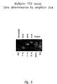
- cells may be further analysed. For example, the cells may be lysed and RNA isolated. RNA may then be subjected to RT-PCR analysis using breast tumor-specific multiplex primers in a Real-time PCR assay as described herein.
- cell capture technologies may be used in conjunction with Real-time PCR to provide a more sensitive tool for detection of metastatic cells expressing breast tumor antigens. Detection of breast cancer cells in bone marrow samples, peripheral blood, and small needle aspiration samples is desirable for diagnosis and prognosis in breast cancer patients.
- compositions and kits according to the present invention utilize two or more oligonucleotide primer pairs for the detection of cancer.
- the ability of such nucleic acid probes to specifically hybridize to a sequence of interest will enable them to be of use in detecting the presence of complementary sequences in a biological sample.
- the probes and/or primers of the present invention may be employed for detection via nucleic acid hybridization.
- nucleic acid segments that comprise a sequence region of at least about 15 nucleotide long contiguous sequence that has the same sequence as, or is complementary to, a 15 nucleotide long contiguous sequence of a polynucleotide to be detected will find particular utility. Longer contiguous identical or complementary sequences, e.g. , those of about 20, 30, 40, 50, 100, 200, 500, 1000 (including all intermediate lengths) and even up to full length sequences will also be of use in certain embodiments.
- Oligonucleotide primers having sequence regions consisting of contiguous nucleotide stretches of 10-14, 15-20, 30, 50, or even of 100-200 nucleotides or so (including intermediate lengths as well), identical or complementary to a polynucleotide to be detected , are particularly contemplated as primers for use in amplification reactions such as, e.g. , the polymerase chain reaction (PCR TM ).
- PCR TM polymerase chain reaction
- a primer of about 15-25 nucleotides in length allows the formation of a duplex molecule that is both stable and selective. Molecules having contiguous complementary sequences over stretches greater than 15 bases in length are generally preferred, though, in order to increase stability and selectivity of the hybrid, and thereby improve the quality and degree of specific hybrid molecules obtained.
- Primers may be selected from any portion of the polynucleotide to be detected. All that is required is to review the sequence, such as those exemplary polynucleotides set forth in SEQ ID NO: 1, 3, 5-7, 11, 13, 15, 17, 19-24, 30, 32, 73-75 ( Figures 3-6, respectively) and SEQ ID NO:76 (lipophilin B) or to any continuous portion of the sequence, from about 15-25 nucleotides in length up to and including the full length sequence, that one wishes to utilize as a primer.
- the choice of primer sequences may be governed by various factors. For example, one may wish to employ primers from towards the termini of the total sequence.
- the exemplary primers disclosed herein may optionally be used for their ability to selectively form duplex molecules with complementary stretches of the entire polynucleotide of interest such as those set forth in SEQ ID NO: 1, 3, 5-7, 11, 13, 15, 17, 19-24, 30, 32, 73-75 ( Figures 3-6, respectively), and SEQ ID NO:76 (lipophilin B).
- the present invention further provides the nucleotide sequence of various exemplary oligonucleotide primers and probes, set forth in SEQ ID NOs: 33-71, that may be used, as described in further detail herein, according to the methods of the present invention for the detection of cancer.
- Oligonucleotide primers according to the present invention may be readily prepared routinely by methods commonly available to the skilled artisan including, for example, directly synthesizing the primers by chemical means, as is commonly practiced using an automated oligonucleotide synthesizer.
- one will typically desire to employ varying conditions of hybridization to achieve varying degrees of selectivity of probe towards target sequence.
- relatively stringent conditions e . g ., one will select relatively low salt and/or high temperature conditions, such as provided by a salt concentration of from about 0.02 M to about 0.15 M salt at temperatures of from about 50°C to about 70°C.
- Such selective conditions tolerate little, if any, mismatch between the probe and the template or target strand, and would be particularly suitable for isolating related sequences.
- Each of the specific embodiments outlined herein for the detection of cancer has in common the detection of a tissue- and/or tumor-specific polynucleotide via the hybridization of one or more oligonucleotide primers and/or probes. Depending on such factors as the relative number of cancer cells present in the biological sample and/or the level of polynucleotide expression within each cancer cell, it may be preferred to perform an amplification step prior to performing the steps of detection.
- At least two oligonucleotide primers may be employed in a polymerase chain reaction (PCR) based assay to amplify a portion of a breast tumor cDNA derived from a biological sample, wherein at least one of the oligonucleotide primers is specific for ( i . e ., hybridizes to) a polynucleotide encoding the breast tumor protein.
- the amplified cDNA may optionally be subjected to a fractionation step such as, for example, gel electrophoresis.
- PCR TM polymerase chain reaction
- the primers will bind to the target and the polymerase will cause the primers to be extended along the target sequence by adding on nucleotides.
- the extended primers will dissociate from the target to form reaction products, excess primers will bind to the target and to the reaction product and the process is repeated.
- reverse transcription and PCR TM amplification procedure may be performed in order to quantify the amount of mRNA amplified.
- Polymerase chain reaction methodologies are well known in the art.
- RNA is extracted from a biological sample, such as blood, serum, lymph node, bone marrow, sputum, urine and tumor biopsy samples, and is reverse transcribed to produce cDNA molecules.
- PCR amplification using at least one specific primer generates a cDNA molecule, which may be separated and visualized using, for example, gel electrophoresis.
- Amplification may be performed on biological samples taken from a patient and from an individual who is not afflicted with a cancer.
- the amplification reaction may be performed on several dilutions of cDNA spanning two orders of magnitude. A two-fold or greater increase in expression in several dilutions of the test patient sample as compared to the same dilutions of the non-cancerous sample is typically considered positive.
- any of a variety of commercially available kits may be used to perform the amplification step.
- One such amplification technique is inverse PCR (see Triglia et al., Nucl. Acids Res. 16 :8186, 1988), which uses restriction enzymes to generate a fragment in the known region of the gene. The fragment is then circularized by intramolecular ligation and used as a template for PCR with divergent primers derived from the known region.
- sequences adjacent to a partial sequence may be retrieved by amplification with a primer to a linker sequence and a primer specific to a known region. The amplified sequences are typically subjected to a second round of amplification with the same linker primer and a second primer specific to the known region.
- RACE Rapid amplification of cDNA ends
- This technique involves the use of an internal primer and an external primer, which hybridizes to a polyA region or vector sequence, to identify sequences that are 5' and 3' of a known sequence. Additional techniques include capture PCR (Lagerstrom et al., PCR Methods Applic. 1 :111-19, 1991) and walking PCR (Parker et al., Nucl. Acids. Res. 19 :3055-60, 1991). Other methods employing amplification may also be employed to obtain a full length cDNA sequence.
- LCR ligase chain reaction
- Qbeta Replicase described in PCT Intl. Pat. Appl. Publ. No. PCT/US87/00880, incorporated herein by reference in its entirety, may also be used as still another amplification method in the present invention.
- a replicative sequence of RNA that has a region complementary to that of a target is added to a sample in the presence of an RNA polymerase.
- the polymerase will copy the replicative sequence that can then be detected.
- An isothermal amplification method in which restriction endonucleases and ligases are used to achieve the amplification of target molecules that contain nucleotide 5'-[ ⁇ -thio]triphosphates in one strand of a restriction site (Walker et al ., 1992, incorporated herein by reference in its entirety), may also be useful in the amplification of nucleic acids in the present invention.
- Strand Displacement Amplification is another method of carrying out isothermal amplification of nucleic acids which involves multiple rounds of strand displacement and synthesis, i . e . nick translation.
- a similar method, called Repair Chain Reaction (RCR) is another method of amplification which may be useful in the present invention and is involves annealing several probes throughout a region targeted for amplification, followed by a repair reaction in which only two of the four bases are present. The other two bases can be added as biotinylated derivatives for easy detection.
- RCR Repair Chain Reaction
- a similar approach is used in SDA.
- CPR cyclic probe reaction
- a probe having a 3' and 5' sequences of non-target DNA and an internal or “middle" sequence of the target protein specific RNA is hybridized to DNA which is present in a sample.
- the reaction is treated with RNaseH, and the products of the probe are identified as distinctive products by generating a signal that is released after digestion.
- the original template is annealed to another cycling probe and the reaction is repeated.
- CPR involves amplifying a signal generated by hybridization of a probe to a target gene specific expressed nucleic acid.
- modified primers are used in a PCR-like, template and enzyme dependent synthesis.
- the primers may be modified by labeling with a capture moiety (e.g ., biotin) and/or a detector moiety (e.g ., enzyme).
- a capture moiety e.g ., biotin
- a detector moiety e.g ., enzyme
- an excess of labeled probes is added to a sample.
- the probe binds and is cleaved catalytically. After cleavage, the target sequence is released intact to be bound by excess probe. Cleavage of the labeled probe signals the presence of the target sequence.
- nucleic acid amplification procedures include transcription-based amplification systems (TAS) (Kwoh et al ., 1989; PCT Intl. Pat. Appl. Publ. No. WO 88/10315), including nucleic acid sequence based amplification (NASBA) and 3SR
- TAS transcription-based amplification systems
- NASBA nucleic acid sequence based amplification
- 3SR nucleic acid sequence based amplification
- the nucleic acids can be prepared for amplification by standard phenol/chloroform extraction, heat denaturation of a sample, treatment with lysis buffer and minispin columns for isolation of DNA and RNA or guanidinium chloride extraction of RNA.
- amplification techniques involve annealing a primer that has sequences specific to the target sequence.
- DNA/RNA hybrids are digested with RNase H while double stranded DNA molecules are heat-denatured again. In either case the single stranded DNA is made fully double stranded by addition of second target-specific primer, followed by polymerization. The double stranded DNA molecules are then multiply transcribed by a polymerase such as T7 or SP6. In an isothermal cyclic reaction, the RNAs are reverse transcribed into DNA, and transcribed once again with a polymerase such as T7 or SP6. The resulting products, whether truncated or complete, indicate target-specific sequences.
- a polymerase such as T7 or SP6
- ssRNA single-stranded RNA
- dsDNA double-stranded DNA
- the ssRNA is a first template for a first primer oligonucleotide, which is elongated by reverse transcriptase (RNA-dependent DNA polymerase).
- RNA-dependent DNA polymerase reverse transcriptase
- the RNA is then removed from resulting DNA:RNA duplex by the action of ribonuclease H (RNase H, an RNase specific for RNA in a duplex with either DNA or RNA).
- RNase H ribonuclease H
- the resultant ssDNA is a second template for a second primer, which also includes the sequences of an RNA polymerase promoter (exemplified by T7 RNA polymerase) 5' to its homology to its template.
- This primer is then extended by DNA polymerase (exemplified by the large "Klenow" fragment of E. coli DNA polymerase I), resulting as a double-stranded DNA (“dsDNA”) molecule, having a sequence identical to that of the original RNA between the primers and having additionally, at one end, a promoter sequence.
- This promoter sequence can be used by the appropriate RNA polymerase to make many RNA copies of the DNA. These copies can then re-enter the cycle leading to very swift amplification. With proper choice of enzymes, this amplification can be done isothermally without addition of enzymes at each cycle. Because of the cyclical nature of this process, the starting sequence can be chosen to be in the form of either DNA or RNA.
- PCT Intl. Pat. Appl. Publ. No. WO 89/06700 disclose a nucleic acid sequence amplification scheme based on the hybridization of a promoter/primer sequence to a target single-stranded DNA ("ssDNA") followed by transcription of many RNA copies of the sequence. This scheme is not cyclic; i . e . new templates are not produced from the resultant RNA transcripts.
- Other amplification methods include "RACE” (Frohman, 1990), and “one-sided PCR” (Ohara, 1989) which are well-known to those of skill in the art.
- kits for use within any of the above diagnostic methods.
- Such kits typically comprise two or more components necessary for performing a diagnostic assay.
- Components may be compounds, reagents, containers and/or equipment.
- one container within a kit may contain a monoclonal antibody or fragment thereof that specifically binds to a breast tumor protein.
- Such antibodies or fragments may be provided attached to a support material, as described above.
- One or more additional containers may enclose elements, such as reagents or buffers, to be used in the assay.
- Such kits may also, or alternatively, contain a detection reagent as described above that contains a reporter group suitable for direct or indirect detection of antibody binding.
- kits that are suitable for performing the detection methods of the present invention.
- Exemplary kits comprise oligonucleotide primer pairs each one of which specifically hybridizes to a distinct polynucleotide.
- kits according to the present invention may also comprise a nucleic acid polymerase and suitable buffer.
- Exemplary oligonucleotide primers suitable for kits of the present invention are disclosed herein by SEQ ID NOs: 33-71.
- Exemplary polynucleotides suitable for kits of the present invention are disclosed in SEQ ID NO:73, SEQ ID NO:74, SEQ ID NO:75, SEQ ID NO:1, SEQ ID NO:3, SEQ ID NO:5, SEQ ID NO:6, SEQ ID NO:7, SEQ ID NO:11, SEQ ID NO:13, SEQ ID NO:15, SEQ ID NO:17, SEQ ID NO:19, SEQ ID NO:20, SEQ ID NO:21, SEQ ID NO:22, SEQ ID NO:23, SEQ ID NO:24, SEQ ID NO:30, SEQ ID NO:32, and lipophilin B.
- kits may be designed to detect the level of mRNA encoding a breast tumor protein in a biological sample.
- kits generally comprise at least one oligonucleotide probe or primer, as described above, that hybridizes to a polynucleotide encoding a breast tumor protein.
- Such an oligonucleotide may be used, for example, within a PCR or hybridization assay. Additional components that may be present within such kits include a second oligonucleotide and/or a diagnostic reagent or container to facilitate the detection of a polynucleotide encoding a breast tumor protein.
- compositions useful in the methods disclosed herein comprise two or more oligonucleotide primer pairs each one of which specifically hybridizes to a distinct polynucleotide.
- oligonucleotide primers suitable for compositions of the present invention are disclosed herein by SEQ ID NOs: 33-71.
- Exemplary polynucleotides suitable for compositions of the present invention are disclosed in SEQ ID NO:73, SEQ ID NO:74, SEQ ID NO:75, SEQ ID NO:1, SEQ ID NO:3, SEQ ID NO:5, SEQ ID NO:6, SEQ ID NO:7, SEQ ID NO:11, SEQ ID NO:13, SEQ ID NO:15, SEQ ID NO:17, SEQ ID NO:19, SEQ ID NO:20, SEQ ID NO:21, SEQ ID NO:22, SEQ ID NO:23, SEQ ID NO:24, SEQ ID NO:30, SEQ ID NO:32, and lipophilin B.
- This example discloses the use of differential display to enrich for polynucleotides that are over-expressed in breast tumor tissues.
- This example discloses the preparation of a breast tumor cDNA subtraction library enriched in breast tumor specific polynucleotides.
- cDNA library subtraction was performed as described with some modification. See , Hara, T. et al., Blood 84 : 189-199 (1994).
- the breast tumor library (tracer) that was made from a pool of three breast tumors was subtracted with normal breast library (driver) to identify breast tumor specific genes. More recent subtractions utilized 6-10 normal tissues as driver to subtract out common genes more efficiently, with an emphasis on essential tissues along with one "immunological" tissue (e.g ., spleen, lymph node, or PBMC), to assist in the removal of cDNAs derived from lymphocyte infiltration in tumors.
- immunological tissue e.g ., spleen, lymph node, or PBMC
- the breast tumor specific subtracted cDNA library was generated as follows: driver cDNA library was digested with EcoRI, NotI, and SfuI (SfuI cleaves the vector), filled in with DNA polymerase klenow fragment. After phenol-chloroform extraction and ethanol precipitation, the DNA was labeled with Photoprobe biotin and dissolved in H 2 O. Tracer cDNA library was digested with BamHI and XhoI, phenol chloroform extracted, passed through Chroma spin-400 columns, ethanol precipitated, and mixed with driver DNA for hybridization at 68°C for 20 hours [long hybridization (LH)].
- reaction mixture was then subjected to the streptavidin treatment followed by phenol/chloroform extraction for a total of four times.
- Subtracted DNA was precipitated and subjected to a hybridization at 68°C for 2 hours with driver DNA again [short hybridization (SH)].
- SH short hybridization
- cDNA library subtraction was repeated by subtracting the tracer cDNA library with the driver cDNA library plus abundant cDNAs from primary subtractions. This resulted in the depletion of these abundant sequences and the generation of subtraction libraries that contain less abundant sequences.
- plasmid DNA was prepared from 100-200 independent clones, which were randomly picked from the subtracted library, and characterized by DNA sequencing. The determined cDNA and expected amino acid sequences for the isolated cDNAs were compared to known sequences using the most recent Genbank and human EST databases.
- This example discloses PCR subtraction to enrich for breast tumor specific polynucleotides.
- PCR-subtraction was performed essentially as described in the literature. See, Diatchenko, L. et al., Proc Natl Acad Sci U S A . 93 :6025-6030 (1996) and Yang, G.P. et al., Nucleic acids Res. 27 :1517-23 (1999). Briefly, this type of subtraction works by ligating two different adapters to different aliquots of a restriction enzyme digested tester (breast tumor) cDNA sample, followed by mixing of the testers separately with excess driver (without adapters). This first hybridization results in normalization of single stranded tester specific cDNA due to the second order kinetics of hybridization.
- RNA Prior to cDNA synthesis RNA was treated with DNase I (Ambion) in the presence of RNasin (Promega Biotech, Madison, WI) to remove DNA contamination.
- RNasin Promega Biotech, Madison, WI
- the cDNA for use in real-time PCR tissue panels was prepared using 25 ⁇ l Oligo dT (Boehringer-Mannheim) primer with superscript II reverse transcriptase (Gibco BRL, Bethesda, MD).
- the isolation and characterization of the breast-specific antigens B511S and B533S is described in U.S. Patent Application 09/346,327, filed July 2, 1999, the disclosure of which is hereby incorporated by reference in its entirety.
- the determined cDNA sequence for B511S is provided in SEQ ID NO: 30, with the corresponding amino acid sequence being provided in SEQ ID NO: 31.
- the determined cDNA sequence for B533S is provided in SEQ ID NO: 32.
- the isolation and characterization of the breast-specific antigen B726P is described in U.S. Patent Applications 09/285,480, filed April 2, 1999, and 09/433,826, filed November 3, 1999.
- the determined cDNA sequences for splice variants of B726P are provided in SEQ ID NO: 13, 15, 17 and 19-24, with the corresponding amino acid sequences being provided in SEQ ID NO: 14, 16, 18 and 25-29.
- the determined cDNA sequence for B311D is provided in SEQ ID NO:11, with the corresponding amino acid sequence being provided in SEQ ID NO:12.
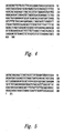
- cDNA sequences for mammaglobin are provided in Figs. 4 and 5, with the cDNA sequence for GABA ⁇ being provided in Fig 6 and are disclosed in SEQ ID NOs: 73-75, respectively.
- the isolation and characteization of the breast-specific antigen lipophilin B has been described in U.S. Patent Application 09/780,842, filed February 8, 2001 .
- the determined cDNA sequence for lipophilin B is provided in SEQ ID NO:76, with the corresponding amino acid sequence being provided in SEQ ID NO:77.
- the nucleotide sequences of several sequence variants of lipophilin B are also described in the 09/780,842 application.
- This example discloses the use of microarray analyses to identify polynucleotides that are at least two-fold overexpressed in breast tumor tissue samples as compared to normal breast tissue samples.
- mRNA expression of the polynucleotides of interest was performed as follows.
- cDNA for the different genes was prepared as described above and arrayed on a glass slide (Incyte, Palo Alto, CA). The arrayed cDNA was then hybridized with a 1:1 mixture of Cy3 or Cy5 fluorescent labeled first strand cDNAs obtained from polyA+ RNA from breast tumors, normal breast and normal tissues and other tumors as described in Shalon, D. et al., Genome Res. 6 :639-45 (1996).
- Cy3 Probe 1
- Cy5 Probe 2
- DNA microarray analyses was used primarily as a screening tool to determine tissue/tumor specificity of cDNA's recovered from the differential display, cDNA library and PCR subtractions, prior to more rigorous analysis by quantitative RT-PCR, northern blotting, and immunohistochemistry.
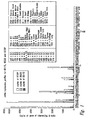
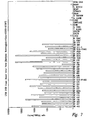
- Microarray analysis was performed on two microchips. A total of 3603 subtracted cDNA's and 197 differential display templates were evaluated to identify 40 candidates for further analysis by quantitative PCR. From these candidates, several were chosen on the basis of favorable tissue specificity profiles, including B305D, B311D, B726P, B511S and B533S, indicating their overexpression profiles in breast tumors and/or normal breast versus other normal tissues. It was evident that the expression of these genes showed a high degree of specificity for breast tumors and/or breast tissue. In addition, these genes have in many cases complementary expression profiles.
- mammaglobin The two known breast-specific genes, mammaglobin and ⁇ -aminobutyrate type A receptor ⁇ subunit (GABA ⁇ ) were also subjected to microarray analysis. mRNA expression of mammaglobin has been previously described to be upregulated in proliferating breast tissue, including breast tumors. See, (Watson et al., Cancer Res., 56 : 860-5 (1996); Watson et al., Cancer Res., 59 : 3028-3031 (1999); Watson et al., Oncogene. 16 :817-24 (1998)).
- This example discloses the use of quantitative Real-time PCR to confirm the microarray identification polynucleotide that are at least two-fold overexpressed in breast tumor tissue samples as compared to normal breast tissue samples.
- the 7700 system uses a forward and a reverse primer in combination with a specific probe designed to anneal to sequence between the forward and reverse primer. This probe was conjugated at the 5'end with a fluorescent reporter dye and a quencher dye at the other 3' end (Taqman TM ).
- the concentrations used in the quantitative PCR for the forward primers for mammaglobin, GABA ⁇ , B305D C form, B311D, B511S, B533S and B726P were 900, 900, 300, 900, 900, 300 and 300nM respectively.
- the reverse primers they were 300, 900, 900, 900, 300, 900 and 900nM respectively.
- Primers and probes so produced were used in the universal thermal cycling program in real-time PCR. They were titrated to determine the optimal concentrations using a checkerboard approach. A pool of cDNA from target tumors was used in this optimization process. The reaction was performed in 25 ⁇ l volumes. The final probe concentration in all cases was 160nM.
- dATP, dCTP and dGTP were at 0.2mM and dUTP at 0.4mM.
- Amplitaq gold and Amperase UNG (PE Biosystems, Foster City, CA) were used at 0.625 units and 0.25 units per reaction.
- MgCl 2 was at a final concentration of 5mM.
- Trace amounts of glycerol, gelatin and Tween 20 (Sigma Chem Co, St Louis, MO) were added to stabilize the reaction. Each reaction contained 2 ⁇ l of diluted template.
- the cDNA from RT reactions prepared as above was diluted 1:10 for the gene of interest and 1:100 for ⁇ -Actin.
- the reaction mix (25 ⁇ l) included 2.5 ⁇ l of SYBR green buffer, 2 ⁇ l of cDNA template and 2.5 ⁇ l each of the forward and reverse primers for the gene of interest. This mix also contained 3mM MgCl 2 , 0.25units of AmpErase UNG, 0.625 units of Amplitaq gold, 0.08% glycerol, 0.05% gelatin, 0.0001% Tween 20 and 1mM dNTP mix. In both formats, 40 cycles of amplification were performed.
- a standard curve was generated for each run using the plasmid containing the gene of interest. Standard curves were generated using the Ct values determined in the real-time PCR which were related to the initial cDNA concentration used in the assay. Standard dilutions ranging from 20-2x10 6 copies of the gene of interest were used for this purpose. In addition, a standard curve was generated for the housekeeping gene ⁇ -actin ranging from 200fg-2000pg to enable normalization to a constant amount of ⁇ -Actin. This allowed the evaluation of the over-expression levels seen with each of the genes.
- the genes B311D, B533S and B726P were evaluated in quantitative PCR as described above on two different panels consisting of: (a) breast tumor, breast normal and normal tissues; and (b) breast tumor metastases (primarily lymph nodes), using the primers and probes shown above in Table 1.
- the data for panel (a) is shown in Figure 1 for all three genes.
- the three genes showed identical breast tissue expression profiles. However, the relative level of gene expression was very different in each case.
- B311D in general was expressed at lower levels than B533S and both less than B726P, but all three were restricted to breast tissue.
- the quantitative PCR thus confirmed there was a differential expression between normal breast tissue and breast tumors for all three genes, and that approximately 50% of breast tumors over-expressed these genes.
- Mammaglobin is a homologue of a rabbit uteroglobin and the rat steroid binding protein subunit C3 and is a low molecular weight protein that is highly glycosylated. In contrast to its homologs, mammaglobin has been reported to be breast specific and over-expression has been described in breast tumor biopsies (23%) and primary and metastatic breast tumors ( ⁇ 75%) with reports of the detection of mammaglobin mRNA expression in 91 % of lymph nodes from metastatic breast cancer patients.
- B511S The breast-specific gene B511S, while having a different profile of reactivity on breast tumors and normal breast tissue to mammaglobin, reacted with the same subset of normal tissues as mammaglobin.
- B511S by PSORT analysis is indicated to have an ORF of 90aa and to be a secreted protein as is the case for mammaglobin.
- B511S has no evidence of a transmembrane domain but may harbor a cleavable signal sequence.
- Mammaglobin detected 14/24 of distant metastatic breast tumors, 31/42 breast tumors and exhibited ten-fold over-expression in tumors and metastases as compared to normal breast tissue.
- B511S detected 33/42 breast tumors and 14/24 distant metastases, while a combination of B511S with mammaglobin would be predicted to detect 38/42 breast tumors and 17/24 metastatic lesions (Table 2 above).
- the quantitative level of expression of B511S and mammaglobin were also in similar ranges, in concordance with the microarray profiles observed for these two genes. Other genes that were additive with mammaglobin are shown in Table 3.
- B305D was shown to be highly over-expressed in breast tumors, prostate tumors, normal prostate tissue and testis compared to normal tissues, including normal breast tissue. Different splice variants of B305D have been identified with form A and C being the most abundant but all tested have similar tissue profiles in quantitative PCR. The A and C forms contain ORF's of 320 and 385 aa, respectively. B305D is predicted by PSORT to be a Type II membrane protein that comprises a series of ankyrin repeats. A known gene shown to be complementary with B305D, in breast tumors, was GABA ⁇ .
- This gene is a member of the GABA A receptor family and encodes a protein that has 30-40% amino acid homology with other family members, and has been shown by Northern blot analysis to be over-expressed in lung, thymus and prostate at low levels and highly over-expressed in uterus. Its expression in breast tissue has not been previously described. This is in contrast to other GABA A receptors that have appreciable expression in neuronal tissues. Tissue expression profiling of this gene showed it to be over-expressed in breast tumors in an inverse relationship to the B305D gene (Table 3). GABA ⁇ detected 15/25 tumors and 6/21 metastases including 4 tumors and 5 metastases missed by mammaglobin.
- B305D detected 13/25 breast tumors and 8/21 metastases, again including 3 tumors and 2 metastases missed by mammaglobin.
- a combination of just B305D and the GABA ⁇ would be predicted to identify 22/25 breast tumors and 14/21 metastases.
- the combination of B305D and GABA ⁇ with mammaglobin in detecting breast metastases is shown in Table 3 above and Figures. 3A and 3B. This combination detected 20/21 of the breast metastases as well as 25/25 breast tumors that were evaluated on the same panels for all three genes. The one breast metastasis that was negative for these three genes was strongly positive for B726P (Figs. 3A and 3B).
- an immunocapture (cell capture) method was employed to first enrich for epithelial cells prior to RT-PCR analysis.
- Immunomagnetic polystyrene beads coated with specific monoclonal antibodies to two glycoproteins on the surface of epithelial cells were used for this purpose.
- Such an enrichment procedure increased the sensitivity of detection ( ⁇ 100 fold) as compared to direct isolation of poly A + RNA, as shown in Table 4.
- Mammaglobin-positive cells (MB415) were spiked into whole blood at various concentrations and then extracted using either epithelial cell enrichment or direct isolation from blood. Using enrichment procedures, mammaglobin mRNA was found to be detectable at much lower levels than when direct isolation was used.
- Whole blood samples from patients with metatastic breast cancer were subsequently treated with the immunomagnetic beads.
- Poly A + RNA was then isolated, cDNA prepared and run in quantitative PCR using two gene specific primers (Table 1) and a fluorescent probe (Taqman TM ). As observed in breast cancer tissues, complementation was also seen in the detection of circulating tumor cells derived from breast cancers.
- mammaglobin PCR detected circulating tumor cells in a high percentage of blood samples, albeit at low levels, from metastatic breast cancer patients (20/32) when compared to the normal blood samples (Table 5) but several of the other genes tested to date further increased this detection rate. This included B726P, B305D, B311D, B533S and GABA ⁇ .
- the detection level of mammaglobin in blood samples from metastatic breast cancer patients is higher than described previously (62 vs. 49%), despite testing smaller blood volumes, probably because of the use of epithelial marker-specific enrichment in our study. A combination of all the genes tested indicate that 27/32 samples were positive by one or more of these genes.
- mammaglobin, GABA ⁇ , B305D (C form) and B726P specific primers and specific Taqman probes were employed in different combinations to analyze their combined mRNA expression profile in breast metastases (B. met) and breast tumor (B. tumors) samples using real-time PCR.
- the forward and reverse primers and probes employed for mammaglobin, B305D (C form) and B726P are shown in Table 1.
- the forward primer and probe employed for GABA ⁇ are shown in Table 1, with the reverse primer being as follows: TTCAAATATAAGTGAAGAAAAAATTAG-TAGATCAA (SEQ ID NO:51).
- GABA ⁇ mammaglobin and ⁇ aminobutyrate type A receptor ⁇ subunit
- B511S and B533S are two cDNA fragments isolated using cDNA library subtraction approach (manuscript in preparation) while the B726P cDNA fragment was derived from PCR subtraction (Jiang et al., Proceedings of the Amer Assoc Cancer Res. 40:Abstract #216, 32 (1999); Xu et al., Proceedings of the Amer Assoc Cancer Res. 40 :Abstract #2115, 319 (1999); and Molesh et al., Proceedings of Amer Assoc Cancer Res. 41 :Abstract #4330, 681 (2000).
- B311D, B533S and B726P showed identical breast tissue expression profile by quantitative PCR analysis. These genes were evaluated in quantitative PCR on two different panels consisting of (a) breast tumor, breast normal and normal tissues and (b) panel of breast tumor metastases (primarily lymph nodes). Primers and probes used are shown in Table 1. The data for panel (a) is shown in Figure 2 for all three genes. Overall, the expression profiles are comparable and are in the same rank order, however, the levels of expression are considerably different. B311D in general was expressed at lower levels than B533S and both less than B726P but all three were restricted to breast tissue. All three sequences were used to search against the Genbank database. Both B311D and B533S sequences contain different repetitive sequences and an ORF has not been identified for either. B726P is a novel gene, with mRNA splicing yielding several different putative ORF's.
- B511S detected 33/42 breast tumors and 14/24 distant metastases.
- a combination of B511S with mammaglobin would be predicted to detect 38/42 breast tumors and 17/24 metastatic lesions.
- the quantitative level of expression of B511S and mammaglobin were also in similar ranges, in concordance with the microarray profiles observed for these two genes.
- B305D was highly over-expressed in breast tumors, prostate tumors, normal prostate tissue and testis compared to normal tissues including normal breast tissue. Different splice variants of B305D were identified with the forms A and C being the most abundant. All forms tested had similar tissue profiles in quantitative PCR. The A and C forms contain ORF's of 320 and 385 aa, respectively.
- tissue expression profiling of this gene showed it to be over-expressed in breast tumors in an inverse relationship to the B305D gene (Table 3).
- GABA ⁇ detected 15/25 tumors and 6/21 metastases including 4 tumors and 5 metastases missed by mammaglobin.
- B305D detected 13/25 breast tumors and 8/21 metastases again including 3 tumors and 2 metastases missed by mammaglobin.
- a combination of just B305D and the GABA ⁇ would be predicted to identify 22/25 breast tumors and 14/21 metastases. This combination detected 20/21 of the breast metastases as well as 25/25 breast tumors that were evaluated on the same panels for all three genes. The one breast metastasis that was negative for these three genes was strongly positive for B726P.
- GABA ⁇ GABA ⁇ in breast tumors was also a novel observation. While the expression of several genes complemented that seen with mammaglobin, two genes in particular, B305D and GABA ⁇ added to the diagnostic sensitivity of mammaglobin detection. A combination of these three genes detected 45/46 (97.8%) breast tumors and metastases evaluated. Inclusion of B726P enabled the detection of all 25 of the breast tumors and 21 distant metastases.
- This example discloses the enhanced sensitivity achieved by use of the immunocapture cell capture methodology for enrichment of circulating breast cancer cells.
- Epithelial cells were enriched from blood samples with an immunomagnetic bead separation method (Dynal A.S, Oslo, Norway) utilizing magnetic beads coated with monoclonal antibodies specific for glycopolypeptide antigens on the surface of human epithelial cells.
- an immunomagnetic bead separation method (Dynal A.S, Oslo, Norway) utilizing magnetic beads coated with monoclonal antibodies specific for glycopolypeptide antigens on the surface of human epithelial cells.
- Examples suitable cell-surface antigens are described, for example, in Momburg, F. et al., Cancer Res. , 41 :2883-91 (1997); Naume, B. et al., Journal of Hemotherapy . 6 :103-113 (1997); Naume, B. et al., Int J Cancer.
- RNA samples were finally suspended in 10mM Tris HCl pH 8 and subjected to reverse transcription. The RNA was then subjected to Real time PCR using gene specific primers and probes with reaction conditions as outlined herein above. ⁇ -Actin content was also determined and used for normalization. Samples with gene of interest copies/ng ⁇ -actin greater than the mean of the normal samples + 3 standard deviations were considered positive. Real time PCR on blood samples was performed exclusively using the Taqman TM procedure but extending to 50 cycles.
- Mammaglobin mRNA using enrichment procedures was found to be detectable at much lower levels than when direct isolation was used.
- Whole blood samples from patients with metatastic breast cancer were subsequently treated with the immunomagnetic beads, polyA + RNA was then isolated, cDNA made and run in quantitative PCR using two gene specific primers to mammaglobin and a fluorescent probe (Taqman TM ).
- Taqman TM fluorescent probe
- complementation was also seen in the detection of circulating tumor cells derived from breast cancers.
- mammaglobin PCR detected circulating tumor cells in a high percentage of bloods, albeit at low levels, from metastatic breast cancer (20/32) when compared to the normal blood samples.
- This Multiplex assay was designed to detect LipophilinB expression instead of Mammaglobin. Due to their similar expression profiles, LipophilinB can replace Mammaglobin in this Multiplex PCR assay for breast cancer detection.
- the assay was carried out as follows: LipophilinB, B899P (Gaba), B305D, and B726P specific primers, and specific Taqman probes, were used to analyze their combined mRNA expression profile in breast tumors. The primers and probes are shown below:
- the assay conditions were:
- LipophilinB expression was detected in 14 out of 27 breast tumor samples.
- the Multiplex assay for LipophilinB, B899P, B305D-C and B726P detected an expression signal in 27 out of 27 tumors with the detection level above 10 mRNA copies/1000 pg actin in the majority of samples and above 100 mRNA copies/1000 pg actin in 5 out of the 27 samples tested (Figure 8).
- the Multiplex Real-time PCR assay described above was used to detect the expression of Mammaglobin (or LipophilinB), Gaba (B899P), B305D-C and B726P simultaneously. According to this Example, assay conditions and primer sequences were optimized to achieve parallel amplification of four PCR products with different lengths. Positive samples of this assay can be further characterized by gel electrophoresis and the expressed gene(s) of interest can be determined according to the detected amplicon size(s).
- Mammaglobin or LipophilinB
- Gaba B899P
- B305D B726P specific primers and specific Taqman probes were used to simultaneously detect their expression.
- the primers and probes used in this example are shown below.
- Primer locations and assay conditions were optimized to achieve parallel amplification of four PCR products with different sizes.
- the assay conditions were:
- each primer set in the multiplex assay results in a band of unique length
- expression signals of the four genes of interest can be measured individually by agarose gel analysis ( see, Figure 9), or the combined expression signal of all four genes can be measured in real-time on an ABI 7700 Prism sequence detection system (PE Biosystems, Foster City, CA).
- the expression of LipophilinB can also be detected instead of Mammaglobin.
- specific primers have been described herein, different primer sequences, different primer or probe labeling and different detection systems could be used to perform this multiplex assay.
- a second fluorogenic reporter dye could be incorporated for parallel detection of a reference gene by real-time PCR.
- a SYBR Green detection system could be used instead of the Taqman probe approach.
- the Multiplex Real-time PCR assay described herein can detect the expression of Mammaglobin, Gaba (B899P), B305D-C and B726P simultaneously.
- the combined expression levels of these genes is measured in real-time on an ABI 7700 Prism sequence detection system (PE Biosystems, Foster City, CA).
- Individually expressed genes can also be identified due to different amplicon sizes via gel electrophoresis.
- samples derived from non-DNase treated RNAs e.g . lymph node cDNA
- DNase-treatment for small RNA-samples e.g.
- intron-spanning primer pairs have been designed to exclude the amplification of genomic DNA and therefore to eliminate nonspecific and false positive signals. False positive signal is caused by genomic DNA contamination in cDNA specimens.
- the optimized Multiplex assay described herein excludes the amplification of genomic DNA and allows specific detection of target gene expression without the necessity of prior DNase treatment of RNA samples. Moreover the genomic match and the location of the Intron-Exon border could be verified with these primer sets.
- Figure 10 shows a comparison of the multiplex assay using intron-exon border spanning primers (bottom panel) and the multiplex assay using non-optimized primers (top panel), to detect breast cancer cells in a panel of lymph node tissues. This experiment shows that reduction in background resulting from genomic DNA contamination in samples is achieved using the intron-exon spanning primers of the present invention.
- Lymph node staging is important for determining appropriate adjuvant hormone and chemotherapy.
- Sentinel lymph node biopsy (SLNB) has the potential to improve detection of metastases and to provide prognostic values to lead to therapy with minimal morbidity associated with complete lymph node dissection.
- SLNB implements mapping of the one or two lymph nodes which primarily drain the tumor and therefore are most likely to harbor metastatic disease (the sentinel nodes). Routine pathological' analysis of lymph nodes result in a high false-negative rate: one-third of women with pathologically negative lymph nodes develop recurrent disease [Bland: The Breast: Saunders 1991].
- a more sensitive detection technique for tumor cells would be RT-PCR but its application is limited by lack of a single specific markers.
- the multimarker assay described above increases the likelihood of cancer detection across the population without producing false-positive results from normal lymph nodes.
- Sentinel lymph nodes are located with dyes and/or radiolabelled colloid injected in the primary lesion site and sentinel lymph node biopsy allows pathological examination for micrometastatic deposits, staging of the axilla and therefore can avoid unnecessary axillary dissection.
- Nodal micrometastases can be located with staining (haematoxylin or eosin) or immunohistochemical analysis for cytokeratin proteins.
- Immunocytochemical staining techniques can produce frequent false-negative results by missing small metastatic foci due to inadequate sectioning of the node. Immunohistochemistry can result in false-positive results due to illegitimate expression of cytokeratins (reticulum cells) or in false-negative results when using the antibody Ber-Ep4 which corresponding antigen is not expressed on all tumor cells.
- the multiplex assay described herein could provide a more sensitive detection tool for positive sentinel lymph nodes. Moreover the detection of breast cancer cells in bone marrow samples, peripheral blood and small needle aspiration samples is desirable for diagnosis and prognosis in breast cancer patients.
Landscapes
- Chemical & Material Sciences (AREA)
- Life Sciences & Earth Sciences (AREA)
- Organic Chemistry (AREA)
- Health & Medical Sciences (AREA)
- Proteomics, Peptides & Aminoacids (AREA)
- Zoology (AREA)
- Wood Science & Technology (AREA)
- Engineering & Computer Science (AREA)
- Analytical Chemistry (AREA)
- Genetics & Genomics (AREA)
- Immunology (AREA)
- Molecular Biology (AREA)
- General Engineering & Computer Science (AREA)
- Biotechnology (AREA)
- Biophysics (AREA)
- Physics & Mathematics (AREA)
- Biochemistry (AREA)
- Bioinformatics & Cheminformatics (AREA)
- Microbiology (AREA)
- General Health & Medical Sciences (AREA)
- Chemical Kinetics & Catalysis (AREA)
- Pathology (AREA)
- Hospice & Palliative Care (AREA)
- Oncology (AREA)
- Measuring Or Testing Involving Enzymes Or Micro-Organisms (AREA)
- Medicines Containing Antibodies Or Antigens For Use As Internal Diagnostic Agents (AREA)
- Investigating Or Analysing Biological Materials (AREA)
Claims (35)
- Procédé destiné à détecter la présence d'une cellule du cancer du sein, ledit procédé comprenant les étapes de :(a) mise en contact d'un échantillon biologique obtenu d'un patient avec un premier oligonucléotide qui s'hybride avec un premier polynucléotide choisi dans le groupe consistant en la mammaglobine et la lipophiline B ;(b) mise en contact dudit échantillon biologique avec un deuxième oligonucléotide qui s'hybride avec une deuxième séquence polynucléotidique comprenant une séquence polynucléotidique GABAn (B899P) ;(c) détection dans ledit échantillon biologique d'une quantité d'oligonucléotide hybridé des étapes (a) et (b) ; et(d) comparaison de la quantité de polynucléotide qui s'hybride avec les oligonucléotides des étapes (a) et (b) avec une valeur seuil prédéterminée, et, à partir de là, détermination de la présence ou de l'absence d'un cancer chez le patient.
- Procédé selon la revendication 1 dans lequel ledit premier polynucléotide est choisi dans le groupe consistant en les polynucléotides décrits dans les séquences ayant pour identificateurs SEQ ID N° 73, SEQ ID N° 74 et SEQ ID N° 76, ou le complémentaire de ceux-ci ; et dans lequel ledit deuxième polynucléotide comprend le polynucléotide décrit dans la séquence ayant pour identificateur SEQ ID N° 75, ou le complémentaire de celui-ci.
- Procédé selon la revendication 2 comprenant en outre :(e) la mise en contact dudit échantillon biologique avec un troisième oligonucléotide qui s'hybride avec un troisième polynucléotide choisi dans le groupe consistant en les polynucléotides décrits dans les séquences ayant pour identificateurs SEQ ID N° 5, SEQ ID N° 6 et SEQ ID N° 7 ; ou le complémentaire de ceux-ci ;(f) la mise en contact dudit échantillon biologique avec un quatrième oligonucléotide qui s'hybride avec un quatrième polynucléotide choisi dans le groupe consistant en les polynucléotides décrits dans les séquences ayant pour identificateurs SEQ ID N° 13, SEQ ID N° 15, SEQ ID N° 17, SEQ ID N° 19, SEQ ID N° 20, SEQ ID N° 21, SEQ ID N° 22, SEQ ID N° 23 et SEQ ID N° 24 ; ou le complémentaire de ceux-ci et
dans lequel l'étape (c) comprend la détection dans ledit échantillon biologique d'une quantité de l'oligonucléotide hybridé des étapes (a), (b), (e), et (f). - Procédé selon la revendication 2 comprenant en outre :(e) la mise en contact dudit échantillon biologique avec un troisième oligonucléotide qui s'hybride avec un troisième polynucléotide choisi dans le groupe consistant en un polynucléotide décrit dans les séquences ayant pour identificateurs SEQ ID N° 1, SEQ ID N° 3, SEQ ID N° 5, SEQ ID N° 6 et SEQ ID N° 7 ; ou le complémentaire de celui-ci ;(f) la mise en contact dudit échantillon biologique avec un quatrième oligonucléotide qui s'hybride avec un quatrième polynucléotide choisi dans le groupe consistant en un polynucléotide décrit dans la séquence ayant pour identificateur SEQ ID N° 11, ou le complémentaire de cclui-ci ;(g) la mise en contact dudit échantillon biologique avec un cinquième oligonucléotide qui s'hybride avec un cinquième polynucléotide choisi dans le groupe consistant en un polynucléotide décrit dans les séquences ayant pour identificateurs SEQ ID N° 13, 15 et 17 ; ou le complémentaire de celui-ci ;(h) la mise en contact dudit échantillon biologique avec un sixième oligonucléotide qui s'hybride avec un sixième polynucléotide choisi dans le groupe consistant en un polynucléotide décrit dans les séquences ayant pour identificateurs SEQ ID N° 19, SEQ ID N° 20, SEQ ID N° 21, SEQ ID N° 22, SEQ ID N° 23 et SEQ ID N° 24 ; ou le complémentaire de celui-ci ;(i) la mise en contact dudit échantillon biologique avec un septième oligonucléotide qui s'hybride avec un septième polynucléotide décrit dans la séquence ayant pour identificateur SEQ ID N° 30 ou le complémentaire de celui-ci ;(j) la mise en contact dudit échantillon biologique avec un huitième oligonucléotide qui s'hybride avec un huitième polynucléotide décrit dans la séquence ayant pour identificateur SEQ ID N° 32 ou le complémentaire de celui-ci ; et(k) la mise en contact dudit échantillon biologique avec un neuvième oligonucléotide qui s'hybride avec un polynucléotide décrit dans la séquence ayant pour identificateur SEQ ID N° 76 ou le complémentaire de celui-ci ;dans lequel l'étape (c) comprend la détection dans ledit échantillon biologique d'une quantité d'oligonucléotide hybridé des étapes (a), (b), (e), (f), (g), (h), (i), (j), et (k).
- Procédé destiné à détecter la présence d'une cellule du cancer du sein, ledit procédé comprenant les étapes suivantes :(a) la mise en contact d'un échantillon biologique obtenu d'un patient avec une première paire d'oligonucléotides, ladite première paire comprenant un premier oligonucléotide et un deuxième oligonucléotide dans lesquels le premier oligonucléotide et ledit deuxième oligonucléotide s'hybrident avec un premier polynucléotide et le complémentaire de celui-ci, respectivement, dans lesquels ledit premier polynucléotide comprend de la mammaglobine ou de la lipophiline B ;(b) la mise en contact dudit échantillon biologique avec une deuxième paire d'oligonucléotides, ladite deuxième paire comprenant un troisième oligonucléotide et un quatrième oligonucléotide dans lesquels lesdits troisième et quatrième oligonucléotides s'hybrident avec un deuxième polynucléotide et le complémentaire de celui-ci, respectivement, et dans lesquels ledit deuxième polynucléotide comprend une GABAπ (B899P);(c) l'amplification dudit premier polynucléotide et dudit deuxième polynucléotide; et(d) la détection dudit premier polynucléotide amplifié et dudit deuxième polynucléotide amplifié ;dans lequel la présence dudit premier polynucléotide amplifié ou dudit deuxième polynucléotide amplifié indique la présence d'une cellule du cancer du sein chez ledit patient.
- Procédé selon la revendication 5 dans lequel ledit premier polynucléotide est choisi dans le groupe consistant en les polynucléotides décrits dans les séquences ayant pour identificateurs SEQ ID N° 73, SEQ ID N° 74 et SEQ ID N° 76 ; et dans lequel ledit deuxième polynucléotide comprend le polynucléotide décrit dans la séquence ayant pour identificateur SEQ ID N° 75.
- Procédé selon la revendication 6 comprenant en outre la mise en contact dudit échantillon biologique avec une troisième paire d'oligonucléotides, ladite troisième paire comprenant une amorce directe et une amorce inverse qui s'hybrident avec un troisième polynucléotide et le complémentaire de celui-ci, respectivement, dans lequel ledit troisième polynucléotide est choisi dans le groupe consistant en les polynucléotides décrits dans les séquences ayant pour identificateurs SEQ ID N° 5, SEQ ID N° 6 et SEQ ID N° 7; dans lequel l'étape (d) comprend en outre l'amplification dudit troisième polynucléotide ; et dans lequel l'étape (e) comprend en outre la détection dudit troisième polynucléotide.
- Procédé selon la revendication 7 comprenant en outre la mise en contact dudit échantillon biologique avec une quatrième paire d'oligonucléotides, ladite quatrième paire comprenant une amorce directe et une amorce inverse qui s'hybrident avec un quatrième polynucléotide et le complémentaire de celui-ci, respectivement, dans lequel ledit quatrième polynucléotide est choisi dans le groupe consistant en les polynucléotides décrits dans les séquences ayant pour identificateurs SEQ ID N° 13, SEQ ID N° 15, SEQ ID N° 17, SEQ ID N° 19, SEQ ID N° 20, SEQ ID N° 21, SEQ ID N° 22, SEQ ID N° 23 et SEQ ID N° 24 ; dans lequel l'étape (d) comprend en outre l'amplification dudit quatrième polynucléotide; et dans lequel l'étape (e) comprend en outre la détection dudit quatrième polynucléotide.
- Procédé selon l'une quelconque des revendications 1 à 8 dans lequel ledit échantillon biologique est choisi parmi le sang, le sérum, un ganglion lymphatique, la moelle osseuse, un crachat, l'urine et un échantillon de biopsie de tumeur.
- Procédé selon la revendication 9 dans lequel ledit échantillon biologique est choisi parmi le sang, un ganglion lymphatique et la moelle osscuse.
- Procédé selon la revendication 9 dans lequel ledit ganglion lymphatique est un ganglion lymphatique sentinelle.
- Procédé selon l'une quelconque des revendications 5 à 8 dans lequel lesdits oligonucléotides sont choisis dans le groupe consistant en oligonucléotides décrits dans les séquences ayant pour identificateurs SEQ ID N° 33 à 38, 48 à 51, et 53 à 62.
- Procédé selon l'une quelconque des revendications 5 à 8 dans lequel l'étape (e) comprend une étape choisie parmi la détection d'un marqueur radioactif et la détection d'un fluorophore.
- Procédé selon l'une quelconque des revendications 1 à 8 dans lequel ladite étape de détection comprend une étape de fractionnement.
- Procédé selon l'une quelconque des revendications 5 à 8 dans lequel lesdits oligonucléotides sont des oligonucléotides à recouvrement d'intron(s).
- Procédé selon la revendication 15 dans lequel lesdits oligonucléotides à recouvrement d'intron(s) sont choisis dans le groupe consistant en les oligonucléotides décrits dans les séquences ayant pour identificateurs SEQ ID N° 36 à 44 et 48 à 62.
- Procédé selon l'une quelconque des revendications 5 à 8 dans lequel la détection dudit premier, deuxième, troisième ou quatrième polynucléotide amplifié comprend la mise en contact dudit premier, deuxième, troisième ou quatrième polynucléotide amplifié avec une sonde oligonucléotidique marquée qui s'hybride, dans des conditions modérément stringentes, audit premier, deuxième, troisième ou quatrième polynucléotide.
- Procédé selon la revendication 17 dans lequel ladite sonde oligonucléotidique marquée comprend un fragment détectable choisi dans le groupe consistant en un marqueur radioactif et un fluorophore.
- Procédé selon l'une quelconque des revendications 1 à 8 comprenant en outre une étape d'enrichissement de ladite cellule cancéreuse à partir dudit échantillon biologique avant d'hybrider ladite ou lesdites amorce(s) oligonucléotidique(s).
- Procédé selon revendications 1 à 8 dans lequel ladite étape d'enrichissement de ladite cellule cancéreuse à partir dudit échantillon biologique est réalisé par une méthodologie choisie dans le groupe consistant en la capture de cellules et la déplétion de cellules.
- Procédé selon la revendication 20 dans lequel la capture de cellules est réalisée par immunocapture, ladite immunocapture comprenant les étapes suivantes :(a) adsorption d'un anticorps à la surface desdites cellules cancéreuses ; et(b) séparation desdites cellules cancéreuses adsorbées par anticorps du restant dudit échantillon biologique.
- Procédé selon la revendication 21 dans lequel ledit anticorps est adressé à un antigène choisi dans le groupe consistant en CD2, CD3, CD4, CD5, CD8, CD10, CD11b, CD14, CD15, CD16, CD19, CD20, CD24, CD25, CD29, CD33, CD34, CD36, CD38, CD41, CD45, CD45RA, CD45RO, CD56, CD66B, CD66e, HLA-DR, IgE et TCRαβ.
- Procédé selon la revendication 21 dans lequel ledit anticorps est adressé à l'antigène du cancer du sein.
- Procédé selon l'une quelconque des revendications 21 à 23 dans lequel ledit anticorps est un anticorps monoclonal.
- Procédé selon la revendication 21 dans lequel ledit anticorps est conjugué avec des billes magnétiques.
- Procédé selon la revendication 21 dans lequel ledit anticorps est formulé dans un complexe d'anticorps tétramériques.
- Procédé selon la revendication 21 dans lequel la déplétion des cellules est réalisée au moyen d'un procédé comprenant les étapes suivantes :(a) réticulation des globules rouges et des leucocytes, et(b) fractionnement desdits globules rouges et leucocytes réticulés à partir du restant dudit échantillon biologique.
- Procédé selon l'une quelconque des revendications 5 à 8 dans lequel ladite étape d'amplification est réalisée par une méthodologie d'amplification polynucléotidique choisie dans le groupe consistant en la réaction en chaîne de la polymérase transcriptase inverse (RT-PCR), la PCR inverse, la RACE, la réaction en chaîne par ligase (LCR), la Qbêta Réplicase, l'amplification isotherme, l'amplification par déplacement de brin (SDA), la réaction en chaîne par rouleau (RCR), la réaction par sonde cyclique (CPR), les systèmes d'amplification fondée sur la transcription (TAS), l'amplification fondée sur la séquence d'acide nucléique (NASBA) et le 3SR.
- Composition destinée à détecter une cellule de cancer du sein dans un échantillon biologique d'un patient, ladite composition comprenant :(a) un premier oligonucléotide ; et(b) un deuxième oligonucléotide ;dans laquelle ledit premier oligonucléotide et ledit deuxième oligonucléotide ont une longueur d'au moins 15 nucléotides et s'hybrident spécifiquement avec un premier polynucléotide ou le complémentaire de celui-ci et avec un deuxième polynucléotide ou le complémentaire de celui-ci, respectivement ; dans laquelle ledit premier polynucléotide est choisi dans le groupe consistant en les séquences ayant pour identificateurs SEQ ID N° 73, SEQ ID N° 74 et SEQ ID N° 76; et dans laquelle le dit deuxième polynucléotide comprend le polynucléotide décrit dans la séquence ayant pour identificateur SEQ ID N° 75.
- Composition selon la revendication 29 comprenant en outre un troisième et un quatrième oligonucléotides dans laquelle lesdits troisième et quatrième oligonucléotides s'hybrident avec un troisième polynucléotide ou le complémentaire de celui-ci et un quatrième polynucléotide ou le complémentaire de celui-ci, respectivement ; dans laquelle ledit troisième polynucléotide est choisi dans le groupe consistant en les polynucléotides décrits dans les séquences ayant pour identificateurs SEQ ID N° 5, SEQ ID N° 6 et SEQ ID N° :7 ; et dans laquelle ledit quatrième polynucléotide est choisi dans le groupe consistant en les polynucléotides décrits dans les séquences ayant pour identificateurs SEQ ID N° 13, SEQ ID N° 15, SEQ ID N° 17, SEQ ID N° 19, SEQ ID N° 20, SEQ ID N° 21, SEQ ID N° 22, SEQ ID N° 23 et SEQ ID N° 24.
- Composition selon la revendication 30 ou selon la revendication 31 dans laquelle lesdits oligonucléotides sont choisis dans le groupe consistant en les oligonucléotides décrits dans les séquences ayant pour identificateurs SEQ ID N° 33 à 44, 48 à 62, et 72.
- Composition destinée à détecter la cellule du cancer du sein dans un échantillon biologique d'un patient, ladite composition comprenant :(a) une première paire d'oligonucléotides ; et(b) une deuxième paire d'oligonucléotides ;dans laquelle ladite première paire d'oligonucléotides et ladite deuxième paire d'oligonucléotides ont une longueur d'au moins 10 nucléotides et s'hybrident spécifiquement avec un premier polynucléotide ou le complémentaire de celui-ci et avec un deuxième polynucléotide ou le complémentaire de celui-ci, respectivement ;
dans laquelle ledit premier polynucléotide est choisi dans le groupe consistant en les séquences ayant pour identificateurs SEQ ID N° 73, SEQ ID N° 74 et SEQ ID N° 76 ; et dans laquelle ledit deuxième polynucléotide comprend le polynucléotide décrit dans la séquence ayant pour identificateur SEQ ID N° 75. - Composition selon la revendication 32 comprenant en outre une troisième paire d'oligonucléotides et une quatrième paire d'oligonucléotides dans laquelle ladite troisième paire d'oligonucléotides et ladite quatrième paire d'oligonucléotides s'hybrident avec un troisième polynucléotide ou le complémentaire de celui-ci et avec un quatrième polynucléotide ou le complémentaire de celui-ci, respectivement ; dans laquelle le troisième polynucléotide est choisi dans le groupe consistant en les polynucléotides décrits dans les séquences ayant pour identificateurs SEQ ID N° 5, SEQ ID N° 6 et SEQ ID N° 7 ; et dans laquelle le quatrième polynucléotide est choisi dans le groupe consistant en les polynucléotides décrits dans les séquences ayant pour identificateurs SEQ ID N° 13, SEQ ID N° 15, SEQ ID N° 17, SEQ ID N° 19, SEQ ID N° 20, SEQ ID N° 21, SEQ ID N° 22, SEQ ID N° 23 et SEQ ID N° 24.
- Composition selon la revendication 32 ou selon la revendication 33 dans laquelle lesdits oligonucléotides sont choisis dans le groupe consistant en les oligonucléotides décrits dans les séquences ayant pour identificateurs SEQ ID N° 33 à 44, 48 à 62, et 72.
- Kit destiné à la détection du cancer du sein, comprenant la composition de l'une quelconque des revendications 29 à 34.
Applications Claiming Priority (9)
| Application Number | Priority Date | Filing Date | Title |
|---|---|---|---|
| US19424100P | 2000-04-03 | 2000-04-03 | |
| US194241P | 2000-04-03 | ||
| US21986200P | 2000-07-20 | 2000-07-20 | |
| US219862P | 2000-07-20 | ||
| US22130000P | 2000-07-27 | 2000-07-27 | |
| US221300P | 2000-07-27 | ||
| US25659200P | 2000-12-18 | 2000-12-18 | |
| US256592P | 2000-12-18 | ||
| PCT/US2001/010631 WO2001075171A2 (fr) | 2000-04-03 | 2001-04-02 | Methodes, compositions et necessaires correspondants servant au depistage et a la surveillance du cancer du sein |
Publications (2)
| Publication Number | Publication Date |
|---|---|
| EP1272668A2 EP1272668A2 (fr) | 2003-01-08 |
| EP1272668B1 true EP1272668B1 (fr) | 2007-02-14 |
Family
ID=27498025
Family Applications (1)
| Application Number | Title | Priority Date | Filing Date |
|---|---|---|---|
| EP01926549A Expired - Lifetime EP1272668B1 (fr) | 2000-04-03 | 2001-04-02 | Methodes, compositions et necessaires correspondants servant au depistage et a la surveillance du cancer du sein |
Country Status (9)
| Country | Link |
|---|---|
| US (1) | US20020009738A1 (fr) |
| EP (1) | EP1272668B1 (fr) |
| JP (1) | JP2004505611A (fr) |
| AT (1) | ATE353974T1 (fr) |
| AU (1) | AU2001253079A1 (fr) |
| CA (1) | CA2404978A1 (fr) |
| DE (1) | DE60126592T2 (fr) |
| ES (1) | ES2281416T3 (fr) |
| WO (1) | WO2001075171A2 (fr) |
Cited By (6)
| Publication number | Priority date | Publication date | Assignee | Title |
|---|---|---|---|---|
| US8137912B2 (en) | 2006-06-14 | 2012-03-20 | The General Hospital Corporation | Methods for the diagnosis of fetal abnormalities |
| US8168389B2 (en) | 2006-06-14 | 2012-05-01 | The General Hospital Corporation | Fetal cell analysis using sample splitting |
| US8195415B2 (en) | 2008-09-20 | 2012-06-05 | The Board Of Trustees Of The Leland Stanford Junior University | Noninvasive diagnosis of fetal aneuploidy by sequencing |
| US8921102B2 (en) | 2005-07-29 | 2014-12-30 | Gpb Scientific, Llc | Devices and methods for enrichment and alteration of circulating tumor cells and other particles |
| US10591391B2 (en) | 2006-06-14 | 2020-03-17 | Verinata Health, Inc. | Diagnosis of fetal abnormalities using polymorphisms including short tandem repeats |
| US10704090B2 (en) | 2006-06-14 | 2020-07-07 | Verinata Health, Inc. | Fetal aneuploidy detection by sequencing |
Families Citing this family (57)
| Publication number | Priority date | Publication date | Assignee | Title |
|---|---|---|---|---|
| US7241876B2 (en) * | 1996-01-11 | 2007-07-10 | Corixa Corporation | Compositions and methods for the therapy and diagnosis of breast cancer |
| US20030125536A1 (en) * | 1996-01-11 | 2003-07-03 | Corixa Corporation | Compositions and methods for the therapy and diagnosis of breast cancer |
| US6656480B2 (en) * | 1996-01-11 | 2003-12-02 | Corixa Corporation | Compositions and methods for the treatment and diagnosis of breast cancer |
| US20030044859A1 (en) * | 1996-08-19 | 2003-03-06 | Henslee Jerry G. | Reagents and methods useful for detecting diseases of the breast |
| US8034920B2 (en) | 1997-10-31 | 2011-10-11 | Abbott Laboratories | Nucleic acid primers and probes for detecting breast cells |
| US20040033230A1 (en) * | 1997-12-24 | 2004-02-19 | Corixa Corporation | Compositions and methods for the therapy and diagnosis of breast cancer |
| US6969518B2 (en) * | 1998-12-28 | 2005-11-29 | Corixa Corporation | Compositions and methods for the therapy and diagnosis of breast cancer |
| US7598226B2 (en) * | 1998-12-28 | 2009-10-06 | Corixa Corporation | Compositions and methods for the therapy and diagnosis of breast cancer |
| US6692952B1 (en) * | 1999-11-10 | 2004-02-17 | Massachusetts Institute Of Technology | Cell analysis and sorting apparatus for manipulation of cells |
| AU1777201A (en) * | 1999-11-16 | 2001-05-30 | Eos Biotechnology, Inc. | Novel methods of diagnosing and determining prognosis of breast cancer, compositions, and methods of screening for breast cancer modulators |
| WO2001040269A2 (fr) * | 1999-11-30 | 2001-06-07 | Corixa Corporation | Compositions et methodes destinees au traitement et au diagnostic du cancer du sein |
| WO2001047959A2 (fr) * | 1999-11-30 | 2001-07-05 | Ludwig Institute For Cancer Research | Molecules d'acides nucleiques isoles codant pour des antigenes associes au cancers, ces antigenes et leur utilisation |
| AU2001280443A1 (en) * | 2000-06-22 | 2002-01-02 | Corixa Corporation | Compositions and methods for the therapy and diagnosis of breast cancer |
| US7601825B2 (en) | 2001-03-05 | 2009-10-13 | Agensys, Inc. | Nucleic acid and corresponding protein entitled 121P1F1 useful in treatment and detection of cancer |
| US6924358B2 (en) * | 2001-03-05 | 2005-08-02 | Agensys, Inc. | 121P1F1: a tissue specific protein highly expressed in various cancers |
| US20030044813A1 (en) * | 2001-03-30 | 2003-03-06 | Old Lloyd J. | Cancer-testis antigens |
| AU2003243151A1 (en) | 2002-08-16 | 2004-03-03 | Agensys, Inc. | Nucleic acid and corresponding protein entitled 251p5g2 useful in treatment and detection of cancer |
| EP2359689B1 (fr) | 2002-09-27 | 2015-08-26 | The General Hospital Corporation | Dispositif microfluidique pour la séparation de cellules et usage du dispositif |
| CN1230531C (zh) * | 2002-12-09 | 2005-12-07 | 清华大学 | 从样品中分离细胞粒子的方法 |
| CN1223680C (zh) * | 2002-12-10 | 2005-10-19 | 清华大学 | 扩增靶细胞或病毒的核酸的方法 |
| JP2006519620A (ja) * | 2003-03-04 | 2006-08-31 | アークチュラス バイオサイエンス,インコーポレイティド | 乳がんにおけるer状態の弁別特性 |
| EP1454994A1 (fr) * | 2003-03-07 | 2004-09-08 | Université de la Méditerranée | Procédé stamdardisé et optimisé d'amplification en chaine par transcription inversé et amplification quantitative en temps réel pour la détection d'une pathologie résiduelle minime de la leucémie |
| CA2464874C (fr) * | 2003-05-01 | 2013-10-22 | Veridex, Llc | Essai peroperatoire du ganglion lymphatique |
| US7267950B2 (en) * | 2003-05-01 | 2007-09-11 | Veridex, Lcc | Rapid extraction of RNA from cells and tissues |
| US20050014168A1 (en) * | 2003-06-03 | 2005-01-20 | Arcturus Bioscience, Inc. | 3' biased microarrays |
| AU2004250131A1 (en) * | 2003-06-13 | 2004-12-29 | The General Hospital Corporation | Microfluidic systems for size based removal of red blood cells and platelets from blood |
| EP1651775A2 (fr) * | 2003-06-18 | 2006-05-03 | Arcturus Bioscience, Inc. | Survie apres cancer du sein et recurrence de ce type de cancer |
| US20060003391A1 (en) * | 2003-08-11 | 2006-01-05 | Ring Brian Z | Reagents and methods for use in cancer diagnosis, classification and therapy |
| US20050112622A1 (en) * | 2003-08-11 | 2005-05-26 | Ring Brian Z. | Reagents and methods for use in cancer diagnosis, classification and therapy |
| WO2005017539A2 (fr) * | 2003-08-14 | 2005-02-24 | The General Hospital Corporation | Imagerie de pathologies |
| US20050266433A1 (en) * | 2004-03-03 | 2005-12-01 | Ravi Kapur | Magnetic device for isolation of cells and biomolecules in a microfluidic environment |
| US7374927B2 (en) * | 2004-05-03 | 2008-05-20 | Affymetrix, Inc. | Methods of analysis of degraded nucleic acid samples |
| US20090298052A1 (en) * | 2004-06-04 | 2009-12-03 | David Atkins | Diagnosing or Predicting the Course of Breast Cancer |
| US20080131916A1 (en) * | 2004-08-10 | 2008-06-05 | Ring Brian Z | Reagents and Methods For Use In Cancer Diagnosis, Classification and Therapy |
| US20060051796A1 (en) * | 2004-09-09 | 2006-03-09 | Inga Boell | Real time PCR with the addition of pyrophosphatase |
| JP2008517626A (ja) * | 2004-10-28 | 2008-05-29 | アボット・ラボラトリーズ | 乳房細胞を検出するための核酸プライマー及びプローブ |
| FR2878254B1 (fr) * | 2004-11-22 | 2010-08-20 | Bio Rad Pasteur | Composition pour l'amplification d'acides nucleiques |
| US20070026415A1 (en) * | 2005-07-29 | 2007-02-01 | Martin Fuchs | Devices and methods for enrichment and alteration of circulating tumor cells and other particles |
| US20070026417A1 (en) * | 2005-07-29 | 2007-02-01 | Martin Fuchs | Devices and methods for enrichment and alteration of circulating tumor cells and other particles |
| US20070026414A1 (en) * | 2005-07-29 | 2007-02-01 | Martin Fuchs | Devices and methods for enrichment and alteration of circulating tumor cells and other particles |
| US20070196820A1 (en) | 2005-04-05 | 2007-08-23 | Ravi Kapur | Devices and methods for enrichment and alteration of cells and other particles |
| US20070059680A1 (en) * | 2005-09-15 | 2007-03-15 | Ravi Kapur | System for cell enrichment |
| US20070026416A1 (en) * | 2005-07-29 | 2007-02-01 | Martin Fuchs | Devices and methods for enrichment and alteration of circulating tumor cells and other particles |
| US20090181421A1 (en) * | 2005-07-29 | 2009-07-16 | Ravi Kapur | Diagnosis of fetal abnormalities using nucleated red blood cells |
| US20070059774A1 (en) * | 2005-09-15 | 2007-03-15 | Michael Grisham | Kits for Prenatal Testing |
| US20070059781A1 (en) * | 2005-09-15 | 2007-03-15 | Ravi Kapur | System for size based separation and analysis |
| DE102006021257A1 (de) * | 2006-04-28 | 2007-11-08 | Eberhard-Karls-Universität Tübingen Universitätsklinikum | Nachweis von transgener DNA (t DNA) |
| DE102008031699A1 (de) * | 2008-07-04 | 2010-01-14 | Protagen Ag | Markersequenzen für Prostataentzündungserkrankungen, Prostatakarzinom und deren Verwendung |
| WO2010010951A1 (fr) * | 2008-07-24 | 2010-01-28 | Kuroda Masahiko | Méthode de détection de l’expression d’arnm indicateur agissant comme indicateur de maladies associées à l’endométriose, et sonde utilisée dans la méthode |
| WO2011024157A1 (fr) * | 2009-08-23 | 2011-03-03 | Rosetta Genomics Ltd. | Séquences d'acides nucléiques liées au cancer |
| US8187979B2 (en) * | 2009-12-23 | 2012-05-29 | Varian Semiconductor Equipment Associates, Inc. | Workpiece patterning with plasma sheath modulation |
| KR101472789B1 (ko) * | 2012-05-10 | 2014-12-15 | 한국수력원자력 주식회사 | 저준위 전리방사선에 민감한 유전자 검출 방법 및 상기 방법으로 검출된 유전자 |
| KR101445560B1 (ko) | 2012-05-10 | 2014-09-29 | 한국수력원자력 주식회사 | 저준위 전리방사선에 민감한 유전자 검출 방법 및 상기 방법으로 검출된 유전자 |
| US20140314792A1 (en) * | 2013-03-15 | 2014-10-23 | Institució Catalana De Recerca I Estudis Avançats | Method for the diagnosis, prognosis and treatment of cancer metastasis |
| WO2014145620A2 (fr) * | 2013-03-15 | 2014-09-18 | The Broad Institute, Inc. | Nouvelles sondes d'hybridation et leurs utilisations |
| WO2016092524A1 (fr) | 2014-12-11 | 2016-06-16 | Inbiomotion S.L. | Eléments de liaison pour c-maf humain |
| CN107338290A (zh) * | 2017-06-26 | 2017-11-10 | 安徽普元生物科技股份有限公司 | 人乳球蛋白mRNA核酸检测试剂盒(PCR‑荧光探针法) |
Citations (4)
| Publication number | Priority date | Publication date | Assignee | Title |
|---|---|---|---|---|
| DE19736715A1 (de) * | 1996-08-17 | 1998-02-19 | Invitek Gmbh | Mittel und seine Verwendung zur komplexen molekularen Tumordiagnostik |
| WO1998024928A2 (fr) * | 1996-12-06 | 1998-06-11 | Niels Pallisgaard | Detection d'anomalies chromosomiques |
| WO1999014230A1 (fr) * | 1997-09-18 | 1999-03-25 | Washington University | La mammaglobine, proteine secretee du cancer du sein specifique de la glande mammaire |
| DE19813839A1 (de) * | 1998-03-20 | 1999-09-23 | Metagen Gesellschaft Fuer Genomforschung Mbh | Menschliche Nukleinsäuresequenzen aus Brusttumorgewebe |
Family Cites Families (2)
| Publication number | Priority date | Publication date | Assignee | Title |
|---|---|---|---|---|
| AU3895597A (en) * | 1996-07-26 | 1998-02-20 | Genzyme Corporation | Whole cell assay |
| ZA982968B (en) * | 1997-04-09 | 1998-10-27 | Corixa Corp | Compositions and methods for the treatment and diagnosis of breast cancer |
-
2001
- 2001-04-02 ES ES01926549T patent/ES2281416T3/es not_active Expired - Lifetime
- 2001-04-02 AU AU2001253079A patent/AU2001253079A1/en not_active Abandoned
- 2001-04-02 JP JP2001573043A patent/JP2004505611A/ja active Pending
- 2001-04-02 WO PCT/US2001/010631 patent/WO2001075171A2/fr active IP Right Grant
- 2001-04-02 US US09/825,301 patent/US20020009738A1/en not_active Abandoned
- 2001-04-02 DE DE60126592T patent/DE60126592T2/de not_active Expired - Fee Related
- 2001-04-02 AT AT01926549T patent/ATE353974T1/de not_active IP Right Cessation
- 2001-04-02 EP EP01926549A patent/EP1272668B1/fr not_active Expired - Lifetime
- 2001-04-02 CA CA002404978A patent/CA2404978A1/fr not_active Abandoned
Patent Citations (4)
| Publication number | Priority date | Publication date | Assignee | Title |
|---|---|---|---|---|
| DE19736715A1 (de) * | 1996-08-17 | 1998-02-19 | Invitek Gmbh | Mittel und seine Verwendung zur komplexen molekularen Tumordiagnostik |
| WO1998024928A2 (fr) * | 1996-12-06 | 1998-06-11 | Niels Pallisgaard | Detection d'anomalies chromosomiques |
| WO1999014230A1 (fr) * | 1997-09-18 | 1999-03-25 | Washington University | La mammaglobine, proteine secretee du cancer du sein specifique de la glande mammaire |
| DE19813839A1 (de) * | 1998-03-20 | 1999-09-23 | Metagen Gesellschaft Fuer Genomforschung Mbh | Menschliche Nukleinsäuresequenzen aus Brusttumorgewebe |
Non-Patent Citations (1)
| Title |
|---|
| ZEHENTNER B, DILLON DC, JIANG Y ET AL: "Application of a multigene reverse transcription PCR for the detection of mammaglobin and complimentary transcribed genes in breast cancer lymph nodes", CLINICAL CHEMISTRY, vol. 48, no. 8, August 2002 (2002-08-01), US, pages 1225 - 1231 * |
Cited By (19)
| Publication number | Priority date | Publication date | Assignee | Title |
|---|---|---|---|---|
| US8921102B2 (en) | 2005-07-29 | 2014-12-30 | Gpb Scientific, Llc | Devices and methods for enrichment and alteration of circulating tumor cells and other particles |
| US9273355B2 (en) | 2006-06-14 | 2016-03-01 | The General Hospital Corporation | Rare cell analysis using sample splitting and DNA tags |
| US10155984B2 (en) | 2006-06-14 | 2018-12-18 | The General Hospital Corporation | Rare cell analysis using sample splitting and DNA tags |
| US9347100B2 (en) | 2006-06-14 | 2016-05-24 | Gpb Scientific, Llc | Rare cell analysis using sample splitting and DNA tags |
| US11781187B2 (en) | 2006-06-14 | 2023-10-10 | The General Hospital Corporation | Rare cell analysis using sample splitting and DNA tags |
| US11674176B2 (en) | 2006-06-14 | 2023-06-13 | Verinata Health, Inc | Fetal aneuploidy detection by sequencing |
| US8168389B2 (en) | 2006-06-14 | 2012-05-01 | The General Hospital Corporation | Fetal cell analysis using sample splitting |
| US9017942B2 (en) | 2006-06-14 | 2015-04-28 | The General Hospital Corporation | Rare cell analysis using sample splitting and DNA tags |
| US8137912B2 (en) | 2006-06-14 | 2012-03-20 | The General Hospital Corporation | Methods for the diagnosis of fetal abnormalities |
| US10704090B2 (en) | 2006-06-14 | 2020-07-07 | Verinata Health, Inc. | Fetal aneuploidy detection by sequencing |
| US10591391B2 (en) | 2006-06-14 | 2020-03-17 | Verinata Health, Inc. | Diagnosis of fetal abnormalities using polymorphisms including short tandem repeats |
| US8372584B2 (en) | 2006-06-14 | 2013-02-12 | The General Hospital Corporation | Rare cell analysis using sample splitting and DNA tags |
| US8296076B2 (en) | 2008-09-20 | 2012-10-23 | The Board Of Trustees Of The Leland Stanford Junior University | Noninvasive diagnosis of fetal aneuoploidy by sequencing |
| US8195415B2 (en) | 2008-09-20 | 2012-06-05 | The Board Of Trustees Of The Leland Stanford Junior University | Noninvasive diagnosis of fetal aneuploidy by sequencing |
| US10669585B2 (en) | 2008-09-20 | 2020-06-02 | The Board Of Trustees Of The Leland Stanford Junior University | Noninvasive diagnosis of fetal aneuploidy by sequencing |
| US12054777B2 (en) | 2008-09-20 | 2024-08-06 | The Board Of Trustees Of The Leland Standford Junior University | Noninvasive diagnosis of fetal aneuploidy by sequencing |
| US8682594B2 (en) | 2008-09-20 | 2014-03-25 | The Board Of Trustees Of The Leland Stanford Junior University | Noninvasive diagnosis of fetal aneuploidy by sequencing |
| US9404157B2 (en) | 2008-09-20 | 2016-08-02 | The Board Of Trustees Of The Leland Stanford Junior University | Noninvasive diagnosis of fetal aneuploidy by sequencing |
| US9353414B2 (en) | 2008-09-20 | 2016-05-31 | The Board Of Trustees Of The Leland Stanford Junior University | Noninvasive diagnosis of fetal aneuploidy by sequencing |
Also Published As
| Publication number | Publication date |
|---|---|
| JP2004505611A (ja) | 2004-02-26 |
| ES2281416T3 (es) | 2007-10-01 |
| DE60126592T2 (de) | 2007-11-22 |
| US20020009738A1 (en) | 2002-01-24 |
| DE60126592D1 (de) | 2007-03-29 |
| WO2001075171A2 (fr) | 2001-10-11 |
| AU2001253079A1 (en) | 2001-10-15 |
| WO2001075171B1 (fr) | 2003-02-06 |
| WO2001075171A3 (fr) | 2002-08-08 |
| CA2404978A1 (fr) | 2001-10-11 |
| EP1272668A2 (fr) | 2003-01-08 |
| ATE353974T1 (de) | 2007-03-15 |
Similar Documents
| Publication | Publication Date | Title |
|---|---|---|
| EP1272668B1 (fr) | Methodes, compositions et necessaires correspondants servant au depistage et a la surveillance du cancer du sein | |
| US7709233B2 (en) | Method enabling use of extracellular RNA extracted from plasma or serum to detect, monitor or evaluate cancer | |
| JP2008505644A (ja) | 食道癌、結腸癌、頭頸部癌、およびメラノーマにおけるマーカーの同定 | |
| US20090291438A1 (en) | Methods for Analysis of Extracelluar RNA Species | |
| CN1653192A (zh) | 通过对核酸的检测与分级分离来评价疾病状态的方法 | |
| US20090298052A1 (en) | Diagnosing or Predicting the Course of Breast Cancer | |
| CN109423515B (zh) | 一组用于肝癌检测的基因标志物及其应用 | |
| US20030170631A1 (en) | Methods, compositions and kits for the detection and monitoring of breast cancer | |
| KR20190026769A (ko) | 유전자 발현 프로파일을 사용하여 폐암을 진단하기 위한 조성물 및 방법 | |
| US20070042368A1 (en) | Detection and monitoring of lung cancer | |
| JP2004105013A (ja) | 肝細胞癌検出法 | |
| EP1682679B1 (fr) | Marqueur moleculaire | |
| EP3775289A1 (fr) | Biomarqueurs à base de méthylation pour le dépistage, le diagnostic ou le pronostic du cancer du sein | |
| WO2014036444A1 (fr) | Dosages pour la détection de prame | |
| KR102478811B1 (ko) | 멀티플렉스 pcr 플랫폼 기반의 알츠하이머 질환 진단을 위한 신규한 마커 조성물 및 이의 용도 | |
| EP1660676B1 (fr) | Diagnostic du risque de cancer du sein | |
| JP2004344167A (ja) | 術中のリンパ節アッセイ | |
| CN115927607A (zh) | 生物标志物在胃癌诊断中的应用 | |
| TW201015069A (en) | Markers for detection of gastric cancer |
Legal Events
| Date | Code | Title | Description |
|---|---|---|---|
| PUAI | Public reference made under article 153(3) epc to a published international application that has entered the european phase |
Free format text: ORIGINAL CODE: 0009012 |
|
| 17P | Request for examination filed |
Effective date: 20021104 |
|
| AK | Designated contracting states |
Kind code of ref document: A2 Designated state(s): AT BE CH CY DE DK ES FI FR GB GR IE IT LI LU MC NL PT SE TR |
|
| AX | Request for extension of the european patent |
Free format text: AL;LT;LV;MK;RO;SI |
|
| 17Q | First examination report despatched |
Effective date: 20050614 |
|
| GRAP | Despatch of communication of intention to grant a patent |
Free format text: ORIGINAL CODE: EPIDOSNIGR1 |
|
| RIC1 | Information provided on ipc code assigned before grant |
Ipc: C12Q 1/68 20060101AFI20060619BHEP Ipc: G01N 33/543 20060101ALI20060619BHEP |
|
| GRAS | Grant fee paid |
Free format text: ORIGINAL CODE: EPIDOSNIGR3 |
|
| GRAA | (expected) grant |
Free format text: ORIGINAL CODE: 0009210 |
|
| RAP1 | Party data changed (applicant data changed or rights of an application transferred) |
Owner name: CORIXA CORPORATION |
|
| AK | Designated contracting states |
Kind code of ref document: B1 Designated state(s): AT BE CH CY DE DK ES FI FR GB GR IE IT LI LU MC NL PT SE TR |
|
| PG25 | Lapsed in a contracting state [announced via postgrant information from national office to epo] |
Ref country code: FI Free format text: LAPSE BECAUSE OF FAILURE TO SUBMIT A TRANSLATION OF THE DESCRIPTION OR TO PAY THE FEE WITHIN THE PRESCRIBED TIME-LIMIT Effective date: 20070214 Ref country code: DK Free format text: LAPSE BECAUSE OF FAILURE TO SUBMIT A TRANSLATION OF THE DESCRIPTION OR TO PAY THE FEE WITHIN THE PRESCRIBED TIME-LIMIT Effective date: 20070214 Ref country code: AT Free format text: LAPSE BECAUSE OF FAILURE TO SUBMIT A TRANSLATION OF THE DESCRIPTION OR TO PAY THE FEE WITHIN THE PRESCRIBED TIME-LIMIT Effective date: 20070214 |
|
| REG | Reference to a national code |
Ref country code: GB Ref legal event code: FG4D |
|
| REG | Reference to a national code |
Ref country code: CH Ref legal event code: EP |
|
| REF | Corresponds to: |
Ref document number: 60126592 Country of ref document: DE Date of ref document: 20070329 Kind code of ref document: P |
|
| REG | Reference to a national code |
Ref country code: IE Ref legal event code: FG4D |
|
| PG25 | Lapsed in a contracting state [announced via postgrant information from national office to epo] |
Ref country code: SE Free format text: LAPSE BECAUSE OF FAILURE TO SUBMIT A TRANSLATION OF THE DESCRIPTION OR TO PAY THE FEE WITHIN THE PRESCRIBED TIME-LIMIT Effective date: 20070514 |
|
| REG | Reference to a national code |
Ref country code: CH Ref legal event code: NV Representative=s name: BOVARD AG PATENTANWAELTE |
|
| PG25 | Lapsed in a contracting state [announced via postgrant information from national office to epo] |
Ref country code: PT Free format text: LAPSE BECAUSE OF FAILURE TO SUBMIT A TRANSLATION OF THE DESCRIPTION OR TO PAY THE FEE WITHIN THE PRESCRIBED TIME-LIMIT Effective date: 20070716 |
|
| ET | Fr: translation filed | ||
| REG | Reference to a national code |
Ref country code: ES Ref legal event code: FG2A Ref document number: 2281416 Country of ref document: ES Kind code of ref document: T3 |
|
| PLBE | No opposition filed within time limit |
Free format text: ORIGINAL CODE: 0009261 |
|
| STAA | Information on the status of an ep patent application or granted ep patent |
Free format text: STATUS: NO OPPOSITION FILED WITHIN TIME LIMIT |
|
| 26N | No opposition filed |
Effective date: 20071115 |
|
| PG25 | Lapsed in a contracting state [announced via postgrant information from national office to epo] |
Ref country code: GR Free format text: LAPSE BECAUSE OF FAILURE TO SUBMIT A TRANSLATION OF THE DESCRIPTION OR TO PAY THE FEE WITHIN THE PRESCRIBED TIME-LIMIT Effective date: 20070515 |
|
| PG25 | Lapsed in a contracting state [announced via postgrant information from national office to epo] |
Ref country code: IE Free format text: LAPSE BECAUSE OF NON-PAYMENT OF DUE FEES Effective date: 20070402 |
|
| PG25 | Lapsed in a contracting state [announced via postgrant information from national office to epo] |
Ref country code: MC Free format text: LAPSE BECAUSE OF NON-PAYMENT OF DUE FEES Effective date: 20070430 |
|
| PGFP | Annual fee paid to national office [announced via postgrant information from national office to epo] |
Ref country code: GB Payment date: 20090312 Year of fee payment: 9 |
|
| PG25 | Lapsed in a contracting state [announced via postgrant information from national office to epo] |
Ref country code: CY Free format text: LAPSE BECAUSE OF FAILURE TO SUBMIT A TRANSLATION OF THE DESCRIPTION OR TO PAY THE FEE WITHIN THE PRESCRIBED TIME-LIMIT Effective date: 20070214 |
|
| PGFP | Annual fee paid to national office [announced via postgrant information from national office to epo] |
Ref country code: ES Payment date: 20090420 Year of fee payment: 9 |
|
| PG25 | Lapsed in a contracting state [announced via postgrant information from national office to epo] |
Ref country code: LU Free format text: LAPSE BECAUSE OF NON-PAYMENT OF DUE FEES Effective date: 20070402 |
|
| PGFP | Annual fee paid to national office [announced via postgrant information from national office to epo] |
Ref country code: DE Payment date: 20090430 Year of fee payment: 9 Ref country code: FR Payment date: 20090406 Year of fee payment: 9 Ref country code: IT Payment date: 20090418 Year of fee payment: 9 Ref country code: NL Payment date: 20090409 Year of fee payment: 9 |
|
| PG25 | Lapsed in a contracting state [announced via postgrant information from national office to epo] |
Ref country code: TR Free format text: LAPSE BECAUSE OF FAILURE TO SUBMIT A TRANSLATION OF THE DESCRIPTION OR TO PAY THE FEE WITHIN THE PRESCRIBED TIME-LIMIT Effective date: 20070214 |
|
| PGFP | Annual fee paid to national office [announced via postgrant information from national office to epo] |
Ref country code: BE Payment date: 20090525 Year of fee payment: 9 |
|
| PGFP | Annual fee paid to national office [announced via postgrant information from national office to epo] |
Ref country code: CH Payment date: 20090430 Year of fee payment: 9 |
|
| BERE | Be: lapsed |
Owner name: CORIXA CORP. Effective date: 20100430 |
|
| REG | Reference to a national code |
Ref country code: NL Ref legal event code: V1 Effective date: 20101101 |
|
| REG | Reference to a national code |
Ref country code: CH Ref legal event code: PL |
|
| GBPC | Gb: european patent ceased through non-payment of renewal fee |
Effective date: 20100402 |
|
| REG | Reference to a national code |
Ref country code: FR Ref legal event code: ST Effective date: 20101230 |
|
| PG25 | Lapsed in a contracting state [announced via postgrant information from national office to epo] |
Ref country code: NL Free format text: LAPSE BECAUSE OF NON-PAYMENT OF DUE FEES Effective date: 20101101 |
|
| PG25 | Lapsed in a contracting state [announced via postgrant information from national office to epo] |
Ref country code: CH Free format text: LAPSE BECAUSE OF NON-PAYMENT OF DUE FEES Effective date: 20100430 Ref country code: LI Free format text: LAPSE BECAUSE OF NON-PAYMENT OF DUE FEES Effective date: 20100430 Ref country code: DE Free format text: LAPSE BECAUSE OF NON-PAYMENT OF DUE FEES Effective date: 20101103 |
|
| PG25 | Lapsed in a contracting state [announced via postgrant information from national office to epo] |
Ref country code: IT Free format text: LAPSE BECAUSE OF NON-PAYMENT OF DUE FEES Effective date: 20100402 Ref country code: GB Free format text: LAPSE BECAUSE OF NON-PAYMENT OF DUE FEES Effective date: 20100402 Ref country code: BE Free format text: LAPSE BECAUSE OF NON-PAYMENT OF DUE FEES Effective date: 20100430 |
|
| REG | Reference to a national code |
Ref country code: ES Ref legal event code: FD2A Effective date: 20110714 |
|
| PG25 | Lapsed in a contracting state [announced via postgrant information from national office to epo] |
Ref country code: ES Free format text: LAPSE BECAUSE OF NON-PAYMENT OF DUE FEES Effective date: 20110704 |
|
| PG25 | Lapsed in a contracting state [announced via postgrant information from national office to epo] |
Ref country code: ES Free format text: LAPSE BECAUSE OF NON-PAYMENT OF DUE FEES Effective date: 20100403 |
|
| PG25 | Lapsed in a contracting state [announced via postgrant information from national office to epo] |
Ref country code: FR Free format text: LAPSE BECAUSE OF NON-PAYMENT OF DUE FEES Effective date: 20100430 |