EP1270592B1 - Monoklonaler Antikörper, mbAb 1E8, welcher für die zwei ersten N-terminalen Aminosäuren von Amyloid-beta-Peptiden spezifisch ist und dessen Verwendung zum Nachweis von Amyloid-beta Peptiden und/oder sAPPa - Google Patents
Monoklonaler Antikörper, mbAb 1E8, welcher für die zwei ersten N-terminalen Aminosäuren von Amyloid-beta-Peptiden spezifisch ist und dessen Verwendung zum Nachweis von Amyloid-beta Peptiden und/oder sAPPa Download PDFInfo
- Publication number
- EP1270592B1 EP1270592B1 EP01114192A EP01114192A EP1270592B1 EP 1270592 B1 EP1270592 B1 EP 1270592B1 EP 01114192 A EP01114192 A EP 01114192A EP 01114192 A EP01114192 A EP 01114192A EP 1270592 B1 EP1270592 B1 EP 1270592B1
- Authority
- EP
- European Patent Office
- Prior art keywords
- peptides
- peptide
- sds
- concentration
- patients
- Prior art date
- Legal status (The legal status is an assumption and is not a legal conclusion. Google has not performed a legal analysis and makes no representation as to the accuracy of the status listed.)
- Expired - Lifetime
Links
- 150000001413 amino acids Chemical group 0.000 title claims description 12
- 238000001514 detection method Methods 0.000 title description 39
- 108010090849 Amyloid beta-Peptides Proteins 0.000 title description 15
- 102000013455 Amyloid beta-Peptides Human genes 0.000 title description 15
- 108090000765 processed proteins & peptides Proteins 0.000 claims description 284
- 102000004196 processed proteins & peptides Human genes 0.000 claims description 194
- 108010064539 amyloid beta-protein (1-42) Proteins 0.000 claims description 72
- 108010064397 amyloid beta-protein (1-40) Proteins 0.000 claims description 63
- XSQUKJJJFZCRTK-UHFFFAOYSA-N Urea Chemical compound NC(N)=O XSQUKJJJFZCRTK-UHFFFAOYSA-N 0.000 claims description 55
- 239000003599 detergent Substances 0.000 claims description 46
- 238000003119 immunoblot Methods 0.000 claims description 45
- 239000000203 mixture Substances 0.000 claims description 37
- 238000001556 precipitation Methods 0.000 claims description 32
- 239000004202 carbamide Substances 0.000 claims description 27
- 238000011282 treatment Methods 0.000 claims description 27
- 210000004556 brain Anatomy 0.000 claims description 24
- 238000001155 isoelectric focusing Methods 0.000 claims description 21
- 230000008859 change Effects 0.000 claims description 12
- DBMJMQXJHONAFJ-UHFFFAOYSA-M Sodium laurylsulphate Chemical compound [Na+].CCCCCCCCCCCCOS([O-])(=O)=O DBMJMQXJHONAFJ-UHFFFAOYSA-M 0.000 claims description 8
- 235000019333 sodium laurylsulphate Nutrition 0.000 claims description 8
- 239000003550 marker Substances 0.000 claims description 7
- 238000002264 polyacrylamide gel electrophoresis Methods 0.000 claims description 4
- 239000002981 blocking agent Substances 0.000 claims description 3
- 210000004408 hybridoma Anatomy 0.000 claims description 3
- 238000004925 denaturation Methods 0.000 claims description 2
- 230000036425 denaturation Effects 0.000 claims description 2
- 230000008105 immune reaction Effects 0.000 claims 1
- 230000001939 inductive effect Effects 0.000 claims 1
- 230000009871 nonspecific binding Effects 0.000 claims 1
- 238000005375 photometry Methods 0.000 claims 1
- 210000001175 cerebrospinal fluid Anatomy 0.000 description 177
- 208000024827 Alzheimer disease Diseases 0.000 description 131
- 238000002415 sodium dodecyl sulfate polyacrylamide gel electrophoresis Methods 0.000 description 116
- 101000652482 Homo sapiens TBC1 domain family member 8 Proteins 0.000 description 84
- 102100030302 TBC1 domain family member 8 Human genes 0.000 description 84
- 206010012289 Dementia Diseases 0.000 description 78
- 239000000523 sample Substances 0.000 description 77
- 239000000499 gel Substances 0.000 description 60
- 238000001114 immunoprecipitation Methods 0.000 description 46
- 238000000034 method Methods 0.000 description 35
- 208000037265 diseases, disorders, signs and symptoms Diseases 0.000 description 32
- 238000003505 heat denaturation Methods 0.000 description 32
- DGVVWUTYPXICAM-UHFFFAOYSA-N β‐Mercaptoethanol Chemical compound OCCS DGVVWUTYPXICAM-UHFFFAOYSA-N 0.000 description 32
- 210000004379 membrane Anatomy 0.000 description 30
- 239000000872 buffer Substances 0.000 description 29
- 102000002659 Amyloid Precursor Protein Secretases Human genes 0.000 description 28
- 230000009467 reduction Effects 0.000 description 27
- 108010043324 Amyloid Precursor Protein Secretases Proteins 0.000 description 26
- 230000002829 reductive effect Effects 0.000 description 26
- 230000035945 sensitivity Effects 0.000 description 26
- 238000000926 separation method Methods 0.000 description 25
- 102000013918 Apolipoproteins E Human genes 0.000 description 24
- 108010025628 Apolipoproteins E Proteins 0.000 description 24
- 201000010099 disease Diseases 0.000 description 24
- 238000001419 two-dimensional polyacrylamide gel electrophoresis Methods 0.000 description 24
- 208000009829 Lewy Body Disease Diseases 0.000 description 23
- 201000002832 Lewy body dementia Diseases 0.000 description 23
- 239000012528 membrane Substances 0.000 description 23
- 101710088172 HTH-type transcriptional regulator RipA Proteins 0.000 description 22
- 101150004354 npp-22 gene Proteins 0.000 description 22
- 238000010814 radioimmunoprecipitation assay Methods 0.000 description 22
- HEMHJVSKTPXQMS-UHFFFAOYSA-M Sodium hydroxide Chemical compound [OH-].[Na+] HEMHJVSKTPXQMS-UHFFFAOYSA-M 0.000 description 21
- 230000001722 neurochemical effect Effects 0.000 description 20
- 238000004113 cell culture Methods 0.000 description 19
- 210000004027 cell Anatomy 0.000 description 18
- 238000003745 diagnosis Methods 0.000 description 17
- 230000000694 effects Effects 0.000 description 17
- 238000007710 freezing Methods 0.000 description 17
- 230000008014 freezing Effects 0.000 description 17
- 108700028369 Alleles Proteins 0.000 description 16
- 238000002965 ELISA Methods 0.000 description 16
- 210000002381 plasma Anatomy 0.000 description 16
- 239000012228 culture supernatant Substances 0.000 description 15
- 210000001519 tissue Anatomy 0.000 description 15
- 238000010790 dilution Methods 0.000 description 14
- 239000012895 dilution Substances 0.000 description 14
- 238000001378 electrochemiluminescence detection Methods 0.000 description 14
- 238000011156 evaluation Methods 0.000 description 14
- 239000000243 solution Substances 0.000 description 14
- 230000002123 temporal effect Effects 0.000 description 14
- 239000011324 bead Substances 0.000 description 13
- 238000005259 measurement Methods 0.000 description 13
- IAZDPXIOMUYVGZ-UHFFFAOYSA-N Dimethylsulphoxide Chemical compound CS(C)=O IAZDPXIOMUYVGZ-UHFFFAOYSA-N 0.000 description 12
- 235000018102 proteins Nutrition 0.000 description 12
- 102000004169 proteins and genes Human genes 0.000 description 12
- 108090000623 proteins and genes Proteins 0.000 description 12
- 239000006228 supernatant Substances 0.000 description 12
- 238000005406 washing Methods 0.000 description 12
- 102000007590 Calpain Human genes 0.000 description 11
- 108010032088 Calpain Proteins 0.000 description 11
- 238000013019 agitation Methods 0.000 description 11
- 230000015572 biosynthetic process Effects 0.000 description 11
- 239000003153 chemical reaction reagent Substances 0.000 description 11
- 238000002360 preparation method Methods 0.000 description 11
- 238000011161 development Methods 0.000 description 10
- 230000018109 developmental process Effects 0.000 description 10
- 238000011002 quantification Methods 0.000 description 10
- 229940024606 amino acid Drugs 0.000 description 9
- 235000001014 amino acid Nutrition 0.000 description 9
- 210000003169 central nervous system Anatomy 0.000 description 9
- 230000000875 corresponding effect Effects 0.000 description 9
- 230000001419 dependent effect Effects 0.000 description 9
- 239000002207 metabolite Substances 0.000 description 9
- 241000700199 Cavia porcellus Species 0.000 description 8
- 241000287828 Gallus gallus Species 0.000 description 8
- 210000001638 cerebellum Anatomy 0.000 description 8
- 230000004069 differentiation Effects 0.000 description 8
- 238000001962 electrophoresis Methods 0.000 description 8
- 238000005516 engineering process Methods 0.000 description 8
- 230000006870 function Effects 0.000 description 8
- 239000000463 material Substances 0.000 description 8
- 239000011550 stock solution Substances 0.000 description 8
- 239000000126 substance Substances 0.000 description 8
- 238000012360 testing method Methods 0.000 description 8
- -1 However Substances 0.000 description 7
- 238000004458 analytical method Methods 0.000 description 7
- 210000004899 c-terminal region Anatomy 0.000 description 7
- 238000004364 calculation method Methods 0.000 description 7
- 239000012133 immunoprecipitate Substances 0.000 description 7
- 238000011534 incubation Methods 0.000 description 7
- 239000000137 peptide hydrolase inhibitor Substances 0.000 description 7
- 239000011148 porous material Substances 0.000 description 7
- 230000008569 process Effects 0.000 description 7
- 229920000936 Agarose Polymers 0.000 description 6
- CKLJMWTZIZZHCS-REOHCLBHSA-N L-aspartic acid Chemical compound OC(=O)[C@@H](N)CC(O)=O CKLJMWTZIZZHCS-REOHCLBHSA-N 0.000 description 6
- 241001465754 Metazoa Species 0.000 description 6
- OKKJLVBELUTLKV-UHFFFAOYSA-N Methanol Chemical compound OC OKKJLVBELUTLKV-UHFFFAOYSA-N 0.000 description 6
- 239000007983 Tris buffer Substances 0.000 description 6
- 229940009098 aspartate Drugs 0.000 description 6
- 239000012472 biological sample Substances 0.000 description 6
- 230000015556 catabolic process Effects 0.000 description 6
- 230000002255 enzymatic effect Effects 0.000 description 6
- 238000002649 immunization Methods 0.000 description 6
- 230000003053 immunization Effects 0.000 description 6
- 239000011159 matrix material Substances 0.000 description 6
- 235000013336 milk Nutrition 0.000 description 6
- 239000008267 milk Substances 0.000 description 6
- 210000004080 milk Anatomy 0.000 description 6
- 208000007153 proteostasis deficiencies Diseases 0.000 description 6
- 239000011734 sodium Substances 0.000 description 6
- LENZDBCJOHFCAS-UHFFFAOYSA-N tris Chemical compound OCC(N)(CO)CO LENZDBCJOHFCAS-UHFFFAOYSA-N 0.000 description 6
- HRPVXLWXLXDGHG-UHFFFAOYSA-N Acrylamide Chemical compound NC(=O)C=C HRPVXLWXLXDGHG-UHFFFAOYSA-N 0.000 description 5
- 208000037259 Amyloid Plaque Diseases 0.000 description 5
- 241000700198 Cavia Species 0.000 description 5
- 201000011240 Frontotemporal dementia Diseases 0.000 description 5
- 241000699666 Mus <mouse, genus> Species 0.000 description 5
- 241000283973 Oryctolagus cuniculus Species 0.000 description 5
- 239000002033 PVDF binder Substances 0.000 description 5
- FAPWRFPIFSIZLT-UHFFFAOYSA-M Sodium chloride Chemical compound [Na+].[Cl-] FAPWRFPIFSIZLT-UHFFFAOYSA-M 0.000 description 5
- OWMVSZAMULFTJU-UHFFFAOYSA-N bis-tris Chemical compound OCCN(CCO)C(CO)(CO)CO OWMVSZAMULFTJU-UHFFFAOYSA-N 0.000 description 5
- 238000005119 centrifugation Methods 0.000 description 5
- 208000037976 chronic inflammation Diseases 0.000 description 5
- 208000037893 chronic inflammatory disorder Diseases 0.000 description 5
- 230000002596 correlated effect Effects 0.000 description 5
- 230000029087 digestion Effects 0.000 description 5
- 229940042399 direct acting antivirals protease inhibitors Drugs 0.000 description 5
- 231100000673 dose–response relationship Toxicity 0.000 description 5
- 238000012377 drug delivery Methods 0.000 description 5
- 238000002474 experimental method Methods 0.000 description 5
- 238000002060 fluorescence correlation spectroscopy Methods 0.000 description 5
- 230000000971 hippocampal effect Effects 0.000 description 5
- 230000002757 inflammatory effect Effects 0.000 description 5
- 229920002981 polyvinylidene fluoride Polymers 0.000 description 5
- 239000000843 powder Substances 0.000 description 5
- 230000001681 protective effect Effects 0.000 description 5
- 238000003466 welding Methods 0.000 description 5
- ODHCTXKNWHHXJC-VKHMYHEASA-N 5-oxo-L-proline Chemical class OC(=O)[C@@H]1CCC(=O)N1 ODHCTXKNWHHXJC-VKHMYHEASA-N 0.000 description 4
- 108010001336 Horseradish Peroxidase Proteins 0.000 description 4
- 108010029485 Protein Isoforms Proteins 0.000 description 4
- 102000001708 Protein Isoforms Human genes 0.000 description 4
- PXIPVTKHYLBLMZ-UHFFFAOYSA-N Sodium azide Chemical compound [Na+].[N-]=[N+]=[N-] PXIPVTKHYLBLMZ-UHFFFAOYSA-N 0.000 description 4
- 108010090804 Streptavidin Proteins 0.000 description 4
- 210000001124 body fluid Anatomy 0.000 description 4
- 239000010839 body fluid Substances 0.000 description 4
- 239000007853 buffer solution Substances 0.000 description 4
- 229910052799 carbon Inorganic materials 0.000 description 4
- 208000015114 central nervous system disease Diseases 0.000 description 4
- 230000001684 chronic effect Effects 0.000 description 4
- 239000013256 coordination polymer Substances 0.000 description 4
- 208000035475 disorder Diseases 0.000 description 4
- 238000009826 distribution Methods 0.000 description 4
- 239000011521 glass Substances 0.000 description 4
- 230000002209 hydrophobic effect Effects 0.000 description 4
- 239000003112 inhibitor Substances 0.000 description 4
- 239000002985 plastic film Substances 0.000 description 4
- 229920006255 plastic film Polymers 0.000 description 4
- 241000894007 species Species 0.000 description 4
- 238000012546 transfer Methods 0.000 description 4
- 238000001262 western blot Methods 0.000 description 4
- JKMHFZQWWAIEOD-UHFFFAOYSA-N 2-[4-(2-hydroxyethyl)piperazin-1-yl]ethanesulfonic acid Chemical compound OCC[NH+]1CCN(CCS([O-])(=O)=O)CC1 JKMHFZQWWAIEOD-UHFFFAOYSA-N 0.000 description 3
- 108091003079 Bovine Serum Albumin Proteins 0.000 description 3
- PGGUOGKHUUUWAF-ROUUACIJSA-N Calpeptin Chemical compound CCCC[C@@H](C=O)NC(=O)[C@H](CC(C)C)NC(=O)OCC1=CC=CC=C1 PGGUOGKHUUUWAF-ROUUACIJSA-N 0.000 description 3
- 239000006144 Dulbecco’s modified Eagle's medium Substances 0.000 description 3
- LFQSCWFLJHTTHZ-UHFFFAOYSA-N Ethanol Chemical compound CCO LFQSCWFLJHTTHZ-UHFFFAOYSA-N 0.000 description 3
- WSFSSNUMVMOOMR-UHFFFAOYSA-N Formaldehyde Chemical compound O=C WSFSSNUMVMOOMR-UHFFFAOYSA-N 0.000 description 3
- 239000007995 HEPES buffer Substances 0.000 description 3
- TZYWCYJVHRLUCT-VABKMULXSA-N N-benzyloxycarbonyl-L-leucyl-L-leucyl-L-leucinal Chemical compound CC(C)C[C@@H](C=O)NC(=O)[C@H](CC(C)C)NC(=O)[C@H](CC(C)C)NC(=O)OCC1=CC=CC=C1 TZYWCYJVHRLUCT-VABKMULXSA-N 0.000 description 3
- 102000003729 Neprilysin Human genes 0.000 description 3
- 108090000028 Neprilysin Proteins 0.000 description 3
- 102100022033 Presenilin-1 Human genes 0.000 description 3
- BQCADISMDOOEFD-UHFFFAOYSA-N Silver Chemical compound [Ag] BQCADISMDOOEFD-UHFFFAOYSA-N 0.000 description 3
- 239000004809 Teflon Substances 0.000 description 3
- 229920006362 Teflon® Polymers 0.000 description 3
- 150000001540 azides Chemical class 0.000 description 3
- 230000008901 benefit Effects 0.000 description 3
- 108010002974 benzyloxycarbonylleucyl-leucyl-leucine aldehyde Proteins 0.000 description 3
- UDSAIICHUKSCKT-UHFFFAOYSA-N bromophenol blue Chemical compound C1=C(Br)C(O)=C(Br)C=C1C1(C=2C=C(Br)C(O)=C(Br)C=2)C2=CC=CC=C2S(=O)(=O)O1 UDSAIICHUKSCKT-UHFFFAOYSA-N 0.000 description 3
- 108010082989 calpeptin Proteins 0.000 description 3
- 230000003247 decreasing effect Effects 0.000 description 3
- 238000003748 differential diagnosis Methods 0.000 description 3
- 239000003814 drug Substances 0.000 description 3
- 238000007876 drug discovery Methods 0.000 description 3
- 108091007739 gamma-secretases Proteins 0.000 description 3
- 102000038383 gamma-secretases Human genes 0.000 description 3
- 238000000294 immobilised pH gradient-based isoelectric focusing Methods 0.000 description 3
- 238000004519 manufacturing process Methods 0.000 description 3
- 230000035772 mutation Effects 0.000 description 3
- 230000002981 neuropathic effect Effects 0.000 description 3
- 239000013610 patient sample Substances 0.000 description 3
- 229920002401 polyacrylamide Polymers 0.000 description 3
- 238000011160 research Methods 0.000 description 3
- 229910052709 silver Inorganic materials 0.000 description 3
- 239000004332 silver Substances 0.000 description 3
- 239000000725 suspension Substances 0.000 description 3
- 230000009261 transgenic effect Effects 0.000 description 3
- UMCMPZBLKLEWAF-BCTGSCMUSA-N 3-[(3-cholamidopropyl)dimethylammonio]propane-1-sulfonate Chemical compound C([C@H]1C[C@H]2O)[C@H](O)CC[C@]1(C)[C@@H]1[C@@H]2[C@@H]2CC[C@H]([C@@H](CCC(=O)NCCC[N+](C)(C)CCCS([O-])(=O)=O)C)[C@@]2(C)[C@@H](O)C1 UMCMPZBLKLEWAF-BCTGSCMUSA-N 0.000 description 2
- JYCQQPHGFMYQCF-UHFFFAOYSA-N 4-tert-Octylphenol monoethoxylate Chemical compound CC(C)(C)CC(C)(C)C1=CC=C(OCCO)C=C1 JYCQQPHGFMYQCF-UHFFFAOYSA-N 0.000 description 2
- 102100021257 Beta-secretase 1 Human genes 0.000 description 2
- BTBUEUYNUDRHOZ-UHFFFAOYSA-N Borate Chemical compound [O-]B([O-])[O-] BTBUEUYNUDRHOZ-UHFFFAOYSA-N 0.000 description 2
- 241000208199 Buxus sempervirens Species 0.000 description 2
- OKTJSMMVPCPJKN-UHFFFAOYSA-N Carbon Chemical compound [C] OKTJSMMVPCPJKN-UHFFFAOYSA-N 0.000 description 2
- 102000014914 Carrier Proteins Human genes 0.000 description 2
- 241000283073 Equus caballus Species 0.000 description 2
- SXRSQZLOMIGNAQ-UHFFFAOYSA-N Glutaraldehyde Chemical compound O=CCCCC=O SXRSQZLOMIGNAQ-UHFFFAOYSA-N 0.000 description 2
- PEDCQBHIVMGVHV-UHFFFAOYSA-N Glycerine Chemical compound OCC(O)CO PEDCQBHIVMGVHV-UHFFFAOYSA-N 0.000 description 2
- DHMQDGOQFOQNFH-UHFFFAOYSA-N Glycine Chemical compound NCC(O)=O DHMQDGOQFOQNFH-UHFFFAOYSA-N 0.000 description 2
- 101000894895 Homo sapiens Beta-secretase 1 Proteins 0.000 description 2
- 102000004895 Lipoproteins Human genes 0.000 description 2
- 108090001030 Lipoproteins Proteins 0.000 description 2
- KWYHDKDOAIKMQN-UHFFFAOYSA-N N,N,N',N'-tetramethylethylenediamine Chemical compound CN(C)CCN(C)C KWYHDKDOAIKMQN-UHFFFAOYSA-N 0.000 description 2
- FSVCELGFZIQNCK-UHFFFAOYSA-N N,N-bis(2-hydroxyethyl)glycine Chemical compound OCCN(CCO)CC(O)=O FSVCELGFZIQNCK-UHFFFAOYSA-N 0.000 description 2
- 125000001429 N-terminal alpha-amino-acid group Chemical group 0.000 description 2
- 241001494479 Pecora Species 0.000 description 2
- 239000004743 Polypropylene Substances 0.000 description 2
- 108010036933 Presenilin-1 Proteins 0.000 description 2
- 108010036908 Presenilin-2 Proteins 0.000 description 2
- 102100022036 Presenilin-2 Human genes 0.000 description 2
- CDBYLPFSWZWCQE-UHFFFAOYSA-L Sodium Carbonate Chemical compound [Na+].[Na+].[O-]C([O-])=O CDBYLPFSWZWCQE-UHFFFAOYSA-L 0.000 description 2
- 229940122618 Trypsin inhibitor Drugs 0.000 description 2
- 101710162629 Trypsin inhibitor Proteins 0.000 description 2
- 230000028654 Type IV pili-dependent aggregation Effects 0.000 description 2
- 238000010521 absorption reaction Methods 0.000 description 2
- 230000002776 aggregation Effects 0.000 description 2
- 238000004220 aggregation Methods 0.000 description 2
- 230000003321 amplification Effects 0.000 description 2
- 238000010171 animal model Methods 0.000 description 2
- 238000013459 approach Methods 0.000 description 2
- 238000003556 assay Methods 0.000 description 2
- 108091007737 beta-secretases Proteins 0.000 description 2
- 239000007998 bicine buffer Substances 0.000 description 2
- 230000033228 biological regulation Effects 0.000 description 2
- KGBXLFKZBHKPEV-UHFFFAOYSA-N boric acid Chemical compound OB(O)O KGBXLFKZBHKPEV-UHFFFAOYSA-N 0.000 description 2
- 239000004327 boric acid Substances 0.000 description 2
- 210000005013 brain tissue Anatomy 0.000 description 2
- 239000001913 cellulose Substances 0.000 description 2
- 229920002678 cellulose Polymers 0.000 description 2
- 208000010877 cognitive disease Diseases 0.000 description 2
- 230000000052 comparative effect Effects 0.000 description 2
- 235000014510 cooky Nutrition 0.000 description 2
- 238000005138 cryopreservation Methods 0.000 description 2
- 229940079593 drug Drugs 0.000 description 2
- 239000003792 electrolyte Substances 0.000 description 2
- 238000011067 equilibration Methods 0.000 description 2
- 239000003540 gamma secretase inhibitor Substances 0.000 description 2
- 238000000338 in vitro Methods 0.000 description 2
- 238000001727 in vivo Methods 0.000 description 2
- 230000005764 inhibitory process Effects 0.000 description 2
- 230000003834 intracellular effect Effects 0.000 description 2
- PGLTVOMIXTUURA-UHFFFAOYSA-N iodoacetamide Chemical compound NC(=O)CI PGLTVOMIXTUURA-UHFFFAOYSA-N 0.000 description 2
- 239000007788 liquid Substances 0.000 description 2
- 238000007885 magnetic separation Methods 0.000 description 2
- 238000001840 matrix-assisted laser desorption--ionisation time-of-flight mass spectrometry Methods 0.000 description 2
- 239000002609 medium Substances 0.000 description 2
- ZIUHHBKFKCYYJD-UHFFFAOYSA-N n,n'-methylenebisacrylamide Chemical compound C=CC(=O)NCNC(=O)C=C ZIUHHBKFKCYYJD-UHFFFAOYSA-N 0.000 description 2
- 238000003199 nucleic acid amplification method Methods 0.000 description 2
- 230000000803 paradoxical effect Effects 0.000 description 2
- 230000036961 partial effect Effects 0.000 description 2
- 229920001155 polypropylene Polymers 0.000 description 2
- 230000004481 post-translational protein modification Effects 0.000 description 2
- 238000004321 preservation Methods 0.000 description 2
- 238000012216 screening Methods 0.000 description 2
- 238000004904 shortening Methods 0.000 description 2
- SQGYOTSLMSWVJD-UHFFFAOYSA-N silver(1+) nitrate Chemical compound [Ag+].[O-]N(=O)=O SQGYOTSLMSWVJD-UHFFFAOYSA-N 0.000 description 2
- 239000011780 sodium chloride Substances 0.000 description 2
- 238000010186 staining Methods 0.000 description 2
- 239000002753 trypsin inhibitor Substances 0.000 description 2
- 238000003260 vortexing Methods 0.000 description 2
- XLYOFNOQVPJJNP-UHFFFAOYSA-N water Substances O XLYOFNOQVPJJNP-UHFFFAOYSA-N 0.000 description 2
- SSXPFNXUSFJJEI-BHEJXMHWSA-N (2s)-2-[[(2s)-2-[[(2s)-1-[(2s)-2-[[(2s)-2-[[2-[[(2s)-1-[(2s)-1-[(2s)-2-[[(2s)-6-amino-2-[[(2s)-2-amino-4-methylsulfanylbutanoyl]amino]hexanoyl]amino]-5-(diaminomethylideneamino)pentanoyl]pyrrolidine-2-carbonyl]pyrrolidine-2-carbonyl]amino]acetyl]amino]-3- Chemical compound CSCC[C@H](N)C(=O)N[C@@H](CCCCN)C(=O)N[C@@H](CCCN=C(N)N)C(=O)N1CCC[C@H]1C(=O)N1[C@H](C(=O)NCC(=O)N[C@@H](CC=2C=CC=CC=2)C(=O)N[C@@H](CO)C(=O)N2[C@@H](CCC2)C(=O)N[C@@H](CC=2C=CC=CC=2)C(=O)N[C@@H](CCCN=C(N)N)C(O)=O)CCC1 SSXPFNXUSFJJEI-BHEJXMHWSA-N 0.000 description 1
- VHJLVAABSRFDPM-UHFFFAOYSA-N 1,4-dithiothreitol Chemical compound SCC(O)C(O)CS VHJLVAABSRFDPM-UHFFFAOYSA-N 0.000 description 1
- IRLPACMLTUPBCL-KQYNXXCUSA-N 5'-adenylyl sulfate Chemical compound C1=NC=2C(N)=NC=NC=2N1[C@@H]1O[C@H](COP(O)(=O)OS(O)(=O)=O)[C@@H](O)[C@H]1O IRLPACMLTUPBCL-KQYNXXCUSA-N 0.000 description 1
- 241001136792 Alle Species 0.000 description 1
- 102000004400 Aminopeptidases Human genes 0.000 description 1
- 108090000915 Aminopeptidases Proteins 0.000 description 1
- 101710137189 Amyloid-beta A4 protein Proteins 0.000 description 1
- 101710151993 Amyloid-beta precursor protein Proteins 0.000 description 1
- 102100022704 Amyloid-beta precursor protein Human genes 0.000 description 1
- 208000002109 Argyria Diseases 0.000 description 1
- 102000003846 Carbonic anhydrases Human genes 0.000 description 1
- 108090000209 Carbonic anhydrases Proteins 0.000 description 1
- 108010078791 Carrier Proteins Proteins 0.000 description 1
- 102000005600 Cathepsins Human genes 0.000 description 1
- 108010084457 Cathepsins Proteins 0.000 description 1
- 208000020406 Creutzfeldt Jacob disease Diseases 0.000 description 1
- 208000003407 Creutzfeldt-Jakob Syndrome Diseases 0.000 description 1
- 208000010859 Creutzfeldt-Jakob disease Diseases 0.000 description 1
- KCXVZYZYPLLWCC-UHFFFAOYSA-N EDTA Chemical compound OC(=O)CN(CC(O)=O)CCN(CC(O)=O)CC(O)=O KCXVZYZYPLLWCC-UHFFFAOYSA-N 0.000 description 1
- 102000004190 Enzymes Human genes 0.000 description 1
- 108090000790 Enzymes Proteins 0.000 description 1
- 208000026097 Factitious disease Diseases 0.000 description 1
- 239000004471 Glycine Substances 0.000 description 1
- 244000068988 Glycine max Species 0.000 description 1
- 235000010469 Glycine max Nutrition 0.000 description 1
- 101000746373 Homo sapiens Granulocyte-macrophage colony-stimulating factor Proteins 0.000 description 1
- QNAYBMKLOCPYGJ-REOHCLBHSA-N L-alanine Chemical compound C[C@H](N)C(O)=O QNAYBMKLOCPYGJ-REOHCLBHSA-N 0.000 description 1
- 102000004407 Lactalbumin Human genes 0.000 description 1
- 108090000942 Lactalbumin Proteins 0.000 description 1
- GDBQQVLCIARPGH-UHFFFAOYSA-N Leupeptin Natural products CC(C)CC(NC(C)=O)C(=O)NC(CC(C)C)C(=O)NC(C=O)CCCN=C(N)N GDBQQVLCIARPGH-UHFFFAOYSA-N 0.000 description 1
- 238000000585 Mann–Whitney U test Methods 0.000 description 1
- 108010036176 Melitten Proteins 0.000 description 1
- 108700018740 Met-Lys- bradykinin Proteins 0.000 description 1
- 241000699670 Mus sp. Species 0.000 description 1
- 101100377539 Neurospora crassa (strain ATCC 24698 / 74-OR23-1A / CBS 708.71 / DSM 1257 / FGSC 987) ncd-2 gene Proteins 0.000 description 1
- 108010058846 Ovalbumin Proteins 0.000 description 1
- 229910019142 PO4 Inorganic materials 0.000 description 1
- 102000035195 Peptidases Human genes 0.000 description 1
- 108091005804 Peptidases Proteins 0.000 description 1
- NBIIXXVUZAFLBC-UHFFFAOYSA-N Phosphoric acid Chemical compound OP(O)(O)=O NBIIXXVUZAFLBC-UHFFFAOYSA-N 0.000 description 1
- 108010065081 Phosphorylase b Proteins 0.000 description 1
- 206010035226 Plasma cell myeloma Diseases 0.000 description 1
- 229920001213 Polysorbate 20 Polymers 0.000 description 1
- 239000004365 Protease Substances 0.000 description 1
- 229940124158 Protease/peptidase inhibitor Drugs 0.000 description 1
- 206010052276 Pseudodementia Diseases 0.000 description 1
- VMHLLURERBWHNL-UHFFFAOYSA-M Sodium acetate Chemical compound [Na+].CC([O-])=O VMHLLURERBWHNL-UHFFFAOYSA-M 0.000 description 1
- 208000006011 Stroke Diseases 0.000 description 1
- 229930006000 Sucrose Natural products 0.000 description 1
- CZMRCDWAGMRECN-UGDNZRGBSA-N Sucrose Chemical compound O[C@H]1[C@H](O)[C@@H](CO)O[C@@]1(CO)O[C@@H]1[C@H](O)[C@@H](O)[C@H](O)[C@@H](CO)O1 CZMRCDWAGMRECN-UGDNZRGBSA-N 0.000 description 1
- QAOWNCQODCNURD-UHFFFAOYSA-L Sulfate Chemical compound [O-]S([O-])(=O)=O QAOWNCQODCNURD-UHFFFAOYSA-L 0.000 description 1
- 102000004142 Trypsin Human genes 0.000 description 1
- 108090000631 Trypsin Proteins 0.000 description 1
- 238000001793 Wilcoxon signed-rank test Methods 0.000 description 1
- 108010091545 acetylleucyl-leucyl-norleucinal Proteins 0.000 description 1
- 230000004913 activation Effects 0.000 description 1
- 239000013543 active substance Substances 0.000 description 1
- 235000004279 alanine Nutrition 0.000 description 1
- ROOXNKNUYICQNP-UHFFFAOYSA-N ammonium persulfate Chemical compound [NH4+].[NH4+].[O-]S(=O)(=O)OOS([O-])(=O)=O ROOXNKNUYICQNP-UHFFFAOYSA-N 0.000 description 1
- 239000012935 ammoniumperoxodisulfate Substances 0.000 description 1
- 239000000959 ampholyte mixture Substances 0.000 description 1
- 206010002022 amyloidosis Diseases 0.000 description 1
- 239000000427 antigen Substances 0.000 description 1
- 108091007433 antigens Proteins 0.000 description 1
- 102000036639 antigens Human genes 0.000 description 1
- CKLJMWTZIZZHCS-REOHCLBHSA-L aspartate group Chemical group N[C@@H](CC(=O)[O-])C(=O)[O-] CKLJMWTZIZZHCS-REOHCLBHSA-L 0.000 description 1
- 230000009286 beneficial effect Effects 0.000 description 1
- 230000002146 bilateral effect Effects 0.000 description 1
- 108091008324 binding proteins Proteins 0.000 description 1
- 235000015895 biscuits Nutrition 0.000 description 1
- 230000000903 blocking effect Effects 0.000 description 1
- 229940098773 bovine serum albumin Drugs 0.000 description 1
- 239000012928 buffer substance Substances 0.000 description 1
- 125000003178 carboxy group Chemical group [H]OC(*)=O 0.000 description 1
- 239000000969 carrier Substances 0.000 description 1
- 230000007910 cell fusion Effects 0.000 description 1
- 210000003710 cerebral cortex Anatomy 0.000 description 1
- 230000002490 cerebral effect Effects 0.000 description 1
- 238000006243 chemical reaction Methods 0.000 description 1
- 230000006020 chronic inflammation Effects 0.000 description 1
- 238000003759 clinical diagnosis Methods 0.000 description 1
- 238000004040 coloring Methods 0.000 description 1
- 208000030251 communication disease Diseases 0.000 description 1
- 238000010835 comparative analysis Methods 0.000 description 1
- 239000012141 concentrate Substances 0.000 description 1
- 230000001143 conditioned effect Effects 0.000 description 1
- 238000010411 cooking Methods 0.000 description 1
- 238000006731 degradation reaction Methods 0.000 description 1
- 229960003964 deoxycholic acid Drugs 0.000 description 1
- KXGVEGMKQFWNSR-LLQZFEROSA-N deoxycholic acid Chemical compound C([C@H]1CC2)[C@H](O)CC[C@]1(C)[C@@H]1[C@@H]2[C@@H]2CC[C@H]([C@@H](CCC(O)=O)C)[C@@]2(C)[C@@H](O)C1 KXGVEGMKQFWNSR-LLQZFEROSA-N 0.000 description 1
- 230000003001 depressive effect Effects 0.000 description 1
- 238000001035 drying Methods 0.000 description 1
- 238000013399 early diagnosis Methods 0.000 description 1
- 235000013601 eggs Nutrition 0.000 description 1
- 210000002257 embryonic structure Anatomy 0.000 description 1
- 239000006167 equilibration buffer Substances 0.000 description 1
- 238000000605 extraction Methods 0.000 description 1
- 235000013861 fat-free Nutrition 0.000 description 1
- 239000012894 fetal calf serum Substances 0.000 description 1
- 238000011049 filling Methods 0.000 description 1
- 239000012530 fluid Substances 0.000 description 1
- 238000005187 foaming Methods 0.000 description 1
- 239000011888 foil Substances 0.000 description 1
- 239000012520 frozen sample Substances 0.000 description 1
- 238000002523 gelfiltration Methods 0.000 description 1
- 235000011187 glycerol Nutrition 0.000 description 1
- 210000001320 hippocampus Anatomy 0.000 description 1
- 238000003384 imaging method Methods 0.000 description 1
- 230000001900 immune effect Effects 0.000 description 1
- 230000028993 immune response Effects 0.000 description 1
- 230000002055 immunohistochemical effect Effects 0.000 description 1
- 238000003364 immunohistochemistry Methods 0.000 description 1
- 230000001771 impaired effect Effects 0.000 description 1
- 230000003993 interaction Effects 0.000 description 1
- 238000011835 investigation Methods 0.000 description 1
- 238000004255 ion exchange chromatography Methods 0.000 description 1
- 150000002500 ions Chemical class 0.000 description 1
- 238000002372 labelling Methods 0.000 description 1
- 108010052968 leupeptin Proteins 0.000 description 1
- GDBQQVLCIARPGH-ULQDDVLXSA-N leupeptin Chemical compound CC(C)C[C@H](NC(C)=O)C(=O)N[C@@H](CC(C)C)C(=O)N[C@H](C=O)CCCN=C(N)N GDBQQVLCIARPGH-ULQDDVLXSA-N 0.000 description 1
- 230000004807 localization Effects 0.000 description 1
- 210000002540 macrophage Anatomy 0.000 description 1
- 230000014759 maintenance of location Effects 0.000 description 1
- 210000001161 mammalian embryo Anatomy 0.000 description 1
- 238000013507 mapping Methods 0.000 description 1
- 230000000873 masking effect Effects 0.000 description 1
- 238000000691 measurement method Methods 0.000 description 1
- 230000007246 mechanism Effects 0.000 description 1
- VDXZNPDIRNWWCW-UHFFFAOYSA-N melitten Chemical compound NCC(=O)NC(C(C)CC)C(=O)NCC(=O)NC(C)C(=O)NC(C(C)C)C(=O)NC(CC(C)C)C(=O)NC(CCCCN)C(=O)NC(C(C)C)C(=O)NC(CC(C)C)C(=O)NC(C(C)O)C(=O)NC(C(C)O)C(=O)NCC(=O)NC(CC(C)C)C(=O)N1CCCC1C(=O)NC(C)C(=O)NC(CC(C)C)C(=O)NC(C(C)CC)C(=O)NC(CO)C(=O)NC(C(=O)NC(C(C)CC)C(=O)NC(CCCCN)C(=O)NC(CCCNC(N)=N)C(=O)NC(CCCCN)C(=O)NC(CCCNC(N)=N)C(=O)NC(CCC(N)=O)C(=O)NC(CCC(N)=O)C(N)=O)CC1=CNC2=CC=CC=C12 VDXZNPDIRNWWCW-UHFFFAOYSA-N 0.000 description 1
- 210000002418 meninge Anatomy 0.000 description 1
- 230000004060 metabolic process Effects 0.000 description 1
- 244000005700 microbiome Species 0.000 description 1
- 239000011859 microparticle Substances 0.000 description 1
- 230000004048 modification Effects 0.000 description 1
- 238000012986 modification Methods 0.000 description 1
- 239000000178 monomer Substances 0.000 description 1
- 201000006417 multiple sclerosis Diseases 0.000 description 1
- 201000000050 myeloid neoplasm Diseases 0.000 description 1
- 230000001537 neural effect Effects 0.000 description 1
- 230000000926 neurological effect Effects 0.000 description 1
- 210000002569 neuron Anatomy 0.000 description 1
- 230000007121 neuropathological change Effects 0.000 description 1
- 238000006386 neutralization reaction Methods 0.000 description 1
- QYSGYZVSCZSLHT-UHFFFAOYSA-N octafluoropropane Chemical compound FC(F)(F)C(F)(F)C(F)(F)F QYSGYZVSCZSLHT-UHFFFAOYSA-N 0.000 description 1
- 229940092253 ovalbumin Drugs 0.000 description 1
- 230000008506 pathogenesis Effects 0.000 description 1
- 230000007170 pathology Effects 0.000 description 1
- 239000008188 pellet Substances 0.000 description 1
- NBIIXXVUZAFLBC-UHFFFAOYSA-K phosphate Chemical compound [O-]P([O-])([O-])=O NBIIXXVUZAFLBC-UHFFFAOYSA-K 0.000 description 1
- 239000010452 phosphate Substances 0.000 description 1
- 239000008363 phosphate buffer Substances 0.000 description 1
- 229920003023 plastic Polymers 0.000 description 1
- 239000004033 plastic Substances 0.000 description 1
- 229920000729 poly(L-lysine) polymer Polymers 0.000 description 1
- 238000006116 polymerization reaction Methods 0.000 description 1
- 235000010486 polyoxyethylene sorbitan monolaurate Nutrition 0.000 description 1
- 239000000256 polyoxyethylene sorbitan monolaurate Substances 0.000 description 1
- 229920001184 polypeptide Polymers 0.000 description 1
- 230000001323 posttranslational effect Effects 0.000 description 1
- 235000019419 proteases Nutrition 0.000 description 1
- 230000012846 protein folding Effects 0.000 description 1
- 239000012268 protein inhibitor Substances 0.000 description 1
- 229940121649 protein inhibitor Drugs 0.000 description 1
- 230000017854 proteolysis Effects 0.000 description 1
- 229940043131 pyroglutamate Drugs 0.000 description 1
- 238000000611 regression analysis Methods 0.000 description 1
- 239000000310 rehydration solution Substances 0.000 description 1
- 238000012552 review Methods 0.000 description 1
- 238000011309 routine diagnosis Methods 0.000 description 1
- 238000005070 sampling Methods 0.000 description 1
- 210000002966 serum Anatomy 0.000 description 1
- 229910001961 silver nitrate Inorganic materials 0.000 description 1
- 229910000029 sodium carbonate Inorganic materials 0.000 description 1
- PODWXQQNRWNDGD-UHFFFAOYSA-L sodium thiosulfate pentahydrate Chemical compound O.O.O.O.O.[Na+].[Na+].[O-]S([S-])(=O)=O PODWXQQNRWNDGD-UHFFFAOYSA-L 0.000 description 1
- 238000001228 spectrum Methods 0.000 description 1
- 210000000952 spleen Anatomy 0.000 description 1
- 238000010561 standard procedure Methods 0.000 description 1
- 238000003860 storage Methods 0.000 description 1
- 239000000758 substrate Substances 0.000 description 1
- 239000005720 sucrose Substances 0.000 description 1
- 238000003786 synthesis reaction Methods 0.000 description 1
- 239000012622 synthetic inhibitor Substances 0.000 description 1
- 238000011191 terminal modification Methods 0.000 description 1
- 238000010257 thawing Methods 0.000 description 1
- 230000001960 triggered effect Effects 0.000 description 1
- 239000012588 trypsin Substances 0.000 description 1
- 210000004881 tumor cell Anatomy 0.000 description 1
- 238000000539 two dimensional gel electrophoresis Methods 0.000 description 1
- 239000011534 wash buffer Substances 0.000 description 1
- 235000021241 α-lactalbumin Nutrition 0.000 description 1
Images


Classifications
-
- G—PHYSICS
- G01—MEASURING; TESTING
- G01N—INVESTIGATING OR ANALYSING MATERIALS BY DETERMINING THEIR CHEMICAL OR PHYSICAL PROPERTIES
- G01N33/00—Investigating or analysing materials by specific methods not covered by groups G01N1/00 - G01N31/00
- G01N33/48—Biological material, e.g. blood, urine; Haemocytometers
- G01N33/50—Chemical analysis of biological material, e.g. blood, urine; Testing involving biospecific ligand binding methods; Immunological testing
- G01N33/53—Immunoassay; Biospecific binding assay; Materials therefor
- G01N33/558—Immunoassay; Biospecific binding assay; Materials therefor using diffusion or migration of antigen or antibody
- G01N33/561—Immunoelectrophoresis
-
- C—CHEMISTRY; METALLURGY
- C07—ORGANIC CHEMISTRY
- C07K—PEPTIDES
- C07K16/00—Immunoglobulins [IGs], e.g. monoclonal or polyclonal antibodies
- C07K16/18—Immunoglobulins [IGs], e.g. monoclonal or polyclonal antibodies against material from animals or humans
-
- G—PHYSICS
- G01—MEASURING; TESTING
- G01N—INVESTIGATING OR ANALYSING MATERIALS BY DETERMINING THEIR CHEMICAL OR PHYSICAL PROPERTIES
- G01N33/00—Investigating or analysing materials by specific methods not covered by groups G01N1/00 - G01N31/00
- G01N33/48—Biological material, e.g. blood, urine; Haemocytometers
- G01N33/50—Chemical analysis of biological material, e.g. blood, urine; Testing involving biospecific ligand binding methods; Immunological testing
- G01N33/68—Chemical analysis of biological material, e.g. blood, urine; Testing involving biospecific ligand binding methods; Immunological testing involving proteins, peptides or amino acids
- G01N33/6893—Chemical analysis of biological material, e.g. blood, urine; Testing involving biospecific ligand binding methods; Immunological testing involving proteins, peptides or amino acids related to diseases not provided for elsewhere
- G01N33/6896—Neurological disorders, e.g. Alzheimer's disease
-
- G—PHYSICS
- G01—MEASURING; TESTING
- G01N—INVESTIGATING OR ANALYSING MATERIALS BY DETERMINING THEIR CHEMICAL OR PHYSICAL PROPERTIES
- G01N2800/00—Detection or diagnosis of diseases
- G01N2800/28—Neurological disorders
- G01N2800/2814—Dementia; Cognitive disorders
- G01N2800/2821—Alzheimer
Definitions
- the invention relates to a monoclonal antibody and to the use of the Antibody for the detection of A ⁇ peptides and / or sAPP ⁇ . This is particularly important to the neurochemical diagnosis of neuropsychatric diseases considering A ⁇ peptide concentrations and again in particular to diagnose dementia Body fluid or tissue samples.
- FCA18 antibody is known to be only the first terminal amino acid of the A ⁇ protein recognizes, although it is against the first eight N-terminal Amino acids of the A ⁇ protein was pulled.
- the invention is therefore based on the primary object, a means for accurate and reproducible determination of concentrations of A ⁇ peptides in one Provide body fluid or tissue sample. It is also about the Optimizing the application of this means and the correlations that can be measured with it between A ⁇ peptide concentrations and neuropsychatric disorders derive that in future neurochemical diagnostics neuropsychatric Diseases are usable.
- the means by which the object of the invention is achieved is the monoclonal antibody, which is called mAb 1E8 (cell culture UM 1998 clone 1E8) and that of hybridomas is produced by the DSMZ-German Collection of Microorganisms and Zellkulturen GmbH, Braunschweig, on December 19, 2000 and the DSMZ recording number DSM ACC2485 has been assigned.
- mAb 1E8 cell culture UM 1998 clone 1E8
- DSMZ-German Collection of Microorganisms and Zellkulturen GmbH Braunschweig, on December 19, 2000 and the DSMZ recording number DSM ACC2485 has been assigned.
- the antibody mAb 1E8 can be radioactively labeled. But it is also in connection with a secondary antibody can be used for its labeling.
- the mAb 1E8 antibody can be used in a Western immunoblott.
- the effective selectivity of the mAb 1E8 antibody can be increased by: the use of the antibody unspecific binding sites with a blocking agent be blocked.
- a synthetic available under the trade name "Roti-Block" In this context, reagent has become different from the usual use of Milk powder has been found to be very beneficial as a blocking agent, as it is largely selectivity obtained the effective avidity - and thus sensitivity to detection - of Antibody mAb 1E8 increased. This does not apply in general, as for example another one commercially available mAb (6E10) is not compatible with this procedure.
- a ⁇ -SDS-PAGE By Addition of urea and depending on the concentration of detergent, however, gel pore size, pH and temperature may be specific to an amino acid primary sequence Conformational change of the A ⁇ peptides can be induced, the run lengths of the carboxy-terminally different lengths A ⁇ peptides A ⁇ 1-x and A ⁇ 2-x in SDS-PAGE makes it distinguishable. This method is also referred to here as A ⁇ -SDS-PAGE. From others contained in the respective body fluid or tissue sample Substances can be the A ⁇ peptides beforehand by isoelectric focusing in one to the Direction of the SDS-PAGE vertical direction.
- a concentration of the A ⁇ peptide A ⁇ 1-42 can be determined, which is already known that absolute increases in brain homogenate and lowering in cerebrospinal fluid occur, for example, in correlation with Alzheimer's disease.
- the group is selected which comprises cerebrospinal fluid, brain homogenate and mixtures thereof, check for the presence of a detectable concentration of the A ⁇ peptide A ⁇ 2-42.
- a detection limit is 100 pg / ml or higher. If it is exceeded can indicate the presence of a dementia disease from the group of protein folding diseases in the patient from whom the sample is taken.
- the group of protein folding diseases also includes Lewy body dementia and Creutzfeldt-Jakob disease. In many patients with these Diseases could detect high concentrations of the A ⁇ peptide A ⁇ 2-42 in CSF samples be determined.
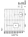
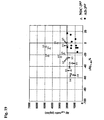
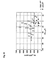
- a Concentration ratio can be determined, which is selected from the group which a Ratio between a concentration of A ⁇ peptide A ⁇ 1-42 to a concentration of A ⁇ peptide A ⁇ 1-40, a ratio between a concentration of A ⁇ peptide A ⁇ 1-42 to a concentration of the A ⁇ peptide A ⁇ 1-38 and a relationship between a concentration of the A ⁇ peptide A ⁇ 1-38 to a concentration of the A ⁇ peptide A ⁇ 1-40.
- the Determinations of these concentration ratios are based on the new finding that various neuropsychatric disorders significant shifts in these relative concentration of A ⁇ peptides compared to a comparison patient group occur.
- Alzheimer's disease limit This limit is in the cerebrospinal fluid typically between 0.285 and 0.300.
- the concentration ratio A ⁇ 1-42 / A ⁇ 1-40 also shows one when falling below one predetermined limit value that there is Alzheimer's disease.
- the Limit in CSF typically between 0.130 and 0.145.
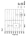
- Concentration quotient 1-42 / A ⁇ 1-38 has been found to satisfy an inequality: A * A ⁇ 1-42 / A ⁇ 1-38 + B> A ⁇ 1-38 / A ⁇ 1-40
- a and B are constants for those in the cerebrospinal fluid 0.2 ⁇ A ⁇ 0.8 and 0.5 * A ⁇ B ⁇ 2 * A and B ⁇ 0.9 applies.
- the neurochemical diagnostic options described so far and below are was developed using the antibody mAb 1E8. But they are the same with other means for determining the concentrations of the individual A ⁇ peptides A ⁇ 1-x and A ⁇ 2-x feasible. This can lead to shifts in the specified limit values by different specificities of the detection means for the individual A ⁇ peptides relative come to the specificities of the antibody mAb 1E8.
- concentrations on only the three mainly occurring A ⁇ peptides also enables the one described here Perform diagnostics using other concentration determination methods. So for example, the concentrations of the three A ⁇ peptides A ⁇ 1-38, A ⁇ 1-40 and A ⁇ 1-42 can be determined with special essays for these three A ⁇ peptides.
- this Exceeding a predetermined limit for the presence of a chronic inflammatory disease of the central nervous system indicates, this predetermined Limit typically and when using the antibody mAb 1E8 to determine the concentration the A ⁇ peptide in the cerebrospinal fluid is between 15.0 and 15.7%.
- This predetermined limit is between 8 and 9%.
- Using the antibody mAb 1E8 for determining the concentration of A ⁇ peptides in the cerebrospinal fluid can be on the range 8.3 to 8.8% can be limited.
- the relative limit is exceeded when a predetermined limit is exceeded A ⁇ 1-42% share on the presence of chronic inflammatory disease of the central Nervous system. This limit lies in the liquor between 9.1 and 9.6%.
- the relative proportion A ⁇ 1-40% indicates the presence of Alzheimer's disease, if a specified limit is exceeded, which is typically between 59 and 61%.
- cryopreservation of samples that are taken before digestion with a detergent which then also after digestion of the sample with a detergent determinable concentration of A ⁇ peptides reduced. From this, the requirement can be derived, the samples before cold preservation and any other cold treatment, including sample treatment with the Subdue detergent.
- a sample can thus be divided into at least two subsamples, the first of which Partial sample of the sample treatment with the detergent before or instead of a precipitation treatment is subjected to the second subsample of precipitation treatment is subjected to the detergent before or instead of sample treatment. Subsequently the concentrations of the A ⁇ peptide A ⁇ 1-42 determined in both sample parts compared with each other.
- the precipitation treatment mentioned can besides one Cold treatment also include an immunoaffinity procedure. It is particularly interesting a difference ⁇ A ⁇ 1-42 between those determined in the two sample parts Determine concentrations of the A ⁇ peptide A ⁇ 1-42. This value is a highly significant one Indicates the presence of a protein folding disease.
- the A ⁇ peptides to which the Antibody mAb 1E8 is bound with an anti-mAb 1E8 antibody secondary antibodies are labeled.
- the one directed against the antibody mAb 1E8 secondary antibodies can already be provided with a marker which can be registered in terms of quantity or after his immune response with the antibody mAb 1E8 with a quantitative markers that can be registered.
- the new antibody is also suitable for pure concentration of A ⁇ peptides A ⁇ 1-x and / or A ⁇ 1-x and / or A ⁇ 2-x and / or suitable for sAPP ⁇ .
- Another possible use is the differentiation of A ⁇ peptides A ⁇ 1-x and A ⁇ 2-x of A ⁇ peptides A ⁇ n-x with n> 2, because the antibody mAb 1E8 a has pronounced N-terminal specificity and clearly on A ⁇ peptides Aßn-x with n> 2 binds less ( ⁇ 5%) when under the specific conditions of A ⁇ -SDS-PAGE / Immunoblot is used.
- the invention is described below by characterizing and describing a Process for the preparation of the antibody mAb 1E8 and in the form of a description of Applications of the antibody mAb 1E8 explained and described in more detail.
- the monoclonal antibody mAb 1E8 was produced on behalf of Schering AG with the contract company "nano Tools Antiköpertechnik” in Denzlingen after their Standard procedure.
- the immunization and screening strategy was developed in Agreement with Schering AG.
- mice For the immunization of Balb / c mice, the entire A ⁇ protein (1-42) was used (10 ⁇ g / immunization). There was primary immunization and 3 themselves subsequent booster immunizations. The immunizations were carried out at intervals of 2 weeks each. The animal was then sacrificed and the spleen for cell fusion with a Mouse myeloma cell line used. The fused cells were placed on 96-well tissue culture plates transferred and cultivated in the presence of feeder macrophages.
- peptides 1-16 were covalent coupled to appropriately activated ELISA plates and for screening with the Hybridoma supernatants used.
- the A ⁇ 1-16 positive clones were then removed cloned and tested repeatedly. After expansion of the clones, cryo-preservation took place.
- the antibody was enriched using the Ion exchange chromatography performed under non-denaturing conditions.
- the analysis showed that the antibody 1E8, belonging to the IgG1 kappa subclass, is an N-terminal linear epitope detected, which consists of the first 8 N-terminal amino acids of the A ⁇ sequence is being trained.
- This antibody proved to be in subsequent experiments suitable for the detection of native A ⁇ in Western Bot, immunoprecipitation, Immunohistochemistry and ELISA.
- SDS-PAGE sodium lauryl sulfate polyacrylamide gel electrophoresis
- the conformational change is highly specific for the amino acid primary sequence of the respective A ⁇ peptides and leads to a reproducible change in the effective molecular radius. With that you can Separate numerous A ⁇ peptides, some of which are only one at the N- and C-terminal Distinguish amino acid and due to the small difference in mass by conventional SDS-PAGE cannot be separated.
- the change of conformation is achieved under the conditions of the multiphasic buffer system by the addition of Urea triggered above a concentration of 6M.
- the A ⁇ peptide specific The change in conformation occurs at a defined pH and ionic strength in addition to the molarity of the urea used by the pore size of the polyacrylamide gel used Detergent concentration (SDS) and temperature determined during separation.
- SDS Detergent concentration
- the optimal separation gel matrix for the separation of a broad spectrum N- and C-terminal (Nt, Ct) modified A ⁇ peptides were found with 12% T / 5% C / 8M
- the A ⁇ -SDS-PAGE was combined with isoelectric focusing (IEF) within a first analytical dimension using carrier ampholytes or immobilized pH gradients (IPG) for two-dimensional electrophoresis (A ⁇ -2D-PAGE and A ⁇ -IPG-2D-PAGE) ,
- IPG immobilized pH gradients
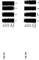
- the electrophoretic running behavior of the synthetic A ⁇ peptides A ⁇ 1-33 , A ⁇ 1-34 , A ⁇ 1-35 , A ⁇ 1-37 , A ⁇ 1-38 , A ⁇ was determined using A ⁇ -SDS-PAGE and A ⁇ -IPG-2D-PAGE 1-39 , A ⁇ 1-40 , A ⁇ 1-42 , A ⁇ 2-40 , A ⁇ 3-40 , A ⁇ 3p-40 , A ⁇ 2-42 , A ⁇ 3-42 and A ⁇ 3p-42 .
- the detection was carried out by Western immunoblot (PVDF membrane) and "enhanced chemiluminescence (ECL)" (A ⁇ -SDS-PAGE / immunoblot, A ⁇ -2D-PAGE / immunoblot, A ⁇ -IPG-2D-PAGE / immunoblot).
- the monoclonal antibody mAb 1E8 was used for this, for which an unusually high N-terminal specificity was demonstrated.
- the mAb 1E8 recognizes the corresponding A ⁇ peptides 2-x (eg A ⁇ 2-40 , A ⁇ 2-42 ) with detection sensitivity similar to that of the A ⁇ peptides 1- x (e.g.
- a ⁇ 1-40 , A ⁇ 1-42 The synthetic A ⁇ peptides A ⁇ 3-40 or A ⁇ 3-42 , in which the amino acid alanine is also missing at the N-terminal, and their pyroglutamate derivatives, on the other hand, are no longer detected in physiologically relevant concentrations.
- the sensitivity of detection in the Western immunoblot could be selectively improved for the mAb 1E8 by using a synthetic reagent for blocking unspecific binding sites (Immunoblot-2) instead of the milk powder (Immunoblot-1), which is otherwise mostly used.
- intra- and interassay variation coefficients of less than 10% can be realized for 20 pg A ⁇ peptide.
- the detection sensitivity is 0.3 pg for A ⁇ 1-40 and 0.6 pg for A ⁇ 1-42 .
- Other Western immunoblot methods with equally good sensitivity and variation coefficients for the detection of A ⁇ peptides are not yet known.
- the sensitivity mentioned above is a prerequisite for neurochemical diagnosis of dementia in the cerebrospinal fluid if the A ⁇ peptides are to be quantified directly after SDS / heat denaturation using a Western immunoblot and CCD camera, ie without prior selective enrichment by immunoprecipitation. It was also possible to demonstrate that the selectivity of the A ⁇ -SDS-PAGE / immunoblot for neurochemical dementia diagnostics can be increased significantly by this sample pretreatment if the SDS / heat denaturation takes place before the CSF samples are frozen.
- the A ⁇ -SDS-PAGE / Immunoblot-2 realizes for the first time the direct quantification of sAPP ⁇ and A ⁇ peptides using a CCD camera in only 10 ⁇ l human or animal (guinea pigs, rabbits) CSF samples. With this method it could be demonstrated for the first time that in addition to A ⁇ 1-40 and A ⁇ 1-42 three further carboxy-terminal (C-terminal) truncated A ⁇ peptides (A ⁇ 1-37 , A ⁇ 1-38 , A ⁇ 1-39 ) are highly conserved in human and animal cerebrospinal fluid.
- a ⁇ 1-42 is not the second most common A ⁇ peptide in human cerebrospinal fluid after A ⁇ 1-40, as previously assumed, but A ⁇ 1-38 . It could also be shown that the three additional C-terminally truncated A ⁇ peptides can also be detected in human plasma, but here with a significantly lower concentration and a different relative distribution. The ratio A ⁇ 1-42 / A ⁇ 1-38 in particular appears to be different depending on the CNS.
- the N-terminally shortened A ⁇ peptide is also present in the cerebrospinal fluid 2-42 demonstrable that the brain homogenates of patients with AD are usually extensive is increased.
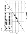
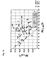
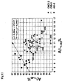
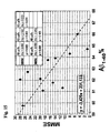
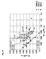
- the relationship between A ⁇ 1-38 % and A ⁇ 1-42 % or the correlation between the A ⁇ peptide ratios A ⁇ 1-38 / A ⁇ 1-40 and A ⁇ 1-42 / A ⁇ 1-38 shows a comparatively high correlation in AD and is diagnostically promising.
- the surprisingly clear difference between the absolute and proportionate concentrations of the A ⁇ peptide species with regard to their suitability for neurochemical dementia diagnosis is probably due to the fact that disease-specific changes in the ⁇ -secretase activity occur and these are better mapped by a change in the relative A ⁇ peptide proportions.
- a ⁇ peptides and other APP metabolites SDS / heat are denatured.
- immunoprecipitation IP
- IP immunoprecipitation
- a detergent (SDS) -dissociable fraction can be distinguished from an A ⁇ peptide fraction which is directly accessible to antibodies within immunoprecipitation or ELISA procedures, ie without simultaneous treatment with detergents. This differentiation is probably explained by the high affinity binding of the A ⁇ peptides to other proteins or A ⁇ auto-aggregates.
- the SDS-dissociable fraction is significantly higher than the antibody-dissociable fraction. This phenomenon is particularly pronounced specifically for A ⁇ 1-42 .
- a ⁇ 1-42 in contrast to A ⁇ 1-40 - a cold precipitation-related (KP) reduction can be demonstrated by freezing the CSF samples.
- KP-related depression is probably mainly carried by the aggregate-bound fraction of A ⁇ 1-42 and leads to an AD-typical decrease in the CSF levels of A ⁇ 1-42 in a considerable part of the patients without Alzheimer's dementia (NDC). This effect is particularly evident in patients with at least one ApoE ⁇ 4 allele and probably explains why, even in patients without AD but with ⁇ 4 alleles, comparatively low concentrations of A ⁇ 1-42 are measured in the previously frozen cerebrospinal fluid.
- a ⁇ -SDS-PAGE / immunoblot in connection with the sample preparation mentioned above is also promising for the early and preclinical diagnosis of AD, since it is to be expected that borderline-deep A ⁇ 1-42 CSF levels indicate an onset of AD, especially when they cannot be explained by a CP-dependent humiliation.
- prospective studies in patients with mild cognitive disorders should be used to check whether a pronounced KP-dependent reduction in A ⁇ 1-42 per se has predictive value for the later development of AD.
- composition or molecular primary structure of A ⁇ 1-42 -binding proteins within the complex need not necessarily be changed, but the affinity of the binding could be different depending on the conformation of A ⁇ 1-42 with the same primary structure.
- the finding that a specific difference in the detergent-dissociable fraction of A ⁇ 1-42 can be detected in the cerebrospinal fluid in patients with and without AD can also be used for other methods of neurochemical dementia diagnosis (ELISA, fluorescence correlation spectroscopy).
- the A ⁇ -SDS-PAGE / immunoblot can be used for the neuropathological post-mortem diagnosis of dementia.
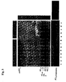
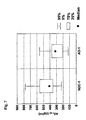
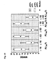
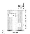
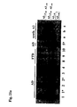
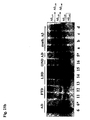
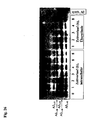
- RIPA detergent-soluble fraction of A ⁇ peptides in brain homogenates of patients with AD
- other dementia diseases and controls disease and brain region-specific expression patterns of the A ⁇ peptides 1-37, 1-38, 1-40, 1-42 and 2-42 are shown.
- Particularly striking is the massive increase in A ⁇ 2-42 in the RIPA-soluble fraction of the brain homogenates in AD and patients with Lewy-body dementia (LBD).
- LBD Lewy-body dementia
- these high concentrations of A ⁇ 2-42 are observed when the patients simultaneously show a pronounced ⁇ -amyloid pathology (LBD, CERAD C).
- a ⁇ 1-42 is also regularly and significantly increased in AD and LBD (CERAD C).
- the concentration of the other A ⁇ peptides showed a high interindividual variance. This could be an indication of phenotypic subtypes of sporadic AD or, depending on the course, the degree of severity of dementia.
- the RIPA detergent mix used here is not able to solubilize mature neuritic ⁇ -amyloid plaques.
- the extensively increased concentrations of A ⁇ 2-42 cannot therefore be explained by A ⁇ 2-42 from this ⁇ -amyloid plaque fraction. Accordingly, it is also unlikely that A ⁇ 2-42 will mainly arise from unspecific ⁇ -amyloid plaque-associated post-translational changes.
- the high intracerebral concentrations of A ⁇ 2-42 are pathophysiologically relevant, since the absence of aspartate increases the tendency of A ⁇ 1-42 to aggregate and this N-terminal modification apparently precedes the formation of mature ⁇ -amyloid plaques.
- the carboy-terminally truncated Aß peptides 1-37, 1-38 and 1-39 could also be used regularly in the cisternal liquor from guinea pigs and rabbits can be detected. Further successfully detected in homogenates and supernatants (short-term culture) of hippocampals Tissue sections of the adult guinea pig.
- the A ⁇ peptide quartet - and additionally A ⁇ 2-42 - can also be detected in the supernatants of a neuroglioma tumor cell line (H4) which overexpresses human APP751 with a Swedish double mutation ( human APP751 Sw ).
- H4 neuroglioma tumor cell line
- protease inhibitors which are potential inhibitors of ⁇ - / ⁇ -secretases, in addition to the known dose-dependent reduction in A ⁇ 1-40 and A ⁇ 1-42, it was also possible to reduce the C-terminally shortened A ⁇ peptides 1-37. 1-38, and 1-39 can be detected. In addition, the formation of A ⁇ 2-42 was inhibited.
- AD has a certain isoform of ⁇ -secretase (BACE) is overexpressed or the physiologically produced A ⁇ 2-42 is reduced catabolized in AD.
- BACE ⁇ -secretase
- a ⁇ 1-40 and A ⁇ 1-42 / 1-43 in human lumbar cerebrospinal fluid has previously been described (Ida et al., 1996).
- the A ⁇ peptides cannot be differentiated here by electrophoretic separation, but rather on the blot membrane by C-terminally selective monoclonal antibodies.
- a ⁇ 1-42 must be concentrated before separation by concentration of the sample. The concentration takes place without prior SDS denaturation, which appears to be methodologically problematic due to the high tendency of A ⁇ 1-42 to aggregate.
- separate electrophoresis must be carried out using this method.
- the multiphasic buffer system used for the A ⁇ -SDS-PAGE / immunoblot (Wiltfang et al., 1991) combines the advantages of special buffer systems for proteins (Laemmli, 1970) and peptides (Schagger and von Jagow, 1987). Accordingly, with this SDS-PAGE method, both proteins and peptides can be separated in a homogeneous polyacrylamide separation gel system with high resolution. Furthermore, the electrophoretic separation of A ⁇ 1-40 and A ⁇ 1-42 was described using the urea version (Wiltfang et al., 1991) of the latter SDS-PAGE method (Klafki et al., 1996).
- SDS-PAGE can be combined as a second analytical dimension with isoelectric focusing (IEF) in the first dimension as 2D-PAGE (O'Farrell, 1975; O'Farrell et al., 1977).
- Isoelectric focusing when using highly spread immobilized pH gradients (Gorg et al., 1995; Gorg et al., 1997; Righetti and Bossi, 1997) can reveal minimal charge differences.
- the two-dimensional A ⁇ -SDS-PAGE / immunoblot can therefore also be used for the high-resolution analysis of post-translational modifications of the APP metabolites.
- the A ⁇ -SDS-PAGE / immunoblot also allows the quantification of sAPP ⁇ , which is reduced in the CSF for AD (Sennvik et al., 2000).
- the urea-containing separating gel compartment is combined with an upper (cathodic) separating gel without urea and a larger pore size.
- the quantitative A ⁇ -SDS-PAGE / immunoblot allows simultaneous and ultra-sensitive Determination of a spectrum of APP metabolites that are highly relevant to the Early neurochemical diagnosis and pathogenesis of AD.
- the procedure can also used in animal and clinical evaluation of new pharmaceuticals that interfere with the metabolism or catabolism of A ⁇ peptides.
- the SDS-PAGE was carried out using the Bio-Rad Mini Protean II electrophoresis System.
- the size of the gel compartments used was as follows: separating gel length about 54 mm; Collection gel length about 5 mm (corresponding to a volume of 250 ⁇ l); Kammgel assume about 12-15 mm; Gelicke 0.50 mm each, gel width 85 mm each. Separating and collecting gels for the second analytical dimension within the A ⁇ -2D-PAGE have a gel thickness of 1.0 mm.
- a 15-tooth sample application comb is used for the sample application (tooth width approx. 3 mm, tooth spacing 2 mm).
- the resulting sample application pocket in the comb gel measures approximately 3x10 mm.
- Sample application quantity should not exceed 10 ⁇ l To reliably prevent carry-over between the sample wells. The samples will be after filling the cathode buffer.
- Electrophoresis a) 12 mA / 0.5 mm Gelicke at constant current over 2h, b) 1.0 mm gels of the second analytical Dimension: 60 volt / 1.0 mm gel thickness for 10 min, 120 volt / 1.0 mm gel thickness over 1 hour 45min.
- the urea version of the Bicin / Tris SDS-PAGE was used for the separating gel compartments Wiltfang et al. (Wiltfang et al., 1991) used and for the presented Applications modified in essential aspects.
- Table 1 summarizes the concentrated buffers for the gel compartments, cathode buffers, anode buffers and the acrylamide stock solution together.
- the gel composition for A ⁇ -SDS-PAGE for optimized separation of APP metabolites and A ⁇ peptides are found in human or animal biological samples in Table 2.
- the SDS / heat denaturation prior to concentration of the sample should result in proteolysis, precipitation and autoaggregation of the A ⁇ peptides during concentration can be avoided.
- the reduced SDS concentration in SDS-SB-3 is necessary because of higher SDS concentrations after triple concentration of the samples and with an order volume of approx. 10 ⁇ l to impair the running behavior of the A ⁇ peptides at the anodic end of the lead urea-containing separating gel.
- the SDS concentration is 0.5% w / v in SDS-SB-3 but still high enough for complete SDS / heat denaturation of the Sample.
- the APP metabolites immobilized by means of magnetic Dynabeads are removed from the antigen binding after the final washing step at 37 ° C. for 10 min in an ultrasonic bath using SDS-SB-1 or SDS-PB-3 (each without 2-mercaptoethanol! eluted. After adding 2-mercaptoethanol to 2.5% w / v, the mixture is heated to 95 ° C. for 5 min. When using SDS-PB-3, the samples can then be concentrated again three times by concentration to dry matter and absorption with H 2 O dd using SpeedVac.
- the carrier ampholyte IEF in round gels of the first analytical dimension and the vertical A ⁇ -SDS-PAGE of the second analytical dimension are generated with the Mini-Protean II 2-D Cell system implemented by Bio-Rad.
- Glass tubes ( ⁇ 1mm) are used for gel polymerization with the monomer solution from the table 4b filled.
- the IEF round gels are polymerized to a length of 60 mm.
- 20 ⁇ l sample after direct absorption in IEF-SB (10 ⁇ l liquor plus 10 ⁇ l IEF-SB) or 10 ⁇ l eluate from the Immunoprecipitation in IEF-SB (Table 4a) are applied and with the cathodic Electrolytes covered. This side of the glass tube is cathode with the top Electrolyte chamber connected.
- the composition of anolyte and catholyte for the Carrier ampholyte IEF can be found in Table 4c.
- the round gels are ejected from the glass tubes using water pressure and left for 5 min. incubated in the IEF equilibration buffer (Table 4d) at room temperature.
- the IEF gels are then placed on the collecting gel (see above) of the A ⁇ -SDS-PAGE and fixed in their position with the IEF agarose solution (Table 4d).
- a sample bag for the comparative application of synthetic A ⁇ peptides or M r marker proteins is formed in the hot agarose using a Teflon tooth.
- the A ⁇ -SDS-PAGE was carried out as set out in Tables 1 and 2 (separating gel: 12% T / 5% C / 8M Urea). A comb gel is not polymerized. The gel thickness of the separating gels used is 1mm. The electrophoresis is carried out at room temperature: 10 min. / 60 V, 90 min. / 120 V.
- the sample is taken up in IPG-SB (Table 5a). Dry matter and using MSP Magnetically immobilized Dynabeads (see below) are immediately in front of the IPG-SB IEF recorded and 10 min. incubated in an ultrasonic bath at 37 ° C. Intake more fluid biological samples: After removal of the mixed bed ion exchanger (Serdolit MB-1) the IPB-SB in Eppendorf aliquoted (e.g. 100 ⁇ l) and using SpeedVac Room temperature concentrated to dry matter. The IPG-SB presented as a dry substance is taken up in a volume ratio of 1: 1 with sample (e.g. 100 ⁇ l) and after the vortex step (1 min.) For 10 min. incubated in an ultrasonic bath at 37 ° C.
- IPG "DryStrips” The IEF using the commercial IPG “DryStrips” was carried out according to the protocol of the Manufacturer (Amersham Pharmacia Biotech / Brief Instructions 71-5009-57, Edition AA, 99-04). IPG "DryStrips” (4-7, linear pH gradient, length 7cm) were used overnight Room temperature using the rehydration solution from Table 5a Gel thickness of 0.5 mm rehydrated.
- the sample application device (“sample cups") is placed on the basic side of the IPG strips, approximately at pH 6.5, applied (cathodic application) and 30 ⁇ l Sample applied.
- the IPG-IEF takes place for 30 min / 300V, 30 min / 800V, 30 min. / 1400V and 5 h / 2000V ( ⁇ 12500 Vxh).
- the "DryStrips" for 2x10 min. equilibrated (Table 5b).
- the first equilibration solution contains DTT (50 mg / 5ml), the second solution iodoacetamide (240 mg / 5 ml) for the neutralization of excess DTT, which otherwise leads to staining artifacts if the gels are silver colored.
- the second equilibration step is omitted for Western immunoblots.
- the equilibrated IPG "DryStrips" are fixed on the stacking gel of the A ⁇ -SDS-PAGE using an agarose solution (Table 5c).
- a sample bag for the comparative application of synthetic A ⁇ peptides or M r marker proteins is placed in the hot agarose using a Teflon tooth.
- the electrophoresis is carried out according to 3.1.2
- PI stock solution protein inhibitor cocktail stock solution
- the beads activated in this way can be stored for up to three months without any significant loss of capacity.
- the activated beads are 3x3 min. washed with 250 ⁇ l PBS / BSA without the addition of Na azide.
- 25 ⁇ l of activated DynaBeads (approx. 1,675 x10 7 beads) are mixed with 268 ⁇ l CSF / PI stock solution. (200 ⁇ l CSF + 68 ⁇ l PI stock solution) and mixed with 732 ⁇ l 50 mM HEPES buffer, pH 7.4, in 1 ml in Eppendorf cups. Incubation takes place for 20 h at 4 ° C on a shaker (constant agitation of the sample). The beads are then immobilized in the MSP stand and the supernatant removed. Then the beads are 4 x 5 min. washed at room temperature with 1 ml PBS / 0.1% BSA. Finally, the beads are 1 x 3 min.
- the magnetically immobilized beads are sampled with 25 ⁇ l SDS-PB-1 for 5 min. at 95 ° C.
- 25 ⁇ l IEF-SB or IPG-SB is taken up and for 10 min. incubated at 37 ° C in an ultrasonic bath.
- 4 ⁇ l sample is applied, corresponding to the amount of A ⁇ peptide present in 32 ⁇ l CSF volume.
- 10 ⁇ l are applied, corresponding to the amount of A ⁇ peptide present in 80 ⁇ l CSF volume.
- 200 ⁇ l CSF are mixed with 200 ⁇ l 5-fold concentrated RIPA 0.5x buffer (Table 6) and brought to 1 ml with 600 ⁇ l H 2 O dd in Eppendorf Cups.
- the immunoprecipitation is carried out according to the method described under A.
- the RIPA 0.5x buffer contains protease inhibitors (Table 6).
- Brain tissue (approx. 50 mg) is homogenized with 1 ml RIPA 1x buffer (Table 6) in 1.5 ml Eppendorf reaction vessels using an ultrasonic finger and then for 5 min. (4 ° C) centrifuged at 20,000 g. The supernatant is removed and the protein content of the homogenate supernatant is adjusted to 3 mg / ml with RIPA 1x buffer and 1 ml brain homogenate together with 50 ⁇ l activated DynaBeads (approx. 3.35 x10 7 beads) are immunoprecipitated as described under (a).
- the RIPA 1.0x buffer contains protease inhibitors (Table 6).
- 400 ⁇ l cell culture supernatant are mixed with 100 ⁇ l 5-fold concentrated RIPA 0.5x buffer (alternatively: 800 ⁇ l cell-culture supernatant with 200 ⁇ l 5-fold concentrated RIPA 0.5x buffer) and 25 ⁇ l activated DynaBeads and immunoprecipitated as described under (a).
- RIPA 0.5x buffer alternatively: 800 ⁇ l cell-culture supernatant with 200 ⁇ l 5-fold concentrated RIPA 0.5x buffer
- 25 ⁇ l activated DynaBeads and immunoprecipitated as described under (a).
- 6 ⁇ l sample are applied, corresponding to the amount of A ⁇ peptide present in 96 ⁇ l cell culture supernatant volume.
- the transfer is carried out by means of a Semidry Western blot and a multiphase buffer system on PVDF detection membranes.
- the blot buffers are listed in Table 8.
- the structure of the blot sandwich from the anode to the cathode is as follows: A filter paper layer with buffer A, a filter paper layer and the PVDF membrane with buffer B, gel and finally two filter paper layers with buffer C. Gels are immediately after the electrophoresis for approx. Incubated in buffer C for 10 sec. Extra strong filter paper from BioRad is used as filter paper.
- the Immobilon-P membrane from Millipore After examining PVDF membranes from different manufacturers, the Immobilon-P membrane from Millipore showed the least background staining and the most effective immobilization of the A ⁇ peptides, especially A ⁇ 1-42 , within the immunoblot protocol. According to the manufacturer's instructions, the Immobilon-P membranes are wetted with methanol before use, then for 1 min. incubated in H 2 O dd and then transferred to buffer B. The transfer takes place for A ⁇ -SDS-PAGE ( ⁇ 0.5 mm) for 30 min. or A ⁇ -2D-PAGE gels ( ⁇ 1.0 mm) for 45 min. at room temperature with 1 mA / cm 2 .
- the Immobilon-P membrane is washed in H 2 O dd for approx. 30 sec. And 3 min. cooked in the microwave in PBS (without Tween-20!).
- the cooking step is essential to achieve the maximum sensitivity.
- the PVDF membrane is placed on a Teflon pad and excess washing buffer is removed by placing a layer of KIMWIPES® Lite 200 laboratory wipes. An incubation for 5 min follows. at room temperature with 0.1 ml / cm 2 ECLPlus TM solution. In order to remove excess ECLPlus TM solution, the membrane is placed between two layers of gel blotting paper and transferred to a "development folder" for signal detection, which ensures optimal detection and prevents the membrane from drying out.
- the ECL detection after Western immunoblot was carried out using Hyperfilm TM exposure for 5 min.
- the densitometric evaluation was carried out using Laser scanners (Epson GT 9000) and evaluation software (Biometra, BioDoc software).
- each gel performed a dilution series of a mixture of the synthetic A ⁇ peptides 1-37, 1-38, 1-39, 1-40 and 1-42 (A ⁇ 1-37 : 5, 10, 20, 40, 80 pg; A ⁇ 1 -38 : 15, 30, 60, 90, 120 pg; A ⁇ 1-39 : 5, 10, 20, 30, 60 pg; A ⁇ 1-40 : 25, 50, 100, 200, 300 pg; A ⁇ 1-42 : 5, 10, 20, 40, 80 pg).
- the ECL detection using a CCD camera was carried out at a resolution of 80x80 ⁇ m using serial exposure times for 5, 20, 60 and 120 seconds.
- the "Quantity One" evaluation software Bio-RAD Laboratories, Hercules, CA, USA) was used to quantify the gels relative to their respective calibration series.
- Eggs of the White Leghorn chicken breed are incubated at 37 ° C in the incubator for 10 days. On day 10, the chicken embryo is removed under sterile conditions and the brain is dissected. The anterior cerebral vesicles are separated, the adjacent meninges are freed and collected in HEPES-buffered DMEM. The tissue obtained in this way is subjected to trypsin digestion for 15 minutes and, after being washed three times with DMEM, drawn up several times through a cannula.
- the pellet is taken up in cultivation medium (DMEM + 5% fetal calf serum + 5% chicken serum) and drawn up again through a cannula.
- cultivation medium DMEM + 5% fetal calf serum + 5% chicken serum
- the cell density of the suspension is set to 1.5 million cells / ml and this is plated into the culture vessels, so that a cell density of 375,000 cells / cm 2 results.
- the culture vessels were previously coated with a poly-L solution (0.1 mg / ml poly-L-lysine in 0.1M borate / NaOH buffer, sterile filtered, pH 8.4) over 24 hours.
- a poly-L solution 0.1 mg / ml poly-L-lysine in 0.1M borate / NaOH buffer, sterile filtered, pH 8.4
- the lumbar cerebrospinal fluid was examined in a total of 130 patients. In five of these patients A ⁇ peptides were also measured in the blood plasma.
- NDC neuropsychiatric diseases
- AD sporadic Alzheimer's disease
- Were methodologically determined examined several NDC and AD groups, each with different patients, whereby related patient collectives through consecutive Arabic numbering are marked (e.g. NDC-1, AD-1).
- the collectives NDC-1 and NDC-2 also contain Patients with dementia diseases other than AD.
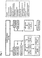
- Fig. 1 gives an overview of the patient groups and their hierarchical assignment. Table 10a-d lists the patients who are in several at the same time Patient groups.
- the clinical diagnosis was carried out according to ICD-10.
- the diagnosis of Alzheimer's dementia was developed according to the internationally used criteria of the "Work Group of the National Institute of Neurological and Communicative Disorders and Stroke (NINCDS) "and the guidelines of the "Alzheimer's Disease and Related Disorders Association (ARDA)” (McKhann et al., 1984).
- NINCDS National Institute of Neurological and Communicative Disorders and Stroke
- ARDA Alzheimer's dementia and Related Disorders Association
- the samples were taken exclusively within the clinical routine diagnostics. None was used for the measurements presented here additional cerebrospinal fluid volume gained. The retrospective investigation was accordingly only if there is still aliquoted CSF after routine diagnosis Was available.
- the CID-3 collective consisted of five patients with multiple sclerosis and five patients with unclear aetiology of the chronic inflammatory CNS process.
- NDC-3 KP cold precipitation-related depression
- a ⁇ peptides with missing or only partial separation are in one line.
- the proof was done by silver staining of the separating gels.
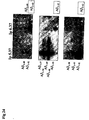
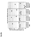
- the A ⁇ peptides 2-40 / 3-40 can be separated from 1-42 by means of A ⁇ -IPG-2D-PAGE, since the lack of aspartate the isoelectric point by a pH unit from 5.37 to 6.37 shifts (see Fig. 2a & c and Fig. 24a).
- a ⁇ -SDS-PAGE is used in the upper (cathodic) compartment in the urea-containing Separating gel sAPP ⁇ separated using Western Immunoblot-1/2 (mAb 1E8) can be detected (see Fig. 3). Due to the molar mass of> 100,000 sAPP ⁇ isoforms become less effective compared to the A ⁇ peptides much higher variance from the small-pore urea-containing separating gels to the Detection membrane blotted (intraassay coefficient of variation in CSF> 20%).
- Blotting efficiency and separation of the sAPP ⁇ isoforms can be significantly improved if the urea-containing separation gel system with a cathodic non-urea Separating gel compartment of larger pore size but otherwise the same Buffer composition is combined (10 T%, 5 C%, no urea). If unchanged The separation length of the urea-containing compartment becomes the quality of the A ⁇ peptide separation not impaired because the A ⁇ peptides still have the large-pore compartment going through concentrated within the "moving boundary".
- the mAb 1E8 shows an astonishingly high N-terminal specificity in Western immunoblot 1/2, since only A ⁇ -petids are detectable in the lower pg range ( ⁇ 200 pg), the N-terminal maximum are shortened by an amino acid (aspartate). Accordingly, the A ⁇ peptides 3-40, 3p-40, 3-42 and 3p-42 not proven using the mAb 1E8.
- the proof succeeds here by means of the N-terminal specific mAb 6E10, which is commercially available, but in Detected about ten to thirty times less sensitive depending on the blot conditions.
- the detection sensitivity of the Western Immunoblot-1 (milk powder block, mAb 1E8) is 1pg (A ⁇ 1-40 ) to 2 pg (A ⁇ 1-42 ) with film exposure and 3 pg (A ⁇ 1-40 ) to 6 pg ( A ⁇ 1-42 ) when recording signals with the CCD camera.
- the detection sensitivity of the Western Immunoblot-2 (Roti-Block, mAb 1E8) is 0.3 pg (A ⁇ 1-40 ) to 0.6 pg (A ⁇ 1-42 ) when exposed to film and 1 pg (A ⁇ 1-40 ) to 2 pg (A ⁇ 1-42 ) when recording signals with the CCD camera.
- the detection sensitivity of the commercially available N-terminal selective mAb 6E10 in Western immunoblot with milk powder block is 10 pg (A ⁇ 1-40 ) to 20 pg (A ⁇ 1-42 ) with film exposure and 30 pg (A ⁇ 1-40 ) up to 60 pg (A ⁇ 1-42 ) when recording signals with the CCD camera.
- the mAb 6E10 cannot be used with Rotiblock. This enabled detection sensitivity to be increased by up to 30 times compared to the mAb 6E10.
- SDS-PAGE separation gel systems with 8 M urea can be loaded with a maximum of about 5 ⁇ l CSF per mm 2 gel surface, regardless of the money dimensions used, if optimal electrophoretic separation is to be achieved in almost all patient samples.
- This is natively frozen and then SDS / heat denatured CSF or CSF which was SDS / heat denatured before freezing.
- 5 ⁇ l per mm 2 corresponds to a liquor volume of about 10 ⁇ l in the mini gel system used. This results in sensitivity of 200 pg / ml for the detection of A ⁇ 1-42 in human cerebrospinal fluid using A ⁇ -SDS-PAGE / Immunoblot-2 and CCD camera.
- a detection sensitivity of at least 200 pg / ml is a prerequisite for the neurochemical dementia diagnosis of AD by determining the A ⁇ peptides in the cerebrospinal fluid and cannot be achieved, for example, with the commercially available mAb 6E10.
- Quantification using a CCD camera offers considerable advantages over film exposure.
- the light signal can be recorded linearly over 3.8 powers of ten, and the duration of the signal recording can also be precisely controlled over a wide range.
- APP metabolites with a large difference in their signal intensity such as sAPP ⁇ and A ⁇ 1-42 , can be quantified over two measurement times (eg 10 s and 3 min).
- a ⁇ -SDS-PAGE / Immunoblot-1/2 can be used to regularly detect a characteristic A ⁇ peptide quintet in human lumbar CSF (Fig. 3 ).
- a ⁇ 1-40 three additional A ⁇ peptides are shown, which were initially designated as A ⁇ 1-x a , A ⁇ 1-x b and A ⁇ 1-x c and by means of A ⁇ -IPG-2D-PAGE / immunoblot with comigration synthetic A ⁇ peptides could be identified as A ⁇ 1-37 / 38/39 (Fig. 2).
- the A ⁇ peptides 1-37, 1-38 and 1-39 are not detectable in carboxy terminal selective immunoprecipitation against 1-40 and 1-42 in the cerebrospinal fluid by means of A ⁇ -SDS-PAGE / immunoblot.
- the A ⁇ peptides 1-37 / 38/39 could be detected in human cerebrospinal fluid using MALDI-TOF.
- the A ⁇ peptides 1-33 / 1-34 and 1-35 are generally only detectable or undetectable in the cerebrospinal fluid in the A ⁇ SDS-PAGE / immunoblot above (cathode side) from 1-37 in the cerebrospinal fluid.
- the A ⁇ peptides 1-37, 1-38, 1-39, 1-40 and 1-42 are highly and significantly correlated, as can be seen in FIG. 10. This speaks for a tight enzymatic regulation of their formation.
- the synthetic A ⁇ peptides 1-37, 1-38 and 1-39 were not yet available when the first patient groups (NCD-1, AD-1, NDC-2 KP ) were examined.
- the ratio A ⁇ 1-42 to A ⁇ 1-38 was therefore determined here using the quotient of the densitometrically determined area units of the respective bands of a gel track.
- the quotient A ⁇ 1-42 / A ⁇ 1-40 was also expressed over the area units. Since A ⁇ 1-38 , A ⁇ 1-40 and A ⁇ 1-42 show a different intensity of the ECL signal with the same concentration and the same conditions in the Western immunoblot, the quotients of the area units cannot match the corresponding quotients of the A ⁇ determined via the calibration degrees -Peptide concentrations are equated.
- an additional band with the retention factor (Rf) of A ⁇ 2-42 can be detected below (anodically) from A ⁇ 1-42 using A ⁇ -SDS-PAGE / Immunoblot-2 and CCD camera (Fig. 23a ).
- This band could be identified in the cerebrospinal fluid in AD after prior immunoprecipitation (RIPA-IP, mAb 1E8) using A ⁇ -IPG-2D-PAGE / Immunoblot-2 and CCD camera as A ⁇ 2-42 (Fig. 24 b).
- a ⁇ 2-42 has not been demonstrated in non-demented control patients .
- a ⁇ 2-42 is also massively increased with typical brain regional distribution in patients with sporadic AD intracerebrally (cf. 4.2.5).
- a ⁇ 2-42 was yet available for the determination of A ⁇ peptides in the cerebrospinal fluid in the patient groups mentioned under 3.9. Accordingly, no quantitative data on A ⁇ 2-42 are available for these patients. In the meantime, however, another patient population was measured, which is not yet listed under 3.9. A ⁇ 2-42 was detectable in some of the patients with AD-4, in some patients with dementias other than AD (nAD-4) and in some patients with non-dementia (NDC-4). In addition, the A ⁇ 1-42 / A ⁇ 1-40 ratio was significantly reduced in the A ⁇ 2-42 positive patients compared to the other patients.
- Table 12 gives the clinical data and individual measurements of the patients and table 13 summarizes the statistical characteristics of the patient groups AD-1 and NDC-1.
- the CSF samples were analyzed using A ⁇ -SDS-PAGE / Immunoblot-1 and densitometric film evaluation.
- FIG. 4 The significant reduction in A ⁇ 1-42 in human lumbar cerebrospinal fluid in patients with AD-1 compared to NDC-1 is shown in FIG. 4.
- a ⁇ 1-40 is also significantly decreased in AD-1, but not to the extent that is evident for A ⁇ 1-42 (see Table 14).
- the quotient A ⁇ 1-42 / A ⁇ 1-40 is also significantly reduced (Fig. 5, Table 14).
- the likewise highly significant reduction in the quotient A ⁇ 1-42 / A ⁇ 1-38 has not been described previously (FIG. 6, Table 14).
- the limit values for the group comparison AD-3 versus NDC-3 were determined for A ⁇ 1-42 and the latter two A ⁇ peptide ratios using the respective "receiver operating characteristics (ROC)" (Table 15).
- the differentiation of the patient groups AD-1 versus NDC-1 with a limit value of 802.5 pg / ml A ⁇ 1-42 was feasible with a specificity of 74% and a sensitivity of 87%.
- the quotient A ⁇ 1-42 / A ⁇ 1-40 has a diagnostic specificity of 71% and a sensitivity of 93% for the differentiation of the AD-1 versus NDC-1 groups.
- the corresponding specificity and sensitivity for the quotient A ⁇ 1-42 / A ⁇ 1-38 is 84% and 0.86%.
- a ⁇ 1-42 was compared after immunoprecipitation (IP without detergent, mAb 6E10) and A ⁇ -SDS-PAGE / Immunoblot-1 with densitometric film evaluation. 7 and Table 14, A ⁇ 1-42 differentiates the patient groups AD-1 and NDC-1 less well after previous IP.
- concentration of A ⁇ 1-42 in the cerebrospinal fluid determined after immunoprecipitation and A ⁇ -SDS-PAGE / Immunoblot-1 with densitometric film evaluation in a sub-collective of patients with AD-1 agrees well with the concentration that was determined using the commercial ELISAA ⁇ 1-42 ( Hulstaert et al., 1999) in AD-1 (Table 13).
- Table 19 gives the clinical data and individual measurements of the patients and table 20 summarizes the statistical characteristics of the patient groups AD-1 and NDC-1.
- the CSF samples were analyzed using A ⁇ -SDS-PAGE / Immunoblot-2 and a CCD camera.
- a ⁇ 1-42 is present in human CSF in a fraction that is only partially accessible to monoclonal antibodies without prior treatment with detergents.
- the detergents used can release peptides from non-covalent protein-peptide bonds, for example as a result of the hydrophobic interaction. Accordingly, the higher cerebrospinal fluid levels of A ⁇ 1-42 after the use of detergents (SDS heat denaturation, RIPA-IP) compared to processes without the use of detergents (IP without detergent, ELISAA ⁇ 1-42 ) are likely due to high affinity binding and epitope masking of A ⁇ 1 -42 due to other proteins or A ⁇ peptide aggregates.
- SDS heat denaturation RIPA-IP
- IP without detergent, ELISAA ⁇ 1-42 the use of the ionic detergent SDS at higher concentrations (0.5% w / v) and temperature (95 ° C) is even more effective than the RIPA detergent mix.
- a ⁇ 1-42 is particularly sensitive to cold precipitation compared to A ⁇ 1-40 and shows different cold precipitation behavior in patients with AD and NDC depending on the disease (see 4.2.4).
- synthetic A ⁇ 1-42 with comparable concentration dissolved in water shows a significantly lower cold precipitation. It is therefore likely that the cold precipitation-related reduction of A ⁇ 1-42 in human cerebrospinal fluid is mainly due to the aggregate-bound portion of the peptide. In the case of comparatively hydrophobic aggregates - for example lipoprotein-containing complexes - a loss due to cold precipitation would not be surprising.
- ⁇ 4-positive patients of the NDC-3KP collective show a particularly high rate of KP-related lowering of A ⁇ 1-42 and approximately the same low CSF level as ⁇ 4-positive AD-3KP Have patients. Approximately equally deep A ⁇ 1-42 CSF levels are also demonstrated below for patients in the OND-3 ⁇ 4plus and AD-3 ⁇ 4plus groups.
- the A ⁇ peptide quintet could also be detected in plasma by immunoprecipitation and A ⁇ -SDS-PAGE / immunoblot with a CCD camera.
- concentrations in the plasma are 30 to 60 times lower and both compartments each show specific patterns of the percentage A ⁇ peptide fractions.
- the ratio A ⁇ 1-42 / A ⁇ 1-38 in particular is CNS-specific: CSF 0.80 (0.79-0.92), plasma 1.70 (1.69-1.75); Median (quartile).
- a ⁇ 1-40 and A ⁇ 1-42 are increasingly overexpressed in familial AD forms with APP point mutations in the vicinity of the ⁇ -secretase interface, whereas A ⁇ 1-42 occurs more frequently in the case of mutations in the vicinity of the ⁇ -secretase interface and the ratio A ⁇ 1-42 to A ⁇ 1-40 increases significantly. It can therefore be assumed that, when considering the percentage of A ⁇ peptides, more and more disease-specific changes in ⁇ -secretase activity are shown.
- FIG. 9 A and B compare the subgroups OND-3 ⁇ 4minus, OND-3 ⁇ 4plus and AD-3 ⁇ 4plus.
- OND-3 ⁇ 4plus in comparison to OND-3 ⁇ 4minus, all A ⁇ peptides in the cerebrospinal fluid tend to be reduced and accordingly the total A ⁇ peptide concentration (Fig. 9A).
- the reduction in A ⁇ 1-42 is particularly evident within the A ⁇ peptide quintet.
- a ⁇ 1-42 is selectively reduced in AD-3 ⁇ 4plus, but to the same extent as in OND-3 ⁇ 4plus.
- the total amount of A ⁇ peptides is not decreased here (Fig.
- the A ⁇ peptide quintet in the cerebrospinal fluid is closely correlated with one another and the percentages of the A ⁇ peptides and their total concentration have surprisingly low coefficients of variation for biological parameters.
- FIGS. 11-13 patients with AD-3 and CID-3 can be differentiated from OND-3 patients via A ⁇ 1-38 %, A ⁇ 1-40 % and A ⁇ 1-42 %.
- FIG 11 shows a significant negative correlation between A ⁇ 1-38 % and A ⁇ 1-42 % for AD-3 patients.
- the AD-3 patient 143 was not taken into account in the calculation of the regression line and is identified here as an outlier, as also below (see FIGS. 12 & 13).
- this patient showed an early stage of AD (MMSE 27/30). In terms of differential diagnosis, depressive pseudo-dementia had previously been discussed.
- the severity of dementia increases in the direction of the arrowhead of the degrees of regression.
- the relationship between the percentage of A ⁇ peptide and the severity of dementia is discussed in more detail below (see FIGS. 14 & 15).
- the AD-3 collective can be separated from the groups CID-3 and OND-3 without overlapping.
- the specific relationship between A ⁇ 1-38 % and A ⁇ 1-42 % in AD suggests that patients with AD can be differentiated even better using a function similar to the regression line between A ⁇ 1-38 % and A ⁇ 1-42 %.
- Fig. 12 shows A ⁇ 1-40 % depending on A ⁇ 1-42 %. An AD-specific correlation between these parameters is significant (without patient 143) but less narrow. The severity of dementia increases in the direction of the arrow.
- Fig. 13 shows A ⁇ 1-40 % depending on A ⁇ 1-38 %.
- AD-3 patients with A ⁇ 1-38 % ⁇ 16.0 or A ⁇ 1-40 %> 63 predominantly show severe dementia (MMSE ⁇ 10), otherwise a moderate to mild degree of dementia (MMSE>) 10). This relationship is particularly pronounced in patients who simultaneously fall below or exceed both limit values (FIG. 14; see also FIG. 13).
- the correlation matrix for the relationship between the percentage of A ⁇ peptide and the severity of dementia is shown in FIG. 15. Significant relationships are found for A ⁇ 1-37 % and A ⁇ 1-40 %. It is striking that the group of carboxy-terminally shortened Aß peptides, in contrast to A ⁇ , shows 1-40 % positive correlation coefficients for the latter connection. No significant connection was found between the absolute concentrations of the A ⁇ peptides in the cerebrospinal fluid and the severity of the dementia, ie here too disease-specific relationships only become clear when the percentages of A ⁇ peptides are considered.
- the A ⁇ peptide ratios A ⁇ 1-38 / A ⁇ 1-40 , A ⁇ 1-42 / A ⁇ 1-38 and A ⁇ 1-42 / A ⁇ 1-40 allow a differentiation between the groups AD-3, CID-3 and OND-3 , This has the advantage that now only three A ⁇ peptides have to be quantified in order to differentiate the three patient groups, but leads to a certain loss of diagnostic discriminatory power. It therefore makes sense to develop an ELISA triplet for neurochemical dementia diagnosis and identification of patients with chronic inflammatory CNS diseases (A ⁇ 1-38 , A ⁇ 1-40 , A ⁇ 1-42 ). This approach should be combined with a detergent-dependent sample preparation (see 4.2.4).
- Fig. 16 shows A ⁇ 1-38 / A ⁇ 1-40 depending on A ⁇ 1-42 / A ⁇ 1-38 .
- the severity of dementia increases in the direction of the arrowhead of the regression line (see FIG. 18).
- the remaining AD-3 patients are classified correctly and no NDC-3 patient is incorrectly assigned to the AD-3 collective.
- Fig. 17 shows A ⁇ 1-38 / A ⁇ 1-40 depending on A ⁇ 42 / A ⁇ 1-40 .
- the severity of dementia increases in the direction of the arrowhead of the regression line (see FIG. 18).
- Fig. 18 makes it clear that AD-3 patients with a ratio of A ⁇ 1-38 / A ⁇ 1-40 of less than 0.26 show on average severe AD ((MMSE ⁇ 10), but otherwise a moderate to mild degree of dementia have (MMSE> 10).
- a ⁇ 1-42 in cerebrospinal fluid is particularly sensitive to cold precipitation compared to other A ⁇ peptides.
- freshly obtained liquor was aliquoted.
- An aliquot was frozen untreated at -80 ° C.
- the SDS-SB presented as a dry substance in Eppendorf sample vessels was recorded. This aliquot was frozen after SDS / heat denaturation.
- the comparative analysis was then carried out using A ⁇ -SDS-PAGE / Immunoblot-1 and densitometric film evaluation.
- Ten neuropsychiatric control patients without Alzheimer's dementia were examined.
- a ⁇ 1-40 and A ⁇ 1-42 could be determined in nine of these patients. Only A ⁇ 1-40 could be evaluated in one patient.
- Table 16a shows that selectively emphasized for A ⁇ 1-42 with high interindividual variance a portion of the peptide is lost due to cold precipitation.
- a value for% ⁇ A ⁇ 1-42 of "-10" means that A ⁇ 1-42 was reduced by freezing due to cold precipitation by 10% compared to the "protective" pretreatment with SDS / heat denaturation due to freezing of native liquor. Since the samples could not be measured before freezing, it cannot be ruled out that an additional portion of A ⁇ 1-42 may be lost in the samples due to cold precipitation, which cannot be prevented by pretreatment with SDS / heat denaturation.
- the maximum absolute and percentage drops observed for A ⁇ 1-42 are -798.3 pg and -44.5%, respectively.
- the patient groups NDC-3 KP and AD-3 KP were then used to investigate whether disease-specific differences in cold precipitation from A ⁇ 1-42 were evident in the cerebrospinal fluid.
- a ⁇ 1-42 in the cerebrospinal fluid was quantified using A ⁇ -SDS-PAGE / Immunoblot-2 and a CCD camera.
- the differential sample pretreatment was carried out as for the collective NDC-2 KP .
- 19 shows the concentrations of A ⁇ 1-42 after freezing of native cerebrospinal fluid as a function of (A ⁇ 1-42 native) on the cold precipitation.
- the mean cold precipitation-related decrease in A ⁇ 1-42 in NDC-3 KP is -24.6% ⁇ 18.8 (MW ⁇ SD), which is in good agreement with the above data for the NDC-2 KP patients (-29.9 ⁇ 10.9, MW ⁇ SD ).
- the NDC-3 KP patients again showed a considerable inter-individual variance for the extent of the cold precipitation-related decrease in A ⁇ 1-42 in the cerebrospinal fluid.
- these two patient subgroups differ particularly clearly in their CP-related depression of A ⁇ 1-42 .
- This is particularly strong in the ⁇ 4-positive NDC-3KP patients and is almost completely absent in the ⁇ 4-positive AD-3KP patients.
- the ⁇ 4-positive patients from the AD-3 KP group had low CSF levels of A ⁇ 1-42 despite "protective" SDS / heat denaturation before freezing.
- the two patient groups AD-3 KP and NCD-3 KP should be differentiated much better after pretreatment with SDS / heat denaturation by determining A ⁇ 1-42 in cerebrospinal fluid (A ⁇ 1-42 SDS).
- a ⁇ 1-42 is present in human cerebrospinal fluid more than in A ⁇ 1-40 in a fraction that can be reduced depending on the KP.
- a ⁇ 1-42 can at least partially be released from this fraction and the CP-related degradation can be reduced.
- This A ⁇ 1-42 binding fraction is probably a comparatively hydrophobic higher molecular complex with the participation of A ⁇ 1-42 and probably other proteins (eg lipoproteins).
- AD - in contrast to NDC - A ⁇ 1-42 can hardly be displaced from this fraction even by strong detergents, which indicates an AD-specific composition of this complex.
- a ⁇ 1-42 in the cerebrospinal fluid in AD would also be specifically reduced if the samples were measured directly after detergent treatment, ie without freezing beforehand. This would allow the samples to be stored at room temperature after detergent treatment in the presence of SDS and protease inhibitors (3.1.3.1b, SDS-SB-3) until they were measured, since they would be very effectively protected against autoaggregation, precipitation, non-specific protease activity and germ colonization.
- differential sample preparation shown above can also be combined with ELISA methods or the use of fluorescence correlation spectroscopy (FCS) for the determination of A ⁇ 1-42 in the cerebrospinal fluid.
- FCS fluorescence correlation spectroscopy
- the detection sensitivity of the ELISA from BioSource for the determination of A ⁇ 1-42 in human cerebrospinal fluid is, for example, 10 pg / ml.
- This sensitivity of detection when 100 ⁇ l sample is applied, allows the SDS / heat denatured CSF to be diluted at least five times before the measurement. According to our own results, the resulting concentration of 0.1% SDS (w / v) does not impair the N-terminal capture antibodies used in the first step in this ELISA. This can also be demonstrated analogously for the N-terminal selective antibodies 1E8 and 6E10 used within the RIPA-IP.
- the signal intensity is proportional to the A ⁇ peptides bound within such aggregates.
- the sensitivity of the method also allows the sample to be diluted after SDS / heat denaturation to SDS concentrations of, for example, 0.1% w / v.
- Rasp tissue frontotemporal cortex, cerebellum
- FDD frontotemporal dementia
- LBD Lewy-body dementia
- non-demented controls was homogenized in the presence of RIPA detergent buffer (3.4.6).
- An immunoprecipitation was then carried out in the presence of RIPA.
- a ⁇ 1-38 , A ⁇ 1-40 , A ⁇ 1-42 and A ⁇ 2-42 could be detected in the RIPA detergent extractable fraction of the A ⁇ peptides (FIGS. 21a, b).
- a ⁇ 2-42 was determined by MALDI-TOF analysis directly from the blot membrane (data not shown), A ⁇ -SDS-PAGE / Immunoblot-2 (Fig.
- FIG. 23b A ⁇ -IPG-2D-PAGE / Immunoblot-2
- FIG. 24c A ⁇ -IPG-2D-PAGE / Immunoblot-2
- a ⁇ 2-42 is also detected in CSF samples in AD (Fig. 23a) and in cell culture supernatants (Fig. 23a, Fig. 28a, b).
- Patients with AD were characterized by a massive increase in A ⁇ 1-42 and A ⁇ 2-42 compared to non-demented controls and patients with FTD. This increase was much more pronounced in the frontotemporal cortex than in the cerebellum (Fig. 22).
- Patients with LBD showed increases in A ⁇ 1-42 and A ⁇ 2-42 depending on the number of additional ⁇ -amyloid plaques typical for Alzheimer's disease, which are recorded using the CERAD classification (FIG. 21b): Patients with LBD CERAD A had significantly less A ⁇ 1-42 and A ⁇ 2-42 as patients with LBD CERAD C.
- FIG. 21a, b tissue-specific expression of A ⁇ 1-38 was noticeable in AD, since A ⁇ 1-38 was significantly lower or not detectable in the cerebellum relative to the frontotemporal cortex and, in addition, a previously unidentified band below A ⁇ 1-38 could be measured in some patients (Fig. 22). Since the carboxy-terminal cut is made by ⁇ -secretase (s), there is an indication of a possibly different tissue-specific expression of ⁇ -secretase (s). Since the cerebellum shows little neuropathological changes typical of Alzheimer's in comparison to other brain regions with AD, this finding could be pathophysiologically relevant.
- a ⁇ 2-42 The extremely high concentrations of A ⁇ 2-42 , sometimes in the amount of A ⁇ 1-42 , in a RIPA extractable him preparation have not been described so far. In some patients, A ⁇ 1-40 was also relatively high.
- the A ⁇ -SDS-PAGE / immunoblot can thus be used for the neuropathological differential diagnosis of dementia diseases and, if necessary, contribute to the differentiation of subgroups of sporadic AD via biochemical phenotyping.
- a ⁇ 1-37 / 38/39 in addition to A ⁇ 1-40 and A ⁇ 1-42 are also detectable in the cisternal cerebrospinal fluid of the adult guinea pig (Fig. 25a) and rabbit (Fig. 25b).
- the variance of the measured values is significantly reduced if the SDS / heat denaturation is carried out analogously to the patient samples before the samples freeze.
- This sample pretreatment is relevant for drug discovery projects using guinea pigs or rabbits as an animal model, since certain substance effects (eg secretase inhibition) can be detected in this way with significantly fewer animals per treatment and control group.
- a ⁇ 1-37 / 38/39 are secreted in addition to A ⁇ 1-40 and A ⁇ 1-42 in the supernatant and can also be detected intracellularly (Fig. 26).
- a ⁇ peptide pattern in neuroglioma cells (H4) which were transfected with human APP751 Sw was compared. 28a & b shows that here, in addition to A ⁇ 1-40 and A ⁇ 1-42 , the C-terminally truncated A ⁇ peptides 1-37 / 38/39 are released into the cell culture supernatants. In addition, A ⁇ 2-42 can be identified.
- a ⁇ peptides 1-37 / 38/39 also result from the combined ⁇ - / ⁇ -secretase cut.
- the reduction of 2-42 may be due to the inhibition of ⁇ - / ⁇ -secretase activity, or to a reduced substrate supply (A ⁇ 1-42 ) for a downstream N-terminal aminopeptidase (see below).
- a ⁇ 1-37 Another alternative must be considered for the formation of A ⁇ 1-37 . It has recently been described that the neutral endopeptidase (NEP) is significantly involved in the catabolism of A ⁇ peptides through the combined section 10/11 and 37/38 (Iwata et al., 2000). A ⁇ 1-37 could also result from the combination of BACE cut and NEP cut 37/38.
- NEP neutral endopeptidase
Landscapes
- Health & Medical Sciences (AREA)
- Life Sciences & Earth Sciences (AREA)
- Immunology (AREA)
- Chemical & Material Sciences (AREA)
- Engineering & Computer Science (AREA)
- Molecular Biology (AREA)
- Biomedical Technology (AREA)
- Hematology (AREA)
- Urology & Nephrology (AREA)
- General Health & Medical Sciences (AREA)
- Medicinal Chemistry (AREA)
- Microbiology (AREA)
- Biochemistry (AREA)
- Biotechnology (AREA)
- Pathology (AREA)
- Food Science & Technology (AREA)
- Proteomics, Peptides & Aminoacids (AREA)
- Physics & Mathematics (AREA)
- Analytical Chemistry (AREA)
- Organic Chemistry (AREA)
- Cell Biology (AREA)
- General Physics & Mathematics (AREA)
- Neurology (AREA)
- Biophysics (AREA)
- Genetics & Genomics (AREA)
- Neurosurgery (AREA)
- Chemical Kinetics & Catalysis (AREA)
- Electrochemistry (AREA)
- Peptides Or Proteins (AREA)
- Preparation Of Compounds By Using Micro-Organisms (AREA)
Description
In der Anwendung des Antikörpers mAb 1E8 hat sich herausgestellt, dass für diesen Antikörper eine Fraktion der Aβ-Peptide Aβ1-x und Aβ2-x zugänglich ist, die von der Probenvorbehandlung abhängig ist. Die nachweisbaren Aβ-Konzentrationen könnten dadurch maximiert werden, dass die Aβ-Peptide in den Proben durch eine Behandlung mit einem Detergenz aufgeschlossen wurden. Bewährt hat sich hierzu eine SDS-Hitzedenaturierung.
- a bis d:
- Mix synthetischer A□-Peptide (Verdünnungsreihe)
- 5 und 6:
- temporaler Kortex bei AD
- 11 und 12:
- temporaler Kortex bei FTD
- 13 bis 15:
- temporaler Kortex bei LBD; (13) LBD CERAD A, (14) LBD CERAD C, (15) LBD CERAD C
- 16:
- temporaler Kortex bei NDC
a bis c: Mix synthetischer Aß-Peptide (Verdünnungsreihe)
- 1 bis 7:
- Cerebellum
- 1 * bis 7*:
- temporaler Kortex, * Immunopräzipitate zwangzigfach verdünnt
oben: eine zweidimensionale Auftrennung synthetischer Aβ-Peptide. Durch die N-terminale Verkürzung um Aspartat wird der Isoelektrische Punkt (Ip) um eine pH-Einheit basischer. Mitte: Immunopräzipitation (mAb 1E8) und zweidimensionale Auftrennung von Liquor eines Patienten mit AD, der in der Aβ-SDS-PAGE die Bande mit dem Rf-Wert für Aβ2-42 zeigte. unten: Immunopräzipitation (mAb 1E8) RIPA-löslicher Aβ-Peptide und zweidimensionale Auftrennung aus dem temporalen Kortex bei AD. Die Bande mit dem Rf-Wert für A□2-42 in der Aβ-SDS-PAGE wird auch zweidimensional als Aβ2-42 identifiziert.
- 1 bis 3:
- 10µl Liquor von Meerschweinchen 1,2 und 3
- a bis d:
- Verdünnungsreihe synthetischer Aβ-Peptide.
- 1 bis 3:
- 10µl Liquor
- a:
- synthetische Aβ-Peptide.
0 bis 8, intrazellulär: Zeitverlauf der intrazellulären Konzentration von Aβ-Peptiden im hippokampalen Gewebeschnitt unmittelbar nach Gewinnung (0) bis zu acht Stunden (8; Doppelmessung) in Kurzzeitkultur. 1 bis 8, Überstände: Zeitverlauf der in die Kulturüberstände freigesetzten Aβ-Peptide. synth. Aβ: synthetisches Aβ1-40 und Aβ1-42
Im Panel a ist: (1) DMSO-Kontrolle; (2) 50 µM Calpaininhibitor-1; (3) 100 µM Calpaininhibitor-3; (4) 5 µM MG132; (5) 25 µM Calpeptin; (a-d) Mix synthetischer A□-Peptide.
Im Panel b ist: (1,1*) DMSO-Kontrolle; (2,2*) 12.5 µM Calpaininhibitor-1; (3,3*) 25 µM Calpaininhibitor-1; (4,4*) 50 µM Calpaininhibitor-1; (a-e) Mix synthetischer Aβ-Peptide; * Doppelbestimmung.
330 µl zentrifugierter Liquor wird nativ bei -80°C in 1.5 ml Eppendorfgefäßen eingefroren und gelagert. Nach Auftauen und Vortexschritt wird mit 300 µl Liquor und 2.5% v/v 2-Mercaptoethanol der vorgelegte SDS-SB-3 aufgenommen und nach Vortexschritt für 5 min bei 95°C erhitzt. Anschließend erfolgt die Aβ-SDSPAGE.
Der als Trockensubstanz vorgelegte SDS-SB-3 wird mit 300 µl zentrifugiertem Liquor aufgenommen und nach Vortexschritt für 5 min. bei 95°C erhitzt (keine Zugabe von 2-Mercaptoethanol !). Anschließend wird der SDS-/Hitzedenaturierte Liquor bei -80°C gelagert. Vor Aβ-SDS-PAGE wird die Probe nach Zugabe von 2.5% v/v 2-Mercaptoethanol erneut für 5 min. bei 95°C erhitzt.
Nach SDS-/Hitzedenaturierung, aber vor Zugabe von 2-Mercaptoethanol, kann die Liquorprobe, oder auch andere biologische Proben, mittels SpeedVac auf Trockensubstanz eingeengt werden und mit 100 µl H2Odd und 2.5% v/v 2-Mercaptoethanol aufgenommen werden (3-fache Konzentrierung). Vor Aβ-SDS-PAGE erfolgt erneut Erhitzen auf 95°C für 5 min.
Der Aufbau des Blotsandwich von der Anode zur Kathode ist wie folgt: Eine Filterpapierlage mit Puffer A, eine Filterpapierlage und die PVDF-Membran mit Puffer B, Gel und abschließend zwei Filterpapierlagen mit Puffer C. Gele werden unmittelbar im Anschluß an die Elektrophorese für ca. 10 sec. in Puffer C inkubiert. Als Filterpapier wird extrastarkes Filterpapier der Firma BioRad verwendet. Nach Untersuchung von PVDF-Membranen unterschiedlicher Hersteller ergab die Immobilon-P Membran der Firma Millipore, die geringste Hintergrundfärbung und effektivste Immobilisation der Aβ-Peptide, insbesondere von Aβ1-42, innerhalb des Immunoblotprotokolls. Die Immobilon-P Membranen werden entsprechend den Herstellerangaben vor Gebrauch mit Methanol benetzt, anschließend für 1 min. in H2Odd inkubiert und dann in Puffer B überführt. Der Transfer erfolgt für Aβ-SDS-PAGE (⊘ 0.5 mm) für 30 min. oder Aβ-2D-PAGE Gele (⊘ 1.0 mm) für 45 min. bei Raumtemperatur mit 1 mA/cm2.
- Blockschritt: 1 h bei Raumtemperatur in 4 ml PBS-T-M / cm2 Membran
- Inkubation mit primärem mAb: 15 h bei 4° C und abschließend 30 min. bei Raumtemp. in einer 1:4000 Verdünnung des mAb 1E8 (Schering AG, Berlin, Deutschland) oder in einer 1:1000 Verdünnung des mAb 6E10 (aufgereinigt: 1 mg/ml; Senetek Drug Delivery Technologies, Inc., St. Louis, Mo, USA) in 0.074 ml PBS-T-M /cm2 (Einschweißen in Kunststoffolie, hochfrequenter Agitation mit Rotationsmixer)
- Waschschritt 1: 3x10 min. mit PBS-T (4 ml/cm2) bei Raumtemp.
- Inkubation mit sekundärem mAb: 1 h bei Raumtemp. mit einer 1:3000 Verdünnung des sekundären mAb (biotinylierter anti-Maus IgG, Pferd, H+L; Vector Laboratories, Burlingame, CA, USA) in 0.074 ml PBS-T-M / cm2 Membran (Einschweißen in Kunststoffolie, hochfrequente Agitation mit Rotationsmixer)
- Waschschritt 2: wie Waschschritt 1
- Streptavidin-Avidin Verstärkung: 1 h bei Raumtemperatur mit 1:3000 Verdünnung von "Streptavidin biotinylated horseradish peroxidase complex RPN 1051" (Amersham, Buckinghamshire, England) in PBS-T mit 0.26ml/cm2 Membran (Einschweißen in Kunststoffolie, hochfrequente Agitation mit Rotationsmixer)
- Waschschritt 3: wie Waschschritt 1
- ECL-Entwicklung: 5 min. bei Raumtemp. mit 0.1 ml/cm2 ECLPlus™ Lösung (RPN 2132; Amersham, Buckinghamshire, England) gemäß Angaben des Herstellers. Anschließend Entfernung von überschüssigem Reagenz (5 sec. zwischen 2 Folien Filterpapier) und Einschlagen der feuchten Membran in Frischhaltefolie.
- Blockschritt: 1 h bei Raumtemperatur in 25ml 1:10 Roti-Block/H2Odd
- Inkubation mit primärem mAb: 15 h bei 4° C und abschließend 30 min. bei Raumtemp. in einer 1:4000 Verdünnung des mAb 1E8 (Schering AG, Berlin, Deutschland). Der mAb 6E10 ist bedingt durch ein hohes Hintergrundsignal nicht kompatibel mit Roti-Block.
- Waschschritt 1: 3x 10 min. mit PBS-T (4 ml/cm2) bei Raumtemp.
- Inkubation mit sekundärem Ab: 1 h bei Raumtemp. mit einer 1:3000 Verdünnung des sekundären mAb (biotinylierter anti-Maus IgG, Pferd, H+L; Vector Laboratories, Burlingame, CA, USA) in 0.074 ml PBS-T/cm2 Membran (Einschweißen in Kunststoffolie, hochfrequente Agitation mit Rotationsmixer)
- Waschschritt 2: wie Waschschritt 1
- Streptavidin-Avidin Verstärkung: 1 h bei Raumtemperatur mit 1:3000 Verdünnung von "Streptavidin biotinylated horseradish peroxidase complex RPN 1051" (Amersham, Buckinghamshire, England) in PBS-T mit 0.26ml/cm2 Membran (Einschweißen in Kunststoffolie, hochfrequente Agitation mit Rotationsmixer)
- Waschschritt 3: wie Waschschritt 1
- ECL-Entwicklung: 5 min. bei Raumtemp. mit 0.1 ml/cm2 ECLPlus™ Lösung (RPN 2132; Amersham, Buckinghamshire, England) gemäß Angaben des Herstellers. Anschließend Entfernung von überschüssigem Reagenz (5 sec. zwischen 2 Folien Filterpapier) und Einschlagen der feuchten Membran in Frischhaltefolie.
- NDC-1: n = 30, Alter = 59.2 ± 12.6 (MW ± SD), Geschlecht: 19/11 (weiblich/männlich).
- AD-1: n = 35, Alter = 69.7 ± 8.8 (MW ± SD), Geschlecht: 18/17 (weiblich/männlich). Zehn der AD-1 und 20 der NDC-1 Patienten wurden vergleichend mittels Immunopräzipitation (mAb 6E10, IP ohne Detergentien ) und Aβ-SDS-PAGE/ Immunoblot-1 untersucht (vergl. Tabelle 12 und 13). Alle Patienten des Kollektivs AD-1 wurden vergleichend mit einem kommerziellem ELISAAβ1-42 untersucht (vergl. Tabelle 13).
- NDC-2KP: n = 10, Alter = 45.8 ± 13.4 (MW ± SD), Geschlecht: 6/4 (weiblich/männlich)
- NDC-3: n = 47, Alter = 45.2 ± 15.8 (MW ± SD), Geschlecht: 19/28 (weiblich/männlich).
- AD-3: n = 12, Alter = 73.0 ± 7.9 (MW ± SD), Geschlecht: 9/3 (weiblich/männlich).
Bei 27 der NDC-3 Patienten wurde die Konzentration von Aβ1-42 im Liquor vergleichend mit einem kommerziellem ELISA (Hulstaert et al., 1999) bestimmt. Innerhalb des Kollektivs NDC-3 wurde zwischen Patienten mit chronisch entzündlichen ZNS-Erkrankungen ("chronic inflammatory CNS disease", CID) und Patienten mit anderen neuropsychiatrischen Erkrankungen ("other neuropsychiatric disease", OND) differenziert.
- OND-3: n=37, Alter = 45.3 ± 16.4 (MW ± SD), Geschlecht: 15/22 (weiblich/männlich).
- CID-3: n=10, Alter = 44.9 ± 14.2 (MW ± SD), Geschlecht: 4/6 (weiblich/männlich).
- NDC-3KP: n=15, Alter = 44.6 ± 15.0 (MW±SD), Geschlecht: 5/10 (weiblich/männlich).
- AD-3KP: n = 11, Alter = 70.9 ± 9.0 (MW±SD), Geschlecht: 9/2 (weiblich/männlich).
Synthetisches Aβ1-42 mit vergleichbarer Konzentration in Wasser gelöst zeigt dagegen eine deutlich geringere Kältepräzipitation. Damit wird wahrscheinlich, dass die kältepräzipitationsbedingte Reduktion von Aβ1-42 im humanen Liquor überwiegend auf den Aggregat-gebundenen Anteil des Peptids entfällt. Im Fall vergleichsweise hydrophober Aggregate - beispielweise Lipoprotein-haltiger Komplexe - wäre ein Verlust durch Kältepräzipitation nicht überraschend. In diesem Zusammenhang wird weiter unten gezeigt (4.2.4), dass ε4-positive Patienten des NDC-3KP Kollektivs eine besonders hohe Rate von KP-bedingter Erniedrigung von Aβ1-42 zeigen und annähernd gleich tiefe Liquorspiegel wie ε4-positive AD-3KP Patienten haben. Annähernd gleich tiefe Aβ1-42 Liquorspiegel werden weiter unten auch für Patienten der Gruppen OND-3ε4plus und AD-3ε4plus nachgewiesen.
- CID-3 und AD-3 zeigen erhöhte Anteile von Aβ1-38% und Aβ1-39% im Vergleich zu OND-3
- Aβ1-40% ist hochsignifikant bei AD-3 erhöht
- Aβ1-42% ist hochsignifikant bei AD-3 erniedrigt und differenziert die Patienten mit AD deutlich besser als die zugehörige Aβ-Peptidkonzentration
Differentialdiagnostisch war in der Vorgeschichte eine depressive Pseudodemenz diskutiert worden.
Bemerkenswert ist hier, dass kein NDC-3 Patient oberhalb von Aβ1-40% = 63 liegt und der Schnittpunkt dieser Grenzwertlinie mit der Regressionsgeraden, gleichzeitig die Grenzwertlinie Aβ1-38% = 16 vorhersagt.
Die KP-bedingte Erniedrigung von Aβ1-42 wird um den Anteil vermindert, der durch die "protektive" SDS-/Hitzedenaturierung verhindert werden kann, wodurch die NDC-3KP Patienten jetzt im Mittel deutlich höhere Aβ1-42 Liquorspiegel (Aβ1-42SDS) haben. Die Aβ1-42 Liquorspiegel bei AD-3KP bleiben dagegen weitgehend unverändert tief.
Und
Bei einem Aβ1-42SDS Grenzwert von 2300 pg/ml statt 2100 pg/ml werden drei NDC Patienten (3/15) falsch und alle AD Patienten korrekt klassifiziert. Wird zusätzlich der Grenzwert %ΔAβ1-42 = -20% berücksichtigt, werden nur zwei NDC Patienten (2/15) falsch und alle AD Patienten korrekt klassifiziert. Damit kann die Aβ1-42SDS Schwellenkonzentration um 200 pg/ml steigen, ohne dass sich gleichzeitig die diagnostische Spezifität verringert.
Aβ1-42 liegt im humanen Liquor im Vergleich zu Aβ1-40 vermehrt in einer Fraktion vor, die KP-abhängig reduziert werden kann. Durch Einsatz von Detergentien kann Aβ1-42 zumindest partiell aus dieser Fraktion freigesetzt werden und die KP-bedingte Erniedrigung reduziert werden. Bei dieser Aβ1-42 bindenden Fraktion handelt es sich wahrscheinlich um vergleichsweise hydrophobe höhermolekulare Komplexe unter Beteiligung von Aβ1-42 und wahrscheinlich anderer Proteine (z.B. Lipoproteine).
(Anmerkung: Durch Analyse von Fraktionen aus der Gelfiltration (SEC-FPLC) von humanem Liquor mittels Aβ-SDS-PAGE/Immunoblot konnte gezeigt werden, dass selektiv betont für Aβ1-42 ein beträchtlicher Anteil in einer hochmolekularen Fraktion transportiert wird).
Damit kann der Aβ-SDS-PAGE/Immunoblot für die neuropathologische Differentialdiagnostik dementieller Erkrankungen eingesetzt werden und ggf. über eine biochemische Phänotypisierung zur Differenzierung von Subgruppen der sporadischen AD beitragen.
- Aβ -
- β-Amyloid
- AD -
- Alzheimer Demenz
- APP -
- Amyloidvorläuferprotein
- FAD -
- familiäre AD, d.h. genetisch bedingt
- PS-1 -
- Presenilin 1
- PS-2 -
- Presenilin 2,
- Bis -
- N, N'-Methylenbisacrylamide
- Bicin -
- N,N'-bis-[2-Hydroxyethyl]-Glycin
- %T -
- Gesamtacrylamid Monomer Konzentration (w/v)
- %C -
- Anteil von Bis an der Gesamtmenge des Acrylamid Monomers
- (w/w) Aβ-SDS-PAGE -
- β-Amyloid Natrium Laurylsulfat Polyacrylamidgelelektrophorese
- Aβ-2D-PAGE -
- β-Amyloid-zweidimensionale-Polyacrylamidgelelektrophorese
- IPG -
- immobilisierter pH Gradient
- Aβ1-n -
- Aβ1-n

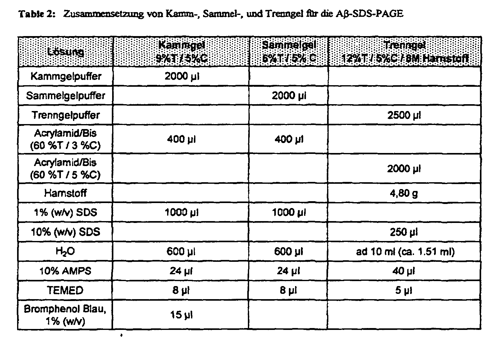


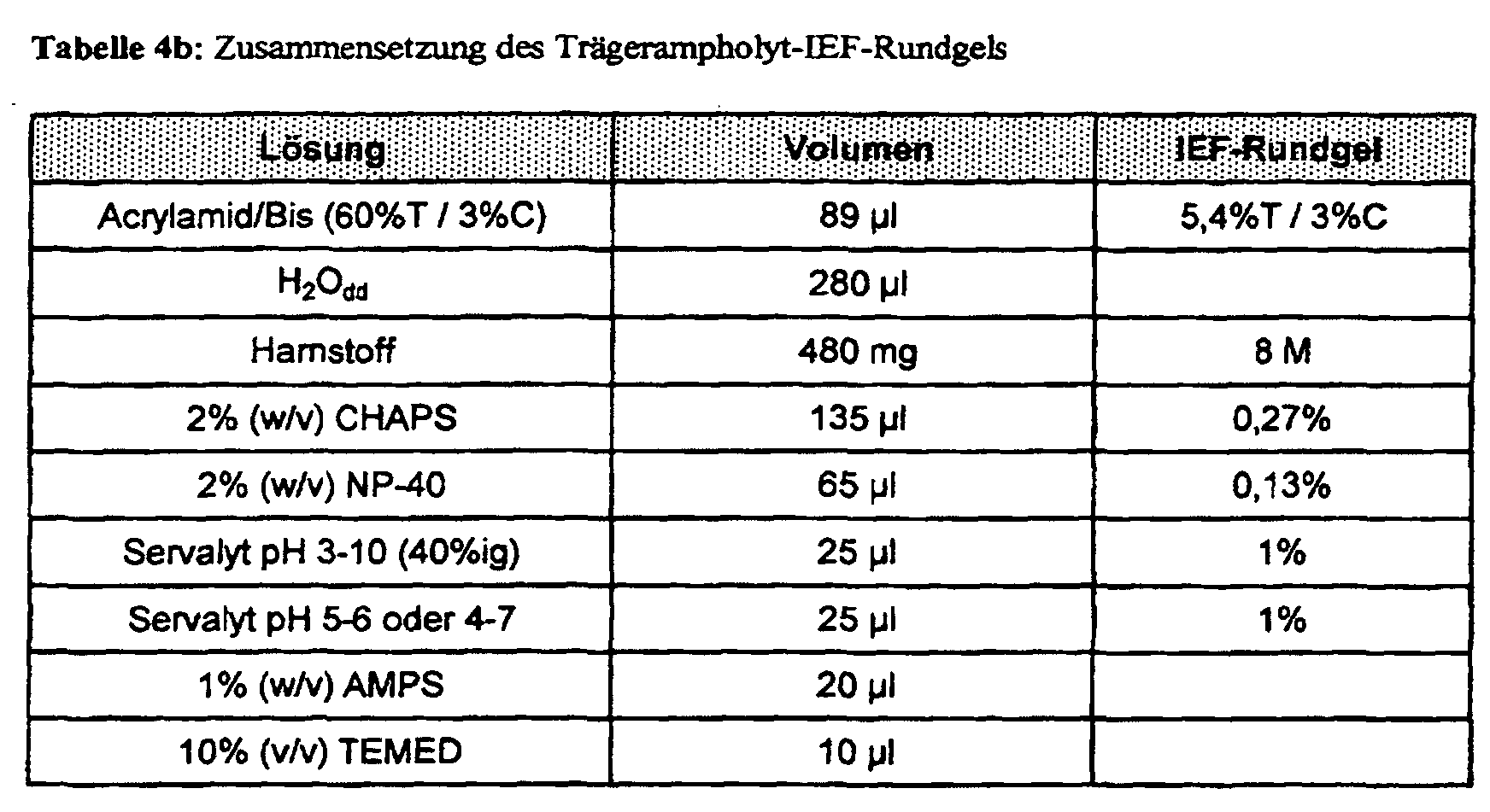
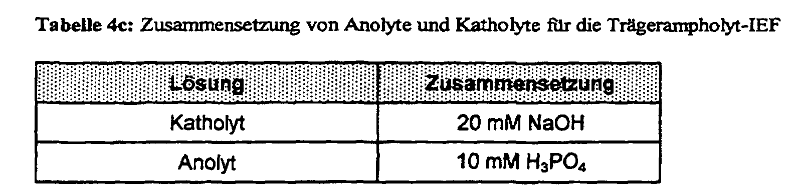
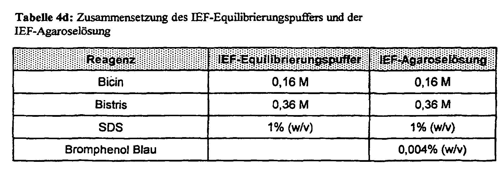
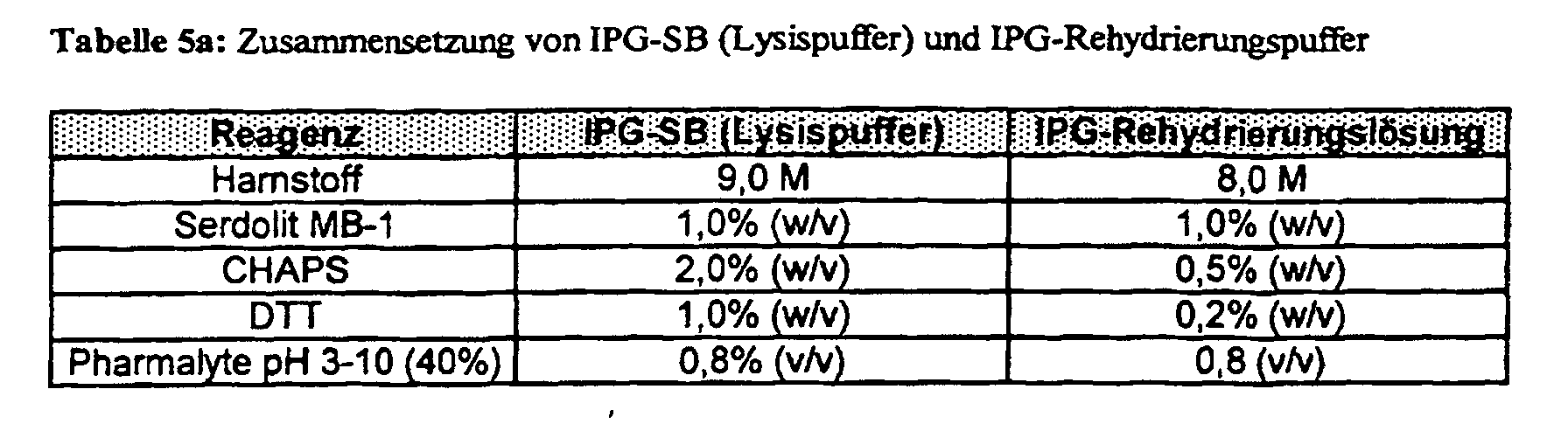

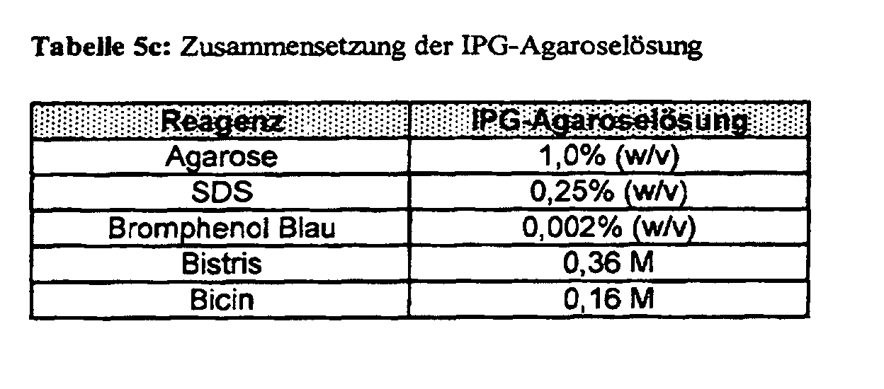
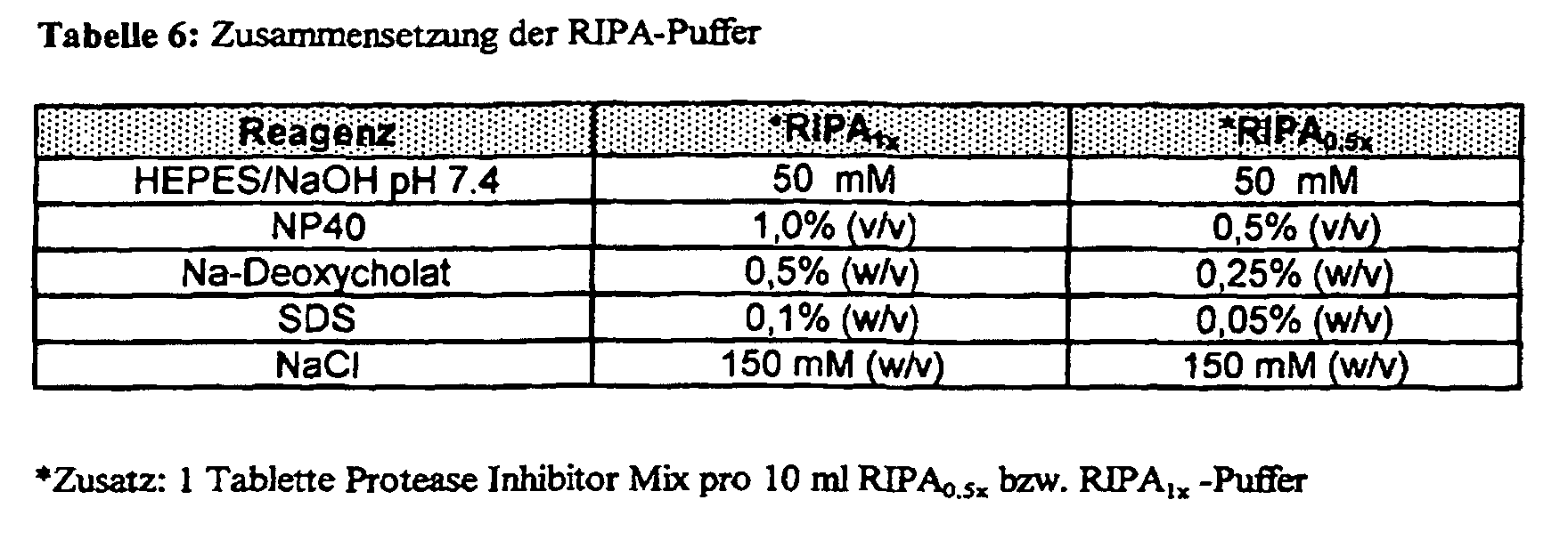
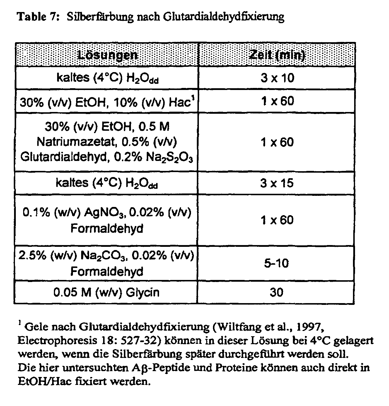
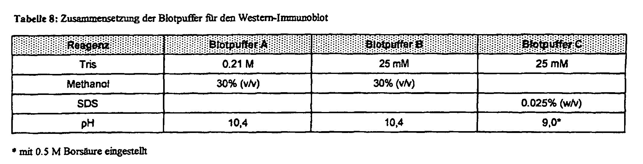
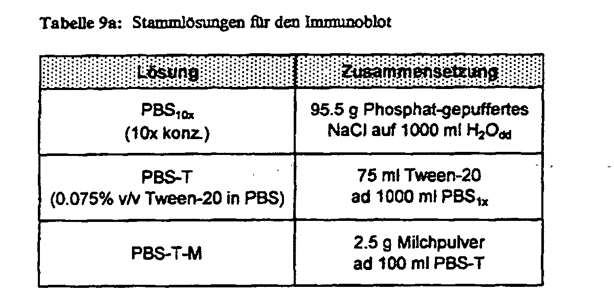
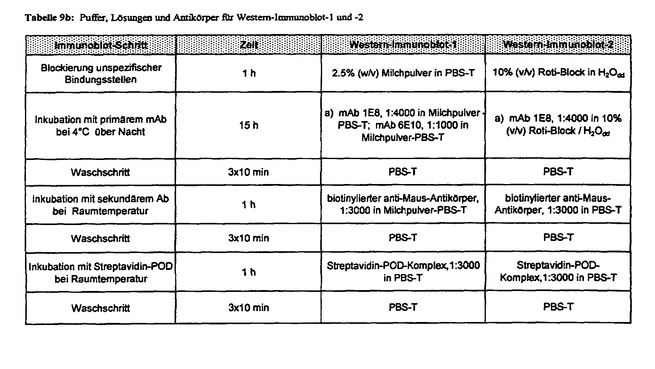
| Zusammenstellung der Kollektive mit Schnittmengen gemeinsamer Patienten. NDC-3KP ist vollständig Teilkollektiv von NDC-3. AD-3KP ist weitgehend Teilkollektiv von AD-3 | |
| Tabelle 5a) | NDC1 (n=30) |
| NDC-2KP (n=10) | Schnittmenge (n=2): NP55, NP57 |
| Tabelle 5b) | NDC-3 (n=47) |
| Schnittmenge (n=15): | |
| NDC-3KP | NP213, NP344, NP345, |
| (n=15; | NP352, NP355, NP356, |
| Teilmenge von | NP364, NP374, NP402, |
| NDC-3) | NP412, NP419, NP421, |
| NP457, NP490, NP 526 | |
| Tabelle 5c) | AD-1 (n=35) |
| AD-3 (n=12) | Schnittmenge (n=3): NP37, NP52, NP66 |
| AD-3KP (n=11) | Schnittmenge (n=3): NP52, NP66, NP69 |
| Tabelle 5d) | AD-3 (n=12) |
| AD-3KP (n=11; | Schnittmenge (n=9): |
| Schnittmenge | NP45, NP52, NP58, NP66, |
| plus | NP111, NP143, NP190, |
| NP69/NP197) | NP319, NP320 |
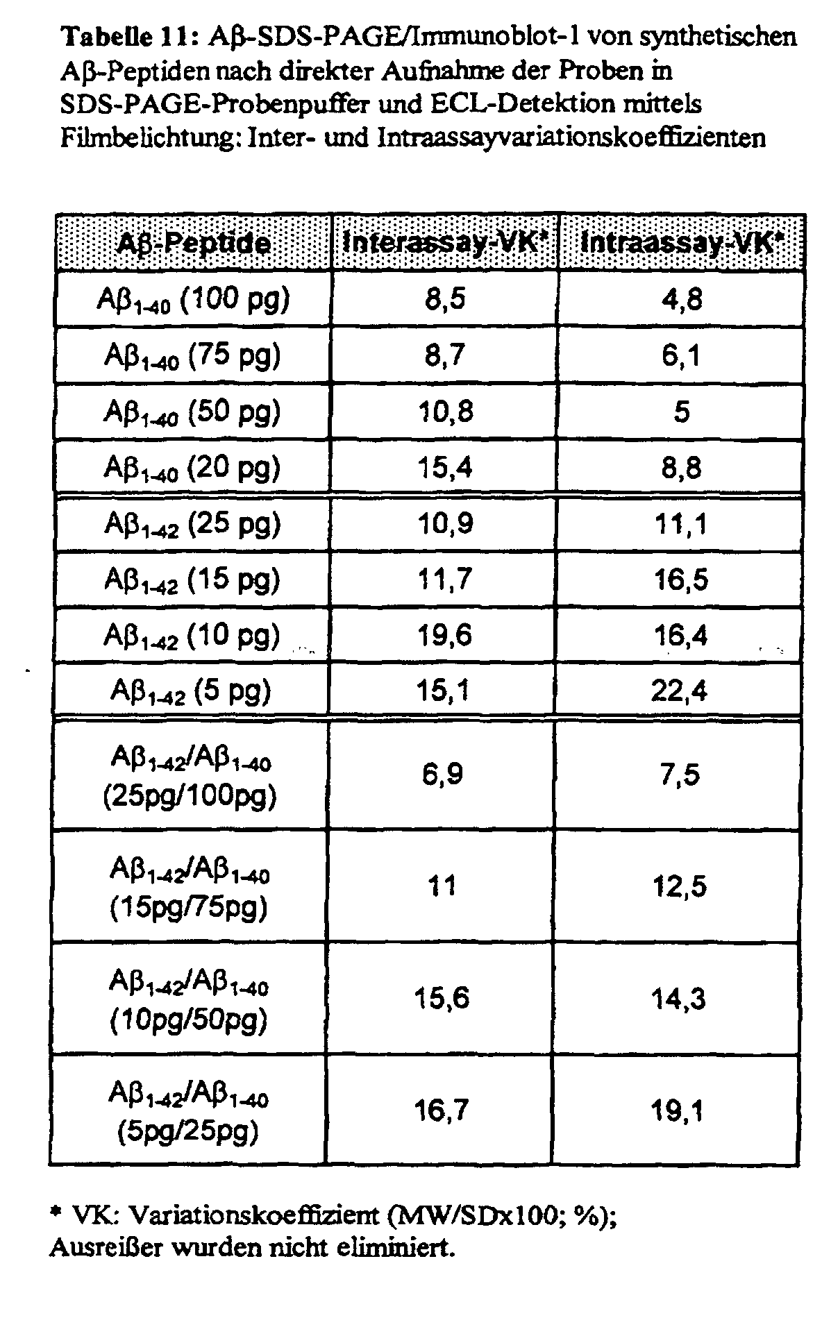
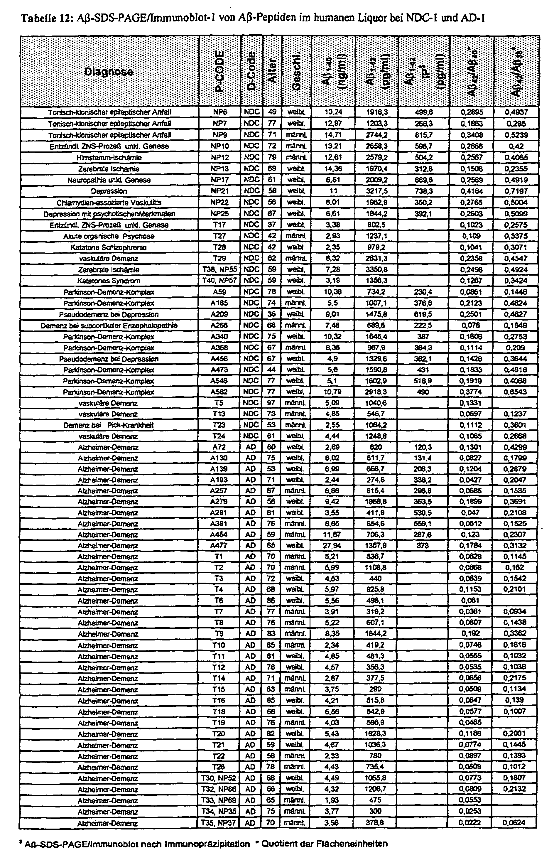
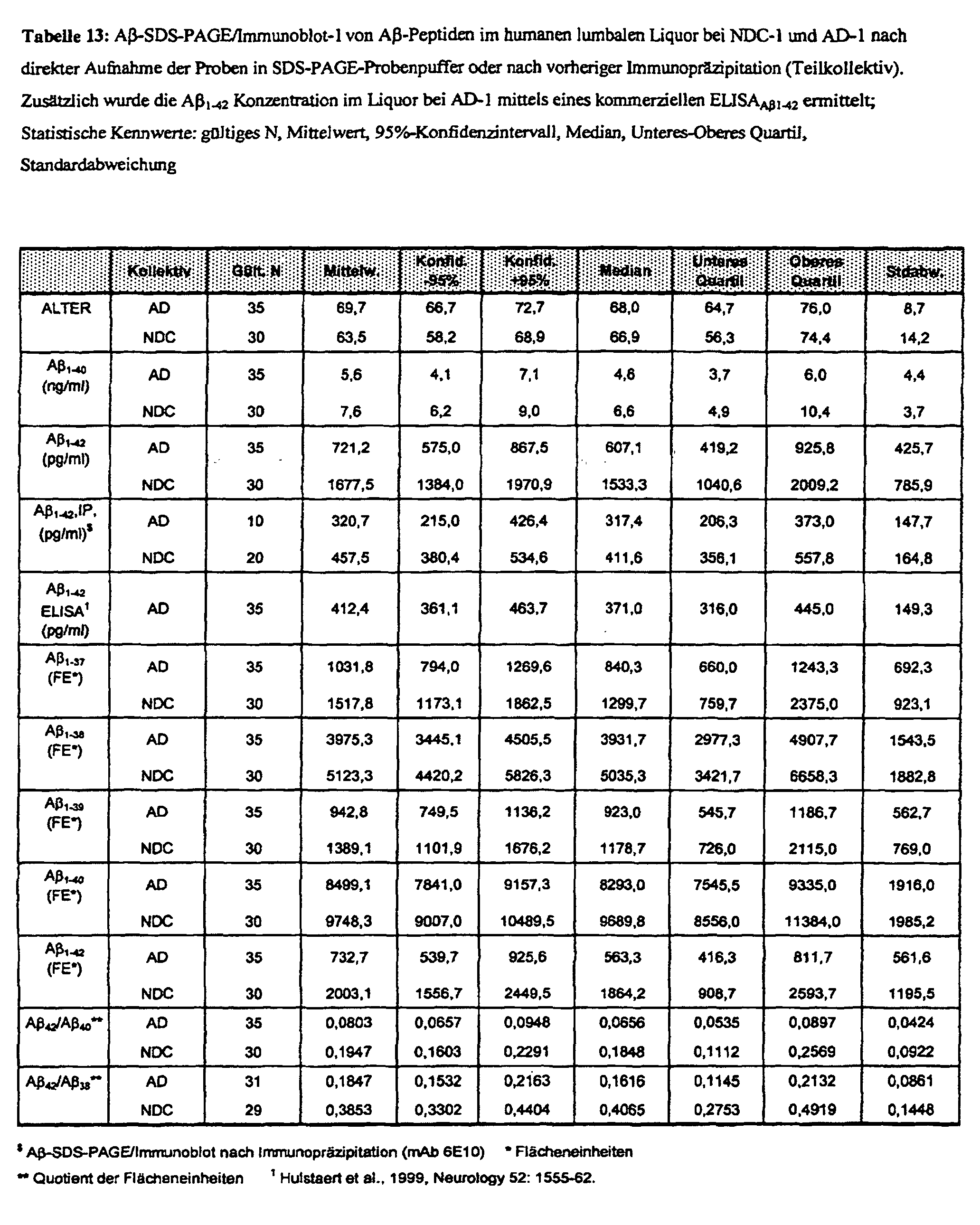

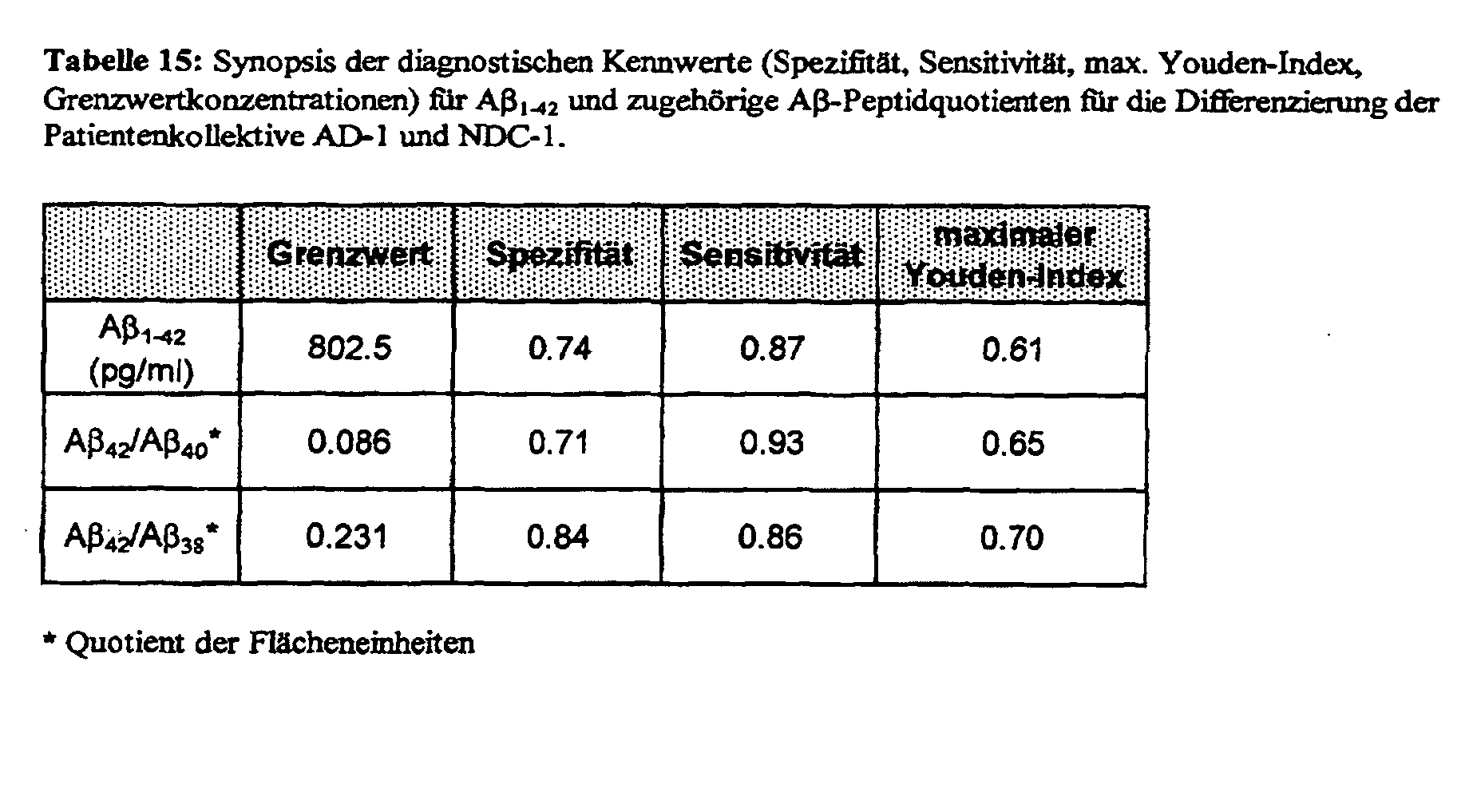
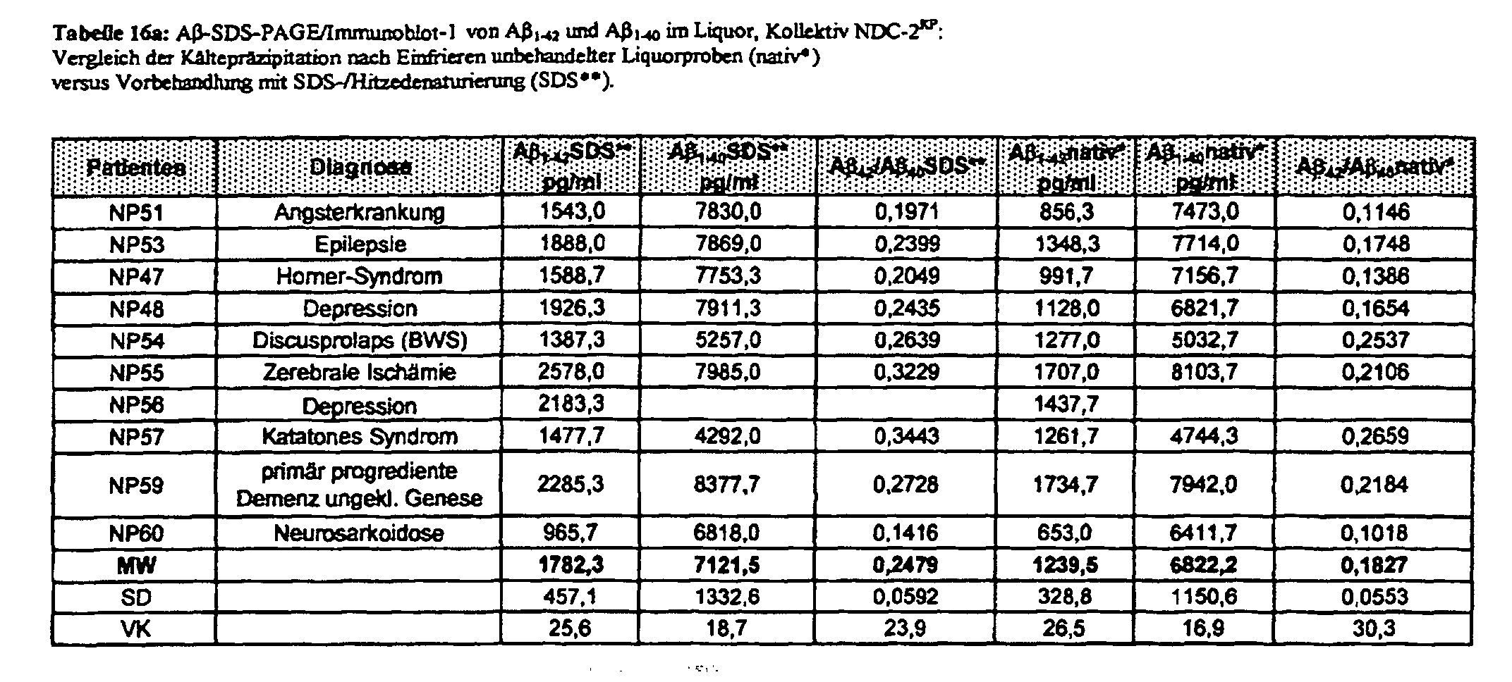


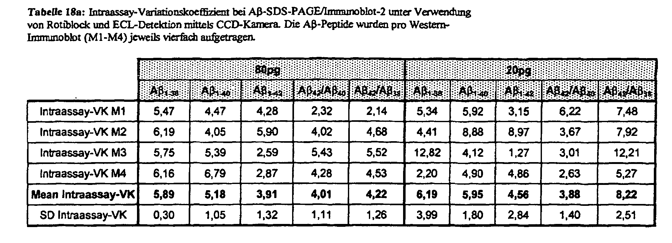
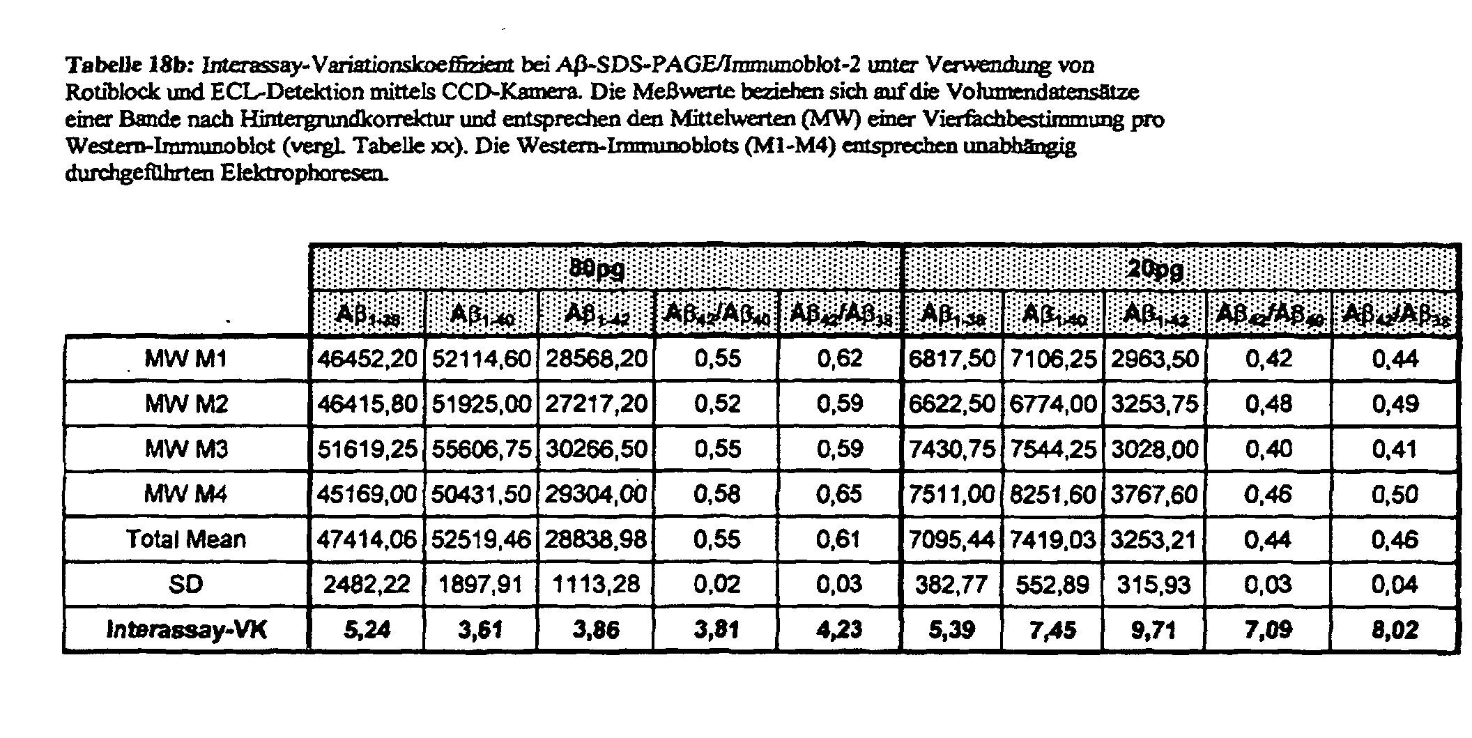
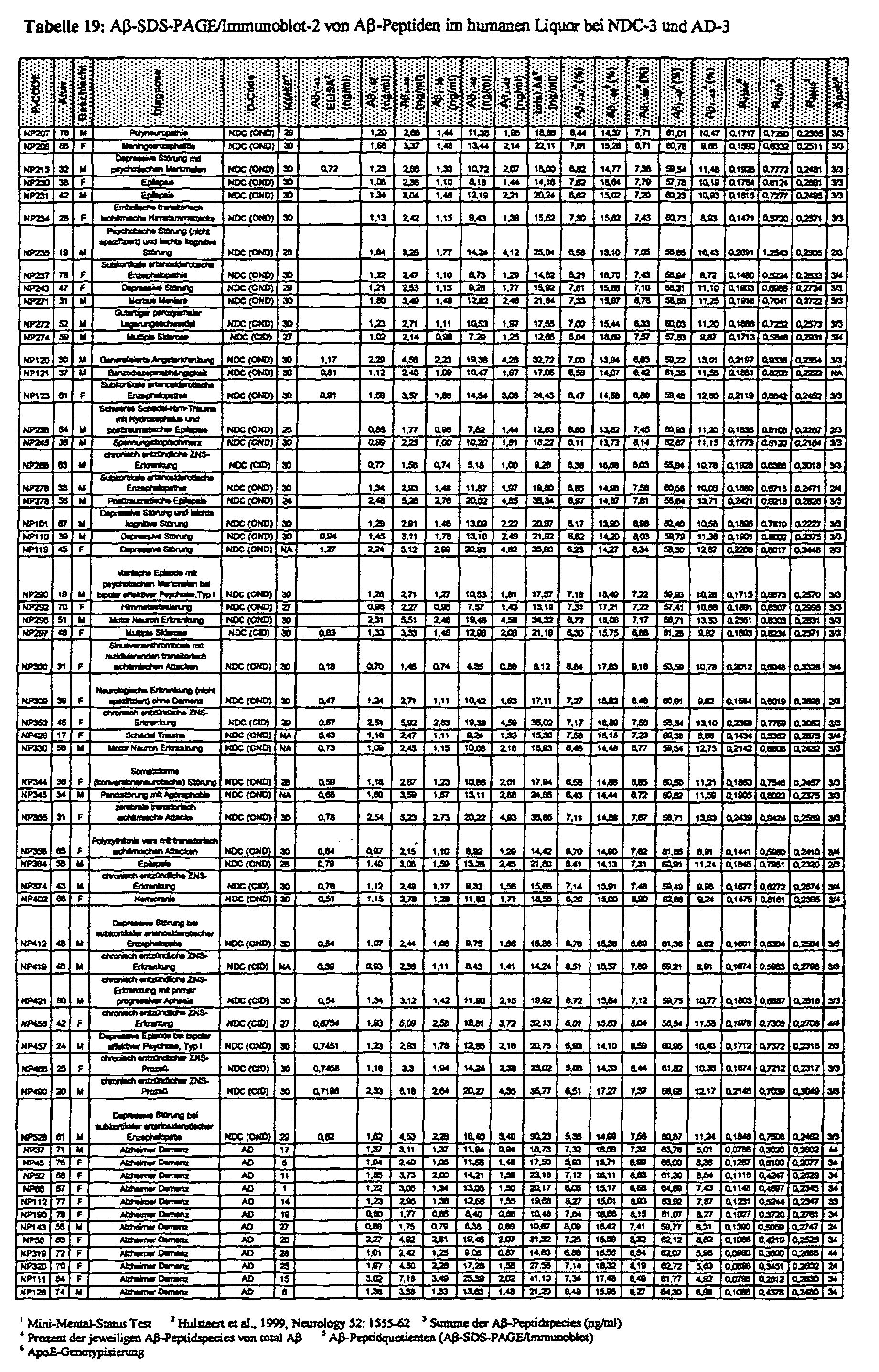

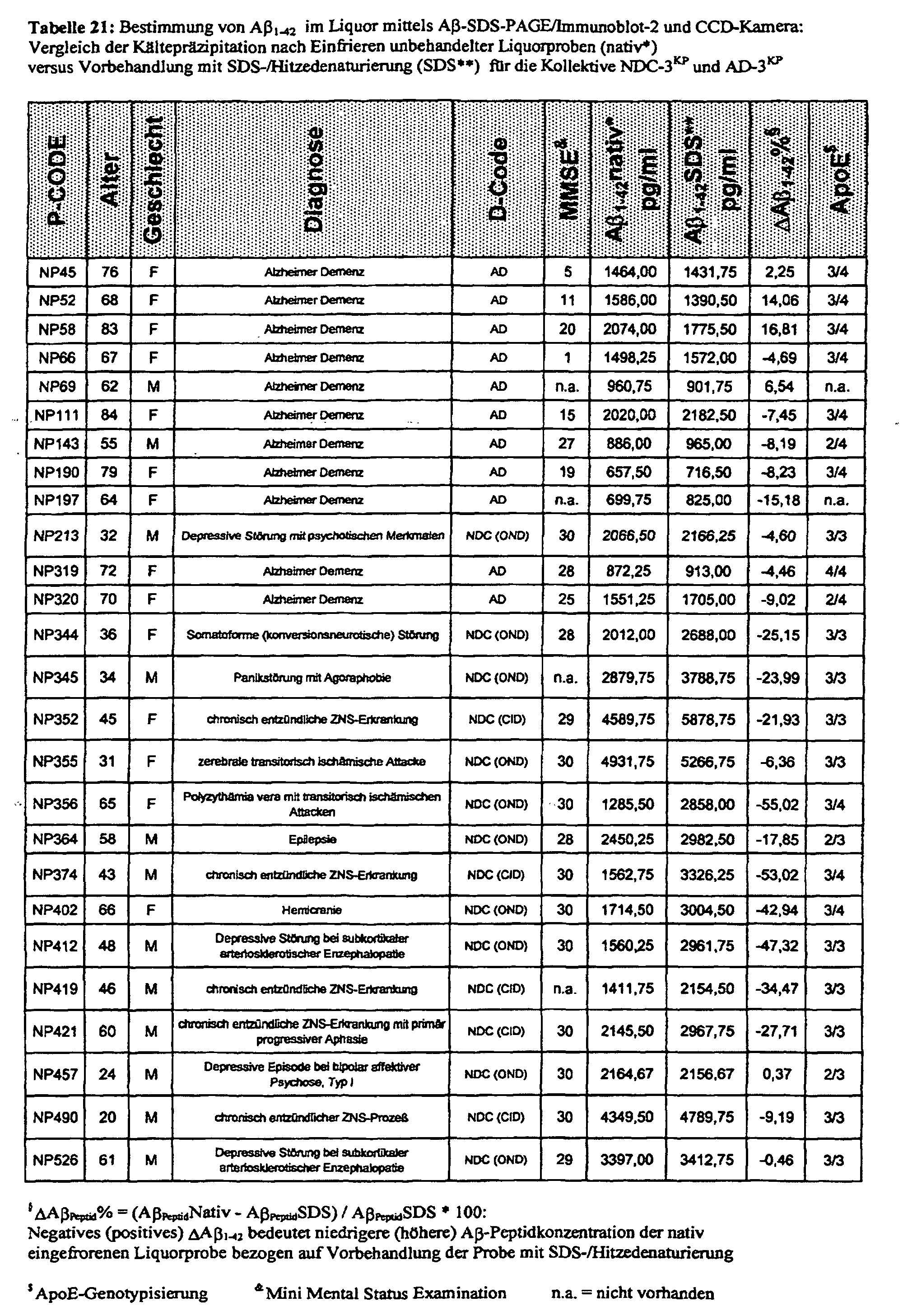
Claims (20)
- Monoklonaler Antikörper, der als mAb 1E8 bezeichnet wird, der von Hybridomen produziert wird, die bei der DSMZ-Deutsche Sammlung von Mikroorganismen und Zellkulturen GmbH, Braunschweig, am 19.12.2000 hinterlegt wurden und denen die DSMZ-Aufnahmenummer DSM ACC2485 zugeordnet wurde.
- Antikörper mAb 1E8 nach Anspruch 1, der radioaktiv markiert ist.
- Verwendung des Antikörpers mAb 1E8 nach Anspruch 1 oder 2 zum Nachweis von Aβ-Peptiden Aβ1-x und/oder Aβ2-x und/oder sAPPα.
- Verwendung des Antikörpers mAb 1E8 nach Anspruch 3 in einem Western-Immunoblot.
- Verwendung des Antikörpers mAb 1E8 nach Anspruch 4, dadurch gekennzeichnet, dass unspezifische Bindungsstellen zuvor mit einem Blockiermittel blockiert werden.
- Verwendung des Antikörpers mAb 1E8 nach einem der Ansprüche 3 bis 5, dadurch gekennzeichnet, dass die Aβ-Peptide und das sAPPα zuvor durch eine Natrium-Laurylsulfat-Polyacrylamid-Gelelektrophorese (SDS-PAGE) voneinander getrennt werden, wobei durch Harnstoffzugabe eine Aminosäureprimärsequenz-spezifische Konformationsänderung der Aβ-Peptide induziert wird.
- Verwendung des Antikörpers mAb 1E8 nach Anspruch 6, dadurch gekennzeichnet, dass die Aβ Peptide zuvor durch isoelektrische Fokussierung separiert werden.
- Verwendung des Antikörpers mAb 1E8 nach Anspruch 6 oder 7, dadurch gekennzeichnet, dass in einer Probe, die aus der Gruppe ausgewählt ist, welche Liquor, Hirnhomogeniesaat, Plasma und Mischungen davon umfasst, eine Konzentration des Aβ-Peptids Aβ1-42 bestimmt wird.
- Verwendung des Antikörpers mAb 1E8 nach Anspruch 6 oder 7, dadurch gekennzeichnet, dass eine Probe, die aus der Gruppe ausgewählt ist, welche Liquor, Hirnhomogeniesaat, Plasma und Mischungen davon umfasst, auf das Vorliegen einer nachweisbaren Konzentration des Aβ-Peptids Aβ2-42 überprüft wird, die bei 100 pg/ml oder höher liegt.
- Verwendung des Antikörpers mAb 1E8 nach Anspruch 8 und 9, dadurch gekennzeichnet, dass ein Verhältnis zwischen der Konzentration des Aβ-Peptids Aβ2-42 zu der Konzentration des Aβ-Peptids Aβ1-42 in der Probe bestimmt wird.
- Verwendung des Antikörpers mAb 1E8 nach Anspruch 6 oder 7, dadurch gekennzeichnet, dass in einer Probe, die aus der Gruppe ausgewählt ist, welche Liquor, Himhomogeniesaat, Plasma und Mischungen davon umfasst, mindestens ein Konzentrationsverhältnis bestimmt wird, das aus der Gruppe ausgewählt ist, welche ein Verhältnis zwischen einer Konzentration des Aβ-Peptids Aβ1-42 zu einer Konzentration des Aβ-Peptids Aβ1-40, ein Verhältnis zwischen einer Konzentration des Aβ-Peptids Aβ1-42 zu einer Konzentration des Aβ-Peptids Aβ1-38 und ein Verhältnis zwischen einer Konzentration des Aβ-Peptids Aβ1-38 zu einer Konzentration des Aβ-Peptids Aβ1-40 umfasst.
- Verwendung des Antikörpers mAb 1E8 nach Anspruch 6 oder 7, dadurch gekennzeichnet, dass in einer Probe, die aus der Gruppe ausgewählt ist, welche Liquor, Hirnhomogeniesaat, Plasma und Mischungen davon umfasst, mindestens ein relativer Anteil Aβ1-n% einer Konzentration eines Aβ-Peptids Aβ1-n an einer Konzentration von Aβ-Peptiden Aβ1-x bestimmt wird, wobei der relative Anteil Aβ1-n% aus der Gruppe ausgewählt ist, welche einen relativen Anteil Aβ1-42% einer Konzentration des Aβ-Peptids Aβ1-42, einen relativen Anteil Aβ1-40% einer Konzentration des Aβ-Peptids Aβ1-40 und einen relativen Anteil Aβ1-38% einer Konzentration des Aβ-Peptids Aβ1-38 umfasst, und wobei die Konzentration von Aβ-Peptiden Aβ1-x mindestens die Konzentration der Aβ-Peptide Aβ1-38, Aβ1-40 und Aβ1-42 aus der Gruppe der Aβ-Peptide Aβ1-37, Aβ1-38, Aβ1-39, Aβ1-40 und Aβ1-42 umfasst.
- Verwendung des Antikörpers mAb 1E8 nach einem der Ansprüche 1 bis 12, dadurch gekennzeichnet, dass die Aβ-Peptide zuvor durch eine Behandlung mit einem Detergenz aufgeschlossen werden.
- Verwendung des Antikörpers mAb 1E8 nach Anspruch 13, dadurch gekennzeichnet, dass die Aβ-Peptide durch eine SDS-Hitzedenaturierung aufgeschlossen werden.
- Verwendung des Antikörpers mAb 1E8 nach Anspruch 8 und 13 oder 14, dadurch gekennzeichnet, dass die Probe in mindestens zwei Teilproben unterteilt wird, dass eine erste Teilprobe der Probenbehandlung mit dem Detergenz vor oder statt einer Präzipitationsbehandlung unterworfen wird, die zumindest auf Teilmengen des Aβ-Peptids Aβ1-42 gerichtet ist, dass eine zweite Teilprobe der Präzipitationsbehandlung vor oder statt der Probenbehandlung mit dem Detergenz unterworfen wird, und dass eine Differenz ΔAβ1-42 zwischen den in beiden Probenteilen bestimmten Konzentrationen des Aβ-Peptids Aβ1-42 bestimmt wird.
- Verwendung des Antikörpers mAb 1E8 nach einem der Ansprüche 1 bis 15, dadurch gekennzeichnet, dass die Aβ-Peptide, an die der Antikörper mAb 1E8 gebunden ist, mit einem gegen den Antikörper mAb 1E8 gerichtete sekundären Antikörper markiert werden.
- Verwendung des Antikörpers mAb 1E8 nach Anspruch 16, dadurch gekennzeichnet, dass der gegen den Antikörper mAb 1E8 gerichtete sekundäre Antikörper bereits mit einem mengenmäßig registrierbaren Marker versehen ist oder nach seiner Immunreaktion mit dem Antikörper mAb 1E8 mit einem mengenmäßig registrierbaren Marker versehen wird.
- Verwendung des Antikörpers mAb 1E8 nach Anspruch 16 oder 17, dadurch gekennzeichnet, dass die Menge des markierten Antikörper mAb 1E8 photometrisch mit einer CCD-Kamera bestimmt wird.
- Verwendung des Antikörpers mAb 1E8 nach Anspruch 1 oder 2 zum Aufkonzentrieren von Aβ-Peptiden Aβ1-x und Aβ2-x.
- Verwendung des Antikörpers mAb 1E8 nach Anspruch 1 oder 2 zur Unterscheidung von Aβ-Peptiden Aβ1-x und Aβ2-x von Aβ-Peptiden Aβn-x mit n>2.
Priority Applications (6)
| Application Number | Priority Date | Filing Date | Title |
|---|---|---|---|
| AT01114192T ATE277952T1 (de) | 2001-06-12 | 2001-06-12 | Monoklonaler antikörper, mbab 1e8, welcher für die zwei ersten n-terminalen aminosäuren von amyloid-beta-peptiden spezifisch ist und dessen verwendung zum nachweis von amyloid-beta peptiden und/oder sappa |
| ES01114192T ES2228697T3 (es) | 2001-06-12 | 2001-06-12 | Anticuerpo monoclonal, mab 1e8, que es especifico para los dos primeros aminoacidos de peptidos beta-amiloides y su utilizacion en la deteccion de peptidos beta-amiloides y/o sapp alfa. |
| DE50103881T DE50103881D1 (de) | 2001-06-12 | 2001-06-12 | Monoklonaler Antikörper, mbAb 1E8, welcher für die zwei ersten N-terminalen Aminosäuren von Amyloid-beta-Peptiden spezifisch ist und dessen Verwendung zum Nachweis von Amyloid-beta Peptiden und/oder sAPPa |
| EP01114192A EP1270592B1 (de) | 2001-06-12 | 2001-06-12 | Monoklonaler Antikörper, mbAb 1E8, welcher für die zwei ersten N-terminalen Aminosäuren von Amyloid-beta-Peptiden spezifisch ist und dessen Verwendung zum Nachweis von Amyloid-beta Peptiden und/oder sAPPa |
| US10/170,272 US6849416B2 (en) | 2001-06-12 | 2002-06-11 | Monoclonal antibody |
| JP2002171495A JP2003012700A (ja) | 2001-06-12 | 2002-06-12 | mAb1E8で表わされるモノクロナール抗体及びAβ−ペプチド及び(又は)sAPPαの検出にこの抗体を使用する方法 |
Applications Claiming Priority (1)
| Application Number | Priority Date | Filing Date | Title |
|---|---|---|---|
| EP01114192A EP1270592B1 (de) | 2001-06-12 | 2001-06-12 | Monoklonaler Antikörper, mbAb 1E8, welcher für die zwei ersten N-terminalen Aminosäuren von Amyloid-beta-Peptiden spezifisch ist und dessen Verwendung zum Nachweis von Amyloid-beta Peptiden und/oder sAPPa |
Publications (2)
| Publication Number | Publication Date |
|---|---|
| EP1270592A1 EP1270592A1 (de) | 2003-01-02 |
| EP1270592B1 true EP1270592B1 (de) | 2004-09-29 |
Family
ID=8177701
Family Applications (1)
| Application Number | Title | Priority Date | Filing Date |
|---|---|---|---|
| EP01114192A Expired - Lifetime EP1270592B1 (de) | 2001-06-12 | 2001-06-12 | Monoklonaler Antikörper, mbAb 1E8, welcher für die zwei ersten N-terminalen Aminosäuren von Amyloid-beta-Peptiden spezifisch ist und dessen Verwendung zum Nachweis von Amyloid-beta Peptiden und/oder sAPPa |
Country Status (6)
| Country | Link |
|---|---|
| US (1) | US6849416B2 (de) |
| EP (1) | EP1270592B1 (de) |
| JP (1) | JP2003012700A (de) |
| AT (1) | ATE277952T1 (de) |
| DE (1) | DE50103881D1 (de) |
| ES (1) | ES2228697T3 (de) |
Cited By (12)
| Publication number | Priority date | Publication date | Assignee | Title |
|---|---|---|---|---|
| CN102192985A (zh) * | 2010-03-18 | 2011-09-21 | 上海依科赛生物制品有限公司 | 一种人体β淀粉样蛋白试剂盒 |
| US8409580B2 (en) | 2003-07-25 | 2013-04-02 | Ac Immune Sa | Therapeutic vaccine targeted against P-glycoprotein 170 for inhibiting multidrug resistance in the treatment of cancers |
| US8497072B2 (en) | 2005-11-30 | 2013-07-30 | Abbott Laboratories | Amyloid-beta globulomer antibodies |
| US8691224B2 (en) | 2005-11-30 | 2014-04-08 | Abbvie Inc. | Anti-Aβ globulomer 5F7 antibodies |
| US8877190B2 (en) | 2006-11-30 | 2014-11-04 | Abbvie Inc. | Aβ conformer selective anti-Aβ globulomer monoclonal antibodies |
| US8895004B2 (en) | 2007-02-27 | 2014-11-25 | AbbVie Deutschland GmbH & Co. KG | Method for the treatment of amyloidoses |
| US8926983B2 (en) | 2003-02-21 | 2015-01-06 | Ac Immune Sa | Method for improving memory in AD patients |
| US8987419B2 (en) | 2010-04-15 | 2015-03-24 | AbbVie Deutschland GmbH & Co. KG | Amyloid-beta binding proteins |
| US9062101B2 (en) | 2010-08-14 | 2015-06-23 | AbbVie Deutschland GmbH & Co. KG | Amyloid-beta binding proteins |
| US9176150B2 (en) | 2003-01-31 | 2015-11-03 | AbbVie Deutschland GmbH & Co. KG | Amyloid beta(1-42) oligomers, derivatives thereof and antibodies thereto, methods of preparation thereof and use thereof |
| US9289488B2 (en) | 2010-08-12 | 2016-03-22 | Ac Immune Sa | Vaccine engineering |
| US9687447B2 (en) | 2010-10-26 | 2017-06-27 | Ac Immune S.A. | Method for preparing liposome-based constructs |
Families Citing this family (21)
| Publication number | Priority date | Publication date | Assignee | Title |
|---|---|---|---|---|
| EP1469312A1 (de) * | 2003-04-18 | 2004-10-20 | Friedrich-Alexander-Universität Erlangen-Nürnberg | Diagnose der Alzheimer Krankheit |
| EP1480041A1 (de) * | 2003-05-22 | 2004-11-24 | Innogenetics N.V. | Verfahren zur Voraussage, zur Diagnose und zur vergleichenden Diagnose von Alzheimer |
| US20080026995A1 (en) * | 2003-07-25 | 2008-01-31 | Ac Immune Sa | Therapeutic vaccine targeted against p-glycoprotein 170 for inhibiting multidrug resistance in the treatment of cancers |
| CN101048662A (zh) * | 2004-10-28 | 2007-10-03 | 三光纯药株式会社 | 阿尔茨海默氏病的检验方法及诊断试剂 |
| RU2551782C2 (ru) | 2005-12-12 | 2015-05-27 | Ац Иммуне Са | Специфические в отношении амилоида бета (а бета) 1-42 моноклональные антитела, обладающие терапевтическими свойствами |
| EP1811304A1 (de) * | 2006-01-18 | 2007-07-25 | Friedrich-Alexander-Universität Erlangen-Nürnberg | Große Aß-Peptid-bindende Partikel (LAPS) bei der Diagnose und Behandlung von Alzheimer-Demenz |
| TWI551607B (zh) * | 2006-07-14 | 2016-10-01 | Ac免疫公司 | 人類化抗體 |
| FR2904113A1 (fr) * | 2006-07-21 | 2008-01-25 | Exonhit Therapeutics S A Sa | Procedes et outils pour la therapie de pathologies neurodegeneratives |
| WO2008058760A1 (en) * | 2006-11-17 | 2008-05-22 | Friedrich-Alexander-Universität Erlangen-Nürnberg | Method of differentially diagnosing dementias |
| EP2170389B1 (de) * | 2007-06-12 | 2014-10-29 | AC Immune S.A. | Humanisierte antikörper gegen amyloid beta |
| US8048420B2 (en) | 2007-06-12 | 2011-11-01 | Ac Immune S.A. | Monoclonal antibody |
| US8613923B2 (en) | 2007-06-12 | 2013-12-24 | Ac Immune S.A. | Monoclonal antibody |
| RU2482876C2 (ru) * | 2007-10-05 | 2013-05-27 | Дженентек, Инк. | Применение антитела против амилоида-бета при глазных заболеваниях |
| WO2010006318A2 (en) * | 2008-07-11 | 2010-01-14 | Life Technologies Corporation | Electrophoretically enhanced detection of analytes on a solid support |
| WO2010046332A1 (en) * | 2008-10-20 | 2010-04-29 | INSERM (Institut National de la Santé et de la Recherche Médicale) | Method for detecting soluble amyloid precursor protein (app) alpha and/or soluble app beta |
| DE102010014684A1 (de) | 2010-04-09 | 2011-10-13 | Universität Duisburg-Essen | Neue Ansätze zur Alzheimer-Diagnose |
| CN103179981B (zh) | 2010-07-30 | 2017-02-08 | Ac免疫有限公司 | 安全和功能性的人源化抗β‑淀粉样蛋白抗体 |
| EP2811018B1 (de) | 2012-02-03 | 2017-03-22 | Shionogi & Co., Ltd. | ANTI-sAPPß-ANTIKÖRPER |
| CN105143856B (zh) * | 2013-04-24 | 2019-06-18 | 欧蒙医学诊断技术有限公司 | 用于自动化评估孵育的免疫印迹条的方法 |
| NL2022504B1 (en) | 2019-02-04 | 2020-08-19 | Elstar Dynamics Patents B V | Improved optical modulator |
| US12007660B2 (en) | 2021-06-24 | 2024-06-11 | Elstar Dynamics Patents B.V. | Light modulator, substrate comprising electrodes and smart glazing |
-
2001
- 2001-06-12 DE DE50103881T patent/DE50103881D1/de not_active Expired - Fee Related
- 2001-06-12 AT AT01114192T patent/ATE277952T1/de not_active IP Right Cessation
- 2001-06-12 ES ES01114192T patent/ES2228697T3/es not_active Expired - Lifetime
- 2001-06-12 EP EP01114192A patent/EP1270592B1/de not_active Expired - Lifetime
-
2002
- 2002-06-11 US US10/170,272 patent/US6849416B2/en not_active Expired - Fee Related
- 2002-06-12 JP JP2002171495A patent/JP2003012700A/ja active Pending
Cited By (21)
| Publication number | Priority date | Publication date | Assignee | Title |
|---|---|---|---|---|
| US9176150B2 (en) | 2003-01-31 | 2015-11-03 | AbbVie Deutschland GmbH & Co. KG | Amyloid beta(1-42) oligomers, derivatives thereof and antibodies thereto, methods of preparation thereof and use thereof |
| US8926983B2 (en) | 2003-02-21 | 2015-01-06 | Ac Immune Sa | Method for improving memory in AD patients |
| US8409580B2 (en) | 2003-07-25 | 2013-04-02 | Ac Immune Sa | Therapeutic vaccine targeted against P-glycoprotein 170 for inhibiting multidrug resistance in the treatment of cancers |
| US9975946B2 (en) | 2004-02-20 | 2018-05-22 | Ac Immune Sa | Antibodies obtainable using supramolecular constructs |
| US8497072B2 (en) | 2005-11-30 | 2013-07-30 | Abbott Laboratories | Amyloid-beta globulomer antibodies |
| US8691224B2 (en) | 2005-11-30 | 2014-04-08 | Abbvie Inc. | Anti-Aβ globulomer 5F7 antibodies |
| US10538581B2 (en) | 2005-11-30 | 2020-01-21 | Abbvie Inc. | Anti-Aβ globulomer 4D10 antibodies |
| US10323084B2 (en) | 2005-11-30 | 2019-06-18 | Abbvie Inc. | Monoclonal antibodies against amyloid beta protein and uses thereof |
| US10208109B2 (en) | 2005-11-30 | 2019-02-19 | Abbvie Inc. | Monoclonal antibodies against amyloid beta protein and uses thereof |
| US9540432B2 (en) | 2005-11-30 | 2017-01-10 | AbbVie Deutschland GmbH & Co. KG | Anti-Aβ globulomer 7C6 antibodies |
| US8877190B2 (en) | 2006-11-30 | 2014-11-04 | Abbvie Inc. | Aβ conformer selective anti-Aβ globulomer monoclonal antibodies |
| US9951125B2 (en) | 2006-11-30 | 2018-04-24 | Abbvie Inc. | Aβ conformer selective anti-Aβ globulomer monoclonal antibodies |
| US9359430B2 (en) | 2006-11-30 | 2016-06-07 | Abbvie Inc. | Abeta conformer selective anti-Abeta globulomer monoclonal antibodies |
| US9394360B2 (en) | 2006-11-30 | 2016-07-19 | Abbvie Inc. | Aβ conformer selective anti-Aβ globulomer monoclonal antibodies |
| US8895004B2 (en) | 2007-02-27 | 2014-11-25 | AbbVie Deutschland GmbH & Co. KG | Method for the treatment of amyloidoses |
| CN102192985A (zh) * | 2010-03-18 | 2011-09-21 | 上海依科赛生物制品有限公司 | 一种人体β淀粉样蛋白试剂盒 |
| US8987419B2 (en) | 2010-04-15 | 2015-03-24 | AbbVie Deutschland GmbH & Co. KG | Amyloid-beta binding proteins |
| US9289488B2 (en) | 2010-08-12 | 2016-03-22 | Ac Immune Sa | Vaccine engineering |
| US10047121B2 (en) | 2010-08-14 | 2018-08-14 | AbbVie Deutschland GmbH & Co. KG | Amyloid-beta binding proteins |
| US9062101B2 (en) | 2010-08-14 | 2015-06-23 | AbbVie Deutschland GmbH & Co. KG | Amyloid-beta binding proteins |
| US9687447B2 (en) | 2010-10-26 | 2017-06-27 | Ac Immune S.A. | Method for preparing liposome-based constructs |
Also Published As
| Publication number | Publication date |
|---|---|
| JP2003012700A (ja) | 2003-01-15 |
| US20030166019A1 (en) | 2003-09-04 |
| ES2228697T3 (es) | 2005-04-16 |
| EP1270592A1 (de) | 2003-01-02 |
| DE50103881D1 (de) | 2004-11-04 |
| ATE277952T1 (de) | 2004-10-15 |
| US6849416B2 (en) | 2005-02-01 |
Similar Documents
| Publication | Publication Date | Title |
|---|---|---|
| EP1270592B1 (de) | Monoklonaler Antikörper, mbAb 1E8, welcher für die zwei ersten N-terminalen Aminosäuren von Amyloid-beta-Peptiden spezifisch ist und dessen Verwendung zum Nachweis von Amyloid-beta Peptiden und/oder sAPPa | |
| DE69632734T2 (de) | QUANTIFIZIERUNG VON p97 ZUR DIAGNOSE UND ÜBERWACHUNG DER ALZHEIMHER-KRANKHEIT | |
| DE69926248T2 (de) | Test zum nachweis von prionen-proteinen die in einer mit krankheit assoziierten konformationen vorliegen | |
| DE60121958T2 (de) | Frühdiagnose von konformationellen erkrankungen | |
| DE60123752T2 (de) | Diagnose von tauopathien durch bestimmung des verhältnisses von tau/phospho-tau | |
| DE60032932T2 (de) | Verfahren zur diagnose einer von einem prionenstamm verursachten übertragbaren subakuten spongiformen enzephalopathie in einer biologischen probe | |
| DE69737557T2 (de) | Verfahren zur Herstellung von Mittel zur Therapie der übertragbaren Spongiformen Enzephalopathie und nicht-infektiösen Blut- und Gewebeprodukten sowie die hergestellten Produkte | |
| DE69113273T2 (de) | Zellnekrosnachweis durch prüfung von spektrin und seine abbauprodukte. | |
| EP3505935B1 (de) | Verfahren zur diagnose einer bornavirus-infektion | |
| DE69713498T2 (de) | Methode zum nachweis übertragbarer spongiformer enzephalopathien | |
| DE60129674T2 (de) | Verfahren zur diagnose von transmissiblen spongiformen encephalopathien | |
| WO2011124376A1 (de) | Neue ansätze zur alzheimer-diagnose | |
| EP1304336B1 (de) | Antikörper zum spezifischen Nachweis von pathogenen Prionen humanen Ursprungs und damit durchgeführte Nachweisverfahren | |
| EP1303537B1 (de) | Ho-1 suppressor als diagnostischer und prognostischer test für demenz-krankheiten | |
| EP3194436B1 (de) | Monoklonaler antikörper gegen humanes tau-protein | |
| WO2004005920A2 (de) | Verfahren zur anreicherung und zum nachweis von pathologisch veränderten prion-proteinen (prpsc) | |
| DE10131912A1 (de) | Verwendung von 14-3-3 Proteinen und Verfahren zu deren Bestimmung | |
| DE69628713T2 (de) | Marker und Reagenz für Diabetes mellitus und Diabetes-mellitus-Komplikationen | |
| DE60218848T2 (de) | Verfahren zur trennung und/oder zum nachweis und/oder zur identifizierung und/oder zur quantifizierung von prionproteinen | |
| DE602004008488T2 (de) | Entfernung von prionen durch den gebrauch von metalloxid partikeln | |
| EP1902317B1 (de) | Verfahren zur selektiven bestimmung pathologischer proteinablagerungen | |
| EP2097755A1 (de) | In vitro verfahren zur diagnose und frühdiagnose von neurodegenerativen erkrankungen | |
| WO2000050904A1 (de) | Testkit zur analytik der faktor-viii-spaltenden protease | |
| DE69321893T2 (de) | Verfahren zum Nachweis von Alzheimer-Krankheit | |
| WO2015181088A1 (de) | Verfahren zur diagnose einer durch den alternativen weg des komplementsystems vermittelten krankheit oder eines risikos hierfür |
Legal Events
| Date | Code | Title | Description |
|---|---|---|---|
| PUAI | Public reference made under article 153(3) epc to a published international application that has entered the european phase |
Free format text: ORIGINAL CODE: 0009012 |
|
| 17P | Request for examination filed |
Effective date: 20020809 |
|
| AK | Designated contracting states |
Kind code of ref document: A1 Designated state(s): AT BE CH CY DE DK ES FI FR GB GR IE IT LI LU MC NL PT SE TR |
|
| AX | Request for extension of the european patent |
Free format text: AL;LT;LV;MK;RO;SI |
|
| 17Q | First examination report despatched |
Effective date: 20030425 |
|
| RAP1 | Party data changed (applicant data changed or rights of an application transferred) |
Owner name: WILTFANG, JENS |
|
| AKX | Designation fees paid |
Designated state(s): AT BE CH CY DE DK ES FI FR GB GR IE IT LI LU MC NL PT SE TR |
|
| AXX | Extension fees paid |
Extension state: LT Payment date: 20030116 Extension state: SI Payment date: 20030116 Extension state: LV Payment date: 20030116 Extension state: AL Payment date: 20030116 Extension state: MK Payment date: 20030116 Extension state: RO Payment date: 20030116 |
|
| GRAP | Despatch of communication of intention to grant a patent |
Free format text: ORIGINAL CODE: EPIDOSNIGR1 |
|
| GRAS | Grant fee paid |
Free format text: ORIGINAL CODE: EPIDOSNIGR3 |
|
| GRAA | (expected) grant |
Free format text: ORIGINAL CODE: 0009210 |
|
| AK | Designated contracting states |
Kind code of ref document: B1 Designated state(s): AT BE CH CY DE DK ES FI FR GB GR IE IT LI LU MC NL PT SE TR |
|
| AX | Request for extension of the european patent |
Extension state: AL LT LV MK RO SI |
|
| PG25 | Lapsed in a contracting state [announced via postgrant information from national office to epo] |
Ref country code: NL Free format text: LAPSE BECAUSE OF FAILURE TO SUBMIT A TRANSLATION OF THE DESCRIPTION OR TO PAY THE FEE WITHIN THE PRESCRIBED TIME-LIMIT Effective date: 20040929 Ref country code: FI Free format text: LAPSE BECAUSE OF FAILURE TO SUBMIT A TRANSLATION OF THE DESCRIPTION OR TO PAY THE FEE WITHIN THE PRESCRIBED TIME-LIMIT Effective date: 20040929 Ref country code: TR Free format text: LAPSE BECAUSE OF FAILURE TO SUBMIT A TRANSLATION OF THE DESCRIPTION OR TO PAY THE FEE WITHIN THE PRESCRIBED TIME-LIMIT Effective date: 20040929 |
|
| REG | Reference to a national code |
Ref country code: GB Ref legal event code: FG4D Free format text: NOT ENGLISH |
|
| REG | Reference to a national code |
Ref country code: CH Ref legal event code: EP |
|
| REG | Reference to a national code |
Ref country code: IE Ref legal event code: FG4D Free format text: GERMAN |
|
| REF | Corresponds to: |
Ref document number: 50103881 Country of ref document: DE Date of ref document: 20041104 Kind code of ref document: P |
|
| REG | Reference to a national code |
Ref country code: CH Ref legal event code: NV Representative=s name: RIEDERER HASLER & PARTNER PATENTANWAELTE AG |
|
| PG25 | Lapsed in a contracting state [announced via postgrant information from national office to epo] |
Ref country code: GR Free format text: LAPSE BECAUSE OF FAILURE TO SUBMIT A TRANSLATION OF THE DESCRIPTION OR TO PAY THE FEE WITHIN THE PRESCRIBED TIME-LIMIT Effective date: 20041229 Ref country code: DK Free format text: LAPSE BECAUSE OF FAILURE TO SUBMIT A TRANSLATION OF THE DESCRIPTION OR TO PAY THE FEE WITHIN THE PRESCRIBED TIME-LIMIT Effective date: 20041229 Ref country code: SE Free format text: LAPSE BECAUSE OF FAILURE TO SUBMIT A TRANSLATION OF THE DESCRIPTION OR TO PAY THE FEE WITHIN THE PRESCRIBED TIME-LIMIT Effective date: 20041229 |
|
| GBT | Gb: translation of ep patent filed (gb section 77(6)(a)/1977) |
Effective date: 20050113 |
|
| LTIE | Lt: invalidation of european patent or patent extension |
Effective date: 20040929 |
|
| NLV1 | Nl: lapsed or annulled due to failure to fulfill the requirements of art. 29p and 29m of the patents act | ||
| REG | Reference to a national code |
Ref country code: ES Ref legal event code: FG2A Ref document number: 2228697 Country of ref document: ES Kind code of ref document: T3 |
|
| PG25 | Lapsed in a contracting state [announced via postgrant information from national office to epo] |
Ref country code: CY Free format text: LAPSE BECAUSE OF FAILURE TO SUBMIT A TRANSLATION OF THE DESCRIPTION OR TO PAY THE FEE WITHIN THE PRESCRIBED TIME-LIMIT Effective date: 20050612 Ref country code: LU Free format text: LAPSE BECAUSE OF NON-PAYMENT OF DUE FEES Effective date: 20050612 |
|
| PG25 | Lapsed in a contracting state [announced via postgrant information from national office to epo] |
Ref country code: MC Free format text: LAPSE BECAUSE OF NON-PAYMENT OF DUE FEES Effective date: 20050630 |
|
| ET | Fr: translation filed | ||
| PLBE | No opposition filed within time limit |
Free format text: ORIGINAL CODE: 0009261 |
|
| STAA | Information on the status of an ep patent application or granted ep patent |
Free format text: STATUS: NO OPPOSITION FILED WITHIN TIME LIMIT |
|
| 26N | No opposition filed |
Effective date: 20050630 |
|
| PGFP | Annual fee paid to national office [announced via postgrant information from national office to epo] |
Ref country code: IT Payment date: 20060630 Year of fee payment: 6 |
|
| PG25 | Lapsed in a contracting state [announced via postgrant information from national office to epo] |
Ref country code: PT Free format text: LAPSE BECAUSE OF NON-PAYMENT OF DUE FEES Effective date: 20050228 |
|
| PGFP | Annual fee paid to national office [announced via postgrant information from national office to epo] |
Ref country code: ES Payment date: 20090624 Year of fee payment: 9 Ref country code: IE Payment date: 20090619 Year of fee payment: 9 |
|
| PGFP | Annual fee paid to national office [announced via postgrant information from national office to epo] |
Ref country code: AT Payment date: 20090615 Year of fee payment: 9 Ref country code: FR Payment date: 20090615 Year of fee payment: 9 |
|
| PG25 | Lapsed in a contracting state [announced via postgrant information from national office to epo] |
Ref country code: IT Free format text: LAPSE BECAUSE OF NON-PAYMENT OF DUE FEES Effective date: 20070612 |
|
| PGFP | Annual fee paid to national office [announced via postgrant information from national office to epo] |
Ref country code: CH Payment date: 20090617 Year of fee payment: 9 |
|
| PGFP | Annual fee paid to national office [announced via postgrant information from national office to epo] |
Ref country code: DE Payment date: 20090622 Year of fee payment: 9 Ref country code: GB Payment date: 20090618 Year of fee payment: 9 |
|
| PGFP | Annual fee paid to national office [announced via postgrant information from national office to epo] |
Ref country code: BE Payment date: 20090715 Year of fee payment: 9 |
|
| BERE | Be: lapsed |
Owner name: *WILTFANG JENS Effective date: 20100630 |
|
| REG | Reference to a national code |
Ref country code: CH Ref legal event code: PL |
|
| GBPC | Gb: european patent ceased through non-payment of renewal fee |
Effective date: 20100612 |
|
| REG | Reference to a national code |
Ref country code: FR Ref legal event code: ST Effective date: 20110228 |
|
| REG | Reference to a national code |
Ref country code: IE Ref legal event code: MM4A |
|
| PG25 | Lapsed in a contracting state [announced via postgrant information from national office to epo] |
Ref country code: CH Free format text: LAPSE BECAUSE OF NON-PAYMENT OF DUE FEES Effective date: 20100630 Ref country code: DE Free format text: LAPSE BECAUSE OF NON-PAYMENT OF DUE FEES Effective date: 20110101 Ref country code: LI Free format text: LAPSE BECAUSE OF NON-PAYMENT OF DUE FEES Effective date: 20100630 Ref country code: IE Free format text: LAPSE BECAUSE OF NON-PAYMENT OF DUE FEES Effective date: 20100614 |
|
| PG25 | Lapsed in a contracting state [announced via postgrant information from national office to epo] |
Ref country code: AT Free format text: LAPSE BECAUSE OF NON-PAYMENT OF DUE FEES Effective date: 20100612 Ref country code: FR Free format text: LAPSE BECAUSE OF NON-PAYMENT OF DUE FEES Effective date: 20100630 |
|
| PG25 | Lapsed in a contracting state [announced via postgrant information from national office to epo] |
Ref country code: BE Free format text: LAPSE BECAUSE OF NON-PAYMENT OF DUE FEES Effective date: 20100630 |
|
| REG | Reference to a national code |
Ref country code: ES Ref legal event code: FD2A Effective date: 20110714 |
|
| PG25 | Lapsed in a contracting state [announced via postgrant information from national office to epo] |
Ref country code: ES Free format text: LAPSE BECAUSE OF NON-PAYMENT OF DUE FEES Effective date: 20110704 Ref country code: GB Free format text: LAPSE BECAUSE OF NON-PAYMENT OF DUE FEES Effective date: 20100612 |
|
| PG25 | Lapsed in a contracting state [announced via postgrant information from national office to epo] |
Ref country code: ES Free format text: LAPSE BECAUSE OF NON-PAYMENT OF DUE FEES Effective date: 20100613 |