EP1243926A2 - Diffusionskontrollierende Sensorschicht - Google Patents
Diffusionskontrollierende Sensorschicht Download PDFInfo
- Publication number
- EP1243926A2 EP1243926A2 EP02014011A EP02014011A EP1243926A2 EP 1243926 A2 EP1243926 A2 EP 1243926A2 EP 02014011 A EP02014011 A EP 02014011A EP 02014011 A EP02014011 A EP 02014011A EP 1243926 A2 EP1243926 A2 EP 1243926A2
- Authority
- EP
- European Patent Office
- Prior art keywords
- sensor layer
- substances
- sensor
- sensors
- sensor system
- Prior art date
- Legal status (The legal status is an assumption and is not a legal conclusion. Google has not performed a legal analysis and makes no representation as to the accuracy of the status listed.)
- Withdrawn
Links
- 239000000126 substance Substances 0.000 claims abstract description 133
- 238000000034 method Methods 0.000 claims abstract description 60
- 230000000694 effects Effects 0.000 claims abstract description 39
- 238000001514 detection method Methods 0.000 claims abstract description 34
- 230000004071 biological effect Effects 0.000 claims abstract description 31
- 239000000203 mixture Substances 0.000 claims abstract description 21
- 239000011159 matrix material Substances 0.000 claims abstract description 15
- 238000000926 separation method Methods 0.000 claims abstract description 9
- 238000004611 spectroscopical analysis Methods 0.000 claims abstract description 6
- 230000001419 dependent effect Effects 0.000 claims abstract description 3
- 108700008625 Reporter Genes Proteins 0.000 claims description 23
- 238000003384 imaging method Methods 0.000 claims description 21
- 230000029918 bioluminescence Effects 0.000 claims description 20
- 238000005415 bioluminescence Methods 0.000 claims description 20
- 230000008569 process Effects 0.000 claims description 20
- 238000004809 thin layer chromatography Methods 0.000 claims description 12
- 229920000936 Agarose Polymers 0.000 claims description 11
- 239000000654 additive Substances 0.000 claims description 10
- 230000006698 induction Effects 0.000 claims description 9
- 239000000725 suspension Substances 0.000 claims description 9
- 238000013375 chromatographic separation Methods 0.000 claims description 8
- 238000004587 chromatography analysis Methods 0.000 claims description 8
- 238000011534 incubation Methods 0.000 claims description 7
- 108090000790 Enzymes Proteins 0.000 claims description 6
- 102000004190 Enzymes Human genes 0.000 claims description 6
- 239000003153 chemical reaction reagent Substances 0.000 claims description 6
- 239000000975 dye Substances 0.000 claims description 6
- 230000003287 optical effect Effects 0.000 claims description 6
- 238000010791 quenching Methods 0.000 claims description 6
- 230000000171 quenching effect Effects 0.000 claims description 6
- 239000006285 cell suspension Substances 0.000 claims description 5
- 230000008859 change Effects 0.000 claims description 5
- 239000000872 buffer Substances 0.000 claims description 3
- 238000001962 electrophoresis Methods 0.000 claims description 3
- 230000031700 light absorption Effects 0.000 claims description 3
- 238000000816 matrix-assisted laser desorption--ionisation Methods 0.000 claims description 3
- 238000001179 sorption measurement Methods 0.000 claims description 3
- 230000003595 spectral effect Effects 0.000 claims description 3
- 239000000758 substrate Substances 0.000 claims description 3
- 238000001069 Raman spectroscopy Methods 0.000 claims description 2
- 238000003958 fumigation Methods 0.000 claims description 2
- 238000004949 mass spectrometry Methods 0.000 claims description 2
- 210000004027 cell Anatomy 0.000 claims 8
- 230000033228 biological regulation Effects 0.000 claims 2
- 210000003527 eukaryotic cell Anatomy 0.000 claims 2
- 239000002245 particle Substances 0.000 claims 2
- 229920000058 polyacrylate Polymers 0.000 claims 2
- 210000001236 prokaryotic cell Anatomy 0.000 claims 2
- 239000012876 carrier material Substances 0.000 claims 1
- 238000002329 infrared spectrum Methods 0.000 claims 1
- 238000002371 ultraviolet--visible spectrum Methods 0.000 claims 1
- 238000010168 coupling process Methods 0.000 abstract description 6
- 238000009826 distribution Methods 0.000 abstract description 6
- 230000008878 coupling Effects 0.000 abstract description 5
- 238000005859 coupling reaction Methods 0.000 abstract description 5
- 239000000463 material Substances 0.000 abstract description 5
- 239000013543 active substance Substances 0.000 abstract description 4
- 238000011835 investigation Methods 0.000 abstract description 4
- 238000013507 mapping Methods 0.000 abstract 1
- 238000012306 spectroscopic technique Methods 0.000 abstract 1
- 238000012360 testing method Methods 0.000 description 39
- MYSWGUAQZAJSOK-UHFFFAOYSA-N ciprofloxacin Chemical compound C12=CC(N3CCNCC3)=C(F)C=C2C(=O)C(C(=O)O)=CN1C1CC1 MYSWGUAQZAJSOK-UHFFFAOYSA-N 0.000 description 23
- LZZYPRNAOMGNLH-UHFFFAOYSA-M Cetrimonium bromide Chemical compound [Br-].CCCCCCCCCCCCCCCC[N+](C)(C)C LZZYPRNAOMGNLH-UHFFFAOYSA-M 0.000 description 20
- 229960003405 ciprofloxacin Drugs 0.000 description 16
- 239000000523 sample Substances 0.000 description 14
- 238000005259 measurement Methods 0.000 description 11
- 239000000243 solution Substances 0.000 description 10
- 241000588724 Escherichia coli Species 0.000 description 9
- 238000004440 column chromatography Methods 0.000 description 8
- 238000009792 diffusion process Methods 0.000 description 8
- 238000004128 high performance liquid chromatography Methods 0.000 description 7
- 239000011888 foil Substances 0.000 description 6
- 239000013612 plasmid Substances 0.000 description 6
- XLYOFNOQVPJJNP-UHFFFAOYSA-N water Substances O XLYOFNOQVPJJNP-UHFFFAOYSA-N 0.000 description 6
- 229910052782 aluminium Inorganic materials 0.000 description 5
- XAGFODPZIPBFFR-UHFFFAOYSA-N aluminium Chemical compound [Al] XAGFODPZIPBFFR-UHFFFAOYSA-N 0.000 description 5
- 231100000433 cytotoxic Toxicity 0.000 description 5
- 230000001472 cytotoxic effect Effects 0.000 description 5
- 229920000642 polymer Polymers 0.000 description 5
- IJGRMHOSHXDMSA-UHFFFAOYSA-N Atomic nitrogen Chemical compound N#N IJGRMHOSHXDMSA-UHFFFAOYSA-N 0.000 description 4
- 230000001580 bacterial effect Effects 0.000 description 4
- 239000011324 bead Substances 0.000 description 4
- 230000015572 biosynthetic process Effects 0.000 description 4
- 238000006243 chemical reaction Methods 0.000 description 4
- 230000006870 function Effects 0.000 description 4
- 230000001939 inductive effect Effects 0.000 description 4
- 244000005700 microbiome Species 0.000 description 4
- 239000000123 paper Substances 0.000 description 4
- 101150079601 recA gene Proteins 0.000 description 4
- KKORZNPFZXORNH-UHFFFAOYSA-N 4-oxo-3h-quinoline-2-carboxylic acid Chemical class C1=CC=C2C(=O)CC(C(=O)O)=NC2=C1 KKORZNPFZXORNH-UHFFFAOYSA-N 0.000 description 3
- 241000894006 Bacteria Species 0.000 description 3
- 101100075747 Drosophila melanogaster Lztr1 gene Proteins 0.000 description 3
- 241000196324 Embryophyta Species 0.000 description 3
- QJPWUUJVYOJNMH-VKHMYHEASA-N L-homoserine lactone Chemical compound N[C@H]1CCOC1=O QJPWUUJVYOJNMH-VKHMYHEASA-N 0.000 description 3
- 230000027151 SOS response Effects 0.000 description 3
- VYPSYNLAJGMNEJ-UHFFFAOYSA-N Silicium dioxide Chemical compound O=[Si]=O VYPSYNLAJGMNEJ-UHFFFAOYSA-N 0.000 description 3
- 239000007864 aqueous solution Substances 0.000 description 3
- 102000005936 beta-Galactosidase Human genes 0.000 description 3
- 108010005774 beta-Galactosidase Proteins 0.000 description 3
- 239000000969 carrier Substances 0.000 description 3
- 150000001875 compounds Chemical class 0.000 description 3
- 238000005516 engineering process Methods 0.000 description 3
- 238000011156 evaluation Methods 0.000 description 3
- 230000004927 fusion Effects 0.000 description 3
- 239000011521 glass Substances 0.000 description 3
- 238000009863 impact test Methods 0.000 description 3
- 229910052751 metal Inorganic materials 0.000 description 3
- 239000002184 metal Substances 0.000 description 3
- 239000004033 plastic Substances 0.000 description 3
- 229920003023 plastic Polymers 0.000 description 3
- 238000004062 sedimentation Methods 0.000 description 3
- 230000035945 sensitivity Effects 0.000 description 3
- 239000000741 silica gel Substances 0.000 description 3
- 229910002027 silica gel Inorganic materials 0.000 description 3
- 239000007787 solid Substances 0.000 description 3
- 238000005507 spraying Methods 0.000 description 3
- 238000010998 test method Methods 0.000 description 3
- 229920001817 Agar Polymers 0.000 description 2
- 241001465754 Metazoa Species 0.000 description 2
- 102000001218 Rec A Recombinases Human genes 0.000 description 2
- 108010055016 Rec A Recombinases Proteins 0.000 description 2
- 239000004480 active ingredient Substances 0.000 description 2
- 239000008272 agar Substances 0.000 description 2
- 230000004888 barrier function Effects 0.000 description 2
- 230000000975 bioactive effect Effects 0.000 description 2
- 239000011248 coating agent Substances 0.000 description 2
- 238000000576 coating method Methods 0.000 description 2
- 238000007824 enzymatic assay Methods 0.000 description 2
- 239000010408 film Substances 0.000 description 2
- 239000000499 gel Substances 0.000 description 2
- 229910010272 inorganic material Inorganic materials 0.000 description 2
- 230000002452 interceptive effect Effects 0.000 description 2
- 239000012528 membrane Substances 0.000 description 2
- 238000002703 mutagenesis Methods 0.000 description 2
- 231100000350 mutagenesis Toxicity 0.000 description 2
- 229930014626 natural product Natural products 0.000 description 2
- 229910052757 nitrogen Inorganic materials 0.000 description 2
- 238000007639 printing Methods 0.000 description 2
- 108090000623 proteins and genes Proteins 0.000 description 2
- 230000001105 regulatory effect Effects 0.000 description 2
- 230000008263 repair mechanism Effects 0.000 description 2
- 230000004044 response Effects 0.000 description 2
- 239000012488 sample solution Substances 0.000 description 2
- 238000010532 solid phase synthesis reaction Methods 0.000 description 2
- 230000000638 stimulation Effects 0.000 description 2
- 239000012085 test solution Substances 0.000 description 2
- HETSDWRDICBRSQ-UHFFFAOYSA-N 3h-quinolin-4-one Chemical class C1=CC=C2C(=O)CC=NC2=C1 HETSDWRDICBRSQ-UHFFFAOYSA-N 0.000 description 1
- NIXOWILDQLNWCW-UHFFFAOYSA-M Acrylate Chemical compound [O-]C(=O)C=C NIXOWILDQLNWCW-UHFFFAOYSA-M 0.000 description 1
- 241000589155 Agrobacterium tumefaciens Species 0.000 description 1
- 241000607620 Aliivibrio fischeri Species 0.000 description 1
- 208000035143 Bacterial infection Diseases 0.000 description 1
- 241000252867 Cupriavidus metallidurans Species 0.000 description 1
- 108020004414 DNA Proteins 0.000 description 1
- 102000053602 DNA Human genes 0.000 description 1
- 230000033616 DNA repair Effects 0.000 description 1
- 240000003517 Elaeocarpus dentatus Species 0.000 description 1
- 108060001084 Luciferase Proteins 0.000 description 1
- 241000219823 Medicago Species 0.000 description 1
- 235000017587 Medicago sativa ssp. sativa Nutrition 0.000 description 1
- 239000002202 Polyethylene glycol Substances 0.000 description 1
- 241000589196 Sinorhizobium meliloti Species 0.000 description 1
- 230000000996 additive effect Effects 0.000 description 1
- 238000004458 analytical method Methods 0.000 description 1
- 239000003242 anti bacterial agent Substances 0.000 description 1
- 239000004599 antimicrobial Substances 0.000 description 1
- 238000003556 assay Methods 0.000 description 1
- 208000022362 bacterial infectious disease Diseases 0.000 description 1
- TZCXTZWJZNENPQ-UHFFFAOYSA-L barium sulfate Chemical compound [Ba+2].[O-]S([O-])(=O)=O TZCXTZWJZNENPQ-UHFFFAOYSA-L 0.000 description 1
- 230000008901 benefit Effects 0.000 description 1
- 238000004166 bioassay Methods 0.000 description 1
- 239000012620 biological material Substances 0.000 description 1
- 230000031018 biological processes and functions Effects 0.000 description 1
- 230000001851 biosynthetic effect Effects 0.000 description 1
- 238000005266 casting Methods 0.000 description 1
- 238000000423 cell based assay Methods 0.000 description 1
- ZCDOYSPFYFSLEW-UHFFFAOYSA-N chromate(2-) Chemical compound [O-][Cr]([O-])(=O)=O ZCDOYSPFYFSLEW-UHFFFAOYSA-N 0.000 description 1
- 239000008199 coating composition Substances 0.000 description 1
- 238000012733 comparative method Methods 0.000 description 1
- 238000007796 conventional method Methods 0.000 description 1
- 238000011161 development Methods 0.000 description 1
- IJKVHSBPTUYDLN-UHFFFAOYSA-N dihydroxy(oxo)silane Chemical compound O[Si](O)=O IJKVHSBPTUYDLN-UHFFFAOYSA-N 0.000 description 1
- 238000007598 dipping method Methods 0.000 description 1
- 238000007876 drug discovery Methods 0.000 description 1
- 238000001035 drying Methods 0.000 description 1
- 238000010828 elution Methods 0.000 description 1
- 230000007613 environmental effect Effects 0.000 description 1
- 230000002255 enzymatic effect Effects 0.000 description 1
- 238000002474 experimental method Methods 0.000 description 1
- 239000012634 fragment Substances 0.000 description 1
- 238000001879 gelation Methods 0.000 description 1
- 230000002068 genetic effect Effects 0.000 description 1
- 231100000024 genotoxic Toxicity 0.000 description 1
- 230000001738 genotoxic effect Effects 0.000 description 1
- 238000013537 high throughput screening Methods 0.000 description 1
- 238000003018 immunoassay Methods 0.000 description 1
- 239000012535 impurity Substances 0.000 description 1
- 238000011065 in-situ storage Methods 0.000 description 1
- 239000004615 ingredient Substances 0.000 description 1
- 239000011147 inorganic material Substances 0.000 description 1
- 230000003993 interaction Effects 0.000 description 1
- 238000005342 ion exchange Methods 0.000 description 1
- 238000002955 isolation Methods 0.000 description 1
- 101150066555 lacZ gene Proteins 0.000 description 1
- 239000007788 liquid Substances 0.000 description 1
- 101150046454 luxG gene Proteins 0.000 description 1
- 238000004519 manufacturing process Methods 0.000 description 1
- 238000001819 mass spectrum Methods 0.000 description 1
- 230000007246 mechanism Effects 0.000 description 1
- 238000002844 melting Methods 0.000 description 1
- 238000010369 molecular cloning Methods 0.000 description 1
- 230000035772 mutation Effects 0.000 description 1
- 235000015097 nutrients Nutrition 0.000 description 1
- 238000011022 operating instruction Methods 0.000 description 1
- 239000011368 organic material Substances 0.000 description 1
- 239000003973 paint Substances 0.000 description 1
- 239000007793 ph indicator Substances 0.000 description 1
- 229920001223 polyethylene glycol Polymers 0.000 description 1
- 238000002360 preparation method Methods 0.000 description 1
- 238000012545 processing Methods 0.000 description 1
- 238000000159 protein binding assay Methods 0.000 description 1
- 230000002468 redox effect Effects 0.000 description 1
- 230000009467 reduction Effects 0.000 description 1
- 239000012088 reference solution Substances 0.000 description 1
- 238000005096 rolling process Methods 0.000 description 1
- 239000002904 solvent Substances 0.000 description 1
- 238000001228 spectrum Methods 0.000 description 1
- 229910001220 stainless steel Inorganic materials 0.000 description 1
- 239000010935 stainless steel Substances 0.000 description 1
- 238000010561 standard procedure Methods 0.000 description 1
- 230000002311 subsequent effect Effects 0.000 description 1
- 230000031068 symbiosis, encompassing mutualism through parasitism Effects 0.000 description 1
- 238000003786 synthesis reaction Methods 0.000 description 1
- 231100000331 toxic Toxicity 0.000 description 1
- 230000002588 toxic effect Effects 0.000 description 1
- 231100000419 toxicity Toxicity 0.000 description 1
- 230000001988 toxicity Effects 0.000 description 1
Images


Classifications
-
- G—PHYSICS
- G01—MEASURING; TESTING
- G01N—INVESTIGATING OR ANALYSING MATERIALS BY DETERMINING THEIR CHEMICAL OR PHYSICAL PROPERTIES
- G01N33/00—Investigating or analysing materials by specific methods not covered by groups G01N1/00 - G01N31/00
- G01N33/48—Biological material, e.g. blood, urine; Haemocytometers
- G01N33/50—Chemical analysis of biological material, e.g. blood, urine; Testing involving biospecific ligand binding methods; Immunological testing
- G01N33/5005—Chemical analysis of biological material, e.g. blood, urine; Testing involving biospecific ligand binding methods; Immunological testing involving human or animal cells
- G01N33/5008—Chemical analysis of biological material, e.g. blood, urine; Testing involving biospecific ligand binding methods; Immunological testing involving human or animal cells for testing or evaluating the effect of chemical or biological compounds, e.g. drugs, cosmetics
-
- C—CHEMISTRY; METALLURGY
- C12—BIOCHEMISTRY; BEER; SPIRITS; WINE; VINEGAR; MICROBIOLOGY; ENZYMOLOGY; MUTATION OR GENETIC ENGINEERING
- C12Q—MEASURING OR TESTING PROCESSES INVOLVING ENZYMES, NUCLEIC ACIDS OR MICROORGANISMS; COMPOSITIONS OR TEST PAPERS THEREFOR; PROCESSES OF PREPARING SUCH COMPOSITIONS; CONDITION-RESPONSIVE CONTROL IN MICROBIOLOGICAL OR ENZYMOLOGICAL PROCESSES
- C12Q1/00—Measuring or testing processes involving enzymes, nucleic acids or microorganisms; Compositions therefor; Processes of preparing such compositions
- C12Q1/02—Measuring or testing processes involving enzymes, nucleic acids or microorganisms; Compositions therefor; Processes of preparing such compositions involving viable microorganisms
- C12Q1/18—Testing for antimicrobial activity of a material
-
- G—PHYSICS
- G01—MEASURING; TESTING
- G01N—INVESTIGATING OR ANALYSING MATERIALS BY DETERMINING THEIR CHEMICAL OR PHYSICAL PROPERTIES
- G01N21/00—Investigating or analysing materials by the use of optical means, i.e. using sub-millimetre waves, infrared, visible or ultraviolet light
- G01N21/75—Systems in which material is subjected to a chemical reaction, the progress or the result of the reaction being investigated
- G01N21/76—Chemiluminescence; Bioluminescence
Definitions
- the invention relates to a diffusion-controlling Sensor layer, a device and a method for detection the biological effects of substances.
- Microtiter plate formats are generally used to test biological effects or formats derived from it. Common feature of this Techniques is the implementation of the effectiveness assay in closed compartments (Microtiter plate well, vial, sensor point in sensor array).
- the substance to be tested in discrete volumes of liquid e.g. B. within the microtiter plate wells, brought into contact with the sensor system.
- Biological effect is in the individual microtiter plate wells about the respective reaction of the sensor system, z. B. Color change detected in the presence of bioactive substances [High Throughput Screening, John P. Devlin, Marcel Dekker INC, New York, 1997].
- the sensor organisms are in a suspension in that they can diffuse freely. In a suspension, sedimentation can occur of sensor organisms come during the detection process. Besides, will no uniform coating of different materials (glass, plastic, Metal) and surfaces (smooth, rough, porous).
- test methods corresponding to the state of the art do indeed yield statements of effectiveness to a sample as a whole, but they are discontinuous due to their Working method is unable to determine the local distribution of biological activity map the surface of objects under investigation.
- Suitable samples for effectiveness tests using known methods are pure substances. In practice, however, there are mostly mixtures of substances.
- EP 588 139 describes how the biological action of substances through a combination of chromatographic separation of the test items Substances in chromatographic zones with a subsequent test of the biological Effect (toxicity) of the individual separated fractions, checked becomes.
- the individual fractions are in contact with luminous microorganisms brought about by a local change in their bioluminescence to the individual Fractions show the biological effects of this fraction.
- the substances are separated into the fractions e.g. by means of thin layer chromatography (TLC) or column chromatography (HPLC).
- TLC thin layer chromatography
- HPLC column chromatography
- column chromatography the luminescent microorganism suspension from the eluate from the chromatographic column continuously mixed and the bioluminescence of the mixture measured.
- WO 97/16569 is the effect detection of substances that only by Solid phase synthesis, e.g. obtained on polymer beads.
- enzymatic assays and binding assays are listed.
- a general application to test substances of any origin is not addressed.
- the possibilities for direct application that are important for practical use Coupling with chromatographic separation techniques or with spectroscopic Structure elucidation methods are using the method described in WO 97/16569 not feasible.
- WO 94/02515 also discloses the release of substances from polymer beads as well as a subsequent effect detection (cell-based assays). Here too no more general applications are addressed.
- EP 063 810 A1 describes a special embodiment of immunoassays for described diagnostic applications. The registration is aimed at production and application of test strips for medically relevant tests.
- US 5,766,963 describes an enzymatic assay with an enzyme as a sensor discloses, in which test substances by solid phase synthesis on solid supports e.g. Beads are drawn up and in contact with a colloidal matrix, e.g. Brought agarose become. The substances diffuse from the solid carriers when exposed to light into the matrix and form zones of high substance concentration in the vicinity of the carrier. In the matrix, the substances come into contact with the enzyme. The Interaction between substance and enzyme produces a measurable photometric Effect e.g. Fluorescence emission. This fluorescence emission can have a large Surface can be detected with a fluorimeter or a CCD camera.
- a colloidal matrix e.g. Brought agarose
- the object of the invention is to provide a simpler, compared to the prior art, more sensitive, faster, more usable and with less artifact risk Affected possibility to record the biological effects of substances to find.
- the matrix can be a side valence gel, e.g. B. agarose, a polymer gel, e.g. Acrylate or a viscous solution, e.g. Be polyethylene glycol in water. Is agar because of the relatively high gelation point, especially with temperature-unstable sensors not very suitable.
- Suitable Sensors are e.g. B. microorganisms, especially cells with reporter gene constructs, Enzyme systems, antibodies and fluorescence sensors.
- the sensor layer forms a diffusion barrier for those in contact with it Substances. Different substances diffuse at different speeds and to different degrees into the sensor layer because the mass transport in the sensor matrix Substance-specific, inter alia depending on polarity and molecular size runs. This creates a concentration gradient within the sensor layer for the individual fractions or substances to be tested. For each Detection principle with the associated sensor is at a certain distance from the carrier an optimal concentration of the substance or fraction to be tested available. The concentration-dependent effect can also be within the profile to be watched. In addition, disruptive substances such as impurities by the diffusion process spatially effective within the sensor layer Substances separated.
- additives can be introduced into the sensor layer that the Control the detection process directly by changing the detection sensitivity Selectivity and affect the kinetics of the sensor layer.
- Such an addition is e.g. B. a buffer to regulate the vitality status of sensor cells.
- the diffusion-controlling sensor layer can also contain indicator substances (e.g. pH indicators, redox dyes) record the spatially resolved statements on pH values or redox properties on surfaces of objects under examination enable.
- indicator substances e.g. pH indicators, redox dyes
- bioluminescent substrates e.g. ⁇ -galactosidase expression with subsequent dye formation or Fluorescence or chemiluminescence reaction.
- the layer thickness of the sensor layer is preferably 0.1 to 10 mm, particularly preferably 0.5 to 3 mm, very particularly preferably 0.5 to 0.8 mm.
- the sensor layer contains 50 ml of sensor layer mass 2 to 8 ml of reporter gene cell suspension, particularly preferably 3 to 5 ml Reporter gene.
- the reporter cell suspension used preferably has an optical density of 0.6 to 1.4 at 660 nm wavelength.
- the signals emitted by the individual sensors must be different. Examples include a multi-sensor layer with several reporter gene cell lines, the different biological activities with different signals such as B. Show bioluminescence, GFP fluorescence, ⁇ -galactosidase expression. Instead of Several different sensors can also use reporter gene cell lines that contain several display different effects simultaneously. The signals of the Multi-sensor layer can be evaluated in parallel.
- the solution to the problem according to the invention is also a method for testing the biological effectiveness of substances.
- the sample is first placed on or in brought the surface of a carrier, unless it is already part of a carrier is. Either the sensor layer itself or an additional one can serve as the carrier Carrier. Unless the sensor layer itself serves as a carrier, the carrier becomes then covered with the sensor layer according to the invention. Then the effect of the substance determined on the sensors in the sensor layer.
- the carrier itself can be smooth, structured or porous Objects made of glass, plastics, metal or other organic or inorganic Materials and materials serve.
- plastics plastics, metal or other organic or inorganic Materials and materials serve.
- paper, membranes, Films, foils or polymer beads are suitable. It can also directly use biological material such as tissue sections or plant leaves serve as a sample, so here is the sample is an integral part of the carrier.
- the sensor layer can be applied to the carrier by pouring, dipping, Rolling, spraying or as a film.
- the substance to be tested becomes direct applied to the sensor layer e.g. through microdosing systems or printing technology Processes in which substances are transferred to the layer.
- the printing process is, for example, the needles of a stamp to immerse in the substances to be tested, which are, for example, in the wells of a Microtiter plate. Each individual substance wets a needle tip. The Stamp with the needles is then pressed into the sensor layer. Alternatively, you can also the sample substances are applied to paper and this paper with the are pressed onto the sensor layer by the side coated with the sample substances.
- the Determination of the effect of the substances contained in the sample on the sensors an incubation step.
- the sensor layer or the one with the sensor layer occupied supports according to the requirements of the cells used under defined Conditions regarding temperature, humidity and fumigation a given Time stored. After that, the effect of the substance to be tested is only on the sensors determined.
- the process according to the invention allows enrichment processes to be carried out directly to combine the effectiveness test.
- Enrichments are e.g. B. necessary if it is weakly active substances.
- the ingredients of a sample solution can by unspecific or by specific adsorption on suitable carriers such as Membranes, ion exchange matrices, affinity matrices, thin layer chromatography plates or enriched with paper.
- Enrichment can be done by direct Contact of the wearer with a sufficiently large volume of the sample solution can be achieved. This can also be done using special chromatography sample application techniques with enriching effect, e.g. B. concentration layers from the Thin layer chromatography or steep solvent gradients can be used.
- the Substances enriched and immobilized on the carrier matrix can be used Covering with the active sensor layer directly tested for its biological effect become.
- Typical reactions of a sensor a substance are e.g. B. induction or quenching of light emission Bioluminescence or chemiluminescence processes, induction or the Quenching of fluorescence emissions as well as the integral or spectral change of light absorption.
- the biological activity is for the positions on the support, where a certain substance is located, due to the respective sensor mechanism displayed.
- a number of detection principles e.g. B. bioluminescence, fluorescence, dye formation - allow multiple observations of the detected effect to different Times so that even detailed kinetic results on the course the effect can be obtained.
- Detection methods via measurement are preferred of light emission versus measurements of spectral change in light absorption, since significantly higher sensitivities can be achieved here and with the Measurement of light emission after significantly shorter time signals are detectable.
- the evaluation of the image data can be qualitative in the sense of a yes / no statement biological activity and quantitative for evaluating levels of effectiveness and the spatial distribution of the activity take place.
- Image processing programs can do this or also visually comparative methods are used, each with known reference effects are calibrated.
- the substance to be tested on the carrier can be separated or, for example, by Thin layer chromatography or column chromatography separated into fractions, available.
- the substance to be tested on the carrier can also be electrophoretic or by another analytical or preparative separation technique in fractions separated, available. Such a separation is preferably carried out if the sample to be tested is a mixture of substances.
- Chromatographic separation of the fractions using a chromatography column the eluate from the chromatography column is either continuous applied continuously or at intervals to different locations on the support, e.g. B. in the form of a series of spots or by spraying.
- the separation into the fractions can be done with a structural examination of the Fractions present individual substances are coupled, so that one by the Biological effect of a fraction proven by sensor layer with the structural information can be linked via the individual substance.
- the structural information is obtained from a spectroscopic examination.
- the Results of the spectroscopic analysis only evaluated after using the Sensor layer the biological effect of the individual substances has been determined.
- the results of the spectroscopic examination are preferably evaluated only for the biologically active individual substances.
- part of the eluate is either continuous or continuous Intervals applied to different locations on the wearer while simultaneously another part of the eluate is branched off and by a mass spectrometer or a NMR spectrometer or an IR spectrometer is spectroscoped (e.g. using the chromatographic coupling techniques HPLC / MS, HPLC / NMR, HPLC / IR)
- the eluate from the chromatography column is preferably used before branching off spectroscopic portion detected by a UV detector.
- the mixture of substances is chromatographically separated into individual substance zones using thin-layer chromatography or electrophoresis on the support, so z.
- B. from the individual substance zones, using Matrix Assisted Laser Desorption ionization (MALDI) mass spectroscopy or Raman spectroscopy or other spectroscopic methods (UV-VIS, IR) spectra are recorded, before the carrier is coated with the sensor layer.
- MALDI Matrix Assisted Laser Desorption ionization
- UV-VIS, IR spectroscopic methods
- the solution of the object according to the invention further consists in a device consisting of a sensor layer according to the invention, which is in contact with the investigating substance and an imaging system in its detection area part or all of the sensor layer is located.
- the sensors of the sensor layer preferably show their activity by emission or Extinguishing of light emission, which is then carried out by an appropriate imaging System is detected.
- Fig. 1 shows schematically a device according to the invention.
- an adsorbed substance 2 as a sample.
- the diffusion-controlling sensor layer 3 On the carrier 1 with the substance 2 is the diffusion-controlling sensor layer 3.
- an imaging system 4 Above the sensor layer 3 there is an imaging system 4 which detects the optical signal 5.
- the substance 2 located on the carrier 1 diffuses 6 into the sensor layer 3 and triggers the optical signal 5 at the sensors 7.
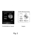
- Example 1 shows an image of biologically active structures on a solid support with the sensor layer according to the invention.
- the effective one Substance ciprofloxacin using an inkjet printer inkjet printer (HP DeskJet 870 Cxi) in Form a test graphic applied to a carrier (Fig. 2, right side).
- a carrier an aluminum foil coated with silica gel 60 from Merck (item no. 1.05553).
- the sensor layer contained genetically engineered ones Reporter gene cells that determine the biological activity of ciprofloxacin Display bioluminescence specifically for effects. That through the ciprofloxacin order The local distribution of effects on the paper carrier was determined using the information in the Sensor layer induced bioluminescence with a video imaging system registered.
- FIG. 2 left side and shows a graphic resolution of approx. 20 lpi (lines per inch) for the entire bioluminescence imaging process. This is enough to e.g. B. Perform impact tests as spot tests with a spot density of more than 25 spots / cm 2 .
- the reporter gene cell consists of the Escherichia coli strain SM101, which carries the recombinant plasmid pEBZ181.
- This plasmid encodes a fusion between the recA promoter from E. coli and the structural genes of the bacterial glow from Vibrio fischeri ( lux genes C, D, A, B, E and G ).
- the recA promoter-bearing Bam HI fragment from the plasmid pUA80 Barbe J, Fernandez de Henestrosa AR, Calero S, and Gibert I (1991) Chromogenic Method for rapid isolation of recA- like mutants of Gram -negative bacteria. J. Bacteriol.
- Plasmid pEBZ181 was introduced into Escherichia coli strain SM101 (obtained from the E. coli Genetic Stock Center) using standard methods [Sambrook J, Fritsch EF, and Maniatis T (1989) Molecular cloning: A laboratory manual (2nd edn), Cold Spring Harbor Laboratory Press] , Yale University, New Haven, USA) and used for bioimaging experiments.
- the RecA protein is a major regulator of this repair mechanism, with SOS induction leading to increased expression of the recA gene.
- Measurement of the synthesis of the RecA protein [Little JW and Mount DW (1982) The SOS regulatory system of Escherichia coli. Cell 29: 11-22; Witkin EM (1976) Ultraviolet mutagenesis and inducible DNA repair in Escherichia coli. Bacteriol. Rev. 40: 864-907] or of recA reporter gene fusions [Nunoshiba T and Nishioka H (1991) Rec-lac test for detecting SOS-inducing activity of environmental genotoxic substances. Mutation Res.
- strain E. coli SM101 (pEBZ181) carries a plasmid with a recA-lux reporter gene fusion , it is suitable for the detection of 4-quinolone carboxylic acids or other SOS-inducing compounds. The presence of such compounds leads to an increased expression of the luciferase genes and thus to a stimulation of bioluminescence.
- the test graphic was created with Corel Draw (Version 8).
- the line width for the line grid is 0.1 mm.
- the dot matrix contains 2 50 square dots (edge length 1 mm) in 1/2 inch. The dots were printed with a color saturation of 50%, all other elements of the test graphic were printed with 100% color saturation.
- the aluminum foil was dried under a stream of nitrogen, into a suitable one Stainless steel frame with raised edge inserted, horizontally on a leveling table positioned and even with the sensor layer mass obtained under 2. watered so that a layer thickness of 2 mm was achieved. After curing the Sensor layer at room temperature was the coated support at 28 ° C for 60 Minutes incubated.
- Fig. 2 is the bioluminescence image that shows the effect of the on the silica gel / aluminum foil printed ciprofloxacins, along with the original test graphic shown.
- Example 2 shows an application of the sensor layer for activity tests in the presence cytotoxic substances and a comparison with an analog microtiter plate test.
- the diffusion-controlling Sensor layer for testing substance mixtures the disruptive components included, are used, because the interference components are diffusion and Adsorption processes separated in situ, so the risk of false negatives Results is degraded.
- the SOS reporter gene system see example 1 the superiority of the sensor layer in the Comparison to microtiter plate formats.
- the microtiter plate was incubated for 60 min at 28 ° C. and then measured with the "Molecular Light Imager NightOWL" video imaging system from EG&G Berthold.
- the mean gray values were determined for the individual wells.
- the relative bioluminescence was determined by dividing these gray values by the value for The CTAB-free reference solution was determined.
- the mean values of the relative bioluminescence intensity as a function of the CTAB concentration were determined: CTAB [ng / 200 ⁇ l] rel. Bioluminescence-zenz 0 1.00 50 0.88 100 0.87 200 0.78 500 0.44
- results in the figure below are the results compared for the sensor layer format.
- the DC plates dried under a stream of nitrogen were coated with a suspension of the luminescent sensor bacteria (see Example 1) in 0.6% agarose with a layer thickness of approx. 2 mm on a leveling table.
- the DC plates were then incubated at 28 ° C. for 60 min.
- the induced bioluminescence was then detected by imaging with the high-resolution CCD Low Light Imaging System "Molecular Light Imager NightOWL" from EG&G Berthold. Imaging conditions: recording time 60 s; camera position optimized for DC plate format of 10 x 20 cm
- the mean gray values were determined in order to make a comparison with the results for the microtiter plate format described above, the analog concentrations were determined from the amounts of substance applied to the thin layer plate.
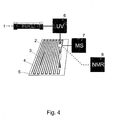
- Fig. 4 shows schematically how a mixture of substances by column chromatography is separated into fractions and at the same time spectroscopic data on the individual Fractions are won.
- Sample 1 is separated by column chromatography (HPLC).
- HPLC column chromatography
- the eluate from the Chromatography column is detected by a UV detector 6. Via a split system part of the eluate is applied to a support 4 by spraying or spotting applied 2.
- the column chromatography eluate placed in a grid on the carrier so that all substances from the column chromatography run be deposited there separately.
- Another part of the eluate is simultaneously in a mass spectrometer 7 or an NMR spectrometer 8 branched off and spectroscoped there (HPLC / MS or HPLC / NMR coupling).
- the carrier if necessary after drying, covered with the sensor layer.
- the sensor layer shows the positions of the biologically active fractions 3.
Landscapes
- Health & Medical Sciences (AREA)
- Life Sciences & Earth Sciences (AREA)
- Engineering & Computer Science (AREA)
- Chemical & Material Sciences (AREA)
- Immunology (AREA)
- Molecular Biology (AREA)
- Organic Chemistry (AREA)
- Biomedical Technology (AREA)
- General Health & Medical Sciences (AREA)
- Proteomics, Peptides & Aminoacids (AREA)
- Hematology (AREA)
- Microbiology (AREA)
- Biotechnology (AREA)
- Urology & Nephrology (AREA)
- Zoology (AREA)
- Wood Science & Technology (AREA)
- Bioinformatics & Cheminformatics (AREA)
- Physics & Mathematics (AREA)
- Analytical Chemistry (AREA)
- Biochemistry (AREA)
- Toxicology (AREA)
- Biophysics (AREA)
- Pathology (AREA)
- Cell Biology (AREA)
- General Physics & Mathematics (AREA)
- Medicinal Chemistry (AREA)
- Food Science & Technology (AREA)
- Tropical Medicine & Parasitology (AREA)
- General Engineering & Computer Science (AREA)
- Genetics & Genomics (AREA)
- Measuring Or Testing Involving Enzymes Or Micro-Organisms (AREA)
- Investigating Or Analysing Materials By The Use Of Chemical Reactions (AREA)
- Investigating Or Analyzing Materials By The Use Of Fluid Adsorption Or Reactions (AREA)
- Investigating, Analyzing Materials By Fluorescence Or Luminescence (AREA)
- Investigating Or Analysing Biological Materials (AREA)
- Other Investigation Or Analysis Of Materials By Electrical Means (AREA)
- Investigating Or Analysing Materials By Optical Means (AREA)
- Apparatus Associated With Microorganisms And Enzymes (AREA)
- Glass Compositions (AREA)
- Investigating Or Analyzing Materials By The Use Of Electric Means (AREA)
Abstract
Description
- eine oder mehrere Substanzen, die auf oder in der Oberfläche eines Trägers verteilt angeordnet sind,
- eine Sensorschicht, die eine diffusionskontrollierende Matrix und darin eine Art von suspendierten Sensoren umfaßt, die in Kontakt mit den Substanzen steht, und
- ein bildgebendes System, in dessen Detektionsbereich sich ein Teil oder die gesamte Sensorschicht befindet und das geeignet ist die Emission der Sensoren zu detektieren.
- Aufnahme unterschiedlicher Sensorsysteme für multiple Detektion
- Aufnahme von Zusätzen zur Steuerung und Unterstützung des Detektionsprozesses
- Wirkung als Diffusionsbarriere oder Substanz-selektive Filterschicht.
- Fig. 1
- Schematischer Aufbau einer erfindungsgemäßen Vorrichtung.
- Fig. 2
- Ergebnis einer Messung der örtlichen Verteilung von biologischer Aktivität auf einer Oberfläche mit der erfindungsgemäßen Sensorschicht.
- Fig. 3
- Vergleich zwischen konventionellem und erfindungsgemäßem Verfahren bei der Messung an einem Substanzgemisch mit toxischen Bestandteilen.
- Fig. 4
- Versuchsaufbau zur Kopplung von chromatographischer Trennung und Spektroskopie
- 32 ml 1 % Agarose (Agarose MP Boehringer Mannheim GmbH Art. Nr. 1388983); diese niedrig schmelzende Agarose erlaubt es, temperaturempfindliche Reportergenzellen bei Temperaturen unter 40 °C ohne Schädigung zu suspendieren.
- 4 ml LB Medium 200 g/l (GIBCO BRL Art. Nr. 12780-052); der Zusatz von LB-Medium dient zur Aufrechterhaltung der vitalen Funktionen der Reportergenzellen während der Inkubationszeit.
- 4 ml Bakteriensuspension in LB-Medium 20 g/l; damit ergab sich eine optische Dichte von 1,2 bei 660 nm Wellenlänge; über das Volumen der zugesetzten Bakteriensuspension wird die Zelldichte, d. h. die Dichte der Sensoren in der Sensorschicht gesteuert, um das Signal-Rausch-Verhältnis und die Ortsauflösung zu optimieren.
- 10 ml Wasser; dieser Zusatz regelt die Viskosität der Gießmasse und die mechanische Stabilität der Sensorschicht nach dem Aushärten.
- 1.
- 25 ng Ciprofloxacinhydrochlorid-Monohydrat
- 2.
- 25 ng Ciprofloxacinhydrochlorid-Monohydrat + 50 ng CTAB
- 3.
- 25 ng Ciprofloxacinhydrochlorid-Monohydrat + 100 ng CTAB
- 4.
- 25 ng Ciprofloxacinhydrochlorid-Monohydrat + 200 ng CTAB
- 5.
- 25 ng Ciprofloxacinhydrochlorid-Monohydrat + 500 ng CTAB
| CTAB [ng/200µl] | rel. Biolumines-zenz |
| 0 | 1,00 |
| 50 | 0,88 |
| 100 | 0,87 |
| 200 | 0,78 |
| 500 | 0,44 |
- 1.
- 1 µl wässrige Lösung von 10 ng Ciprofloxacinhydrochlorid-Monohydrat
- 2.
- 1 µl wässrige Lösung von 10 ng Ciprofloxacinhydrochlorid-Monohydrat + 50 ng CTAB
- 3.
- 1 µl wässrige Lösung von 10 ng Ciprofloxacinhydrochlorid-Monohydrat + 100 ng CTAB
| CTAB [ng/200µl] | rel. Biolumines-zenz |
| 0 | 1,000 |
| 250 | 1,002 |
| 500 | 1,023 |
Claims (37)
- Sensorsystem zur ortsaufgelösten Abbildung der biologischen Wirkung von Substanzen aufweisendeine oder mehrere Substanzen, die auf oder in der Oberfläche eines Trägers verteilt angeordnet sind,eine Sensorschicht, die eine diffusionskontrollierende Matrix und darin eine Art von suspendierten Sensoren umfaßt, die in Kontakt mit den Substanzen steht, undein bildgebendes System, in dessen Detektionsbereich sich ein Teil oder die gesamte Sensorschicht befindet und das geeignet ist die Emission der Sensoren zu detektieren.
- Sensorsystem nach Anspruch 1, dadurch gekennzeichnet, dass die Sensorschicht selber als Träger dient.
- Sensorsystem nach Anspruch 1 oder 2, dadurch gekennzeichnet, dass die Matrix ein Gel wie Agarose, Polyacrylate oder eine viskose Lösung ist.
- Sensorsystem nach einem der Ansprüche 1 bis 3, dadurch gekennzeichnet, dass die Sensoren prokaryotische oder eukaryotische Zellen wie Reportergenzellen, subzelluläre Partikel, Enzymsysteme, Antikörper, Fluoreszenzsensoren oder Indikatorfarbstoffe sind.
- Sensorsystem nach einem der Ansprüche 1 bis 4, dadurch gekennzeichnet, dass mehrere verschiedene Arten von Sensoren oder eine Art von Sensoren, die verschiedene biologische Wirkungen anzeigen kann, in der Sensorschicht suspendiert sind.
- Sensorsystem nach einem der Ansprüche 1 bis 5, dadurch gekennzeichnet, dass die Sensorschicht Zusätze enthält, die den Detektionsprozess kontrollieren oder unterstützen.
- Sensorsystem nach Anspruch 6, dadurch gekennzeichnet, dass die Zusätze Puffer für die Regulierung des Vitalitätsstatus von Sensorzellen sind.
- Sensorsystem nach einem der Ansprüche 1 bis 7, dadurch gekennzeichnet, dass die Sensorschicht Biolumineszenzsubstrate, Chemolumineszenzreagenzien oder Fluoreszenzreagenzien enthält.
- Sensorsystem nach einem der Ansprüche 1 bis 8, dadurch gekennzeichnet, dass die Sensorschicht aus mehreren Teilschichten besteht, wobei die Teilschichten unterschiedlich dick sein können und sich durch Art und Menge von Sensoren und/oder Zusätzen unterscheiden.
- Sensorsystem nach einem der Ansprüche 4 bis 9, dadurch gekennzeichnet, dass in 50 ml Sensorschichtmasse bevorzugt 2 bis 8 ml, besonders bevorzugt 3 bis 5 ml Suspension von Reportergenzellen enthalten sind.
- Sensorsystem nach Anspruch 10, dadurch gekennzeichnet, dass die Suspension von Reportergenzellen eine optische Dichte von 0,6 bis 1,4 bei 660 nm hat.
- Sensorsystem nach einem der Ansprüche 1 bis 11 dadurch gekennzeichnet, dass die Dicke der Sensorschicht 0,1 bis 10 mm, vorzugsweise 0,5 bis 3 mm, besonders bevorzugt 0,5 bis 0,8 mm beträgt.
- Sensorsystem nach einem der Ansprüche 1 bis 12, dadurch gekennzeichnet, dass die Sensoren der Sensorschicht ihre Aktivität durch Emission oder Löschung von Lichtemission anzeigen.
- Verfahren zur ortsaufgelösten Abbildung der biologischen Wirkung von Substanzen, dadurch gekennzeichnet, dassa.) eine oder mehrere Substanzen verteilt auf oder in die Oberfläche eines Trägers gebracht werden, oder bereits Bestandteil einer zu testenden Oberfläche sind,b.) der Träger mit einer Sensorschicht, die eine diffusionskontrollierende Matrix und darin eine Art von suspendierten Sensoren umfaßt und in Kontakt mit den Substanzen steht, belegt wird, sofern die Sensorschicht nicht selbst als Träger dient,c.) die Wirkung des oder der Substanzen auf die Sensoren in der Sensorschicht durch ein bildgebendes System, in dessen Detektionsbereich sich ein Teil oder die gesamte Sensorschicht befindet und das geeignet ist die Emission der Sensoren zu detektieren, bestimmt wird.
- Verfahren nach Anspruch 14, dadurch gekennzeichnet, dass die Sensorschicht selber als Träger verwendet wird.
- Verfahren nach Anspruch 14 oder 15, dadurch gekennzeichnet, dass bei Verwendung einer Sensorschicht, die Zellen als Sensoren enthält, vor der Bestimmung der Wirkung der in der Probe enthaltenen Substanzen auf die Sensoren ein Inkubationsschritt eingelegt wird, indem die Sensorschicht oder der mit der Sensorschicht belegte Träger entsprechend den Anforderungen der eingesetzten Zellinien unter definierten Bedingungen bezüglich Temperatur, Feuchtigkeit und Begasung eine vorgegebene Zeit gelagert wird.
- Verfahren nach einem der Ansprüche 14 bis 16, dadurch gekennzeichnet, dass die Wirkung der Substanz auf die Sensoren in einer ortsabhängigen Induktion oder Löschung der Lichtemission aus Biolumineszenz- oder Chemolumineszenzprozessen, der Induktion oder der Löschung von Fluoreszenzemissionen sowie einer integralen oder spektralen Änderung der Lichtabsorption besteht.
- Verfahren nach einem der Ansprüche 14 bis 17, dadurch gekennzeichnet, dass die Substanzen durch spezifische oder unspezifische Adsorption an geeigneten Trägermaterialien angereichert werden, bevor sie in Kontakt mit der Sensorschicht gebracht werden.
- Verfahren nach einem der Ansprüche 14 bis 18, dadurch gekennzeichnet, dass die Substanzen ein Substanzgemisch bilden, das chromatographisch oder elektrophoretisch oder mit anderen analytischen oder präparativen Separationstechniken in Fraktionen getrennt wird, bevor es in Kontakt mit der Sensorschicht gebracht wird.
- Verfahren nach Anspruch 19, dadurch gekennzeichnet, dass die chromatographische Trennung in Fraktionen in einer Chomatographiesäule stattfindet und das Eluat fortlaufend entweder kontinuierlich oder in Intervallen auf verschiedene Stellen des Trägers aufgebracht wird.
- Verfahren nach Anspruch 19, dadurch gekennzeichnet, dass die chromatographische Trennung in Fraktionen durch Dünnschichtchromatographie oder Elektrophorese auf dem Träger erfolgt, der mit der Sensorschicht belegt wird.
- Verfahren nach einem der Ansprüche 14 bis 21, dadurch gekennzeichnet, dass die Substanzen ein Substanzgemisch bilden und die Erfassung der biologischen Wirkung der Einzelsubstanzen des Substanzgemisches mit einer Detektion der Struktur der Einzelsubstanzen verknüpft wird, indem das Substanzgemisch chromatographisch oder elektrophoretisch oder mit anderen analytischen oder präparativen Separationstechniken in Fraktionen getrennt und jede Fraktion spektroskopisch untersucht wird, bevor sie in Kontakt mit der Sensorschicht gebracht wird.
- Verfahren nach Anspruch 22, dadurch gekennzeichnet, dass eine Auswertung der Daten der spektroskopischen Untersuchung nur für die Fraktionen erfolgt, an denen durch die Sensorschicht eine biologische Wirkung nachgewiesen werden kann.
- Verfahren nach Anspruch 22 oder 23, dadurch gekennzeichnet, dass die chromatographische Trennung des Substanzgemisches in Fraktionen in einer Chomatographiesäule stattfindet und ein Teil des Eluats fortlaufend auf verschiedene Stellen des Trägers aufgebracht wird, während gleichzeitig ein anderer Teil des Eluats abgezweigt und durch ein Massenspektrometer oder ein NMR-Spektrometer oder ein IR-Spektrometer spektroskopiert wird.
- Verfahren nach Anspruch 24, dadurch gekennzeichnet, dass das Eluat aus der Chromatographiesäule vor Abzweigung des zu spektroskopierenden Anteils durch einen UV-Detektor detektiert wird.
- Verfahren nach Anspruch 22 oder 23, dadurch gekennzeichnet, dass die chromatographische Trennung des Substanzgemisches in einzelne Substanzzonen mittels Dünnschichtchromatographie oder Elektrophorese auf dem Träger erfolgt und von den einzelnen Substanzzonen durch MALDI-Massenspektroskopie oder Raman-Spektroskopie oder andere spektroskopische Verfahren wie UV-VIS oder IR Spektren aufgenommen werden bevor der Träger mit der Sensorschicht belegt wird.
- Verfahren nach einem der Ansprüche 14 bis 26, dadurch gekennzeichnet, dass die Matrix ein Gel wie Agarose, Polyacrylate oder eine viskose Lösung ist.
- Verfahren nach einem der Ansprüche 14 bis 27, dadurch gekennzeichnet, dass mehrere verschiedene Arten von Sensoren oder eine Art von Sensoren, die verschiedene biologische Wirkungen anzeigen kann, in der Sensorschicht suspendiert sind.
- Verfahren nach einem der Ansprüche 14 bis 28, dadurch gekennzeichnet, dass die Sensoren prokaryotische oder eukaryotische Zellen wie Reportergenzellen, subzelluläre Partikel, Enzymsysteme, Antikörper, Fluoreszenzsensoren oder Indikatorfarbstoffe sind.
- Verfahren nach einem der Ansprüche 14 bis 29, dadurch gekennzeichnet, dass die Sensorschicht Zusätze enthält, die den Detektionsprozess kontrollieren oder unterstützen.
- Verfahren nach Anspruch 30, dadurch gekennzeichnet, dass die Zusätze Puffer für die Regulierung des Vitalitätsstatus von Sensorzellen sind.
- Verfahren nach einem der Ansprüche 14 bis 31, dadurch gekennzeichnet, dass die Sensorschicht Biolumineszenzsubstrate, Chemolumineszenzreagenizien oder Fluoreszenzreagenzien enthält.
- Verfahren nach einem der Ansprüche 14 bis 32, dadurch gekennzeichnet, dass die Sensorschicht aus mehreren Teilschichten besteht, wobei die Teilschichten unterschiedlich dick sein können und sich durch Art und Menge von Sensoren und/oder Zusätzen unterscheiden.
- Verfahren nach einem der Ansprüche 29 bis 34, dadurch gekennzeichnet, dass in 50 ml Sensorschichtmasse bevorzugt 2 bis 8 ml, besonders bevorzugt 3 bis 5 ml Suspension von Reportergenzellen enthalten sind.
- Sensorsystem nach Anspruch 34, dadurch gekennzeichnet, dass die Suspension von Reportergenzellen eine optische Dichte von 0,6 bis 1,4 bei 660 nm hat.
- Sensorsystem nach einem der Ansprüche 14 bis 35 dadurch gekennzeichnet, dass die Dicke der Sensorschicht 0,1 bis 10 mm, vorzugsweise 0,5 bis 3 mm, besonders bevorzugt 0,5 bis 0,8 mm beträgt.
- Sensorsystem nach einem der Ansprüche 14 bis 36, dadurch gekennzeichnet, dass die Sensoren der Sensorschicht ihre Aktivität durch Emission oder Löschung von Lichtemission anzeigen.
Applications Claiming Priority (3)
| Application Number | Priority Date | Filing Date | Title |
|---|---|---|---|
| DE19915310A DE19915310A1 (de) | 1999-04-03 | 1999-04-03 | Diffusionskontrollierende Sensorschicht |
| DE19915310 | 1999-04-03 | ||
| EP00105379A EP1043582B1 (de) | 1999-04-03 | 2000-03-21 | Diffusionskontrollierende Sensorschicht |
Related Parent Applications (1)
| Application Number | Title | Priority Date | Filing Date |
|---|---|---|---|
| EP00105379A Division EP1043582B1 (de) | 1999-04-03 | 2000-03-21 | Diffusionskontrollierende Sensorschicht |
Publications (2)
| Publication Number | Publication Date |
|---|---|
| EP1243926A2 true EP1243926A2 (de) | 2002-09-25 |
| EP1243926A3 EP1243926A3 (de) | 2003-09-03 |
Family
ID=7903543
Family Applications (2)
| Application Number | Title | Priority Date | Filing Date |
|---|---|---|---|
| EP02014011A Withdrawn EP1243926A3 (de) | 1999-04-03 | 2000-03-21 | Diffusionskontrollierende Sensorschicht |
| EP00105379A Expired - Lifetime EP1043582B1 (de) | 1999-04-03 | 2000-03-21 | Diffusionskontrollierende Sensorschicht |
Family Applications After (1)
| Application Number | Title | Priority Date | Filing Date |
|---|---|---|---|
| EP00105379A Expired - Lifetime EP1043582B1 (de) | 1999-04-03 | 2000-03-21 | Diffusionskontrollierende Sensorschicht |
Country Status (10)
| Country | Link |
|---|---|
| US (1) | US20070292912A1 (de) |
| EP (2) | EP1243926A3 (de) |
| JP (1) | JP2000321208A (de) |
| AT (1) | ATE274183T1 (de) |
| CA (1) | CA2303529A1 (de) |
| DE (2) | DE19915310A1 (de) |
| DK (1) | DK1043582T3 (de) |
| ES (1) | ES2226626T3 (de) |
| IL (1) | IL135394A0 (de) |
| PT (1) | PT1043582E (de) |
Families Citing this family (3)
| Publication number | Priority date | Publication date | Assignee | Title |
|---|---|---|---|---|
| JP4552363B2 (ja) * | 2001-06-15 | 2010-09-29 | 株式会社島津製作所 | 液体クロマトグラフ質量分析装置 |
| DE10133273A1 (de) | 2001-07-09 | 2003-01-30 | Bayer Cropscience Ag | Vorrichtung und Verfahren zum Nachweis der Photosynthese-Hemmung |
| FR2856792B1 (fr) * | 2003-06-27 | 2006-09-15 | Commissariat Energie Atomique | Procede et dispositif de dosage d'un echantillon biologique ou chimique |
Family Cites Families (23)
| Publication number | Priority date | Publication date | Assignee | Title |
|---|---|---|---|---|
| US4162208A (en) * | 1975-02-03 | 1979-07-24 | Aladjem Frederick J | Quantitative protein analysis by immunodiffusion |
| DE2948904C2 (de) * | 1979-12-05 | 1986-06-19 | Max-Planck-Gesellschaft zur Förderung der Wissenschaften e.V., 3400 Göttingen | Vorrichtung zur optischen Messung von Konzentrationen von Stoffen |
| JPH076984B2 (ja) * | 1985-05-31 | 1995-01-30 | 株式会社日立製作所 | 複数項目分析方法 |
| GB8621261D0 (en) * | 1986-09-03 | 1986-10-08 | Barnard G J R | Enhanced luminescent assay |
| US5156810A (en) * | 1989-06-15 | 1992-10-20 | Biocircuits Corporation | Biosensors employing electrical, optical and mechanical signals |
| US5330718A (en) * | 1989-08-16 | 1994-07-19 | Puritan-Bennett Corporation | Sensor element and method for making the same |
| GB2244135B (en) * | 1990-05-04 | 1994-07-13 | Gen Electric Co Plc | Sensor devices |
| DE4221036A1 (de) * | 1992-06-26 | 1994-01-05 | Siemens Ag | Bildverarbeitungsanordnung |
| AT399402B (de) * | 1992-07-09 | 1995-05-26 | Avl Verbrennungskraft Messtech | Sensormembran eines optischen sensors zur bestimmung eines physikalischen oder chemischen parameters einer probe |
| DE4230264A1 (de) * | 1992-09-10 | 1994-03-17 | Bayer Ag | Analytisches Verfahren zur Untersuchung von Gemischen auf toxische Bestandteile |
| US5565324A (en) * | 1992-10-01 | 1996-10-15 | The Trustees Of Columbia University In The City Of New York | Complex combinatorial chemical libraries encoded with tags |
| AT401526B (de) * | 1993-02-10 | 1996-09-25 | Scheirer Winfried | Reagenzlösung zur stabilisierung der lumineszenz bei der luciferasemessung |
| DE4304728C2 (de) * | 1993-02-13 | 1997-04-10 | Igor Dr Popov | Verfahren und Testbesteck zur Bestimmung von Ascorbinsäure in biologischen Proben |
| EP0720658A1 (de) * | 1993-09-23 | 1996-07-10 | E.I. Du Pont De Nemours And Company | Elektrophoretisches verfahren zur isolierung und trennung von mikroorganismen |
| US5766963A (en) * | 1996-01-26 | 1998-06-16 | Pharmacopeia, Inc. | Combination hydroxypropylamine library |
| DE19631395A1 (de) * | 1996-08-02 | 1998-02-05 | Basf Ag | Verfahren zum Wirkstoff-Screening |
| EP1150122A3 (de) * | 1996-12-05 | 2003-02-12 | Idego ApS | Sensorlaminate und vielfach unterteilte Flüssigkeitsspender-Vorrichtungen zum nachweis von Immunoassay-Zielmolekülen in biologischen Flüssigkeiten |
| WO1998045704A2 (en) * | 1997-04-07 | 1998-10-15 | Bioimage A/S | A method for extracting quantitative information relating to an influence on a cellular response |
| US6200762B1 (en) * | 1997-08-01 | 2001-03-13 | Aurora Biosciences Corporation | Photon reducing agents and compositions for fluorescence assays |
| DE19734800A1 (de) * | 1997-08-12 | 1999-02-18 | Heide Prof Dr Schnabl | Verfahren zum Nachweis von ökotoxikologisch wirksamen Schadstoffen mittels pflanzlicher Zellsysteme |
| US6117643A (en) * | 1997-11-25 | 2000-09-12 | Ut Battelle, Llc | Bioluminescent bioreporter integrated circuit |
| US5942409A (en) * | 1998-07-31 | 1999-08-24 | Byk Gulden Lomberg Chemische Fabrik Gmbh | Process for identification of substances modulating ureI dependent mechanisms of Helicobacter pylori metabolism |
| DE29815139U1 (de) * | 1998-08-14 | 1999-01-14 | I.U.T. - Privates Institut für Umwelttoxikologie GmbH, 17489 Greifswald | Vorrichtung zur Erfassung der toxischen und mutagenen Wirkung von Chemikalien und Substanzgemischen |
-
1999
- 1999-04-03 DE DE19915310A patent/DE19915310A1/de not_active Withdrawn
-
2000
- 2000-03-21 EP EP02014011A patent/EP1243926A3/de not_active Withdrawn
- 2000-03-21 DK DK00105379T patent/DK1043582T3/da active
- 2000-03-21 ES ES00105379T patent/ES2226626T3/es not_active Expired - Lifetime
- 2000-03-21 AT AT00105379T patent/ATE274183T1/de not_active IP Right Cessation
- 2000-03-21 PT PT00105379T patent/PT1043582E/pt unknown
- 2000-03-21 EP EP00105379A patent/EP1043582B1/de not_active Expired - Lifetime
- 2000-03-21 DE DE50007429T patent/DE50007429D1/de not_active Expired - Fee Related
- 2000-03-27 JP JP2000086250A patent/JP2000321208A/ja not_active Withdrawn
- 2000-03-31 CA CA002303529A patent/CA2303529A1/en not_active Abandoned
- 2000-03-31 IL IL13539400A patent/IL135394A0/xx unknown
-
2007
- 2007-08-16 US US11/893,800 patent/US20070292912A1/en not_active Abandoned
Also Published As
| Publication number | Publication date |
|---|---|
| EP1243926A3 (de) | 2003-09-03 |
| ES2226626T3 (es) | 2005-04-01 |
| DE50007429D1 (de) | 2004-09-23 |
| EP1043582A3 (de) | 2001-05-23 |
| EP1043582A2 (de) | 2000-10-11 |
| PT1043582E (pt) | 2004-12-31 |
| US20070292912A1 (en) | 2007-12-20 |
| JP2000321208A (ja) | 2000-11-24 |
| ATE274183T1 (de) | 2004-09-15 |
| CA2303529A1 (en) | 2000-10-03 |
| EP1043582B1 (de) | 2004-08-18 |
| DK1043582T3 (da) | 2004-12-06 |
| DE19915310A1 (de) | 2000-10-05 |
| IL135394A0 (en) | 2001-05-20 |
Similar Documents
| Publication | Publication Date | Title |
|---|---|---|
| DE69620403T2 (de) | Dämpfungsreagenzien und tests für enzym-vermittelte-lumineszenz | |
| EP0425587B1 (de) | Verfahren zur feststellung biologischer aktivitäten in einer probe und vorrichtung zur durchführung des verfahrens | |
| DE69024631T2 (de) | Verfahren und Apparat zum Nachweis biologischer Aktivitäten in einer Probe | |
| DE60034315T2 (de) | Chemisches und biochemisches nachweisverfahren und vorrichtung | |
| EP1144586A2 (de) | Verfahren, gefäss und vorrichtung zur überwachung der stoffwechselaktivität von zellkulturen in flüssigen medien | |
| DE19621312A1 (de) | Maskierung der Hintergrundfluoreszenz und Signalverstärkung bei der optischen Analyse biologisch medizinischer Assays | |
| DE2841896C3 (de) | Verfahren zum Nachweis einer toxischen Substanz | |
| DE3833628A1 (de) | Verfahren zum nachweis und zur identifikation toxischer substanzen mit hilfe klonierter mikroorganismen | |
| EP1407040A2 (de) | Vorrichtung und verfahren zum nachweis der photosynthese-hemmung | |
| DE2856251A1 (de) | Fuellungen fuer indikatorraeume | |
| DE68926935T2 (de) | Vorrichtung zur Verstärkung von Fluoreszens und Kinetik und Verfahren zur Anwendung | |
| EP1397672A1 (de) | Sauerstoffsensoren auf mikrotiterplatte | |
| DE10155185A1 (de) | Verfahren zur Charakterisierung und/oder Identifizierung von Wirkmechanismen antimikrobieller Prüfsubstanzen | |
| DE3430935C2 (de) | Verfahren zur Bestimmung der Ionenstärke einer Elektrolytlösung sowie Meßeinrichtung zur Durchführung des Verfahrens | |
| EP1043582B1 (de) | Diffusionskontrollierende Sensorschicht | |
| DE60309589T2 (de) | Verfahren zum nachweis von arsenionen durch indikatorbakterien | |
| DE2728077A1 (de) | Verfahren zum faerben von mikroorganismen | |
| EP1359226A2 (de) | Verfahren und Vorrichtung zum Nachweis biologisch aktiver Substanzen | |
| EP0588139B1 (de) | Analytisches Verfahren zur Untersuchung von Gemischen auf toxische Bestandteile | |
| WO2007065423A1 (de) | Verfahren und vorrichtung zur detektion von mikroorganismen und/oder deren aktivität | |
| DE102020119332B3 (de) | Optisches nachweisverfahren | |
| DE19605753C2 (de) | Verfahren und Vorrichtung zur Detektion von stoffwechselaktiven, unlädierten, ungestressten Mikroorganismen - quantitativ und qualitativ im "Sub-ppb-Bereich" innerhalb von Minuten | |
| DE19919539C1 (de) | Verfahren zur Messung der Aktivität einer biologisch wirksamen Substanz in einem histologischen Präparat | |
| DE60028201T2 (de) | Überwachung von genexpression | |
| DE102015221969A1 (de) | Verfahren zur automatisierten Detektion einer Aktivität von Wirkstoffen in einer Lösung |
Legal Events
| Date | Code | Title | Description |
|---|---|---|---|
| PUAI | Public reference made under article 153(3) epc to a published international application that has entered the european phase |
Free format text: ORIGINAL CODE: 0009012 |
|
| AC | Divisional application: reference to earlier application |
Ref document number: 1043582 Country of ref document: EP |
|
| AK | Designated contracting states |
Kind code of ref document: A2 Designated state(s): AT BE CH CY DE DK ES FI FR GB GR IE IT LI LU MC NL PT SE |
|
| PUAL | Search report despatched |
Free format text: ORIGINAL CODE: 0009013 |
|
| AK | Designated contracting states |
Kind code of ref document: A3 Designated state(s): AT BE CH CY DE DK ES FI FR GB GR IE IT LI LU MC NL PT SE |
|
| RIC1 | Information provided on ipc code assigned before grant |
Ipc: 7G 01N 21/76 A |
|
| 17P | Request for examination filed |
Effective date: 20040303 |
|
| AKX | Designation fees paid |
Designated state(s): AT BE CH CY DE DK ES FI FR GB GR IE IT LI LU MC NL PT SE |
|
| 17Q | First examination report despatched |
Effective date: 20040910 |
|
| RAP1 | Party data changed (applicant data changed or rights of an application transferred) |
Owner name: BAYER INNOVATION GMBH |
|
| STAA | Information on the status of an ep patent application or granted ep patent |
Free format text: STATUS: THE APPLICATION IS DEEMED TO BE WITHDRAWN |
|
| 18D | Application deemed to be withdrawn |
Effective date: 20050321 |