EP1233060A2 - Procédé pour la détérmination de l'activité du facteur de régulation du cycle cellulaire et procédé pour la diagnose de cancer utilisant le même - Google Patents
Procédé pour la détérmination de l'activité du facteur de régulation du cycle cellulaire et procédé pour la diagnose de cancer utilisant le même Download PDFInfo
- Publication number
- EP1233060A2 EP1233060A2 EP02003139A EP02003139A EP1233060A2 EP 1233060 A2 EP1233060 A2 EP 1233060A2 EP 02003139 A EP02003139 A EP 02003139A EP 02003139 A EP02003139 A EP 02003139A EP 1233060 A2 EP1233060 A2 EP 1233060A2
- Authority
- EP
- European Patent Office
- Prior art keywords
- labeling
- substrate
- cancer
- cyclin
- sample
- Prior art date
- Legal status (The legal status is an assumption and is not a legal conclusion. Google has not performed a legal analysis and makes no representation as to the accuracy of the status listed.)
- Granted
Links
Images


Classifications
-
- C—CHEMISTRY; METALLURGY
- C07—ORGANIC CHEMISTRY
- C07K—PEPTIDES
- C07K16/00—Immunoglobulins [IGs], e.g. monoclonal or polyclonal antibodies
- C07K16/40—Immunoglobulins [IGs], e.g. monoclonal or polyclonal antibodies against enzymes
-
- C—CHEMISTRY; METALLURGY
- C12—BIOCHEMISTRY; BEER; SPIRITS; WINE; VINEGAR; MICROBIOLOGY; ENZYMOLOGY; MUTATION OR GENETIC ENGINEERING
- C12N—MICROORGANISMS OR ENZYMES; COMPOSITIONS THEREOF; PROPAGATING, PRESERVING, OR MAINTAINING MICROORGANISMS; MUTATION OR GENETIC ENGINEERING; CULTURE MEDIA
- C12N9/00—Enzymes; Proenzymes; Compositions thereof; Processes for preparing, activating, inhibiting, separating or purifying enzymes
- C12N9/10—Transferases (2.)
- C12N9/12—Transferases (2.) transferring phosphorus containing groups, e.g. kinases (2.7)
- C12N9/1205—Phosphotransferases with an alcohol group as acceptor (2.7.1), e.g. protein kinases
-
- C—CHEMISTRY; METALLURGY
- C12—BIOCHEMISTRY; BEER; SPIRITS; WINE; VINEGAR; MICROBIOLOGY; ENZYMOLOGY; MUTATION OR GENETIC ENGINEERING
- C12Q—MEASURING OR TESTING PROCESSES INVOLVING ENZYMES, NUCLEIC ACIDS OR MICROORGANISMS; COMPOSITIONS OR TEST PAPERS THEREFOR; PROCESSES OF PREPARING SUCH COMPOSITIONS; CONDITION-RESPONSIVE CONTROL IN MICROBIOLOGICAL OR ENZYMOLOGICAL PROCESSES
- C12Q1/00—Measuring or testing processes involving enzymes, nucleic acids or microorganisms; Compositions therefor; Processes of preparing such compositions
- C12Q1/48—Measuring or testing processes involving enzymes, nucleic acids or microorganisms; Compositions therefor; Processes of preparing such compositions involving transferase
- C12Q1/485—Measuring or testing processes involving enzymes, nucleic acids or microorganisms; Compositions therefor; Processes of preparing such compositions involving transferase involving kinase
-
- G—PHYSICS
- G01—MEASURING; TESTING
- G01N—INVESTIGATING OR ANALYSING MATERIALS BY DETERMINING THEIR CHEMICAL OR PHYSICAL PROPERTIES
- G01N33/00—Investigating or analysing materials by specific methods not covered by groups G01N1/00 - G01N31/00
- G01N33/48—Biological material, e.g. blood, urine; Haemocytometers
- G01N33/50—Chemical analysis of biological material, e.g. blood, urine; Testing involving biospecific ligand binding methods; Immunological testing
- G01N33/53—Immunoassay; Biospecific binding assay; Materials therefor
- G01N33/574—Immunoassay; Biospecific binding assay; Materials therefor for cancer
- G01N33/57407—Specifically defined cancers
-
- G—PHYSICS
- G01—MEASURING; TESTING
- G01N—INVESTIGATING OR ANALYSING MATERIALS BY DETERMINING THEIR CHEMICAL OR PHYSICAL PROPERTIES
- G01N33/00—Investigating or analysing materials by specific methods not covered by groups G01N1/00 - G01N31/00
- G01N33/48—Biological material, e.g. blood, urine; Haemocytometers
- G01N33/50—Chemical analysis of biological material, e.g. blood, urine; Testing involving biospecific ligand binding methods; Immunological testing
- G01N33/53—Immunoassay; Biospecific binding assay; Materials therefor
- G01N33/574—Immunoassay; Biospecific binding assay; Materials therefor for cancer
- G01N33/57484—Immunoassay; Biospecific binding assay; Materials therefor for cancer involving compounds serving as markers for tumor, cancer, neoplasia, e.g. cellular determinants, receptors, heat shock/stress proteins, A-protein, oligosaccharides, metabolites
-
- G—PHYSICS
- G01—MEASURING; TESTING
- G01N—INVESTIGATING OR ANALYSING MATERIALS BY DETERMINING THEIR CHEMICAL OR PHYSICAL PROPERTIES
- G01N33/00—Investigating or analysing materials by specific methods not covered by groups G01N1/00 - G01N31/00
- G01N33/48—Biological material, e.g. blood, urine; Haemocytometers
- G01N33/50—Chemical analysis of biological material, e.g. blood, urine; Testing involving biospecific ligand binding methods; Immunological testing
- G01N33/58—Chemical analysis of biological material, e.g. blood, urine; Testing involving biospecific ligand binding methods; Immunological testing involving labelled substances
- G01N33/581—Chemical analysis of biological material, e.g. blood, urine; Testing involving biospecific ligand binding methods; Immunological testing involving labelled substances with enzyme label (including co-enzymes, co-factors, enzyme inhibitors or substrates)
-
- G—PHYSICS
- G01—MEASURING; TESTING
- G01N—INVESTIGATING OR ANALYSING MATERIALS BY DETERMINING THEIR CHEMICAL OR PHYSICAL PROPERTIES
- G01N33/00—Investigating or analysing materials by specific methods not covered by groups G01N1/00 - G01N31/00
- G01N33/48—Biological material, e.g. blood, urine; Haemocytometers
- G01N33/50—Chemical analysis of biological material, e.g. blood, urine; Testing involving biospecific ligand binding methods; Immunological testing
- G01N33/58—Chemical analysis of biological material, e.g. blood, urine; Testing involving biospecific ligand binding methods; Immunological testing involving labelled substances
- G01N33/582—Chemical analysis of biological material, e.g. blood, urine; Testing involving biospecific ligand binding methods; Immunological testing involving labelled substances with fluorescent label
-
- G—PHYSICS
- G01—MEASURING; TESTING
- G01N—INVESTIGATING OR ANALYSING MATERIALS BY DETERMINING THEIR CHEMICAL OR PHYSICAL PROPERTIES
- G01N2333/00—Assays involving biological materials from specific organisms or of a specific nature
- G01N2333/90—Enzymes; Proenzymes
- G01N2333/91—Transferases (2.)
- G01N2333/912—Transferases (2.) transferring phosphorus containing groups, e.g. kinases (2.7)
- G01N2333/91205—Phosphotransferases in general
- G01N2333/9121—Phosphotransferases in general with an alcohol group as acceptor (2.7.1), e.g. general tyrosine, serine or threonine kinases
Definitions
- the present invention relates to a method for determining the activity of a cell cycle regulatory factor without using a radioisotope and a method for diagnosing a cancer using the method.
- Cell proliferation is a fundamental and important feature of living things.
- the cell proliferation involves division of a single cell into two daughter cells, and somatic cells divide through a plurality of sequential reactions including growth of cells, replication of DNAs, distribution of chromosomes and division of cells. This chain of sequential reactions is referred to as a cell cycle.
- the cell cycle is divided into four phases, that is, a synthetic (S) phase during which the replication of DNAs takes place, a mitotic (M) phase during which the division of cells takes place, a gap 1 (G1) phase which is an interphase from the M phase to the next S phase and a gap 2 (G2) phase which is an interphase from the S phase to the next M phase.
- cells receive a signal for proliferation, prepare for the replication of DNAs, and make metabolism and growth which are necessary for the division of cells.
- the cells prepare for the division.
- a transit point is experimentally assumed which is called an R point (restriction point) for mammalian cells and START for yeast.
- R point resistance point
- cells multiply in response to proliferation signals from the outside.
- the cells receive the signals in the G1 phase and progress the cell cycle. After passing through a certain point in the G1 phase, the cell cycle progresses from the S phase to the G2 phase, the M phase and then the G1 phase without stopping even if the proliferation signals are not received any longer.
- This certain point is the R point or START, and is a so-to-speak point at which the entering into cell cycle progression is determined.
- the cells can leave the cell cycle and enter a resting (G0) phase during which the cell do not grow or multiply.
- G0 resting
- the cells entering the resting phase if given a suitable signal, can be returned to the G1 phase and induced to grow and divide again. It is considered that a lot of non-growing and non-multiplying cells of multicellular organisms are in the G0 phase.
- CDKs cyclin-dependent kinases
- CDKIs CDK inhibitors
- the CDKs exist in cytoplasm as inactive form.
- the CDKs are activated, e.g., phosphorylated, and move into nuclei in the cells.
- the CDKs bind to cyclin molecules to form complexes with cyclin (referred to as activated CDKs hereinafter) and positively regulate the progress of the cell cycle at various steps of the cell cycle.
- activated CDKs referred to as activated CDKs hereinafter
- the CDKIs inactivate the CDKs by biding to the activated CDKs or CDK simple substances, thereby regulating the cell cycle negatively.
- CDK1 binds to cyclin A or B
- CDK2 binds to cyclin A or E
- CDK4 and CDK6 bind to cyclin D1, D2 or D3, to be activated.
- the activated CDKs control specific phases of the cell cycle.
- Table 1 shows CDKs concerning the control of the cell cycle, cyclins which functionally bind to the CDKs, and phases of the cell cycle during which the activated CDKs act.
- the activated CDKs are enzymes which phosphorylate serine residue and threonine residue in a protein as a substrate.
- the activated CDK1 and CDK2 react well on histone H1 as a substrate and the activated CDK 4 and CDK6 react well on Rb (retinoblastoma protein) as a substrate.
- Rb retinoblastoma protein
- the CDKs and cyclins regulate the cell cycle in close association with each other.
- the multiplication of cyclin D1 gene is observed in a great number of cases of esophageal cancer, while over expression of cyclin D1 gene is observed in a great number of cases of stomach cancer and colon cancer.
- the multiplication of cyclin E gene is observed in stomach cancer and colon cancer but is not observed in esophageal cancer. Excessive expression of cyclin E in stomach and large bowel takes place with great frequency in cases of adenoma and adenocarcinoma and shows a significant correlation with malignancy such as invasion, progress of stages, metastasis and the like.
- CDK1 The expression and kinase activity of CDK1 are remarkably accelerated in most cases of stomach cancer and colon cancer as compared with normal mucosal tissue. It is known that augmented expression of cyclin genes correlates with the progress and malignancy of various cancers (see Wataru Yasui, Sysmex Journal Web., p.1 to p.10, vol.1, 2000).
- CDK2 decreases and the cell cycle arrest and the division of cells is controlled.
- the expression of CDK2 increases at the R point, it means that the cell cycle fails to stop, i.e., it means a state of a disease such as cancer.
- stomach cancer or colon cancer may be expected because stomach cancer and colon cancer involve accelerated gene expression of the cyclin D1 which bind specifically to CDK4 or CDK6.
- the activity of the CDKs is determined using radioisotopes. More particularly, in the presence of a CDK which is extracted from a cell lysate by an immunoprecipitation method using an anti-CDK antibody and whose activity is unknown, 32 P-labelled adenosine 5'-O-(3-triphosphate) (ATP) is reacted with serine residue or threonine residue in a substrate to introduce monophosphate group derived from the 32 P-labelled ATP. The amount of 32 P taken by the substrate is detected by autoradiography or by a scintillation counter. Thereby the amount of the phosphorylated substrate is measured and the activity of the CDK is calculated from the amount of the phosphorylated substrate.
- ATP adenosine 5'-O-(3-triphosphate)
- the present invention is to provide a method for determining the activity of a cell cycle regulatory factor comprising the steps of:
- the present invention is to provide a method of diagnosing a cancer based on a result obtained by determination.
- a sample is first prepared.
- the sample which contains a cyclin-dependent kinase (CDK)/cyclin complex may contain a single type or plural types of CDK/cyclin complexes, but may preferably contain a single type of CDK/cyclin complex.
- the sample which contains a CDK/cyclin complex (referred to as an activated CDK hereinafter) and is used in the method of the present invention is prepared by solubilizing cells and separating a sample containing the activated CDK to be determined from a liquid containing the solubilized cells.
- CDKs usable in the present invention include CDK1, CDK2, CDK3, CDK4, CDK5, CDK6 and CDK7.
- the sample is prepared from cells derived from animals including human beings such as a tissue sample (e.g., a biopsy sample, a surgically resected sample, etc.).
- a tissue sample e.g., a biopsy sample, a surgically resected sample, etc.
- the sample is to be tested as to whether or not it contains a CDK/cyclin complex as well as its activity. Since simple CDKs exist in cytoplasm and turn into activated CDKs by binding to cyclins in nuclei, cells need to be solubilized to extract the activated CDKs.
- the cells are first solubilized by chemically or physically destroying the cell membranes and nuclear envelopes thereof. More particularly, the cells are preferably pulverized using a Waring blender, sucked and discharged using a syringe or ultrasonicated in a buffer containing a surfactant, a protease inhibitor and a phosphatase inhibitor, for example.
- the surfactant is used for destroying cell membranes and nuclear envelopes so that intracellular substances can be taken out.
- the surfactant should have such a surface active property that the activated CDK is not decomposed. Examples thereof include Nonidet P-40, Triton X-100, deoxycholic acid and CHAPS.
- the concentration of the surfactant is preferably 1 w/v% or less.
- the protease inhibitor is used for preventing the CDK and cyclin molecule, which are proteins, from being destroyed when mixed with intracellular substances when the cell membranes and nuclear envelopes are destroyed.
- examples thereof include a mixture of a metalloprotease inhibitor such as EDTA, EGTA, etc., a serine protease inhibitor such as PMSF, trypsin inhibitor, chymotrypsin, etc., and/or a cysteine protease inhibitor such as iodoacetamide, E-64, etc., and a protease inhibitor cocktail commercially available from Sigma which contains such protease inhibitors premixed.
- the phosphotase inhibitor is used for preventing the activated CDK, which is a protein itself, from changing its activity by hydrolysis of its phosphate group.
- Examples thereof include a serine/threonine phosphotase inhibitor such as sodium fluoride and a tyrosine phosphotase inhibitor such as sodium orthovanadate (Na 3 VO 4 ).
- the total amount of proteins in the cell lysate may be measured using a DC protein kit using bovine IgG as a reference.
- the sample containing the activated CDK whose activity is to be determined is prepared from the thus obtained cell lysate.
- the sample containing the activated CDK can be prepared, for example, by an immunoprecipitation method.
- an anti-CDK antibody having a specificity to one of the CDKs 1 to 7 to be determined.
- the cell lysate containing a specific amount of protein is reacted with an anti-CDK antibody corresponding to the activated CDK to be determined and a suspension of sepharose beads (a beads content of 4 to 6 v/v%) coated with Protein A, Protein G or anti-rabbit IgG antibody as material for catching the anti-CDK antibody at 0 to 10 °C for one to two hours. Since these beads are insoluble, the complex of the anti-CDK antibody and the CDK bound to the beads become insoluble and precipitate.
- the activated CDK are contained together with the simple CDK, the complex of the activated CDK and CDKI and the complex of CDK and CDKI in the prepared sample.
- the inactivated CDK does not involve monothiophosphorylation of the substrate in the presence of ATP- ⁇ S. If the present invention is carried out on a sample containing the inactivated CDK for determining the activity of the activated CDK, the activity of the inactivated CDK is not detected and only the activity of the activated CDK is determined.
- a buffer solution for washing the beads contains magnesium chloride since the activated CDK needs to form a complex with magnesium in order that ATP- ⁇ S acts on the substrate and the activated CDK later.
- the buffer solution also contains, for example, dithiothreitol (DTT) as a stabilizer necessary for stabilizing the molecular structure of the substrate.
- DTT dithiothreitol
- the buffer solution may contain albumin, a trace of a surfactant and/or the like.
- the method of the present invention includes monothiophosphorylating the serine or threonine residue of the substrate in the presence of the activated CDK, labeling the resulting thiophosphorate group and measuring the label.
- the activated CDK bound to the CDK antibody caught by the beads may be used as an activated CDK.
- a substrate which is a substrate for the CDK, is reacted with adenosine 5'-O-(3-thiotriphosphate) (ATP- ⁇ S) to introduce a monothiophosphate group derived from ATP- ⁇ S into the serine group and threonine group of the substrate.
- ATP- ⁇ S adenosine 5'-O-(3-thiotriphosphate)
- activated CDKs act to react ATP with the serine or threonine group of the substrate to introduce a monophosphate group derived from ATP.
- ATP- ⁇ S is used instead of ATP to introduce the monothiophosphate group instead of the monophosphate group in the serine or threonine group of the substrate.
- a liquid of pH 6.5 to 8.5, preferably 7.4, containing 0.1 to 1.0 mg/mL of the substrate is reacted with 10 to 100 equivalents of ATP- ⁇ S with respect to 1 equivalent of the substrate in the presence of the activated CDK at 25 to 40°C, preferably 37°C, for 5 minutes to 1 hour, preferably 10 minutes.
- the sample contains not only the activated CDK but also the inactivated CDK.
- the activated CDK catalyzes the thiophosphate group introduction reaction, the inactivated CDK does not participate in the method of the present invention.
- histone H1 and Rb may be mentioned for the activated CDK1 and CDK2 and for the activated CD4 and CDK6, respectively.
- the residue is substituted by an amino acid residue such as alanine which does not contain thiol group. This is for avoiding measurement errors owing to the labeling of the thiol group of the cysteine residue essentially present in the substrate at the same time when sulfur atom of thiophosphate group of the substrate (into which thiophosphate group derived from ATP- ⁇ S by the action of the activated CDK) is labeled with the labeling fluorophore or the labeling enzyme.
- a substrate which essentially contains the cysteine residue in its molecules it may be possible to produce, from the substrate, a substrate of which the cysteine residue is substituted by an amino acid residue such as alanine which does not contain the thiol residue, by PCR or by modifying a gene of the substrate by site mutagenesis and expressing the modified gene.
- a recombinant vector is obtained by cloning with use of oligonucleotide primers Rb-1 (5' - ACA GGA TCC TTG CAG TAT GCT TCC - 3'), Rb-2 (5' - GCT GTT AGC TAC CAT CTG ATT TAT - 3'), Rb-3 (5' - ATG GTA GCT AAC AGC GAC CGT GTG - 3') and Rb-7 (5' - GCG AAT TCA ATC CAT GCT ATC ATT - 3'); the recombinant vector is expressed to obtain a recombinant DNA in which a nucleotide coding cysteine residue is substituted by a nucleotide coding alanine residue; and the recombinant DNA is expressed to produce a substrate in which the cysteine reside is substituted by the alanine reside.
- a liquid of pH 7.5 to 9.0, preferably 8.5, containing 0.1 to 1.0 mg/mL of the substrate into which the thiophosphate group is introduced is reacted with 10 to 100 equivalents of a labeling fluorophore or a labeling enzyme having a functional group which reacts with the thiol group, with respect to 1 equivalent of the substrate for 10 minutes to 2 hours.
- This reaction is stopped by adding a free thiol, for example, ⁇ -ME ( ⁇ -mercaptoethanol), DTT (dithiothreitol) or the like.
- the amount of fluorescence from the labeling fluorophore is measured.
- the measured amount of fluorescence is compared with a reference curve which has been produced beforehand from the amount of fluorescence measured about the known amount of the substrate, and thereby the amount of the labeled substrate is calculated.
- the amount of the labeled substrate is regarded as an activity value of the activated CDK contained in the sample.
- the labeling enzyme is reacted with a substance which generates an optically detectable substance by reaction with the labeling enzyme.
- the amount of the generated product is optically measured and the measured amount is compared with a reference curve which has been produced beforehand, and thereby the activity value of the activated CDK contained in the sample is calculated.
- the optically detectable substance means a substance whose existence can be detected by measuring fluorescence, absorbance and/or the like of the substance.
- the labeling fluorophore capable of binding to the sulfur atom of the thiophosphate group may be mentioned fluorescein, coumarin, eosin, phenanthroline, pyrene, Rhodamine and the like, among which fluorescein is preferred.
- the labeling fluorophores In order that the labeling fluorophores binds to the sulfur atom of the thiophosphate group, the labeling fluorophores have functional groups such as an alkyl halide, maleimide, aziridine site and the like which react with the thiol group for labeling the sulfur atom of the thiophosphate group.
- the labeling fluorophore may be introduced to thiophosphate group by reacting the molecule with biotin which has a functional group reacting with sulfur atom of thiophosphate group, for example, iodoacetylbiotin, and then reacting the molecule with a labeling fluorophore covalent-bound to avidin for taking advantage of the affinity of biotin to avidin.
- biotin which has a functional group reacting with sulfur atom of thiophosphate group, for example, iodoacetylbiotin
- the labeling enzyme may be introduced to sulfur atom of thiophosphate group by introducing iodoacetylbiotin to sulfur atom and then reacting the molecule with a labeling enzyme covalent-bound to avidin which has affinity to biotin.
- a labeling enzyme covalent-bound to avidin which has affinity to biotin.
- enzymes may be mentioned ⁇ galactosidase, alkaline phosphatase, peroxidase and the like, among which peroxidase is preferred.
- the amount of the labeled substrate may be measured by measuring the amount of fluorescence from the labeling fluorophore or by allowing a substance which generates an optically detectable product by reaction with the labeling enzyme to act on the substrate labeled with the labeling enzyme and then optically measuring the generated product.
- the labeling fluorophore is excited by a specific wavelength and analyzed by a fluorescent image analyzer.
- the wavelength of applied light may vary depending upon the type of a labeling fluorophore used. For example, light of 488 nm wavelength is applied for excitation where the labeling fluorophore is fluorescein.
- a substrate which will produce a fluorophore by reaction with the labeling enzyme is added to the substrate labeled with the labeling enzyme in order to produce the fluorophore by reaction with the labeling enzyme.
- the produced fluorophore is excited by light having a specific wavelength and the emitted fluorescence is detected.
- the substrate which produces a fluorophore by reaction with the labeling enzyme may be ECL-plus in the case where the labeling enzyme is peroxidase.
- the substrate may be selected as appropriate depending upon a labeling enzyme used.
- the amount of the labeling fluorophore or the amount of the fluorophore produced by the reaction is measured and applied to the reference curve made beforehand in order that the activity of the activated CDK is calculated.
- the reaction liquid of the labeled substrate needs to be diluted to such a degree that the amount of fluorescence from the labeling fluorophore or the fluorophore produced by the reaction with the labeling enzyme falls within the range of the reference curve.
- the reaction liquid may be diluted 100 to 500 fold.
- diluents may be used TBS (50 mM Tris-HCl of pH 7.5, 150 mM NaCl), water, an aqueous sodium chloride solution and the like.
- the concentration of sodium chloride may preferably be in the range of 100 to 500 mM.
- the dilution is taken into account when the activity of the activated CDK is calculated.
- the resulting activity of the activated CDK is the activity of the activated CDK in a specific amount of protein taken from the total protein of the prepared sample.
- the reference curve is preferably produced beforehand using a known amount of the substrate to which thiol group is introduced. Also biotinylated actin may be used instead. Biotinylated actin is known to have the same behavior to reaction with the labeling fluorophore and the labeling enzyme as the substrate to which thiol groups has been introduced. In this case, the activity of the activated CDK is required to be calculated from the amount of biotinylated actin.
- the present invention also provides a method for diagnosing cancers such as stomach cancer, colon cancer, breast cancer, lung cancer, esophageal cancer, prostate cancer, hepatic cancer, kidney cancer, bladder cancer, skin cancer, uterine cancer, cerebral tumor, osteosarcoma and myeloma, from the results of the CDK activity determined by the determination method of the present invention.
- cancers such as stomach cancer, colon cancer, breast cancer, lung cancer, esophageal cancer, prostate cancer, hepatic cancer, kidney cancer, bladder cancer, skin cancer, uterine cancer, cerebral tumor, osteosarcoma and myeloma
- HeLa cells (carcinoma cells of uterine cervix) were lysed in a lysis buffer containing 0.1 w/v% NP40 (surfactant Nonidet P-40), 50 mM Tris-HCl, pH 7.4, 5 mM EDTA, 50 mM sodium fluoride, 1 mM sodium orthovanadate and 100 ⁇ L/mL protease inhibitor cocktail (Sigma), in a proportion of 1 ⁇ 10 7 cells/5 mL buffer, by 10 times repeated sucking and discharging with a 5-mL syringe provided with a 23G needle. A cell lysate was thus prepared. Insolubles were removed by centrifugation at 4 °C at 15,000 rpm for 5 minutes. The total amount of protein contained in the supernatant was measured by a DC protein kit (Bio-Rad) using bovine IgG as reference.
- NP40 surfactant Nonidet P-40
- 50 mM Tris-HCl pH 7.4
- a sample was prepared by adding 10 ⁇ g of the lysed protein in the total amount to 500 ⁇ L of the lysis buffer in an Eppendorf tube of 1.5 mL volume. To the prepared sample, 10 ⁇ L of polyclonal anti-CDK antibody (Santa Cruz Biotechnology) were added.
- the beads were washed once with 1 mL of a kinase buffer containing 50 mM Tris-HCl, pH 7.4, 10 mM magnesium chloride (MgCl 2 ) and 1 mM DTT. The beads were suspended again in 15 ⁇ L of the kinase buffer.
- a kinase buffer containing 50 mM Tris-HCl, pH 7.4, 10 mM magnesium chloride (MgCl 2 ) and 1 mM DTT.
- the membrane was washed once with 50 mL of TBS-T (a TBS solution containing 0.05 w/v% Tween 20).
- TBS-T a TBS solution containing 0.05 w/v% Tween 20
- BSA bovine serum albumin
- the membrane was blocked with 3 w/v% of BSA in TBS-T at room temperature for 30 minutes.
- the membrane was reacted with avidin-peroxidase (Vector) (50,000-fold diluted with TBS-T) at room temperature for 10 minutes.
- the membrane was washed with 50 mL of TBS-T three times. Then the membrane was reacted with ECL-plus (Amersham) for 5 minutes. A solution of ECL-plus was prepared according to the manufacturer's instructions. The reaction was stopped by washing the membrane with 200 mL of water. Bands of fluorescence were visualized by Molecular Imager (Bio-Rad) and quantified.
- Tissue having a wet weight of 10 mg to 50 mg was put in a Eppendorf tube (1.5 mL volume), to which 800 ⁇ L of the lysis buffer mentioned in the first step of Exemplary Method 1 were added.
- the mixture was ground down with a pestle.
- a basic movement of the pestle turning right to left at 90° at a pressing force of 5 kg was repeated 10 times.
- the resulting crude liquid of solubilized cells was passed through a syringe (1 mL volume) fed with glass wool (about 0.1 g weight) and provided with a disk filter (Milipore) having a pore size of 0.45 ⁇ m at the tip thereof. Thereby was prepared a liquid of solubilized cells from which insolubles and lipid were removed.
- the total amount of protein contained in the supernatant was measured by a DC protein kit (Bio-Rad) using bovine IgG as reference.
- the Second Step was carried out in the same as in the second step of Exemplary Method 1.
- the membrane was washed with 50 mM TBS-T (a TBS solution containing 0.05 w/v% Tween 20) for 10 minutes three times with oscillation. Thereafter the membrane was washed with 200 mL of water and dried. Bands of fluorescence were visualized by Molecular Imager (Bio-Rad) and quantified by an image analyzer.
- This step was carried out in a manner similar to the first step of Exemplary Method 1 using K562 cell line.
- Samples were prepared in graded concentrations of 0, 25, 50, 100 and 200 ⁇ g/mL of the total amount of protein of solubilized K562 cells in 500 L of the lysis buffer and put in Eppendorf tubes. To each of the samples, 10 ⁇ L of polyclonal anti-CDK antibody (200 ⁇ g/mL, Santa Cruz Biotechnology) were added. To the resulting samples, a 1 : 1 (sepharose beads : lysis buffer) slurry of 40 ⁇ L sepharose beads coated with Protein A was added. The samples were incubated at 4 °C for an hour with continuous rotation. The beads were taken out of the samples and washed with 1 mL of the lysis buffer twice. Then the beads were washed once with 100 mM Tris-HCl of pH 7.4 and 100 mM sodium chloride and further with 100 mM Tris-HCl of pH 7.4.
- kinase buffer solution containing histone H1 40 mM Tris-HCl, pH7.4, 18 mM magnesium chloride, 2 mM ATP- ⁇ S, 6 ⁇ g/test histone H1
- histone H1 40 mM Tris-HCl, pH7.4, 18 mM magnesium chloride, 2 mM ATP- ⁇ S, 6 ⁇ g/test histone H1
- the resulting suspension was incubated at 37 °C for 90 minutes with continuous oscillation.
- the beads were precipitated by centrifugation at 1,000 rpm for 10 seconds and 36 ⁇ L of supernatant were collected.
- To 36 ⁇ L of the supernatant were added 30 ⁇ L of a binding buffer containing 150 mM Tris-HCl and 2.5 mM EDTA of pH 9.2.
- the diluted reaction liquid 50 ⁇ L, was placed and absorbed onto a PVDF membrane using a slot blotter.
- the obtained membrane was blocked with 1 w/v% BSA for 30 minutes and washed with TBS for 5 minutes.
- Reaction was conducted in a solution of avidin-FITC (Pierce) (500-fold diluted with TBS) at 37°C for 60 minutes. After the reaction; the membrane was washed with TBS three times and with water once, and dried. Bands of fluorescence were visualized by Molecular Imager (Bio-Rad) and measured.
- Production Example 1 Production of a recombinant vector coding for a Rb (retinoblastoma protein) whose cysteine residue is substituted by alanine residue and a protein produced by expression of the vector.
- Rb retinoblastoma protein
- a two-stage PCR was carried out using oligonucleotide primer with pJ3 ⁇ vector containing the full length of cDNA of human Rb.
- primer Rb-1 (5' - ACA GGA TCC TTG CAG TAT GCT TCC - 3', into which a BamHI site (as underlined) was introduced)
- Rb-7 (5' - GC G AAT TC A ATC CAT GCT ATC ATT - 3', into which a EcoRI site (as underlined) was introduced), which were primers at both ends
- a primer Rb-2 whose 853 position was changed into Ala codon (AGC) (5' - GCT GTT AGC TAC CAT CTG ATT TAT - 3', the point modified codon is shown as underlined) and its complementary primer Rb-3 (5' - ATG GTA GCT AAC AGC GAC CGT GTG - 3'
- the PCR was carried out with a primer set of Rb-1/Rb-2 and a primer set of Rb-3/Rb-7 using the total length of cDNA of human Rb as a template under the following reaction conditions, to obtain PCR fragments 1 and 2, which were complementary in regions corresponding to the primer Rb-2 and the primer Rb-3.
- PCR was carried out with a primer set of Rb-1/Rb-7 at both ends under the following reaction conditions, to amplify a DNA fragment of 470 bp corresponding to Leu769 to Asp921 in which Cys 853 was substituted by Ala.
- composition of Reaction Liquid Klenow-treated PCR fragment 1 and 2 Taq DNA polymerase (TaKaRa Ex Taq, Takara Shuzo) 0.03U Buffer for TaKaRa Ex Taq (Takara Shuzo) MgCl 2 (Takara Shuzo) 2 mM dNTPs (Takara Shuzo) 250 ⁇ M Primer Rb-1 1 ⁇ M Primer Rb-7 1 ⁇ M Total 50 ⁇ L
- PCR was carried out using primer Rb-9 (5' - GC G AAT T CA TGA AAT TCT TAG TCA - 3', into which the EcorRI site was introduced as underlined) and primer Rb-5 (5' - GTT CTC GAG TCA ATC CAT GCT ATC ATT - 3', into which the XhoI site was introduced as underlined) under the following conditions, to amplify the DNA fragment of 540 bp to which the secretion signal was added.
- primer Rb-9 5' - GC G AAT T CA TGA AAT TCT TAG TCA - 3', into which the EcorRI site was introduced as underlined
- primer Rb-5 5' - GTT CTC GAG TCA ATC CAT GCT ATC ATT - 3', into which the XhoI site was introduced as underlined
- composition of Reaction Liquid pMe1BacA-Rb 250 ng Taq DNA polymerase (TaKaRa Ex Taq, Takara Shuzo) 0.03U Buffer for TaKaRa Ex Taq (Takara Shuzo) MgCl 2 (Takara Shuzo) 2 mM dNTPs (Takara Shuzo) 250 ⁇ M Primer Rb-9 1 ⁇ M Primer Rb-5 1 ⁇ M Total 50 ⁇ L
- the DNA fragment of 540 bp containing the secretion signal amplified in the previous step 5 was digested with EcoRI and XhoI, and then was inserted at the EcoRI site and at the XhoI site of pFastBac1 (Lifetech).
- the expression plasmid obtained in the previous step 6 was used for isolating a recombinant virus, that is, according to the manufacturer's instructed protocol.
- the secretion of the expressed protein into a medium (CELL405, JRH Biosciences) was confirmed by Western blotting using an anti-human Rb polyclonal antibody (Rb(C-15), Santa Cruz), and the medium was harvested five days after the infection.
- the medium containing the Rb recombinant protein obtained in the previous step (3) was exchanged with 50 mM MES buffer (pH 6.0) by PD-10 column (Pharmacia), and then the protein is eluted by CM-5pW column (Tosoh) using a linear gradient of 0 to 1 M NaCl.
- the elution of the Rb recombinant protein at about 0.3 M NaCl was confirmed by Western blotting using an anti-human Rb polyclonal antibody (Rb(C-15), Santa Cruz).
- the obtained protein was used in place of histone H1 as a substrate in the third step of Exemplary Method 1 for determination of CDK4 or CDK6.
- Samples were prepared by placing the solubilized K562 cells and the lysis buffer into Eppendorf tubes of 1.5 mL volume so that 0, 50, 100, 125 and 250 ⁇ g of the total amount of protein were in 500 ⁇ L of the lysis buffer.
- 10 ⁇ L of a polyclonal anti-CDK 4 antibody 200 ⁇ g/mL Santa Cruz Biotechnology
- a 1 : 1 (sepharose beads : lysis buffer) slurry of 40 ⁇ L sepharose beads coated with Protein A was added to the resulting samples, which were incubated at 4 °C for an hour with continuous rotation.
- the beads were taken out of the samples and washed with 1 mL of the lysis buffer twice. Then the beads were washed once with 100 mM Tris-HCl of pH 7.4 and 100 mM sodium chloride and further washed once with 100 mM Tris-HCl of pH 7.4.
- the supernatant was treated with an equivalent amount (86 ⁇ L) of an SDS-sample loading buffer (0.125 M tris-HCl, pH6.8, 4% SDS, 10 % ⁇ -ME, 25 % glycerine, bromophenol blue) at 100°C for 5 minutes.
- SDS-sample loading buffer (0.125 M tris-HCl, pH6.8, 4% SDS, 10 % ⁇ -ME, 25 % glycerine, bromophenol blue
- the samples prepared in the third step was subjected to SDS-PAGE under the condition of 20 ⁇ L/lane (pre-cast gel, 4-20 % gradient, 10 mm ⁇ 10 mm, Daiichi Kagaku Yakuhin).
- the conditions of the SDS-PAGE were in compliance with the instruction protocol of Daiichi Kagaku Yakuhin (60 mA, 40 minutes).
- the protein extended in the gel was electrically transferred to a PVDF membrane (10V, 30 minutes, Western blotting). The obtained membrane was blocked with 4 w/v % BSA for 30 minutes and washed with TBS-T for 5 minutes.
- the membrane was reacted in a solution of avidin FITC (Pierce) (diluted 1,000 fold with TBS-T) at 37°C for 30 minutes. After reaction, the membrane was washed with TBT-T twice and with water once, and dried. Bands of fluorescence were visualized by Molecular Imager (Bio-Rad) and measured.
- Samples of BA are prepared in graded concentrations from 0 to 1000 ng/mL and each of the samples, 50 mL, is put into a slot.
- the sample in the slot is treated as in the fourth step of Exemplary Method 1.
- BA is labeled with peroxidase
- BA produces a fluorophore from ECL-plus (fluorescent substrate) in the fourth step.
- the amount of fluorescence is measured for the fluorophores generated in the samples containing the graded concentrations of BA and is indicated by count (CNT) values.
- CNT count
- Example 1 Measurement of activity of activated CDK1 of sample treated according to Exemplary Method 1
- Example 2 Determination of activity of activated CDK1 of sample treated according to Exemplary Method 2
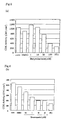
- Sample 3 was prepared using the anti-CDK2 antibody according to Exemplary Method 3. As controls, Sample 4 was prepared according to Exemplary Method 3 without using the anti-CDK2 antibody, and Sample 5 was prepared according to Exemplary Method 3 except that a non-specific IgG antibody was used instead of the anti-CDK2 antibody in the second step.
- the fluorescence bands of Samples 3 to 5 are shown in Fig. 3. The amount of fluorescence of the bands were numerically represented by Molecular Imager (Bio-Rad). The obtained amount of fluorescence is shown in Table 3.
- the amount of fluorescence was measured about samples having graded concentrations of 0 to 200 ⁇ g/mL produced according to Exemplary Method 4 and represented by CNT values.
- the obtained data were plotted with the concentration of K562 cell line in abscissa and the CNT value in ordinate to produce a reference curve.
- the obtained reference curve is shown in Fig. 4.
- a specimen was prepared according to Exemplary Method 4 except that an unknown sample was used instead of the sample of solubilized K562 cells.
- the amount of fluorescence of the specimen was measured, and the obtained CNT value was converted to a CDK2 activity of the solubilized K562 cells using the above-produced reference curve to calculate the activity.
- Example 5 Determination of activity of activated CDK4 of samples treated by Exemplary Method 5
- Sample 6 was prepared according to Exemplary Method 5 using the anti-CDK4 antibody.
- Sample 7 was prepared according to Exemplary Method 5 except that the anti-CDK4 antibody was not used.
- the bands of fluorescence of Samples 6 and 7 are shown in Fig. 5.
- This step was carried out in the same manner as in the first step of the Exemplary Method 1 using K562 cell line (premyelocytic leukemia).
- a sample was prepared by placing the solubilized K562 cells and the lysis buffer into an Eppendorf tube of 1.5 mL volume so that 250 ⁇ g of the total amount of protein of the solubilized K562 cells were in 500 ⁇ L of the lysis buffer.
- 10 ⁇ L of a polyclonal anti-CDK2 antibody 200 ⁇ g/mL, Santa Cruz Biotechnology
- a 1 : 1 (sepharose beads : lysis buffer) slurry of 40 ⁇ L sepharose beads coated with Protein A was added to the resulting sample, which was incubated at 4 °C for an hour with continuous rotation.
- the beads were taken out of the sample and washed with 1 mL of the lysis buffer twice. Then the beads were washed once with 100 mM Tris-HCl of pH 7.4 and 100 mM sodium chloride and further washed once with 100 mM Tris-HCl of pH 7.4.
- Butyrolactone I (Calbiochem) which were inhibitors to CDK1 and CDK2 and Staurosporine (Calbiochem) which had a broad inhibition spectrum including inhibition to CDK2.
- Phosphorylation solutions 40 mM Tris-HCl, pH 7.4, 18 mM magnesium chloride 2 mM ATP- ⁇ S, 6 ⁇ g/test histone H1
- Phosphorylation solutions 50 ⁇ L, containing histone H1 and the CDK inhibitors (final concentrations of 0, 1, 10, 30, 100 ⁇ M of Butyrolactone, or final concentrations of 0, 0.3, 1, 10, 30 ⁇ M of Staurosporine) were added to the beads.
- the resulting suspensions were incubated at 37°C for 90 minutes with continuous oscillation.
- the beads were precipitated by centrifugation at 1,000 rpm for 10 seconds, and 36 ⁇ L of supernatant was collected.
- a binding buffer containing 150 mM Tris-HCl and 5 mM of EDTA of pH 9.2 was added to 36 ⁇ L of the supernatant.
- 20 ⁇ L of 50 mM PEO-iodoacetylbiotin (Pierce) solution 50 mM phosphate buffer, pH 6.0
- the mixture was treated with an equivalent amount (86 ⁇ L) of an SDS-sample loading buffer (0.125 M tris-HCl, pH6.8, 4% SDS, 10 % ⁇ -ME, 25 % glycerine, bromophenol blue) at 100°C for 5 minutes.
- an SDS-sample loading buffer (0.125 M tris-HCl, pH6.8, 4% SDS, 10 % ⁇ -ME, 25 % glycerine, bromophenol blue
- the samples prepared in the third step was subjected to SDS-PAGE under the condition of 20 ⁇ L/lane (pre-cast gel, 4-20 % gradient, 10 mm ⁇ 10 mm, Daiichi Kagaku Yakuhin).
- the conditions of the SDS-PAGE were in compliance with the instructions of Daiichi Kagaku Yakuhin (60 mA, 40 minutes).
- the protein extended in the gel was electrically transferred to a PVDF membrane (10V, 30 minutes, Western blotting). The obtained membrane was blocked with a 4 w/v % BSA for 30 minutes and washed with TBS-T for 5 minutes.
- the membrane was reacted in a solution of avidin FITC (Pierce) (diluted 1,000 fold with TBS-T) at 37°C for"30 minutes. After reaction, the membrane was washed with TBS-T twice and with water once, and dried. Bands of fluorescence were visualized by Molecular Imager (Bio-Rad) and measured.
- the visualized bands of fluorescence were numerically and graphically represented by an image analyzer.
- the results are shown in Fig. 6, wherein none represents the result of the above step without addition of the CDK inhibitors, DMSO represents the result of the above step with addition only of a solvent (dimethylsulfoxide) for the CDK inhibitors and IP (-) represents the result of the above step without addition of the CDK2 antibody.
- DMSO represents the result of the above step with addition only of a solvent (dimethylsulfoxide) for the CDK inhibitors
- IP (-) represents the result of the above step without addition of the CDK2 antibody.
- the activity of CDK2 was inhibited dependently upon the amounts of the inhibitors.
- the results shows that the determined activity was specific to CDK2.
- the method of the present invention can measure the activity of cell cycle controlling factors accurately without using radioisotopes.
Landscapes
- Health & Medical Sciences (AREA)
- Life Sciences & Earth Sciences (AREA)
- Chemical & Material Sciences (AREA)
- Engineering & Computer Science (AREA)
- Molecular Biology (AREA)
- Immunology (AREA)
- Organic Chemistry (AREA)
- Biomedical Technology (AREA)
- Biochemistry (AREA)
- Urology & Nephrology (AREA)
- Hematology (AREA)
- General Health & Medical Sciences (AREA)
- Biotechnology (AREA)
- Microbiology (AREA)
- Medicinal Chemistry (AREA)
- Wood Science & Technology (AREA)
- Analytical Chemistry (AREA)
- Physics & Mathematics (AREA)
- Zoology (AREA)
- Proteomics, Peptides & Aminoacids (AREA)
- Genetics & Genomics (AREA)
- Cell Biology (AREA)
- Bioinformatics & Cheminformatics (AREA)
- Food Science & Technology (AREA)
- General Physics & Mathematics (AREA)
- Pathology (AREA)
- Biophysics (AREA)
- General Engineering & Computer Science (AREA)
- Oncology (AREA)
- Hospice & Palliative Care (AREA)
- Measuring Or Testing Involving Enzymes Or Micro-Organisms (AREA)
- Investigating Or Analysing Biological Materials (AREA)
Priority Applications (1)
| Application Number | Priority Date | Filing Date | Title |
|---|---|---|---|
| EP05019765A EP1609854B1 (fr) | 2001-02-14 | 2002-02-14 | Procédé pour la détérmination de l'activité du facteur de régulation du cycle cellulaire et procédé pour la diagnose de cancer utilisant le même |
Applications Claiming Priority (2)
| Application Number | Priority Date | Filing Date | Title |
|---|---|---|---|
| JP2001037115 | 2001-02-14 | ||
| JP2001037115 | 2001-02-14 |
Related Child Applications (1)
| Application Number | Title | Priority Date | Filing Date |
|---|---|---|---|
| EP05019765A Division EP1609854B1 (fr) | 2001-02-14 | 2002-02-14 | Procédé pour la détérmination de l'activité du facteur de régulation du cycle cellulaire et procédé pour la diagnose de cancer utilisant le même |
Publications (3)
| Publication Number | Publication Date |
|---|---|
| EP1233060A2 true EP1233060A2 (fr) | 2002-08-21 |
| EP1233060A3 EP1233060A3 (fr) | 2003-10-15 |
| EP1233060B1 EP1233060B1 (fr) | 2007-11-14 |
Family
ID=18900293
Family Applications (2)
| Application Number | Title | Priority Date | Filing Date |
|---|---|---|---|
| EP02003139A Expired - Lifetime EP1233060B1 (fr) | 2001-02-14 | 2002-02-14 | Procédé pour la détérmination de l'activité du facteur de régulation du cycle cellulaire et procédé pour la diagnose de cancer utilisant le même |
| EP05019765A Expired - Lifetime EP1609854B1 (fr) | 2001-02-14 | 2002-02-14 | Procédé pour la détérmination de l'activité du facteur de régulation du cycle cellulaire et procédé pour la diagnose de cancer utilisant le même |
Family Applications After (1)
| Application Number | Title | Priority Date | Filing Date |
|---|---|---|---|
| EP05019765A Expired - Lifetime EP1609854B1 (fr) | 2001-02-14 | 2002-02-14 | Procédé pour la détérmination de l'activité du facteur de régulation du cycle cellulaire et procédé pour la diagnose de cancer utilisant le même |
Country Status (4)
| Country | Link |
|---|---|
| US (2) | US7338774B2 (fr) |
| EP (2) | EP1233060B1 (fr) |
| AT (2) | ATE393821T1 (fr) |
| DE (2) | DE60223432T2 (fr) |
Cited By (9)
| Publication number | Priority date | Publication date | Assignee | Title |
|---|---|---|---|---|
| EP1600513A1 (fr) * | 2003-02-26 | 2005-11-30 | Sysmex Corporation | Procede pour analyser une cellule |
| WO2006021374A1 (fr) * | 2004-08-26 | 2006-03-02 | Eberhard-Karls- Universität Tübingen | Traitement de cellules biologiques transformees ou infectees |
| WO2006113862A2 (fr) * | 2005-04-20 | 2006-10-26 | Boehringer Ingelheim International Gmbh | Procedes de determination de l'activite d'une phosphoryltransferase au moyen d'adenosine 5'-triphosphate (atp)-gamma-s |
| EP1750131A1 (fr) * | 2005-08-01 | 2007-02-07 | Sysmex Corporation | Procédé de jugement de la caractéristique de la tumeur maligne |
| EP1764615A1 (fr) * | 2005-09-14 | 2007-03-21 | Sysmex Corporation | Appareil de détermination des caractéristiques d'un tissu |
| EP1770170A1 (fr) * | 2005-09-30 | 2007-04-04 | Sysmex Corporation | Equipement (ou trousse) de réactifs pour déterminer les charactéristiques des tissus |
| EP1767943A3 (fr) * | 2005-09-27 | 2007-05-23 | Sysmex Corporation | Procédé de détection de substances à analyser dans un échantillon |
| CN106324255A (zh) * | 2016-08-17 | 2017-01-11 | 山东博科生物产业有限公司 | 一种稳定性强的生化用特种蛋白液体复合质控品 |
| CN106876715A (zh) * | 2017-03-30 | 2017-06-20 | 湖北金泉新材料有限责任公司 | 一种含碳纳米管的正极浆料、其制备方法及用途 |
Families Citing this family (8)
| Publication number | Priority date | Publication date | Assignee | Title |
|---|---|---|---|---|
| DE602005024964D1 (de) * | 2004-05-31 | 2011-01-05 | Sysmex Corp | Verfahren zur beurteilung der malignität einer säuger-krebszelle |
| US20110136131A1 (en) * | 2004-06-01 | 2011-06-09 | Sysmex Corporation | Method for measuring enzyme activity and column for use in measuring enzyme activity |
| JP5111902B2 (ja) * | 2007-03-14 | 2013-01-09 | シスメックス株式会社 | 癌の診断支援装置 |
| EP2028600B1 (fr) | 2007-08-24 | 2016-10-26 | Sysmex Corporation | Système de support de diagnostic pour cancer, support de diagnostic pour procédé pour fournir des informations sur un cancer, et produit de programme informatique |
| JP2010057486A (ja) | 2008-09-02 | 2010-03-18 | Sysmex Corp | 化学療法に対するがん患者の応答の予測方法 |
| JP5830249B2 (ja) | 2010-03-31 | 2015-12-09 | シスメックス株式会社 | 癌細胞のアンスラサイクリン系抗癌剤への感受性の判定方法及びコンピュータプログラム |
| JP6002379B2 (ja) | 2011-11-29 | 2016-10-05 | シスメックス株式会社 | 癌の再発リスクの判定方法及びその利用 |
| JP6214449B2 (ja) * | 2014-03-31 | 2017-10-18 | シスメックス株式会社 | キナーゼ活性の測定方法 |
Citations (2)
| Publication number | Priority date | Publication date | Assignee | Title |
|---|---|---|---|---|
| WO1998002571A1 (fr) * | 1996-07-16 | 1998-01-22 | The Regents Of The University Of California | Dosages de proteines kinases faisant appel a des substrats proteiques fluorescents |
| WO1999029894A1 (fr) * | 1997-12-05 | 1999-06-17 | Pharmacia & Upjohn Company | Essais de criblage a haut rendement a base de fluorescence pour proteines kinases et phosphatases |
Family Cites Families (5)
| Publication number | Priority date | Publication date | Assignee | Title |
|---|---|---|---|---|
| US4252783A (en) * | 1979-06-18 | 1981-02-24 | Syva Company | Reducing fluorescent background in fluorescent immunoassays |
| DE3049033A1 (de) * | 1980-12-24 | 1982-07-22 | Licentia Patent-Verwaltungs-Gmbh, 6000 Frankfurt | "ringinterferometer" |
| US5518911A (en) * | 1995-01-06 | 1996-05-21 | Onyx Pharmaceuticals, Inc. | Human PAK65 |
| WO1999007705A1 (fr) * | 1997-08-07 | 1999-02-18 | The Regents Of The University Of California | Purines inhibant des proteine kinases, des proteines g et des polymerases |
| GB2334578A (en) * | 1998-02-18 | 1999-08-25 | Univ Liverpool | Diagnosis of cancer involving assay of levels of cyclin-dependent kinase (CDK) isoenzymes |
-
2002
- 2002-02-14 DE DE60223432T patent/DE60223432T2/de not_active Expired - Lifetime
- 2002-02-14 DE DE60226353T patent/DE60226353T2/de not_active Expired - Lifetime
- 2002-02-14 AT AT05019765T patent/ATE393821T1/de not_active IP Right Cessation
- 2002-02-14 EP EP02003139A patent/EP1233060B1/fr not_active Expired - Lifetime
- 2002-02-14 EP EP05019765A patent/EP1609854B1/fr not_active Expired - Lifetime
- 2002-02-14 US US10/074,041 patent/US7338774B2/en not_active Expired - Fee Related
- 2002-02-14 AT AT02003139T patent/ATE378400T1/de not_active IP Right Cessation
-
2007
- 2007-05-10 US US11/747,077 patent/US20070264624A1/en not_active Abandoned
Patent Citations (2)
| Publication number | Priority date | Publication date | Assignee | Title |
|---|---|---|---|---|
| WO1998002571A1 (fr) * | 1996-07-16 | 1998-01-22 | The Regents Of The University Of California | Dosages de proteines kinases faisant appel a des substrats proteiques fluorescents |
| WO1999029894A1 (fr) * | 1997-12-05 | 1999-06-17 | Pharmacia & Upjohn Company | Essais de criblage a haut rendement a base de fluorescence pour proteines kinases et phosphatases |
Non-Patent Citations (4)
Cited By (17)
| Publication number | Priority date | Publication date | Assignee | Title |
|---|---|---|---|---|
| EP1600513A4 (fr) * | 2003-02-26 | 2007-09-12 | Sysmex Corp | Procede pour analyser une cellule |
| EP1600513A1 (fr) * | 2003-02-26 | 2005-11-30 | Sysmex Corporation | Procede pour analyser une cellule |
| US7501257B2 (en) | 2003-02-26 | 2009-03-10 | Sysmex Corporation | Molecular diagnostic method of a cancer tissue or a cancer cell |
| WO2006021374A1 (fr) * | 2004-08-26 | 2006-03-02 | Eberhard-Karls- Universität Tübingen | Traitement de cellules biologiques transformees ou infectees |
| EP1634603A1 (fr) * | 2004-08-26 | 2006-03-15 | Eberhard-Karls-Universität Tübingen Universitätsklinikum | Traitement de cellules biologiques transformées ou infectées |
| US7939266B2 (en) | 2004-08-26 | 2011-05-10 | Eberhard-Karls-Universität Tübingen Universitätsklinikum | Treatment of transformed or infected biological cells |
| WO2006113862A2 (fr) * | 2005-04-20 | 2006-10-26 | Boehringer Ingelheim International Gmbh | Procedes de determination de l'activite d'une phosphoryltransferase au moyen d'adenosine 5'-triphosphate (atp)-gamma-s |
| WO2006113862A3 (fr) * | 2005-04-20 | 2007-03-08 | Boehringer Ingelheim Int | Procedes de determination de l'activite d'une phosphoryltransferase au moyen d'adenosine 5'-triphosphate (atp)-gamma-s |
| US7348159B2 (en) | 2005-04-20 | 2008-03-25 | Boehringer Ingelheim International Gmbh | Methods for determining phosphoryltransferase activity using adenosine 5'-triphosphate (ATP)-gamma-S |
| US7634362B2 (en) | 2005-08-01 | 2009-12-15 | Sysmex Corporation | Method for judging feature of malignant tumor |
| EP1750131A1 (fr) * | 2005-08-01 | 2007-02-07 | Sysmex Corporation | Procédé de jugement de la caractéristique de la tumeur maligne |
| EP1764615A1 (fr) * | 2005-09-14 | 2007-03-21 | Sysmex Corporation | Appareil de détermination des caractéristiques d'un tissu |
| EP1767943A3 (fr) * | 2005-09-27 | 2007-05-23 | Sysmex Corporation | Procédé de détection de substances à analyser dans un échantillon |
| US8309298B2 (en) | 2005-09-27 | 2012-11-13 | Sysmex Corporation | Method for detecting protein |
| EP1770170A1 (fr) * | 2005-09-30 | 2007-04-04 | Sysmex Corporation | Equipement (ou trousse) de réactifs pour déterminer les charactéristiques des tissus |
| CN106324255A (zh) * | 2016-08-17 | 2017-01-11 | 山东博科生物产业有限公司 | 一种稳定性强的生化用特种蛋白液体复合质控品 |
| CN106876715A (zh) * | 2017-03-30 | 2017-06-20 | 湖北金泉新材料有限责任公司 | 一种含碳纳米管的正极浆料、其制备方法及用途 |
Also Published As
| Publication number | Publication date |
|---|---|
| ATE378400T1 (de) | 2007-11-15 |
| EP1233060A3 (fr) | 2003-10-15 |
| EP1233060B1 (fr) | 2007-11-14 |
| DE60223432T2 (de) | 2008-09-18 |
| DE60226353D1 (de) | 2008-06-12 |
| ATE393821T1 (de) | 2008-05-15 |
| DE60223432D1 (de) | 2007-12-27 |
| DE60226353T2 (de) | 2009-06-10 |
| US20070264624A1 (en) | 2007-11-15 |
| EP1609854B1 (fr) | 2008-04-30 |
| US7338774B2 (en) | 2008-03-04 |
| US20020164673A1 (en) | 2002-11-07 |
| EP1609854A1 (fr) | 2005-12-28 |
Similar Documents
| Publication | Publication Date | Title |
|---|---|---|
| US20070264624A1 (en) | Method for determining activity of cell cycle regulatory factor and method for diagnosing cancer using the same | |
| Plesca et al. | DNA damage response and apoptosis | |
| Mahimainathan et al. | Inactivation of platelet-derived growth factor receptor by the tumor suppressor PTEN provides a novel mechanism of action of the phosphatase | |
| US8173602B2 (en) | Detecting CYP24 expression level as a marker for predisposition to cancer | |
| Wingate et al. | The tumor-specific hyperactive forms of cyclin E are resistant to inhibition by p21 and p27 | |
| Li et al. | Serum thymidine kinase 1 is a prognostic and monitoring factor in patients with non-small cell lung cancer | |
| Mi et al. | Protein phosphatase-1α regulates centrosome splitting through Nek2 | |
| US8921057B2 (en) | Method of assessing properties of mammalian cells, and method of diagnosing cancer using the same | |
| Gillett et al. | Cyclin‐dependent kinase inhibitor p27Kipl expression and interaction with other cell cycle‐associated proteins in mammary carcinoma | |
| JP4036655B2 (ja) | 細胞周期調節因子の活性の測定法と該測定法に使用される試薬 | |
| Santamaría et al. | PTOV-1, a novel protein overexpressed in prostate cancer, shuttles between the cytoplasm and the nucleus and promotes entry into the S phase of the cell division cycle | |
| Hukkelhoven et al. | Tyrosine phosphorylation of the p21 cyclin-dependent kinase inhibitor facilitates the development of proneural glioma | |
| JP2002537854A (ja) | タンパク質チロシンホスファターゼの無傷細胞アッセイ | |
| Guan et al. | Mutations of phosphorylation sites Ser10 and Thr187 of p27Kip1 abolish cytoplasmic redistribution but do not abrogate G0/1 phase arrest in the HepG2 cell line | |
| Włowiec et al. | Cdk1 is a marker of proliferation in human lymphoid cells | |
| Said et al. | Mouse mammary hyperplasias and neoplasias exhibit different patterns of cyclins D1 and D2 binding to cdk4 | |
| US20230417751A1 (en) | Analysis of protein kinases in live cells | |
| US20110136131A1 (en) | Method for measuring enzyme activity and column for use in measuring enzyme activity | |
| US20220364143A1 (en) | Analysis of protein kinases in live cells | |
| EP2400033B1 (fr) | Procédé pour déterminer la sensibilité de cellules tumorales à un inhibiteur de la tyrosine kinase et produit de programme informatique | |
| Edwards et al. | Phosphotyrosine Kinase Assays as a Prescreen for Inhibitors of EGFr | |
| WO2000070352A1 (fr) | Traitement du cancer | |
| Wingate et al. | The Role of the Low Molecular Weight (LMW) Isoforms of Cyclin E in Breast Cancer Tumorigenesis | |
| Ducruet | Regulation of CDC25A in human tumor cells by cyclin-dependent kinase 2 | |
| US20120100561A1 (en) | A pkn3/rhoc macromolecular complex and methods of use therefor |
Legal Events
| Date | Code | Title | Description |
|---|---|---|---|
| PUAI | Public reference made under article 153(3) epc to a published international application that has entered the european phase |
Free format text: ORIGINAL CODE: 0009012 |
|
| AK | Designated contracting states |
Kind code of ref document: A2 Designated state(s): AT BE CH CY DE DK ES FI FR GB GR IE IT LI LU MC NL PT SE TR |
|
| AX | Request for extension of the european patent |
Free format text: AL;LT;LV;MK;RO;SI |
|
| PUAL | Search report despatched |
Free format text: ORIGINAL CODE: 0009013 |
|
| AK | Designated contracting states |
Kind code of ref document: A3 Designated state(s): AT BE CH CY DE DK ES FI FR GB GR IE IT LI LU MC NL PT SE TR |
|
| AX | Request for extension of the european patent |
Extension state: AL LT LV MK RO SI |
|
| 17P | Request for examination filed |
Effective date: 20040122 |
|
| 17Q | First examination report despatched |
Effective date: 20040225 |
|
| AKX | Designation fees paid |
Designated state(s): AT BE CH CY DE DK ES FI FR GB GR IE IT LI LU MC NL PT SE TR |
|
| GRAP | Despatch of communication of intention to grant a patent |
Free format text: ORIGINAL CODE: EPIDOSNIGR1 |
|
| GRAS | Grant fee paid |
Free format text: ORIGINAL CODE: EPIDOSNIGR3 |
|
| GRAA | (expected) grant |
Free format text: ORIGINAL CODE: 0009210 |
|
| AK | Designated contracting states |
Kind code of ref document: B1 Designated state(s): AT BE CH CY DE DK ES FI FR GB GR IE IT LI LU MC NL PT SE TR |
|
| REG | Reference to a national code |
Ref country code: GB Ref legal event code: FG4D |
|
| REG | Reference to a national code |
Ref country code: CH Ref legal event code: EP |
|
| REG | Reference to a national code |
Ref country code: IE Ref legal event code: FG4D |
|
| REF | Corresponds to: |
Ref document number: 60223432 Country of ref document: DE Date of ref document: 20071227 Kind code of ref document: P |
|
| PG25 | Lapsed in a contracting state [announced via postgrant information from national office to epo] |
Ref country code: SE Free format text: LAPSE BECAUSE OF FAILURE TO SUBMIT A TRANSLATION OF THE DESCRIPTION OR TO PAY THE FEE WITHIN THE PRESCRIBED TIME-LIMIT Effective date: 20080214 Ref country code: LI Free format text: LAPSE BECAUSE OF FAILURE TO SUBMIT A TRANSLATION OF THE DESCRIPTION OR TO PAY THE FEE WITHIN THE PRESCRIBED TIME-LIMIT Effective date: 20071114 Ref country code: CH Free format text: LAPSE BECAUSE OF FAILURE TO SUBMIT A TRANSLATION OF THE DESCRIPTION OR TO PAY THE FEE WITHIN THE PRESCRIBED TIME-LIMIT Effective date: 20071114 Ref country code: ES Free format text: LAPSE BECAUSE OF FAILURE TO SUBMIT A TRANSLATION OF THE DESCRIPTION OR TO PAY THE FEE WITHIN THE PRESCRIBED TIME-LIMIT Effective date: 20080225 |
|
| REG | Reference to a national code |
Ref country code: CH Ref legal event code: PL |
|
| PG25 | Lapsed in a contracting state [announced via postgrant information from national office to epo] |
Ref country code: AT Free format text: LAPSE BECAUSE OF FAILURE TO SUBMIT A TRANSLATION OF THE DESCRIPTION OR TO PAY THE FEE WITHIN THE PRESCRIBED TIME-LIMIT Effective date: 20071114 |
|
| PG25 | Lapsed in a contracting state [announced via postgrant information from national office to epo] |
Ref country code: DK Free format text: LAPSE BECAUSE OF FAILURE TO SUBMIT A TRANSLATION OF THE DESCRIPTION OR TO PAY THE FEE WITHIN THE PRESCRIBED TIME-LIMIT Effective date: 20071114 |
|
| ET | Fr: translation filed | ||
| PG25 | Lapsed in a contracting state [announced via postgrant information from national office to epo] |
Ref country code: BE Free format text: LAPSE BECAUSE OF FAILURE TO SUBMIT A TRANSLATION OF THE DESCRIPTION OR TO PAY THE FEE WITHIN THE PRESCRIBED TIME-LIMIT Effective date: 20071114 |
|
| PLBE | No opposition filed within time limit |
Free format text: ORIGINAL CODE: 0009261 |
|
| STAA | Information on the status of an ep patent application or granted ep patent |
Free format text: STATUS: NO OPPOSITION FILED WITHIN TIME LIMIT |
|
| PG25 | Lapsed in a contracting state [announced via postgrant information from national office to epo] |
Ref country code: PT Free format text: LAPSE BECAUSE OF FAILURE TO SUBMIT A TRANSLATION OF THE DESCRIPTION OR TO PAY THE FEE WITHIN THE PRESCRIBED TIME-LIMIT Effective date: 20080414 |
|
| 26N | No opposition filed |
Effective date: 20080815 |
|
| PG25 | Lapsed in a contracting state [announced via postgrant information from national office to epo] |
Ref country code: MC Free format text: LAPSE BECAUSE OF NON-PAYMENT OF DUE FEES Effective date: 20080228 |
|
| PG25 | Lapsed in a contracting state [announced via postgrant information from national office to epo] |
Ref country code: IE Free format text: LAPSE BECAUSE OF NON-PAYMENT OF DUE FEES Effective date: 20080214 Ref country code: GR Free format text: LAPSE BECAUSE OF FAILURE TO SUBMIT A TRANSLATION OF THE DESCRIPTION OR TO PAY THE FEE WITHIN THE PRESCRIBED TIME-LIMIT Effective date: 20080215 |
|
| PG25 | Lapsed in a contracting state [announced via postgrant information from national office to epo] |
Ref country code: CY Free format text: LAPSE BECAUSE OF FAILURE TO SUBMIT A TRANSLATION OF THE DESCRIPTION OR TO PAY THE FEE WITHIN THE PRESCRIBED TIME-LIMIT Effective date: 20071114 |
|
| PG25 | Lapsed in a contracting state [announced via postgrant information from national office to epo] |
Ref country code: FI Free format text: LAPSE BECAUSE OF FAILURE TO SUBMIT A TRANSLATION OF THE DESCRIPTION OR TO PAY THE FEE WITHIN THE PRESCRIBED TIME-LIMIT Effective date: 20071114 |
|
| PG25 | Lapsed in a contracting state [announced via postgrant information from national office to epo] |
Ref country code: LU Free format text: LAPSE BECAUSE OF NON-PAYMENT OF DUE FEES Effective date: 20080214 |
|
| PG25 | Lapsed in a contracting state [announced via postgrant information from national office to epo] |
Ref country code: TR Free format text: LAPSE BECAUSE OF FAILURE TO SUBMIT A TRANSLATION OF THE DESCRIPTION OR TO PAY THE FEE WITHIN THE PRESCRIBED TIME-LIMIT Effective date: 20071114 |
|
| PGFP | Annual fee paid to national office [announced via postgrant information from national office to epo] |
Ref country code: NL Payment date: 20140110 Year of fee payment: 13 |
|
| PGFP | Annual fee paid to national office [announced via postgrant information from national office to epo] |
Ref country code: IT Payment date: 20140210 Year of fee payment: 13 Ref country code: FR Payment date: 20140211 Year of fee payment: 13 |
|
| PGFP | Annual fee paid to national office [announced via postgrant information from national office to epo] |
Ref country code: GB Payment date: 20140212 Year of fee payment: 13 |
|
| PGFP | Annual fee paid to national office [announced via postgrant information from national office to epo] |
Ref country code: DE Payment date: 20140417 Year of fee payment: 13 |
|
| REG | Reference to a national code |
Ref country code: DE Ref legal event code: R119 Ref document number: 60223432 Country of ref document: DE |
|
| REG | Reference to a national code |
Ref country code: NL Ref legal event code: V1 Effective date: 20150901 |
|
| PG25 | Lapsed in a contracting state [announced via postgrant information from national office to epo] |
Ref country code: NL Free format text: LAPSE BECAUSE OF NON-PAYMENT OF DUE FEES Effective date: 20150901 |
|
| GBPC | Gb: european patent ceased through non-payment of renewal fee |
Effective date: 20150214 |
|
| REG | Reference to a national code |
Ref country code: FR Ref legal event code: ST Effective date: 20151030 |
|
| PG25 | Lapsed in a contracting state [announced via postgrant information from national office to epo] |
Ref country code: IT Free format text: LAPSE BECAUSE OF NON-PAYMENT OF DUE FEES Effective date: 20150214 |
|
| PG25 | Lapsed in a contracting state [announced via postgrant information from national office to epo] |
Ref country code: GB Free format text: LAPSE BECAUSE OF NON-PAYMENT OF DUE FEES Effective date: 20150214 Ref country code: DE Free format text: LAPSE BECAUSE OF NON-PAYMENT OF DUE FEES Effective date: 20150901 |
|
| PG25 | Lapsed in a contracting state [announced via postgrant information from national office to epo] |
Ref country code: FR Free format text: LAPSE BECAUSE OF NON-PAYMENT OF DUE FEES Effective date: 20150302 |