EP1203007B1 - Nucleic acid ligands to hepatocyte growth factor/scatter factor (hgf/sf) and its receptor c-met - Google Patents
Nucleic acid ligands to hepatocyte growth factor/scatter factor (hgf/sf) and its receptor c-met Download PDFInfo
- Publication number
- EP1203007B1 EP1203007B1 EP00948923A EP00948923A EP1203007B1 EP 1203007 B1 EP1203007 B1 EP 1203007B1 EP 00948923 A EP00948923 A EP 00948923A EP 00948923 A EP00948923 A EP 00948923A EP 1203007 B1 EP1203007 B1 EP 1203007B1
- Authority
- EP
- European Patent Office
- Prior art keywords
- artificial sequence
- rna
- nucleic acid
- modified
- pyrimidines
- Prior art date
- Legal status (The legal status is an assumption and is not a legal conclusion. Google has not performed a legal analysis and makes no representation as to the accuracy of the status listed.)
- Expired - Lifetime
Links
Images


Classifications
-
- C—CHEMISTRY; METALLURGY
- C07—ORGANIC CHEMISTRY
- C07H—SUGARS; DERIVATIVES THEREOF; NUCLEOSIDES; NUCLEOTIDES; NUCLEIC ACIDS
- C07H21/00—Compounds containing two or more mononucleotide units having separate phosphate or polyphosphate groups linked by saccharide radicals of nucleoside groups, e.g. nucleic acids
-
- A—HUMAN NECESSITIES
- A61—MEDICAL OR VETERINARY SCIENCE; HYGIENE
- A61P—SPECIFIC THERAPEUTIC ACTIVITY OF CHEMICAL COMPOUNDS OR MEDICINAL PREPARATIONS
- A61P15/00—Drugs for genital or sexual disorders; Contraceptives
-
- A—HUMAN NECESSITIES
- A61—MEDICAL OR VETERINARY SCIENCE; HYGIENE
- A61P—SPECIFIC THERAPEUTIC ACTIVITY OF CHEMICAL COMPOUNDS OR MEDICINAL PREPARATIONS
- A61P17/00—Drugs for dermatological disorders
- A61P17/06—Antipsoriatics
-
- A—HUMAN NECESSITIES
- A61—MEDICAL OR VETERINARY SCIENCE; HYGIENE
- A61P—SPECIFIC THERAPEUTIC ACTIVITY OF CHEMICAL COMPOUNDS OR MEDICINAL PREPARATIONS
- A61P19/00—Drugs for skeletal disorders
- A61P19/08—Drugs for skeletal disorders for bone diseases, e.g. rachitism, Paget's disease
- A61P19/10—Drugs for skeletal disorders for bone diseases, e.g. rachitism, Paget's disease for osteoporosis
-
- A—HUMAN NECESSITIES
- A61—MEDICAL OR VETERINARY SCIENCE; HYGIENE
- A61P—SPECIFIC THERAPEUTIC ACTIVITY OF CHEMICAL COMPOUNDS OR MEDICINAL PREPARATIONS
- A61P27/00—Drugs for disorders of the senses
- A61P27/02—Ophthalmic agents
- A61P27/06—Antiglaucoma agents or miotics
-
- A—HUMAN NECESSITIES
- A61—MEDICAL OR VETERINARY SCIENCE; HYGIENE
- A61P—SPECIFIC THERAPEUTIC ACTIVITY OF CHEMICAL COMPOUNDS OR MEDICINAL PREPARATIONS
- A61P29/00—Non-central analgesic, antipyretic or antiinflammatory agents, e.g. antirheumatic agents; Non-steroidal antiinflammatory drugs [NSAID]
-
- A—HUMAN NECESSITIES
- A61—MEDICAL OR VETERINARY SCIENCE; HYGIENE
- A61P—SPECIFIC THERAPEUTIC ACTIVITY OF CHEMICAL COMPOUNDS OR MEDICINAL PREPARATIONS
- A61P35/00—Antineoplastic agents
-
- A—HUMAN NECESSITIES
- A61—MEDICAL OR VETERINARY SCIENCE; HYGIENE
- A61P—SPECIFIC THERAPEUTIC ACTIVITY OF CHEMICAL COMPOUNDS OR MEDICINAL PREPARATIONS
- A61P7/00—Drugs for disorders of the blood or the extracellular fluid
- A61P7/02—Antithrombotic agents; Anticoagulants; Platelet aggregation inhibitors
-
- A—HUMAN NECESSITIES
- A61—MEDICAL OR VETERINARY SCIENCE; HYGIENE
- A61P—SPECIFIC THERAPEUTIC ACTIVITY OF CHEMICAL COMPOUNDS OR MEDICINAL PREPARATIONS
- A61P9/00—Drugs for disorders of the cardiovascular system
- A61P9/10—Drugs for disorders of the cardiovascular system for treating ischaemic or atherosclerotic diseases, e.g. antianginal drugs, coronary vasodilators, drugs for myocardial infarction, retinopathy, cerebrovascula insufficiency, renal arteriosclerosis
-
- C—CHEMISTRY; METALLURGY
- C12—BIOCHEMISTRY; BEER; SPIRITS; WINE; VINEGAR; MICROBIOLOGY; ENZYMOLOGY; MUTATION OR GENETIC ENGINEERING
- C12N—MICROORGANISMS OR ENZYMES; COMPOSITIONS THEREOF; PROPAGATING, PRESERVING, OR MAINTAINING MICROORGANISMS; MUTATION OR GENETIC ENGINEERING; CULTURE MEDIA
- C12N15/00—Mutation or genetic engineering; DNA or RNA concerning genetic engineering, vectors, e.g. plasmids, or their isolation, preparation or purification; Use of hosts therefor
- C12N15/09—Recombinant DNA-technology
- C12N15/11—DNA or RNA fragments; Modified forms thereof; Non-coding nucleic acids having a biological activity
- C12N15/115—Aptamers, i.e. nucleic acids binding a target molecule specifically and with high affinity without hybridising therewith ; Nucleic acids binding to non-nucleic acids, e.g. aptamers
-
- G—PHYSICS
- G01—MEASURING; TESTING
- G01N—INVESTIGATING OR ANALYSING MATERIALS BY DETERMINING THEIR CHEMICAL OR PHYSICAL PROPERTIES
- G01N33/00—Investigating or analysing materials by specific methods not covered by groups G01N1/00 - G01N31/00
- G01N33/48—Biological material, e.g. blood, urine; Haemocytometers
- G01N33/50—Chemical analysis of biological material, e.g. blood, urine; Testing involving biospecific ligand binding methods; Immunological testing
- G01N33/53—Immunoassay; Biospecific binding assay; Materials therefor
- G01N33/5308—Immunoassay; Biospecific binding assay; Materials therefor for analytes not provided for elsewhere, e.g. nucleic acids, uric acid, worms, mites
-
- G—PHYSICS
- G01—MEASURING; TESTING
- G01N—INVESTIGATING OR ANALYSING MATERIALS BY DETERMINING THEIR CHEMICAL OR PHYSICAL PROPERTIES
- G01N33/00—Investigating or analysing materials by specific methods not covered by groups G01N1/00 - G01N31/00
- G01N33/48—Biological material, e.g. blood, urine; Haemocytometers
- G01N33/50—Chemical analysis of biological material, e.g. blood, urine; Testing involving biospecific ligand binding methods; Immunological testing
- G01N33/74—Chemical analysis of biological material, e.g. blood, urine; Testing involving biospecific ligand binding methods; Immunological testing involving hormones or other non-cytokine intercellular protein regulatory factors such as growth factors, including receptors to hormones and growth factors
-
- A—HUMAN NECESSITIES
- A61—MEDICAL OR VETERINARY SCIENCE; HYGIENE
- A61K—PREPARATIONS FOR MEDICAL, DENTAL OR TOILETRY PURPOSES
- A61K38/00—Medicinal preparations containing peptides
-
- C—CHEMISTRY; METALLURGY
- C12—BIOCHEMISTRY; BEER; SPIRITS; WINE; VINEGAR; MICROBIOLOGY; ENZYMOLOGY; MUTATION OR GENETIC ENGINEERING
- C12N—MICROORGANISMS OR ENZYMES; COMPOSITIONS THEREOF; PROPAGATING, PRESERVING, OR MAINTAINING MICROORGANISMS; MUTATION OR GENETIC ENGINEERING; CULTURE MEDIA
- C12N2310/00—Structure or type of the nucleic acid
- C12N2310/10—Type of nucleic acid
- C12N2310/16—Aptamers
-
- C—CHEMISTRY; METALLURGY
- C12—BIOCHEMISTRY; BEER; SPIRITS; WINE; VINEGAR; MICROBIOLOGY; ENZYMOLOGY; MUTATION OR GENETIC ENGINEERING
- C12N—MICROORGANISMS OR ENZYMES; COMPOSITIONS THEREOF; PROPAGATING, PRESERVING, OR MAINTAINING MICROORGANISMS; MUTATION OR GENETIC ENGINEERING; CULTURE MEDIA
- C12N2310/00—Structure or type of the nucleic acid
- C12N2310/30—Chemical structure
- C12N2310/32—Chemical structure of the sugar
- C12N2310/322—2'-R Modification
-
- G—PHYSICS
- G01—MEASURING; TESTING
- G01N—INVESTIGATING OR ANALYSING MATERIALS BY DETERMINING THEIR CHEMICAL OR PHYSICAL PROPERTIES
- G01N2333/00—Assays involving biological materials from specific organisms or of a specific nature
- G01N2333/435—Assays involving biological materials from specific organisms or of a specific nature from animals; from humans
- G01N2333/475—Assays involving growth factors
- G01N2333/4753—Hepatocyte growth factor; Scatter factor; Tumor cytotoxic factor II
Definitions
- Described herein are methods for identifying and preparing high affinity nucleic acid ligands of hepatocyte growth factor/scatter factor (HGF) and its receptor c-met.
- the method utilized herein for identifying such nucleic acid ligands is called SELEX, an acronym for Systematic Evolution of Ligands by EXponential enrichment.
- the invention is also directed towards therapeutic and diagnostic reagents for diseases in which elevated HGF and c-met activity are causative factors.
- Hepatocyte growth factor/scatter factor (abbreviated herein as HGF) is a potent cytokine which, through interaction with its receptor c-met, stimulates proliferation, morphogenesis, and migration of a wide variety of cell types, predominantly epithelial.
- HGF and c-met are involved in several cellular processes involved in tumorigenesis, notably angiogenesis and motogenesis, the latter having been implicated in the migration of cells required for metastasis (reviewed in Jiang and Hiscox (1997) Histol Histopathol. 12:537-55 ; Tamagnone and Comoglio (1997) Cytokine Growth Factor Rev. 8:129-42 ; Jiang, et al. (1999) Crit Rev Oncol Hematol.
- proteases that degrade the extracellular matrix also activate HGF, which in turn up-regulates urokinase type plasminogen activator (uPA) and its receptor, resulting in an activating loop feeding the invasive and migratory processes required for metastatic cancer.
- uPA urokinase type plasminogen activator
- HGF and the c-met receptor are expressed at abnormally high levels in a large variety of solid tumors.
- the levels of HGF and/or c-met have been measured in human tumor tissues (reviewed in Jiang (1999) Crit Rev Oncol Hematol. 29:209-48 ).
- High levels of HGF and/or c-met have been observed in liver, breast, pancreas, lung, kidney, bladder, ovary, brain, prostate, gallbladder and myeloma tumors in addition to many others.
- Elevated levels of HGF and c-met have also been observed in non-oncological settings, such as hypertension ( Morishita, Aoki et al. (1997) J Atheroscler Thromb. 4:12-9 ; Nakamura, Moriguchi et al. (1998), Biochem Biophys Res Commun. 242:238-43 ), arteriosclerosis ( Nishimura, Ushiyama et al. (1997) J Hypertens. 15:1137-42 ; Morishita, Nakamura et al. (1998) J Atheroscler Thromb. 4:128-34 ), myocardial infarction ( Sato, Yoshinouchi et al. (1998) J Cardiol. 32:77-82 ), and rheumatoid arthritis ( Koch, Halloran et al. (1996) Arthritis Rheum. 39:1566-75 ), raising the possibility of additional therapeutic and diagnostic applications.
- hypertension Morishita, Aoki
- HGF/c-met The role of HGF/c-met in metastasis has been elucidated in mice using cell lines transformed with HGF/c-met (reviewed in Jeffers, Rong et al. (1996) J Mol Med. 74:505-13 ).
- human breast carcinoma cells expressing HGF/c-met were injected in the mouse mammary fat pad, resulting in eventual lung metastases in addition to the primary tumor ( Meiners, Brinkmann et al. (1998) Oncogene. 16:9-20 ).
- transgenic mice which overexpress HGF become tumor-laden at many loci ( Takayama, LaRochelle et al. (1997) Proc. Natl. Acad. Sci. U.S.A. 94:701-6 ).
- HGF and VEGF were recently reported to have an additive or synergistic effect on mitogenesis of human umbilical vein endothelial cells (HUVECs) ( Van Belle, Witzenbichler et al. (1998) Circulation. 97:381-90 ). Similar combined effects are likely to contribute to angiogenesis and metastasis.
- Human HGF protein is expressed as a single peptide chain of 728 amino acids (reviewed in Mizuno and Nakamura (1993) Exs. 65:1-29 ; Rubin, Bottaro et al. (1993) Biochim Biophys Acta. 1155:357-71 ; Jiang (1999) Crit Rev Oncol Hematol. 29:209-48 ).
- the amino-terminal 31 residue signal sequence of HGF is cleaved upon export; followed by proteolytic cleavage by uPA and/or other proteases.
- the mature protein is a heterodimer consisting of a 463 residue ⁇ -subunit and a 234 residue ⁇ -subunit, linked via a single disulfide bond.
- HGF is homologous to plasminogen: its ⁇ -subunit contains an N-terminal plasminogen-activator-peptide (PAP) followed by four kringle domains, and the ⁇ -subunit is a serine protease-like domain, inactive because it lacks critical catalytic amino acids.
- PAP N-terminal plasminogen-activator-peptide
- the recently solved crystal structure of an HGF fragment containing PAP and the first kringle domain indicate that this region is responsible for heparin binding and dimerization ( Chirgadze, Hepple et al. (1999) Nat Struct Biol. 6:72-9 ), in addition to receptor interaction.
- Human c-met protein is exported to the cell surface via a 23 amino acid signal sequence (reviewed in Comoglio (1993) Exs. 65:131-65 ; Rubin (1993) Biochim Biophys Acta. 1155:357-71 ; Jiang (1999) Crit Rev Oncol Hematol. 29:209-48 ).
- the exported form of c-met is initially a pro-peptide which is proteolytically cleaved.
- the mature protein is a heterodimer consisting of an extracellular 50 kDa ⁇ -subunit bound by disulfide bonds to a 140 kDa ⁇ -subunits.
- the ⁇ -subunit has a presumed membrane-spanning sequence and a 435 amino acid intracellular domain containing a typical tyrosine kinase.
- HGF is produced primarily by mesenchymal cells, while c-met is mainly expressed on cells of epithelial origin. HGF is very highly conserved at the amino acid level between species. This homology extends into the functional realm as observed in mitogenic stimulation of hepatocytes in culture by HGF across species, including human, rat, mouse, pig and dog. This indicates that human HGF can be used cross-specifically in a variety of assays.
- HGF and c-met Given the roles of HGF and c-met in disease, it would be desirable to have agents that bind to and inhibit the activity of these proteins. It would also be desirable to have agents that can quantitate the levels of HGF and c-met in individual in order to gather diagnostic and prognostic information.
- the integrins are a class of heterodimeric integral membrane proteins, one or more of which are expressed by most cell types ( Hynes (1992) Cell 69:11-25 ). Some 16 homologous ⁇ subunits and 8 homologous ⁇ subunits associate in various combinations to yield an extensive family of receptors. Each integrin heterodimer has a large extracellular domain that mediates binding to specific ligands. These ligands may include plasma proteins, proteins expressed on the surface of adjacent cells, or components of the extracellular matrix. Several of the integrins show affinity for more than one ligand and many have overlapping specificities ( Hynes (1992) Cell 69:11-25 ). Both the ⁇ and ⁇ subunits contribute to a small intracellular domain that contacts components of the actin cytoskeleton, thus forming a physical link between proteins outside and inside the cell.
- Integrins play an important role in cellular adhesion and migration, and these properties are controlled by the cell, partly by modulation of integrin affinity for its ligands (so-called "inside-out” signaling). Conversely, the presence or absence of integrin ligation provides specific information about the cellular microenvironment, and in many instances integrins serve as a conduit for signal transduction. Ligand binding by an integrin may promote its incorporation into focal adhesions, the assembly of cytoskeletal and intracellular signaling molecules into supra-molecular complexes, and the initiation of a cascade of downstream signaling events including protein phosphorylation, calcium release, and an increase in intracellular pH (reviewed by Schwartz et al. (1995) Ann. Rev. Cell Dev.
- the ⁇ 3 -containing integrins are among the best studied of the receptor superfamily.
- the ⁇ 3 subunit forms heterodimers with either ⁇ v , ( ⁇ v ⁇ 3 ) or ⁇ IIb ( ⁇ IIb ⁇ 3 ). While these integrins show substantial overlap in ligand specificity, they play very different roles in normal physiology and in disease.
- ⁇ v ⁇ 3 is expressed by activated endothelial cells, smooth muscle cells, osteoclasts, and, at a very low level, by platelets. It is also expressed by a variety of tumor cell types.
- the integrin binds to a number of plasma proteins or proteins of the extracellular matrix, many of which are associated with sites of inflammation or wound healing ( Albelda (1991) Am. J. Resp. Cell Mol. Biol. 4:195-203 ). These include vitronectin, fibronectin, osteopontin, von Willebrand factor, thrombospondin, fibrinogen, and denatured collagen Type I ( Hynes (1992) Cell 69:11-25 ). Each of these proteins share a common sequence motif, arginine-glycine-aspartic acid (RGD), that forms the core of the integrin binding site.
- RGD arginine-glycine-aspartic acid
- ⁇ v ⁇ 3 has been most intensely studied in the context of new blood vessel formation (angiogenesis) where it mediates the adhesion and migration of endothelial cells through the extracellular matrix.
- Angiogenesis in adults is normally associated with the cyclical development of the corpus luteum and endometrium and with the formation of granulation tissue during wound repair. In the latter case, microvascular endothelial cells form vascular sprouts that penetrate into the temporary matrix within a wound. These cells transiently express ⁇ v ⁇ 3 and inhibition of the ligand binding function of the integrin temporarily inhibits the formation of granulation tissue ( Clark et al. (1996) Am. J. Pathol. 148:1407-21 ).
- Solid tumors are unable to grow to significant size without an independent blood supply. It is currently hypothesized that the acquisition of an angiogenic phenotype is one of the limiting steps in the growth of primary tumors and of tumors at secondary sites ( Folkman (1995) Nat. Med. 1:27-31 ).
- the vasculature that penetrates a tumor mass provides a source of oxygen and nutrients, it also serves as a conduit for metastatic cells to leave the primary tumor and migrate throughout the body.
- inhibition of angiogenesis may limit both the growth and metastasis of cancerous lesions.
- inhibition of ligand-binding by endothelial ⁇ v ⁇ 3 prevented the formation of new blood vessels ( Brooks et al.
- ⁇ v ⁇ 3 is not only expressed by the microvasculature within tumors, but in some cases, is also found on the surface of tumor cells themselves.
- expression of ⁇ v ⁇ 3 integrin has been detected in tissue sections from tumors of melanocytic and astroglial origin ( Albelda et al. (1990) Canc. Res. 50:6757-64 ; Gladson and Cheresh (1991) J. Clin. Invest. 88:1924-32 ), and the level of integrin expression has been correlated with the stage or metastatic potential of the tumor ( Albelda et al. (1990) Canc. Res. 50:6757-64 ; Gladson (1996) Am. J. Pathol.
- melanoma cells grown in vitro in a three-dimensional matrix of denatured collagen undergo apoptosis upon ⁇ v ⁇ 3 blockade.
- Vitaxin is a chimeric Fab fragment derived from the ⁇ v ⁇ 3 -specific monoclonal antibody, LM609 ( Wu et al. (1998) Proc. Nat. Acad. Sci. 95:6037-42 ).
- LM609 Wu et al. (1998) Proc. Nat. Acad. Sci. 95:6037-42 ).
- EMD121974 is a cyclic pentapeptide inhibitor of ⁇ v ⁇ 3 .
- a Phase I study of this compound in Kaposi's sarcoma, brain tumors and solid tumors is scheduled to begin in 1999.
- Angiogenesis (and ⁇ v ⁇ 3 ) are implicated in the pathology of several other diseases, including psoriasis ( Creamer et al. (1995) Am. J. Pathol. 147:1661-7 ), rheumatoid arthritis ( Walsh et al. (1998) Am. J. Pathol. 152:691-702 ; Storgard et al. (1999) J. Clin. Invest. 103:47-54 ), endometriosis ( Healy et al. (1998) Hum. Reprod. Update 4:736-40 ), and several proliferative diseases of the eye ( Casaroli Marano et al. (1995) Exp. Eye Res.
- Atheromatous plaque and restenosis following angioplasty are pathologies characterized by thickening of the intima, the innermost layer of the arterial wall.
- the proliferation and/or migration of smooth muscle cells into the neointima with concomitant deposition of fibrous extracellular proteins contributes to vessel wall thickening and subsequent vessel occlusion.
- Platelets may also contribute to the development of restenotic lesions through adhesion to endothelial cells and the release of growth factors and cytokines that stimulate the underlying smooth muscle cell layer ( Le Breton et al. (1996) J. Am. Coll. Cardiol. 28:1643-51 ).
- ⁇ v ⁇ 3 integrin is expressed on arterial smooth muscle cells ( Hoshiga et al.
- ⁇ v ⁇ 3 blockade with RGD-containing peptides or a monoclonal antibody was found to limit neointimal hyperplasia in several animal models of restenosis following arterial injury ( Choi et al. (1994) J. Vasc. Surg. 19:125-34 ; Srivatsa et al. (1997) Cardiovasc. Res. 36:408-28 ; Slepian et al. (1998) Circulation 97:1818-27 ; Coleman et al. (1999) Circ. Res. 84:1268-76 ).
- ⁇ v ⁇ 3 mediates the attachment of osteoclasts to matrix proteins, particularly osteopontin, on the surface of bone. Osteoclasts are responsible for the resorption of bone in normal physiology as well as in pathological conditions such as osteoporosis.
- a monoclonal antibody specific for ⁇ v ⁇ 3 inhibited the binding and resorption of bone particles by osteoclasts in vitro ( Ross et al. (1993) J. Biol. Chem. 268:9901-7 ).
- an RGD-containing protein, echistatin was shown to block parathyroid-stimulated bone resorption in an animal model, as monitored by serum calcium levels ( Fisher et al. (1993) Endocrin. 132:1411-3 ). Inhibitors of ⁇ v ⁇ 3 integrin are thus considered of potential utility in treating debilitating bone loss such as occurs in osteoporosis.
- ⁇ IIb ⁇ 3 (also referred to as GPIIbIIIa) is the major integrin on the surface of platelets where it mediates the adhesion of activated platelets to the plasma protein fibrinogen ( Nachman and Leung (1982) J. Clin. Invest. 69:263-9 ; Shattil et al. (1985) J. Biol. Chem. 260:11107-14 ). During clot formation, fibrinogen dimers cross-link platelets to one another through the integrin receptor. ⁇ IIb ⁇ 3 also binds to several other plasma and cell matrix proteins, including von Willebrand factor, vitronectin, and fibronectin ( Faull and Ginsberg (1996) J. Am. Soc. Nephrol. 7:1091-7 ).
- Clot formation is a tightly regulated process that balances the need for rapid response to vascular injury, with the risk of aberrant occlusion of critical vessels.
- the ⁇ IIb ⁇ 3 heterodimer is constitutively expressed on the surface of resting platelets at approximately 80,000 copies per cell ( Wagner et al. (1996) Blood 88:907-14 ); however, the affinity of the integrin for fibrinogen is very low on these cells.
- Activation of platelets by ADP, epinephrine, collagen or thrombin leads to a dramatic enhancement in integrin ligand binding activity ( Bennett and Vilaire (1979) J. Clin. Invest. 64:1393-401 ; Marguerie et al. (1979) J. Biol. Chem.
- Inhibitors of ⁇ IIb ⁇ 3 ligand binding have been primarily explored in the context of cardiovascular disease ( Chong (1998) Am. J. Health Syst. Pharm. 55:2363-86 ; Topol et al. (1999) Lancet 353:227-31 ), but may have application in any of a number of indications where thrombus formation is suspected or is likely.
- Three ⁇ IIb ⁇ 3 inhibitors, Reopro, Integrilin and Aggrastat, have been approved for use in patients experiencing acute coronary syndrome and/or in patients who are undergoing percutaneous coronary intervention.
- Reopro (Centocor/Eli Lilly) is a humanized murine monoclonal antibody Fab fragment with specificity for the ⁇ 3 chain of ⁇ IIb ⁇ 3 .
- Integrilin (COR Therapeutics) is a cyclic heptapeptide based on the integrin binding site of barbourin, an ⁇ IIb ⁇ 3 inhibitory protein derived from snake venom.
- Aggrastat (Merck & Co.) is a non-peptide small molecule antagonist of the integrin.
- Reopro cross-reacts with ⁇ v ⁇ 3 , a fact that may account for the greater reduction in long-term rates of death and non-fatal myocardial infarction associated with its use (see above).
- a significant effort is underway to identify new inhibitors of the platelet integrin with characteristics not found in the cohort of approved drugs.
- compounds with specificity for the active, ligand-binding conformation of ⁇ IIb ⁇ 3 may reduce the risk of bleeding complications associated with the existing anti-clotting therapies.
- Orally available compounds would be particularly useful for longer term therapy of patients at risk for recurrent myocardial infarction or unstable angina.
- SELEX process Systematic Evolution of Ligands by EXponential enrichment
- nucleic acids have three dimensional structural diversity not unlike proteins.
- the SELEX process is a method for the in vitro evolution of nucleic acid molecules with highly specific binding to target molecules and is described in U.S. Patent Application Serial No. 07/536,428, filed June 11, 1990 , entitled “Systematic Evolution of Ligands by Exponential Enrichment," now abandoned, U.S. Patent No. 5,475,096 , entitled “Nucleic Acid Ligands," and U.S. Patent No.
- the SELEX process provides a class of products which are referred to as nucleic acid ligands or aptamers, each having a unique sequence, and which has the property of binding specifically to a desired target compound or molecule.
- Each SELEX-identified nucleic acid ligand is a specific ligand of a given target compound or molecule.
- the SELEX process is based on the unique insight that nucleic acids have sufficient capacity for forming a variety of two- and three-dimensional structures and sufficient chemical versatility available within their monomers to act as ligands (form specific binding pairs) with virtually any chemical compound, whether monomeric or polymeric. Molecules of any size or composition can serve as targets.
- the SELEX method applied to the application of high affinity binding involves selection from a mixture of candidate oligonucleotides and step-wise iterations of binding, partitioning and amplification, using the same general selection scheme, to achieve virtually any desired criterion of binding affinity and selectivity.
- the SELEX method includes steps of contacting the mixture with the target under conditions favorable for binding, partitioning unbound nucleic acids from those nucleic acids which have bound specifically to target molecules, dissociating the nucleic acid-target complexes, amplifying the nucleic acids dissociated from the nucleic acid-target complexes to yield a ligand-enriched mixture of nucleic acids, then reiterating the steps of binding, partitioning, dissociating and amplifying through as many cycles as desired to yield highly specific high affinity nucleic acid ligands to the target molecule.
- nucleic acids as chemical compounds can form a wide array of shapes, sizes and configurations, and are capable of a far broader repertoire of binding and other functions than those displayed by nucleic acids in biological systems.
- 5,567,588 entitled “Systematic Evolution of Ligands by Exponential Enrichment: Solution SELEX,” describes a SELEX-based method which achieves highly efficient partitioning between oligonucleotides having high and low affinity for a target molecule.
- the SELEX method encompasses the identification of high-affinity nucleic acid ligands containing modified nucleotides conferring improved characteristics on the ligand, such as improved in vivo stability or improved delivery characteristics. Examples of such modifications include chemical substitutions at the ribose and/or phosphate and/or base positions.
- SELEX process-identified nucleic acid ligands containing modified nucleotides are described in U.S. Patent No. 5,660,985 , entitled "High Affinity Nucleic Acid Ligands Containing Modified Nucleotides," that describes oligonucleotides containing nucleotide derivatives chemically modified at the 5- and 2'-positions of pyrimidines.
- the SELEX method encompasses combining selected oligonucleotides with other selected oligonucleotides and non-oligonucleotide functional units as described in U.S. Patent No. 5,637,459 , entitled “Systematic Evolution of Ligands by Exponential Enrichment: Chimeric SELEX,” and U.S. Patent No. 5,683,867 , entitled “Systematic Evolution of Ligands by Exponential Enrichment: Blended SELEX,” respectively.
- These applications allow the combination of the broad array of shapes and other properties, and the efficient amplification and replication properties of oligonucleotides with the desirable properties of other molecules.
- the SELEX method further encompasses combining selected nucleic acid ligands with lipophilic compounds or non-immunogenic, high molecular weight compounds in a diagnostic or therapeutic complex as described in U.S. Patent No. 6,011,020 , entitled "Nucleic Acid Ligand Complexes.”
- nucleic acid ligands that act as inhibitors of HGF.
- nucleic acid ligands to HGF and c-met to diagnose and treat hypertension, arteriosclerosis, myocardial infarction, and rheumatoid arthritis.
- nucleic acid ligands to HGF singly or in combination with other nucleic acid ligands that inhibit VEGF and/or bFGF, and/or possibly other angiogenesis factors.
- nucleic acid ligands to HGF Methods are provided for generating nucleic acid ligands to HGF.
- the methods use the SELEX process for ligand generation.
- the nucleic acid ligands to HGF provided by the invention are useful as therapeutic and diagnostic agents for a number of diseases.
- the central method utilized herein for identifying nucleic acid ligands is called the SELEX process, an acronym for S ystematic E volution of L igands by E xponential enrichment.
- the method comprises: a) contacting the candidate mixture of nucleic acids with HGF or c-met, or expressed domains or peptides corresponding to HGF or c-met; b) partitioning between members of said candidate mixture on the basis of affinity to HGF or c-met; and c) amplifying the selected molecules to yield a mixture of nucleic acids enriched for nucleic acid sequences with a relatively higher affinity for binding to HGF or c-met.
- the method comprises: a) contacting the candidate mixture of nucleic acids with integrins, or expressed domains or peptides corresponding to integrins; b) partitioning between members of said candidate mixture on the basis of affinity to integrins; and c) amplifying the selected molecules to yield a mixture of nucleic acids enriched for nucleic acid sequences with a relatively higher affinity for binding to integrins.
- nucleic acid ligand is a non-naturally occurring nucleic acid having a desirable action on a target.
- Nucleic acid ligands are often referred to as “aptamers” .
- the term aptamer is used interchangeably with nucleic acid ligand throughout this application.
- a desirable action includes, but is not limited to, binding of the target, catalytically changing the target, reacting with the target in a way which modifies/alters the target or the functional activity of the target, covalently attaching to the target as in a suicide inhibitor, facilitating the reaction between the target and another molecule.
- the action is specific binding affinity for a target molecule, such target molecule being a three dimensional chemical structure other than a polynucleotide that binds to the nucleic acid ligand through a mechanism which predominantly depends on Watson/Crick base pairing or triple helix binding, wherein the nucleic acid ligand is not a nucleic acid having the known physiological function of being bound by the target molecule.
- the targets are HGF, c-met and integrins or portions thereof.
- Nucleic acid ligands include nucleic acids that are identified from a candidate mixture of nucleic acids, said nucleic acid ligand being a ligand of a given target, by the method comprising: a) contacting the candidate mixture with the target, wherein nucleic acids having an increased affinity to the target relative to the candidate mixture may be partitioned from the remainder of the candidate mixture; b) partitioning the increased affinity nucleic acids from the remainder of the candidate mixture; and c) amplifying the increased affinity nucleic acids to yield a ligand-enriched mixture of nucleic acids.
- candidate mixture is a mixture of nucleic acids of differing sequence from which to select a desired ligand.
- the source of a candidate mixture can be from naturally-occurring nucleic acids or fragments thereof, chemically synthesized nucleic acids, enzymatically synthesized nucleic acids or nucleic acids made by a combination of the foregoing techniques.
- each nucleic acid has fixed sequences surrounding a randomized region to facilitate the amplification process.
- nucleic acid means either DNA, RNA, single-stranded or double-stranded, and any chemical modifications thereof. Modifications include, but are not limited to, those which provide other chemical groups that incorporate additional charge, polarizability, hydrogen bonding, electrostatic interaction, and fluxionality to the nucleic acid ligand bases or to the nucleic acid ligand as a whole.
- modifications include, but are not limited to, 2'-position sugar modifications, 5-position pyrimidine modifications, 8-position purine modifications, modifications at exocyclic amines, substitution of 4-thiouridine, substitution of 5-bromo or 5-iodo-uracil; backbone modifications, methylations, unusual base-pairing combinations such as the isobases isocytidine and isoguanidine and the like. Modifications can also include 3' and 5' modifications such as capping.
- SELEX methodology involves the combination of selection of nucleic acid ligands that interact with a target in a desirable manner, for example binding to a protein, with amplification of those selected nucleic acids. Optional iterative cycling of the selection/amplification steps allows selection of one or a small number of nucleic acids which interact most strongly with the target from a pool which contains a very large number of nucleic acids. Cycling of the selection/amplification procedure is continued until a selected goal is achieved.
- the SELEX methodology is employed to obtain nucleic acid ligands to HGF, c-met and integrins or portions thereof. The SELEX methodology is described in the SELEX Patent Applications.
- SELEX target or “target” means any compound or molecule of interest for which a ligand is desired.
- a target can be a protein, peptide, carbohydrate, polysaccharide, glycoprotein, hormone, receptor, antigen, antibody, virus, substrate, metabolite, transition state analog, cofactor, inhibitor, drug, dye, nutrient, growth factor, etc. without limitation.
- the SELEX targets are HGF, c-met and integrins.
- the SELEX targets in this application include purified integrins, HGF and c-met, and fragments thereof, and short peptides or expressed protein domains comprising HGF or c-met.
- targets are fusion proteins comprising portions of HGF or c-met and other proteins.
- solid support is defined as any surface to which molecules may be attached through either covalent or non-covalent bonds. This includes, but is not limited to, membranes, microtiter plates, magnetic beads, charged paper, nylon, Langmuir-Bodgett films, functionalized glass, germanium, silicon, PTFE, polystyrene, gallium arsenide, gold, and silver. Any other material known in the art that is capable of having functional groups such as amino, carboxyl, thiol or hydroxyl incorporated on its surface, is also contemplated. This includes surfaces with any topology, including, but not limited to, spherical surfaces and grooved surfaces.
- HGF refers to hepatocyte growth factor/scatter factor. This includes purified hepatocyte growth factor/scatter factor, fragments of hepatocyte growth factor/scatter factor, chemically synthesized fragments of hepatocyte growth factor/scatter factor, derivatives or mutated versions of hepatocyte growth factor/scatter factor, and fusion proteins comprising hepatocyte growth factor/scatter factor and another protein. "HGF” as used herein also includes hepatocyte growth factor/scatter factor isolated from species other than humans.
- c-met refers to the receptor for HGF. This includes purified receptor, fragments of receptor, chemically synthesized fragments of receptor, derivatives or mutated versions of receptor, and fusion proteins comprising the receptor and another protein. "c-met” as used herein also includes the HGF receptor isolated from a species other than humans.
- integrin refers to any integrin. This includes purified integrins, fragments of integrins, chemically synthesized fragments of integrins, derivatives or mutated versions of integrins, and fusion proteins comprising the integrin and another protein. "Integrins” as used herein also includes integrins isolated from a species other than humans.
- the nucleic acid ligands of the present invention are derived from the SELEX methodology.
- the SELEX process is described in U.S. Patent Application Serial No. 07/536,428 , entitled “Systematic Evolution of Ligands by Exponential Enrichment," now abandoned, U.S. Patent No. 5,475,096 , entitled “Nucleic Acid Ligands” and U.S. Patent No. 5,270,163 (see also WO 91/19813 ), entitled “Methods for Identifying Nucleic Acid Ligands.”
- These applications, each specifically incorporated herein by reference, are collectively called the SELEX Patent Applications.
- the SELEX process provides a class of products which are nucleic acid molecules, each having a unique sequence, and each of which has the property of binding specifically to a desired target compound or molecule.
- Target molecules are preferably proteins, but can also include among others carbohydrates, peptidoglycans and a variety of small molecules.
- SELEX methodology can also be used to target biological structures, such as cell surfaces or viruses, through specific interaction with a molecule that is an integral part of that biological structure.
- the SELEX process may be defined by the following series of steps:
- the SELEX method encompasses the identification of high-affinity nucleic acid ligands containing modified nucleotides conferring improved characteristics on the ligand, such as improved in vivo stability or improved delivery characteristics. Examples of such modifications include chemical substitutions at the ribose and/or phosphate and/or base positions.
- SELEX-identified nucleic acid ligands containing modified nucleotides are described in U.S. Patent No. 5,660,985 , entitled "High Affinity Nucleic Acid Ligands Containing Modified Nucleotides," that describes oligonucleotides containing nucleotide derivatives chemically modified at the 5- and 2'-positions of pyrimidines.
- the SELEX method encompasses combining selected oligonucleotides with other selected oligonucleotides and non-oligonucleotide functional units as described in U.S. Patent No. 5,637,459 , entitled “Systematic Evolution of Ligands by Exponential Enrichment: Chimeric SELEX,” and U.S. Patent No. 5,683,867 , entitled “Systematic Evolution of Ligands by Exponential Enrichment: Blended SELEX,” respectively.
- These applications allow the combination of the broad array of shapes and other properties, and the efficient amplification and replication properties, of oligonucleotides with the desirable properties of other molecules.
- nucleic acid ligand can be made to increase the in vivo stability of the nucleic acid ligand or to enhance or to mediate the delivery of the nucleic acid ligand. See, e.g., U.S. Patent Application Serial No. 08/117,991, filed September 8 1993 , now abandoned and U.S. Patent No.
- nucleic acid ligands contemplated in this invention include, but are not limited to, those which provide other chemical groups that incorporate additional charge, polarizability, hydrophobicity, hydrogen bonding, electrostatic interaction, and fluxionality to the nucleic acid ligand bases or to the nucleic acid ligand as a whole.
- Such modifications include, but are not limited to, 2'-position sugar modifications, 5-position pyrimidine modifications, 8-position purine modifications, modifications at exocyclic amines, substitution of 4-thiouridine, substitution of 5-bromo or 5-iodo-uracil; backbone modifications, phosphorothioate or alkyl phosphate modifications, methylations, unusual base-pairing combinations such as the isobases isocytidine and isoguanidine and the like. Modifications can also include 3' and 5' modifications such as capping.
- the nucleic acid ligands are RNA molecules that are 2'-fluoro (2'-F) modified on the sugar moiety of pyrimidine residues.
- the modifications can be pre- or post-SELEX process modifications.
- Pre-SELEX process modifications yield nucleic acid ligands with both specificity for their SELEX target and improved in vivo stability.
- Post-SELEX process modifications made to 2'-OH nucleic acid ligands can result in improved in vivo stability without adversely affecting the binding capacity of the nucleic acid ligand.
- Other modifications are known to one of ordinary skill in the art. Such modifications may be made post-SELEX process (modification of previously identified unmodified ligands) or by incorporation into the SELEX process.
- the nucleic acid ligands become covalently attached to their targets upon irradiation of the nucleic acid ligand with light having a selected wavelength.
- Methods for obtaining such nucleic acid ligands are detailed in U.S. Patent Application Serial No. 08/123,935, filed September 17, 1993 , entitled “Photoselection of Nucleic Acid Ligands," now abandoned, U.S. Patent No. 5,763,177 and U.S. Patent No. 6,001,577 , both entitled “Systematic Evolution of Nucleic Acid Ligands by Exponential Enrichment: Photoselection of Nucleic Acid Ligands and Solution SELEX,” each of which is specifically incorporated herein by reference in its entirety.
- nucleic acid ligands of the invention are prepared through the SELEX methodology that is outlined above and thoroughly enabled in the SELEX applications incorporated herein by reference in their entirety.
- the SELEX process can be performed using purified HGF or c-met, or fragments thereof as a target.
- full-length HGF or c-met, or discrete domains of HGF or c-met can be produced in a suitable expression system.
- the SELEX process can be performed using as a target a synthetic peptide that includes sequences found in HGF or c-met. Determination of the precise number of amino acids needed for the optimal nucleic acid ligand is routine experimentation for skilled artisans.
- the SELEX process is carried out using HGF or c-met attached to a solid support.
- a candidate mixture of single stranded RNA molecules is then contacted with the solid support.
- the single stranded RNA molecules have a 2'-fluoro modification on C and U residues, rather than a 2'-OH group.
- the solid support is washed to remove unbound candidate nucleic acid ligand.
- the nucleic acid ligands that bind to the HGF or c-met protein are then released into solution, then reverse transcribed by reverse transcriptase and amplified using the Polymerase Chain Reaction.
- the amplified candidate mixture is then used to begin the next round of the SELEX process.
- the solid support can be a nitrocellulose filter. Nucleic acids in the candidate mixture that do not interact with the immobilized HGF or c-met can be removed from this nitrocellulose filter by application of a vacuum.
- the HGF or c-met target is adsorbed on a dry nitrocellulose filter, and nucleic acids in the candidate mixture that do not bind to the HGF or c-met are removed by washing in buffer.
- the solid support is a microtiter plate comprised of, for example, polystyrene.
- the HGF or c-met protein is used as a target for Truncate SELEX, described in U.S. Patent Application Serial No. 09/275,850, filed March 24 1999 , entitled “Truncation SELEX Method,” incorporated herein by reference in its entirety.
- the nucleic acid ligands thus obtained are assayed for their ability to inhibit the HGF/c-met interaction. In one embodiment, this is accomplished by performing a cell migration assay. Certain cell types, such as A549 lung carcinoma cells, will show increased migration through a Matrigel-coated filter insert (Becton Dickinson) in the presence of HGF. Thus, the degree of inhibition of HGF activity in the presence of an HGF or c-met nucleic acid ligand can be assayed by determining the number of cells that have migrated through the filter in the presence of HGF.
- nucleic acid ligands Given that elevated levels of c-met and HGF are observed in hypertension, arteriosclerosis, myocardial infarction, and rheumatoid arthritis, nucleic acid ligands will serve as useful therapeutic and diagnostic agents for these diseases.
- inhibitory nucleic acid ligands of HGF and c-met are administered, along with a pharmaceutically accepted excipient to an individual suffering from one of these diseases. Modifications of these nucleic acid ligands are made in some embodiments to impart increased stability upon the nucleic acid ligands in the presence of bodily fluids. Such modifications are described and enabled in the SELEX applications cited above.
- nucleic acid ligands to HGF and c-met are used to measure the levels of these proteins in an individual in order to obtain prognostic and diagnostic information. Elevated levels of c-met and HGF are associated with tumors in the liver, breast, pancreas, lung, kidney, bladder, ovary, brain, prostrate, and gallbladder. Elevated levels of HGF and c-met are also associated with myeloma.
- nucleic acid ligands that inhibit the HGF/c-met interaction are used to inhibit tumorigenesis, by inhibiting, for example, angiogenesis and motogenesis.
- a nucleic acid ligand to HGF is used in combination with nucleic acid ligands to VEGF (vascular endothelial growth factor) and/or bFGF (basic fibroblast growth factor) to inhibit tumor metastasis and angiogenesis.
- VEGF vascular endothelial growth factor
- bFGF basic fibroblast growth factor
- the use of multiple nucleic acid ligands is likely to have an additive or synergistic effect on tumor suppression.
- Nucleic acid ligands that inhibit VEGF are described in U.S. Patent No. 5,849, 479 , U.S. Patent No. 5,811,533 and U.S. Patent Application Serial No.
- the SELEX process can be performed using purified integrins, or fragments thereof as a target. Alternatively, full-length integrins, or discrete domains of integrins, can be produced in a suitable expression system. Alternatively, the SELEX process can be performed using as a target a synthetic peptide that includes sequences found in an integrin. Determination of the precise number of amino acids needed for the optimal nucleic acid ligand is routine experimentation for skilled artisans.
- the SELEX process is carried out using integrins attached to polystyrene beads.
- a candidate mixture of single stranded RNA molecules is then contacted with the beads.
- the single stranded RNA molecules have a 2'-fluoro modification on C and U residues, rather than a 2'-OH group.
- the beads are washed to remove unbound candidate nucleic acid ligands.
- the nucleic acid ligands that bind to the integrin are then released into solution, reverse transcribed by reverse transcriptase and amplified using the Polymerase Chain Reaction (PCR).
- PCR Polymerase Chain Reaction
- the amplified candidate mixture is then used to begin the next round of the SELEX process.
- Example 5 illustrates one possible way of performing the SELEX process using integrins as targets.
- the nucleic acid ligands thus obtained are assayed for their ability to inhibit the interaction of the integrin with its cognate ligand. In one embodiment, this is accomplished by first coating microtiter plates with the appropriate integrin(s). A ligand for the integrin, such as vitronectin or fibrinogen, is then biotinylated and contacted with the coated integrin in the presence of the nucleic acid ligand to be assayed.
- a ligand for the integrin such as vitronectin or fibrinogen
- the microtiter plate After incubation for a suitable period of time, the microtiter plate is washed, and the amount of vitronectin or fibrinogen binding to integrin is quantitated by adding a streptavidin-alkaline phosphatase conjugate, followed by a colorimetric substrate for alkaline phosphatase, such as p-nitrophenyl phosphate.
- the alkaline phosphatase signal in each well of the plate is thus inversely proportional to the effectiveness of the nucleic acid ligand as an inhibitor of the interaction between the bound integrin and its cognate ligand.
- the nucleic acid ligands are analyzed using binding to human platelets as an assay. This can be done, for example, by fluorescently labeling the nucleic acid ligand by any of the numerous techniques known in the art. The fluorescent nucleic acid ligand is then contacted with platelets, and the amount of nucleic acid ligand is be quantitated using Fluorescence Activated Cell Sorting (FACS).
- FACS Fluorescence Activated Cell Sorting
- nucleic acid ligands are labeled with a radiolabel used in the art of radioimaging.
- a nucleic acid ligand can be conjugated to the isotope 99m Tc using one of a number of techniques known in the art.
- the radiolabelled nucleic acid can then be studied in an animal model of venous thrombosis.
- a human blood clot can be generated in rabbit vein by first isolating the vein in situ by ligation, and then infusing the vein with human platelet-rich plasma and heparin to induce the formation of a blood clot.
- Radiolabelled nucleic acid ligand Blood flow through the vein is then re-established, and the radiolabelled nucleic acid ligand is introduced into the animals blood supply. The distribution of the radiolabelled nucleic acid ligand can then be studied in the rabbit's tissues to determine whether the nucleic acid ligand has accumulated in the clot, rather than in other areas.
- the nucleic acid ligands provided have a number of potential uses as therapeutic and diagnostic agents. Nucleic acid ligands that inhibit the interaction between platelet-expressed integrins and their cognate ligands are administered, along with pharmaceutically accepted excipients, in order to prevent the formation of blood clots in patients susceptible to deep vein thrombosis. The nucleic acid ligands are used to treat acute thrombosis formation during and following percutaneous coronary intervention. The nucleic acid ligands are used to treat patients with acute coronary syndromes such as unstable angina or myocardial infarction.
- Radiolabelled nucleic acid ligands to platelet-expressed integrins are administered to individuals who are to undergo major surgery, or have suffered major trauma.
- Such nucleic acid ligands can function as imaging agents for the detection of thrombi, by showing sites in the body where large aggregations of platelets are present. If a thrombosis is detected by radioimaging at a critical site in the body, then anticoagulant and thrombolytic treatment -including treatment with the inhibitory nucleic acid ligands of the instant invention-- can be given locally.
- nucleic acid ligand imaging agent provides a nucleic acid ligand imaging agent.
- anticoagulant and thrombolytic treatments which can cause harm if administered prophylactically by allowing internal bleeding to continue without efficient clotting-- can be given only to those individuals who definitely have a dangerous thrombosis.
- these treatments can be specifically injected at the site where the thrombosis has been detected by the nucleic acid ligand, instead of injecting higher concentrations into the bloodstream in the hope that some active agent will be carried to all potential sites of thrombosis.
- Nucleic acid ligands to ⁇ v ⁇ 3 integrin can be used to inhibit tumor growth and metastasis. They can also be used to treat ocular diseases including, but not limited to, diabetic retinopathy, retinopathy of prematurity, and macular degeneration. Other diseases for which ⁇ v ⁇ 3 nucleic acid ligands are useful therapeutic agents include, but are not limited to, endometriosis, psoriasis, rheumatoid arthritis, stroke, osteoporosis, and restenosis.
- the human c-met-IgG 1 -His 6 fusion protein --described from the amino to the carboxyl terminus-- consists of 932 amino acids from the extracellular domains of the ⁇ and ⁇ chains of c-met, a factor Xa cleavage site, 231 amino acids from human IgG 1 (Fc domain), and a (His) 6 tag. This protein is referred to in the text and figures as c-met.
- a similar fusion protein containing the vascular endothelial growth factor receptor KDR will be referred to as KDR.
- Anti-HGF monoclonal antibody MAB294 was purchased from R&D Systems, Inc. Human IgG 1 was produced in-house by stable expression from Chinese hamster ovary cells.
- HGF SELEX experiments were done by two closely-related partitioning methods, both involving separating free from bound RNA on nitrocellulose filters.
- Conventional SELEX involves mixing target protein and RNA library in HBSMC buffer (hepes-buffered saline, 25 mM hepes, 137 mM NaCl, 5 mM KCl plus 1 mM CaCl 2 , 1 mM MgCl 2 , pH 7.4), followed by filtration on nitrocellulose under vacuum. Maintaining vacuum, the filter is washed in buffer, followed by vacuum release and RNA extraction.
- HBSMC buffer hepes-buffered saline, 25 mM hepes, 137 mM NaCl, 5 mM KCl plus 1 mM CaCl 2 , 1 mM MgCl 2 , pH 7.4
- the protein is applied to a dry nitrocellulose 13 mm filter, allowed to adsorb for several minutes, then pre-incubated in Buffer S (HBSMC buffer plus 0.02% each of ficoll, polyvinylpyrrolidone, and human serum albumin) for 10 minutes at 37°C to remove unbound protein.
- Buffer S HBSMC buffer plus 0.02% each of ficoll, polyvinylpyrrolidone, and human serum albumin
- the wash buffer is removed, and then the RNA library is added in the same buffer, and incubated with the protein-bound filter.
- the filters are washed by repeated incubations in fresh buffer, followed by RNA extraction.
- RNA sequence libraries containing either a 30 or 40 nucleotide randomized region sequence ( Figure 1 ).
- the RNA libraries were transcribed from the corresponding synthetic DNA templates that were generated by Klenow extension (Sambrook, Fritsch et al. (1989) 3:B.12).
- the DNA templates were transcribed in 1 mL reactions, each containing 0.25 nM template, 0.58 ⁇ M T7 RNA polymerase, 1 mM each of ATP and GTP, 3 mM each of 2'-F-CTP and 2'-F-UTP, 40 mM Tris-HCl (pH 8.0), 12 mM MgCl 2 , 1 mM spermidine, 5 mM DTT, 0.002% Triton X-100 and 4% polyethylene glycol (w/v) for at least 4 hours at 37°C.
- the full-length transcription products were purified by denaturing polyacrylamide gel electrophoresis.
- Radiolabeled RNA was obtained from transcription reactions as described above, but containing 0.2 nM ATP and 100 ⁇ Ci of ⁇ - 32 P-ATP.
- radiolabeled RNA was obtained by labeling the 5'-end of RNA with ⁇ - 32 P -ATP (NEN-DuPont), catalyzed by T4 polynucleotide kinase (New England Biolabs).
- T4 polynucleotide kinase New England Biolabs.
- transcription reactions included 5 mM guanosine.
- RNA pools were suspended in HBSMC buffer to which HGF protein was added, and incubated at 37°C for 30 minutes to 3 hours depending on the round. Binding reactions were then filtered under suction through 0.45 ⁇ m nitrocellulose filters (Millipore), pre-wet with binding buffer. The filters were immediately washed with at least 5 mL of HBSMC buffer. For each binding reaction, a protein-minus control reaction was done in parallel in order to determine the amount of background binding to the filters. The amount of RNA retained on the filters was quantified by Cherenkov counting, and compared with the amount input into the reactions. Filter-retained RNA was extracted with phenol and chloroform, and isolated by ethanol precipitation in the presence of 1-2 ⁇ g glycogen.
- RNA was subsequently used as a template for avian myeloblastosis virus reverse transcriptase (AMV-RT, Life Sciences) to obtain cDNA.
- AMV-RT avian myeloblastosis virus reverse transcriptase
- Figure 1 One hundred pmoles of the 3'-primer was added to the RNA and annealed by heating for 3 minutes at 70°C, followed by chilling on ice.
- the 50 ⁇ L reaction contained 5 U AMV-RT, 0.4 mM each of dNTPs, 50 mM Tris-HCl (pH 8.3), 60 mM NaCl, 6 mM Mg(OAc) 2 , and 10 mM DTT, which was incubated for 45 minutes at 48°C.
- the cDNA was amplified by PCR with the 5'- and the 3'-primers ( Figure 1 ), and the resulting DNA template was transcribed to obtain RNA for the next round of SELEX.
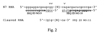
- Truncate SELEX was performed by the hybridization method described in U.S. Patent Application Serial No. 09/275,850, filed March 24 1999 , entitled "Truncation SELEX Method," incorporated herein by reference in its entirety. Briefly, 2'-F-RNA pools were body-labeled during transcription and cleaved by RNaseH using specific cleavage primers to remove the fixed sequences from the SELEX pool ( Figure 2 ). This RNA was then bound to target protein HGF and recovered following partitioning as in a conventional filter SELEX experiment.
- RNA was then biotinlyated at its 3' end and hybridized overnight under appropriate conditions with single-stranded full-length complementary strand DNA obtained from the starting SELEX pool, from which the RNA had been transcribed.
- the RNA/DNA complexes were then captured on streptavidin-coated magnetic beads and extensively washed to remove non-hybridized DNA.
- the bound DNA in the captured RNA/DNA complexes was then eluted by heat denaturation and amplified using conventional SELEX PCR primers. To complete the cycle, the resulting DNA was then used as a transcription template for generating RNA to be cleaved by RNaseH, and used in the next round of truncate SELEX.
- a polystyrene well was pre-blocked in 400 ⁇ L of blocking agent for 60 minutes at 37°C.
- the blocking agent was removed and the desired amount of RNA in 100 ⁇ L binding buffer was added and incubated for 60 minutes at 37°C.
- White, polystyrene breakaway wells (catalog #950-2965) used for partitioning were from VWR (Denver, CO).
- the blocking agents, I-block and Superblock were purchased from Tropix (Bedford, MA) and Pierce (Rockford, IL), respectively.
- the preadsorbtion was done to remove any nucleic acids which might bind to the well or the blocking agent.
- the random and round one libraries were not preadsorbed to plates to avoid loss of unique sequences.
- C-met protein was diluted in HBSMCK (50 mM HEPES, pH 7.4, 140 mM NaCl, 3 mM KCl, 1 mM CaCl 2 , 1 mM MgCl 2 ), and was adsorbed to polystyrene wells by incubating 100 ⁇ L of diluted protein per well for 60 minutes at 37°C.
- the wells were each washed with three 400 ⁇ L aliquots of HIT buffer (HBSMCK, 0.1% I-block, 0.05 % Tween 20), and then blocked in 400 ⁇ L of blocking agent for 60 minutes at 37°C.
- SELEX was initiated by incubating 100 ⁇ L of RNA in the protein-bound well for 60 minutes at 37°C.
- RNA bound to c-met was eluted by adding 100 ⁇ L water and heating at 95°C for 5 minutes and then cooled on ice, followed by reverse transcription.
- RNA concentrations were kept as low as possible --between 1 and 20 pM-- to ensure equilibrium in conditions of protein excess.
- Oligonucleotides were incubated for 15 minutes at 37°C with varying amounts of the protein in 43 ⁇ L of the binding buffer. Thirty-two microliters of each binding mixture was placed on pre-wet 0.45 ⁇ m nitrocellulose filters under suction. Each well was immediately washed with 0.5 mL binding buffer. The amount of radioactivity retained on the filters was quantitated by imaging. The radioactivity that bound to filters in the absence of protein was used for background correction. The percentage of input oligonucleotide retained on each filter spot was plotted against the corresponding log protein concentration. The nonlinear least square method was used to obtain the dissociation constant (K d ; reference Jellinek, Lynott et al. (1993) Proc. Natl. Acad. Sci. USA. 90:11227-31 ).
- Competitor titration curves were generated essentially as a standard binding curve, except that the protein and RNA concentrations were kept constant, and the competitor concentration was varied. Competitors were also added at a fixed concentration in binding experiments to increase stringency for purposes of comparing pool binding affinities. In these experiments, the competitor concentration was chosen based on the results from the competitor titration curves.
- RNA aptamers 5' and 3' boundaries of RNA aptamers were determined by the method of partial alkaline hydrolysis as described ( Jellinek, Green et al. (1994) Biochemistry. 33:10450-6 ).
- RNA was routinely synthesized by standard cyanoethyl chemistry as modified ( Green, Jellinek et al. (1995) Chem Biol. 2:683-95 ).
- 2'-Fluoro-pyrimidine phosphoramidite monomers were obtained from JBL Scientific (San Luis Obispo, CA); 2'-OMe purine, 2'-OH purine, hexyl amine, and the dT polystyrene solid support were obtained from Glen Research (Sterling, VA).
- RNA oligomers were synthesized with an amino-linker at the 5'-position. This was subsequently reacted with NHS-ester 40K-PEG manufactured by Shearwater Polymers, Inc. (Huntsville, AL), and purified by HPLC on a reverse-phase preparative column.
- RNA was separated by standard nitrocellulose filtration. Bound RNA was recovered and analyzed by high-resolution gel electrophoresis. The fragmented alkaline-hydrolyzed aptamers which were not exposed to HGF were run to establish the cleavage patterns of the unselected aptamers. Hydrolysis occurs only at 2'-OH-purines. If a given position requires 2'-OH for optimal binding to HGF, it appears as a relatively darker band compared to the unselected aptamer at that position.
- HGF SELEX experiments were done in total. The first three were done by conventional filter SELEX, while the latter two were done by the hybridization truncate SELEX method described in U.S. Patent Application Serial No. 09/275,850, filed March 24 1999 , entitled "Truncation SELEX Method," incorporated herein by reference in its entirety.
- HGF SELEX 1 was done with 30N7 2'-F-RNA for thirteen rounds of conventional filter binding.
- HGF SELEX 2 was done with 30N8 2'-F-RNA for thirteen rounds of conventional filter binding.
- HGF SELEX 3 was done with 30N7 2'-F-RNA for seven rounds by spot filter binding, followed by eight rounds of filter binding.
- HGF SELEX 4 was done by hybridization filter SELEX for three rounds, starting with pool 8 from HGF SELEX 1.
- HGF SELEX 5 was done by hybridization filter SELEX for three rounds, starting with pool 11 from HGF SELEX 3.
- HBSMC buffer was used in conventional SELEX reactions, and in spot filter SELEX, blocking agents were added as described in Materials and Methods.
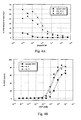
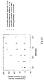
- HGF SELEX 1 reached its maximal binding by round 8, with a binding affinity of approximately 0.1 nM' ( Figure 3A ; earlier rounds and round 9 were examined in other experiments).
- HGF SELEX 2 reached its maximal binding by round 10, with a binding affinity of approximately 0.1 nM ( Figure 3B ).
- HGF SELEX 3 reached its maximal binding by round 11, after seven rounds of spot filter partitioning followed by four rounds of conventional filter SELEX (see Figure 4B ).
- a SELEX experiment which was deemed complete was characterized by cloning and sequencing (see below).
- HGF like other proteins which have large clusters of positively charged amino acids, exhibits a high degree of non-specific binding to polyanionic compounds.
- random RNA pools bind to HGF with low nanomolar affinity, similar to the value reported for HGF binding to heparin, a polyanionic sulfated polysaccharide known to have an important biological role in HGF function ( Zioncheck, Richardson et al. (1995) J Biol Chem. 270:16871-8 ).
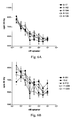
- Competition binding to heparin as well as the non-specific competitor tRNA was done to provide an additional means of evaluating SELEX progress. This was done because the binding of random and evolved RNA pools to HGF occurs in a high-affinity range that makes it difficult to monitor progress. In other words, random RNA binds so well to HGF that the affinity enhancement of the evolved pools may not be adequately assessed in conventional binding experiments in the absence of competitor.
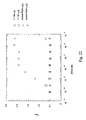
- RNA pools from HGF SELEX 3 were subjected to competition with heparin ( Figure 4A ). This experiment demonstrates that random RNA is considerably more sensitive to competition for binding to HGF than are the evolved pools. These data are compared to those obtained from a binding curve with the same three RNA pools ( Figure 4B ). In the absence of heparin competition, binding of random RNA to HGF is nearly as good as that of the evolved pools, whereas the heparin competition reveals that the evolved pools are significantly different in composition from random RNA. In addition, while rounds 8 and 11 are indistinguishable in conventional binding curves, round 11 exhibits improved binding based on increased resistance to heparin competition. These data contributed to the choice of round 11 as the maximally binding pool from which we cloned and sequenced.
- HGF SELEXes 1, 2 and 3 Following determination of pool binding affinities for HGF, the optimal SELEX pools were cloned and sequenced in order to isolate and characterize individual aptamers. Data from 30N7 HGF SELEX 1 and 3 are summarized in Table 2, including binding affinities for many of the aptamers. A similar data set was generated for 30N8 HGF SELEX 2 (Table 3). Sequences from HGF SELEX 1, 2 and 3 are designated 8-seq. number, 10-seq. number, and 11-seq. number, respectively, referring to the total number of SELEX rounds each cloned pool was subjected to. Sequences were analyzed and organized into groups with significant homology. Motifs were analyzed and predicted structures were drawn in order to analyze key features responsible for binding to HGF.
- HGF HGF-mediated stimulation of cell proliferation .
- HGF while not a potent mitogen, does stimulate moderate proliferation of many cell lines, which can be measured by incorporation of 3 H-thymidine.
- the inhibitory activity of HGF aptamers was assayed by measuring their effect on proliferation of human umbilical vein endothelial cells (HUVECs), or monkey bronchial epithelial (4MBr-5) cells. Based on the binding data and sequence family analysis, fourteen aptamers were chosen for analysis in vitro because they bind to HGF with high affinity and are representative of different sequence families. The sequences are shown in Table 4, aligned by a rough consensus that contains bases in common to several families. All sequences are 30N7 except 10-2 which is 30N8.
- HGF stimulates proliferation of HUVECs by about two-to-three-fold (data not shown).
- Boundaries and truncation Boundary determinations were done for a subset of aptamers that demonstrated in vitro inhibition of HGF activity. Using a standard alkaline hydrolysis procedure with 5'-end-labeled RNA, the 3'-boundaries of aptamers 8-17, 8-102, 8-104, 8-126, 10-1 and 10-2 were examined. Additionally, 3'-end-labeled RNA was used for 5'-boundary experiments with aptamers 8-17 and 8-102. These experiments were mostly uninformative, probably because the high degree of non-specific binding of RNA fragments, regardless of size, obscured the binding of truncated high-affinity aptamers to HGF.
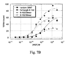
- aptamer 8-102 which had a plausible 3'-boundary between two possible endpoints which made sense with respect to computer-predicted structures ( Figure 7A ). Based on the boundary data and structural data, two truncates of aptamer 8-102 were synthesized and analyzed for binding to HGF. The sequence of the full-length aptamer and the two truncates are shown, with fixed regions underlined:
- Truncate SELEX In order to generate additional short aptamers, advanced rounds of the earlier SELEX experiments were subjected to additional rounds of truncate SELEX, using the Truncation SELEX method described in U.S. Patent Application Serial No. 09/275,850, filed March 24 1999 , entitled "Truncation SELEX Method," incorporated herein by reference in its entirety. Binding of RNaseH cleaved pools was examined to determine which were the appropriate rounds to use to initiate truncate SELEX (data not shown). None of the RNaseH-cleaved evolved pools was clearly superior to another in binding to HGF, therefore, the pools which had been previously cloned were chosen to use in truncate SELEX.
- Three rounds of hybridization truncate SELEX were done in parallel, using as starting pools HGF SELEX 1 round 8 and HGF SELEX 3 round 11.
- the truncate SELEX rounds were done at equi-molar RNA and protein, starting at 1 nM and decreasing to 0.5 and 0.1 nM. Signal-to-noise ratios were very high during selection. Subsequent manipulations were satisfactory even though the amount of recovered RNA was sub-picomolar.
- the two pools were cloned and sequenced, and binding affinities were determined (Table 5).
- the truncated aptamer with the best binding affinity, Tr51 is among several sequences which are novel, that is, they were not found in the clones sequenced from the full-length SELEX pools.
- the emergence of novel sequences suggests that the truncate SELEX succeeded in amplifying aptamers that were relatively rare in the full-length pools.
- Aptamer Tr51 appeared more frequently than any other sequence, consistent with the observation that it has better binding affinity than any other truncate.
- Other sequences that appeared multiple times also tend to be those with binding affinities near or better than the pool K d of 1-2 nM.
- the modified aptamer designated NX22354, was tested for inhibition of HGF-mediated proliferation 4MBr-5 cells ( Figure 11A ).
- the data indicate that the 36mer-PEG aptamer inhibits HGF, and that it performs at least as well as the full-length aptamer 8-17, which had previously exhibited the strongest inhibition of all aptamers tested.
- the non-PEGylated 36mer did not inhibit HGF, suggesting that the addition of PEG and/or the 3'-cap contribute to the aptamer's bioactivity.
- HGF-mediated stimulation of cell migration HGF readily stimulates cell movement, hence the name, scatter factor.
- the inhibitory effect of HGF aptamers was assayed by measuring their effect on A549 cell migration across a Matrigel coated membrane with 8.0 micron pores as described in Materials and Methods (Table 6).
- the NX22354 aptamer fully inhibited HGF-mediated migration at both 1 and 0.2 ⁇ M concentrations, but at 0.04 ⁇ M, the effect was negligible.
- the monoclonal antibody control (sample 3) was moderately effective at the 1 ⁇ g/mL dose, which is above its published EC 50 value of 0.1-0.3 ⁇ g/mL for inhibition of 4MBr-5 cell proliferation.
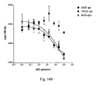
- HGF and VEGF aptamers have an additive stimulatory effect on HUVEC proliferation ( Van Belle (1998) Circulation. 97:381-90 ). This effect was observed when VEGF and HGF were added, singly and in combination, to HUVECs, and incorporation of 3 H-thymidine was measured ( Figure 13 ). As expected, stimulation by HGF was relatively weak compared with that of VEGF and together, the stimulatory effect was greater than that elicited by VEGF alone.
- each cytokine was added at 10 ng/mL for optimal stimulation in the aptamer inhibition experiments.
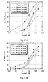
- the effect of adding one or both aptamers to the doubly-stimulated cells in the presence of both growth factors was then tested ( Figure 14A ). It was observed that each aptamer partially inhibits the stimulation and that both aptamers result in complete inhibition. Interestingly, the magnitude of the inhibitory effect of each aptamer roughly corresponds with the magnitude of the stimulation conferred by each cytokine. This observation suggests that the stimulatory effect of each cytokine can be inhibited independently, and that the two cytokines stimulate HUVECs independently.
- FIGs 14B and C depict controls in which each cytokine was administered separately, demonstrating that the HGF and VEGF aptamers do not cross-react, that is, each aptamer affects only the cytokine against which it was selected.
- HGF stimulated cells inhibition by the HGF aptamer NX22354 was observed, but not by the VEGF aptamer NX1838 ( Figure 14B ).
- stimulation by VEGF was inhibited by the VEGF aptamer NX1838, but was unaffected by the HGF aptamer NX22354 ( Figure 14C ).
- HGF like VEGF
- HGF aptamers which inhibit other growth factors suggest further combinations of the VEGF or the HGF aptamer in combination with other aptamers, for example, aptamers that inhibit bFGF, platelet-derived growth factor (PDGF), transforming growth factor beta (TGF), keratinocyte growth factor (KGF), and/or their receptors allowing for the possibility that any combination of these inhibitors may be relevant.
- PDGF platelet-derived growth factor
- TGF transforming growth factor beta
- KGF keratinocyte growth factor
- the goal is to have an array of aptamer-inhibitors of cytokines and their receptors and to be able to tailor combination treatments for specific disease states.
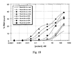
- the set of OMe aptamers were also examined for binding to HGF (data not shown).
- the binding data indicate that the OMe1 and OMe3 bind as well as the parent unsubstituted 36mer, whereas OMe2 and OMe4 bind do not bind as well. This suggests that the substitutions in OMe2 and OMe4 are less well tolerated with respect to HGF binding in solution, consistent with the fact that OMe2 and OMe4 are substituted at A25 and G5, respectively.
- NX22354 tolerates 2'-OMe substitution at all purines except G5 and A25 (aptamer 2x Sub 2'-OH) with minimal loss of binding affinity.
- the other two positions in question apparently are not required to be 2'-OH since aptamer 4x Sub 2'-OH binds no better than aptamer 2x Sub 2'-OH.
- c-met SELEX In the c-met plate SELEX experiments, the concentration of nucleic acids was lowered initially, but then raised in later rounds so that the ratio of the nucleic acid to protein would be very high. This was done in order to create conditions of high stringency which may select for higher affinity aptamers. Stringency was also applied by increasing the number of washes.
- c-met protein used in SELEX is an IgG fusion protein
- random 40N7 and round 7c RNA pools were tested for binding to human IgG 1 and c-met.
- the binding dissociation constants are set forth in Table 8.
- the affinity of round 7c RNA for both IgG 1 and c-met proteins improved about 50-fold. There are several interpretations of this result. Aptamers may have been selected which bind with better affinity to both proteins. This assumes that the difference in binding between IgG 1 and c-met is due to c-met specific aptamers. However, the two proteins were made in different cell lines that may have different glycosylation patterns which could influence binding. Thus, if the differences in affinity are due to differences between the free IgG 1 protein and the IgG 1 domain in c-met, then there might be few if any c-met specific aptamers in the round 7 pool.
- IgG aptamers by PCR. Another approach for determining if IgG 1 aptamers are present in the SELEX pools was to subject them to PCR. Predominant IgG 1 aptamers have been isolated from N7 type libraries that have a known sequence (Nikos Pagratis and Chinh Dang, personal communication). For the PCR, a DNA oligonucleotide:
- the ML-124 3'-primer ML-34; 5'-CGCAGGATCCTAATACGACTCACTATA-3' (SEQ ID NO: 189), was used with a 5'-primer containing the T7-promoter sequence present in all cloned aptamers to amplify 40N7 series nucleic acids pools: random, 1a, 2a, 3a and 4a (data not shown). Since IgG 1 aptamers have not been isolated from an N8 type library, this analysis was not done for the 30N8 SELEX. PCR of random and c-met SELEX round 1a pools yielded no signal after 20 cycles.
- IgG 1 aptamers appeared relatively early in the 40N7 SELEX experiment.
- PCR was done with a nucleic acid pool from a SELEX known to lack IgG 1 aptamers.
- PCR was done with pools from either an N7-based IgG 1 or CTLA4-IgG 1 SELEX. IgG 1 aptamers were first isolated from both of these SELEXes. The negative control had no detectable IgG 1 aptamers after 20 PCR cycles. The positive controls had detectable signals after 10 PCR cycles.
- Clones 7b-4 is the most frequent clone in Family 1 and is representative of almost all of the sequences isolated from the 7b-30N8 library.
- Clones 7b-10 and 7b-12 are the two clones from the 7b-30N8 library that had different sequences. From the 7c-40N7 pool, the chosen representatives were: Family 1 (clone 7c-1); Family 2 (clone 7c-4); Family 3 (clone 7c-23); Family 4 (clone 7c-26); Family 5 (clone 7c-25); and the presumed IgG1 Family (clone 7c-3).
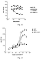
- Results are shown for only two clones, including 7c-1, which was the only one observed to bind to c-met better than KDR ( Figure 19A ).
- Clone 7c-1 which appeared twice in the 40N7 series, may exhibit biphasic binding behavior with a high affinity binding K d of ⁇ 50 pM and a lower affinity binding K d of ⁇ 5 nM.
- Clone 7c-3 and all others besides 7c-1 are presumed to be IgG 1 aptamers.
- ⁇ v ⁇ 3 integrin was isolated from human placenta and purified by immunoaffinity chromatography essentially as described by ( Smith and Cheresh (1988) J. Biol. Chem. 263:18726-31 ). In brief, human placentas were diced and the tissue fragments were extracted in a buffer containing 100 mM octyl- ⁇ -D-glucopyranoside detergent (Calbiochem, San Diego, CA). The extract was cleared by centrifugation and applied to an immunoaffinity column ( ⁇ v ⁇ 3 -specific monoclonal antibody LM609 affixed to Affi-Gel 10, (Chemicon International, Inc., Temecula, CA)).
- Protein bound to the column was eluted with a low-pH buffer and fractions were immediately neutralized and analyzed for integrin content by SDS-polyacrylamide gel electrophoresis. Integrin-containing fractions were pooled and aliquots of the purified material were stored at -80°C.
- Purified human ⁇ v ⁇ 3 was also purchased from Chemicon International, Inc, as was human ⁇ v ⁇ 5 integrin.
- ⁇ IIb ⁇ and fibrinogen were purchased from Enzyme Research Laboratories, Inc. (South Bend, IN). Vitronectin was purified from outdated human plasma according to the procedure of ( Yatohgo et al. (1988) Cell Struct. Func. 13:281-92 ), using heparin affinity chromatography.
- a DNA template library of sequence was prepared by chemical synthesis.
- the italicized nucleotides correspond to a T7 RNA polymerase promoter. There are 40N residues (a, g, t or c).
- a short DNA primer "3N8": 5'- gcctgttgtgagcctcctgtcgaa-3' (SEQ ID NO:191) was annealed to the template and extended using Klenow DNA polymerase (New England Biolabs, Beverly, MA).
- the double-stranded DNA product served as a product for T7 RNA polymerase transcription (enzyme obtained from Enzyco, Inc., Denver, CO) to generate a library of random-sequence RNAs. 2'-fluoro-CTP and -UTP were used in place of the 2'-OH-pyrimidines.
- the purified protein was diluted 1000-fold from detergent-containing storage buffer into 50 mM MES (2-[N-morpholino]ethanesulfonic acid), pH 6.1, 150 mM NaCl, 2 mM CaCl 2 , to a final concentration of approximately 0.2 ⁇ g/mL.
- MES 2-[N-morpholino]ethanesulfonic acid
- pH 6.1 150 mM NaCl
- 2 mM CaCl 2 a final concentration of approximately 0.2 ⁇ g/mL.
- 3.2 ⁇ polystyrene particles IDEXX Laboratories, Inc., Westbrook, ME
- the beads were collected by centrifugation and blocked by incubation in 3% BSA in MES buffer (above) for one hour at room temperature.
- Blocked beads were washed several times by resuspension in binding buffer (50 mM Tris-HCl, pH 7.4 (at 37°C), 145 mM NaCl, 4 mM KCl, 1 mM MgCl 2 , 2 mM CaCl 2 , 0.1 mM MnCl 2 , 0.01% BSA).
- binding buffer 50 mM Tris-HCl, pH 7.4 (at 37°C), 145 mM NaCl, 4 mM KCl, 1 mM MgCl 2 , 2 mM CaCl 2 , 0.1 mM MnCl 2 , 0.01% BSA.
- binding buffer 50 mM Tris-HCl, pH 7.4 (at 37°C), 145 mM NaCl, 4 mM KCl, 1 mM MgCl 2 , 2 mM CaCl 2 , 0.1 mM MnCl 2 , 0.01% BSA.
- RNAs that remained bound to the beads were eluted overnight at 37°C in binding buffer plus 100 ⁇ M cyclic RGD peptide ("cRGD") (GPenGRGDSPCA, Life Technologies, Gibco BRL, Gaithersburg, MD). Eluted RNAs were extracted with phenol, then chloroform:isoamyl alcohol (24:1), and ethanol precipitated. The RNA pellet was resuspended and annealed to primer 3N8 for reverse transcription using avian myeloblastosis virus reverse transcriptase (Life Sciences, Inc., St. Moscow, FL).
- cRGD cyclic RGD peptide
- the cDNA pool was amplified by the polymerase chain reaction using the 3N8 primer and primer "5N8": 5'-taatacgactcactatagggagacaagaataaacgctcaa-3' (SEQ ID NO: 192) and T. aquaticus DNA polymerase (Perkin Elmer-Cetus, Foster City, CA). Transcription of the PCR product generated an RNA pool to initiate a new round of selection. For the first round of selection 1 nmol of RNA (approximately 6 x 10 14 sequences) was incubated at 2 ⁇ M concentration with a volume of bead suspension equivalent to 50 pmol of protein (assuming all the integrin had adsorbed to the beads). In subsequent rounds, the concentration of RNA and protein-coated beads were both reduced to demand higher affinity binding interactions.
- RNA affinity was determined by titration of biotinylated RNA with a small quantity of immobilized integrin. Bound RNA was detected through the biotin moiety.
- Biotinylated RNA was prepared according to standard transcription protocols, but including a 5-fold molar excess of a 5'-biotin-modified GMP over GTP in the reaction mixture. Methods for synthesizing 5'-biotin-modified guanosine nucleotides are described in WO 98/30720 entitled "Bioconjugation of Oligonucleotides," specifically incorporated herein by reference in its entirety.
- the modified nucleotide is incorporated at the 5' end of the transcript in proportion to its representation in the guanosine pool.
- 96-well microtiter plates (Immulon 2, Dynatech Laboratories, Inc., Chantilly, VA) were coated overnight at 4°C with 100 ⁇ L purified ⁇ v ⁇ 3 at a concentration of 0.25 ⁇ g/mL in 20 mM Tris-HCl, pH 7.5, 150 mM NaCl, 1 mM MgCl 2 , 2 mM CaCl 2 , 0.1 mM MnCl 2 .
- Coating concentrations were 0.8 ⁇ g/mL for ⁇ IIb ⁇ 3 and 0.3 ⁇ g/mL for ⁇ v ⁇ 3 .
- Wells were blocked with 200 ⁇ L of a solution of 3% BSA in the same buffer (1 hour at room temperature) then rinsed 3 times with 200 ⁇ L binding buffer (50 mM Tris-HCl, pH 7.5, 137 mM NaCl, 2.7 mM KCl, 1 mM MgCl 2 , 2 mM CaCl 2 , 0.1 mM MnCl 2 , 0.1% BSA).
- Individual RNAs or RNA pools were denatured briefly at 93°C in binding buffer without divalent cations or BSA, then serially diluted in the same buffer.
- 50 ⁇ L binding buffer containing 2X-concentrations of divalent cations and BSA were added to each well, followed by 50 ⁇ L RNA dilution.
- RNAs were allowed to incubate in the integrin-coated wells at 37°C for 30-60 minutes. Unbound RNAs were removed by 3 rapid washes in binding buffer.
- 100 ⁇ L of a 1:2500 dilution in binding buffer of streptavidin-alkaline phosphatase conjugate (Calbiochem) were incubated in each well for 30 minutes at room temperature, followed by three rapid washes, as above.
- 100 ⁇ L/well p-nitrophenyl phosphate (Sigma Chemical Co., St. Louis, MO) was added and incubated at room temperature for 30 minutes. Color development was monitored by absorbance at 405 nm.
- Binding data were fit to an equation that describes the fraction of RNA or protein bound as a function of K D , and the total concentrations of RNA and protein in the binding reaction for both monophasic and biphasic binding behavior ( Green et al. (1996) Biochem. 35:14413-24 ).
- RNA specifically bound to the integrin-coated beads had increased substantially (data not shown).
- immobilized ⁇ v ⁇ 3 showed no detectable affinity for random sequence RNA
- the round 7 RNA pool bound with an equilibrium dissociation constant (K D ) of approximately 4 x 10- 7 M ( Figure 20 ).
- K D equilibrium dissociation constant
- the round 7 affinity-enriched pool was cloned and sequences were determined for individual molecules in the mixture. Of 92 sequences obtained, 35 (38%) were very highly related to one another, in many cases differing at no more than a single base position.
- Table 11 shows the sequences of the major family of 2'-F-pyrimidine RNAs with high affinity for ⁇ v ⁇ 3 (Family 1).
- Clone names indicate the selected RNA pool from which each sequence was derived (round 7, round 15 or round 17) followed by a unique clone number. Note that in several cases identical sequences were isolated from different RNA pools; in these cases, both clone names are given. (Clones 17.12A and B were isolated as end-to-end inserts in a single plasmid.) Numbers in parentheses indicate the frequency with which a particular sequence was isolated; if no number is given the clone was obtained only once from the selected RNA pool.
- Family 2 sequences isolated from the ⁇ v ⁇ 3 SELEX are shown in Table 12. The short motif (CCUGCC) held in common among all the sequences is indicated in boldface letters. In Table 13, sequences with no obvious relationship to Families 1 or 2 are shown.
- aptamers selected for high-affinity binding to a particular target protein show relatively weak binding to other related proteins, except in cases where the degree of homology is very high (for example, see ( Green et al. (1996) Biochem. 35:14413-24 ; Ruckman et al. (1998) J. Biol. Chem. 273:20556-67 )).
- Significant homology exists within the families of integrin alpha and beta sub-units, and both alpha and beta sub-units are shared among members of the integrin superfamily.
- the affinities were determined using the methods described above.
- the Family 1 aptamer 17.16 (SEQ ID NO:249) was chosen as a representative of the major sequence family.
- Figure 21 shows that aptamer 17.16 bound with identical affinity to purified, immobilized ⁇ v ⁇ 3 and to the platelet integrin, ⁇ v ⁇ 3 in a 96-well plate binding assay. Although these two proteins share the ⁇ 3 sub-unit in common, an alignment of the ⁇ v and ⁇ IIB amino acid sequences shows only 36% overall sequence identity ( Fitzgerald et al. (1987) Biochem. 26:8158-65 ). Short stretches of exact sequence identity, 5 to 9 amino acids in length, do occur, primarily within four putative calcium-binding domains of each ⁇ sub-unit.
- Binding of aptamer 17.16 to integrin ⁇ v ⁇ 5 was also tested.
- the ⁇ 5 sub-unit shares 56% sequence identity with ⁇ 3 and is more closely related to ⁇ 3 than other members of the beta sub-unit family ( McLean et al. (1990) J. Biol. Chem. 265:17126-31 ; Suzuki et al. (1990) Proc. Nat. Acad. Sci. 87:5354-8 ).
- No aptamer binding to immobilized integrin ⁇ v ⁇ 5 was observed ( Figure 22 ), although an ⁇ v -specific antibody detected the presence of ⁇ v ⁇ 5 protein adsorbed to the surface of the well (data not shown).
- aptamer 17.16 and by extension the other members of sequence Family 1, bind primarily to the ⁇ 3 sub-unit of ⁇ v ⁇ 3 .
- the high-affinity binding of the aptamer to the platelet integrin, ⁇ IIb ⁇ 3 extends its range of potential application to indications involving detection of platelets or inhibition of their function.
- RNA sequences with high affinity for a particular target While the SELEX process identifies RNA sequences with high affinity for a particular target, the procedure used in this example was designed to bias for the recovery of ligand binding site inhibitors by the inclusion of a cRGD peptide competitor in the elution buffer.
- aptamer 17.16 SEQ ID NO:249
- purified vitronectin and fibrinogen were biotinylated and incubated with one or both of the immobilized integrins in the presence or absence of varying concentrations of the aptamer or a non-binding control RNA.
- 96-well microtiter plates were coated as described above with either ⁇ v ⁇ 3 or ⁇ IIb ⁇ 3 and blocked with BSA.
- a fixed concentration of biotinylated ligand (fibrinogen: 6 nM final; vitronectin: 10 nM final) was pre-mixed in binding buffer (see “Measurement of Aptamer Binding Affinities," above) with varying concentrations of aptamer, control RNA, cyclic RGD peptide, antibody, or unmodified ligand.
- the mixtures were incubated in the integrin-coated wells for 60 minutes at room temperature.
- biotinylated ligand was detected by addition of 100 ⁇ L /well 1:500 dilution streptavidin-alkaline phosphatase conjugate (Calbiochem) (30 minutes at room temperature) followed by 100 ⁇ L /well p-nitrophenyl phosphate, as described above. Absorbance was read at 405 nm. The data were fit to an equation that describes mutually exclusive binding of two ligands to a single target species ( Gill et al. (1991) J. Mol. Biol. 220:307-24 ). The concentration of competitor that inhibited 50% of the maximum signal above background (IC 50 ) was determined from the fitted curve.
- FIG. 23A shows inhibition of biotinylated vitronectin binding to immobilized ⁇ v ⁇ 3 .
- Aptamer 17.16 inhibited the binding interaction with an IC 50 of 4.7 nM while the control RNA showed no inhibition.
- the IC 50 of RGD peptide inhibition was 1.4 nM and that of LM609 was 2.7 nM.
- Unmodified vitronectin inhibited the binding of the biotinylated material with an IC 50 of 59 nM.
- IC 50 values for the data in Figure 23B were: 17.16, 9.5 nM; control RNA, not measurable; RGD peptide, 1.0 nM; LM609, 6.3 nM; unmodified fibrinogen, 43 nM.
- IC 50 values for Figure 23C were: 17.16, 6.5 nM; control RNA, not measurable; RGD peptide, 21 nM; unmodified fibrinogen, 15 nM.
- aptamer 17.16 is an effective competitor of ⁇ 3 integrin ligand binding and, on a molar basis, has an inhibitory potency nearly equivalent to that of a bivalent antibody.
- Aptamer 17.16 (SEQ ID NO:249) was selected for binding to purified human ⁇ v ⁇ 3 adsorbed to the surface of a polystyrene bead.
- In vitro assays to measure the affinity of the aptamer for purified ⁇ 3 integrins were also done in the context of hydrophobically-adsorbed protein.
- an important test of aptamer function was to determine its capacity to bind to native protein on the surface of cells. Human platelets were chosen for this purpose because of their ease of isolation and their high level of expression of integrin ⁇ IIb ⁇ 3 .
- Platelet-rich plasma was prepared from freshly-drawn citrated human blood by centrifugation at 1000 rpm for 15 minutes in a table top centrifuge.
- activated platelets cells were incubated for 15 minutes at room temperature at 2 x 10 7 /mL in calcium- and magnesium-free Dulbecco's PBS with 2.5 U/mL thrombin and 5 mM Gly-Pro-Arg-Pro (GPRP) to inhibit platelet aggregation.
- GPRP Gly-Pro-Arg-Pro
- Cells were diluted 1:10 into binding buffer (20 mM HEPES, pH 7.5, 111 mM NaCl, 5 mM KCl, 1 mM MgCl 2 , 1 mM CaCl 2 , 0.1 % BSA, 0.01 % NaN 3 ). Resting cells were diluted similarly, without exposure to thrombin or GPRP. The activation state of resting and thrombin-treated cells was monitored by staining with fluorophore-conjugated antibodies to CD61 ( ⁇ 3 integrin subunit), which binds to all platelets, and to CD62 (P-selectin), a marker of platelet activation.
- binding buffer 20 mM HEPES, pH 7.5, 111 mM NaCl, 5 mM KCl, 1 mM MgCl 2 , 1 mM CaCl 2 , 0.1 % BSA, 0.01 % NaN 3 .
- Resting cells were diluted similarly, without exposure to
- RNAs were obtained from Becton-Dickinson Immunocytometry Systems, San Jose, CA. Fluorescein-conjugated RNAs were diluted in water to 4 ⁇ M and denatured briefly at 93°C, then diluted to 2 ⁇ M with 2X-concentrated binding buffer. RNAs were then serially diluted in binding buffer. Each dilution was mixed 1:1 with resting or activated platelets and allowed to incubate in the dark at room temperature for 30 minutes. The incubation mixtures were applied directly to a Becton Dickinson FACSCalibur flow cytometer to determine the mean fluorescence intensity of the sample. Under such equilibrium binding conditions, an estimate of the K D for aptamer binding to the cell surface integrin could be obtained.
- Non-specific RNA binding to platelets was measured using a control RNA of similar length and base composition to aptamer 17.16. Non-specific binding became significant at concentrations above approximately 100 nM. Specific binding of the aptamer was distinguished from non-specific binding by the addition of 5 mM EDTA to the sample: EDTA had no effect on the binding of the control RNA but reduced aptamer binding to the level of the control. Specific binding of the aptamer was thus defined as the difference between the fluorescence intensity of the sample before the addition of EDTA (specific + non-specific) and the fluorescence intensity after the addition of EDTA (non-specific only).
- Figure 24 shows representative data for the EDTA-sensitive component of aptamer binding to both resting and thrombin-activated human platelets.
- the maximum binding signal is approximately 2-fold higher to activated platelets, consistent with the slightly higher level of ⁇ IIb ⁇ 3 on such cells ( Wagner et al. (1996) Blood 88:907-14 ).
- the estimated K D for aptamer binding to platelets was approximately 10 nM for both cell populations, equivalent to the value determined for binding in vitro to purified ⁇ IIb ⁇ 3 .
- aptamer 17.16 binds to both resting and activated platelets with an affinity equivalent to that reported for Reopro (abciximab, chimeric 7E3 Fab), an approved ⁇ IIb ⁇ 3 antagonist ( Mousa et al. (1998) J. Pharm. Exp. Ther. 286:1277-84 ).
- aptamer 17.16 was labeled at the 5' end with technitium-99m ( 99m Tc) and its biodistribution was monitored in a rabbit model of venous thrombosis.
- a clot is generated in situ in the isolated jugular vein of a rabbit from human platelet-rich plasma. Blood flow across the clot is re-established and the radiolabeled aptamer (or a non-binding control RNA) are introduced into the bloodstream via the ipsilateral ear vein. The distribution of the radiolabel into various tissues is reported as the per cent of the injected dose per gram of tissue.
- RNA was conjugated to Hi 15 at 50 mg/mL aptamer in 30% dimethylformamide with 5 molar equivalents of Hi 15 -NHS buffered in 100 mM NaBorate pH 9.3, for 30 minutes at room temperature.
- the conjugation reactions were washed over a 30,000 molecular weight cut-off filter (Microcon 30, Amicon, Inc., Beverly, MA) to remove excess Hi 15 cage.
- RNAs were then labeled with 99m Tc in the following manner: to 1 nmol Hi 15- aptamer was added 200 ⁇ L of 100 mM sodium phosphate buffer, pH 8.5, 23 mg/mL NaTartrate, and 50 ⁇ L [ 99m Tc] pertechnetate (5.0 mCi) eluted from a 99 Mo column (Syncor, Denver) within 12 hours prior to use.
- the labeling reaction was initiated by the addition of 10 ⁇ L 5 mg/mL SnCl 2 . The reaction mixture was incubated for 15 minutes at 90° C.
- rabbits were anesthetized with isofluorane.
- a two centimeter section of the right jugular vein was isolated in situ and all the branches were ligated.
- a catheter was inserted into the facial vein.
- the isolated vein segment was temporarily ligated above and below the catheter.
- the vein segment was flushed with saline.
- 1000 USP units of heparin was administered intravenously.
- 300-400 ⁇ L of fresh human platelet-rich plasma (citrate) activated with calcium and thrombin was instilled into the isolated vein segment and allowed to clot.
- aptamer 17.16 radiolabel accumulated in the clot to a significant degree by one hour after injection, while similar accumulation was not observed with the control RNA ( Figure 25 ). Blood clearance of the radiolabel was apparently rapid and mediated primarily by a renal mechanism as judged by moderate accumulation of radioactivity in the kidney for both the aptamer and control RNA.
- aptamers specific for ⁇ IIb ⁇ 3 or for other proteins expressed at high levels on the surface of platelets or within the matrix of a clot will serve as useful agents for rapid imaging of thrombi.
- Table 1 Binding affinities of HGF SELEX 1 pools with and without competitor tRNA.
- HGF truncate SELEX 30N sequences. Trunc Seq # a # of hits Sequence of random region (G)G-30N-CA Identity to full-length b K d (nM) SEQ ID No: GGACGAUGCGG CGAGUGCCUGUUUAUGUCAUCGUCC NX22354 0.1 13 Tr7 (5) CGGUGUGAACCUGUUUAUGUCCGCGUACCC 8-122 0.67 132 Tr45 (3) UGGGAACCUAUUUAUGUCAUCUCCGUCCC 11-202 1.7 133 Tr70 UGGGAACCUAUUUAUGUCAUCGUCUGUGCC New 2.4.
- NX22354 is a synthetic truncate based on boundary experiments, derived from sequence 8-102, shown here for comparative purposes.
- c ND not determined.
- d (4,2) refers to 4 occurrences in the first series and two in the second series. Table 6. Invasion of A549 cells through Matrigel is inhibited by HGF aptamer NX22354.
- NX22354 (parent) 13 HGFOMe1 G GACG A UGCGGCG A GUGCCU G UUUAUGUCAUC G UCCg 156 HGFOMe2 G G ACGAU G CGGCG A GUGCCUGUUU A UGUCAUCGUCCg 157 HGFOMe3 GG A CGAUGC G GCGA G UGCCUGUUUAU G UCAUCGUCCg 158 HGFOMe4 GGAC G AUGCG G CGAGU G CCUGUUUAUGUC A UCGUCCg 159 Parent 36mer sequence of NX22354 (purines marked with asterisks). The substituted positions are indicated by underlines. The OMe1 sequence has five substitutions while the others have four.
Landscapes
- Health & Medical Sciences (AREA)
- Life Sciences & Earth Sciences (AREA)
- Engineering & Computer Science (AREA)
- Chemical & Material Sciences (AREA)
- Molecular Biology (AREA)
- General Health & Medical Sciences (AREA)
- Organic Chemistry (AREA)
- Biomedical Technology (AREA)
- Medicinal Chemistry (AREA)
- Bioinformatics & Cheminformatics (AREA)
- Biotechnology (AREA)
- Biochemistry (AREA)
- Genetics & Genomics (AREA)
- Immunology (AREA)
- Hematology (AREA)
- Urology & Nephrology (AREA)
- Pharmacology & Pharmacy (AREA)
- Nuclear Medicine, Radiotherapy & Molecular Imaging (AREA)
- Chemical Kinetics & Catalysis (AREA)
- Public Health (AREA)
- Animal Behavior & Ethology (AREA)
- General Chemical & Material Sciences (AREA)
- Veterinary Medicine (AREA)
- Physics & Mathematics (AREA)
- Microbiology (AREA)
- Analytical Chemistry (AREA)
- General Physics & Mathematics (AREA)
- Pathology (AREA)
- Food Science & Technology (AREA)
- General Engineering & Computer Science (AREA)
- Wood Science & Technology (AREA)
- Cell Biology (AREA)
- Zoology (AREA)
- Rheumatology (AREA)
- Endocrinology (AREA)
- Physical Education & Sports Medicine (AREA)
- Orthopedic Medicine & Surgery (AREA)
- Biophysics (AREA)
- Tropical Medicine & Parasitology (AREA)
- Ophthalmology & Optometry (AREA)
Applications Claiming Priority (6)
| Application Number | Priority Date | Filing Date | Title |
|---|---|---|---|
| US364543 | 1982-04-01 | ||
| US53642890A | 1990-06-11 | 1990-06-11 | |
| US09/364,539 US6344321B1 (en) | 1990-06-11 | 1999-07-29 | Nucleic acid ligands which bind to hepatocyte growth factor/scatter factor (HGF/SF) or its receptor c-met |
| US364539 | 1999-07-29 | ||
| US09/364,543 US6331394B1 (en) | 1991-06-10 | 1999-07-29 | Nucleic acid ligands to integrins |
| PCT/US2000/020139 WO2001009159A1 (en) | 1990-06-11 | 2000-07-24 | Nucleic acid ligands to hepatocyte growth factor/scatter factor (hgf/sf) or its receptor c-met and to integrins |
Publications (3)
| Publication Number | Publication Date |
|---|---|
| EP1203007A1 EP1203007A1 (en) | 2002-05-08 |
| EP1203007A4 EP1203007A4 (en) | 2003-06-04 |
| EP1203007B1 true EP1203007B1 (en) | 2009-04-15 |
Family
ID=48986297
Family Applications (1)
| Application Number | Title | Priority Date | Filing Date |
|---|---|---|---|
| EP00948923A Expired - Lifetime EP1203007B1 (en) | 1990-06-11 | 2000-07-24 | Nucleic acid ligands to hepatocyte growth factor/scatter factor (hgf/sf) and its receptor c-met |
Country Status (5)
| Country | Link |
|---|---|
| US (2) | US6344321B1 (enExample) |
| EP (1) | EP1203007B1 (enExample) |
| JP (1) | JP2003506024A (enExample) |
| CA (1) | CA2381004A1 (enExample) |
| WO (1) | WO2001009159A1 (enExample) |
Families Citing this family (52)
| Publication number | Priority date | Publication date | Assignee | Title |
|---|---|---|---|---|
| US20070166741A1 (en) * | 1998-12-14 | 2007-07-19 | Somalogic, Incorporated | Multiplexed analyses of test samples |
| US7645743B2 (en) * | 1999-12-22 | 2010-01-12 | Altermune, Llc | Chemically programmable immunity |
| CA2328356A1 (en) * | 1999-12-22 | 2001-06-22 | Itty Atcravi | Recreational vehicles |
| EP2351855A1 (en) * | 2000-09-26 | 2011-08-03 | Duke University | RNA aptamers and methods for identifying the same |
| US20030118585A1 (en) | 2001-10-17 | 2003-06-26 | Agy Therapeutics | Use of protein biomolecular targets in the treatment and visualization of brain tumors |
| EP2070939B1 (en) | 2001-05-25 | 2014-04-02 | Duke University | Modulators of pharmacological agents |
| US10590418B2 (en) | 2001-07-23 | 2020-03-17 | The Board Of Trustees Of The Leland Stanford Junior University | Methods and compositions for RNAi mediated inhibition of gene expression in mammals |
| ATE556720T1 (de) | 2001-07-23 | 2012-05-15 | Univ Leland Stanford Junior | Verfahren und zusammensetzungen zur rnai vermittelten inhibierung der genexpression in säugetieren |
| US7435542B2 (en) * | 2002-06-24 | 2008-10-14 | Cornell Research Foundation, Inc. | Exhaustive selection of RNA aptamers against complex targets |
| US8853376B2 (en) | 2002-11-21 | 2014-10-07 | Archemix Llc | Stabilized aptamers to platelet derived growth factor and their use as oncology therapeutics |
| US7670631B2 (en) * | 2003-03-12 | 2010-03-02 | ALFAMA—Investigação e Desenvolvimento de Produtos Farmacêuticos, Lda. | Method for the prevention of malaria infection of humans by hepatocyte growth factor antagonists |
| AU2004232848A1 (en) * | 2003-04-21 | 2004-11-04 | Archemix Corp. | Stabilized aptamers to platelet derived growth factor and their use as oncology therapeutics |
| US7727969B2 (en) * | 2003-06-06 | 2010-06-01 | Massachusetts Institute Of Technology | Controlled release nanoparticle having bound oligonucleotide for targeted delivery |
| CA2528343A1 (en) * | 2003-06-06 | 2005-01-06 | Genentech, Inc. | Modulating the interaction between hgf beta chain and c-met |
| US20050123932A1 (en) * | 2003-12-09 | 2005-06-09 | Mekbib Astatke | Nucleic acid-chelating agent conjugates |
| US20060193821A1 (en) * | 2004-03-05 | 2006-08-31 | Diener John L | Aptamers to the human IL-12 cytokine family and their use as autoimmune disease therapeutics |
| US20070066550A1 (en) * | 2004-03-05 | 2007-03-22 | Diener John L | Aptamers to the human IL-12 cytokine family and their use as autoimmune disease therapeutics |
| CA2562948A1 (en) * | 2004-04-13 | 2005-11-24 | (Osi) Eyetech, Inc. | Enhanced biologically active conjugates |
| WO2006135431A2 (en) * | 2004-10-19 | 2006-12-21 | Texas Tech University System | Inhibition of metallo-beta-lactamase by rna |
| JP4910195B2 (ja) * | 2005-07-05 | 2012-04-04 | 株式会社リボミック | 免疫グロブリンgに結合する核酸とその利用法 |
| EP1918372A4 (en) * | 2005-07-05 | 2009-08-12 | Ribomic Inc | FOR BINDING TO IMMOBILIZABLE LOBULIN G-NUCLEIC ACID AND USE THEREOF |
| ES2538038T3 (es) * | 2006-01-17 | 2015-06-16 | Somalogic, Inc. | Análisis multiplexado de muestras de ensayo |
| JP4698559B2 (ja) | 2006-11-24 | 2011-06-08 | Necソフト株式会社 | ウサギ由来のIgG抗体に結合性を有する核酸分子 |
| US7947447B2 (en) | 2007-01-16 | 2011-05-24 | Somalogic, Inc. | Method for generating aptamers with improved off-rates |
| US8975026B2 (en) | 2007-01-16 | 2015-03-10 | Somalogic, Inc. | Method for generating aptamers with improved off-rates |
| US20110136099A1 (en) | 2007-01-16 | 2011-06-09 | Somalogic, Inc. | Multiplexed Analyses of Test Samples |
| WO2009111644A2 (en) * | 2008-03-05 | 2009-09-11 | The Regents Of The University Of Michigan | Compositions and methods for diagnosing and treating pancreatic cancer |
| NZ590127A (en) | 2008-07-08 | 2012-11-30 | Oncomed Pharm Inc | Notch1 receptor binding agents and methods of use thereof |
| CN102459298A (zh) * | 2009-05-05 | 2012-05-16 | 阿尔特姆恩科技责任有限公司 | 化学可编程免疫 |
| US8841429B2 (en) | 2009-11-03 | 2014-09-23 | Vivonics, Inc. | Nucleic acid ligands against infectious prions |
| US8236570B2 (en) * | 2009-11-03 | 2012-08-07 | Infoscitex | Methods for identifying nucleic acid ligands |
| TWI535445B (zh) | 2010-01-12 | 2016-06-01 | 安可美德藥物股份有限公司 | Wnt拮抗劑及治療和篩選方法 |
| WO2011099576A1 (ja) | 2010-02-12 | 2011-08-18 | 国立大学法人 東京大学 | Fgf2に対するアプタマー及びその使用 |
| TWI796132B (zh) | 2010-02-24 | 2023-03-11 | 美商免疫遺傳股份有限公司 | 葉酸受體1抗體類和免疫共軛物類及彼等之用途 |
| SMT201900549T1 (it) | 2010-03-12 | 2019-11-13 | Debiopharm Int Sa | Molecole leganti il cd37 e loro immunoconiugati |
| WO2011125458A1 (ja) | 2010-04-02 | 2011-10-13 | 富士レビオ株式会社 | 抗癌剤の効果の診断用マーカー |
| US8822667B2 (en) | 2010-07-26 | 2014-09-02 | Nec Solution Innovators, Ltd. | Nucleic acid molecule capable of binding to c-Met and use thereof |
| WO2012019024A2 (en) | 2010-08-04 | 2012-02-09 | Immunogen, Inc. | Her3-binding molecules and immunoconjugates thereof |
| AU2011341018A1 (en) * | 2010-12-10 | 2013-07-25 | Merck Patent Gmbh | Aptamer labeled with F-19 nucleus for targeted molecular imaging by MRI |
| US20140301989A1 (en) * | 2011-06-17 | 2014-10-09 | Brian Johnstone | Methods for increasing the potency and efficacy of stem cells |
| GB201114662D0 (en) | 2011-08-24 | 2011-10-12 | Altermune Technologies Llc | Chemically programmable immunity |
| CN107049910A (zh) * | 2012-02-10 | 2017-08-18 | 株式会社加帕尼克 | 干细胞培养上清的生发育发用途、化妆品或皮肤形成促进剂以及蛋白质的离子导入方法 |
| JP5989893B2 (ja) * | 2012-04-18 | 2016-09-07 | レミン ワン, | インテグリンβサブユニットの静脈血栓塞栓の診断における使用 |
| CN104388422B (zh) * | 2014-10-24 | 2017-08-25 | 集美大学 | 寡核苷酸序列及其制备方法与应用 |
| CN111542326B (zh) * | 2017-08-11 | 2024-06-04 | 希望之城 | 转铁蛋白受体(TfR)的RNA适体 |
| BR112021016046A2 (pt) | 2019-02-15 | 2021-11-09 | Editas Medicine Inc | Células assassinas naturais modificadas (nk) para imunoterapia |
| CN114853869B (zh) * | 2019-12-10 | 2023-12-26 | 湖南赛奥维生物技术有限公司 | 一种碱性成纤维细胞生长因子替代物及其组合物和应用 |
| US20250262301A1 (en) | 2020-10-26 | 2025-08-21 | Shoreline Biosciences, Inc. | Methods of inducing antibody-dependent cellular cytotoxicity (adcc) using modified natural killer (nk) cells |
| BR112023018844A2 (pt) | 2021-04-07 | 2023-10-10 | Century Therapeutics Inc | Composições e métodos para geração de células t gama-delta de células-tronco pluripotentes induzidas |
| CN117479952A (zh) | 2021-04-07 | 2024-01-30 | 世纪治疗股份有限公司 | 用于嵌合抗原受体细胞的组合的人工细胞死亡/报告系统多肽及其用途 |
| JP2024513906A (ja) | 2021-04-07 | 2024-03-27 | センチュリー セラピューティクス,インコーポレイテッド | 人工多能性幹細胞からアルファ-ベータt細胞を生成するための組成物および方法 |
| CA3225138A1 (en) | 2021-06-23 | 2022-12-29 | Editas Medicine, Inc. | Engineered cells for therapy |
Family Cites Families (14)
| Publication number | Priority date | Publication date | Assignee | Title |
|---|---|---|---|---|
| DE3590766C2 (enExample) | 1985-03-30 | 1991-01-10 | Marc Genf/Geneve Ch Ballivet | |
| WO1989006694A1 (en) | 1988-01-15 | 1989-07-27 | Trustees Of The University Of Pennsylvania | Process for selection of proteinaceous substances which mimic growth-inducing molecules |
| DE69128350T2 (de) * | 1990-06-11 | 1998-03-26 | Nexstar Pharmaceuticals Inc | Nukleinsäureliganden |
| US5731424A (en) * | 1990-06-11 | 1998-03-24 | Nexstar Pharmaceuticals, Inc. | High affinity TGFβ nucleic acid ligands and inhibitors |
| US5472841A (en) * | 1990-06-11 | 1995-12-05 | Nexstar Pharmaceuticals, Inc. | Methods for identifying nucleic acid ligands of human neutrophil elastase |
| US5837834A (en) * | 1990-06-11 | 1998-11-17 | Nexstar Pharmaceuticals, Inc. | High affinity HKGF nucleic acid ligands and inhibitors |
| US5683867A (en) * | 1990-06-11 | 1997-11-04 | Nexstar Pharmaceuticals, Inc. | Systematic evolution of ligands by exponential enrichment: blended SELEX |
| US5874218A (en) * | 1990-06-11 | 1999-02-23 | Nexstar Pharmaceuticals, Inc. | Method for detecting a target compound in a substance using a nucleic acid ligand |
| JPH06508022A (ja) | 1991-02-21 | 1994-09-14 | ギリアド サイエンシズ,インコーポレイテッド | 生体分子に特異的なアプタマーおよび生産方法 |
| EP0520158A1 (en) * | 1991-05-10 | 1992-12-30 | PHARMACIA S.p.A. | Truncated forms of the hepatocyte growth factor (HGF) receptor |
| US5756291A (en) * | 1992-08-21 | 1998-05-26 | Gilead Sciences, Inc. | Aptamers specific for biomolecules and methods of making |
| EP0662130B1 (en) * | 1992-09-18 | 1997-11-26 | THE GOVERNMENT OF THE UNITED STATES OF AMERICA as represented by THE SECRETARY OF THE DEPARTMENT OF HEALTH AND HUMAN SERVICES | A method of producing hepatocyte growth factor and a cell line |
| EP1793006A3 (en) * | 1993-09-08 | 2007-08-22 | Gilead Sciences, Inc. | Nucleic acid ligands and improved methods for producing the same |
| EP1741780A3 (en) * | 1995-06-02 | 2007-03-28 | Gilead Sciences, Inc. | High affinity oligonucleotide ligands to growth factors |
-
1999
- 1999-07-29 US US09/364,539 patent/US6344321B1/en not_active Expired - Fee Related
-
2000
- 2000-07-24 WO PCT/US2000/020139 patent/WO2001009159A1/en not_active Ceased
- 2000-07-24 EP EP00948923A patent/EP1203007B1/en not_active Expired - Lifetime
- 2000-07-24 CA CA002381004A patent/CA2381004A1/en not_active Abandoned
- 2000-07-24 JP JP2001513966A patent/JP2003506024A/ja active Pending
-
2006
- 2006-08-22 US US11/508,024 patent/US20070010473A1/en not_active Abandoned
Also Published As
| Publication number | Publication date |
|---|---|
| US6344321B1 (en) | 2002-02-05 |
| JP2003506024A (ja) | 2003-02-18 |
| EP1203007A1 (en) | 2002-05-08 |
| WO2001009159A1 (en) | 2001-02-08 |
| CA2381004A1 (en) | 2001-02-08 |
| US20070010473A1 (en) | 2007-01-11 |
| EP1203007A4 (en) | 2003-06-04 |
Similar Documents
| Publication | Publication Date | Title |
|---|---|---|
| EP1203007B1 (en) | Nucleic acid ligands to hepatocyte growth factor/scatter factor (hgf/sf) and its receptor c-met | |
| US20090075922A1 (en) | Nucleic Acid Ligands Which Bind to Hepatocyte Growth Factor/Scatter Factor (HGF/SF) or its Receptor c-met | |
| AU781234B2 (en) | High affinity TGF beta nucleic acid ligands and inhibitors | |
| CA3009846C (en) | Complement binding aptamers and anti-c5 agents useful in the treatment of ocular disorders | |
| AU767501B2 (en) | Tenascin-C nucleic acid ligands | |
| CN108148840A (zh) | 用于治疗补体-相关失调的适体治疗 | |
| US6331394B1 (en) | Nucleic acid ligands to integrins | |
| US6762290B1 (en) | High affinity vascular endothelial growth factor (VEGF) receptor nucleic acid ligands and inhibitors | |
| AU783347B2 (en) | Nucleic acid ligands to hepatocyte growth factor/scatter factor (HGF/SF) or its receptor C-Met and to integrins | |
| AU2004242462B2 (en) | Nucleic acid ligands to hepatocyte growth factor/scatter factor (HGF/SF) or its receptor C-Met and to integrins | |
| AU2008201752A1 (en) | Nucleic acid ligands to hepatocyte growth factor/scatter factor (HGF/SF) or its receptor C-Met and to integrins | |
| US20060084797A1 (en) | High affinity TGFbeta nucleic acid ligands and inhibitors | |
| AU2013204622B2 (en) | Complement Binding Aptamers and Anti-C5 Agents Useful in the Treatment of Ocular Disorders |
Legal Events
| Date | Code | Title | Description |
|---|---|---|---|
| PUAI | Public reference made under article 153(3) epc to a published international application that has entered the european phase |
Free format text: ORIGINAL CODE: 0009012 |
|
| 17P | Request for examination filed |
Effective date: 20020228 |
|
| AK | Designated contracting states |
Kind code of ref document: A1 Designated state(s): AT BE CH CY DE DK ES FI FR GB GR IE IT LI LU MC NL PT SE |
|
| AX | Request for extension of the european patent |
Free format text: AL PAYMENT 20020228;LT PAYMENT 20020228;LV PAYMENT 20020228;MK PAYMENT 20020228;RO PAYMENT 20020228;SI PAYMENT 20020228 |
|
| A4 | Supplementary search report drawn up and despatched |
Effective date: 20030424 |
|
| RTI1 | Title (correction) |
Free format text: NUCLEIC ACID LIGANDS TO HEPATOCYTE GROWTH FACTOR/SCATTER FACTOR (HGF/SF) AND ITS RECEPTOR C-MET |
|
| GRAP | Despatch of communication of intention to grant a patent |
Free format text: ORIGINAL CODE: EPIDOSNIGR1 |
|
| GRAS | Grant fee paid |
Free format text: ORIGINAL CODE: EPIDOSNIGR3 |
|
| GRAA | (expected) grant |
Free format text: ORIGINAL CODE: 0009210 |
|
| AK | Designated contracting states |
Kind code of ref document: B1 Designated state(s): AT BE CH CY DE DK ES FI FR GB GR IE IT LI LU MC NL PT SE |
|
| AX | Request for extension of the european patent |
Extension state: AL LT LV MK RO SI |
|
| REG | Reference to a national code |
Ref country code: CH Ref legal event code: EP Ref country code: GB Ref legal event code: FG4D |
|
| REG | Reference to a national code |
Ref country code: IE Ref legal event code: FG4D |
|
| REF | Corresponds to: |
Ref document number: 60042021 Country of ref document: DE Date of ref document: 20090528 Kind code of ref document: P |
|
| LTIE | Lt: invalidation of european patent or patent extension |
Effective date: 20090415 |
|
| NLV1 | Nl: lapsed or annulled due to failure to fulfill the requirements of art. 29p and 29m of the patents act | ||
| PG25 | Lapsed in a contracting state [announced via postgrant information from national office to epo] |
Ref country code: ES Free format text: LAPSE BECAUSE OF FAILURE TO SUBMIT A TRANSLATION OF THE DESCRIPTION OR TO PAY THE FEE WITHIN THE PRESCRIBED TIME-LIMIT Effective date: 20090726 Ref country code: PT Free format text: LAPSE BECAUSE OF FAILURE TO SUBMIT A TRANSLATION OF THE DESCRIPTION OR TO PAY THE FEE WITHIN THE PRESCRIBED TIME-LIMIT Effective date: 20090915 Ref country code: FI Free format text: LAPSE BECAUSE OF FAILURE TO SUBMIT A TRANSLATION OF THE DESCRIPTION OR TO PAY THE FEE WITHIN THE PRESCRIBED TIME-LIMIT Effective date: 20090415 Ref country code: AT Free format text: LAPSE BECAUSE OF FAILURE TO SUBMIT A TRANSLATION OF THE DESCRIPTION OR TO PAY THE FEE WITHIN THE PRESCRIBED TIME-LIMIT Effective date: 20090415 |
|
| PG25 | Lapsed in a contracting state [announced via postgrant information from national office to epo] |
Ref country code: NL Free format text: LAPSE BECAUSE OF FAILURE TO SUBMIT A TRANSLATION OF THE DESCRIPTION OR TO PAY THE FEE WITHIN THE PRESCRIBED TIME-LIMIT Effective date: 20090415 Ref country code: SE Free format text: LAPSE BECAUSE OF FAILURE TO SUBMIT A TRANSLATION OF THE DESCRIPTION OR TO PAY THE FEE WITHIN THE PRESCRIBED TIME-LIMIT Effective date: 20090715 |
|
| PG25 | Lapsed in a contracting state [announced via postgrant information from national office to epo] |
Ref country code: DK Free format text: LAPSE BECAUSE OF FAILURE TO SUBMIT A TRANSLATION OF THE DESCRIPTION OR TO PAY THE FEE WITHIN THE PRESCRIBED TIME-LIMIT Effective date: 20090415 |
|
| PLBE | No opposition filed within time limit |
Free format text: ORIGINAL CODE: 0009261 |
|
| STAA | Information on the status of an ep patent application or granted ep patent |
Free format text: STATUS: NO OPPOSITION FILED WITHIN TIME LIMIT |
|
| PG25 | Lapsed in a contracting state [announced via postgrant information from national office to epo] |
Ref country code: MC Free format text: LAPSE BECAUSE OF NON-PAYMENT OF DUE FEES Effective date: 20090731 Ref country code: BE Free format text: LAPSE BECAUSE OF FAILURE TO SUBMIT A TRANSLATION OF THE DESCRIPTION OR TO PAY THE FEE WITHIN THE PRESCRIBED TIME-LIMIT Effective date: 20090415 |
|
| 26N | No opposition filed |
Effective date: 20100118 |
|
| REG | Reference to a national code |
Ref country code: IE Ref legal event code: MM4A |
|
| PG25 | Lapsed in a contracting state [announced via postgrant information from national office to epo] |
Ref country code: IE Free format text: LAPSE BECAUSE OF NON-PAYMENT OF DUE FEES Effective date: 20090724 |
|
| PG25 | Lapsed in a contracting state [announced via postgrant information from national office to epo] |
Ref country code: GR Free format text: LAPSE BECAUSE OF FAILURE TO SUBMIT A TRANSLATION OF THE DESCRIPTION OR TO PAY THE FEE WITHIN THE PRESCRIBED TIME-LIMIT Effective date: 20090716 |
|
| PGFP | Annual fee paid to national office [announced via postgrant information from national office to epo] |
Ref country code: CH Payment date: 20100714 Year of fee payment: 11 |
|
| PGFP | Annual fee paid to national office [announced via postgrant information from national office to epo] |
Ref country code: FR Payment date: 20100805 Year of fee payment: 11 Ref country code: DE Payment date: 20100721 Year of fee payment: 11 |
|
| PGFP | Annual fee paid to national office [announced via postgrant information from national office to epo] |
Ref country code: GB Payment date: 20100721 Year of fee payment: 11 |
|
| PG25 | Lapsed in a contracting state [announced via postgrant information from national office to epo] |
Ref country code: IT Free format text: LAPSE BECAUSE OF FAILURE TO SUBMIT A TRANSLATION OF THE DESCRIPTION OR TO PAY THE FEE WITHIN THE PRESCRIBED TIME-LIMIT Effective date: 20090415 |
|
| PG25 | Lapsed in a contracting state [announced via postgrant information from national office to epo] |
Ref country code: LU Free format text: LAPSE BECAUSE OF NON-PAYMENT OF DUE FEES Effective date: 20090724 |
|
| PG25 | Lapsed in a contracting state [announced via postgrant information from national office to epo] |
Ref country code: CY Free format text: LAPSE BECAUSE OF FAILURE TO SUBMIT A TRANSLATION OF THE DESCRIPTION OR TO PAY THE FEE WITHIN THE PRESCRIBED TIME-LIMIT Effective date: 20090415 |
|
| REG | Reference to a national code |
Ref country code: CH Ref legal event code: PL |
|
| GBPC | Gb: european patent ceased through non-payment of renewal fee |
Effective date: 20110724 |
|
| REG | Reference to a national code |
Ref country code: FR Ref legal event code: ST Effective date: 20120330 |
|
| PG25 | Lapsed in a contracting state [announced via postgrant information from national office to epo] |
Ref country code: FR Free format text: LAPSE BECAUSE OF NON-PAYMENT OF DUE FEES Effective date: 20110801 Ref country code: DE Free format text: LAPSE BECAUSE OF NON-PAYMENT OF DUE FEES Effective date: 20120201 Ref country code: LI Free format text: LAPSE BECAUSE OF NON-PAYMENT OF DUE FEES Effective date: 20110731 Ref country code: CH Free format text: LAPSE BECAUSE OF NON-PAYMENT OF DUE FEES Effective date: 20110731 |
|
| REG | Reference to a national code |
Ref country code: DE Ref legal event code: R119 Ref document number: 60042021 Country of ref document: DE Effective date: 20120201 |
|
| PG25 | Lapsed in a contracting state [announced via postgrant information from national office to epo] |
Ref country code: GB Free format text: LAPSE BECAUSE OF NON-PAYMENT OF DUE FEES Effective date: 20110724 |