EP1156061A1 - Reagenzien für die Diagnose von rheumatoiden Arthritis - Google Patents
Reagenzien für die Diagnose von rheumatoiden Arthritis Download PDFInfo
- Publication number
- EP1156061A1 EP1156061A1 EP01119504A EP01119504A EP1156061A1 EP 1156061 A1 EP1156061 A1 EP 1156061A1 EP 01119504 A EP01119504 A EP 01119504A EP 01119504 A EP01119504 A EP 01119504A EP 1156061 A1 EP1156061 A1 EP 1156061A1
- Authority
- EP
- European Patent Office
- Prior art keywords
- cell
- sequence
- cells
- membrane protein
- gene
- Prior art date
- Legal status (The legal status is an assumption and is not a legal conclusion. Google has not performed a legal analysis and makes no representation as to the accuracy of the status listed.)
- Granted
Links
- 206010039073 rheumatoid arthritis Diseases 0.000 title claims abstract description 47
- 239000003153 chemical reaction reagent Substances 0.000 title claims abstract description 7
- 238000003745 diagnosis Methods 0.000 title claims description 4
- 210000004027 cell Anatomy 0.000 claims abstract description 128
- 229920001184 polypeptide Polymers 0.000 claims abstract description 37
- 108090000765 processed proteins & peptides Proteins 0.000 claims abstract description 37
- 102000004196 processed proteins & peptides Human genes 0.000 claims abstract description 37
- 210000003719 b-lymphocyte Anatomy 0.000 claims abstract description 26
- 239000013598 vector Substances 0.000 claims abstract description 16
- 238000012258 culturing Methods 0.000 claims abstract description 9
- 238000004519 manufacturing process Methods 0.000 claims abstract description 6
- 125000000539 amino acid group Chemical group 0.000 claims abstract description 4
- 230000010261 cell growth Effects 0.000 claims description 3
- 125000003275 alpha amino acid group Chemical group 0.000 claims 8
- 108090000623 proteins and genes Proteins 0.000 abstract description 52
- 108010052285 Membrane Proteins Proteins 0.000 abstract description 34
- 102000018697 Membrane Proteins Human genes 0.000 abstract description 33
- 230000001131 transforming effect Effects 0.000 abstract description 4
- 238000003759 clinical diagnosis Methods 0.000 abstract description 3
- 238000000034 method Methods 0.000 description 52
- 210000004408 hybridoma Anatomy 0.000 description 21
- 239000002609 medium Substances 0.000 description 20
- 108091003079 Bovine Serum Albumin Proteins 0.000 description 19
- 108020004414 DNA Proteins 0.000 description 19
- 239000012894 fetal calf serum Substances 0.000 description 19
- 108091032973 (ribonucleotides)n+m Proteins 0.000 description 17
- 239000013612 plasmid Substances 0.000 description 17
- 210000002437 synoviocyte Anatomy 0.000 description 17
- 239000000427 antigen Substances 0.000 description 15
- 102000036639 antigens Human genes 0.000 description 15
- 108091007433 antigens Proteins 0.000 description 15
- 238000004458 analytical method Methods 0.000 description 13
- 230000003053 immunization Effects 0.000 description 13
- 241000588724 Escherichia coli Species 0.000 description 12
- 239000007853 buffer solution Substances 0.000 description 11
- 238000002649 immunization Methods 0.000 description 11
- 108091034057 RNA (poly(A)) Proteins 0.000 description 10
- 238000012216 screening Methods 0.000 description 10
- 206010035226 Plasma cell myeloma Diseases 0.000 description 9
- 238000001943 fluorescence-activated cell sorting Methods 0.000 description 9
- 201000000050 myeloid neoplasm Diseases 0.000 description 9
- 238000002360 preparation method Methods 0.000 description 9
- 241000124008 Mammalia Species 0.000 description 8
- 239000012980 RPMI-1640 medium Substances 0.000 description 8
- PXIPVTKHYLBLMZ-UHFFFAOYSA-N Sodium azide Chemical compound [Na+].[N-]=[N+]=[N-] PXIPVTKHYLBLMZ-UHFFFAOYSA-N 0.000 description 8
- 150000001413 amino acids Chemical group 0.000 description 8
- 108020004999 messenger RNA Proteins 0.000 description 8
- 210000004271 bone marrow stromal cell Anatomy 0.000 description 7
- 230000007910 cell fusion Effects 0.000 description 7
- 230000014509 gene expression Effects 0.000 description 7
- 239000000203 mixture Substances 0.000 description 6
- 238000010369 molecular cloning Methods 0.000 description 6
- 229940126619 mouse monoclonal antibody Drugs 0.000 description 6
- NHBKXEKEPDILRR-UHFFFAOYSA-N 2,3-bis(butanoylsulfanyl)propyl butanoate Chemical compound CCCC(=O)OCC(SC(=O)CCC)CSC(=O)CCC NHBKXEKEPDILRR-UHFFFAOYSA-N 0.000 description 5
- 239000006144 Dulbecco’s modified Eagle's medium Substances 0.000 description 5
- KCXVZYZYPLLWCC-UHFFFAOYSA-N EDTA Chemical compound OC(=O)CN(CC(O)=O)CCN(CC(O)=O)CC(O)=O KCXVZYZYPLLWCC-UHFFFAOYSA-N 0.000 description 5
- 235000001014 amino acid Nutrition 0.000 description 5
- 238000006243 chemical reaction Methods 0.000 description 5
- 238000010367 cloning Methods 0.000 description 5
- MHMNJMPURVTYEJ-UHFFFAOYSA-N fluorescein-5-isothiocyanate Chemical compound O1C(=O)C2=CC(N=C=S)=CC=C2C21C1=CC=C(O)C=C1OC1=CC(O)=CC=C21 MHMNJMPURVTYEJ-UHFFFAOYSA-N 0.000 description 5
- 238000009396 hybridization Methods 0.000 description 5
- XJMOSONTPMZWPB-UHFFFAOYSA-M propidium iodide Chemical compound [I-].[I-].C12=CC(N)=CC=C2C2=CC=C(N)C=C2[N+](CCC[N+](C)(CC)CC)=C1C1=CC=CC=C1 XJMOSONTPMZWPB-UHFFFAOYSA-M 0.000 description 5
- 235000018102 proteins Nutrition 0.000 description 5
- 102000004169 proteins and genes Human genes 0.000 description 5
- 238000000746 purification Methods 0.000 description 5
- 210000002536 stromal cell Anatomy 0.000 description 5
- 241000283707 Capra Species 0.000 description 4
- 239000002202 Polyethylene glycol Substances 0.000 description 4
- IQFYYKKMVGJFEH-XLPZGREQSA-N Thymidine Chemical compound O=C1NC(=O)C(C)=CN1[C@@H]1O[C@H](CO)[C@@H](O)C1 IQFYYKKMVGJFEH-XLPZGREQSA-N 0.000 description 4
- 229960000723 ampicillin Drugs 0.000 description 4
- AVKUERGKIZMTKX-NJBDSQKTSA-N ampicillin Chemical compound C1([C@@H](N)C(=O)N[C@H]2[C@H]3SC([C@@H](N3C2=O)C(O)=O)(C)C)=CC=CC=C1 AVKUERGKIZMTKX-NJBDSQKTSA-N 0.000 description 4
- 210000000170 cell membrane Anatomy 0.000 description 4
- 239000012228 culture supernatant Substances 0.000 description 4
- 238000005516 engineering process Methods 0.000 description 4
- FDGQSTZJBFJUBT-UHFFFAOYSA-N hypoxanthine Chemical compound O=C1NC=NC2=C1NC=N2 FDGQSTZJBFJUBT-UHFFFAOYSA-N 0.000 description 4
- 238000007798 limiting dilution analysis Methods 0.000 description 4
- 229920001223 polyethylene glycol Polymers 0.000 description 4
- 108091008146 restriction endonucleases Proteins 0.000 description 4
- 210000004989 spleen cell Anatomy 0.000 description 4
- DGVVWUTYPXICAM-UHFFFAOYSA-N β‐Mercaptoethanol Chemical compound OCCS DGVVWUTYPXICAM-UHFFFAOYSA-N 0.000 description 4
- 101710128836 Large T antigen Proteins 0.000 description 3
- 229930193140 Neomycin Natural products 0.000 description 3
- 238000000636 Northern blotting Methods 0.000 description 3
- 108091028043 Nucleic acid sequence Proteins 0.000 description 3
- 241000609499 Palicourea Species 0.000 description 3
- DBMJMQXJHONAFJ-UHFFFAOYSA-M Sodium laurylsulphate Chemical compound [Na+].CCCCCCCCCCCCOS([O-])(=O)=O DBMJMQXJHONAFJ-UHFFFAOYSA-M 0.000 description 3
- 238000001042 affinity chromatography Methods 0.000 description 3
- 239000001506 calcium phosphate Substances 0.000 description 3
- 229910000389 calcium phosphate Inorganic materials 0.000 description 3
- 235000011010 calcium phosphates Nutrition 0.000 description 3
- 239000001913 cellulose Substances 0.000 description 3
- 229920002678 cellulose Polymers 0.000 description 3
- 238000000432 density-gradient centrifugation Methods 0.000 description 3
- 238000009826 distribution Methods 0.000 description 3
- 238000000684 flow cytometry Methods 0.000 description 3
- 239000001963 growth medium Substances 0.000 description 3
- 230000002209 hydrophobic effect Effects 0.000 description 3
- 239000003550 marker Substances 0.000 description 3
- 238000005259 measurement Methods 0.000 description 3
- 244000005700 microbiome Species 0.000 description 3
- 229960004927 neomycin Drugs 0.000 description 3
- 210000003200 peritoneal cavity Anatomy 0.000 description 3
- 239000000047 product Substances 0.000 description 3
- 230000003362 replicative effect Effects 0.000 description 3
- 239000006228 supernatant Substances 0.000 description 3
- QORWJWZARLRLPR-UHFFFAOYSA-H tricalcium bis(phosphate) Chemical compound [Ca+2].[Ca+2].[Ca+2].[O-]P([O-])([O-])=O.[O-]P([O-])([O-])=O QORWJWZARLRLPR-UHFFFAOYSA-H 0.000 description 3
- 229910052720 vanadium Inorganic materials 0.000 description 3
- KZMAWJRXKGLWGS-UHFFFAOYSA-N 2-chloro-n-[4-(4-methoxyphenyl)-1,3-thiazol-2-yl]-n-(3-methoxypropyl)acetamide Chemical compound S1C(N(C(=O)CCl)CCCOC)=NC(C=2C=CC(OC)=CC=2)=C1 KZMAWJRXKGLWGS-UHFFFAOYSA-N 0.000 description 2
- TVZGACDUOSZQKY-LBPRGKRZSA-N 4-aminofolic acid Chemical compound C1=NC2=NC(N)=NC(N)=C2N=C1CNC1=CC=C(C(=O)N[C@@H](CCC(O)=O)C(O)=O)C=C1 TVZGACDUOSZQKY-LBPRGKRZSA-N 0.000 description 2
- 102000007469 Actins Human genes 0.000 description 2
- 108010085238 Actins Proteins 0.000 description 2
- 206010003445 Ascites Diseases 0.000 description 2
- IJGRMHOSHXDMSA-UHFFFAOYSA-N Atomic nitrogen Chemical compound N#N IJGRMHOSHXDMSA-UHFFFAOYSA-N 0.000 description 2
- DWRXFEITVBNRMK-UHFFFAOYSA-N Beta-D-1-Arabinofuranosylthymine Natural products O=C1NC(=O)C(C)=CN1C1C(O)C(O)C(CO)O1 DWRXFEITVBNRMK-UHFFFAOYSA-N 0.000 description 2
- 241000701822 Bovine papillomavirus Species 0.000 description 2
- 102000016928 DNA-directed DNA polymerase Human genes 0.000 description 2
- 108010014303 DNA-directed DNA polymerase Proteins 0.000 description 2
- IAZDPXIOMUYVGZ-UHFFFAOYSA-N Dimethylsulphoxide Chemical compound CS(C)=O IAZDPXIOMUYVGZ-UHFFFAOYSA-N 0.000 description 2
- 241000620209 Escherichia coli DH5[alpha] Species 0.000 description 2
- 241000206602 Eukaryota Species 0.000 description 2
- 238000012413 Fluorescence activated cell sorting analysis Methods 0.000 description 2
- UGQMRVRMYYASKQ-UHFFFAOYSA-N Hypoxanthine nucleoside Natural products OC1C(O)C(CO)OC1N1C(NC=NC2=O)=C2N=C1 UGQMRVRMYYASKQ-UHFFFAOYSA-N 0.000 description 2
- 102100034343 Integrase Human genes 0.000 description 2
- 102000000588 Interleukin-2 Human genes 0.000 description 2
- 108010002350 Interleukin-2 Proteins 0.000 description 2
- 101100326371 Neurospora crassa (strain ATCC 24698 / 74-OR23-1A / CBS 708.71 / DSM 1257 / FGSC 987) bst-1 gene Proteins 0.000 description 2
- ISWSIDIOOBJBQZ-UHFFFAOYSA-N Phenol Chemical compound OC1=CC=CC=C1 ISWSIDIOOBJBQZ-UHFFFAOYSA-N 0.000 description 2
- 241001505332 Polyomavirus sp. Species 0.000 description 2
- 108010076504 Protein Sorting Signals Proteins 0.000 description 2
- 108010092799 RNA-directed DNA polymerase Proteins 0.000 description 2
- FAPWRFPIFSIZLT-UHFFFAOYSA-M Sodium chloride Chemical compound [Na+].[Cl-] FAPWRFPIFSIZLT-UHFFFAOYSA-M 0.000 description 2
- 108010022394 Threonine synthase Proteins 0.000 description 2
- 102000006601 Thymidine Kinase Human genes 0.000 description 2
- 108020004440 Thymidine kinase Proteins 0.000 description 2
- 102000004142 Trypsin Human genes 0.000 description 2
- 108090000631 Trypsin Proteins 0.000 description 2
- 241000700605 Viruses Species 0.000 description 2
- 239000002253 acid Substances 0.000 description 2
- 239000000853 adhesive Substances 0.000 description 2
- 230000001070 adhesive effect Effects 0.000 description 2
- 238000000246 agarose gel electrophoresis Methods 0.000 description 2
- 239000003513 alkali Substances 0.000 description 2
- 229960003896 aminopterin Drugs 0.000 description 2
- 238000000149 argon plasma sintering Methods 0.000 description 2
- IQFYYKKMVGJFEH-UHFFFAOYSA-N beta-L-thymidine Natural products O=C1NC(=O)C(C)=CN1C1OC(CO)C(O)C1 IQFYYKKMVGJFEH-UHFFFAOYSA-N 0.000 description 2
- 230000004071 biological effect Effects 0.000 description 2
- 239000000872 buffer Substances 0.000 description 2
- 238000010805 cDNA synthesis kit Methods 0.000 description 2
- AIYUHDOJVYHVIT-UHFFFAOYSA-M caesium chloride Chemical compound [Cl-].[Cs+] AIYUHDOJVYHVIT-UHFFFAOYSA-M 0.000 description 2
- 239000003795 chemical substances by application Substances 0.000 description 2
- 230000000295 complement effect Effects 0.000 description 2
- 239000003599 detergent Substances 0.000 description 2
- 102000004419 dihydrofolate reductase Human genes 0.000 description 2
- 230000000694 effects Effects 0.000 description 2
- 238000004520 electroporation Methods 0.000 description 2
- 239000013604 expression vector Substances 0.000 description 2
- 230000013595 glycosylation Effects 0.000 description 2
- 238000006206 glycosylation reaction Methods 0.000 description 2
- 230000012010 growth Effects 0.000 description 2
- 230000003834 intracellular effect Effects 0.000 description 2
- 238000002372 labelling Methods 0.000 description 2
- 238000010841 mRNA extraction Methods 0.000 description 2
- 210000004379 membrane Anatomy 0.000 description 2
- 239000012528 membrane Substances 0.000 description 2
- 238000002156 mixing Methods 0.000 description 2
- 239000013600 plasmid vector Substances 0.000 description 2
- 230000035755 proliferation Effects 0.000 description 2
- 230000005855 radiation Effects 0.000 description 2
- 238000003127 radioimmunoassay Methods 0.000 description 2
- 238000005185 salting out Methods 0.000 description 2
- 239000000523 sample Substances 0.000 description 2
- 239000006152 selective media Substances 0.000 description 2
- 238000000926 separation method Methods 0.000 description 2
- 238000012163 sequencing technique Methods 0.000 description 2
- 235000019333 sodium laurylsulphate Nutrition 0.000 description 2
- 210000000952 spleen Anatomy 0.000 description 2
- 210000001258 synovial membrane Anatomy 0.000 description 2
- 229940104230 thymidine Drugs 0.000 description 2
- 238000001890 transfection Methods 0.000 description 2
- 238000003211 trypan blue cell staining Methods 0.000 description 2
- 239000012588 trypsin Substances 0.000 description 2
- 241000701161 unidentified adenovirus Species 0.000 description 2
- HMUNWXXNJPVALC-UHFFFAOYSA-N 1-[4-[2-(2,3-dihydro-1H-inden-2-ylamino)pyrimidin-5-yl]piperazin-1-yl]-2-(2,4,6,7-tetrahydrotriazolo[4,5-c]pyridin-5-yl)ethanone Chemical compound C1C(CC2=CC=CC=C12)NC1=NC=C(C=N1)N1CCN(CC1)C(CN1CC2=C(CC1)NN=N2)=O HMUNWXXNJPVALC-UHFFFAOYSA-N 0.000 description 1
- JKMHFZQWWAIEOD-UHFFFAOYSA-N 2-[4-(2-hydroxyethyl)piperazin-1-yl]ethanesulfonic acid Chemical compound OCC[NH+]1CCN(CCS([O-])(=O)=O)CC1 JKMHFZQWWAIEOD-UHFFFAOYSA-N 0.000 description 1
- QKNYBSVHEMOAJP-UHFFFAOYSA-N 2-amino-2-(hydroxymethyl)propane-1,3-diol;hydron;chloride Chemical compound Cl.OCC(N)(CO)CO QKNYBSVHEMOAJP-UHFFFAOYSA-N 0.000 description 1
- 229920001817 Agar Polymers 0.000 description 1
- 241000770536 Bacillus thermophilus Species 0.000 description 1
- 241000894006 Bacteria Species 0.000 description 1
- UXVMQQNJUSDDNG-UHFFFAOYSA-L Calcium chloride Chemical compound [Cl-].[Cl-].[Ca+2] UXVMQQNJUSDDNG-UHFFFAOYSA-L 0.000 description 1
- 241000699800 Cricetinae Species 0.000 description 1
- 241000699802 Cricetulus griseus Species 0.000 description 1
- 102000012410 DNA Ligases Human genes 0.000 description 1
- 108010061982 DNA Ligases Proteins 0.000 description 1
- 102000004594 DNA Polymerase I Human genes 0.000 description 1
- 108010017826 DNA Polymerase I Proteins 0.000 description 1
- 108090000204 Dipeptidase 1 Proteins 0.000 description 1
- 102000004190 Enzymes Human genes 0.000 description 1
- 108090000790 Enzymes Proteins 0.000 description 1
- 101900252899 Escherichia coli Xanthine-guanine phosphoribosyltransferase Proteins 0.000 description 1
- 102000004269 Granulocyte Colony-Stimulating Factor Human genes 0.000 description 1
- 108010017080 Granulocyte Colony-Stimulating Factor Proteins 0.000 description 1
- 239000007995 HEPES buffer Substances 0.000 description 1
- 101001002657 Homo sapiens Interleukin-2 Proteins 0.000 description 1
- 108060003951 Immunoglobulin Proteins 0.000 description 1
- 108010050904 Interferons Proteins 0.000 description 1
- QIVBCDIJIAJPQS-VIFPVBQESA-N L-tryptophane Chemical compound C1=CC=C2C(C[C@H](N)C(O)=O)=CNC2=C1 QIVBCDIJIAJPQS-VIFPVBQESA-N 0.000 description 1
- GUBGYTABKSRVRQ-QKKXKWKRSA-N Lactose Natural products OC[C@H]1O[C@@H](O[C@H]2[C@H](O)[C@@H](O)C(O)O[C@@H]2CO)[C@H](O)[C@@H](O)[C@H]1O GUBGYTABKSRVRQ-QKKXKWKRSA-N 0.000 description 1
- 239000006142 Luria-Bertani Agar Substances 0.000 description 1
- 241001465754 Metazoa Species 0.000 description 1
- 241001529936 Murinae Species 0.000 description 1
- 241000711408 Murine respirovirus Species 0.000 description 1
- 101710163270 Nuclease Proteins 0.000 description 1
- 108700026244 Open Reading Frames Proteins 0.000 description 1
- 102000010292 Peptide Elongation Factor 1 Human genes 0.000 description 1
- 108010077524 Peptide Elongation Factor 1 Proteins 0.000 description 1
- 102000004160 Phosphoric Monoester Hydrolases Human genes 0.000 description 1
- 108090000608 Phosphoric Monoester Hydrolases Proteins 0.000 description 1
- 108091000080 Phosphotransferase Proteins 0.000 description 1
- 229920001213 Polysorbate 20 Polymers 0.000 description 1
- 108020005067 RNA Splice Sites Proteins 0.000 description 1
- 239000012979 RPMI medium Substances 0.000 description 1
- 101710141795 Ribonuclease inhibitor Proteins 0.000 description 1
- 229940122208 Ribonuclease inhibitor Drugs 0.000 description 1
- 102100037968 Ribonuclease inhibitor Human genes 0.000 description 1
- 102000006382 Ribonucleases Human genes 0.000 description 1
- 108010083644 Ribonucleases Proteins 0.000 description 1
- 240000004808 Saccharomyces cerevisiae Species 0.000 description 1
- 235000014680 Saccharomyces cerevisiae Nutrition 0.000 description 1
- MTCFGRXMJLQNBG-UHFFFAOYSA-N Serine Natural products OCC(N)C(O)=O MTCFGRXMJLQNBG-UHFFFAOYSA-N 0.000 description 1
- 229930006000 Sucrose Natural products 0.000 description 1
- CZMRCDWAGMRECN-UGDNZRGBSA-N Sucrose Chemical compound O[C@H]1[C@H](O)[C@@H](CO)O[C@@]1(CO)O[C@@H]1[C@H](O)[C@@H](O)[C@H](O)[C@@H](CO)O1 CZMRCDWAGMRECN-UGDNZRGBSA-N 0.000 description 1
- 210000001744 T-lymphocyte Anatomy 0.000 description 1
- 239000004098 Tetracycline Substances 0.000 description 1
- 239000013504 Triton X-100 Substances 0.000 description 1
- 229920004890 Triton X-100 Polymers 0.000 description 1
- QIVBCDIJIAJPQS-UHFFFAOYSA-N Tryptophan Natural products C1=CC=C2C(CC(N)C(O)=O)=CNC2=C1 QIVBCDIJIAJPQS-UHFFFAOYSA-N 0.000 description 1
- 150000007513 acids Chemical class 0.000 description 1
- 239000002671 adjuvant Substances 0.000 description 1
- 239000008272 agar Substances 0.000 description 1
- BFNBIHQBYMNNAN-UHFFFAOYSA-N ammonium sulfate Chemical compound N.N.OS(O)(=O)=O BFNBIHQBYMNNAN-UHFFFAOYSA-N 0.000 description 1
- 229910052921 ammonium sulfate Inorganic materials 0.000 description 1
- 235000011130 ammonium sulphate Nutrition 0.000 description 1
- 230000003321 amplification Effects 0.000 description 1
- 238000000376 autoradiography Methods 0.000 description 1
- 239000012752 auxiliary agent Substances 0.000 description 1
- WZSDNEJJUSYNSG-UHFFFAOYSA-N azocan-1-yl-(3,4,5-trimethoxyphenyl)methanone Chemical compound COC1=C(OC)C(OC)=CC(C(=O)N2CCCCCCC2)=C1 WZSDNEJJUSYNSG-UHFFFAOYSA-N 0.000 description 1
- 239000002585 base Substances 0.000 description 1
- 230000015572 biosynthetic process Effects 0.000 description 1
- 210000001185 bone marrow Anatomy 0.000 description 1
- 239000001110 calcium chloride Substances 0.000 description 1
- 229910001628 calcium chloride Inorganic materials 0.000 description 1
- 238000010370 cell cloning Methods 0.000 description 1
- 238000003163 cell fusion method Methods 0.000 description 1
- 239000007795 chemical reaction product Substances 0.000 description 1
- 238000003776 cleavage reaction Methods 0.000 description 1
- 238000010276 construction Methods 0.000 description 1
- 210000004748 cultured cell Anatomy 0.000 description 1
- 235000018417 cysteine Nutrition 0.000 description 1
- XUJNEKJLAYXESH-UHFFFAOYSA-N cysteine Natural products SCC(N)C(O)=O XUJNEKJLAYXESH-UHFFFAOYSA-N 0.000 description 1
- 230000000593 degrading effect Effects 0.000 description 1
- 230000001419 dependent effect Effects 0.000 description 1
- 238000000502 dialysis Methods 0.000 description 1
- 230000002708 enhancing effect Effects 0.000 description 1
- ZMMJGEGLRURXTF-UHFFFAOYSA-N ethidium bromide Chemical compound [Br-].C12=CC(N)=CC=C2C2=CC=C(N)C=C2[N+](CC)=C1C1=CC=CC=C1 ZMMJGEGLRURXTF-UHFFFAOYSA-N 0.000 description 1
- 229960005542 ethidium bromide Drugs 0.000 description 1
- 210000003527 eukaryotic cell Anatomy 0.000 description 1
- 238000001400 expression cloning Methods 0.000 description 1
- 230000001605 fetal effect Effects 0.000 description 1
- 239000012634 fragment Substances 0.000 description 1
- 230000004927 fusion Effects 0.000 description 1
- 238000001641 gel filtration chromatography Methods 0.000 description 1
- 238000007429 general method Methods 0.000 description 1
- ZJYYHGLJYGJLLN-UHFFFAOYSA-N guanidinium thiocyanate Chemical compound SC#N.NC(N)=N ZJYYHGLJYGJLLN-UHFFFAOYSA-N 0.000 description 1
- 102000055277 human IL2 Human genes 0.000 description 1
- 230000001900 immune effect Effects 0.000 description 1
- 238000003018 immunoassay Methods 0.000 description 1
- 238000010185 immunofluorescence analysis Methods 0.000 description 1
- 102000018358 immunoglobulin Human genes 0.000 description 1
- 239000007924 injection Substances 0.000 description 1
- 238000002347 injection Methods 0.000 description 1
- 238000011835 investigation Methods 0.000 description 1
- 238000002955 isolation Methods 0.000 description 1
- 210000003734 kidney Anatomy 0.000 description 1
- 239000008101 lactose Substances 0.000 description 1
- 239000007788 liquid Substances 0.000 description 1
- 210000004962 mammalian cell Anatomy 0.000 description 1
- 239000000463 material Substances 0.000 description 1
- MYWUZJCMWCOHBA-VIFPVBQESA-N methamphetamine Chemical compound CN[C@@H](C)CC1=CC=CC=C1 MYWUZJCMWCOHBA-VIFPVBQESA-N 0.000 description 1
- 238000002703 mutagenesis Methods 0.000 description 1
- 229910052757 nitrogen Inorganic materials 0.000 description 1
- 238000003199 nucleic acid amplification method Methods 0.000 description 1
- 108020004707 nucleic acids Proteins 0.000 description 1
- 102000039446 nucleic acids Human genes 0.000 description 1
- 150000007523 nucleic acids Chemical class 0.000 description 1
- 235000016709 nutrition Nutrition 0.000 description 1
- 230000035764 nutrition Effects 0.000 description 1
- 210000001672 ovary Anatomy 0.000 description 1
- 102000020233 phosphotransferase Human genes 0.000 description 1
- 239000002504 physiological saline solution Substances 0.000 description 1
- 210000004180 plasmocyte Anatomy 0.000 description 1
- 229940093430 polyethylene glycol 1500 Drugs 0.000 description 1
- 239000000256 polyoxyethylene sorbitan monolaurate Substances 0.000 description 1
- 235000010486 polyoxyethylene sorbitan monolaurate Nutrition 0.000 description 1
- XOJVVFBFDXDTEG-UHFFFAOYSA-N pristane Chemical compound CC(C)CCCC(C)CCCC(C)CCCC(C)C XOJVVFBFDXDTEG-UHFFFAOYSA-N 0.000 description 1
- 210000001948 pro-b lymphocyte Anatomy 0.000 description 1
- 210000001236 prokaryotic cell Anatomy 0.000 description 1
- 239000012264 purified product Substances 0.000 description 1
- 239000011535 reaction buffer Substances 0.000 description 1
- 239000011541 reaction mixture Substances 0.000 description 1
- 230000001105 regulatory effect Effects 0.000 description 1
- 239000011369 resultant mixture Substances 0.000 description 1
- 239000003161 ribonuclease inhibitor Substances 0.000 description 1
- 230000007017 scission Effects 0.000 description 1
- 230000035945 sensitivity Effects 0.000 description 1
- 210000002966 serum Anatomy 0.000 description 1
- 239000011780 sodium chloride Substances 0.000 description 1
- 239000001488 sodium phosphate Substances 0.000 description 1
- 229910000162 sodium phosphate Inorganic materials 0.000 description 1
- 230000003381 solubilizing effect Effects 0.000 description 1
- 239000000243 solution Substances 0.000 description 1
- 241000894007 species Species 0.000 description 1
- 230000003393 splenic effect Effects 0.000 description 1
- 239000005720 sucrose Substances 0.000 description 1
- 239000004094 surface-active agent Substances 0.000 description 1
- 238000012360 testing method Methods 0.000 description 1
- 229960002180 tetracycline Drugs 0.000 description 1
- 229930101283 tetracycline Natural products 0.000 description 1
- 235000019364 tetracycline Nutrition 0.000 description 1
- 150000003522 tetracyclines Chemical class 0.000 description 1
- 210000001519 tissue Anatomy 0.000 description 1
- 230000002463 transducing effect Effects 0.000 description 1
- 238000010361 transduction Methods 0.000 description 1
- 230000026683 transduction Effects 0.000 description 1
- 238000012546 transfer Methods 0.000 description 1
- RYFMWSXOAZQYPI-UHFFFAOYSA-K trisodium phosphate Chemical compound [Na+].[Na+].[Na+].[O-]P([O-])([O-])=O RYFMWSXOAZQYPI-UHFFFAOYSA-K 0.000 description 1
- 238000000108 ultra-filtration Methods 0.000 description 1
- 238000005199 ultracentrifugation Methods 0.000 description 1
- 241001430294 unidentified retrovirus Species 0.000 description 1
- 238000011144 upstream manufacturing Methods 0.000 description 1
- LEONUFNNVUYDNQ-UHFFFAOYSA-N vanadium atom Chemical compound [V] LEONUFNNVUYDNQ-UHFFFAOYSA-N 0.000 description 1
- 108700026220 vif Genes Proteins 0.000 description 1
- 238000005406 washing Methods 0.000 description 1
Images

Classifications
-
- C—CHEMISTRY; METALLURGY
- C07—ORGANIC CHEMISTRY
- C07K—PEPTIDES
- C07K14/00—Peptides having more than 20 amino acids; Gastrins; Somatostatins; Melanotropins; Derivatives thereof
- C07K14/435—Peptides having more than 20 amino acids; Gastrins; Somatostatins; Melanotropins; Derivatives thereof from animals; from humans
- C07K14/705—Receptors; Cell surface antigens; Cell surface determinants
-
- C—CHEMISTRY; METALLURGY
- C07—ORGANIC CHEMISTRY
- C07K—PEPTIDES
- C07K14/00—Peptides having more than 20 amino acids; Gastrins; Somatostatins; Melanotropins; Derivatives thereof
- C07K14/435—Peptides having more than 20 amino acids; Gastrins; Somatostatins; Melanotropins; Derivatives thereof from animals; from humans
- C07K14/52—Cytokines; Lymphokines; Interferons
-
- A—HUMAN NECESSITIES
- A61—MEDICAL OR VETERINARY SCIENCE; HYGIENE
- A61K—PREPARATIONS FOR MEDICAL, DENTAL OR TOILETRY PURPOSES
- A61K39/00—Medicinal preparations containing antigens or antibodies
Definitions
- the present invention relates to a reagent for the diagnosis of rheumatoid arthritis.
- the gene of the present invention encodes a novel membrane protein polypeptide enhancing pre-B cell growth-supporting ability on the surface of synovial cells derived from patients with rheumatoid arthritis (RA).
- RA rheumatoid arthritis
- a homogeneous and purified novel membrane protein polypeptide having pre-B cell growth-supporting ability can be produced in large quantities by transforming appropriate host cells with a suitable vector in which the gene of the present invention is inserted.
- RA rheumatoid arthritis
- the present inventors have obtained a novel mouse monoclonal antibody RS38 which responds to SynSV6-14 but does not respond to the healthy bone marrow stromal cell line NFSV1-1 and recognizes a membrane protein different from the above Bst-1 at the process of producing various mouse monoclonal antibodies recognizing a membrane protein expressed on the synovial cell derived from patients with rheumatoid arthritis (RA) but not expressed on the cell derived from healthy donors.
- RA rheumatoid arthritis
- the present inventors have succeeded in isolating clones encoding a novel membrane protein responding to said RS38, according to screening a cDNA library prepared from a synovial cell line derived from patients with rheumatoid arthritis (RA) by using the RS38 antibody, which has led to the completion of the present invention.
- RA rheumatoid arthritis
- the present invention for accomplishing the above object consists of a reagent for the diagnosis of rheumatoid arthritis comprising a monoclonal antibody recognizing a polypeptide containing an amino acid sequence shown in sequence No. 1 of the sequence table or a part of the amino acid sequence having the function of supporting pre-B cell growth.
- the monoclonal antibody of the present invention may be prepared in the following manner essentially.
- the antibody of the present invention may be prepared by using a synovial cell derived from patients with rheumatoid arthritis (RA) having pre-B cell growth-supporting ability as an antigen, immunizing it according to an ordinary immunization method, cell-fusing the immunized cell according to an ordinary cell fusion method and cloning the fused cell according to an ordinary cloning method.
- RA rheumatoid arthritis
- a preferable method for producing the monoclonal antibody of the present invention may be exemplified a method comprising using the cell line SynSV6-14, derived from the synovial membrane of patients with rheumatoid arthritis (RA) and established as a culture cell, as the above-mentioned antigen, fusing the plasma cell (immunocyte) of a mammal immunized with said antigen with a myeloma cell of a mammal such as a mouse, cloning the obtained fused cell (hybridoma), selecting clones producing the antibody of the present invention recognizing SynSV6-14 of them, and culturing them to recover the objective antibody.
- RA rheumatoid arthritis
- mammals to be immunized with the antigen are not particularly restricted; it is preferable to select one taking compatibility with a myeloma cell to be used for cell fusion into consideration and generally, a mouse, a rat and a hamster are used.
- Immunization is performed according to a general method, for example, by administering cultured cells of the cell line SynSV6-14 derived from the synovial membrane of patients with rheumatoid arthritis (RA) into the peritoneal cavity of a mammal according to injection. More specifically, it is preferable to dilute it with or suspend it in PBS or physiological saline to a proper amount and administer it into an animal several times every 4-21 days, together with an ordinary adjuvant if required. In addition, an ordinary carrier (Schlepper) may be employed on the above administration. As an immunocyte, a splenic cell obtained after the final administration of the above cell line is used preferably.
- a myeloma cell of a mammal as the other parent cell to be fused with the above immunocyte may be preferably used known various cell lines including P3 (P3X63Ag8.653) [J. Immunol., 123:1548 (1978)] , p3-U1 [Current Topics in Micro-biology and Immunology, 81:1-7 (1978) ] , NS-1 [Eur. J. Immumol., 6:511-519 (1976)] , MPC-11 [Cell, 8:405-415 (1976)] , SP2/0 [Nature, 276: 269-270 (1978)] , FO [J. Immunol. Meth., 35:1-21 (1980) ] , S194 [J. Exp. Med., 148:313-323 (1978)] and R210 [ Nature, 277:131-133 (1979) ] .
- P3 P3X63Ag8.653
- the cell fusion of the above immunocyte with a myeloma cell may be performed essentially according to a known method, for example, a method by Milstein et al. [Methods Enzymol., 73:3-46 (1981) ] .
- the above cell fusion may be performed, for example, in an ordinary nutrition medium in the presence of a fusion-accelerating agent.
- a fusion-accelerating agent include polyethylene glycol (PEG) and Sendai virus (HVJ), and moreover, auxiliary agents such as dimethyl sulfoxide may be added properly if required in order to enhance the fusing effect.
- PEG polyethylene glycol
- HVJ Sendai virus
- auxiliary agents such as dimethyl sulfoxide may be added properly if required in order to enhance the fusing effect.
- the former is preferably used in an amount 1-10 times that of the latter.
- Examples of a medium used in the above cell fusion include an RPMI-1640 medium and an MEM medium suitable for the proliferation of the above myeloma cell line and other mediums ordinarily used for the culture of this kind of cell, and in addition, supplementary serum such as fetal calf serum (FCS) may be used together.
- FCS fetal calf serum
- Cell fusion is performed by mixing prescribed amounts of the above immunocytes and myeloma cells thoroughly in the above medium, adding a PEG solution preheated to about 37 °C , for example, PEG with an average molecular weight of the order of 1,000-6,000, to the medium ordinarily at a concentration of about 30-60 % (W/V) and mixing them. Subsequently, by repeating the operations of adding proper mediums to them successively and centrifuging the reaction mixture, and removing the supernatants can be formed an objective hybridoma.
- a PEG solution preheated to about 37 °C for example, PEG with an average molecular weight of the order of 1,000-6,000
- Said hybridoma is selected by culturing in an ordinary selective medium, for example, an HAT medium (medium containing hypoxanthine, aminopterin and thymidine).
- an HAT medium medium containing hypoxanthine, aminopterin and thymidine.
- the culture in said HAT medium is continued for a time sufficient for cells other than objective hybridomas (non-fused cells) to die out, ordinarily for several days to several weeks.
- the screening and monocloning of the hybridomas producing the objective antibody are performed according to an ordinary limiting dilution analysis.
- the thus prepared hybridomas producing the monoclonal antibody of the present invention may be subcultured in an ordinary medium and stored in liquid nitrogen for a long time.
- the monoclonal antibody of the present invention may be employed a method comprising culturing said hybridomas according to an ordinary method and obtaining it from the supernatants or a method comprising administering a hybridoma into a appropriate mammal to proliferate and obtaining it from its ascites.
- the former is suitable for obtaining an antibody with a high purity and the latter is suitable for the mass production of the antibody.
- the antibody obtained according to the above method may be purified to have a high purity employing an ordinary purification means such as a salting out technique, gel filtration and affinity chromatography.
- the thus prepared monoclonal antibody of the present invention makes it possible to identify synovial cells of patients with rheumatoid arthritis (RA) expressing a novel membrane protein of an antigen with a high sensitivity and a high precision according to an ordinary immunological means such as radioimmunoassay (RIA), enzyme immunoassay (EIA) and immunofluorescence analysis.
- RA rheumatoid arthritis
- RIA radioimmunoassay
- EIA enzyme immunoassay
- immunofluorescence analysis immunofluorescence analysis.
- the gene of the present invention is obtained by preparing mRNA from a synovial cell of patients with rheumatoid arthritis (RA) expressing a membrane protein having human pre-B cell growth-supporting ability, and then converting it into a double-stranded cDNA according to a known method.
- a cell used for preparing the mRNA can be mentioned, for example, a cell line SynSV6-14 used as an immune source of a hybridoma RS38, but it is not limited to the cell line and therefore any type of cells expressing the membrane protein having human pre-B cell growth-supporting ability may be used.
- SynSV6-8 was used in the present invention.
- RNA for obtaining mRNA For the preparation of the total RNA for obtaining mRNA can be employed a method for obtaining the total RNA which consists of performing cesium chloride density-gradient centrifugation after a guanidine thiocyanate treatment [Chirgwin et al., Biochemistry, 18:5294 (1979)] , a method which consists of performing a surfactant treatment and a phenol treatment in the presence of the ribonuclease inhibitor of a vanadium complex [Berger & Birkenmeier, Biochemistry, 18:5143 (1979) ] , and other known methods.
- the preparation of mRNA from the total RNA can be accomplished by recovering poly(A) + RNA from the total RNA according to, for example, affinity column chromatography using an oligo (dT)-bound carrier, for example, cephalose or cellulose, or a batch method.
- poly(A) + RNA can be further purified according to sucrose density-gradient centrifugation.
- sucrose density-gradient centrifugation there can be mentioned a method for obtaining poly(A) + RNA directly without preparing RNA or a convenient method using a commercially available kit.
- a DNA (cDNA) complementary to mRNA is synthesized by using mRNA as a template, and using an oligo (dT) complementary to a poly-A-chain sited at the 3' end as a primer, and then treating it with reverse transcriptase.
- the double-stranded cDNA can be also obtained by degrading mRNA according to an alkaline treatment, subjecting the obtained single-stranded cDNA as a template to a treatment with reverse transcriptase or DNA polymerase (e.g., Klenow fragment), and then treating it with SI nuclease, or treating it directly with RNase and DNA polymerase [Maniatis et al., Molecular Cloning, Cold Spring Harbor Laboratory (1982) and Gubler & Hoffman, Gene, 25:263 (1983) ] .
- reverse transcriptase or DNA polymerase e.g., Klenow fragment
- the cDNA library can be obtained by inserting the thus obtained cDNA into a proper vector, for example, an EK-type plasmid vector such as pBR322 and pSC101, and a phage vector such as ⁇ gt10, and then transforming Escherichia coli with said vector (e.g., X17.76, HB101, DH1, DH5) or the like (refer, for example, to "Molecular Cloning" above).
- a proper vector for example, an EK-type plasmid vector such as pBR322 and pSC101, and a phage vector such as ⁇ gt10
- Escherichia coli e.g., X17.76, HB101, DH1, DH5
- host cells of other prokaryotes and eukaryotes can be transformed by using a suitable expression vector in which the double-stranded cDNA obtained according to the above-mentioned method is inserted.
- the ligation of the double-stranded cDNA to the vector can be performed by adding a proper chemically-synthesized DNA adapter thereto, and subjecting it with a vector DNA cleaved by means of a restriction enzyme in advance to a treatment with T4 phage DNA ligase in the presence of ATP.
- the expression vector of the present invention contains a replicative origin, a selective marker, a promoter located in the upstream region of a gene to be expressed, an RNA splice site and a polyadenylated signal.
- virus promoters such as retrovirus, polyoma virus, adenovirus and simian virus (SV) 40, and promoters derived from cells such as human polypeptide chain elongation factor 1 ⁇ (HEF-1 ⁇ ).
- SV40 virus promoters
- HEF-1 ⁇ human polypeptide chain elongation factor 1 ⁇
- replicative origin those derived from SV40 polyoma virus, adenovirus and bovine papilloma virus (BPV), and as a selective marker can be used a phosphotransferase APH (3') II or I (neo) gene, a thymidine kinase (TK) gene, an Escherichia coli xanthine-guanine phosphoribosyl transferase (Ecogpt) gene and a dihydrofolate reductase (DHFR) gene.
- the host cell is transformed with a replicon derived from species capable of being fitted for hosts, namely, a plasmid vector containing a replicative origin and a regulation sequence.
- a vector which has a marker gene capable of imparting the selectivity of a phenotype to transformed cells is preferable.
- Escherichia coli as a host cell, it can be transformed using pBR322, a vector originated from the host cell [ Boliver et al., Gene, 2:95 (1975)] .
- the pBR322 contains an ampicillin resistant gene and a tetracycline resistant gene, and therefore transformants can be identified by utilizing either of these resistant properties.
- a promoter needed for the gene expression of a prokaryotic host cell can be mentioned a promoter of a ⁇ -lactamase gene [Chang et al., Nature, 275:615 (1978) ] , a lactose promoter [Goeddle et al., Nature, 281:544 (1979)] , a tryptophan promoter [Goeddle et al., Nucleic Acid Res., 8:4057 (1980)] , a tac promoter and the like preferably; however, it is not limited to them.
- prokaryotic host cell of hosts to be used in the expression system of the present invention can be mentioned Escherichia coli, Basillus subtilis, Bacillus thermophilus and the like preferably; however, it is not limited to them.
- eukaryotic host cell can be mentioned eukaryotic microorganisms such as Saccharomyces cerevisiae, and cells derived from mammals such as a COS cell, a Chinese hamster ovary (CHO) cell, a C127 cell, a 3T3 cell, a Hela cell, a BHK cell, a namalwa cell and a human fetal renal cell (293 cell) preferably; however, it is not limited to them.
- a COS cell a Chinese hamster ovary (CHO) cell
- C127 cell a C127 cell
- 3T3 cell a Hela cell
- BHK cell a namalwa cell
- human fetal renal cell 293 cell
- the culture of the transformants of the present invention may be performed by selecting culture conditions suitable for host cells appropriately.
- the isolation of a cDNA encoding a membrane protein having pre-B cell growth-supporting ability of the present invention can be performed, for example, by using pre-B cell growth-supporting ability as an index or according to a method such as direct expression cloning using an antibody.
- the measurement of pre-B cell growth-supporting ability can be performed by using a murine pre-B cell line DW34 [Eur. J. Immunol., 18:1767 (1988)] . That is, a cell expressing the membrane protein having pre-B growth-supporting ability is cultured until it becomes subconfluent on 24-well plates (preferable density being about 50 %) and a proper amount of radiation is irradiated thereupon, DW34 of 1 to 2 x 10 3 per well is added thereto, and cultured in the RPMI-1640 medium containing 10 % FCS under the condition of 5 % CO 2 at 37 °C for about 4 to 6 days. The degree of the enhancement of the growth-supporting ability can be found by examining the number of viable cells of DW34 in each well according to trypan blue dye exclusion.
- the desired gene could be cloned by repeating the steps, which consist of selecting a transformant expressing a membrane protein according to flow cytometry by means of an FACScan using a monoclonal antibody RS38 recognizing the novel membrane protein on the synovial cell of patients with rheumatoid arthritis (RA), preparing a transformant again by sorting the plasmid DNA used for the preparation of the transformant, and then screening the transformant according to flow cytometry.
- steps consist of selecting a transformant expressing a membrane protein according to flow cytometry by means of an FACScan using a monoclonal antibody RS38 recognizing the novel membrane protein on the synovial cell of patients with rheumatoid arthritis (RA), preparing a transformant again by sorting the plasmid DNA used for the preparation of the transformant, and then screening the transformant according to flow cytometry.
- a transduced transformant (293T cell) was cultured on well plates and removed from the plates with PBS containing 0.02 % EDTA, and after the cell was washed with an FACS buffer solution composed of PBS containing 2 % FCS and 0.02 % NaN 3 , it was reacted with RS38 as a primary antibody. Subsequently, after the unreacted primary antibody was removed by washing it with an FACS buffer solution, it was further reacted with a secondary antibody, an FITC-labeled antibody (FITC-labeled anti-mouse goat Ig antibody), dead cells were stained with propidium iodide, and viable cells were analyzed by an FACScan to select transformants responding strongly to RS38.
- an FACS buffer solution composed of PBS containing 2 % FCS and 0.02 % NaN 3
- cDNA encoding a membrane protein polypeptide having novel pre-B cell growth-supporting ability shown in sequence No. 2 of the sequence table
- steps consist of treating Escherichia coli (DH5) containing the cDNA used for the preparation of transformants responding to the antibody with alkali to select a group of plasmids containing the desired gene, subdividing the group of plasmids into some groups of plasmids, transducing them into 293T cells again, and then selecting transformants according to FACScan analysis using the above-mentioned monoclonal antibody RS38.
- the Escherichia coli DH5 ⁇ strain containing pRS38-pUC19 with the cDNA inserted into the XbaI cleavage sites of a pUC19 vector was deposited at National Institute of Bioscience & Human Technology, Agency of Industrial Science and Technology in Japan, which is an international depositary authority according to Budapest Treaty on the international recognition of the deposit of microorganisms for the purpose of patent procedure, on October 5, 1993, under the name of Escherichia Coli DH5 ⁇ (pRS38-pUC19) with accession No. FERM BP-4434.
- genes of eukaryotes are thought to show polymorphism as known according to human interferon genes [e.g., Nishi et al., J. Biochem., 97: 153 (1985)] , and in some cases at least one amino acid is substituted according to this polymorphism, and in other cases amino acids do not change at all though there are changes in the DNA sequence.
- polypeptides having at least one more or less amino acid than the amino acid sequence shown in sequence No. 1 of the sequence table, or some polypeptides substituted with at least one amino acid may also have the same function as that of the novel membrane protein of the present invention (pre-B cell growth-supporting ability).
- IL-2 human interleukin-2
- a known protein gene and a gene shown in sequence No. 2 of the sequence table can be ligated by means of a proper restriction enzyme or adapter to yield a polypeptide bound to the known protein.
- the known protein gene can be mentioned immunoglobulin, and it may be bound to a Fc portion thereof using the gene shown in sequence No. 2 of the sequence table instead of the variable region site thereof [(Zettlmeissl et a1., DNA AND CELL BIOLOGY, 9:347-353 (1990) ] .
- glycosylation occurs in many cases, and the glycosylation can be regulated according to the conversion of at least one amino acid; in this case, too, it may have the same function as that of the novel membrane protein polypeptide of the present invention. Therefore, even the genes in which the site encoding the membrane protein polypeptide of the present invention are modified artificially according to various methods as above and polypeptides can be included in the present invention so far as the polypeptides obtained from the genes have the same function as that of the membrane protein polypeptide of the present invention.
- genes to be hybridized with genes shown in sequence No. 2 of the sequence table and polypeptides are also included in the present invention so far as the polypeptides expressed from the genes have the same function as that of the membrane protein polypeptide of the present invention (pre-B cell growth-supporting ability).
- hybridization may be carried out according to employing ordinary hybridization conditions (for example, refer to the above-mentioned "Molecular Cloning").
- the desired homogeneous and purified soluble membrane protein polypeptide having pre-B cell growth-supporting ability can be obtained by culturing a transformant transformed with a gene encoding the polypeptide, solubilizing the yielded polypeptide with a proper detergent, subjecting the resultant polypeptide to separation and purification.
- a proper detergent include Nonidet P-40 (NP-40), Sodium Dodecyl Sulphate (SDS), Triton X-100, Tween 20 and the like.
- soluble membrane proteins can be also prepared according to gene engineering. Namely, as shown in Fig. 3, since RS38 is guessed to be a cell membrane through-type protein having a cell membrane through domain and an intracellular domain at the side of the N terminal, soluble RS38 with the 49th Asn of sequence No. 1 of the sequence table as the N end can be prepared by employing a PCR-mutagenesis method [M. Kamman et al., Nucl. Acids Res., 15:5404 (1989)] . In this case, as a signal sequence may be used known ones and examples thereof include the signal sequence of Bst-1 (Japanese Patent Application No. 5-141178/1993) and that of G-CSF (Japanese Patent Publication No. 2-5395/1990).
- the membrane protein polypeptide of the present invention can be separated and purified properly by selecting and combining various types of chromatograpy such as affinity chromatography using the above-mentioned monoclonal antibody, ultrafiltration, salting out, dialysis and the like.
- 0.8 ml of an aliqout of the stromal cells of 1 x 10 7 cells/ml derived from healthy donors in PBS were mixed with 10 ⁇ g of the plasmid, and the mixture was incubated on ice for 10 minutes, subjected to electroporation under the conditions of 250 V and at an electrostatic capacity of 250 ⁇ F, further incubated on ice for 10 minutes, suspended in the RPMI-1640 medium (manufactured by GIBCO) containing 10 % FCS (manufactured by Bioproducts), and cultured in a 10-centimeter culture dish.
- RPMI-1640 medium manufactured by GIBCO
- FCS manufactured by Bioproducts
- the culture medium was changed every three days, and colonies of well-grown adhesive cells were harvested about 2 weeks later with a small piece of filter paper impregnated with trypsin to obtain a stromal cell line (NFSV1-1) derived from the bone marrow of healthy donors [J. Immunol., 149:4088 (1992)] .
- NFSV1-1 stromal cell line
- Synovial cells derived from patients with rheumatoid arthritis were electroporated with a pAct-SVT plasmid containing an SV40 large T antigen cDNA and a chick ⁇ -actin promoter [BBRC, 186:129-134 (1992)] by means of a Gene Pulser (manufactured by BioLad).
- the culture medium was changed every three days, and colonies of well-grown adhesive cells were harvested about 2 weeks later with a small piece of filter paper impregnated with trypsin to obtain synovial cell lines (SynSV6-8 and SynSV6-14) derived from patients with rheumatoid arthritis (RA).
- Synovial cell lines Synovial cell lines (SynSV6-8 and SynSV6-14) derived from patients with rheumatoid arthritis (RA).
- the synovial cell line SynSV6-14 derived from patients with rheumatoid arthritis (RA) having pre-B cell growth-supporting ability obtained in the above Referential Example 2 was used as an antigen for immunization.
- RA rheumatoid arthritis
- FCS fetal calf serum
- the cells were treated with 0.02 % EDTA and PBS, and recovered from a culture flask of the incubator according to pipetting.
- the cells were suspended into the RPMI medium at a rate of about 1 x 10 7 cells/ml, and immunized to a BALB/C mouse (4-week old, female, manufactured by S. L. C. of Japan).
- a BALB/C mouse 4-week old, female, manufactured by S. L. C. of Japan.
- 1 x 10 7 /ml cells were injected into the peritoneal cavity of the mouse, and 2 to 3 weeks later, 1 x 10 7 /ml cells were injected as additional immunization.
- 1 x 10 7 /ml cells were injected 2 to 3 times as additional immunization, and 3 days after the final immunization, the mouse was sacrificed and the spleen was obtained for fusion.
- the obtained fused cells were introduced into 96-well plates in the DMEM medium containing 10 % FCS, and cultured in an incubator containing 5 % CO 2 , at 37 °C. From the following day, the medium was replaced with the HAT selective medium (complete RPMI-1640 medium containing 1.0 x 10 -4 M hypoxanthine, 4.0 x 10 -7 M aminopterin and 1.6 x 10 -5 M thymidine having 10 % FCS and 50 ⁇ M 2-mercaptoethanol added thereto) slowly, and the culture was continued. After the culture was initiated, half of the supernatant was replaced with a new HAT medium 2 times per week, and the culture was continued to maintain proliferation.
- HAT selective medium complete RPMI-1640 medium containing 1.0 x 10 -4 M hypoxanthine, 4.0 x 10 -7 M aminopterin and 1.6 x 10 -5 M thymidine having 10 % FCS and 50 ⁇ M 2-mercaptoethanol added thereto
- fused cells were cloned according to limiting dilution analysis.
- the above hybridoma and the spleen cells of a BALB/C mouse were prepared in prescribed amounts, inoculated onto 96-well plates at a rate of 1 to 10 hybridoma(s) per well, and cultured in an incubator containing 5 % CO 2 , at 37 °C.
- the operation of cloning hybridomas grown was repeated in the same manner according to ordinary limiting dilution analysis until they became a single clone theoretically. Clones yielding the desired antibody were screened using the above antigen.
- fused cells (hybridomas) was performed according to indirect fluorescent antibody technique, flow cytometry analysis by means of a flow cytometer.
- the screening of clones yielding the objective antibody was performed using a) SynSV6-14 (antigen for immunization) and b) the bone marrow stromal cell line NFSV1-1 derived from healthy donors as target cells. Namely, after immunizing the synovial cell line (SynSV6-14) derived from patients with rheumatoid arthritis (RA) having pre-B cell growth-supporting ability to a BALB/C mouse, the screening of monoclonal antibodies responding to SynSV6-14 but not responding to the bone marrow stromal cell line (NFSV1-1) derived from healthy donors having no pre-B cell growth-supporting ability was performed as follows.
- the first screening was performed using SynSV6-14, a cell to be subjected to reaction, as an antigen for immunization. First of all, culture supernatants responding to SynSV6-14 were selected with a view to selecting fused cell clones responding to said SynSV6-14, and then a primary screening was performed.
- the bone marrow stromal cell line NFSV1-1 derived from healthy donors was used as a cell to be subjected to reaction and analyzed by means of a flow cytometer as above.
- a hybridoma yielding antibodies responding more strongly to SynSV6-14 was obtained according to it.
- hybridoma yielding antibodies responding to SynSV6-14 but not responding to the bone marrow stromal cell line NFSV1-1 derived from healthy donors was isolated.
- the antibodies yielded by the hybridoma were IgM, ⁇ -type.
- the hybridoma yielding the above monoclonal antibody RS38 is a novel fused cell prepared from a BALB/C mouse spleen cell and mouse-myeloma P3X63Ag8.653 as parent, cells and was deposited at National Institute of Bioscience & Human Technology, Agency of Industrial Science and Technology in Japan, which is an international depositary authority according to Budapest Treaty on the international recognition of the deposit of microorganisms for the purpose of patent procedure, on October 5, 1993, under the name of Mouse-Mouse hybridoma RS38 with accession No. of FERM BP-4433.
- the fused cells prepared in the above 2) were cultured according to ordinary procedure, and the antibodies yielded in the culture supernatant were purified according to ordinary procedure.
- hybridomas were collected from the wells with the highest antibody titer to the above antigen, and one well in which the growth of cells could be recognized was taken out, and the obtained culture cells were expanded into a tissue culture flask under the conditions of 5 % CO 2 at 37 °C and were grown.
- the obtained cells were injected into the peritoneal cavity of a BALB/C mouse (6-week old, female, manufactured by S. L. C. of Japan) with pristan dosed. And 10 to 14 days after, the ascites was collected, salted out with 50 % ammonium sulfate, dialyzed with PBS and purified with a QAE column.
- the antibodies were further salted out and dialyzed sufficiently to obtain a purified product of about 6 mg/ml.
- the distribution of the RS38 antigen in various cell lines was analyzed by means of an FACScan. That is, each of various cell lines was suspended in an FACS buffer solution comprising PBS containing 2 % FCS and 0.02 % NaN 3 , in the presence of 10 ⁇ g/ml of the mouse monoclonal antibody RS38 obtained as a primary antibody in Example 1, and incubated on ice for 20 minutes. After washed with an FACS buffer solution twice, the resultant product was further incubated on ice for 15 minutes using an FITC-labeled anti-mouse Ig goat antibody (manufactured by Cappel) as a secondary antibody.
- FACS buffer solution comprising PBS containing 2 % FCS and 0.02 % NaN 3
- poly (A) + RNA from the synovial cell line SynSV6-8 derived from patients with rheumatoid arthritis (RA) was performed using a Fast TrackTM mRNA isolation kit version 3.2 (manufactured by Invitrogen). That is, SynSV6-8 cells for twenty 10-centimeter culture dishes were homogenized, and then the total RNA was prepared according to the procedure attached to the kit. Further, the poly (A) + RNA was purified by means of oligo d(T) cellulose attached to the kit according to the procedure attached to the kit.
- a double-stranded cDNA was synthesized using the above poly (A) + RNA of 5 ⁇ g as a material according to the procedure attached to a cDNA synthesis kit, Time SaverTM cDNA synthesis kit (manufactured by Pharmacia), and a BstXI adapter (manufactured by Invitrogen) was ligated thereto by means of a DNA ligation kit (manufactured by Takara Shuzo) according to the procedure attached to the kit. The removal of the free BstXI adapter was performed by means of the Size Sep 400 Spin Column attached to the kit according to the procedure attached to the kit to obtain about 100 ⁇ l of an adapter-ligated double-stranded cDNA.
- the constructed cDNA library was transformed into the Escherichia coli cell line DH5 (manufactured by Toyobo) and was presumed to be an independent clone with the total size of about 2 x 10 5 . Fifty pools, each pool comprising 2,000 to 4,000 clones of transduced Escherichia coli, were prepared and then used in the following tests.
- the amplification of a cDNA was performed by culturing the above pooled Escherichia coli in the LB medium containing 50 ⁇ g/ml of ampicillin [Molecular Cloning: A Laboratory Manual, Sambrook et al., Cold Spring Harbor Laboratory Press (1989)] , and a plasmid DNA was recovered from the Escherichia coli according to an alkaline method [Molecular Cloning: A Laboratory Manual, Sambrook et al., Cold Spring Harbor Laboratory Press (1989) ] .
- the degree of the purification of the obtained plasmid DNA was enhanced by repeating ultra-centrifugation according to cesium chloride/ethidium bromide density-gradient centrifugation, and the purified plasmid DNA was transfected to a 293T cell [ cell line prepared by transfecting an SV40 large T antigen cDNA into a 293 cell (Transformed primary embryonal kidney, human ATCC CRL 1573)] according to a calcium phosphate method.
- 2 ⁇ g of the purified plasmid DNA was dissolved into 100 ⁇ l of a buffer solution containing 1 mM of Tris-HCl and 0.1 mM of EDTA, and after 14 ⁇ l of 2M CaCl 2 were added thereto, the resultant mixture was mixed with a buffer solution composed of 50 mM of HEPES (pH: 7.1), 280 mM of NaCl and 1.5 mM of sodium phosphate slowly, and then the obtained mixture was incubated at room temperature for 30 minutes and added to the 293T cells in 24-well plates.
- a buffer solution composed of 50 mM of HEPES (pH: 7.1), 280 mM of NaCl and 1.5 mM of sodium phosphate slowly, and then the obtained mixture was incubated at room temperature for 30 minutes and added to the 293T cells in 24-well plates.
- the 293T cells were cultured in the DMEM (manufactured by GIBCO) medium containing 10 % fetal calf serum (FCS, manufactured by Bioproducts) under the conditions of 37 °C and 5 % CO 2 for 2 days.
- DMEM manufactured by GIBCO
- FCS fetal calf serum
- the transduced 293T cells were removed from the 24-well plates in PBS containing 0.02 % EDTA, and washed with an FACS buffer solution comprising PBS containing 2 % FCS and 0.02 % NaN 3 twice, and then suspended in 20 ⁇ l of the FACS buffer solution in the presence of 10 ⁇ g/ml of the above-mentioned monoclonal antibody RS38 as a primary antibody and incubated on ice for 20 minutes. After they were washed with the FACS buffer solution twice, they were further incubated on ice for 15 minutes, using an FITC-labeled anti-mouse Ig goat antibody (manufactured by Cappel) as a secondary antibody.
- PI Propidium iodide
- the plasmid DNAs recovered from Escherichia coli of 2,000 to 4,000 clones as one pool according to an alkaline method were transfected to 293T cells according to the above method, and the transfected cells were subjected to screening according to the above FACS analysis. A peak strongly stained with the mouse monoclonal antibody RS38 was recognized in the 25th pool of the 293T cells.
- the plasmid DNA was transduced into Escherichia coli DH5 (manufactured by GIBCO BRL) again, and it was inoculated onto an LB agar plate containing 50 ⁇ g/ml of ampicillin.
- the clone was subjected to a sequence reaction using an Auto Read sequencing kit (manufactured by Pharmacia) and an Auto Cycle sequencing kit (manufactured by Pharmacia) according to the procedure attached to the kits, and the determination of its DNA sequence was performed by means of an A. L. F. TM DNA sequenator (manufactured by Pharmacia).
- an A. L. F. TM DNA sequenator manufactured by Pharmacia
- Preparation of poly (A) + RNA from the synovial cell line SynSV6-14 derived from patients with RA, the bone marrow stromal cell line RASV5-5 derived from patients with RA and the bone marrow stromal cell line NFSV1-1 derived from healthy donors was performed by using a Fast TrackTM mRNA isolation kit version 3.2 (manufactured by Invitrogen) to perform northern blotting analysis (Molecular Cloning; A Laboratory Manual, Sambrook et al., Cold Spring Habor Laboratory Press, 1989). That is, cell lines cultured in ten 10-centimeter culture dishes were recovered and homogenized, and then the total RNA was prepared according to the procedure attached to the kit. Further, the poly (A) + RNA was purified by means of oligo d(T) cellulose attached to the kit according to the procedure attached to the kit.
- Labelling a probe was performed by using a Multiprime DNA labelling system (manufactured by Amersham). That is, a 315bp fragment was prepared by digesting an insert believed to encode RS38 inserted into the BstXI site of pEF-BOS with a restriction enzyme HindIII and then a labelled probe was prepared according to the procedure attached to the kit.
- Hybridization was performed at 65 °C overnight by using a Gilbert & Church buffer comprising 0.5 M NaPO 4 , 1mM EDTA, 7 % SDS and 1 % BSA, pH 7.0. After the completion of hybridization, the membrane was washed with 2 x SSC at room temperature four times and further with 0.1 x SSC and 0.1 % SDS at 55 °C twice, and then placed face to face with an X-ray film to perform autoradiography overnight. As a result, an apparent band of about 1.0 kb was detected on SynSV6-14 as shown in Fig. 1.
- the novel molecule was transfected into a BALB3T3 cell and the expression in mammalian cells was examined.
- the cell was suspended in the DMEM medium (manufactured by GIBCO) containing 2 mg/ml of G418 and 10 % FCS (manufactured by Bioproducts) and cultured in 24-well plates.
- the replacement of a culture medium was performed every three days, and about 2 weeks later, transformed cell lines BALB3T3 38-1, 38-3 and 38-9 responding to the above mouse monoclonal antibody RS38 were obtained from the well forming a single colony of well-grown adhesion cells having neomycin resistance.
- a control cell was obtained a transformed cell line BALB3T3 38-4 not responding to RS38 but having neomycin resistance.
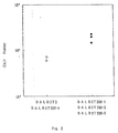
- the biological properties of the novel molecule were analyzed using a mouse pre-B cell line DW34 growing dependent on stromal cells with the number of grown cells as an index according to the following method.
- transduced cell lines BALB3T3 38-1, 38-3 and 38-9 and control cell lines BALB3T3 and BALB3T3 38-4 were cultured in 24-well plates until they became subconfluent, radiation of 30 Gy was irradiated thereupon, DW34 of 2 x 10 3 per well was added therein, and the resultant product was cultured in the RPMI-1640 (manufactured by GIBCO) medium containing 10 % FCS (manufactured by Bioproducts) under the conditions of 37 °C and 5 % CO 2 for 4 days.
- the number of viable cells of DW34 in each well was counted with trypan blue dye exclusion to analyze a growth-supporting ability.
- the growth of DW34 was enhanced in the transduced cell lines BALB3T3 38-1, 38-3 and 38-9 in comparison with control cell lines BALB3T3 and BALB3T3 38-4 as shown in Fig. 2.
- the present invention relates to a gene encoding a novel membrane protein polypeptide having pre-B cell growth-supporting ability, a vector containing said gene, transformants transformed by said vector, and a method for producing a novel membrane protein having pre-B cell growth-supporting ability using said gene, and therefore the gene of the present invention is capable of encoding the membrane protein on the synovial cell derived from patients with rheumatoid arthritis (RA).
- RA rheumatoid arthritis
- the homogeneous and purified novel membrane protein polypeptide can be produced in large quantities by inserting the gene of the present invention into a suitable vector and then transforming ordinary host cells, and in addition, monoclonal antibodies recognizing said membrane protein polypeptide can be produced by using the synovial cell derived from patients with rheumatoid arthritis (RA) as an antigen for immunization.
- RA rheumatoid arthritis
Landscapes
- Health & Medical Sciences (AREA)
- Chemical & Material Sciences (AREA)
- Life Sciences & Earth Sciences (AREA)
- Organic Chemistry (AREA)
- Medicinal Chemistry (AREA)
- Proteomics, Peptides & Aminoacids (AREA)
- Gastroenterology & Hepatology (AREA)
- Toxicology (AREA)
- Biochemistry (AREA)
- Biophysics (AREA)
- General Health & Medical Sciences (AREA)
- Genetics & Genomics (AREA)
- Zoology (AREA)
- Molecular Biology (AREA)
- Cell Biology (AREA)
- Immunology (AREA)
- Preparation Of Compounds By Using Micro-Organisms (AREA)
- Peptides Or Proteins (AREA)
- Micro-Organisms Or Cultivation Processes Thereof (AREA)
- Immobilizing And Processing Of Enzymes And Microorganisms (AREA)
- Investigating Or Analysing Materials By The Use Of Chemical Reactions (AREA)
- Separation Using Semi-Permeable Membranes (AREA)
- Investigating Or Analysing Biological Materials (AREA)
- Nitrogen And Oxygen Or Sulfur-Condensed Heterocyclic Ring Systems (AREA)
- Measuring Or Testing Involving Enzymes Or Micro-Organisms (AREA)
Priority Applications (2)
| Application Number | Priority Date | Filing Date | Title |
|---|---|---|---|
| EP05019755A EP1705185A3 (de) | 1993-10-15 | 1994-10-14 | Reagenzie für die Diagnose von rheumatischer Arthritis |
| EP07011422A EP1862474A1 (de) | 1993-10-15 | 1994-10-14 | Reagenz zur Diagnose rheumatoider Arthritis |
Applications Claiming Priority (3)
| Application Number | Priority Date | Filing Date | Title |
|---|---|---|---|
| JP28162293 | 1993-10-15 | ||
| JP28162293 | 1993-10-15 | ||
| EP94929665A EP0733643B1 (de) | 1993-10-15 | 1994-10-14 | Das polypeptid eines membran-proteins mit der funktion zur unterstützung des wachstums von prä-b zellen und sein gen |
Related Parent Applications (1)
| Application Number | Title | Priority Date | Filing Date |
|---|---|---|---|
| EP94929665A Division EP0733643B1 (de) | 1993-10-15 | 1994-10-14 | Das polypeptid eines membran-proteins mit der funktion zur unterstützung des wachstums von prä-b zellen und sein gen |
Related Child Applications (1)
| Application Number | Title | Priority Date | Filing Date |
|---|---|---|---|
| EP05019755A Division EP1705185A3 (de) | 1993-10-15 | 1994-10-14 | Reagenzie für die Diagnose von rheumatischer Arthritis |
Publications (2)
| Publication Number | Publication Date |
|---|---|
| EP1156061A1 true EP1156061A1 (de) | 2001-11-21 |
| EP1156061B1 EP1156061B1 (de) | 2006-05-17 |
Family
ID=17641696
Family Applications (4)
| Application Number | Title | Priority Date | Filing Date |
|---|---|---|---|
| EP07011422A Withdrawn EP1862474A1 (de) | 1993-10-15 | 1994-10-14 | Reagenz zur Diagnose rheumatoider Arthritis |
| EP01119504A Expired - Lifetime EP1156061B1 (de) | 1993-10-15 | 1994-10-14 | Reagenzien für die Diagnose von rheumatoiden Arthritis |
| EP05019755A Withdrawn EP1705185A3 (de) | 1993-10-15 | 1994-10-14 | Reagenzie für die Diagnose von rheumatischer Arthritis |
| EP94929665A Expired - Lifetime EP0733643B1 (de) | 1993-10-15 | 1994-10-14 | Das polypeptid eines membran-proteins mit der funktion zur unterstützung des wachstums von prä-b zellen und sein gen |
Family Applications Before (1)
| Application Number | Title | Priority Date | Filing Date |
|---|---|---|---|
| EP07011422A Withdrawn EP1862474A1 (de) | 1993-10-15 | 1994-10-14 | Reagenz zur Diagnose rheumatoider Arthritis |
Family Applications After (2)
| Application Number | Title | Priority Date | Filing Date |
|---|---|---|---|
| EP05019755A Withdrawn EP1705185A3 (de) | 1993-10-15 | 1994-10-14 | Reagenzie für die Diagnose von rheumatischer Arthritis |
| EP94929665A Expired - Lifetime EP0733643B1 (de) | 1993-10-15 | 1994-10-14 | Das polypeptid eines membran-proteins mit der funktion zur unterstützung des wachstums von prä-b zellen und sein gen |
Country Status (14)
| Country | Link |
|---|---|
| US (2) | US5914252A (de) |
| EP (4) | EP1862474A1 (de) |
| KR (1) | KR100260873B1 (de) |
| CN (2) | CN1239518C (de) |
| AT (2) | ATE326483T1 (de) |
| AU (1) | AU688801B2 (de) |
| DE (2) | DE69434741T2 (de) |
| DK (1) | DK0733643T3 (de) |
| ES (1) | ES2176260T3 (de) |
| HK (2) | HK1017364A1 (de) |
| PT (1) | PT733643E (de) |
| TW (2) | TW474991B (de) |
| WO (1) | WO1995010536A1 (de) |
| ZA (1) | ZA948016B (de) |
Families Citing this family (11)
| Publication number | Priority date | Publication date | Assignee | Title |
|---|---|---|---|---|
| ZA943348B (en) * | 1993-05-21 | 1995-11-16 | Hirano Toshio | A gene encoding a polypeptide having pre-B cell growth-supporting ability |
| US7049408B2 (en) * | 1993-10-15 | 2006-05-23 | Toshio Hirano | Antibody reactive with a protein having pre-B cell growth-supporting ability |
| ATE297219T1 (de) | 1997-02-12 | 2005-06-15 | Chugai Pharmaceutical Co Ltd | Antikörper als arzneimittel gegen lymphocytische tumore (ausschliesslich myelome) |
| CA2282631A1 (en) * | 1997-02-28 | 1998-09-03 | Chugai Seiyaku Kabushiki Kaisha | Lymphocyte activation inhibitors |
| EP1213028B1 (de) | 1999-08-23 | 2008-08-13 | Chugai Seiyaku Kabushiki Kaisha | Verstärker der hm1.24 antigen-expression |
| WO2001077362A1 (fr) * | 2000-04-06 | 2001-10-18 | Chugai Seiyaku Kabushiki Kaisha | Dosage immunologique d'anticorps anti hm1 . 24 |
| US7931897B2 (en) | 2001-02-07 | 2011-04-26 | Chugai Seiyaku Kabushiki Kaisha | Therapeutic agent for hematopoietic tumors |
| US20080219974A1 (en) * | 2002-03-01 | 2008-09-11 | Bernett Matthew J | Optimized antibodies that target hm1.24 |
| CA2570602A1 (en) | 2004-06-11 | 2006-01-26 | Ginkgo Biomedical Research Institute Co., Ltd. | Agents for regulating the activity of interferon-producing cells |
| WO2008036688A2 (en) * | 2006-09-18 | 2008-03-27 | Xencor, Inc. | Optimized antibodies that target hm1.24 |
| CN104031150A (zh) * | 2007-10-16 | 2014-09-10 | Sbi生物技术有限公司 | 抗bst2抗体 |
Citations (1)
| Publication number | Priority date | Publication date | Assignee | Title |
|---|---|---|---|---|
| EP0314415A2 (de) * | 1987-10-26 | 1989-05-03 | Immunex Corporation | Interleukin-7 |
Family Cites Families (2)
| Publication number | Priority date | Publication date | Assignee | Title |
|---|---|---|---|---|
| ZA943348B (en) * | 1993-05-21 | 1995-11-16 | Hirano Toshio | A gene encoding a polypeptide having pre-B cell growth-supporting ability |
| ATE297219T1 (de) * | 1997-02-12 | 2005-06-15 | Chugai Pharmaceutical Co Ltd | Antikörper als arzneimittel gegen lymphocytische tumore (ausschliesslich myelome) |
-
1994
- 1994-10-13 TW TW089127943A patent/TW474991B/zh not_active IP Right Cessation
- 1994-10-13 ZA ZA948016A patent/ZA948016B/xx unknown
- 1994-10-13 TW TW083109496A patent/TW442493B/zh not_active IP Right Cessation
- 1994-10-14 AT AT01119504T patent/ATE326483T1/de not_active IP Right Cessation
- 1994-10-14 CN CNB981083315A patent/CN1239518C/zh not_active Expired - Lifetime
- 1994-10-14 ES ES94929665T patent/ES2176260T3/es not_active Expired - Lifetime
- 1994-10-14 DK DK94929665T patent/DK0733643T3/da active
- 1994-10-14 DE DE69434741T patent/DE69434741T2/de not_active Expired - Fee Related
- 1994-10-14 AU AU78638/94A patent/AU688801B2/en not_active Expired
- 1994-10-14 EP EP07011422A patent/EP1862474A1/de not_active Withdrawn
- 1994-10-14 WO PCT/JP1994/001732 patent/WO1995010536A1/ja active IP Right Grant
- 1994-10-14 EP EP01119504A patent/EP1156061B1/de not_active Expired - Lifetime
- 1994-10-14 PT PT94929665T patent/PT733643E/pt unknown
- 1994-10-14 DE DE69431068T patent/DE69431068T2/de not_active Expired - Lifetime
- 1994-10-14 KR KR1019960701931A patent/KR100260873B1/ko active IP Right Grant
- 1994-10-14 US US08/624,650 patent/US5914252A/en not_active Expired - Lifetime
- 1994-10-14 AT AT94929665T patent/ATE221082T1/de active
- 1994-10-14 EP EP05019755A patent/EP1705185A3/de not_active Withdrawn
- 1994-10-14 CN CN94194257A patent/CN1056616C/zh not_active Expired - Lifetime
- 1994-10-14 EP EP94929665A patent/EP0733643B1/de not_active Expired - Lifetime
-
1999
- 1999-05-24 HK HK99102283A patent/HK1017364A1/xx not_active IP Right Cessation
-
2001
- 2001-03-28 US US09/818,648 patent/US6489126B2/en not_active Expired - Lifetime
-
2002
- 2002-02-07 HK HK02100949.0A patent/HK1039623A1/zh unknown
Patent Citations (1)
| Publication number | Priority date | Publication date | Assignee | Title |
|---|---|---|---|---|
| EP0314415A2 (de) * | 1987-10-26 | 1989-05-03 | Immunex Corporation | Interleukin-7 |
Non-Patent Citations (1)
| Title |
|---|
| GOODWIN RG ET AL: "Human interleukin 7: molecular cloning and *growth* factor activity on human and murine B-lineage cells.", PROC NATL ACAD SCI U S A, JAN 1989, 86 (1) P302-6, UNITED STATES, XP002077974 * |
Also Published As
| Publication number | Publication date |
|---|---|
| WO1995010536A1 (fr) | 1995-04-20 |
| CN1239518C (zh) | 2006-02-01 |
| EP0733643A1 (de) | 1996-09-25 |
| HK1039623A1 (zh) | 2002-05-03 |
| EP1705185A3 (de) | 2006-10-04 |
| PT733643E (pt) | 2002-11-29 |
| ZA948016B (en) | 1995-05-31 |
| EP0733643B1 (de) | 2002-07-24 |
| TW474991B (en) | 2002-02-01 |
| EP0733643A4 (de) | 1999-05-06 |
| US20020161190A1 (en) | 2002-10-31 |
| HK1017364A1 (en) | 1999-11-19 |
| ES2176260T3 (es) | 2002-12-01 |
| DE69431068T2 (de) | 2003-02-13 |
| KR100260873B1 (ko) | 2000-07-01 |
| US5914252A (en) | 1999-06-22 |
| ATE221082T1 (de) | 2002-08-15 |
| EP1862474A1 (de) | 2007-12-05 |
| ATE326483T1 (de) | 2006-06-15 |
| AU7863894A (en) | 1995-05-04 |
| CN1203921A (zh) | 1999-01-06 |
| AU688801B2 (en) | 1998-03-19 |
| DE69434741T2 (de) | 2007-04-26 |
| DE69431068D1 (de) | 2002-08-29 |
| EP1156061B1 (de) | 2006-05-17 |
| CN1135759A (zh) | 1996-11-13 |
| TW442493B (en) | 2001-06-23 |
| DK0733643T3 (da) | 2002-10-14 |
| US6489126B2 (en) | 2002-12-03 |
| EP1705185A2 (de) | 2006-09-27 |
| CN1056616C (zh) | 2000-09-20 |
| DE69434741D1 (de) | 2006-06-22 |
Similar Documents
| Publication | Publication Date | Title |
|---|---|---|
| RU2139934C1 (ru) | Гуманизированные антитела и их использование | |
| Ishikawa et al. | Molecular cloning and chromosomal mapping of a bone marrow stromal cell surface gene, BST2, that may be involved in pre-B-cell growth | |
| US5216132A (en) | Soluble t-cell antigen receptor chimeric antigens | |
| EA017303B1 (ru) | Модифицированные гуманизированные антитела против интерлейкина-18 и их применение | |
| WO1995021919A2 (en) | Protein having tpo activity | |
| EP1156061B1 (de) | Reagenzien für die Diagnose von rheumatoiden Arthritis | |
| CZ61796A3 (en) | Monoclonal antibody causing spotosis of myeloid cells by binding to myeloid cells, its fragments and hybridoma producing thereof | |
| KR19980066046A (ko) | 고역가의 CTLA4-Ig 융합단백질 | |
| US20060160992A1 (en) | Method of identifying cells expressing a protein having pre-B cell growth-supporting ability | |
| JPH0678772A (ja) | ヒトインターロイキン−5受容体のα鎖又はその一部、特にshIL5RαをコードするDNA及びそれを含むベクター、このベクターで形質転換された宿主細胞、ならびにその製造及び利用方法 | |
| US6232453B1 (en) | Gene encoding a polypeptide having pre-B cell growth-supporting ability | |
| CA2174081C (en) | Membrane protein polypeptide having function of supporting pre-b cell growth and gene therefor | |
| JP3412930B2 (ja) | プレb細胞増殖支持能を有する膜タンパクポリペプチド及びその遺伝子 | |
| JP3365859B2 (ja) | プレb細胞増殖支持能を有するポリペプチドをコードする遺伝子 | |
| AU1671895A (en) | Protein having tpo activity | |
| JP2000106880A (ja) | 新規なヒトb細胞表面タンパク質及びこれをコードするdna | |
| MXPA98006628A (en) | Gene of response of differentiation terminal honey honey noved |
Legal Events
| Date | Code | Title | Description |
|---|---|---|---|
| PUAI | Public reference made under article 153(3) epc to a published international application that has entered the european phase |
Free format text: ORIGINAL CODE: 0009012 |
|
| 17P | Request for examination filed |
Effective date: 20010827 |
|
| AC | Divisional application: reference to earlier application |
Ref document number: 733643 Country of ref document: EP |
|
| AK | Designated contracting states |
Kind code of ref document: A1 Designated state(s): AT BE CH DE DK ES FR GB GR IE IT LI LU MC NL PT SE |
|
| AKX | Designation fees paid |
Free format text: AT BE CH DE DK ES FR GB GR IE IT LI LU MC NL PT SE |
|
| 17Q | First examination report despatched |
Effective date: 20040622 |
|
| GRAP | Despatch of communication of intention to grant a patent |
Free format text: ORIGINAL CODE: EPIDOSNIGR1 |
|
| GRAS | Grant fee paid |
Free format text: ORIGINAL CODE: EPIDOSNIGR3 |
|
| GRAA | (expected) grant |
Free format text: ORIGINAL CODE: 0009210 |
|
| AC | Divisional application: reference to earlier application |
Ref document number: 0733643 Country of ref document: EP Kind code of ref document: P |
|
| AK | Designated contracting states |
Kind code of ref document: B1 Designated state(s): AT BE CH DE DK ES FR GB GR IE IT LI LU MC NL PT SE |
|
| PG25 | Lapsed in a contracting state [announced via postgrant information from national office to epo] |
Ref country code: IT Free format text: LAPSE BECAUSE OF FAILURE TO SUBMIT A TRANSLATION OF THE DESCRIPTION OR TO PAY THE FEE WITHIN THE PRESCRIBED TIME-LIMIT;WARNING: LAPSES OF ITALIAN PATENTS WITH EFFECTIVE DATE BEFORE 2007 MAY HAVE OCCURRED AT ANY TIME BEFORE 2007. THE CORRECT EFFECTIVE DATE MAY BE DIFFERENT FROM THE ONE RECORDED. Effective date: 20060517 Ref country code: BE Free format text: LAPSE BECAUSE OF FAILURE TO SUBMIT A TRANSLATION OF THE DESCRIPTION OR TO PAY THE FEE WITHIN THE PRESCRIBED TIME-LIMIT Effective date: 20060517 Ref country code: AT Free format text: LAPSE BECAUSE OF FAILURE TO SUBMIT A TRANSLATION OF THE DESCRIPTION OR TO PAY THE FEE WITHIN THE PRESCRIBED TIME-LIMIT Effective date: 20060517 Ref country code: CH Free format text: LAPSE BECAUSE OF FAILURE TO SUBMIT A TRANSLATION OF THE DESCRIPTION OR TO PAY THE FEE WITHIN THE PRESCRIBED TIME-LIMIT Effective date: 20060517 Ref country code: LI Free format text: LAPSE BECAUSE OF FAILURE TO SUBMIT A TRANSLATION OF THE DESCRIPTION OR TO PAY THE FEE WITHIN THE PRESCRIBED TIME-LIMIT Effective date: 20060517 Ref country code: NL Free format text: LAPSE BECAUSE OF FAILURE TO SUBMIT A TRANSLATION OF THE DESCRIPTION OR TO PAY THE FEE WITHIN THE PRESCRIBED TIME-LIMIT Effective date: 20060517 |
|
| REG | Reference to a national code |
Ref country code: GB Ref legal event code: FG4D |
|
| REG | Reference to a national code |
Ref country code: CH Ref legal event code: EP |
|
| REG | Reference to a national code |
Ref country code: IE Ref legal event code: FG4D |
|
| REF | Corresponds to: |
Ref document number: 69434741 Country of ref document: DE Date of ref document: 20060622 Kind code of ref document: P |
|
| PG25 | Lapsed in a contracting state [announced via postgrant information from national office to epo] |
Ref country code: SE Free format text: LAPSE BECAUSE OF FAILURE TO SUBMIT A TRANSLATION OF THE DESCRIPTION OR TO PAY THE FEE WITHIN THE PRESCRIBED TIME-LIMIT Effective date: 20060817 Ref country code: DK Free format text: LAPSE BECAUSE OF FAILURE TO SUBMIT A TRANSLATION OF THE DESCRIPTION OR TO PAY THE FEE WITHIN THE PRESCRIBED TIME-LIMIT Effective date: 20060817 |
|
| PG25 | Lapsed in a contracting state [announced via postgrant information from national office to epo] |
Ref country code: ES Free format text: LAPSE BECAUSE OF FAILURE TO SUBMIT A TRANSLATION OF THE DESCRIPTION OR TO PAY THE FEE WITHIN THE PRESCRIBED TIME-LIMIT Effective date: 20060828 |
|
| PG25 | Lapsed in a contracting state [announced via postgrant information from national office to epo] |
Ref country code: IE Free format text: LAPSE BECAUSE OF NON-PAYMENT OF DUE FEES Effective date: 20061016 |
|
| PG25 | Lapsed in a contracting state [announced via postgrant information from national office to epo] |
Ref country code: PT Free format text: LAPSE BECAUSE OF FAILURE TO SUBMIT A TRANSLATION OF THE DESCRIPTION OR TO PAY THE FEE WITHIN THE PRESCRIBED TIME-LIMIT Effective date: 20061017 |
|
| ET | Fr: translation filed | ||
| PG25 | Lapsed in a contracting state [announced via postgrant information from national office to epo] |
Ref country code: MC Free format text: LAPSE BECAUSE OF NON-PAYMENT OF DUE FEES Effective date: 20061031 |
|
| NLV1 | Nl: lapsed or annulled due to failure to fulfill the requirements of art. 29p and 29m of the patents act | ||
| REG | Reference to a national code |
Ref country code: CH Ref legal event code: PL |
|
| PLBE | No opposition filed within time limit |
Free format text: ORIGINAL CODE: 0009261 |
|
| STAA | Information on the status of an ep patent application or granted ep patent |
Free format text: STATUS: NO OPPOSITION FILED WITHIN TIME LIMIT |
|
| 26N | No opposition filed |
Effective date: 20070220 |
|
| PG25 | Lapsed in a contracting state [announced via postgrant information from national office to epo] |
Ref country code: GR Free format text: LAPSE BECAUSE OF FAILURE TO SUBMIT A TRANSLATION OF THE DESCRIPTION OR TO PAY THE FEE WITHIN THE PRESCRIBED TIME-LIMIT Effective date: 20060818 |
|
| REG | Reference to a national code |
Ref country code: HK Ref legal event code: WD Ref document number: 1039623 Country of ref document: HK |
|
| PG25 | Lapsed in a contracting state [announced via postgrant information from national office to epo] |
Ref country code: LU Free format text: LAPSE BECAUSE OF NON-PAYMENT OF DUE FEES Effective date: 20061014 |
|
| PGFP | Annual fee paid to national office [announced via postgrant information from national office to epo] |
Ref country code: DE Payment date: 20081014 Year of fee payment: 15 |
|
| PGFP | Annual fee paid to national office [announced via postgrant information from national office to epo] |
Ref country code: FR Payment date: 20081014 Year of fee payment: 15 |
|
| PGFP | Annual fee paid to national office [announced via postgrant information from national office to epo] |
Ref country code: GB Payment date: 20081008 Year of fee payment: 15 |
|
| REG | Reference to a national code |
Ref country code: FR Ref legal event code: ST Effective date: 20100630 |
|
| PG25 | Lapsed in a contracting state [announced via postgrant information from national office to epo] |
Ref country code: FR Free format text: LAPSE BECAUSE OF NON-PAYMENT OF DUE FEES Effective date: 20091102 Ref country code: DE Free format text: LAPSE BECAUSE OF NON-PAYMENT OF DUE FEES Effective date: 20100501 |
|
| PG25 | Lapsed in a contracting state [announced via postgrant information from national office to epo] |
Ref country code: GB Free format text: LAPSE BECAUSE OF NON-PAYMENT OF DUE FEES Effective date: 20091014 |