EP1015888B1 - Verfahren zur messung der assoziation von teilstrukturen pathologischer proteinablagerungen - Google Patents
Verfahren zur messung der assoziation von teilstrukturen pathologischer proteinablagerungen Download PDFInfo
- Publication number
- EP1015888B1 EP1015888B1 EP98948990A EP98948990A EP1015888B1 EP 1015888 B1 EP1015888 B1 EP 1015888B1 EP 98948990 A EP98948990 A EP 98948990A EP 98948990 A EP98948990 A EP 98948990A EP 1015888 B1 EP1015888 B1 EP 1015888B1
- Authority
- EP
- European Patent Office
- Prior art keywords
- pathological protein
- protein depositions
- pathological
- depositions
- substructures
- Prior art date
- Legal status (The legal status is an assumption and is not a legal conclusion. Google has not performed a legal analysis and makes no representation as to the accuracy of the status listed.)
- Expired - Lifetime
Links
- 102000004169 proteins and genes Human genes 0.000 title claims abstract description 115
- 108090000623 proteins and genes Proteins 0.000 title claims abstract description 115
- 230000001575 pathological effect Effects 0.000 title claims abstract description 90
- 238000000034 method Methods 0.000 title claims abstract description 49
- 239000000523 sample Substances 0.000 claims abstract description 80
- 230000008021 deposition Effects 0.000 claims abstract description 38
- 238000000151 deposition Methods 0.000 claims abstract description 38
- 238000001514 detection method Methods 0.000 claims abstract description 36
- 208000024827 Alzheimer disease Diseases 0.000 claims abstract description 31
- 208000037265 diseases, disorders, signs and symptoms Diseases 0.000 claims abstract description 26
- 201000010099 disease Diseases 0.000 claims abstract description 23
- 238000004220 aggregation Methods 0.000 claims abstract description 13
- 210000001124 body fluid Anatomy 0.000 claims abstract description 11
- 239000010839 body fluid Substances 0.000 claims abstract description 11
- 239000007791 liquid phase Substances 0.000 claims abstract description 6
- 238000002060 fluorescence correlation spectroscopy Methods 0.000 claims description 26
- 238000005259 measurement Methods 0.000 claims description 26
- 210000001175 cerebrospinal fluid Anatomy 0.000 claims description 23
- 208000037259 Amyloid Plaque Diseases 0.000 claims description 17
- 108090000765 processed proteins & peptides Proteins 0.000 claims description 16
- 238000009792 diffusion process Methods 0.000 claims description 12
- 206010002022 amyloidosis Diseases 0.000 claims description 11
- 210000001519 tissue Anatomy 0.000 claims description 10
- 239000002245 particle Substances 0.000 claims description 7
- 239000000126 substance Substances 0.000 claims description 7
- 238000011156 evaluation Methods 0.000 claims description 6
- 102000004196 processed proteins & peptides Human genes 0.000 claims description 6
- 230000001537 neural effect Effects 0.000 claims description 5
- 238000010521 absorption reaction Methods 0.000 claims description 4
- 230000004770 neurodegeneration Effects 0.000 claims description 4
- 208000015122 neurodegenerative disease Diseases 0.000 claims description 4
- 210000000056 organ Anatomy 0.000 claims description 4
- 208000022256 primary systemic amyloidosis Diseases 0.000 claims description 4
- 208000023105 Huntington disease Diseases 0.000 claims description 3
- 208000005531 Immunoglobulin Light-chain Amyloidosis Diseases 0.000 claims description 3
- 208000018737 Parkinson disease Diseases 0.000 claims description 3
- 108091005804 Peptidases Proteins 0.000 claims description 3
- 206010035226 Plasma cell myeloma Diseases 0.000 claims description 3
- 208000024777 Prion disease Diseases 0.000 claims description 3
- 239000004365 Protease Substances 0.000 claims description 3
- 102000007056 Recombinant Fusion Proteins Human genes 0.000 claims description 3
- 108010008281 Recombinant Fusion Proteins Proteins 0.000 claims description 3
- 102000015736 beta 2-Microglobulin Human genes 0.000 claims description 3
- 108010081355 beta 2-Microglobulin Proteins 0.000 claims description 3
- -1 cerebrospinal fluid Substances 0.000 claims description 3
- 239000003599 detergent Substances 0.000 claims description 3
- 238000001506 fluorescence spectroscopy Methods 0.000 claims description 3
- 239000012634 fragment Substances 0.000 claims description 3
- 238000000053 physical method Methods 0.000 claims description 3
- 230000028327 secretion Effects 0.000 claims description 3
- 208000001072 type 2 diabetes mellitus Diseases 0.000 claims description 3
- 208000023769 AA amyloidosis Diseases 0.000 claims description 2
- 208000023761 AL amyloidosis Diseases 0.000 claims description 2
- 102000004190 Enzymes Human genes 0.000 claims description 2
- 108090000790 Enzymes Proteins 0.000 claims description 2
- 102000035195 Peptidases Human genes 0.000 claims description 2
- 206010036790 Productive cough Diseases 0.000 claims description 2
- 210000004369 blood Anatomy 0.000 claims description 2
- 239000008280 blood Substances 0.000 claims description 2
- 230000003196 chaotropic effect Effects 0.000 claims description 2
- 238000002983 circular dichroism Methods 0.000 claims description 2
- 230000005281 excited state Effects 0.000 claims description 2
- 150000002500 ions Chemical class 0.000 claims description 2
- 238000004020 luminiscence type Methods 0.000 claims description 2
- 210000002751 lymph Anatomy 0.000 claims description 2
- 208000023356 medullary thyroid gland carcinoma Diseases 0.000 claims description 2
- 230000003287 optical effect Effects 0.000 claims description 2
- 230000010287 polarization Effects 0.000 claims description 2
- 238000006862 quantum yield reaction Methods 0.000 claims description 2
- 230000005855 radiation Effects 0.000 claims description 2
- 210000003802 sputum Anatomy 0.000 claims description 2
- 208000024794 sputum Diseases 0.000 claims description 2
- 208000013818 thyroid gland medullary carcinoma Diseases 0.000 claims description 2
- 238000012546 transfer Methods 0.000 claims description 2
- 210000002700 urine Anatomy 0.000 claims description 2
- 208000034846 Familial Amyloid Neuropathies Diseases 0.000 claims 1
- 208000007487 Familial Cerebral Amyloid Angiopathy Diseases 0.000 claims 1
- 206010019889 Hereditary neuropathic amyloidosis Diseases 0.000 claims 1
- 208000010544 human prion disease Diseases 0.000 claims 1
- 230000002981 neuropathic effect Effects 0.000 claims 1
- 238000004611 spectroscopical analysis Methods 0.000 claims 1
- 201000007905 transthyretin amyloidosis Diseases 0.000 claims 1
- 238000002525 ultrasonication Methods 0.000 claims 1
- 235000018102 proteins Nutrition 0.000 description 82
- 108091000054 Prion Proteins 0.000 description 50
- 102000029797 Prion Human genes 0.000 description 50
- 239000000243 solution Substances 0.000 description 46
- 239000011734 sodium Substances 0.000 description 25
- 239000008363 phosphate buffer Substances 0.000 description 24
- 208000020406 Creutzfeldt Jacob disease Diseases 0.000 description 22
- 208000010859 Creutzfeldt-Jakob disease Diseases 0.000 description 22
- 208000003407 Creutzfeldt-Jakob Syndrome Diseases 0.000 description 17
- 230000008569 process Effects 0.000 description 11
- 235000001014 amino acid Nutrition 0.000 description 10
- 238000002474 experimental method Methods 0.000 description 10
- 239000000463 material Substances 0.000 description 10
- 150000001413 amino acids Chemical group 0.000 description 9
- 238000013459 approach Methods 0.000 description 9
- 210000004556 brain Anatomy 0.000 description 9
- 208000018756 Variant Creutzfeldt-Jakob disease Diseases 0.000 description 8
- 229940024606 amino acid Drugs 0.000 description 8
- 208000005881 bovine spongiform encephalopathy Diseases 0.000 description 8
- 230000002776 aggregation Effects 0.000 description 6
- IQFVPQOLBLOTPF-HKXUKFGYSA-L congo red Chemical compound [Na+].[Na+].C1=CC=CC2=C(N)C(/N=N/C3=CC=C(C=C3)C3=CC=C(C=C3)/N=N/C3=C(C4=CC=CC=C4C(=C3)S([O-])(=O)=O)N)=CC(S([O-])(=O)=O)=C21 IQFVPQOLBLOTPF-HKXUKFGYSA-L 0.000 description 6
- 239000000284 extract Substances 0.000 description 6
- 239000007850 fluorescent dye Substances 0.000 description 6
- 208000015181 infectious disease Diseases 0.000 description 6
- 230000002458 infectious effect Effects 0.000 description 6
- 102000013455 Amyloid beta-Peptides Human genes 0.000 description 5
- 108010090849 Amyloid beta-Peptides Proteins 0.000 description 5
- 239000013543 active substance Substances 0.000 description 5
- 238000003745 diagnosis Methods 0.000 description 5
- 238000002372 labelling Methods 0.000 description 5
- 208000008864 scrapie Diseases 0.000 description 5
- 230000035945 sensitivity Effects 0.000 description 5
- 230000009885 systemic effect Effects 0.000 description 5
- 238000012360 testing method Methods 0.000 description 5
- KZSNJWFQEVHDMF-BYPYZUCNSA-N L-valine Chemical compound CC(C)[C@H](N)C(O)=O KZSNJWFQEVHDMF-BYPYZUCNSA-N 0.000 description 4
- KZSNJWFQEVHDMF-UHFFFAOYSA-N Valine Natural products CC(C)C(N)C(O)=O KZSNJWFQEVHDMF-UHFFFAOYSA-N 0.000 description 4
- 238000004458 analytical method Methods 0.000 description 4
- 238000006243 chemical reaction Methods 0.000 description 4
- 239000004474 valine Substances 0.000 description 4
- 102000001049 Amyloid Human genes 0.000 description 3
- 108010094108 Amyloid Proteins 0.000 description 3
- 239000004475 Arginine Substances 0.000 description 3
- DCXYFEDJOCDNAF-UHFFFAOYSA-N Asparagine Natural products OC(=O)C(N)CC(N)=O DCXYFEDJOCDNAF-UHFFFAOYSA-N 0.000 description 3
- DCXYFEDJOCDNAF-REOHCLBHSA-N L-asparagine Chemical compound OC(=O)[C@@H](N)CC(N)=O DCXYFEDJOCDNAF-REOHCLBHSA-N 0.000 description 3
- CKLJMWTZIZZHCS-REOHCLBHSA-N L-aspartic acid Chemical compound OC(=O)[C@@H](N)CC(O)=O CKLJMWTZIZZHCS-REOHCLBHSA-N 0.000 description 3
- WHUUTDBJXJRKMK-VKHMYHEASA-N L-glutamic acid Chemical compound OC(=O)[C@@H](N)CCC(O)=O WHUUTDBJXJRKMK-VKHMYHEASA-N 0.000 description 3
- AGPKZVBTJJNPAG-WHFBIAKZSA-N L-isoleucine Chemical compound CC[C@H](C)[C@H](N)C(O)=O AGPKZVBTJJNPAG-WHFBIAKZSA-N 0.000 description 3
- ROHFNLRQFUQHCH-UHFFFAOYSA-N Leucine Natural products CC(C)CC(N)C(O)=O ROHFNLRQFUQHCH-UHFFFAOYSA-N 0.000 description 3
- 239000004472 Lysine Substances 0.000 description 3
- KDXKERNSBIXSRK-UHFFFAOYSA-N Lysine Natural products NCCCCC(N)C(O)=O KDXKERNSBIXSRK-UHFFFAOYSA-N 0.000 description 3
- 241000699673 Mesocricetus auratus Species 0.000 description 3
- ODKSFYDXXFIFQN-UHFFFAOYSA-N arginine Natural products OC(=O)C(N)CCCNC(N)=N ODKSFYDXXFIFQN-UHFFFAOYSA-N 0.000 description 3
- 229960001230 asparagine Drugs 0.000 description 3
- 235000009582 asparagine Nutrition 0.000 description 3
- 229940009098 aspartate Drugs 0.000 description 3
- 238000009826 distribution Methods 0.000 description 3
- 239000000975 dye Substances 0.000 description 3
- 230000005284 excitation Effects 0.000 description 3
- 229930195712 glutamate Natural products 0.000 description 3
- ZDXPYRJPNDTMRX-UHFFFAOYSA-N glutamine Natural products OC(=O)C(N)CCC(N)=O ZDXPYRJPNDTMRX-UHFFFAOYSA-N 0.000 description 3
- 229960000310 isoleucine Drugs 0.000 description 3
- AGPKZVBTJJNPAG-UHFFFAOYSA-N isoleucine Natural products CCC(C)C(N)C(O)=O AGPKZVBTJJNPAG-UHFFFAOYSA-N 0.000 description 3
- 230000001717 pathogenic effect Effects 0.000 description 3
- 238000006116 polymerization reaction Methods 0.000 description 3
- 125000002924 primary amino group Chemical group [H]N([H])* 0.000 description 3
- 108010057077 prion protein (90-231) Proteins 0.000 description 3
- 238000012216 screening Methods 0.000 description 3
- 239000012064 sodium phosphate buffer Substances 0.000 description 3
- 102000013498 tau Proteins Human genes 0.000 description 3
- 108010026424 tau Proteins Proteins 0.000 description 3
- YBJHBAHKTGYVGT-ZKWXMUAHSA-N (+)-Biotin Chemical compound N1C(=O)N[C@@H]2[C@H](CCCCC(=O)O)SC[C@@H]21 YBJHBAHKTGYVGT-ZKWXMUAHSA-N 0.000 description 2
- 102000009091 Amyloidogenic Proteins Human genes 0.000 description 2
- 108010048112 Amyloidogenic Proteins Proteins 0.000 description 2
- DHMQDGOQFOQNFH-UHFFFAOYSA-N Glycine Chemical compound NCC(O)=O DHMQDGOQFOQNFH-UHFFFAOYSA-N 0.000 description 2
- QNAYBMKLOCPYGJ-REOHCLBHSA-N L-alanine Chemical compound C[C@H](N)C(O)=O QNAYBMKLOCPYGJ-REOHCLBHSA-N 0.000 description 2
- ROHFNLRQFUQHCH-YFKPBYRVSA-N L-leucine Chemical compound CC(C)C[C@H](N)C(O)=O ROHFNLRQFUQHCH-YFKPBYRVSA-N 0.000 description 2
- KDXKERNSBIXSRK-YFKPBYRVSA-N L-lysine Chemical compound NCCCC[C@H](N)C(O)=O KDXKERNSBIXSRK-YFKPBYRVSA-N 0.000 description 2
- MTCFGRXMJLQNBG-UHFFFAOYSA-N Serine Natural products OCC(N)C(O)=O MTCFGRXMJLQNBG-UHFFFAOYSA-N 0.000 description 2
- AYFVYJQAPQTCCC-UHFFFAOYSA-N Threonine Natural products CC(O)C(N)C(O)=O AYFVYJQAPQTCCC-UHFFFAOYSA-N 0.000 description 2
- 239000004473 Threonine Substances 0.000 description 2
- 235000004279 alanine Nutrition 0.000 description 2
- 244000052616 bacterial pathogen Species 0.000 description 2
- 230000015572 biosynthetic process Effects 0.000 description 2
- 210000005013 brain tissue Anatomy 0.000 description 2
- 239000003086 colorant Substances 0.000 description 2
- 238000005314 correlation function Methods 0.000 description 2
- 230000001419 dependent effect Effects 0.000 description 2
- 238000002405 diagnostic procedure Methods 0.000 description 2
- 230000000694 effects Effects 0.000 description 2
- 230000003619 fibrillary effect Effects 0.000 description 2
- 230000035557 fibrillogenesis Effects 0.000 description 2
- 238000001215 fluorescent labelling Methods 0.000 description 2
- 238000009434 installation Methods 0.000 description 2
- NOESYZHRGYRDHS-UHFFFAOYSA-N insulin Chemical compound N1C(=O)C(NC(=O)C(CCC(N)=O)NC(=O)C(CCC(O)=O)NC(=O)C(C(C)C)NC(=O)C(NC(=O)CN)C(C)CC)CSSCC(C(NC(CO)C(=O)NC(CC(C)C)C(=O)NC(CC=2C=CC(O)=CC=2)C(=O)NC(CCC(N)=O)C(=O)NC(CC(C)C)C(=O)NC(CCC(O)=O)C(=O)NC(CC(N)=O)C(=O)NC(CC=2C=CC(O)=CC=2)C(=O)NC(CSSCC(NC(=O)C(C(C)C)NC(=O)C(CC(C)C)NC(=O)C(CC=2C=CC(O)=CC=2)NC(=O)C(CC(C)C)NC(=O)C(C)NC(=O)C(CCC(O)=O)NC(=O)C(C(C)C)NC(=O)C(CC(C)C)NC(=O)C(CC=2NC=NC=2)NC(=O)C(CO)NC(=O)CNC2=O)C(=O)NCC(=O)NC(CCC(O)=O)C(=O)NC(CCCNC(N)=N)C(=O)NCC(=O)NC(CC=3C=CC=CC=3)C(=O)NC(CC=3C=CC=CC=3)C(=O)NC(CC=3C=CC(O)=CC=3)C(=O)NC(C(C)O)C(=O)N3C(CCC3)C(=O)NC(CCCCN)C(=O)NC(C)C(O)=O)C(=O)NC(CC(N)=O)C(O)=O)=O)NC(=O)C(C(C)CC)NC(=O)C(CO)NC(=O)C(C(C)O)NC(=O)C1CSSCC2NC(=O)C(CC(C)C)NC(=O)C(NC(=O)C(CCC(N)=O)NC(=O)C(CC(N)=O)NC(=O)C(NC(=O)C(N)CC=1C=CC=CC=1)C(C)C)CC1=CN=CN1 NOESYZHRGYRDHS-UHFFFAOYSA-N 0.000 description 2
- 238000009593 lumbar puncture Methods 0.000 description 2
- 238000011160 research Methods 0.000 description 2
- 238000011895 specific detection Methods 0.000 description 2
- 238000006467 substitution reaction Methods 0.000 description 2
- 238000009210 therapy by ultrasound Methods 0.000 description 2
- PSLCKQYQNVNTQI-BHFSHLQUSA-N (2s)-2-aminobutanedioic acid;(2s)-2-aminopentanedioic acid Chemical compound OC(=O)[C@@H](N)CC(O)=O.OC(=O)[C@@H](N)CCC(O)=O PSLCKQYQNVNTQI-BHFSHLQUSA-N 0.000 description 1
- UPUDVKWQBVIKBG-UHFFFAOYSA-N 1-[(3,4-diethoxyphenyl)methyl]-6,7-diethoxyisoquinolin-2-ium;chloride Chemical compound [Cl-].C1=C(OCC)C(OCC)=CC=C1CC1=[NH+]C=CC2=CC(OCC)=C(OCC)C=C12 UPUDVKWQBVIKBG-UHFFFAOYSA-N 0.000 description 1
- VGIRNWJSIRVFRT-UHFFFAOYSA-N 2',7'-difluorofluorescein Chemical compound OC(=O)C1=CC=CC=C1C1=C2C=C(F)C(=O)C=C2OC2=CC(O)=C(F)C=C21 VGIRNWJSIRVFRT-UHFFFAOYSA-N 0.000 description 1
- 208000003808 Amyloid Neuropathies Diseases 0.000 description 1
- 108090001008 Avidin Proteins 0.000 description 1
- 102000055006 Calcitonin Human genes 0.000 description 1
- 108060001064 Calcitonin Proteins 0.000 description 1
- 208000005145 Cerebral amyloid angiopathy Diseases 0.000 description 1
- 241000699800 Cricetinae Species 0.000 description 1
- 102000012192 Cystatin C Human genes 0.000 description 1
- 108010061642 Cystatin C Proteins 0.000 description 1
- 238000002965 ELISA Methods 0.000 description 1
- 102000004878 Gelsolin Human genes 0.000 description 1
- 108090001064 Gelsolin Proteins 0.000 description 1
- 239000004471 Glycine Substances 0.000 description 1
- 108060003951 Immunoglobulin Proteins 0.000 description 1
- 102000004877 Insulin Human genes 0.000 description 1
- 108090001061 Insulin Proteins 0.000 description 1
- 102000036770 Islet Amyloid Polypeptide Human genes 0.000 description 1
- 108010041872 Islet Amyloid Polypeptide Proteins 0.000 description 1
- ODKSFYDXXFIFQN-BYPYZUCNSA-P L-argininium(2+) Chemical compound NC(=[NH2+])NCCC[C@H]([NH3+])C(O)=O ODKSFYDXXFIFQN-BYPYZUCNSA-P 0.000 description 1
- HNDVDQJCIGZPNO-YFKPBYRVSA-N L-histidine Chemical compound OC(=O)[C@@H](N)CC1=CN=CN1 HNDVDQJCIGZPNO-YFKPBYRVSA-N 0.000 description 1
- FFEARJCKVFRZRR-BYPYZUCNSA-N L-methionine Chemical compound CSCC[C@H](N)C(O)=O FFEARJCKVFRZRR-BYPYZUCNSA-N 0.000 description 1
- 102000004895 Lipoproteins Human genes 0.000 description 1
- 108090001030 Lipoproteins Proteins 0.000 description 1
- 241001465754 Metazoa Species 0.000 description 1
- 102000016943 Muramidase Human genes 0.000 description 1
- 108010014251 Muramidase Proteins 0.000 description 1
- 108010062010 N-Acetylmuramoyl-L-alanine Amidase Proteins 0.000 description 1
- 229910019142 PO4 Inorganic materials 0.000 description 1
- 241001494479 Pecora Species 0.000 description 1
- 108010071690 Prealbumin Proteins 0.000 description 1
- 102100037486 Reverse transcriptase/ribonuclease H Human genes 0.000 description 1
- 102000019355 Synuclein Human genes 0.000 description 1
- 108050006783 Synuclein Proteins 0.000 description 1
- 102000009190 Transthyretin Human genes 0.000 description 1
- 241000700605 Viruses Species 0.000 description 1
- 239000004480 active ingredient Substances 0.000 description 1
- VREFGVBLTWBCJP-UHFFFAOYSA-N alprazolam Chemical compound C12=CC(Cl)=CC=C2N2C(C)=NN=C2CN=C1C1=CC=CC=C1 VREFGVBLTWBCJP-UHFFFAOYSA-N 0.000 description 1
- 230000003321 amplification Effects 0.000 description 1
- 238000001574 biopsy Methods 0.000 description 1
- 229960002685 biotin Drugs 0.000 description 1
- 235000020958 biotin Nutrition 0.000 description 1
- 239000011616 biotin Substances 0.000 description 1
- 239000000872 buffer Substances 0.000 description 1
- BBBFJLBPOGFECG-VJVYQDLKSA-N calcitonin Chemical compound N([C@H](C(=O)N[C@@H](CC(C)C)C(=O)NCC(=O)N[C@@H](CCCCN)C(=O)N[C@@H](CC(C)C)C(=O)N[C@@H](CO)C(=O)N[C@@H](CCC(N)=O)C(=O)N[C@@H](CCC(O)=O)C(=O)N[C@@H](CC(C)C)C(=O)N[C@@H](CC=1NC=NC=1)C(=O)N[C@@H](CCCCN)C(=O)N[C@@H](CC(C)C)C(=O)N[C@@H](CCC(N)=O)C(=O)N[C@@H]([C@@H](C)O)C(=O)N[C@@H](CC=1C=CC(O)=CC=1)C(=O)N1[C@@H](CCC1)C(=O)N[C@@H](CCCNC(N)=N)C(=O)N[C@@H]([C@@H](C)O)C(=O)N[C@@H](CC(N)=O)C(=O)N[C@@H]([C@@H](C)O)C(=O)NCC(=O)N[C@@H](CO)C(=O)NCC(=O)N[C@@H]([C@@H](C)O)C(=O)N1[C@@H](CCC1)C(N)=O)C(C)C)C(=O)[C@@H]1CSSC[C@H](N)C(=O)N[C@@H](CO)C(=O)N[C@@H](CC(N)=O)C(=O)N[C@@H](CC(C)C)C(=O)N[C@@H](CO)C(=O)N[C@@H]([C@@H](C)O)C(=O)N1 BBBFJLBPOGFECG-VJVYQDLKSA-N 0.000 description 1
- 229960004015 calcitonin Drugs 0.000 description 1
- 230000001364 causal effect Effects 0.000 description 1
- 210000004027 cell Anatomy 0.000 description 1
- 239000000084 colloidal system Substances 0.000 description 1
- 150000001875 compounds Chemical class 0.000 description 1
- 238000005520 cutting process Methods 0.000 description 1
- 238000011157 data evaluation Methods 0.000 description 1
- 230000007423 decrease Effects 0.000 description 1
- 206010012601 diabetes mellitus Diseases 0.000 description 1
- 238000011847 diagnostic investigation Methods 0.000 description 1
- 238000010586 diagram Methods 0.000 description 1
- 238000010790 dilution Methods 0.000 description 1
- 239000012895 dilution Substances 0.000 description 1
- 208000035475 disorder Diseases 0.000 description 1
- 238000010494 dissociation reaction Methods 0.000 description 1
- 230000005593 dissociations Effects 0.000 description 1
- 238000001035 drying Methods 0.000 description 1
- 230000007515 enzymatic degradation Effects 0.000 description 1
- 229940088598 enzyme Drugs 0.000 description 1
- 239000012530 fluid Substances 0.000 description 1
- 239000011521 glass Substances 0.000 description 1
- HNDVDQJCIGZPNO-UHFFFAOYSA-N histidine Natural products OC(=O)C(N)CC1=CN=CN1 HNDVDQJCIGZPNO-UHFFFAOYSA-N 0.000 description 1
- 230000001900 immune effect Effects 0.000 description 1
- 102000018358 immunoglobulin Human genes 0.000 description 1
- 238000010348 incorporation Methods 0.000 description 1
- 238000011534 incubation Methods 0.000 description 1
- 230000002401 inhibitory effect Effects 0.000 description 1
- 229940125396 insulin Drugs 0.000 description 1
- 238000011835 investigation Methods 0.000 description 1
- 206010023497 kuru Diseases 0.000 description 1
- 239000003446 ligand Substances 0.000 description 1
- 229960000274 lysozyme Drugs 0.000 description 1
- 239000004325 lysozyme Substances 0.000 description 1
- 235000010335 lysozyme Nutrition 0.000 description 1
- 239000003550 marker Substances 0.000 description 1
- 230000007246 mechanism Effects 0.000 description 1
- 230000001404 mediated effect Effects 0.000 description 1
- 229930182817 methionine Natural products 0.000 description 1
- 238000013508 migration Methods 0.000 description 1
- 230000005012 migration Effects 0.000 description 1
- 230000003278 mimic effect Effects 0.000 description 1
- 239000000203 mixture Substances 0.000 description 1
- 239000003068 molecular probe Substances 0.000 description 1
- 239000000178 monomer Substances 0.000 description 1
- 230000001722 neurochemical effect Effects 0.000 description 1
- 238000003199 nucleic acid amplification method Methods 0.000 description 1
- 210000000496 pancreas Anatomy 0.000 description 1
- 244000052769 pathogen Species 0.000 description 1
- 230000008506 pathogenesis Effects 0.000 description 1
- 230000007170 pathology Effects 0.000 description 1
- NBIIXXVUZAFLBC-UHFFFAOYSA-K phosphate Chemical compound [O-]P([O-])([O-])=O NBIIXXVUZAFLBC-UHFFFAOYSA-K 0.000 description 1
- 239000010452 phosphate Substances 0.000 description 1
- 229920000642 polymer Polymers 0.000 description 1
- 238000002360 preparation method Methods 0.000 description 1
- 238000011002 quantification Methods 0.000 description 1
- 230000035484 reaction time Effects 0.000 description 1
- 238000011084 recovery Methods 0.000 description 1
- 230000009467 reduction Effects 0.000 description 1
- 239000013074 reference sample Substances 0.000 description 1
- 230000010076 replication Effects 0.000 description 1
- 238000007790 scraping Methods 0.000 description 1
- 238000001338 self-assembly Methods 0.000 description 1
- 238000011896 sensitive detection Methods 0.000 description 1
- 238000000926 separation method Methods 0.000 description 1
- 210000002027 skeletal muscle Anatomy 0.000 description 1
- 229910000162 sodium phosphate Inorganic materials 0.000 description 1
- 239000001488 sodium phosphate Substances 0.000 description 1
- 239000008279 sol Substances 0.000 description 1
- 239000007787 solid Substances 0.000 description 1
- 238000000527 sonication Methods 0.000 description 1
- 241000894007 species Species 0.000 description 1
- 238000007447 staining method Methods 0.000 description 1
- 208000024891 symptom Diseases 0.000 description 1
- 210000001685 thyroid gland Anatomy 0.000 description 1
- 230000007704 transition Effects 0.000 description 1
- 238000013519 translation Methods 0.000 description 1
- RYFMWSXOAZQYPI-UHFFFAOYSA-K trisodium phosphate Chemical compound [Na+].[Na+].[Na+].[O-]P([O-])([O-])=O RYFMWSXOAZQYPI-UHFFFAOYSA-K 0.000 description 1
- 238000002604 ultrasonography Methods 0.000 description 1
Images


Classifications
-
- G—PHYSICS
- G01—MEASURING; TESTING
- G01N—INVESTIGATING OR ANALYSING MATERIALS BY DETERMINING THEIR CHEMICAL OR PHYSICAL PROPERTIES
- G01N33/00—Investigating or analysing materials by specific methods not covered by groups G01N1/00 - G01N31/00
- G01N33/48—Biological material, e.g. blood, urine; Haemocytometers
- G01N33/50—Chemical analysis of biological material, e.g. blood, urine; Testing involving biospecific ligand binding methods; Immunological testing
- G01N33/68—Chemical analysis of biological material, e.g. blood, urine; Testing involving biospecific ligand binding methods; Immunological testing involving proteins, peptides or amino acids
- G01N33/6893—Chemical analysis of biological material, e.g. blood, urine; Testing involving biospecific ligand binding methods; Immunological testing involving proteins, peptides or amino acids related to diseases not provided for elsewhere
- G01N33/6896—Neurological disorders, e.g. Alzheimer's disease
Definitions
- the present invention relates to a method for diagnostic Detection of pathological protein deposits related diseases.
- a number of different diseases are associated with the appearance of pathological protein deposits linked. It is often unclear whether the protein deposits are only one phenomenon Clinical picture or whether these protein deposits are causal are responsible for the disease itself as pathogens. So neurodegenerative diseases are known in which, for example in the brain of those affected as amyloid plaques designated protein deposits are detectable. To such Diseases include, for example, Alzheimer's disease, bovine spongiform encephalopathy (BSE), Creutzfeldt-Jakob disease (CJD), Kuru's disease, scrapie as well as possibly other diseases in the past were also referred to as "slow virus” diseases.
- BSE bovine spongiform encephalopathy
- CJD Creutzfeldt-Jakob disease
- Kuru's disease scrapie as well as possibly other diseases in the past were also referred to as "slow virus” diseases.
- Maggio describes in U.S. Pat. No. 5,434,050 a method for Diagnosis of Alzheimer's disease by adding Aß peptides on Aß aggregates, which as a solid, bound structure, z. B. as brain cutting material. This diagnostic method is, however, only practicable on the living patient would she undergo a serious medical intervention Obtaining brain biopsy material.
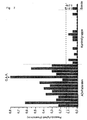
- Figure 1 shows a diagram of the inventive method for Diagnostics of pathological protein deposits.
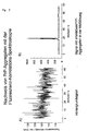
- Figure 2 shows the results of an experiment for detection of prion-protein aggregates using fluorescence correlation spectroscopy.
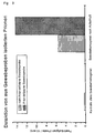
- Figure 3 shows the results of an experiment for detection isolated prions from tissue samples by fluorescence-labeled solubilized prion proteins (PrP-Cy2).
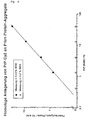
- Figure 4 shows the sensitivity of the homologous attachment of fluorescence-labeled solubilized prion proteins (PrP-Cy2) of prion protein aggregates from tissue samples and the influence of SDS concentration.
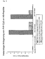
- FIG. 5 shows the homologous detection of peptide aggregates that the majority of the amyloid deposits in Alzheimer's Represent disease with fluorescently labeled soluble Aß (1-42) peptide.
- Figure 6 shows the results of a heterologous experiment Detection of peptide aggregates, the majority of amyloids Depict deposits in Alzheimer's disease with fluorescence-labeled solubilized prion protein (PrP-Cy2).
- Figure 7 shows the results of an experiment on the specific Detection of aggregates in the cerebrospinal fluid of patients in whom the Alzheimer's disease was diagnosed.
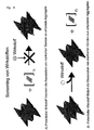
- FIG. 8 shows a scheme for screening active substances.
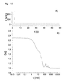
- Figure 9 shows the cross-correlation function of fluorescence-labeled Prion protein (90-231).
- Figure 10 shows the attachment of a recombinant probe Prion protein and a monoclonal antibody to a prion protein aggregate.
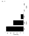
- FIG. 11 shows the frequency distribution of the fluorescence photons for CJD-positive (left) and CJD-negative (right) CSF samples.
- Figure 12 shows the frequency of fluorescence photons Channels with more than 150 counts / channel in CJD-positive and CJD negative CSF samples.
- Figure 13 shows the influence of Congorot on the deposition of the specific probe Aßl-42CY2.
- the method according to the invention for the diagnostic detection of Diseases includes measuring the association of substructures of pathological protein deposits, pathological Structures forming protein deposits, pathological protein deposits corresponding structures and / or pathological Protein deposits as a probe, on substructures of the pathological Protein deposition, pathological protein deposition forming structures, pathological protein deposits corresponding Structures and / or pathological protein deposits as targets.
- the inventive method is characterized in that the target is detected in the liquid phase, whereby in the case a detection of Alzheimer's disease the liquid phase was obtained from body fluids or a body fluid is.
- the association of the probe with the target is measured before self-aggregation of the probe predominates.
- the association of the probe to the target occurs opposite the Self-aggregation of the probe in the background can be a safe measurement can no longer be guaranteed.
- measurement times in the minute and hour range are possible, which makes the method seem sufficient to be used in routine laboratories to be applied.
- the times when Measurements are naturally dependent on the respective Measurement conditions, but are relative in preliminary tests easy to determine.
- the measurement time in particular can be influenced are concentrations of the target / probe to call. For example, are the probe concentrations right self-aggregation becomes high compared to the target concentration use the probes earlier than would be the case if the probes are present in low concentration or even the probe concentration is of a similar order of magnitude as that Target concentration or below.
- Measurement times of less than one hour are preferred, in particular less than 30 minutes, preferred. These measuring times are advantageous for executing the diagnosis in routine operation.
- the probe and / or the target preferably have a detectable property.
- the partial structures of the pathological protein deposits are preferred monomeric or oligomeric units of pathological Protein deposits.
- the substructures can also be homologous, in particular structurally homologous to monomers or oligomers Units of pathological protein deposits.
- the probe can be the same (homologous detection) or one another type (heterologous detection) of pathological protein deposits originate, which is used as a target. So can for example, as a target a pathological protein deposits forming structure, corresponding to a pathological protein deposits Structure and / or pathological protein deposits itself that comes from a prion disorder like scrapie or BSE originate, are used, whereas the probe, for example also come from structures other than those mentioned can. For example, it is possible to associate the Substructures (probe) made from amyloid deposits from BSE stem from measuring protein deposits associated with Alzheimer's Disease related.
- the substructures that are used according to the invention can preferably by treating pathological protein deposits with physical methods like ultrasound treatment, Exposure to temperature changes, chemical Methods such as treatment with solutions of different ionic strength, Treatment with solutions of chaotropic ions, treatment with detergents and / or enzymes, especially proteases, be preserved. So it is possible, for example, from on Scrapie diseased neuronal tissue through amyloid plaques enzymatic degradation followed by ultrasound treatment in Presence of detergents to break down into substructures that can subsequently build up fibrillar structures again.
- the Substructures of the pathological protein deposits can also be recombinant proteins, protein fragments or peptides, which sequence homologies to the corresponding amyloid plaques can have different origins and types. As a probe there are also pathological protein deposits corresponding Structures into consideration. Such structures mimic areas of actual pathological protein deposits and occur interactively with the targets.
- pathological protein deposits As targets on which the association of probes is measured, come structures forming pathological protein deposits into consideration. This means structures that are themselves not yet the actual pathological protein deposits represent, but monomeric or oligomeric units of pathological protein deposits or larger aggregates of the Substructures of the pathological protein deposits, their Association should be measured. As alternative the pathological protein deposits themselves come in Consideration.
- the probe and / or the target preferably have a detectable one Property on.
- the detectable property is either intrinsically in the aforementioned structures or Protein deposits, especially in the partial structure or can be introduced later.
- the at least one detectable property is particularly by physical Methods, preferably recorded spectroscopically.
- detectable Property of the aforementioned structures or protein deposits partial structure in particular comes for example Size or dimension. Also features like Structure, measurable with circular dichroism, optical properties such as luminescence, fluorescence or absorption used to measure the association of the probes with the targets become. For example, the intrinsic fluorescence of Structures as well as structures marked later fluorescent properties are used.
- the diagnostic Others can capture, interacting with the targets kicking substances are used.
- substances that have an affinity for the targets for example antibodies or avidin / biotin with the Allow targets to interact.
- FCS Fluorescence correlation spectroscopy
- FCS Fluorescence correlation spectroscopy
- the detectable properties are, for example, by Fluorescence markings, the low molecular, but also high molecular Groups can be made.
- labeled antibodies bound to the targets mark the latter.
- the binding of probes can then be measured accordingly. It can be of different colors Markings of different probes are made and a cross correlation is carried out on these. It is it is also possible to label the probes with appropriate labels, the either low molecular weight fluorescent ligands and / or corresponding labeling conjugates.
- the pathological protein deposits preferably originate from amyloid plaques which are associated with neurodegenerative diseases such as Alzheimer's disease, bovine spongiform encephalopathy (BSE), Creutzfeldt-Jakob disease, scrapie (scraping disease), Huntington's disease, Parkinson's disease or other amyloid plaques Organs as the neural system, such as protein deposits associated with diabetes.
- a suitable source for the pathological protein deposits are organ extracts, preferably brain extracts from diseased animals. These can be Syrian golden hamsters infected with prions, for example.
- Amyloid disease Occurrence Amyloid-forming protein Alzheimer's disease neurally Amyloid- ⁇ -protein Transmissible spongiform encephalopathies neurally Prion protein Chorea huntington neurally huntingtin Parkinson's disease neurally synuclein Inheritable Cebral Amyloid Angiophatia neurally Cystatin C.
- a amyloidosis Primary aystemic amyloidosis (AL amyloidosis) systemic immunoglobulin Reactive aystemic amyloidosis (AA amyloidosis) systemic lipoproteins Family Amyloid Polyneuropathy systemic transthyretin Type II diabetes pancreas Islet Amyloid Polypeptide Injection-localized amyloidosis insulin Medullary carcinoma of the thyroid gland thyroid calcitonin Beta-2 microglobulin amyloidosis skeletal muscle Beta-2 microglobulin Inherited non-European amyloidosis systemic lysozyme Finnish inherited systemic amyloidosis systemic gelsolin
- the substructures of the pathological protein deposits can can also be recombinant proteins, protein fragments or peptides, which sequence homologies have to amyloid plaques. So can, for example, conservative amino acid substitutions in highly conserved regions are taken into account as follows: each Isoleucine, valine and leucine amino acids can be used against another of these amino acids can be exchanged, aspartate can be replaced by glutamate and vice versa, glutamine for asparagine and vice versa, serine against threonine and vice versa.
- each of the amino acids isoleucine, valine and leucine can be used against any other of these amino acids, aspartate against glutamate and vice versa, glutamine against asparagine and vice versa, Serine against threonine and vice versa, glycine against alanine and vice versa, alanine against valine and vice versa, methionine against each of the amino acids leucine, isoleucine or valine, lysine against Arginine and vice versa, one of the amino acids or aspartate Glutamate against one of the amino acids arginine or lysine, histidine against one of the amino acids arginine or lysine, against glutamine Glutamate and vice versa and asparagine versus aspartate and vice versa, be exchanged.
- body fluids such as cerebrospinal fluid, Blood, lymph, urine, or secretions like sputum in question.
- body fluids or secretions become samples taken and used to measure the association of potentially in pathological protein deposits contained in these samples forming structures, pathological protein deposits corresponding Structures and / or pathological protein deposits, contacted with probes and incubated.
- pathological protein deposits that are indicative for an associated disease then results in positive diagnostic signal mediated by the association the added probes.
- Body fluids are opposite Tissues are advantageous because they are incubated directly can, whereas tissue regularly only unlocked Need to become. This can be done for example by mechanical or chemical treatment or combinations thereof.
- substructures are determined, especially translational diffusion velocities, Rotational diffusion rates, Lifetimes of excited states, polarization of radiation, Energy transfer, quantum yield, particle number or concentration, Differences in intensity into consideration. Becomes the translation diffusion rate determined, it can be at slowly diffusing associates may be desirable this to be recorded multiple times using a scanning process.
- the combination of the germ-induced attachment a variety of labeled probes to serve as germs pathological protein deposits with the extremely sensitive Detection methods using confocal optics result in a corresponding large amplification of the measurement signal.
- Im through lumbar puncture (Neuffle et al., Clinical Guide to Laboratory Diagnostics, Liquor obtained by Fischer-Verlag 1998) was able to produce individual Molecules of pathological protein deposits clearly can be detected.
- a screening process for the detection of active substances Treatment of pathological protein deposits Diseases analogous to the method according to the invention is assumes that in the presence of suspected active substances the association of substructures of the pathological protein deposits, pathological protein deposits Structures corresponding to pathological protein deposits Structures and / or pathological protein deposits Substructures of pathological protein deposition, pathological Structures forming protein deposits, pathological Structures corresponding to protein deposits and / or pathological Protein deposits are measured as targets.
- An alternative is first an association of substructures of pathological protein deposits, pathological protein deposits forming structures, pathological protein deposits corresponding structures and / or pathological Protein deposits, on substructures of the pathological Protein deposition, pathological protein deposition Structures corresponding to pathological protein deposits Structures and / or pathological protein deposits as Targets carried out and then reversing the association observed under the influence of suspected active substances. As Compounds are then identified in the active ingredient Are able to reverse the association in a significant way to effect.
- a third alternative is that the dissociation of protein deposits even under the influence of suspected active substances is measured.
- Soluble Aß (1-42) could in 10 mM Na phosphate buffer, pH 7.0 be stabilized with 0.2% SDS (Example 4, 6).
- SDS 0.2% SDS
- the soluble, monomeric bis oligomeric fluorescence-labeled proteins or peptides under Thinning of the SDS to 0.02% SDS with amyloid aggregates (Infectious prion particles: Examples 1 to 3; Aß: Examples 4 to 5) and the fluorescence signal by means of fluorescence correlation spectroscopy detected.
- FCS fluorescence correlation spectroscopy
- PrP infectious prion particles
- FIG. 2 shows the result of the measurement of the fluorescence intensity (in relative units / RE) over a period of 60 seconds.
- the fluctuation signal of the solubilized one can be seen in FIG. 2a PrP-Cy2, which has a signal intensity between 110 and 130 RE having.
- An evaluation of the signal using autocorrelation gives a diffusion time of approximately 250 ⁇ s, which is typical corresponds to the monomeric protein.
- Figure 2b is additional a signal peak with a fluorescence intensity of approx. 950 RE recognizable by the slow diffusion of a larger one Aggregates to which the previously solubilized fluorescent label PrP-Cy2 is attached, is caused. Since it is is only a one-off event is a determination of Diffusion time or molecular weight using autocorrelation not possible. A clear signal detection and assignment for individual aggregates, however, the occurrence is successful isolated peaks with drastic increase in fluorescence intensity.
- FIG. 3 shows the peak frequency for the individual approaches are plotted.
- the number of Peaks caused by diffusion of high molecular aggregates by the confocal volume element see example 1. It can be seen that the prion-infected tissue sample is a significantly higher peak frequency compared to the sample with not infected brain extract.
- the amount of PrP rods in the test approach was gradual lowered to the detection limit.
- the measurement in 0.2% SDS serves as a control.
- the peak frequency expressed in peaks per 10 min measuring time, was in Figure 4 depending on the amount of PrP in the test batch applied. Over the measured concentration range of 40 ng to 400 pg is a direct linear dependence on the peak frequency to recognize. The evaluation of the number of peaks leaves thereby a direct quantification of the prion concentration in the sample to be measured. In 0.2% SDS there is no installation in existing PrP structures possible.
- soluble Aß (1-42) -Cy2 was used in the same concentration measured.
- the results of the experiment are summarized graphically in FIG.
- the evaluation of the peak frequency (cf. 1 - 3) shows that when incubated in 0.01% SDS a label of Aß fibrils (SDS concentration identical to that of PrP aggregates) Main components of the amyloid deposits in Alzheimer's Represent disease, is possible with this procedure. Incorporation of Aß-Cy2 (1-42) in the presence of an increased SDS concentration (0.1%) is not possible. Also a marker of Aß, which is in a soluble (non-fibrillary) structure, can not. The experiment shows that detection of amyloid deposits associated with Alzheimer's Disease is possible.
- Aß peptide (1-42) A) 1 ⁇ l solution 1 (1:10) B) 2.5 ⁇ l solution 2 16.5 ⁇ l Na phosphate buffer, pH 7.0 1 ⁇ l solution 1 (1:10) 2.5 ⁇ l solution 3 16.5 ⁇ l Na phosphate buffer, pH 7.0 C) 1 ⁇ l solution 1 (1:10) 2.5 ⁇ l solution 2 16.5 ⁇ l Na phosphate buffer, pH 7.0, 0.2% SDS
- soluble PrP-Cy2 was used in the same concentration measured at 0.01% SDS.
- the peak frequency was (see Example 2) measured ( Figure 6). It shows that both Aß (1-42) and Aß (1-40) clearly labeled with PrP-Cy2 if they have a fibrillary structure, and the labeling reaction takes place at 0.01% SDS. When attached on non-fibrillar structures and increased SDS concentration only a small background signal can be seen.
- liquor spinal fluid used as a sample in which specifically between patients, who suffer from Alzheimer's disease and a healthy one Control group can be distinguished.
- the CSF samples were taken directly and without further treatment or Concentration with fluorescence-labeled Aß1-42-Cy2 (0.1 ng / ⁇ l) at 0.02% SDS and immediately for 20 min Measure FCS (Fig. 7).
- the detected aggregates in CSF samples from a psychiatric diagnostic point of view of Alzheimer's dementia (AD) patients and not sick control patients were compared. It shows that all patients suffering from Alzheimer's dementia have a significantly higher signal intensity in the cerebrospinal fluid, than the corresponding control group, which has a similar age composition had.
- the patient designated CAA shows the highest signal intensity of all diagnosed patients on.
- This patient has congophilic angiopathy, a special form of Alzheimer's disease, in which one Deposition of amyloid- ⁇ -peptides in the capillaries of the brain vessels takes place, the migration of which is gradually destroyed becomes.
- the most massive transition from Ate aggregates take place in the cerebrospinal fluid, leading to the strong Leads to an increase in signal intensity.
- the measurement results of the Control group show that here also aggregates can be detected.
- This result also shows the probe ("probe") alone (Aß 1-42-Cy2) and is based on self-aggregation effects during the 20-minute measurement period or on existing ones Aß aggregates.
- the measurement results of the probe were in 0.01% or 0.2% SDS achieved.
- Recombinant prion protein (90-231) (hereinafter called rPrP), that is homologous to the protease-resistant part of Hamster PrP, was made with amino reactive fluorescent dyes of the excitation wavelengths 488 nm (Oregon Green, Molecular Probes) and 633 nm (Cy5, Amersham). Protein concentrations and labeling ratio were measured by absorption measurement at 280 nm, 496 nm and 650 nm determined. The marking efficiency was included 10% (rPrP-Cy5) or 4% (rPrp-OrG) with equimolar addition of amino reactive fluorescent dye.
- the aggregation process was achieved by repeated inclusion of Auto and cross correlation curves in a two-color cross correlation FCS device (ConfoCor prototype, Zeiss) tracked.
- the Correlation curves were obtained using a three-dimensional diffusion model modeled, whereby for the cross correlation curves a diffusion parameter ⁇ rg was used. It was with one Fitted model for spherical molecules.
- Figure 9 shows the plot of the cross-correlation amplitude in the course of the aggregation (squares).
- a i + B j -> AB ij , k 5 ⁇ 10 6 M -1 ,
- Preaggregated rPrP was used for the aggregation approach of Example 7 added in concentrations of 0.2 to 20 ng / ⁇ l.
- the process of attachment was through consecutive Recording of auto and cross correlation curves followed.
- the monoclonal antibody IgM 15b3 was analogous to the Prp probes amino functional labeled with fluorescent dye.
- the marked Antibody was used at a final concentration of 20 nM.
- the entry of a marked aggregate into focus generates a fluorescent peak. Dominates with the right size the peak signal the cross correlation curve.
- Figure 10 shows the attachment of rPrP probe and mAB to a rPrP aggregate.
- Time trace of the cross-correlation fluorescence signal is included in Figure 10 .
- two-color cross-correlation curve is indicated by the large aggregate at the beginning of the Track dominates.
- One application is the detection of PrP aggregates in the cerebrospinal fluid of CJD patients. There were three CJD-negative and three CJD-positive CSF samples examined. About the number of events With large fluorescence bursts, CJD-positive and CJD-negative samples can be differentiated.
- the samples were measured in a ConfoCor FCS setup.
- the excitation took place at a wavelength of 488 nm (Ar + laser) with a power of 5 * 104 W / cm 2 .
- the emitted fluorescent light was collected through a microscope objective (40x /1.2 NA, Zeiss) and imaged confocally on an avalanche photodiode. This acts as a photon counter that supplies a signal pulse for each photon.
- the signal pulses were evaluated in parallel on a hardware correlator card using a signal divider and recorded on a scaler card with a channel width of 250 ⁇ s.
- the total measurement time was 15 minutes, which corresponds to 3.6 million channels.
- the evaluation was based on the number and amount the fluorescence peaks. A distribution of the number of photons per channel is shown in a histogram.
- the number of photons gives the height of the peaks, while the number of channels is a measure of the integral over the area of all peaks. A higher proportion of large samples can be seen in the CJD positive samples Fluorescence peaks. By setting a threshold you can clearly differentiate between CJD positive and negative samples.
- Figure 12 shows the frequency of fluorescence photons from channels with more than 150 counts / channel in CJD-positive (CJD +) ./ CJD-negative (CJD) CSF samples with a channel width of 250 ⁇ s and a concentration of the probe rPrP-Cy2 of 20 nM.
- p315 etc. is an internal patient identification number.
- Congo red is known for its inhibitory effect on the Fibrillogenesis.
- Figure 13 shows the influence of Congo red on the attachment of specific probe Aß-42-CY2 on synthetic Aßl-42 aggregates.
- FCS a drop in the Signal intensity of the homologous attachment to 20% was observed become.
Landscapes
- Life Sciences & Earth Sciences (AREA)
- Health & Medical Sciences (AREA)
- Engineering & Computer Science (AREA)
- Biomedical Technology (AREA)
- Hematology (AREA)
- Chemical & Material Sciences (AREA)
- Urology & Nephrology (AREA)
- Molecular Biology (AREA)
- Immunology (AREA)
- Proteomics, Peptides & Aminoacids (AREA)
- Analytical Chemistry (AREA)
- Microbiology (AREA)
- Biotechnology (AREA)
- Neurosurgery (AREA)
- Neurology (AREA)
- Food Science & Technology (AREA)
- Medicinal Chemistry (AREA)
- Physics & Mathematics (AREA)
- Cell Biology (AREA)
- Biochemistry (AREA)
- General Health & Medical Sciences (AREA)
- General Physics & Mathematics (AREA)
- Pathology (AREA)
- Investigating Or Analysing Biological Materials (AREA)
- Investigating, Analyzing Materials By Fluorescence Or Luminescence (AREA)
- Investigating Or Analysing Materials By The Use Of Chemical Reactions (AREA)
Applications Claiming Priority (7)
| Application Number | Priority Date | Filing Date | Title |
|---|---|---|---|
| DE19752712 | 1907-11-28 | ||
| DE19741486 | 1997-09-19 | ||
| DE19741486 | 1997-09-19 | ||
| DE1997152712 DE19752712C2 (de) | 1997-11-28 | 1997-11-28 | Verfahren zur Messung der Assoziation von Teilstrukturen pathologischer Proteinablagerungen |
| DE19818917 | 1998-04-28 | ||
| DE19818917 | 1998-04-28 | ||
| PCT/EP1998/005958 WO1999015903A1 (de) | 1997-09-19 | 1998-09-18 | Verfahren zur messung der assoziation von teilstrukturen pathologischer proteinablagerungen |
Publications (2)
| Publication Number | Publication Date |
|---|---|
| EP1015888A1 EP1015888A1 (de) | 2000-07-05 |
| EP1015888B1 true EP1015888B1 (de) | 2002-08-28 |
Family
ID=27217757
Family Applications (1)
| Application Number | Title | Priority Date | Filing Date |
|---|---|---|---|
| EP98948990A Expired - Lifetime EP1015888B1 (de) | 1997-09-19 | 1998-09-18 | Verfahren zur messung der assoziation von teilstrukturen pathologischer proteinablagerungen |
Country Status (7)
| Country | Link |
|---|---|
| US (1) | US6498017B2 (enExample) |
| EP (1) | EP1015888B1 (enExample) |
| JP (1) | JP4223681B2 (enExample) |
| AT (1) | ATE223058T1 (enExample) |
| AU (1) | AU9541398A (enExample) |
| DE (1) | DE59805338D1 (enExample) |
| WO (1) | WO1999015903A1 (enExample) |
Families Citing this family (45)
| Publication number | Priority date | Publication date | Assignee | Title |
|---|---|---|---|---|
| WO2001023894A1 (de) * | 1999-09-28 | 2001-04-05 | Evotec Oai Ag | Quantitative analyse und typisierung subzellulärer partikel |
| DE19952955A1 (de) | 1999-11-03 | 2001-05-17 | Acgt Progenomics Ag | Verfahren zur Charakterisierung und zur Auftrennung von molekularen Assoziaten |
| DE10013854A1 (de) * | 2000-03-17 | 2001-09-20 | Zeiss Carl Jena Gmbh | Anordnung und Verfahren zur Untersuchung von Wirkstoffen zur Beeinflussung intrazellulärer Vorgänge |
| WO2003090599A2 (en) * | 2002-04-25 | 2003-11-06 | Brainsgate Ltd. | Methods and apparatus for modifying properties of the bbb and cerebral circulation by using the neuroexcitatory and/or neuroinhibitory effects of odorants on nerves in the head |
| US7640062B2 (en) | 2000-05-08 | 2009-12-29 | Brainsgate Ltd. | Methods and systems for management of alzheimer's disease |
| US7146209B2 (en) * | 2000-05-08 | 2006-12-05 | Brainsgate, Ltd. | Stimulation for treating eye pathologies |
| US6603546B1 (en) * | 2000-07-21 | 2003-08-05 | I.S.S. (Usa) Inc. | Rapid high throughput spectrometer and method |
| US20050026165A1 (en) * | 2001-05-31 | 2005-02-03 | Cindy Orser | Detection of conformationally altered proteins and prions |
| MXPA03011000A (es) | 2001-05-31 | 2004-02-27 | Arete Associates | Metodo de sensor de proteinas deformadas. |
| EP1321771A1 (de) * | 2001-12-21 | 2003-06-25 | Evotec Analytical Systems GmbH | Verfahren zur qualitativen und/oder quantitativen Bestimmung von Aggregaten |
| EP1572937B1 (en) * | 2002-04-09 | 2012-02-08 | The Scripps Research Institute | Motif-grafted hybrid polypeptides and uses thereof |
| WO2004043218A2 (en) * | 2002-11-14 | 2004-05-27 | Brainsgate Ltd. | Surgical tools and techniques for stimulation |
| US7684859B2 (en) * | 2002-04-25 | 2010-03-23 | Brainsgate Ltd. | Stimulation of the OTIC ganglion for treating medical conditions |
| WO2005008212A2 (en) * | 2003-06-20 | 2005-01-27 | The Regents Of The University Of California | Modulated excitation fluorescence analysis |
| US20060035242A1 (en) * | 2004-08-13 | 2006-02-16 | Michelitsch Melissa D | Prion-specific peptide reagents |
| EP1600774A1 (de) * | 2004-03-12 | 2005-11-30 | Ludwig-Maximilians-Universität München | Verfahren zur Identifizierung von Substanzen zur Beeinflussung der Aggregationen von Proteinen |
| US9233245B2 (en) | 2004-02-20 | 2016-01-12 | Brainsgate Ltd. | SPG stimulation |
| US8055347B2 (en) | 2005-08-19 | 2011-11-08 | Brainsgate Ltd. | Stimulation for treating brain events and other conditions |
| US8010189B2 (en) * | 2004-02-20 | 2011-08-30 | Brainsgate Ltd. | SPG stimulation for treating complications of subarachnoid hemorrhage |
| JP4338590B2 (ja) | 2004-06-03 | 2009-10-07 | 独立行政法人科学技術振興機構 | 蛍光相関分光法による抗原の迅速検出及び/又は測定法。 |
| US20090299418A1 (en) * | 2004-08-23 | 2009-12-03 | Brainsgate Ltd. | Concurrent bilateral spg modulation |
| US20060057671A1 (en) * | 2004-09-10 | 2006-03-16 | Orser Cindy S | Immobilized probes and methods of detecting conformationally altered prion proteins |
| WO2006076683A2 (en) * | 2005-01-13 | 2006-07-20 | Novartis Vaccines And Diagnostics Inc. | Isolation and detection of pathogenic prions |
| CA2594840A1 (en) * | 2005-01-13 | 2006-07-20 | Novartis Vaccines And Diagnostics Inc. | Elisa assays using prion-specific peptide reagents |
| CA2597912C (en) * | 2005-02-15 | 2013-06-04 | Adlyfe, Inc. | Method for detecting misfolded proteins and prions |
| US20090253217A1 (en) | 2005-09-12 | 2009-10-08 | Japan Science And Technology Agency | Method of Quickly Detecting Antigen Using Fluorescence Correlation Spectroscopy or Fluorescence Cross-Correlation Spectroscopy |
| ES2323725T3 (es) * | 2006-07-28 | 2009-07-23 | Vista Ventures Gmbh | Procedimiento para la deteccion de oligomeros de amiloide beta en fluidos corporales. |
| MX2009001079A (es) * | 2006-07-28 | 2009-02-10 | Adlyfe Inc | Sondas de peptido para diagnosis y terapeutica. |
| WO2008022035A2 (en) * | 2006-08-10 | 2008-02-21 | The Scripps Research Institute | Methods for identifying cellular modulators of disaggregation activity or aggregation activity in an animal |
| US20090210026A1 (en) * | 2006-08-17 | 2009-08-20 | Brainsgate Ltd. | Spg stimulation for enhancing neurogenesis and brain metabolism |
| JP2010043865A (ja) * | 2006-12-12 | 2010-02-25 | Olympus Corp | 異常型プリオンの検出方法 |
| US7860569B2 (en) | 2007-10-18 | 2010-12-28 | Brainsgate, Ltd. | Long-term SPG stimulation therapy for prevention of vascular dementia |
| CN102083450A (zh) * | 2008-04-30 | 2011-06-01 | 诺华有限公司 | 致病性构象异构体的分析 |
| US20110174064A1 (en) * | 2008-10-02 | 2011-07-21 | Ramot At Tel-Aviv University Ltd. | Method and system for emitting light |
| US20100129290A1 (en) * | 2008-11-26 | 2010-05-27 | I.S.T. Corporation | Smart contrast agent and detection method for detecting transition metal ions |
| US20100227794A1 (en) * | 2008-11-26 | 2010-09-09 | I.S.T. Corporation | Smart contrast agent and method for detecting transition metal ions and treating related disorders |
| US10512794B2 (en) | 2016-12-16 | 2019-12-24 | Brainsonix Corporation | Stereotactic frame |
| US9061133B2 (en) | 2012-12-27 | 2015-06-23 | Brainsonix Corporation | Focused ultrasonic transducer navigation system |
| US10974078B2 (en) | 2012-12-27 | 2021-04-13 | Brainsonix Corporation | Treating degenerative dementia with low intensity focused ultrasound pulsation (LIFUP) device |
| US9675796B2 (en) | 2013-11-10 | 2017-06-13 | Brainsgate Ltd. | Implant and delivery system for neural stimulator |
| US10271907B2 (en) | 2015-05-13 | 2019-04-30 | Brainsgate Ltd. | Implant and delivery system for neural stimulator |
| AU2017378419A1 (en) * | 2016-12-15 | 2019-07-04 | Brainsonix Corporation | Treating degenerative dementia using low intensity focused ultrasound pulsation (lifup) device |
| GB201805466D0 (en) * | 2018-04-03 | 2018-05-16 | Vukojevic Vladana | Methods |
| US11759661B2 (en) | 2020-05-20 | 2023-09-19 | Brainsonix Corporation | Ultrasonic transducer treatment device |
| US12257446B2 (en) | 2020-08-24 | 2025-03-25 | Brainsonix Corporation | Systems and methods for neuromodulation of neuronal circuits using transcranial focused microwave pulses |
Family Cites Families (4)
| Publication number | Priority date | Publication date | Assignee | Title |
|---|---|---|---|---|
| WO1991016628A1 (en) * | 1990-04-24 | 1991-10-31 | The Regents Of The University Of California | Purification, detection and methods of use of protease nexin-2 |
| WO1993023432A1 (en) * | 1992-05-15 | 1993-11-25 | New York University | Soluble prion polypeptides, and methods for detecting and purifying thereof |
| EP1416280A3 (en) | 1995-09-14 | 2005-08-10 | The Regents of the University of California | Antibodies specific for native PrPsc |
| DK0862653T3 (da) * | 1995-10-26 | 2002-03-11 | Ernst Ludwig Prof Winnacker | Nukleinsyremolekyler, som er i stand til at skelne mellem PrPc- og PrPSc-isoformerne af prionproteiner og fremgangsmåder til fremstilling deraf |
-
1998
- 1998-09-18 AU AU95413/98A patent/AU9541398A/en not_active Abandoned
- 1998-09-18 WO PCT/EP1998/005958 patent/WO1999015903A1/de not_active Ceased
- 1998-09-18 US US09/508,959 patent/US6498017B2/en not_active Expired - Lifetime
- 1998-09-18 DE DE59805338T patent/DE59805338D1/de not_active Expired - Lifetime
- 1998-09-18 EP EP98948990A patent/EP1015888B1/de not_active Expired - Lifetime
- 1998-09-18 AT AT98948990T patent/ATE223058T1/de not_active IP Right Cessation
- 1998-09-18 JP JP2000513150A patent/JP4223681B2/ja not_active Expired - Lifetime
Also Published As
| Publication number | Publication date |
|---|---|
| JP2001517800A (ja) | 2001-10-09 |
| JP4223681B2 (ja) | 2009-02-12 |
| ATE223058T1 (de) | 2002-09-15 |
| US20020042121A1 (en) | 2002-04-11 |
| DE59805338D1 (de) | 2002-10-02 |
| EP1015888A1 (de) | 2000-07-05 |
| WO1999015903A1 (de) | 1999-04-01 |
| AU9541398A (en) | 1999-04-12 |
| US6498017B2 (en) | 2002-12-24 |
Similar Documents
| Publication | Publication Date | Title |
|---|---|---|
| EP1015888B1 (de) | Verfahren zur messung der assoziation von teilstrukturen pathologischer proteinablagerungen | |
| DE60123752T2 (de) | Diagnose von tauopathien durch bestimmung des verhältnisses von tau/phospho-tau | |
| DE60121958T2 (de) | Frühdiagnose von konformationellen erkrankungen | |
| DE2953524A1 (de) | Biologisches messverfahren | |
| WO2011124376A1 (de) | Neue ansätze zur alzheimer-diagnose | |
| DE69404460T2 (de) | Zell-test für alzheimersche krankheit | |
| EP1354189A2 (de) | Schnelltest für biologische substanzen mittels ftir | |
| WO2014207049A2 (de) | Verfahren zur ermittlung von indikatoren zur bestimmung von krankheiten | |
| EP3260865A2 (de) | Verfahren zur behandlung von blut, blutprodukten und organen | |
| EP1181552B1 (de) | Verfahren zur diagnose tse-induzierter veränderungen in geweben mittels infrarotspektroskopie | |
| WO2013092951A2 (de) | Standard zur quantifizierung von pathogenen aggregaten aus körpereigenen proteinen | |
| DE69731103T2 (de) | Verfahren zur proteinextraktion | |
| DE69920487T2 (de) | Tau als marker für frühe schäden des zentralen nervensystems | |
| DE102020114278A1 (de) | Bestimmung krankheitsspezifischer Protein-Aggregate in Stuhlproben | |
| DE19752712C2 (de) | Verfahren zur Messung der Assoziation von Teilstrukturen pathologischer Proteinablagerungen | |
| DE60109549T2 (de) | Verfahren zur Trennung von 5-Hydroxycreatinin | |
| EP1902317B1 (de) | Verfahren zur selektiven bestimmung pathologischer proteinablagerungen | |
| DE102011053775A1 (de) | Verfahren und Vorrichtungen zum Nachweis von Tau-Proteinablagerungen im Auge | |
| DE102007005147A1 (de) | Verfahren und Vorrichtung zur Untersuchung der Anheftung oder Ablösung lebender oder toter Zellen oder zellähnlicher Partikel oder sonstiger Oberflächenbelegung an Oberflächen mittels Plasmonenresonanz sowie Verwendung dieses Verfahrens und dieser Vorrichtung | |
| EP1216417A1 (de) | Quantitative analyse und typisierung subzellulärer partikel | |
| EP1454144A1 (de) | Verfahren zur quantitativ analytischen bestimmung von aggregaten | |
| EP1600774A1 (de) | Verfahren zur Identifizierung von Substanzen zur Beeinflussung der Aggregationen von Proteinen | |
| EP1373904A1 (de) | Verfahren zur verstärkung der bildung von aggregaten aus proteinuntereinheiten | |
| DE10112097A1 (de) | Verfahren zur Erfassung der Pathogenese und/oder zur Diagnose von 'transmissiblen spongiformen Enzephalopathien | |
| EP1583970A1 (de) | Elisa-verfahren zum nachweis von guanylat-bindungsprotein-1 (gbp-1) |
Legal Events
| Date | Code | Title | Description |
|---|---|---|---|
| PUAI | Public reference made under article 153(3) epc to a published international application that has entered the european phase |
Free format text: ORIGINAL CODE: 0009012 |
|
| 17P | Request for examination filed |
Effective date: 20000316 |
|
| AK | Designated contracting states |
Kind code of ref document: A1 Designated state(s): AT BE CH CY DE DK ES FI FR GB GR IE IT LI LU MC NL PT SE |
|
| GRAG | Despatch of communication of intention to grant |
Free format text: ORIGINAL CODE: EPIDOS AGRA |
|
| 17Q | First examination report despatched |
Effective date: 20011029 |
|
| RAP1 | Party data changed (applicant data changed or rights of an application transferred) |
Owner name: EVOTEC OAI AG |
|
| GRAG | Despatch of communication of intention to grant |
Free format text: ORIGINAL CODE: EPIDOS AGRA |
|
| GRAG | Despatch of communication of intention to grant |
Free format text: ORIGINAL CODE: EPIDOS AGRA |
|
| GRAH | Despatch of communication of intention to grant a patent |
Free format text: ORIGINAL CODE: EPIDOS IGRA |
|
| GRAH | Despatch of communication of intention to grant a patent |
Free format text: ORIGINAL CODE: EPIDOS IGRA |
|
| GRAA | (expected) grant |
Free format text: ORIGINAL CODE: 0009210 |
|
| AK | Designated contracting states |
Kind code of ref document: B1 Designated state(s): AT BE CH CY DE DK ES FI FR GB GR IE IT LI LU MC NL PT SE |
|
| PG25 | Lapsed in a contracting state [announced via postgrant information from national office to epo] |
Ref country code: NL Free format text: LAPSE BECAUSE OF FAILURE TO SUBMIT A TRANSLATION OF THE DESCRIPTION OR TO PAY THE FEE WITHIN THE PRESCRIBED TIME-LIMIT Effective date: 20020828 Ref country code: IT Free format text: LAPSE BECAUSE OF FAILURE TO SUBMIT A TRANSLATION OF THE DESCRIPTION OR TO PAY THE FEE WITHIN THE PRESCRIBED TIME-LIMIT;WARNING: LAPSES OF ITALIAN PATENTS WITH EFFECTIVE DATE BEFORE 2007 MAY HAVE OCCURRED AT ANY TIME BEFORE 2007. THE CORRECT EFFECTIVE DATE MAY BE DIFFERENT FROM THE ONE RECORDED. Effective date: 20020828 Ref country code: GR Free format text: LAPSE BECAUSE OF FAILURE TO SUBMIT A TRANSLATION OF THE DESCRIPTION OR TO PAY THE FEE WITHIN THE PRESCRIBED TIME-LIMIT Effective date: 20020828 Ref country code: FR Free format text: LAPSE BECAUSE OF FAILURE TO SUBMIT A TRANSLATION OF THE DESCRIPTION OR TO PAY THE FEE WITHIN THE PRESCRIBED TIME-LIMIT Effective date: 20020828 Ref country code: FI Free format text: LAPSE BECAUSE OF FAILURE TO SUBMIT A TRANSLATION OF THE DESCRIPTION OR TO PAY THE FEE WITHIN THE PRESCRIBED TIME-LIMIT Effective date: 20020828 |
|
| REF | Corresponds to: |
Ref document number: 223058 Country of ref document: AT Date of ref document: 20020915 Kind code of ref document: T |
|
| REG | Reference to a national code |
Ref country code: GB Ref legal event code: FG4D Free format text: NOT ENGLISH |
|
| REG | Reference to a national code |
Ref country code: CH Ref legal event code: EP |
|
| REG | Reference to a national code |
Ref country code: CH Ref legal event code: NV Representative=s name: SCHMAUDER & PARTNER AG PATENTANWALTSBUERO |
|
| PG25 | Lapsed in a contracting state [announced via postgrant information from national office to epo] |
Ref country code: LU Free format text: LAPSE BECAUSE OF NON-PAYMENT OF DUE FEES Effective date: 20020918 Ref country code: AT Free format text: LAPSE BECAUSE OF NON-PAYMENT OF DUE FEES Effective date: 20020918 |
|
| PG25 | Lapsed in a contracting state [announced via postgrant information from national office to epo] |
Ref country code: CY Free format text: LAPSE BECAUSE OF FAILURE TO SUBMIT A TRANSLATION OF THE DESCRIPTION OR TO PAY THE FEE WITHIN THE PRESCRIBED TIME-LIMIT Effective date: 20020930 Ref country code: BE Free format text: LAPSE BECAUSE OF NON-PAYMENT OF DUE FEES Effective date: 20020930 |
|
| REF | Corresponds to: |
Ref document number: 59805338 Country of ref document: DE Date of ref document: 20021002 |
|
| REG | Reference to a national code |
Ref country code: IE Ref legal event code: FG4D Free format text: GERMAN |
|
| PG25 | Lapsed in a contracting state [announced via postgrant information from national office to epo] |
Ref country code: SE Free format text: LAPSE BECAUSE OF FAILURE TO SUBMIT A TRANSLATION OF THE DESCRIPTION OR TO PAY THE FEE WITHIN THE PRESCRIBED TIME-LIMIT Effective date: 20021128 Ref country code: DK Free format text: LAPSE BECAUSE OF FAILURE TO SUBMIT A TRANSLATION OF THE DESCRIPTION OR TO PAY THE FEE WITHIN THE PRESCRIBED TIME-LIMIT Effective date: 20021128 |
|
| PG25 | Lapsed in a contracting state [announced via postgrant information from national office to epo] |
Ref country code: PT Free format text: LAPSE BECAUSE OF FAILURE TO SUBMIT A TRANSLATION OF THE DESCRIPTION OR TO PAY THE FEE WITHIN THE PRESCRIBED TIME-LIMIT Effective date: 20021211 |
|
| GBT | Gb: translation of ep patent filed (gb section 77(6)(a)/1977) |
Effective date: 20021130 |
|
| NLV1 | Nl: lapsed or annulled due to failure to fulfill the requirements of art. 29p and 29m of the patents act | ||
| PG25 | Lapsed in a contracting state [announced via postgrant information from national office to epo] |
Ref country code: ES Free format text: LAPSE BECAUSE OF FAILURE TO SUBMIT A TRANSLATION OF THE DESCRIPTION OR TO PAY THE FEE WITHIN THE PRESCRIBED TIME-LIMIT Effective date: 20030228 |
|
| BERE | Be: lapsed |
Owner name: *EVOTEC OAI A.G. Effective date: 20020930 |
|
| PG25 | Lapsed in a contracting state [announced via postgrant information from national office to epo] |
Ref country code: MC Free format text: LAPSE BECAUSE OF NON-PAYMENT OF DUE FEES Effective date: 20030401 |
|
| EN | Fr: translation not filed | ||
| PLBE | No opposition filed within time limit |
Free format text: ORIGINAL CODE: 0009261 |
|
| STAA | Information on the status of an ep patent application or granted ep patent |
Free format text: STATUS: NO OPPOSITION FILED WITHIN TIME LIMIT |
|
| 26N | No opposition filed |
Effective date: 20030530 |
|
| REG | Reference to a national code |
Ref country code: CH Ref legal event code: PCAR Free format text: SCHMAUDER & PARTNER AG PATENT- UND MARKENANWAELTE VSP;ZWAENGIWEG 7;8038 ZUERICH (CH) |
|
| PGFP | Annual fee paid to national office [announced via postgrant information from national office to epo] |
Ref country code: CH Payment date: 20170925 Year of fee payment: 20 Ref country code: GB Payment date: 20170925 Year of fee payment: 20 |
|
| PGFP | Annual fee paid to national office [announced via postgrant information from national office to epo] |
Ref country code: IE Payment date: 20170921 Year of fee payment: 20 |
|
| PGFP | Annual fee paid to national office [announced via postgrant information from national office to epo] |
Ref country code: DE Payment date: 20170929 Year of fee payment: 20 |
|
| REG | Reference to a national code |
Ref country code: DE Ref legal event code: R071 Ref document number: 59805338 Country of ref document: DE |
|
| REG | Reference to a national code |
Ref country code: CH Ref legal event code: PL |
|
| REG | Reference to a national code |
Ref country code: GB Ref legal event code: PE20 Expiry date: 20180917 |
|
| REG | Reference to a national code |
Ref country code: IE Ref legal event code: MK9A |
|
| PG25 | Lapsed in a contracting state [announced via postgrant information from national office to epo] |
Ref country code: GB Free format text: LAPSE BECAUSE OF EXPIRATION OF PROTECTION Effective date: 20180917 |
|
| PG25 | Lapsed in a contracting state [announced via postgrant information from national office to epo] |
Ref country code: IE Free format text: LAPSE BECAUSE OF EXPIRATION OF PROTECTION Effective date: 20180918 |