EP0951292B1 - Behandlung einer ischämischen störung und zur verbesserung des infarktergebnis - Google Patents
Behandlung einer ischämischen störung und zur verbesserung des infarktergebnis Download PDFInfo
- Publication number
- EP0951292B1 EP0951292B1 EP97944453A EP97944453A EP0951292B1 EP 0951292 B1 EP0951292 B1 EP 0951292B1 EP 97944453 A EP97944453 A EP 97944453A EP 97944453 A EP97944453 A EP 97944453A EP 0951292 B1 EP0951292 B1 EP 0951292B1
- Authority
- EP
- European Patent Office
- Prior art keywords
- stroke
- selectin
- cerebral
- mice
- surgery
- Prior art date
- Legal status (The legal status is an assumption and is not a legal conclusion. Google has not performed a legal analysis and makes no representation as to the accuracy of the status listed.)
- Expired - Lifetime
Links
- 230000000302 ischemic effect Effects 0.000 title claims abstract description 81
- UGFAIRIUMAVXCW-UHFFFAOYSA-N Carbon monoxide Chemical compound [O+]#[C-] UGFAIRIUMAVXCW-UHFFFAOYSA-N 0.000 claims abstract description 62
- 229910002091 carbon monoxide Inorganic materials 0.000 claims abstract description 62
- 210000004556 brain Anatomy 0.000 claims description 85
- 238000001356 surgical procedure Methods 0.000 claims description 77
- 208000028867 ischemia Diseases 0.000 claims description 69
- 238000011282 treatment Methods 0.000 claims description 44
- 210000002216 heart Anatomy 0.000 claims description 40
- 210000004369 blood Anatomy 0.000 claims description 39
- 239000008280 blood Substances 0.000 claims description 39
- 210000004072 lung Anatomy 0.000 claims description 29
- 238000002054 transplantation Methods 0.000 claims description 26
- 208000037265 diseases, disorders, signs and symptoms Diseases 0.000 claims description 24
- 208000035475 disorder Diseases 0.000 claims description 22
- 229910052760 oxygen Inorganic materials 0.000 claims description 19
- 230000002537 thrombolytic effect Effects 0.000 claims description 15
- 239000001301 oxygen Substances 0.000 claims description 13
- QVGXLLKOCUKJST-UHFFFAOYSA-N atomic oxygen Chemical compound [O] QVGXLLKOCUKJST-UHFFFAOYSA-N 0.000 claims description 12
- 239000007789 gas Substances 0.000 claims description 11
- 201000010875 transient cerebral ischemia Diseases 0.000 claims description 11
- 238000002360 preparation method Methods 0.000 claims description 10
- 210000000056 organ Anatomy 0.000 claims description 9
- 208000023516 stroke disease Diseases 0.000 claims description 8
- 239000008194 pharmaceutical composition Substances 0.000 claims description 7
- IJGRMHOSHXDMSA-UHFFFAOYSA-N Atomic nitrogen Chemical compound N#N IJGRMHOSHXDMSA-UHFFFAOYSA-N 0.000 claims description 6
- 208000032109 Transient ischaemic attack Diseases 0.000 claims description 5
- 239000011261 inert gas Substances 0.000 claims description 5
- XKRFYHLGVUSROY-UHFFFAOYSA-N Argon Chemical compound [Ar] XKRFYHLGVUSROY-UHFFFAOYSA-N 0.000 claims description 4
- 239000003570 air Substances 0.000 claims description 4
- 230000002265 prevention Effects 0.000 claims description 4
- 206010002388 Angina unstable Diseases 0.000 claims description 3
- 206010053567 Coagulopathies Diseases 0.000 claims description 3
- 208000018262 Peripheral vascular disease Diseases 0.000 claims description 3
- 208000010378 Pulmonary Embolism Diseases 0.000 claims description 3
- 206010050496 Reversible ischaemic neurological deficit Diseases 0.000 claims description 3
- 208000007814 Unstable Angina Diseases 0.000 claims description 3
- 206010047249 Venous thrombosis Diseases 0.000 claims description 3
- 238000012084 abdominal surgery Methods 0.000 claims description 3
- 230000035602 clotting Effects 0.000 claims description 3
- 201000004332 intermediate coronary syndrome Diseases 0.000 claims description 3
- 210000004185 liver Anatomy 0.000 claims description 3
- 208000010125 myocardial infarction Diseases 0.000 claims description 3
- 229910052757 nitrogen Inorganic materials 0.000 claims description 3
- 210000000496 pancreas Anatomy 0.000 claims description 3
- 208000007056 sickle cell anemia Diseases 0.000 claims description 3
- 238000007631 vascular surgery Methods 0.000 claims description 3
- 229910052786 argon Inorganic materials 0.000 claims description 2
- 210000001124 body fluid Anatomy 0.000 claims description 2
- 239000010839 body fluid Substances 0.000 claims description 2
- 238000000034 method Methods 0.000 abstract description 115
- 238000009825 accumulation Methods 0.000 abstract description 77
- 210000000265 leukocyte Anatomy 0.000 abstract description 18
- 102100022641 Coagulation factor IX Human genes 0.000 abstract description 11
- 108010076282 Factor IX Proteins 0.000 abstract description 11
- 229960004222 factor ix Drugs 0.000 abstract description 11
- 230000015271 coagulation Effects 0.000 abstract description 8
- 238000005345 coagulation Methods 0.000 abstract description 8
- 239000005557 antagonist Substances 0.000 abstract description 4
- 108010035766 P-Selectin Proteins 0.000 description 377
- 102000008212 P-Selectin Human genes 0.000 description 363
- 208000006011 Stroke Diseases 0.000 description 272
- 241000699670 Mus sp. Species 0.000 description 217
- 241001465754 Metazoa Species 0.000 description 182
- 230000010410 reperfusion Effects 0.000 description 173
- 210000000440 neutrophil Anatomy 0.000 description 140
- 210000000224 granular leucocyte Anatomy 0.000 description 129
- 238000002474 experimental method Methods 0.000 description 115
- 230000014509 gene expression Effects 0.000 description 114
- 108010064593 Intercellular Adhesion Molecule-1 Proteins 0.000 description 111
- 102000015271 Intercellular Adhesion Molecule-1 Human genes 0.000 description 111
- 210000002889 endothelial cell Anatomy 0.000 description 100
- 230000003727 cerebral blood flow Effects 0.000 description 99
- 206010061216 Infarction Diseases 0.000 description 97
- 230000007574 infarction Effects 0.000 description 96
- 206010021143 Hypoxia Diseases 0.000 description 94
- 206010008118 cerebral infarction Diseases 0.000 description 93
- 230000000694 effects Effects 0.000 description 90
- 206010008089 Cerebral artery occlusion Diseases 0.000 description 86
- 201000007309 middle cerebral artery infarction Diseases 0.000 description 86
- 230000002490 cerebral effect Effects 0.000 description 79
- 230000035508 accumulation Effects 0.000 description 76
- 206010008111 Cerebral haemorrhage Diseases 0.000 description 73
- 208000020658 intracerebral hemorrhage Diseases 0.000 description 73
- 201000006474 Brain Ischemia Diseases 0.000 description 72
- 206010008120 Cerebral ischaemia Diseases 0.000 description 72
- 230000001965 increasing effect Effects 0.000 description 65
- 241000700159 Rattus Species 0.000 description 64
- 230000007954 hypoxia Effects 0.000 description 54
- FAPWRFPIFSIZLT-UHFFFAOYSA-M Sodium chloride Chemical compound [Na+].[Cl-] FAPWRFPIFSIZLT-UHFFFAOYSA-M 0.000 description 52
- 230000003447 ipsilateral effect Effects 0.000 description 52
- 238000010172 mouse model Methods 0.000 description 51
- 210000003657 middle cerebral artery Anatomy 0.000 description 50
- 238000005259 measurement Methods 0.000 description 48
- 108010047303 von Willebrand Factor Proteins 0.000 description 47
- 102100036537 von Willebrand factor Human genes 0.000 description 47
- 229960001134 von willebrand factor Drugs 0.000 description 47
- 238000004321 preservation Methods 0.000 description 46
- 108010054147 Hemoglobins Proteins 0.000 description 45
- 102000001554 Hemoglobins Human genes 0.000 description 45
- 238000003556 assay Methods 0.000 description 41
- 230000000747 cardiac effect Effects 0.000 description 41
- 230000002829 reductive effect Effects 0.000 description 39
- 230000001146 hypoxic effect Effects 0.000 description 38
- 239000000243 solution Substances 0.000 description 37
- PKDBCJSWQUOKDO-UHFFFAOYSA-M 2,3,5-triphenyltetrazolium chloride Chemical compound [Cl-].C1=CC=CC=C1C(N=[N+]1C=2C=CC=CC=2)=NN1C1=CC=CC=C1 PKDBCJSWQUOKDO-UHFFFAOYSA-M 0.000 description 36
- 241001529936 Murinae Species 0.000 description 36
- 150000001875 compounds Chemical class 0.000 description 35
- 210000001519 tissue Anatomy 0.000 description 35
- 230000002631 hypothermal effect Effects 0.000 description 34
- 230000017531 blood circulation Effects 0.000 description 32
- 230000009467 reduction Effects 0.000 description 32
- 230000004083 survival effect Effects 0.000 description 32
- 241000699666 Mus <mouse, genus> Species 0.000 description 30
- 230000001052 transient effect Effects 0.000 description 30
- 102000029816 Collagenase Human genes 0.000 description 29
- 108060005980 Collagenase Proteins 0.000 description 29
- 229960002424 collagenase Drugs 0.000 description 29
- 230000003511 endothelial effect Effects 0.000 description 29
- 230000007971 neurological deficit Effects 0.000 description 29
- 241000282414 Homo sapiens Species 0.000 description 28
- 230000001976 improved effect Effects 0.000 description 28
- 238000010186 staining Methods 0.000 description 28
- 230000000903 blocking effect Effects 0.000 description 27
- 230000007115 recruitment Effects 0.000 description 27
- 239000011780 sodium chloride Substances 0.000 description 26
- LOKCTEFSRHRXRJ-UHFFFAOYSA-I dipotassium trisodium dihydrogen phosphate hydrogen phosphate dichloride Chemical compound P(=O)(O)(O)[O-].[K+].P(=O)(O)([O-])[O-].[Na+].[Na+].[Cl-].[K+].[Cl-].[Na+] LOKCTEFSRHRXRJ-UHFFFAOYSA-I 0.000 description 24
- 230000007246 mechanism Effects 0.000 description 24
- 239000002953 phosphate buffered saline Substances 0.000 description 24
- 230000002792 vascular Effects 0.000 description 24
- 210000004027 cell Anatomy 0.000 description 23
- 108090000623 proteins and genes Proteins 0.000 description 23
- 230000027455 binding Effects 0.000 description 22
- 230000000007 visual effect Effects 0.000 description 22
- 108050006318 Haem oxygenases Proteins 0.000 description 21
- 102000016761 Haem oxygenases Human genes 0.000 description 21
- 210000005013 brain tissue Anatomy 0.000 description 20
- 230000008021 deposition Effects 0.000 description 18
- 238000001802 infusion Methods 0.000 description 18
- 239000000523 sample Substances 0.000 description 18
- 208000032843 Hemorrhage Diseases 0.000 description 17
- 230000004224 protection Effects 0.000 description 17
- 102000004169 proteins and genes Human genes 0.000 description 17
- 210000004269 weibel-palade body Anatomy 0.000 description 17
- 230000028023 exocytosis Effects 0.000 description 16
- 235000018102 proteins Nutrition 0.000 description 16
- 230000001225 therapeutic effect Effects 0.000 description 16
- HVAUUPRFYPCOCA-AREMUKBSSA-N 2-O-acetyl-1-O-hexadecyl-sn-glycero-3-phosphocholine Chemical compound CCCCCCCCCCCCCCCCOC[C@@H](OC(C)=O)COP([O-])(=O)OCC[N+](C)(C)C HVAUUPRFYPCOCA-AREMUKBSSA-N 0.000 description 15
- 206010002091 Anaesthesia Diseases 0.000 description 15
- WSFSSNUMVMOOMR-UHFFFAOYSA-N Formaldehyde Chemical compound O=C WSFSSNUMVMOOMR-UHFFFAOYSA-N 0.000 description 15
- 241000282412 Homo Species 0.000 description 15
- 108010003541 Platelet Activating Factor Proteins 0.000 description 15
- 230000037005 anaesthesia Effects 0.000 description 15
- 239000003146 anticoagulant agent Substances 0.000 description 15
- 210000003748 coronary sinus Anatomy 0.000 description 15
- 230000001419 dependent effect Effects 0.000 description 15
- 239000002202 Polyethylene glycol Substances 0.000 description 14
- 210000003484 anatomy Anatomy 0.000 description 14
- 210000001772 blood platelet Anatomy 0.000 description 14
- 238000001990 intravenous administration Methods 0.000 description 14
- 239000000203 mixture Substances 0.000 description 14
- 230000001991 pathophysiological effect Effects 0.000 description 14
- 230000007310 pathophysiology Effects 0.000 description 14
- 229920001223 polyethylene glycol Polymers 0.000 description 14
- 238000002560 therapeutic procedure Methods 0.000 description 14
- 208000008457 Neurologic Manifestations Diseases 0.000 description 13
- 206010063837 Reperfusion injury Diseases 0.000 description 13
- 210000000269 carotid artery external Anatomy 0.000 description 13
- 208000026106 cerebrovascular disease Diseases 0.000 description 13
- 230000007423 decrease Effects 0.000 description 13
- 230000002950 deficient Effects 0.000 description 13
- 230000004941 influx Effects 0.000 description 13
- 239000007924 injection Substances 0.000 description 13
- 238000002347 injection Methods 0.000 description 13
- 238000004519 manufacturing process Methods 0.000 description 13
- 230000001404 mediated effect Effects 0.000 description 13
- 210000005166 vasculature Anatomy 0.000 description 13
- 239000012981 Hank's balanced salt solution Substances 0.000 description 12
- 208000032382 Ischaemic stroke Diseases 0.000 description 12
- 239000004677 Nylon Substances 0.000 description 12
- 210000004004 carotid artery internal Anatomy 0.000 description 12
- 230000001054 cortical effect Effects 0.000 description 12
- YPHMISFOHDHNIV-FSZOTQKASA-N cycloheximide Chemical compound C1[C@@H](C)C[C@H](C)C(=O)[C@@H]1[C@H](O)CC1CC(=O)NC(=O)C1 YPHMISFOHDHNIV-FSZOTQKASA-N 0.000 description 12
- 230000006698 induction Effects 0.000 description 12
- 229920001778 nylon Polymers 0.000 description 12
- 210000001147 pulmonary artery Anatomy 0.000 description 12
- ZOOGRGPOEVQQDX-UUOKFMHZSA-N 3',5'-cyclic GMP Chemical compound C([C@H]1O2)OP(O)(=O)O[C@H]1[C@@H](O)[C@@H]2N1C(N=C(NC2=O)N)=C2N=C1 ZOOGRGPOEVQQDX-UUOKFMHZSA-N 0.000 description 11
- 101000935040 Homo sapiens Integrin beta-2 Proteins 0.000 description 11
- 102100025390 Integrin beta-2 Human genes 0.000 description 11
- 102000003800 Selectins Human genes 0.000 description 11
- 108090000184 Selectins Proteins 0.000 description 11
- 238000004458 analytical method Methods 0.000 description 11
- 238000010171 animal model Methods 0.000 description 11
- 210000001367 artery Anatomy 0.000 description 11
- 230000008901 benefit Effects 0.000 description 11
- 238000006243 chemical reaction Methods 0.000 description 11
- 108020004999 messenger RNA Proteins 0.000 description 11
- 230000010412 perfusion Effects 0.000 description 11
- 239000000047 product Substances 0.000 description 11
- 230000009261 transgenic effect Effects 0.000 description 11
- 238000011830 transgenic mouse model Methods 0.000 description 11
- 206010015548 Euthanasia Diseases 0.000 description 10
- 108010048049 Factor IXa Proteins 0.000 description 10
- 241000699660 Mus musculus Species 0.000 description 10
- 102000003978 Tissue Plasminogen Activator Human genes 0.000 description 10
- 108090000373 Tissue Plasminogen Activator Proteins 0.000 description 10
- 210000001168 carotid artery common Anatomy 0.000 description 10
- 239000003795 chemical substances by application Substances 0.000 description 10
- ZOOGRGPOEVQQDX-UHFFFAOYSA-N cyclic GMP Natural products O1C2COP(O)(=O)OC2C(O)C1N1C=NC2=C1NC(N)=NC2=O ZOOGRGPOEVQQDX-UHFFFAOYSA-N 0.000 description 10
- 230000003111 delayed effect Effects 0.000 description 10
- 238000003364 immunohistochemistry Methods 0.000 description 10
- 208000031225 myocardial ischemia Diseases 0.000 description 10
- 238000001243 protein synthesis Methods 0.000 description 10
- 238000011002 quantification Methods 0.000 description 10
- 230000014616 translation Effects 0.000 description 10
- 239000003981 vehicle Substances 0.000 description 10
- XLYOFNOQVPJJNP-UHFFFAOYSA-N water Substances O XLYOFNOQVPJJNP-UHFFFAOYSA-N 0.000 description 10
- 239000011575 calcium Substances 0.000 description 9
- 150000003278 haem Chemical class 0.000 description 9
- 230000002008 hemorrhagic effect Effects 0.000 description 9
- 230000000977 initiatory effect Effects 0.000 description 9
- 239000012528 membrane Substances 0.000 description 9
- 230000037000 normothermia Effects 0.000 description 9
- 230000001681 protective effect Effects 0.000 description 9
- 208000037816 tissue injury Diseases 0.000 description 9
- KSFOVUSSGSKXFI-GAQDCDSVSA-N CC1=C/2NC(\C=C3/N=C(/C=C4\N\C(=C/C5=N/C(=C\2)/C(C=C)=C5C)C(C=C)=C4C)C(C)=C3CCC(O)=O)=C1CCC(O)=O Chemical compound CC1=C/2NC(\C=C3/N=C(/C=C4\N\C(=C/C5=N/C(=C\2)/C(C=C)=C5C)C(C=C)=C4C)C(C)=C3CCC(O)=O)=C1CCC(O)=O KSFOVUSSGSKXFI-GAQDCDSVSA-N 0.000 description 8
- MWUXSHHQAYIFBG-UHFFFAOYSA-N Nitric oxide Chemical compound O=[N] MWUXSHHQAYIFBG-UHFFFAOYSA-N 0.000 description 8
- 239000000427 antigen Substances 0.000 description 8
- 102000036639 antigens Human genes 0.000 description 8
- 108091007433 antigens Proteins 0.000 description 8
- 230000009286 beneficial effect Effects 0.000 description 8
- 210000000275 circle of willis Anatomy 0.000 description 8
- 230000006378 damage Effects 0.000 description 8
- 230000002939 deleterious effect Effects 0.000 description 8
- 210000003038 endothelium Anatomy 0.000 description 8
- 239000003527 fibrinolytic agent Substances 0.000 description 8
- 210000003205 muscle Anatomy 0.000 description 8
- 230000002107 myocardial effect Effects 0.000 description 8
- 230000007959 normoxia Effects 0.000 description 8
- 230000008506 pathogenesis Effects 0.000 description 8
- 230000000144 pharmacologic effect Effects 0.000 description 8
- 230000008569 process Effects 0.000 description 8
- AAEVYOVXGOFMJO-UHFFFAOYSA-N prometryn Chemical compound CSC1=NC(NC(C)C)=NC(NC(C)C)=N1 AAEVYOVXGOFMJO-UHFFFAOYSA-N 0.000 description 8
- 229950003776 protoporphyrin Drugs 0.000 description 8
- 229960000187 tissue plasminogen activator Drugs 0.000 description 8
- ZAUYNCUCMJDAHW-UHFFFAOYSA-N 1-ethenylpyrrolidin-2-one;hydrogen peroxide;molecular iodine Chemical compound OO.II.C=CN1CCCC1=O ZAUYNCUCMJDAHW-UHFFFAOYSA-N 0.000 description 7
- GWZYPXHJIZCRAJ-UHFFFAOYSA-N Biliverdin Natural products CC1=C(C=C)C(=C/C2=NC(=Cc3[nH]c(C=C/4NC(=O)C(=C4C)C=C)c(C)c3CCC(=O)O)C(=C2C)CCC(=O)O)NC1=O GWZYPXHJIZCRAJ-UHFFFAOYSA-N 0.000 description 7
- RCNSAJSGRJSBKK-NSQVQWHSSA-N Biliverdin IX Chemical compound N1C(=O)C(C)=C(C=C)\C1=C\C1=C(C)C(CCC(O)=O)=C(\C=C/2C(=C(C)C(=C/C=3C(=C(C=C)C(=O)N=3)C)/N\2)CCC(O)=O)N1 RCNSAJSGRJSBKK-NSQVQWHSSA-N 0.000 description 7
- OYPRJOBELJOOCE-UHFFFAOYSA-N Calcium Chemical compound [Ca] OYPRJOBELJOOCE-UHFFFAOYSA-N 0.000 description 7
- 238000002965 ELISA Methods 0.000 description 7
- YQEZLKZALYSWHR-UHFFFAOYSA-N Ketamine Chemical compound C=1C=CC=C(Cl)C=1C1(NC)CCCCC1=O YQEZLKZALYSWHR-UHFFFAOYSA-N 0.000 description 7
- 206010024404 Leukostasis Diseases 0.000 description 7
- 102000003896 Myeloperoxidases Human genes 0.000 description 7
- 108090000235 Myeloperoxidases Proteins 0.000 description 7
- 108010023197 Streptokinase Proteins 0.000 description 7
- 238000000692 Student's t-test Methods 0.000 description 7
- 208000027418 Wounds and injury Diseases 0.000 description 7
- QBUVFDKTZJNUPP-UHFFFAOYSA-N biliverdin-IXalpha Natural products N1C(=O)C(C)=C(C=C)C1=CC1=C(C)C(CCC(O)=O)=C(C=C2C(=C(C)C(C=C3C(=C(C=C)C(=O)N3)C)=N2)CCC(O)=O)N1 QBUVFDKTZJNUPP-UHFFFAOYSA-N 0.000 description 7
- 229910052791 calcium Inorganic materials 0.000 description 7
- BPKIGYQJPYCAOW-FFJTTWKXSA-I calcium;potassium;disodium;(2s)-2-hydroxypropanoate;dichloride;dihydroxide;hydrate Chemical compound O.[OH-].[OH-].[Na+].[Na+].[Cl-].[Cl-].[K+].[Ca+2].C[C@H](O)C([O-])=O BPKIGYQJPYCAOW-FFJTTWKXSA-I 0.000 description 7
- 230000004087 circulation Effects 0.000 description 7
- 108700042971 cyanomethemoglobin Proteins 0.000 description 7
- 238000003306 harvesting Methods 0.000 description 7
- 238000001727 in vivo Methods 0.000 description 7
- 230000008595 infiltration Effects 0.000 description 7
- 238000001764 infiltration Methods 0.000 description 7
- 239000003112 inhibitor Substances 0.000 description 7
- 229960003299 ketamine Drugs 0.000 description 7
- 230000033001 locomotion Effects 0.000 description 7
- 210000004379 membrane Anatomy 0.000 description 7
- 210000005164 penile vein Anatomy 0.000 description 7
- 238000011552 rat model Methods 0.000 description 7
- 238000003860 storage Methods 0.000 description 7
- 229960005202 streptokinase Drugs 0.000 description 7
- 238000012360 testing method Methods 0.000 description 7
- BPICBUSOMSTKRF-UHFFFAOYSA-N xylazine Chemical compound CC1=CC=CC(C)=C1NC1=NCCCS1 BPICBUSOMSTKRF-UHFFFAOYSA-N 0.000 description 7
- 229960001600 xylazine Drugs 0.000 description 7
- 108091032973 (ribonucleotides)n+m Proteins 0.000 description 6
- NCGICGYLBXGBGN-UHFFFAOYSA-N 3-morpholin-4-yl-1-oxa-3-azonia-2-azanidacyclopent-3-en-5-imine;hydrochloride Chemical compound Cl.[N-]1OC(=N)C=[N+]1N1CCOCC1 NCGICGYLBXGBGN-UHFFFAOYSA-N 0.000 description 6
- 206010065384 Cerebral hypoperfusion Diseases 0.000 description 6
- 102000009123 Fibrin Human genes 0.000 description 6
- 108010073385 Fibrin Proteins 0.000 description 6
- BWGVNKXGVNDBDI-UHFFFAOYSA-N Fibrin monomer Chemical compound CNC(=O)CNC(=O)CN BWGVNKXGVNDBDI-UHFFFAOYSA-N 0.000 description 6
- NTYJJOPFIAHURM-UHFFFAOYSA-N Histamine Chemical compound NCCC1=CN=CN1 NTYJJOPFIAHURM-UHFFFAOYSA-N 0.000 description 6
- OKKJLVBELUTLKV-UHFFFAOYSA-N Methanol Chemical compound OC OKKJLVBELUTLKV-UHFFFAOYSA-N 0.000 description 6
- 241000283973 Oryctolagus cuniculus Species 0.000 description 6
- 241000288906 Primates Species 0.000 description 6
- 208000007536 Thrombosis Diseases 0.000 description 6
- 239000003963 antioxidant agent Substances 0.000 description 6
- 235000006708 antioxidants Nutrition 0.000 description 6
- 238000007675 cardiac surgery Methods 0.000 description 6
- 230000015556 catabolic process Effects 0.000 description 6
- 230000008859 change Effects 0.000 description 6
- 239000012636 effector Substances 0.000 description 6
- 210000003743 erythrocyte Anatomy 0.000 description 6
- 229950003499 fibrin Drugs 0.000 description 6
- 238000000760 immunoelectrophoresis Methods 0.000 description 6
- 239000011159 matrix material Substances 0.000 description 6
- 210000004088 microvessel Anatomy 0.000 description 6
- 230000003287 optical effect Effects 0.000 description 6
- 230000037361 pathway Effects 0.000 description 6
- 239000002504 physiological saline solution Substances 0.000 description 6
- 239000000126 substance Substances 0.000 description 6
- 230000000451 tissue damage Effects 0.000 description 6
- 231100000827 tissue damage Toxicity 0.000 description 6
- 108091003079 Bovine Serum Albumin Proteins 0.000 description 5
- 238000011740 C57BL/6 mouse Methods 0.000 description 5
- IAZDPXIOMUYVGZ-UHFFFAOYSA-N Dimethylsulphoxide Chemical compound CS(C)=O IAZDPXIOMUYVGZ-UHFFFAOYSA-N 0.000 description 5
- 102000004890 Interleukin-8 Human genes 0.000 description 5
- 108090001007 Interleukin-8 Proteins 0.000 description 5
- 229940121825 P-selectin antagonist Drugs 0.000 description 5
- 108010079274 Thrombomodulin Proteins 0.000 description 5
- 238000002835 absorbance Methods 0.000 description 5
- 230000004913 activation Effects 0.000 description 5
- 239000011543 agarose gel Substances 0.000 description 5
- 238000013459 approach Methods 0.000 description 5
- -1 bile acid salts Chemical class 0.000 description 5
- 230000002612 cardiopulmonary effect Effects 0.000 description 5
- 230000001413 cellular effect Effects 0.000 description 5
- 239000003153 chemical reaction reagent Substances 0.000 description 5
- 230000002596 correlated effect Effects 0.000 description 5
- 231100000673 dose–response relationship Toxicity 0.000 description 5
- 239000000839 emulsion Substances 0.000 description 5
- 230000002401 inhibitory effect Effects 0.000 description 5
- 230000003993 interaction Effects 0.000 description 5
- 230000002452 interceptive effect Effects 0.000 description 5
- 239000000463 material Substances 0.000 description 5
- 239000002609 medium Substances 0.000 description 5
- 230000000926 neurological effect Effects 0.000 description 5
- 210000005259 peripheral blood Anatomy 0.000 description 5
- 239000011886 peripheral blood Substances 0.000 description 5
- 239000003761 preservation solution Substances 0.000 description 5
- 230000002685 pulmonary effect Effects 0.000 description 5
- 210000003625 skull Anatomy 0.000 description 5
- 238000011631 stroke animal model Methods 0.000 description 5
- 210000003606 umbilical vein Anatomy 0.000 description 5
- 239000011534 wash buffer Substances 0.000 description 5
- 238000005406 washing Methods 0.000 description 5
- FUTVBRXUIKZACV-UHFFFAOYSA-L zinc;3-[18-(2-carboxyethyl)-8,13-bis(ethenyl)-3,7,12,17-tetramethylporphyrin-21,23-diid-2-yl]propanoic acid Chemical compound [Zn+2].[N-]1C(C=C2C(=C(C=C)C(C=C3C(=C(C=C)C(=C4)[N-]3)C)=N2)C)=C(C)C(CCC(O)=O)=C1C=C1C(CCC(O)=O)=C(C)C4=N1 FUTVBRXUIKZACV-UHFFFAOYSA-L 0.000 description 5
- YJTKZCDBKVTVBY-UHFFFAOYSA-N 1,3-Diphenylbenzene Chemical group C1=CC=CC=C1C1=CC=CC(C=2C=CC=CC=2)=C1 YJTKZCDBKVTVBY-UHFFFAOYSA-N 0.000 description 4
- JXCNUCFTHLGXDQ-UHFFFAOYSA-N 2h-tetrazol-2-ium;chloride Chemical compound Cl.C1=NN=NN1 JXCNUCFTHLGXDQ-UHFFFAOYSA-N 0.000 description 4
- 241000282472 Canis lupus familiaris Species 0.000 description 4
- 206010048964 Carotid artery occlusion Diseases 0.000 description 4
- 208000022306 Cerebral injury Diseases 0.000 description 4
- 108010024212 E-Selectin Proteins 0.000 description 4
- 102100023471 E-selectin Human genes 0.000 description 4
- 102000004190 Enzymes Human genes 0.000 description 4
- 108090000790 Enzymes Proteins 0.000 description 4
- 108010010803 Gelatin Proteins 0.000 description 4
- 241000124008 Mammalia Species 0.000 description 4
- 238000000636 Northern blotting Methods 0.000 description 4
- 241000283984 Rodentia Species 0.000 description 4
- 102100026966 Thrombomodulin Human genes 0.000 description 4
- ATJFFYVFTNAWJD-UHFFFAOYSA-N Tin Chemical compound [Sn] ATJFFYVFTNAWJD-UHFFFAOYSA-N 0.000 description 4
- 206010060872 Transplant failure Diseases 0.000 description 4
- 230000003190 augmentative effect Effects 0.000 description 4
- 230000015572 biosynthetic process Effects 0.000 description 4
- 229940098773 bovine serum albumin Drugs 0.000 description 4
- 239000011449 brick Substances 0.000 description 4
- 238000004364 calculation method Methods 0.000 description 4
- 239000006229 carbon black Substances 0.000 description 4
- 239000000969 carrier Substances 0.000 description 4
- 230000009089 cytolysis Effects 0.000 description 4
- 238000007405 data analysis Methods 0.000 description 4
- 230000034994 death Effects 0.000 description 4
- 238000010790 dilution Methods 0.000 description 4
- 239000012895 dilution Substances 0.000 description 4
- 239000003937 drug carrier Substances 0.000 description 4
- 210000001671 embryonic stem cell Anatomy 0.000 description 4
- 229940088598 enzyme Drugs 0.000 description 4
- 239000008273 gelatin Substances 0.000 description 4
- 229920000159 gelatin Polymers 0.000 description 4
- 235000019322 gelatine Nutrition 0.000 description 4
- 235000011852 gelatine desserts Nutrition 0.000 description 4
- 230000002055 immunohistochemical effect Effects 0.000 description 4
- 229910052738 indium Inorganic materials 0.000 description 4
- APFVFJFRJDLVQX-UHFFFAOYSA-N indium atom Chemical compound [In] APFVFJFRJDLVQX-UHFFFAOYSA-N 0.000 description 4
- 230000001939 inductive effect Effects 0.000 description 4
- 208000014674 injury Diseases 0.000 description 4
- 238000003780 insertion Methods 0.000 description 4
- 230000037431 insertion Effects 0.000 description 4
- 239000007928 intraperitoneal injection Substances 0.000 description 4
- 210000005246 left atrium Anatomy 0.000 description 4
- 230000004048 modification Effects 0.000 description 4
- 238000012986 modification Methods 0.000 description 4
- 210000001616 monocyte Anatomy 0.000 description 4
- 230000001473 noxious effect Effects 0.000 description 4
- 229920000642 polymer Polymers 0.000 description 4
- 108090000765 processed proteins & peptides Proteins 0.000 description 4
- 230000000770 proinflammatory effect Effects 0.000 description 4
- 230000002035 prolonged effect Effects 0.000 description 4
- 230000004044 response Effects 0.000 description 4
- 230000009870 specific binding Effects 0.000 description 4
- 230000002269 spontaneous effect Effects 0.000 description 4
- 208000024891 symptom Diseases 0.000 description 4
- 229960000103 thrombolytic agent Drugs 0.000 description 4
- 230000005945 translocation Effects 0.000 description 4
- 238000005199 ultracentrifugation Methods 0.000 description 4
- 238000012800 visualization Methods 0.000 description 4
- 239000011800 void material Substances 0.000 description 4
- 108010088751 Albumins Proteins 0.000 description 3
- 102000009027 Albumins Human genes 0.000 description 3
- 206010002329 Aneurysm Diseases 0.000 description 3
- 241000283690 Bos taurus Species 0.000 description 3
- 244000025254 Cannabis sativa Species 0.000 description 3
- 241000283707 Capra Species 0.000 description 3
- CURLTUGMZLYLDI-UHFFFAOYSA-N Carbon dioxide Chemical compound O=C=O CURLTUGMZLYLDI-UHFFFAOYSA-N 0.000 description 3
- 102000016289 Cell Adhesion Molecules Human genes 0.000 description 3
- 108010067225 Cell Adhesion Molecules Proteins 0.000 description 3
- LYCAIKOWRPUZTN-UHFFFAOYSA-N Ethylene glycol Chemical compound OCCO LYCAIKOWRPUZTN-UHFFFAOYSA-N 0.000 description 3
- 241000282324 Felis Species 0.000 description 3
- PEDCQBHIVMGVHV-UHFFFAOYSA-N Glycerine Chemical compound OCC(O)CO PEDCQBHIVMGVHV-UHFFFAOYSA-N 0.000 description 3
- HTTJABKRGRZYRN-UHFFFAOYSA-N Heparin Chemical compound OC1C(NC(=O)C)C(O)OC(COS(O)(=O)=O)C1OC1C(OS(O)(=O)=O)C(O)C(OC2C(C(OS(O)(=O)=O)C(OC3C(C(O)C(O)C(O3)C(O)=O)OS(O)(=O)=O)C(CO)O2)NS(O)(=O)=O)C(C(O)=O)O1 HTTJABKRGRZYRN-UHFFFAOYSA-N 0.000 description 3
- 206010061218 Inflammation Diseases 0.000 description 3
- 102000000589 Interleukin-1 Human genes 0.000 description 3
- 108010002352 Interleukin-1 Proteins 0.000 description 3
- 102000004889 Interleukin-6 Human genes 0.000 description 3
- 108090001005 Interleukin-6 Proteins 0.000 description 3
- 108010023244 Lactoperoxidase Proteins 0.000 description 3
- 102000045576 Lactoperoxidases Human genes 0.000 description 3
- HEMHJVSKTPXQMS-UHFFFAOYSA-M Sodium hydroxide Chemical compound [OH-].[Na+] HEMHJVSKTPXQMS-UHFFFAOYSA-M 0.000 description 3
- 230000002411 adverse Effects 0.000 description 3
- 238000013019 agitation Methods 0.000 description 3
- VREFGVBLTWBCJP-UHFFFAOYSA-N alprazolam Chemical compound C12=CC(Cl)=CC=C2N2C(C)=NN=C2CN=C1C1=CC=CC=C1 VREFGVBLTWBCJP-UHFFFAOYSA-N 0.000 description 3
- 229940127219 anticoagulant drug Drugs 0.000 description 3
- 230000004872 arterial blood pressure Effects 0.000 description 3
- 239000012298 atmosphere Substances 0.000 description 3
- 210000004227 basal ganglia Anatomy 0.000 description 3
- 230000005540 biological transmission Effects 0.000 description 3
- 210000002459 blastocyst Anatomy 0.000 description 3
- 230000000740 bleeding effect Effects 0.000 description 3
- 210000004204 blood vessel Anatomy 0.000 description 3
- 239000006227 byproduct Substances 0.000 description 3
- 210000001715 carotid artery Anatomy 0.000 description 3
- 210000003710 cerebral cortex Anatomy 0.000 description 3
- 210000004720 cerebrum Anatomy 0.000 description 3
- 230000000875 corresponding effect Effects 0.000 description 3
- 230000003247 decreasing effect Effects 0.000 description 3
- 239000003085 diluting agent Substances 0.000 description 3
- 230000003292 diminished effect Effects 0.000 description 3
- 238000009826 distribution Methods 0.000 description 3
- 239000003814 drug Substances 0.000 description 3
- 108010094102 enzymobeads Proteins 0.000 description 3
- 230000003480 fibrinolytic effect Effects 0.000 description 3
- 230000006870 function Effects 0.000 description 3
- 239000000499 gel Substances 0.000 description 3
- 238000010363 gene targeting Methods 0.000 description 3
- 210000004602 germ cell Anatomy 0.000 description 3
- 210000004907 gland Anatomy 0.000 description 3
- 230000023597 hemostasis Effects 0.000 description 3
- 229960002897 heparin Drugs 0.000 description 3
- 229920000669 heparin Polymers 0.000 description 3
- 229960001340 histamine Drugs 0.000 description 3
- RAXXELZNTBOGNW-UHFFFAOYSA-N imidazole Natural products C1=CNC=N1 RAXXELZNTBOGNW-UHFFFAOYSA-N 0.000 description 3
- 238000007654 immersion Methods 0.000 description 3
- 238000007901 in situ hybridization Methods 0.000 description 3
- 238000000338 in vitro Methods 0.000 description 3
- 230000004054 inflammatory process Effects 0.000 description 3
- 230000005764 inhibitory process Effects 0.000 description 3
- 229940100601 interleukin-6 Drugs 0.000 description 3
- 239000000543 intermediate Substances 0.000 description 3
- 230000003834 intracellular effect Effects 0.000 description 3
- 238000007912 intraperitoneal administration Methods 0.000 description 3
- 229940057428 lactoperoxidase Drugs 0.000 description 3
- 238000011694 lewis rat Methods 0.000 description 3
- 239000003446 ligand Substances 0.000 description 3
- 210000002540 macrophage Anatomy 0.000 description 3
- 239000003550 marker Substances 0.000 description 3
- 210000004925 microvascular endothelial cell Anatomy 0.000 description 3
- 238000012544 monitoring process Methods 0.000 description 3
- 230000003961 neuronal insult Effects 0.000 description 3
- 239000003921 oil Substances 0.000 description 3
- 239000007800 oxidant agent Substances 0.000 description 3
- 230000001590 oxidative effect Effects 0.000 description 3
- 230000002093 peripheral effect Effects 0.000 description 3
- 150000004032 porphyrins Chemical class 0.000 description 3
- 230000002980 postoperative effect Effects 0.000 description 3
- 239000002243 precursor Substances 0.000 description 3
- FYPMFJGVHOHGLL-UHFFFAOYSA-N probucol Chemical compound C=1C(C(C)(C)C)=C(O)C(C(C)(C)C)=CC=1SC(C)(C)SC1=CC(C(C)(C)C)=C(O)C(C(C)(C)C)=C1 FYPMFJGVHOHGLL-UHFFFAOYSA-N 0.000 description 3
- 229960003912 probucol Drugs 0.000 description 3
- MCJGNVYPOGVAJF-UHFFFAOYSA-N quinolin-8-ol Chemical compound C1=CN=C2C(O)=CC=CC2=C1 MCJGNVYPOGVAJF-UHFFFAOYSA-N 0.000 description 3
- 102000005962 receptors Human genes 0.000 description 3
- 108020003175 receptors Proteins 0.000 description 3
- 238000011084 recovery Methods 0.000 description 3
- 230000028327 secretion Effects 0.000 description 3
- 210000001154 skull base Anatomy 0.000 description 3
- 239000007787 solid Substances 0.000 description 3
- 238000009987 spinning Methods 0.000 description 3
- 239000006228 supernatant Substances 0.000 description 3
- 230000002459 sustained effect Effects 0.000 description 3
- 230000002103 transcriptional effect Effects 0.000 description 3
- UFTFJSFQGQCHQW-UHFFFAOYSA-N triformin Chemical compound O=COCC(OC=O)COC=O UFTFJSFQGQCHQW-UHFFFAOYSA-N 0.000 description 3
- YBJHBAHKTGYVGT-ZKWXMUAHSA-N (+)-Biotin Chemical compound N1C(=O)N[C@@H]2[C@H](CCCCC(=O)O)SC[C@@H]21 YBJHBAHKTGYVGT-ZKWXMUAHSA-N 0.000 description 2
- NNVNAJGCZFCILW-UHFFFAOYSA-N 1'-methylspiro[3,4-dihydrochromene-2,4'-piperidine]-4-amine;hydrochloride Chemical compound Cl.C1CN(C)CCC21OC1=CC=CC=C1C(N)C2 NNVNAJGCZFCILW-UHFFFAOYSA-N 0.000 description 2
- 102000007469 Actins Human genes 0.000 description 2
- 108010085238 Actins Proteins 0.000 description 2
- 201000010000 Agranulocytosis Diseases 0.000 description 2
- 102000002260 Alkaline Phosphatase Human genes 0.000 description 2
- 108020004774 Alkaline Phosphatase Proteins 0.000 description 2
- CIWBSHSKHKDKBQ-JLAZNSOCSA-N Ascorbic acid Chemical compound OC[C@H](O)[C@H]1OC(=O)C(O)=C1O CIWBSHSKHKDKBQ-JLAZNSOCSA-N 0.000 description 2
- 238000011746 C57BL/6J (JAX™ mouse strain) Methods 0.000 description 2
- 102100037676 CCAAT/enhancer-binding protein zeta Human genes 0.000 description 2
- 102000053642 Catalytic RNA Human genes 0.000 description 2
- 108090000994 Catalytic RNA Proteins 0.000 description 2
- 102000002045 Endothelin Human genes 0.000 description 2
- 108050009340 Endothelin Proteins 0.000 description 2
- 206010015866 Extravasation Diseases 0.000 description 2
- 108010014173 Factor X Proteins 0.000 description 2
- 102000003886 Glycoproteins Human genes 0.000 description 2
- 108090000288 Glycoproteins Proteins 0.000 description 2
- MHAJPDPJQMAIIY-UHFFFAOYSA-N Hydrogen peroxide Chemical compound OO MHAJPDPJQMAIIY-UHFFFAOYSA-N 0.000 description 2
- 102000015696 Interleukins Human genes 0.000 description 2
- 108010063738 Interleukins Proteins 0.000 description 2
- 108010092694 L-Selectin Proteins 0.000 description 2
- 102000016551 L-selectin Human genes 0.000 description 2
- 208000007201 Myocardial reperfusion injury Diseases 0.000 description 2
- 208000032026 No-Reflow Phenomenon Diseases 0.000 description 2
- 241001504519 Papio ursinus Species 0.000 description 2
- 229930040373 Paraformaldehyde Natural products 0.000 description 2
- 229920001213 Polysorbate 20 Polymers 0.000 description 2
- 229940123578 Selectin antagonist Drugs 0.000 description 2
- UIIMBOGNXHQVGW-UHFFFAOYSA-M Sodium bicarbonate Chemical compound [Na+].OC([O-])=O UIIMBOGNXHQVGW-UHFFFAOYSA-M 0.000 description 2
- 108090000190 Thrombin Proteins 0.000 description 2
- 108010000499 Thromboplastin Proteins 0.000 description 2
- 102000002262 Thromboplastin Human genes 0.000 description 2
- 239000012190 activator Substances 0.000 description 2
- 239000000654 additive Substances 0.000 description 2
- 239000000443 aerosol Substances 0.000 description 2
- 230000004075 alteration Effects 0.000 description 2
- 229940024606 amino acid Drugs 0.000 description 2
- 235000001014 amino acid Nutrition 0.000 description 2
- 150000001413 amino acids Chemical group 0.000 description 2
- 125000003277 amino group Chemical group 0.000 description 2
- 230000003872 anastomosis Effects 0.000 description 2
- 230000008485 antagonism Effects 0.000 description 2
- 210000002551 anterior cerebral artery Anatomy 0.000 description 2
- 230000003078 antioxidant effect Effects 0.000 description 2
- 210000001130 astrocyte Anatomy 0.000 description 2
- 230000003305 autocrine Effects 0.000 description 2
- 230000000975 bioactive effect Effects 0.000 description 2
- 239000000872 buffer Substances 0.000 description 2
- 150000001720 carbohydrates Chemical class 0.000 description 2
- 235000014633 carbohydrates Nutrition 0.000 description 2
- 229910002092 carbon dioxide Inorganic materials 0.000 description 2
- 238000004113 cell culture Methods 0.000 description 2
- 230000003788 cerebral perfusion Effects 0.000 description 2
- 210000001175 cerebrospinal fluid Anatomy 0.000 description 2
- 239000002975 chemoattractant Substances 0.000 description 2
- 239000002299 complementary DNA Substances 0.000 description 2
- 238000012790 confirmation Methods 0.000 description 2
- 210000004351 coronary vessel Anatomy 0.000 description 2
- 239000012228 culture supernatant Substances 0.000 description 2
- 230000001086 cytosolic effect Effects 0.000 description 2
- 231100000050 cytotoxic potential Toxicity 0.000 description 2
- 230000006735 deficit Effects 0.000 description 2
- 238000006731 degradation reaction Methods 0.000 description 2
- 230000000994 depressogenic effect Effects 0.000 description 2
- 238000010586 diagram Methods 0.000 description 2
- 201000010099 disease Diseases 0.000 description 2
- 229940079593 drug Drugs 0.000 description 2
- 230000004064 dysfunction Effects 0.000 description 2
- 230000008497 endothelial barrier function Effects 0.000 description 2
- ZUBDGKVDJUIMQQ-UBFCDGJISA-N endothelin-1 Chemical compound C([C@@H](C(=O)N[C@@H](CC(C)C)C(=O)N[C@@H](CC(O)=O)C(=O)N[C@@H]([C@@H](C)CC)C(=O)N[C@@H]([C@@H](C)CC)C(=O)N[C@@H](CC=1C2=CC=CC=C2NC=1)C(O)=O)NC(=O)[C@H]1NC(=O)[C@H](CC=2C=CC=CC=2)NC(=O)[C@@H](CC=2C=CC(O)=CC=2)NC(=O)[C@H](C(C)C)NC(=O)[C@H]2CSSC[C@@H](C(N[C@H](CO)C(=O)N[C@@H](CO)C(=O)N[C@H](CC(C)C)C(=O)N[C@@H](CCSC)C(=O)N[C@H](CC(O)=O)C(=O)N[C@@H](CCCCN)C(=O)N[C@@H](CCC(O)=O)C(=O)N2)=O)NC(=O)[C@@H](CO)NC(=O)[C@H](N)CSSC1)C1=CNC=N1 ZUBDGKVDJUIMQQ-UBFCDGJISA-N 0.000 description 2
- 210000003989 endothelium vascular Anatomy 0.000 description 2
- 230000036251 extravasation Effects 0.000 description 2
- 210000004744 fore-foot Anatomy 0.000 description 2
- 238000009472 formulation Methods 0.000 description 2
- 244000144993 groups of animals Species 0.000 description 2
- 230000012010 growth Effects 0.000 description 2
- 239000001963 growth medium Substances 0.000 description 2
- 210000003128 head Anatomy 0.000 description 2
- 230000036541 health Effects 0.000 description 2
- 238000010438 heat treatment Methods 0.000 description 2
- 230000000004 hemodynamic effect Effects 0.000 description 2
- 229910052739 hydrogen Inorganic materials 0.000 description 2
- 238000003384 imaging method Methods 0.000 description 2
- 238000003318 immunodepletion Methods 0.000 description 2
- 230000005847 immunogenicity Effects 0.000 description 2
- 238000012744 immunostaining Methods 0.000 description 2
- 230000006872 improvement Effects 0.000 description 2
- 230000028709 inflammatory response Effects 0.000 description 2
- XKTZWUACRZHVAN-VADRZIEHSA-N interleukin-8 Chemical compound C([C@H](NC(=O)[C@H](CC(O)=O)NC(=O)[C@H](CC=1C2=CC=CC=C2NC=1)NC(=O)[C@@H](NC(C)=O)CCSC)C(=O)N[C@@H](CC(O)=O)C(=O)N[C@@H](CC(O)=O)C(=O)N[C@@H](CC(C)C)C(=O)N[C@@H](CC(N)=O)C(=O)N[C@@H](CC=1C=CC=CC=1)C(=O)N[C@@H]([C@@H](C)O)C(=O)NCC(=O)N[C@@H](CCSC)C(=O)N1[C@H](CCC1)C(=O)N1[C@H](CCC1)C(=O)N[C@@H](C)C(=O)N[C@H](CC(O)=O)C(=O)N[C@H](CCC(O)=O)C(=O)N[C@H](CC(O)=O)C(=O)N[C@H](CC=1C=CC(O)=CC=1)C(=O)N[C@H](CO)C(=O)N1[C@H](CCC1)C(N)=O)C1=CC=CC=C1 XKTZWUACRZHVAN-VADRZIEHSA-N 0.000 description 2
- 229940096397 interleukin-8 Drugs 0.000 description 2
- 230000000968 intestinal effect Effects 0.000 description 2
- 238000010253 intravenous injection Methods 0.000 description 2
- 238000011835 investigation Methods 0.000 description 2
- 238000002372 labelling Methods 0.000 description 2
- 230000023404 leukocyte cell-cell adhesion Effects 0.000 description 2
- 239000002502 liposome Substances 0.000 description 2
- 230000004807 localization Effects 0.000 description 2
- 230000027928 long-term synaptic potentiation Effects 0.000 description 2
- 230000007774 longterm Effects 0.000 description 2
- 210000003141 lower extremity Anatomy 0.000 description 2
- 210000004698 lymphocyte Anatomy 0.000 description 2
- CXKWCBBOMKCUKX-UHFFFAOYSA-M methylene blue Chemical compound [Cl-].C1=CC(N(C)C)=CC2=[S+]C3=CC(N(C)C)=CC=C3N=C21 CXKWCBBOMKCUKX-UHFFFAOYSA-M 0.000 description 2
- 229960000907 methylthioninium chloride Drugs 0.000 description 2
- 235000013336 milk Nutrition 0.000 description 2
- 239000008267 milk Substances 0.000 description 2
- 210000004080 milk Anatomy 0.000 description 2
- 150000002772 monosaccharides Chemical class 0.000 description 2
- 230000003562 morphometric effect Effects 0.000 description 2
- 238000013425 morphometry Methods 0.000 description 2
- 230000001537 neural effect Effects 0.000 description 2
- 230000016273 neuron death Effects 0.000 description 2
- 238000006386 neutralization reaction Methods 0.000 description 2
- 239000002840 nitric oxide donor Substances 0.000 description 2
- 108020004707 nucleic acids Proteins 0.000 description 2
- 102000039446 nucleic acids Human genes 0.000 description 2
- 150000007523 nucleic acids Chemical class 0.000 description 2
- 235000019198 oils Nutrition 0.000 description 2
- 229920001542 oligosaccharide Polymers 0.000 description 2
- 150000002482 oligosaccharides Chemical class 0.000 description 2
- 239000012188 paraffin wax Substances 0.000 description 2
- 229920002866 paraformaldehyde Polymers 0.000 description 2
- 230000001717 pathogenic effect Effects 0.000 description 2
- 230000001575 pathological effect Effects 0.000 description 2
- 230000008289 pathophysiological mechanism Effects 0.000 description 2
- 230000035699 permeability Effects 0.000 description 2
- 239000000546 pharmaceutical excipient Substances 0.000 description 2
- 239000000256 polyoxyethylene sorbitan monolaurate Substances 0.000 description 2
- 235000010486 polyoxyethylene sorbitan monolaurate Nutrition 0.000 description 2
- 229920001184 polypeptide Polymers 0.000 description 2
- 230000003389 potentiating effect Effects 0.000 description 2
- 239000003755 preservative agent Substances 0.000 description 2
- 238000003825 pressing Methods 0.000 description 2
- 230000009862 primary prevention Effects 0.000 description 2
- 230000037452 priming Effects 0.000 description 2
- 102000004196 processed proteins & peptides Human genes 0.000 description 2
- 230000000750 progressive effect Effects 0.000 description 2
- 230000036593 pulmonary vascular resistance Effects 0.000 description 2
- 230000020874 response to hypoxia Effects 0.000 description 2
- 108091092562 ribozyme Proteins 0.000 description 2
- 238000005096 rolling process Methods 0.000 description 2
- 239000002412 selectin antagonist Substances 0.000 description 2
- 210000002966 serum Anatomy 0.000 description 2
- 239000002356 single layer Substances 0.000 description 2
- 150000003384 small molecules Chemical class 0.000 description 2
- 239000002904 solvent Substances 0.000 description 2
- 230000000638 stimulation Effects 0.000 description 2
- 210000001913 submandibular gland Anatomy 0.000 description 2
- 238000003786 synthesis reaction Methods 0.000 description 2
- CWERGRDVMFNCDR-UHFFFAOYSA-M thioglycolate(1-) Chemical compound [O-]C(=O)CS CWERGRDVMFNCDR-UHFFFAOYSA-M 0.000 description 2
- 125000003396 thiol group Chemical group [H]S* 0.000 description 2
- 229960004072 thrombin Drugs 0.000 description 2
- 230000000699 topical effect Effects 0.000 description 2
- 231100000331 toxic Toxicity 0.000 description 2
- 230000002588 toxic effect Effects 0.000 description 2
- 238000012546 transfer Methods 0.000 description 2
- 230000009466 transformation Effects 0.000 description 2
- 230000008733 trauma Effects 0.000 description 2
- AEGSYIKLTCZUEZ-UHFFFAOYSA-K tri(quinolin-8-yloxy)indigane Chemical compound [In+3].C1=CN=C2C([O-])=CC=CC2=C1.C1=CN=C2C([O-])=CC=CC2=C1.C1=CN=C2C([O-])=CC=CC2=C1 AEGSYIKLTCZUEZ-UHFFFAOYSA-K 0.000 description 2
- 239000003656 tris buffered saline Substances 0.000 description 2
- 238000007492 two-way ANOVA Methods 0.000 description 2
- 238000010200 validation analysis Methods 0.000 description 2
- 230000001457 vasomotor Effects 0.000 description 2
- 210000003462 vein Anatomy 0.000 description 2
- MTCFGRXMJLQNBG-REOHCLBHSA-N (2S)-2-Amino-3-hydroxypropansäure Chemical compound OC[C@H](N)C(O)=O MTCFGRXMJLQNBG-REOHCLBHSA-N 0.000 description 1
- LNOBZXNCABUBKK-UHFFFAOYSA-N 2,3,5-triphenyltetrazolium Chemical compound C1=CC=CC=C1C(N=[N+]1C=2C=CC=CC=2)=NN1C1=CC=CC=C1 LNOBZXNCABUBKK-UHFFFAOYSA-N 0.000 description 1
- NHBKXEKEPDILRR-UHFFFAOYSA-N 2,3-bis(butanoylsulfanyl)propyl butanoate Chemical compound CCCC(=O)OCC(SC(=O)CCC)CSC(=O)CCC NHBKXEKEPDILRR-UHFFFAOYSA-N 0.000 description 1
- GXVUZYLYWKWJIM-UHFFFAOYSA-N 2-(2-aminoethoxy)ethanamine Chemical compound NCCOCCN GXVUZYLYWKWJIM-UHFFFAOYSA-N 0.000 description 1
- CFBILACNYSPRPM-UHFFFAOYSA-N 2-amino-2-(hydroxymethyl)propane-1,3-diol;2-[[1,3-dihydroxy-2-(hydroxymethyl)propan-2-yl]amino]acetic acid Chemical compound OCC(N)(CO)CO.OCC(CO)(CO)NCC(O)=O CFBILACNYSPRPM-UHFFFAOYSA-N 0.000 description 1
- QKNYBSVHEMOAJP-UHFFFAOYSA-N 2-amino-2-(hydroxymethyl)propane-1,3-diol;hydron;chloride Chemical compound Cl.OCC(N)(CO)CO QKNYBSVHEMOAJP-UHFFFAOYSA-N 0.000 description 1
- BMUDPLZKKRQECS-UHFFFAOYSA-K 3-[18-(2-carboxyethyl)-8,13-bis(ethenyl)-3,7,12,17-tetramethylporphyrin-21,24-diid-2-yl]propanoic acid iron(3+) hydroxide Chemical compound [OH-].[Fe+3].[N-]1C2=C(C)C(CCC(O)=O)=C1C=C([N-]1)C(CCC(O)=O)=C(C)C1=CC(C(C)=C1C=C)=NC1=CC(C(C)=C1C=C)=NC1=C2 BMUDPLZKKRQECS-UHFFFAOYSA-K 0.000 description 1
- 101710177959 32 kDa heat shock protein Proteins 0.000 description 1
- 125000004042 4-aminobutyl group Chemical group [H]C([*])([H])C([H])([H])C([H])([H])C([H])([H])N([H])[H] 0.000 description 1
- JBMAUZQHVVHPEL-UHFFFAOYSA-N 4-hydroxy-3-nitrobenzenesulfonic acid Chemical compound OC1=CC=C(S(O)(=O)=O)C=C1[N+]([O-])=O JBMAUZQHVVHPEL-UHFFFAOYSA-N 0.000 description 1
- 125000003143 4-hydroxybenzyl group Chemical group [H]C([*])([H])C1=C([H])C([H])=C(O[H])C([H])=C1[H] 0.000 description 1
- BTJIUGUIPKRLHP-UHFFFAOYSA-N 4-nitrophenol Chemical compound OC1=CC=C([N+]([O-])=O)C=C1 BTJIUGUIPKRLHP-UHFFFAOYSA-N 0.000 description 1
- 101710169336 5'-deoxyadenosine deaminase Proteins 0.000 description 1
- BDDLHHRCDSJVKV-UHFFFAOYSA-N 7028-40-2 Chemical compound CC(O)=O.CC(O)=O.CC(O)=O.CC(O)=O BDDLHHRCDSJVKV-UHFFFAOYSA-N 0.000 description 1
- YUFCOOWNNHGGOD-UMMCILCDSA-N 8-bromo-3',5'-cyclic GMP Chemical compound C([C@H]1O2)OP(O)(=O)O[C@H]1[C@@H](O)[C@@H]2N1C(N=C(NC2=O)N)=C2N=C1Br YUFCOOWNNHGGOD-UMMCILCDSA-N 0.000 description 1
- QTBSBXVTEAMEQO-UHFFFAOYSA-M Acetate Chemical compound CC([O-])=O QTBSBXVTEAMEQO-UHFFFAOYSA-M 0.000 description 1
- 102000011767 Acute-Phase Proteins Human genes 0.000 description 1
- 108010062271 Acute-Phase Proteins Proteins 0.000 description 1
- 102000055025 Adenosine deaminases Human genes 0.000 description 1
- 229920000936 Agarose Polymers 0.000 description 1
- GUBGYTABKSRVRQ-XLOQQCSPSA-N Alpha-Lactose Chemical compound O[C@@H]1[C@@H](O)[C@@H](O)[C@@H](CO)O[C@H]1O[C@@H]1[C@@H](CO)O[C@H](O)[C@H](O)[C@H]1O GUBGYTABKSRVRQ-XLOQQCSPSA-N 0.000 description 1
- 206010002660 Anoxia Diseases 0.000 description 1
- 241000976983 Anoxia Species 0.000 description 1
- DCXYFEDJOCDNAF-UHFFFAOYSA-N Asparagine Natural products OC(=O)C(N)CC(N)=O DCXYFEDJOCDNAF-UHFFFAOYSA-N 0.000 description 1
- BVKZGUZCCUSVTD-UHFFFAOYSA-M Bicarbonate Chemical compound OC([O-])=O BVKZGUZCCUSVTD-UHFFFAOYSA-M 0.000 description 1
- 102000015081 Blood Coagulation Factors Human genes 0.000 description 1
- 108010039209 Blood Coagulation Factors Proteins 0.000 description 1
- 208000031872 Body Remains Diseases 0.000 description 1
- 206010048962 Brain oedema Diseases 0.000 description 1
- 101100387911 Caenorhabditis elegans dop-2 gene Proteins 0.000 description 1
- 229920002134 Carboxymethyl cellulose Polymers 0.000 description 1
- 241000282693 Cercopithecidae Species 0.000 description 1
- 208000037823 Cerebral ischemia/reperfusion injury Diseases 0.000 description 1
- 206010010099 Combined immunodeficiency Diseases 0.000 description 1
- IVOMOUWHDPKRLL-KQYNXXCUSA-N Cyclic adenosine monophosphate Chemical class C([C@H]1O2)OP(O)(=O)O[C@H]1[C@@H](O)[C@@H]2N1C(N=CN=C2N)=C2N=C1 IVOMOUWHDPKRLL-KQYNXXCUSA-N 0.000 description 1
- CMSMOCZEIVJLDB-UHFFFAOYSA-N Cyclophosphamide Chemical compound ClCCN(CCCl)P1(=O)NCCCO1 CMSMOCZEIVJLDB-UHFFFAOYSA-N 0.000 description 1
- 102000004127 Cytokines Human genes 0.000 description 1
- 108090000695 Cytokines Proteins 0.000 description 1
- FBPFZTCFMRRESA-KVTDHHQDSA-N D-Mannitol Chemical compound OC[C@@H](O)[C@@H](O)[C@H](O)[C@H](O)CO FBPFZTCFMRRESA-KVTDHHQDSA-N 0.000 description 1
- 229920002307 Dextran Polymers 0.000 description 1
- 240000006497 Dianthus caryophyllus Species 0.000 description 1
- 235000009355 Dianthus caryophyllus Nutrition 0.000 description 1
- 229940123546 E-selectin antagonist Drugs 0.000 description 1
- 206010014498 Embolic stroke Diseases 0.000 description 1
- 108010080805 Factor XIa Proteins 0.000 description 1
- 241000282326 Felis catus Species 0.000 description 1
- 102000008857 Ferritin Human genes 0.000 description 1
- 108050000784 Ferritin Proteins 0.000 description 1
- 238000008416 Ferritin Methods 0.000 description 1
- 102000003974 Fibroblast growth factor 2 Human genes 0.000 description 1
- 108090000379 Fibroblast growth factor 2 Proteins 0.000 description 1
- 208000010496 Heart Arrest Diseases 0.000 description 1
- 101100113588 Homo sapiens CS gene Proteins 0.000 description 1
- 101001046686 Homo sapiens Integrin alpha-M Proteins 0.000 description 1
- 101000782195 Homo sapiens von Willebrand factor Proteins 0.000 description 1
- 108010001336 Horseradish Peroxidase Proteins 0.000 description 1
- 108090000144 Human Proteins Proteins 0.000 description 1
- 102000003839 Human Proteins Human genes 0.000 description 1
- 108091006905 Human Serum Albumin Proteins 0.000 description 1
- 102000008100 Human Serum Albumin Human genes 0.000 description 1
- 206010020565 Hyperaemia Diseases 0.000 description 1
- 206010058558 Hypoperfusion Diseases 0.000 description 1
- 102100022338 Integrin alpha-M Human genes 0.000 description 1
- 102000004125 Interleukin-1alpha Human genes 0.000 description 1
- 108010082786 Interleukin-1alpha Proteins 0.000 description 1
- 206010022680 Intestinal ischaemia Diseases 0.000 description 1
- ODKSFYDXXFIFQN-BYPYZUCNSA-N L-arginine Chemical compound OC(=O)[C@@H](N)CCCN=C(N)N ODKSFYDXXFIFQN-BYPYZUCNSA-N 0.000 description 1
- 229930064664 L-arginine Natural products 0.000 description 1
- 235000014852 L-arginine Nutrition 0.000 description 1
- DCXYFEDJOCDNAF-REOHCLBHSA-N L-asparagine Chemical compound OC(=O)[C@@H](N)CC(N)=O DCXYFEDJOCDNAF-REOHCLBHSA-N 0.000 description 1
- 229940121930 L-selectin antagonist Drugs 0.000 description 1
- GUBGYTABKSRVRQ-QKKXKWKRSA-N Lactose Natural products OC[C@H]1O[C@@H](O[C@H]2[C@H](O)[C@@H](O)C(O)O[C@@H]2CO)[C@H](O)[C@@H](O)[C@H]1O GUBGYTABKSRVRQ-QKKXKWKRSA-N 0.000 description 1
- 208000004852 Lung Injury Diseases 0.000 description 1
- 108010064548 Lymphocyte Function-Associated Antigen-1 Proteins 0.000 description 1
- 229930195725 Mannitol Natural products 0.000 description 1
- 238000000585 Mann–Whitney U test Methods 0.000 description 1
- 208000004535 Mesenteric Ischemia Diseases 0.000 description 1
- 241001417093 Moridae Species 0.000 description 1
- NQTADLQHYWFPDB-UHFFFAOYSA-N N-Hydroxysuccinimide Chemical compound ON1C(=O)CCC1=O NQTADLQHYWFPDB-UHFFFAOYSA-N 0.000 description 1
- 208000012902 Nervous system disease Diseases 0.000 description 1
- 206010029260 Neuroblastoma Diseases 0.000 description 1
- 239000000020 Nitrocellulose Substances 0.000 description 1
- SNIOPGDIGTZGOP-UHFFFAOYSA-N Nitroglycerin Chemical compound [O-][N+](=O)OCC(O[N+]([O-])=O)CO[N+]([O-])=O SNIOPGDIGTZGOP-UHFFFAOYSA-N 0.000 description 1
- 239000000006 Nitroglycerin Substances 0.000 description 1
- 229910019142 PO4 Inorganic materials 0.000 description 1
- 101150072307 PS gene Proteins 0.000 description 1
- 241000282520 Papio Species 0.000 description 1
- 241001494479 Pecora Species 0.000 description 1
- 240000007711 Peperomia pellucida Species 0.000 description 1
- 102000003992 Peroxidases Human genes 0.000 description 1
- 229940099471 Phosphodiesterase inhibitor Drugs 0.000 description 1
- 102100029740 Poliovirus receptor Human genes 0.000 description 1
- 229920000037 Polyproline Polymers 0.000 description 1
- 239000004372 Polyvinyl alcohol Substances 0.000 description 1
- ZLMJMSJWJFRBEC-UHFFFAOYSA-N Potassium Chemical compound [K] ZLMJMSJWJFRBEC-UHFFFAOYSA-N 0.000 description 1
- 238000002123 RNA extraction Methods 0.000 description 1
- 238000010802 RNA extraction kit Methods 0.000 description 1
- 101001079613 Rattus norvegicus Heme oxygenase 1 Proteins 0.000 description 1
- MTCFGRXMJLQNBG-UHFFFAOYSA-N Serine Natural products OCC(N)C(O)=O MTCFGRXMJLQNBG-UHFFFAOYSA-N 0.000 description 1
- 206010041067 Small cell lung cancer Diseases 0.000 description 1
- 208000036982 Spinal cord ischaemia Diseases 0.000 description 1
- 229920002472 Starch Polymers 0.000 description 1
- 235000021355 Stearic acid Nutrition 0.000 description 1
- OUUQCZGPVNCOIJ-UHFFFAOYSA-M Superoxide Chemical compound [O-][O] OUUQCZGPVNCOIJ-UHFFFAOYSA-M 0.000 description 1
- 206010069363 Traumatic lung injury Diseases 0.000 description 1
- 239000013504 Triton X-100 Substances 0.000 description 1
- 229920004890 Triton X-100 Polymers 0.000 description 1
- 206010053648 Vascular occlusion Diseases 0.000 description 1
- 206010047163 Vasospasm Diseases 0.000 description 1
- 210000001015 abdomen Anatomy 0.000 description 1
- 230000003187 abdominal effect Effects 0.000 description 1
- 238000010521 absorption reaction Methods 0.000 description 1
- 239000002253 acid Substances 0.000 description 1
- 230000002378 acidificating effect Effects 0.000 description 1
- 230000001154 acute effect Effects 0.000 description 1
- 206010069351 acute lung injury Diseases 0.000 description 1
- 206010000891 acute myocardial infarction Diseases 0.000 description 1
- 230000010062 adhesion mechanism Effects 0.000 description 1
- 102000019997 adhesion receptor Human genes 0.000 description 1
- 108010013985 adhesion receptor Proteins 0.000 description 1
- 239000000853 adhesive Substances 0.000 description 1
- 230000001070 adhesive effect Effects 0.000 description 1
- 239000002671 adjuvant Substances 0.000 description 1
- 230000002776 aggregation Effects 0.000 description 1
- 238000004220 aggregation Methods 0.000 description 1
- 230000032683 aging Effects 0.000 description 1
- 239000000556 agonist Substances 0.000 description 1
- 229910052782 aluminium Inorganic materials 0.000 description 1
- XAGFODPZIPBFFR-UHFFFAOYSA-N aluminium Chemical compound [Al] XAGFODPZIPBFFR-UHFFFAOYSA-N 0.000 description 1
- WLDHEUZGFKACJH-UHFFFAOYSA-K amaranth Chemical compound [Na+].[Na+].[Na+].C12=CC=C(S([O-])(=O)=O)C=C2C=C(S([O-])(=O)=O)C(O)=C1N=NC1=CC=C(S([O-])(=O)=O)C2=CC=CC=C12 WLDHEUZGFKACJH-UHFFFAOYSA-K 0.000 description 1
- 125000000539 amino acid group Chemical group 0.000 description 1
- 238000000540 analysis of variance Methods 0.000 description 1
- 230000007953 anoxia Effects 0.000 description 1
- 239000003242 anti bacterial agent Substances 0.000 description 1
- 230000002429 anti-coagulating effect Effects 0.000 description 1
- 229940124599 anti-inflammatory drug Drugs 0.000 description 1
- 230000003110 anti-inflammatory effect Effects 0.000 description 1
- 229940088710 antibiotic agent Drugs 0.000 description 1
- 210000000702 aorta abdominal Anatomy 0.000 description 1
- 239000007864 aqueous solution Substances 0.000 description 1
- 230000008321 arterial blood flow Effects 0.000 description 1
- 235000010323 ascorbic acid Nutrition 0.000 description 1
- 229960005070 ascorbic acid Drugs 0.000 description 1
- 239000011668 ascorbic acid Substances 0.000 description 1
- 229960001230 asparagine Drugs 0.000 description 1
- 235000009582 asparagine Nutrition 0.000 description 1
- 230000001746 atrial effect Effects 0.000 description 1
- 230000002238 attenuated effect Effects 0.000 description 1
- 238000011888 autopsy Methods 0.000 description 1
- YEESUBCSWGVPCE-UHFFFAOYSA-N azanylidyneoxidanium iron(2+) pentacyanide Chemical compound [Fe++].[C-]#N.[C-]#N.[C-]#N.[C-]#N.[C-]#N.N#[O+] YEESUBCSWGVPCE-UHFFFAOYSA-N 0.000 description 1
- 230000004888 barrier function Effects 0.000 description 1
- 230000006399 behavior Effects 0.000 description 1
- 230000002146 bilateral effect Effects 0.000 description 1
- 239000003613 bile acid Substances 0.000 description 1
- 230000004071 biological effect Effects 0.000 description 1
- 230000033228 biological regulation Effects 0.000 description 1
- 229960002685 biotin Drugs 0.000 description 1
- 235000020958 biotin Nutrition 0.000 description 1
- 239000011616 biotin Substances 0.000 description 1
- 239000003114 blood coagulation factor Substances 0.000 description 1
- 238000004820 blood count Methods 0.000 description 1
- 230000036772 blood pressure Effects 0.000 description 1
- 239000001045 blue dye Substances 0.000 description 1
- 229910021538 borax Inorganic materials 0.000 description 1
- 208000006752 brain edema Diseases 0.000 description 1
- 238000009395 breeding Methods 0.000 description 1
- 230000001488 breeding effect Effects 0.000 description 1
- 210000000621 bronchi Anatomy 0.000 description 1
- 230000003185 calcium uptake Effects 0.000 description 1
- 239000002775 capsule Substances 0.000 description 1
- 125000000837 carbohydrate group Chemical group 0.000 description 1
- 239000001569 carbon dioxide Substances 0.000 description 1
- UBAZGMLMVVQSCD-UHFFFAOYSA-N carbon dioxide;molecular oxygen Chemical compound O=O.O=C=O UBAZGMLMVVQSCD-UHFFFAOYSA-N 0.000 description 1
- 150000004649 carbonic acid derivatives Chemical class 0.000 description 1
- 125000003178 carboxy group Chemical group [H]OC(*)=O 0.000 description 1
- 239000001768 carboxy methyl cellulose Substances 0.000 description 1
- 235000010948 carboxy methyl cellulose Nutrition 0.000 description 1
- 150000001732 carboxylic acid derivatives Chemical class 0.000 description 1
- 239000008112 carboxymethyl-cellulose Substances 0.000 description 1
- 238000012754 cardiac puncture Methods 0.000 description 1
- 230000001101 cardioplegic effect Effects 0.000 description 1
- 238000013216 cat model Methods 0.000 description 1
- 230000010261 cell growth Effects 0.000 description 1
- 210000000170 cell membrane Anatomy 0.000 description 1
- 239000013553 cell monolayer Substances 0.000 description 1
- 230000004098 cellular respiration Effects 0.000 description 1
- 210000003169 central nervous system Anatomy 0.000 description 1
- 238000005119 centrifugation Methods 0.000 description 1
- 210000001627 cerebral artery Anatomy 0.000 description 1
- 239000013522 chelant Substances 0.000 description 1
- 210000000038 chest Anatomy 0.000 description 1
- 230000001684 chronic effect Effects 0.000 description 1
- 239000004927 clay Substances 0.000 description 1
- 239000000701 coagulant Substances 0.000 description 1
- 230000006448 coagulant property Effects 0.000 description 1
- 208000010877 cognitive disease Diseases 0.000 description 1
- 230000006957 competitive inhibition Effects 0.000 description 1
- 238000010668 complexation reaction Methods 0.000 description 1
- 238000002591 computed tomography Methods 0.000 description 1
- 230000021615 conjugation Effects 0.000 description 1
- 238000013270 controlled release Methods 0.000 description 1
- 230000001276 controlling effect Effects 0.000 description 1
- 229920001577 copolymer Polymers 0.000 description 1
- 201000010251 cutis laxa Diseases 0.000 description 1
- 125000004122 cyclic group Chemical group 0.000 description 1
- 229960004397 cyclophosphamide Drugs 0.000 description 1
- 210000000172 cytosol Anatomy 0.000 description 1
- 238000000586 desensitisation Methods 0.000 description 1
- 238000001514 detection method Methods 0.000 description 1
- 239000003599 detergent Substances 0.000 description 1
- 230000002542 deteriorative effect Effects 0.000 description 1
- 238000011161 development Methods 0.000 description 1
- 230000018109 developmental process Effects 0.000 description 1
- 235000014113 dietary fatty acids Nutrition 0.000 description 1
- 230000029087 digestion Effects 0.000 description 1
- 239000000539 dimer Substances 0.000 description 1
- 229940042399 direct acting antivirals protease inhibitors Drugs 0.000 description 1
- 238000002224 dissection Methods 0.000 description 1
- 239000012153 distilled water Substances 0.000 description 1
- 238000005553 drilling Methods 0.000 description 1
- 230000000857 drug effect Effects 0.000 description 1
- 239000003995 emulsifying agent Substances 0.000 description 1
- 239000003623 enhancer Substances 0.000 description 1
- 230000007613 environmental effect Effects 0.000 description 1
- 210000003617 erythrocyte membrane Anatomy 0.000 description 1
- 150000002148 esters Chemical class 0.000 description 1
- HQPMKSGTIOYHJT-UHFFFAOYSA-N ethane-1,2-diol;propane-1,2-diol Chemical compound OCCO.CC(O)CO HQPMKSGTIOYHJT-UHFFFAOYSA-N 0.000 description 1
- 229940093476 ethylene glycol Drugs 0.000 description 1
- 238000011156 evaluation Methods 0.000 description 1
- 230000000763 evoking effect Effects 0.000 description 1
- 230000005713 exacerbation Effects 0.000 description 1
- 230000003090 exacerbative effect Effects 0.000 description 1
- 230000007717 exclusion Effects 0.000 description 1
- 239000000284 extract Substances 0.000 description 1
- 239000000194 fatty acid Substances 0.000 description 1
- 229930195729 fatty acid Natural products 0.000 description 1
- 150000004665 fatty acids Chemical class 0.000 description 1
- ZNOLGFHPUIJIMJ-UHFFFAOYSA-N fenitrothion Chemical compound COP(=S)(OC)OC1=CC=C([N+]([O-])=O)C(C)=C1 ZNOLGFHPUIJIMJ-UHFFFAOYSA-N 0.000 description 1
- 239000012091 fetal bovine serum Substances 0.000 description 1
- 239000000796 flavoring agent Substances 0.000 description 1
- 235000019634 flavors Nutrition 0.000 description 1
- 239000012530 fluid Substances 0.000 description 1
- 238000011010 flushing procedure Methods 0.000 description 1
- 239000011888 foil Substances 0.000 description 1
- 125000002485 formyl group Chemical class [H]C(*)=O 0.000 description 1
- 238000005194 fractionation Methods 0.000 description 1
- 239000000446 fuel Substances 0.000 description 1
- YFHXZQPUBCBNIP-UHFFFAOYSA-N fura-2 Chemical compound CC1=CC=C(N(CC(O)=O)CC(O)=O)C(OCCOC=2C(=CC=3OC(=CC=3C=2)C=2OC(=CN=2)C(O)=O)N(CC(O)=O)CC(O)=O)=C1 YFHXZQPUBCBNIP-UHFFFAOYSA-N 0.000 description 1
- 238000011556 gerbil model Methods 0.000 description 1
- 125000000291 glutamic acid group Chemical group N[C@@H](CCC(O)=O)C(=O)* 0.000 description 1
- 229960003711 glyceryl trinitrate Drugs 0.000 description 1
- 150000002334 glycols Chemical class 0.000 description 1
- 125000003147 glycosyl group Chemical group 0.000 description 1
- 239000008187 granular material Substances 0.000 description 1
- 210000003714 granulocyte Anatomy 0.000 description 1
- 125000005179 haloacetyl group Chemical group 0.000 description 1
- 210000005003 heart tissue Anatomy 0.000 description 1
- 208000018578 heart valve disease Diseases 0.000 description 1
- 210000001308 heart ventricle Anatomy 0.000 description 1
- 229940109738 hematin Drugs 0.000 description 1
- 238000007490 hematoxylin and eosin (H&E) staining Methods 0.000 description 1
- 210000000548 hind-foot Anatomy 0.000 description 1
- 238000000265 homogenisation Methods 0.000 description 1
- 230000006801 homologous recombination Effects 0.000 description 1
- 238000002744 homologous recombination Methods 0.000 description 1
- 230000005745 host immune response Effects 0.000 description 1
- 239000000017 hydrogel Substances 0.000 description 1
- 210000003823 hyoid bone Anatomy 0.000 description 1
- 230000006702 hypoxic induction Effects 0.000 description 1
- 230000028993 immune response Effects 0.000 description 1
- 210000000987 immune system Anatomy 0.000 description 1
- 238000003119 immunoblot Methods 0.000 description 1
- 238000002513 implantation Methods 0.000 description 1
- 238000010874 in vitro model Methods 0.000 description 1
- 238000011065 in-situ storage Methods 0.000 description 1
- 230000002779 inactivation Effects 0.000 description 1
- 238000010348 incorporation Methods 0.000 description 1
- 230000004968 inflammatory condition Effects 0.000 description 1
- 230000002757 inflammatory effect Effects 0.000 description 1
- 239000004615 ingredient Substances 0.000 description 1
- 102000006495 integrins Human genes 0.000 description 1
- 108010044426 integrins Proteins 0.000 description 1
- 239000007925 intracardiac injection Substances 0.000 description 1
- 238000007917 intracranial administration Methods 0.000 description 1
- 229940028435 intralipid Drugs 0.000 description 1
- 230000006623 intrinsic pathway Effects 0.000 description 1
- 208000037906 ischaemic injury Diseases 0.000 description 1
- 239000008101 lactose Substances 0.000 description 1
- 201000003723 learning disability Diseases 0.000 description 1
- 201000002364 leukopenia Diseases 0.000 description 1
- FKDHHVKWGRFRTG-UHFFFAOYSA-N linsidomine Chemical compound [N-]1OC(=N)C=[N+]1N1CCOCC1 FKDHHVKWGRFRTG-UHFFFAOYSA-N 0.000 description 1
- 150000002632 lipids Chemical class 0.000 description 1
- 239000007788 liquid Substances 0.000 description 1
- 238000011068 loading method Methods 0.000 description 1
- 230000007787 long-term memory Effects 0.000 description 1
- 231100000053 low toxicity Toxicity 0.000 description 1
- 231100000515 lung injury Toxicity 0.000 description 1
- 230000002934 lysing effect Effects 0.000 description 1
- 238000002595 magnetic resonance imaging Methods 0.000 description 1
- 238000012423 maintenance Methods 0.000 description 1
- 238000013227 male C57BL/6J mice Methods 0.000 description 1
- 239000000594 mannitol Substances 0.000 description 1
- 235000010355 mannitol Nutrition 0.000 description 1
- 210000001664 manubrium Anatomy 0.000 description 1
- 230000035800 maturation Effects 0.000 description 1
- 210000002793 maxillary artery Anatomy 0.000 description 1
- 230000015654 memory Effects 0.000 description 1
- 229910021645 metal ion Inorganic materials 0.000 description 1
- DJGAAPFSPWAYTJ-UHFFFAOYSA-M metamizole sodium Chemical compound [Na+].O=C1C(N(CS([O-])(=O)=O)C)=C(C)N(C)N1C1=CC=CC=C1 DJGAAPFSPWAYTJ-UHFFFAOYSA-M 0.000 description 1
- 239000000693 micelle Substances 0.000 description 1
- 239000004530 micro-emulsion Substances 0.000 description 1
- 210000005063 microvascular endothelium Anatomy 0.000 description 1
- 230000002438 mitochondrial effect Effects 0.000 description 1
- HDGCWLZQBOZSGG-UHFFFAOYSA-J molport-023-275-924 Chemical compound C1=C(N2[Sn](Cl)(Cl)N34)C(C)=C(C=C)C2=CC(=N2)C(C)=C(CCC(O)=O)C2=CC3=C(CCC(O)=O)C(C)=C4C=C2C(C)=C(C=C)C1=N2 HDGCWLZQBOZSGG-UHFFFAOYSA-J 0.000 description 1
- 210000005087 mononuclear cell Anatomy 0.000 description 1
- 210000002864 mononuclear phagocyte Anatomy 0.000 description 1
- 208000037891 myocardial injury Diseases 0.000 description 1
- 210000004165 myocardium Anatomy 0.000 description 1
- VMGAPWLDMVPYIA-HIDZBRGKSA-N n'-amino-n-iminomethanimidamide Chemical compound N\N=C\N=N VMGAPWLDMVPYIA-HIDZBRGKSA-N 0.000 description 1
- VTPSNRIENVXKCI-UHFFFAOYSA-N n-(2,4-dimethylphenyl)-3-hydroxynaphthalene-2-carboxamide Chemical compound CC1=CC(C)=CC=C1NC(=O)C1=CC2=CC=CC=C2C=C1O VTPSNRIENVXKCI-UHFFFAOYSA-N 0.000 description 1
- 210000002569 neuron Anatomy 0.000 description 1
- 230000006764 neuronal dysfunction Effects 0.000 description 1
- 230000000324 neuroprotective effect Effects 0.000 description 1
- 229920001220 nitrocellulos Polymers 0.000 description 1
- 229960002460 nitroprusside Drugs 0.000 description 1
- QIQXTHQIDYTFRH-UHFFFAOYSA-N octadecanoic acid Chemical compound CCCCCCCCCCCCCCCCCC(O)=O QIQXTHQIDYTFRH-UHFFFAOYSA-N 0.000 description 1
- OQCDKBAXFALNLD-UHFFFAOYSA-N octadecanoic acid Natural products CCCCCCCC(C)CCCCCCCCC(O)=O OQCDKBAXFALNLD-UHFFFAOYSA-N 0.000 description 1
- 239000000082 organ preservation Substances 0.000 description 1
- 230000002018 overexpression Effects 0.000 description 1
- 230000003647 oxidation Effects 0.000 description 1
- 238000007254 oxidation reaction Methods 0.000 description 1
- 238000006213 oxygenation reaction Methods 0.000 description 1
- 230000036961 partial effect Effects 0.000 description 1
- 239000000137 peptide hydrolase inhibitor Substances 0.000 description 1
- KHIWWQKSHDUIBK-UHFFFAOYSA-N periodic acid Chemical compound OI(=O)(=O)=O KHIWWQKSHDUIBK-UHFFFAOYSA-N 0.000 description 1
- 108040007629 peroxidase activity proteins Proteins 0.000 description 1
- 230000002085 persistent effect Effects 0.000 description 1
- WVDDGKGOMKODPV-ZQBYOMGUSA-N phenyl(114C)methanol Chemical compound O[14CH2]C1=CC=CC=C1 WVDDGKGOMKODPV-ZQBYOMGUSA-N 0.000 description 1
- NBIIXXVUZAFLBC-UHFFFAOYSA-K phosphate Chemical compound [O-]P([O-])([O-])=O NBIIXXVUZAFLBC-UHFFFAOYSA-K 0.000 description 1
- 239000010452 phosphate Substances 0.000 description 1
- 239000002571 phosphodiesterase inhibitor Substances 0.000 description 1
- 238000006303 photolysis reaction Methods 0.000 description 1
- 230000015843 photosynthesis, light reaction Effects 0.000 description 1
- 230000035790 physiological processes and functions Effects 0.000 description 1
- 239000000902 placebo Substances 0.000 description 1
- 229940068196 placebo Drugs 0.000 description 1
- 229940012957 plasmin Drugs 0.000 description 1
- 229920003023 plastic Polymers 0.000 description 1
- 239000004033 plastic Substances 0.000 description 1
- 230000009805 platelet accumulation Effects 0.000 description 1
- 231100000572 poisoning Toxicity 0.000 description 1
- 230000000607 poisoning effect Effects 0.000 description 1
- 229920001983 poloxamer Polymers 0.000 description 1
- 229920001993 poloxamer 188 Polymers 0.000 description 1
- 229920001987 poloxamine Polymers 0.000 description 1
- 229920000747 poly(lactic acid) Polymers 0.000 description 1
- 239000004626 polylactic acid Substances 0.000 description 1
- 235000010482 polyoxyethylene sorbitan monooleate Nutrition 0.000 description 1
- 108010026466 polyproline Proteins 0.000 description 1
- 229920001451 polypropylene glycol Polymers 0.000 description 1
- 229920000053 polysorbate 80 Polymers 0.000 description 1
- 229920002451 polyvinyl alcohol Polymers 0.000 description 1
- 229920000036 polyvinylpyrrolidone Polymers 0.000 description 1
- 239000001267 polyvinylpyrrolidone Substances 0.000 description 1
- 235000013855 polyvinylpyrrolidone Nutrition 0.000 description 1
- 230000030786 positive chemotaxis Effects 0.000 description 1
- 230000001323 posttranslational effect Effects 0.000 description 1
- 229910052700 potassium Inorganic materials 0.000 description 1
- 239000011591 potassium Substances 0.000 description 1
- 239000000843 powder Substances 0.000 description 1
- 125000002924 primary amino group Chemical group [H]N([H])* 0.000 description 1
- 238000011809 primate model Methods 0.000 description 1
- 230000001737 promoting effect Effects 0.000 description 1
- 239000011253 protective coating Substances 0.000 description 1
- 238000000159 protein binding assay Methods 0.000 description 1
- 230000009145 protein modification Effects 0.000 description 1
- 230000009822 protein phosphorylation Effects 0.000 description 1
- 230000003331 prothrombotic effect Effects 0.000 description 1
- 210000004879 pulmonary tissue Anatomy 0.000 description 1
- 210000003492 pulmonary vein Anatomy 0.000 description 1
- 238000011555 rabbit model Methods 0.000 description 1
- 239000002516 radical scavenger Substances 0.000 description 1
- 150000003254 radicals Chemical class 0.000 description 1
- 208000022064 reactive hyperemia Diseases 0.000 description 1
- 230000009257 reactivity Effects 0.000 description 1
- 230000001105 regulatory effect Effects 0.000 description 1
- 238000009877 rendering Methods 0.000 description 1
- BOLDJAUMGUJJKM-LSDHHAIUSA-N renifolin D Natural products CC(=C)[C@@H]1Cc2c(O)c(O)ccc2[C@H]1CC(=O)c3ccc(O)cc3O BOLDJAUMGUJJKM-LSDHHAIUSA-N 0.000 description 1
- 230000008439 repair process Effects 0.000 description 1
- 238000011160 research Methods 0.000 description 1
- 239000012723 sample buffer Substances 0.000 description 1
- 238000005070 sampling Methods 0.000 description 1
- 210000004761 scalp Anatomy 0.000 description 1
- 231100000241 scar Toxicity 0.000 description 1
- 238000012216 screening Methods 0.000 description 1
- 239000013049 sediment Substances 0.000 description 1
- 239000012679 serum free medium Substances 0.000 description 1
- 208000002491 severe combined immunodeficiency Diseases 0.000 description 1
- 230000035939 shock Effects 0.000 description 1
- 230000019491 signal transduction Effects 0.000 description 1
- 208000000587 small cell lung carcinoma Diseases 0.000 description 1
- 229910000030 sodium bicarbonate Inorganic materials 0.000 description 1
- HRZFUMHJMZEROT-UHFFFAOYSA-L sodium disulfite Chemical compound [Na+].[Na+].[O-]S(=O)S([O-])(=O)=O HRZFUMHJMZEROT-UHFFFAOYSA-L 0.000 description 1
- 229940001584 sodium metabisulfite Drugs 0.000 description 1
- 235000010262 sodium metabisulphite Nutrition 0.000 description 1
- 235000010339 sodium tetraborate Nutrition 0.000 description 1
- 238000000527 sonication Methods 0.000 description 1
- 241000894007 species Species 0.000 description 1
- 238000002798 spectrophotometry method Methods 0.000 description 1
- 238000012453 sprague-dawley rat model Methods 0.000 description 1
- 238000010561 standard procedure Methods 0.000 description 1
- 235000019698 starch Nutrition 0.000 description 1
- 239000008107 starch Substances 0.000 description 1
- 239000008117 stearic acid Substances 0.000 description 1
- 230000004936 stimulating effect Effects 0.000 description 1
- 230000035882 stress Effects 0.000 description 1
- 235000000346 sugar Nutrition 0.000 description 1
- 125000002653 sulfanylmethyl group Chemical group [H]SC([H])([H])[*] 0.000 description 1
- 239000013589 supplement Substances 0.000 description 1
- 239000000725 suspension Substances 0.000 description 1
- 238000013268 sustained release Methods 0.000 description 1
- 239000012730 sustained-release form Substances 0.000 description 1
- 230000009885 systemic effect Effects 0.000 description 1
- 238000012353 t test Methods 0.000 description 1
- 239000000454 talc Substances 0.000 description 1
- 235000012222 talc Nutrition 0.000 description 1
- 229910052623 talc Inorganic materials 0.000 description 1
- RTKIYNMVFMVABJ-UHFFFAOYSA-L thimerosal Chemical compound [Na+].CC[Hg]SC1=CC=CC=C1C([O-])=O RTKIYNMVFMVABJ-UHFFFAOYSA-L 0.000 description 1
- 229940033663 thimerosal Drugs 0.000 description 1
- 210000000115 thoracic cavity Anatomy 0.000 description 1
- 125000000341 threoninyl group Chemical group [H]OC([H])(C([H])([H])[H])C([H])(N([H])[H])C(*)=O 0.000 description 1
- 230000001732 thrombotic effect Effects 0.000 description 1
- 210000001685 thyroid gland Anatomy 0.000 description 1
- 230000036962 time dependent Effects 0.000 description 1
- 239000012859 tissue stain Substances 0.000 description 1
- 238000004448 titration Methods 0.000 description 1
- 239000008181 tonicity modifier Substances 0.000 description 1
- 230000032258 transport Effects 0.000 description 1
- PYHOFAHZHOBVGV-UHFFFAOYSA-N triazane Chemical compound NNN PYHOFAHZHOBVGV-UHFFFAOYSA-N 0.000 description 1
- BSVBQGMMJUBVOD-UHFFFAOYSA-N trisodium borate Chemical compound [Na+].[Na+].[Na+].[O-]B([O-])[O-] BSVBQGMMJUBVOD-UHFFFAOYSA-N 0.000 description 1
- 210000003954 umbilical cord Anatomy 0.000 description 1
- 238000012762 unpaired Student’s t-test Methods 0.000 description 1
- 210000002700 urine Anatomy 0.000 description 1
- 210000001186 vagus nerve Anatomy 0.000 description 1
- 210000005167 vascular cell Anatomy 0.000 description 1
- 230000003966 vascular damage Effects 0.000 description 1
- 230000006492 vascular dysfunction Effects 0.000 description 1
- 208000021331 vascular occlusion disease Diseases 0.000 description 1
- 235000015112 vegetable and seed oil Nutrition 0.000 description 1
- 235000019871 vegetable fat Nutrition 0.000 description 1
- 238000009423 ventilation Methods 0.000 description 1
- 230000002861 ventricular Effects 0.000 description 1
- 238000010792 warming Methods 0.000 description 1
- 229920003169 water-soluble polymer Polymers 0.000 description 1
- 239000001993 wax Substances 0.000 description 1
- 238000001262 western blot Methods 0.000 description 1
- 239000000080 wetting agent Substances 0.000 description 1
Images
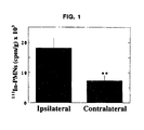
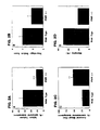


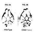


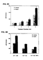


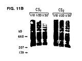


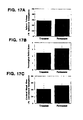
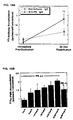


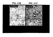

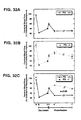


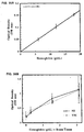


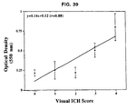

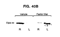

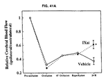
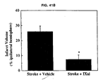


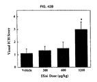
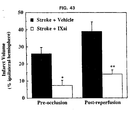
Classifications
-
- A—HUMAN NECESSITIES
- A61—MEDICAL OR VETERINARY SCIENCE; HYGIENE
- A61K—PREPARATIONS FOR MEDICAL, DENTAL OR TOILETRY PURPOSES
- A61K33/00—Medicinal preparations containing inorganic active ingredients
-
- A—HUMAN NECESSITIES
- A61—MEDICAL OR VETERINARY SCIENCE; HYGIENE
- A61K—PREPARATIONS FOR MEDICAL, DENTAL OR TOILETRY PURPOSES
- A61K38/00—Medicinal preparations containing peptides
- A61K38/16—Peptides having more than 20 amino acids; Gastrins; Somatostatins; Melanotropins; Derivatives thereof
- A61K38/43—Enzymes; Proenzymes; Derivatives thereof
- A61K38/46—Hydrolases (3)
- A61K38/48—Hydrolases (3) acting on peptide bonds (3.4)
- A61K38/482—Serine endopeptidases (3.4.21)
- A61K38/4846—Factor VII (3.4.21.21); Factor IX (3.4.21.22); Factor Xa (3.4.21.6); Factor XI (3.4.21.27); Factor XII (3.4.21.38)
-
- A—HUMAN NECESSITIES
- A61—MEDICAL OR VETERINARY SCIENCE; HYGIENE
- A61P—SPECIFIC THERAPEUTIC ACTIVITY OF CHEMICAL COMPOUNDS OR MEDICINAL PREPARATIONS
- A61P11/00—Drugs for disorders of the respiratory system
-
- A—HUMAN NECESSITIES
- A61—MEDICAL OR VETERINARY SCIENCE; HYGIENE
- A61P—SPECIFIC THERAPEUTIC ACTIVITY OF CHEMICAL COMPOUNDS OR MEDICINAL PREPARATIONS
- A61P7/00—Drugs for disorders of the blood or the extracellular fluid
- A61P7/02—Antithrombotic agents; Anticoagulants; Platelet aggregation inhibitors
-
- A—HUMAN NECESSITIES
- A61—MEDICAL OR VETERINARY SCIENCE; HYGIENE
- A61P—SPECIFIC THERAPEUTIC ACTIVITY OF CHEMICAL COMPOUNDS OR MEDICINAL PREPARATIONS
- A61P7/00—Drugs for disorders of the blood or the extracellular fluid
- A61P7/06—Antianaemics
-
- A—HUMAN NECESSITIES
- A61—MEDICAL OR VETERINARY SCIENCE; HYGIENE
- A61P—SPECIFIC THERAPEUTIC ACTIVITY OF CHEMICAL COMPOUNDS OR MEDICINAL PREPARATIONS
- A61P9/00—Drugs for disorders of the cardiovascular system
- A61P9/10—Drugs for disorders of the cardiovascular system for treating ischaemic or atherosclerotic diseases, e.g. antianginal drugs, coronary vasodilators, drugs for myocardial infarction, retinopathy, cerebrovascula insufficiency, renal arteriosclerosis
-
- C—CHEMISTRY; METALLURGY
- C07—ORGANIC CHEMISTRY
- C07K—PEPTIDES
- C07K14/00—Peptides having more than 20 amino acids; Gastrins; Somatostatins; Melanotropins; Derivatives thereof
- C07K14/435—Peptides having more than 20 amino acids; Gastrins; Somatostatins; Melanotropins; Derivatives thereof from animals; from humans
- C07K14/705—Receptors; Cell surface antigens; Cell surface determinants
- C07K14/7056—Lectin superfamily, e.g. CD23, CD72
- C07K14/70564—Selectins, e.g. CD62
-
- C—CHEMISTRY; METALLURGY
- C07—ORGANIC CHEMISTRY
- C07K—PEPTIDES
- C07K16/00—Immunoglobulins [IGs], e.g. monoclonal or polyclonal antibodies
- C07K16/18—Immunoglobulins [IGs], e.g. monoclonal or polyclonal antibodies against material from animals or humans
- C07K16/28—Immunoglobulins [IGs], e.g. monoclonal or polyclonal antibodies against material from animals or humans against receptors, cell surface antigens or cell surface determinants
- C07K16/2803—Immunoglobulins [IGs], e.g. monoclonal or polyclonal antibodies against material from animals or humans against receptors, cell surface antigens or cell surface determinants against the immunoglobulin superfamily
- C07K16/2821—Immunoglobulins [IGs], e.g. monoclonal or polyclonal antibodies against material from animals or humans against receptors, cell surface antigens or cell surface determinants against the immunoglobulin superfamily against ICAM molecules, e.g. CD50, CD54, CD102
-
- C—CHEMISTRY; METALLURGY
- C07—ORGANIC CHEMISTRY
- C07K—PEPTIDES
- C07K16/00—Immunoglobulins [IGs], e.g. monoclonal or polyclonal antibodies
- C07K16/18—Immunoglobulins [IGs], e.g. monoclonal or polyclonal antibodies against material from animals or humans
- C07K16/28—Immunoglobulins [IGs], e.g. monoclonal or polyclonal antibodies against material from animals or humans against receptors, cell surface antigens or cell surface determinants
- C07K16/2851—Immunoglobulins [IGs], e.g. monoclonal or polyclonal antibodies against material from animals or humans against receptors, cell surface antigens or cell surface determinants against the lectin superfamily, e.g. CD23, CD72
- C07K16/2854—Immunoglobulins [IGs], e.g. monoclonal or polyclonal antibodies against material from animals or humans against receptors, cell surface antigens or cell surface determinants against the lectin superfamily, e.g. CD23, CD72 against selectins, e.g. CD62
-
- G—PHYSICS
- G01—MEASURING; TESTING
- G01N—INVESTIGATING OR ANALYSING MATERIALS BY DETERMINING THEIR CHEMICAL OR PHYSICAL PROPERTIES
- G01N33/00—Investigating or analysing materials by specific methods not covered by groups G01N1/00 - G01N31/00
- G01N33/48—Biological material, e.g. blood, urine; Haemocytometers
- G01N33/50—Chemical analysis of biological material, e.g. blood, urine; Testing involving biospecific ligand binding methods; Immunological testing
- G01N33/5005—Chemical analysis of biological material, e.g. blood, urine; Testing involving biospecific ligand binding methods; Immunological testing involving human or animal cells
- G01N33/5008—Chemical analysis of biological material, e.g. blood, urine; Testing involving biospecific ligand binding methods; Immunological testing involving human or animal cells for testing or evaluating the effect of chemical or biological compounds, e.g. drugs, cosmetics
- G01N33/5082—Supracellular entities, e.g. tissue, organisms
- G01N33/5085—Supracellular entities, e.g. tissue, organisms of invertebrates
-
- A—HUMAN NECESSITIES
- A61—MEDICAL OR VETERINARY SCIENCE; HYGIENE
- A61K—PREPARATIONS FOR MEDICAL, DENTAL OR TOILETRY PURPOSES
- A61K39/00—Medicinal preparations containing antigens or antibodies
- A61K2039/505—Medicinal preparations containing antigens or antibodies comprising antibodies
-
- C—CHEMISTRY; METALLURGY
- C07—ORGANIC CHEMISTRY
- C07K—PEPTIDES
- C07K2317/00—Immunoglobulins specific features
- C07K2317/70—Immunoglobulins specific features characterized by effect upon binding to a cell or to an antigen
- C07K2317/76—Antagonist effect on antigen, e.g. neutralization or inhibition of binding
Definitions
- Acute neutrophil recruitment to postischemic cardiac or pulmonary tissue has deleterious effects in the early reperfusion period, but the mechanisms and effects of neutrophil influx in the pathogenesis of evolving stroke remains controversial.
- the present invention provides for a use of a gas comprising carbon monoxide in a sufficient amount for the preparation of a pharmaceutical composition for the treatment or prevention of an ischemic disorder over a sufficient period of time in a subject. Further preferred embodiments of the present invention are characterized herein and in the claims.
- a method for treating an ischemic disorder in a subject which includes administering to the subject a pharmaceutically acceptable form of a selectin antagonist in a sufficient amount over a sufficient time period to prevent white blood cell accumulation so as to treat the ischemic disorder in the subject.
- the selectin antagonist may be a peptide mimetic, a nucleic acid molecule, a ribozyme, a polypeptide, a small molecule, a carbohydrate molecule, a monosaccharide, an oligosaccharide or an antibody.
- the selectin may be a P-selectin, an E-selectin, or an L-selectin.
- the antibody may be a P-selectin antibody.
- the antibody may further include a polyclonal antibody or a monoclonal antibody.
- the P-selectin antagonist may include a nitric oxide (NO) precursor such as L-arginine, an NO donor such as nitroglycerin or nitroprusside, a cyclic nucleotide analog such as a cyclic GMP or cyclic AMP analog, or a phosphodiesterase inhibitor.
- NO nitric oxide
- the pharmaceutically acceptable form of P-selectin antagonist may include a P-selectin antagonist and a pharmaceutically acceptable carrier.
- the carrier may include an aerosol, intravenous, oral or topical carrier.
- the white blood cell may be a neutrophil or a monocyte.
- the subject may be a mammal.
- the mammal may be a human, a cow, a pig, a sheep, a dog, a cat, a monkey, a fowl or any animal model of a human disease or disorder.
- the ischemic disorder may include, but is not limited to a peripheral vascular disorder, a venous thrombosis, a pulmonary embolus, a myocardial infarction, a transient ischemic attack, unstable angina, a reversible ischemic neurological deficit, sickle cell anemia or a stroke disorder.
- the subject may be undergoing heart surgery, lung surgery, spinal surgery, brain surgery, vascular surgery, abdominal surgery, or organ transplantation surgery.
- the organ transplantation surgery may include heart, lung, pancreas or liver transplantation surgery.
- the present invention provides a use of a gas comprising carbon monoxide in a sufficient amount for the preparation of a pharmaceutical composition for the treatment or prevention of an ischemic disorder over a sufficient period of time in a subject.
- the administration of carbon monoxide may be via inhalation by the subject or via extracorporeal exposure to blood or body fluids of the subject.
- the amount of carbon monoxide which may be sufficient to treat the subject includes but is not limited to from about 0.0001% carbon monoxide in an inert gas to about 2% carbon monoxide in an inert gas.
- the inert gas may be oxygen, nitrogen, argon or air.
- the amount of carbon monoxide administered may be 0.1% carbon monoxide in air.
- the period of time sufficient to administer carbon monoxide to a subject to treat an ischemic disorder includes but is not limited to from about 1 day before surgery to about 1 day after surgery.
- the period of time may be from about 12 hours before surgery to about 12 hours after surgery.
- the period of time may further include from about 12 hours before surgery to about 1 hour after surgery.
- the period of time may further include from about 1 hour before surgery to about 1 hour after surgery.
- the period of time may further include from about 20 minutes before surgery to about 1 hour after surgery.
- the period of time sufficient to treat an ischemic disorder in a subject who is not undergoing surgery may include before and during any physical manifestation of such disorder.
- Administration of carbon monoxide is preferable before the manifestation in order to lessen such manifestation of an ischemic disorder. Carbon monoxide administration has been shown as described hereinbelow to be protective of ischemia in a subject if.administered prior to surgery.
- the "ischemic disorder” encompasses and is not limited to a peripheral vascular disorder, a venous thrombosis, a pulmonary embolus, a myocardial infarction, a transient ischemic attack, lung ischemia, unstable angina, a reversible ischemic neurological deficit, adjunct thromolytic activity, excessive clotting conditions, sickle cell anemia or a stroke disorder.
- the subject may be undergoing heart surgery, lung surgery, spinal surgery, brain surgery, vascular surgery, abdominal surgery, or organ transplantation surgery.
- the organ transplantation surgery may include heart, lung, pancreas or liver transplantation surgery.
- the carbon monoxide may be administered in an indirect manner. Rather than the subject directly inhaling or receiving carbon monoxide gas or a gas mixture, the subject may be given compunds to stimulate the in vivo production of carbon monoxide.
- Such compounds may include but are not limited to heme, ferritin, hematin, endogenous precursors to heme oxygenase or heme oxygenase stimulators.
- the subject may be exposed to an environment of low oxygen level compared to the normal atmosphere.
- Heme oxygenase is an endogenous enzyme which synthesizes carbon monoxide from precursor heme (it is part of the normal way in which heme is degraded and metabolized in the body).
- levels of both the messenger RNA which codes for heme oxygenase protein and the protein itself were increased.
- the activity of the enzyme was increased, as indicated by measurements of carbon monoxide in the tissue.
- a method for treating an ischemic disorder in a subject which comprises administering to the subject a pharmaceutically acceptable form of inactivated Factor IX in a sufficient amount over a sufficient period of time to inhibit coagulation so as to treat the ischemic disorder in the subject.
- the sufficient amount may include but is not limited to from about 75 ⁇ g/kg to about 550 ⁇ g/kg.
- the amount may be 300 ⁇ g/kg.
- the pharmaceutically acceptable form of inactivated Factor IX includes inactivated Factor IX and a pharmaceutically acceptable carrier.
- the Factor IX may be inactivated by the standard methods known to one of skill in the art, such as heat inactivation. Factor IX may be inactivated or Factor IX activity may be inhibited by an antagonist.
- Such antagonist may be a peptide mimetic, a nucleic acid molecule, a ribozyme, a polypeptide, a small molecule, a carbohydrate molecule, a monosaccharide, an oligosaccharide or an antibody.
- the present application describes a method for identifying a compound that is capable of improving an ischemic disorder in a subject which includes: a) administering the compound to an animal, which animal is a stroke animal model; b) measuring stroke outcome in the animal, and c) comparing the stroke outcome in step (b) with that of the stroke animal model in the absence of the compound so as to identify a compound capable of improving an ischemic disorder in a subject.
- the stroke animal model includes a murine model of focal cerebral ischemia and reperfusion.
- the stroke outcome may be measured by physical examination, magnetic resonance imaging, laser doppler flowmetry, triphenyl tetrazolium chloride staining, chemical assessment of neurological deficit, computed tomography scan, or cerebral cortical blood flow.
- the stroke outcome in a human may be measured also by clinical measurements, quality of life scores and neuropsychometric testing.
- the compound may include a P-selectin antagonist, an E-selectin antagonist or an L-selectin antagonist.
- the present application further describes a method for identifying a compound that is capable of preventing the accumulation of white blood cells in a subject which includes:a) administering the compound to an animal, which animal is a stroke animal model; b) measuring stroke outcome in the animal, and c) comparing the stroke outcome in step (b) with that of the stroke animal model in the absence of the compound so as to identify a compound capable of preventing the accumulation of white blood cells in the subject.
- the white blood cell may be, but is not limited to, a neutrophil, a platlet or a monocyte.
- the compound may be but is not limited to a selectin inhibitor, a monocyte inhibitor, a platelet inhibitor or a neutrophil inhibitor.
- the selectin inhibitor may be but is not limited to a P-selectin, E-selectin or L-selectin inhibitor.
- Selectins are expressed on the surface of platlets and such selectin inhibitors or antagonists as described herein may prevent the expression of such selectins on the surface of the cell.
- the prevention of expression may be at the transcriptional, translational, post-translational levels or preventing the movement of such selectins through the cytosol and preventing delivery at the cell surface.
- the present application describes the treatment of ischemic disorders by inhibiting the ability of the neutrophil, monocyte or other white blood cell to adhere properly. This may be accomplished removing the counter ligand, such as CD18. It has been demonstrated as discussed hereinbelow, that "knock-out" CD18 mice (mice that do not have expression of the normal CD18 gene) are protected from adverse ischemic conditions.
- the endothelial cells on the surface of the vessels in the subject may also be a target for treatment.
- administration of TPA as a thrombolytic agent caused some visible hemorrhaging along with improvement of the stroke disorder.
- administration of a P-selectin antagonist also improved stroke disorder in the animal model, but without the coincident hemorrhaging.
- the disclosure of the present application may be used in conjunction with a thrombolytic therapy to increase efficacy of such therapy or to enable lower doses of such therapy to be administered to the subject so as to reduce side effects of the thrombolytic therapy.
- suitable pharmaceutically acceptable carrier encompasses any of the standard pharmaceutically accepted carriers, such as phosphate buffered saline solution, water, emulsions such as an oil/water emulsion or a triglyceride emulsion, various types of wetting agents, tablets, coated tablets and capsules.
- a triglyceride emulsion useful in intravenous and intraperitoneal administration of the compounds is the triglyceride emulsion commercially known as Intralipid ® .
- Such carriers typically contain excipients such as starch, milk, sugar, certain types of clay, gelatin, stearic acid, talc, vegetable fats or oils, gums, glycols, or other known excipients.
- excipients such as starch, milk, sugar, certain types of clay, gelatin, stearic acid, talc, vegetable fats or oils, gums, glycols, or other known excipients.
- Such carriers may also include flavor and color additives or other ingredients.
- compositions including therapeutically effective amounts of protein compositions and compounds capable of treating stroke disorder or improving stroke outcome in the subject of the invention together with suitable diluents, preservatives, solubilizers, emulsifiers, adjuvants and/or carriers useful in treatment of neuronal degradation due to aging, a learning disability, or a neurological disorder.
- compositions are liquids or lyophilized or otherwise dried formulations and include diluents of various buffer content (e.g., Tris-HCl., acetate, phosphate), pH and ionic strength, additives such as albumin or gelatin to prevent absorption to surfaces, detergents (e.g., Tween 20, Tween 80, Pluronic F68, bile acid salts), solubilizing agents (e.g., glycerol, polyethylene glycerol), anti-oxidants (e.g., ascorbic acid, sodium metabisulfite), preservatives (e.g., Thimerosal, benzyl alcohol, parabens), bulking substances or tonicity modifiers (e.g., lactose, mannitol), covalent attachment of polymers such as polyethylene glycol to the compound, complexation with metal ions, or incorporation of the compound into or onto particulate preparations of polymeric compounds such as polylactic acid, polglycolic acid
- compositions will influence the physical state, solubility, stability, rate of in vivo release, and rate of in vivo clearance of the compound or composition.
- the choice of compositions will depend on the physical and chemical properties of the compound capable of alleviating the symptoms of the stroke disorder or improving the stroke outcome in the subject.
- Controlled or sustained release compositions include formulation in lipophilic depots (e.,g., fatty acids, waxes, oils). Also comprehended by the present application are particulate compositions coated with polymers (e.g., poloxamers or poloxamines) and the compound coupled to antibodies directed against tissue-specific receptors, ligands or antigens or coupled to ligands of tissue-specific receptors. Others aspects of the compositions described herein incorporate particulate forms protective coatings, protease inhibitors or permeation enhancers for various routes of administration, including parenteral, pulmonary, nasal and oral.
- lipophilic depots e.,g., fatty acids, waxes, oils.
- particulate compositions coated with polymers e.g., poloxamers or poloxamines
- Others aspects of the compositions described herein incorporate particulate forms protective coatings, protease inhibitors or permeation enhancers for various routes of administration, including parenteral, pulmonary, nasal and oral.
- Portions of the compound of the present application may be "labeled" by association with a detectable marker substance (e.g., radiolabeled with 125 I or biotinylated) to provide reagents useful in detection and quantification of compound or its receptor bearing cells or its derivatives in solid tissue and fluid samples such as blood, cerebral spinal fluid or urine.
- a detectable marker substance e.g., radiolabeled with 125 I or biotinylated
- Such modifications may also increase the compound's solubility in aqueous solution, eliminate aggregation, enhance the physical and chemical stability of the compound, and greatly reduce the immunogenicity and reactivity of the compound.
- the desired in vivo biological activity may be achieved by the administration of such polymer-compound adducts less frequently or in lower doses than with the unmodified compound.
- PEG polyethylene glycol
- Attachment of polyethylene glycol (PEG) to compounds is particularly useful because PEG has very low toxicity in mammals (Carpenter et al., 1971).
- PEG adduct of adenosine deaminase was approved in the United States for use in humans for the treatment of severe combined immunodeficiency syndrome.
- a second advantage afforded by the conjugation of PEG is that of effectively reducing the immunogenicity and antigenicity of heterologous compounds.
- a PEG adduct of a human protein might be useful for the treatment of disease in other mammalian species without the risk of triggering a severe immune response.
- the compound of the present invention capable of alleviating symptoms of a cognitive disorder of memory or learning may be delivered in a microencapsulation device so as to reduce or prevent an host immune response against the compound or against cells which may produce the compound.
- the compound of the present invention may also be delivered microencapsulated in a membrane, such as a liposome.
- Polymers such as PEG may be conveniently attached to one or more reactive amino acid residues in a protein such as the alpha-amino group of the amino terminal amino acid, the epsilon amino groups of lysine side chains, the sulfhydryl groups of cysteine side chains, the carboxyl groups of aspartyl and glutamyl side chains, the alpha-carboxyl group of the carboxy-terminal amino acid, tyrosine side chains, or to activated derivatives of glycosyl chains attached to certain asparagine, serine or threonine residues.
- a protein such as the alpha-amino group of the amino terminal amino acid, the epsilon amino groups of lysine side chains, the sulfhydryl groups of cysteine side chains, the carboxyl groups of aspartyl and glutamyl side chains, the alpha-carboxyl group of the carboxy-terminal amino acid, tyrosine side chains, or to activated derivatives of
- PEG reagents for reaction with protein amino groups include active esters of carboxylic acid or carbonate derivatives, particularly those in which the leaving groups are N-hydroxysuccinimide, p-nitrophenol, imidazole or 1-hydroxy-2-nitrobenzene-4-sulfonate.
- PEG derivatives containing maleimido or haloacetyl groups are useful reagents for the modification of protein free sulfhydryl groups.
- PEG reagents containing amino hydrazine or hydrazide groups are useful for reaction with aldehydes generated by periodate oxidation of carbohydrate groups in proteins.
- the present application describes a pharmaceutical composition which comprises an agent capable of treating a stroke disorder or improving stroke outcome and a pharmaceutically acceptable carrier.
- the carrier may include but is not limited to a diluent, an aerosol, a topical carrier, an aquous solution, a nonaqueous solution or a solid carrier.
- EC endothelial cell
- PMN polymorphonuclear leukocyte
- WP Weibel-Palade body
- vWF von Willebrand factor
- EGTA ethyleneglycol bis (aminoethylether)tetraacetic acid
- HBSS Hank's balanced salt solution
- CS coronary sinus
- IL interleukin
- PAF platelet activating factor
- ICAM-1 intercellular adhesion molecule-1
- HUVEC human umbilical vein EC
- LR lactated Ringer's solution
- MCAO middle cerebral artery occlusion
- rt-PA recombinant tissue plasminogen activator
- HO-I or HOI or HO I heme oxygenase I
- ICH intracerebral hemorrhage
- OD optical density
- MCA middle cerebral artery
- rt-PA recombinant tissue-type plasminogen activator
- EXAMPLE 1 Cerebral Protection in Homozygous Null ICAM-1 Mice Following Middle Cerebral Artery Occlusion: Role of Neutrophil Adhesion in the Pathogenesis of Stroke
- PMNs polymorphonuclear leukocytes
- ICM-1 Intercellular Adhesion Molecule-1
- MCA intraluminal middle cerebral artery
- PMN accumulation monitored by deposition of 111 Indium-labelled PMNs in postischemic cerebral tissue, was increased 2.5-fold in the ipsilateral (infarcted) hemisphere compared with the contralateral (noninfarcted) hemisphere (p ⁇ 0.01).
- ICAM-1 mRNA was increased in the ipsilateral hemisphere, with immunohistochemistry localizing increased ICAM-1 expression on cerebral microvascular endothelium.
- ICAM-1 -/- mice demonstrated a 3.7-fold reduction in infarct volume (p ⁇ 0.005), a 35% increase in survival (p ⁇ 0.05), and reduced neurologic deficit compared with ICAM-1 +/+ controls. Cerebral blood flow to the infarcted hemisphere was 3.1-fold greater in ICAM-1 -/- mice compared with ICAM-1 +/+ controls (p ⁇ 0.01), suggesting an important role for ICAM-1 in the genesis of post-ischemic cerebral no-reflow. Because PMN-depleted and ICAM-1 deficient mice are relatively resistant to cerebral ischemia-reperfusion injury, these studies suggest an important role for ICAM-1-mediated PMN adhesion in the pathophysiology of evolving stroke.
- mice Experiments were performed with transgenic ICAM-1 deficient mice created as previously reported 24 by gene targeting in J1 embryonic stem cells, injected into C57BL/6 blastocysts to obtain germline transmission, and backcrossed to obtain homozygous null ICAM-1 mice. All experiments were performed with ICAM-1 -/- or wild-type (ICAM-1+/+) cousin mice from the fifth generation of backcrossings with C57BL/6 mice. Animals were seven to nine weeks of age and weighed between 25-36 grams at the time of experiments.
- ICAM-1 -/- or wild-type (ICAM-1+/+) cousin mice from the fifth generation of backcrossings with C57BL/6 mice. Animals were seven to nine weeks of age and weighed between 25-36 grams at the time of experiments.
- neutrophil depletion of C57BL/6 mice was accomplished by administering polyclonal rabbit anti-mouse neutrophil antibody 25 (Accurate Scientific, Westbury NY) preadsorbed to red blood cells as a daily intraperitoneal injection (0.3 mL of 1:12 solution) for 3 days. Experiments in these mice were performed on the fourth day after confirming agranulocytosis by Wright-Giemsa stained peripheral blood smears.
- Transient Middle Cerebral Artery Occlusion 26 Mice were anesthetized with an intraperitoneal injection of 0.3 ml of ketamine (10 mg/cc) and xylazine (0.5 mg/cc). Animals were positioned supine on a rectal-temperature controlled operating surface (Yellow Springs Instruments, Inc., Yellow Springs, OH). Animal core temperature was maintained at 36-38°C intraoperatively and for 90 minutes post-operatively. Middle cerebral artery occlusion was performed as follows; A midline neck incision was created to expose the right carotid sheath under the operating microscope (16-25X zoom, Zeiss, Thornwood, NY).
- the common carotid artery was freed from its sheath, and isolated with a 4-0 silk, and the occipital and pterygopalatine arteries were each isolated and divided. Distal control of the internal carotid artery was obtained and the external carotid was cauterized and divided just proximal to its bifurcation into the lingual and maxillary divisions. Transient carotid occlusion was accomplished by advancing a 13 mm heat-blunted 5-0 nylon suture via the external carotid stump to the origin of the middle cerebral artery. After placement of the occluding suture, the external carotid artery stump was cauterized to prevent bleeding through the arteriotomy, and arterial flow was reestablished. In all cases, the duration of carotid occlusion was less than two minutes. After 45 minutes, the occluding suture was withdrawn to establish reperfusion. These procedures have been previously described in detail 26 .
- Data are expressed as the ratio of the doppler signal intensity of the ischemic compared with the nonischemic hemisphere.
- this method does not quantify cerebral blood flow per gram of tissue, use of laser doppler flow measurements at precisely defined anatomic landmarks serves as a means of comparing cerebral blood flows in the same animal serially over time.
- the surgical procedure/intraluminal middle cerebral artery occlusion and reperfusion were considered to be technically adequate if ⁇ 50% reduction in relative cerebral blood flow was observed immediately following placement of the intraluminal occluding suture and a ⁇ 33% increase in flow over baseline occlusion was observed immediately following removal of the occluding suture.
- the neutrophils were gently pelleted (450 g), and resuspended in PBS to a final concentration of 1.0 x 10 6 cells/ml.
- 100 ⁇ L of radiolabelled PMNs admixed with physiologic saline to a total volume of 0.3 mL ( ⁇ 3 x 10 6 cpm) was administered by penile vein injection.
- brains were obtained as described, and neutrophil deposition quantified as cpm/gm of each hemisphere.
- mice Twenty-four hours after middle cerebral artery occlusion and reperfusion, prior to giving anesthesia, mice were examined for neurological deficit using a four-tiered grading system 26 : A score of (1) was given if the animal demonstrated normal spontaneous movements; a score of (2) was given if the animal was noted to be turning to the right (clockwise circles) when viewed from above (i.e., towards the contralateral side); a score of (3) was given if the animal was observed to spin longitudinally (clockwise when viewed from the tail); a score of (4) was given if the animal was crouched on all fours, unresponsive to noxious stimuli.
- This scoring system has been previously described in mice 26 , and is based upon similar scoring systems used in rats 28,29 which are based upon the contralateral movement of animals with stroke; following cerebral infarction, the contralateral side is "weak" and so the animal tends to turn towards the weakened side.
- Previous work in rats 28 and mice 26 demonstrates that larger cerebral infarcts are associated with a greater degree of contralateral movement, up to the point where the infarcts are so large that the animal remains unresponsive.
- mice were given 0.3 mL of ketamine (10 mg/ml) and xylazine (0.5 mg/ml), and final cerebral blood flow measurements were obtained.
- Humane euthanasia was performed by decapitation under anesthesia, and brains were removed and placed in a mouse brain matrix (Activational Systems Inc., Warren, MI) for 1 mm sectioning. Sections were immersed in 2% 2,3,5, triphenyl, 2H-tetrazolium chloride (TTC, Sigma Chemical Co., St. Louis, MO) in 0.9% phosphate-buffered saline, incubated for 30 minutes at 37°C, and placed in 10% formalin 26,31-32 . Infarcted brain was visualized as an area of unstained tissue, in contrast to viable tissue, which stains brick red. Infarct volumes were calculated from planimetered serial sections and expressed as the percentage of infarct in the ipsilateral hemisphere.
- RNA extraction and Northern blot analysis 24 hours following focal ischemia and reperfusion, brains were obtained and divided into ipsilateral (infarct) and contralateral (noninfarct) hemispheres.
- total RNA was extracted from each hemisphere using an RNA isolation kit (Stratagene, La Jolla, CA). Equal amounts of RNA (20 ⁇ g/lane) were loaded onto a 1.4% agarose gel containing 2.2 M formaldehyde for size fractionation, and then transferred overnight to nylon (Nytran) membranes with 10X SSC buffer by capillary pressure.
- a murine ICAM-1 cDNA probe 33 (1.90 kb, ATCC, Rockville, MD) was labelled with 32 P- ⁇ -dCTP by random primer labelling (Prime-A-Gene kit, Promega), hybridized to blots at 42°C, followed by 3 washes of 1X SSC/0.05% SDS. Blots were developed with X-Omat AR film exposed with light screens at -70' for 7 days. A ⁇ -actin probe (ATCC) was used to confirm equal RNA loading.
- mice were immunodepleted of neutrophils beginning three days prior to surgery. When surgery was performed on the fourth day, nearly complete agranulocytosis was evident on smears of peripheral blood.
- ICAM-1 Expression in Murine Stroke To establish the effect of cerebral ischemia/reperfusion in the murine model, ICAM-1 mRNA levels were evaluated following cerebral ischemia and reperfusion in wild type mice. Ipsilateral (infarcted) cerebral hemisphere demonstrated increased ICAM-1 mRNA by Northern blot analysis compared with RNA obtained from the contralateral (noninfarcted) hemisphere from the same animal ( Figure 3). To evaluate ICAM-1 antigen expression in this murine model, wild type mice were subjected to 45 minutes of ischemia followed by 23 hours of reperfusion, and the cerebral microvasculature examined by immunohistochemistry.
- ICAM-1 antigen expression was not detectable in the cerebral microvasculature contralateral to the infarct ( Figure 4A), but was markedly increased on the ipsilateral side, with prominent ICAM-1 staining of cerebral endothelial cells ( Figure 4B).
- TTC staining to quantify cerebral infarct volumes 26,30-32,37,53 .
- TTC 2,3,5 triphenyl, 2H-tetrazolium chloride
- TTC staining is unreliable before 2 hours of ischemia have elapsed; beyond 36 hours, cells infiltrating into the infarcted tissue can stain positively with TTC, thereby obscuring the clear demarcation between infarcted and noninfarcted tissues seen with earlier staining 31 .
- the size of the infarct delineated by TTC staining correlates well with infarct size delineated by hematoxylin and eosin staining 30,32 , direct morphometric measurements tend to overestimate infarct volumes due to cerebral edema, especially during the first 3 days following the ischemic event 32 .
- Endothelial cells appear to be the chief regulators of neutrophil traffic, regulating the processes of neutrophil chemoattraction, adhesion, and emigration from the vasculature 55 .
- endothelial cells When exposed to a hypoxic environment as a paradigm for tissue ischemia, endothelial cells synthesize the potent neutrophil chemoattractant and activator Interleukin-8 (IL-8) 9 , the blockade of which appears to be beneficial in a lung model of ischemia and reperfusion 6 .
- IL-8 Interleukin-8
- hypoxic endothelial cells synthesize the proinflammatory cytokine Interleukin-1 8 , which can upregulate endothelial expression of the neutrophil adhesion molecules E-selectin and ICAM-1 in an autocrine fashion 8,9,56 .
- Other neutrophil adhesion mechanisms may also be activated in the brain following ischemia, such as release of P-selectin from preformed storage pools within Weibel-Palade body membranes 10 .
- P-selectin expression was rapidly and persistently enhanced following focal middle cerebral artery ischemia and reperfusion 18 .
- P-selectin-dependent neutrophil recruitment appears to be deleterious following cardiac ischemia and reperfusion 57 , its pathophysiologic relevance in the setting of stroke has not yet been determined. While hypoxia induces de novo synthesis of the bioactive lipid platelet activating factor (PAF) 11 , in a spinal cord ischemia reperfusion model, PAF antagonism offered no incremental benefit when given simultaneously with antibody to CD11/CD18 48 .
- PAF bioactive lipid platelet activating factor
- ICAM-1 in the pathophysiology of stroke appears to be of particular relevance in humans for several reasons.
- Increased cerebrovascular ICAM-1 expression has been demonstrated in primates by 4 hours of ischemia and reperfusion, particularly in the lenticulostriate microvasculature 18 .
- An autopsy study of recent cerebral infarcts in humans also demonstrated increased ICAM-1 expression 20 .
- rats also express cerebral vascular ICAM-1 within 24 hours in both a photochemically-induced model of rat cerebral ischemia 19 as well as a middle cerebral artery occlusion model 12 , these data suggested the potential usefulness of transgenic ICAM-1 deficient mice in elucidating the pathophysiologic significance of increased post-cerebral ischemic ICAM-1 expression.
- the time frame of ICAM-1 expression (increased by 4 to 24 hours) in these models suggests that ICAM-1 mediated neutrophil-endothelial interactions may be targeted in future pharmacologic strategies to improve human stroke outcome, as this time frame represents a realistic clinical window for therapeutic intervention.
- EXAMPLE 2 Hypoxia-Induced Exocytosis of Endothelial Cell Weibel-Palade Bodies: A Mechanism for Rapid Neutrophil Recruitment Following Cardiac Preservation
- hypoxia is an important priming event for the vascular dysfunction which accompanies reperfusion, with endothelial cells (ECs) and neutrophils (PMNs) playing a central role. It was hypothesized that EC Weibel-Palade (WP) body exocytosis during the hypoxic/ischemic period during organ preservation permits brisk PMN recruitment into post-ischemic tissue, a process further amplified in an oxidant-rich mileu.
- WP Weibel-Palade
- Endothelial cells adapt to hypoxia with a characteristic repertoire of responses (1), ranging from increased expression of endothelin (2) to increased synthesis of basic fibroblast growth factor (3).
- hypoxia selectively upregulates EC expression of Interleukins- 1 (4), 6 (5), and 8 (6), platelet activating factor (PAF) (7,8), and ICAM-1 (4), which serve to fuel neutrophil (PMN) recruitment, adhesion, and activation at ischemic loci.
- PAF platelet activating factor
- ICAM-1 which serve to fuel neutrophil (PMN) recruitment, adhesion, and activation at ischemic loci.
- P-selectin may figure prominently in the earliest phases of PMN adhesion to the reperfused vasculature, as ECs may rapidly express pre-formed P-selectin from subplasmalemmal storage sites in Weibel-Palade body (9) membranes in response to the abundant oxygen free-radicals generated in the reperfusion milieu (10-12).
- hypoxia per se or hypothermic cardiac preservation, as occurs during cardiac surgery, in which the pO 2 within the coronary bed declines to pO 2 ⁇ 20 Torr (15) would result in WP body exocytosis.
- experiments were undertaken to determine the role of P-selectin-dependent PMN adhesion in the cardiac graft failure which characteristically follows a period of prolonged hypothermic preservation. The results show that hypoxia is sufficient to induce EC WP body exocytosis, even in the absence of reoxygenation (and presence of antioxidants), and that the resulting P-selectin expression causes ECs to bind PMNs in vitro.
- P-selectin expression following hypothermic cardiac preservation can be completely abrogated by either neutrophil depletion, P-selectin blockade, or by transplanting hearts whose endothelial cells fail to express P-selectin. Because WP body exocytosis also occurs in patients undergoing open heart surgery during the period of hypothermic cardiac preservation, these data suggest that P-selectin blockade may represent a target for pharmacological intervention to improve cardiac preservation in humans.
- Endothelial cell culture and exposure of cells to H or H/R Endothelial cell culture and exposure of cells to H or H/R.
- Human umbilical vein ECs were prepared from umbilical cords and grown in culture by the method of Jaffe (16) as modified by Thornton (17). Experiments utilized confluent ECs (passages 1-4) grown in Medium 199 supplemented with fetal bovine serum (15%; Gemini, Calabasas, CA), human serum (5%; Gemini), endothelial growth supplement (Sigma, St. Louis, MO), heparin (90 ⁇ g/ml; Sigma) and antibiotics, as described (17). When ECs achieved confluence, experiments were performed by placing cultures in an environmental chamber (Coy Laboratory Products, Ann Arbor, MI) which provided a controlled temperature (37°C) and atmosphere with the indicated amount of oxygen, carbon dioxide (5%) and the balance made up of nitrogen.
- an environmental chamber Coy Laboratory Products, Ann Arbor, MI
- ECs were plated into 24 well plates, rinsed 3 times with Hank's balanced salt solution, and then exposed to hypoxia or to normoxia for the indicated durations.
- vWF vWF was measured
- cells were maintained in serum-free medium. All other EC experiments were performed in the EC growth medium described above.
- a commercially available ELISA American Diagnostica, Greenwich, CT
- ELISA American Diagnostica, Greenwich, CT
- EC P-selectin expression was determined by measuring the specific binding of a murine monoclonal anti-human P-selectin antibody (WAPS 12.2 clone, Endogen, Cambridge, MA; this is an IgG1 which recognizes a calcium sensitive epitope and blocks P-selectin dependent neutrophil adhesion.
- Antibody was radiolabelled with 125 I by the lactoperoxidase method (19) using Enzymobeads (Bio-Rad, Hercules, CA), stored at 4°C and used within one week of labelling. Binding assays were performed on HUVECs plated on 96 well plates, in which fresh M199 with 0.1% bovine serum albumin (Sigma, St.
- HBSS/A bovine serum albumin
- Monolayers were then exposed to 10 5 cpm of 125 I-labelled anti-P-selectin antibody (WAPS 12.2) in the presence of 200 ⁇ g/mL of either unlabelled blocking antibody (WAPS 12.2) or nonblocking anti-P-selectin IgG of the same isotype (anti-GMP-140, AC1.2 clone, Becton-Dickinson, San Jose, CA) (20,21).
- WAPS 12.2 unlabelled blocking antibody
- anti-GMP-140, AC1.2 clone anti-GMP-140, AC1.2 clone, Becton-Dickinson, San Jose, CA
- monolayers were washed 4 times with HBSS/A, and bound antibody was eluted with 1% triton X-100 in PBS (200 ⁇ L/well) and counted.
- cycloheximide (10 ⁇ g/mL, Sigma) was added at the start of the 4 hour normoxic or hypoxic period, as indicated.
- ECs were incubated with methionine- and cysteine-poor minimal essential medium (Gibco, Grand Island, NY) in the presence of 35 S-methionine and 35 S-cysteine (either in the presence or absence of cyloheximide, 10 ⁇ g/mL) (3). Following 4 hours of normoxic exposure, trichloroacetic acid-precipitable material was collected and counted.
- Cardiac transplants were performed in the Ono-Lindsey heterotopic isograft model of cardiac transplantation (15,18,22). Briefly, male Lewis rats (250-300 grams, Harlan Sprague Dawley, Indianapolis, IN) were anesthetized, heparinized, and the donor heart rapidly harvested following hypothermic high potassium cardioplegic arrest.
- Hearts were preserved by flushing the coronary arteries with 4°C lactated Ringer's (LR) solution (Baxter, Edison, NJ), sixteen hours of immersion in the same solution at 4°C, followed by heterotopic transplantation into gender/strain matched recipients, with sequential donor and recipient aortic and donor pulmonary arterial/recipient inferior vena caval anastomoses performed. Graft survival was assessed by the presence/absence of cardiac electrical/mechanical activity exactly ten minutes following reestablishment of blood flow, after which the graft was excised and neutrophil infiltration was quantified by myeloperoxidase activity, measured as previously described (15,18).
- LR Ringer's
- neutrophil depletion of recipient rats was accomplished by administering a polyclonal rabbit anti-rat neutrophil antibody (23-25) (Accurate Scientific, Westbury NY) as a single intravenous injection 24 hours prior to the transplantation procedure. Neutrophil depletion in these animals was confirmed and quantified by counting remaining neutrophils, identified on Wright-Giemsa stained smears of peripheral blood. In other experiments, a blocking anti-P-selectin IgG (250 ⁇ g/rat, Cytel, San Diego, CA) (13,14,26) was administered intravenously 10 minutes prior to the onset of reperfusion.
- Murine heart transplants were performed in an identical fashion using homozygous P-selectin null or wild-type control male mice with a C57BL/6J background (27), with the harvested hearts immediately flushed free of native blood with 1.0 mL of 4°C LR administered down a cross-clamped aortic root followed by period of hypothermic preservation consisting of three hours of immersion in lactated Ringer's solution at 4°C.
- Coronary sinus samples were obtained from a retrogade perfusion catheter which was routinely placed in patients undergoing cardiopulmonary bypass. Plasma samples were centrifuged for 5 min at 1500 x g to sediment cellular elements, and the plasma aliquoted and frozen at -70°C until the time of assay. ELISAs were performed for vWF (as described above) and thrombomodulin (Asserchron Thrombomodulin, Diagnostica Stago).
- vWF immunoelectrophoresis Multimeric composition of the vWF in coronary sinus plasma samples and endothelial cell supernatants was evaluated by performing agarose gel immunoelectrophoresis. Samples were diluted 1:10, 1:20, and 1:30 (as indicated) and incubated for 30 minutes at 37° C in Native Sample Buffer (Bio-Rad). Samples (20 ⁇ L) were then electrophoresed in a 1.5% agarose gel (0.675 g Low M r agarose, Bio-Rad; 0.045 g SDS; 45 mL Tris-Tricine SDS Buffer [Bio-Rad]).
- the membrane was immersed overnight in washing buffer containing 1g/dL gelatin and a 1:500 dilution of rabbit anti-human vWF serum (American Bioproducts, Parsippany, NJ).
- the membrane was immersed for 3 hours with gentle shaking in washing buffer containing lg/dL gelatin and 16.6 ⁇ L of goat anti-rabbit horseradish peroxidase conjugated IgG (Bio-Rad), and developed with 60 mL of HRP Developer (30 mg HRP Developer powder, Bio-Rad; 10 mL methanol; 50 mL tris-buffered saline; 50 ⁇ L of 30% hydrogen peroxide added just prior to use).
- HRP Developer 30 mg HRP Developer powder, Bio-Rad; 10 mL methanol; 50 mL tris-buffered saline; 50 ⁇ L of 30% hydrogen peroxide added just prior to use.
- hypoxia-induced P-selectin expression To determine whether protein synthesis was required for hypoxia-induced P-selectin expression, a separate experiment was performed in which cyloheximide was given at the onset of normoxia or H, and binding of radiolabelled anti-P-selectin IgG determined at the 4 hour time point. This experiment demonstrated that even with > 85% inhibition of protein synthesis (Fig. 9B, Inset), hypoxia still increased endothelial P-selectin expression, albeit at reduced levels (Fig. 9B). To establish that hypoxia-induced cell-surface P-selectin may participate in neutrophil binding, human neutrophils radiolabelled with 111 indium oxine were incubated with hypoxic ECs; enhanced binding to hypoxic monolayers was observed. Hypoxia-induced 111 In-PMN binding was blocked by the addition of a blocking anti-P-selectin IgG, but not by a nonblocking anti-P-selectin IgG (Fig. 9
- the polyclonal rabbit anti-rat PMN antibody used (23-25) eliminated virtually all circulating PMNs in the recipients (PMN count 1471 ⁇ 56 vs 67 ⁇ 11 PMNs/mm 3 for control and immunodepleted animals, respectively, p ⁇ 0.001), with little effect on other cell types.
- PMN count 1471 ⁇ 56 vs 67 ⁇ 11 PMNs/mm 3 for control and immunodepleted animals, respectively, p ⁇ 0.001 eliminated virtually all circulating PMNs in the recipients (PMN count 1471 ⁇ 56 vs 67 ⁇ 11 PMNs/mm 3 for control and immunodepleted animals, respectively, p ⁇ 0.001), with little effect on other cell types.
- Fig. 10A, Prsvd (-) PMN There was a significant reduction in graft myeloperoxidase activity and an increase in graft survival.
- platelets may also interact with PMNs via a P-selectin-dependent mechanism (39).
- a P-selectin-dependent mechanism 39
- donor hearts from homozygous P-selectin deficient mice could be flushed free of blood, so that P-selectin null coronary endothelial cells could be transplanted into wild type recipients with P-selectin containing platelets.
- the vasculature plays a critical role in maintaining the extracellular milieu of organs subjected to ischemia and reperfusion, a role which is chiefly orchestrated by the ECs lining the endovascular lumen.
- the EC responds to a period of oxygen deprivation by striking phenotypic modulation, becoming prothrombotic (46) and proinflammatory (1,4,6).
- ECs exposed to hypoxia secrete the proinflammatory cytokines IL-1 (4) and IL-8 (6) which may serve to direct leukocyte traffic to areas of ischemia. Because these processes require de novo protein synthesis, they do not explain the immediate events which occur following a period of hypothermic preservation.
- PAF platelet activating factor
- P-selectin should be important following hypothermic myocardial preservation; recent studies have demonstrated that P-selectin is an important mediator of neutrophil-induced reperfusion damage following normothermic ischemia, as has been shown in rabbit ear (26) and feline cardiac ischemia (14) models. Because oxidants cause expression of P-selectin at the EC surface (10), it was important in these studies to evaluate the role of the hypoxic period alone as it may prime ECs to recruit the first wave of PMNs, with further PMN recruitment amplified with the onslaught of reactive oxygen intermediates produced in the reperfusion microenvironment.
- hypoxia might induce EC P-selectin expression
- these experiments (7) were actually performed following reoxygenation, a condition which is known to induce both superoxide (18,55) and neutrophil adherence to cultured ECs (56).
- the experiments described herein were performed entirely within a hypoxic environment to completely prevent the possibility of reoxygenation, and antioxidants failed to block hypoxia-induced P-selectin expression, suggesting that the observations described herein reflect hypoxia hypoxia per se rather than reoxygenation.
- mice The recent advent of genetically altered mice provides a unique opportunity to evaluate the role of single gene products in the pathophysiology of stroke.
- cerebral ischemia Although there is an increasing number of reports about the effect of cerebral ischemia in transgenic mice, to date, there exists no detailed description of the murine models involved, nor is there a detailed analysis of potentially important procedural variables which may effect stroke outcome.
- Most descriptions of a murine model (1,4,8,9,14,17-19,23,24) are devolved descriptions of the widely used rat models of focal cerebral ischemia (22,26).
- strain related differences in the susceptibility of mice to cerebral ischemia (4), few technical considerations have been addressed in published studies.
- This study presents a detailed rendering of a reproducible murine model of focal cerebral infarction based on modifications of the original rat model (26).
- This study identifies procedural variables that have a large impact on stroke outcome which have not been previously reported in technical descriptions of murine stroke models. These variables include suture length and gauge, methods of vascular control, temperature regulation in mice, and differences between strains commonly used in the breeding of transgenic animals.
- As the model described lends itself to the study of either permanent or transient focal cerebral ischemia, evidence is presented that with carefully chosen ischemia times, infarct volume and mortality in reperfused animals can be made to approximate those seen with permanent occlusion. Understanding potential model-dependent sources of variability in stroke outcome can help to clarify divergent results between different laboratories. Adoption of a standardized model which yields consistent results is an important first step towards the use of transgenic mice in the study of the pathophysiology of stroke.
- mice of three different strains Male mice of three different strains (C57 BlackJ6, CD-1 and 129J) were purchased from Jackson Laboratories (Bar Harbor, ME). Animals were eight to ten weeks of age and weighed between 18-37 grams (as indicated) at the time of experiments. Mice were anesthetized with an intraperitoneal injection of 0.3 ml of ketamine (10 mg/cc) and xylazine (0.5 mg/cc). An additional dose of 0.1 cc was given prior to withdrawal of the catheter in animals undergoing transient ischemia. On the day following surgery, anesthesia was repeated immediately prior to laser doppler flow measurement and humane euthansia. These procedures have been approved by the Institutional Animal Care and Use Committee at Columbia University, and are in accordance with AALAC guidelines for the humane care and use of laboratory animals.
- Surgical Set-up The animal was positioned supine on a gauze pad which rests on a temperature controlled operating surface (Yellow Springs Instruments, Inc.[YSI], Yellow Springs, OH). A rectal temperature probe (YSI) was inserted, in order to regulate the temperature of the operating surface to maintain a constant animal core temperature of 36-38 °C. To facilitate exposure, the right hindpaw and left forepaw were taped to the operating surface, the right forepaw was taped to the animal's chest, and the tail was taped to the rectal probe (Figure 12A). A midline neck incision was made by gently lifting the loose skin between the manubrium and the jaw and excising a 1 cm 2 circle of skin.
- the occipital artery which arises from the proximal external carotid artery and courses postero-laterally across the proximal internal carotid artery (ICA) to enter the digastric muscle, was isolated at its origin, and divided using a Malis bipolar microcoagulator (Codman-Schurtleff, Randolph, MA). This enabled better visualization of the ICA as it courses posteriorly and cephalad underneath the stylohyoid muscle towards the skull base.
- ICA Just before the ICA enters the skull it gives off a pterygopalatine branch, which courses laterally and cranially. This branch was identified, isolated, and divided at its origin, during which time the CCA-ICA axis straightens.
- a 4.0 silk suture was then placed around the internal carotid artery for distal control, the end of which was loosely taped to the operating surface.
- the external carotid artery was placed in view. Its cranio-medial course was skeletonized and its first branch, the superior thyroid artery, was cauterized and divided. Skeletonization was subsequently carried out distally by elevation of the hyoid bone to expose the artery's bifurcation into the lingual and maxillary arteries. Just proximal to this bifurcation the external carotid was cauterized and divided. Sufficient tension was then applied to the silk sutures surrounding the proximal common, and distal internal, carotid arteries to occlude blood flow, with care taken not to traumatize the arterial wall. Tape on the occluding sutures was readjusted to maintain occlusion.
- MCA middle cerebral artery
- the duration of carotid occlusion was less than two minutes.
- the sutures surrounding the proximal and distal CCA, as well as the sternocleidomastoid muscle were cut and withdrawn.
- the aneurysm clip was removed from the submandibular gland and the gland was laid over the operative field. The skin edges were then approximated with one surgical staple and the animal removed from the table.
- Transient cerebral ischemia experiments required reexploration of the wound to remove the occluding suture.
- initial wound closure was performed with a temporary aneurysm clip rather than a surgical staple to provide quick access to the carotid.
- Proximal control with a 4-0 silk suture was reestablished prior to removal of the occluding suture to minimize bleeding from the external carotid stump.
- cautery of the external carotid artery stump was begun early, before the distal suture has completely cleared the stump. Once the suture was completely removed, the stump is more extensively cauterized.
- mice Twenty-four hours after middle cerebral artery occlusion, surviving mice were reanesthetized with 0.3 cc of ketamine (10 mg/ml) and xylazine (0.5 mg/ml). After final weights, temperatures and cerebral blood flow readings were taken (as described below), animals were perfused with 5 ml of a 0.15 % solution of methylene blue and saline to enhance visualization of the cerebral arteries. Animals were then decapitated, and the brains were removed.
- TTC 2,3,5-triphenyltetrazolium chloride
- Transcranial measurements of cerebral blood flow were made using laser doppler flowmetry (Perimed, Inc., Piscataway, NJ) after reflection of the skin overlying the calvarium, as previously described (10) (transcranial readings were consistently the same as those made after craniectomy in pilot studies).
- animals were placed in a stereotactic head frame, after which they underwent midline skin incision from the nasion to the superior nuchal line. The skin was swept laterally, and a 0.7 mm straight laser doppler probe (model #PF2B) was lowered onto the cortical surface, wetted with a small amount of physiologic saline.
- Readings were obtained 2 mm posterior to the bregma, both 3 mm and 6 mm to each side of midline using a sterotactic micromanipulator, keeping the angle of the probe perpendicular to the cortical surface.
- Relative cerebral blood flow measurements were made immediately after anesthesia, after occlusion of the MCA, and immediately prior to euthanasia, and are expressed as the ratio of the doppler signal intensity of the ischemic compared with the nonischemic hemisphere. For animals subjected to transient cerebral ischemia, additional measurements were made just before and just after withdrawal of the suture, initiating reperfusion.
- Temperature Core temperature during the peri-infarct period was carefully controlled throughout the experimental period. Prior to surgery, a baseline rectal temperature was recorded (YSI Model 74 Thermistemp rectal probe, Yellow Springs Instruments, Inc., Yellow Springs, OH). Intraoperatively, temperature was controlled using a thermocouple-controlled operating surface. Following MCA occlusion, animals were placed for 90 minutes in an incubator, with animal temperature maintained at 37°C using the rectal probe connected via thermocouple to a heating source in the incubator. Temperature was similarly controlled in those animals subjected to transient ischemia, including a 45 minute (ischemic) period as well as a 90 minute post-ischemic period in the incubator. Following placement in the core-temperature incubator, animals were returned to their cages for the remaining duration of pre-sacrifice observation.
- ischemic ischemic
- mice Prior to giving anesthesia at the time of euthanasia, mice were examined for obvious neurological deficit using a four-tiered grading system: (1) normal spontaneous movements, (2) animal circling towards the right, (3) animal spinning to the right, (4) animal crouched on all fours, unresponsive to noxious stimuli. This system was shown in preliminary studies to accurately predict infarct size, and is based on systems developed for use in rats (6).
- Stroke volumes, neurologic outcome scores, cerebral blood flows and arterial blood gas data were compared using an unpaired Student's t-test. Values are expressed as means ⁇ SEM, with a p ⁇ 0.05 considered statistically significant. Mortality data, where presented was evaluated using chi-squared analysis.
- mice of two different sizes were subjected to permanent focal cerebral ischemia.
- experiments were performed under normothermic conditions in mice of the same strain (CD1), using occluding sutures of identical length and bore (12 mm 6-0 nylon). Under these conditions, small mice (23 ⁇ 0.4 g) sustained consistently large infarct volumes (28 ⁇ 9% of ipsilateral hemisphere).
- mice have led to an increasing use of murine models of focal cerebral ischemia to impute specific gene products in the pathogenesis of stroke.
- recent publications describe the use of an intraluminal suture to occlude the middle cerebral artery to create permanent and/or transient cerebral ischemia in mice, there has been only scant description of the necessary modifications of the original technical report in rats (8,14,17-19,24,26).
- the experiments described herein not only provide a detailed technical explanation of a murine model suitable for either permanent or transient focal middle cerebral artery ischemia, but also address potential sources of variability in the model.
- mice 129J and C57/B16 commonly used to produce transgenic mice via homologous recombination in embryonic stem cells (11)
- the data suggest an important caveat to experiments performed with transgenic mice. Because early founder progeny from the creation of transgenic animals with these strains have a mixed 129J /C57/B16 background, ideally experiments should be performed either with sibling controls or after a sufficient number of backcrossings to ensure strain purity.
- mice Although these data were obtained using CD1 mice, similar studies have been performed and found these results to be true with other mouse strains as well, such as C57/B16 (unpublished data). Previously published reports use mice of many different sizes (from 21 g to 35 g), as well as different suture diameters and lengths which are often unreported (14,17). The studies indicate that animal and suture size are important methodological issues which must be addressed in scientific reports.
- hypothermia protects a number of organs from ischemic injury, including the brain.
- Studies performed in rats have demonstrated that intraischemic hypothermia up to 1 hour post-MCA occlusion is protective (2,15), reducing both mortality and infarct volumes with temperatures of 34.5 degrees.
- these results have been extrapolated to murine models of cerebral ischemia in that studies often describe maintenance of normothermia in animals, the post-MCA occlusion temperature monitoring periods have been extremely brief (“immediately after surgery” or "10 minutes after surgery”) (4,14).
- indices of stroke outcome may be measured earlier than 24 hours
- the 24 hour observation time was elected because observation at this time permits the study of delayed penumbral death, which is likely to be clinically relevant to the pathophysiology of stroke in humans.
- 24 hours has been shown in a rat model to be sufficient for full infarct maturation (3,12,15,16).
- the occlusion and transection of the external carotid artery branches and the pterygopalatine artery in the murine model is achieved with electrocautery alone.
- Previous reports of murine surgery have been unclear as to whether or not the pterygopalatine artery was taken (17,24).
- Others have described a method with permanent occlusion of the common carotid artery and trans-carotid insertion of the suture without attention to either the external carotid system or the pterygopalatine artery. While effective for permanent occlusion, this latter method makes reperfusion studies impossible.
- MAP mean arterial pressure
- Preoperative anesthetized animals prior to carotid dissection; Sham, anesthetized animals undergoing the surgical described in the text, immediately prior to introduction of the occluding suture; Stroke, anesthetized animals undergoing the surgical described in the text, immediately after introduction of the occluding suture.
- EXAMPLE 4 Exacerbation of Cerebral Injury In Mice Which Express the P-Selectin Gene: Identication of P-selectin Blockade as a New Target for the Treatment of Stroke
- Ischemic stroke consitutes the third leading cause of death in the United States today 1 .
- NINDS 2 and ECASS 3 rt-PA acute stroke studies have suggested that there are potential therapeutic benefits of early reperfusion 4
- the increased mortality observed following streptokinase treatment of acute ischemic stroke 5 highlights the sobering fact that there is at the present time no clearly effective treatment for evolving stroke.
- This void in the current medical armamentarium for the treatment of stroke has led to a number of innovative approaches 6 , yet other than rt-PA, none have reached the clinical realm.
- mice Experiments were performed with transgenic P-selectin deficient mice, created as previously reported 9 by gene targeting in J1 embryonic stem cells, injected into C57BL/6 blastocysts to obtain germline transmission, and backcrossed to obtain homozygous null P-selectin mice (PS -/-). Experiments were performed with PS -/- or wild-type (PS +/+) cousin mice from the third generation of backcrossings with C57BL/6J mice. Animals were seven to twelve weeks of age and weighed between 25-36 grams at the time of experiments.
- mice were anesthetized (0.3 cc of 10 mg/cc ketamine and 0.5 mg/cc xylazine, i.p.), and positioned supine on a rectal temperature-contrclled operating surface (Yellow Springs Instruments, Inc., Yellow Springs, OE). Animal core temperature was maintained at 37 ⁇ 1°C intraoperatively and for 90 minutes post-operatively. A midline neck incision was created to expose the right carotid sheath under the operating microscope (16-25X zoom, Zeiss, Thornwood, NY).
- the common carotid artery was isolated with a 4-0 silk, and the occipital, pterygopalatine, and external carotid arteries were each isolated and divided.
- Middle cerebral artery occlusion (MCAO) was accomplished by advancing a 13 mm heat-blunted 5-0 nylon suture via the external carotid stump. After placement of the occluding suture, the external carotid artery stump was cauterized, and the wound was closed. After 45 minutes, the occluding suture was withdrawn to establish reperfusion.
- Cerebrovascular anatomy was determined in representative animals in the following manner. Mice were anesthetized, and an antemortem injection (0.1 mL) of India ink:carbon black:methanol:physiological saline (1:1:1:1, v:v:v:v) was given by left ventricular puncture. Brains were prepared by rapid decapitation followed by immersion in 10% formalin at 4°C for 2 days, after which the inferior surfaces were photographed to demonstrate the vascular pattern of the Circle of Willis.
- Radioiodinated antibodies were prepared as follows. Monoclonal rat anti-murine P-selectin IgG (Clone RB 40.34, Pharmingen Co., San Diego, CA) 14 and non-immune rat IgG (Sigma Chemical Co., St. Louis, MO) were radiolabeled with 125 I by the lactoperoxidase method 15 using Enzymobeads (Bio-Rad, Hercules, CA). Radiolabelled PMNs were prepared in the following manner.
- Citrated blood from wild type mice was diluted 1:1 with NaCl (0.9%) followed by gradient ultracentrifugation on Ficoll-Hypaque (Pharmacia, Piscataway, NJ). Following hypotonic lysis of residual erythrocytes (20 sec exposure to distilled H 2 O followed by reconstitution with 1.8% NaCl), the PMNs were suspended in phosphate buffered saline (PBS). Neutrophils (5-7.5 x 10 6 ) were suspended in PBS with 100 ⁇ Ci of Indium oxine (Amersham Mediphysics, Port Washington, NY), and subjected to gentle agitation for 15 minutes at 37 'C. After washing with PBS, the PMNs were gently pelleted (450 x g), and resuspended in PBS to a final concentration of 1.0 x 10 6 cells/mL.
- PBS phosphate buffered saline
- mice Prior to giving anesthesia mice were examined for neurological deficit 22 h after reperfusion using a four-tiered grading system 11 : a score of 1 was given if the animal demonstrated normal spontaneous movements; a score of 2 was given if the animal was noted to be turning towards the ipsilateral side; a score of 3 was given if the animal was observed to spin longitudinally (clockwise when viewed from the tail); a score of 4 was given if the animal was unresponsive to noxious stimuli.
- This scoring system has been previously described in mice 7,11 , and is based upon similar scoring systems used in rats 16,17 .
- mice were anesthesized and final cerebral blood flow measurements obtained. Humane euthanasia was performed by decapitation, and brains were removed and placed in a mouse brain matrix (Activational Systems Inc., Warren, MI) for 1 mm sectioning. Sections were immersed in 2% 2,3,5-triphenyl-2H-tetrazolium chloride (TTC, Sigma Chemical Co., St. Louis, MO) in 0.9% phosphate-buffered saline, incubated for 30 minutes at 37°C, and placed in 10% formalin 18 . Infarcted brain was visualized as an area of unstained tissue.
- TTC 2,3,5-triphenyl-2H-tetrazolium chloride
- Infarct volumes were calculated from planimetered serial sections and expressed as the percentage of infarct in the ipsilateral hemisphere. This method of calculating infarct volumes has been used previously 7,11,13,18 , and has been correlated with the other functional indices of stroke outcome which are described above.
- Unlabeled Antibodies for experiments in which unlabeled antibodies were administered, one of two different antibody types was used; either a blocking monoclonal rat anti-murine P-selectin IgG (Clone RB 40.34, Pharmingen Co., San Diego, CA) 14,19,20 or non-immune rat IgG (Sigma Chemical Co., St. Louis, MO).
- Antibodies were prepared as 30 ⁇ g in 0.2 mL phosphate buffered saline containing 0.1% bovine serum albumin, which was then administered into the penile vein 10 minutes prior to middle cerebral artery occlusion.
- radiolabelled PMNs were administered intravenously 10 minutes prior to middle cerebral artery occlusion as a 100 ⁇ L injection (radiolabelled PMNs were admixed with physiologic saline to a total volume of 0.15 mL; ⁇ 3 x 10 6 cpm/ ⁇ L).
- the time at which measurements were made are indicated in the text, using the methods described above to determine cerebral blood flow, infarction volumes, and mortality.
- mice were sacrificed at the indicated time points and brains were immediately removed and divided into ipsilateral (postischemic) and contralateral hemispheres. Deposition of radiolabeled antibodies or neutrophils was measured and expressed as ipsilateral/ contralateral cpm.
- P-selectin Expression in Murine Stroke Because P-selectin mediates the initial phase of leukocyte adhesion to activated endothelial cells 21 , early cerebral P-selectin expression was examined in a murine model of reperfused stroke. Mice given a 125 I-labelled rat monoclonal anti-murine P-selectin IgG prior to surgery demonstrated a 216% increase in accumulation of the antibody at 30 minutes of reperfusion compared with sham operated animals (p ⁇ 0.001, Figure 18A).
- Stroke Outcome The functional significance of P-selectin expression was tested by comparing indices of stroke outcome in PS -/- mice to those in PS +/+ controls.
- mice receiving the blocking antibody were observed to have improved post-reperfusion cerebral blood flows by thirty minutes ( Figure 22A), as well as reduced neurological deficits ( Figure 22B), reduced cerebral infarction volumes ( Figure 22C), and a trend towards reduced mortality compared with controls ( Figure 22D).
- a murine model of focal cerebral ischemia and reperfusion consisting of intraluminal middle cerebral artery (MCA) occlusion for 45 minutes followed by 22 hours of reperfusion was used in two groups of mice; transgenic mice that were homozygous null for P-selectin (PS -/-), and wild type cousin controls (PS +/+). Cerebral infarct volumes were calculated from planimetered serial sections stained with triphenyltetrazolium chloride, and expressed as the percentage of infarcted tissue in the ipsilateral hemisphere. Neurologic outcome was based on animal behavior observed by a blinded investigator (1: no deficit; 2: circling; 3: spinning; 4: immobile).
- Ipsilateral cortical cerebral blood flow was determined by laser doppler flowmetry and expressed as percent of contralateral cortical CBF.
- PS -/- mice showed a 3.8-fold reduction in infarct volumes compared with PS +/+ controls (7.6 ⁇ 4.4% vs 29.2 ⁇ 10.1%, p ⁇ 0.05).
- Carbon monoxide gas a toxic byproduct of heme catabolism, is involved in long-term potentiation and memory in the central nervous system.
- other physiologic roles for CO production in the brain are unknown.
- endogenous CO production may confer a cerebral protective role in stroke.
- heme oxygenase type I was induced at the mRNA (by Northern blot) and protein levels (by Western blot), localized to the cerebral vascular endothelium in the ischemic hemisphere by in situ hybridization and immunohistochemistry. Local production of CO by direct measurement was observed in the ischemic zone.
- HSP32 induction The physiological significance of this HSP32 induction has been teleologically ascribed to the stoichiometric liberation of CO by heme oxygenase I (HOI). In most, experimental studies, HOI induction serves only as an incidental marker of cellular oxidant stress. The recent identification of an anti-inflammatory role for HOI in a model of peritoneal inflammation has been ascribed to the production of the natural antioxidant biliverdin during the process of heme catabolism.
- Blockade of CO production using tin or zinc protoporphyrin IX was associated with an increase in cerebral infarct volume and mortality, whereas exposing animals to CO immediately prior to ischemia conferred significant dose-dependent cerebral protection within a narrow therapeutic window. Biliverdin administration was without effect in this model. Taken together, these data indicate that ischemic brain tissue produces large amounts of CO, the production of which confers cerebral protection that limits the amount of tissue destroyed during stroke.
- Protoporphyrin preparation and administration Tin protoporphyrin IX dichloride (20 mg, Porphyrin Products, Logan, UT), zinc protoporphyrin IX (17 mg, Porphyrin Products, Logan, UT), or biliverdin (18 mg, Porphyrin Products, Logan, UT) was initially dissolved in dimethyl sulfoxide (2 mL). An aliquot of this solution (200 ⁇ L) was added to normal saline (9.8 mL) and this mixture was vcrtexed vigorously to yield a 2.7 X 10 -4 M solution of the protoporphyrin. The solution container was wrapped in aluminum foil to prevent photolysis of the protoporphyrin and stored at 4 °C until used.
- Micro-osmotic pumps (#1003D, Alza Corp., Palo Alto, CA) were loaded with this protoporphyrin solution (91 ⁇ L/pump) and implanted subcutaneously in the anesthetized mouse via a 1 cm dorsal midline incision 24 h prior to the start of surgery. These pumps administer drug solution at a rate of 0.95 ⁇ 0.02 ⁇ L/h. At the time of surgery an additional dose of the protoporphyrin solution was administered (0.3 mL, i.v.) prior to insertion of the intralumenal occluding catheter. Each animal received the following total (injection + pump) drug amounts over the course of the study: tin protoporphyrin (0.070 mg), zinc protoporphyrin (0.059 mg), or biliverdin (0.061 mg).
- hypoxia or free radicals induce P-selectin (PS) translocation to the endothelial cell (EC) surface, where it participates in neutrophil (PMN) adhesion during reperfusion.
- PS P-selectin
- EC endothelial cell
- PMN neutrophil
- ECs exposed to hypoxia demonstrated a 50% increase in vWF release (p ⁇ 0.005) ( ⁇ WF is packaged with PS), paralleled by an 80% increase in surface PS expression (p ⁇ 0.0001), measured by specific binding of a radiolabeled anti-PS antibody.
- Factor IX is a clotting factor which exists in humans and other mammals, and is an important part of the coagulation pathway. In the normal scheme of coagulation, Factor IX is activated by either Factor XIa or a tissue factor/VIIa complex to its active form, Factor IXa. Factor IXa then can activate Factor X, which triggers the final part of the coagulation cascade, leading to thrombosis. Because Factor X can be activated by one of two pathways, either the extrinsic (via VIIa/tissue factor) or the intrinsic pathways (via Factor IXa), we hypothesized that inhibiting Factor IXa might lead to impairment of some forms of hemostasis, but leave hemostasis in response to tissue injury intact.
- Factor IXai is Factor IX which has been chemically modified so as to still resemble Factor IXa (ant therefore, can compete with native Factor IXa), but which lacks its activity. This can "overwhelm” or cause a competitive inhibition of the normal Factor IXa-dependent pathway of coagulation. Because Factor IXa binds to endothelium and platelets and perhaps other sites, blocking the activity of Factor IXa may also be possible by administering agents which interfere with the binding of Factor IXa (or by interfering with the activation of Factor IX).
- mice received an intravenous bolus of 300 ⁇ g/kg of Factor IXai just prior to surgery. Strokes were created by intraluminal occlusion of the right middle cerebral artery. When stroke outcomes were measured 24 hours later, animals that had received Factor IXai had smaller infarct volumes, improved cerebral perfusion, less neurological deficits, and reduced mortality compared with controls which underwent the same surgery but which did not receive Factor IXai.
- Example 10 Exacerbation of Cerebral Injury In Mice Which Express the P-Selectin Gene: Identification of P-selectin Blockade as a New Target for the Treatment of Stroke
- neutrophil accumulation in the ischemic cortex of mice expressing the P-selectin gene was demonstrated to be significantly greater than that in homozygous P-selectin null mice (PS -/-).
- Reduced neutrophil influx was accompanied by greater postischemic cerebral reflow (measured by laser doppler) in the PS -/- mice.
- PS -/- mice demonstrated smaller infarct volumes (five-fold reduction, p ⁇ 0.05) and improved survival compared with PS +/+ mice (88% vs. 44%, p ⁇ 0.05).
- Ischemic stroke consitutes the third leading cause of death in the United States today 1 .
- NINDS 2 and ECASS 3 rt-PA acute stroke studies have suggested that there are potential therapeutic benefits of early reperfusion 4
- the increased mortality observed following streptokinase treatment of acute ischemic stroke 5 highlights the sobering fact that there is at the present time no clearly effective treatment for evolving stroke.
- This void in the current medical armamentarium for the treatment of stroke has led to a number of innovative approaches 6 , yet other than rt-PA, none have reached the clinical realm.
- mice Experiments were performed with transgenic P-selectin deficient mice created as previously reported 9 by gene targeting in J1 embryonic stem cells, injected into C57BL/6 blastocysts to obtain germline transmission, and backcrossed to obtain homozygous null P-selectin mice (PS -/-). Experiments were performed with PS -/- or wild-type (PS +/+) cousin mice from the third generation of backcrossings with C57BL/6J mice. Animals were seven to twelve weeks of age and weighed between 25-36 grams at the time of experiments.
- mice were anesthetized (0.3 cc of 10 mg/cc ketamine and 0.5 mg/cc xylazine, i.p.), and positioned supine on a rectal temperature-controlled operating surface (Yellow Springs Instruments, Inc., Yellow Springs, OH). Animal core temperature was maintained at 37 ⁇ 1°C intraoperatively and for 90 minutes post-operatively. A midline neck incision was created to expose the right carotid sheath under the operating microscope (16-25X zoom, Zeiss, Thornwood, NY).
- the common carotid artery was isolated with a 4-0 silk, and the occipital, pterygopalatine, and external carotid arteries were each isolated and divided.
- Middle cerebral artery occlusion (MCAO) was accomplished by advancing a 13 mm heat-blunted 5-0 nylon suture via the external carotid stump. After placement of the occluding suture, the external carotid artery stump was cauterized, and the wound was closed. After 45 minutes, the occluding suture was withdrawn to establish reperfusion.
- Radioiodinated antibodies were prepared as follows. Monoclonal rat anti-murine P-selectin IgG (Clone RB 40.34, Pharmingen Co., San Diego, CA) 14 and non-immune rat IgG (Sigma Chemical Co., St. Louis, MO) were radiolabeled with 125 I by the lactoperoxidase method 15 using Enzymobeads (Bio-Rad, Hercules, CA). Radiolabelled PMNs were prepared in the following manner.
- mice were anesthesized and final cerebral blood flow measurements obtained. Humane euthanasia was performed by decapitation, and brains were removed and placed in a mouse brain matrix (Activational Systems Inc., Warren, MI) for 1 mm sectioning. Sections were immersed in 2% 2,3,5-triphenyl-2H-tetrazolium chloride (TTC, Sigma Chemical Co., St. Louis, MO) in 0.9% phosphate-buffered saline, incubated for 30 minutes at 37°C, and placed in 10% formalin 16 . Infarcted brain was visualized as an area of unstained tissue.
- TTC 2,3,5-triphenyl-2H-tetrazolium chloride
- Infarct volumes were calculated from planimetered serial sections and expressed as the percentage of infarct in the ipsilateral hemisphere. This method of calculating infarct volumes has been used previously by our group 7,11 and others 16,17 , and has been correlated with the other functional indices of stroke outcome which are described above.
- Unlabelled Antibodies for experiments in which unlabelled antibodies were administered, one of two different antibody types was used; either a blocking monoclonal rat anti-murine P-selectin IgG (Clone RB 40.34, Pharmingen Co., San Diego, CA) 14,18,19 or non-immune rat IgG (Sigma Chemical Co., St. Louis, MO).
- Antibodies were prepared as 30 ⁇ g in 0.2 mL phosphate buffered saline containing 0.1% bovine serum albumin, which was then administered into the penile vein 10 minutes prior to middle cerebral artery occlusion.
- radiolabelled antibodies (0.15 mL, ⁇ 2.6 x 10 5 cpm/ ⁇ L) were injected intravenously 10 minutes prior to middle cerebral artery occlusion.
- radiolabelled PMNs were administered intravenously 10 minutes prior to middle cerebral artery occlusion as a 100 ⁇ L injection (radiolabelled PMNs were admixed with physiologic saline to a total volume of 0.15 mL; ⁇ 3 x 10 6 cpm/ ⁇ L).
- the time at which measurements were made are indicated in the text, using the methods described above to determine cerebral blood flow, infarction volumes, and mortality.
- mice were sacrificed at the indicated time points and brains were immediately removed and divided into ipsilateral (postischemic) and contralateral hemispheres. Deposition of radiolabeled antibodies or neutrophils was measured and expressed as ipsilateral/contralateral cpm.
- P-selectin Expression in Murine Stroke Because P-selectin mediates the initial phase of leukocyte adhesion to activated endothelial cells 20 , we examined early cerebral P-selectin expression in a murine model of reperfused stroke. Mice given a 125 I-labelled rat monoclonal anti-murine P-selectin IgG prior to surgery demonstrated a 216% increase in accumulation of the antibody at 30 minutes of reperfusion compared with sham operated animals (p ⁇ 0.001, Figure 31A).
- Stroke Outcome The functional significance of P-selectin expression was tested by comparing indices of stroke outcome in PS -/- mice to those in PS +/+ controls.
- PS -/- mice were significantly protected from the effects of focal cerebral ischemia and reperfusion, based on a 77% reduction in infarct volume (p ⁇ 0.01) compared with P-selectin +/+ controls (Figure 33A). This reduction in infarct volume was accompanied by increased survival in the PS -/- animals (p ⁇ 0.05; Figure 33B).
- mice receiving the blocking antibody immediately prior to middle cerebral artery occlusion were observed to have improved post-reperfusion cerebral blood flows by thirty minutes, as well as reduced cerebral infarction volumes and a trend towards reduced mortality compared with controls ( Figure 34, leftmost 6 bars).
- the current study uses a relatively noninvasive technique (laser doppler) to obtain serial measurements of relative cerebral blood flow, in order to establish the existence, time course, and P-selectin-dependence of post-ischemic cerebrovascular no-reflow.
- laser doppler a relatively noninvasive technique
- the base preservation solution consisted of modified Euro-Collins (EC) solution (Na + 10 mEq/L, K + 115 mEq/L, Cl - 15 mEq/L, HPO 4 2- 85 mEq/L, H2PO 4 - 15 mEq/L, HCO 3 - 10 mEq/L).
- EC Euro-Collins
- Inbred male Lewis rats 250-300 gms were used for all experiments according to a protocol approved by the Institutional Animal Care and Use Committee at Columbia University, in accordance with guidelines set forth by the American Academy for Accreditation of Laboratory Animal Care (AAALAC).
- Lung transplant experiments were performed in the following manner. Donor rats were given 500 units of heparin intravenously, and the pulmonary artery (PA) was flushed with a 30 mL volume of 4°C preservation solution at a constant pressure of 20 mm Hg. When lungs are preserved in this manner, most of the infused flush solution comes out of a left atrial vent created in the lung donor, as well as out of the pulmonary veins following transection.
- PA pulmonary artery
- the left lung was then harvested, a cuff was placed on each vascular stump, a cylinder was inserted into the bronchus, and the lung was submerged for 6 hours in 4 °C preservation solution which was identical to the PA flush solution.
- Gender/strain/size matched rats were anesthetized, intubated, and ventilated with 100% O 2 using a rodent ventilator (Harvard Apparatus, South Natick, MA).
- Orthotopic left lung transplantation was performed through a left thoracotomy using a rapid cuff technique for all anastomoses, with warm ischemic times maintained below 5 minutes. The hilar cross-clamp was released, re-establishing blood flow and ventilation to the transplanted lung.
- a snare was then passed around the right PA, and Millar catheters (2F; Millar Instruments, Houston, TX) were introduced into the main PA and the left atrium (LA).
- Millar catheters (2F; Millar Instruments, Houston, TX) were introduced into the main PA and the left atrium (LA).
- a Doppler flow probe Transonics, Ithaca, NY was placed around the main PA.
- rats were placed in a bell jar, and carbon monoxide was administered at various concentrations (0.01%, 0.03%, or 0.1%), with the remainder of the gas mixture consisting of room air. (The gas was passed through a jar of water prior to administration, in order to humidify it for animal comfort).
- rats were anesthetized and lungs harvested as described above. These donor lungs were used in subsequent lung transplant experiments.
- Example 12 Use of a Spectrophotometric Hemoglobin Assay To Objectively Quantify Intracerebral Hemorrhage in Mice
- ICH was (1) graded by a scale based on maximal hemorrhage diameter on coronal sections, and (2) quantified by a spectrophotometric assay measuring cyanomethemoglobin in chemically reduced extracts of homogenized murine brain. This spectrophotometric assay was validated using known quantities of hemoglobin or autologous blood added to a separate cohort of homogenized brains.
- Ischemic stroke accounts for the greatest majority of presentations of acute stroke. There has thus been a tremendous interest in designing strategies which can promptly and effectively restore blood flow to the ischemic region of brain.
- heparin may be effective in incipient stroke (TIAs) 3
- TIAs incipient stroke
- its use during the acute phases of stroke may be associated with a high degree of morbidity and intracerebral hemorrhage 1-4
- the dismal outcomes in the streptokinase trials for acute stroke led to the reluctance of clinicians to thrombolyse acute stroke for the subsequent three decades 5,6 .
- mice Male C57BL/6J mice were purchased from Jackson Laboratories (Bar Harbor, ME), and were used between 8 to 10 weeks old (22-32 g). All procedures were performed according to an institutionally approved protocol and are in accordance with the guidelines provided by the American Academy of Accreditation of Laboratory Animal Care (AAALAC).
- AAAALAC American Academy of Accreditation of Laboratory Animal Care
- the hemoglobin content of brains subjected to the experimental procedures below was quantified using a spectrophotometric assay as follows.
- Whole brain tissue was obtained from freshly euthanized control or experimental animals, and each brain was treated individually as follows. Distilled water (250 ⁇ l) was added to each brain, followed by homogenization for 30 sec (Brinkman Instruments, Inc., Westbury, NY), sonication on ice with a pulse ultrasonicator for 1 minute (SmithKline Corporation, Collegeville, PA), and centrifugation at 13,000 rpm for 30 minutes (Baxter Scientific Products, Deerfield, IL). After collecting the hemoglobin-containing supernatant, 80 ⁇ l of Drabkin's reagent (purchased from Sigma Diagnostics, St.
- mice were positioned prone in a stereotactic head frame.
- the calvarium was exposed by a midline scalp incision from the nasion to the superior nuchal line and then the skin was retracted laterally.
- a variable speed drill (Dremel, Racine, WI) a 1.0-mm burrhole was made 2.0 mm posterior to the bregma and 2.0 mm to the right of midline.
- a single 22-gauge angiocathether needle was inserted using stereotactic guidance into the right deep cortex/basal ganglia (coordinates: 2.0 mm posterior, 2.0 mm lateral).
- the needle was attached by a plastic tubing to a microinfusion syringe and solutions were infused into the brain at a rate of 0.25 ⁇ l per minute for 4 minutes with an infusion pump (Bioanalytical Systems, West Lafayette, IN).
- Focal cerebral ischemia was produced in animals by transient right middle cerebral artery occlusion using a method previously described in detail 28,29 . Briefly, a heat-blunted 12 or 13 mm 5-0 or 6-0 gauge nylon suture was passed into the right internal carotid artery to the level of the middle cerebral artery. After 45 minutes, the occluding suture was removed to reestablish perfusion. Immediately following removal of the occluding suture, animals received either intravenous tissue plasminogen activator (10 mg/kg in 0.2 ml normal saline solution, Stroke + rt-PA) or normal saline solution (Stroke + Saline) given by dorsal penile vein injection.
- tissue plasminogen activator (10 mg/kg in 0.2 ml normal saline solution, Stroke + rt-PA) or normal saline solution (Stroke + Saline) given by dorsal penile vein injection.
- TTC 2,3,5-triphenyltetrazolium chloride
- ICH The degree of ICH was first scored visually by a blinded observer.
- brains obtained from mice which had survived to the 24 hour time point following the procedure (collagenase-induced hemorrhage or MCA occlusion) were placed in a mouse brain matrix (Activational Systems Inc., Warren, MI) to obtain 1 mm serial coronal sections. Sections were inspected by a blinded observer and brains were given an ICH score from a graded scale based on maximal hemorrhage diameter seen on any of the sections [ICH score 0, no hemorrhage; 1, ⁇ 1 mm; 2, 1-2 mm; 3, >2-3 mm; 4, > 3 mm]. Slices from each brain were then pooled, homogenized, and then treated according to the procedures described above for the spectrophotometric hemoglobin assay.
- the assay was performed on murine intracerebral hemorrhage caused by two different procedures, intraparenchymal collagenase infusion or middle cerebral artery occlusion/reperfusion.
- collagenase B was applied as a local infusion through a burrhole, in order to weaken the vascular wall to promote ICH (Collagenase group).
- rt-PA immediate administration of rt-PA following the procedure (Collagenase + rt-PA group).
- rt-PA intravenous thrombolytic agents
- this spectrophotometric assay permits the quantification of ICH even when there are patchy or multifocal hemorrhages, which would be otherwise difficult to identify or isolate.
- intracerebral hemorrhages by two different methods: (1) intracerebral injection of collagenase (to weaken the vascular wall, as might occur with an aneurysm or with trauma), and (2) in a model of stroke.
- a cohort of animals also received rt-PA, in order to validate the model at the high end of the spectrum of ICH. Because there has been no established gold-standard measurement for ICH in mice, our spectrophotometric measurements were compared to ICH size as independently assessed by visual scoring.
- the absorbance peak of cyanomethemoglobin is broad in this area, and hence 550 nm may be used without the need to correct for the absorbance of ferri- or ferrocyanide (the extinction coefficients for cyanomethemoglobin at 551 nm and 540 nm are 11.5 and 11.1, respectively, compared with the 41-fold lower extinction coefficient of ferri- or ferrocyanide 38 ).
- the current data illustrate how a simple and inexpensive spectrophotometric assay for hemoglobin can provide a useful method for quantifying ICH. This technique should prove especially useful to evaluate the hemorrhagic potential of newly developed thrombolytic or anticoagulant therapies for the treatment of stroke.
- Example 13 Active-site Blocked Factor IXa Limits Microvascular Thrombosis and Cerebral Injury In Murine Stroke Without Increasing Intracerebral Hemorrhage
- IXai Active-site blocked Factor IXa
- IXai Active-site blocked Factor IXa
- mice subjected to focal cerebral ischemia and reperfusion demonstrated reduced microvascular fibrin and platelet deposition, increased cerebral perfusion, and significantly smaller cerebral infarcts than vehicle-treated controls.
- Factor IXai-mediated cerebroprotection was dose-dependent, not associated with intracerebral hemorrhage at therapeutically effective doses, and was seen even when Factor IXai was administered after the onset of cerebral ischemia.
- Factor IXai represents a new strategy to treat stroke in evolution without increasing the risk of intracerebral hemorrhage.
- ischemic brain Timely reestablishment of blood flow to ischemic brain represents the current treatment paradigm for acute stroke 1-3 .
- Administration of a thrombolytic agent may not achieve this desired clinical result.
- Perfusion often fails to return to preischemic levels (postischemic hypoperfusion), suggesting that ischemic injury is not produced solely by the original occlusion, but that there is also an element of microcirculatory failure.
- thrombolysis of acute stroke is associated with an increased risk of intracerebral hemorrhage (ICH) 1-4 , indicating that there remains a clear need to identify new agents which can promote reperfusion without increasing the risk of ICH.
- ICH intracerebral hemorrhage
- the vascular wall is modified from its quiescent, anti-adhesive, antithrombotic state, to one which promotes leukocyte adhesion and thrombosis.
- active recruitment of leukocytes by adhesion receptors expressed in the ipsilateral microvasculature, such as ICAM-1 5 and P-selectin 6 potentiates postischemic hypoperfusion.
- CBF postischemic cerebral blood flow
- the second set of experiments tested the hypothesis that selective blockade of the intrinsic pathway of coagulation could limit microvascular thrombosis, thereby protecting the brain in stroke.
- the strategy of selective inhibition of the intrinsic pathway of coagulation was chosen because it is primarily responsible for intravascular thrombosis. Heparin, hirudin, and fibrinolytic agents interfere with the final common pathway of coagulation to inhibit the formation or accelerate the lysis of fibrin, and therefore increase the propensity for ICH.
- Murine stroke model Transient focal cerebral ischemia was induced in mice by intralumenal occlusion of the middle cerebral artery (45 minutes) and reperfusion (22 hrs) as previously reported 7 .
- Serial measurements of relative cerebral blood flow (CBF) were recorded via laser doppler flowmetry 7 and infarct volumes (% ipsilateral hemisphere) determined by planimetric/volumetric analysis of triphenyl tetrazolium chloride (TTC)-stained serial cerebral sections 7 .
- CBF relative cerebral blood flow
- 111 Indium-platelet studies Platelet accumulation was determined using 111 Indium labeled platelets, collected and prepared as previously described 8 . Immediately prior to surgery, mice were given 5 x 10 6 111 In-labeled-platelets intravenously; deposition was quantified after 24 hours by as ipsilateral cpm/contralateral cpm.
- Fibrin immunoblotting/immunostaining The accumulation of fibrin was measured following sacrifice (of fully heparinized animals) using immunoblotting/immunostaining procedures which have been recently described and validated 9 . Because fibrin is extremely insoluble, brain tissue extracts were prepared by plasmin digestion, then applied to a standard SDS-polyacrylamide gel for electrophoresis, followed by immunoblotting using a polyclonal rabbit anti-human antibody prepared to gamma-gamma chain dimers present in cross-linked fibrin which can detect murine fibrin, with relatively little cross-reactivity with fibrinogen 10 . Fibrin accumulation was reported as an ipsilateral to contralateral ratio. In additional experiments, brains were embedded in paraffin, sectioned, and immunostained using the same anti-fibrin antibody.
- ICH Spectrophotometric hemoglobin assay and visual ICH score: ICH was quantified by a spectrophotometric-based assay which we have developed and validated 11,12 .
- mouse brains were homogenized, sonicated, centrifuged, and methemoglobin in the supernatants converted (using Drabkin's reagent). to cyanomethemoglobin, the concentration of which was assessed by measuring O.D. at 550 nm against a standard curve generated with known amounts of hemoglobin.
- Factor IXai 13 Factor IXai was prepared by selectively modifying the active site histidine residue on Factor IXa, using dansyl-glu-gly-arg-chloromethylketone. Proplex was applied to a preparative column containing immobilized calcium-dependent monoclonal antibody to Factor IX.
- the column was washed, eluted with EDTA-containing buffer, and Factor IX in the eluate (confirmed as a single band on SDS-PAGE) was then activated by applying Factor XIa (incubating in the presence of CaCl 2 ). Purified Factor IXa was reacted with a 100-fold molar excess of dansyl-glu-gly-arg chloromethylketone, and the mixture dialyzed. The final product (IXai), devoid cf procoagulant activity, migrates identically to IXa on SDS-PAGE.
- Fractor IXai This material was then used for experiments following filtration (0.2 ⁇ m) and chromatography on DeToxi-gel columns, to remove any trace endotoxin contamination (in sample aliquots, there was no detectable lipopolysaccharide). IXai was subsequently frozen into aliquots at -80°C until the time of use. For those experiments in which IXai was used, it was given as a single intravenous bolus at the indicated times and at the indicated doses.
- a suture is introduced into the cerebral vasculature so that it occludes the orifice of the right middle cerebral artery, rendering the subtended territory ischemic.
- a reperfused model of stroke is created; mice so treated demonstrate focal neurological deficits as well as clear-cut areas of cerebral infarction.
- this model provides an excellent opportunity to investigate "downstream" events that occur within the cerebral microvasculature in response to the period of interrupted blood flow.
- the role of microvascular thrombosis was investigated as follows.
- fibrin immunostaining clearly localized the increased fibrin to the lumina of ipsilateral intracerebral microvessels [Figure 40C].
- IXai might be effective when given following initiation of cerebral ischemia.
- Streptokinase given in the first several ( ⁇ 6) hours following stroke onset, was associated with an increased rate of hemorrhagic transformation (up to 67%); although there was increased early mortality, surviving patients suffered less residual disability.
- Administration of tissue-type plasminogen activator (tPA) within 7 hours (particularly within 3 hours) of stroke onset resulted in increased early mortality and increased rates of hemorrhagic conversion (between 7-20%), although survivors demonstrated less residual disability.
- tissue-type plasminogen activator tPA
- tPA tissue-type plasminogen activator
- several animal models of stroke have been examined. These models generally consist of the administration of clotted blood into the internal carotid artery followed by administration of a thrombolytic agent.
- tPA administration within 2 hours of stroke improved cerebral blood flow and reduced infarct size by up to 77% 14,15 .
- tPA was effective at restoring blood flow and reducing infarct size, with occasional appearance of intracerebral hemorrhage 16,17 .
- these studies indicate that there is an attendant increased risk of intracerebral hemorrhage with this therapeutic approach.
- the potency of Factor IXai as an anticoagulant agent stems from the integral role of activated Factor IX in the coagulation cascade. Not only does a strategy of Factor IXa blockade appear to be effective in the setting of stroke, but it also appears to be effective at preventing progressive coronary artery occlusion induced following the initial application of electric current to the left circumflex coronary artery in dogs 13 .
- administering represents a novel therapy for the treatment of stroke.
- This therapy not only reduces microcirculatory thrombosis, improves postischemic cerebral blood flow, and reduces cerebral tissue injury following stroke, but it can do so even if given after the onset of cerebral ischemia and without increasing the risk of ICH.
- This combination of beneficial properties and relatively low downside risk of hemorrhagic transformation makes this an extremely attractive approach for further testing and potential clinical trials in human stroke.
Landscapes
- Health & Medical Sciences (AREA)
- Chemical & Material Sciences (AREA)
- Life Sciences & Earth Sciences (AREA)
- Immunology (AREA)
- Organic Chemistry (AREA)
- Engineering & Computer Science (AREA)
- General Health & Medical Sciences (AREA)
- Medicinal Chemistry (AREA)
- Bioinformatics & Cheminformatics (AREA)
- Veterinary Medicine (AREA)
- Pharmacology & Pharmacy (AREA)
- Animal Behavior & Ethology (AREA)
- Molecular Biology (AREA)
- Public Health (AREA)
- Proteomics, Peptides & Aminoacids (AREA)
- Biochemistry (AREA)
- Hematology (AREA)
- Nuclear Medicine, Radiotherapy & Molecular Imaging (AREA)
- Biomedical Technology (AREA)
- Biophysics (AREA)
- Chemical Kinetics & Catalysis (AREA)
- Genetics & Genomics (AREA)
- General Chemical & Material Sciences (AREA)
- Urology & Nephrology (AREA)
- Cell Biology (AREA)
- Toxicology (AREA)
- Epidemiology (AREA)
- Diabetes (AREA)
- Gastroenterology & Hepatology (AREA)
- Microbiology (AREA)
- Food Science & Technology (AREA)
- Zoology (AREA)
- Inorganic Chemistry (AREA)
- Pulmonology (AREA)
- Heart & Thoracic Surgery (AREA)
- Tropical Medicine & Parasitology (AREA)
- Biotechnology (AREA)
- Vascular Medicine (AREA)
- Cardiology (AREA)
- Physics & Mathematics (AREA)
Claims (12)
- Verwendung eines Gases, umfassend Kohlenmonoxid in einer ausreichenden Menge für die Herstellung eines Arzneimittels zur Behandlung oder Vorbeugung einer ischämischen Erkrankung über einen ausreichenden Zeitraum in einem Individuum.
- Verwendung nach Anspruch 1, wobei die Menge an Kohlenmonoxid 0,0001% Kohlenmonoxid bis 2% Kohlenmonoxid in einem inerten Gas umfasst.
- Verwendung nach Anspruch 2, wobei das inerte Gas Luft, Sauerstoff, Argon oder Stickstoff umfasst.
- Verwendung nach einem der Ansprüche 1 bis 3, wobei das Arzneimittel für die Inhalation angepasst ist.
- Verwendung nach einem der Ansprüche 1 bis 3, wobei das Arzneimittel angepasst ist, um es außerhalb des Körpers Blut oder Körperflüssigkeiten auszusetzen.
- Verwendung nach einem der Ansprüche 1 bis 5, wobei die ischämische Erkrankung eine periphere Gefäßerkrankung, venöse Thrombose, Lungenembolie, einen Herzinfarkt, eine transitorische ischämische Attacke, Lungenischämie, instabile Angina, einen reversiblen ischämischen neurologischen Ausfall, eine zusätzliche thrombolytische Aktivität, einen Zustand übermäßiger Gerinnung, Sichelzellanämie oder einen Schlaganfall umfasst.
- Verwendung nach einem der Ansprüche 1 bis 6, wobei sich das Individuum einer Operation unterzieht, ausgewählt aus der Gruppe bestehend aus Herzoperation, Lungenoperation, Spinaloperation, Gehirnoperation, Gefäßoperation, Bauchoperation oder Organtransplantationsoperation.
- Verwendung nach Anspruch 7, wobei die Organtransplantationsoperation eine Herz-, Lungen-, Bauchspeicheldrüsen- oder Lebertransplantationsoperation umfasst.
- Verwendung nach Anspruch 7 oder 8, wobei der Zeitraum etwa einen Tag vor der Operation bis etwa einen Tag nach der Operation umfasst.
- Verwendung nach Anspruch 7 oder 8, wobei der Zeitraum etwa 12 Stunden vor der Operation bis etwa 12 Stunden nach der Operation umfasst.
- Verwendung nach Anspruch 7 oder 8, wobei der Zeitraum etwa 12 Stunden vor der Operation bis etwa 1 Stunde nach der Operation umfasst.
- Verwendung nach Anspruch 7 oder 8, wobei der Zeitraum etwa 1 Stunde vor der Operation bis etwa 1 Stunde nach der Operation umfasst.
Priority Applications (1)
| Application Number | Priority Date | Filing Date | Title |
|---|---|---|---|
| EP07006886A EP1829550A3 (de) | 1996-09-27 | 1997-09-25 | Verfahren zur Behandlung einer ischämischen Erkrankung und Verbesserung der Folgen eines Schlaganfalls |
Applications Claiming Priority (3)
| Application Number | Priority Date | Filing Date | Title |
|---|---|---|---|
| US72144796A | 1996-09-27 | 1996-09-27 | |
| US721447 | 1996-09-27 | ||
| PCT/US1997/017229 WO1998013058A1 (en) | 1996-09-27 | 1997-09-25 | Methods for treating an ischemic disorder and improving stroke outcome |
Related Child Applications (1)
| Application Number | Title | Priority Date | Filing Date |
|---|---|---|---|
| EP07006886A Division EP1829550A3 (de) | 1996-09-27 | 1997-09-25 | Verfahren zur Behandlung einer ischämischen Erkrankung und Verbesserung der Folgen eines Schlaganfalls |
Publications (3)
| Publication Number | Publication Date |
|---|---|
| EP0951292A1 EP0951292A1 (de) | 1999-10-27 |
| EP0951292A4 EP0951292A4 (de) | 2004-08-04 |
| EP0951292B1 true EP0951292B1 (de) | 2007-04-04 |
Family
ID=24898028
Family Applications (2)
| Application Number | Title | Priority Date | Filing Date |
|---|---|---|---|
| EP07006886A Withdrawn EP1829550A3 (de) | 1996-09-27 | 1997-09-25 | Verfahren zur Behandlung einer ischämischen Erkrankung und Verbesserung der Folgen eines Schlaganfalls |
| EP97944453A Expired - Lifetime EP0951292B1 (de) | 1996-09-27 | 1997-09-25 | Behandlung einer ischämischen störung und zur verbesserung des infarktergebnis |
Family Applications Before (1)
| Application Number | Title | Priority Date | Filing Date |
|---|---|---|---|
| EP07006886A Withdrawn EP1829550A3 (de) | 1996-09-27 | 1997-09-25 | Verfahren zur Behandlung einer ischämischen Erkrankung und Verbesserung der Folgen eines Schlaganfalls |
Country Status (10)
| Country | Link |
|---|---|
| EP (2) | EP1829550A3 (de) |
| JP (1) | JP2001501612A (de) |
| AT (1) | ATE358492T1 (de) |
| AU (1) | AU4594297A (de) |
| CA (1) | CA2266640C (de) |
| DE (1) | DE69737562T2 (de) |
| DK (1) | DK0951292T3 (de) |
| ES (1) | ES2285740T3 (de) |
| PT (1) | PT951292E (de) |
| WO (1) | WO1998013058A1 (de) |
Cited By (1)
| Publication number | Priority date | Publication date | Assignee | Title |
|---|---|---|---|---|
| US8128963B2 (en) | 1996-09-27 | 2012-03-06 | The Trustees Of Columbia University In The City Of New York | Methods for treating ischemic disorders using carbon monoxide |
Families Citing this family (38)
| Publication number | Priority date | Publication date | Assignee | Title |
|---|---|---|---|---|
| EP1712226A3 (de) * | 1996-04-05 | 2007-03-21 | The General Hospital Corporation | Behandlung von einem Hemoglobinopathy |
| US6315995B1 (en) * | 1996-09-27 | 2001-11-13 | The Trustees Of Columbia University In The City Of New York | Methods for treating an ischemic disorder and improving stroke outcome |
| WO1999049803A1 (en) * | 1998-04-01 | 1999-10-07 | The Trustees Of Columbia University In The City Of New York | Method for inhibiting thrombosis in a patient whose blood is subjected to extracorporeal circulation |
| US7678390B2 (en) | 1999-04-01 | 2010-03-16 | Yale University | Carbon monoxide as a biomarker and therapeutic agent |
| FR2816212A1 (fr) * | 2000-11-03 | 2002-05-10 | Air Liquide Sante Int | Utilisation de monoxyde de carbone (co) dans le traitement des inflammations du systeme cardiovasculaire |
| US20030198639A1 (en) * | 2002-04-16 | 2003-10-23 | Frenette Paul S. | Methods of treating sickle cell disease |
| GB0111872D0 (en) * | 2001-05-15 | 2001-07-04 | Northwick Park Inst For Medica | Therapeutic agents and methods |
| HUP0400371A3 (en) | 2001-06-21 | 2010-06-28 | Beth Israel Hospital | Use of carbon monoxide for preparation of pharmaceutical composition available for improving survival or function of organ following transplantation |
| US6472390B1 (en) * | 2001-11-13 | 2002-10-29 | Duke University | Use of therapeutic dosages for nitric oxide donors which do not significantly lower blood pressure or pulmonary artery pressure |
| AU2003208525B9 (en) | 2002-02-04 | 2009-08-27 | Alfama-Investigacao e Desenvolvimento de Produtos Farmaceuticos Lda | Amend the invention title to read Method for treating a mammal by administration of a compound having the ability to release CO, compounds having the ability to release CO and pharmaceutical compositions thereof |
| UA86344C2 (ru) * | 2002-02-13 | 2009-04-27 | Бет Ізрейел Діконісс Медікал Сентер, Інк. | Способ лечения сосудистых заболеваний |
| AU2003226366A1 (en) | 2002-04-15 | 2003-11-03 | Beth Israel Deaconess Medical Center | Use of heme oxygenase-1 and products of heme degradation |
| EP1499328B1 (de) | 2002-04-15 | 2015-09-16 | University of Pittsburgh - Of the Commonwealth System of Higher Education | Verfahren zur behandlung von nekrotisierender enterocolitis |
| EA200401365A1 (ru) | 2002-04-15 | 2005-04-28 | Юниверсити Оф Питтсбург Оф Дзе Коммонвелт Систем Оф Хайер Эдьюкейшн | Способы лечения илеуса |
| WO2003096977A2 (en) * | 2002-05-17 | 2003-11-27 | Yale University | Methods of treating hepatitis |
| DE10230165A1 (de) | 2002-07-04 | 2004-01-15 | Ino Therapeutics Gmbh | Verfahren und Vorrichtung zur Administration von Kohlenmonoxid |
| JP2006514621A (ja) * | 2002-11-07 | 2006-05-11 | ユニバーシティー オブ ピッツバーグ オブ ザ コモンウェルス システム オブ ハイヤー エデュケーション | 出血性ショックの治療 |
| JP2007509163A (ja) * | 2003-10-22 | 2007-04-12 | フレッド ハッチンソン キャンサー リサーチ センター | 組織および臓器においてスタシスを誘導する方法、組成物、および装置 |
| EP1552840A1 (de) * | 2004-01-07 | 2005-07-13 | Aga Ab | Mischung aus Xenon und Kohlenmonoxyd zum Schutz von Zellen |
| WO2007028050A1 (en) | 2005-09-02 | 2007-03-08 | Glycomimetics, Inc. | Heterobifunctional pan-selectin inhibitors |
| WO2009126556A1 (en) | 2008-04-08 | 2009-10-15 | Glycomimetics, Inc. | Pan-selectin inhibitor with enhanced pharmacokinetic activity |
| EP2356224A4 (de) * | 2008-11-10 | 2012-12-26 | Sinai School Medicine | Verfahren zur hemmung von gewebeschäden in zusammenhang mit entzündungen durch hemmen der neutrophilaktivität |
| CN101732348B (zh) * | 2008-11-11 | 2015-01-14 | 威世药业(如皋)有限公司 | 牛痘疫苗致炎兔皮提取物在制备急性脑血管疾病治疗药物中的用途 |
| WO2012037034A1 (en) | 2010-09-14 | 2012-03-22 | Glycomimetics, Inc. | E-selectin antagonists |
| PT2699242T (pt) | 2011-04-19 | 2018-01-22 | Alfama Inc | Moléculas de libertação de monóxido de carbono e utilizações das mesmas |
| ES2628634T3 (es) | 2011-07-21 | 2017-08-03 | Alfama, Inc. | Moléculas liberadoras de monóxido de carbono-rutenio y usos de las mismas |
| SI2794626T1 (en) | 2011-12-22 | 2018-02-28 | Glycomimetics, Inc. | E-SELECTINE ANTAGONIST COMPOUNDS |
| WO2013162078A1 (ja) * | 2012-04-27 | 2013-10-31 | 学校法人日本大学 | 上皮及び内皮損傷の治療剤 |
| WO2014089269A1 (en) | 2012-12-07 | 2014-06-12 | Glycomimetics, Inc. | Compounds, compositions and methods using e-selectin antagonists for mobilization of hematopoietic cells |
| EP3227310B1 (de) | 2014-12-03 | 2019-07-31 | GlycoMimetics, Inc. | Heterobifunktionelle inhibitoren von e-selektinen und cxcr4-chemokinrezeptoren |
| WO2017151708A1 (en) | 2016-03-02 | 2017-09-08 | Glycomimetics, Inc. | Methods for the treatment and/or prevention of cardiovescular disease by inhibition of e-selectin |
| EP3497131B1 (de) | 2016-08-08 | 2022-03-09 | GlycoMimetics, Inc. | Kombination von t-zell-checkpoint-inhibitoren mit e-selectin- oder cxcr4-inhibitoren oder mit heterobifunktionellen e-selectin- wie auch cxcr4-inhibitoren |
| KR20240046288A (ko) | 2016-10-07 | 2024-04-08 | 글리코미메틱스, 인크. | 매우 강력한 다량체성 e-셀렉틴 길항물질 |
| EP3596096A1 (de) | 2017-03-15 | 2020-01-22 | GlycoMimetics, Inc. | Galactopyranosyl-cyclohexyl-derivate als e-selectin-antagonisten |
| JP7275131B2 (ja) | 2017-11-30 | 2023-05-17 | グリコミメティクス, インコーポレイテッド | 骨髄浸潤リンパ球を動員する方法、およびその使用 |
| BR112020013198A2 (pt) | 2017-12-29 | 2020-12-01 | Glycomimetics, Inc. | inibidores heterobifuncionais de e-selectina e galectina-3 |
| WO2019173229A1 (en) | 2018-03-05 | 2019-09-12 | Glycomimetics, Inc. | Methods for treating acute myeloid leukemia and related conditions |
| US11845771B2 (en) | 2018-12-27 | 2023-12-19 | Glycomimetics, Inc. | Heterobifunctional inhibitors of E-selectin and galectin-3 |
Citations (1)
| Publication number | Priority date | Publication date | Assignee | Title |
|---|---|---|---|---|
| WO1995035105A1 (de) * | 1994-06-18 | 1995-12-28 | Lts Lohmann Therapie-Systeme Gmbh & Co. Kg | Transdermales therapeutisches system mit wirkstoffen, die kohlenmonoxid-quellen darstellen |
Family Cites Families (7)
| Publication number | Priority date | Publication date | Assignee | Title |
|---|---|---|---|---|
| US5378464A (en) * | 1989-03-08 | 1995-01-03 | Board Of Regents Of The University Of Oklahoma | Modulation of inflammatory responses by administration of GMP-140 or antibody to GMP-140 |
| DE69333447T2 (de) * | 1992-05-22 | 2009-09-10 | Montana State University, Bozeman | Antikörper mit spezifikäten gegen mehrere adhäsionsmoleküle |
| US5591835A (en) * | 1992-06-29 | 1997-01-07 | Glycomed Incorporated | Substituted lactose derivatives |
| WO1994005314A1 (en) * | 1992-09-08 | 1994-03-17 | Centocor, Inc. | Peptide inhibitors of leukocyte adhesion |
| JPH08503617A (ja) * | 1992-12-01 | 1996-04-23 | プロテイン デザイン ラブズ、インコーポレーテッド | L−セレクチンに反応性のヒト化抗体 |
| CA2182215A1 (en) * | 1994-01-28 | 1995-08-03 | Reiji Kannagi | Anti-inflammatory containing monoclonal antibodies having reactivity with sialyl-lewis x sugar chains originating in hemangioendothelial cell membrane |
| DE4403057C1 (de) * | 1994-02-02 | 1994-12-08 | Biotest Pharma Gmbh | Thrombozytenstabilisierende Faktor IX-Fragmente, deren Herstellung sowie sie enthaltende Arzneimittel |
-
1997
- 1997-09-25 CA CA2266640A patent/CA2266640C/en not_active Expired - Fee Related
- 1997-09-25 ES ES97944453T patent/ES2285740T3/es not_active Expired - Lifetime
- 1997-09-25 PT PT97944453T patent/PT951292E/pt unknown
- 1997-09-25 DE DE69737562T patent/DE69737562T2/de not_active Expired - Lifetime
- 1997-09-25 AT AT97944453T patent/ATE358492T1/de active
- 1997-09-25 JP JP10515905A patent/JP2001501612A/ja active Pending
- 1997-09-25 AU AU45942/97A patent/AU4594297A/en not_active Abandoned
- 1997-09-25 DK DK97944453T patent/DK0951292T3/da active
- 1997-09-25 EP EP07006886A patent/EP1829550A3/de not_active Withdrawn
- 1997-09-25 EP EP97944453A patent/EP0951292B1/de not_active Expired - Lifetime
- 1997-09-25 WO PCT/US1997/017229 patent/WO1998013058A1/en active IP Right Grant
Patent Citations (1)
| Publication number | Priority date | Publication date | Assignee | Title |
|---|---|---|---|---|
| WO1995035105A1 (de) * | 1994-06-18 | 1995-12-28 | Lts Lohmann Therapie-Systeme Gmbh & Co. Kg | Transdermales therapeutisches system mit wirkstoffen, die kohlenmonoxid-quellen darstellen |
Cited By (1)
| Publication number | Priority date | Publication date | Assignee | Title |
|---|---|---|---|---|
| US8128963B2 (en) | 1996-09-27 | 2012-03-06 | The Trustees Of Columbia University In The City Of New York | Methods for treating ischemic disorders using carbon monoxide |
Also Published As
| Publication number | Publication date |
|---|---|
| DE69737562D1 (de) | 2007-05-16 |
| ATE358492T1 (de) | 2007-04-15 |
| WO1998013058A1 (en) | 1998-04-02 |
| JP2001501612A (ja) | 2001-02-06 |
| EP1829550A2 (de) | 2007-09-05 |
| CA2266640A1 (en) | 1998-04-02 |
| DE69737562T2 (de) | 2007-12-27 |
| DK0951292T3 (da) | 2007-08-13 |
| EP1829550A3 (de) | 2008-04-30 |
| AU4594297A (en) | 1998-04-17 |
| CA2266640C (en) | 2011-05-24 |
| PT951292E (pt) | 2007-06-18 |
| EP0951292A1 (de) | 1999-10-27 |
| EP0951292A4 (de) | 2004-08-04 |
| ES2285740T3 (es) | 2007-11-16 |
Similar Documents
| Publication | Publication Date | Title |
|---|---|---|
| EP0951292B1 (de) | Behandlung einer ischämischen störung und zur verbesserung des infarktergebnis | |
| US6316403B1 (en) | Methods for treating an ischemic disorder and improving stroke outcome | |
| US8128963B2 (en) | Methods for treating ischemic disorders using carbon monoxide | |
| WO1998013058A9 (en) | Methods for treating an ischemic disorder and improving stroke outcome | |
| US6315995B1 (en) | Methods for treating an ischemic disorder and improving stroke outcome | |
| Singbartl et al. | Platelet, but not endothelial, P‐selectin is critical for neutrophil‐mediated acute postischemic renal failure | |
| Pinsky et al. | Elucidation of the thromboregulatory role of CD39/ectoapyrase in the ischemic brain | |
| Mannucci | Treatment of von Willebrand's disease | |
| US5990079A (en) | Agents affecting thrombosis and hemostasis | |
| Drake et al. | Selective cellular expression of tissue factor in human tissues. Implications for disorders of hemostasis and thrombosis. | |
| Berndt et al. | Identification of aspartic acid 514 through glutamic acid 542 as a glycoprotein Ib-IX complex receptor recognition sequence in von Willebrand factor. Mechanism of modulation of von Willebrand factor by ristocetin and botrocetin | |
| Yarovoi et al. | Factor VIII ectopically expressed in platelets: efficacy in hemophilia A treatment | |
| Poelstra et al. | Intraglomerular platelet aggregation and experimental glomerulonephritis | |
| Bassuk et al. | Liver fatty acid binding protein is the mitosis-associated polypeptide target of a carcinogen in rat hepatocytes. | |
| US20060003930A1 (en) | CD39/ecto-adpase for treatment of thrombotic and ischemic disorders | |
| US20050250688A1 (en) | Methods for treating an ischemic disorder and improving stroke outcome | |
| Bochenek et al. | EPCR-PAR1 biased signaling regulates perfusion recovery and neovascularization in peripheral ischemia | |
| Levene et al. | Expression of abnormal von Willebrand factor by endothelial cells from a patient with type IIA von Willebrand disease. | |
| Lo et al. | Role of catalytic and lysine-binding sites in plasmin-induced neutrophil adherence to endothelium. | |
| MXPA99002996A (en) | Methods for treating an ischemic disorder and improving stroke outcome | |
| US6750201B1 (en) | Compositions and methods for promoting internalization and degradation of urokinase-type plasminogen activator | |
| WO2002017711A9 (en) | Therapeutics and diagnostics based on a novel signal transduction system in platelets | |
| CA2306953A1 (en) | Compositions and methods for promoting internalization and degradation of urokinase-type plasminogen activator | |
| Xu et al. | Tissue dynamics of von Willebrand factor characterized by a novel fluorescent protein–von Willebrand factor chimera | |
| Mariani et al. | Congenital deficiencies of the vitamin K dependent clotting factors |
Legal Events
| Date | Code | Title | Description |
|---|---|---|---|
| PUAI | Public reference made under article 153(3) epc to a published international application that has entered the european phase |
Free format text: ORIGINAL CODE: 0009012 |
|
| 17P | Request for examination filed |
Effective date: 19990427 |
|
| AK | Designated contracting states |
Kind code of ref document: A1 Designated state(s): AT BE CH DE DK ES FI FR GB GR IE IT LI LU MC NL PT SE |
|
| RIC1 | Information provided on ipc code assigned before grant |
Ipc: 7G 01N 33/50 B Ipc: 7C 07K 16/28 B Ipc: 7C 07K 16/18 B Ipc: 7C 07K 16/00 B Ipc: 7C 07K 14/745 B Ipc: 7C 07K 14/435 B Ipc: 7C 07K 14/00 B Ipc: 7C 07K 5/00 B Ipc: 7A 61K 39/395 B Ipc: 7A 61K 38/36 B Ipc: 7A 61K 38/17 B Ipc: 7A 61K 38/02 B Ipc: 7A 61K 38/00 A |
|
| RIC1 | Information provided on ipc code assigned before grant |
Ipc: 7A 61K 38/48 B Ipc: 7A 61K 33/00 B Ipc: 7G 01N 33/50 B Ipc: 7C 07K 16/28 B Ipc: 7C 07K 16/18 B Ipc: 7C 07K 16/00 B Ipc: 7C 07K 14/745 B Ipc: 7C 07K 14/435 B Ipc: 7C 07K 14/00 B Ipc: 7C 07K 5/00 B Ipc: 7A 61K 39/395 B Ipc: 7A 61K 38/36 B Ipc: 7A 61K 38/17 B Ipc: 7A 61K 38/02 B Ipc: 7A 61K 38/00 A |
|
| A4 | Supplementary search report drawn up and despatched |
Effective date: 20040616 |
|
| 17Q | First examination report despatched |
Effective date: 20050419 |
|
| RIC1 | Information provided on ipc code assigned before grant |
Ipc: A61P 9/10 20060101ALI20060707BHEP Ipc: A61K 33/00 20060101AFI20060707BHEP |
|
| RTI1 | Title (correction) |
Free format text: TREATING AN ISCHEMIC DISORDER AND IMPROVING STROKE OUTCOME |
|
| GRAP | Despatch of communication of intention to grant a patent |
Free format text: ORIGINAL CODE: EPIDOSNIGR1 |
|
| GRAS | Grant fee paid |
Free format text: ORIGINAL CODE: EPIDOSNIGR3 |
|
| GRAA | (expected) grant |
Free format text: ORIGINAL CODE: 0009210 |
|
| AK | Designated contracting states |
Kind code of ref document: B1 Designated state(s): AT BE CH DE DK ES FI FR GB GR IE IT LI LU MC NL PT SE |
|
| REG | Reference to a national code |
Ref country code: GB Ref legal event code: FG4D |
|
| REG | Reference to a national code |
Ref country code: CH Ref legal event code: EP |
|
| REF | Corresponds to: |
Ref document number: 69737562 Country of ref document: DE Date of ref document: 20070516 Kind code of ref document: P |
|
| REG | Reference to a national code |
Ref country code: IE Ref legal event code: FG4D |
|
| REG | Reference to a national code |
Ref country code: PT Ref legal event code: SC4A Free format text: AVAILABILITY OF NATIONAL TRANSLATION Effective date: 20070605 |
|
| REG | Reference to a national code |
Ref country code: SE Ref legal event code: TRGR |
|
| REG | Reference to a national code |
Ref country code: GR Ref legal event code: EP Ref document number: 20070402047 Country of ref document: GR |
|
| REG | Reference to a national code |
Ref country code: CH Ref legal event code: NV Representative=s name: E. BLUM & CO. AG PATENT- UND MARKENANWAELTE VSP |
|
| REG | Reference to a national code |
Ref country code: DK Ref legal event code: T3 |
|
| ET | Fr: translation filed | ||
| REG | Reference to a national code |
Ref country code: ES Ref legal event code: FG2A Ref document number: 2285740 Country of ref document: ES Kind code of ref document: T3 |
|
| PLBE | No opposition filed within time limit |
Free format text: ORIGINAL CODE: 0009261 |
|
| STAA | Information on the status of an ep patent application or granted ep patent |
Free format text: STATUS: NO OPPOSITION FILED WITHIN TIME LIMIT |
|
| 26N | No opposition filed |
Effective date: 20080107 |
|
| PGFP | Annual fee paid to national office [announced via postgrant information from national office to epo] |
Ref country code: MC Payment date: 20100914 Year of fee payment: 14 Ref country code: IE Payment date: 20100922 Year of fee payment: 14 Ref country code: ES Payment date: 20100923 Year of fee payment: 14 |
|
| PGFP | Annual fee paid to national office [announced via postgrant information from national office to epo] |
Ref country code: SE Payment date: 20100914 Year of fee payment: 14 Ref country code: LU Payment date: 20100922 Year of fee payment: 14 Ref country code: FR Payment date: 20101005 Year of fee payment: 14 Ref country code: FI Payment date: 20100914 Year of fee payment: 14 |
|
| PGFP | Annual fee paid to national office [announced via postgrant information from national office to epo] |
Ref country code: GR Payment date: 20100928 Year of fee payment: 14 Ref country code: GB Payment date: 20100921 Year of fee payment: 14 |
|
| PGFP | Annual fee paid to national office [announced via postgrant information from national office to epo] |
Ref country code: PT Payment date: 20100917 Year of fee payment: 14 Ref country code: NL Payment date: 20100916 Year of fee payment: 14 Ref country code: DK Payment date: 20100913 Year of fee payment: 14 |
|
| PGFP | Annual fee paid to national office [announced via postgrant information from national office to epo] |
Ref country code: IT Payment date: 20100928 Year of fee payment: 14 |
|
| REG | Reference to a national code |
Ref country code: PT Ref legal event code: MM4A Free format text: LAPSE DUE TO NON-PAYMENT OF FEES Effective date: 20120326 |
|
| REG | Reference to a national code |
Ref country code: NL Ref legal event code: V1 Effective date: 20120401 |
|
| PG25 | Lapsed in a contracting state [announced via postgrant information from national office to epo] |
Ref country code: MC Free format text: LAPSE BECAUSE OF NON-PAYMENT OF DUE FEES Effective date: 20110930 |
|
| REG | Reference to a national code |
Ref country code: DK Ref legal event code: EBP |
|
| GBPC | Gb: european patent ceased through non-payment of renewal fee |
Effective date: 20110925 |
|
| REG | Reference to a national code |
Ref country code: GR Ref legal event code: ML Ref document number: 20070402047 Country of ref document: GR Effective date: 20120403 |
|
| PG25 | Lapsed in a contracting state [announced via postgrant information from national office to epo] |
Ref country code: PT Free format text: LAPSE BECAUSE OF NON-PAYMENT OF DUE FEES Effective date: 20120326 Ref country code: FI Free format text: LAPSE BECAUSE OF NON-PAYMENT OF DUE FEES Effective date: 20110925 Ref country code: IT Free format text: LAPSE BECAUSE OF NON-PAYMENT OF DUE FEES Effective date: 20110925 |
|
| REG | Reference to a national code |
Ref country code: IE Ref legal event code: MM4A |
|
| REG | Reference to a national code |
Ref country code: FR Ref legal event code: ST Effective date: 20120531 |
|
| REG | Reference to a national code |
Ref country code: SE Ref legal event code: EUG |
|
| PG25 | Lapsed in a contracting state [announced via postgrant information from national office to epo] |
Ref country code: IE Free format text: LAPSE BECAUSE OF NON-PAYMENT OF DUE FEES Effective date: 20110925 Ref country code: NL Free format text: LAPSE BECAUSE OF NON-PAYMENT OF DUE FEES Effective date: 20120401 |
|
| PG25 | Lapsed in a contracting state [announced via postgrant information from national office to epo] |
Ref country code: GR Free format text: LAPSE BECAUSE OF NON-PAYMENT OF DUE FEES Effective date: 20120403 Ref country code: FR Free format text: LAPSE BECAUSE OF NON-PAYMENT OF DUE FEES Effective date: 20110930 Ref country code: GB Free format text: LAPSE BECAUSE OF NON-PAYMENT OF DUE FEES Effective date: 20110925 |
|
| PG25 | Lapsed in a contracting state [announced via postgrant information from national office to epo] |
Ref country code: DK Free format text: LAPSE BECAUSE OF NON-PAYMENT OF DUE FEES Effective date: 20110930 |
|
| PG25 | Lapsed in a contracting state [announced via postgrant information from national office to epo] |
Ref country code: SE Free format text: LAPSE BECAUSE OF NON-PAYMENT OF DUE FEES Effective date: 20110926 |
|
| REG | Reference to a national code |
Ref country code: ES Ref legal event code: FD2A Effective date: 20130530 |
|
| PG25 | Lapsed in a contracting state [announced via postgrant information from national office to epo] |
Ref country code: LU Free format text: LAPSE BECAUSE OF NON-PAYMENT OF DUE FEES Effective date: 20110925 |
|
| PG25 | Lapsed in a contracting state [announced via postgrant information from national office to epo] |
Ref country code: ES Free format text: LAPSE BECAUSE OF NON-PAYMENT OF DUE FEES Effective date: 20110926 |
|
| PGFP | Annual fee paid to national office [announced via postgrant information from national office to epo] |
Ref country code: DE Payment date: 20130927 Year of fee payment: 17 Ref country code: AT Payment date: 20130925 Year of fee payment: 17 |
|
| PGFP | Annual fee paid to national office [announced via postgrant information from national office to epo] |
Ref country code: CH Payment date: 20131021 Year of fee payment: 17 Ref country code: BE Payment date: 20130927 Year of fee payment: 17 |
|
| REG | Reference to a national code |
Ref country code: DE Ref legal event code: R119 Ref document number: 69737562 Country of ref document: DE |
|
| REG | Reference to a national code |
Ref country code: CH Ref legal event code: PL |
|
| REG | Reference to a national code |
Ref country code: AT Ref legal event code: MM01 Ref document number: 358492 Country of ref document: AT Kind code of ref document: T Effective date: 20140925 |
|
| REG | Reference to a national code |
Ref country code: DE Ref legal event code: R119 Ref document number: 69737562 Country of ref document: DE Effective date: 20150401 |
|
| PG25 | Lapsed in a contracting state [announced via postgrant information from national office to epo] |
Ref country code: BE Free format text: LAPSE BECAUSE OF NON-PAYMENT OF DUE FEES Effective date: 20140930 |
|
| PG25 | Lapsed in a contracting state [announced via postgrant information from national office to epo] |
Ref country code: CH Free format text: LAPSE BECAUSE OF NON-PAYMENT OF DUE FEES Effective date: 20140930 Ref country code: DE Free format text: LAPSE BECAUSE OF NON-PAYMENT OF DUE FEES Effective date: 20150401 Ref country code: LI Free format text: LAPSE BECAUSE OF NON-PAYMENT OF DUE FEES Effective date: 20140930 |
|
| PG25 | Lapsed in a contracting state [announced via postgrant information from national office to epo] |
Ref country code: AT Free format text: LAPSE BECAUSE OF NON-PAYMENT OF DUE FEES Effective date: 20140925 |