EP0936889B1 - Systeme ameliore d'imagerie mammaire / de biopsie a echographie ciblee - Google Patents
Systeme ameliore d'imagerie mammaire / de biopsie a echographie ciblee Download PDFInfo
- Publication number
- EP0936889B1 EP0936889B1 EP97911697A EP97911697A EP0936889B1 EP 0936889 B1 EP0936889 B1 EP 0936889B1 EP 97911697 A EP97911697 A EP 97911697A EP 97911697 A EP97911697 A EP 97911697A EP 0936889 B1 EP0936889 B1 EP 0936889B1
- Authority
- EP
- European Patent Office
- Prior art keywords
- ultrasound
- interest
- ray
- body region
- predetermined
- Prior art date
- Legal status (The legal status is an assumption and is not a legal conclusion. Google has not performed a legal analysis and makes no representation as to the accuracy of the status listed.)
- Expired - Lifetime
Links
- 238000002604 ultrasonography Methods 0.000 title claims abstract description 72
- 210000000481 breast Anatomy 0.000 title claims abstract description 60
- 238000001574 biopsy Methods 0.000 title claims abstract description 55
- 238000003384 imaging method Methods 0.000 title claims abstract description 44
- 238000012285 ultrasound imaging Methods 0.000 claims abstract description 52
- 210000000746 body region Anatomy 0.000 claims abstract description 27
- 238000000034 method Methods 0.000 claims description 30
- 239000000523 sample Substances 0.000 claims description 23
- 230000006835 compression Effects 0.000 claims description 22
- 238000007906 compression Methods 0.000 claims description 22
- 230000000875 corresponding effect Effects 0.000 claims description 20
- 230000002596 correlated effect Effects 0.000 claims description 11
- 230000005855 radiation Effects 0.000 claims description 8
- 230000003100 immobilizing effect Effects 0.000 claims description 3
- 230000003902 lesion Effects 0.000 abstract description 24
- 238000003745 diagnosis Methods 0.000 description 6
- 230000008901 benefit Effects 0.000 description 4
- 238000009607 mammography Methods 0.000 description 4
- 238000001514 detection method Methods 0.000 description 3
- 230000007246 mechanism Effects 0.000 description 3
- 238000012544 monitoring process Methods 0.000 description 3
- 238000012552 review Methods 0.000 description 3
- 238000012216 screening Methods 0.000 description 3
- 239000007787 solid Substances 0.000 description 3
- 206010028980 Neoplasm Diseases 0.000 description 2
- 201000011510 cancer Diseases 0.000 description 2
- 230000001276 controlling effect Effects 0.000 description 2
- 230000036210 malignancy Effects 0.000 description 2
- NJPPVKZQTLUDBO-UHFFFAOYSA-N novaluron Chemical compound C1=C(Cl)C(OC(F)(F)C(OC(F)(F)F)F)=CC=C1NC(=O)NC(=O)C1=C(F)C=CC=C1F NJPPVKZQTLUDBO-UHFFFAOYSA-N 0.000 description 2
- 230000003287 optical effect Effects 0.000 description 2
- 230000007704 transition Effects 0.000 description 2
- 208000034656 Contusions Diseases 0.000 description 1
- 206010011732 Cyst Diseases 0.000 description 1
- 206010018852 Haematoma Diseases 0.000 description 1
- 208000032843 Hemorrhage Diseases 0.000 description 1
- 230000000712 assembly Effects 0.000 description 1
- 238000000429 assembly Methods 0.000 description 1
- 230000005540 biological transmission Effects 0.000 description 1
- 208000034158 bleeding Diseases 0.000 description 1
- 230000000740 bleeding effect Effects 0.000 description 1
- 239000002537 cosmetic Substances 0.000 description 1
- 208000031513 cyst Diseases 0.000 description 1
- 238000011161 development Methods 0.000 description 1
- 230000018109 developmental process Effects 0.000 description 1
- 238000002059 diagnostic imaging Methods 0.000 description 1
- 239000012530 fluid Substances 0.000 description 1
- 230000003118 histopathologic effect Effects 0.000 description 1
- 230000010354 integration Effects 0.000 description 1
- 230000005865 ionizing radiation Effects 0.000 description 1
- 238000003825 pressing Methods 0.000 description 1
- 238000012545 processing Methods 0.000 description 1
- 230000009467 reduction Effects 0.000 description 1
- 238000004088 simulation Methods 0.000 description 1
- 230000001225 therapeutic effect Effects 0.000 description 1
- 238000012549 training Methods 0.000 description 1
- 238000012800 visualization Methods 0.000 description 1
Images

Classifications
-
- A—HUMAN NECESSITIES
- A61—MEDICAL OR VETERINARY SCIENCE; HYGIENE
- A61B—DIAGNOSIS; SURGERY; IDENTIFICATION
- A61B6/00—Apparatus or devices for radiation diagnosis; Apparatus or devices for radiation diagnosis combined with radiation therapy equipment
- A61B6/50—Apparatus or devices for radiation diagnosis; Apparatus or devices for radiation diagnosis combined with radiation therapy equipment specially adapted for specific body parts; specially adapted for specific clinical applications
- A61B6/502—Apparatus or devices for radiation diagnosis; Apparatus or devices for radiation diagnosis combined with radiation therapy equipment specially adapted for specific body parts; specially adapted for specific clinical applications for diagnosis of breast, i.e. mammography
-
- A—HUMAN NECESSITIES
- A61—MEDICAL OR VETERINARY SCIENCE; HYGIENE
- A61B—DIAGNOSIS; SURGERY; IDENTIFICATION
- A61B6/00—Apparatus or devices for radiation diagnosis; Apparatus or devices for radiation diagnosis combined with radiation therapy equipment
- A61B6/04—Positioning of patients; Tiltable beds or the like
- A61B6/0407—Supports, e.g. tables or beds, for the body or parts of the body
- A61B6/0435—Supports, e.g. tables or beds, for the body or parts of the body with means for imaging suspended breasts
-
- A—HUMAN NECESSITIES
- A61—MEDICAL OR VETERINARY SCIENCE; HYGIENE
- A61B—DIAGNOSIS; SURGERY; IDENTIFICATION
- A61B6/00—Apparatus or devices for radiation diagnosis; Apparatus or devices for radiation diagnosis combined with radiation therapy equipment
- A61B6/44—Constructional features of apparatus for radiation diagnosis
- A61B6/4417—Constructional features of apparatus for radiation diagnosis related to combined acquisition of different diagnostic modalities
-
- A—HUMAN NECESSITIES
- A61—MEDICAL OR VETERINARY SCIENCE; HYGIENE
- A61B—DIAGNOSIS; SURGERY; IDENTIFICATION
- A61B8/00—Diagnosis using ultrasonic, sonic or infrasonic waves
- A61B8/08—Detecting organic movements or changes, e.g. tumours, cysts, swellings
- A61B8/0825—Detecting organic movements or changes, e.g. tumours, cysts, swellings for diagnosis of the breast, e.g. mammography
-
- A—HUMAN NECESSITIES
- A61—MEDICAL OR VETERINARY SCIENCE; HYGIENE
- A61B—DIAGNOSIS; SURGERY; IDENTIFICATION
- A61B8/00—Diagnosis using ultrasonic, sonic or infrasonic waves
- A61B8/40—Positioning of patients, e.g. means for holding or immobilising parts of the patient's body
- A61B8/406—Positioning of patients, e.g. means for holding or immobilising parts of the patient's body using means for diagnosing suspended breasts
-
- A—HUMAN NECESSITIES
- A61—MEDICAL OR VETERINARY SCIENCE; HYGIENE
- A61B—DIAGNOSIS; SURGERY; IDENTIFICATION
- A61B8/00—Diagnosis using ultrasonic, sonic or infrasonic waves
- A61B8/42—Details of probe positioning or probe attachment to the patient
- A61B8/4209—Details of probe positioning or probe attachment to the patient by using holders, e.g. positioning frames
- A61B8/4218—Details of probe positioning or probe attachment to the patient by using holders, e.g. positioning frames characterised by articulated arms
-
- A—HUMAN NECESSITIES
- A61—MEDICAL OR VETERINARY SCIENCE; HYGIENE
- A61B—DIAGNOSIS; SURGERY; IDENTIFICATION
- A61B8/00—Diagnosis using ultrasonic, sonic or infrasonic waves
- A61B8/52—Devices using data or image processing specially adapted for diagnosis using ultrasonic, sonic or infrasonic waves
- A61B8/5215—Devices using data or image processing specially adapted for diagnosis using ultrasonic, sonic or infrasonic waves involving processing of medical diagnostic data
- A61B8/5238—Devices using data or image processing specially adapted for diagnosis using ultrasonic, sonic or infrasonic waves involving processing of medical diagnostic data for combining image data of patient, e.g. merging several images from different acquisition modes into one image
-
- A—HUMAN NECESSITIES
- A61—MEDICAL OR VETERINARY SCIENCE; HYGIENE
- A61B—DIAGNOSIS; SURGERY; IDENTIFICATION
- A61B90/00—Instruments, implements or accessories specially adapted for surgery or diagnosis and not covered by any of the groups A61B1/00 - A61B50/00, e.g. for luxation treatment or for protecting wound edges
- A61B90/10—Instruments, implements or accessories specially adapted for surgery or diagnosis and not covered by any of the groups A61B1/00 - A61B50/00, e.g. for luxation treatment or for protecting wound edges for stereotaxic surgery, e.g. frame-based stereotaxis
- A61B90/14—Fixators for body parts, e.g. skull clamps; Constructional details of fixators, e.g. pins
- A61B90/17—Fixators for body parts, e.g. skull clamps; Constructional details of fixators, e.g. pins for soft tissue, e.g. breast-holding devices
-
- A—HUMAN NECESSITIES
- A61—MEDICAL OR VETERINARY SCIENCE; HYGIENE
- A61B—DIAGNOSIS; SURGERY; IDENTIFICATION
- A61B10/00—Other methods or instruments for diagnosis, e.g. instruments for taking a cell sample, for biopsy, for vaccination diagnosis; Sex determination; Ovulation-period determination; Throat striking implements
- A61B10/02—Instruments for taking cell samples or for biopsy
- A61B10/0233—Pointed or sharp biopsy instruments
-
- A—HUMAN NECESSITIES
- A61—MEDICAL OR VETERINARY SCIENCE; HYGIENE
- A61B—DIAGNOSIS; SURGERY; IDENTIFICATION
- A61B17/00—Surgical instruments, devices or methods, e.g. tourniquets
- A61B17/34—Trocars; Puncturing needles
- A61B17/3403—Needle locating or guiding means
-
- A—HUMAN NECESSITIES
- A61—MEDICAL OR VETERINARY SCIENCE; HYGIENE
- A61B—DIAGNOSIS; SURGERY; IDENTIFICATION
- A61B6/00—Apparatus or devices for radiation diagnosis; Apparatus or devices for radiation diagnosis combined with radiation therapy equipment
- A61B6/52—Devices using data or image processing specially adapted for radiation diagnosis
- A61B6/5211—Devices using data or image processing specially adapted for radiation diagnosis involving processing of medical diagnostic data
- A61B6/5229—Devices using data or image processing specially adapted for radiation diagnosis involving processing of medical diagnostic data combining image data of a patient, e.g. combining a functional image with an anatomical image
- A61B6/5247—Devices using data or image processing specially adapted for radiation diagnosis involving processing of medical diagnostic data combining image data of a patient, e.g. combining a functional image with an anatomical image combining images from an ionising-radiation diagnostic technique and a non-ionising radiation diagnostic technique, e.g. X-ray and ultrasound
-
- Y—GENERAL TAGGING OF NEW TECHNOLOGICAL DEVELOPMENTS; GENERAL TAGGING OF CROSS-SECTIONAL TECHNOLOGIES SPANNING OVER SEVERAL SECTIONS OF THE IPC; TECHNICAL SUBJECTS COVERED BY FORMER USPC CROSS-REFERENCE ART COLLECTIONS [XRACs] AND DIGESTS
- Y10—TECHNICAL SUBJECTS COVERED BY FORMER USPC
- Y10S—TECHNICAL SUBJECTS COVERED BY FORMER USPC CROSS-REFERENCE ART COLLECTIONS [XRACs] AND DIGESTS
- Y10S128/00—Surgery
- Y10S128/915—Ultrasound mammography
-
- Y—GENERAL TAGGING OF NEW TECHNOLOGICAL DEVELOPMENTS; GENERAL TAGGING OF CROSS-SECTIONAL TECHNOLOGIES SPANNING OVER SEVERAL SECTIONS OF THE IPC; TECHNICAL SUBJECTS COVERED BY FORMER USPC CROSS-REFERENCE ART COLLECTIONS [XRACs] AND DIGESTS
- Y10—TECHNICAL SUBJECTS COVERED BY FORMER USPC
- Y10S—TECHNICAL SUBJECTS COVERED BY FORMER USPC CROSS-REFERENCE ART COLLECTIONS [XRACs] AND DIGESTS
- Y10S128/00—Surgery
- Y10S128/916—Ultrasound 3-D imaging
Definitions
- Needle localized surgical biopsy means have recently been giving way to stereotactic x-ray biopsy with automated core needles and tissue removal systems.
- a patient is typically positioned prone (e.g., on a solid table) with the breast immobilized within a predetermined frame of reference (e.g., the breast passes through an opening in the table and is immobilized between opposing compression plates).
- Stereotactic X-ray images are then generated (e.g., via x-ray film or digital imaging) for review by medical personnel to identify a specific location of interest (e.g., corresponding with a potential lesion or suspicious mass) within the predetermined frame of reference.
- a puncture instrument mounted in predetermined relation to the predetermined frame of reference, is then positioned/utilized to obtain a sample of tissue from the location of interest.
- current state-of-the-art breast biopsy systems include the MAMMOTEST® and MAMMOVISION® products offered by Fischer Imaging Corporation of Denver, Colorado. Such system is further described in U.S. Patent Nos. 5,078,142 , 5,240,011 and 5,415,169 , hereby incorporated by reference in their entirety.
- ultrasound may be preferred due to the lack of ionizing radiation and the availability of real time imaging to reduce procedure time.
- US-A-5,479,927 discloses an apparatus that combines mammography equipment with an ultrasonic transducer to generate ultrasonic images of the internal structure of breast tissue that are in geometric registration with a mammogram.
- the apparatus includes a radiolucent and sonolucent compression plate, and in alternative embodiments, a gantry driven ultrasound transducer or a phased array ultrasonic transducer.
- a further aim of the present invention is to provide an enhanced imaging/biopsy system for obtaining spatially correlated three-dimensional image information regarding a location of interest in the body, such system being apt for the obtainment of three-dimensional image information regarding a potential lesion or suspicious mass in a female patient's breast. It is a further objective to provide such information in a manner allowing for enhanced use of tissue removal systems used for obtaining tissue samples from the body, including specifically, tissue from a potential lesion or suspicious mass within a female patient's breast.
- the present invention provides for the transmission of x-ray radiation through a selected body region-of-interest within a predetermined, three-dimensional frame of reference to obtain x-ray image data corresponding with one or more x-ray images.
- an ultrasound signal is directed into a limited, selectively targeted portion of the x-rayed body region of interest to provide ultrasound image data corresponding with one or more ultrasound images of the targeted portion of the selected body region.
- the x-ray and ultrasound image data are acquired in spatial co-relation by utilizing x-ray imaging means and ultrasound imaging means each supportably positioned in known co-relation to the predetermined, three-dimensional frame of reference.
- This arrangement allows the x-ray and ultrasound image data to combinatively provide correlated, three-dimensional image data corresponding with the body region of interest.
- the spatially correlated information allows for an enhanced medical diagnosis of a given location of interest within the body region (e.g., potential lesion or suspicious mass in a breast application) and enhanced biopsy options in relation thereto.
- the ultrasound imaging means is positionable in direct contact with the body region of interest for optimal ultrasound image acquisition.
- opposing compression plates are employed to immobilize a patient's breast within the predetermined, three-dimensional frame of reference, wherein an opening is provided in one of the compression plates for selectively positioning an ultrasound imaging head (e.g., comprising a linear ultrasound transducer array) therethrough in contact with the patient's breast for imaging.
- an ultrasound imaging head e.g., comprising a linear ultrasound transducer array
- a locating means e.g., an image data processor with display/user interface
- a biopsy means is provided for obtaining a sample from the identified location of interest.
- the biopsy means may include positioning means for selectively and supportably positioning an elongated puncture instrument or other tissue removal system relative to the predetermined, three-dimensional frame of reference, including for example positioning at a desired entry angle.
- the ultrasound imaging means may also comprise a means for selectively positioning an elongated ultrasound imaging head in a known position relative to the predetermined, three-dimensional frame of reference, including angulation of the ultrasound imaging head relative to the predetermined frame of reference.
- the imaging head may be angled to image a layer, or "slice," of the body region of interest from a direction orthogonal to a direction from which an angled puncture instrument or other tissue-removal system may be advanced within such layer (i.e., the longitudinal axes of the imaging head and puncture instrument are parallel).
- Such ultrasound imaging allows for processor simulation/display of a biopsy procedure using a tissue-removal system from a given biopsy position, as well as real-time imaging/control of a biopsy device as it is actually advanced into the body region of interest.
- the acquired x-ray images may be employed to select a limited, or targeted, portion of the x-rayed body region of interest to be imaged utilizing the ultrasound signal.
- targeted ultrasound imaging avoids the acquisition, storage and processing of unneeded imaging data, and otherwise facilitates efficient use of medical personnel time, and otherwise advantageously accommodates direct contact with the body portion to be imaged.
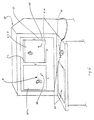
- Figs. 1-6 illustrate a diagnostic ultrasound/x-ray biopsy system embodiment of the present invention, as adapted for mammography/breast biopsy use.
- the system comprises a support assembly 10 having a patient table 12 with breast-opening 14 therethrough, an immobilization assembly 30 for immobilizing a patient's breast within a predetermined XYZ frame of reference under the opening 14 of table 12, an x-ray imaging assembly 40 for providing two-dimensional x-ray images (e.g., X-Y images) of the patient's immobilized breast in correlated spatial relation to the predetermined XYZ frame of reference, and an ultrasound imaging assembly 100 for providing orthogonal depth-profile images (e.g., X-Z, Y-Z and/or X,Y-Z images) of the immobilized breast in correlated spatial relation to the predetermined XYZ frame of reference.
- an immobilization assembly 30 for immobilizing a patient's breast within a predetermined XYZ frame of reference under the opening 14 of table 12
- an x-ray imaging assembly 40 for providing two-dimensional x-ray images (e.g., X-Y images) of the patient's immobil
- a biopsy assembly 50 having puncture instrument 52 is also provided for obtaining samples from a patient's breast while the breast is immobilized in the predetermined XYZ frame of reference.
- a display/processor assembly 60 is provided for recording/displaying the various images obtained/generated, for determining the coordinates of a user-identified location of interest within the breast and for monitoring/controlling/simulating the position of the various positionable assembly components.
- the illustrated embodiment may utilize the x-ray, automated biopsy and other functionalities embodied in the current MAMMOTEST® and MAMMOVISION® products of Fischer Imaging Corp. of Denver, Colorado, U.S.A.
- the present invention allows for the integration and effective use of ultrasound imaging with such products, thereby allowing medical equipment cost efficiencies to be realized.
- the MAMMOTEST® and MAMMOVISION® products include features corresponding with the disclosures in U.S. Patent Nos. 5,078,142 , 5,240,011 and 5,415,169 , which are incorporated by reference in their entirety.
- Support assembly 10 further includes pedestal 16 and cantilevered first and second support arms 20 and 22, respectively, for supportably interfacing the breast immobilization assembly 30, x-ray imaging assembly 40, ultrasound imaging assembly 100 and biopsy assembly 50 in a predetermined spatially correlated manner.
- First and second supports arms 20 and 22 can be jointly pivoted relative to pedestal 16, thereby providing imaging/biopsy access to the breast from different directions (e.g., 0°, +90° and -90° relative to the table longitudinal axis).
- second support arm 22 can be selectively pivoted relative to first support arm 20, to provide for stereotactic x-ray imaging (e.g., +15° and -15° relative to the first support arm longitudinal axis).
- Breast immobilization assembly 30 is supported on first support arm 20 and includes a stationary face plate 32 and opposing compression paddle 34 for immobilizing a patient's breast therebetween.
- Compression paddle 34 is x-ray transmittent and further includes a window 36 for direct breast access by the ultrasound imaging assembly 100 and/or biopsy assembly 50.
- Compression paddle 34 is selectively positionable along first support arm 20 (e.g., via motorized and position sensor systems) for controlled, registered movement toward/away from face plate 32 to accommodate breast positioning/removal and differing breast sizes.
- Compression paddle 34 can be readily removed from/interconnected to the first support arm 20 to accommodate the selective use of compression paddles of differing sizes, shapes, window positions, etc. As shown in Fig.
- X-ray imaging assembly 40 includes x-ray tube source 42 mounted on the end of second support arm 22 and x-ray receiver/imager 44 mounted on first support arm 20.
- x-ray tube source 42 provides x-ray radiation having a center axis C substantially perpendicular to the fronts of face plate 34 and x-ray receiver/imager 44, such x-ray radiation having a focal point positioned along the center axis C at a determinable location between the face plate 32 and compression paddle 34 during use.
- the x-ray receiver/imager 44 is disposed in abutting relation with the face plate 32.
- X-ray receiver/imager 44 may comprise an image receptor consisting of a removable radiographic film cassette (e.g., for full-field breast imaging) and/or digital camera (e.g., for partial field, real-time imaging/display).
- a partial field, digital CCD camera 46 e.g., having a 5 mm X 10 mm or 5 mm X 5mm imaging area
- ultrasound probe 110 may include an elongated housing 112 with an elongated ultrasound transducer module 114 positioned therein.
- Ultrasound transducer module 114 provides an ultrasound signal having a focal point on a signal center axis at a location between compression paddle 34 and face plate 32.
- Ultrasound transducer module 114 may include, for example, a phased linear array of ultrasound transducers positioned along a longitudinal axis of the ultrasound probe 110.
- the ultrasound probe 110 emits signal pulses and detects corresponding echo pulses to generate the depth-profile images.
- Arm assembly 130 is provided to allow the ultrasound imaging probe 110 to be rotated about one or more of selected X, Y and Z axes to obtain a desired pitch, roll and/or yaw orientation).
- arm assembly 130 can be controlled to selectively rotate the longitudinal axis, or pitch, of probe 110 so that the ultrasound signal may be employed to obtain depth-profile image in a plane, or "slice," within which an upwardly angled punction instrument 52 of biopsy assembly 50 may be orthogonally advanced, as will be further discussed.
- arm assembly 130 includes pivot arm 132 pivotally interconnected to XYZ ultrasound positioning assembly 140 via a lock/release mechanism (not shown) for selective, centered rotation of probe 116 about axis YY.
- Arm assembly 130 further includes arm 134 rotably interconnected to arm 132 via a lock/release mechanism (not shown) for selective, centered rotation of probe 116 about axis XX, and arm 136 rotably interconnected to arm 134 via a lock/release mechanism (not shown) for selective, centered rotation of probe 116 about axis ZZ.
- Internal optical encoders may be provided at the various arm interconnections, wherein the pitch, roll and/or yaw of probe 110 is automatically determinable in relation to the predetermined XYZ frame of reference.
- internal automated micropositioners may also be utilized under processor control.
- the ultrasound signal may be scanned to obtain depth-profile information for a predetermined layer, or "slice," within the region of interest.
- scanning may be provided electrically by driving a phased linear array of transducers comprising probe 110 in a known manner and/or via manual or automatic-driven control of XYZ positioning assembly 140 to mechanically move ultrasound imaging head 110.
- display/processor 60 includes a display screen 62 for displaying the acquired x-ray images on a first portion 62a and displaying corresponding depth-profile ultrasound images on a second portion 62b, each in registered co-relation to the predetermined XYZ frame of reference.
- Display/ processor 60 may further include a user interface means 64, e.g., keyboard 65 and mouse 66 and screen point cursor 68 (e.g., on both display portions 62a, 62b), wherein a user may identify (e.g., click upon) a specific location-of-interest within both an x-ray image and corresponding ultrasound image (e.g., corresponding with a potential lesion or suspicious mass), e.g, for automatic processor determination of the three-dimensional coordinates of the location within the predetermined XYZ frame of reference.
- a user interface means 64 e.g., keyboard 65 and mouse 66 and screen point cursor 68 (e.g., on both display portions 62a, 62b)
- a user may identify (e.g., click upon) a specific location-of-interest within both an x-ray image and corresponding ultrasound image (e.g., corresponding with a potential lesion or suspicious mass), e.g, for automatic processor determination of the three-dimensional coordinates of
- User interface means may further allow for user selection/display of a particular desired ultrasound depth-profile image, e.g., via mouse 66 and screen "slice" cursor 70 on the x-ray image display portion 62a. More particularly, screen "slice" cursor 70 may be employed to identify a particular slice, or layer, of an X-Y x-ray image for which a corresponding ultrasound depth-profile image is to be obtained (e.g., thereby resulting in processor-assisted positioning and imaging using probe 110) and/or accessed and displayed (e.g., where such ultrasound depth-profile image has been previously obtained/stored for selective subsequent review).
- screen "slice" cursor 70 may be employed to identify a particular slice, or layer, of an X-Y x-ray image for which a corresponding ultrasound depth-profile image is to be obtained (e.g., thereby resulting in processor-assisted positioning and imaging using probe 110) and/or accessed and displayed (e.g., where such ultrasound depth-profile
- display/processor 60 may be operatively interconnected (e.g., via electrical lines 80) to the various positionable assembly components for monitoring/controlling their respective positions relative to the predetermined XYZ frame of reference, including the positionable components of immobilization assembly 30, x-ray imaging assembly 40, ultrasound imaging assembly 110 and biopsy assembly 50.
- display/processor 60 may determine the three-dimensional coordinates of a specific location of interest, as described above, and in turn assist/control the positioning of biopsy assembly 50 so as to position the assembly for obtainment of a tissue sample from the location of interest.
- the display/processor 60 may also be employable to visually project, or simulate, the entry of a punction instrument 52 into a given location of interest, thereby allowing physicians the opportunity to insure an optimal positioning for biopsy entry prior to an actual biopsy procedure. Since three-dimensional visualization of a potential lesion/suspicious mass can be provided by the present invention, and since the disclosed arrangement allows for breast access by biopsy assembly 50 from a plurality of aspects (e.g., by selective mounting on either side of or coaxial along support arm 20), such simulated biopsy modeling may prone to be of particular advantage.
- the present invention allows for spatial correlation of the x-ray and ultrasound images utilizing various techniques.
- the X-Y x-ray images obtained can be readily correlated to the predetermined XYZ frame of reference since the position and attributes of x-ray source 42 and x-ray receiver/imager 44 are each known in relation to the predetermined XYZ frame of reference.
- the two X-Y stereotactic x-ray images can be employed to obtain a Z location for particular location of interest relative to the predetermined XYZ frame of reference utilizing known triangulation techniques, as will be appreciated by those skilled in the art.
- the XYZ positioning of ultrasound imaging head 110 is determinable relative to the predetermined XYZ frame of reference, as noted above.
- the ultrasound imaging head 110 will emit/detect ultrasound signals in substantially the same plane as the surface of compression paddle 34 contacting the imaged breast.
- the position of such surface relative to the predetermined XYZ frame of reference e.g., the Z distance to face plate 32
- a depth profile comprising a potential lesion/suspicious mass can be spatially related in a reliable manner to the acquired x-ray images.
- a patient can be positioned on the table 12 with a breast positioned through opening 14.
- Compression paddle 34 is then advanced along first support arm 20 to compress the breast to define a cross-sectional imaging area having a common thickness and to otherwise immobilize the breast in a set position within the predetermined XYZ frame of reference.
- X-ray imaging assembly 40 is then selectively positioned to obtain the desired x-ray images. Such x-ray images are then reviewed using display/processor 60, to identify, analyze and or otherwise confirm the presence and location of a potential lesion or suspicious mass for ultrasound imaging. Alternatively, the general location of a potential lesion or suspicious mass may already be known due to prior x-ray screening.
- the patient should be positioned/repositioned so that the potential lesion or suspicious mass is positioned within the accessible field of view of ultrasound imaging head 110 when it is maneuvered through the window 36 of compression paddle 34 in direct contact with the imaged breast.
- new x-ray and corresponding ultrasound images should be generated for each position in which a breast is immobilized within the predetermined XYZ frame of reference.
- ultrasound imaging probe 110 is positioned through the window 36 and a series of ultrasound images are obtained and displayed on display/processor 60.
- Cursor 66 control of the ultrasound images taken across the area of interest provides additional, valuable information as to the type of potential lesion/suspicious mass originally noted on an original mammogram. For example, with proper training of ultrasound and x-ray imaging techniques, physicians may rule out the possibility of a solid mass in favor of a fluid-filled cyst. Or, additional ultrasound characteristics may aid the physician in making a definitive diagnosis.
- the specific location from which tissue is to be obtained can be identified using mouse 66 to position screen point cursor 68 on both the x-ray image and correlated ultrasound depth-profile image on display/processor 60. Three-dimensional coordinates can then be determined and utilized by display/processor 60 to control positioning of biopsy assembly 50.
- specific attributes of the particular punction subassembly 54 utilized will have been previously entered into by display/processor 60.
- display/processor 60 may be employed to simulate the advancement of punction instrument 52 into the breast from a given potential position, thereby allowing the physician to determine if breast biopsy access from a different position may be more desirable.
Landscapes
- Health & Medical Sciences (AREA)
- Life Sciences & Earth Sciences (AREA)
- Engineering & Computer Science (AREA)
- Medical Informatics (AREA)
- Surgery (AREA)
- Heart & Thoracic Surgery (AREA)
- Animal Behavior & Ethology (AREA)
- Nuclear Medicine, Radiotherapy & Molecular Imaging (AREA)
- Veterinary Medicine (AREA)
- Pathology (AREA)
- Public Health (AREA)
- Biomedical Technology (AREA)
- General Health & Medical Sciences (AREA)
- Molecular Biology (AREA)
- Radiology & Medical Imaging (AREA)
- Biophysics (AREA)
- Physics & Mathematics (AREA)
- High Energy & Nuclear Physics (AREA)
- Optics & Photonics (AREA)
- Oral & Maxillofacial Surgery (AREA)
- Computer Vision & Pattern Recognition (AREA)
- Dentistry (AREA)
- Neurosurgery (AREA)
- Apparatus For Radiation Diagnosis (AREA)
- Ultra Sonic Daignosis Equipment (AREA)
Abstract
Claims (8)
- Appareil médical, comprenant :un moyen d'imagerie par rayons X (40) positionné de façon supportée dans une co-relation connue sur un cadre de référence tridimensionnel prédéterminé pour transmettre un rayonnement de rayons X à travers une région d'intérêt d'un corps positionnée à l'intérieur dudit cadre de référence tridimensionnel prédéterminé, et pour fournir des données d'image par rayons X correspondant à une ou plusieurs images par rayons X de la région d'intérêt du corps ;un moyen d'imagerie par ultrasons (100), positionné de façon supportée dans une co-relation connue sur le cadre de référence tridimensionnel prédéterminé et positionnable en contact direct avec ladite région d'intérêt du corps, pour diriger un signal ultrasonore directement à l'intérieur de ladite région d'intérêt du corps positionnée à l'intérieur dudit cadre de référence tridimensionnel prédéterminé, et pour fournir des données d'image par ultrasons correspondant à une ou plusieurs images par ultrasons de la région d'intérêt du corps en co-relation spatiale avec lesdites images par rayons X, dans lequel lesdites données d'image par rayons X et par ultrasons fournissent de façon combinée des données d'image tridimensionnelle corrélées correspondant à la région d'intérêt du corps, dans lequel ladite région d'intérêt du corps est un sein humain, caractérisé en ce que l'appareil comprend en outre :un moyen d'immobilisation (30) pour immobiliser ledit sein entre un premier et un deuxième éléments de compression (34, 32), ledit premier élément de compression (34) ayant une ouverture (36) à travers celui-ci, dans lequel ledit moyen d'imagerie par ultrasons (100) est sélectivement positionnable à travers ladite ouverture (36) pour être en contact direct avec le sein.
- Appareil médical, selon la revendication 1, comprenant en outre :un moyen pour utiliser (60) lesdites données d'image par rayons X et par ultrasons pour identifier un emplacement d'intérêt à l'intérieur de ladite région d'intérêt du corps ; etun moyen de biopsie (50), positionnable dans une relation prédéterminée par rapport audit cadre de référence tridimensionnel prédéterminé, pour obtenir un échantillon provenant dudit emplacement d'intérêt.
- Appareil médical, selon la revendication 2, dans lequel :ledit moyen de biopsie (50) inclut :un instrument de ponction (52) ; etun moyen de positionnement (56) pour positionner sélectivement ledit instrument de ponction selon un angle d'entrée par rapport audit cadre de référence tridimensionnel prédéterminé ; etledit moyen d'imagerie par ultrasons comprend :une sonde d'imagerie par ultrasons (110) ; etun moyen pour positionner sélectivement (140) ladite sonde d'imagerie par rapport audit cadre de référence tridimensionnel prédéterminé,dans lequel l'axe longitudinal de la sonde à ultrasons (110) est aligné avec l'angle d'entrée de l'instrument de ponction (52) de telle sorte que l'instrument de ponction (52) est imagé en temps réel lorsqu'il est fait pénétrer dans l'emplacement d'intérêt.
- Appareil médical, selon la revendication 1, ledit moyen d'imagerie par rayons X (40) comprenant :une caméra numérique (46) sélectivement mobile et positionnable à l'intérieur d'un plan substantiellement perpendiculaire à un axe central dudit rayonnement de rayons X ; etcomprenant en outre :un moyen d'affichage et d'interface utilisateur (60) pour une identification par l'utilisateur, en utilisant une image par rayons X acquise et affichée, d'une image par ultrasons acquise devant être affichée.
- Appareil médical, selon la revendication 1, comprenant :un moyen d'affichage (62) pour afficher lesdites images par rayons X et par ultrasons dans une corrélation enregistrée.
- Procédé d'utilisation lors de l'exécution d'une procédure médicale, comprenant :le positionnement de façon supportée d'un moyen d'imagerie par rayons X (40) et d'un moyen d'imagerie par ultrasons (100) dans une co-relation connue sur un cadre de référence tridimensionnel prédéterminé, dans lequel une région d'intérêt d'un corps est positionnée à l'intérieur dudit cadre de référence prédéterminé ; dans lequel le moyen d'imagerie par ultrasons est positionnable en contact direct avec ladite région d'intérêt du corps ;l'imagerie par rayons X de ladite région d'intérêt du corps avec un rayonnement de rayons X en utilisant ledit moyen d'imagerie par rayons X (40) pour obtenir des données d'image par rayons X correspondant à une ou plusieurs images par rayons X ;l'utilisation de ladite ou desdites images par rayons X pour identifier un emplacement d'intérêt à l'intérieur d'une partie limitée sélectionnée de ladite région d'intérêt du corps ; etl'imagerie par ultrasons uniquement de ladite partie limitée sélectionnée de ladite région d'intérêt du corps avec un signal ultrasonore dirigé directement à l'intérieur de ladite région d'intérêt du corps en utilisant ledit moyen d'imagerie par ultrasons (100) pour obtenir des données d'image par ultrasons correspondant à une ou plusieurs images par ultrasons, dans lequel lesdites données d'image par rayons X et par ultrasons fournissent de façon combinée des données d'image tridimensionnelle corrélées correspondant à la partie limitée sélectionnée ; etla mise en contact direct de ladite partie limitée sélectionnée de ladite région d'intérêt du corps à travers une ouverture dans un moyen d'immobilisation avec ledit moyen d'imagerie par ultrasons (100).
- Procédé selon la revendication 6, ladite étape d'imagerie par ultrasons comprenant en outre :le balayage dudit signal ultrasonore à travers ladite partie limitée sélectionnée de ladite région d'intérêt du corps.
- Procédé selon la revendication 7, comprenant en outre :la génération d'un modèle tridimensionnel dudit emplacement d'intérêt en utilisant lesdites données d'image par rayons X et d'image par ultrasons.
Applications Claiming Priority (3)
| Application Number | Priority Date | Filing Date | Title |
|---|---|---|---|
| US730107 | 1996-10-15 | ||
| US08/730,107 US5776062A (en) | 1996-10-15 | 1996-10-15 | Enhanced breast imaging/biopsy system employing targeted ultrasound |
| PCT/US1997/018468 WO1998016149A2 (fr) | 1996-10-15 | 1997-10-14 | Systeme ameliore d'imagerie mammaire / de biopsie a echographie ciblee |
Publications (4)
| Publication Number | Publication Date |
|---|---|
| EP0936889A2 EP0936889A2 (fr) | 1999-08-25 |
| EP0936889A4 EP0936889A4 (fr) | 1999-12-01 |
| EP0936889B1 true EP0936889B1 (fr) | 2010-07-28 |
| EP0936889B8 EP0936889B8 (fr) | 2010-11-24 |
Family
ID=24933944
Family Applications (1)
| Application Number | Title | Priority Date | Filing Date |
|---|---|---|---|
| EP97911697A Expired - Lifetime EP0936889B8 (fr) | 1996-10-15 | 1997-10-14 | Systeme ameliore d'imagerie mammaire / de biopsie a echographie ciblee |
Country Status (5)
| Country | Link |
|---|---|
| US (3) | US5776062A (fr) |
| EP (1) | EP0936889B8 (fr) |
| AU (1) | AU4900997A (fr) |
| DE (1) | DE69739947D1 (fr) |
| WO (1) | WO1998016149A2 (fr) |
Cited By (1)
| Publication number | Priority date | Publication date | Assignee | Title |
|---|---|---|---|---|
| DE102017210604A1 (de) * | 2017-06-23 | 2018-12-27 | Siemens Healthcare Gmbh | Kompressionseinheit für ein kombiniertes Röntgen-/Ultraschall-Untersuchungsgerät |
Families Citing this family (151)
| Publication number | Priority date | Publication date | Assignee | Title |
|---|---|---|---|---|
| US5983123A (en) | 1993-10-29 | 1999-11-09 | United States Surgical Corporation | Methods and apparatus for performing ultrasound and enhanced X-ray imaging |
| FR2751109B1 (fr) * | 1996-07-09 | 1998-10-09 | Ge Medical Syst Sa | Procede de localisation d'un element d'interet contenu dans un objet tridimensionnel, en particulier lors d'un examen de stereotaxie en mammographie |
| EP0925025A2 (fr) * | 1996-08-15 | 1999-06-30 | Life Imaging Systems Inc. | Systeme et procede servant a effectuer une biopsie percutanee dans le sein par ultrasonographie tridimensionnelle |
| FR2754612B1 (fr) * | 1996-10-10 | 1998-11-20 | Ge Medical Syst Sa | Systeme de localisation de cassettes de prise d'images numeriques |
| US5776062A (en) * | 1996-10-15 | 1998-07-07 | Fischer Imaging Corporation | Enhanced breast imaging/biopsy system employing targeted ultrasound |
| US6731966B1 (en) | 1997-03-04 | 2004-05-04 | Zachary S. Spigelman | Systems and methods for targeting a lesion |
| DE19809460C1 (de) * | 1998-03-06 | 1999-09-30 | Siemens Ag | Medizinisches Zielgerät zur atemadaptierten Punktion |
| WO2000009014A1 (fr) * | 1998-08-17 | 2000-02-24 | Kari Richter | Appareil combinant radiographie et ultrasonographie |
| EP1143845A4 (fr) * | 1998-11-25 | 2004-10-06 | Fischer Imaging Corp | Systeme d'interface utilisateur pour imageur mammographique |
| AU2003203755B2 (en) * | 1998-11-25 | 2005-04-28 | Rubicor Medical, Inc. | Breast Stabilization Devices and Imaging and Interventional Methods Using Same |
| US6574499B1 (en) | 1998-11-25 | 2003-06-03 | Xdata Corporation | Mammography method and apparatus |
| AU4694900A (en) * | 1999-05-03 | 2000-11-17 | Biotrack, Inc. | Systems and methods for targeting a breast lesion |
| US6421454B1 (en) * | 1999-05-27 | 2002-07-16 | Litton Systems, Inc. | Optical correlator assisted detection of calcifications for breast biopsy |
| US6396940B1 (en) | 1999-05-27 | 2002-05-28 | Litton Systems, Inc. | Optical correlator based automated pathologic region of interest selector for integrated 3D ultrasound and digital mammography |
| US6512943B1 (en) | 2000-05-22 | 2003-01-28 | Wisconsin Alumni Research Foundation | Combined ultrasound-radionuclide device for percutaneous ultrasound-guided biopsy and method of use |
| ATE353593T1 (de) | 2000-07-07 | 2007-03-15 | Medical Positioning Inc | Patientenunterstützung und verfahren zur dekubitusbrustbiopsie |
| US6367104B1 (en) * | 2000-07-07 | 2002-04-09 | Medical Positioning, Inc. | Patient support apparatus and method for performing decubitus breast biopsy |
| US7940966B2 (en) * | 2000-11-24 | 2011-05-10 | U-Systems, Inc. | Full-field breast image data processing and archiving |
| US7103205B2 (en) * | 2000-11-24 | 2006-09-05 | U-Systems, Inc. | Breast cancer screening with ultrasound image overlays |
| US7597663B2 (en) | 2000-11-24 | 2009-10-06 | U-Systems, Inc. | Adjunctive ultrasound processing and display for breast cancer screening |
| US7615008B2 (en) * | 2000-11-24 | 2009-11-10 | U-Systems, Inc. | Processing and displaying breast ultrasound information |
| US7556602B2 (en) * | 2000-11-24 | 2009-07-07 | U-Systems, Inc. | Breast cancer screening with adjunctive ultrasound mammography |
| US6678546B2 (en) * | 2001-01-30 | 2004-01-13 | Fischer Imaging Corporation | Medical instrument guidance using stereo radiolocation |
| US6558337B2 (en) * | 2001-04-17 | 2003-05-06 | Wisconsin Alumni Research Foundation | Positioner for medical devices such as biopsy needles |
| IL143060A0 (en) * | 2001-05-09 | 2002-04-21 | Sonnet Medical Ltd In Formatio | Method and apparatus for breast imaging utilizing ultrasound |
| US6478739B1 (en) | 2001-05-11 | 2002-11-12 | The Procter & Gamble Company | Ultrasonic breast examination system |
| US6491632B1 (en) * | 2001-06-26 | 2002-12-10 | Geoffrey L. Taylor | Method and apparatus for photogrammetric orientation of ultrasound images |
| US20030045798A1 (en) * | 2001-09-04 | 2003-03-06 | Richard Hular | Multisensor probe for tissue identification |
| US6546279B1 (en) * | 2001-10-12 | 2003-04-08 | University Of Florida | Computer controlled guidance of a biopsy needle |
| WO2003037046A2 (fr) | 2001-10-19 | 2003-05-01 | Hologic, Inc. | Systeme et procede de mammographie utilisant des palpateurs de compression excentres, la collimation automatique et une grille anti-diffusion retractable |
| US7609806B2 (en) | 2004-10-18 | 2009-10-27 | Hologic Inc. | Mammography system and method employing offset compression paddles, automatic collimations, and retractable anti-scatter grid |
| US20030149364A1 (en) * | 2002-02-01 | 2003-08-07 | Ajay Kapur | Methods, system and apparatus for digital imaging |
| US6971991B2 (en) * | 2002-03-08 | 2005-12-06 | Imperium, Inc. | Apparatus for multimodal plane wave ultrasound imaging |
| US6707878B2 (en) | 2002-04-15 | 2004-03-16 | General Electric Company | Generalized filtered back-projection reconstruction in digital tomosynthesis |
| US7783089B2 (en) * | 2002-04-15 | 2010-08-24 | General Electric Company | Method and apparatus for providing mammographic image metrics to a clinician |
| US6882700B2 (en) * | 2002-04-15 | 2005-04-19 | General Electric Company | Tomosynthesis X-ray mammogram system and method with automatic drive system |
| US20030194050A1 (en) * | 2002-04-15 | 2003-10-16 | General Electric Company | Multi modality X-ray and nuclear medicine mammography imaging system and method |
| US7218766B2 (en) * | 2002-04-15 | 2007-05-15 | General Electric Company | Computer aided detection (CAD) for 3D digital mammography |
| US6724856B2 (en) * | 2002-04-15 | 2004-04-20 | General Electric Company | Reprojection and backprojection methods and algorithms for implementation thereof |
| US7826883B2 (en) * | 2002-04-23 | 2010-11-02 | Devicor Medical Products, Inc. | Localization mechanism for an MRI compatible biopsy device |
| US20030199753A1 (en) * | 2002-04-23 | 2003-10-23 | Ethicon Endo-Surgery | MRI compatible biopsy device with detachable probe |
| US6748047B2 (en) * | 2002-05-15 | 2004-06-08 | General Electric Company | Scatter correction method for non-stationary X-ray acquisitions |
| EP2272434A1 (fr) | 2002-05-31 | 2011-01-12 | U-Systems, Inc. | Dépistage du cancer du sein au moyen d'une mammographie ultrasonore auxiliaire |
| US20040082856A1 (en) * | 2002-07-16 | 2004-04-29 | Alfred E. Mann Institute For Biomedical Engineering, University Of Southern California | Support bra for ultrasonic breast scanner |
| US20040022447A1 (en) * | 2002-07-31 | 2004-02-05 | General Electric Company | Method and system for image compression and decompression using span of interest of an imaging sequence |
| US7438692B2 (en) * | 2002-10-18 | 2008-10-21 | Mark Tsonton | Localization mechanism for an MRI compatible biopsy device |
| WO2006058160A2 (fr) | 2004-11-26 | 2006-06-01 | Hologic, Inc. | Systeme et procede radiographiques multimode integrant mammographie/tomosynthese |
| CN100444802C (zh) * | 2003-01-17 | 2008-12-24 | 朴熙鹏 | 可变形物的超声波检查装置 |
| EP1620014A4 (fr) * | 2003-04-16 | 2009-06-17 | Eastern Virginia Med School | Systeme et procede de production d'images par ultrasons independantes de l'operateur |
| US8083678B2 (en) * | 2003-04-16 | 2011-12-27 | Eastern Virginia Medical School | System, method and medium for acquiring and generating standardized operator independent ultrasound images of fetal, neonatal and adult organs |
| US6846289B2 (en) * | 2003-06-06 | 2005-01-25 | Fischer Imaging Corporation | Integrated x-ray and ultrasound medical imaging system |
| US20050089205A1 (en) * | 2003-10-23 | 2005-04-28 | Ajay Kapur | Systems and methods for viewing an abnormality in different kinds of images |
| US7313259B2 (en) * | 2003-11-26 | 2007-12-25 | General Electric Company | Method, system and computer program product for multi-modality registration using virtual cursors |
| EP1711119A1 (fr) * | 2004-01-23 | 2006-10-18 | Traxyz Medical, Inc. | Procedes et appareil permettant de realiser des interventions sur des emplacements cibles dans le corps |
| DE102004034240A1 (de) * | 2004-07-15 | 2006-02-16 | Siemens Ag | Gerät zur medizinische Bildgebung |
| ES2253997B1 (es) * | 2004-07-29 | 2007-07-16 | Udiat Centre Diagnostic, S.A. | Sistema digital para realizar biopsia estereotaxica. |
| US20060074287A1 (en) * | 2004-09-30 | 2006-04-06 | General Electric Company | Systems, methods and apparatus for dual mammography image detection |
| WO2006043859A1 (fr) * | 2004-10-18 | 2006-04-27 | Mobile Robotics Sweden Ab | Robot d'examen ultrasonore |
| EP1816957A4 (fr) * | 2004-11-02 | 2009-10-21 | Metrohealth System | Procede et appareil permettant de determiner une correlation entre les coordonnees spatiales dans un sein |
| US10026338B2 (en) * | 2004-11-30 | 2018-07-17 | The Regents Of The University Of California | Embedded motion sensing technology for integration within commercial ultrasound probes |
| US10726741B2 (en) | 2004-11-30 | 2020-07-28 | The Regents Of The University Of California | System and method for converting handheld diagnostic ultrasound systems into ultrasound training systems |
| US11627944B2 (en) | 2004-11-30 | 2023-04-18 | The Regents Of The University Of California | Ultrasound case builder system and method |
| DE102005039658B3 (de) * | 2005-08-22 | 2007-07-19 | Siemens Ag | Laservorrichtung für ein Mammographiegerät |
| WO2013078476A1 (fr) | 2011-11-27 | 2013-05-30 | Hologic, Inc. | Système et procédé pour générer une image 2d en utilisant des données d'images de mammographie et/ou de tomosynthèse |
| US8303505B2 (en) * | 2005-12-02 | 2012-11-06 | Abbott Cardiovascular Systems Inc. | Methods and apparatuses for image guided medical procedures |
| US8532745B2 (en) | 2006-02-15 | 2013-09-10 | Hologic, Inc. | Breast biopsy and needle localization using tomosynthesis systems |
| US20070282221A1 (en) * | 2006-06-02 | 2007-12-06 | U-Systems, Inc. | Ultrasound assisted and x-ray assisted biopsy devices |
| WO2007130526A2 (fr) * | 2006-05-02 | 2007-11-15 | U-Systems, Inc. | balayage À ultrasons et biopsie assistÉe par ultrasons |
| SE530549C2 (sv) * | 2006-10-31 | 2008-07-08 | Xcounter Ab | System för avbildning av ett bröst genom datortomografi |
| WO2008054279A1 (fr) * | 2006-10-31 | 2008-05-08 | Xcounter Ab | Dispositif d'imagerie et système pour l'imagerie |
| JP5481038B2 (ja) | 2007-04-05 | 2014-04-23 | 株式会社東芝 | 超音波診断装置、乳房イメージングシステム及び乳房イメージングプログラム |
| US8133242B1 (en) | 2007-04-27 | 2012-03-13 | Q-Tech Medical Incorporated | Image-guided extraluminal occlusion |
| US10201324B2 (en) | 2007-05-04 | 2019-02-12 | Delphinus Medical Technologies, Inc. | Patient interface system |
| US8870771B2 (en) | 2007-05-04 | 2014-10-28 | Barbara Ann Karmanos Cancer Institute | Method and apparatus for categorizing breast density and assessing cancer risk utilizing acoustic parameters |
| US8066644B2 (en) * | 2007-05-17 | 2011-11-29 | Vanderbilt University | System, method and device for positioning a target located within soft tissue in a path of an instrument |
| WO2009026587A1 (fr) * | 2007-08-23 | 2009-02-26 | Fischer Medical Technologies, Inc. | Mammographie par tomodensitométrie calculée améliorée et système de biopsie |
| WO2009050712A2 (fr) * | 2007-10-15 | 2009-04-23 | Yissum Research Development Company Of The Hebrew Univercity Of Jerusalem | Procédé, système et programme informatique pour la caractérisation d'un tissu |
| DE102008009967A1 (de) * | 2008-02-20 | 2009-09-17 | Siemens Aktiengesellschaft | Mammographieanlage und Verfahren zur sono- und röntgenograpischen Untersuchung einer Brust |
| US8375054B2 (en) * | 2008-04-03 | 2013-02-12 | Siemens Aktiengesellschaft | Findings navigator |
| US10603008B2 (en) * | 2009-02-19 | 2020-03-31 | Tessonics Corporation | Ultrasonic device for assessment of internal tooth structure |
| JP5373450B2 (ja) * | 2009-03-31 | 2013-12-18 | 富士フイルム株式会社 | 生検装置及び生検装置の動作方法 |
| US8556815B2 (en) * | 2009-05-20 | 2013-10-15 | Laurent Pelissier | Freehand ultrasound imaging systems and methods for guiding fine elongate instruments |
| US10039527B2 (en) * | 2009-05-20 | 2018-08-07 | Analogic Canada Corporation | Ultrasound systems incorporating spatial position sensors and associated methods |
| CN106420066B (zh) * | 2009-10-08 | 2020-08-25 | 霍罗吉克公司 | 将穿刺活检组件引导到目标位置的方法及x射线成像系统 |
| JP5317933B2 (ja) * | 2009-11-17 | 2013-10-16 | 富士フイルム株式会社 | 画像表示装置、及びそのプログラム |
| CN102933153A (zh) * | 2010-01-29 | 2013-02-13 | 弗吉尼亚大学专利基金会 | 用于定位解剖结构或探针引导的超声 |
| CN102843959B (zh) | 2010-02-12 | 2014-11-12 | 戴尔菲纳斯医疗科技公司 | 表征组织对治疗方案的病理反应的方法 |
| CN102869301B (zh) | 2010-02-12 | 2016-06-29 | 戴尔菲纳斯医疗科技公司 | 表征病人的组织的方法 |
| US8211121B1 (en) | 2010-03-06 | 2012-07-03 | Q-Tech Medical Incorporated | Methods and apparatus for image-guided extraluminal occlusion using clamping jaws |
| EP2577279A4 (fr) * | 2010-06-03 | 2015-01-14 | Caperay Medical Pty Ltd | Système de balayage à deux modalités pour détecter un cancer du sein |
| US20120133600A1 (en) | 2010-11-26 | 2012-05-31 | Hologic, Inc. | User interface for medical image review workstation |
| CA2829349C (fr) | 2011-03-08 | 2021-02-09 | Hologic, Inc. | Systeme et procede pour une imagerie de seins a double energie et/ou a injection d'un agent de contraste pour un depistage, un diagnostic et une biopsie |
| DE102011006058A1 (de) * | 2011-03-24 | 2012-09-27 | Siemens Aktiengesellschaft | Tomosynthesegerät und Verfahren zu seinem Betrieb |
| JP6057985B2 (ja) | 2011-04-26 | 2017-01-11 | ユニバーシティ オブ バージニア パテント ファウンデーション | 超音波を使用する骨表面画像再構成 |
| US12042134B2 (en) | 2011-09-16 | 2024-07-23 | Hologic, Inc. | Breast biopsy lateral arm system |
| US11284869B2 (en) | 2011-09-16 | 2022-03-29 | Hologic, Inc. | Breast biopsy lateral arm system |
| ES2795416T3 (es) | 2011-09-16 | 2020-11-23 | Hologic Inc | Sistema de brazo lateral para biopsia de mama |
| US9295449B2 (en) | 2012-01-23 | 2016-03-29 | Ultrasonix Medical Corporation | Landmarks for ultrasound imaging |
| US8914925B2 (en) * | 2012-02-08 | 2014-12-23 | Wayne County Employees' Retirement System | Mobile diagnostic assembly |
| JP6240097B2 (ja) | 2012-02-13 | 2017-11-29 | ホロジック インコーポレイティッド | 合成画像データを使用してトモシンセシススタックをナビゲートする方法 |
| US20130303895A1 (en) * | 2012-05-14 | 2013-11-14 | Delphinus Medical Technologies, Inc. | System and Method for Performing an Image-Guided Biopsy |
| US9439622B2 (en) | 2012-05-22 | 2016-09-13 | Covidien Lp | Surgical navigation system |
| US9439627B2 (en) | 2012-05-22 | 2016-09-13 | Covidien Lp | Planning system and navigation system for an ablation procedure |
| US8750568B2 (en) | 2012-05-22 | 2014-06-10 | Covidien Lp | System and method for conformal ablation planning |
| US9439623B2 (en) | 2012-05-22 | 2016-09-13 | Covidien Lp | Surgical planning system and navigation system |
| US9498182B2 (en) | 2012-05-22 | 2016-11-22 | Covidien Lp | Systems and methods for planning and navigation |
| US11631342B1 (en) | 2012-05-25 | 2023-04-18 | The Regents Of University Of California | Embedded motion sensing technology for integration within commercial ultrasound probes |
| CN102697539B (zh) * | 2012-06-27 | 2016-04-27 | 青岛银泰医疗科技有限公司 | 一种新型的三维穿刺定位系统 |
| US20140073907A1 (en) | 2012-09-12 | 2014-03-13 | Convergent Life Sciences, Inc. | System and method for image guided medical procedures |
| US9763641B2 (en) | 2012-08-30 | 2017-09-19 | Delphinus Medical Technologies, Inc. | Method and system for imaging a volume of tissue with tissue boundary detection |
| US20150245816A1 (en) * | 2012-09-13 | 2015-09-03 | Koninklijke Philips N.V. | Ultrasound imaging device operated by mobile display device and ultrasound imaging system |
| US10157491B2 (en) | 2012-12-28 | 2018-12-18 | Koninklijke Philips N.V. | Real-time scene-modeling combining 3D ultrasound and 2D X-ray imagery |
| US10123770B2 (en) | 2013-03-13 | 2018-11-13 | Delphinus Medical Technologies, Inc. | Patient support system |
| US9271686B2 (en) | 2013-03-14 | 2016-03-01 | West Virginia University | Endorectal prostate probe composed of a combined mini gamma camera and ultrasound sensor |
| US10568560B2 (en) | 2013-03-14 | 2020-02-25 | West Virginia University | Endorectal prostate probe with combined PET and US modalities |
| US10092358B2 (en) | 2013-03-15 | 2018-10-09 | Hologic, Inc. | Tomosynthesis-guided biopsy apparatus and method |
| KR101656776B1 (ko) * | 2013-09-03 | 2016-09-12 | 삼성전자주식회사 | 초음파 진단 장치 및 초음파 진단 장치의 동작 방법 |
| US10380919B2 (en) | 2013-11-21 | 2019-08-13 | SonoSim, Inc. | System and method for extended spectrum ultrasound training using animate and inanimate training objects |
| US11364005B2 (en) | 2013-10-24 | 2022-06-21 | Hologic, Inc. | System and method for navigating x-ray guided breast biopsy |
| AU2015222981B2 (en) | 2014-02-28 | 2019-01-31 | Hologic, Inc. | System and method for generating and displaying tomosynthesis image slabs |
| WO2015140782A1 (fr) * | 2014-03-18 | 2015-09-24 | Doron Kwiat | Procédé de biopsie et aspects cliniques pour l'imagerie et la biopsie |
| CN106470615B (zh) * | 2014-05-28 | 2021-01-15 | 通用电气公司 | 活检方法和相关活检装置 |
| US10285667B2 (en) | 2014-08-05 | 2019-05-14 | Delphinus Medical Technologies, Inc. | Method for generating an enhanced image of a volume of tissue |
| US9950194B2 (en) | 2014-09-09 | 2018-04-24 | Mevion Medical Systems, Inc. | Patient positioning system |
| US9949719B2 (en) | 2014-12-16 | 2018-04-24 | General Electric Company | Breast imaging method and system |
| US9855014B2 (en) | 2014-12-16 | 2018-01-02 | General Electric Company | Compression paddle for use in breast imaging |
| US11600201B1 (en) | 2015-06-30 | 2023-03-07 | The Regents Of The University Of California | System and method for converting handheld diagnostic ultrasound systems into ultrasound training systems |
| US10786224B2 (en) | 2016-04-21 | 2020-09-29 | Covidien Lp | Biopsy devices and methods of use thereof |
| US10372876B2 (en) * | 2017-01-20 | 2019-08-06 | Agfa Healthcare Inc. | System and method for providing breast image data |
| US10896628B2 (en) | 2017-01-26 | 2021-01-19 | SonoSim, Inc. | System and method for multisensory psychomotor skill training |
| US11399790B2 (en) | 2017-03-30 | 2022-08-02 | Hologic, Inc. | System and method for hierarchical multi-level feature image synthesis and representation |
| EP3600051B1 (fr) | 2017-03-30 | 2024-05-01 | Hologic, Inc. | Procédé de synthèse de données d'image de basse dimension à partir de données d'image de grande dimension à l'aide d'une augmentation de grille d'objet |
| JP7174710B2 (ja) | 2017-03-30 | 2022-11-17 | ホロジック, インコーポレイテッド | 合成乳房組織画像を生成するための標的オブジェクト増強のためのシステムおよび方法 |
| WO2018236565A1 (fr) | 2017-06-20 | 2018-12-27 | Hologic, Inc. | Procédé et système d'imagerie médicale à auto-apprentissage dynamique |
| GB2566942B (en) * | 2017-09-22 | 2020-06-03 | Caperay Medical Pty Ltd | Multimodal imaging system and method |
| EP3510930B1 (fr) | 2018-01-15 | 2021-07-14 | Hologic, Inc. | Support de grossissement automatisé et configurable |
| US11331161B2 (en) | 2018-03-23 | 2022-05-17 | Covidien Lp | Surgical assemblies facilitating tissue marking and methods of use thereof |
| EP3787514A4 (fr) * | 2018-04-30 | 2022-01-05 | Memorial Sloan-Kettering Cancer Center | Palettes de compression pour biopsies mammaires |
| AU2019262183A1 (en) | 2018-05-04 | 2020-09-10 | Hologic, Inc. | Biopsy needle visualization |
| EP3833290A1 (fr) | 2018-08-10 | 2021-06-16 | Covidien LP | Systèmes de visualisation d'ablation |
| JP7084291B2 (ja) * | 2018-12-07 | 2022-06-14 | 富士フイルム株式会社 | トモシンセシス撮影支援装置、方法およびプログラム |
| US11810473B2 (en) | 2019-01-29 | 2023-11-07 | The Regents Of The University Of California | Optical surface tracking for medical simulation |
| US11495142B2 (en) | 2019-01-30 | 2022-11-08 | The Regents Of The University Of California | Ultrasound trainer with internal optical tracking |
| US11517294B2 (en) | 2019-05-07 | 2022-12-06 | Covidien Lp | Biopsy devices and methods of use thereof |
| US11883206B2 (en) | 2019-07-29 | 2024-01-30 | Hologic, Inc. | Personalized breast imaging system |
| DE202020006045U1 (de) | 2019-09-27 | 2024-07-02 | Hologic Inc. | KI-System zum Vorhersagen von Lesezeit und Lesekomplexität zum Überprüfen von 2D-/3D-Brustbildern |
| EP3832689A3 (fr) | 2019-12-05 | 2021-08-11 | Hologic, Inc. | Systèmes et procédés pour améliorer la durée de vie d'un tube à rayons x |
| US11471118B2 (en) | 2020-03-27 | 2022-10-18 | Hologic, Inc. | System and method for tracking x-ray tube focal spot position |
| US11481038B2 (en) | 2020-03-27 | 2022-10-25 | Hologic, Inc. | Gesture recognition in controlling medical hardware or software |
| KR102522627B1 (ko) * | 2020-09-17 | 2023-04-17 | 주식회사 제이시스메디칼 | 초음파 발생부의 집속 깊이의 변경이 가능한 초음파 의료 장치 |
| US11013492B1 (en) | 2020-11-04 | 2021-05-25 | Philip B. Kivitz | Ultrasound sonographic imaging system and method |
Family Cites Families (49)
| Publication number | Priority date | Publication date | Assignee | Title |
|---|---|---|---|---|
| US2707662A (en) * | 1949-05-27 | 1955-05-03 | Picker X Ray Corp Waite Mfg | Tiltably X-ray table with extension panel |
| US3165630A (en) * | 1961-06-13 | 1965-01-12 | Polaroid Corp | Table for holding and positioning a female subject and film during breast x-ray exposures |
| US3973126A (en) * | 1975-07-31 | 1976-08-03 | General Electric Company | Mammography |
| US3963933A (en) * | 1975-08-18 | 1976-06-15 | General Electric Company | Mammography fixture |
| US4051380A (en) * | 1976-03-31 | 1977-09-27 | Lasky Harold J | Apparatus and method for supporting and positioning the body to facilitate radiographic mammography procedures |
| US4099880A (en) * | 1976-08-11 | 1978-07-11 | Tsutomu Kano | Method and an apparatus for stereoscopic measurement utilizing a three-dimensional image |
| US4249539A (en) * | 1979-02-09 | 1981-02-10 | Technicare Corporation | Ultrasound needle tip localization system |
| DE2936259A1 (de) * | 1979-09-07 | 1981-03-19 | Siemens AG, 1000 Berlin und 8000 München | Vorrichtung zum punktieren von koerperinternen organen, gefaessen o.dgl. |
| US4341120A (en) * | 1979-11-09 | 1982-07-27 | Diasonics Cardio/Imaging, Inc. | Ultrasonic volume measuring system |
| US4485819A (en) * | 1980-01-21 | 1984-12-04 | Wolfgang Igl | Mechanical accessory for commercially available compound apparatuses for echo mammography |
| WO1983002053A1 (fr) * | 1981-12-14 | 1983-06-23 | Kossoff, George | Dispositif d'examen par ultrasons d'objets deformables |
| JPS6041955A (ja) * | 1983-08-19 | 1985-03-05 | 株式会社東芝 | Ct用寝台昇降装置 |
| US4576175A (en) * | 1983-09-06 | 1986-03-18 | Moshe Epstein | Biopsy attachment for ultrasonic probe |
| SE8306243L (sv) * | 1983-11-14 | 1985-05-15 | Cytex Medicinteknik Ab | Lokaliseringsmetodik |
| US4567896A (en) * | 1984-01-20 | 1986-02-04 | Elscint, Inc. | Method and apparatus for calibrating a biopsy attachment for ultrasonic imaging apparatus |
| JPS60163643A (ja) * | 1984-02-07 | 1985-08-26 | テルモ株式会社 | 超音波測定装置 |
| US4671292A (en) * | 1985-04-30 | 1987-06-09 | Dymax Corporation | Concentric biopsy probe |
| US4618973A (en) * | 1985-11-01 | 1986-10-21 | Lasky Harold J | Mammographic X-ray apparatus |
| US4791934A (en) * | 1986-08-07 | 1988-12-20 | Picker International, Inc. | Computer tomography assisted stereotactic surgery system and method |
| SE459150B (sv) * | 1986-09-19 | 1989-06-12 | Anders Wallner | Anordning foer mammografisk stereotaktisk punktion av patologiska foeraendringar i det kvinnliga broestet |
| US4750487A (en) * | 1986-11-24 | 1988-06-14 | Zanetti Paul H | Stereotactic frame |
| US4875478A (en) * | 1987-04-10 | 1989-10-24 | Chen Harry H | Portable compression grid & needle holder |
| EP0297354B1 (fr) * | 1987-06-30 | 1993-09-01 | Siemens Aktiengesellschaft | Dispositif de biopsie pour un appareil diagnostique à rayon-X |
| US4869247A (en) * | 1988-03-11 | 1989-09-26 | The University Of Virginia Alumni Patents Foundation | Video tumor fighting system |
| US4899756A (en) * | 1988-07-18 | 1990-02-13 | Sonek Jiri D | Articulated needle guide for ultrasound imaging and method of using same |
| US5078142A (en) * | 1989-11-21 | 1992-01-07 | Fischer Imaging Corporation | Precision mammographic needle biopsy system |
| US5240011A (en) * | 1991-11-27 | 1993-08-31 | Fischer Imaging Corporation | Motorized biopsy needle positioner |
| US5415169A (en) * | 1989-11-21 | 1995-05-16 | Fischer Imaging Corporation | Motorized mammographic biopsy apparatus |
| US5662109A (en) * | 1990-12-14 | 1997-09-02 | Hutson; William H. | Method and system for multi-dimensional imaging and analysis for early detection of diseased tissue |
| US5129911A (en) * | 1991-03-11 | 1992-07-14 | Siczek Bernard W | Orbital aiming device |
| US5569266A (en) * | 1991-03-11 | 1996-10-29 | Fischer Imaging Corporation | Magnetic resonance imaging device useful for guiding a medical instrument |
| US5409497A (en) * | 1991-03-11 | 1995-04-25 | Fischer Imaging Corporation | Orbital aiming device for mammo biopsy |
| DE4143540C2 (de) * | 1991-10-24 | 1996-08-08 | Siemens Ag | Therapieeinrichtung zur Behandlung eines Patienten mit fokussierten akustischen Wellen |
| US5289520A (en) * | 1991-11-27 | 1994-02-22 | Lorad Corporation | Stereotactic mammography imaging system with prone position examination table and CCD camera |
| US5320111A (en) * | 1992-02-07 | 1994-06-14 | Livingston Products, Inc. | Light beam locator and guide for a biopsy needle |
| FR2694881B1 (fr) * | 1992-07-31 | 1996-09-06 | Univ Joseph Fourier | Procede de determination de la position d'un organe. |
| DE4309597A1 (de) * | 1993-03-22 | 1994-09-29 | Kari Dr Richter | Verfahren zur bildgebenden Darstellung einer Partie des menschlichen Körpers |
| US5411026A (en) * | 1993-10-08 | 1995-05-02 | Nomos Corporation | Method and apparatus for lesion position verification |
| US5474072A (en) * | 1993-10-29 | 1995-12-12 | Neovision Corporation | Methods and apparatus for performing sonomammography |
| IL107523A (en) * | 1993-11-07 | 2000-01-31 | Ultraguide Ltd | Articulated needle guide for ultrasound imaging and method of using same |
| JP2833456B2 (ja) * | 1993-11-22 | 1998-12-09 | 株式会社東芝 | 体内挿入型超音波検査装置 |
| US5526394A (en) * | 1993-11-26 | 1996-06-11 | Fischer Imaging Corporation | Digital scan mammography apparatus |
| US5398690A (en) * | 1994-08-03 | 1995-03-21 | Batten; Bobby G. | Slaved biopsy device, analysis apparatus, and process |
| US5584292A (en) * | 1994-10-31 | 1996-12-17 | Grumman Aerospace Corporation | Digital X-ray camera for precision mammographic needle biopsy system |
| US5660185A (en) * | 1995-04-13 | 1997-08-26 | Neovision Corporation | Image-guided biopsy apparatus with enhanced imaging and methods |
| US5640956A (en) * | 1995-06-07 | 1997-06-24 | Neovision Corporation | Methods and apparatus for correlating ultrasonic image data and radiographic image data |
| US5769086A (en) * | 1995-12-06 | 1998-06-23 | Biopsys Medical, Inc. | Control system and method for automated biopsy device |
| US5776062A (en) * | 1996-10-15 | 1998-07-07 | Fischer Imaging Corporation | Enhanced breast imaging/biopsy system employing targeted ultrasound |
| EP1143845A4 (fr) * | 1998-11-25 | 2004-10-06 | Fischer Imaging Corp | Systeme d'interface utilisateur pour imageur mammographique |
-
1996
- 1996-10-15 US US08/730,107 patent/US5776062A/en not_active Expired - Lifetime
-
1997
- 1997-10-14 WO PCT/US1997/018468 patent/WO1998016149A2/fr active Application Filing
- 1997-10-14 AU AU49009/97A patent/AU4900997A/en not_active Abandoned
- 1997-10-14 EP EP97911697A patent/EP0936889B8/fr not_active Expired - Lifetime
- 1997-10-14 DE DE69739947T patent/DE69739947D1/de not_active Expired - Lifetime
-
1998
- 1998-07-06 US US09/111,094 patent/US6102866A/en not_active Expired - Lifetime
-
2009
- 2009-02-05 US US12/365,918 patent/US20090143674A1/en not_active Abandoned
Cited By (1)
| Publication number | Priority date | Publication date | Assignee | Title |
|---|---|---|---|---|
| DE102017210604A1 (de) * | 2017-06-23 | 2018-12-27 | Siemens Healthcare Gmbh | Kompressionseinheit für ein kombiniertes Röntgen-/Ultraschall-Untersuchungsgerät |
Also Published As
| Publication number | Publication date |
|---|---|
| WO1998016149A2 (fr) | 1998-04-23 |
| WO1998016149A3 (fr) | 1998-10-22 |
| EP0936889A4 (fr) | 1999-12-01 |
| US6102866A (en) | 2000-08-15 |
| AU4900997A (en) | 1998-05-11 |
| EP0936889A2 (fr) | 1999-08-25 |
| DE69739947D1 (de) | 2010-09-09 |
| US20090143674A1 (en) | 2009-06-04 |
| US5776062A (en) | 1998-07-07 |
| EP0936889B8 (fr) | 2010-11-24 |
Similar Documents
| Publication | Publication Date | Title |
|---|---|---|
| EP0936889B1 (fr) | Systeme ameliore d'imagerie mammaire / de biopsie a echographie ciblee | |
| US7496398B2 (en) | Spatially correlated x-ray and ultrasound mammographic imaging systems and method | |
| US11730442B2 (en) | System and method for fusing three dimensional image data from a plurality of different imaging systems for use in diagnostic imaging | |
| JP5143333B2 (ja) | 異なる種類の画像において異常部を観察するための画像処理を行うシステム及び方法 | |
| AU694865B2 (en) | Image-guided biopsy apparatus with enhanced imaging and methods | |
| US8123691B2 (en) | Ultrasonic diagnostic apparatus for fixedly displaying a puncture probe during 2D imaging | |
| JP4934263B2 (ja) | ディジタル・イメージング方法、システム及び装置 | |
| US20230098305A1 (en) | Systems and methods to produce tissue imaging biomarkers | |
| JP2937344B2 (ja) | 超音波治療装置 | |
| CA2217976C (fr) | Appareil a biopsie a guidage par image, et a capacite d'imagerie renforcee, et procedes correspondants |
Legal Events
| Date | Code | Title | Description |
|---|---|---|---|
| PUAI | Public reference made under article 153(3) epc to a published international application that has entered the european phase |
Free format text: ORIGINAL CODE: 0009012 |
|
| 17P | Request for examination filed |
Effective date: 19990511 |
|
| AK | Designated contracting states |
Kind code of ref document: A2 Designated state(s): DE FR IT |
|
| A4 | Supplementary search report drawn up and despatched |
Effective date: 19991019 |
|
| AK | Designated contracting states |
Kind code of ref document: A4 Designated state(s): DE FR IT |
|
| RIC1 | Information provided on ipc code assigned before grant |
Free format text: 6A 61B 6/00 A, 6A 61B 8/08 B |
|
| 17Q | First examination report despatched |
Effective date: 20040123 |
|
| 17Q | First examination report despatched |
Effective date: 20040123 |
|
| GRAP | Despatch of communication of intention to grant a patent |
Free format text: ORIGINAL CODE: EPIDOSNIGR1 |
|
| GRAS | Grant fee paid |
Free format text: ORIGINAL CODE: EPIDOSNIGR3 |
|
| GRAA | (expected) grant |
Free format text: ORIGINAL CODE: 0009210 |
|
| AK | Designated contracting states |
Kind code of ref document: B1 Designated state(s): DE FR IT |
|
| REF | Corresponds to: |
Ref document number: 69739947 Country of ref document: DE Date of ref document: 20100909 Kind code of ref document: P |
|
| RAP2 | Party data changed (patent owner data changed or rights of a patent transferred) |
Owner name: FISCHER IMAGING CORPORATION |
|
| RAP4 | Party data changed (patent owner data changed or rights of a patent transferred) |
Owner name: FISCHER IMAGING CORPORATION |
|
| REG | Reference to a national code |
Ref country code: FR Ref legal event code: TP |
|
| PLBE | No opposition filed within time limit |
Free format text: ORIGINAL CODE: 0009261 |
|
| STAA | Information on the status of an ep patent application or granted ep patent |
Free format text: STATUS: NO OPPOSITION FILED WITHIN TIME LIMIT |
|
| 26N | No opposition filed |
Effective date: 20110429 |
|
| REG | Reference to a national code |
Ref country code: DE Ref legal event code: R081 Ref document number: 69739947 Country of ref document: DE Owner name: SIEMENS HEALTHCARE GMBH, DE Free format text: FORMER OWNER: FISCHER IMAGING CORP., DENVER, COL., US Effective date: 20110502 Ref country code: DE Ref legal event code: R081 Ref document number: 69739947 Country of ref document: DE Owner name: SIEMENS AKTIENGESELLSCHAFT, DE Free format text: FORMER OWNER: FISCHER IMAGING CORP., DENVER, COL., US Effective date: 20110502 |
|
| REG | Reference to a national code |
Ref country code: DE Ref legal event code: R097 Ref document number: 69739947 Country of ref document: DE Effective date: 20110429 |
|
| PGFP | Annual fee paid to national office [announced via postgrant information from national office to epo] |
Ref country code: FR Payment date: 20111024 Year of fee payment: 15 |
|
| REG | Reference to a national code |
Ref country code: FR Ref legal event code: ST Effective date: 20130628 |
|
| PG25 | Lapsed in a contracting state [announced via postgrant information from national office to epo] |
Ref country code: FR Free format text: LAPSE BECAUSE OF NON-PAYMENT OF DUE FEES Effective date: 20121031 |
|
| REG | Reference to a national code |
Ref country code: DE Ref legal event code: R081 Ref document number: 69739947 Country of ref document: DE Owner name: SIEMENS HEALTHCARE GMBH, DE Free format text: FORMER OWNER: SIEMENS AKTIENGESELLSCHAFT, 80333 MUENCHEN, DE |
|
| PGFP | Annual fee paid to national office [announced via postgrant information from national office to epo] |
Ref country code: IT Payment date: 20161027 Year of fee payment: 20 |
|
| PGFP | Annual fee paid to national office [announced via postgrant information from national office to epo] |
Ref country code: DE Payment date: 20161220 Year of fee payment: 20 |
|
| REG | Reference to a national code |
Ref country code: DE Ref legal event code: R071 Ref document number: 69739947 Country of ref document: DE |