EP0880699B1 - Verdrängungstest auf einer porösen membran - Google Patents
Verdrängungstest auf einer porösen membran Download PDFInfo
- Publication number
- EP0880699B1 EP0880699B1 EP96938636A EP96938636A EP0880699B1 EP 0880699 B1 EP0880699 B1 EP 0880699B1 EP 96938636 A EP96938636 A EP 96938636A EP 96938636 A EP96938636 A EP 96938636A EP 0880699 B1 EP0880699 B1 EP 0880699B1
- Authority
- EP
- European Patent Office
- Prior art keywords
- membrane
- analyte
- labelled
- sample
- target analyte
- Prior art date
- Legal status (The legal status is an assumption and is not a legal conclusion. Google has not performed a legal analysis and makes no representation as to the accuracy of the status listed.)
- Expired - Lifetime
Links
- 239000012528 membrane Substances 0.000 title claims abstract description 106
- 238000003556 assay Methods 0.000 title claims abstract description 32
- 238000006073 displacement reaction Methods 0.000 title abstract description 17
- 239000012491 analyte Substances 0.000 claims abstract description 107
- 230000027455 binding Effects 0.000 claims abstract description 64
- 239000007788 liquid Substances 0.000 claims abstract description 12
- 238000000034 method Methods 0.000 claims description 14
- 230000003993 interaction Effects 0.000 claims description 13
- VYPSYNLAJGMNEJ-UHFFFAOYSA-N Silicium dioxide Chemical compound O=[Si]=O VYPSYNLAJGMNEJ-UHFFFAOYSA-N 0.000 claims description 4
- 238000003018 immunoassay Methods 0.000 claims description 3
- 230000003287 optical effect Effects 0.000 claims description 3
- 239000000020 Nitrocellulose Substances 0.000 claims description 2
- 229920002678 cellulose Polymers 0.000 claims description 2
- 239000001913 cellulose Substances 0.000 claims description 2
- 239000000835 fiber Substances 0.000 claims description 2
- 229920001220 nitrocellulos Polymers 0.000 claims description 2
- TWNQGVIAIRXVLR-UHFFFAOYSA-N oxo(oxoalumanyloxy)alumane Chemical compound O=[Al]O[Al]=O TWNQGVIAIRXVLR-UHFFFAOYSA-N 0.000 claims description 2
- 239000004800 polyvinyl chloride Substances 0.000 claims description 2
- 229920000915 polyvinyl chloride Polymers 0.000 claims description 2
- 239000000377 silicon dioxide Substances 0.000 claims description 2
- 230000009969 flowable effect Effects 0.000 claims 1
- 239000012530 fluid Substances 0.000 claims 1
- 230000005484 gravity Effects 0.000 claims 1
- 229920006395 saturated elastomer Polymers 0.000 abstract description 8
- 239000000523 sample Substances 0.000 description 32
- 238000001514 detection method Methods 0.000 description 12
- 238000012360 testing method Methods 0.000 description 12
- 239000000427 antigen Substances 0.000 description 9
- 102000036639 antigens Human genes 0.000 description 9
- 108091007433 antigens Proteins 0.000 description 9
- 230000002745 absorbent Effects 0.000 description 5
- 239000002250 absorbent Substances 0.000 description 5
- VHRGRCVQAFMJIZ-UHFFFAOYSA-N cadaverine Chemical compound NCCCCCN VHRGRCVQAFMJIZ-UHFFFAOYSA-N 0.000 description 4
- LOKCTEFSRHRXRJ-UHFFFAOYSA-I dipotassium trisodium dihydrogen phosphate hydrogen phosphate dichloride Chemical compound P(=O)(O)(O)[O-].[K+].P(=O)(O)([O-])[O-].[Na+].[Na+].[Cl-].[K+].[Cl-].[Na+] LOKCTEFSRHRXRJ-UHFFFAOYSA-I 0.000 description 4
- 239000002953 phosphate buffered saline Substances 0.000 description 4
- 239000011148 porous material Substances 0.000 description 4
- 238000009738 saturating Methods 0.000 description 4
- 239000000243 solution Substances 0.000 description 4
- 239000000015 trinitrotoluene Substances 0.000 description 4
- XTFIVUDBNACUBN-UHFFFAOYSA-N 1,3,5-trinitro-1,3,5-triazinane Chemical compound [O-][N+](=O)N1CN([N+]([O-])=O)CN([N+]([O-])=O)C1 XTFIVUDBNACUBN-UHFFFAOYSA-N 0.000 description 3
- SPSSULHKWOKEEL-UHFFFAOYSA-N 2,4,6-trinitrotoluene Chemical compound CC1=C([N+]([O-])=O)C=C([N+]([O-])=O)C=C1[N+]([O-])=O SPSSULHKWOKEEL-UHFFFAOYSA-N 0.000 description 3
- 239000011324 bead Substances 0.000 description 3
- 238000012875 competitive assay Methods 0.000 description 3
- 238000002474 experimental method Methods 0.000 description 3
- 238000011534 incubation Methods 0.000 description 3
- 230000009871 nonspecific binding Effects 0.000 description 3
- 239000004677 Nylon Substances 0.000 description 2
- 239000007983 Tris buffer Substances 0.000 description 2
- 238000004458 analytical method Methods 0.000 description 2
- 239000003242 anti bacterial agent Substances 0.000 description 2
- 229940088710 antibiotic agent Drugs 0.000 description 2
- 239000012736 aqueous medium Substances 0.000 description 2
- 238000004166 bioassay Methods 0.000 description 2
- 239000000463 material Substances 0.000 description 2
- 239000011159 matrix material Substances 0.000 description 2
- 239000000126 substance Substances 0.000 description 2
- LENZDBCJOHFCAS-UHFFFAOYSA-N tris Chemical compound OCC(N)(CO)CO LENZDBCJOHFCAS-UHFFFAOYSA-N 0.000 description 2
- XLYOFNOQVPJJNP-UHFFFAOYSA-N water Substances O XLYOFNOQVPJJNP-UHFFFAOYSA-N 0.000 description 2
- 108010032595 Antibody Binding Sites Proteins 0.000 description 1
- 102000014914 Carrier Proteins Human genes 0.000 description 1
- KRHYYFGTRYWZRS-UHFFFAOYSA-M Fluoride anion Chemical compound [F-] KRHYYFGTRYWZRS-UHFFFAOYSA-M 0.000 description 1
- 102000004856 Lectins Human genes 0.000 description 1
- 108090001090 Lectins Proteins 0.000 description 1
- 239000004952 Polyamide Substances 0.000 description 1
- 229920004890 Triton X-100 Polymers 0.000 description 1
- 239000013504 Triton X-100 Substances 0.000 description 1
- 239000000443 aerosol Substances 0.000 description 1
- -1 antibodies Substances 0.000 description 1
- 235000013361 beverage Nutrition 0.000 description 1
- 108091008324 binding proteins Proteins 0.000 description 1
- 239000008280 blood Substances 0.000 description 1
- 210000004369 blood Anatomy 0.000 description 1
- 210000001124 body fluid Anatomy 0.000 description 1
- 239000010839 body fluid Substances 0.000 description 1
- 239000005018 casein Substances 0.000 description 1
- BECPQYXYKAMYBN-UHFFFAOYSA-N casein, tech. Chemical compound NCCCCC(C(O)=O)N=C(O)C(CC(O)=O)N=C(O)C(CCC(O)=N)N=C(O)C(CC(C)C)N=C(O)C(CCC(O)=O)N=C(O)C(CC(O)=O)N=C(O)C(CCC(O)=O)N=C(O)C(C(C)O)N=C(O)C(CCC(O)=N)N=C(O)C(CCC(O)=N)N=C(O)C(CCC(O)=N)N=C(O)C(CCC(O)=O)N=C(O)C(CCC(O)=O)N=C(O)C(COP(O)(O)=O)N=C(O)C(CCC(O)=N)N=C(O)C(N)CC1=CC=CC=C1 BECPQYXYKAMYBN-UHFFFAOYSA-N 0.000 description 1
- 235000021240 caseins Nutrition 0.000 description 1
- 230000005465 channeling Effects 0.000 description 1
- 238000013461 design Methods 0.000 description 1
- 239000003599 detergent Substances 0.000 description 1
- 238000010494 dissociation reaction Methods 0.000 description 1
- 230000005593 dissociations Effects 0.000 description 1
- 238000003255 drug test Methods 0.000 description 1
- 230000007613 environmental effect Effects 0.000 description 1
- 230000002255 enzymatic effect Effects 0.000 description 1
- 239000002360 explosive Substances 0.000 description 1
- GNBHRKFJIUUOQI-UHFFFAOYSA-N fluorescein Chemical compound O1C(=O)C2=CC=CC=C2C21C1=CC=C(O)C=C1OC1=CC(O)=CC=C21 GNBHRKFJIUUOQI-UHFFFAOYSA-N 0.000 description 1
- 235000013305 food Nutrition 0.000 description 1
- 239000012510 hollow fiber Substances 0.000 description 1
- 230000003100 immobilizing effect Effects 0.000 description 1
- 239000007924 injection Substances 0.000 description 1
- 238000002347 injection Methods 0.000 description 1
- 239000002523 lectin Substances 0.000 description 1
- 238000005259 measurement Methods 0.000 description 1
- 239000003068 molecular probe Substances 0.000 description 1
- 239000000825 pharmaceutical preparation Substances 0.000 description 1
- 229920002647 polyamide Polymers 0.000 description 1
- 238000002360 preparation method Methods 0.000 description 1
- 210000003296 saliva Anatomy 0.000 description 1
- 210000000582 semen Anatomy 0.000 description 1
- 230000035945 sensitivity Effects 0.000 description 1
- 239000007787 solid Substances 0.000 description 1
- 239000000758 substrate Substances 0.000 description 1
- ANRHNWWPFJCPAZ-UHFFFAOYSA-M thionine Chemical compound [Cl-].C1=CC(N)=CC2=[S+]C3=CC(N)=CC=C3N=C21 ANRHNWWPFJCPAZ-UHFFFAOYSA-M 0.000 description 1
- 210000002700 urine Anatomy 0.000 description 1
Images


Classifications
-
- G—PHYSICS
- G01—MEASURING; TESTING
- G01N—INVESTIGATING OR ANALYSING MATERIALS BY DETERMINING THEIR CHEMICAL OR PHYSICAL PROPERTIES
- G01N33/00—Investigating or analysing materials by specific methods not covered by groups G01N1/00 - G01N31/00
- G01N33/48—Biological material, e.g. blood, urine; Haemocytometers
- G01N33/50—Chemical analysis of biological material, e.g. blood, urine; Testing involving biospecific ligand binding methods; Immunological testing
- G01N33/53—Immunoassay; Biospecific binding assay; Materials therefor
- G01N33/543—Immunoassay; Biospecific binding assay; Materials therefor with an insoluble carrier for immobilising immunochemicals
- G01N33/54366—Apparatus specially adapted for solid-phase testing
- G01N33/54386—Analytical elements
- G01N33/54387—Immunochromatographic test strips
- G01N33/54388—Immunochromatographic test strips based on lateral flow
Definitions
- the present invention relates generally to assays and more specifically to displacement-type assays.
- displacement assays are faster than competitive assays.
- a displacement assay generally provides a smaller signal than a competitive assay.
- the available binding sites of the antibody are saturated or nearly saturated with labelled analyte before the unlabelled analyte is added. Since equilibrium (with labelled analyte and unlabelled analyte continually binding, releasing and competing with each other for rebinding to the available binding sites on the antibody in a steady state) has not been achieved, most of the labelled analyte in a displacement assay remains bound to the antibody and unable to provide a signal.
- the relatively small signal provided by the displacement assay places an additional value on assuring the consistency of assay conditions.
- the bead-containing columns described in USP 5,183,740 for displacement assays must be carefully stored, prepared, and loaded to assure chemical and physical consistency (i.e., porosity, avoidance of channeling) from test to test. The need for this careful preparation and testing increases the labor, skill, and costs needed to perform accurate displacement assays. Additionally, the problems associated with the use of bead-containing columns limit the lower detection limit for displacement assays.
- Kidwell discloses a displacement assay in which samples pass through a membrane having an antibody immobilized thereon. The binding sites of the immobilized antibody are bound to an enzymatically labelled analyte. Analyte from the sample displaces the labelled analyte, causing the labelled analyte and the remainder of the sample to pass into a superabsorbent layer. The superabsorbent layer contains a substrate for the enzymatic label and any needed indicator.
- the Kidwell patent teaches the need for a flow rate of about 0.02 ml/min and interaction times of about 1 to 5 min to assure a detectable interaction between the analyte and the antibody. In many situations, even faster results are desirable. Additionally, the Kidwell microassay card is not reusable.
- bioassays capable of detecting minute quantities of an analyte in under one minute.
- Membranes useful in the present invention are typically non-absorbent (with respect to aqueous materials) materials.
- the non-absorbent membrane assists in providing a fast flow-through rate. Additionally, the use of a non-absorbent membrane allows the membrane, once used, to be readily rinsed of sample and reused. If displacement has occurred, reloading with labelled analyte is an option.
- membranes useful in the present invention have thicknesses, exposed surface areas, and porosities that allow detection of the analyte with an interaction time of about 0.1 sec to about 30 seconds, and typically about 1 sec to about 15 seconds, between a sample suspected of containing of the analyte and the membrane having a labelled analyte of the analyte thereon.
- the pore sizes in the membrane are about 0.2-1.0 microns, and are typically about 0.45 microns. Of course, other pore sizes may be used to achieve the desired interaction time.
- the thickness and surface area of the membrane can be adjusted to provide the desired interaction time.
- any non-absorbent membrane of appropriate pore size and density of sites for immobilizing binding elements for the analyte, may be used.
- the membrane may be a polyamide (e.g., Nylon TM membranes such as Immunodyne ABC TM (a Nylon TM 6,6 membrane made by Pall Biosupport, Port Washington, New York)) or a polyvinylidine fluoride, such as Immobilon TM or Durapore TM membranes made by Millipore, Bedford, Massachusetts.
- suitable membranes include, but are not limited to, cellulose, nitrocellulose, silica fiber, aluminum oxide, and polyvinyl chloride.
- Binding elements may be immobilized on the selected membrane in any manner that assures the availability on the immobilized binding element of at least one binding site for selectively binding the labelled analyte and target analyte in an aqueous medium.
- the binding element may be immobilized either throughout the thickness of the membrane, or on only one or both surfaces thereof.
- the binding element may be any substance that can be immobilized on the membrane and that specifically binds the target analyte and its labelled analog. Binding elements include, but are not limited to, lectins, antibodies, antibiotics, and binding proteins other than antibodies and antibiotics.
- binding elements Once the binding elements have been immobilized on the membrane, their available binding sites for selectively binding with the analyte will usually be essentially saturated with a labelled analog of the analyte (denoted herein as a "labelled analyte"). Saturation of the available binding sites with the labelled analyte enhances sensitivity by assuring that the maximum number of analyte molecules will displace labelled analytes, rather than binding directly to unoccupied binding sites.
- labelled analyte labelled analog of the analyte
- the membrane is oriented in a manner with respect to sample flow that allows the sample to flow past the complex of binding element and labelled analyte on the membrane over the desired interaction time.
- the sample flows through normal to the plane of the membrane.
- the membrane support may be a hollow fiber configured so the sample flows along the hollow center before passing through the membrane.
- the flow of the sample through the membrane may be passive (i.e., gravitational or capillary flow) or active (flow resulting entirely or partly from the action of a flow pump, manual pressure, or vacuum).
- Fluorophores are particularly useful labels. Suitable fluorophores include, but are not limited to Fluorescein, Cadaverine, Texas Red TM (Molecular Probes, Eugene, OR) and Cyanine 5 TM (BDS, Pennsylvania). If used, the fluorophore label is typically one that is detectable in the visible to near infrared range.
- the processed sample e.g., the effluent from a sample column or the portion of the sample that has passed through and beyond the labelled portion of a test strip
- the detection means for this analysis includes a readout for informing the user that a threshold amount of the label has been detected in the sample.
- the detection means also includes a light source for exciting the fluorophore-labelled analytes.
- the detection system can use various methods of optical measurement, including but not limited to a spectrophotometer, infrared spectrometer, fluorimeter, optical biosensor, or the eye.
- the present invention is useful in the detection, in aqueous media, of any analyte that specifically binds to the binding element.
- the invention may be used, for example, to detect the presence of analytes in body fluids (blood, semen, saliva, urine, etc.), water, pharmaceutical preparations, environmental samples, aerosols, foods, and beverages. If the sample suspected of containing the analyte is originally in a viscous liquid, solid, gaseous state, the sample is preferably further dissolved in water before being exposed to the membrane.
- binding elements for multiple analytes can be immobilized on a single membrane.
- Membranes containing the same or a different binding element can be arranged in stacks. Where multiple binding elements for multiple analytes are used, different labels on the labelled analytes can be used to distinguish which analyte is present.
- Fig. 1 schematically shows a device 10 according to the present invention where the membrane is normal to sample flow.
- Membrane 12 with binding elements covalently bound or otherwise immobilized thereto and available binding sites saturated with a labelled analyte of the analyte, is positioned across column 14.
- An aqueous sample entering the top of column 14 flows through membrane 12.
- Analyte in the sample interacts with membrane 12 and displaces the labelled analyte from membrane 12.
- the labelled analyte if it does not displace another labelled analyte or unlabelled analyte from the membrane, joins the effluent from column 14.
- the aqueous sample effluent from column 14 then enters line 16, which carries the effluent to detector 18 for detecting the presence of the labelled analyte in the effluent from column 14.
- Fig. 2 shows an alternative embodiment of the present invention, where the membrane is also normal to sample flow.
- Porous membrane 102 with binding elements covalently bound or otherwise immobilized thereto and available binding sites saturated with a labelled analyte of the analyte, is positioned across column 104 having an open tip.
- the membrane typically extends fully across the width of column 104.
- the open tip of column 104 is inserted into the top of container 106 (typically through a septum (not shown) which holds a sample suspected of containing the analyte.
- Suction means 105 can apply a vacuum to pull sample from container 106 through membrane 102 into column 104.
- Any label in the column may be detected by a detection means external to the column.
- column 104 is preferably transparent to, or includes a suitably placed window transparent to, the energy used for detection.
- FIG. 2 shows the suction means as a plunger and column 104 as the syringe housing the plunger
- Fig. 3 shows a design similar to that used by Vacuutainers TM .
- Evacuated tube 204 has porous membrane 205, with binding elements covalently bound or otherwise immobilized thereto and available binding sites saturated with a labelled analyte of the analyte, thereacross. To prevent the flow of sample between the outer edge of porous membrane 205 and the inner wall of evacuated tube 204, the membrane typically extends fully across the width of evacuated tube 204.
- the open end of evacuated tube 204 is sealed by cap 206 having flange 208 extending about the rim of open end of tube 204.
- Tip 210 extends from cap 206 opposite to hollow needle 212, which also extends from cap 206. Needle 212 extends to near septum 214 when tube 204 is placed, with only slight pressure, within flange 208. Septum 214 maintains the vacuum in the portion 216 of tube 204. Although septum 214 is essentially impermeable to gas or liquid, it is punctured by needle 212 once tube 204 is fully inserted into flange 208.
- portion 216 Upon the puncture of septum 214, the vacuum within portion 216 draws liquid from sample container 218 through tip 210, into hollow needle 212, through membrane 205 and into portion 216. Any label within portion 216 can be detected as with other embodiments of the invention.
- the distance between the bottom of septum 214 and the bottom of membrane 205 should be greater than the height of needle 212. This embodiment of the invention assures that the flow across membrane 205 is consistent from sample to sample.
- the monoclonal 11B3 antibody (mouse lgG 1 ) with specificity for TNT (trinitrotoluene) was immobilized onto the Immunodyne® ABC membrane with a pore size of 0.45 ⁇ m.
- the 11B3 antibody 100 ⁇ l of a 2 nmol/ml solution in phosphate buffered saline (PBS), was attached to the membrane by either placing the solution in a test tube, with subsequent addition of the membrane, or pipetting the antibody into a column that already contained the membrane. Whether in a column or a test tube, membranes were incubated with the antibody for four hours at room temperature. Following incubation, the antibody solution was removed.
- PBS phosphate buffered saline
- Membranes exposed to antibody in a test tube were placed in a column. Any unreacted binding sites on the membrane were blocked with the addition of 100 ⁇ l of 1M Tris for approximately 30 minutes. To reduce nonspecific binding, the membranes were arained and washed three times with PBS containing 0.01% Triton X-100® detergent.
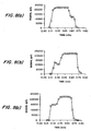
- the labelled analyte was prepared by attaching the fluorophore CY5® (BDS, Pennsylvania) to trinitrobenzyl cadaverine (CY5-TNB). To saturate the antibody binding site with the labelled antigen, a solution of the CY5-TN3 (4 nmoles in 50 ⁇ l PBS) was added to each column, and the columns were placed on a rocking bed overnight. The columns were connected to the fluorimeter and, washed briefly. Samples were introduced at a flow rate of 1 mL/min. Analyte injections were made in triplicate with concentrations ranging between 18.75 ng/mL and 1200 ng/mL. Fig. 2 illustrates data obtained for a membrane assay prepared with the test tube incubation method. A fluorescence signal peak was obtained at all analyte concentrations which was proportional to the amount of analyte added to the column.
- Fig. 3 represents data from a membrane assay prepared by saturating the immobilized antibody with labelled analyte in the column as opposed to in a test tube. Again, an increase in signal intensity with increasing analyte concentration was observed. However, a plateau was seen between an analyte concentration of 700 ng/mL and 1200 ng/mL where a negligible increase in signal intensity was observed despite a two-fold increase in analyte concentration suggesting that there is less labelled analyte on the membrane available for displacement, compared to the membrane prepared in the test tube.
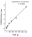
- a monoclonal antibody with specificity for the explosive, cyclonite (RDX) was immobilized onto the membrane.
- the procedure for immobilization was identical to the one used for the anti-TNT antibody.
- 100 ⁇ l of 0.5% casein was used instead of Tris in order to block the remaining binding sites on the membrane.
- Fig. 4 represents data from a single membrane assay prepared by saturating the antibody directly in the column. A linear relationship between signal intensity and analyte concentration is observed. The lower limit of detection for this assay is at 5 ng/ml which corresponds to part per billion (ppb) levels.
Landscapes
- Health & Medical Sciences (AREA)
- Immunology (AREA)
- Life Sciences & Earth Sciences (AREA)
- Engineering & Computer Science (AREA)
- Molecular Biology (AREA)
- Biomedical Technology (AREA)
- Chemical & Material Sciences (AREA)
- Hematology (AREA)
- Urology & Nephrology (AREA)
- Biotechnology (AREA)
- Biochemistry (AREA)
- Cell Biology (AREA)
- Food Science & Technology (AREA)
- Medicinal Chemistry (AREA)
- Physics & Mathematics (AREA)
- Analytical Chemistry (AREA)
- Microbiology (AREA)
- General Health & Medical Sciences (AREA)
- General Physics & Mathematics (AREA)
- Pathology (AREA)
- Investigating Or Analysing Biological Materials (AREA)
- Measuring Or Testing Involving Enzymes Or Micro-Organisms (AREA)
- Separation Using Semi-Permeable Membranes (AREA)
Claims (20)
- Testverfahren zur Detektion eines Zielanalyten, folgende Schritte umfassend:das Bereitstellen einer porösen Membran mit darauf immobilisierten Bindungselementen, wobei jedes der Bindungselemente zumindest eine Bindungsstelle aufweist, die zur spezifischen Bindung an diesen Zielanalyten fähig ist;das Aussetzen dieser Bindungsstellen gegenüber einem markierten Analogon des Zielanalyts, um Komplexe aus membranimmobilisierten Bindungselementen und markierten Analoga zu bilden;das Strömenlassen einer wässrigen Probe, die vermutlich den Zielanalyten enthält, normal zur und durch die Membran mit den Komplexen darauf hindurch, und zwar mit einer Strömungsgeschwindigkeit, die es dem Zielanalyten erlaubt, den markierten Analyten unter Ungleichgewichtsbedingungen aus den Komplexen zu verdrängen, wobei die Strömungsgeschwindigkeit zudem eine Wechselwirkungsdauer zwischen dem Analyten und der Membran von etwa 0,1 s bis etwa 30 s bereitstellt;das Detektieren des verdrängten markierten Analyten, wobei die Menge des verdrängten markierten Analyten proportional zur Konzentration des Zielanalyten in der Probe ist.
- Verfahren nach Anspruch 1, worin das Bindungselement ein Antikörper ist.
- Verfahren nach Anspruch 1, worin das markierten Analogon fluoreszenzmarkiert ist.
- Verfahren nach Anspruch 1, worin die Wechselwirkungsdauer nicht mehr als etwa 15 Sekunden beträgt.
- Verfahren nach Anspruch 1, worin die Membran nichtabsorbierend ist.
- Verfahren nach Anspruch 5, worin die Membran aus der aus Cellulose, Nitrocellulose, Silicafasern, Aluminiumoxid und Polyvinylchlorid bestehenden Gruppe ausgewählt ist.
- Verfahren nach Anspruch 5, das nach dem Detektionsschritt weiters die folgenden Schritte umfasst:das Abspülen der Probe von der Membran;das Strömenlassen einer zweiten wässrigen, flüssigen Probe, die vermutlich den Zielanalyten enthält, sodass die zweite flüssige Probe normal zur und durch die abgespülte Membran mit den Komplexen darauf hindurch strömt, und zwar mit einer Strömungsgeschwindigkeit, die es dem Zielanalyten in der zweiten Probe erlaubt, den markierten Analyten unter Ungleichgewichtsbedingungen aus den Komplexen zu verdrängen, um stromab von der Membran einen fließfähigen, flüssigen Ausfluss zu erhalten, der das markierte Analogon enthält, das vom Zielanalyten in der zweiten Probe verdrängt wurde, wobei die Strömungsgeschwindigkeit zudem eine Wechselwirkungsdauer zwischen dem Analyten und der Membran von etwa 0,1 s bis etwa 30 s bereitstellt;das Detektieren des verdrängten markierten Analyten, wobei die Menge des verdrängten markierten Analyten proportional zur Konzentration des Zielanalyten in der Probe ist.
- Vorrichtung zum Testen einer wässrigen Probe, die vermutlich einen Zielanalyten enthält, umfassend:eine poröse Membran mit darauf immobilisierten Bindungselementen mit zumindest einer Bindungsstelle, die zur spezifischen Bindung an den Zielanalyten fähig ist, wobei im Wesentlichen alle der Bindungsstellen auf der Membran von einem markierten Analogon des Zielanalyts besetzt sind, um Komplexe aus membranimmobilisierten Bindungselementen und markierten Analoga zu bilden;ein Strömungsmittel zum Strömenlassen einer wässrigen Probe, die vermutlich den Zielanalyten enthält, normal zur und durch die Membran mit den Komplexen darauf hindurch, und zwar mit einer Strömungsgeschwindigkeit, die es dem Zielanalyten erlaubt, den markierten Analyten unter Ungleichgewichtsbedingungen aus den Komplexen zu verdrängen, um eine behandelte Probe zu erhalten, wobei die Strömungsgeschwindigkeit zudem eine Wechselwirkungsdauer zwischen dem Analyten und der Membran von etwa 0,1 s bis etwa 30 s bereitstellt;und ein Detektionsmittel zum Detektieren der Gegenwart des markierten Analogons in der verarbeiteten Probe.
- Vorrichtung nach Anspruch 8, worin das markierte Analogon fluoreszenzmarkiert ist.
- Vorrichtung nach Anspruch 9, worin das Detektionsmittel weiters eine Lichtquelle zum Anregen jeglichen fluoreszenzmarkierten Analogons in der verarbeiteten Probe umfasst.
- Vorrichtung nach Anspruch 8, worin das Bindungselement ein Antikörper ist.
- Vorrichtung nach Anspruch 10, worin das Detektionsmittel weiters zur quantitativen Bestimmung der Menge des markierten Analogons in der verarbeiteten Probe geeignet ist.
- Vorrichtung nach Anspruch 10, worin das Detektionsmittel weiters ein Spektralphotometer, Infrarotspektrometer, Fluorimeter oder einen optischen Biosensor umfasst.
- Vorrichtung nach Anspruch 8, worin das Strömungsmittel eine Leitung umfasst, durch welche die Probe aufgrund von Einwirkung der Schwerkraft oder einer manuellen Kraft strömt.
- Vorrichtung nach Anspruch 8, worin die Membran nichtabsorbierend ist.
- Vorrichtung nach Anspruch 15, worin nach der Verwendung der Vorrichtung zum Testen einer ersten wässrigen Probe, die vermutlich den flüssigen Analyten enthält, gemäß einem Verfahren nach Anspruch 1 die Membran gespült und die Vorrichtung wiederverwendet werden kann.
- Vorrichtung nach Anspruch 8, worin das Strömungsmittel zur Bereitstellung einer Wechselwirkungsdauer zwischen dem Analyten und der Membran von nicht mehr als etwa 15 s geeignet ist.
- Vorrichtung zum Testen einer wässrigen Probe, die vermutlich den Zielanalyten enthält, umfassend:einen Behälter, wobei der Behälter ein offenes Ende zur Aufnahme einer flüssigen Probe, die vermutlich einen Analyten enthält, ein geschlossenes Ende, eine poröse Membran, die sich über die gesamte Breite des Behälters erstreckt, und ein Sammelgefäß zwischen der Membran und dem geschlossenen Ende aufweist, wobei auf der Membran Bindungselemente immobilisiert sind, wobei jedes der Bindungselemente zumindest eine Bindungsstelle aufweist, die zur spezifischen Bindung an den Zielanalyten fähig ist, wobei im Wesentlichen alle der Bindungsstellen auf der Membran von einem markierten Analogon des Zielanalyts besetzt sind, um Komplexe aus membranimmobilisierten Bindungselementen und markierten Analyten zu bilden;ein Strömungsmittel zum Strömenlassen einer flüssigen Probe durch das offene Ende, durch die Membran und in das Sammelgefäß, und zwar mit einer Strömungsgeschwindigkeit, die es dem Zielanalyten erlaubt, den markierten Analyten unter Ungleichgewichtsbedingungen aus den Komplexen zu verdrängen, wobei die Strömungsgeschwindigkeit eine Wechselwirkungsdauer zwischen dem Analyten und der Membran von etwa 0,1 s bis etwa 30 s bereitstellt;ein Detektionsmittel zum Detektieren der Gegenwart des markierten Analogons im Sammelgefäß.
- Vorrichtung zum Immuntesten einer wässrigen Probe, die vermutlich einen Zielanalyten enthält, umfassend:einen Behälter, wobei der Behälter ein offenes Ende und ein geschlossenes Ende aufweist;einen Verschluss, in den das offene Ende des Behälters in einer ersten Position, in der sich das geschlossene Ende des Behälters in einem ersten Abstand von diesem Verschluss befindet, und in einer zweiten Position, in der sich das geschlossene Ende des Behälters in einem zweiten Abstand von diesem Verschluss befindet, einpassbar ist, wobei der zweite Abstand geringer ist als der erste Abstand;eine sich vom Verschluss nach außen erstreckende Spitze zur Aufnahme der wässrigen Probe;eine Hohlnadel, die sich vom Verschluss in zur Spitze entgegengesetzter Richtung erstreckt;ein Septum, das sich über die gesamte Breite des Behälters erstreckt, wobei das Septum zwischen dem geschlossenen Ende des Behälters und einem Ende der Nadel distal zum Verschluss angeordnet ist, wenn der Behälter sich in seiner ersten Position befindet, wobei das Septum im Wesentlichen fluidundurchlässig ist;eine poröse Membran zwischen dem Septum und dem geschlossenen Ende des Behälters, wobei sich die Membran über die gesamte Breite des Behälters erstreckt, und ein Sammelgefäß zwischen der Membran und dem geschlossenen Ende, wobei auf der Membran Bindungselemente immobilisiert sind, wobei jedes der Bindungselemente zumindest eine Bindungsstelle aufweist, die zur spezifischen Bindung an den Zielanalyten fähig ist, wobei im Wesentlichen alle der Bindungsstellen auf der Membran von einem markierten Analogon des Zielanalyts besetzt sind, um Komplexe aus membranimmobilisierten Bindungselementen und markierten Analyten zu bilden;ein evakuiertes Sammelgefäß zwischen der Membran und dem geschlossenen Ende des Behälters;ein Detektionsmittel zum Detektieren der Gegenwart des markierten Analogons im Sammelgefäß;wobei die Hohlnadel so positioniert ist, dass sie das Septum, nicht aber die Membran durchsticht, wenn der Behälter sich in der zweiten Position befindet.
- Vorrichtung nach Anspruch 19, die weiters ein Probenhaltemittel mit einer flüssigen Probe darin umfasst, worin das Durchstechen der Membran dazu führt, dass die flüssige Probe vom Probenhaltemittel durch das offene Ende, durch die Membran und in den Sammelbehälter strömt.
Applications Claiming Priority (3)
| Application Number | Priority Date | Filing Date | Title |
|---|---|---|---|
| US583912 | 1996-01-11 | ||
| US08/583,912 US6750031B1 (en) | 1996-01-11 | 1996-01-11 | Displacement assay on a porous membrane |
| PCT/US1996/016981 WO1997025619A1 (en) | 1996-01-11 | 1996-10-18 | Displacement assay on a porous membrane |
Publications (3)
| Publication Number | Publication Date |
|---|---|
| EP0880699A1 EP0880699A1 (de) | 1998-12-02 |
| EP0880699A4 EP0880699A4 (de) | 2002-04-10 |
| EP0880699B1 true EP0880699B1 (de) | 2006-05-24 |
Family
ID=24335115
Family Applications (1)
| Application Number | Title | Priority Date | Filing Date |
|---|---|---|---|
| EP96938636A Expired - Lifetime EP0880699B1 (de) | 1996-01-11 | 1996-10-18 | Verdrängungstest auf einer porösen membran |
Country Status (5)
| Country | Link |
|---|---|
| US (2) | US6750031B1 (de) |
| EP (1) | EP0880699B1 (de) |
| AT (1) | ATE327507T1 (de) |
| DE (1) | DE69636173T2 (de) |
| WO (1) | WO1997025619A1 (de) |
Families Citing this family (105)
| Publication number | Priority date | Publication date | Assignee | Title |
|---|---|---|---|---|
| US5891740A (en) * | 1997-04-02 | 1999-04-06 | The Perkin-Elmer Corporation | Detection of low level hydrophobic analytes in environmental samples using agglutination reaction capillary slide test and apparatus therefor |
| US6036924A (en) | 1997-12-04 | 2000-03-14 | Hewlett-Packard Company | Cassette of lancet cartridges for sampling blood |
| GB9726888D0 (en) * | 1997-12-20 | 1998-02-18 | Eev Ltd | Detection |
| US6391005B1 (en) | 1998-03-30 | 2002-05-21 | Agilent Technologies, Inc. | Apparatus and method for penetration with shaft having a sensor for sensing penetration depth |
| US8497131B2 (en) * | 1999-10-06 | 2013-07-30 | Becton, Dickinson And Company | Surface enhanced spectroscopy-active composite nanoparticles comprising Raman-active reporter molecules |
| US7192778B2 (en) * | 1999-10-06 | 2007-03-20 | Natan Michael J | Surface enhanced spectroscopy-active composite nanoparticles |
| US8641644B2 (en) | 2000-11-21 | 2014-02-04 | Sanofi-Aventis Deutschland Gmbh | Blood testing apparatus having a rotatable cartridge with multiple lancing elements and testing means |
| US6861263B2 (en) * | 2001-01-26 | 2005-03-01 | Surromed, Inc. | Surface-enhanced spectroscopy-active sandwich nanoparticles |
| US9427532B2 (en) | 2001-06-12 | 2016-08-30 | Sanofi-Aventis Deutschland Gmbh | Tissue penetration device |
| US9795747B2 (en) | 2010-06-02 | 2017-10-24 | Sanofi-Aventis Deutschland Gmbh | Methods and apparatus for lancet actuation |
| JP4209767B2 (ja) | 2001-06-12 | 2009-01-14 | ペリカン テクノロジーズ インコーポレイテッド | 皮膚の性状の一時的変化に対する適応手段を備えた自動最適化形切開器具 |
| WO2002100460A2 (en) | 2001-06-12 | 2002-12-19 | Pelikan Technologies, Inc. | Electric lancet actuator |
| US9226699B2 (en) | 2002-04-19 | 2016-01-05 | Sanofi-Aventis Deutschland Gmbh | Body fluid sampling module with a continuous compression tissue interface surface |
| WO2002100252A2 (en) | 2001-06-12 | 2002-12-19 | Pelikan Technologies, Inc. | Blood sampling apparatus and method |
| US8337419B2 (en) | 2002-04-19 | 2012-12-25 | Sanofi-Aventis Deutschland Gmbh | Tissue penetration device |
| US7749174B2 (en) | 2001-06-12 | 2010-07-06 | Pelikan Technologies, Inc. | Method and apparatus for lancet launching device intergrated onto a blood-sampling cartridge |
| US7699791B2 (en) | 2001-06-12 | 2010-04-20 | Pelikan Technologies, Inc. | Method and apparatus for improving success rate of blood yield from a fingerstick |
| US7041068B2 (en) | 2001-06-12 | 2006-05-09 | Pelikan Technologies, Inc. | Sampling module device and method |
| US7981056B2 (en) | 2002-04-19 | 2011-07-19 | Pelikan Technologies, Inc. | Methods and apparatus for lancet actuation |
| SE0102922D0 (sv) * | 2001-08-31 | 2001-08-31 | Astrazeneca Ab | Method and apparatus for sample preparation |
| JP4381820B2 (ja) * | 2002-03-11 | 2009-12-09 | ポーリスツィン、ジャヌス・ビー | 生体組織検査用マイクロ抽出装置 |
| US20090026122A1 (en) * | 2002-03-11 | 2009-01-29 | Janusz | Biocompatible solid-phase microextraction coatings and methods for their preparation |
| US7232689B2 (en) * | 2002-03-11 | 2007-06-19 | Pawliszyn Janusz B | Calibration procedure for investigating biological systems |
| US9870907B2 (en) | 2002-03-11 | 2018-01-16 | Jp Scientific Limited | Probe for extraction of molecules of interest from a sample |
| US9733234B2 (en) | 2002-03-11 | 2017-08-15 | Jp Scientific Limited | Probe for extraction of molecules of interest from a sample |
| US8598325B2 (en) | 2002-03-11 | 2013-12-03 | Janusz B. Pawliszyn | Solid-phase microextraction coatings and methods for their preparation |
| GB2387130A (en) * | 2002-04-04 | 2003-10-08 | Fluid Technologies Plc | Hollow fibre filter membrane unit with microorganism detector, and associated usage |
| US7674232B2 (en) | 2002-04-19 | 2010-03-09 | Pelikan Technologies, Inc. | Method and apparatus for penetrating tissue |
| US8360992B2 (en) | 2002-04-19 | 2013-01-29 | Sanofi-Aventis Deutschland Gmbh | Method and apparatus for penetrating tissue |
| US7976476B2 (en) | 2002-04-19 | 2011-07-12 | Pelikan Technologies, Inc. | Device and method for variable speed lancet |
| US9795334B2 (en) | 2002-04-19 | 2017-10-24 | Sanofi-Aventis Deutschland Gmbh | Method and apparatus for penetrating tissue |
| US8267870B2 (en) | 2002-04-19 | 2012-09-18 | Sanofi-Aventis Deutschland Gmbh | Method and apparatus for body fluid sampling with hybrid actuation |
| US7232451B2 (en) | 2002-04-19 | 2007-06-19 | Pelikan Technologies, Inc. | Method and apparatus for penetrating tissue |
| US7229458B2 (en) | 2002-04-19 | 2007-06-12 | Pelikan Technologies, Inc. | Method and apparatus for penetrating tissue |
| US7547287B2 (en) | 2002-04-19 | 2009-06-16 | Pelikan Technologies, Inc. | Method and apparatus for penetrating tissue |
| US8784335B2 (en) | 2002-04-19 | 2014-07-22 | Sanofi-Aventis Deutschland Gmbh | Body fluid sampling device with a capacitive sensor |
| US7175642B2 (en) | 2002-04-19 | 2007-02-13 | Pelikan Technologies, Inc. | Methods and apparatus for lancet actuation |
| US7892183B2 (en) | 2002-04-19 | 2011-02-22 | Pelikan Technologies, Inc. | Method and apparatus for body fluid sampling and analyte sensing |
| US9314194B2 (en) | 2002-04-19 | 2016-04-19 | Sanofi-Aventis Deutschland Gmbh | Tissue penetration device |
| US7708701B2 (en) | 2002-04-19 | 2010-05-04 | Pelikan Technologies, Inc. | Method and apparatus for a multi-use body fluid sampling device |
| US7901362B2 (en) | 2002-04-19 | 2011-03-08 | Pelikan Technologies, Inc. | Method and apparatus for penetrating tissue |
| US7297122B2 (en) | 2002-04-19 | 2007-11-20 | Pelikan Technologies, Inc. | Method and apparatus for penetrating tissue |
| US7291117B2 (en) | 2002-04-19 | 2007-11-06 | Pelikan Technologies, Inc. | Method and apparatus for penetrating tissue |
| US8221334B2 (en) | 2002-04-19 | 2012-07-17 | Sanofi-Aventis Deutschland Gmbh | Method and apparatus for penetrating tissue |
| US9248267B2 (en) | 2002-04-19 | 2016-02-02 | Sanofi-Aventis Deustchland Gmbh | Tissue penetration device |
| US8702624B2 (en) | 2006-09-29 | 2014-04-22 | Sanofi-Aventis Deutschland Gmbh | Analyte measurement device with a single shot actuator |
| US7717863B2 (en) | 2002-04-19 | 2010-05-18 | Pelikan Technologies, Inc. | Method and apparatus for penetrating tissue |
| US7491178B2 (en) | 2002-04-19 | 2009-02-17 | Pelikan Technologies, Inc. | Method and apparatus for penetrating tissue |
| US8579831B2 (en) | 2002-04-19 | 2013-11-12 | Sanofi-Aventis Deutschland Gmbh | Method and apparatus for penetrating tissue |
| US7371247B2 (en) | 2002-04-19 | 2008-05-13 | Pelikan Technologies, Inc | Method and apparatus for penetrating tissue |
| US7909778B2 (en) | 2002-04-19 | 2011-03-22 | Pelikan Technologies, Inc. | Method and apparatus for penetrating tissue |
| US7648468B2 (en) | 2002-04-19 | 2010-01-19 | Pelikon Technologies, Inc. | Method and apparatus for penetrating tissue |
| US7331931B2 (en) | 2002-04-19 | 2008-02-19 | Pelikan Technologies, Inc. | Method and apparatus for penetrating tissue |
| US8372016B2 (en) | 2002-04-19 | 2013-02-12 | Sanofi-Aventis Deutschland Gmbh | Method and apparatus for body fluid sampling and analyte sensing |
| US20060234229A1 (en) * | 2002-06-03 | 2006-10-19 | Van Beuningen Marinus G J | Novel method for monitoring biomolecular interactions |
| US20040101900A1 (en) * | 2002-11-25 | 2004-05-27 | Mauro J. Mattew | Assay for rapid detection of TNT |
| US8574895B2 (en) | 2002-12-30 | 2013-11-05 | Sanofi-Aventis Deutschland Gmbh | Method and apparatus using optical techniques to measure analyte levels |
| US20040147619A1 (en) * | 2003-01-23 | 2004-07-29 | Conocophillips Company | Chlorine-containing synthesis gas catalyst |
| WO2004107975A2 (en) | 2003-05-30 | 2004-12-16 | Pelikan Technologies, Inc. | Method and apparatus for fluid injection |
| WO2004107964A2 (en) | 2003-06-06 | 2004-12-16 | Pelikan Technologies, Inc. | Blood harvesting device with electronic control |
| WO2006001797A1 (en) | 2004-06-14 | 2006-01-05 | Pelikan Technologies, Inc. | Low pain penetrating |
| US8282576B2 (en) | 2003-09-29 | 2012-10-09 | Sanofi-Aventis Deutschland Gmbh | Method and apparatus for an improved sample capture device |
| US9351680B2 (en) | 2003-10-14 | 2016-05-31 | Sanofi-Aventis Deutschland Gmbh | Method and apparatus for a variable user interface |
| US7943395B2 (en) * | 2003-11-21 | 2011-05-17 | Kimberly-Clark Worldwide, Inc. | Extension of the dynamic detection range of assay devices |
| EP1706026B1 (de) | 2003-12-31 | 2017-03-01 | Sanofi-Aventis Deutschland GmbH | Verfahren und vorrichtung zur verbesserung der fluidströmung und der probennahme |
| US7822454B1 (en) | 2005-01-03 | 2010-10-26 | Pelikan Technologies, Inc. | Fluid sampling device with improved analyte detecting member configuration |
| AU2005211907A1 (en) * | 2004-02-11 | 2005-08-25 | Pamgene B.V. | A device for analysing an interaction between target and probe molecules |
| US20060257991A1 (en) * | 2004-02-27 | 2006-11-16 | Mcdevitt John T | Integration of fluids and reagents into self-contained cartridges containing particle-based sensor elements and membrane-based sensor elements |
| EP1735618A2 (de) | 2004-02-27 | 2006-12-27 | Board of Regents, The University of Texas System | System und verfahren zur integration von flüssigkeiten und reagenzien in geschlossenen partikel- und membransensorelemente enthaltenden kartuschen |
| US7781226B2 (en) * | 2004-02-27 | 2010-08-24 | The Board Of Regents Of The University Of Texas System | Particle on membrane assay system |
| US8101431B2 (en) | 2004-02-27 | 2012-01-24 | Board Of Regents, The University Of Texas System | Integration of fluids and reagents into self-contained cartridges containing sensor elements and reagent delivery systems |
| US20060257941A1 (en) * | 2004-02-27 | 2006-11-16 | Mcdevitt John T | Integration of fluids and reagents into self-contained cartridges containing particle and membrane sensor elements |
| US8105849B2 (en) | 2004-02-27 | 2012-01-31 | Board Of Regents, The University Of Texas System | Integration of fluids and reagents into self-contained cartridges containing sensor elements |
| US8828203B2 (en) | 2004-05-20 | 2014-09-09 | Sanofi-Aventis Deutschland Gmbh | Printable hydrogels for biosensors |
| US9775553B2 (en) | 2004-06-03 | 2017-10-03 | Sanofi-Aventis Deutschland Gmbh | Method and apparatus for a fluid sampling device |
| EP1765194A4 (de) | 2004-06-03 | 2010-09-29 | Pelikan Technologies Inc | Verfahren und gerät für eine flüssigkeitsentnahmenvorrichtung |
| US8652831B2 (en) | 2004-12-30 | 2014-02-18 | Sanofi-Aventis Deutschland Gmbh | Method and apparatus for analyte measurement test time |
| WO2006098804A2 (en) | 2005-03-11 | 2006-09-21 | Chembio Diagnostic Systems, Inc. | Dual path immunoassay device |
| US7189522B2 (en) | 2005-03-11 | 2007-03-13 | Chembio Diagnostic Systems, Inc. | Dual path immunoassay device |
| EP1910824A4 (de) | 2005-05-31 | 2012-11-21 | Labnow Inc | Verfahren und zusammensetzungen in zusammenhang mit der erstellung und verwendung eines weissen blutbildes |
| JP2009523406A (ja) * | 2005-11-15 | 2009-06-25 | オクソニカ・インコーポレーテッド | 生体剤(bioagents)の検出のためのSERSに基づく方法 |
| US8409411B2 (en) | 2005-12-02 | 2013-04-02 | State Of Oregon Acting By And Through The State Board Of Higher Education On Behalf Of Portland State University | Nano-porous membrane based sensors |
| US8409863B2 (en) | 2005-12-14 | 2013-04-02 | Becton, Dickinson And Company | Nanoparticulate chemical sensors using SERS |
| US7723100B2 (en) | 2006-01-13 | 2010-05-25 | Becton, Dickinson And Company | Polymer coated SERS nanotag |
| EP1977242B1 (de) * | 2006-01-27 | 2016-08-03 | Becton Dickinson and Company | Querfluss-immuntest mit eingekapselter erkennungsmodalität |
| EP2044402B2 (de) * | 2006-07-24 | 2016-11-30 | Becton Dickinson and Company | Vorrichtung und Verfahren zur Durchführung eines Assays mittels magnetischer Partikeln. |
| US7879622B1 (en) * | 2006-08-11 | 2011-02-01 | University Of South Florida | Barrier-permeable proxy reporter analysis |
| US20090047673A1 (en) * | 2006-08-22 | 2009-02-19 | Cary Robert B | Miniaturized lateral flow device for rapid and sensitive detection of proteins or nucleic acids |
| US8101403B2 (en) | 2006-10-04 | 2012-01-24 | University Of Washington | Method and device for rapid parallel microfluidic molecular affinity assays |
| US8613214B2 (en) * | 2008-01-09 | 2013-12-24 | Orono Spectral Solutions, Inc. | Apparatus and method for determining analyte content in a fluid |
| US9386944B2 (en) | 2008-04-11 | 2016-07-12 | Sanofi-Aventis Deutschland Gmbh | Method and apparatus for analyte detecting device |
| JP2011522521A (ja) | 2008-05-05 | 2011-08-04 | ロスアラモス ナショナル セキュリティ,エルエルシー | 高度に単純化された側方流動ベースの核酸サンプル調製および受動的流体流動制御 |
| US20100022916A1 (en) | 2008-07-24 | 2010-01-28 | Javanbakhsh Esfandiari | Method and Apparatus for Collecting and Preparing Biological Samples for Testing |
| WO2010027812A2 (en) * | 2008-08-25 | 2010-03-11 | University Of Washington | Microfluidic systems incorporating flow-through membranes |
| US9375169B2 (en) | 2009-01-30 | 2016-06-28 | Sanofi-Aventis Deutschland Gmbh | Cam drive for managing disposable penetrating member actions with a single motor and motor and control system |
| US8965476B2 (en) | 2010-04-16 | 2015-02-24 | Sanofi-Aventis Deutschland Gmbh | Tissue penetration device |
| GB201017447D0 (en) * | 2010-10-15 | 2010-12-01 | Moorlodge Biotech Ventures Ltd | Assay device |
| US8603835B2 (en) | 2011-02-10 | 2013-12-10 | Chembio Diagnostic Systems, Inc. | Reduced step dual path immunoassay device and method |
| ES2583135T3 (es) | 2011-04-20 | 2016-09-19 | Mesa Biotech, Inc. | Dispositivo integrado para la detección e identificación de ácidos nucleicos |
| AU2015241521B2 (en) | 2014-04-02 | 2019-12-19 | Chembio Diagnostic Systems, Inc. | Immunoassay utilizing trapping conjugate |
| US20160116466A1 (en) | 2014-10-27 | 2016-04-28 | Chembio Diagnostic Systems, Inc. | Rapid Screening Assay for Qualitative Detection of Multiple Febrile Illnesses |
| US20160310948A1 (en) | 2015-04-24 | 2016-10-27 | Mesa Biotech, Inc. | Fluidic Test Cassette |
| GB201510850D0 (en) * | 2015-06-19 | 2015-08-05 | Cambridge Molecular Diagnositcs Ltd | Nucleic acid amplification and detection assays |
| US10545077B2 (en) | 2016-03-02 | 2020-01-28 | Jp Scientific Limited | Solid phase microextraction coating |
| JP6803400B2 (ja) | 2016-05-10 | 2020-12-23 | ジェイ・ピィ・サイエンティフィック・リミテッドJp Scientific Limited | 固相マイクロ抽出装置上に吸着された分析物を脱着および検出するためのシステムおよび方法 |
Family Cites Families (18)
| Publication number | Priority date | Publication date | Assignee | Title |
|---|---|---|---|---|
| US3800780A (en) * | 1972-02-23 | 1974-04-02 | Angelika Elliott | Vacuum indicator |
| US4258001A (en) | 1978-12-27 | 1981-03-24 | Eastman Kodak Company | Element, structure and method for the analysis or transport of liquids |
| US4895809A (en) * | 1984-01-09 | 1990-01-23 | Varian Associates, Inc. | Immobilized antigen-antibody displacement process |
| US4859583A (en) | 1985-02-25 | 1989-08-22 | Amoco Corporation | Chemiluminescent immunochemical technique for low molecular weight antigens |
| US4916056A (en) | 1986-02-18 | 1990-04-10 | Abbott Laboratories | Solid-phase analytical device and method for using same |
| US4812293A (en) * | 1986-06-30 | 1989-03-14 | Becton, Dickinson And Company | Vacuum actuated assay device and method of using same |
| US4920046A (en) | 1987-02-20 | 1990-04-24 | Becton, Dickinson And Company | Process, test device, and test kit for a rapid assay having a visible readout |
| US5206177A (en) * | 1988-01-21 | 1993-04-27 | Boehringer Mannheim Corporation | Apparatus for determining an analyte and method therefor |
| US5045479A (en) * | 1988-07-05 | 1991-09-03 | The Johns Hopkins University | Continuous flow competitive assay with reference system |
| US5024238A (en) * | 1989-01-10 | 1991-06-18 | Cancer Diagnostics, Inc. | Blood withdrawing apparatus and antigen testing method |
| US5766933A (en) * | 1989-04-26 | 1998-06-16 | Diagnostic Products Corporation | Method and element for measuring analytes in biological fluids using immobilized binder--analyte labeled complex |
| US5183740A (en) | 1990-02-23 | 1993-02-02 | The United States Of America As Represented By The Secretary Of The Navy | Flow immunosensor method and apparatus |
| ES2089057T3 (es) * | 1990-07-18 | 1996-10-01 | Abbott Lab | Un reactivo sustituto de un analito para uso en metodos de ensayo de fijacion especifica, dispositivos y kits. |
| US5200321A (en) | 1990-09-07 | 1993-04-06 | The United States Of America As Represented By The Secretary Of The Navy | Microassay on a card |
| DE4229591C1 (de) * | 1992-09-04 | 1994-03-24 | Draegerwerk Ag | Immunologisches Verfahren zur Bestimmung eines Analyten |
| US5354654A (en) * | 1993-07-16 | 1994-10-11 | The United States Of America As Represented By The Secretary Of The Navy | Lyophilized ligand-receptor complexes for assays and sensors |
| US5602037A (en) * | 1994-06-30 | 1997-02-11 | Dade International, Inc. | Combination reagent holding and test device |
| US6016712A (en) * | 1997-09-18 | 2000-01-25 | Accumetrics | Device for receiving and processing a sample |
-
1996
- 1996-01-11 US US08/583,912 patent/US6750031B1/en not_active Expired - Fee Related
- 1996-10-18 EP EP96938636A patent/EP0880699B1/de not_active Expired - Lifetime
- 1996-10-18 WO PCT/US1996/016981 patent/WO1997025619A1/en active IP Right Grant
- 1996-10-18 DE DE69636173T patent/DE69636173T2/de not_active Expired - Lifetime
- 1996-10-18 AT AT96938636T patent/ATE327507T1/de not_active IP Right Cessation
-
2001
- 2001-07-13 US US09/907,888 patent/US6808937B2/en not_active Expired - Fee Related
Also Published As
| Publication number | Publication date |
|---|---|
| DE69636173D1 (de) | 2006-06-29 |
| EP0880699A4 (de) | 2002-04-10 |
| ATE327507T1 (de) | 2006-06-15 |
| DE69636173T2 (de) | 2007-03-29 |
| WO1997025619A1 (en) | 1997-07-17 |
| US20020028475A1 (en) | 2002-03-07 |
| EP0880699A1 (de) | 1998-12-02 |
| US6808937B2 (en) | 2004-10-26 |
| US6750031B1 (en) | 2004-06-15 |
Similar Documents
| Publication | Publication Date | Title |
|---|---|---|
| EP0880699B1 (de) | Verdrängungstest auf einer porösen membran | |
| JP4040110B2 (ja) | 膜ベースでの検定のための分析装置 | |
| US7910381B2 (en) | Immuno gold lateral flow assay | |
| FI96639C (fi) | Omavarainen immunokoe-elementti | |
| KR100513216B1 (ko) | 바이오센서 | |
| US5183740A (en) | Flow immunosensor method and apparatus | |
| EP1789793B1 (de) | Kombinationstest für alkohol und drogen | |
| US7569397B2 (en) | Immunoassay devices and use thereof | |
| US20110097734A1 (en) | Flow Control Technique for Assay Devices | |
| EP0274911B1 (de) | Diagnostisches Hilfsmittel, seine Herstellung und seine Verwendung | |
| US20150017656A1 (en) | Rapid Lateral Flow Assay Method for Detecting Low Quantity Liquid or Dry Samples | |
| CA1289471C (en) | Dry test strip for devices using oxygen demanding detection system | |
| US8142735B2 (en) | Test apparatus | |
| CA1302244C (en) | Dry test strips having a red blood cell exclusion layer preventing interference by red blood cells in analyte detection visualization | |
| EP0402023A1 (de) | Diagnostisches Gerät mit verlängerter Durchflussmembran und Verfahren | |
| WO2014168580A1 (en) | Double-chamber bi-directional reverse flow device | |
| CA2241324C (en) | Displacement assay on a porous membrane | |
| Kusterbeck et al. | OFFICE OF NAVAL RESEARCH DEPARTMENT OF THE NAVY CODE OOCC3 ARLINGTON VA 22217-5660 | |
| JP3007410B2 (ja) | マルチウエルテスト | |
| JP2002116206A (ja) | 免疫検定装置 | |
| MXPA98005608A (en) | Proof of displacement on a membrane by | |
| KR102015360B1 (ko) | 측방 유동 분석에서의 신호 증폭 키트 및 그의 제작 방법 | |
| JPS63139248A (ja) | 酵素免疫センサ−および抗原あるいは抗体の検知方法 | |
| US20140134049A1 (en) | Lateral Flow Assay Device for Measuring Low Quantity Sample | |
| JPH0244255A (ja) | バイオセンサーおよびその作動方法 |
Legal Events
| Date | Code | Title | Description |
|---|---|---|---|
| PUAI | Public reference made under article 153(3) epc to a published international application that has entered the european phase |
Free format text: ORIGINAL CODE: 0009012 |
|
| 17P | Request for examination filed |
Effective date: 19980619 |
|
| AK | Designated contracting states |
Kind code of ref document: A1 Designated state(s): AT BE DE ES FR GB IT NL |
|
| A4 | Supplementary search report drawn up and despatched |
Effective date: 20020226 |
|
| AK | Designated contracting states |
Kind code of ref document: A4 Designated state(s): AT BE DE ES FR GB IT NL |
|
| 17Q | First examination report despatched |
Effective date: 20040310 |
|
| GRAP | Despatch of communication of intention to grant a patent |
Free format text: ORIGINAL CODE: EPIDOSNIGR1 |
|
| GRAS | Grant fee paid |
Free format text: ORIGINAL CODE: EPIDOSNIGR3 |
|
| GRAA | (expected) grant |
Free format text: ORIGINAL CODE: 0009210 |
|
| AK | Designated contracting states |
Kind code of ref document: B1 Designated state(s): AT BE DE ES FR GB IT NL |
|
| PG25 | Lapsed in a contracting state [announced via postgrant information from national office to epo] |
Ref country code: NL Free format text: LAPSE BECAUSE OF FAILURE TO SUBMIT A TRANSLATION OF THE DESCRIPTION OR TO PAY THE FEE WITHIN THE PRESCRIBED TIME-LIMIT Effective date: 20060524 Ref country code: IT Free format text: LAPSE BECAUSE OF FAILURE TO SUBMIT A TRANSLATION OF THE DESCRIPTION OR TO PAY THE FEE WITHIN THE PRESCRIBED TIME-LIMIT;WARNING: LAPSES OF ITALIAN PATENTS WITH EFFECTIVE DATE BEFORE 2007 MAY HAVE OCCURRED AT ANY TIME BEFORE 2007. THE CORRECT EFFECTIVE DATE MAY BE DIFFERENT FROM THE ONE RECORDED. Effective date: 20060524 Ref country code: BE Free format text: LAPSE BECAUSE OF FAILURE TO SUBMIT A TRANSLATION OF THE DESCRIPTION OR TO PAY THE FEE WITHIN THE PRESCRIBED TIME-LIMIT Effective date: 20060524 Ref country code: AT Free format text: LAPSE BECAUSE OF FAILURE TO SUBMIT A TRANSLATION OF THE DESCRIPTION OR TO PAY THE FEE WITHIN THE PRESCRIBED TIME-LIMIT Effective date: 20060524 |
|
| REG | Reference to a national code |
Ref country code: GB Ref legal event code: FG4D |
|
| REF | Corresponds to: |
Ref document number: 69636173 Country of ref document: DE Date of ref document: 20060629 Kind code of ref document: P |
|
| PG25 | Lapsed in a contracting state [announced via postgrant information from national office to epo] |
Ref country code: ES Free format text: LAPSE BECAUSE OF FAILURE TO SUBMIT A TRANSLATION OF THE DESCRIPTION OR TO PAY THE FEE WITHIN THE PRESCRIBED TIME-LIMIT Effective date: 20060904 |
|
| NLV1 | Nl: lapsed or annulled due to failure to fulfill the requirements of art. 29p and 29m of the patents act | ||
| ET | Fr: translation filed | ||
| PLBE | No opposition filed within time limit |
Free format text: ORIGINAL CODE: 0009261 |
|
| STAA | Information on the status of an ep patent application or granted ep patent |
Free format text: STATUS: NO OPPOSITION FILED WITHIN TIME LIMIT |
|
| 26N | No opposition filed |
Effective date: 20070227 |
|
| PGFP | Annual fee paid to national office [announced via postgrant information from national office to epo] |
Ref country code: FR Payment date: 20101209 Year of fee payment: 15 |
|
| PGFP | Annual fee paid to national office [announced via postgrant information from national office to epo] |
Ref country code: DE Payment date: 20101129 Year of fee payment: 15 |
|
| PGFP | Annual fee paid to national office [announced via postgrant information from national office to epo] |
Ref country code: GB Payment date: 20101104 Year of fee payment: 15 |
|
| GBPC | Gb: european patent ceased through non-payment of renewal fee |
Effective date: 20111018 |
|
| REG | Reference to a national code |
Ref country code: FR Ref legal event code: ST Effective date: 20120629 |
|
| PG25 | Lapsed in a contracting state [announced via postgrant information from national office to epo] |
Ref country code: DE Free format text: LAPSE BECAUSE OF NON-PAYMENT OF DUE FEES Effective date: 20120501 |
|
| REG | Reference to a national code |
Ref country code: DE Ref legal event code: R119 Ref document number: 69636173 Country of ref document: DE Effective date: 20120501 |
|
| PG25 | Lapsed in a contracting state [announced via postgrant information from national office to epo] |
Ref country code: GB Free format text: LAPSE BECAUSE OF NON-PAYMENT OF DUE FEES Effective date: 20111018 Ref country code: FR Free format text: LAPSE BECAUSE OF NON-PAYMENT OF DUE FEES Effective date: 20111102 |