EP0765478B1 - Positive und positive/negative zellselektion vermittelt durch peptiofreisetzung - Google Patents
Positive und positive/negative zellselektion vermittelt durch peptiofreisetzung Download PDFInfo
- Publication number
- EP0765478B1 EP0765478B1 EP95923835A EP95923835A EP0765478B1 EP 0765478 B1 EP0765478 B1 EP 0765478B1 EP 95923835 A EP95923835 A EP 95923835A EP 95923835 A EP95923835 A EP 95923835A EP 0765478 B1 EP0765478 B1 EP 0765478B1
- Authority
- EP
- European Patent Office
- Prior art keywords
- antibody
- cell
- peptide
- cells
- antigen
- Prior art date
- Legal status (The legal status is an assumption and is not a legal conclusion. Google has not performed a legal analysis and makes no representation as to the accuracy of the status listed.)
- Expired - Lifetime
Links
- 108090000765 processed proteins & peptides Proteins 0.000 title claims abstract description 400
- 230000001404 mediated effect Effects 0.000 title abstract description 13
- 210000004027 cell Anatomy 0.000 claims abstract description 537
- 238000000034 method Methods 0.000 claims abstract description 138
- 239000011324 bead Substances 0.000 claims abstract description 105
- 108091007433 antigens Proteins 0.000 claims abstract description 97
- 102000036639 antigens Human genes 0.000 claims abstract description 97
- 239000000427 antigen Substances 0.000 claims abstract description 95
- 239000006285 cell suspension Substances 0.000 claims abstract description 62
- 101710160107 Outer membrane protein A Proteins 0.000 claims abstract description 26
- 239000007787 solid Substances 0.000 claims abstract description 14
- 102000004196 processed proteins & peptides Human genes 0.000 claims description 157
- 150000001413 amino acids Chemical class 0.000 claims description 50
- 238000002823 phage display Methods 0.000 claims description 49
- 108090000623 proteins and genes Proteins 0.000 claims description 43
- 238000004458 analytical method Methods 0.000 claims description 42
- 238000000926 separation method Methods 0.000 claims description 39
- 108010069514 Cyclic Peptides Proteins 0.000 claims description 36
- 102000001189 Cyclic Peptides Human genes 0.000 claims description 36
- 102000004169 proteins and genes Human genes 0.000 claims description 32
- 230000005298 paramagnetic effect Effects 0.000 claims description 27
- 239000000203 mixture Substances 0.000 claims description 26
- 230000000890 antigenic effect Effects 0.000 claims description 25
- 230000005291 magnetic effect Effects 0.000 claims description 18
- 125000000539 amino acid group Chemical group 0.000 claims description 17
- 108010047041 Complementarity Determining Regions Proteins 0.000 claims description 15
- 210000004408 hybridoma Anatomy 0.000 claims description 15
- 108010067902 Peptide Library Proteins 0.000 claims description 14
- 241001494479 Pecora Species 0.000 claims description 10
- 125000003275 alpha amino acid group Chemical group 0.000 claims description 9
- 238000006073 displacement reaction Methods 0.000 claims description 9
- 229910052739 hydrogen Inorganic materials 0.000 claims description 6
- 239000000592 Artificial Cell Substances 0.000 claims description 5
- 241000283707 Capra Species 0.000 claims description 5
- 108060003951 Immunoglobulin Proteins 0.000 claims description 5
- -1 columns Substances 0.000 claims description 5
- 102000018358 immunoglobulin Human genes 0.000 claims description 5
- 229910052727 yttrium Inorganic materials 0.000 claims description 5
- 239000012510 hollow fiber Substances 0.000 claims description 4
- 239000004793 Polystyrene Substances 0.000 claims description 3
- 229910052799 carbon Inorganic materials 0.000 claims description 3
- 238000000302 molecular modelling Methods 0.000 claims description 3
- 229940126619 mouse monoclonal antibody Drugs 0.000 claims description 3
- 229910052757 nitrogen Inorganic materials 0.000 claims description 3
- 229920002223 polystyrene Polymers 0.000 claims description 3
- 229910052700 potassium Inorganic materials 0.000 claims description 3
- 241000283690 Bos taurus Species 0.000 claims description 2
- 241000283073 Equus caballus Species 0.000 claims description 2
- 241001465754 Metazoa Species 0.000 claims description 2
- 241000283973 Oryctolagus cuniculus Species 0.000 claims description 2
- 108700011201 Streptococcus IgG Fc-binding Proteins 0.000 claims description 2
- 241000282898 Sus scrofa Species 0.000 claims description 2
- 238000010353 genetic engineering Methods 0.000 claims description 2
- 239000011521 glass Substances 0.000 claims description 2
- 150000004676 glycans Chemical class 0.000 claims description 2
- 229920001282 polysaccharide Polymers 0.000 claims description 2
- 239000005017 polysaccharide Substances 0.000 claims description 2
- 229910052717 sulfur Inorganic materials 0.000 claims description 2
- 101000582398 Staphylococcus aureus Replication initiation protein Proteins 0.000 claims 1
- 229910052698 phosphorus Inorganic materials 0.000 claims 1
- 102100031573 Hematopoietic progenitor cell antigen CD34 Human genes 0.000 description 87
- 101000777663 Homo sapiens Hematopoietic progenitor cell antigen CD34 Proteins 0.000 description 87
- 102000013925 CD34 antigen Human genes 0.000 description 70
- 108050003733 CD34 antigen Proteins 0.000 description 69
- 235000001014 amino acid Nutrition 0.000 description 49
- 239000003153 chemical reaction reagent Substances 0.000 description 46
- 210000000130 stem cell Anatomy 0.000 description 41
- 210000005087 mononuclear cell Anatomy 0.000 description 39
- 238000003556 assay Methods 0.000 description 34
- 230000000694 effects Effects 0.000 description 34
- 108091028043 Nucleic acid sequence Proteins 0.000 description 30
- 238000001943 fluorescence-activated cell sorting Methods 0.000 description 27
- 235000018102 proteins Nutrition 0.000 description 24
- FAPWRFPIFSIZLT-UHFFFAOYSA-M Sodium chloride Chemical compound [Na+].[Cl-] FAPWRFPIFSIZLT-UHFFFAOYSA-M 0.000 description 23
- 210000001185 bone marrow Anatomy 0.000 description 22
- 238000012300 Sequence Analysis Methods 0.000 description 20
- 238000012360 testing method Methods 0.000 description 20
- 206010028980 Neoplasm Diseases 0.000 description 19
- 239000000872 buffer Substances 0.000 description 19
- 239000006228 supernatant Substances 0.000 description 19
- 239000002202 Polyethylene glycol Substances 0.000 description 18
- 229920001223 polyethylene glycol Polymers 0.000 description 18
- 238000002474 experimental method Methods 0.000 description 17
- 239000000047 product Substances 0.000 description 17
- YBJHBAHKTGYVGT-ZKWXMUAHSA-N (+)-Biotin Chemical compound N1C(=O)N[C@@H]2[C@H](CCCCC(=O)O)SC[C@@H]21 YBJHBAHKTGYVGT-ZKWXMUAHSA-N 0.000 description 16
- 108090001008 Avidin Proteins 0.000 description 16
- 230000002860 competitive effect Effects 0.000 description 16
- 230000008569 process Effects 0.000 description 15
- 238000012545 processing Methods 0.000 description 15
- 125000000637 arginyl group Chemical group N[C@@H](CCCNC(N)=N)C(=O)* 0.000 description 14
- 230000003993 interaction Effects 0.000 description 14
- 210000005259 peripheral blood Anatomy 0.000 description 14
- 239000011886 peripheral blood Substances 0.000 description 14
- 239000013615 primer Substances 0.000 description 14
- 238000005516 engineering process Methods 0.000 description 13
- 239000000243 solution Substances 0.000 description 13
- 230000003321 amplification Effects 0.000 description 12
- 239000003795 chemical substances by application Substances 0.000 description 12
- 238000009169 immunotherapy Methods 0.000 description 12
- 238000003199 nucleic acid amplification method Methods 0.000 description 12
- 238000010926 purge Methods 0.000 description 12
- 108090001069 Chymopapain Proteins 0.000 description 11
- ISWQCIVKKSOKNN-UHFFFAOYSA-L Tiron Chemical compound [Na+].[Na+].OC1=CC(S([O-])(=O)=O)=CC(S([O-])(=O)=O)=C1O ISWQCIVKKSOKNN-UHFFFAOYSA-L 0.000 description 11
- 210000003719 b-lymphocyte Anatomy 0.000 description 11
- 229960002685 biotin Drugs 0.000 description 11
- 239000011616 biotin Substances 0.000 description 11
- 229960002976 chymopapain Drugs 0.000 description 11
- 229940009600 gammagard Drugs 0.000 description 11
- 210000003958 hematopoietic stem cell Anatomy 0.000 description 11
- 239000002245 particle Substances 0.000 description 11
- 238000010187 selection method Methods 0.000 description 11
- 239000011780 sodium chloride Substances 0.000 description 11
- 239000013598 vector Substances 0.000 description 11
- 230000035899 viability Effects 0.000 description 11
- 108020004414 DNA Proteins 0.000 description 10
- 239000012591 Dulbecco’s Phosphate Buffered Saline Substances 0.000 description 10
- 210000004369 blood Anatomy 0.000 description 10
- 239000008280 blood Substances 0.000 description 10
- 201000011510 cancer Diseases 0.000 description 10
- 125000001165 hydrophobic group Chemical group 0.000 description 10
- 206010006187 Breast cancer Diseases 0.000 description 9
- 208000026310 Breast neoplasm Diseases 0.000 description 9
- HEMHJVSKTPXQMS-UHFFFAOYSA-M Sodium hydroxide Chemical compound [OH-].[Na+] HEMHJVSKTPXQMS-UHFFFAOYSA-M 0.000 description 9
- 238000011534 incubation Methods 0.000 description 9
- 210000003819 peripheral blood mononuclear cell Anatomy 0.000 description 9
- 238000011160 research Methods 0.000 description 9
- 210000004881 tumor cell Anatomy 0.000 description 9
- 239000004475 Arginine Substances 0.000 description 8
- ODKSFYDXXFIFQN-UHFFFAOYSA-N arginine Natural products OC(=O)C(N)CCCNC(N)=N ODKSFYDXXFIFQN-UHFFFAOYSA-N 0.000 description 8
- 235000020958 biotin Nutrition 0.000 description 8
- 239000000463 material Substances 0.000 description 8
- 239000007790 solid phase Substances 0.000 description 8
- 238000006467 substitution reaction Methods 0.000 description 8
- 238000012546 transfer Methods 0.000 description 8
- 108091034117 Oligonucleotide Proteins 0.000 description 7
- 210000001744 T-lymphocyte Anatomy 0.000 description 7
- 238000002617 apheresis Methods 0.000 description 7
- 210000004698 lymphocyte Anatomy 0.000 description 7
- 230000003278 mimic effect Effects 0.000 description 7
- 238000007363 ring formation reaction Methods 0.000 description 7
- 238000012163 sequencing technique Methods 0.000 description 7
- 239000000725 suspension Substances 0.000 description 7
- VEXZGXHMUGYJMC-UHFFFAOYSA-N Hydrochloric acid Chemical compound Cl VEXZGXHMUGYJMC-UHFFFAOYSA-N 0.000 description 6
- 108010021625 Immunoglobulin Fragments Proteins 0.000 description 6
- 102000008394 Immunoglobulin Fragments Human genes 0.000 description 6
- WHUUTDBJXJRKMK-VKHMYHEASA-N L-glutamic acid Chemical compound OC(=O)[C@@H](N)CCC(O)=O WHUUTDBJXJRKMK-VKHMYHEASA-N 0.000 description 6
- 108010052285 Membrane Proteins Proteins 0.000 description 6
- 102000018697 Membrane Proteins Human genes 0.000 description 6
- UIIMBOGNXHQVGW-UHFFFAOYSA-M Sodium bicarbonate Chemical compound [Na+].OC([O-])=O UIIMBOGNXHQVGW-UHFFFAOYSA-M 0.000 description 6
- 238000001514 detection method Methods 0.000 description 6
- 230000006870 function Effects 0.000 description 6
- 108010055341 glutamyl-glutamic acid Proteins 0.000 description 6
- 210000003714 granulocyte Anatomy 0.000 description 6
- 210000001616 monocyte Anatomy 0.000 description 6
- 239000002773 nucleotide Substances 0.000 description 6
- 125000003729 nucleotide group Chemical group 0.000 description 6
- 239000000126 substance Substances 0.000 description 6
- 238000011282 treatment Methods 0.000 description 6
- 238000005406 washing Methods 0.000 description 6
- QTBSBXVTEAMEQO-UHFFFAOYSA-N Acetic acid Chemical compound CC(O)=O QTBSBXVTEAMEQO-UHFFFAOYSA-N 0.000 description 5
- 101710125418 Major capsid protein Proteins 0.000 description 5
- VMHLLURERBWHNL-UHFFFAOYSA-M Sodium acetate Chemical compound [Na+].CC([O-])=O VMHLLURERBWHNL-UHFFFAOYSA-M 0.000 description 5
- 239000004098 Tetracycline Substances 0.000 description 5
- 230000001580 bacterial effect Effects 0.000 description 5
- 230000015572 biosynthetic process Effects 0.000 description 5
- 230000000903 blocking effect Effects 0.000 description 5
- DEGAKNSWVGKMLS-UHFFFAOYSA-N calcein Chemical compound O1C(=O)C2=CC=CC=C2C21C1=CC(CN(CC(O)=O)CC(O)=O)=C(O)C=C1OC1=C2C=C(CN(CC(O)=O)CC(=O)O)C(O)=C1 DEGAKNSWVGKMLS-UHFFFAOYSA-N 0.000 description 5
- 238000000423 cell based assay Methods 0.000 description 5
- 238000006243 chemical reaction Methods 0.000 description 5
- 238000010367 cloning Methods 0.000 description 5
- 230000005714 functional activity Effects 0.000 description 5
- 238000010230 functional analysis Methods 0.000 description 5
- 229960000318 kanamycin Drugs 0.000 description 5
- SBUJHOSQTJFQJX-NOAMYHISSA-N kanamycin Chemical compound O[C@@H]1[C@@H](O)[C@H](O)[C@@H](CN)O[C@@H]1O[C@H]1[C@H](O)[C@@H](O[C@@H]2[C@@H]([C@@H](N)[C@H](O)[C@@H](CO)O2)O)[C@H](N)C[C@@H]1N SBUJHOSQTJFQJX-NOAMYHISSA-N 0.000 description 5
- 239000003446 ligand Substances 0.000 description 5
- 239000007788 liquid Substances 0.000 description 5
- 229960002378 oftasceine Drugs 0.000 description 5
- COLNVLDHVKWLRT-QMMMGPOBSA-N phenylalanine group Chemical group N[C@@H](CC1=CC=CC=C1)C(=O)O COLNVLDHVKWLRT-QMMMGPOBSA-N 0.000 description 5
- 238000002360 preparation method Methods 0.000 description 5
- 238000000746 purification Methods 0.000 description 5
- 239000000523 sample Substances 0.000 description 5
- 239000001509 sodium citrate Substances 0.000 description 5
- NLJMYIDDQXHKNR-UHFFFAOYSA-K sodium citrate Chemical compound O.O.[Na+].[Na+].[Na+].[O-]C(=O)CC(O)(CC([O-])=O)C([O-])=O NLJMYIDDQXHKNR-UHFFFAOYSA-K 0.000 description 5
- 229960002180 tetracycline Drugs 0.000 description 5
- 229930101283 tetracycline Natural products 0.000 description 5
- 235000019364 tetracycline Nutrition 0.000 description 5
- 150000003522 tetracyclines Chemical class 0.000 description 5
- 241001515965 unidentified phage Species 0.000 description 5
- 108091003079 Bovine Serum Albumin Proteins 0.000 description 4
- KOSRFJWDECSPRO-WDSKDSINSA-N Glu-Glu Chemical compound OC(=O)CC[C@H](N)C(=O)N[C@@H](CCC(O)=O)C(O)=O KOSRFJWDECSPRO-WDSKDSINSA-N 0.000 description 4
- 102000004269 Granulocyte Colony-Stimulating Factor Human genes 0.000 description 4
- 108010017080 Granulocyte Colony-Stimulating Factor Proteins 0.000 description 4
- QIVBCDIJIAJPQS-VIFPVBQESA-N L-tryptophane Chemical compound C1=CC=C2C(C[C@H](N)C(O)=O)=CNC2=C1 QIVBCDIJIAJPQS-VIFPVBQESA-N 0.000 description 4
- 206010070834 Sensitisation Diseases 0.000 description 4
- PXIPVTKHYLBLMZ-UHFFFAOYSA-N Sodium azide Chemical compound [Na+].[N-]=[N+]=[N-] PXIPVTKHYLBLMZ-UHFFFAOYSA-N 0.000 description 4
- QIVBCDIJIAJPQS-UHFFFAOYSA-N Tryptophan Natural products C1=CC=C2C(CC(N)C(O)=O)=CNC2=C1 QIVBCDIJIAJPQS-UHFFFAOYSA-N 0.000 description 4
- 238000013459 approach Methods 0.000 description 4
- 229940125691 blood product Drugs 0.000 description 4
- 229940098773 bovine serum albumin Drugs 0.000 description 4
- 238000002512 chemotherapy Methods 0.000 description 4
- 230000003247 decreasing effect Effects 0.000 description 4
- 229930195712 glutamate Natural products 0.000 description 4
- 229930027917 kanamycin Natural products 0.000 description 4
- 229930182823 kanamycin A Natural products 0.000 description 4
- 108091005601 modified peptides Proteins 0.000 description 4
- 230000007193 modulation by symbiont of host erythrocyte aggregation Effects 0.000 description 4
- 239000000816 peptidomimetic Substances 0.000 description 4
- COLNVLDHVKWLRT-UHFFFAOYSA-N phenylalanine Natural products OC(=O)C(N)CC1=CC=CC=C1 COLNVLDHVKWLRT-UHFFFAOYSA-N 0.000 description 4
- 230000005855 radiation Effects 0.000 description 4
- 230000008929 regeneration Effects 0.000 description 4
- 238000011069 regeneration method Methods 0.000 description 4
- 230000008313 sensitization Effects 0.000 description 4
- 239000001632 sodium acetate Substances 0.000 description 4
- 235000017281 sodium acetate Nutrition 0.000 description 4
- 239000011550 stock solution Substances 0.000 description 4
- 238000003786 synthesis reaction Methods 0.000 description 4
- 101100346189 Caenorhabditis elegans mpc-1 gene Proteins 0.000 description 3
- 101710132601 Capsid protein Proteins 0.000 description 3
- 108010022366 Carcinoembryonic Antigen Proteins 0.000 description 3
- 102100025475 Carcinoembryonic antigen-related cell adhesion molecule 5 Human genes 0.000 description 3
- 101710094648 Coat protein Proteins 0.000 description 3
- 108091035707 Consensus sequence Proteins 0.000 description 3
- 239000003155 DNA primer Substances 0.000 description 3
- BWGNESOTFCXPMA-UHFFFAOYSA-N Dihydrogen disulfide Chemical compound SS BWGNESOTFCXPMA-UHFFFAOYSA-N 0.000 description 3
- 102100021181 Golgi phosphoprotein 3 Human genes 0.000 description 3
- 101710141454 Nucleoprotein Proteins 0.000 description 3
- WCUXLLCKKVVCTQ-UHFFFAOYSA-M Potassium chloride Chemical compound [Cl-].[K+] WCUXLLCKKVVCTQ-UHFFFAOYSA-M 0.000 description 3
- 101710083689 Probable capsid protein Proteins 0.000 description 3
- KOSRFJWDECSPRO-UHFFFAOYSA-N alpha-L-glutamyl-L-glutamic acid Natural products OC(=O)CCC(N)C(=O)NC(CCC(O)=O)C(O)=O KOSRFJWDECSPRO-UHFFFAOYSA-N 0.000 description 3
- 230000000692 anti-sense effect Effects 0.000 description 3
- CKLJMWTZIZZHCS-REOHCLBHSA-N aspartic acid group Chemical group N[C@@H](CC(=O)O)C(=O)O CKLJMWTZIZZHCS-REOHCLBHSA-N 0.000 description 3
- 210000004899 c-terminal region Anatomy 0.000 description 3
- 230000006727 cell loss Effects 0.000 description 3
- 239000011248 coating agent Substances 0.000 description 3
- 238000000576 coating method Methods 0.000 description 3
- 125000004122 cyclic group Chemical group 0.000 description 3
- XUJNEKJLAYXESH-UHFFFAOYSA-N cysteine Natural products SCC(N)C(O)=O XUJNEKJLAYXESH-UHFFFAOYSA-N 0.000 description 3
- 125000000151 cysteine group Chemical group N[C@@H](CS)C(=O)* 0.000 description 3
- 238000013461 design Methods 0.000 description 3
- 238000010494 dissociation reaction Methods 0.000 description 3
- 230000005593 dissociations Effects 0.000 description 3
- 239000004220 glutamic acid Substances 0.000 description 3
- 230000002209 hydrophobic effect Effects 0.000 description 3
- 230000005847 immunogenicity Effects 0.000 description 3
- 208000015181 infectious disease Diseases 0.000 description 3
- 238000002955 isolation Methods 0.000 description 3
- 230000007774 longterm Effects 0.000 description 3
- 238000004519 manufacturing process Methods 0.000 description 3
- 230000007246 mechanism Effects 0.000 description 3
- 238000005457 optimization Methods 0.000 description 3
- 238000010647 peptide synthesis reaction Methods 0.000 description 3
- 229920000136 polysorbate Polymers 0.000 description 3
- 239000013641 positive control Substances 0.000 description 3
- 229910000030 sodium bicarbonate Inorganic materials 0.000 description 3
- 230000036964 tight binding Effects 0.000 description 3
- 125000000430 tryptophan group Chemical group [H]N([H])C(C(=O)O*)C([H])([H])C1=C([H])N([H])C2=C([H])C([H])=C([H])C([H])=C12 0.000 description 3
- VHUUQVKOLVNVRT-UHFFFAOYSA-N Ammonium hydroxide Chemical compound [NH4+].[OH-] VHUUQVKOLVNVRT-UHFFFAOYSA-N 0.000 description 2
- 101710169873 Capsid protein G8P Proteins 0.000 description 2
- 241000237074 Centris Species 0.000 description 2
- 102100035359 Cerebellar degeneration-related protein 2-like Human genes 0.000 description 2
- 206010009944 Colon cancer Diseases 0.000 description 2
- KCXVZYZYPLLWCC-UHFFFAOYSA-N EDTA Chemical compound OC(=O)CN(CC(O)=O)CCN(CC(O)=O)CC(O)=O KCXVZYZYPLLWCC-UHFFFAOYSA-N 0.000 description 2
- 241000702371 Enterobacteria phage f1 Species 0.000 description 2
- 229920001917 Ficoll Polymers 0.000 description 2
- ZHNUHDYFZUAESO-UHFFFAOYSA-N Formamide Chemical compound NC=O ZHNUHDYFZUAESO-UHFFFAOYSA-N 0.000 description 2
- WHUUTDBJXJRKMK-UHFFFAOYSA-N Glutamic acid Natural products OC(=O)C(N)CCC(O)=O WHUUTDBJXJRKMK-UHFFFAOYSA-N 0.000 description 2
- DHMQDGOQFOQNFH-UHFFFAOYSA-N Glycine Chemical compound NCC(O)=O DHMQDGOQFOQNFH-UHFFFAOYSA-N 0.000 description 2
- 208000009329 Graft vs Host Disease Diseases 0.000 description 2
- 101100220044 Homo sapiens CD34 gene Proteins 0.000 description 2
- 101000737792 Homo sapiens Cerebellar degeneration-related protein 2-like Proteins 0.000 description 2
- 108091006905 Human Serum Albumin Proteins 0.000 description 2
- 102000008100 Human Serum Albumin Human genes 0.000 description 2
- MHAJPDPJQMAIIY-UHFFFAOYSA-N Hydrogen peroxide Chemical compound OO MHAJPDPJQMAIIY-UHFFFAOYSA-N 0.000 description 2
- TWRXJAOTZQYOKJ-UHFFFAOYSA-L Magnesium chloride Chemical compound [Mg+2].[Cl-].[Cl-] TWRXJAOTZQYOKJ-UHFFFAOYSA-L 0.000 description 2
- 101710156564 Major tail protein Gp23 Proteins 0.000 description 2
- 241001529936 Murinae Species 0.000 description 2
- 206010029260 Neuroblastoma Diseases 0.000 description 2
- 229920000805 Polyaspartic acid Polymers 0.000 description 2
- 108010020346 Polyglutamic Acid Proteins 0.000 description 2
- 101710193132 Pre-hexon-linking protein VIII Proteins 0.000 description 2
- 101710120037 Toxin CcdB Proteins 0.000 description 2
- XSQUKJJJFZCRTK-UHFFFAOYSA-N Urea Chemical compound NC(N)=O XSQUKJJJFZCRTK-UHFFFAOYSA-N 0.000 description 2
- 238000002835 absorbance Methods 0.000 description 2
- 229960000583 acetic acid Drugs 0.000 description 2
- 239000002253 acid Substances 0.000 description 2
- 235000011114 ammonium hydroxide Nutrition 0.000 description 2
- ROOXNKNUYICQNP-UHFFFAOYSA-N ammonium persulfate Chemical compound [NH4+].[NH4+].[O-]S(=O)(=O)OOS([O-])(=O)=O ROOXNKNUYICQNP-UHFFFAOYSA-N 0.000 description 2
- 230000009833 antibody interaction Effects 0.000 description 2
- 210000002960 bfu-e Anatomy 0.000 description 2
- 238000007413 biotinylation Methods 0.000 description 2
- 230000006287 biotinylation Effects 0.000 description 2
- 210000002798 bone marrow cell Anatomy 0.000 description 2
- 210000000481 breast Anatomy 0.000 description 2
- 201000008275 breast carcinoma Diseases 0.000 description 2
- 238000004422 calculation algorithm Methods 0.000 description 2
- 230000001413 cellular effect Effects 0.000 description 2
- 238000005119 centrifugation Methods 0.000 description 2
- 230000008859 change Effects 0.000 description 2
- 229920001429 chelating resin Polymers 0.000 description 2
- 230000000295 complement effect Effects 0.000 description 2
- 239000002299 complementary DNA Substances 0.000 description 2
- 150000001875 compounds Chemical class 0.000 description 2
- 235000018417 cysteine Nutrition 0.000 description 2
- 230000001419 dependent effect Effects 0.000 description 2
- VHJLVAABSRFDPM-QWWZWVQMSA-N dithiothreitol Chemical compound SC[C@@H](O)[C@H](O)CS VHJLVAABSRFDPM-QWWZWVQMSA-N 0.000 description 2
- 239000000975 dye Substances 0.000 description 2
- 230000008030 elimination Effects 0.000 description 2
- 238000003379 elimination reaction Methods 0.000 description 2
- 230000002255 enzymatic effect Effects 0.000 description 2
- MHMNJMPURVTYEJ-UHFFFAOYSA-N fluorescein-5-isothiocyanate Chemical compound O1C(=O)C2=CC(N=C=S)=CC=C2C21C1=CC=C(O)C=C1OC1=CC(O)=CC=C21 MHMNJMPURVTYEJ-UHFFFAOYSA-N 0.000 description 2
- 238000009472 formulation Methods 0.000 description 2
- 235000013922 glutamic acid Nutrition 0.000 description 2
- 208000024908 graft versus host disease Diseases 0.000 description 2
- 238000004128 high performance liquid chromatography Methods 0.000 description 2
- 239000001257 hydrogen Substances 0.000 description 2
- 230000005764 inhibitory process Effects 0.000 description 2
- 238000013101 initial test Methods 0.000 description 2
- 238000012482 interaction analysis Methods 0.000 description 2
- 230000014759 maintenance of location Effects 0.000 description 2
- 206010061289 metastatic neoplasm Diseases 0.000 description 2
- 244000005700 microbiome Species 0.000 description 2
- 238000012986 modification Methods 0.000 description 2
- 230000004048 modification Effects 0.000 description 2
- 108010091748 peptide A Proteins 0.000 description 2
- 239000004033 plastic Substances 0.000 description 2
- 229920003023 plastic Polymers 0.000 description 2
- 108010064470 polyaspartate Proteins 0.000 description 2
- 229920002643 polyglutamic acid Polymers 0.000 description 2
- 238000001556 precipitation Methods 0.000 description 2
- 125000002924 primary amino group Chemical group [H]N([H])* 0.000 description 2
- 238000000159 protein binding assay Methods 0.000 description 2
- 230000017854 proteolysis Effects 0.000 description 2
- 238000003127 radioimmunoassay Methods 0.000 description 2
- 230000009467 reduction Effects 0.000 description 2
- 238000012552 review Methods 0.000 description 2
- 238000012216 screening Methods 0.000 description 2
- 208000000587 small cell lung carcinoma Diseases 0.000 description 2
- 235000017557 sodium bicarbonate Nutrition 0.000 description 2
- PFNFFQXMRSDOHW-UHFFFAOYSA-N spermine Chemical compound NCCCNCCCCNCCCN PFNFFQXMRSDOHW-UHFFFAOYSA-N 0.000 description 2
- 238000010561 standard procedure Methods 0.000 description 2
- 238000003860 storage Methods 0.000 description 2
- 238000002560 therapeutic procedure Methods 0.000 description 2
- 210000001519 tissue Anatomy 0.000 description 2
- 238000004448 titration Methods 0.000 description 2
- 230000002463 transducing effect Effects 0.000 description 2
- 238000013519 translation Methods 0.000 description 2
- 238000010200 validation analysis Methods 0.000 description 2
- XLYOFNOQVPJJNP-UHFFFAOYSA-N water Substances O XLYOFNOQVPJJNP-UHFFFAOYSA-N 0.000 description 2
- UVGHPGOONBRLCX-NJSLBKSFSA-N (2,5-dioxopyrrolidin-1-yl) 6-[5-[(3as,4s,6ar)-2-oxo-1,3,3a,4,6,6a-hexahydrothieno[3,4-d]imidazol-4-yl]pentanoylamino]hexanoate Chemical compound C([C@H]1[C@H]2NC(=O)N[C@H]2CS1)CCCC(=O)NCCCCCC(=O)ON1C(=O)CCC1=O UVGHPGOONBRLCX-NJSLBKSFSA-N 0.000 description 1
- OSBLTNPMIGYQGY-UHFFFAOYSA-N 2-amino-2-(hydroxymethyl)propane-1,3-diol;2-[2-[bis(carboxymethyl)amino]ethyl-(carboxymethyl)amino]acetic acid;boric acid Chemical compound OB(O)O.OCC(N)(CO)CO.OC(=O)CN(CC(O)=O)CCN(CC(O)=O)CC(O)=O OSBLTNPMIGYQGY-UHFFFAOYSA-N 0.000 description 1
- QKNYBSVHEMOAJP-UHFFFAOYSA-N 2-amino-2-(hydroxymethyl)propane-1,3-diol;hydron;chloride Chemical compound Cl.OCC(N)(CO)CO QKNYBSVHEMOAJP-UHFFFAOYSA-N 0.000 description 1
- UAIUNKRWKOVEES-UHFFFAOYSA-N 3,3',5,5'-tetramethylbenzidine Chemical compound CC1=C(N)C(C)=CC(C=2C=C(C)C(N)=C(C)C=2)=C1 UAIUNKRWKOVEES-UHFFFAOYSA-N 0.000 description 1
- QTBSBXVTEAMEQO-UHFFFAOYSA-M Acetate Chemical compound CC([O-])=O QTBSBXVTEAMEQO-UHFFFAOYSA-M 0.000 description 1
- HRPVXLWXLXDGHG-UHFFFAOYSA-N Acrylamide Chemical compound NC(=O)C=C HRPVXLWXLXDGHG-UHFFFAOYSA-N 0.000 description 1
- 229920001817 Agar Polymers 0.000 description 1
- 239000004254 Ammonium phosphate Substances 0.000 description 1
- 208000003950 B-cell lymphoma Diseases 0.000 description 1
- 102100024222 B-lymphocyte antigen CD19 Human genes 0.000 description 1
- 102100022005 B-lymphocyte antigen CD20 Human genes 0.000 description 1
- UXVMQQNJUSDDNG-UHFFFAOYSA-L Calcium chloride Chemical compound [Cl-].[Cl-].[Ca+2] UXVMQQNJUSDDNG-UHFFFAOYSA-L 0.000 description 1
- 108091026890 Coding region Proteins 0.000 description 1
- 108020004705 Codon Proteins 0.000 description 1
- 102000004127 Cytokines Human genes 0.000 description 1
- 108090000695 Cytokines Proteins 0.000 description 1
- 150000008574 D-amino acids Chemical class 0.000 description 1
- 102000053602 DNA Human genes 0.000 description 1
- ZGTMUACCHSMWAC-UHFFFAOYSA-L EDTA disodium salt (anhydrous) Chemical compound [Na+].[Na+].OC(=O)CN(CC([O-])=O)CCN(CC(O)=O)CC([O-])=O ZGTMUACCHSMWAC-UHFFFAOYSA-L 0.000 description 1
- 238000002965 ELISA Methods 0.000 description 1
- 108090000790 Enzymes Proteins 0.000 description 1
- 102000004190 Enzymes Human genes 0.000 description 1
- 241000724791 Filamentous phage Species 0.000 description 1
- 238000012413 Fluorescence activated cell sorting analysis Methods 0.000 description 1
- GHASVSINZRGABV-UHFFFAOYSA-N Fluorouracil Chemical compound FC1=CNC(=O)NC1=O GHASVSINZRGABV-UHFFFAOYSA-N 0.000 description 1
- 239000004471 Glycine Substances 0.000 description 1
- 102000004457 Granulocyte-Macrophage Colony-Stimulating Factor Human genes 0.000 description 1
- 108010017213 Granulocyte-Macrophage Colony-Stimulating Factor Proteins 0.000 description 1
- 229920000209 Hexadimethrine bromide Polymers 0.000 description 1
- 101000980825 Homo sapiens B-lymphocyte antigen CD19 Proteins 0.000 description 1
- 101000897405 Homo sapiens B-lymphocyte antigen CD20 Proteins 0.000 description 1
- 108010001336 Horseradish Peroxidase Proteins 0.000 description 1
- ONIBWKKTOPOVIA-BYPYZUCNSA-N L-Proline Chemical compound OC(=O)[C@@H]1CCCN1 ONIBWKKTOPOVIA-BYPYZUCNSA-N 0.000 description 1
- 150000008575 L-amino acids Chemical group 0.000 description 1
- ROHFNLRQFUQHCH-YFKPBYRVSA-N L-leucine Chemical compound CC(C)C[C@H](N)C(O)=O ROHFNLRQFUQHCH-YFKPBYRVSA-N 0.000 description 1
- ROHFNLRQFUQHCH-UHFFFAOYSA-N Leucine Natural products CC(C)CC(N)C(O)=O ROHFNLRQFUQHCH-UHFFFAOYSA-N 0.000 description 1
- FYYHWMGAXLPEAU-UHFFFAOYSA-N Magnesium Chemical compound [Mg] FYYHWMGAXLPEAU-UHFFFAOYSA-N 0.000 description 1
- KWYHDKDOAIKMQN-UHFFFAOYSA-N N,N,N',N'-tetramethylethylenediamine Chemical compound CN(C)CCN(C)C KWYHDKDOAIKMQN-UHFFFAOYSA-N 0.000 description 1
- 102000003729 Neprilysin Human genes 0.000 description 1
- 108090000028 Neprilysin Proteins 0.000 description 1
- 108010069196 Neural Cell Adhesion Molecules Proteins 0.000 description 1
- 102100027347 Neural cell adhesion molecule 1 Human genes 0.000 description 1
- 208000015914 Non-Hodgkin lymphomas Diseases 0.000 description 1
- 208000012868 Overgrowth Diseases 0.000 description 1
- 108090000526 Papain Proteins 0.000 description 1
- 239000004698 Polyethylene Substances 0.000 description 1
- 229920002594 Polyethylene Glycol 8000 Polymers 0.000 description 1
- 239000004353 Polyethylene glycol 8000 Substances 0.000 description 1
- 101800001357 Potential peptide Proteins 0.000 description 1
- 102400000745 Potential peptide Human genes 0.000 description 1
- 238000012356 Product development Methods 0.000 description 1
- ONIBWKKTOPOVIA-UHFFFAOYSA-N Proline Natural products OC(=O)C1CCCN1 ONIBWKKTOPOVIA-UHFFFAOYSA-N 0.000 description 1
- 102000007327 Protamines Human genes 0.000 description 1
- 108010007568 Protamines Proteins 0.000 description 1
- 239000004365 Protease Substances 0.000 description 1
- 108010076504 Protein Sorting Signals Proteins 0.000 description 1
- 108010008281 Recombinant Fusion Proteins Proteins 0.000 description 1
- 102000007056 Recombinant Fusion Proteins Human genes 0.000 description 1
- 208000007660 Residual Neoplasm Diseases 0.000 description 1
- 241000283984 Rodentia Species 0.000 description 1
- 239000006146 Roswell Park Memorial Institute medium Substances 0.000 description 1
- 108020004682 Single-Stranded DNA Proteins 0.000 description 1
- 108010090804 Streptavidin Proteins 0.000 description 1
- 241000194017 Streptococcus Species 0.000 description 1
- QAOWNCQODCNURD-UHFFFAOYSA-L Sulfate Chemical compound [O-]S([O-])(=O)=O QAOWNCQODCNURD-UHFFFAOYSA-L 0.000 description 1
- 108700005078 Synthetic Genes Proteins 0.000 description 1
- 102100036011 T-cell surface glycoprotein CD4 Human genes 0.000 description 1
- 239000008051 TBE buffer Substances 0.000 description 1
- 108010006785 Taq Polymerase Proteins 0.000 description 1
- 239000007983 Tris buffer Substances 0.000 description 1
- 238000005411 Van der Waals force Methods 0.000 description 1
- 238000010317 ablation therapy Methods 0.000 description 1
- 238000003916 acid precipitation Methods 0.000 description 1
- DPKHZNPWBDQZCN-UHFFFAOYSA-N acridine orange free base Chemical compound C1=CC(N(C)C)=CC2=NC3=CC(N(C)C)=CC=C3C=C21 DPKHZNPWBDQZCN-UHFFFAOYSA-N 0.000 description 1
- 230000002411 adverse Effects 0.000 description 1
- 239000008272 agar Substances 0.000 description 1
- 238000013019 agitation Methods 0.000 description 1
- 239000000908 ammonium hydroxide Substances 0.000 description 1
- 229910001870 ammonium persulfate Inorganic materials 0.000 description 1
- 229910000148 ammonium phosphate Inorganic materials 0.000 description 1
- 235000019289 ammonium phosphates Nutrition 0.000 description 1
- 230000001745 anti-biotin effect Effects 0.000 description 1
- 230000000259 anti-tumor effect Effects 0.000 description 1
- 230000009830 antibody antigen interaction Effects 0.000 description 1
- 230000009831 antigen interaction Effects 0.000 description 1
- 239000007864 aqueous solution Substances 0.000 description 1
- 230000008901 benefit Effects 0.000 description 1
- DZBUGLKDJFMEHC-UHFFFAOYSA-N benzoquinolinylidene Natural products C1=CC=CC2=CC3=CC=CC=C3N=C21 DZBUGLKDJFMEHC-UHFFFAOYSA-N 0.000 description 1
- 230000000975 bioactive effect Effects 0.000 description 1
- 230000004071 biological effect Effects 0.000 description 1
- 239000010836 blood and blood product Substances 0.000 description 1
- 238000010322 bone marrow transplantation Methods 0.000 description 1
- 239000001110 calcium chloride Substances 0.000 description 1
- 229910001628 calcium chloride Inorganic materials 0.000 description 1
- 229940041514 candida albicans extract Drugs 0.000 description 1
- 239000004202 carbamide Substances 0.000 description 1
- 125000003178 carboxy group Chemical group [H]OC(*)=O 0.000 description 1
- 125000002057 carboxymethyl group Chemical group [H]OC(=O)C([H])([H])[*] 0.000 description 1
- 230000015556 catabolic process Effects 0.000 description 1
- 238000004113 cell culture Methods 0.000 description 1
- 210000000170 cell membrane Anatomy 0.000 description 1
- 238000011964 cellular and gene therapy Methods 0.000 description 1
- 239000002738 chelating agent Substances 0.000 description 1
- XMEVHPAGJVLHIG-FMZCEJRJSA-N chembl454950 Chemical compound [Cl-].C1=CC=C2[C@](O)(C)[C@H]3C[C@H]4[C@H]([NH+](C)C)C(O)=C(C(N)=O)C(=O)[C@@]4(O)C(O)=C3C(=O)C2=C1O XMEVHPAGJVLHIG-FMZCEJRJSA-N 0.000 description 1
- 239000003638 chemical reducing agent Substances 0.000 description 1
- 230000000973 chemotherapeutic effect Effects 0.000 description 1
- 208000014921 colon small cell neuroendocrine carcinoma Diseases 0.000 description 1
- 208000029742 colonic neoplasm Diseases 0.000 description 1
- 230000001332 colony forming effect Effects 0.000 description 1
- 230000009918 complex formation Effects 0.000 description 1
- 230000001268 conjugating effect Effects 0.000 description 1
- 108050003126 conotoxin Proteins 0.000 description 1
- 238000010276 construction Methods 0.000 description 1
- 230000002596 correlated effect Effects 0.000 description 1
- 230000000875 corresponding effect Effects 0.000 description 1
- 230000009089 cytolysis Effects 0.000 description 1
- 238000006731 degradation reaction Methods 0.000 description 1
- 230000001627 detrimental effect Effects 0.000 description 1
- MNNHAPBLZZVQHP-UHFFFAOYSA-N diammonium hydrogen phosphate Chemical compound [NH4+].[NH4+].OP([O-])([O-])=O MNNHAPBLZZVQHP-UHFFFAOYSA-N 0.000 description 1
- 230000029087 digestion Effects 0.000 description 1
- 238000010790 dilution Methods 0.000 description 1
- 239000012895 dilution Substances 0.000 description 1
- 230000003467 diminishing effect Effects 0.000 description 1
- 201000010099 disease Diseases 0.000 description 1
- 208000037265 diseases, disorders, signs and symptoms Diseases 0.000 description 1
- 150000002019 disulfides Chemical class 0.000 description 1
- 230000009881 electrostatic interaction Effects 0.000 description 1
- 238000010828 elution Methods 0.000 description 1
- 239000012149 elution buffer Substances 0.000 description 1
- 238000006911 enzymatic reaction Methods 0.000 description 1
- 229940088598 enzyme Drugs 0.000 description 1
- 150000002170 ethers Chemical class 0.000 description 1
- DNJIEGIFACGWOD-UHFFFAOYSA-N ethyl mercaptane Natural products CCS DNJIEGIFACGWOD-UHFFFAOYSA-N 0.000 description 1
- 238000011156 evaluation Methods 0.000 description 1
- 230000001747 exhibiting effect Effects 0.000 description 1
- 210000004700 fetal blood Anatomy 0.000 description 1
- 238000013100 final test Methods 0.000 description 1
- 238000000684 flow cytometry Methods 0.000 description 1
- 239000007850 fluorescent dye Substances 0.000 description 1
- 229960002949 fluorouracil Drugs 0.000 description 1
- 238000007429 general method Methods 0.000 description 1
- 239000012362 glacial acetic acid Substances 0.000 description 1
- 125000003630 glycyl group Chemical group [H]N([H])C([H])([H])C(*)=O 0.000 description 1
- 239000003102 growth factor Substances 0.000 description 1
- 230000003394 haemopoietic effect Effects 0.000 description 1
- 238000003306 harvesting Methods 0.000 description 1
- 208000037584 hereditary sensory and autonomic neuropathy Diseases 0.000 description 1
- 125000000487 histidyl group Chemical group [H]N([H])C(C(=O)O*)C([H])([H])C1=C([H])N([H])C([H])=N1 0.000 description 1
- 210000005260 human cell Anatomy 0.000 description 1
- 230000001900 immune effect Effects 0.000 description 1
- 230000028993 immune response Effects 0.000 description 1
- 210000000987 immune system Anatomy 0.000 description 1
- 229940127121 immunoconjugate Drugs 0.000 description 1
- 238000003365 immunocytochemistry Methods 0.000 description 1
- 238000000338 in vitro Methods 0.000 description 1
- 238000010348 incorporation Methods 0.000 description 1
- 238000001802 infusion Methods 0.000 description 1
- 230000000977 initiatory effect Effects 0.000 description 1
- 230000002452 interceptive effect Effects 0.000 description 1
- 238000001990 intravenous administration Methods 0.000 description 1
- 150000002596 lactones Chemical class 0.000 description 1
- 208000032839 leukemia Diseases 0.000 description 1
- 230000000670 limiting effect Effects 0.000 description 1
- 239000012263 liquid product Substances 0.000 description 1
- 239000006166 lysate Substances 0.000 description 1
- 230000002101 lytic effect Effects 0.000 description 1
- 239000011777 magnesium Substances 0.000 description 1
- 229910052749 magnesium Inorganic materials 0.000 description 1
- 229910001629 magnesium chloride Inorganic materials 0.000 description 1
- 238000007885 magnetic separation Methods 0.000 description 1
- 230000036210 malignancy Effects 0.000 description 1
- 230000010534 mechanism of action Effects 0.000 description 1
- 208000037819 metastatic cancer Diseases 0.000 description 1
- 208000011575 metastatic malignant neoplasm Diseases 0.000 description 1
- 239000004005 microsphere Substances 0.000 description 1
- 230000009456 molecular mechanism Effects 0.000 description 1
- 239000003068 molecular probe Substances 0.000 description 1
- 238000012544 monitoring process Methods 0.000 description 1
- 239000013642 negative control Substances 0.000 description 1
- 230000001537 neural effect Effects 0.000 description 1
- 230000000955 neuroendocrine Effects 0.000 description 1
- 230000007935 neutral effect Effects 0.000 description 1
- 238000004806 packaging method and process Methods 0.000 description 1
- 229940055729 papain Drugs 0.000 description 1
- 235000019834 papain Nutrition 0.000 description 1
- 238000005191 phase separation Methods 0.000 description 1
- 235000019446 polyethylene glycol 8000 Nutrition 0.000 description 1
- 229940085678 polyethylene glycol 8000 Drugs 0.000 description 1
- 229920000642 polymer Polymers 0.000 description 1
- 239000001103 potassium chloride Substances 0.000 description 1
- 235000011164 potassium chloride Nutrition 0.000 description 1
- 230000001737 promoting effect Effects 0.000 description 1
- XJMOSONTPMZWPB-UHFFFAOYSA-M propidium iodide Chemical compound [I-].[I-].C12=CC(N)=CC=C2C2=CC=C(N)C=C2[N+](CCC[N+](C)(CC)CC)=C1C1=CC=CC=C1 XJMOSONTPMZWPB-UHFFFAOYSA-M 0.000 description 1
- 229940048914 protamine Drugs 0.000 description 1
- 230000005588 protonation Effects 0.000 description 1
- 238000003908 quality control method Methods 0.000 description 1
- 108010043277 recombinant soluble CD4 Proteins 0.000 description 1
- 230000006798 recombination Effects 0.000 description 1
- 230000000717 retained effect Effects 0.000 description 1
- 108010004093 retinal S antigen peptide M Proteins 0.000 description 1
- 238000005070 sampling Methods 0.000 description 1
- 238000013207 serial dilution Methods 0.000 description 1
- 239000012279 sodium borohydride Substances 0.000 description 1
- 229910000033 sodium borohydride Inorganic materials 0.000 description 1
- 229960001790 sodium citrate Drugs 0.000 description 1
- 229940074404 sodium succinate Drugs 0.000 description 1
- ZDQYSKICYIVCPN-UHFFFAOYSA-L sodium succinate (anhydrous) Chemical compound [Na+].[Na+].[O-]C(=O)CCC([O-])=O ZDQYSKICYIVCPN-UHFFFAOYSA-L 0.000 description 1
- 238000002764 solid phase assay Methods 0.000 description 1
- 238000001179 sorption measurement Methods 0.000 description 1
- 125000006850 spacer group Chemical group 0.000 description 1
- 229940063675 spermine Drugs 0.000 description 1
- 238000007619 statistical method Methods 0.000 description 1
- 239000008223 sterile water Substances 0.000 description 1
- 229910021653 sulphate ion Inorganic materials 0.000 description 1
- 238000002198 surface plasmon resonance spectroscopy Methods 0.000 description 1
- 230000002195 synergetic effect Effects 0.000 description 1
- 230000008685 targeting Effects 0.000 description 1
- 229960004989 tetracycline hydrochloride Drugs 0.000 description 1
- 229940126622 therapeutic monoclonal antibody Drugs 0.000 description 1
- 150000003568 thioethers Chemical class 0.000 description 1
- 231100000167 toxic agent Toxicity 0.000 description 1
- 239000003440 toxic substance Substances 0.000 description 1
- 238000002054 transplantation Methods 0.000 description 1
- LENZDBCJOHFCAS-UHFFFAOYSA-N tris Chemical compound OCC(N)(CO)CO LENZDBCJOHFCAS-UHFFFAOYSA-N 0.000 description 1
- 239000000439 tumor marker Substances 0.000 description 1
- 238000012762 unpaired Student’s t-test Methods 0.000 description 1
- 229960005486 vaccine Drugs 0.000 description 1
- 231100000747 viability assay Toxicity 0.000 description 1
- 238000003026 viability measurement method Methods 0.000 description 1
- 239000012224 working solution Substances 0.000 description 1
- 239000012138 yeast extract Substances 0.000 description 1
- DGVVWUTYPXICAM-UHFFFAOYSA-N β‐Mercaptoethanol Chemical compound OCCS DGVVWUTYPXICAM-UHFFFAOYSA-N 0.000 description 1
Images


Classifications
-
- G—PHYSICS
- G01—MEASURING; TESTING
- G01N—INVESTIGATING OR ANALYSING MATERIALS BY DETERMINING THEIR CHEMICAL OR PHYSICAL PROPERTIES
- G01N33/00—Investigating or analysing materials by specific methods not covered by groups G01N1/00 - G01N31/00
- G01N33/48—Biological material, e.g. blood, urine; Haemocytometers
- G01N33/50—Chemical analysis of biological material, e.g. blood, urine; Testing involving biospecific ligand binding methods; Immunological testing
- G01N33/53—Immunoassay; Biospecific binding assay; Materials therefor
- G01N33/569—Immunoassay; Biospecific binding assay; Materials therefor for microorganisms, e.g. protozoa, bacteria, viruses
- G01N33/56966—Animal cells
- G01N33/56972—White blood cells
-
- C—CHEMISTRY; METALLURGY
- C07—ORGANIC CHEMISTRY
- C07K—PEPTIDES
- C07K1/00—General methods for the preparation of peptides, i.e. processes for the organic chemical preparation of peptides or proteins of any length
- C07K1/04—General methods for the preparation of peptides, i.e. processes for the organic chemical preparation of peptides or proteins of any length on carriers
- C07K1/047—Simultaneous synthesis of different peptide species; Peptide libraries
-
- C—CHEMISTRY; METALLURGY
- C07—ORGANIC CHEMISTRY
- C07K—PEPTIDES
- C07K16/00—Immunoglobulins [IGs], e.g. monoclonal or polyclonal antibodies
- C07K16/18—Immunoglobulins [IGs], e.g. monoclonal or polyclonal antibodies against material from animals or humans
- C07K16/28—Immunoglobulins [IGs], e.g. monoclonal or polyclonal antibodies against material from animals or humans against receptors, cell surface antigens or cell surface determinants
- C07K16/2896—Immunoglobulins [IGs], e.g. monoclonal or polyclonal antibodies against material from animals or humans against receptors, cell surface antigens or cell surface determinants against molecules with a "CD"-designation, not provided for elsewhere
-
- C—CHEMISTRY; METALLURGY
- C07—ORGANIC CHEMISTRY
- C07K—PEPTIDES
- C07K16/00—Immunoglobulins [IGs], e.g. monoclonal or polyclonal antibodies
- C07K16/18—Immunoglobulins [IGs], e.g. monoclonal or polyclonal antibodies against material from animals or humans
- C07K16/28—Immunoglobulins [IGs], e.g. monoclonal or polyclonal antibodies against material from animals or humans against receptors, cell surface antigens or cell surface determinants
- C07K16/30—Immunoglobulins [IGs], e.g. monoclonal or polyclonal antibodies against material from animals or humans against receptors, cell surface antigens or cell surface determinants from tumour cells
- C07K16/3015—Breast
-
- C—CHEMISTRY; METALLURGY
- C07—ORGANIC CHEMISTRY
- C07K—PEPTIDES
- C07K5/00—Peptides containing up to four amino acids in a fully defined sequence; Derivatives thereof
- C07K5/04—Peptides containing up to four amino acids in a fully defined sequence; Derivatives thereof containing only normal peptide links
- C07K5/10—Tetrapeptides
- C07K5/1021—Tetrapeptides with the first amino acid being acidic
-
- C—CHEMISTRY; METALLURGY
- C07—ORGANIC CHEMISTRY
- C07K—PEPTIDES
- C07K7/00—Peptides having 5 to 20 amino acids in a fully defined sequence; Derivatives thereof
- C07K7/04—Linear peptides containing only normal peptide links
- C07K7/06—Linear peptides containing only normal peptide links having 5 to 11 amino acids
-
- C—CHEMISTRY; METALLURGY
- C07—ORGANIC CHEMISTRY
- C07K—PEPTIDES
- C07K7/00—Peptides having 5 to 20 amino acids in a fully defined sequence; Derivatives thereof
- C07K7/04—Linear peptides containing only normal peptide links
- C07K7/08—Linear peptides containing only normal peptide links having 12 to 20 amino acids
-
- C—CHEMISTRY; METALLURGY
- C12—BIOCHEMISTRY; BEER; SPIRITS; WINE; VINEGAR; MICROBIOLOGY; ENZYMOLOGY; MUTATION OR GENETIC ENGINEERING
- C12N—MICROORGANISMS OR ENZYMES; COMPOSITIONS THEREOF; PROPAGATING, PRESERVING, OR MAINTAINING MICROORGANISMS; MUTATION OR GENETIC ENGINEERING; CULTURE MEDIA
- C12N5/00—Undifferentiated human, animal or plant cells, e.g. cell lines; Tissues; Cultivation or maintenance thereof; Culture media therefor
- C12N5/0081—Purging biological preparations of unwanted cells
-
- G—PHYSICS
- G01—MEASURING; TESTING
- G01N—INVESTIGATING OR ANALYSING MATERIALS BY DETERMINING THEIR CHEMICAL OR PHYSICAL PROPERTIES
- G01N33/00—Investigating or analysing materials by specific methods not covered by groups G01N1/00 - G01N31/00
- G01N33/48—Biological material, e.g. blood, urine; Haemocytometers
- G01N33/50—Chemical analysis of biological material, e.g. blood, urine; Testing involving biospecific ligand binding methods; Immunological testing
- G01N33/53—Immunoassay; Biospecific binding assay; Materials therefor
- G01N33/569—Immunoassay; Biospecific binding assay; Materials therefor for microorganisms, e.g. protozoa, bacteria, viruses
- G01N33/56966—Animal cells
-
- C—CHEMISTRY; METALLURGY
- C07—ORGANIC CHEMISTRY
- C07K—PEPTIDES
- C07K2317/00—Immunoglobulins specific features
- C07K2317/30—Immunoglobulins specific features characterized by aspects of specificity or valency
- C07K2317/34—Identification of a linear epitope shorter than 20 amino acid residues or of a conformational epitope defined by amino acid residues
Definitions
- the invention relates to peptides used to mediate cell release from antibody binding, methods of isolating such peptides, and methods for the specific release of target cells captured by antibody selection from a heterogeneous cell suspension.
- the general field is also known as cell selection.
- the selection of one or more specific cell phenotypes from a heterogeneous cell composition has particular utility for cellular and gene therapies.
- a heterogeneous cell composition e.g. blood or bone marrow
- the selection of cells expressing the CD34 antigen has utility in several therapies, such as a part of an adjunctive treatment for cancer (Civin, U.S patent numbers: 5,035,994; 4,965,204; 5,081,030; 5,130,144).
- the selection of specific target cells for genetic manipulation is also of particular interest.
- quiescent CD34+ cells may be selected by treating a hematopoietic cell culture with a chemical such as 5-fluorouracil which selectively kills dividing cells (Berardi, A.C. et al., Science 267:104-108, 1995).
- a chemical such as 5-fluorouracil which selectively kills dividing cells
- One particularly useful approach utilizes the selective binding of antibodies. Antibodies naturally bind to a specific antigen expressed by only certain cells. By matching an antibody to a specific cellular antigen, such cells may be physically removed or identified in a heterogeneous cell population.
- antibody selection see Areman, E. et al., Eds. Bone Marrow and Stem Cell Processing , F.A. Davis Company, Philadelphia, 1992, and Gee, A.P., et al, Eds. Advances in Bone Marrow Purging and Processing , Wiley-Liss, New York, 1993.
- Cellular selection techniques generally fall with two broad categories, negative cell selection and positive cell selection. As the terms imply, negative selection involves the removal of selected cell phenotypes from a population, while positive selection involves the selection or isolation of a specific cell phenotype from a larger heterogeneous cell population.
- Negative cell selection techniques have found use in the removal of potentially harmful cells from a patient's or a donor's blood or bone marrow.
- a treatment for metastatic cancer may involve removal of a sample of the patient's bone marrow prior to ablative chemotherapy or radiation, with the intent to replace the patient's bone marrow cells after the ablative therapy in order to replenish hematopoietic cells.
- negative cell selection or purging is applied to the patient's bone marrow sample prior to reinfusion.
- positive selection involves targeting and separating a specific cell phenotype from a heterogeneous cell population.
- cells expressing the CD34 antigen have been selected for use in bone marrow transplantation (Gee, et al., supra ).
- selection techniques employing toxic agents, e.g., (lytic agents) have been employed to eliminate certain cell types, the selectivity of such approaches are limited to removal or elimination of certain cells, not the affirmative selection of a specific cell type.
- the use of antibodies for binding to specific cells has found widespread utility in positive selection techniques (Gee, et al., supra ).
- One approach involves tagging or binding to the antibody a fluorescent dye and passing the antibody bound to the cell through a sorter. The cells to which the antibodies bind are identified and segregated by fluorescence-activated cell sorting (FACS).
- FACS fluorescence-activated cell sorting
- Another technique involves the binding of the antibody to a solid phase support or particle. Passing a cell composition past the antibody bearing support allows the antibodies to bind and hold the desired cells, thus removing the desired cells from the composition.
- Incubating a cell composition with antibody bearing particle, i.e., paramagnetic particles, allows for the separation of the particle bound cells from the remainder of the population, i.e., through magnetic separation (Gee et al., supra , pp.293-302).
- the captured cells must be released from any solid support after the selection process, but in such a manner so as to maintain viability of the captured cells. Further, some researchers maintain that continued binding of an antibody or antibody fragment to the cell effects the usefulness of the cell (Berardi, et al. supra ).
- Certain cell types may tolerate low levels of reducing agents such as dithiothreitol and/or chelating agents such as EDTA, while other target cells may not remain viable even under very mild reducing or chelating conditions.
- reducing agents such as dithiothreitol and/or chelating agents such as EDTA
- avidin/biotin based techniques typically an antibody which is specific for the target cell is biotinylated according to one of several standard methods (Avidin-Biotin Chemistry: A Handbook, Eds. Savage, MD, et al., Pierce Chemical Co, 1992).
- the target cell is bound by the biotinylated antibody, which in turn is bound to an avidin-coated solid phase, usually in column form.
- the non-bound cells are then recovered, and the negatively selected cells bound to avidin are discarded.
- biotinylating agents have chemically cleavable covalent bonds within their spacer arms or form cleavable covalent bonds with target proteins (Sigler, G.F. US Patent Nos: 4,798,795 and 4,709,037; Wilchek, M., et al, German Pat. App. DE 3629194 A; Avidin-Biotin Chemistry: A Handbook , supra , p.41).
- the bonds are cleaved under reducing conditions employing dithiothreitol, mercaptoethanol, or sodium borohydride, but these conditions are generally too damaging to cells to be considered for selection of cells which must remain functional.
- biotin-analog is covalently bound to a primary antibody which binds to the cell of interest.
- the cell/antibody/biotin-analog complex is bound by a secondary anti-biotin antibody, bound to a solid support, for separation from the heterogeneous cell mixture. Then the cell/antibody/biotin-analog complex is released from the secondary antibody by competition with biotin. This method necessarily leaves the antibody bound to the cell.
- Tissue culture flasks may be coated with a primary antibody which binds the target cells; after the unbound cells are washed away, the target cells are released by striking the sides of the flask (Lebkowski, JS, et al., Transplantation 53:1101-1019, 1992).
- Another method for positive cell selection employs a "sandwich” technique which involves avidin bound to a biotinylated secondary antibody which binds a primary antibody, which in turn binds the target cell to form a complex.
- the target cell is removed from the avidin by agitation to disrupt the interaction between the secondary and primary antibodies (Berenson, R.J., et al., US Patent Nos: 5,215,927 and 5,225,353).
- Mechanical release is disadvantageous for the obvious reason that cells may sustain damage during the release process, and it has been reported that low numbers of viable cells are recovered after mechanical release (Egeland, T., et al., Scand J Immunol 27: 439-444, 1988). There is also the possibility that antibody fragments might adhere to the cells.
- Another method for cell release involves proteolysis by enzymes such as papain and chymopapain.
- the target cells may be bound to magnetic beads via a primary antibody which is in turn bound to magnetic beads.
- the cells are released from the beads by proteolysis of the cell surface antigen or the antibody, or both (Hardwick, A., et al., J Hematotherapy 1:379-386, 1992; Civin, CE, et al., In Bone Marrow purging and Processing Progress in Clinical And Biological Research, Vol. 333, Eds. S. Gross, et al., Alan R.
- Another technique involves the competitive displacement of the antibody from the cell antigen using additional antibody or antibody fragments.
- additional antibody or antibody fragments effects the release of a cell from a solid support, at least a portion of an antibody remains bound to the resulting cell, which may be detrimental (Berardi, et al., supra ).
- the invention provides a non-enzymatic method for the positive selection of target cells from a heterogeneous cell suspension.
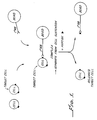
- the method entails forming within the cell suspension a complex comprising a cell separation means such as a paramagnetic bead linked to a primary antibody, which in turn is bound to a cell surface antigen on the target cells (see Figure 1).
- the complex is separated from the cell suspension, and then contacted with a specific peptide which binds to the primary antibody and thereby releases the target cell from the complex.
- a paramagnetic bead is linked to the primary antibody by a protein means such as a secondary antibody.
- a protein means such as a secondary antibody.
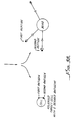
- This embodiment of the invention entails forming within the heterogeneous cell suspension a complex comprising the target cell bound to a primary antibody, which in turn is bound by a secondary antibody linked to the paramagnetic bead (see Figure 2).
- the complex is separated from the cell suspension, and then contacted with a specific peptide which binds to the primary antibody and thereby releases the target cell from the complex.
- the paramagnetic bead, linked to secondary and primary antibodies, is then separated from the target cell by conventional magnetic means.
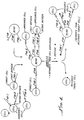
- the invention also provides methods for double positive cell selection, wherein a target cell bearing two desired antigens is selected from a heterogeneous cell suspension (see Figure 3A and 3B).
- the invention also provides methods for positive/positive cell selection wherein two different target cells, each bearing a different desired antigen, are selected from a heterogeneous cell suspension.
- the invention also provides methods for positive/negative cell selection wherein a target cell having a first antigen is selected from a heterogeneous cell suspension containing also undesired cells having the first antigen as well as a second antigen (see Figure 4). Positive/negative selection methods may also be applied to a cell suspension in which undesired cells are inadvertently trapped in the cell suspension containing the desired cells ( Figure 4).
- An exemplary method for positive/negative cell selection entails forming within the heterogeneous cell suspension a complex comprising a target cell having a first antigen bound to a first primary antibody, which in turn is bound by a secondary antibody coupled to a paramagnetic bead; the paramagnetic bead of the complex is also linked to a second primary antibody which is bound to a second antigen on an undesired cell.
- the complex is separated from the cell suspension, and then contacted with a specific peptide which binds to the first primary antibody, thereby displacing the primary antibody from the first antigen and releasing the target cell.
- the complexes of the paramagnetic beads attached to the primary and secondary antibodies and to the undesired cells are then separated by conventional magnetic means from the released target cell.
- the method provides a peptide which binds to a monoclonal antibody bound to a cell surface antigen on a target cell, displaces the antibody from the cell surface antigen, and thereby releases the target cell from the antibody.
- the invention also provides methods and specific peptide compositions for positive selection and specific release of target human hematopoietic stem/progenitor cells bound by the monoclonal anti-CD34 antibodies produced by the hybridomas designated ATCC HB 11646 and ATCC HB 11885, as well as the commercially available antibody 561 (Dynal, Oslo, Norway).
- the invention also provides methods and specific peptide compositions for positive selection and specific release of target human breast cancer cells bound by the monoclonal anti-breast cancer antibody 9187 produced by the hybridoma designated ATCC HB 11884.
- the invention also provides a method for identifying a specific peptide useful for the release of a target cell from the binding of a specific monoclonal antibody.
- the method comprises first selecting a candidate releasing peptide by at least one of the following means:
- the candidate peptide is then tested for its ability to displace the antigen as measured by FACS release and by release of cells bound to magnetic beads, or by biospecific interaction analysis (BIAcoreTM, Pharmacia).
- An exemplary method for identifying a peptide useful for releasing a cell bound by a specific monoclonal antibody comprises coating a solid support with a biotinylated or non-biotinylated form of the antibody, contacting the antibody with a plurality of peptides of a random peptide library, selecting at least one peptide which specifically binds to the antibody, contacting the antibody bound to the target cell with the selected peptide, and determining the ability of the selected peptide to detach the antibody from the target cell, thereby releasing the target cell.
- Figure 1 depicts a method for positive cell selection whereby a target cell is bound to a primary antibody and a cell separation means, separated from the cell suspension, and then contacted with a specific peptide which binds to the primary antibody and thereby releases the target cell.
- Figure 2 depicts a preferred method for positive selection wherein the primary antibody is linked to the cell separation means by a secondary antibody.
- Figures 3A and 3B depict a method for double positive cell selection and release whereby a target cell with two desired antigens is separated from a heterogeneous cell suspension and then released by incubation with two specific peptides.
- Figure 4 depicts a method for positive/negative cell selection whereby a target cell bearing a desired first antigen is selected from a heterogeneous cell suspension containing undesired cells bearing a second, undesired antigen.
- target cells may also be separated from undesired cells which bear both the desired first antigen and a second, undesired antigen.
- the invention provides methods and peptide compositions for the positive and positive/negative selection of target cells from a heterogeneous cell suspension.
- the methods are based on the identification of specific peptides which effect the displacement and release of a specific target cell from a specific monoclonal antibody.
- the peptide-mediated release is enzyme-free, and thus leaves the cell surface proteins intact. Moreover, peptide-mediated release leaves the target cell free of bound antibody or antibody fragments.
- the general method of the invention entails forming within a heterogeneous cell suspension a complex comprising the target cell, a monoclonal primary antibody bound to a cell surface protein on the target cell, and a cell separation means linked to the primary antibody and thus to the target cell.
- the complex is then separated from the cell suspension, and contacted with a specific peptide which binds to the primary antibody, thus displacing and releasing the target cell from the primary antibody and the cell separation means.
- the cell separation means linked to the antibody is then separated from the released target cell by conventional means.
- contacting refers to bringing into close proximity the peptide and the antigen/antibody complex such that weak intermolecular forces may be disrupted.
- binding refers to the binding of antibody to antigen by a combination of relatively weak non-covalent forces, including hydrophobic and hydrogen bonds, van der Waals force, and ionic interaction.
- the affinity of antibody-antigen binding is in the range of 5 X 10 4 to 10 12 liters per mole, more usually 10 6 - 10 9 l/M (Alberts, B., et al., Eds., Molecular Biology of the Cell, Garland Publishing, New York and London, 1983, p.969-970).
- lact refers to the peptide of the invention causing the antibody to become unbound from its cognate antigen by interruption of the weak non-covalent binding forces described above.
- release refers to the cell being unbound from the antibody/solid support, thereby leaving the cell free to flow with the elution fraction from a separation system.
- the peptide of the invention acts as an "epitope-mimicking" peptide, thus competing for the antigen-binding site on the antibody, and thereby displacing the antibody from its cognate antigen.
- the fact that the mechanism of action of the peptide of the invention is unknown does not detract from the importance and power of the invention.
- the peptide of the invention preferably contains fewer than 30 amino acid residues, more preferably 4 to 20 amino acid residues, most preferably 4 to 10 amino acid residues.
- the present invention also contemplates analogues of peptides formed by conservative amino acid substitutions, substitutions of non-natural amino acids, cyclization of peptides, and peptidomimetics modeled on identified releasing peptides.
- Analogues of synthetic peptides can be made by substituting individual residues with non-natural or unusual amino acids. Sequences of bioactive peptides are originally derived from proteins which are made up of the naturally occurring twenty L-amino acid residues. However, the process of chemical synthesis used to construct synthetic peptides allows for the substitution of alternate residues including D-amino acids, infrequently occurring natural amino acids, or non-natural synthetic amino acid analogues (Bodansky, M, 1984, Principles of Peptide Synthesis, Springer-Verlag, Berlin).
- These alternate residues can be used (a) to replace chemically reactive residues and improve the stability of the synthetic peptide, (b) to provide analytic labels useful in the detection of the synthetic peptide, and (c) to modulate the bioactivity of the synthetic peptide by increasing or decreasing the binding affinity of the antibody for the peptide.
- Cyclization of peptides Analogues of synthetic linear peptides can be made by chemically converting the structures to cyclic forms. Cyclization of linear peptides can modulate bioactivity by increasing or decreasing the potency of binding to the target protein (Pelton, J.T., et al., Proc. Natl. Acad. Sci., U.S.A., 82:236-239). Linear peptides are very flexible and tend to adopt many different conformations in solution. Cyclization acts to constrain the number of available conformations, and thus, favor the more active or inactive structures of the peptide.
- the immunogenicity of synthetic peptides has been correlated with the experimentally observed conformational preferences in solution (Dyson, H., et al., 1988, Annual Review of Biophysics and Biophysical Chemistry, 17:305-324). Differences in immunogenicity may be indicative of differences in binding affinity of specific antibodies for cyclic peptides.
- Cyclization of linear peptides is accomplished either by forming a peptide bond between the free N-terminal and C-terminal ends (homodetic cyclopeptides) or by forming a new covalent bond between amino acid backbone and/or side chain groups located near the N- or C-terminal ends (heterodetic cyclopeptides) (Bodanszky, N., 1984, supra ).
- the latter cyclizations use alternate chemical strategies to form covalent bonds, e.g. disulfides, lactones, ethers, or thioethers. Linear peptides of more than five residues can be cyclized relatively easily.
- the propensity of the peptide to form a beta-turn conformation in the central four residues facilitates the formation of both homo- and heterodetic cyclopeptides.
- the presence of proline or glycine residues at the N- or C-terminal ends also facilitates the formation of cyclopeptides, especially from linear peptides shorter than six residues in length. Examples of cyclized releasing peptides are shown in Example 14 below.
- Peptidomimetics technology is the design of molecular mimics of peptides. The ability to successfully design such molecules depends upon the understanding of the properties of the linear peptide sequence and the conformation in which it is presented to the antibody. The synthesis of mimetics can provide compounds exhibiting greater biological activity, improved solubility, and stability (Nakanishi, H., et al., 1993, Peptidomimetics of the immunoglobulin supergene family - a review. Gene 137:51-56).
- cell separation means refers to well-known means such as paramagnetic beads, columns, hollow fibers, glass beads, polysaccharide beads, and polystyrene tissue culture flasks.
- paramagnetic bead or “bead” will be used to illustrate a cell separation means.
- this invention is not limited to the use of paramagnetic beads as the separation means. Paramagnetic beads are separated from cell suspensions by the use of magnets (Hardwick, R.A., et al., J Hematotherapy 1:379-386, 1992).
- linking means include :
- the protein means for binding the primary antibody can be Staphyloccocus aureus Protein A, Streptococcus Protein G, or an immunoglobulin which binds to the monoclonal primary antibody.
- the latter is known as a "secondary antibody".
- the secondary antibody can be a polyclonal antibody or a monoclonal antibody.
- a polyclonal antibody is typically raised in an animal such as a rabbit, sheep, goat, horse, pig, or bovine species.
- a monoclonal antibody is typically raised in a small rodent such as mouse or rat according to the basic method of Köhler and Milstein.
- the term "secondary antibody” will be used to illustrate the protein means for binding the primary antibody.
- the invention can be applied to positive selection of any type of target cell.
- it is first necessary to provide a monoclonal antibody which binds to a specific cell surface antigen on the target cell.
- a monoclonal antibody specific for the target cell Given a monoclonal antibody specific for the target cell, the experimental examples below can be followed to identify a specific peptide sequence which will bind to the monoclonal antibody and displace the target cell, thereby releasing the target cell from the antibody.
- a given monoclonal antibody binds to a small portion of its cognate antigen, known as its epitope, which consists of as few as 3-6 amino acid residues (Pellequer, J.L., et al., Methods in Enzymology 208:176, 1991).
- the amino acid residues may be in sequence, or they may be discontinuous within the antigen sequence. When the amino acid residues of the antigen sequence are discontinuous, it is thought that they are presented in close proximity for recognition by the cognate antibody through three-dimensional folding of the antigen.
- This specific peptide may be an "epitope-mimicking" peptide, which acts by direct competition at the binding site, or it may be a peptide which displaces the antibody by any other mechanism.
- phage-display technique large libraries of random amino acid sequences are screened in biopanning or antibody binding assays (see Example 1 below). Examples of random peptide libraries are phage-displayed linear 6mer and 15mer libraries, constrained (cyclized) XCX 6 CX (described in Example 14 below), and a conotoxin XCCX 3 CX 5 C library.
- PIN random peptide libraries are displayed on isolated pins which then are screened for their ability to bind the antibody, as read out on ELISA-type assays.
- Random peptide libraries based on phage display or pin-peptide display are reviewed in Wells, J.A., et al., Current Opinion in Biotechnology 3:355-362, 1992, and in Scott, J.A., Trends in Biochemical Sciences, 17:241-245, 1992.
- Random peptide libraries may also be screened using antibody bound to beads (see Example 13 below).
- Candidate releasing peptides can also be identified by computer-assisted analysis of potential antigenic peaks in the protein antigen (see Example 11 below).
- Candidate releasing peptides can also be identified by analyzing complementarity-determining regions (CDR's) in the antibody of interest. Translation of available cDNA sequences of the variable light and variable heavy chains of a particular antibody permit the delineation of the CDRs by comparison to the database of protein sequences compiled in the book Sequences of Proteins of Immunological Interest, Fifth Edition, Volume 1, Editors: E.A. Kabat, et al., 1991 (see table on page xvi). Studies have shown that in some cases CDR peptides can mimic the activity of an antibody molecule (Williams, W.V., et al. Proc. Natl. Acad. Sci. U.S.A. 86:5537, 1989). CDR peptides may bind their cognate antibody, thus effecting displacement of the antibody from the antigen.
- CDR's complementarity-determining regions
- biospecific interaction analysis using surface plasmon resonance detection through the use of the Pharmacia BIAcoreTM system may be utilized.
- This technology provides the ability to determine binding constants and dissociation constants of antibody-antigen interactions. Analysis of multiple antibodies and the number of biopanning steps (at set antibody concentrations) required to identify a tight-binding consensus peptide sequence will provide a database on which to compare kinetic binding parameters with the ability to identify tight binding peptides and their activity as competitive agents. If a particular antibody/antigen interaction is determined to be extremely tight, then the researcher may choose to work with a different antibody.
- the use of the BIAcoreTM system requires purified antibody and a source of soluble antigen.
- Phage display-selected clones can be used as a source of peptide antigen and directly analyzed for antibody binding.
- CD34 antigen was obtained from detergent-solubilized CD34 protein from KG1a cells.
- BIAcoreTM technology was also applied to anti-CD4 antibodies; in this case, the source of antigen was commercially available recombinant soluble CD4 protein (Agmed, Bedford, MA).
- the candidate releasing peptides identified by the above described means are then screened for displacement of the antibody from the cell surface antigen, typically in assays using cells bearing the antigen.
- the specific peptide effects the displacement of the target cells by either (1) mimicking the epitope on the cell surface antigen, thereby competing against the epitope for antibody binding, or (2) binding to a site on the antibody and causing a conformational change, thus altering the antibody such that it can no longer bind to its epitope on the cell surface antigen.
- Evidence was obtained using labeled peptide and antibody that at least one of the identified peptides of the invention binds to its cognate antibody (data not shown).
- the methods of the invention can identify a specific peptide that acts to release the target cell by any mechanism.
- peptide which binds to a monoclonal antibody bound to a cell surface antigen on a target cell, displacing the antibody from the cell surface antigen, and releasing the target cell from the antibody refers to a peptide which acts to release the target cell by any molecular mechanism.
- Candidate releasing peptides can be identified by any one or several of the following means:
- a candidate peptide Once a candidate peptide has been identified, its ability to displace the antigen is tested by incubating the peptide with cells bound by the antibody. Release of cells from antibody is typically determined by FACScan or release from magnetic beads.
- One type of random peptide library which can be used in the practice of the invention is the hexapeptide phage display library described by Scott and Smith (Science 249:386-390, 1990).
- a monoclonal antibody would have to be biotinylated in order to bind tightly to an avidin coated plate to yield a sufficient signal to identify a peptide which binds to the antibody.
- many monoclonal antibodies cannot be biotinylated without diminishing or destroying their binding functions.
- the following describes exemplary methods for identifying a specific peptide useful for the release of a target cell bound by a specific monoclonal antibody.
- the methods involve first coating the monoclonal antibody onto a plastic plate so that the antibody attaches to the plate.
- the antibody binds to the plastic by non-specific interactions, thought to be electrostatic interactions.
- the monoclonal antibody may be biotinylated and then attached to an avidin-coated plate by exploiting the tight binding of avidin to biotin.
- Protein A or Protein G coated plates it is well known that Protein A and Protein G, from Staphyococcus and Streptococcus organisms, bind relatively tightly to certain immunoglobulin isotypes such as IgG and IgM.
- beads can be used as the solid support for the monoclonal antibody; antibody is coated onto or bound to beads using the same methods as for coating on plates. Once the monoclonal antibody has been coated onto the plate or beads, the attached antibody is contacted with a plurality of phage displaying a random peptide library, and then the non-bound phage are rinsed away. The bound phage are then eluted, grown, and amplified. This process is known as "biopanning".
- the phage DNA encoding the selected peptides is subjected to DNA sequence analysis to determine the candidate peptides for release of target cells.
- the candidate peptides are synthesized by conventional means. To increase solubility of the peptides, they may be synthesized with additional flanking sequences of hydrophilic amino acid residues, typically residues of amino acids which are polar or charged.
- the candidate peptides are then tested for their ability to displace the antibody from its cognate antigen on the surface of the target cell. It is understood that the mere fact that a peptide is bound by the antibody does not ensure that the peptide would displace the antigen. A candidate peptide might be bound less tightly by the antibody than the antigen is bound, thus the peptide might not compete successfully for binding and would not displace the antibody from its cognate antigen.
- Another way of expressing this problem is that the antibody might have greater affinity for its cognate antigen than it has for the candidate peptide. It is also very likely that a candidate peptide could bind an antibody without interfering with or binding to its antigen binding site (epitope). Fortunately, it was discovered that this method of the invention can successfully identify peptides which not only bind to the antibody, but also displace the antibody from its cognate antigen, thereby releasing the target cell from the antibody.
- a complex comprising the target cell bound to a primary antibody, which in turn is linked to a cell separation means, preferably a paramagnetic bead.
- the complex is separated from the cell suspension by conventional means, preferably a magnet.
- the primary antibody within the separated complex is then contacted with a specific peptide which binds to the primary antibody and displaces the antibody from the target cell, thereby releasing the target cell from the complex.
- the paramagnetic bead linked to antibody is then separated from the released target cell, yielding a purified target cell with its cell surface proteins intact, and without antibody or antibody fragments bound to its surface.
- FIG. 2 A preferred embodiment of the invention is depicted in Figure 2.
- the primary antibody is not directly coupled to the bead, but rather is linked to the bead by a secondary antibody, which in turn is coupled to the bead to form the complex.
- the complex is separated from the cell suspension and contacted with the specific peptide, thereby releasing the target cell.
- FIG. 3A and 3B Double Positive Cell Selection
- a target cell bearing two different antigens is positively selected from a heterogeneous cell suspension containing non-target cells bearing only one of the antigens.
- the cell suspension is incubated with first and second primary antibodies, each of which binds to only one of the two different antigens on the target cell.
- a complex is formed by adding to the cell suspension a paramagnetic bead coupled to a secondary antibody which binds to both primary antibodies.
- the complex is separated from the cell suspension and then contacted with a specific peptide which binds to the first primary antibody, thereby releasing the cell which bears the first antigen but not the second antigen.
- This cell is separated from the remaining cell-antibody-bead complex.
- the remaining target-cell-antibody-bead complex is then contacted with a second specific peptide which binds to the second primary antibody, thus displacing the target cell from the second primary antibody and releasing the target cell from the bead.
- FIG. 4 Another embodiment of the invention is depicted in Figure 4 whereby a target cell bearing a first antigen is positively selected from a heterogeneous cell suspension which also contains undesired cells bearing the first antigen as well as a second antigen (Positive/Negative Selection).
- the positive/negative selection method of the invention is also useful for removing contaminating, undesired cells which do not bear the first antigen, but only a second antigen.
- Positive/negative selection is especially desirable when, for instance, autologous CD34+ cells are to be selected from blood or bone marrow of a cancer patient.
- the selected CD34+ cells are destined for re-infusion to the patient to reconstitute his bone marrow after high-dose chemotherapy or radiation.
- Positive selection of CD34+ cells alone is thought to reduce the tumor burden in the selected cell sample by several logs. However, it would be most desirable to negatively select against cancer cells as an added precaution against the possibility that reinfused cancer cells might contribute to relapse. Positive/negative cell selection can be conducted either simulataneously (concomitantly) or sequentially.
- a complex which comprises the target cell bound to a first primary antibody, which is linked to a bead, which in turn is linked to a second primary antibody bound to an undesired cell.
- the first primary antibody can be an anti-CD34 antibody
- the second primary antibody can be an anti-B cell antibody, or a mixture of several antibodies against undesired cell types.
- Anti-B cell antibodies are especially useful in purging of positively selected CD34+ cell populations from patients with B-cell lymphomas.
- the complex is separated from the cell suspension, and then contacted with a specific peptide which binds to the first primary antibody, thereby displacing the first primary antibody from its cognate antigen on the target cell surface and releasing the target cell from the complex.
- the undesired cell remains bound to the bead via the second primary antibody.
- the undesired cell can be separated from the released target cell, yielding a purified population of target cells separated from undesired cells.
- Sequential positive/negative cell selection In this method, the positive selection step is conducted first as described above, using only the antibody against the desired antigen (for instance CD34) and release by a specific peptide.
- the positively selected cells retain antigens on their surfaces due to the non-enzymatic peptide-mediated release, making a subsequent selection step possible.
- the positively selected cells are then incubated with the second primary antibody or mix of antibodies directed against undesired antigens such as B-cell antigens.
- the cells bound by the second primary antibody(s) are captured by conventional means, and the unbound cells are collected for reinfusion to the patient.
- Positive/negative selection is especially important for the further purification of positively selected CD34+ cells.
- the positively selected CD34+ population will be over 90% pure, which represents a 3 log depletion of B cells, for instance (see Example 19 below).
- Addition of a negative selection step further depletes undesired cells up to a 4 log depletion or greater.
- the negative selection step is known as "purging".
- Negative selection can be optimized so that the resulting cell composition is substantially free of undesired cells.
- the term "substantially free" of undesired cells means that no undesired cells are detected using standard sampling and analysis by, for instance, immunocytochemistry, morphology, or FACScanTM.
- the negative selection technique can be used also for depletion of T lymphocytes from allografts, thus greatly reducing the risk of graft-versus-host disease (GVHD).
- GVHD graft-versus-host disease
- the extent of depletion of undesired cells is dependent on, among other factors, the antibody/bead/cell/peptide ratios utilized. These ratios can be optimized to yield the desired log depletion of, for instance, B cells or T lymphocytes.
- a 3-4 log reduction in tumor cells reinfused to the patient is expected to reduce incidence of relapse.
- a heterogeneous cell suspension of human bone marrow, peripheral blood, or cord blood contains a very small number of stem cells (typically 0.2 to 2.0%).
- the stem cells are target cells which are to be positively selected for further use such as in vitro culture or reinfusion to a patient.
- Human hematopoietic stem/progenitor cells are so named because they have the capacity to proliferate many times over, and to differentiate into all hematopoietic cells types.
- stem cells refers to human hematopoietic stem/progenitor cells.
- Stem cells bear a characteristic cell surface antigen known as CD34.
- CD34 cell surface antigen
- monoclonal antibodies have been produced which specifically bind to CD34. It is assumed that each monoclonal antibody binds to a different epitope on the CD34 antigen, since it is statistically very unlikely that several different monoclonal antibodies would be produced against the identical epitope.
- a peptide identified as effective for displacing a given anti-CD34 monoclonal antibody is likely to displace only this specific antibody, and not other monoclonal anti-CD34 antibodies.
- a complex is formed comprising human stem cells which are bound by the mouse monoclonal antibody 9069 (1° AB), which is in turn bound by a sheep-anti-mouse antibody (2° AB), which is coupled to a paramagnetic bead.
- the complex is separated from the cell suspension by magnetic means.
- the 9069 antibody (°1 antibody) is contacted with a specific peptide which binds to the 9069 antibody and displaces it from the CD34 antigen on the stem cell, thereby releasing the stem cell.
- the paramagnetic bead linked to the sheep anti-mouse antibody and the 9069 antibody is then separated from the released stem cell using a magnetic means. This provides a highly purified suspension of stem cells with their surface proteins intact, including the CD34 antigen protein. Moreover, the stem cell does not have residual antibody or antibody fragments bound to its surface.
- the invention also provides a method for positive/positive cell selection whereby two desired target cells can be positively selected from blood or bone marrow. For instance, it may be desirable to positively select both stem cells and T-lymphocytes.
- T-lymphocytes bear the cell surface antigen known as CD3.
- Specific subsets of T-lymphocytes bear cell surface antigens known as CD4 and CD8.
- a monoclonal antibody against the desired class of T-lymphocyte can be provided and used to screen peptide libraries as described in Example 1 below.
- a specific peptide which displaces the anti-T-lympocyte antibody is selected and used in conjunction with a peptide that displaces the anti-CD34 antibody.
- both the anti-CD34 antibody and the anti-T-lymphocyte antibody are incubated with the cell suspension, and the two types of target cells are bound by their specific primary antibodies.
- the primary antibody-bound cells are bound to secondary antibodies coupled to beads, they are separated from the cell suspension, and then displaced from the beads by contact with the two specific peptides.
- a substantially pure suspension of stem cells and T-lymphocytes is obtained.
- the invention provides a method for double positive cell selection whereby, for instance, a subset of CD34+ cells bearing other cell surface markers may be positively selected.
- the invention also provides a method for positive/negative cell selection whereby target CD34+ cells may be positively selected from a suspension of blood or bone marrow which also contains undesired CD34+ cells which bear a second antigen such as a tumor marker.
- a second antigen such as a tumor marker.
- the use of autologous stem cell transplant would involve harvesting a portion of the patient's bone marrow or peripheral blood prior to treatment, and there is a risk that the bone marrow might harbor tumor cells which would then proliferate when they were reinfused to the patient.
- the addition of a negative purging step allows removal of any autologous tumor cells non-specifically captured in the positive selected fraction.
- the types of cancer for which autologous bone marrow transplant would be indicated include neuroblastoma, breast carcinoma, small cell lung carcinoma, and colon carcinoma.
- the positive selection of CD34+ cells reduces the risk of transfer of cancer cells because it is believed that very few or no CD34+ cells are metastatic tumor cells. However, a higher degree of confidence can be attained through the use of positive/negative cell selection.
- cancer antigens There are several cell surface antigens identified as indicative of the tumorous nature of a cell, and antibodies are available which bind to these tumor antigens. For instance, to select against neuroblastoma cells, antibodies against the following antigens can be used: G D2 , NCAM, 459, HSAN, UJ13A, and UJ167.11 (In: Bone Marrow Processing and Purging , Ed. Adrian P. Gee, CRC Press, Boca Raton, Florida, 1991.) To select against breast carcinoma cells, a panel of antibodies which bind to a wide range of breast antigens can be used. Likewise, to select against small cell lung carcinoma cells, a panel of antibodies directed against neural, epithelial, and neuroendocrine antigens can be used. The carcinoembryonic antigen (CEA) is present on a wide variety of breast and colon cancer cells, and antibodies against CEA are useful in selecting against these tumor cell types.
- CEA carcinoembryonic antigen
- a complex comprising the target CD34+ stem cell, the 9069 anti-CD34 antibody (°1 AB-I), the sheep anti-mouse antibody (2° AB), a bead, the second primary antibody or panel of antibodies directed against tumor antigen(s) (1° AB-II), and the undesired cell, which may or may not also bear the CD34+ antigen.
- the complex is separated from the suspension and contacted with a specific peptide which binds to the 9069 antibody, displacing the 9069 antibody from the CD34 antigen, and thus releasing the target stem cell from the complex.
- the bead bound to the antibodies and the undesired cell is then separated from the released stem cell, yielding a purified suspension of CD34+ stem cells which has been purged of cells bearing the tumor antigens.
- any of the above described selection methods may be used to positively select human hematopoietic CD34+ cells by binding the stem cells with the 9069 antibody produced by ATCC HB 11646, and then releasing the stem cells by contacting the 9069 antibody with a peptide selected from the list below.
- peptide sequences are shown in the one-letter amino acid symbols recommended by the IUPAC-IUB Biochemical Nomenclature Committee (see PatentIn User Manual of the U.S. Patent and Trademark Office, November 1990, page 101).
- J 1 and J 2 are selected from the group consisting of 0 - 6 amino acid residues.
- J 1 and J 2 contain hydrophilic, polar, or charged amino acid residues to aid the solubility of the peptide in aqueous solution.
- hydrophilic, polar, or charged amino acids are: G, S, T, C, Y, N, Q, D, E, H, K and R.
- any of the above listed peptides can have an amino terminal amino acid residue which is acetylated. Also, any of the above listed peptides can have a carboxy terminal amino acid residue which is amidated.
- the invention also provides peptides which can release cells bound by the anti-CD34 antibody designated 9079, which is produced by the hybridoma deposited under the Budapest treaty with the ATCC, designated ATCC HB-11885, effective May 9, 1995.
- the following peptides are 9079-releasing peptides:
- the invention also provides peptides which can release cells bound by the anti-CD34 antibody designated 561, commercially available from Dynal, Oslo, Norway.
- the following peptides are linear 561-releasing peptides:
- the invention also provides peptides which can release cells bound by the anti-human breast cancer antibody designated 9187, which is produced by the hybridoma deposited under the Budapest treaty with the ATCC, designated ATCC HB-11884, effective May 9, 1995. It is useful to positively select breast cancer cells from a patient's blood or bone marrow for several different techniques including culture of cancer cells to determine chemotherapeutic susceptibility, and to provide a cancer cell population for production of a patient-specific vaccine or therapeutic monoclonal antibody. Peptides which release cells bound by antibody 9187 are:
- compositions and methods of the invention may also be applied to epitope/antibody assays for cell quantitation. For instance, it would be clinically valuable to have a quick, simple, and standardized assay to determine the number of CD34+ cells in an apheresis product or a positively selected cell composition. Currently, the number of specific cells in a composition is determined by flow cytometry, which requires expensive equipment and a skilled operator.
- peptide epitopes for antibodies which recognize cell surface determinants also allows construction of diagnostic cell-based assays, for example.
- a peptide capable of releasing a specific cell of interest from a specific monoclonal antibody is provided.
- the peptide can be bound to a solid support such as a synthetic bead or immobilized to another type of solid phase, to construct an "artificial cell target" for antibody binding.
- a standard binding curve is then established, in which decreasing amounts of the peptide/bead complex are contacted with a constant concentration of the specific monoclonal antibody. This yields a range of signal for antibody binding to bead.
- the signal might be generated in several ways. Conjugating the antibody, or using a secondary antibody conjugate, allows collection of a magnetic bead/peptide/antibody complex, and quantitation of the captured antibody. Alternatively, the capture of a fluorescent bead/peptide complex through the antibody molecule allows similar quantitation of binding, through captured fluorescence.
- Similar indirect binding assays can be performed for antibody binding on peptide epitope immobilized to a solid phase.
- Test material containing unknown CD34+ target cell numbers can inhibit antibody binding to a peptide coated on a solid phase.
- Cell concentration can be determined following establishment of a control standard curve.
- the value of a solid phase assay is its adaptibility to a rapid read out system. For example, diagnostic systems which deliver electronic signal proportional to antibody binding have been developed, and this might allow an in-line quantitation of target cell concentration tied to cell selection hardware. Again, a diagnostic assay of this sort would provide an estimation of starting target cell concentration, allowing optimization of cell capture reagents and improved system performance.
- Monoclonal anti-CD34 antibodies designated "9069” were produced by standard methods from hybridomas obtained from Baxter-Hyland (Lansdorp clone 9.C.5, Terry Fox Laboratories and Becton-Dickinson). The hybridoma which produces antibody 9069 is deposited under the terms of the Budapest treaty with the American Type Culture Collection, Rockville, Maryland, USA.
- the input phage was 100 ⁇ l of the amplified eluate.
- the plate containing bound 9069 antibody and phage was incubated for 4hr at 4°C, and then washed 12X with TBS/TWEEN.
- the bound phage was eluted by adding 400 ⁇ l elution buffer (0.1 N HCl, pH adjusted to 2.2 with glycine, plus 1 mg/ml BSA) and gently rocking the plate for about 10 minutes.
- the eluate was then pipetted into a 500 ⁇ l microfuge tube containing 75 ⁇ l 1 M Tris.HCl, pH 9.1, to yield a final pH of 7 - 8.5.
- the eluate was then concentrated using a 30 kD AmiconTM filter.
- the concentrated eluate was used to infect K91 Kan starved cells for 30 minutes at room temperature.
- the production of gpIII was induced by addition of 0.2 ⁇ g/ml Tet-NZY for 60 minutes at 37°C.
- the phage were then grown and amplified overnight at 37°C.
- the phage were harvested and subjected to two rounds of polyethylene glycol (PEG) precipitation. Serial dilutions were made and both input and output phage were titered. Three more rounds of biopanning and titering were conducted. After the fourth round of biopanning and titering, 100 clones were selected and grown overnight at 37°C. The supernatant was collected and subjected to two rounds of PEG precipitation, followed by one round of acetic acid precipitation.
- PEG polyethylene glycol
- DNA sequence analysis was performed using an Applied Biosystems Inc. (Foster City, CA) 373 Automated DNA Analysis System. Cycle sequencing utilizing Taq polymerase was performed following the procedures of Applied Biosystems. Oligonucleotide primers were purchased from Operon Technologies Inc. (Alameda, CA).
- Selected phage clones were analyzed by DNA sequence determination of the random hexamer region of the pIII gene.
- Specific oligonucleotide primers were designed based on the published nucleotide sequence of the bacteriophage f1 (Hill, D.F., et al., J Virology 44:32-46, 1982). The 5' primer was from nucleotides 1533-1556 and the 3' primer the complement of nucleotides 1714-1737.
- KG1a is a human cell line (ATCC #CCL 246.1) that expresses CD34 antigen on its cell membrane and is used as a model system for initial testing or optimization of conditions for positive selection of CD34+ cells.
- Anti-CD34 antibody 9069 (0.0125 microgram), was preincubated with phage supernatants ( 0, 50, or 300 microliters) prepared as in Example 1. Subsequent incubation with KG1a cells (10 6 ) was for 30 minutes at room temperature (about 22°C). Irrelevant phage clones selected with a different anti-CD34 antibody were used as negative specificity controls. Detection of cell-bound anti-CD34 antibody was determined by addition of 10 micrograms of FITC-goat anti-mouse IgG (FITC-GAM) followed by FACScan analysis.
- FITC-goat anti-mouse IgG FITC-GAM
- Results Addition of selected phage clone supernatants to the anti-CD34 antibody resulted in a loss of detectable cell surface antibody binding. These results were indicated by a shift in total fluorescense from the antibody alone position of KG1a cells (the farthest right) towards the left, indicating a decrease in bound antibody.
- Table 3 provides a summary of two experiments testing the binding of phage display selected clones to the 9069, anti-CD34 antibody. Approximately 50-86% of total antibody binding was observed after the addition of phage supernatants expressing peptide epitopes. Subsequent testing utilized specific peptides representing the hexamer sequences with limited flanking sequences as indicated in Table 4.
- the peptides were able to bind to the anti-CD34 antibody, 9069, and thus decrease the amount of KG1a cells bound with antibody.
- Anti-CD34 antibody, 9069 (0.1 microgram) was incubated with KG1a cells (10 6 ) for 30 minutes at room temperature (about 22°C). Molar excesses of 10 5 to 10 6 times the amount of peptide to antibody were tested for the ability to displace the prebound antibody. Peptides were incubated with the antibody-cell complexes for 30 minutes at room temperature (approximately 22°C). Remaining bound antibody was detected using the FITC-goat anti-mouse IgG reagent described in Example 3.
- Table 5 lists the peptide sequences tested and the percent of inhibition of antibody binding detected. These data represent the ability of peptides to displace the prebound antibody from the KG1a cells.
- CD34+ cells were also isolated from human mobilized peripheral blood using an automated cell separation apparatus. Chymopapain was used as the control releasing agent, and 25 mg of the peptide 9069N (Table 11) was used as the test releasing agent.
- the purity of the positively selected CD34+ cells was greater than 90% for both the chymopapain and peptide released cells.
- the peptide release method yielded 14 x 10 6 cells, while the chymopapain release method yielded 20 x 10 6 cells.
- the peptide release method yielded 19 x 10 6 cells, while the chymopapain release method yielded 22 x 10 6 cells.
- the positively selected CD34+ cells from the first experiment were grown in culture with cytokines for 12 days.
- the peptide-released cells showed a 100-fold expansion in cell number, while the chymopapain-released cells showed a 68-fold expansion.
- Example 4 Additional experiments performed as detailed in Example 4 demonstrate that certain properties of the peptide sequences selected by phage display may be important in their ability to bind to the anti-CD34 antibody and to effectively displace the antibody prebound to the CD34 antigen expressed on the cell surface of KG1a cells.
- Table 11 summarizes the peptide hexamer motifs examined, the exact peptide sequences tested, a brief description of their relevant features and their beta-turn potential (Previlige, P., Jr., and Fasman, G.D. Chou-Fasman Prediction of Secondary Structure of Proteins: The Chou-Fasman-Previlige Algorithm in Prediction of Protein Structure and the Prinicples of Protein Conformation, 1989, ed. G.D. Fasman, Plenum Press, New York).
- biopanning of the phage display library could have identified hexamer sequences exactly matching the natural sequence.
- the beta-turn potential or the ability to assume a loop-like structure is greater for the phage display selected peptides than the natural CD34 hexamer sequences.
- beta turn potential was an important feature of the competitive peptides, hexamer types VIII and IX were designed.
- hexamer motif I appears to be a better competitor than hexamer motif II.
- CD34 cDNA sequence Analysis of the published CD34 cDNA sequence (Simmons, supra ; He, supra ) revealed the identification of two discontiunous regions homologous to the phage display selected hexamer sequences.
- the first region at amino acids 14-19 of the mature, signal peptide processed CD34 protein (epitope 1) is homologous to hexamer motif type I and II.
- the second region at amino acid 76-81 is homologous to hexamer motif type IV and to the inverse of hexamer motif type III (see Table 12).
- Peptides representing both epitope 1 and 2 could potentially have a synergistic effect in detaching and releasing CD34+ cells from antibody 9069.
- Phage display analysis identified a dominant hexapeptide sequence recognized by the anti-CD34 monoclonal antibody, 9069.
- the shortest peptide tested for competitive activity against 9069 antibody bound to KG1a cells had the following sequence: Ac-QQGWFP-KD.
- Tryptophan is known to be unstable and therefore a modified peptide, 9069Q2, was designed in which the trytophan was replaced by a phenylalanine: Ac-QQGFFP-KD. This latter sequence was shown to function as a competitive release reagent in the KG1a cell-based FACS assay.
- the 9069Q2 peptide serves to displace prebound KG1a cells from the 9069 antibody.
- FACScan assays included a negative control of KG1a cells alone and positive control samples of KG1a cells with the 9069 antibody bound and detected with a secondary antibody, goat-anti-mouse IgG-FITC.
- the 9069N peptide was able to displace prebound 9069 antibody from KG1a cells.
- the 9069Q2 peptide was able to displace prebound 9069 antibody from KG1a cells.
- the 9069N peptide defined through phage display contains a potentially unstable tryptophan residue. Replacement of this amino acid with phenylalanine did not abolish the ability of the peptide 9069Q2 to effectively compete off 9069 antibody bound to KG1a cells.
- Previous analyses of the hexapeptides revealed the likely requirement for good beta turn potential. (Prevelige, P.Jr., and Fasman, G.D. Chous-Fasman Prediction of Secondary Structure of Proteins: The Chou-Fasman-Prevelige Algorithm in Prediction of Protein Structure and the Principles of Protein Conformation, 1989, ed. G.D. Fasman, Plenum Press, New York.) Amino acid substitutions resulting in "poor” or "strong" beta-turn potential indicated that functional activity corresponded to the peptide with the most loop potential.
- Tested peptides contain additional flanking sequences either derived from the phage display vector and/or charged residues to aid solubility.
- the 9079 anti-CD34 antibody was used to select linear hexapeptide sequences from a phage display library. Multiple unrelated hexapeptide sequences with no direct homology to the CD34 antigen were identified from third and fourth biopanning phage clones. A fifth biopanning revealed a predominant hexapeptide sequence.
- the current human stem cell isolation system developed by the Immunotherapy Division utilizes the anti-CD34 antibody, 9069. Replacement of the chymopapain treatment to release captured stem cells is desirable. Potential problems of immunogenicity of residual amounts of remaining chymopapain, lot variation with chymopapain and the inability to perform additional negative selections due to stripping of cell surface antigens with the chymopapain treatment were among the reasons for investigating alternative release reagents.
- the original protocols for phage display biopanning of the linear hexapeptide library obtained from Dr. George Smith at the University of Missouri designated the use of biotinylated antibody. Three biopanning steps with the 9079 antibody were performed. The third eluate was stored at 4°C for one year, then subjected to amplification prior to the fourth biopanning. A fifth biopanning was performed from an unamplified fourth biopanning. Phage clones fom the third, fourth and fifth biopannings were subjected to DNA sequence analysis. Multiple hexapeptide sequences were identified in each biopanning. Only in the fifth biopanning did a predominant sequence emerge. None of the selected hexapeptides show direct homology to the CD34 antigen.
- the linear hexapeptide library was obtained from Dr. George Smith at the University of Missouri.
- the random hexapeptide sequence was inserted into the pIII gene of the vector FUSES.
- the 9079 antibody was obtained from Ginny Ofstein in the Bone Marrow Therapies R & D Group, Immunotherapy Division, Santa Ana.
- the hexapeptide library was amplified in 2 L of Terrific brothTM (500ml per 2L flask) as described above. Briefly, K91kan cells were grown to an OD550 ⁇ 2.0 at 225 rpm, 37°C. After 15 minutes at 50 rpm for pili regeneration, the cells were infected with the library at a moi ⁇ 1 (multiplicity of infection of 1 phage particle per cell). Infection was allowed to proceed overnight.
- the amplified library was concentrated with PEG/NaCl from ⁇ 2L to 1 ml.
- the 9079 antibody was biotinylated following the procedure of G. Smith. Three steps of petri plate (35mm) biopanning were performed following the procedures of George Smith. The amount of biotinylated 9079 antibody used per step was: 10 ug-1st biopanning, 10ug-2nd biopanning, 1ug-3rd biopanning ("10-10-1"). Each successive step of biopanning was preceded by an amplification of the eluted phage. 5x10¹⁰ TU of the library were used in the first biopanning.
- Tetracycline/kanamycin resistant colonies from the third biopanning were grown and supernatants containing the bacteriophage were PEG precipitated.
- DNA was prepared from the PEG concentrated phage for DNA sequence analysis.
- DNA sequence was determined following "cycle” sequencing reactions using the Applied Biosystems PRISM fluorescent dideoxy terminators and oligonucleotide primer JTL2.
- a fourth biopanning was performed after amplication of the third eluate.
- Three different concentrations of non-biotinylated 9079 antibody were used: 0.02 ug, 0.1 ug and 1 ug.
- Eluted clones were grown and DNA prepared as above. DNA sequence analysis was performed using JTL2 primer as above.
- a fifth biopanning using 1 ug of non-biotinylated 9079 antibody was performed with the 4th biopanning eluate in the absence of amplification.
- Phage display analysis of the 9079 antibody with a linear hexapeptide library revealed multiple hexapeptide sequences with no apparent direct homology to the CD34 antigen. This result is similar to the results observed with the 561 antibody when biopanned on petri plates (see below).
- the 9079 antibody is capable of blocking recognition of the CD34 antigen by the 561 antibody (Dynal).
- the possibility exisits that both the 561 and 9079 antibodies recognize the region of CD34 containing six cysteine residues and the only arginine residues (amino acids 146-219). Recognition of flexible loops stabilized by charged amino acids may result in the selection of many different hexapeptide sequences.
- the identification of uninserted clones in the analysis of fourth biopanning clones may be a result of the one year long storage of the unamplified 3rd eluate. Uninserted clones are know to grow more efficiently than the hexapeptide-containing clones and may also have better viability during long-term storage. At very low 9079 antibody concentrations (0.02 ug and 0.1 ug) during the 4th biopanning, many non-specific, uninserted clones were eluted. At higher antibody concentration (1 ug) very few uninserted clones were identified. The fifth biopanning was performed in the absence of amplification of the fourth (1 ug) biopanning to avoid enhancement of selecting uninserted clones. Of the fourteen clones analyzed from the 5th biopanning, no uninserted clones were identified.
- biotinylated and non-biotinylated antibody can be used for phage display biopanning.
- the biotinylated 9079 was used for the first three biopanning steps. Based on the successful results of biopanning with non-biotinylated 9069 antibody, the subsequent biopannings with the 9079 antibody were accomplished with non-biotinylated antibody.
- Epitope peptide phage display biopanning with the 9079 antibody revealed multiple hexapeptide sequences until a fifth biopanning step was performed. Whether these sequences actually represent all or portions of discontinuous epitopes of the CD34 antigen is not known. The identification of multiple sequences suggest that mimetopes that mimic the actual epitope sequence may have been selected.
- DNA sequence analysis (10ug-10ug-1ug) of the third biopanning revealed at least 29 different sequences.
- the 5th biopanning (10-10-1-1-1) indicates a predominant sequence has emerged. This biopanning was performed without amplification of the 4th eluate to avoid overgrowth during amplification of uninserted phage vector which was seen in the analysis of the 4th biopanning clones.
- peptides were tested for functional activity as potential stem cell release reagents using the KG1a cell-based FACS assay. In preliminary experiments six peptides showed at least 50% release of 9079 antibody prebound to cells.
- the 9079 antibody was chosen for further study because of it's high binding affinity, it's retention of functional activity upon chemical biotinylation, and the chymopapain-resistant nature of it's recognition of CD34 antigen. Phage display biopanning with the anti-CD34 antibody, 9079, identified multiple hexapeptide sequences (see above). A predominant sequence was identified in the fifth biopanning.
- the peptides were synthesized by Research Genetics and tested without purification.
- the 9069 and 9079 antibodies were obtained from the Baxter Immunotherapy Research group in Santa Ana.
- the 9079 antibody has been deposited with the American Type Culture Collection (ATCC) under the provisions of the Budapest Treaty for patent purposes: deposit number ATCC-HB-11885, date of deposit May 9, 1995.
- the 9069 antibody was used as a positive control and released with the 9069N peptide (Ac-QQGWFP-KD). This control served to test for the KG1a cells and the goat-anti-mouse FITC secondary detection antibody.
- Hexapeptide sequences identified for the 561 antibody also were tested for their ability to displace prebound 9079 antibody.
- Peptides (see Table 18) were purchased from Research Genetics Inc., Hunstville, AL.
- 9079A-G peptides were solubilized in Dulbecco's phosphate buffered saline (DPBS) plus 1% HSA.
- DPBS Dulbecco's phosphate buffered saline
- HSA HSA
- the 9079A-G peptides were tested in the FACScan assay using 10 ⁇ 6 KG1a cells bound with 0.05 ug of the 9079 antibody.
- the 561A-E peptides (see Example 13 below) were tested in the FACScan assay using 10 ⁇ 6 KG1a cells bound with 0.05 ug of the 9079 antibody.
- the 9079H peptide was tested in the FACScan assay using 10 ⁇ 6 KG1a cells bound with 0.05 ug of the 9079 antibody.
- Peptides 9079A, B, C, D, F, and H were solubilized.
- 9079E peptide was insoluble and therefore not tested.
- the 9079A, B, C, D, F, G and H peptides all showed at least 50% release of prebound 9079. None of the 561A-E peptides could release prebound 9079 antibody.
- Peptide 9079C was incorrectly assigned. However, it tested positively. The correct sequence should have been GYTQPK-D.
- This analysis is designed to identify possible exposed surface peaks of the protein combining information from hydrophilicity, surface probability and backbone flexibility predictions with the secondary structure predictions of Chou-Fasman and Robson-Garnier (MacVectorTM User's Manual, International Biotechnologies, Inc., pages B56-B69; Jameson, B.A. et al., 1988 Comput. Applic. in the Biosciences 4:181-186).
- the peptides were synthesized by Research Genetics and tested without purification.
- the 9079 antibody was obtained from the Baxter Immunotherapy Research Group in Santa Ana, California.
- the 561 antibody was obtained from Dynal, AS.
- Peptides (see Table 19) were purchased from Research Genetics, Inc., Huntsville, AL
- Peptides 34A-F did not show any release activity on 9079 antibody prebound to KG1a cells.
- Peptides 34A-F did not show any release activity on 561 antibody prebound to KG1a cells.
- biopanning of the hexapeptide library with the 561 antibody directly attached to magnetic beads identified one (P A N V S L) of three hexapeptides which show good homology (5/6 amino acids, P A N V S T in CD34) to a defined potential antigenic peak (N V S T) of the CD34 antigen. Since this sequence had only four amino acids, this was not among those peaks for which a peptide was designed and tested. It is believed, however, that a peptide containing this 4-amino-acid sequence is a good candidate for a releasing agent for the 561 antibody.
- the accumulated data from the analyses of the 9069, 9079 and 561 anti-CD34 antibodies indicate that determination of the potential antigenic peaks of an antigen protein may save time in defining potential competitive epitope peptides. Correlation of the defined peaks with any known structural data on the antigen and correspondence to phage display-defined peptides will permit the best educated guess on selection of peptide sequences to test for functional activity. If a particular antibody of interest can recognize a linear peptide epitope such as that of the 9069 antibody, then this type of analysis could supersede the initiation of the laborious phage display work. However, if the antibody recognizes a conformational or discontinuous epitope, then this type of analysis can at best support but not define a peptide with functional release activity.
- P A N V S T was the CD34 antigen hexapeptide sequence homologous to the P A N V S L hexapeptide identified by biopanning with direct 561 antibody attached to beads.
- T Q G T F S was the CD34 antigen hexapeptide homologous to T Q G S F W and Q Q G W F P hexapeptides identified by biopanning with the 9069 antibody.
- N S S V Q S was the CD34 antigen hexapeptide homologus to
- N S S V G L hexapeptide identified by biopanning with the 9069 antibody N S S V G L hexapeptide identified by biopanning with the 9069 antibody.
- the hexapeptide library was amplified in 2 L of terrific broth (500 ml per 2 L flask). Briefly, K91Kan cells were grown to an OD550 ⁇ 2.0 at 225 rpm, 37°C. After 15 minutes at 50 rpm for pili regeneration, the cells were infected with 10 ⁇ l ( ⁇ 10 12 physical particles) of the primary library. The amplified library was concentrated with PEG/NaCl from ⁇ 2 L to 1 ml. The amplified library was titered. Seven rounds of biopanning were performed as described in Example 1 above.
- the amount of 561 antibody used per step was: 28 ⁇ g-1st biopanning, 14 ⁇ g-2nd biopanning, 5 or 10 ⁇ g-3rd biopanning, 1 ⁇ g-4th biopanning, 1 ⁇ g-5th biopanning, 2 ⁇ g-6th biopanning, 1.5 ⁇ g-7th biopanning ("28-14-10-1-1-2-1.5 or 28-14-5-1-1-2-1.5".
- Each successive step of biopanning was preceded by an amplification of the eluted phage. 5x10 10 TU of the library were used in the first biopanning.
- Tetracycline/Kanamycin resistant colonies from the third to seventh rounds of biopanning were grown and supernatants containing the bacteriophage were PEG precipitated.
- DNA was prepared from the PEG concentrated phage for DNA sequence analysis. DNA sequence was determined following "cycle" sequencing analysis using the Applied Biosystems PRISM fluorescent dideoxy terminators and oligonucleotide primer JTL2.
- Phage display analysis of the 561 antibody with a linear hexapeptide library revealed 4 predominant hexapeptide sequences with no apparent direct homology to the CD34 antigen. This result is similar to the results observed with the 9079 antibody when biopanned on petri plates.
- the 561 antibody is capable of blocking recognition of the CD34 antigen by the 9079 antibody. It is possible that both the 561 and 9079 antibodies recognize the region of CD34 containing six cysteine residues and the only five arginine residues. Recognition of flexible loops stabilized by charged amino acids may result in the selection of hexapeptide sequences recognized by a discontinuous epitope.
- Biopanning of a hexapeptide library with the 561 antibody resulted in the identification of four predominant sequences (561 peptide A to D). These hexapeptide sequences contain both highly charged and hydrophobic residues which is also supported by the conclusions drawn from the cyclic peptide biopanning analysis (see Example 12) and linear hexapeptide selection using 561-Dynabead (see Example 13).
- the repeated selection of peptides containing arginine residues may be indicative of specific recognition of the region within the CD34 antigen (a.a.#149 to 219) containing the only five arginine residues in the extracellular domain of the protein.
- PANVSL CD34 antigen
- Phage-bearing peptides with high affinity for 561 antibody were selected from those with low affinity peptides using 561 antibodies immobilized on solid-phase petri dishes as described in Example 1 above. However, fine affinity discriminations were difficult, possibly because binding was dictated by both the affinity and the avidity of the phage (Clarkson, T., et al., 1991, Nature 352:624-628).
- An alternative selection method was based on phage peptides binding to 561 directly linked to Dynabeads (561-bead) in solution. The high affinity phage peptides were then enriched by competition for limiting amounts of antibody. It is believed that this scheme forced the many low affinity phage to be out-competed by the binding of rare high affinity variants.
- Peptide epitopes in solution were selected using the hexapeptide library with two different lots of 561-beads CEL R21 and CEL R73.
- Four predominant linear hexapeptide sequences were selected and identified with a major characteristic being their highly charged and hydrophobic nature.
- Three of these four peptides (561 M, P, and Q) were able to release 561 antibody prebound to KG1a or tHL60 cells.
- the linear hexapeptide library was obtained from Dr. George Smith at the University of Missouri.
- the random hexapeptide sequence was inserted into the pIII gene of the vector FUSE5.
- Dynabeads M-450 CD34 (561) batches CEL R21 and CEL R73 were obtained from Dynal A.S. Oslo, Norway.
- Biopanning procedures were conducted as described in Example 1 above.
- the hexapeptide library was amplified in 2 L of terrific broth (500 ml per 2 L flask). Briefly, K9lKan cells were grown to an OD550 ⁇ 2.0 at 225 rpm, 37°C. After 15 minutes at 50 rpm for pili regeneration, the cells were infected with 10 ul ( ⁇ 10 12 physical particles) of the primary library.
- the amplified library was concentrated with PEG/NaCl from ⁇ 2 L to 1 ml. The amplified library was titered. Four rounds of biopanning were performed following the procedures.
- Tetracycline/Kanamycin resistant colonies from the third and fourth biopanning were grown and supernatants containing the bacteriophage were PEG precipitated.
- DNA was prepared from the PEG concentrated phage for DNA sequence analysis.
- DNA sequence was determined following "cycle” sequencing analysis using the Applied Biosystems PRISM fluorescent dyedeoxy terminators and oligonucleotide primer JTL2. DNA sequence analysis was determined for 160 bacterial clones selected from the fourth round of biopanning using CEL R21 561-Dynabeads, two predominant sequences were identified.
- DNA sequence analysis was determined for 160 bacterial clones selected from the third and fouth rounds of biopanning using CEL R21 561-Dynabeads, two additional predominant sequences were identified.
- Phage display biopanning in solution with CEL R21 561-beads selected two predominant linear hexapeptide sequences 561 L: TCTNCH and 561M: ACKWCR. The same biopanning in solution was repeated using a different lot of (CEL R73 ) 561-beads, in addition to peptide M: ACKWCR, two additional predominant sequences were identified 561P: QKTDAY, 561Q: PANVSL. All 4 predominant hexapeptide sequences contain highly charged and hydrophobic residues. (PANVSL) has direct homology to the CD34 antigen (PANVST, a.a.# 93-97).
- the complete epitope of the CD34 antigen recognized by the 561 antibody may be a discontiouous region including the PANVST region at amino acids 93-97 and a loop within the arginine-rich region.
- a dominant cyclic peptide sequence was identified from a constrained loop library, XCX 6 CX.
- X could be any amino acid except Trp or Met.
- CD34 antigen protein sequence did not reveal any direct homologies with the linear hexapeptides. Only five arginine residues are present (from amino acids 150 to 219) in the CD34 antigen extracellular domain. This region also is the stretch of CD34 containing the only six cysteine residues (amino acids 146-211). The structure of the CD34 antigen in this region potentially includes three disulfide-linked loops stabilized by multiple charged residues. This analysis suggests that the 561 antibody may preferentially bind a constrained, cyclic peptide more readily than a linear peptide.
- Biopanning with the 561 antibody of a constrained library in which cyclic peptide loops are expressed on the surface of fd phage was performed.
- a predominant cyclic peptide sequence and multiple variants of the motif were identified. Preparation of the cyclized form of the predominant peptide sequence is a prerequisite to functional testing as a stem cell release reagent.
- the constrained cyclic peptide library obtained from Dr. Jamie Scott (Simon Fraser University, Vancouver, British Columbia) was constructed in the vector F88.4.
- This vector carries a tetracycline resistance gene and has two pVIII genes, the wild-type and a synthetic gene containing the cyclic peptide sequence.
- the pVIII gene encodes the major coat protein of filamentous bacteriophages.
- wild-type coat protein is made in addition to the coat protein containing an additional cyclic peptide loop.
- JTL5 oligonucleotide primer purchased from Operon, Technologies, Inc. JTL5: 5' TTT GAT GCC AAT AGT AGC ACC AAC GAT AAC 3' This primer allows DNA sequence determination of the anti-sense strand of the F88.4/XCX6CX library clones. 561 antibody, 4.7 mg/ml, obtained from Dynal AS. Other materials were as described above.
- the amplified library was concentrated with PEG/NaCl from ⁇ 4.4 L to approximately 9mls.
- the amplified library was titered.
- biopanning Four steps of biopanning were performed as described above. The amount of 561 antibody used per step was: 10 ⁇ g-1st biopanning, 10 ⁇ g-2nd biopanning, 1 ⁇ g-3rd biopanning, and 1 ⁇ g-4th biopanning ("10-10-1-1"). Each successive step of biopanning was preceded by an amplification of the eluted phage. 5x10 10 TU of the library were used in the first biopanning.
- Tetracycline/kanamycin resistant colonies from the fourth biopanning were grown and supernatants containing the bacteriophage were PEG precipitated.
- DNA was prepared from the PEG concentrated phage for DNA sequence analysis.
- DNA sequence was determined following "cycle” sequencing reactions using the Applied Biosystems PRISM fluorescent dideoxy terminators and oligonucleotide primer JTL5.
- Antigenic potential profile of the CD34 antigen was determined using MacVectorTM 4.1 software.
- Phage display biopanning with the 561 antibody selected a predominant cyclic peptide sequence: Q C I D E F L R C I. Multiple variants related to the primary motif also were identified. This analysis indicates that a looped peptide containing six amino acids in the loop can be bound by the 561 antibody. Its specific amino acid composition and sequence are probably analogous to or mimic the natural epitope of the CD34 antigen.
- the structure of the CD34 epitope recognized by the 561 antibody is likely to include a loop, possibly containing hydrophobic residues, stabilized by ionic interactions mediated through charged amino acids. Biopanning the linear hexapeptide library with the 561 antibody directly attached to magnetic beads resulted in the identification of one hexapeptide (P A N V S L) with direct homology to the CD34 antigen (P A N V S T).
- the complete epitope of the CD34 antigen recognized by the 561 antibody may be a discontinuous region including the P A N V S T region at amino acids 93-98 and a loop within the arginine-rich region.
- Functional testing of a cyclic peptide as a stem cell release reagent awaits synthesis of sufficient quantities of the linear form of the predominant cyclic peptide sequence followed by chemical cyclization and HPLC purification of the cyclized peptide. Initial testing will be performed using the KG1a or tHL60 cell-based FACS assay. If the cyclic peptide can compete off prebound 561 antibody, then it will be tested in a small scale bead assay. Final testing would be performed in the Isolex® cell selection system (Baxter Immunotherapy Division, Irvine, CA).
- the cyclic peptide sequence (X C X 6 C X) is encoded from nucleotide positions 70-100 (of the coding region) in a synthetic copy of the p8 gene in the F88.4 vector. Third position nucleotide changes from the wildtype codons prevent genetic recombination with the wild type gene. Both copies of the p8 gene are expressed resulting in a normal major coat protein intermixed with the cyclic peptide containing coat protein packaging the single-stranded DNA of the bacteriophage.
- JTL5 oligonucleotide primer is located on the anti-sense strand (bottom) from nucleotide positions 228-199 (5' ⁇ 3').
- Homology of the cyclic peptides to the CD34 antigen are not direct, one amino acid for another amino acid.
- One alignment has homology to amino acids 168-171 and possibly the arginine at 175; another alignment possibly has homology to amino acids 177-181.
- the potential disulfide-linked loop from amino acids 168 to 184 of the CD34 antigen may be mimicked by a smaller loop such as the cyclic peptide with homology to the beginning and end of the loop.
- the underlined region has antigenic potential as determined using MacVector 4.1 software.
- Homology of the cyclic peptides to the CD34 antigen are not direct, one amino acid for another amino acid.
- Alignment a has homology to amino acids 168-171 and possibly the arginine at 175; alignment b has homology to amino acids 177-181.
- the potential disulfide-linked loopo from amino acids 168-184 of the CD34 antigen may be minicked by a smaller loop such as the cyclic peptide with homolgoy to the beginning and end of the loop.
- peptides identified through phage display technology with the 561, anti-CD34 antibody were tested in a FACS cell-based assay using KG1a cells. All five peptides show significant release activity on pre-bound 561 antibody at pH 4 and not at pH 7.
- the peptides were synthesized by Research Genetics and tested without purification.
- the 9069 antibody was used as a positive control and released with the 9069N peptide (Ac-Q Q G W F P-K D). This control served to test for the KG1a cells and the goat-anti-mouse FITC secondary detection antibody. Hexapeptide sequences identified for the 561 antibody were tested for their ability to displace prebound 561 antibody.
- 561C and 561D peptides were purchased from American Peptide Company, Sunnyvale, California.
- HPLC-purified peptides 561C and D were tested in the cell-based KG1a FACS assay.
- HPLC-purified 561C and D peptides did not function as release reagents in the FACS cell-based assay.
- the purified 561C and D peptides adjusted to ⁇ pH 4 resulted in functional activity as release reagents.
- the observed pH effect on the ability of the phage display-defined peptides to serve as release reagents is specific for the 561 antibody. Titrations of pH with the 9069N peptide has no effect on release activity for the 9069 antibody bound to KG1a cells.
- the ability to select, define and optimize a peptide release reagent for any antibody is dependent upon the specific biochemical properties of the given antibody and its specific interaction with its antigen.
- Reagents which were known to affect electrostatic and hydrogen bonding interactions of proteins were tested in addition to excluded-volume polymers. Included in these studies were sodium chloride, sodium acetate, magnesium chloride, calcium chloride, polyethylene glycol (PEG), ficoll, sodium succinate, sodium citrate, protamine sulphate, spermine, and polybrene.
- a glutamate-rich peptide was tested for its ability to serve as a competitor of a specific anti-glutamate rich epitope antibody (anti-glu-glu) bound to its antigen. This study was initiated to establish the feasibility of constructing a recombinant anti-CD34 molecule containing a glutamate-rich sequence which could then be captured with the specific anti-glu-glu antibody.
- a competitive peptide release reagent was established as a feasible, cost effective reagent. This study also supports the plan to identify and characterize specific peptides for use as release reagents against cell-capture antibodies.
- a glutamate-rich peptide was tested for its ability to serve as a competitor of a specific anti-glutamic acid-rich epitope (anti-glu-glu) antibody bound to its antigen. This assay was performed in a competitive ELISA format. This study was initiated to establish the feasibility of constructing a recombinant anti-CD34 antibody containing a glutamate-rich sequence which could be used to capture human stem cells. A competitive peptide release reagent was established as a potential feasible, cost effective reagent for release of captured stem cells.
- the anti-glu-glu monclonal antibody was also obtained from that group.
- the test reagents included a glu-glu peptide (AEEEEYMPMEG, American Peptide Company, Sunnyvale, CA), glutamic acid, diglutamic acid, poly-glutamic acid and poly-aspartic acid (all from Sigma).
- Horse radish peroxidase conjugated goat anti-mouse IgG (H+L), TMB substrate, and hydrogen peroxide were purchased from KPL (Gaithersburg, MD).
- SCA-EE (15 and 30 ug/ml) was used to coat microtiter dishes.
- Anti-glu-glu antibody (anti-EE) was added from 0 to 2187 ng/ml to establish a titration of the antibody. HRP goat anti-mouse IgG (H+L) and TMB reagent were used to detect bound antibody. Absorbance readings were measured at 450 nm.
- SCA-EE was used to coat microtiter dishes, followed by addition of 50-300ng/ml anti-EE. Plates were washed and then competitors were added: 100nM-100 ⁇ M peptide (A-EEEEYMPME-G) 500nM-500 ⁇ M glutamic acid (E) 250nM-250 ⁇ M diglutamic acid (EE) 1nM- 1 ⁇ M poly glutamic acid (EEEEEEEEEEEE) 1nM- 1 ⁇ M poly aspartic acid (DDDDDDDDDDD)
- Amount of remaining anti-EE monoclonal antibody was detected by the HRP-conjugated goat anti-mouse IgG (H & L) and TMB reagent. Absorbance readings were measured at 450 nm.
- Anti-glu-glu antibody was titrated with SCA-EE. Among the five different reagents analyzed, only the glu-glu peptide could displace bound anti-EE antibody. These experiments verified the ability of a specific short peptide to displace a prebound antibody from its antigen. Other reagents tested were not effective at competing off the antibody. This observation supports the specific nature of the peptide antibody interaction.
- Recombinant forms of the anti-CD34 antibody, 9069 can be constructed to include a glu-glu sequence.
- the anti-glu-glu antibody could be attached to a magnetic bead. Release of captured CD34+ cells would then be accomplished with addition of the glu-glu peptide. Released cells would still have the anti-CD34 antibody attached.
- Biopanning as described in Example 1 above was performed to identify peptides that could release the 9187 anti-breast-cancer monoclonal antibody from cells carrying this breast cancer antigen.
- the hybridoma which produces the 9187 monoclonal antibody (Baxter Hyland, Hayward, California) was deposited with the American Type Culture Collection, Rockville, Maryland, under the provisions of the Budapest Treaty on the international recognition of the deposit of microorganisms for the purposes of patent procedure.
- the 9187 hybridoma was assigned deposit number ATCC HB-11884, effective May 9, 1995.
- the starting peripheral blood mononuclear cell product contained 2.4 x 10 10 to 4.48 x 10 10 mononuclear cells with starting CD34+ cell content of 0.45% to 1.75%.
- the G-CSF mobilized peripheral blood products were obtained from normal volunteer donors.
- calcein viability stain
- 5 ⁇ L of 4 mM calcein was added to 5 mL of DPBS to form a stock solution, which was stored in the dark at 4 °C for no longer than 5 days.
- the working solution of calcein was prepared at a 1:8 dilution of 4 ⁇ M calcein in DPBS which was stored in the dark at 4 °C for no longer than 10 hours.
- the PBMC was washed once in the 600 mL transfer pack with 500 mL of Ca 2+ and Mg 2+ free DPBS containing 1% HSA and 0.2% sodium citrate (processing buffer), and centrifuged at room temperature for 10 min. at 1,000 rpm (200 x g) with no brake.
- a 5% Gammagard® solution was prepared according to the manufacturer's instructions. Ten percent (v/v) of a 5% Gammagard® solution was added sterilely using a syringe into the bag of resuspended PBMC for a 0.5% Gammagard® blocking concentration. The Gammagard®/cell mixture was incubated for 15 min. at room temperature.
- the cells were sensitized with 2.5 mg anti-CD34 monoclonal antibody, 9C5, regardless of the total cell number being processed.
- the sensitization volume with antibody was set at 100 mL, and the appropriate amount of processing buffer was sterilely added using a syringe to the Gammagard® blocked cell suspension followed by 2.5 mL of a 1 mg/mL 9C5 mAb (1 vial).
- the antibody-cell mixture was incubated "end-over-end" for 15 min. at room temperature on a rotator set at 4 rpm.
- the antibody sensitized PBMC were washed two times in 500 mL of processing buffer per wash to remove the unbound antibodies.
- the cells were centrifuged at room temperature for 7.5 min. at 1,500 rpm (400 x g) on low brake. If the supernatant was still reddish after centrifugation, the PBMC were centrifuged again with no brake before decanting the supernatant. Occasionaly this incomplete pelleting of cells was observed when processing > 3 x 10 10 PBMC. After the last wash, most of the supernatant was aspirated, and the pelleted cells were resuspended in the remaining buffer and weighed to determine the cell volume (usually ⁇ 80 mL). A 0.5 mL aliquot of the cell suspension was sterilely removed using a syringe for total cell and viability counts.
- the sensitized cells were slowly injected into the Isolex® 300 primary chamber. Ten milliliters of washed sheep anti-mouse IgG coated paramagnetic beads was then injected into the chamber followed by 10 mL of Gammagard® to obtain a 1:10 v/v of Gammagard® to total rosetting volume. The rosetting was conducted at a volume of 100 mL. Capture of CD34+ cells from PBMC was performed according to the pre-set program in the Isolex® 300SA, as described below.
- the cell/bead rosettes were washed three times in processing buffer according to the pre-set program in the Isolex® 300SA.
- the cell supernatant and wash supernatants were collected and pooled.
- the final supernatant volume was determined, and 0.5 mL was removed using a syringe for total cell count.
- the peptide was dissolved by adjusting the pH of the peptide to ⁇ 7 by dropwise addition of 1 N sodium hydroxide. Human serum albumin and sodium citrate were added to obtain 1% and 0.2% solutions, respectively. The final volume was adjusted to ⁇ 11 mL with DPBS, then sterile filtered as above.
- DPBS DPBS-phosphate buffered bovine serum
- the prepared 9069N peptide stock solution was injected into the chamber containing ⁇ 60 mL of processing buffer. The final volume was adjusted to 100 mL with processing buffer to obtain a 1.0 mg/mL peptide concentration.
- the release of captured cells was performed according to the pre-set program in the Isolex® 300, except , the release volume was set at 100 mL and the incubation time was for 30 min. The released cells were collected in a 600 mL transfer pack, then sterilely transferred to 250 mL conical centrifuge tubes. The volume of the cell suspension was determined, and 0.5 mL was removed for total cell count.
- the positive cell fraction was centrifuged at room temperature for 5 min. at 1,500 rpm (400 x g) with brakes on low. Most of the supernatant was slowly aspirated, and the pelleted cells were resuspended in the remaining supernatant.
- the positive cell fraction was transferred into a 50 mL centrifuge tube and washed once in 50 mL of processing buffer at room temperature for 5 min. at 1,500 rpm (400 x g) with brakes on low . After washing, the positive cell fraction was resuspended in 1% HSA/X-Vivo 10. A 0.5 mL aliquot was removed for total cell and viability counts.
- % Capture 1 (# of MNC in negative fraction x % CD34+ cells in neg. fract) (# of MNC in post-platelet wash x % CD34+ cells in post-platelet) x 100
- % Yield (# of cells in positive fraction x % CD34+ cells in positive fraction) (# of MNC in post-platelet wash x % CD34+ cells in post-platelet) x 100
- the % purity was equal to the % CD34+ cells in the positive fraction. Viability was equal to (Live MNC divided by Tot. MNC) x 100%. Statistical analysis of the capture, purity, yield, and cloning efficiency of selected CD34+ cells was performed by a two tailed unpaired student's t-test. The confidence interval was set at 95%. Cloning Efficiency was equal to (Total colonies counted ⁇ # of cells plated) x100%.
- the peptide-mediated release process for selecting CD34+ cells from G-CSF/GM-CSF mobilized human peripheral blood was a four-hour procedure performed at room temperature. The process included one platelet wash and two antibody washes at 7.5 min./wash. Sensitization with 9C5 mAb (anti-CD34) and rosetting with sheep anti-mouse IgG coated paramagnetic beads were performed in 100 mL total volume for 15 min. and 30 min., respectively. The cell-bead rosettes were incubated with 9069N peptide for 30 min. to release the cells from the beads.
- the process utilized one vial of 9C5 anti-CD34 monoclonal antibody (2.5 mG/vial), one vial of sheep anti-mouse IgG coated paramagnetic beads (4 x 10 9 beads/vial), and 100 mL of a 1.0 mG/mL 9069N peptide as described in "Methods.” The process was performed on full apheresis products.
- the total mononuclear cell numbers were acquired before and after each washing procedure to track mononuclear cell loss at different stages of the selection process.
- a summary of the number of mononuclear cells (MNC) in the starting apheresis product, washed MNC, post-antibody washed MNC, pre-wash positive fraction, and post wash positive fraction is reported in Table 29 below.
- MNC mononuclear cells
- the number of mononuclear cells at the beginning of the process to the end of the platelet wash remained the same.
- Average cell losses of 20.68% and 19.49% were observed in the post antibody washed MNC and post-wash positive fraction, respectively.
- a total of ten selection procedures were performed on G-CSF mobilized human peripheral blood products.
- the releasing agent was prepared from a lyophilized 9069N peptide. The remaining four procedures were performed using a liquid filled 9069N peptide as the releasing agent.
- Table 30 A summary of the number of mononuclear cells and % CD34+ cells after the platelet wash and the CD34+ cell captures, purities, and yields from the ten selection procedures is shown in Table 30 below.
- the peripheral blood products after the platelet wash contained 2.43 x 10 10 to 4.48 x 10 10 mononuclear cells with an average of 3.48 ⁇ 0.80 x 10 10 .
- the CD34+ cells in the post-platelet washed MNC ranged from 0.3% to 1.75% with an average of 0.86 ⁇ 0.51%.
- the capture of CD34+ cells ranged from 0 to 90.19% with an average of 63.91 ⁇ 27.42%.
- the yield of CD34+ cells ranged from 24.99% to 66.32% with an average of 47.63 ⁇ 13.85%.
- the purities ranged from 68.41% to 96.08% with an average purity of 85.70 ⁇ 10.04%. According to this data, the three washing steps conducted in the Isolex® 300SA were sufficient in removing most non-target cells from the cell/bead mixture.
- Colony assays were performed on the CD34+ cell final products. The colonies were counted after day 14 of culture. The colony counts were based on the average colonies counted from triplicate petri dishes containing 2,000 cells plated per petri dish. The types of colonies counted were CFU-GM, Mixed, BFU-E, and Clusters. The average numbers of colonies counted from the 10 CD34+ cell final products were 207 ⁇ 138 CFU-GMs, 8 ⁇ 4 Mixed, 118 ⁇ 56 BFU-Es, and 52 ⁇ 36 Clusters. The average total colonies formed was 386 ⁇ 202, and the cloning efficiency was calculated to be 19.28 ⁇ 10.12%.
- the mononuclear cell populations in the starting product, platelet wash, negative fraction, and positive fraction were analyzed using a lymphocyte gate (low foward and side scatter), monocyte gate, and granulocyte gate based on side scatter vs. FL2 on the leucogate stained fractions using the FACScan.
- the apheresis products processed in the peptide-release validation had approximately 60% of the starting MNC product in the lymphocyte gate, and ⁇ 92% of the MNC in the positive fraction was detected in the lymphocyte gate.
- the average MNC found in the monocyte gate was 29.56 ⁇ 6.90%, and after the platelet wash, the average MNC gated was 27.90 ⁇ 6.80%.
- the average MNC in the monocyte gate of the negative fraction was 26.11 ⁇ 7.56%, while the positive fraction had an average of 5.13 ⁇ 3.40% MNC in the monocyte gate.
- the average starting and post platelet MNC in the granulocyte gate were 9.13 ⁇ 3.38% and 9.61 ⁇ 5.74%, respectively.
- the negative fraction had an average of 10.44 ⁇ 6.45% MNC in the granulocyte gate, while the positive fraction had an average of 2.26 ⁇ 2.24% MNC gated. This data suggests that the platelet wash did not deplete the apheresis product of granulocytes; however, the granulocytes were removed during the washing of cell/bead rosettes.
- Positive selection incorporated cell sensitization with an anti-CD34 antibody (9C5, Baxter Immunotherapy Division, Irvine, CA), rosetting with a sheep anti-mouse coated paramagnetic micro sphere (SAMIgGST beads, Dynal, Oslo, Norway) and cells were release from the bead complexes using the peptide (9069N, Baxter Immunotherapy Division, Irvine, CA).
- Positive CD34+ cell selection alone has been shown to reduce tumor burden of autologous grafts. An additional purging step could potentially reduce tumor level to undetectable. Positive/negative selections allowed for the additional removal of contaminating cells through the use of monospecific antibodies. Positive/negative could be accomplished two ways: Simultaneous; i.e. both the CD 34+ antibody and the purging antibody(s) were added together at the start of the procedure, or Sequential; the positive selection was performed first followed by a negative selection.
- Non-Hodgkins Lymphoma and other B-cell malignancies are examples of diseases which would benefit from positive/negative selection of hematopoietic cells.
- B-cell negative selection is expected to be useful for preparation of purged CD34+ cell populations intended for autograft after high-dose chemotherapy or radiation.
- the mononuclear cell preparations (MNC) were washed once using working buffer consisting of Dulbecco's phosphate buffered saline (Biowhitaker, Walkersville, MD) with 1% human serum albumin and 5% sodium citrate (Baxter Hyland, Los Angeles, CA, v/v, 200 x g, 10 minutes at room temperature). The MNC were then divided into 6 x 10 9 cell aliquots for the procedure and each was treated as follows:
- the cells were then blocked with Gammagard® (0.5%, 15 min, RT; Baxter Hyland Division, Los Angeles, CA).
- Anti-CD34 monoclonal antibody 0.5 mg of 9069 antibody [9C5], ATCC # HB 11646) was added to the cell suspension, the volume adjusted to 20 mL with working buffer and incubated for 15 minutes at room temperature with slow end-over-end rotation.
- the cells were washed twice (5 min, 400 x g) and re-suspended in approximately 5 mL working buffer. SAM beads were used to rosette the sensitized target CD34+ cells.
- Beads (8 x 10 8 per test) were washed 3 times in working buffer using a 2 minute exposure to the MPC-1 magnet (Dynal, Oslo, Norway) for collection.
- the sensitized cells, 2 mL of 5% Gammagard®, and the washed beads were added to an Isolex® 50 chamber.
- the volume was then adjusted to 20 mL with working buffer and incubated for 30 minutes at room temperature with slow end-over-end rotation.
- the bead/cell rosettes were collected using a 2 minute exposure to the Isolex® 50 magnets. Unbound cells were removed by draining the effluent.
- the rosettes were washed 3 times with 20 mL of working buffer using the magnet as described above.
- the effluent and negative washes were pooled for analysis.
- the release was carried out by incubation of the bead/cell rosettes with 9069N peptide (1 mg/mL; 20 mL working buffer) for 30 minutes at room temperature.
- the beads were collected using the magnets and the release cells were drained from the chamber.
- the beads were washed once and the wash was pooled with the released cells.
- the effluent cells were washed once and analyzed for total cell number and phenotype. CD34+ cells and B-cells are monitored throughout the process in order to evaluate performance (purity and yield) and purging (B-cell reduction).
- Positive/Negative Selection - Sequential The process incorporated the positive selection procedure listed above followed by a negative selection step. Once the CD34+ cells had been released and collected as indicated in the positive selection section above, the cells were incubated with 200 ⁇ g of each B-Cell purging antibodies (CD10, 19 and 20, same three antibodies as above) in 10 mL for 15 minutes at room temperature. The positive selected fraction was then washed 2 times in working buffer to remove any unbound antibody. SAM beads (4 x 10 8 ) and the B-cell antibody sensitized cells were incubated in an Isolex® 50 chamber in 10 mL volume at room temperature for 30 minutes. The B-cell rosettes were collected with a magnet and effluent was drained into a test tube. The beads were washed once and pooled with the effluent cells. The final produce was washed and analyzed as listed above.
Landscapes
- Health & Medical Sciences (AREA)
- Chemical & Material Sciences (AREA)
- Life Sciences & Earth Sciences (AREA)
- Organic Chemistry (AREA)
- Molecular Biology (AREA)
- Immunology (AREA)
- Biochemistry (AREA)
- Engineering & Computer Science (AREA)
- General Health & Medical Sciences (AREA)
- Genetics & Genomics (AREA)
- Medicinal Chemistry (AREA)
- Cell Biology (AREA)
- Proteomics, Peptides & Aminoacids (AREA)
- Biophysics (AREA)
- Biomedical Technology (AREA)
- Hematology (AREA)
- Zoology (AREA)
- Urology & Nephrology (AREA)
- Biotechnology (AREA)
- Analytical Chemistry (AREA)
- Microbiology (AREA)
- Virology (AREA)
- Wood Science & Technology (AREA)
- Food Science & Technology (AREA)
- Physics & Mathematics (AREA)
- General Physics & Mathematics (AREA)
- Pathology (AREA)
- Bioinformatics & Cheminformatics (AREA)
- Tropical Medicine & Parasitology (AREA)
- General Engineering & Computer Science (AREA)
- Peptides Or Proteins (AREA)
- Preparation Of Compounds By Using Micro-Organisms (AREA)
- Medicines Containing Material From Animals Or Micro-Organisms (AREA)
- Micro-Organisms Or Cultivation Processes Thereof (AREA)
- Polysaccharides And Polysaccharide Derivatives (AREA)
Claims (44)
- Verfahren zur Selektion einer oder mehrerer Zielzellen aus einer heterogenen Zellsuspension und Entfernung von mindestens einer ersten unerwünschten Zelle aus den ausgewählten Zielzellen, dadurch gekennzeichnet, daß man(a) innerhalb der Zellsuspension eine erste Vielzahl von Komplexen ausbildet, die ein an einen ersten primären Antikörper gebundenes Zelltrennmittel, das an ein erstes Zelloberflächenantigen, das an diesen Zielzellen vorhanden ist, gebunden ist, umfassen,(b) die erste Vielzahl von Komplexen aus der Zellsuspension abtrennt,(c) den ersten primären Antikörper der Komplexe mit einem ersten Peptid in Kontakt bringt, das an den ersten primären Antikörper bindet, um den ersten primären Antikörper vom ersten Zelloberflächenantigen freizusetzen, wodurch die Zielzellen aus den Komplexen freigesetzt werden um eine erste Zielzellenzusammensetzung, die die Zielzellen umfaßt, auszubilden,(d) innerhalb der ersten Zielzellenzusammensetzung eine zweite Vielzahl von Komplexen ausbildet, die ein an einen zweiten primären Antikörper gebundenes Zelltrennmittel umfassen, das an ein zweites Zelloberflächenantigen an der unerwünschten Zelle gebunden ist, umfassen,(e) die zweite Vielzahl von Komplexen von der ersten Zellenzusammensetzung abtrennt, um eine zweite Zielzellenzusammensetzung auszubilden.
- Verfahren nach Anspruch 1, dadurch gekennzeichnet, daß die zweite Zielzellenzusammensetzung im wesentlichen frei ist von den unerwünschten Zellen.
- Verfahren nach Anspruch 2, dadurch gekennzeichnet, daß das Zelltrennmittel an den primären Antikörper durch Proteine gebunden ist, die ausgewählt sind aus der Gruppe bestehend aus Staphylococcus aureus Protein A, Streptococcus Protein G und sekundären Antikörpern.
- Verfahren nach Anspruch 3, dadurch gekennzeichnet, daß der primäre Antikörper ein Maus-monoklonaler Antikörper ist, und das Protein zur Bindung an den primären Antikörpers ein sekundärer Antikörper ist, der Anti-Maus-Immunglobulin umfaßt.
- Verfahren nach Anspruch 4, dadurch gekennzeichnet, daß der sekundäre Antikörper in einem Tier ausgebildet wird, das ausgewählt ist aus der Gruppe bestehend aus Kaninchen, Pferd, Ziege, Schaf, Schwein und Rind.
- Verfahren nach Anspruch 4, dadurch gekennzeichnet, daß der sekundäre Antikörper ein monoklonaler Antikörper ist.
- Verfahren nach Anspruch 4, dadurch gekennzeichnet, daß der sekundäre Antikörper ein durch Gentechnik hergestellter rekombinanter Antikörper ist.
- Verfahren nach Anspruch 1, dadurch gekennzeichnet, daß das Zelltrennmittel ein fester Träger ist, der ausgewählt ist aus der Gruppe bestehend aus paramagnetischen Perlen, Säulen, Hohlfasern, Glasperlen, Polysaccharidperlen und Polystyrol-Gewebekulturflaschen.
- Peptid, das dazu fähig ist, einen an ein Zelloberflächenantigen auf einer Zielzelle gebundenen monoklonalen Antikörper zu verdrängen, wobei der monoklonale Antikörper durch das mit ATCC HB-11646 bezeichnete Hybridom erzeugt wird, und das Peptid eine Aminosäuresequenz besitzt, die ausgewählt ist aus der Gruppe bestehend ausund
 worin X1 = W, Y, S, F oder T; X2 = Q, N, T oder S; und X3 = P, W oder S.
- Peptid, das dazu fähig ist, einen an ein Zelloberflächenantigen an einer Zielzelle gebundenen monoklonalen Antikörper zu verdrängen, wobei der monoklonale Antikörper durch das mit ATCC HB-11646 bezeichnete Hybridom erzeugt wird, und das Peptid eine Aminosäuresequenz besitzt, die ausgewählt ist aus der Gruppe bestehend ausund
 worin J1 und J2 ausgewählt sind aus der Gruppe bestehend aus 0 bis 6 Aminosäureresten.
- Peptid nach Anspruch 10, dadurch gekennzeichnet, daß J1 und J2 Aminosäurereste enthalten ausgewählt aus der Gruppe bestehend aus G, S, T, C, Y, N, Q, D, E, H, K und R.
- Peptid, das dazu fähig ist, einen an ein Zelloberflächenantigen einer Zielzelle gebundenen monoklonalen Antikörper zu verdrängen, wobei der monoklonale Antikörper durch das mit ATCC HB-11646 bezeichnete Hybridom erzeugt wird, und das Peptid eine Aminosäuresequenz besitzt, die ausgewählt ist aus der Gruppe bestehend aus
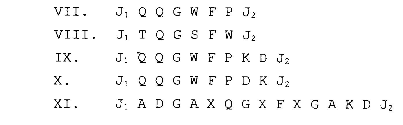 worin J1 und J2 ausgewählt sind aus der Gruppe bestehend aus 0 bis 6 Aminosäureresten.
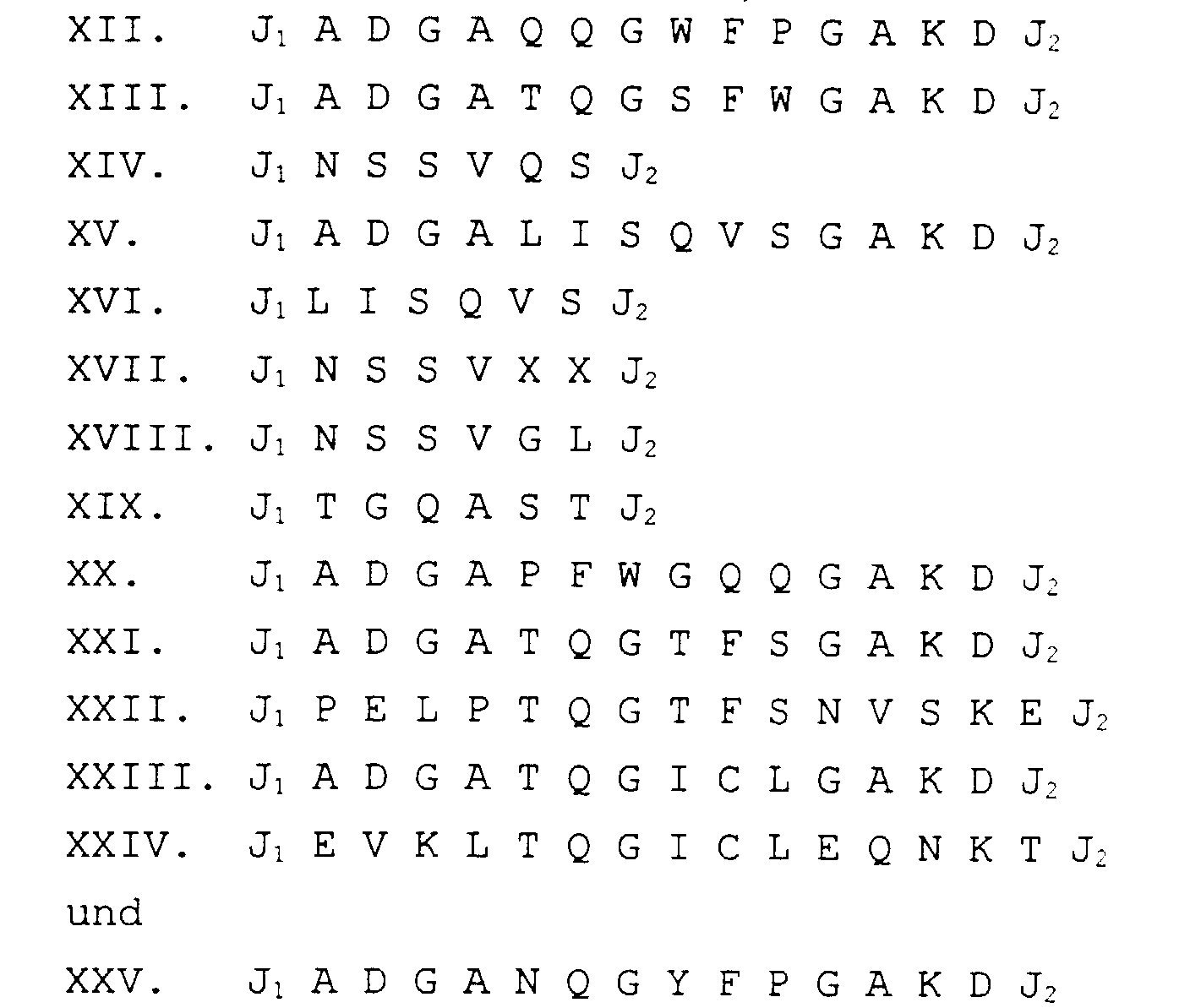
- Peptid nach Anspruch 12, dadurch gekennzeichnet, daß J1 und J2 Aminosäurereste enthalten, die ausgewählt sind aus der Gruppe bestehend aus G, S, T, C, Y, N, Q, D, E, H, K und R.
- Peptid, das dazu fähig ist, einen an ein Zelloberflächenantigen einer Zielzelle gebundenen monoklonalen Antikörper zu verdrängen, wobei der monoklonale Antikörper durch das mit ATCC HB-11885 (9079) bezeichnete Hybridom erzeugt wird, und das Peptid ausgewählt ist aus der Gruppe bestehend aus:
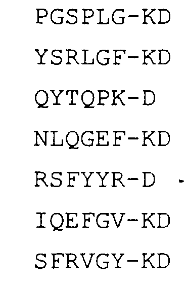

- Peptid, das dazu fähig ist, einen an ein Zelloberflächenantigen einer Zielzelle gebundenen monoklonalen Antikörper zu verdrängen, wobei der monoklonale Antikörper der mit 561 bezeichnete Antikörper ist, und das Peptid ausgewählt ist aus der Gruppe bestehend aus:
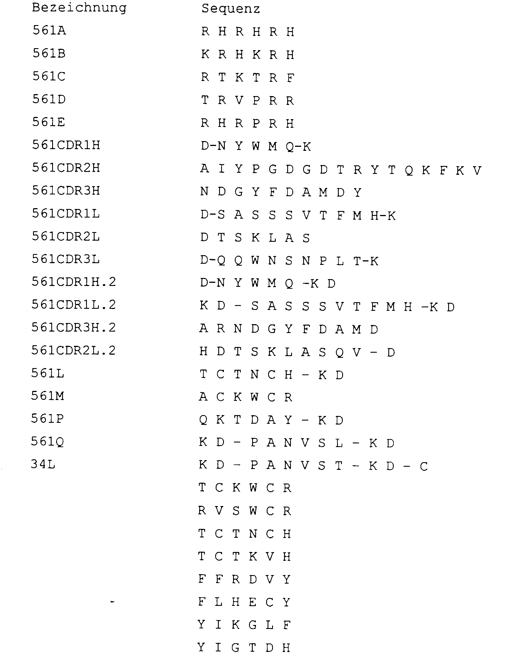

- Peptid, das dazu fähig ist, einen an ein Zelloberflächenantigen einer Zielzelle gebundenen monoklonalen Antikörper zu verdrängen, wobei der monoklonale Antikörper der mit 561 bezeichnete Antikörper ist, und das Peptid ein cyclisches Peptid ist, ausgewählt aus der Gruppe bestehend aus:
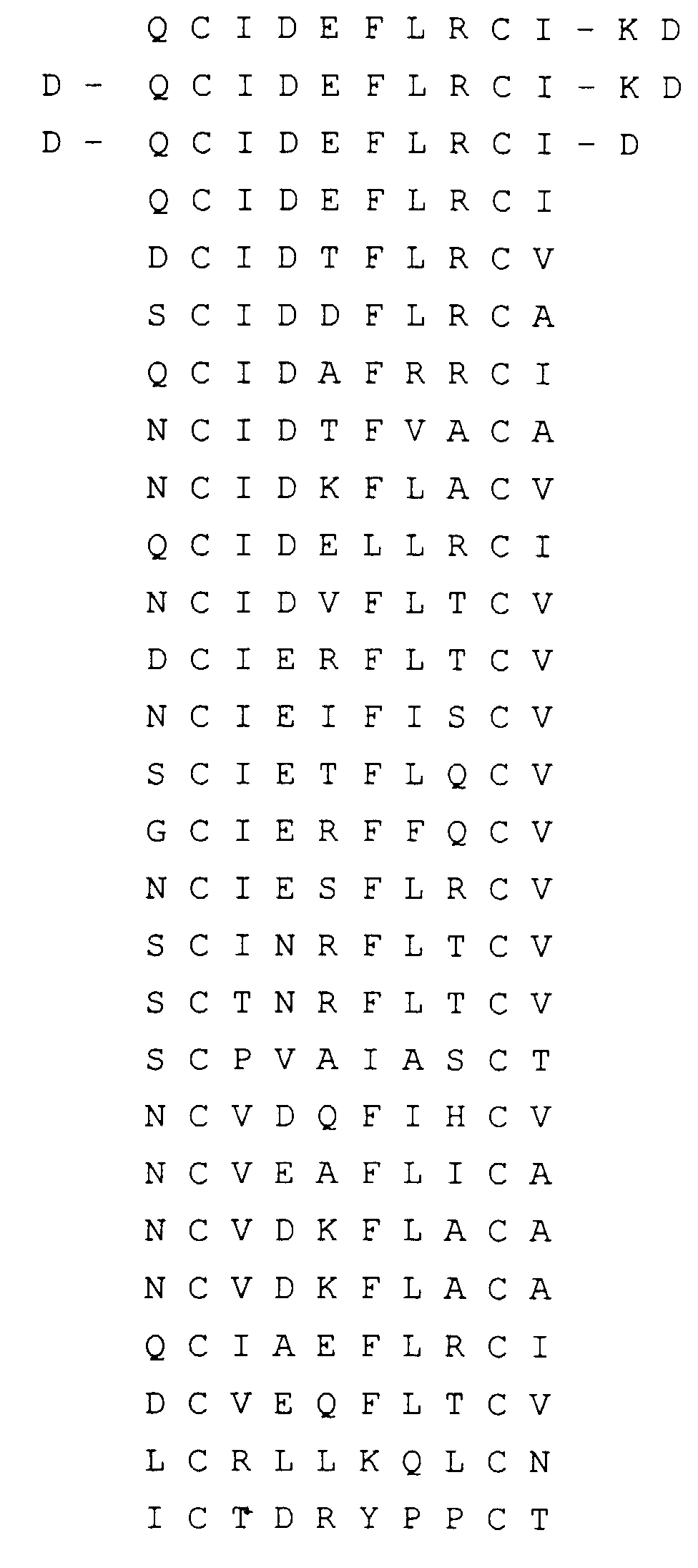
- Peptid, das dazu fähig ist, einen an ein Zelloberflächenantigen einer Zielzelle gebundenen monoklonalen Antikörper zu verdrängen, wobei der monoklonale Antikörper durch das mit ATCC HB-11884 (9187) bezeichnete Hybridom erzeugt wird, und das Peptid ausgewählt ist aus der Gruppe bestehend aus:

- Verfahren zur Identifizierung eines zur Freisetzung einer Zielzelle aus einem an ein Zelloberflächenantigen gebundenen Antikörper brauchbares spezifisches Peptid, dadurch gekennzeichnet, daß man ein Kandidatenpeptid auswählt, indem man mindestens eines der folgenden Verfahren durchführt:und Bestimmen der Fähigkeit des Kandidatenpeptids, den Antikörper von der Zielzelle zu verdrängen, wodurch die Zielzelle freigesetzt wird.(a) Random-Peptidbibliothek-Phagendisplay und Biopanning mit dem monoklonalen Antikörper,(b) Random-Peptidbibliothek-Pindisplay und Binden mit dem monoklonalen Antikörper,(c) Analyse der potentiellen Antigenpeaks des Zelloberflächenantigens,(d) Analyse der Komplementarität-bestimmenden Regionen (CDRs) des monoklonalen Antikörpers,(e) Ausbilden eines theoretisch Molekularmodells der dreidimensionalen Struktur des monoklonalen Antikörpers;
- Verfahren zur Identifizierung eines zur Freisetzung eines Antikörpers von einem Zellantigen brauchbaren spezifischen Peptids, dadurch gekennzeichnet, daß maneinen Komplex dieses Antikörpers und des Zellantigens ausbildet,den Komplex in reaktiven Kontakt mit einem oder mehreren Peptiden bringt,bestimmt, ob der Antikörper aus dem Zellantigen freigesetzt wird, undfeststellt, welches der Peptide die Freisetzung des Antikörpers vom Zellantigen bewirkt hat.
- Verfahren nach Anspruch 19, dadurch gekennzeichnet, daß der Komplex auf einem festen Träger fixiert ist.
- Verfahren nach Anspruch 19, dadurch gekennzeichnet, daß das ein oder mehrere Peptide auf einem festen Träger fixiert sind, und das Peptid identifiziert wird durch Bindung des Antikörpers an ein solches Peptid.
- Verfahren zur Bestimmung der Zahl spezifischer Zellen in einer Zellenzusammensetzung, dadurch gekennzeichnet, daß man(a) einen monoklonalen Antikörper, der an die spezifischen Zellen bindet, bereitstellt,(b) ein Peptid, das dazu fähig ist, diesen monoklonalen Antikörper von den spezifischen Zellen zu verdrängen, bereitstellt, wobei das Peptid an einen festen Träger gebunden ist um ein künstliches Zelltarget auszubilden,(c) eine Standardkurve zur Verdrängung des monoklonalen Antikörpers aus dem künstlichen Zelltarget aufstellt,(d) das künstliche Zelltarget mit dem monoklonalen Antikörper und einer Probe, die eine unbekannte Zahl der spezifischen Zellen, die mit dem künstlichen Zelltarget um die Bindung mit dem monoklonalen Antikörper konkurrieren, in Kontakt bringt, und(e) das in Stufe (d) erhaltene Signal mit den in Stufe (c) erhaltenen Signalen vergleicht.
- Verfahren zur Selektion einer oder mehrerer Zielzellen aus einer heterogenen Zellsuspension, dadurch gekennzeichnet, daß man:(a) die heterogene Zellsuspension in Kontakt mit mindestens einem ersten Antikörper bringt, der dazu fähig ist, mit mindestens einem ersten Antigen auf mindestens einer der Zielzellen zu binden;(b) die Antikörper-gebundenen Zielzellen aus der heterogenen Zellsuspension abtrennt;(c) die an den ersten Antikörper gebundenen Zielzellen in Kontakt mit einem ersten Peptid bringt, das das Antigen von dem ersten Antikörper verdrängt, wodurch die Zielzelle vom ersten Antikörper freigesetzt wird.
- Verfahren nach Anspruch 23, dadurch gekennzeichnet, daß die Stufe der Abtrennung das Binden des ersten Antikörpers an ein Zelltrennmittel und Entfernen der verbleibenden Zellsuspension vom Zelltrennmittel umfaßt
- Verfahren nach Anspruch 24, dadurch gekennzeichnet, daß das Zelltrennmittel eine Säule ist, und die heterogene Zellsuspension durch diese Säule hindurchgeleitet wird, um die Zellsuspension in Kontakt mit dem ersten Antikörper zu bringen.
- Verfahren nach Anspruch 25, dadurch gekennzeichnet, daß die Säule aus Perlen ausgebildet ist.
- Verfahren nach Anspruch 25, dadurch gekennzeichnet, daß die Säule aus Hohlfasern ausgebildet ist.
- Verfahren nach Anspruch 24, dadurch gekennzeichnet, daß das Zelltrennmittel aus paramagnetischen Perlen ausgebildet wird, und die Antikörper-gebundenen Perlen mit der heterogenen Zellsuspension gemischt werden, um die Zellsuspension in Kontakt mit dem ersten Antikörper zu bringen.
- Verfahren nach Anspruch 28, dadurch gekennzeichnet, daß die an den ersten Antikörper gebundenen Perlen und die an den ersten Antikörper gebundenen Zielzellen von der heterogenen Zellsuspension abgetrennt werden, indem man die Mischung der an der ersten Antikörper gebundenen Zielzellen und die heterogene Zellsuspension in ein Magnetfeld bringt, das die an den ersten Antikörper gebundenen Perlen und die an den ersten Antikörper gebundenen Zielzellen festhält, während der Rest der heterogenen Zellsuspension entfernt wird.
- Verfahren nach einem der Ansprüche 23 bis 29, dadurch gekennzeichnet, daß der erste Antikörper an das Zelltrennmittel mittels eines Proteins gebunden ist.
- Verfahren nach Anspruch 30, dadurch gekennzeichnet, daß das Protein ein an das Zelltrennmittel gebundener Antikörper mit einer Affinität für den ersten Antikörper ist.
- Verfahren nach einem der Ansprüche 23 bis 29, dadurch gekennzeichnet, daß es vor der Abtrennstufe außerdem die Stufe umfaßt:(d) In-Kontakt-bringen der heterogenen Zellsuspension mit einem zweiten Antikörper, der dazu fähig ist, ein zweites Antigen auf mindestens einem der Zielzellen zu binden, wobei das zweite Antigen von dem ersten Antigen verschieden ist; und worin die Abtrennstufe die Abtrennung der an den ersten und zweiten Antikörper gebundenen Zielzellen von der heterogenen Zellsuspension umfaßt.
- Verfahren nach Anspruch 32, dadurch gekennzeichnet, daß es eine zusätzliche Stufe der Abtrennung der Zielzellen, die vom ersten Antikörper abgetrennt wurden, von den an den zweiten Antikörper gebundenen Zielzellen umfaßt.
- Verfahren nach Anspruch 33, dadurch gekennzeichnet, daß es die zusätzliche Stufe des In-Kontakt-bringens eines zweiten Peptids, das das Antigen von dem zweiten Antikörper verdrängt, mit den an den zweiten Antikörper gebundenen Zielzellen umfaßt, wodurch die Zielzellen vom zweiten Antikörper freigesetzt werden.
- Verfahren nach einem der Ansprüche 23 bis 34, dadurch gekennzeichnet, daß das erste Peptid und/oder das zweite Peptid (wenn ein zweites Peptid verwendet wird) eine Sequenz von weniger als 30 Aminosäuren aufweist.
- Verfahren nach Anspruch 35, dadurch gekennzeichnet, daß das Peptid eine Sequenz von 4 bis 20 Aminosäuren ist.
- System zum Abtrennen einer Zielzelle von einer heterogenen Zellsuspension, wobei das System umfaßt:(a) ein Zelltrennmittel;(b) mindestens einen ersten an das Zelltrennmittel gebundenen Antikörper, wobei der erste Antikörper dazu fähig ist, mit mindestens einem ersten Antigen an mindestens einer der Zielzellen zu binden; dadurch gekennzeichnet, daß das System zusätzlich umfaßt(c) mindestens ein erstes Peptid, das dazu fähig ist, das Antigen vom ersten Antikörper zu verdrängen, wodurch die Zielzelle vom ersten Antikörper freigesetzt wird.
- System nach Anspruch 37, dadurch gekennzeichnet, daß das Zelltrennmittel eine Säule ist.
- System nach Anspruch 38, dadurch gekennzeichnet, daß die Säule aus Perlen gebildet wird.
- System nach Anspruch 38, dadurch gekennzeichnet, daß die Säule aus Hohlfasern gebildet wird.
- System nach Anspruch 37, dadurch gekennzeichnet, daß das Zelltrennmittel durch magnetische Perlen ausgebildet wird, an die der erste Antikörper gebunden ist.
- System nach einem der Ansprüche 37 bis 41, dadurch gekennzeichnet, daß es außerdem einen an das Zelltrennmittel gebundenen zweiten Antikörper umfaßt, wobei der zweite Antikörper dazu fähig ist, mindestens ein zweites Antigen an mindestens einer der Zielzellen zu binden, wobei das zweite Antigen vom ersten Antigen verschieden ist.
- System nach einem der Ansprüche 37 bis 42, dadurch gekennzeichnet, daß das erste Peptid und/oder das zweite Peptid (wenn ein zweites Peptid vorhanden ist) eine Sequenz von weniger als 30 Aminosäuren aufweist.
- System nach Anspruch 43, dadurch gekennzeichnet, daß das Peptid eine Sequenz von 4 bis 20 Aminosäuren ist.
Applications Claiming Priority (3)
| Application Number | Priority Date | Filing Date | Title |
|---|---|---|---|
| US25942794A | 1994-06-14 | 1994-06-14 | |
| PCT/US1995/007491 WO1995034817A1 (en) | 1994-06-14 | 1995-06-13 | Positive and positive/negative cell selection mediated by peptide release |
| US259427 | 1999-02-26 |
Publications (2)
| Publication Number | Publication Date |
|---|---|
| EP0765478A1 EP0765478A1 (de) | 1997-04-02 |
| EP0765478B1 true EP0765478B1 (de) | 1998-08-05 |
Family
ID=22984902
Family Applications (1)
| Application Number | Title | Priority Date | Filing Date |
|---|---|---|---|
| EP95923835A Expired - Lifetime EP0765478B1 (de) | 1994-06-14 | 1995-06-13 | Positive und positive/negative zellselektion vermittelt durch peptiofreisetzung |
Country Status (10)
| Country | Link |
|---|---|
| US (2) | US5968753A (de) |
| EP (1) | EP0765478B1 (de) |
| JP (1) | JP2849630B2 (de) |
| AT (1) | ATE169409T1 (de) |
| AU (1) | AU697627B2 (de) |
| CA (1) | CA2192455C (de) |
| DE (1) | DE69503903T2 (de) |
| DK (1) | DK0765478T3 (de) |
| ES (1) | ES2121400T3 (de) |
| WO (1) | WO1995034817A1 (de) |
Cited By (2)
| Publication number | Priority date | Publication date | Assignee | Title |
|---|---|---|---|---|
| US7125679B2 (en) | 2001-03-07 | 2006-10-24 | Children's Medical Center Corporation | Methods to screen peptide libraries using minicell display |
| US8399249B2 (en) | 2008-02-21 | 2013-03-19 | Baxter International Inc. | Procedure for the generation of a high producer cell line for the expression of a recombinant anti-CD34 antibody |
Families Citing this family (61)
| Publication number | Priority date | Publication date | Assignee | Title |
|---|---|---|---|---|
| WO1994007138A1 (en) | 1992-09-14 | 1994-03-31 | Fodstad Oystein | Detection of specific target cells in specialized or mixed cell population and solutions containing mixed cell populations |
| NO180658C (no) | 1994-03-10 | 1997-05-21 | Oeystein Fodstad | Fremgangsmåte og anordning for deteksjon av spesifikke målceller i spesialiserte eller blandede cellepopulasjoner og opplösninger som inneholder blandede cellepopulasjoner |
| US6066516A (en) * | 1995-06-26 | 2000-05-23 | Seiko Epson Corporation | Method for forming crystalline semiconductor layers, a method for fabricating thin film transistors, and method for fabricating solar cells and active matrix liquid crystal devices |
| NO961031D0 (no) * | 1996-03-13 | 1996-03-13 | Det Norske Radiumshospital Tum | Fremgangsmåte til å drepe uönskede målceller |
| WO1997044666A1 (en) * | 1996-05-17 | 1997-11-27 | Cytovax Biotechnologies Inc. | Immunological methods of component selection and recovery |
| WO1998032840A1 (en) | 1997-01-24 | 1998-07-30 | Asahi Medical Co., Ltd. | Method for separating cells |
| US7364921B1 (en) * | 1999-01-06 | 2008-04-29 | University Of Medicine And Dentistry Of New Jersey | Method and apparatus for separating biological materials and other substances |
| US6379952B1 (en) * | 1999-02-01 | 2002-04-30 | 3M Innovative Properties Company | Method for cell selection utilizing azlactone-functional supports |
| US6790662B1 (en) | 1999-03-12 | 2004-09-14 | Ortho-Mcneil Pharmaceutical, Inc. | Method of isolating CD8+ cells, and related hybridoma cells antibodies and polypeptides |
| US6391855B1 (en) * | 1999-06-02 | 2002-05-21 | Adherex Technologies, Inc. | Compounds and methods for modulating junctional adhesion molecule-mediated functions |
| US20040002068A1 (en) * | 2000-03-01 | 2004-01-01 | Corixa Corporation | Compositions and methods for the detection, diagnosis and therapy of hematological malignancies |
| DE10014708A1 (de) * | 2000-03-24 | 2001-10-31 | Walter Schubert | Verfahren zur Identifizierung und Anreicherung von zellspezifischen Zielstrukturen |
| DE10014685B4 (de) * | 2000-03-24 | 2004-07-01 | Schubert, Walter, Dr. | Verfahren zur Identifizierung von zellspezifischen Zielstrukturen |
| ATE382633T1 (de) * | 2000-05-23 | 2008-01-15 | Baxter Healthcare Sa | Reagenzien zur zellselektion und verfahren zur anwendung |
| US20030219445A1 (en) * | 2000-05-23 | 2003-11-27 | Nexell Therapeutics, Inc. | Reagents for cell selection and methods of use |
| US6576276B1 (en) | 2000-10-25 | 2003-06-10 | The Coca-Cola Company | CO2-hydrate product and method of manufacture thereof |
| CA2698079C (en) | 2001-02-20 | 2015-01-13 | Ortho-Mcneil Pharmaceutical, Inc. | A cell therapy method for the treatment of tumors |
| US20040071671A1 (en) | 2001-02-20 | 2004-04-15 | Leturcq Didier J. | Cell therapy method for the treatment of tumors |
| KR101083454B1 (ko) | 2001-12-07 | 2011-11-16 | 사이토리 테라퓨틱스, 인크. | 처리된 리포애스퍼레이트 세포로 환자를 치료하기 위한 시스템 및 방법 |
| US20040264323A1 (en) * | 2002-01-28 | 2004-12-30 | Worthington Mark Oscar | Methods and apparatus for logical triggering |
| EP1470424A2 (de) * | 2002-01-31 | 2004-10-27 | Burstein Technologies, Inc. | Verfahren zum auslösen mittels platterillen und entsprechende optische analyse-platten |
| US20030166138A1 (en) * | 2002-02-21 | 2003-09-04 | Todd Kinsella | Cyclic peptides and analogs useful to treat allergies |
| ITUD20020092A1 (it) * | 2002-04-30 | 2002-07-30 | Univ Degli Studi Udine | Metodo per l'identificazione e l'isolamento di cellule staminali di un tessuto umano |
| US8173372B2 (en) * | 2002-05-16 | 2012-05-08 | Absorber Ab | Methods of donor specific crossmatching |
| US9435799B2 (en) * | 2002-07-31 | 2016-09-06 | Janssen Diagnostics, Inc. | Methods and reagents for improved selection of biological materials |
| WO2004014944A1 (ja) * | 2002-08-07 | 2004-02-19 | Okayama Prefecture | 微生物由来のポリアミノ酸またはその誘導体 |
| CA2504443A1 (en) * | 2002-10-30 | 2004-05-13 | Pointilliste, Inc. | Methods for producing polypeptide-tagged collections and capture systems containing the tagged polypeptides |
| ATE524070T1 (de) | 2003-02-20 | 2011-09-15 | Cytori Therapeutics Inc | Verfahren zur verwendung von aus fettgewebe stammenden zellen bei der behandlung von herz- kreislauf-leiden |
| US7956165B2 (en) | 2003-07-24 | 2011-06-07 | Affisink Biotechnology Ltd. | Compositions and methods for purifying and crystallizing molecules of interest |
| IL157086A0 (en) * | 2003-07-24 | 2004-02-08 | Guy Patchornik | Multivalent ligand complexes |
| US7996825B2 (en) * | 2003-10-31 | 2011-08-09 | Hewlett-Packard Development Company, L.P. | Cross-file inlining by using summaries and global worklist |
| US20060018911A1 (en) * | 2004-01-12 | 2006-01-26 | Dana Ault-Riche | Design of therapeutics and therapeutics |
| US9790539B2 (en) * | 2004-06-30 | 2017-10-17 | Russell Biotech, Inc. | Methods and reagents for improved selection of biological molecules |
| EP1836216B1 (de) | 2004-12-10 | 2021-05-05 | DendroCyte BioTech Pty Ltd | Bindungspartner für gegen die antigene dentritischer zellen spezifische anitkörper |
| US20080206276A1 (en) * | 2005-07-08 | 2008-08-28 | Michael Otto | Targeting Poly-Gamma-Glutamic Acid to Treat Staphylococcus Epidermidis and Related Infections |
| US7794705B2 (en) | 2005-11-07 | 2010-09-14 | Amorcyte, Inc. | Compositions and methods of vascular injury repair |
| JP2010510526A (ja) * | 2006-11-22 | 2010-04-02 | スリーエム イノベイティブ プロパティズ カンパニー | 細菌全細胞を捕捉する方法及び細菌について試料を分析する方法 |
| US9012152B2 (en) * | 2007-01-09 | 2015-04-21 | Yissum Research Development Company Of The Hebrew University Of Jerusalem | Anti-bacterial peptides and methods of treating diseases using same |
| KR20100087290A (ko) * | 2007-10-12 | 2010-08-04 | 막스-델부뤽-센트럼 퓌어 몰레쿨라레 메디친 | 인간 Foxp3+ 조절 Treg 세포의 신속한 분리 방법 및 키트 |
| ES2355587T3 (es) * | 2007-10-12 | 2011-03-29 | Max-Delbrück-Centrum Für Molekulare Medizin (Mdc) | MÉTODO Y KIT PARA EL AISLAMIENTO RÁPIDO DE CÉLULAS Foxp3+Treg HUMANAS. |
| US20100028311A1 (en) * | 2008-07-09 | 2010-02-04 | Baxter International Inc. | Using of scaffold comprising fibrin for delivery of stem cells |
| JP5638747B2 (ja) * | 2008-08-20 | 2014-12-10 | 株式会社日立製作所 | がん腫の遺伝子診断のための細胞回収及び単離方法並びにキット |
| US20110274667A1 (en) * | 2008-10-17 | 2011-11-10 | Baxter Healthcare S.A. | Methods for obtaining cell populations from adipose tissue |
| ES2845691T3 (es) | 2009-10-23 | 2021-07-27 | Caladrius Biosciences Inc | Composiciones y usos para el tratamiento de lesiones miocárdicas progresivas debidas a una insuficiencia vascular |
| AT509267A1 (de) * | 2010-01-14 | 2011-07-15 | Apeptico Forschung & Entwicklung Gmbh | Organische verbindungen zur regulierung von vektoriellen ionenkanälen |
| KR101970368B1 (ko) | 2011-05-17 | 2019-04-18 | 론자 워커스빌 아이엔씨. | 인간 다능성 줄기세포의 계대 및 수득을 위한 제제 및 방법 |
| UA112434C2 (uk) | 2011-05-27 | 2016-09-12 | Ґлаксо Ґруп Лімітед | Антигензв'язувальний білок, який специфічно зв'язується з всма |
| US9533010B2 (en) | 2011-10-31 | 2017-01-03 | Amorcyte, Llc | Compositions and methods for treating progressive myocardial injury due to a vascular insufficiency |
| US20130216495A1 (en) | 2012-02-21 | 2013-08-22 | Baxter Healthcare Sa | Pharmaceutical composition comprising cd34+ cells |
| WO2013128381A1 (en) * | 2012-02-28 | 2013-09-06 | Ben Gurion University Of The Negev Research And Development Authority | Combined therapy of alpha-1-antitrypsin and temporal t-cell depletion for preventing graft rejection |
| US20190204312A1 (en) * | 2016-09-16 | 2019-07-04 | Technische Universität München | Magnetic particle-binding peptides |
| JP7272590B2 (ja) * | 2017-07-11 | 2023-05-12 | 国立大学法人富山大学 | 細胞の選択的分離用又は細胞培養用ポリマーにより被覆された基体 |
| WO2019139987A1 (en) | 2018-01-09 | 2019-07-18 | Elstar Therapeutics, Inc. | Calreticulin binding constructs and engineered t cells for the treatment of diseases |
| AU2019297451A1 (en) | 2018-07-03 | 2021-01-28 | Marengo Therapeutics, Inc. | Anti-TCR antibody molecules and uses thereof |
| GB2599228B (en) | 2019-02-21 | 2024-02-07 | Marengo Therapeutics Inc | Multifunctional molecules that bind to T cell related cancer cells and uses thereof |
| AU2020224681A1 (en) | 2019-02-21 | 2021-09-16 | Marengo Therapeutics, Inc. | Antibody molecules that bind to NKp30 and uses thereof |
| PH12021553042A1 (en) * | 2019-06-06 | 2023-09-11 | Immatics Biotechnologies Gmbh | Sorting with counter selection using sequence similar peptides |
| JP2023509708A (ja) | 2020-01-03 | 2023-03-09 | マレンゴ・セラピューティクス,インコーポレーテッド | 抗tcr抗体分子およびその使用 |
| EP4000596A1 (de) * | 2020-11-17 | 2022-05-25 | The Boots Company plc | Tetrapeptid und zusammensetzungen mit tetrapeptiden |
| EP4000595A1 (de) * | 2020-11-17 | 2022-05-25 | The Boots Company plc | Tetrapeptid und zusammensetzungen mit tetrapeptiden |
| WO2023141297A2 (en) * | 2022-01-21 | 2023-07-27 | Marengo Therapeutics, Inc. | Multifunctional molecules comprising g6b binder and/or cd34 binder and uses thereof |
Family Cites Families (24)
| Publication number | Priority date | Publication date | Assignee | Title |
|---|---|---|---|---|
| US5130144B1 (en) * | 1984-02-06 | 1995-08-15 | Univ Johns Hopkins | Human stem cells and monoclonal antibodies |
| US4965204A (en) * | 1984-02-06 | 1990-10-23 | The Johns Hopkins University | Human stem cells and monoclonal antibodies |
| IL76234A0 (en) * | 1985-08-28 | 1986-01-31 | Yeda Res & Dev | New biotinylation reagents |
| US5225353A (en) * | 1986-01-30 | 1993-07-06 | Fred Hutchinson Cancer Research Center | Method for immunoselection of cells using avidin and biotin |
| US5215927A (en) * | 1986-01-30 | 1993-06-01 | Fred Hutchinson Cancer Research Center | Method for immunoselection of cells using avidin and biotin |
| US4798795A (en) * | 1987-02-17 | 1989-01-17 | Hoechst Celanese Corporation | Biotinylating agents |
| US4709037A (en) * | 1987-02-17 | 1987-11-24 | Hoechst Celanese Corporation | Biotinylating agents |
| PT90657B (pt) * | 1988-05-27 | 1995-03-01 | Ortho Pharma Corp | Processo para a preparacao de peptidos que bloqueiam a ligacao de hiv-1 a proteina cd4 receptora |
| US5316943A (en) * | 1988-06-14 | 1994-05-31 | Kidman Gene E | Racemic conversion of using a transaminase |
| US5536475A (en) * | 1988-10-11 | 1996-07-16 | Baxter International Inc. | Apparatus for magnetic cell separation |
| US5081030A (en) * | 1989-04-25 | 1992-01-14 | The Johns Hopkins University | Release of cells from affinity matrices |
| GB8916859D0 (en) * | 1989-07-24 | 1989-09-06 | Dynal As | Hapten linking |
| EP0563212A1 (de) * | 1990-11-23 | 1993-10-06 | Immunodynamics, Inc. | Synthetische immunoaktive peptide mit immunmodulierenden und therapeutischen wirkungen |
| WO1992015683A1 (en) * | 1991-03-06 | 1992-09-17 | MERCK Patent Gesellschaft mit beschränkter Haftung | Humanized and chimeric monoclonal antibodies |
| DE69220397T2 (de) * | 1991-03-13 | 1997-10-02 | Morinaga Milk Industry Co Ltd | Antimikrobielles Peptid und antimikrobieller Wirkstoff |
| WO1993014781A1 (en) * | 1992-01-24 | 1993-08-05 | The Regents Of The University Of California | Novel peptides and method for altering the activity of allosteric proteins |
| WO1993016715A1 (en) * | 1992-02-24 | 1993-09-02 | New England Medical Center Hospitals, Inc. | Fsf-1 and the early detection of fibrosis |
| AU678179B2 (en) * | 1992-07-28 | 1997-05-22 | Steven Kessler | Methods for positive immunoselection of stem cells |
| FR2694938B1 (fr) * | 1992-08-10 | 1996-11-15 | Zagury Jean Francois | Nouveaux peptides, anticorps diriges contre ces peptides, anticorps anti-idiotypiques, application a titre de medicaments, compositions pharmaceutiques et kits de diagnostic les renfermant. |
| CA2150371C (en) * | 1992-12-11 | 2002-09-17 | Dov Borovsky | Materials and methods for control of pests |
| JPH09502351A (ja) * | 1993-09-08 | 1997-03-11 | バクスター、インターナショナル、インコーポレイテッド | 正の細胞選択及び遊離のためのビオチン類縁体接合抗体 |
| US5525503A (en) * | 1993-09-28 | 1996-06-11 | Dana-Farber Cancer Institute, Inc. | Signal transduction via CD28 |
| US5409813A (en) * | 1993-09-30 | 1995-04-25 | Systemix, Inc. | Method for mammalian cell separation from a mixture of cell populations |
| US5610031A (en) * | 1993-10-27 | 1997-03-11 | The General Hospital Corporation | B1k chain of laminin and methods of use |
-
1995
- 1995-06-07 US US08/482,228 patent/US5968753A/en not_active Expired - Lifetime
- 1995-06-07 US US08/482,528 patent/US6017719A/en not_active Expired - Lifetime
- 1995-06-13 JP JP8502411A patent/JP2849630B2/ja not_active Expired - Fee Related
- 1995-06-13 WO PCT/US1995/007491 patent/WO1995034817A1/en not_active Ceased
- 1995-06-13 DE DE69503903T patent/DE69503903T2/de not_active Expired - Lifetime
- 1995-06-13 DK DK95923835T patent/DK0765478T3/da active
- 1995-06-13 AT AT95923835T patent/ATE169409T1/de active
- 1995-06-13 ES ES95923835T patent/ES2121400T3/es not_active Expired - Lifetime
- 1995-06-13 AU AU28261/95A patent/AU697627B2/en not_active Ceased
- 1995-06-13 EP EP95923835A patent/EP0765478B1/de not_active Expired - Lifetime
- 1995-06-13 CA CA002192455A patent/CA2192455C/en not_active Expired - Fee Related
Cited By (2)
| Publication number | Priority date | Publication date | Assignee | Title |
|---|---|---|---|---|
| US7125679B2 (en) | 2001-03-07 | 2006-10-24 | Children's Medical Center Corporation | Methods to screen peptide libraries using minicell display |
| US8399249B2 (en) | 2008-02-21 | 2013-03-19 | Baxter International Inc. | Procedure for the generation of a high producer cell line for the expression of a recombinant anti-CD34 antibody |
Also Published As
| Publication number | Publication date |
|---|---|
| US5968753A (en) | 1999-10-19 |
| WO1995034817A1 (en) | 1995-12-21 |
| JP2849630B2 (ja) | 1999-01-20 |
| JPH09508534A (ja) | 1997-09-02 |
| DK0765478T3 (da) | 1999-05-03 |
| CA2192455A1 (en) | 1995-12-21 |
| ATE169409T1 (de) | 1998-08-15 |
| CA2192455C (en) | 2007-11-20 |
| ES2121400T3 (es) | 1998-11-16 |
| US6017719A (en) | 2000-01-25 |
| EP0765478A1 (de) | 1997-04-02 |
| DE69503903D1 (de) | 1998-09-10 |
| DE69503903T2 (de) | 1999-03-25 |
| AU697627B2 (en) | 1998-10-15 |
| AU2826195A (en) | 1996-01-05 |
Similar Documents
| Publication | Publication Date | Title |
|---|---|---|
| EP0765478B1 (de) | Positive und positive/negative zellselektion vermittelt durch peptiofreisetzung | |
| CA2178984C (en) | Novel antibody compositions for preparing enriched human hematopoietic cell preparations | |
| US6306575B1 (en) | Methods for preparing enriched human hematopoietic cell preparations | |
| US6790662B1 (en) | Method of isolating CD8+ cells, and related hybridoma cells antibodies and polypeptides | |
| CA2346362C (en) | Novel antibody compositions for preparing enriched mesenchymal progenitor preparations | |
| Palmer et al. | Selection of antibodies to cell surface determinants on mouse thymic epithelial cells using a phage display library | |
| AU678179B2 (en) | Methods for positive immunoselection of stem cells | |
| JP7360323B2 (ja) | 骨髄非破壊前処理のための組成物および方法 | |
| JPH07507202A (ja) | ヒトmdr1マルチドラッグ耐性遺伝子産物に対するモノクローナル抗体およびその使用 | |
| CN110713539B (zh) | 一种抗癌胚抗原的抗体及其制备方法和用途 | |
| AU2001274931B2 (en) | Reagents for cell selection and methods of use | |
| CN110862456B (zh) | 一种抗癌胚抗原的抗体及其制备方法和用途 | |
| CN110655581B (zh) | 一种抗癌胚抗原的抗体及其制备方法和用途 | |
| CN112679618A (zh) | 一种靶向trbc1的人源化嵌合抗原受体、t细胞及用途 | |
| EP0202642A2 (de) | Verfahren zur in vitro-Immunisierung von Humanmilzzellen gegen tumorassoziierte Antigene | |
| WO1999061629A1 (fr) | Dispositif pour la separation cellulaire et procede de separation | |
| CN116023496A (zh) | 一种制备通用型car-t细胞的方法及其应用 | |
| JP4712194B2 (ja) | Cd8+細胞を単離する方法、並びに関連するハイブリドーマ細胞、抗体及びポリペプチド | |
| JP3043629B2 (ja) | 抗体103b2 | |
| JP2010520761A (ja) | 間葉系幹細胞亜集団および破骨細胞に対するマーカー、抗体および組換えscFv | |
| WO2001038496A1 (en) | A human normal counterpart of cd4+ cd56+ cutaneous neoplasm | |
| WO2006077232A2 (en) | Multimeric soluble fas ligand for eliminating alloreactive t lymphocyte in allogenic harmatopoietic stem-cell transplantation transplantation | |
| WO1999023207A1 (en) | Monoclonal antibodies to canine cd34 |
Legal Events
| Date | Code | Title | Description |
|---|---|---|---|
| PUAI | Public reference made under article 153(3) epc to a published international application that has entered the european phase |
Free format text: ORIGINAL CODE: 0009012 |
|
| 17P | Request for examination filed |
Effective date: 19961104 |
|
| AK | Designated contracting states |
Kind code of ref document: A1 Designated state(s): AT BE DE DK ES FR GB GR IE IT NL PT SE |
|
| GRAG | Despatch of communication of intention to grant |
Free format text: ORIGINAL CODE: EPIDOS AGRA |
|
| 17Q | First examination report despatched |
Effective date: 19971205 |
|
| GRAG | Despatch of communication of intention to grant |
Free format text: ORIGINAL CODE: EPIDOS AGRA |
|
| GRAH | Despatch of communication of intention to grant a patent |
Free format text: ORIGINAL CODE: EPIDOS IGRA |
|
| GRAH | Despatch of communication of intention to grant a patent |
Free format text: ORIGINAL CODE: EPIDOS IGRA |
|
| GRAA | (expected) grant |
Free format text: ORIGINAL CODE: 0009210 |
|
| AK | Designated contracting states |
Kind code of ref document: B1 Designated state(s): AT BE DE DK ES FR GB GR IE IT NL PT SE |
|
| REF | Corresponds to: |
Ref document number: 169409 Country of ref document: AT Date of ref document: 19980815 Kind code of ref document: T |
|
| REF | Corresponds to: |
Ref document number: 69503903 Country of ref document: DE Date of ref document: 19980910 |
|
| ET | Fr: translation filed | ||
| REG | Reference to a national code |
Ref country code: IE Ref legal event code: FG4D |
|
| REG | Reference to a national code |
Ref country code: ES Ref legal event code: FG2A Ref document number: 2121400 Country of ref document: ES Kind code of ref document: T3 |
|
| REG | Reference to a national code |
Ref country code: PT Ref legal event code: SC4A Free format text: AVAILABILITY OF NATIONAL TRANSLATION Effective date: 19981027 |
|
| REG | Reference to a national code |
Ref country code: DK Ref legal event code: T3 |
|
| PLBE | No opposition filed within time limit |
Free format text: ORIGINAL CODE: 0009261 |
|
| STAA | Information on the status of an ep patent application or granted ep patent |
Free format text: STATUS: NO OPPOSITION FILED WITHIN TIME LIMIT |
|
| 26N | No opposition filed | ||
| REG | Reference to a national code |
Ref country code: PT Ref legal event code: PC4A Free format text: NEXELL THERAPEUTICS INC. US Effective date: 20000128 |
|
| REG | Reference to a national code |
Ref country code: GB Ref legal event code: 732E |
|
| PGFP | Annual fee paid to national office [announced via postgrant information from national office to epo] |
Ref country code: PT Payment date: 20010522 Year of fee payment: 7 |
|
| PGFP | Annual fee paid to national office [announced via postgrant information from national office to epo] |
Ref country code: GB Payment date: 20010613 Year of fee payment: 7 |
|
| REG | Reference to a national code |
Ref country code: GB Ref legal event code: IF02 |
|
| PG25 | Lapsed in a contracting state [announced via postgrant information from national office to epo] |
Ref country code: GB Free format text: LAPSE BECAUSE OF NON-PAYMENT OF DUE FEES Effective date: 20020613 |
|
| PGFP | Annual fee paid to national office [announced via postgrant information from national office to epo] |
Ref country code: GR Payment date: 20021230 Year of fee payment: 8 |
|
| PG25 | Lapsed in a contracting state [announced via postgrant information from national office to epo] |
Ref country code: PT Free format text: LAPSE BECAUSE OF NON-PAYMENT OF DUE FEES Effective date: 20021231 |
|
| GBPC | Gb: european patent ceased through non-payment of renewal fee |
Effective date: 20020613 |
|
| REG | Reference to a national code |
Ref country code: PT Ref legal event code: MM4A Free format text: LAPSE DUE TO NON-PAYMENT OF FEES Effective date: 20021231 |
|
| PG25 | Lapsed in a contracting state [announced via postgrant information from national office to epo] |
Ref country code: GR Free format text: LAPSE BECAUSE OF NON-PAYMENT OF DUE FEES Effective date: 20040107 |
|
| PGFP | Annual fee paid to national office [announced via postgrant information from national office to epo] |
Ref country code: IE Payment date: 20110627 Year of fee payment: 17 Ref country code: SE Payment date: 20110629 Year of fee payment: 17 Ref country code: ES Payment date: 20110628 Year of fee payment: 17 |
|
| PGFP | Annual fee paid to national office [announced via postgrant information from national office to epo] |
Ref country code: NL Payment date: 20110630 Year of fee payment: 17 Ref country code: DK Payment date: 20110627 Year of fee payment: 17 Ref country code: AT Payment date: 20110519 Year of fee payment: 17 |
|
| PGFP | Annual fee paid to national office [announced via postgrant information from national office to epo] |
Ref country code: BE Payment date: 20110627 Year of fee payment: 17 Ref country code: IT Payment date: 20110627 Year of fee payment: 17 |
|
| BERE | Be: lapsed |
Owner name: *BAXTER INTERNATIONAL INC. Effective date: 20120630 |
|
| REG | Reference to a national code |
Ref country code: NL Ref legal event code: V1 Effective date: 20130101 |
|
| REG | Reference to a national code |
Ref country code: DK Ref legal event code: EBP |
|
| REG | Reference to a national code |
Ref country code: SE Ref legal event code: EUG |
|
| REG | Reference to a national code |
Ref country code: AT Ref legal event code: MM01 Ref document number: 169409 Country of ref document: AT Kind code of ref document: T Effective date: 20120613 |
|
| PG25 | Lapsed in a contracting state [announced via postgrant information from national office to epo] |
Ref country code: SE Free format text: LAPSE BECAUSE OF NON-PAYMENT OF DUE FEES Effective date: 20120614 Ref country code: IT Free format text: LAPSE BECAUSE OF NON-PAYMENT OF DUE FEES Effective date: 20120613 |
|
| REG | Reference to a national code |
Ref country code: IE Ref legal event code: MM4A |
|
| PG25 | Lapsed in a contracting state [announced via postgrant information from national office to epo] |
Ref country code: IE Free format text: LAPSE BECAUSE OF NON-PAYMENT OF DUE FEES Effective date: 20120613 Ref country code: NL Free format text: LAPSE BECAUSE OF NON-PAYMENT OF DUE FEES Effective date: 20130101 Ref country code: BE Free format text: LAPSE BECAUSE OF NON-PAYMENT OF DUE FEES Effective date: 20120630 |
|
| PG25 | Lapsed in a contracting state [announced via postgrant information from national office to epo] |
Ref country code: AT Free format text: LAPSE BECAUSE OF NON-PAYMENT OF DUE FEES Effective date: 20120613 |
|
| PG25 | Lapsed in a contracting state [announced via postgrant information from national office to epo] |
Ref country code: DK Free format text: LAPSE BECAUSE OF NON-PAYMENT OF DUE FEES Effective date: 20120702 |
|
| PGFP | Annual fee paid to national office [announced via postgrant information from national office to epo] |
Ref country code: DE Payment date: 20130627 Year of fee payment: 19 |
|
| PGFP | Annual fee paid to national office [announced via postgrant information from national office to epo] |
Ref country code: FR Payment date: 20130702 Year of fee payment: 19 |
|
| REG | Reference to a national code |
Ref country code: ES Ref legal event code: FD2A Effective date: 20131022 |
|
| PG25 | Lapsed in a contracting state [announced via postgrant information from national office to epo] |
Ref country code: ES Free format text: LAPSE BECAUSE OF NON-PAYMENT OF DUE FEES Effective date: 20120614 |
|
| REG | Reference to a national code |
Ref country code: DE Ref legal event code: R119 Ref document number: 69503903 Country of ref document: DE |
|
| REG | Reference to a national code |
Ref country code: DE Ref legal event code: R119 Ref document number: 69503903 Country of ref document: DE Effective date: 20150101 |
|
| REG | Reference to a national code |
Ref country code: FR Ref legal event code: ST Effective date: 20150227 |
|
| PG25 | Lapsed in a contracting state [announced via postgrant information from national office to epo] |
Ref country code: DE Free format text: LAPSE BECAUSE OF NON-PAYMENT OF DUE FEES Effective date: 20150101 |
|
| PG25 | Lapsed in a contracting state [announced via postgrant information from national office to epo] |
Ref country code: FR Free format text: LAPSE BECAUSE OF NON-PAYMENT OF DUE FEES Effective date: 20140630 |