EP0598663B1 - Verfahren zur Vorbehandlung für Blutanalyse - Google Patents
Verfahren zur Vorbehandlung für Blutanalyse Download PDFInfo
- Publication number
- EP0598663B1 EP0598663B1 EP93402790A EP93402790A EP0598663B1 EP 0598663 B1 EP0598663 B1 EP 0598663B1 EP 93402790 A EP93402790 A EP 93402790A EP 93402790 A EP93402790 A EP 93402790A EP 0598663 B1 EP0598663 B1 EP 0598663B1
- Authority
- EP
- European Patent Office
- Prior art keywords
- surfactant
- pretreatment method
- blood analysis
- analysis according
- intensity
- Prior art date
- Legal status (The legal status is an assumption and is not a legal conclusion. Google has not performed a legal analysis and makes no representation as to the accuracy of the status listed.)
- Expired - Lifetime
Links
- 238000004159 blood analysis Methods 0.000 title claims description 26
- 238000002203 pretreatment Methods 0.000 title claims description 25
- 210000004369 blood Anatomy 0.000 claims description 78
- 239000008280 blood Substances 0.000 claims description 78
- LFQSCWFLJHTTHZ-UHFFFAOYSA-N Ethanol Chemical compound CCO LFQSCWFLJHTTHZ-UHFFFAOYSA-N 0.000 claims description 74
- 239000004094 surface-active agent Substances 0.000 claims description 61
- 239000000126 substance Substances 0.000 claims description 60
- 210000000265 leukocyte Anatomy 0.000 claims description 56
- 238000002372 labelling Methods 0.000 claims description 48
- 235000019441 ethanol Nutrition 0.000 claims description 43
- 239000002736 nonionic surfactant Substances 0.000 claims description 40
- 239000003093 cationic surfactant Substances 0.000 claims description 35
- 210000000170 cell membrane Anatomy 0.000 claims description 30
- DDXLVDQZPFLQMZ-UHFFFAOYSA-M dodecyl(trimethyl)azanium;chloride Chemical compound [Cl-].CCCCCCCCCCCC[N+](C)(C)C DDXLVDQZPFLQMZ-UHFFFAOYSA-M 0.000 claims description 21
- KFZMGEQAYNKOFK-UHFFFAOYSA-N Isopropanol Chemical compound CC(C)O KFZMGEQAYNKOFK-UHFFFAOYSA-N 0.000 claims description 18
- 239000002280 amphoteric surfactant Substances 0.000 claims description 18
- 230000003287 optical effect Effects 0.000 claims description 17
- 239000007864 aqueous solution Substances 0.000 claims description 15
- ZMMJGEGLRURXTF-UHFFFAOYSA-N ethidium bromide Chemical group [Br-].C12=CC(N)=CC=C2C2=CC=C(N)C=C2[N+](CC)=C1C1=CC=CC=C1 ZMMJGEGLRURXTF-UHFFFAOYSA-N 0.000 claims description 12
- 229960005542 ethidium bromide Drugs 0.000 claims description 12
- 125000000217 alkyl group Chemical group 0.000 claims description 10
- 125000003710 aryl alkyl group Chemical group 0.000 claims description 10
- 150000003242 quaternary ammonium salts Chemical class 0.000 claims description 8
- DPKHZNPWBDQZCN-UHFFFAOYSA-N acridine orange free base Chemical compound C1=CC(N(C)C)=CC2=NC3=CC(N(C)C)=CC=C3C=C21 DPKHZNPWBDQZCN-UHFFFAOYSA-N 0.000 claims description 7
- DZBUGLKDJFMEHC-UHFFFAOYSA-N benzoquinolinylidene Natural products C1=CC=CC2=CC3=CC=CC=C3N=C21 DZBUGLKDJFMEHC-UHFFFAOYSA-N 0.000 claims description 7
- DVEKCXOJTLDBFE-UHFFFAOYSA-N n-dodecyl-n,n-dimethylglycinate Chemical compound CCCCCCCCCCCC[N+](C)(C)CC([O-])=O DVEKCXOJTLDBFE-UHFFFAOYSA-N 0.000 claims description 7
- 238000011282 treatment Methods 0.000 claims description 7
- 125000003342 alkenyl group Chemical group 0.000 claims description 6
- SXPWTBGAZSPLHA-UHFFFAOYSA-M cetalkonium chloride Chemical compound [Cl-].CCCCCCCCCCCCCCCC[N+](C)(C)CC1=CC=CC=C1 SXPWTBGAZSPLHA-UHFFFAOYSA-M 0.000 claims description 6
- 229960000228 cetalkonium chloride Drugs 0.000 claims description 6
- XJMOSONTPMZWPB-UHFFFAOYSA-M propidium iodide Chemical compound [I-].[I-].C12=CC(N)=CC=C2C2=CC=C(N)C=C2[N+](CCC[N+](C)(CC)CC)=C1C1=CC=CC=C1 XJMOSONTPMZWPB-UHFFFAOYSA-M 0.000 claims description 6
- JUJWROOIHBZHMG-UHFFFAOYSA-N Pyridine Chemical class C1=CC=NC=C1 JUJWROOIHBZHMG-UHFFFAOYSA-N 0.000 claims description 5
- 125000004209 (C1-C8) alkyl group Chemical group 0.000 claims description 4
- RSNSXFVJHAVRKA-UHFFFAOYSA-N 3-bromododecane Chemical compound CCCCCCCCCC(Br)CC RSNSXFVJHAVRKA-UHFFFAOYSA-N 0.000 claims description 4
- KWIUHFFTVRNATP-UHFFFAOYSA-N Betaine Natural products C[N+](C)(C)CC([O-])=O KWIUHFFTVRNATP-UHFFFAOYSA-N 0.000 claims description 4
- KWIUHFFTVRNATP-UHFFFAOYSA-O N,N,N-trimethylglycinium Chemical compound C[N+](C)(C)CC(O)=O KWIUHFFTVRNATP-UHFFFAOYSA-O 0.000 claims description 4
- 150000001450 anions Chemical group 0.000 claims description 4
- 229960003237 betaine Drugs 0.000 claims description 4
- 125000004435 hydrogen atom Chemical group [H]* 0.000 claims description 4
- WOWHHFRSBJGXCM-UHFFFAOYSA-M cetyltrimethylammonium chloride Chemical compound [Cl-].CCCCCCCCCCCCCCCC[N+](C)(C)C WOWHHFRSBJGXCM-UHFFFAOYSA-M 0.000 claims description 3
- KSCQDDRPFHTIRL-UHFFFAOYSA-N auramine O Chemical compound [H+].[Cl-].C1=CC(N(C)C)=CC=C1C(=N)C1=CC=C(N(C)C)C=C1 KSCQDDRPFHTIRL-UHFFFAOYSA-N 0.000 claims description 2
- 229960001927 cetylpyridinium chloride Drugs 0.000 claims description 2
- YMKDRGPMQRFJGP-UHFFFAOYSA-M cetylpyridinium chloride Chemical group [Cl-].CCCCCCCCCCCCCCCC[N+]1=CC=CC=C1 YMKDRGPMQRFJGP-UHFFFAOYSA-M 0.000 claims description 2
- KSCHLNBLIAOANF-UHFFFAOYSA-M ethyl-hexadecyl-dimethylazanium;chloride Chemical compound [Cl-].CCCCCCCCCCCCCCCC[N+](C)(C)CC KSCHLNBLIAOANF-UHFFFAOYSA-M 0.000 claims description 2
- INCIMLINXXICKS-UHFFFAOYSA-M pyronin Y Chemical group [Cl-].C1=CC(=[N+](C)C)C=C2OC3=CC(N(C)C)=CC=C3C=C21 INCIMLINXXICKS-UHFFFAOYSA-M 0.000 claims description 2
- CEYYIKYYFSTQRU-UHFFFAOYSA-M trimethyl(tetradecyl)azanium;chloride Chemical compound [Cl-].CCCCCCCCCCCCCC[N+](C)(C)C CEYYIKYYFSTQRU-UHFFFAOYSA-M 0.000 claims description 2
- 210000004027 cell Anatomy 0.000 description 65
- 239000000523 sample Substances 0.000 description 64
- 239000003153 chemical reaction reagent Substances 0.000 description 61
- 239000000203 mixture Substances 0.000 description 46
- 239000000975 dye Substances 0.000 description 24
- 238000000034 method Methods 0.000 description 23
- 210000004698 lymphocyte Anatomy 0.000 description 21
- XLYOFNOQVPJJNP-UHFFFAOYSA-N water Substances O XLYOFNOQVPJJNP-UHFFFAOYSA-N 0.000 description 20
- 210000004940 nucleus Anatomy 0.000 description 17
- 210000003979 eosinophil Anatomy 0.000 description 16
- 210000003714 granulocyte Anatomy 0.000 description 16
- 210000003743 erythrocyte Anatomy 0.000 description 15
- 239000000243 solution Substances 0.000 description 14
- 238000010186 staining Methods 0.000 description 14
- UHXLXOOPBSHMQJ-UHFFFAOYSA-M 10-dodecyl-3-n,3-n,6-n,6-n-tetramethylacridin-10-ium-3,6-diamine;bromide Chemical compound [Br-].C1=C(N(C)C)C=C2[N+](CCCCCCCCCCCC)=C(C=C(C=C3)N(C)C)C3=CC2=C1 UHXLXOOPBSHMQJ-UHFFFAOYSA-M 0.000 description 12
- 125000002091 cationic group Chemical group 0.000 description 11
- 239000003146 anticoagulant agent Substances 0.000 description 10
- 229940127219 anticoagulant drug Drugs 0.000 description 10
- 230000002934 lysing effect Effects 0.000 description 10
- HEMHJVSKTPXQMS-UHFFFAOYSA-M Sodium hydroxide Chemical compound [OH-].[Na+] HEMHJVSKTPXQMS-UHFFFAOYSA-M 0.000 description 9
- 239000003795 chemical substances by application Substances 0.000 description 9
- 230000006378 damage Effects 0.000 description 9
- 239000012528 membrane Substances 0.000 description 9
- LYCAIKOWRPUZTN-UHFFFAOYSA-N Ethylene glycol Chemical compound OCCO LYCAIKOWRPUZTN-UHFFFAOYSA-N 0.000 description 8
- 210000003850 cellular structure Anatomy 0.000 description 8
- 210000000805 cytoplasm Anatomy 0.000 description 8
- 239000002563 ionic surfactant Substances 0.000 description 8
- 239000000872 buffer Substances 0.000 description 7
- XVLXYDXJEKLXHN-UHFFFAOYSA-M dioc6 Chemical compound [I-].O1C2=CC=CC=C2[N+](CCCCCC)=C1C=CC=C1N(CCCCCC)C2=CC=CC=C2O1 XVLXYDXJEKLXHN-UHFFFAOYSA-M 0.000 description 7
- 230000000694 effects Effects 0.000 description 7
- 239000008187 granular material Substances 0.000 description 7
- 210000001616 monocyte Anatomy 0.000 description 6
- 238000012360 testing method Methods 0.000 description 6
- -1 Tiazole Orange Chemical compound 0.000 description 5
- 210000003924 normoblast Anatomy 0.000 description 5
- 208000031261 Acute myeloid leukaemia Diseases 0.000 description 4
- 208000033776 Myeloid Acute Leukemia Diseases 0.000 description 4
- 229920003171 Poly (ethylene oxide) Polymers 0.000 description 4
- FAPWRFPIFSIZLT-UHFFFAOYSA-M Sodium chloride Chemical compound [Na+].[Cl-] FAPWRFPIFSIZLT-UHFFFAOYSA-M 0.000 description 4
- DKGAVHZHDRPRBM-UHFFFAOYSA-N Tert-Butanol Chemical compound CC(C)(C)O DKGAVHZHDRPRBM-UHFFFAOYSA-N 0.000 description 4
- 239000013504 Triton X-100 Substances 0.000 description 4
- 229920004890 Triton X-100 Polymers 0.000 description 4
- 210000003651 basophil Anatomy 0.000 description 4
- 210000001772 blood platelet Anatomy 0.000 description 4
- 230000009089 cytolysis Effects 0.000 description 4
- WGCNASOHLSPBMP-UHFFFAOYSA-N hydroxyacetaldehyde Natural products OCC=O WGCNASOHLSPBMP-UHFFFAOYSA-N 0.000 description 4
- 210000000440 neutrophil Anatomy 0.000 description 4
- 102000004169 proteins and genes Human genes 0.000 description 4
- 108090000623 proteins and genes Proteins 0.000 description 4
- CSCPPACGZOOCGX-UHFFFAOYSA-N Acetone Chemical compound CC(C)=O CSCPPACGZOOCGX-UHFFFAOYSA-N 0.000 description 3
- OKKJLVBELUTLKV-UHFFFAOYSA-N Methanol Chemical compound OC OKKJLVBELUTLKV-UHFFFAOYSA-N 0.000 description 3
- 125000004432 carbon atom Chemical group C* 0.000 description 3
- 238000006243 chemical reaction Methods 0.000 description 3
- 238000010790 dilution Methods 0.000 description 3
- 239000012895 dilution Substances 0.000 description 3
- 125000003438 dodecyl group Chemical group [H]C([H])([H])C([H])([H])C([H])([H])C([H])([H])C([H])([H])C([H])([H])C([H])([H])C([H])([H])C([H])([H])C([H])([H])C([H])([H])C([H])([H])* 0.000 description 3
- 230000003834 intracellular effect Effects 0.000 description 3
- 238000005259 measurement Methods 0.000 description 3
- 239000008213 purified water Substances 0.000 description 3
- ZNJHFNUEQDVFCJ-UHFFFAOYSA-M sodium;2-[4-(2-hydroxyethyl)piperazin-1-yl]ethanesulfonic acid;hydroxide Chemical compound [OH-].[Na+].OCCN1CCN(CCS(O)(=O)=O)CC1 ZNJHFNUEQDVFCJ-UHFFFAOYSA-M 0.000 description 3
- FDCJDKXCCYFOCV-UHFFFAOYSA-N 1-hexadecoxyhexadecane Chemical compound CCCCCCCCCCCCCCCCOCCCCCCCCCCCCCCCC FDCJDKXCCYFOCV-UHFFFAOYSA-N 0.000 description 2
- IEQAICDLOKRSRL-UHFFFAOYSA-N 2-[2-[2-[2-[2-[2-[2-[2-[2-[2-[2-[2-[2-[2-[2-[2-[2-[2-[2-[2-[2-[2-(2-dodecoxyethoxy)ethoxy]ethoxy]ethoxy]ethoxy]ethoxy]ethoxy]ethoxy]ethoxy]ethoxy]ethoxy]ethoxy]ethoxy]ethoxy]ethoxy]ethoxy]ethoxy]ethoxy]ethoxy]ethoxy]ethoxy]ethoxy]ethanol Chemical compound CCCCCCCCCCCCOCCOCCOCCOCCOCCOCCOCCOCCOCCOCCOCCOCCOCCOCCOCCOCCOCCOCCOCCOCCOCCOCCOCCO IEQAICDLOKRSRL-UHFFFAOYSA-N 0.000 description 2
- WRMNZCZEMHIOCP-UHFFFAOYSA-N 2-phenylethanol Chemical compound OCCC1=CC=CC=C1 WRMNZCZEMHIOCP-UHFFFAOYSA-N 0.000 description 2
- YASYEJJMZJALEJ-UHFFFAOYSA-N Citric acid monohydrate Chemical compound O.OC(=O)CC(O)(C(O)=O)CC(O)=O YASYEJJMZJALEJ-UHFFFAOYSA-N 0.000 description 2
- RTZKZFJDLAIYFH-UHFFFAOYSA-N Diethyl ether Chemical compound CCOCC RTZKZFJDLAIYFH-UHFFFAOYSA-N 0.000 description 2
- SNRUBQQJIBEYMU-UHFFFAOYSA-N Dodecane Natural products CCCCCCCCCCCC SNRUBQQJIBEYMU-UHFFFAOYSA-N 0.000 description 2
- WSFSSNUMVMOOMR-UHFFFAOYSA-N Formaldehyde Chemical compound O=C WSFSSNUMVMOOMR-UHFFFAOYSA-N 0.000 description 2
- 102000001554 Hemoglobins Human genes 0.000 description 2
- 108010054147 Hemoglobins Proteins 0.000 description 2
- 230000009471 action Effects 0.000 description 2
- 230000002776 aggregation Effects 0.000 description 2
- 238000004220 aggregation Methods 0.000 description 2
- 125000003277 amino group Chemical group 0.000 description 2
- 239000012736 aqueous medium Substances 0.000 description 2
- 239000000981 basic dye Substances 0.000 description 2
- 125000001797 benzyl group Chemical group [H]C1=C([H])C([H])=C(C([H])=C1[H])C([H])([H])* 0.000 description 2
- 239000012472 biological sample Substances 0.000 description 2
- 210000003969 blast cell Anatomy 0.000 description 2
- 210000003855 cell nucleus Anatomy 0.000 description 2
- 229960002303 citric acid monohydrate Drugs 0.000 description 2
- 125000002704 decyl group Chemical group [H]C([H])([H])C([H])([H])C([H])([H])C([H])([H])C([H])([H])C([H])([H])C([H])([H])C([H])([H])C([H])([H])C([H])([H])* 0.000 description 2
- 210000003617 erythrocyte membrane Anatomy 0.000 description 2
- 125000001495 ethyl group Chemical group [H]C([H])([H])C([H])([H])* 0.000 description 2
- MHMNJMPURVTYEJ-UHFFFAOYSA-N fluorescein-5-isothiocyanate Chemical compound O1C(=O)C2=CC(N=C=S)=CC=C2C21C1=CC=C(O)C=C1OC1=CC(O)=CC=C21 MHMNJMPURVTYEJ-UHFFFAOYSA-N 0.000 description 2
- 239000007850 fluorescent dye Substances 0.000 description 2
- 230000002209 hydrophobic effect Effects 0.000 description 2
- 230000002401 inhibitory effect Effects 0.000 description 2
- 230000009545 invasion Effects 0.000 description 2
- 150000002632 lipids Chemical class 0.000 description 2
- 230000007246 mechanism Effects 0.000 description 2
- 125000002496 methyl group Chemical group [H]C([H])([H])* 0.000 description 2
- 125000001421 myristyl group Chemical group [H]C([*])([H])C([H])([H])C([H])([H])C([H])([H])C([H])([H])C([H])([H])C([H])([H])C([H])([H])C([H])([H])C([H])([H])C([H])([H])C([H])([H])C([H])([H])C([H])([H])[H] 0.000 description 2
- GSGDTSDELPUTKU-UHFFFAOYSA-N nonoxybenzene Chemical compound CCCCCCCCCOC1=CC=CC=C1 GSGDTSDELPUTKU-UHFFFAOYSA-N 0.000 description 2
- 125000002347 octyl group Chemical group [H]C([*])([H])C([H])([H])C([H])([H])C([H])([H])C([H])([H])C([H])([H])C([H])([H])C([H])([H])[H] 0.000 description 2
- 239000011148 porous material Substances 0.000 description 2
- 238000002360 preparation method Methods 0.000 description 2
- BDERNNFJNOPAEC-UHFFFAOYSA-N propan-1-ol Chemical compound CCCO BDERNNFJNOPAEC-UHFFFAOYSA-N 0.000 description 2
- 239000011780 sodium chloride Substances 0.000 description 2
- ATHGHQPFGPMSJY-UHFFFAOYSA-N spermidine Chemical compound NCCCCNCCCN ATHGHQPFGPMSJY-UHFFFAOYSA-N 0.000 description 2
- 125000006273 (C1-C3) alkyl group Chemical group 0.000 description 1
- DNIAPMSPPWPWGF-GSVOUGTGSA-N (R)-(-)-Propylene glycol Chemical compound C[C@@H](O)CO DNIAPMSPPWPWGF-GSVOUGTGSA-N 0.000 description 1
- FFJCNSLCJOQHKM-CLFAGFIQSA-N (z)-1-[(z)-octadec-9-enoxy]octadec-9-ene Chemical compound CCCCCCCC\C=C/CCCCCCCCOCCCCCCCC\C=C/CCCCCCCC FFJCNSLCJOQHKM-CLFAGFIQSA-N 0.000 description 1
- CMCBDXRRFKYBDG-UHFFFAOYSA-N 1-dodecoxydodecane Chemical compound CCCCCCCCCCCCOCCCCCCCCCCCC CMCBDXRRFKYBDG-UHFFFAOYSA-N 0.000 description 1
- INGWEZCOABYORO-UHFFFAOYSA-N 2-(furan-2-yl)-7-methyl-1h-1,8-naphthyridin-4-one Chemical compound N=1C2=NC(C)=CC=C2C(O)=CC=1C1=CC=CO1 INGWEZCOABYORO-UHFFFAOYSA-N 0.000 description 1
- XNWFRZJHXBZDAG-UHFFFAOYSA-N 2-METHOXYETHANOL Chemical compound COCCO XNWFRZJHXBZDAG-UHFFFAOYSA-N 0.000 description 1
- JKMHFZQWWAIEOD-UHFFFAOYSA-N 2-[4-(2-hydroxyethyl)piperazin-1-yl]ethanesulfonic acid Chemical compound OCC[NH+]1CCN(CCS([O-])(=O)=O)CC1 JKMHFZQWWAIEOD-UHFFFAOYSA-N 0.000 description 1
- GKLGUKVTTNJEBK-UHFFFAOYSA-N 3-hexyl-2-[3-(3-hexyl-1,3-benzoxazol-3-ium-2-yl)prop-2-enylidene]-1,3-benzoxazole Chemical compound O1C2=CC=CC=C2[N+](CCCCCC)=C1\C=C\C=C1/N(CCCCCC)C2=CC=CC=C2O1 GKLGUKVTTNJEBK-UHFFFAOYSA-N 0.000 description 1
- NRLIHGWOBJMMGU-UHFFFAOYSA-N 7-chloro-4-nitro-1,3-benzoxazole Chemical compound [O-][N+](=O)C1=CC=C(Cl)C2=C1N=CO2 NRLIHGWOBJMMGU-UHFFFAOYSA-N 0.000 description 1
- WVDDGKGOMKODPV-UHFFFAOYSA-N Benzyl alcohol Chemical compound OCC1=CC=CC=C1 WVDDGKGOMKODPV-UHFFFAOYSA-N 0.000 description 1
- VEXZGXHMUGYJMC-UHFFFAOYSA-M Chloride anion Chemical compound [Cl-] VEXZGXHMUGYJMC-UHFFFAOYSA-M 0.000 description 1
- 239000003298 DNA probe Substances 0.000 description 1
- SXRSQZLOMIGNAQ-UHFFFAOYSA-N Glutaraldehyde Chemical compound O=CCCCC=O SXRSQZLOMIGNAQ-UHFFFAOYSA-N 0.000 description 1
- 239000007995 HEPES buffer Substances 0.000 description 1
- 239000000232 Lipid Bilayer Substances 0.000 description 1
- 241001465754 Metazoa Species 0.000 description 1
- ATFARZXNCCWOKJ-MGDYUOCISA-N Nc1ccc(\N=N\c2ccc(cc2)-c2ccc(cc2)\N=N\c2c(N)c3c(O)c(\N=N\c4ccccc4)c(cc3cc2S(O)(=O)=O)S(O)(=O)=O)c(N)c1 Chemical compound Nc1ccc(\N=N\c2ccc(cc2)-c2ccc(cc2)\N=N\c2c(N)c3c(O)c(\N=N\c4ccccc4)c(cc3cc2S(O)(=O)=O)S(O)(=O)=O)c(N)c1 ATFARZXNCCWOKJ-MGDYUOCISA-N 0.000 description 1
- 229920001214 Polysorbate 60 Polymers 0.000 description 1
- GLNADSQYFUSGOU-GPTZEZBUSA-J Trypan blue Chemical compound [Na+].[Na+].[Na+].[Na+].C1=C(S([O-])(=O)=O)C=C2C=C(S([O-])(=O)=O)C(/N=N/C3=CC=C(C=C3C)C=3C=C(C(=CC=3)\N=N\C=3C(=CC4=CC(=CC(N)=C4C=3O)S([O-])(=O)=O)S([O-])(=O)=O)C)=C(O)C2=C1N GLNADSQYFUSGOU-GPTZEZBUSA-J 0.000 description 1
- 150000001298 alcohols Chemical class 0.000 description 1
- 150000001299 aldehydes Chemical class 0.000 description 1
- 238000004458 analytical method Methods 0.000 description 1
- 125000000129 anionic group Chemical group 0.000 description 1
- 239000003125 aqueous solvent Substances 0.000 description 1
- 229940116611 betaine 500 mg Drugs 0.000 description 1
- 230000015572 biosynthetic process Effects 0.000 description 1
- 239000010836 blood and blood product Substances 0.000 description 1
- 210000000601 blood cell Anatomy 0.000 description 1
- 229940125691 blood product Drugs 0.000 description 1
- 210000001185 bone marrow Anatomy 0.000 description 1
- 229910052799 carbon Inorganic materials 0.000 description 1
- 125000003178 carboxy group Chemical group [H]OC(*)=O 0.000 description 1
- 230000005779 cell damage Effects 0.000 description 1
- 208000037887 cell injury Diseases 0.000 description 1
- 229940115457 cetyldimethylethylammonium bromide Drugs 0.000 description 1
- 230000008859 change Effects 0.000 description 1
- 238000007796 conventional method Methods 0.000 description 1
- 230000003013 cytotoxicity Effects 0.000 description 1
- 231100000135 cytotoxicity Toxicity 0.000 description 1
- 201000010099 disease Diseases 0.000 description 1
- 208000037265 diseases, disorders, signs and symptoms Diseases 0.000 description 1
- 239000006185 dispersion Substances 0.000 description 1
- 238000004043 dyeing Methods 0.000 description 1
- YQGOJNYOYNNSMM-UHFFFAOYSA-N eosin Chemical compound [Na+].OC(=O)C1=CC=CC=C1C1=C2C=C(Br)C(=O)C(Br)=C2OC2=C(Br)C(O)=C(Br)C=C21 YQGOJNYOYNNSMM-UHFFFAOYSA-N 0.000 description 1
- VUFOSBDICLTFMS-UHFFFAOYSA-M ethyl-hexadecyl-dimethylazanium;bromide Chemical compound [Br-].CCCCCCCCCCCCCCCC[N+](C)(C)CC VUFOSBDICLTFMS-UHFFFAOYSA-M 0.000 description 1
- SFNALCNOMXIBKG-UHFFFAOYSA-N ethylene glycol monododecyl ether Chemical compound CCCCCCCCCCCCOCCO SFNALCNOMXIBKG-UHFFFAOYSA-N 0.000 description 1
- 230000007717 exclusion Effects 0.000 description 1
- 238000002474 experimental method Methods 0.000 description 1
- 238000000684 flow cytometry Methods 0.000 description 1
- 239000012530 fluid Substances 0.000 description 1
- 125000003187 heptyl group Chemical group [H]C([*])([H])C([H])([H])C([H])([H])C([H])([H])C([H])([H])C([H])([H])C([H])([H])[H] 0.000 description 1
- 125000004051 hexyl group Chemical group [H]C([H])([H])C([H])([H])C([H])([H])C([H])([H])C([H])([H])C([H])([H])* 0.000 description 1
- XMBWDFGMSWQBCA-UHFFFAOYSA-N hydrogen iodide Chemical compound I XMBWDFGMSWQBCA-UHFFFAOYSA-N 0.000 description 1
- 125000001165 hydrophobic group Chemical group 0.000 description 1
- 239000004615 ingredient Substances 0.000 description 1
- 150000002500 ions Chemical class 0.000 description 1
- 230000004807 localization Effects 0.000 description 1
- 230000033001 locomotion Effects 0.000 description 1
- 210000001167 myeloblast Anatomy 0.000 description 1
- 238000006386 neutralization reaction Methods 0.000 description 1
- 229920000136 polysorbate Polymers 0.000 description 1
- 230000003389 potentiating effect Effects 0.000 description 1
- 230000008569 process Effects 0.000 description 1
- 238000007789 sealing Methods 0.000 description 1
- 230000009291 secondary effect Effects 0.000 description 1
- 238000000926 separation method Methods 0.000 description 1
- 230000009528 severe injury Effects 0.000 description 1
- 239000001509 sodium citrate Substances 0.000 description 1
- NLJMYIDDQXHKNR-UHFFFAOYSA-K sodium citrate Chemical compound O.O.[Na+].[Na+].[Na+].[O-]C(=O)CC(O)(CC([O-])=O)C([O-])=O NLJMYIDDQXHKNR-UHFFFAOYSA-K 0.000 description 1
- 229940063673 spermidine Drugs 0.000 description 1
- 230000006641 stabilisation Effects 0.000 description 1
- 238000011105 stabilization Methods 0.000 description 1
- 239000003381 stabilizer Substances 0.000 description 1
- 230000004083 survival effect Effects 0.000 description 1
- LVRFPDLCODQMPT-UHFFFAOYSA-M tert-butyl-dimethyl-tetradecylazanium chloride Chemical compound [Cl-].C(CCCCCCCCCCCCC)[N+](C(C)(C)C)(C)C LVRFPDLCODQMPT-UHFFFAOYSA-M 0.000 description 1
- 150000003866 tertiary ammonium salts Chemical class 0.000 description 1
- 230000001988 toxicity Effects 0.000 description 1
- 231100000419 toxicity Toxicity 0.000 description 1
- 238000002834 transmittance Methods 0.000 description 1
- 210000002700 urine Anatomy 0.000 description 1
- 239000002699 waste material Substances 0.000 description 1
- 239000002351 wastewater Substances 0.000 description 1
Images


Classifications
-
- G—PHYSICS
- G01—MEASURING; TESTING
- G01N—INVESTIGATING OR ANALYSING MATERIALS BY DETERMINING THEIR CHEMICAL OR PHYSICAL PROPERTIES
- G01N33/00—Investigating or analysing materials by specific methods not covered by groups G01N1/00 - G01N31/00
- G01N33/48—Biological material, e.g. blood, urine; Haemocytometers
- G01N33/50—Chemical analysis of biological material, e.g. blood, urine; Testing involving biospecific ligand binding methods; Immunological testing
- G01N33/5005—Chemical analysis of biological material, e.g. blood, urine; Testing involving biospecific ligand binding methods; Immunological testing involving human or animal cells
- G01N33/5094—Chemical analysis of biological material, e.g. blood, urine; Testing involving biospecific ligand binding methods; Immunological testing involving human or animal cells for blood cell populations
-
- G—PHYSICS
- G01—MEASURING; TESTING
- G01N—INVESTIGATING OR ANALYSING MATERIALS BY DETERMINING THEIR CHEMICAL OR PHYSICAL PROPERTIES
- G01N15/00—Investigating characteristics of particles; Investigating permeability, pore-volume, or surface-area of porous materials
- G01N15/10—Investigating individual particles
- G01N15/14—Electro-optical investigation, e.g. flow cytometers
- G01N2015/1488—Methods for deciding
-
- Y—GENERAL TAGGING OF NEW TECHNOLOGICAL DEVELOPMENTS; GENERAL TAGGING OF CROSS-SECTIONAL TECHNOLOGIES SPANNING OVER SEVERAL SECTIONS OF THE IPC; TECHNICAL SUBJECTS COVERED BY FORMER USPC CROSS-REFERENCE ART COLLECTIONS [XRACs] AND DIGESTS
- Y10—TECHNICAL SUBJECTS COVERED BY FORMER USPC
- Y10S—TECHNICAL SUBJECTS COVERED BY FORMER USPC CROSS-REFERENCE ART COLLECTIONS [XRACs] AND DIGESTS
- Y10S436/00—Chemistry: analytical and immunological testing
- Y10S436/80—Fluorescent dyes, e.g. rhodamine
-
- Y—GENERAL TAGGING OF NEW TECHNOLOGICAL DEVELOPMENTS; GENERAL TAGGING OF CROSS-SECTIONAL TECHNOLOGIES SPANNING OVER SEVERAL SECTIONS OF THE IPC; TECHNICAL SUBJECTS COVERED BY FORMER USPC CROSS-REFERENCE ART COLLECTIONS [XRACs] AND DIGESTS
- Y10—TECHNICAL SUBJECTS COVERED BY FORMER USPC
- Y10T—TECHNICAL SUBJECTS COVERED BY FORMER US CLASSIFICATION
- Y10T436/00—Chemistry: analytical and immunological testing
- Y10T436/10—Composition for standardization, calibration, simulation, stabilization, preparation or preservation; processes of use in preparation for chemical testing
- Y10T436/101666—Particle count or volume standard or control [e.g., platelet count standards, etc.]
-
- Y—GENERAL TAGGING OF NEW TECHNOLOGICAL DEVELOPMENTS; GENERAL TAGGING OF CROSS-SECTIONAL TECHNOLOGIES SPANNING OVER SEVERAL SECTIONS OF THE IPC; TECHNICAL SUBJECTS COVERED BY FORMER USPC CROSS-REFERENCE ART COLLECTIONS [XRACs] AND DIGESTS
- Y10—TECHNICAL SUBJECTS COVERED BY FORMER USPC
- Y10T—TECHNICAL SUBJECTS COVERED BY FORMER US CLASSIFICATION
- Y10T436/00—Chemistry: analytical and immunological testing
- Y10T436/10—Composition for standardization, calibration, simulation, stabilization, preparation or preservation; processes of use in preparation for chemical testing
- Y10T436/107497—Preparation composition [e.g., lysing or precipitation, etc.]
-
- Y—GENERAL TAGGING OF NEW TECHNOLOGICAL DEVELOPMENTS; GENERAL TAGGING OF CROSS-SECTIONAL TECHNOLOGIES SPANNING OVER SEVERAL SECTIONS OF THE IPC; TECHNICAL SUBJECTS COVERED BY FORMER USPC CROSS-REFERENCE ART COLLECTIONS [XRACs] AND DIGESTS
- Y10—TECHNICAL SUBJECTS COVERED BY FORMER USPC
- Y10T—TECHNICAL SUBJECTS COVERED BY FORMER US CLASSIFICATION
- Y10T436/00—Chemistry: analytical and immunological testing
- Y10T436/25—Chemistry: analytical and immunological testing including sample preparation
- Y10T436/25125—Digestion or removing interfering materials
-
- Y—GENERAL TAGGING OF NEW TECHNOLOGICAL DEVELOPMENTS; GENERAL TAGGING OF CROSS-SECTIONAL TECHNOLOGIES SPANNING OVER SEVERAL SECTIONS OF THE IPC; TECHNICAL SUBJECTS COVERED BY FORMER USPC CROSS-REFERENCE ART COLLECTIONS [XRACs] AND DIGESTS
- Y10—TECHNICAL SUBJECTS COVERED BY FORMER USPC
- Y10T—TECHNICAL SUBJECTS COVERED BY FORMER US CLASSIFICATION
- Y10T436/00—Chemistry: analytical and immunological testing
- Y10T436/25—Chemistry: analytical and immunological testing including sample preparation
- Y10T436/2525—Stabilizing or preserving
Definitions
- the present invention relates to a method for classifying and counting a specific cell in the field of clinical testing or cell study. More particularly, it relates to a method for labeling leukocytes contained in a blood sample.
- erythrocytes, leukocytes and blood platelet contained in blood are different in size and shape with each other, they can be easily distinguished.
- lymphocytes, monocytes, neutrophils, eosinophils and basophils which belong to subclasses of leukocytes are not easily classified.
- components contained in cells such as a nucleus, granules, cytoplasm and endoenzyme in a leukocyte are stained with an appropriate dyeing solution to visualize the existence of the components contained in the cell and their amount and localization, thereby classifying and counting the cells.
- a device for measuring cell volume or a device for measuring a scattered light or fluorescent polarizing light of cell is utilized instead of using a microscope.
- Cells are detected by such a device by measuring the distinctive signals depending on the characteristics of the respective cell in order to classify the cells. If the distinctive signals are not obtained only by using such device, a suitable treatment is subjected to the cells so as to obtain available signals which are different among the cells. Examples of such treatments include a method for lysing cells other than a specific objective cell with a suitable cell lysing agent or a method for detecting the difference in volume of cells, or optical difference of scattered light, fluorescence light, absorbency, or the like. In order to detect the optical difference by using a scattered light, fluorescence light, absorbency and the like, it is preferred to combine a suitable labeling substance such as a dye with at least one component in the cell.
- a cell membrane It is known that components contained in cell are separated from outside by a membrane, called a cell membrane, such that the components are not leaked.
- the cell membrane does not work for complete sealing from outside. Rather, some substances necessary for survival of the cell are incorporated into the cell and waste products unnecessary for the cell are excreted from the cell.
- the cell membrane selects substances accurately and ingeniously to allow substances to pass through. Substances inside and outside the cell pass through various channels for ion passage provided in the membrane and pass through a lipid bilayer by dispersion. Such selective movement of substances continues as far as the cell is alive, but when the cell died, the selective operation is lost.
- a method for distinguishing alive cells from dead cells is known as a dye-exclusion test, in which a dye which can invade into a damaged cell but can not invade into an undamaged cell is used as a labeling substance.
- An example of the well-known dye-exclusion test is to use a dye such as trypan blue and eosin.
- a method for generating optical difference without fixing in which selective substance exclusion function of cell membrane is inhibited to assist to combine the cell components with a dye which generally can not pass through the cell membrane.
- a well known example comprises the steps of lysing cell membrane and cytoplasm with a nonionic surfactant such as Triton X-100; staining the remaining nuclei with proridium iodide, ethidium bromide, etc. to prepare a test sample; followed by measuring a fluorescence light of the cell by a flow cytometer, microspectrophotometer, etc. and determining an amount of DNA.
- Triton X-100 lyses cell membranes and cytoplasms, and it also damage nuclei to no small extent, so that the data of DNA is made inaccurate. Accordingly, stabilization of nuclei, for example adding a stabilizer such as spermidine [N-(3-aminopropyl-1,4-butanediamine] or conducting any fixation is necessitated in this method. Moreover, as the nuclei are naked, it is not possible to precisely classify leukocytes into each subclass by using scattered light and the like.
- quaternary ammonium salt type surfactants for classifying leukocytes into two types i.e., mononucleocyte group and granulocyte group and analyzing them (for example disclosed in WO84/03771 and WO84/02777).
- concentration of the surfactants is high, for example, 40 to 70 g/l for one quaternary ammonium salt type surfactant and 2 to 7 g/l for another, which destroys leukocytes themselves and nakes the nuclei, so that it was not possible to classify and counting leukocytes by measuring optical difference.
- Japanese Laid-Open Patent Application 88896/1986 discloses a method and a reagent for classifying and counting basophils which belong to leukocytes by using a water soluble surfactant in the limited pH range of 1.8 to 2.3 with a blood sample to generate optical difference to the basophils.
- the surfactant is also used at a high concentration of 10 to 20 g/l and the method fails to disclose the use of a labeling substance.
- the European Patent Application 0 430 750 describes a method of treating a blood sample, in flow cytometry, with an aqueous solution comprising a cationic surfactant, such as a tertiary or quaternary ammonium salt at a concentration of 0,03 to 0,10 % (by volume), and a labeling substance (chlorazolblack), said solution having a pH of between 7,0 and 12,0.
- a cationic surfactant such as a tertiary or quaternary ammonium salt at a concentration of 0,03 to 0,10 % (by volume)
- chlorazolblack a labeling substance
- the present invention provides a pretreatment method for blood analysis which comprises selective labeling of leukocytes by treating a blood sample with an aqueous solution comprising at least one surfactant selected from the group consisting of a cationic surfactant and an amphoteric surfactant, and with a labeling substance, characterized in that : the surfactant is contained in the aqueous solution at a concentration of 30 to 5,000 mg/l; the labeling substance is any substance which is capable of passing through the damaged cell membrane and combining with a component contained in leukocyte; and the treatment is performed at a pH of 3.0 to 11.0.
- only leukocytes can be selectively labeled in a simple manner by using untreated blood sample, and classified and counted by an optical means.
- a blood sample used for the pretreatment method for blood analysis of the present invention means a sample derived from blood of human being and animals and may be bone marrow fluid.
- the blood sample must contain leukocytes. However, upon circumstances, the sample may be used after having removing erythrocytes and blood platelet. According to the method of the present invention, leukocytes can be selectively classified and counted by treating the whole blood sample without removing erythrocyte and blood platelet.
- the blood sample is usually treated with an anticoagulant before applying the method.
- an aqueous solution comprising at least one surfactant selected from the group consisting of a cationic surfactant and amphoteric surfactant.
- the aqueous solution of the present invention contains the surfactant dissolved in an aqueous solvent, preferably water.
- the cationic surfactants are quaternary ammonium salt type surfactant or pyridinium salt type surfactant.
- the quaternary ammonium salt type surfactant can be represented by the formula (I): where R 1 , R 2 and R 3 are independently a hydrogen atom, a C 1-8 alkyl or a C 6-8 aralkyl group; R 4 is a C 8-18 alkyl group, a C 8-18 alkenyl or a C 6-18 aralkyl group; and X is an anion.
- Preferable examples of the C 1-8 all or C 6-8 aralkyl group represented by R 1 , R 2 and R 3 are methyl, ethyl, octyl, heptyl, hexyl and benzyl group, among which a C 1-3 alkyl group such as a methyl and ethyl group is more preferable.
- Preferable examples of the C 8-18 alkyl group, C 8-18 alkenyl group or C 6-18 aralkyl group represented by R 4 are octyl, decyl, dodecyl, tetradecyl and benzyl group.
- the pyridinium salt type surfactant can be represented by the formula (II): where R 5 is a C 8-18 all group and X is an anion group.
- R 5 is a C 8-18 all group and X is an anion group.
- Preferable examples of the C 8-18 alkyl group represented by R 5 are a C 10-18 straight chain all group such as decyl, dodecyl and tetradecyl group.
- Specific examples of these cationic surfactants are lauryltrimethylammonium chloride, myristyltrimethylammonium chloride, cetyltrimethylammonium chloride, cetyldimethylethylammonium chloride and benzyldimethylcetylammonium chloride.
- the amphoteric surfactant includes a betaine type surfactant having the formula (III): where R 1 and R 2 are independently a hydrogen atom, a C 1-8 alkyl or a C 6-8 aralkyl group; R 4 is a C 8-18 alkyl group, a C 8-18 alkenyl group and a C 6-18 aralkyl group; and n is 1 or 2.
- R 1 and R 2 are independently a hydrogen atom, a C 1-8 alkyl or a C 6-8 aralkyl group
- R 4 is a C 8-18 alkyl group, a C 8-18 alkenyl group and a C 6-18 aralkyl group
- n is 1 or 2.
- Specific examples of the betaine type surfactant include lauryldimethylaminoacetic betaine and stearyldimethylaminoacetic betaine.
- the aqueous solution comprising the cationic or amphoteric surfactant is used at an concentration not to destroy the whole cell membrane of leukocytes but sufficient to damage a part of the cell membrane.
- the surfactant is used at a concentration of in the range of about 30 to 5,000 mg/l, preferably about 50 to 2,000 mg/l, more preferably about 50 to 1,500 mg/l.
- the surfactant is used in an excess amount, labeling function of some labeling substances is inhibited by the surfactants. Also, the surfactant contained at a higher concentration than the above upper limit acts on cells and make nuclei naked, so that optical difference is hardly measured by using a scattered light.
- the aqueous surfactant solution is preferably used in 2-200 by volume per one volume of the blood sample. It is not recommended to dilute the blood sample too much with the surfactant solution because components of blood other than leukocytes, for example erythrocytes, are not lysed, thereby causing a difficulty of the measurement.
- the labeling substance usable in the present invention is any substance that can pass through damaged cell membrane of leukocyte and be combined with components contained therein.
- a fluorescence dye is preferably used when a flow cytometer for measuring a general fluorescence light and scattered light is used.
- a dye other than fluorescence dye can be used.
- a substance which can generate a dye by reacting with cell components is also preferably used.
- Such labeling substances are known in publications related to a flow cytometer.
- the phrase "combined with cell components” means ionic bonding between the cell components and labeling substance or covalent bonding between proteins (amino group) contained in the cell and the labeling substance.
- labeling substances that can be combined with the cell nucleus (DNA), ethidium bromide (EB) and propidium iodide (PI) are well known. In addition, most of basic dyes can be used for staining nuclei.
- labeling substances that can be combined with RNA include Pyronin Y, Acridine Orange, Tiazole Orange, Acridine Orange 10 dodecyl bromide and Auramine-O.
- labeling substances that can react with proteins in the cell (amino group) include fluorescein isothiocyanate (FITC) and 7-chloro-4-nitrobenzoxazole (NBD-CI).
- dyes other than those generally used for a flow cytometer can be used.
- a dye generally used for measuring electric potential of cell membrane such as 3,3'-dihexyloxacarbocyanine (DiOC6 (3)) can be also used because such a dye passes through a cell membrane so easily that it can stain components contained in the cell such as granules.
- the present invention can be also used in order to develop usage of dyes which have been used for staining cells for the purpose other than classifying and counting leukocytes, or to examine a possibility of usage of dyes which have not been used for staining cells.
- the labeling substance is added at a concentration of 1 to 500 mg/l, preferably about 1 to 200 mg/l.
- nucleate and anucleate cells are distinguished by treating the cationic or amphoteric surfactant, followed by staining with the labeling substance capable of combining cell nucleus (DNA), for example, DNA probes such as propidium iodide and ethidium bromide.
- DNA probes such as propidium iodide and ethidium bromide.
- fluorescent basic dyes are suitably used as the labeling substance.
- the pH range of the solution is preferably adjusted to pH 3.0-11.0, more preferably from pH 4.0 to 11.0. In case of detecting eosinophils by using side scattered light, it is preferable to be at pH 5.0-11.0. Further, if the formation of a bonding between the labeling substance and cell components or the acting of the surfactant on cells depends on pH value, a suitable buffer may be added for maintaining desired pH.
- the method for treating blood sample with the surfactant and the labeling substance is not specifically limited, but is conducted by a known method.
- an aqueous reagent solution comprising the surfactant and the labeling substance is mixed with the blood sample which is pretreated with an anticoagulant.
- the pH of the aqueous reagent solution may be adjusted to the above range by adding a buffer.
- the buffer may be added simultaneously to the reaction system.
- the method of the present invention can be generally carried out at a temperature from a room temperature to slightly elevated temperature, for example about 10 to 50°C.
- the reaction according to the method of the present invention generally proceeds very fast, for example completes in 5 to 30 seconds. Therefore, it is found that the present invention is suitably adapted for classifying and counting leukocytes by a high speed automatic analysis device.
- a nonionic surfactant may be added to the aqueous reagent solution of the cationic or amphoteric surfactant and the labeling substance.
- the nonionic surfactant comprises at least one selected from polyoxyethylene glycol type agents having the formula: R 6 -R 7 -(CH 2 CH 2 O) n -H wherein R 6 is a C 8-22 alkyl group or a C 8-22 alkenyl group, R 7 is -O-, -COO-, or and n is an integer of 10 or more.
- the nonionic surfactant of this type is economically advantageous because it is available at a low price.
- the nonionic surfactant of n (addition molar number) being 10 or more are particularly preferable because of water-solubility and less cytotoxicity.
- Specific examples of the polyoxyethyleneglycol surfactants are 20 molar addition product of polyoxyethyleneglycol nonylphenyl ether, 30 molar addition product of polyoxyethyleneglycol cetyl ether, and 20 molar addition product of polyoxyethyleneglycol oleyl ether.
- nonionic surfactants than the above mentioned polyoxyethyleneglycol type, e.g., those having HLB (hydrophile-liophile balance) value of 13 or more such as Tween type (polyoxyethylene sorbitan alkylate).
- the amount of the above nonionic surfactant is preferred to be used at a concentration of 100 to 10,000 mg/l, more preferably, at a concentration of 100 to 5,000 mg/l, which varies depending upon the kind of the cationic or amphoteric surfactant and other conditions.
- the mechanism of the function of the nonionic surfactant is not made clear, but is considered that the nonionic surfactant is combined with the surface of cell to control the acting of the ionic surfactant toward cell membrane, whereby inhibiting the lysis of cell components with the ionic surfactants.
- the use of the ionic surfactant associated with the nonionic surfactant is preferable when the ionic surfactant has so potent lysing activity in an extent that leukocytes are unnecessarily damaged.
- the nonionic surfactant also will act to accelerate the lysis of erythrocytes, which causes a problem in case of measuring leukocytes contained in blood sample.
- the nonionic surfactant can act to solubilize substances which are precipitated from a reaction solution by neutralization of anionic substances included in cell components or others of the blood sample, with the cationic surfactant.
- it can exert to inhibit aggregation of erythrocytes which remain as a ghost without their complete lysing, which will occur in case where erythrocytes are rich as in a test sample of blood product and the dilution of said test sample with the aqueous reagent solution is obliged to be low. In such case where the dilution ratio of the aqueous reagent solution must be low, the nonionic surfactant is effectively used otherwise it is not necessary.
- the nonionic surfactant generally shows a solubility for effect for a water-insoluble labeling substance, which is the merit of increasing the number of the available labeling substances.
- a water soluble alcohol can be further contained in the aqueous reagent solution when the blood sample is treated with the aqueous reagent solution of at least one cationic or amphoteric surfactant.
- the water soluble alcohols include a water soluble aliphatic alcohol having 2 to 5 carbon atoms such as ethyl alcohol, propyl alcohol, isopropyl alcohol and t-butyl alcohol, among which a branched chain alcohol such as isopropyl alcohol or t-butyl alcohol is preferable.
- the similar effect can be exhibited by using an alkoxyalcohol such as methoxyethanol or an aromatic alcohol such as phenethyl alcohol.
- the concentration of the water soluble alcohol to be added depends on the type of the cationic or amphoteric surfactant or other conditions. In case of using ethyl alcohol or isopropyl alcohol, it is preferably used at a concentration of 50 to 400 ml/l or 25 to 200 ml/l, with its half concentration of each alcohol having one extra carbon atom.
- the water soluble alcohol acts to selectively enhance the function of the ionic surfactant, thereby damaging cell membranes at a lower concentration of the ionic surfactant. Also, it denatures proteins contained in cell and makes them to be insoluble. Accordingly, the use of the water soluble alcohol is effective to minimize the loss of cytoplasms and granules and optimize the damage of the cell membrane. Therefore, the water soluble alcohol also acts for maintaining the effect of obtaining optical difference measured by scattered light and the like. Further, the water soluble alcohol has an effect to facilitate the lysis of erythrocytes.
- the labeled cells can be easily classified and counted, for example, by an optical means such as flow cytometer or the like, or other known means.
- the present invention provides a blood analysis reagent suitable for applying the method of the present invention.
- the preferred reagent used for the present invention is as follows:
- the surfactant of the present invention is believed to function to draw a part of substances which constitutes a cell membrane, probably lipid molecules, thereby yielding pores in cell membrane which can pass a substance which does not usually pass the membrane, that is the damage of cell membrane.
- the effect of the present invention can be achieved by this function.
- a labeling substance such as a dye is allowed to come into a cell and rapidly combine with an intracellular component. For example, in case that the combination is formed by an ionic bond as set forth below, the labeling completes at almost instantly.
- the cationic and amphoteric surfactant having a positive charge in its molecule have a function that the positive charge is ionically bound with the intracellular components having negative charge (for example, RNA with a phosphoric group, protein with a carboxyl group, and the like), whereby the intra-cellular components becomes to be insoluble.
- the intracellular components having negative charge for example, RNA with a phosphoric group, protein with a carboxyl group, and the like
- the insolubilized components do not leak out from the cell even when its membrane is damaged. Moreover, the cell which accumulates the insolubilized components is prevented in leaking of most of cytoplasm, nucleus, and granules of cells. Therefore, optical difference can be unexpectedly obtained by measuring the side scattered light as described hereinafter, when the reagent is suitably prepared.
- Example 1 Concentration of cationic or amphoteric surfactant
- aqueous solution having the following composition: HEPES-NaOH buffer (PH 7.0) 10 mM Labeling substance (Ethidium bromide (EB)) 50 mg/l was mixed with an aqueous solution (1 ml) comprising various surfactant and venous blood (25 ⁇ l) which was treated with an anticoagulant. The resulting solution was put into a flow cytomerter where red fluorescence light and side scattered light were measured to determine the concentration of the surfactant at which staining with EB is available. The results are shown in Table 1.
- Lauryltrimethylammonium chloride 500 mg/l Mrystyltrimethylammonium chloride 100 mg/l Cetyltrimethylammonium chloride 50 mg/l Cetylpyridinium chloride 50 mg/l Lauryldimethylaminoacetic betaine (Anon BL, produced by NIHON YUSHI Co.) 500 mg/l Stearyldimethylaminoacetic betaine 500 mg/l Cetyldimethylethylammonium bromide 50 mg/l Benzyldimethylcetylammonium chloride 50 mg/l
- Figure 1 shows a scattergram by the side scattered light (SSC) and red fluorescence light (RFL) obtained by using lauryltrimethylammonium chloride (hereinafter referred to LTAC) as the surfactant are referred to as the coordinated axes.
- leukocytes can be distinguished from other blood corpuscles by the difference of red fluorescence light signals.
- lymphocytes Ly; leukocytes Othl other than lymphocytes Ly and eosinophils Eo; and eosinophils Eo can be classified and counted.
- Fig. 2 is a histogram of the side scattered light obtained by the same experiment.
- lymphocytes, granulocyte Othl other than lymphocytes and eosinophils, and eosinophils can be classified and counted from Figure 2.
- Figures 3 and 4 are a scattergram and histogram in case of using lauryldimethylaminoacetic betaine (Anon BL) as the surfactant.
- lymphocytes Ly lymphocytes Ly; leukocytes Othl other than lymphocytes Ly and eosinophils Eo; and eosinophils Eo can be also classified and counted.
- FIG. 5 and 6 show the case when benzyldimethylcetylammonium chloride was used as the surfactant. It is seen from these figures that leukocytes were classified and counted only into two groups for eosinophils Eo and other leukocytes Oth2.
- the lysing property of the surfactants can be adjusted by adding a nonionic surfactant, thereby obtaining more detailed classification as described later.
- leukocytes can be unexpectedly classified by using a reagent comprising the ionic surfactant that has been considered not to be available for generating optical difference.
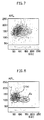
- a reagent having the following composition was prepared.
- EB Ethidium bromide
- Figure 7 shows a scattergram wherein the side scattered light (SSC) and red fluorescence light (RFL) obtained without using an ionic surfactant are referred to as the coordinated axes.
- SSC side scattered light
- RNL red fluorescence light
- the lysing is strengthened by increasing the amount of LTAC, thereby giving an excessive damage to the leukocytes.
- LTAC 500 mg/l it was observed that leukocytes are aggregated into one, so that the leukocytes can not be classified.
- Figures 8 to 16 show that when various nonionic surfactants were contained in the surfactant, the damage of leukocytes was controlled and leukocytes can be classified and counted into lymphocytes Ly, eosinophils Eo, and other leukocytes Othl with respect to every nonionic surfactant that was used.
- the molar number of addition of polyoxyethylene glycol and type of hydrophobic group did not affect the results.
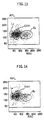
- Example 3 Function of water soluble alcohol
- a reagent having the following composition was prepared.
- venous blood 25 ⁇ l which was treated with an anticoagulant was mixed.
- the red fluorescence light, forward scattered light, and side scattered light were measured by a flow cytometer.
- Figures 17 to 19 show a scattergram wherein the side scattered light (SSC) and red fluorescence light (RFL) obtained when ethanol (100 ml/l) was contained are referred to as the coordinated axes; a histogram thereof; and a scattergram obtained in the same manner as above by using the side scattered light (SSC) and red fluorescence light (RFL).
- SSC side scattered light
- RTL red fluorescence light
- leukocytes are classified and counted into four groups, i.e., lymphocytes Ly, monocytes Mo, eosinophils Eo, and other leukocytes Oth3.
- Figures 20 to 22 show the case that ethanol (200 ml/l) was contained.
- leukocytes are also classified and counted into four groups, i.e., lymphocytes Ly, monocytes Mo, eosinophils Eo, and other leukocytes Oth3.
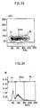
- Figures 23 to 25 show the case that ethanol (400 ml/l) was contained.
- leukocytes are classified and counted into three groups and counted, i.e., lymphocytes Ly, eosinophils Eo, and other leukocytes Oth1.
- a reagent having the following composition were prepared.
- Lauryltrimethylammonium chloride (LTAC) cationic surfactant
- Brij35 polyoxyethyleneglycol lauryl ether/ molar number of addition: 23
- nonionic surfactant 1 g
- Acridine Orange 10-dodecyl bromide (AO-10) labeling substance: dye
- dye 0.100 g Ethyl alcohol 0.100 g
- venous blood 25 ⁇ l which was treated with an anticoagulant was mixed. After 20 seconds at a room temperature, the red fluorescence light, green fluorescence light, forward scattered light, and side scattered light were measured by a flow cytometer.
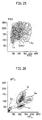
- Figures 26 to 28 show scattergrams obtained as results of normal subjects wherein the red fluorescence light (RFL) and green fluorescence light (GFL), side scattered light (SSC) and red fluorescence light (RFL), and forward scatlight (FSC) and red fluorescence light (RFL) are respectively referred to as the coordinated axes.
- RRL red fluorescence light
- GFL green fluorescence light
- SSC side scattered light
- RNL red fluorescence light
- FSC forward scatlight
- RTL red fluorescence light
- LTAC used in the present invention is a cationic surfactant and has a property of:
- Brij 35 is a nonionic surfactant. It controls the action of LTAC to inhibit from giving a severe damage to the degree that nuclei are made naked or the leukocytes are deformed. The agent also has a function for controlling the aggregation of erythocytes that are converted into ghosts. Further, Brij 35 works for making AO-10 soluble, which is hard to be dissolved in water.
- the dye AO-10 has a dodecyl group at 10th position of Acridine Orange which is often used for staining DNA and RNA simultaneously. Since AO-10 does not usually pass through a cell membrane because of the steric hindrance of the long chain alkyl group, it merely stains cell membranes and the components contained in cells can not be stained. Therefore, the dye has been used only as a hydrophobic probe.
- AO-10 can invade into the cell and combine with components contained therein.
- the dye is used for staining a blood as shown in Examples, it combines with various components in the cell in a specific manner.
- AO-10 combines with RNA present in the cytoplasm by an ionic bond and stains the components orange, while a green fluorescence light is emitted from nucleus and granules thereof. It is inferred that in accordance with a conventionally known function of hydrophobic probe, the long chain alkyl group of AO-10 is invaded through the nucleus membranes and granulocyte membrane to combine with the nucleus and granulocyte.
- a green fluorescence light is emitted. From nucleus and granulocyte of neutrophils, a green light fluorescence light is emitted similar to the case from nucleus and granulocyte of the lymphocytes and monocytes.
- LTAC is combined with RNA by an ionic bond and the electric charges are neutralized, so that RNA are made not to be lost. It is considered that AO-10 is combined with such components which are made not to be lysed.
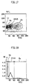
- Example of the present invention a blood sample sampled from patients having a disease in blood were measured by using the reagent composition of the present invention.
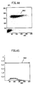
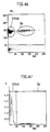
- Figures 29 to 31 show the results obtained from the samples sampled from patients having an acute myelocytic leukemia. Myeloblasts My were classified and counted.
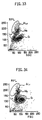
- Figures 32 to 34 show the results obtained from the samples in which Atypical lymphocytes Aly are erupted. Atypical lymphocytes Aly were classified and counted.
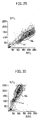
- Figures 35 to 37 show the results obtained from the samples in which erythroblasts Er and immature granulocytes Im were erupted. As seen from these figures, Erythroblasts Er and immature granulocytes Im were classified and counted. Since Blast cells Er and My, Atypical lymphocytes Aly, Immature granulocytes Im include RNA richly therein, they can be detected by staining RNA. In addition, Reticlocytes Ret were partially detected from Figure 37. In this example, Erythrocytes are not completely lysed, but rather, only the membrane of Erythrocyte was damaged. Therefore, cell membranes of the cells which are converted into a ghost and RNA of the cell components are stained, thereby detecting Reticlocytes.
- dyes have not been used for the purpose of the present invention because dyes are hard to pass through cell membranes.
- an effect of dye which is different from the conventional use can be exhibited.
- a reagent having the following composition were prepared.
- Acridine Orange 10-dodecyl bromide (AO-10) label substance
- DiOC6 label substance
- LTAC Lauryltrimethylammonium chloride
- cationic surfactant 0.5 g
- Polyoxyethyleneglycol cetyl ether molecular weight: about 40
- nonionic surfactant 1.0 g Isopropyl alcohol 0.050 liter NaCl 4.0 g HEPES 2.3 g NaOH
- venous blood 25 ⁇ l
- impedance signal were measured by a complex flow cytometer available for detecting not only optical difference but also impedance signals (IMP) based on the change of electric resistance.
- the damage given to leukocytes is made minimum by using a smaller amount of cationic surfactants compared with the amount used in Example 4.
- the reagent composition includes NaCl
- impedance signals can be measured, and as the composition has pH 5.0 or more, eosinophils can be also measured.
- the dye DiOC6 (3) capable for staining granules well was added for facilitating separation of granules when measured by the red fluorescence light and green fluorescence light.
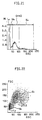
- Figures 38 to 43 show scattergrams and a histogram when using the above regent composition wherein the red fluorescence light (RFL) and green fluorescence light (GFL) (Fig.38), side scattered light (SSC) and red fluorescence light (RFL) (Fig.39), and side scattered light (SSC) and impedance signals (IMP) (Fig.40) are respectively referred to as the coordinated axes; side scattered light histogram (Fig.41); scattergrams wherein side scattered light (SSC) and green fluorescence light (GFL) (Fig.42), and side scattered light (SSC) and forward fluorescence light (FSC) (Fig.43) are respectively referred to as the coordinated axes.
- Ne+Ba denotes a distribution of neutrophils and basophils.
- RNA can be stained. Therefore, Blast cells Er and My, Atypical lymphocytes Aly, Immature granulocytes Im can be classified and counted.
- a reagent having the following composition were prepared.
- Lauryldimethylaminoacetic betaine (amphoteric surfactant) (Anon BL: produced by NIHON YUSHI Co.) 2 g
- Polyoxyethylene glycol nonylphenyl ether (molar number of addition: 20) (nonionic surfactant) 1 g
- Isopropyl alcohol 0.100 liter
- venous blood 400 ⁇ l
- red fluorescence light were measured by a flow cytometer.
- the present example aims to detect a trace amount of leukocytes in a reduced dilution which remain after the treatment by a filter for eliminating leukocytes.
- lysing agent available for obtaining volume differences is used in 100 to 200 times diluted concentration, so that it was not possible to use it for the preparation of the reagent composition having a high concentration such as those of the present example.
- the ratio of blood:lysing agent is 1:7.5.
- more concentrated reagent composition for measurement can be prepared in the present invention.
- reagent compositions having higher concentration at which the measurement is not possible by conventional methods can be used by the method of the present invention.
- a reagent described in the specification of WO 84/003771 (a reagent comprising a cationic surfactant at a high concentration) was prepared as follows: Dodecyltrimethylammonium chloride (cationic surfactant) 55 g/l Tetradecyltrimethyltrimethylammonium chloride (Cationic surfactant) 9 g/l Cyanic pottasium 0.300 g/l Polyoxyethylene glycol alkylphenol ether (Nonionic surfactant) 12 ml/l
- the leukocytes are aggregated into one population, so that they can not be classified by the side scattered light and red fluorescence light based on the optical difference.
- a reagent which comprises a nonionic surfactant was prepared as follows and performed a DNA staining.
- Nonidet P-40 Nonionic surfactant 1 ml/l Sodium Citrate 10 g/l
- the eosinophils can be identified by the side scattered light and red fluorescence light based on the optical difference because they were not naked.
- leukocytes other than eosinophils can not be classified into subclasses.
Claims (22)
- Verfahren zur Vorbehandlung für die Blutanalyse, das die Behandlung der Blutprobe mit einer wäßrigen Lösung, umfassend wenigstens ein Tensid ausgewählt aus der Gruppe bestehend aus einem kationischen Tensid und einem amphoteren Tensid, und mit wenigstens einer markierten Substanz zur selektiven Markierung der Leukozyten umfaßt, dadurch gekennzeichnet, daßdas Tensid in einer Konzentration von 30 bis 5.000 mg/l in der wäßrigen Lösung enthalten ist;die Markierungssubstanz eine ist, die durch eine beschädigte Zellmembran passieren kann und sich mit einer in dem Leukozyten enthaltenen Komponente verbinden kann; unddie Behandlung bei einem pH von 3,0 bis 11,0 durchgeführt wird.
- Verfahren zur Vorbehandlung für die Blutanalyse gemäß Anspruch 1, wobei das Tensid in einer Konzentration von 50 bis 2.000 mg/l enthalten ist.
- Verfahren zur Vorbehandlung für die Blutanalyse gemäß Anspruch 1, wobei das kationische Tensid ein Tensid vom quaternären Ammoniumsalz-Typ oder Pyridiniumsalz-Typ ist.
- Verfahren zur Vorbehandlung für die Blutanalyse gemäß Anspruch 3, wobei das Tensid vom quaternären Ammoniumsalz-Typ die Formel (I) hat:worin R1, R2 und R3 unabhängig voneinander ein Wasserstoffatom, eine C1-8 Alkylgruppe oder eine C6-8 Aralkylgruppe ist, R4 ist eine C8-18 Alkylgruppe, eine C8-18 Alkenylgruppe oder eine C6-18 Aralkylgruppe, und X ist ein Anion.

- Verfahren zur Vorbehandlung für die Blutanalyse gemäß Anspruch 3, wobei das Tensid vom quaternären Ammoniumsalz-Typ wenigstens eines ausgewählt aus der Gruppe bestehend aus Lauryltrimethylammoniumchlorid, Myristyltrimethylammoniumchlorid, Cetyltrimethylammoniumchlorid, Cetyldimethylethylammoniumchlorid und Benzyldimethylcetylammoniumchlorid ist.
- Verfahren zur Vorbehandlung für die Blutanalyse gemäß Anspruch 3, wobei das Tensid vom Pyridiniumsalz-Typ die Formel (II) hat:worin R5 eine C8-18 Alkylgruppe ist und X ein Anion.

- Verfahren zur Vorbehandlung für die Blutanalyse gemäß Anspruch 6, wobei das Tensid vom Pyridiniumsalz-Typ Cetylpyridiniumchlorid ist.
- Verfahren zur Vorbehandlung für die Blutanalyse gemäß Anspruch 1, wobei das amphotere Tensid die Formel (III) hat:worin R1 und R2 unabhängig voneinander ein Wasserstoffatom, eine C1-8 Alkylgruppe oder eine C6-8 Aralkylgruppe ist, R4 ist eine C8-18 Alkylgruppe, eine C8-18 Alkenylgruppe oder eine C8-18 Aralkylgruppe, und n ist 1 oder 2.

- Verfahren zur Vorbehandlung für die Blutanalyse gemäß Anspruch 8, wobei das amphotere Tensid Lauryldimethylaminoessigsäure-betain oder Stearyldimethylaminoessigsäure-betain ist.
- Verfahren zur Vorbehandlung für die Blutanalyse gemäß Anspruch 1, wobei die wäßrige Lösung des Tensids mit 2 bis 200 Volumen pro einem Volumen Blutprobe verwendet wird.
- Verfahren zur Vorbehandlung für die Blutanalyse gemäß Anspruch 1, wobei ein nichtionisches Tensid in einer Konzentration von 100 bis 10.000 mg/l zu der wäßrigen Lösung des Tensids zugegeben wird.
- Verfahren zur Vorbehandlung für die Blutanalyse gemäß Anspruch 11, wobei das nichtionische Tensid in einer Konzentration von 100 bis 5.000 mg/l enthalten ist.
- Verfahren zur Vorbehandlung für die Blutanalyse gemäß Anspruch 1, wobei ein wasserlöslicher Alkohol zu der wäßrigen Tensidlösung zugegeben wird.
- Verfahren zur Vorbehandlung für die Blutanalyse gemäß Anspruch 13, wobei der wasserlösliche Alkohol Ethylalkohol ist und in einer Konzentration von 50 bis 400 ml/l verwendet wird.
- Verfahren zur Vorbehandlung für die Blutanalyse gemäß Anspruch 13, wobei der wasserlösliche Alkohol Isopropylalkohol ist und in einer Konzentration von 25 bis 200 ml/l verwendet wird.
- Verfahren zur Vorbehandlung für die Blutanalyse gemäß Anspruch 1, wobei die Markierungssubstanz ein Farbstoff ist, der sich mit dem Leukozytenkern verbinden kann.
- Verfahren zur Vorbehandlung für die Blutanalyse gemäß Anspruch 16, wobei die Markierungssubstanz Ethidiumbromid oder Propidiumjodid ist.
- Verfahren zur Vorbehandlung für die Blutanalyse gemäß Anspruch 1, wobei die Markierungssubstanz ein Farbstoff ist, der sich mit der in Leukozyten enthaltenen RNA verbinden kann.
- Verfahren zur Vorbehandlung für die Blutanalyse gemäß Anspruch 18, wobei die Markierungssubstanz Pyronin Y, Acridin Orange, Tiazol Orange Acridin Orange 10 Dodecylbromid oder Auramin-O ist.
- Verfahren zur Vorbehandlung für die Blutanalyse gemäß Anspruch 1, wobei die Markierungssubstanz in einer Konzentration von 1 bis 500 mg zu einem Liter wäßriger Tensidlösung zugegeben wird.
- Verfahren zur Vorbehandlung für die Blutanalyse gemäß Anspruch 1, wobei die markierten Leukozyten mit Hilfe einer optischen Vorrichtung klassifiziert und gezählt werden.
- Verfahren zur Vorbehandlung für die Blutanalyse gemäß Anspruch 1, wobei die Behandlung bei einem pH von 4,0 bis 9,0 durchgeführt wird.
Applications Claiming Priority (3)
| Application Number | Priority Date | Filing Date | Title |
|---|---|---|---|
| JP310146/92 | 1992-11-19 | ||
| JP31014692 | 1992-11-19 | ||
| JP31014692 | 1992-11-19 |
Publications (3)
| Publication Number | Publication Date |
|---|---|
| EP0598663A2 EP0598663A2 (de) | 1994-05-25 |
| EP0598663A3 EP0598663A3 (de) | 1995-05-03 |
| EP0598663B1 true EP0598663B1 (de) | 2000-02-02 |
Family
ID=18001723
Family Applications (1)
| Application Number | Title | Priority Date | Filing Date |
|---|---|---|---|
| EP93402790A Expired - Lifetime EP0598663B1 (de) | 1992-11-19 | 1993-11-17 | Verfahren zur Vorbehandlung für Blutanalyse |
Country Status (4)
| Country | Link |
|---|---|
| US (1) | US5496734A (de) |
| EP (1) | EP0598663B1 (de) |
| CN (1) | CN1040469C (de) |
| DE (1) | DE69327775T2 (de) |
Families Citing this family (45)
| Publication number | Priority date | Publication date | Assignee | Title |
|---|---|---|---|---|
| JP3301646B2 (ja) * | 1993-03-19 | 2002-07-15 | シスメックス株式会社 | 幼若細胞測定用試薬 |
| JP3320869B2 (ja) * | 1993-12-22 | 2002-09-03 | シスメックス株式会社 | 白血球分析用試薬 |
| JP3355038B2 (ja) * | 1994-08-03 | 2002-12-09 | シスメックス株式会社 | 白血球分類方法 |
| JP3324050B2 (ja) * | 1994-10-31 | 2002-09-17 | 日本光電工業株式会社 | 白血球分類用試薬および白血球分類方法 |
| US5691204A (en) * | 1995-04-21 | 1997-11-25 | Abbott Laboratories | Compositions and methods for the rapid analysis of reticulocytes |
| FR2735579B1 (fr) * | 1995-06-13 | 1997-09-19 | Hycel Groupe Lisabio | Procede de discrimination et d'isolement de sous-populations de leucocytes a partir d'echantillons sanguins par traitement par du polyoxyethylene 9-lauryl ether, et reactif pour sa mise en oeuvre |
| US5786224A (en) * | 1995-06-29 | 1998-07-28 | Coulter Corporation | Reagent and method for differential determination of leukocytes in blood |
| US5817518A (en) * | 1995-12-18 | 1998-10-06 | Coulter International Corp. | Reagent and method for differential determination of leukocytes in blood |
| TW379284B (en) * | 1996-04-12 | 2000-01-11 | Toa Medical Electronics | Agent for detecting reticulocyte |
| JP4042925B2 (ja) * | 1996-11-20 | 2008-02-06 | シスメックス株式会社 | 幼若白血球の分類計数法 |
| JP3783808B2 (ja) * | 1997-05-19 | 2006-06-07 | シスメックス株式会社 | 白血球分類計数用試薬 |
| US6667177B1 (en) * | 1997-11-11 | 2003-12-23 | Kowa Company, Ltd. | Method for counting leukocytes and apparatus for counting leukocytes |
| US6911313B2 (en) * | 1998-02-06 | 2005-06-28 | Sysmex Corporation | Process for discriminating and counting erythroblasts |
| US6271035B1 (en) * | 1998-10-20 | 2001-08-07 | Coulter International Corp. | Methods and compositions for rapid staining of nucleic acids in whole cells |
| US6214625B1 (en) * | 1999-04-28 | 2001-04-10 | Coulter International Corp. | Composition and method for differentiation of basophils and eosinophils in blood |
| US6210969B1 (en) * | 1999-04-28 | 2001-04-03 | Coulter International Corp. | Composition and method for differentiation of basophil and eosinophil subpopulations of leukocytes in blood |
| US6225124B1 (en) * | 1999-06-02 | 2001-05-01 | Sysmex Corporation | Diluting reagent and method compelling time-independent consistency in MCV assay |
| FR2821428B1 (fr) * | 2001-02-23 | 2004-08-06 | Abx Sa | Reactif et procede pour l'identification et le comptage de cellules biologiques |
| DE10305768A1 (de) * | 2003-02-11 | 2004-08-19 | Justus-Liebig-Universität Giessen | Verfahren zum Färben von lipophilen Zellbestandteilen einer biologischen Probe |
| US7295306B2 (en) * | 2004-04-22 | 2007-11-13 | Kowa Company, Ltd. | Microchip and fluorescent particle counter with microchip |
| FR2873813B1 (fr) * | 2004-07-30 | 2006-11-17 | Abx Sa | Procede et dispositif de caracterisation des composants cellulaires d'un liquide biologique |
| US7803523B2 (en) * | 2004-08-27 | 2010-09-28 | University Health Network | Whole blood preparation for cytometric analysis of cell signaling pathways |
| US7465584B2 (en) * | 2005-01-24 | 2008-12-16 | Sysmex Corporation | Method and reagent for classifying leukocytes in animal blood |
| CN101078720B (zh) * | 2006-05-22 | 2010-12-01 | 深圳迈瑞生物医疗电子股份有限公司 | 一种改进的用于对白细胞进行分类的试剂及方法 |
| CN101078721B (zh) * | 2006-05-23 | 2010-12-22 | 深圳迈瑞生物医疗电子股份有限公司 | 对白细胞进行分类的试剂及方法 |
| CN101349644B (zh) * | 2007-07-20 | 2012-06-27 | 深圳迈瑞生物医疗电子股份有限公司 | 一种白细胞分类试剂和其使用方法 |
| CN105440725A (zh) * | 2008-01-04 | 2016-03-30 | 深圳迈瑞生物医疗电子股份有限公司 | 不对称菁类荧光染料,组合物及在生物样品染色中的用途 |
| WO2009136570A1 (ja) | 2008-05-09 | 2009-11-12 | シスメックス株式会社 | 血液分析装置、血液分析方法および溶血剤 |
| CN101602762B (zh) * | 2008-06-10 | 2013-10-16 | 深圳迈瑞生物医疗电子股份有限公司 | 不对称菁类化合物、其制备方法及应用 |
| US7968279B2 (en) * | 2008-08-13 | 2011-06-28 | Beckman Coulter, Inc. | Reference control for cell by cell analysis |
| CN102144161B (zh) * | 2008-09-04 | 2016-02-17 | 贝克曼考尔特公司 | 泛激酶活化与信号通路的评估 |
| CN101726579B (zh) * | 2008-10-17 | 2014-06-18 | 深圳迈瑞生物医疗电子股份有限公司 | 血液检测试剂和方法 |
| US8367358B2 (en) * | 2008-12-17 | 2013-02-05 | Shenzhen Mindray Bio-Medical Electronics Co., Ltd. | Reagent, kit and method for differentiating and counting leukocytes |
| JP5452058B2 (ja) * | 2009-03-31 | 2014-03-26 | シスメックス株式会社 | 血液分析装置 |
| CN101988082B (zh) * | 2009-07-31 | 2015-04-08 | 深圳迈瑞生物医疗电子股份有限公司 | 白细胞分类计数试剂、试剂盒及其制备方法和白细胞分类计数的方法 |
| CN104169719B (zh) * | 2011-12-29 | 2017-03-08 | 思迪赛特诊断有限公司 | 用于检测生物样品中病原体的方法和系统 |
| JP6001425B2 (ja) * | 2012-11-26 | 2016-10-05 | シスメックス株式会社 | 血球分析方法、血球分析装置およびプログラム |
| US10794809B2 (en) | 2014-02-28 | 2020-10-06 | Sysmex Corporation | Method for urine sample analysis, reagent for urine sample analysis, and reagent kit for urine sample analysis |
| WO2016106688A1 (zh) * | 2014-12-31 | 2016-07-07 | 深圳迈瑞生物医疗电子股份有限公司 | 一种有核红细胞报警方法、装置及流式细胞分析仪 |
| JP6710512B2 (ja) * | 2015-09-14 | 2020-06-17 | シスメックス株式会社 | 血液分析装置、血液分析方法及びコンピュータプログラム |
| CN105728069B (zh) * | 2016-01-30 | 2021-01-19 | 深圳市安测健康信息技术有限公司 | 用于快速自检血液的多通道微流控芯片 |
| CN108414427A (zh) * | 2017-02-10 | 2018-08-17 | 深圳市帝迈生物技术有限公司 | 一种白细胞分类试剂 |
| CN107656071B (zh) * | 2017-11-17 | 2020-04-03 | 南通伊仕生物技术股份有限公司 | 一种NT-ProBNP检测试剂盒及其使用方法 |
| CN107884563A (zh) * | 2017-11-26 | 2018-04-06 | 张延艳 | 一种用于血液分析的医疗装置及其使用方法 |
| EP3789757A4 (de) * | 2018-04-28 | 2021-07-07 | Shenzhen Mindray Bio-Medical Electronics Co., Ltd. | Blutanalysegerät und analyseverfahren |
Family Cites Families (25)
| Publication number | Priority date | Publication date | Assignee | Title |
|---|---|---|---|---|
| US3740143A (en) * | 1970-10-30 | 1973-06-19 | Technicon Instr | Automatic apparatus for determining the percentage population of particulates in a medium |
| US4286963A (en) * | 1979-11-23 | 1981-09-01 | Coulter Electronics, Inc. | Differential lymphoid-myeloid determination of leukocytes in whole blood |
| US4962038A (en) * | 1980-06-16 | 1990-10-09 | Coulter Electronics, Inc. | Multi-purpose blood diluent and lysing agent for differential determination of lymphoid-myeloid population of leukocytes |
| US4528274A (en) * | 1982-07-06 | 1985-07-09 | Coulter Electronics, Inc. | Multi-purpose blood diluent and lysing agent for differential determination of lymphoid-myeloid population of leukocytes |
| DE3238353A1 (de) * | 1982-10-15 | 1984-04-19 | Max Planck Gesellschaft zur Förderung der Wissenschaften e.V., 3400 Göttingen | Verfahren zur simultanen quantitativen bestimmung der blutzellen und reagenz hierfuer |
| US4485175A (en) * | 1983-01-03 | 1984-11-27 | Coulter Electronics, Inc. | Method for three-volume differential determination of lymphocyte, monocyte and granulocyte populations of leukocytes |
| US4751179A (en) * | 1984-05-31 | 1988-06-14 | Coulter Electronics, Inc. | Method and reagents for differential determination of four populations of leukocytes in blood |
| CA1255197A (en) * | 1984-09-24 | 1989-06-06 | Joseph L. Orlik | Leukocyte differentiation composition and method |
| US4745071A (en) * | 1985-09-05 | 1988-05-17 | Sequoia-Turner Corporation | Method for the volumetric differentiation of blood cells types |
| JPH06100596B2 (ja) * | 1986-09-10 | 1994-12-12 | 東亜医用電子株式会社 | フロ−サイトメトリ−による白血球の分類方法 |
| US5175109A (en) * | 1986-09-10 | 1992-12-29 | Toa Medical Electronics Co., Ltd. | Reagent for classifying leukocytes by flow cytometry |
| US5039613A (en) * | 1986-11-27 | 1991-08-13 | Toa Medical Electronics Co., Ltd. | Reagents used in a method of classifying leukocytes by flow cytometry |
| US5389549A (en) * | 1987-05-29 | 1995-02-14 | Toa Medical Electronics Co., Ltd. | Method for classifying leukocytes and a reagent used therefor |
| JP2654811B2 (ja) * | 1988-07-20 | 1997-09-17 | 東亜医用電子株式会社 | 好酸球の計数方法 |
| CA2016699C (en) * | 1989-05-15 | 2003-11-18 | Paul N. Marshall | Lytic agents and uses thereof |
| JP2836865B2 (ja) * | 1989-10-23 | 1998-12-14 | 東亜医用電子株式会社 | 血液中の白血球およびヘモグロビンの測定用試薬 |
| FR2654744B1 (fr) * | 1989-11-20 | 1992-03-13 | Abx Sa | Reactif et methode d'utilisation de celui-ci pour la determination automatique en cytometrie de flux d'au moins une sous-population leucocytaire a partir du sang total. |
| US5242832A (en) * | 1990-03-01 | 1993-09-07 | Toa Medical Electronics Co., Ltd. | Reagent for measurement of leukocytes and hemoglobin in blood |
| US5250438A (en) * | 1990-05-09 | 1993-10-05 | Streck Laboratories, Inc. | Method for differential determination of white blood cells using diazolidinyl urea to stabilize white blood cells |
| JP2941041B2 (ja) * | 1990-11-16 | 1999-08-25 | シスメックス株式会社 | フローサイトメトリーによる白血球の分類方法 |
| US5256571A (en) * | 1991-05-01 | 1993-10-26 | Cytyc Corporation | Cell preservative solution |
| US5262329A (en) * | 1991-06-13 | 1993-11-16 | Carver Jr Edward L | Method for improved multiple species blood analysis |
| JP3067849B2 (ja) * | 1991-07-29 | 2000-07-24 | シスメックス株式会社 | 白血球分類計数用試料調製方法 |
| JP3048260B2 (ja) * | 1991-07-29 | 2000-06-05 | シスメックス株式会社 | 白血球分類計数用試料調製方法 |
| US5422277A (en) * | 1992-03-27 | 1995-06-06 | Ortho Diagnostic Systems Inc. | Cell fixative composition and method of staining cells without destroying the cell surface |
-
1993
- 1993-11-17 DE DE69327775T patent/DE69327775T2/de not_active Expired - Lifetime
- 1993-11-17 EP EP93402790A patent/EP0598663B1/de not_active Expired - Lifetime
- 1993-11-18 CN CN93115347A patent/CN1040469C/zh not_active Expired - Fee Related
-
1995
- 1995-06-02 US US08/458,304 patent/US5496734A/en not_active Expired - Lifetime
Also Published As
| Publication number | Publication date |
|---|---|
| US5496734A (en) | 1996-03-05 |
| DE69327775D1 (de) | 2000-03-09 |
| CN1092172A (zh) | 1994-09-14 |
| CN1040469C (zh) | 1998-10-28 |
| EP0598663A3 (de) | 1995-05-03 |
| DE69327775T2 (de) | 2000-06-21 |
| EP0598663A2 (de) | 1994-05-25 |
Similar Documents
| Publication | Publication Date | Title |
|---|---|---|
| EP0598663B1 (de) | Verfahren zur Vorbehandlung für Blutanalyse | |
| US6664110B1 (en) | Erythroblast diagnostic flow-cytometry method and reagents | |
| KR100316393B1 (ko) | 백혈구분석용시약 | |
| US7625730B2 (en) | Method for classifying and counting leukocytes | |
| EP0678743B1 (de) | Reagenz zur Analyse von Leukozyten und ein Verfahren zum Klassifizieren von Leukozyten | |
| CA1248856A (en) | Method and reagent system for four-population differential determination of leukocytes | |
| US5486477A (en) | Reagent system for improved multiple species blood analysis | |
| JP4042925B2 (ja) | 幼若白血球の分類計数法 | |
| JP3679127B2 (ja) | 血液中の白血球の鑑別式決定のための試薬及び方法 | |
| JP3345135B2 (ja) | 血液分析方法 | |
| US5389549A (en) | Method for classifying leukocytes and a reagent used therefor | |
| GB2077916A (en) | Differential determination of lymphoidmyeloid population of leukocytes and multi-purpose blood diluent and lysing agent therefor | |
| US6977156B2 (en) | Flow cytometry reagent and system | |
| US5843608A (en) | Reagent and method for differential determination of leukocytes in blood | |
| JP2002148261A (ja) | 異常細胞分類計数方法 | |
| JP2009080122A (ja) | 赤芽球の分類計数方法 | |
| EP2166354A1 (de) | Reagens und reagenskit zur analyse primitiver leukozyten | |
| JPH10339729A (ja) | 赤芽球の分類計数用試薬及び分類計数方法 | |
| CN114252633A (zh) | 样本分析系统、样本检测方法、组合物及其应用 |
Legal Events
| Date | Code | Title | Description |
|---|---|---|---|
| PUAI | Public reference made under article 153(3) epc to a published international application that has entered the european phase |
Free format text: ORIGINAL CODE: 0009012 |
|
| AK | Designated contracting states |
Kind code of ref document: A2 Designated state(s): DE FR GB IT |
|
| PUAL | Search report despatched |
Free format text: ORIGINAL CODE: 0009013 |
|
| AK | Designated contracting states |
Kind code of ref document: A3 Designated state(s): DE FR GB IT |
|
| 17P | Request for examination filed |
Effective date: 19951019 |
|
| 17Q | First examination report despatched |
Effective date: 19981023 |
|
| GRAG | Despatch of communication of intention to grant |
Free format text: ORIGINAL CODE: EPIDOS AGRA |
|
| RAP1 | Party data changed (applicant data changed or rights of an application transferred) |
Owner name: SYSMEX CORPORATION |
|
| GRAG | Despatch of communication of intention to grant |
Free format text: ORIGINAL CODE: EPIDOS AGRA |
|
| GRAH | Despatch of communication of intention to grant a patent |
Free format text: ORIGINAL CODE: EPIDOS IGRA |
|
| GRAH | Despatch of communication of intention to grant a patent |
Free format text: ORIGINAL CODE: EPIDOS IGRA |
|
| GRAA | (expected) grant |
Free format text: ORIGINAL CODE: 0009210 |
|
| AK | Designated contracting states |
Kind code of ref document: B1 Designated state(s): DE FR GB IT |
|
| REF | Corresponds to: |
Ref document number: 69327775 Country of ref document: DE Date of ref document: 20000309 |
|
| ITF | It: translation for a ep patent filed |
Owner name: MODIANO & ASSOCIATI S.R.L. |
|
| ET | Fr: translation filed | ||
| PLBE | No opposition filed within time limit |
Free format text: ORIGINAL CODE: 0009261 |
|
| STAA | Information on the status of an ep patent application or granted ep patent |
Free format text: STATUS: NO OPPOSITION FILED WITHIN TIME LIMIT |
|
| 26N | No opposition filed | ||
| REG | Reference to a national code |
Ref country code: GB Ref legal event code: IF02 |
|
| PGFP | Annual fee paid to national office [announced via postgrant information from national office to epo] |
Ref country code: DE Payment date: 20101110 Year of fee payment: 18 |
|
| PGFP | Annual fee paid to national office [announced via postgrant information from national office to epo] |
Ref country code: GB Payment date: 20101117 Year of fee payment: 18 Ref country code: IT Payment date: 20101120 Year of fee payment: 18 |
|
| PGFP | Annual fee paid to national office [announced via postgrant information from national office to epo] |
Ref country code: FR Payment date: 20111118 Year of fee payment: 19 |
|
| GBPC | Gb: european patent ceased through non-payment of renewal fee |
Effective date: 20121117 |
|
| REG | Reference to a national code |
Ref country code: FR Ref legal event code: ST Effective date: 20130731 |
|
| PG25 | Lapsed in a contracting state [announced via postgrant information from national office to epo] |
Ref country code: IT Free format text: LAPSE BECAUSE OF NON-PAYMENT OF DUE FEES Effective date: 20121117 |
|
| REG | Reference to a national code |
Ref country code: DE Ref legal event code: R119 Ref document number: 69327775 Country of ref document: DE Effective date: 20130601 |
|
| PG25 | Lapsed in a contracting state [announced via postgrant information from national office to epo] |
Ref country code: DE Free format text: LAPSE BECAUSE OF NON-PAYMENT OF DUE FEES Effective date: 20130601 |
|
| PG25 | Lapsed in a contracting state [announced via postgrant information from national office to epo] |
Ref country code: FR Free format text: LAPSE BECAUSE OF NON-PAYMENT OF DUE FEES Effective date: 20121130 Ref country code: GB Free format text: LAPSE BECAUSE OF NON-PAYMENT OF DUE FEES Effective date: 20121117 |