EP0576476B1 - Dosage immunologique pour detecter un polypeptide mutant p53 dans des fluides biologiques - Google Patents
Dosage immunologique pour detecter un polypeptide mutant p53 dans des fluides biologiques Download PDFInfo
- Publication number
- EP0576476B1 EP0576476B1 EP92906415A EP92906415A EP0576476B1 EP 0576476 B1 EP0576476 B1 EP 0576476B1 EP 92906415 A EP92906415 A EP 92906415A EP 92906415 A EP92906415 A EP 92906415A EP 0576476 B1 EP0576476 B1 EP 0576476B1
- Authority
- EP
- European Patent Office
- Prior art keywords
- antibody
- mutant
- polypeptide
- sample
- complex
- Prior art date
- Legal status (The legal status is an assumption and is not a legal conclusion. Google has not performed a legal analysis and makes no representation as to the accuracy of the status listed.)
- Expired - Lifetime
Links
Images


Classifications
-
- C—CHEMISTRY; METALLURGY
- C07—ORGANIC CHEMISTRY
- C07K—PEPTIDES
- C07K16/00—Immunoglobulins [IGs], e.g. monoclonal or polyclonal antibodies
- C07K16/18—Immunoglobulins [IGs], e.g. monoclonal or polyclonal antibodies against material from animals or humans
- C07K16/32—Immunoglobulins [IGs], e.g. monoclonal or polyclonal antibodies against material from animals or humans against translation products of oncogenes
-
- C—CHEMISTRY; METALLURGY
- C07—ORGANIC CHEMISTRY
- C07K—PEPTIDES
- C07K14/00—Peptides having more than 20 amino acids; Gastrins; Somatostatins; Melanotropins; Derivatives thereof
- C07K14/435—Peptides having more than 20 amino acids; Gastrins; Somatostatins; Melanotropins; Derivatives thereof from animals; from humans
- C07K14/46—Peptides having more than 20 amino acids; Gastrins; Somatostatins; Melanotropins; Derivatives thereof from animals; from humans from vertebrates
- C07K14/47—Peptides having more than 20 amino acids; Gastrins; Somatostatins; Melanotropins; Derivatives thereof from animals; from humans from vertebrates from mammals
- C07K14/4701—Peptides having more than 20 amino acids; Gastrins; Somatostatins; Melanotropins; Derivatives thereof from animals; from humans from vertebrates from mammals not used
- C07K14/4746—Peptides having more than 20 amino acids; Gastrins; Somatostatins; Melanotropins; Derivatives thereof from animals; from humans from vertebrates from mammals not used p53
-
- G—PHYSICS
- G01—MEASURING; TESTING
- G01N—INVESTIGATING OR ANALYSING MATERIALS BY DETERMINING THEIR CHEMICAL OR PHYSICAL PROPERTIES
- G01N33/00—Investigating or analysing materials by specific methods not covered by groups G01N1/00 - G01N31/00
- G01N33/48—Biological material, e.g. blood, urine; Haemocytometers
- G01N33/50—Chemical analysis of biological material, e.g. blood, urine; Testing involving biospecific ligand binding methods; Immunological testing
- G01N33/53—Immunoassay; Biospecific binding assay; Materials therefor
- G01N33/574—Immunoassay; Biospecific binding assay; Materials therefor for cancer
- G01N33/5748—Immunoassay; Biospecific binding assay; Materials therefor for cancer involving oncogenic proteins
Definitions
- Cellular oncogenes are normal genes which have been conserved throughout evolution and are believed to have normal functional roles in the cell. In their non-activated (wild-type) state such cellular oncogenes are sometimes referred to as proto-oncogenes. Proto-oncogenes are not oncogenic or tumorigenic until they are activated in some way. A number of different genetic mechanisms may cause the somatic mutation of oncogenes that results in the activated oncogenes found in tumor cells. These include point mutations, translocations, gene rearrangement, and gene amplification, all of which may be induced by chemical or physical carcinogenic means or by the integration of a viral genome adjacent to the proto-oncogene sequences in the host DNA. Certain oncogenes, such as ras and wild type p53 oncogenes, when "activated" encode mutant proteins while others such as myc may express elevated levels of normal protein.
- the wild type p53 oncogene encodes wild type p53 polypeptide which functions as a negative regulator of cell division.
- the wild type p53 polypeptide has been found intracellularly in normal cells and tissues at low levels. Mutant p53 polypeptides encoded by activated p53 oncogenes are present intracellularly at high concentrations in mammalian tumors and tumor cell lines.
- the wild type p53 oncogene is conserved across a wide variety of species including man, mouse, rat and frog (1). cDNA sequence analysis has indicated that there are five blocks of very highly conserved sequences (2, 3). These conserved residues have been grouped in blocks beginning at amino acid 117 and ending at amino acid 286 (4). Point mutations, occurring principally in these five blocks of very highly conserved sequences and also in highly conserved regions of the wild type p53 oncogene lying outside of these blocks, produce an activated p53 oncogene (SEQ ID NO. 2 - SEQ ID NO. 7). Changes in these conserved areas have a significant impact on the function of the mutant p53 polypeptide. Changes in these regions of the wild type p53 oncogene generate an activated p53 oncogene which encodes a protein having a conformational change identical to the vast majority of the mutant p53 polypeptides so expressed.
- mutant p53 polypeptide The product of the activated p53 oncogene, i.e. mutant p53 polypeptide, is present at high levels in a high percentage of virtually all classes of human tumors including tumors of the colon, lung, and breast (2). Biochemical analyses of mutant p53 polypeptides demonstrate that activating mutations affect the polypeptide's structure in similar ways. Mutant p53 polypeptides have a much longer half-life as compared to normal p53 polypeptide. In addition, mutant p53 polypeptides are able to complex with the heat-shock-protein-70 family of proteins but not to SV40 large T antigen (5-8).
- mutant and wild-type p53 polypeptides have been distinguished on the basis of differential reactivity with monoclonal antibodies.
- Antibody secreted by clone PAb246 is reactive with wild-type p53 polypeptides but not with any mutant p53 polypeptide tested to date, while antibody secreted by clone PAb240 reacts with all mutant p53 polypeptides tested to date but not with a wild-type p53 polypeptide (5-21).
- the human, wild-type p53 oncogene is found on chromosome 17p. Allelic loss in 17p occur at high frequency in human breast cancer (22), colon cancer (23), astrocytomas (24) and small cell lung carcinoma (25).
- the question of allele loss of the wild-type p53 oncogene was addressed by cytogenetic analysis of a number of colon cancer samples using specific DNA probes (23). The results of such analysis indicated that at least a portion, if not all, of one of the two alleles of the wild-type p53 oncogene was lost. However, there were no large deletions or rearrangements in the p53 oncogene associated with the other allele. In most cases, sequence analysis of cDNA from the tumors demonstrated that the p53 oncogene encoded by the second allele contained activating point or missense mutations in those regions encoding the conserved amino acid sequence boxes (26).
- Mutant p53 polypeptides possess an extended half-life of about 24 hours in comparison to wild-type p53 polypeptides which have a half-life of about 20 minutes.
- the extended half-life of the mutant p53 polypeptide allows it to accumulate to detectable levels in those tumors associated with an activated p53 oncogene.
- wild type p53 polypeptide does not accumulate and is not easily detected. Because the wild-type p53 polypeptide is barely detectable in normal cells, due to its extremely short half-life, the presence of substantial amounts of p53 polypeptide establishes that the protein contains both a mutation resulting in extended half-life and the consequent phenotype of a dominant oncogenic protein.
- Stabilization of the mutant p53 polypeptide may be due to its ability to form complexes with other molecules such as heat shock proteins.
- stabilization of the mutant p53 polypeptide may be due to mutations in the primary sequence of the polypeptides which make them intrinsically more stable.
- stabilization of the mutant p53 polypeptide may be due to post-translational modifications such as hyperphosphorylation (5-8, 27).
- the wild-type p53 oncogene is mutated at high frequency in the majority of human cancers.
- One of the first indications that p53 polypeptide expression may have been associated with some forms of human cancer was the observation that about 9% of patients with breast carcinoma (14 out of 155 samples tested) had auto-antibodies to p53 polypeptides (28).
- US-A-4798787 discloses peptide antibodies and their use in detecting oncogene products. However, US-A-4798787 does not teach or suggest probes or assays for p53 and does not teach or suggest the quantitative determination of a p21 expression product or p53 expression product.
- EP-A2 0 214 520 discloses an immunoassay for the detection of ras encoded proteins. This assay is based on the determination of the presence of an activated oncogene in a biological fluid for a subject. In the Examples, the determination of the presence of ras 21 by an appropriate antibody in serum of cancer patients is described. EP-A2 0 214 520 does not disclose an assay for the detection of a neoplastic condition which is based on the determination of the concentration of mutant p53 in body fluids.
- mutant p53 polypeptides could be detected in 50% of colon cancer, 30% of breast cancer and 70% of lung cancer tumor sections (4). However, mutant p53 polypeptides could not be detected in any normal or premalignant tissues from these patients.
- Other investigations have found overexpression of mutant p53 polypeptide in patients with leukemia or lymphoma (20, 21, 29). It is becoming increasingly apparent that when proper care is taken to preserve the specimen, a high percent of cancer biopsies examined are found to express elevated amounts of mutant p53 polypeptide.
- mutant p53 polypeptides were located in the interior of the cell, one of ordinary skill in the art would not expect to detect mutant p53 polypeptides in substantially cell-free biological fluids.
- this invention is based upon the discovery that normally intracellular, mutant p53 polypeptides may be detected in biological fluids such as serum and that such detection may be utilized to diagnose or monitor neoplastic conditions or states. Accordingly, the establishment of a serum-based diagnostic marker for human cancer would have significant commercial applications. Using assay kits, blood drawn from patients could be routinely and easily assayed for mutant p53 polypeptide concentration. The correlation between the measured mutant p53 polypeptide concentration and the presence of neoplastic disease in the subject patient provides a means for early detection of the cancer. In addition, because of the ease for effecting the invention described herein a much larger segment of the population can be tested. Furthermore, the invention should have wide applicability in basic- and clinical- research applications.
- the present invention as claimed in Claims 1 to 25 provides a method for diagnosing in a subject a neoplastic condition.
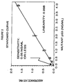
- Figure 1 A standard curve constructed using purified human recombinant mutant p53 polypeptide.
- the assay detects the purified standard with a sensitivity of approximately 30 pg/ml.
- Figure 2 Line graph showing a standard curve constructed using purified recombinant mutant p53 polypeptide (Hup53HIS273) which was expressed using the T7 expression system.
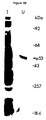
- FIG 4 Photographs of three gels.
- Mammalian cell extracts (lanes II-IV, 1 mg protein/lane), purified p53 (lanes V, 5 ⁇ gs/lane) and molecular weight standards (lanes I) were resolved by electrophoresis on a 10% acrylamide gel.
- the gel was cut into three identical sections and either stained with Coomassie Brilliant Blue G-250® (A) or blotted onto nitrocellulose and incubated with either polyclonal rabbit anti-mutant p53 (HIS175) antibodies (B) or a control rabbit polyclonal antibody of irrelevant specificity (C).
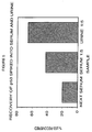
- Figure 5 A bar graph showing recovery of p53 spiked into serum and urine.
- Purified recombinant mutant p53 was added to undiluted normal serum and to diluted (1:5 with Sample Diluent) normal human serum to a final concentration of 4 ng/ml.
- Purified p53 was added to diluted human urine to a final concentration of 2 ng/ml.
- the p53-spiked samples were analyzed in the p53 ELISA assay. The amount of mutant p53 detected is expressed as a percent of the amount actually added to the sample.
- Figure 6 A bar graph showing that a monoclonal antibody specific for p53 polypeptide (p53(Ab-2)) will specifically inhibit the reactivity of a mutant p53 containing human cancer serum sample, whereas an antibody to an unrelated protein (trpE(Ab-1)) has no effect. Neither antibody has any effect on a normal serum sample which does not contain any detectable mutant p53 polypeptide. No reactivity is illustrated as a bar having a height equal to 0.05 ng/ml which is the sensitivity limit of this assay.
- Figure 7 A bar graph showing that immunoprecipitation with a monoclonal antibody specific for p53 polypeptide (p53(Ab-2) can be used to efficiently remove any mutant p53 from a serum sample (Colon Ca) containing mutant p53. Under the same conditions, an antibody to an unrelated protein (trpE(Ab-1) does not remove the mutant p53 polypeptide from the sample. Normal serum which does not contain any detectable mutant p53 polypeptide is unaffected by either treatment. No reactivity is illustrated as a bar having a height equal to 0.05 ng/ml which is the sensitivity limit of this assay.
- the present invention as claimed in Claims 1 to 25 provides a method for diagnosing in a subject, for example, a human, a neoplastic condition.
- a subject for example, a human
- a neoplastic condition the presence of the mutant p53 polypeptide in the sample indicates that the subject has the neoplastic condition.
- neoplastic condition means a state characterized by tumor formation and tumor growth.
- neoplastic conditions which may be diagnosed in accordance with this invention include the following forms of human cancer: breast cancer, colon cancer, lung cancer, lymphoma, hepatoma and leukemia, astrocytomas, and small cell lung carcinomas.
- a biological fluid is any body fluid or any fluid derived from a biological sample.
- body fluids include urine, blood, sputum, amniotic fluid, saliva, any mucous-type bodily secretion or cerebrospinal fluid.
- fluids derived from biological samples include serum, plasma, cell extracts, lung lavage or ascites fluid. Serum is preferred.
- Cell extracts may be derived from tumor cells or tissues, normal tissues, or cell lines continuously growing in culture. It would be clear to those skilled in the art that such cells may be from any mammal such as rats, moles, shrews, monkeys, bats, hares, rabbits, dogs, cats, whales, dolphins, elephants, horses, cows, deer, and man.
- detecting the presence means detection by any manner.
- mutant p53 polypeptide means any polypeptide encoded by an activated p53 oncogene.
- oncogene means a gene that has the potential to cause cancer.
- activated in the case of a p53 oncogene, is a condition which induces the onset of cancer, neoplasia, or more generally oncogenesis. Specifically, the condition is caused by a mutation such as a point mutation in a p53 oncogene.
- the detecting in step (b) comprises: (i) contacting the sample from step (a) with a protein capable of forming a complex with the mutant p53 polypeptide under conditions permitting the complex to be formed; and (ii) determining whether any complex is so formed, the presence of the complex indicating that the subject has the neoplastic condition.
- the protein of step (i) may be an antibody.
- the antibody is a polyclonal antibody.
- the antibody is a monoclonal antibody.
- the term "antibody” means any immunologically reactive molecule (i.e. protein, polypeptide, or fragment thereof) that is naturally occurring or produced by genetic engineering methods or otherwise. Moreover, the antibody may possess an isotype selected from a group comprising an IgG, IgA, IgD, IgE, or IgM isotype or any combination thereof. The antibody may be constructed by fusion of fragments of selected isotypes, expressed in a recombinant system, or generated through hybridoma technology.
- the protein is a heat shock protein.
- the heat shock protein may be selected from the group consisting of HSC 70-72 and HSP 70-72.
- the protein specifically forms a complex with the mutant p53 polypeptide.
- the word "specifically forms a complex” means that the aforementioned protein only recognizes and binds to a mutant p53 polypeptide.
- the protein is an antibody, e.g. the PAb240 monoclonal antibody.
- the protein may be attached to a solid support.
- a solid support include a nylon membrane, a nitrocellulose membrane, a cellulose acetate membrane, an epoxy-activated synthetic copolymer membrane, agarose, Sepharose®, a plastic or glass tube or any part thereof, a plastic or glass plate or bead or any part thereof.
- step (ii) the determination whether any complex is formed comprises: a) contacting the sample from step (i) with a second protein capable of forming a second complex with any complex formed in step (i) under conditions permitting the second complex to be formed; and b) determining the presence of any such second complex so formed, the presence of any such second complex indicating that the subject has the neoplastic condition.
- the second protein may be a heat shock protein.
- the heat shock protein may be selected from the group consisting of HSC 70-72 and HSP 70-72.
- the second protein may be an antibody such as a polyclonal antibody or monoclonal antibody.
- the second protein specifically forms a complex with the mutant p53 polypeptide.
- the protein may be attached to a solid support. Further, the protein may be labeled with a detectable marker.
- the second protein may be labeled with a detectable marker.
- detectable markers include enzymes, biotins, fluorophores, chromophores, heavy metals, paramagnetic isotopes, or radioisotopes.
- the present invention provides a method for diagnosing in a subject a neoplastic condition which comprises quantitatively determining in a sample of a substantially cell-free biological fluid of the subject the concentration of a mutant p53 polypeptide encoded by an activated p53 oncogene, an elevated concentration of the mutant p53 polypeptide in the sample indicating that the subject has the neoplastic condition.
- an elevated concentration is one which is equal to or greater than two standard deviations above the concentration found in samples from normal subjects.
- the quantitatively determining comprises: (i) contacting the sample from step (a) with a protein capable of forming a complex with the mutant p53 polypeptide under conditions permitting the complex to be formed; and (ii) determining the quantity of any complex so formed, the presence of the complex indicating that the subject has the neoplastic condition.
- the protein is a heat shock protein.
- the heat shock protein may be selected from the group consisting of HSC 70-72 and HSP 70-72.
- the protein may be an antibody such as a polyclonal antibody or a monoclonal antibody. Further, the protein may be attached to a solid support. Additionally, the protein may specifically form a complex with the mutant p53 polypeptides.
- An example of such a protein is the PAb 240 monoclonal antibody.
- the quantitative determination whether any complex is formed in step (ii) comprises: a) contacting the sample from step (i) with a second protein capable of forming a second complex with any complex formed in step (i) under conditions permitting the second complex to be formed; and b) determining the quantity of any such second complex so formed and comparing the amount so determined to the amount in a sample from a normal subject the presence of a significantly different amount indicating that the subject has the neoplastic condition.
- the second protein is a heat shock protein.
- the heat shock protein may be selected from the group consisting of HSC 70-72 and HSP 70-72.
- the second protein may be an antibody, e.g. a polyclonal or monoclonal antibody.
- the protein may specifically form a complex with the mutant p53 polypeptide.
- the second protein may be attached to a solid support.
- the second protein may be labeled with a detectable marker.
- the detectable marker may be an enzyme, biotin, a fluorophore, a chromophore, a heavy metal, a paramagnetic isotope, or a radioisotope.
- This invention further provides a method for quantitatively determining the concentration of p53 in a biological fluid sample which comprises (a) contacting a solid support with an excess of a first antibody under conditions permitting the antibody to attach to the surface of the solid support; (b) contacting the resulting solid support to which the first antibody is bound with a biological fluid sample under conditions such that any p53 polypeptide in the biological fluid binds to the antibody and forms a complex therewith; (c) contacting the complex formed in step (b) with a predetermined amount of a second antibody directed to an epitope on p53 different from the epitope to which the first antibody of step (a) is directed, so as to form a complex which includes p53, the first antibody, and the second antibody; (d) quantitatively determining the amount of the complex formed in step (c); and (e) thereby determining the concentration of p53 in the biological fluid sample.
- the first antibody bound to the solid support is a monoclonal antibody and the second antibody is a polyclonal antibody.
- the first antibody bound to the solid support is a polyclonal antibody and the second antibody is a monoclonal antibody.
- the first antibody bound to the solid support is a polyclonal antibody and the second antibody is a polyclonal antibody.
- the first antibody bound to the solid support is a monoclonal antibody and the second antibody is a monoclonal antibody.
- the first antibody may be labeled with a detectable marker.
- the second antibody may be labeled, separately or in addition to the first antibody, with a detectable marker.
- suitable detectable markers includes an enzyme, biotin, a fluorophore, a chromophore, a heavy metal, a paramagnetic isotope, or a radioisotope.
- This invention provides a method for monitoring the course of a neoplastic condition in a subject which comprises quantitatively determining in a first sample of a biological fluid from the subject the presence of a mutant p53 polypeptide according to any of the previously-described methods and comparing the amount so determined with the amount present in subsequent samples from the subject, such samples being taken at different points in time, i.e. one sample taken at a sufficient period after the other sample to allow for tumor growth or regression. A difference in the amounts determined being indicative of the course of the neoplastic condition, e.g. growth or regression.
- a neoplastic condition includes, but is not limited to carcinomas of the lung, bladder, breast, uterus, prostate, colon, adenocarcinoma of the lung, neuroblastomas, melanomas, rhabdomyosarcomas, lymphomas or leukemias.
- Serum or plasma samples collected using heparin, EDTA or oxalate may be stored frozen at -70°C prior to analysis, or alternatively, the samples may be assayed immediately.
- Serum and/or plasma samples may be analyzed neat (i.e. undiluted) or alternatively the samples may be diluted with Sample Diluent (PBS, pH 7.4 containing 50 mM NaCl, 0.1% BSA, 0.5% Tween-20®, 0.02% Thimerosal and 1% (v/v) normal mouse serum). The dilution ratio is dependent on the mutant p53 polypeptide content of the sample and must be determined empirically. Samples are analyzed in duplicate, 100 ⁇ l per well.
- autologous antibodies to mutant p53 polypeptide may be present in patient serum and/or plasma samples.
- the presence of these antibodies may, in some instances, interfere with and/or reduce the sensitivity with which the mutant p53 polypeptide is detected in those samples.
- the following additional procedures should be employed.
- acidification buffer 200 mM glycine-HCl, pH 2.0
- neutralization buffer 2M Tris-HCl, pH 8
- the acidified and neutralized sample may be analyzed directly or after further dilution with Sample Diluent.
- the acidified sample may be applied to a column of Sephadex G-100 superfine® (Pharmacia, Cat No. 17-0061-01).
- the column effluent is monitored using a spectrophotometer adjusted to 280 nm.
- the first peak of 280 nm-absorbing material (containing proteins of molecular mass greater than 100 kDa, including any autologous antibodies) is discarded.
- the remaining peaks, eluting after the first are pooled and analyzed for mutant p53 polypeptide.
- the eluted, pooled sample may be stored frozen at -20°C and/or concentrated by lyophilization prior to analysis for mutant p53 polypeptide.
- Urine Freshly collected urine is pH buffered by the addition of 1/10 volume of urine buffer (1 M Tris-HCl, pH 8.0, containing 0.2% Thimerosal). Buffered samples may be stored frozen or analyzed immediately as described above.
- a fluid extract To measure the concentration of mutant p53 in cells, a fluid extract must first be prepared. Numerous extraction protocols can be used. The following protocol provided is an example of a whole cell extraction procedure and should not be constructed as necessarily being the method of choice. Pellet cells by centrifugation (1000 Xg, for 10 min at 4°C) then wash 3 times with 20 to 30 cell pellet volumes of ice-cold PBS. Pellet cells and discard PBS wash. Add 5 cell pellet volumes of ice-cold swelling buffer (20mM Tris/HCl, 5 mM EDTA, 1 mM PMSF, pH 8) and incubate on ice for 30 minutes. Gently resuspend cells every 10 minutes. Add non-ionic detergent (e.g.
- Antibody and reagent preparation Antibodies may be bound to a solid support and used to capture mutant p53 polypeptide. Affinity purified anti-mutant p53 polypeptide polyclonal antibodies may be used as the reporter reagent.
- Monoclonal antibody coated plates Purified monoclonal antibody from clone PAb421 or alternatively from clone PAb1801 or from clone PAb240 is diluted to 5 ⁇ g/ml in 100 mM sodium carbonate buffer, pH 9. 100 ⁇ l of the diluted antibody solution is added to each well of a 96-well plate (Immulon 1®). The plate is then covered with plastic wrap and allowed to incubate for 16 hours at 4°C, or alternatively, for 3 h at 37°C. Wells are emptied and washed once with 200 ⁇ l Wash Buffer (PBS containing 0.05% Tween-20® and 0.02% Thimerosal).
- Wash Buffer PBS containing 0.05% Tween-20® and 0.02% Thimerosal
- the wells are emptied and blocked for 4 h at room temperature with 200 ⁇ l of blocking buffer (PBS containing 1% BSA, pH 7.4). After blocking, the wells are emptied and washed 3 times using 200 ⁇ l Wash Buffer per well per wash. Coated and blocked wells are stored at 4°C with 200 ⁇ l of PBS containing 0.02% Thimerosal per well. Alternatively, the wells are lyophilized for 4 to 16 h at 4°C then stored in vacuo in sealed bags.
- blocking buffer PBS containing 1% BSA, pH 7.4
- PAb421 is an IgG 2a which binds mammalian wild-type and mutant p53 polypeptides near the carboxyl terminal domain (30).
- PAb1801 is an IgG 1 which binds in the amino terminal domain of human wild-type and mutant p53 polypeptide.
- PAb240 is an IgG 1 isotope which binds only the mutant form of native (i.e. undenatured) human p53 polypeptide. However, this clone will bind mammalian wild-type p53 polypeptide if the protein has been denatured (as in a Western blot). This antibody recognizes a conformationally sensitive epitope near the middle of the mutant p53 polypeptide (residues 156-214 of denatured mouse mutant p53 polypeptide).
- affinity purified polyclonal anti-mutant or wild-type p53 polypeptide antibodies may be coated onto plates according to a method which is identical in every respect with the one described above for monoclonal antibody coated plates.
- a mutant p53 polypeptide standard curve (see Figure 1) is constructed using recombinant human p53 polypeptide mutated to contain a histidine residue at position 273 (Hup53HIS273) (30).
- Hup53HIS273 a histidine residue at position 273
- any of a series of mutations occurring in highly conserved sequences of the human p53 oncogene, and resulting in the formation of a mutant protein having the characteristics previously disclosed would be functionally equivalent.
- the cloning and expressing of mutant p53 polypeptide follows established techniques in molecular biology.
- a cDNA library is constructed (e.g.
- pUC8 or pUC9 plasmids using mRNA extracted from A431 cells (express Hup53HIS273) as described by Harlow et. al. (30) (SEQ ID NO. 1).
- the cDNA library, so constructed, is screened for the presence of p53 oncogene sequences by hybridization with a complementary cDNA probe as described (30). Colonies which carry activated p53 oncogene sequences are isolated, and the plasmids purified as described (31) and sequenced (30, 32). Sequence encoding the mutant p53 polypeptide is then subcloned into a plasmid expression vector such as pUR291 (33).
- This construct encoding a ⁇ -galactosidase-mutant p53 polypeptide fusion protein, is grown in E. coli (strain LE392) cultures for 16-18 h at 37°C in the presence of 100 ⁇ g/ml isopropyl ⁇ -thiogalactoside. Cells are collected, washed with PBS and lysed (31). The mutant p53 polypeptide fusion protein is purified from the cell lysate by immunoaffinity chromatography using a PAb421-agarose column.
- a PAb240-agarose column can be used to purify the mutant p53 polypeptide fusion protein.
- the purified recombinant protein is diluted to an intermediate concentration of 8 ⁇ g/ml in PBS containing 0.1% BSA. This is further diluted into sample diluent (PBS containing 50 mM NaCl, 0.5% Tween-20®, 0.1% BSA and 0.02% Thimerosal, pH 7.4) to make solutions containing mutant p53 polypeptide at concentrations of 0, 0.125, 0.500, 1.0, 2.0, 4.0 and 8.0 ng/ml, or any other concentration appropriate to the dynamic range of the assay.
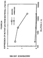
- a second standard curve ( Figure 2) was constructed using recombinant human p53 polypeptide (Hup53HIS273) expressed using another bacterial expression system.
- E. coli strain BL21 (DE3)LysS was transfected with mutant human p53 cDNA cloned into the T7 expression plasmid pT7-7.
- the transfected bacteria were incubated for 3 hours in the presence of 0.4 mM IPTG. Washed bacteria were lysed and purified as described above, diluted with sample diluent and used to construct a standard curve for mutant p53 in the ELISA assay ( Figure 3 and Table 1). Inter- and Intra-assay precision determined at 3 points along the standard curve shown above.
- Rabbit polyclonal antibodies directed against mutant p53 polypeptide Female New Zealand White rabbits are immunized with purified recombinant mutant p53 polypeptide following a standard protocol. Specifically, the mutant p53 polypeptide (0.1 mg) is emulsified with RIBITM adjuvant and administered perilymph nodally. Three weeks later, the rabbits are given an intra-muscular boost (0.05 mg antigen/animal). Boosts are continued at 21 day intervals. Serum is collected and the antibody titer determined by ELISA 10 days following each boost. Once a high titer is achieved, serum is collected and the antibody purified as described below.
- Antisera collected as described above, is titered by ELISA. Purified recombinant mutant p53 polypeptide is absorbed onto microtiter wells (1 ⁇ g/ml in 0.1 M sodium carbonate buffer, pH 9.0; 50 ⁇ l/well) for 16 to 20 h at 4°C. The antigen solution is discarded and unoccupied protein binding sites blocked using 200 ⁇ l of PBS containing 1% BSA (pH 7.3, for 2h at room temperature). Duplicate wells are incubated for 3 hours at 37°C with antisera diluted in buffer (serial 5-fold dilutions starting at 1:500 and ending at 1:312,500; plus a no antiserum control).
- Bound antibody is detected by incubation with peroxidase-conjugated goat anti-rabbit antibody (2-3 h at 37°C), followed by addition of a peroxidase substrate. Absorbance is measured using a dual beam, 96-well microtiter plate reader (Bio-Tek® Instruments, Model EL320). Antiserum titer is defined as the dilution yielding an absorbance which is twice background absorbance.
- Antibodies from rabbits yielding a high serum titer is purified as described below, and further evaluated for suitability as a reporter reagent in a two-site sandwich ELISA assay for mutant p53 polypeptide.
- Antibody purification Antibody (Ig) reactive with human, mutant p53 polypeptide is purified from hyperimmune rabbit sera by immunoaffinity chromatography. Purified, recombinant mutant p53 polypeptide is coupled to CNBr-activated Sepharose 4B® (Pharmacia). Crude antisera is clarified by centrifugation at 10,000 x g for 30 min., and crude antibody obtained by precipitation with ammonium sulfate. The Ig-enriched preparation is applied to the mutant p53 polypeptide-Sepharose®, and the bound antibody eluted using 100mM glycine-HCl, pH 2.5 and neutralized with 2.0 Tris-HCl, pH 8.0. In a second example, IgG reactive with human mutant p53 polypeptide is purified from hyperimmune rabbit sera by affinity chromatography using Protein A-Sepharose® according to a protocol identical to that described above.
- Polyclonal antibody recognizes multiple epitopes common to both mutant and wild-type p53 polypeptides.
- polyclonal antibody directed against a p53 polypeptide is paired with a monoclonal capture antibody which binds both wild-type and mutant p53 polypeptide (i.e. clone PAb421).
- polyclonal antibody directed against a p53 polypeptide is paired with a monoclonal capture antibody which specifically recognizes and binds to mutant p53 polypeptide (i.e. clone PAb240).
- the polyclonal antibody could be made specific for mutant human p53 polypeptides by adsorption against purified wild-type human p53 polypeptide which has been bound to a solid support, such CNBr-Sepharose® or polystyrene tubes.
- Native, wild-type p53 polypeptide may be used for the adsorption of rabbit polyclonal antibodies to yield a mutant p53 polypeptide-specific reagent and as a negative control for the assay.
- p53- ⁇ -galactosidase fusion proteins are purified by SDS-polyacrylamide gel electrophoresis. The band corresponding to the fusion protein is excised from the gel and washed in a solution of 1% SDS, 0.2M Tris/acetate pH 8.0, 0.1% DTT. The protein is then electroeluted, dialyzed against 20mM ammonium bicarbonate, 0.02% SDS and injected into BALB/c mice. Mice are immunized intraperitoneally with 5 ⁇ g on days 0, 7, and 14.
- mice are boosted with 5 ⁇ g of the same antigen and spleen cells are fused to NS1 myeloma fusion partner using established procedures (37).
- Hybridomas are screened by ELISA against mutant p53 polypeptide-B-galactosidase fusion protein. Additionally, clones reacting with mutant p53 polypeptide fusion protein but not with pure ⁇ -galactosidase are subcloned twice by limiting dilution. Hybridomas are grown as ascites-tumors in mice and antibody is purified as previously described (39).
- the solution is gently agitated on a Labquake rotator (Labindustries #400-10) for four hours at 4°C followed by the addition of ethanolamine (Eastman Kodak) at a final concentration of 0.1 M to block unreacted ester groups.
- the gel matrix is washed extensively with PBS by centrifugation at 2,500 RPM for 10 minutes until the gel is free of reactants as judged by obtaining zero absorbance at 280 nm (OD 280 ).
- the capture gel matrix is incubated with sera containing mutant p53 polypeptide and bound protein eluted with sample buffer for analysis by SDS-PAGE.
- Monoclonal or polyclonal antibodies are biotinylated according to a protocol distributed by LKB Laboratories. Two hundred microliters of Act BIOTIN solution prepared by adding 2.0 mg of Act BIOTIN to 0.5 ml of anhydrous dimethylformamide (Pierce), is added to 10 mg of antibody dissolved in 10 ml of 0.2 M sodium bicarbonate pH 8.8 containing 0.15 M NaCl. The reaction is allowed to proceed for 15 minutes at room temperature followed by termination of the reaction with 0.1 ml of 1.0 M ammonium chloride pH 6.0.
- the biotinylated antibody is dialyzed against PBS to remove other salts and 1.0 ml aliquots stored at -20°C. To ensure that biological activity of the antibody is maintained following biotinylation, the antibodies are tested by immunoprecipitation of 35 S methionine labeled cellular extracts containing mutant p53 polypeptides.
- the monoclonal antibody (0.5 mg) which recognizes mutant p53 polypeptide is reacted with decreasing volumes of sample and immunoprecipitated with 0.05 ml of a streptavidin agarose suspension at 0.24 mg/ml.
- Iodination of Purified Antibody For Use in the Antigen-Capture Procedure Aliquots of 20 ⁇ l of Iodo-Gen® (Pierce), which had previously been dissolved in chloroform at a concentration of 10 mg/ml, lyophilized, and kept frozen, are warmed to room temperature. The following are added, in order, to the tube containing Iodo-Gen®; 0.025 ml of Buffer II (0.4 Tris-HCl, 0.4 mM EDTA, pH 7.4), monoclonal antibody at a concentration of 1.0 mg/.1 ml, and 3,7 x 10 7 Bq (1.0 mCi) of Na 125 I (Amersham # IMS.40).
- Buffer II 0.4 Tris-HCl, 0.4 mM EDTA, pH 7.4
- the iodination reaction is allowed to proceed for one minute with gentle shaking and the mixture is subjected to gel filtration chromatography using a G-25 SephadexTM PD10 column (Pharmacia) equilibrated with 10 mM Tris-HCl, 10 mM NaCl, pH 7.8, to remove unreacted free 125 I.
- One-half ml fractions are collected and 10% trichloroacetic acid (TCA) precipitable counts determined before the samples are pooled.
- TCA trichloroacetic acid
- Samples are diluted in 20 microliters of sample buffer containing 6.0 M urea (Ultrapure, BRL), 0.1 M Tris-HCl (Sigma; T-1503), pH 6.8, 15% glycerol (Kodak; 114-9939) 2% sodium dodecyl sulfate (Bio-Rad®); #161-07-10), and electrophoresed on a 5 to 20% acrylamide gradient essentially as described by Laemmli (35).
- sample buffer containing 6.0 M urea (Ultrapure, BRL), 0.1 M Tris-HCl (Sigma; T-1503), pH 6.8, 15% glycerol (Kodak; 114-9939) 2% sodium dodecyl sulfate (Bio-Rad®); #161-07-10
- the samples are boiled for 2 minutes prior to application to 1-1.5 mm wide slab gel in a Bio-Rad® Model 155 Vertical Electrophoresis Cell (Bio-Rad® 165-1420) under constant voltage at 45 volts per gel for 15 hours (Hoeffer power supply; PS 1200 DC).
- Molecular weight standards used are myosin, 200,000; beta-galactosidase, 130,000; phosphorylase B, 92,000; bovine serum albumin, 68,000; ovalbumin, 45,000; carbonic anhydrase, 29,000; soybean trypsin inhibitor, 21,000; lysozyme, 14,400; and cytochrome C, 12,000.
- Gels are stained with 0.1% Coomassie Blue R-250® (Bio-Rad® #16-0400) in 7.0% acetic acid and 10% methanol for five minutes and destained in the same solution without Coomassie® ( Figure 4). Certain gels are stained by a silver technique as described by Merril (36) (Bio-Rad silver staining kit; #161-0443). Gels containing radioactively labeled samples are subjected to autoradiography as described by Bonner and Laskey (37) using Enhance® (New England Nuclear) and type XR-2 X-ray film (Kodak). Molecular weight standards used with gels containing labeled samples are pre-labeled with 14 C (New England Nuclear).
- Quantitation of mutant p53 polypeptide in a biological fluid sample Quantitation is achieved by the construction of a standard curve using known concentrations of purified, recombinant human mutant p53. By comparing the absorbance obtained from a sample containing an unknown amount of mutant p53 with that obtained from the standards, the concentration of mutant p53 in the sample can be determined ( Figures 1 and 2).
- Two site sandwich ELISA assay Two site sandwich ELISA assay .
- purified monoclonal antibody from clone PAb1801 (5 ⁇ g/ml in 0.1 M sodium carbonate buffer, pH 9.0, 100 ⁇ l/well), which binds to both wild-type and mutant p53 polypeptide, is adsorbed onto microtiter wells for 16 to 20 hours at 4°C.
- the antibody solution is removed from the wells and any remaining protein-binding sites are blocked by incubating the wells with PBS containing 1% BSA at pH 7.2 for 2 hours at room temperature (approximately 23°C).
- the wells are washed 4-times with 200 ⁇ l of Wash Buffer (PBS containing 0.1% BSA and 0.05% Tween-20®, pH 7.3) then appropriately marked wells are incubated with varying amounts of p53 polypeptide-B-galactosidase fusion protein (standard), or the biological sample to be assayed, for 16-18 hours at 4°C (alternatively for 3 hours at 37°C).
- Wash Buffer PBS containing 0.1% BSA and 0.05% Tween-20®, pH 7.3
- standard p53 polypeptide-B-galactosidase fusion protein
- the plates are emptied by inverting over paper towels and the well washed 4-times as described above.
- Biotin-conjugated monoclonal antibody from clone PAb421 (reporter antibody) is dissolved in Sample Diluent (PBS containing 50 mM NaCl, 1% normal mouse serum, 0.1% BSA, 0.5% Tween-20® and 0.02% Thimerosal) at a concentration of 10 ⁇ g/ml and incubated (100 ⁇ l/well) for 2-3 hours at 37°C.
- the reporter antibody is bound, in turn, by incubation (1 hour at room temperature) with streptavidin conjugated to horseradish peroxidase (0.4 ⁇ g/ml in Sample Diluent).
- Wells are washed 4-times, as described above, and a peroxidase substrate solution (e.g. ABTS, 100 ⁇ l/well) is added. After a suitable period of incubation (approximately 60-100 minutes at room temperature) the absorbance is measured at 405 nm.
- Sera from 6 normal- and 6 cancer-patient donors was analyzed for mutant p53 polypeptide using the protocol described above.
- the average absorbance (A 405 ) from duplicate wells for each sample was entered into the regression equation of the standard curve in order to calculate the amount of mutant p53 polypeptide in each of the samples.
- the maximum sensitivity of the assay is 100 pg/ml. Therefore, samples having no detectable mutant p53 polypeptide are indicated as having less than ( ⁇ ) 100 pg/ml.
- Table 2 provides a list of the p53 polypeptide concentrations in sera from 6 normal and 6 cancer patients: Patient Diagnosis p53 polypeptide Concentration (pg/ml) 1 Normal ⁇ 100 2 Normal 200 3 Normal 200 4 Normal ⁇ 100 5 Normal ⁇ 100 6 Normal 1,450 7 Breast Cancer 300 8 Colon Cancer 100 9 Stomach Cancer 1,300 10 Colon Cancer 2,900 11 Colon Cancer 100 12 Breast Cancer 200
- mutant and wild-type p53 polypeptides do not distinguish between mutant and wild-type p53 polypeptides and therefore yield a measure of the total (wild-type and mutant) p53 polypeptide concentration in the sample.
- wild-type p53 polypeptide does not accumulate to detectable levels; therefore, by inference, the polypeptides detected and measured in these samples are mutant p53 polypeptides.
- the mutant p53 polypeptide content of the sample may be influenced by the medical condition of the sample donor, i.e. the patient's tumor burden. This burden is affected by the state of progression of the underlying disease as well as any ameliorative therapy such as surgery, chemotherapy or radiation which may have been administered prior to serum sample collection.
- PAb240 is adsorbed onto microtiter wells, since this antibody is specific for the mutant p53 polypeptide.
- the antibody (5 ⁇ g/ml in 0.1 M sodium carbonate buffer, pH 9.0; 100 ⁇ l/well) is allowed to adsorb for 16 to 20 h at 4°C.
- the antibody solution is then removed from the wells and any unoccupied protein binding sites are blocked by incubating the wells with PBS containing 1% BSA at pH 7.2 for two hours at room temperature.
- the wells are washed 4-times with 200 ⁇ l of Wash Buffer (PBS containing 0.1% BSA and 0.05% Tween-20®, pH 7.3) then appropriately marked wells are incubated with varying amounts of purified recombinant mutant p53 polypeptide (standard), or the biological sample to be assayed, for 16-18 h at 4°C or alternatively for 3 h at 37°C.
- Wash Buffer PBS containing 0.1% BSA and 0.05% Tween-20®, pH 7.3
- standard purified recombinant mutant p53 polypeptide
- the plates are emptied by inverting over paper towels, and the wells are washed 4-times as described above.
- Polyclonal antibody from rabbits which recognizes and binds to mutant p53 polypeptide, is dissolved in Sample Diluent (PBS containing 50 mM NaCl, 1% normal mouse serum, 0.1% BSA, 0.5% Tween-20® and 0.02% Thimerosal) at a concentration of 5 ⁇ g/ml and incubated (100 ⁇ l/well) for 2-3 h at 37°C. Bound reporter antibody is, in turn, detected by incubation (1-2 hours at room temperature) with peroxidase conjugated goat anti-rabbit immunoglobulin.
- Sample Diluent PBS containing 50 mM NaCl, 1% normal mouse serum, 0.1% BSA, 0.5% Tween-20® and 0.02% Thimerosal
- a peroxidase substrate such as ABTS (2,2'-azino-di-[3-ethyl-benzthiazoline sulfonate]) is added, and after a suitable period of incubation (approximately 30-60 minutes at room temperature) the absorbance is read at 405 nm.
- Spike and recovery of p53 in normal serum indicated that recovery was better when the serum was diluted (Figure 5).
- purified recombinant mutant p53 was added to undiluted normal human serum and to diluted (1:5 with sample diluent) normal human serum to a final concentration of 4 ng/ml.
- Purified p53 was added to diluted human urine to a final concentration of 2 ng/ml.
- the p53-spiked samples were analyzed in the p53 ELISA assay. The amount of mutant p53 detected is expressed as a percent of the amount actually added to the sample.
- Serum samples were diluted 1:5 with sample diluent and 100 ⁇ l of diluted serum was added to duplicate wells for each of the 21 normal and 67 cancer patient samples.
- the average absorbance (A 405 ) from the duplicate wells for each sample was entered into the regression equation of the standard curve in order to calculate the amount of mutant p53 polypeptide in each of the diluted samples. This amount was then multiplied by the dilution factor (i.e., 5) in order to obtain the amount of mutant p53 polypeptide present in the original, undiluted sera.
- the amount of mutant p53 polypeptide in the samples is expressed in picograms (pg) per milliliter (ml) of undiluted serum. The maximum sensitivity of the assay is 30 pg/ml.
- samples having less than 30 pg/ml of mutant p53 polypeptide cannot be detected in this assay and cannot be resolved from each other or indeed from samples having no mutant p53 polypeptide. Since the samples were diluted 1:5, this sensitivity limit becomes 150 pg/ml (30 X 5). Therefore, in reporting the results of this study, sera samples having no detectable mutant p53 polypeptides are indicated as having less than ( ⁇ ) 150 pg/ml.
- Table 3 provides a list of the measured mutant p53 polypeptide concentration from the sera of 21 normal human donors. Mutant p53 Polypeptide Concentrations in Normal Human Sera Sample mutant p53 polypeptide Concentration (pg/ml) 1 ⁇ 150 2 ⁇ 150 3 ⁇ 150 4 ⁇ 150 5 ⁇ 150 6 ⁇ 150 7 ⁇ 150 8 ⁇ 150 9 ⁇ 150 10 645 11 ⁇ 150 12 ⁇ 150 13 ⁇ 150 14 ⁇ 150 15 ⁇ 150 16 ⁇ 15O 17 515 18 ⁇ 150 19 390 20 ⁇ 150 21 ⁇ 150
- Table 4 provides a list of the measured mutant p53 polypeptide concentration from the sera of 67 cancer patients. The cancers were of diverse types, as indicated in Table 4. Mutant p53 Concentrations in Cancer Patient Sera Sample Cancer (pg/ml) mutant p53 polypeptide Concentration 14-22-11 Breast ⁇ 150 89-79-93 Colon 417 91-56-88 Colon 444 5740 Breast 278 5745 Hodgkins ⁇ 150 5761 Thyroid 194 5755 Prostate ⁇ 150 5753 Breast ⁇ 150 5754 Melanoma 167 86 Colon 167 92 Colon 306 85-21-76 Colon 194 16-48-19 Colon 194 92-23-92 Colon ⁇ 150 127 Lung 361 5387 lung 194 5407 Lung 2,472 5395 Lung ⁇ 150 5405 Colon ⁇ 150 5437 Lung ⁇ 150 5476 Breast ⁇ 150 81 Lung 361 18-13-34 Breast 333 54-99-38 Breast ⁇ 150 5301-18 #30
- Each of these two sera were diluted 1:2 with assay buffer, then further divided into two equal portions labeled “experimental” and "control.”
- To each of the experimental tubes was delivered 7.5 ⁇ g of purified monoclonal antibody p53(Ab-2) clone PAb1801 (ATCC Accession No. HB 10642). This is a very well characterized antibody which specifically binds to p53 polypeptide, but does not itself constitute any component of the two-site sandwich ELISA.
- Each of the control tubes received 7.5 ⁇ g of purified monoclonal antibody trpE(Ab-1).
- This antibody serves as a control since it is of the same isotype (IgG 1 ) as the PAb1801 antibody, but binds specifically to bacterial anthranilate synthetase, a protein which is not present in human serum.
- IgG 1 isotype
- PAb1801 antibody binds specifically to bacterial anthranilate synthetase, a protein which is not present in human serum.
- any IgG1 antibody which is reactive against an irrelevant antigen and which is also not cross-reactive with p53 could serve as a suitable control.
- two other antibodies which would work equally well as controls are ATTC CRL 1640, an anti-DNA polymerase alpha IgG1 clone or ATTC CRL 1713, an anti-E. coli DNA polymerase IgG1 clone.
- clone PAb240 as the capture antibody
- the assay format would be identical to the one described above.
- the polyclonal antibody can be used as the capture antibody with PAb240 as reporter. In this configuration, peroxidase conjugated goat anti-mouse immunoglobulin is substituted for the peroxidase conjugated goat anti-rabbit immunoglobulin in the assay format described above.
- Sera samples or cell extracts to be analyzed are first subjected to SDS-PAGE as described hereinabove on 5-20% acrylamide gradient gels. After electrophoresis, transfer of proteins to nitrocellulose (Schleicher & Schuell, BA 85, 0.45 um) is performed. Proteins are transferred at 70 volts for 2-3 hours at 4°C. The nitrocellulose is incubated in 0.5% non-fat powdered milk (Carnation®) in Phosphate buffered saline pH 7.4 (PBS) containing 0.02% sodium azide for 1 hour at room temperature.
- Carnation® non-fat powdered milk
- PBS Phosphate buffered saline pH 7.4
- the filter is then washed with PBS containing 0.1% Tween 20® (Bio-Rad® Lab) and incubated with 125 [I] labeled PAb240 monoclonal antibody (2.0 x 10 6 CPM/ml) diluted in the same buffer for 1.5 hours at 37°C.
- the filters are washed three times with PBS-Tween-20®, dried and exposed to Kodak XR-2 film at -70°C using intensifying screens.
- the filter is incubated with PAb240 monoclonal antibody then washed with PBS and incubated with alkaline phosphate-conjugated goat anti-mouse IgG.
- the filter is washed with PBS and incubated with a suitable alkaline phosphatase substrate (e.g. 5-bromo-4-chloro-3-indolyl-phosphate) until colored bands develop. Further alternatively, the filter is incubated with polyclonal rabbit anti-p53 antibody, washed with PBS and incubated with alkaline phosphate-conjugated goat anti-rabbit IgG. The filter is washed and developed as described above.
- a suitable alkaline phosphatase substrate e.g. 5-bromo-4-chloro-3-indolyl-phosphate
- Blotted membranes are blocked for 1 h in a solution containing PBS and 1% to 10% BSA or casein in order to block nonspecific protein binding sites.
- the blocked membranes are incubated with radiolabeled antibodies directed against mutant p53 polypeptides (either monoclonal or polyclonal) then subjected to autoradiography.
- the antibodies directed against mutant p53 polypeptides may be enzymatically labeled and detection effected through the use of an appropriate substrate solution.
- a peroxidase-labeled antibody could be incubated with 0.03% 4-chloro-1 Naphthol.
- PAb240 monoclonal antibody is biotinylated according to a protocol distributed by LKB Laboratories. Two hundred microliters of Act BIOTIN solution prepared by adding 2.0 mg of Act BIOTIN to 0.5 ml of anhydrous dimethylformamide (Pierce), is added to 10 mg of PAb240 dissolved in 10 ml of 0.2 M sodium bicarbonate pH 8.8 containing 0.15 M NaCl. The reaction is allowed to proceed for 15 minutes at room temperature followed by termination of the reaction with 0.1 ml of 1.0 M ammonium chloride pH 6.0. The biotinylated antibody is dialyzed against PBS to remove other salts and 1.0 ml aliquots stored at -20°C.
- Act BIOTIN solution prepared by adding 2.0 mg of Act BIOTIN to 0.5 ml of anhydrous dimethylformamide (Pierce), is added to 10 mg of PAb240 dissolved in 10 ml of 0.2 M sodium bicarbonate pH 8.8 containing 0.15 M NaCl. The reaction is allowed to proceed for
- the antibodies are tested by immunoprecipitation of purified, recombinant mutant p53 polypeptides.
- the PAb240 MoAb (0.5 g) is reacted with decreasing volumes of patient sera containing the mutant p53 polypeptide and immunoprecipitated with 0.05 ml of a strepavidinagarose suspension at 0.24 mg/ml resulting in a PAb240-mutant p53 polypeptide-strepavidinagarose complex which is detectable.
- the matrix is pelleted and washed once in 4.0 mls of PBS containing .05% Tween 20®.
- One ml of PBS and biotinylated PAb240 are X added to each tube, incubated at 37°C for 1 hour, pelleted, then washed once in PBS-0.5% Tween 20®.
- An avidin-biotin-horseradish peroxidase complex is prepared by mixing together two drops of solution A (Vectastain, Avidin Biotin complex (ABC) kit, #PK4004, Vector Laboratories) and 2 drops of solution B in 10.0 ml of PBS containing 0.05% Tween 20®.
- Developing solution is made as follows: Solution I is prepared by adding 0.06 ml of 30% hydrogen peroxide to 100 ml of PBS.
- Solution II is prepared by adding 60 mg of horseradish peroxidase developer (BioRad) to 20.0 ml of ice-cold methanol. Immediately before use, 5 parts of solution I is mixed with 1 part of solution II and 1.0 ml added to the matrix. The matrix develops a blue color in samples containing the mutant p53 polypeptide.
- Solid Phase Antigen Capture Procedure Using Iodinated Antibodies as Reporter Antibodies .
- One ml of PAb240 capture affinity matrix is washed two times with 4.0 ml of PBS TDS and one time with 4.0 ml of PBS containing 0.1% BSA (PBS-BSA).
- Capture matrices can be used with either cell extracts or human biological fluids and are incubated in round bottom (12 x 75mm, Enkay) or conical polypropylene tubes (12 x 75 mm, Enkay).
- the gel matrix pellet is suspended in two times its volume of PBS containing 0.1% sodium azide, and 0.01 ml is transferred to a round bottom tube, (12 x 75 mm, Enkay) containing a bacterial growth and protease inhibitor solution (BGIPI, 0.05 ml) of final concentrations of 0.005% TPCK (Sigma), 0.01% soybean trypsin inhibitor (Sigma), 0.01% sodium azide, and 0.01% sodium fluoride and 2.75 ⁇ l (0.054 TIU) of a aprotinin (Sigma) (19.8 TIU/ml).
- BGIPI bacterial growth and protease inhibitor solution
- the gel matrix is washed 2 times with 3.0 ml PBS, and approximately 0.1 ml volume of liquid is retained with the gel matrix pellet.
- the pellet is rocked for 2 hours at 37°C with 0.1 ml of 125 I-labeled antibody directed against mutant p53 polypeptides [monoclonal or polyclonal - approximately 60,000 CPM, 1.0 ⁇ Ci 125 I Iodine per 1.0 microgram protein], washed one time with 3.0 ml of PBS TDS, two times with 3.0 ml PBS, and finally counted in a gamma counter.
- Double diffusion in agar Molten agar is poured onto glass slides or into petri dishes and allowed to harden. Small wells are punched out of the agar a few millimeters apart. 50 ⁇ l of the affinity purified rabbit polyclonal antibody directed against mutant p53 polypeptides is added to one of the punched out wells and 100 ⁇ l patient sera is added to another punched out well located opposite to the antibody-containing well. These samples are allowed to diffuse toward one another in a moist chamber for 18-24 hours. The resultant precipitation lines represent mutant p53 polypeptide complexes which are analyzed visually in indirect light with the aid of a magnifying lens. The antibody and mutant p53 polypeptide diffuse in a radial fashion, creating an arc which approximates a straight line at the leading edges of the diffusing antibody-mutant p53 polypeptide complex.
- Solution Phase Antigen Capture Test A solution phase mutant p53 polypeptide capture test which represents an alternative embodiment of the invention has been formatted. This test requires 0.1 ml of patient sera, and the other test components consist of strepavidinagarose (Bethesda Research Laboratories, Rockville, MD, Catalog No. 5942SA, Lab No. 52101), biotinylated capture antibody PAb240 and 125 Iodine-labeled PAb1801 as reporter antibody.
- strepavidinagarose Bethesda Research Laboratories, Rockville, MD, Catalog No. 5942SA, Lab No. 52101
- biotinylated capture antibody PAb240 and 125 Iodine-labeled PAb1801 as reporter antibody.
- Radioimmunoassay Iodination of recombinant mutant p53 polypeptide is performed as described.
- the radioimmunoassay utilizes 125 I-mutant p53 polypeptide, unlabeled mutant p53 polypeptide, PAb240, goat anti-mouse IgG and Protein A-agarose and is performed as follows.
- Fifty microliters of goat anti-mouse IgG is added (1:1000 in PBS) and incubated at room temperature for 1 hour after which Protein A-Agarose (50 ⁇ l of 10% (v/v)) suspension is added and incubated for 30 min at room temperature.
- the tubes are centrituged, washed three times in PBS-1% BSA-0.5% Triton X-100® and counted for radioactivity.
- the amount of mutant p53 polypeptide in the sample is quantitated from the standard curve constructed with purified unlabeled mutant p53 polypeptide.
- the p53 ELISA assay utilizes monoclonal antibody, clone PAb240, to selectively bind those mutant p53 proteins which express the PAb240 epitope.
- High titer rabbit polyclonal antibodies raised against recombinant mutant p53 (HIS175) are used in the reporter cascade and immunoaffinity purified human recombinant mutant p53 (HIS273) is used as standard in the assay.
- Sensitivity of 50 pg mutant p53 per/ml of sample buffer is routinely achieved, while better than 20 pg/ml sensitivity can be attained under optimal conditions.
- Intra- and inter-assay CVs are better than 5% at the midrange of the standard curve ( Figure 2).
- No cross-reactivity was found to insulin, transferrin, and a variety of growth factors and interleukins measured at concentrations greater than 2,000 times the sensitivity limits of the assay (Table 5).
- Nonspecific cross-reactivity was also demonstrated using extracts from mutant p53-negative cell lines as test samples. Little or no mutant p53 was found in whole cell extracts of K562 and KNRK cells (K-ras transformed NRK cells) whereas a high level of expression was found in the human epidermoid carcinoma line A431 (7.1 ng/mg) ( Figure 8). HL60 cells also showed significant levels of mutant p53 (3.1 ng/mg).
- mutant p53 in this cell line may differ depending on the particular variant being examined. Low to moderate expression of mutant p53 was detected in virally transformed mouse (k-balb), Mink (64 Jiki) and dog (DoCl) cells.
- the p53 Assay may be used to measure mutant p53 in human samples from cancer patients.
Landscapes
- Health & Medical Sciences (AREA)
- Chemical & Material Sciences (AREA)
- Life Sciences & Earth Sciences (AREA)
- Immunology (AREA)
- Organic Chemistry (AREA)
- Molecular Biology (AREA)
- General Health & Medical Sciences (AREA)
- Engineering & Computer Science (AREA)
- Biochemistry (AREA)
- Medicinal Chemistry (AREA)
- Oncology (AREA)
- Genetics & Genomics (AREA)
- Urology & Nephrology (AREA)
- Proteomics, Peptides & Aminoacids (AREA)
- Hematology (AREA)
- Biophysics (AREA)
- Biomedical Technology (AREA)
- Biotechnology (AREA)
- Food Science & Technology (AREA)
- Hospice & Palliative Care (AREA)
- Cell Biology (AREA)
- Zoology (AREA)
- Microbiology (AREA)
- Gastroenterology & Hepatology (AREA)
- Toxicology (AREA)
- Physics & Mathematics (AREA)
- Analytical Chemistry (AREA)
- General Physics & Mathematics (AREA)
- Pathology (AREA)
- Peptides Or Proteins (AREA)
- Preparation Of Compounds By Using Micro-Organisms (AREA)
- Investigating Or Analysing Materials By The Use Of Chemical Reactions (AREA)
- Investigating Or Analysing Biological Materials (AREA)
Claims (25)
- Procédé destiné à déterminer chez un sujet si le sujet présente probablement un état néoplasique, qui consiste à déterminer quantitativement, dans un échantillon d'un fluide biologique essentiellement exempt de cellules du sujet, la concentration en polypeptide p53 mutant présent dans l'échantillon, une concentration en polypeptide p53 mutant dans l'échantillon supérieure à 150 pg/ml étant indicative de la probabilité que le sujet présente un tel état néoplasique.
- Procédé selon la revendication 1, dans lequel la détermination de manière quantitative comprend les étapes consistant :(i) à mettre en contact l'échantillon avec un anticorps capable de former un complexe avec le polypeptide p53 mutant dans des conditions permettant la formation du complexe ; et(ii) à déterminer la quantité de tout complexe ainsi formé.
- Procédé selon la revendication 2, dans lequel ledit anticorps est capable de former un complexe avec p53 mutant mais pas avec p53 de type sauvage.
- Procédé selon la revendication 2 ou 3, dans lequel l'anticorps est un anticorps polyclonal.
- Procédé selon la revendication 2 ou 3, dans lequel l'anticorps est un anticorps monoclonal.
- Procédé selon l'une quelconque des revendications 2 à 5, dans lequel l'anticorps est fixé à un support solide.
- Procédé selon l'une quelconque des revendications 2 à 6, dans lequel la détermination quantitative si un complexe quelconque est formé dans l'étape (ii) comprend les étapes consistant :(a) à mettre en contact l'échantillon provenant de l'étape (i) avec un second anticorps capable de former un second complexe avec tout complexe formé dans l'étape (i) dans des conditions permettant la formation du second complexe ; et(b) à déterminer la quantité d'un tel second complexe quelconque ainsi formé.
- Procédé selon la revendication 7, dans lequel la quantité de tout dit second complexe formé est comparée à la quantité dans un échantillon provenant d'un sujet normal, la présence d'une quantité significativement différente indiquant que le sujet présente l'état néoplasique.
- Procédé selon la revendication 7 ou 8, dans lequel le second anticorps est un anticorps polyclonal.
- Procédé selon la revendication 7 ou 8, dans lequel le second anticorps est un anticorps monoclonal.
- Procédé selon l'une quelconque des revendications 7 à 10, dans lequel le second anticorps est fixé à un support solide.
- Procédé destiné à déterminer chez un sujet si le sujet présente probablement un état néoplasique en déterminant quantitativement la concentration en polypeptide p53 mutant dans un échantillon de fluide biologique essentiellement exempt de cellules qui comprend les étapes consistant :(a) à mettre en contact un support solide avec un excès d'un premier anticorps capable de former un complexe avec le polypeptide p53 mutant dans des conditions permettant à l'anticorps de se fixer à la surface dudit support solide ;(b) à mettre en contact le support solide résultant auquel le premier anticorps est lié avec un échantillon de fluide biologique essentiellement exempt de cellules dans des conditions telles que tout p53 mutant dans le fluide biologique se lie à l'anticorps et forme un complexe avec celui-ci ;(c) à mettre en contact le complexe formé dans l'étape (b) avec une quantité prédéterminée d'un second anticorps dirigé contre un épitope du p53 mutant différent de l'épitope contre lequel est dirigé le premier anticorps de l'étape (a), de façon à former un complexe qui inclut le p53 mutant, le premier anticorps, et le second anticorps ;(d) à déterminer quantitativement la quantité du complexe formé dans l'étape (c) ; et(e) à déterminer ainsi la concentration du p53 mutant dans l'échantillon de fluide biologique, où la présence du polypeptide p53 mutant dans ledit échantillon à une concentration supérieure à 150 pg/ml indique que le sujet présente probablement ledit état néoplasique.
- Procédé selon la revendication 12, dans lequel le premier ou second anticorps est capable de former un complexe avec p53 mutant mais pas avec p53 de type sauvage.
- Procédé selon la revendication 12 ou 13, dans lequel le premier anticorps lié au support solide est un anticorps monoclonal et le second anticorps est un anticorps polyclonal.
- Procédé selon l'une quelconque des revendications 12 à 14, dans lequel le premier anticorps lié au support solide est un anticorps polyclonal et le second anticorps est un anticorps monoclonal.
- Procédé selon l'une quelconque des revendications 12 à 14, dans lequel le premier anticorps lié au support solide est un anticorps monoclonal et le second anticorps est un anticorps monoclonal.
- Procédé selon la revendication 12 ou 13, dans lequel le premier anticorps lié au support solide est un anticorps polyclonal et le second anticorps est un anticorps polyclonal.
- Procédé selon l'une quelconque des revendications 12 à 17, dans lequel le premier anticorps est marqué avec un marqueur détectable.
- Procédé selon l'une quelconque des revendications 7 à 17, dans lequel le second anticorps est marqué avec un marqueur détectable.
- Procédé selon la revendication 18 ou 19, dans lequel le marqueur détectable est une enzyme, la biotine, un fluorophore, un chromophore, un métal lourd, un isotope paramagnétique, ou un radio-isotope.
- Procédé selon l'une quelconque des revendications 1 à 20, dans lequel le fluide biologique est l'urine.
- Procédé selon l'une quelconque des revendications 1 à 20, dans lequel le fluide biologique est: le sérum.
- Procédé selon l'une quelconque des revendications 1 à 20, dans lequel le fluide biologique est un extrait cellulaire.
- Procédé destiné à surveiller l'évolution d'un état néoplasique chez un sujet qui consiste à déterminer quantitativement dans un premier échantillon d'un fluide biologique essentiellement exempt de cellules provenant du sujet la concentration en polypeptide p53 mutant conformément au procédé selon l'une quelconque des revendications 1 à 23, et à comparer la concentration ainsi déterminée à la concentration dans un échantillon suivant du fluide biologique provenant du sujet, de tels échantillons étant prélevés à des moments différents, une différence des concentrations déterminées étant indicative de l'évolution de l'état néoplasique.
- Procédé selon l'une quelconque des revendications 8 à 10, dans lequel une quantité significativement différente est une quantité qui est égale ou supérieure à deux écarts types au-dessus de la concentration en p53 trouvée dans des échantillons provenant de sujets normaux.
Priority Applications (1)
| Application Number | Priority Date | Filing Date | Title |
|---|---|---|---|
| EP99126040A EP1006364A1 (fr) | 1991-02-01 | 1992-01-31 | Dossage immunologique pour détecter un polypeptide mutant P53 dans des fluides biologiques |
Applications Claiming Priority (5)
| Application Number | Priority Date | Filing Date | Title |
|---|---|---|---|
| US64956691A | 1991-02-01 | 1991-02-01 | |
| US649566 | 1991-02-01 | ||
| US71917291A | 1991-06-21 | 1991-06-21 | |
| US719172 | 1991-06-21 | ||
| PCT/US1992/000878 WO1992013970A1 (fr) | 1991-02-01 | 1992-01-31 | Dosage immunologique pour detecter un polypeptide mutant p53 dans des fluides biologiques |
Related Child Applications (1)
| Application Number | Title | Priority Date | Filing Date |
|---|---|---|---|
| EP99126040A Division EP1006364A1 (fr) | 1991-02-01 | 1992-01-31 | Dossage immunologique pour détecter un polypeptide mutant P53 dans des fluides biologiques |
Publications (3)
| Publication Number | Publication Date |
|---|---|
| EP0576476A1 EP0576476A1 (fr) | 1994-01-05 |
| EP0576476A4 EP0576476A4 (en) | 1994-06-29 |
| EP0576476B1 true EP0576476B1 (fr) | 2003-05-28 |
Family
ID=27095640
Family Applications (2)
| Application Number | Title | Priority Date | Filing Date |
|---|---|---|---|
| EP99126040A Withdrawn EP1006364A1 (fr) | 1991-02-01 | 1992-01-31 | Dossage immunologique pour détecter un polypeptide mutant P53 dans des fluides biologiques |
| EP92906415A Expired - Lifetime EP0576476B1 (fr) | 1991-02-01 | 1992-01-31 | Dosage immunologique pour detecter un polypeptide mutant p53 dans des fluides biologiques |
Family Applications Before (1)
| Application Number | Title | Priority Date | Filing Date |
|---|---|---|---|
| EP99126040A Withdrawn EP1006364A1 (fr) | 1991-02-01 | 1992-01-31 | Dossage immunologique pour détecter un polypeptide mutant P53 dans des fluides biologiques |
Country Status (5)
| Country | Link |
|---|---|
| EP (2) | EP1006364A1 (fr) |
| AT (1) | ATE241807T1 (fr) |
| AU (1) | AU1370592A (fr) |
| DE (1) | DE69233079T2 (fr) |
| WO (1) | WO1992013970A1 (fr) |
Families Citing this family (17)
| Publication number | Priority date | Publication date | Assignee | Title |
|---|---|---|---|---|
| WO1993021529A1 (fr) * | 1992-04-14 | 1993-10-28 | Duke University | Procede de detection de tumeurs contenant des complexes de p53 et de hsp70 |
| EP0614531A1 (fr) * | 1992-09-30 | 1994-09-14 | Deutsches Krebsforschungszentrum Stiftung des öffentlichen Rechts | Procede de depistage d'anticorps specifiques de la proteine p53 |
| US5688918A (en) * | 1993-08-02 | 1997-11-18 | Health Research, Inc. | p53as protein and antibody therefor |
| US5726024A (en) * | 1993-08-02 | 1998-03-10 | Health Research, Inc. | p53as protein and antibody therefor |
| US7238523B1 (en) | 1993-08-02 | 2007-07-03 | Health Research, Inc. | p53as protein and antibody therefor |
| DE69433274T2 (de) * | 1993-08-02 | 2004-08-05 | Health Research Inc. | Protein P53as sowie Plasmide und virale Vektoren, die eine zu der das Protein codierende DNA-Sequenz komplementäre cDNA-Sequence enthalten |
| US6965009B1 (en) | 1993-08-02 | 2005-11-15 | Health Research, Inc. | p53 as protein and antibody therefor |
| US5747650A (en) * | 1993-08-02 | 1998-05-05 | Health Research, Inc. | P53AS protein and antibody therefor |
| SE9403953D0 (sv) * | 1994-07-15 | 1994-11-16 | Pharmacia Biotech Ab | Sequence-based diagnosis |
| US5912142A (en) * | 1996-11-22 | 1999-06-15 | Duke University | Gene product over expressed in cancer cells |
| US6350615B1 (en) | 1996-11-22 | 2002-02-26 | Duke University | Gene product over expressed in cancer cells |
| US6072034A (en) * | 1997-11-22 | 2000-06-06 | Duke University | Gene product over expressed in cancer cells |
| IL121041A0 (en) * | 1997-06-09 | 1997-11-20 | Yeda Res & Dev | Immunogenic compositions for induction of anti-tumor immunity |
| WO1999002682A1 (fr) * | 1997-07-09 | 1999-01-21 | Affymetrix, Inc. | Mutants du p53 presents dans les tumeurs |
| CA2214461A1 (fr) | 1997-09-02 | 1999-03-02 | Mcgill University | Methode de depistage pour identifier les individus risquant de contracter des maladies associees a differentes formes polymorphiques du type sauvage p53 |
| JP4678882B2 (ja) * | 2004-10-14 | 2011-04-27 | ジェネンテック, インコーポレイテッド | Cop1分子およびその使用 |
| GB201617564D0 (en) * | 2016-10-17 | 2016-11-30 | Agency For Science Technology And Research And National University Of Singapore And Singapore Health | Anti-p53 antibodies |
Family Cites Families (3)
| Publication number | Priority date | Publication date | Assignee | Title |
|---|---|---|---|---|
| US4699877A (en) * | 1982-11-04 | 1987-10-13 | The Regents Of The University Of California | Methods and compositions for detecting human tumors |
| US4798787A (en) * | 1984-09-19 | 1989-01-17 | Cetus Corporation | Peptide antibodies and their use in detecting oncogene products |
| DE4444969C1 (de) * | 1994-12-16 | 1996-10-24 | Orga Med Dr Med Erwin Klopfer | Nachweis von Antikörpern gegen p53 in Körperflüssigkeiten |
-
1992
- 1992-01-31 EP EP99126040A patent/EP1006364A1/fr not_active Withdrawn
- 1992-01-31 AT AT92906415T patent/ATE241807T1/de not_active IP Right Cessation
- 1992-01-31 DE DE69233079T patent/DE69233079T2/de not_active Expired - Lifetime
- 1992-01-31 WO PCT/US1992/000878 patent/WO1992013970A1/fr active IP Right Grant
- 1992-01-31 EP EP92906415A patent/EP0576476B1/fr not_active Expired - Lifetime
- 1992-01-31 AU AU13705/92A patent/AU1370592A/en not_active Abandoned
Non-Patent Citations (2)
| Title |
|---|
| Finlay, C.A. et al (1988) Mol. Cell. Biol. 8:531-539 * |
| Gannon, J.V. et al (1990) EMBO Journal 9:1595-1602 * |
Also Published As
| Publication number | Publication date |
|---|---|
| DE69233079T2 (de) | 2004-04-01 |
| EP0576476A4 (en) | 1994-06-29 |
| AU1370592A (en) | 1992-09-07 |
| EP1006364A1 (fr) | 2000-06-07 |
| ATE241807T1 (de) | 2003-06-15 |
| DE69233079D1 (de) | 2003-07-03 |
| EP0576476A1 (fr) | 1994-01-05 |
| WO1992013970A1 (fr) | 1992-08-20 |
Similar Documents
| Publication | Publication Date | Title |
|---|---|---|
| EP0576476B1 (fr) | Dosage immunologique pour detecter un polypeptide mutant p53 dans des fluides biologiques | |
| US5411860A (en) | Amplification of human MDM2 gene in human tumors | |
| Schlichtholz et al. | The immune response to p53 in breast cancer patients is directed against immunodominant epitopes unrelated to the mutational hot spot | |
| Evan et al. | Isolation of monoclonal antibodies specific for human c-myc proto-oncogene product | |
| JP3399948B2 (ja) | P90抗体またはプローブを用いた前癌細胞または癌細胞の検出方法 | |
| US5604107A (en) | Detection of neu p185 in cell lysates | |
| US5726288A (en) | Localization and characterization of the Wilms' tumor gene | |
| EP0494135A1 (fr) | DETECTION ET QUANTIFICATION DES PROTEINES APPARENTEES -i(NEU) DANS LES LIQUIDES BIOLOGIQUES DES HUMAINS. | |
| EP0344134B1 (fr) | Anticorps monoclonal spécifique pour une séquence de fibronectine exprimée dans des cellules transformées, hybridome produisant cet anticorps et utilisation de cet anticorps monoclonal pour le diagnostic de tumeurs | |
| CA2827618C (fr) | Compositions et methodes d'utilisation pour la determination de he4a | |
| US6083709A (en) | Immunoassay for detection of mutant P53 polypeptide in serum | |
| JP3536731B2 (ja) | HIV−1p24抗原の免疫測定方法及び試薬 | |
| JP2003116549A (ja) | P90抗体またはプローブを用いた前癌細胞または癌細胞の検出方法 | |
| US5910418A (en) | Antibodies and assay for detecting mutant APC proteins | |
| CA1281641C (fr) | Methode immunologique pour la detection de proteines ras encodees | |
| EP0767381B1 (fr) | Détection, quantification et classification de protéines RAS dans des fluides et des tissus corporels | |
| HAMER et al. | Production and characterization of anti-RAS p21 monoclonal antibodies | |
| US5710255A (en) | Characterization of a novel anti-p110RB monoclonal antibody | |
| US5188967A (en) | Agents for the in vitro diagnosis of malignant cells originating in the digestive tract | |
| JP3888695B2 (ja) | ヒトlect2に対する抗体、それを産生する細胞、その測定法及び測定用キット | |
| JPH1066574A (ja) | 消化管起源の悪性細胞のin vitro検出に使用し得る抗絨毛タンパク質をコードするDNA | |
| US5635389A (en) | Antibodies which recognize and bind human villin | |
| JPH0614041B2 (ja) | 肺表面活性物質の検出方法及びそれに用いる試薬キツト | |
| CA2354375A1 (fr) | Compositions et procedes de traitement de tumeurs | |
| EP0259773A2 (fr) | Un anticorps spécifique d'un antigène de cancer |
Legal Events
| Date | Code | Title | Description |
|---|---|---|---|
| PUAI | Public reference made under article 153(3) epc to a published international application that has entered the european phase |
Free format text: ORIGINAL CODE: 0009012 |
|
| 17P | Request for examination filed |
Effective date: 19930831 |
|
| AK | Designated contracting states |
Kind code of ref document: A1 Designated state(s): AT BE CH DE DK ES FR GB GR IT LI LU MC NL SE |
|
| RHK1 | Main classification (correction) |
Ipc: G01N 33/53 |
|
| A4 | Supplementary search report drawn up and despatched |
Effective date: 19940510 |
|
| AK | Designated contracting states |
Kind code of ref document: A4 Designated state(s): AT BE CH DE DK ES FR GB GR IT LI LU MC NL SE |
|
| 17Q | First examination report despatched |
Effective date: 19961022 |
|
| RAP1 | Party data changed (applicant data changed or rights of an application transferred) |
Owner name: OSI PHARMACEUTICALS, INC. |
|
| GRAG | Despatch of communication of intention to grant |
Free format text: ORIGINAL CODE: EPIDOS AGRA |
|
| RIC1 | Information provided on ipc code assigned before grant |
Free format text: 6G 01N 33/53 A, 6C 07K 14/00 B, 6A 61K 39/00 B, 6B 65D 69/00 B, 6G 01N 33/574 B |
|
| GRAG | Despatch of communication of intention to grant |
Free format text: ORIGINAL CODE: EPIDOS AGRA |
|
| RIC1 | Information provided on ipc code assigned before grant |
Free format text: 7G 01N 33/53 A, 7C 07K 14/00 B, 7A 61K 39/00 B, 7B 65D 69/00 B, 7G 01N 33/574 B |
|
| GRAH | Despatch of communication of intention to grant a patent |
Free format text: ORIGINAL CODE: EPIDOS IGRA |
|
| RIC1 | Information provided on ipc code assigned before grant |
Free format text: 7G 01N 33/53 A, 7C 07K 14/00 B, 7A 61K 39/00 B, 7B 65D 69/00 B, 7G 01N 33/574 B |
|
| GRAH | Despatch of communication of intention to grant a patent |
Free format text: ORIGINAL CODE: EPIDOS IGRA |
|
| GRAG | Despatch of communication of intention to grant |
Free format text: ORIGINAL CODE: EPIDOS AGRA |
|
| GRAH | Despatch of communication of intention to grant a patent |
Free format text: ORIGINAL CODE: EPIDOS IGRA |
|
| GRAG | Despatch of communication of intention to grant |
Free format text: ORIGINAL CODE: EPIDOS AGRA |
|
| GRAA | (expected) grant |
Free format text: ORIGINAL CODE: 0009210 |
|
| GRAG | Despatch of communication of intention to grant |
Free format text: ORIGINAL CODE: EPIDOS AGRA |
|
| AK | Designated contracting states |
Designated state(s): AT BE CH DE DK ES FR GB GR IT LI LU MC NL SE |
|
| PG25 | Lapsed in a contracting state [announced via postgrant information from national office to epo] |
Ref country code: NL Free format text: LAPSE BECAUSE OF FAILURE TO SUBMIT A TRANSLATION OF THE DESCRIPTION OR TO PAY THE FEE WITHIN THE PRESCRIBED TIME-LIMIT Effective date: 20030528 Ref country code: LI Free format text: LAPSE BECAUSE OF FAILURE TO SUBMIT A TRANSLATION OF THE DESCRIPTION OR TO PAY THE FEE WITHIN THE PRESCRIBED TIME-LIMIT Effective date: 20030528 Ref country code: IT Free format text: LAPSE BECAUSE OF FAILURE TO SUBMIT A TRANSLATION OF THE DESCRIPTION OR TO PAY THE FEE WITHIN THE PRE;WARNING: LAPSES OF ITALIAN PATENTS WITH EFFECTIVE DATE BEFORE 2007 MAY HAVE OCCURRED AT ANY TIME BEFORE 2007. THE CORRECT EFFECTIVE DATE MAY BE DIFFERENT FROM THE ONE RECORDED.SCRIBED TIME-LIMIT Effective date: 20030528 Ref country code: CH Free format text: LAPSE BECAUSE OF FAILURE TO SUBMIT A TRANSLATION OF THE DESCRIPTION OR TO PAY THE FEE WITHIN THE PRESCRIBED TIME-LIMIT Effective date: 20030528 Ref country code: BE Free format text: LAPSE BECAUSE OF FAILURE TO SUBMIT A TRANSLATION OF THE DESCRIPTION OR TO PAY THE FEE WITHIN THE PRESCRIBED TIME-LIMIT Effective date: 20030528 Ref country code: AT Free format text: LAPSE BECAUSE OF FAILURE TO SUBMIT A TRANSLATION OF THE DESCRIPTION OR TO PAY THE FEE WITHIN THE PRESCRIBED TIME-LIMIT Effective date: 20030528 |
|
| REG | Reference to a national code |
Ref country code: GB Ref legal event code: FG4D |
|
| REG | Reference to a national code |
Ref country code: CH Ref legal event code: EP |
|
| REF | Corresponds to: |
Ref document number: 69233079 Country of ref document: DE Date of ref document: 20030703 Kind code of ref document: P |
|
| PG25 | Lapsed in a contracting state [announced via postgrant information from national office to epo] |
Ref country code: SE Free format text: LAPSE BECAUSE OF FAILURE TO SUBMIT A TRANSLATION OF THE DESCRIPTION OR TO PAY THE FEE WITHIN THE PRESCRIBED TIME-LIMIT Effective date: 20030828 Ref country code: GR Free format text: LAPSE BECAUSE OF FAILURE TO SUBMIT A TRANSLATION OF THE DESCRIPTION OR TO PAY THE FEE WITHIN THE PRESCRIBED TIME-LIMIT Effective date: 20030828 Ref country code: DK Free format text: LAPSE BECAUSE OF FAILURE TO SUBMIT A TRANSLATION OF THE DESCRIPTION OR TO PAY THE FEE WITHIN THE PRESCRIBED TIME-LIMIT Effective date: 20030828 |
|
| PG25 | Lapsed in a contracting state [announced via postgrant information from national office to epo] |
Ref country code: ES Free format text: LAPSE BECAUSE OF FAILURE TO SUBMIT A TRANSLATION OF THE DESCRIPTION OR TO PAY THE FEE WITHIN THE PRESCRIBED TIME-LIMIT Effective date: 20030908 |
|
| NLV1 | Nl: lapsed or annulled due to failure to fulfill the requirements of art. 29p and 29m of the patents act | ||
| REG | Reference to a national code |
Ref country code: CH Ref legal event code: PL |
|
| PG25 | Lapsed in a contracting state [announced via postgrant information from national office to epo] |
Ref country code: MC Free format text: LAPSE BECAUSE OF NON-PAYMENT OF DUE FEES Effective date: 20040131 Ref country code: LU Free format text: LAPSE BECAUSE OF NON-PAYMENT OF DUE FEES Effective date: 20040131 |
|
| PLBE | No opposition filed within time limit |
Free format text: ORIGINAL CODE: 0009261 |
|
| STAA | Information on the status of an ep patent application or granted ep patent |
Free format text: STATUS: NO OPPOSITION FILED WITHIN TIME LIMIT |
|
| ET | Fr: translation filed | ||
| RAP2 | Party data changed (patent owner data changed or rights of a patent transferred) |
Owner name: BAYER CORPORATION |
|
| 26N | No opposition filed |
Effective date: 20040302 |
|
| REG | Reference to a national code |
Ref country code: FR Ref legal event code: TP |
|
| REG | Reference to a national code |
Ref country code: GB Ref legal event code: 732E |
|
| REG | Reference to a national code |
Ref country code: GB Ref legal event code: 732E Free format text: REGISTERED BETWEEN 20101104 AND 20101110 |
|
| REG | Reference to a national code |
Ref country code: FR Ref legal event code: TP |
|
| PGFP | Annual fee paid to national office [announced via postgrant information from national office to epo] |
Ref country code: DE Payment date: 20110321 Year of fee payment: 20 Ref country code: FR Payment date: 20110120 Year of fee payment: 20 |
|
| PGFP | Annual fee paid to national office [announced via postgrant information from national office to epo] |
Ref country code: GB Payment date: 20110110 Year of fee payment: 20 |
|
| REG | Reference to a national code |
Ref country code: DE Ref legal event code: R071 Ref document number: 69233079 Country of ref document: DE |
|
| REG | Reference to a national code |
Ref country code: DE Ref legal event code: R071 Ref document number: 69233079 Country of ref document: DE |
|
| REG | Reference to a national code |
Ref country code: GB Ref legal event code: PE20 Expiry date: 20120130 |
|
| PG25 | Lapsed in a contracting state [announced via postgrant information from national office to epo] |
Ref country code: DE Free format text: LAPSE BECAUSE OF EXPIRATION OF PROTECTION Effective date: 20120201 |
|
| PG25 | Lapsed in a contracting state [announced via postgrant information from national office to epo] |
Ref country code: GB Free format text: LAPSE BECAUSE OF EXPIRATION OF PROTECTION Effective date: 20120130 |