EP0526721A1 - Endoskop zum Einführen in ein Hohlorgan eines Lebewesens - Google Patents
Endoskop zum Einführen in ein Hohlorgan eines Lebewesens Download PDFInfo
- Publication number
- EP0526721A1 EP0526721A1 EP92110439A EP92110439A EP0526721A1 EP 0526721 A1 EP0526721 A1 EP 0526721A1 EP 92110439 A EP92110439 A EP 92110439A EP 92110439 A EP92110439 A EP 92110439A EP 0526721 A1 EP0526721 A1 EP 0526721A1
- Authority
- EP
- European Patent Office
- Prior art keywords
- outer shaft
- hook elements
- endoscope
- hook
- shaft
- Prior art date
- Legal status (The legal status is an assumption and is not a legal conclusion. Google has not performed a legal analysis and makes no representation as to the accuracy of the status listed.)
- Withdrawn
Links
- 210000000056 organ Anatomy 0.000 title claims abstract description 37
- 238000003780 insertion Methods 0.000 claims abstract description 5
- 230000037431 insertion Effects 0.000 claims abstract description 5
- 238000005452 bending Methods 0.000 claims description 4
- 238000005259 measurement Methods 0.000 description 4
- 210000004291 uterus Anatomy 0.000 description 4
- 239000000523 sample Substances 0.000 description 3
- 241001295925 Gegenes Species 0.000 description 2
- 208000027418 Wounds and injury Diseases 0.000 description 2
- 230000003287 optical effect Effects 0.000 description 2
- 231100000915 pathological change Toxicity 0.000 description 2
- 230000036285 pathological change Effects 0.000 description 2
- 210000002784 stomach Anatomy 0.000 description 2
- 241001465754 Metazoa Species 0.000 description 1
- 210000003815 abdominal wall Anatomy 0.000 description 1
- 230000005540 biological transmission Effects 0.000 description 1
- 238000001839 endoscopy Methods 0.000 description 1
- 208000014674 injury Diseases 0.000 description 1
- 210000003127 knee Anatomy 0.000 description 1
- 210000002414 leg Anatomy 0.000 description 1
- 230000006641 stabilisation Effects 0.000 description 1
- 238000011105 stabilization Methods 0.000 description 1
- 230000008733 trauma Effects 0.000 description 1
- 230000000472 traumatic effect Effects 0.000 description 1
- 230000000007 visual effect Effects 0.000 description 1
Images

Classifications
-
- A—HUMAN NECESSITIES
- A61—MEDICAL OR VETERINARY SCIENCE; HYGIENE
- A61B—DIAGNOSIS; SURGERY; IDENTIFICATION
- A61B1/00—Instruments for performing medical examinations of the interior of cavities or tubes of the body by visual or photographical inspection, e.g. endoscopes; Illuminating arrangements therefor
- A61B1/303—Instruments for performing medical examinations of the interior of cavities or tubes of the body by visual or photographical inspection, e.g. endoscopes; Illuminating arrangements therefor for the vagina, i.e. vaginoscopes
-
- A—HUMAN NECESSITIES
- A61—MEDICAL OR VETERINARY SCIENCE; HYGIENE
- A61B—DIAGNOSIS; SURGERY; IDENTIFICATION
- A61B17/00—Surgical instruments, devices or methods
- A61B17/34—Trocars; Puncturing needles
-
- A—HUMAN NECESSITIES
- A61—MEDICAL OR VETERINARY SCIENCE; HYGIENE
- A61B—DIAGNOSIS; SURGERY; IDENTIFICATION
- A61B5/00—Measuring for diagnostic purposes; Identification of persons
- A61B5/103—Measuring devices for testing the shape, pattern, colour, size or movement of the body or parts thereof, for diagnostic purposes
- A61B5/107—Measuring physical dimensions, e.g. size of the entire body or parts thereof
- A61B5/1076—Measuring physical dimensions, e.g. size of the entire body or parts thereof for measuring dimensions inside body cavities, e.g. using catheters
-
- A—HUMAN NECESSITIES
- A61—MEDICAL OR VETERINARY SCIENCE; HYGIENE
- A61B—DIAGNOSIS; SURGERY; IDENTIFICATION
- A61B17/00—Surgical instruments, devices or methods
- A61B17/34—Trocars; Puncturing needles
- A61B2017/348—Means for supporting the trocar against the body or retaining the trocar inside the body
- A61B2017/3482—Means for supporting the trocar against the body or retaining the trocar inside the body inside
- A61B2017/3484—Anchoring means, e.g. spreading-out umbrella-like structure
- A61B2017/3486—Balloon
-
- A—HUMAN NECESSITIES
- A61—MEDICAL OR VETERINARY SCIENCE; HYGIENE
- A61B—DIAGNOSIS; SURGERY; IDENTIFICATION
- A61B17/00—Surgical instruments, devices or methods
- A61B17/34—Trocars; Puncturing needles
- A61B2017/348—Means for supporting the trocar against the body or retaining the trocar inside the body
- A61B2017/3482—Means for supporting the trocar against the body or retaining the trocar inside the body inside
- A61B2017/3484—Anchoring means, e.g. spreading-out umbrella-like structure
- A61B2017/3488—Fixation to inner organ or inner body tissue
-
- A—HUMAN NECESSITIES
- A61—MEDICAL OR VETERINARY SCIENCE; HYGIENE
- A61B—DIAGNOSIS; SURGERY; IDENTIFICATION
- A61B17/00—Surgical instruments, devices or methods
- A61B17/34—Trocars; Puncturing needles
- A61B2017/348—Means for supporting the trocar against the body or retaining the trocar inside the body
- A61B2017/3492—Means for supporting the trocar against the body or retaining the trocar inside the body against the outside of the body
Definitions
- the invention is based on an endoscope for insertion into a hollow organ of a living being, comprising a hollow outer shaft and an optical system arranged in it.
- a hysteroscope which is designed to expand the uterus with gas and then to maintain this state in this state with the aid of mechanically acting expansion elements and to allow the gas to escape.
- This hysteroscope essentially consists of an adapter used to seal the uterus, an instrument shaft through which an optical system can be inserted and which has a gas connection.
- spreading elements are provided on the distal end of the endoscope, which can be brought to bear against wall areas of the uterus.
- the arrangement and shape of the expansion elements determine the extent to which the uterus can be kept free. A certain stabilization of the tissue can be achieved with this, but no fixation of the hollow organ in the actual sense.
- US Pat. No. 3,717,151 discloses a device for use in connection with catheters, e.g. B. for wound drainage, is known, with which the task is to be able to arrange this device so inside the body of a living being that the once positioned device can not be solved accidentally.
- the device essentially consists of two coaxially arranged sleeves which can be displaced relative to one another, gripping fingers which can be extended from the distal end of the outer sleeve and a fixing collar which can be displaced on the outer sleeve. After inserting this device into a wound or into a body cavity, e.g. B. the stomach of a cow, the gripper fingers are extended from the distal end.
- DE-PS 33 30 921 which describes an optics-free probe for determining the internal dimension of hollow organs, in particular the uterine cavity.
- two expandable sensors provided at the distal end of the probe are spread apart by axially displacing a rod-shaped transmission member until the inner wall of the hollow organ is touched.
- a display device e.g. B. a dial gauge, arranged on which the measurement result can be read. This device is also not able to avoid the evacuation of the hollow organ to be treated during the treatment.
- a holding device which can be guided in the outer shaft and which has a multiplicity of elastically deformable hook elements which can be moved distally out and in from the outer shaft, by means of stop means which are arranged on the outer shaft so as to be adjustable and lockable, by an adjusting device provided in the proximal region of the outer shaft for displacing the Holding device in the outer shaft and by a display device for checking the position of the adjusting device.
- a hollow organ to be treated can be effectively determined under visual control. Furthermore, with the same endoscope z. B. the remaining cross-section of a pathologically modified tubular hollow organ can be determined in that the spreading path of the hook elements up to the optically controllable system on the organ wall can be read. There is now more space available for using the treatment instruments.
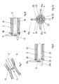
- the endoscope 1 essentially consists of an outer shaft 2 with a stop disk 3 slidably mounted thereon, an inner shaft 4 and an optic 5 that can be inserted into the inner shaft 4.
- a plurality of hook elements 6 are arranged and guided axially in an annular space 8 between the outer shaft 2 and the optics 5.
- the guidance can take place through longitudinal grooves 7 in the outer surface of the inner shaft 4 or through a guide ring provided with corresponding recesses.
- the hook elements 6 consist of a longer, highly elastic wire or band and have an immanent pre-bend 9 in the region of their distal end, which, when the hook element is pushed out, extends beyond the distal end of the outer shaft 2, as shown in FIG. 2, in the radial direction the outer shaft 2 spreads out and thereby forms as a hook part.
- each hook element 6 is provided at its distal end with a further, intrinsic back bend 9 a which runs counter to the aforementioned pre-bend.
- the length of the hook elements 6 is selected so that they can be pulled completely into the outer shaft 2 of the instrument 1, thereby avoiding that the hook elements 6 cause additional traumatization when the endoscope 1 is inserted into a hollow organ 10 or 11 of a living being.
- the hook elements 6 are fixed with their proximal end to a sleeve 16 which can be displaced in the annular space 8 between the outer shaft 2 and the optics 5.
- the sleeve 16 is connected via a connecting screw to an actuating device 13 in the form of a sliding sleeve in the area of the handle 12 of the instrument 1.
- the hollow organ 10 is thus securely fixed and can be precisely examined and treated endoscopically.
- the proposed endoscope can with regard the hook elements can also be designed such that a plurality of rigid, L-shaped hook elements 17 are distributed circumferentially in axial slots 18 at the distal end of the outer shaft 2 and articulated radially pivotably, as shown in FIG.
- the articulation and the shape of the hook elements 16 is chosen so that the hook elements in their non-functional position with respect to the outer shaft 2 do not protrude radially outwards with the aid of the optics 5, as is shown in the lower half in FIG.
- the upper half in FIG. 8 shows the functional position of the hook elements 17.
- the swivel actuation of the hook elements takes place by axially advancing the inner shaft 19 in the outer shaft 2, the inner shaft also receiving the optics 5 first pressing against the knee of the L-shaped hook elements, around them Swing out elements radially, as can be seen from Figure 7 below, and then keeps the other legs of the hook elements pivoted radially outwards by its advanced position, as shown in Figure 8 above.
- the inner shaft 21 has a number of recesses 22 which corresponds to the number of hook elements 17a mounted in the outer shaft 2 with ball joints 20 and which serve to hold the hook elements 17a in an axially parallel position, as in the lower part 9, to be fixed by means of the optics 5.
- the optics 5 and the inner shaft 21 are first retracted proximally until the distal end of the inner shaft 21 is completely proximal to the proximal end of the hook 17a.
- the inner shaft 21 is then rotated such that the cutouts 22, as shown in FIG. 10, no longer point in the direction of the hook elements 17a.
- the inner shaft is rotated again until the positions of the hook elements 17a articulated in the outer shaft 2 and the recesses 22 of the inner shaft 21 overlap, so that the hook elements, e.g. B. when pulling the instrument out of the body cavity, fall back into the rest position.
- the hook elements 17a are also fixed in their rest position in this embodiment simply by advancing the optics 5 towards the distal end.
- the endoscope 1 according to the invention can also be designed as a flexible instrument with flexible shafts 2, 4 or 19, 21 instead of rigid shafts and with a flexible lens instead of a rigid lens 5.
Landscapes
- Health & Medical Sciences (AREA)
- Life Sciences & Earth Sciences (AREA)
- Surgery (AREA)
- Molecular Biology (AREA)
- Biomedical Technology (AREA)
- Veterinary Medicine (AREA)
- Public Health (AREA)
- General Health & Medical Sciences (AREA)
- Pathology (AREA)
- Animal Behavior & Ethology (AREA)
- Medical Informatics (AREA)
- Engineering & Computer Science (AREA)
- Heart & Thoracic Surgery (AREA)
- Physics & Mathematics (AREA)
- Nuclear Medicine, Radiotherapy & Molecular Imaging (AREA)
- Biophysics (AREA)
- Reproductive Health (AREA)
- Gynecology & Obstetrics (AREA)
- Radiology & Medical Imaging (AREA)
- Optics & Photonics (AREA)
- Dentistry (AREA)
- Oral & Maxillofacial Surgery (AREA)
- Endoscopes (AREA)
Applications Claiming Priority (2)
| Application Number | Priority Date | Filing Date | Title |
|---|---|---|---|
| DE4125806A DE4125806A1 (de) | 1991-08-03 | 1991-08-03 | Endoskop zum einfuehren in ein hohlorgan eines lebewesens |
| DE4125806 | 1991-08-03 |
Publications (1)
| Publication Number | Publication Date |
|---|---|
| EP0526721A1 true EP0526721A1 (de) | 1993-02-10 |
Family
ID=6437667
Family Applications (1)
| Application Number | Title | Priority Date | Filing Date |
|---|---|---|---|
| EP92110439A Withdrawn EP0526721A1 (de) | 1991-08-03 | 1992-06-20 | Endoskop zum Einführen in ein Hohlorgan eines Lebewesens |
Country Status (3)
| Country | Link |
|---|---|
| US (1) | US5309894A (OSRAM) |
| EP (1) | EP0526721A1 (OSRAM) |
| DE (1) | DE4125806A1 (OSRAM) |
Cited By (7)
| Publication number | Priority date | Publication date | Assignee | Title |
|---|---|---|---|---|
| US5425357A (en) * | 1991-05-29 | 1995-06-20 | Origin Medsystems, Inc. | Inflatable retraction devices for use in laparoscopic surgery |
| US5505689A (en) * | 1991-05-29 | 1996-04-09 | Origin Medsystems, Inc. | Propertioneal mechanical retraction apparatus |
| US5697946A (en) * | 1994-10-07 | 1997-12-16 | Origin Medsystems, Inc. | Method and apparatus for anchoring laparoscopic instruments |
| GB2320683A (en) * | 1996-12-25 | 1998-07-01 | Asahi Optical Co Ltd | Surgical stripper for use under endoscopic observation |
| FR2770122A1 (fr) * | 1997-10-28 | 1999-04-30 | Asahi Optical Co Ltd | Outil endoscopique de mesure de longueur |
| WO2008121794A1 (en) * | 2007-03-29 | 2008-10-09 | Frantz Medical Development, Ltd. | Securable cannula and method |
| WO2017182117A1 (de) | 2016-04-20 | 2017-10-26 | Rheinisch-Westfälische Technische Hochschule (Rwth) Aachen | Hohlorganankervorrichtung |
Families Citing this family (10)
| Publication number | Priority date | Publication date | Assignee | Title |
|---|---|---|---|---|
| DE4125806A1 (de) * | 1991-08-03 | 1993-02-04 | Wolf Gmbh Richard | Endoskop zum einfuehren in ein hohlorgan eines lebewesens |
| DE4303274C2 (de) * | 1993-02-05 | 1997-02-06 | Wolf Gmbh Richard | Endoskopisches Instrument |
| SE509389C2 (sv) * | 1996-07-24 | 1999-01-18 | Solem Jan Otto | Anordning för anslutning av änden av ett första blodkärl till sidan av ett andra blodkärl |
| US6056762A (en) * | 1997-05-22 | 2000-05-02 | Kensey Nash Corporation | Anastomosis system and method of use |
| US6063114A (en) | 1997-09-04 | 2000-05-16 | Kensey Nash Corporation | Connector system for vessels, ducts, lumens or hollow organs and methods of use |
| JP3331172B2 (ja) * | 1998-06-12 | 2002-10-07 | 旭光学工業株式会社 | 内視鏡用異物回収具 |
| US10258368B2 (en) * | 2010-09-14 | 2019-04-16 | Suremka, Llc | Retractable cannula for surgical procedures |
| US11627985B2 (en) * | 2014-06-10 | 2023-04-18 | Suremka, Llc | Surgical devices and deployment apparatuses |
| EP3471594A4 (en) * | 2016-06-18 | 2019-11-13 | Arthroscopic Innovations, LLC | SURGICAL DEVICES AND METHOD |
| EP3668366A4 (en) * | 2017-08-17 | 2021-04-21 | 270 Surgical Ltd. | MULTI-CAMERA MEDICAL SURGERY LIGHTING DEVICE WITH VARIABLE DIAMETER |
Citations (4)
| Publication number | Priority date | Publication date | Assignee | Title |
|---|---|---|---|---|
| GB1173194A (en) * | 1966-10-03 | 1969-12-03 | American Cystoscope Makers Inc | A Medical Instrument |
| US3717151A (en) * | 1971-03-11 | 1973-02-20 | R Collett | Flesh penetrating apparatus |
| US4016867A (en) * | 1976-04-27 | 1977-04-12 | The United States Of America As Represented By The Secretary Of The Department Of Health, Education And Welfare | Uterine caliper and depth gauge |
| DE8303342U1 (de) * | 1983-07-14 | Storz-Endoskop GmbH, 6207 Schaffhausen | Medizinisches Greifinstrument |
Family Cites Families (17)
| Publication number | Priority date | Publication date | Assignee | Title |
|---|---|---|---|---|
| DE640126C (de) * | 1934-07-29 | 1936-12-24 | Bruno Loewel Dr | Trokar |
| US3570498A (en) * | 1970-03-09 | 1971-03-16 | Charles Weighton | Trocar and cannula for veterinary use |
| US3866599A (en) * | 1972-01-21 | 1975-02-18 | Univ Washington | Fiberoptic catheter |
| US4027510A (en) * | 1974-05-15 | 1977-06-07 | Siegfried Hiltebrandt | Forceps |
| US4168709A (en) * | 1975-03-10 | 1979-09-25 | Bentov Itzhak E | Dilator |
| US3994301A (en) * | 1975-04-14 | 1976-11-30 | S & S Medical Products Co., Inc. | Submammary dissector |
| JPS5641684Y2 (OSRAM) * | 1977-11-24 | 1981-09-30 | ||
| DE3330921C1 (de) * | 1983-08-27 | 1985-02-07 | Karl-Heinz Dr.med. 4000 Düsseldorf Kurz | Geraet zum Bestimmen der Innenmasse von Hohlorganen,insbesondere der Gebaermutterhoehle |
| US4608965A (en) * | 1985-03-27 | 1986-09-02 | Anspach Jr William E | Endoscope retainer and tissue retracting device |
| US4791913A (en) * | 1987-12-14 | 1988-12-20 | Baxter Travenol Laboratories, Inc. | Optical valvulotome |
| US5002560A (en) * | 1989-09-08 | 1991-03-26 | Advanced Cardiovascular Systems, Inc. | Expandable cage catheter with a rotatable guide |
| US5197971A (en) * | 1990-03-02 | 1993-03-30 | Bonutti Peter M | Arthroscopic retractor and method of using the same |
| CA2039414C (en) * | 1990-03-29 | 1995-09-05 | Robert W. Bailey | Abdominal cavity organ retractor |
| DE4021153A1 (de) * | 1990-07-03 | 1992-01-16 | Wolf Gmbh Richard | Organmanipulator |
| US5160341A (en) * | 1990-11-08 | 1992-11-03 | Advanced Surgical Intervention, Inc. | Resorbable urethral stent and apparatus for its insertion |
| US5183033A (en) * | 1991-07-15 | 1993-02-02 | Wilk Peter J | Surgical instrument assembly and apparatus and surgical method |
| DE4125806A1 (de) * | 1991-08-03 | 1993-02-04 | Wolf Gmbh Richard | Endoskop zum einfuehren in ein hohlorgan eines lebewesens |
-
1991
- 1991-08-03 DE DE4125806A patent/DE4125806A1/de active Granted
-
1992
- 1992-06-20 EP EP92110439A patent/EP0526721A1/de not_active Withdrawn
- 1992-07-31 US US07/923,697 patent/US5309894A/en not_active Expired - Fee Related
Patent Citations (4)
| Publication number | Priority date | Publication date | Assignee | Title |
|---|---|---|---|---|
| DE8303342U1 (de) * | 1983-07-14 | Storz-Endoskop GmbH, 6207 Schaffhausen | Medizinisches Greifinstrument | |
| GB1173194A (en) * | 1966-10-03 | 1969-12-03 | American Cystoscope Makers Inc | A Medical Instrument |
| US3717151A (en) * | 1971-03-11 | 1973-02-20 | R Collett | Flesh penetrating apparatus |
| US4016867A (en) * | 1976-04-27 | 1977-04-12 | The United States Of America As Represented By The Secretary Of The Department Of Health, Education And Welfare | Uterine caliper and depth gauge |
Cited By (13)
| Publication number | Priority date | Publication date | Assignee | Title |
|---|---|---|---|---|
| US5505689A (en) * | 1991-05-29 | 1996-04-09 | Origin Medsystems, Inc. | Propertioneal mechanical retraction apparatus |
| US5575759A (en) * | 1991-05-29 | 1996-11-19 | Origin Medsystems, Inc. | Methods of using inflatable retraction devices in laparoscopic surgery |
| US5425357A (en) * | 1991-05-29 | 1995-06-20 | Origin Medsystems, Inc. | Inflatable retraction devices for use in laparoscopic surgery |
| US6524283B1 (en) | 1994-10-07 | 2003-02-25 | Sherwood Services Ag | Method and apparatus for anchoring laparoscopic instruments |
| US5697946A (en) * | 1994-10-07 | 1997-12-16 | Origin Medsystems, Inc. | Method and apparatus for anchoring laparoscopic instruments |
| US7235064B2 (en) | 1994-10-07 | 2007-06-26 | Sherwood Services Ag | Method and apparatus for anchoring laparoscopic instruments |
| GB2320683A (en) * | 1996-12-25 | 1998-07-01 | Asahi Optical Co Ltd | Surgical stripper for use under endoscopic observation |
| US6033359A (en) * | 1997-10-28 | 2000-03-07 | Asahi Kogaku Kogyo Kabushiki Kaisha | Endoscopic length-measuring tool |
| FR2770122A1 (fr) * | 1997-10-28 | 1999-04-30 | Asahi Optical Co Ltd | Outil endoscopique de mesure de longueur |
| WO2008121794A1 (en) * | 2007-03-29 | 2008-10-09 | Frantz Medical Development, Ltd. | Securable cannula and method |
| US8360969B2 (en) | 2007-03-29 | 2013-01-29 | Frantz Medical Development, Ltd. | Securable cannula and method |
| WO2017182117A1 (de) | 2016-04-20 | 2017-10-26 | Rheinisch-Westfälische Technische Hochschule (Rwth) Aachen | Hohlorganankervorrichtung |
| DE102016004811A1 (de) | 2016-04-20 | 2017-10-26 | Rheinisch-Westfälische Technische Hochschule (Rwth) Aachen | Hohlorganankervorrichtung |
Also Published As
| Publication number | Publication date |
|---|---|
| DE4125806C2 (OSRAM) | 1993-06-17 |
| DE4125806A1 (de) | 1993-02-04 |
| US5309894A (en) | 1994-05-10 |
Similar Documents
| Publication | Publication Date | Title |
|---|---|---|
| DE4125806C2 (OSRAM) | ||
| DE69919343T2 (de) | Führungshülse für verschobene wirbelkörper | |
| DE3344934C2 (OSRAM) | ||
| DE3709706C2 (OSRAM) | ||
| DE2800362C3 (de) | Endoskop mit steuerbar beweglicher Führungsröhre für ein Instrument | |
| DE68919258T2 (de) | Vorrichtung zur Entfernung von Ablagerungen in Arterien. | |
| DE69532995T2 (de) | Flexible chirurgische instrumente, die eine spirale mit lumen aufweisen | |
| EP0464463A1 (de) | Organmanipulator | |
| DE2305815A1 (de) | Vorrichtung zum trennen von chirurgischen faeden | |
| EP0605764A1 (de) | Instrument zum Implantieren und Extrahieren von Stents | |
| DE4303274C2 (de) | Endoskopisches Instrument | |
| DE19540731C2 (de) | Endoskopisches Instrument | |
| DE2804058A1 (de) | Medizinisches geraet zur entfernung von fremdkoerpern aus einem koerperhohlraum | |
| EP1052945B1 (de) | Medizinisches rohrschaftinstrument | |
| EP0613386B1 (de) | Sonde für medizinische eingriffe in körperhöhlen | |
| DE2426781C3 (de) | Vorrichtung zum Durchtrennen des verengten Schließmuskels der Mündung des Gallenganges in den Zwölffingerdarm | |
| DE10027342A1 (de) | Behandlungsinstrument für ein Endoskop | |
| DE19515626C2 (de) | Instrument zum Positionieren wenigstens einer Arbeitshülse | |
| DE3620385C1 (en) | Forceps for the percutaneous removal of renal calculi | |
| DE19629537A1 (de) | Trokarhülse | |
| EP0999811B1 (de) | Vorrichtung zur aufweitung und wiederherstellung des tränenkanals am menschlichen auge | |
| DE3511448C2 (de) | Harnleiterkatheter | |
| DE19955614C1 (de) | Falloposkop für die Untersuchung von Eierstöcken und Eileitern | |
| DE10241946A1 (de) | Vorrichtung zur minimal-invasiven chirurgischen Fremdkörperentfernung | |
| WO2001010309A1 (de) | Medizinisches instrument und verfahren zur schaffung eines hohlraums für einen endoskopischen eingriff |
Legal Events
| Date | Code | Title | Description |
|---|---|---|---|
| PUAI | Public reference made under article 153(3) epc to a published international application that has entered the european phase |
Free format text: ORIGINAL CODE: 0009012 |
|
| AK | Designated contracting states |
Kind code of ref document: A1 Designated state(s): BE CH DE FR GB LI |
|
| 17P | Request for examination filed |
Effective date: 19930724 |
|
| 17Q | First examination report despatched |
Effective date: 19950317 |
|
| STAA | Information on the status of an ep patent application or granted ep patent |
Free format text: STATUS: THE APPLICATION HAS BEEN WITHDRAWN |
|
| 18W | Application withdrawn |
Withdrawal date: 19951209 |