EP0480041A1 - Polypeptide a antigene soude - Google Patents
Polypeptide a antigene soude Download PDFInfo
- Publication number
- EP0480041A1 EP0480041A1 EP91905886A EP91905886A EP0480041A1 EP 0480041 A1 EP0480041 A1 EP 0480041A1 EP 91905886 A EP91905886 A EP 91905886A EP 91905886 A EP91905886 A EP 91905886A EP 0480041 A1 EP0480041 A1 EP 0480041A1
- Authority
- EP
- European Patent Office
- Prior art keywords
- polypeptide
- dna
- psghec
- fused
- antigen polypeptide
- Prior art date
- Legal status (The legal status is an assumption and is not a legal conclusion. Google has not performed a legal analysis and makes no representation as to the accuracy of the status listed.)
- Withdrawn
Links
Images


Classifications
-
- C—CHEMISTRY; METALLURGY
- C07—ORGANIC CHEMISTRY
- C07K—PEPTIDES
- C07K14/00—Peptides having more than 20 amino acids; Gastrins; Somatostatins; Melanotropins; Derivatives thereof
- C07K14/435—Peptides having more than 20 amino acids; Gastrins; Somatostatins; Melanotropins; Derivatives thereof from animals; from humans
- C07K14/575—Hormones
- C07K14/61—Growth hormones [GH] (Somatotropin)
-
- C—CHEMISTRY; METALLURGY
- C07—ORGANIC CHEMISTRY
- C07K—PEPTIDES
- C07K14/00—Peptides having more than 20 amino acids; Gastrins; Somatostatins; Melanotropins; Derivatives thereof
- C07K14/005—Peptides having more than 20 amino acids; Gastrins; Somatostatins; Melanotropins; Derivatives thereof from viruses
-
- C—CHEMISTRY; METALLURGY
- C07—ORGANIC CHEMISTRY
- C07K—PEPTIDES
- C07K2319/00—Fusion polypeptide
-
- C—CHEMISTRY; METALLURGY
- C12—BIOCHEMISTRY; BEER; SPIRITS; WINE; VINEGAR; MICROBIOLOGY; ENZYMOLOGY; MUTATION OR GENETIC ENGINEERING
- C12N—MICROORGANISMS OR ENZYMES; COMPOSITIONS THEREOF; PROPAGATING, PRESERVING, OR MAINTAINING MICROORGANISMS; MUTATION OR GENETIC ENGINEERING; CULTURE MEDIA
- C12N2770/00—MICROORGANISMS OR ENZYMES; COMPOSITIONS THEREOF; PROPAGATING, PRESERVING, OR MAINTAINING MICROORGANISMS; MUTATION OR GENETIC ENGINEERING; CULTURE MEDIA ssRNA viruses positive-sense
- C12N2770/00011—Details
- C12N2770/24011—Flaviviridae
- C12N2770/24211—Hepacivirus, e.g. hepatitis C virus, hepatitis G virus
- C12N2770/24222—New viral proteins or individual genes, new structural or functional aspects of known viral proteins or genes
Definitions
- the present invention relates to a fused antigen polypeptide in which an antigen polypeptide derived from a non-A, non-B hepatitis (type C hepatitis)-associated antigen is fused to a polypeptide heterologous to said antigen polypeptide, a recombinant plasmid containing, as an insert, a DNA coding for said fused antigen polypeptide, a method of producing fused antigen polypeptides which uses a microorganism containing any of said recombinant plasmids, and a method of detecting an anti-hepatitis C virus antibody which uses said fused antigen polypeptide.
- the antigen-antibody reaction is utilized in various medical and other scientific fields, and the fused antigen polypeptides of the invention are expected to be useful in a broad range of fields.
- HCV hepatitis C virus
- NANBV non-A, non-B hepatitis virus
- HCV hepatitis C virus
- serodiagnosis of HCV infection constitutes an important diagnostic technique for hepatitis C and for the prevention of HCV infection that may result from blood transfusion and so on.
- serum samples are tested for the presence or absence of an antibody to an HCV-associated antigen.
- HCV-associated genes have been cloned from sera of patients with hepatitis C [Arima et al.: Jikken Igaku (Experimental Medicine), Vol. 7, No. 2, 94-101 (1989); Arima et al.: Gastroenterologia Japonica, 24 , 540-548 (1989)] and have been used in the diagnosis of hepatitis C.
- fused antigen polypeptides each composed of a hepatitis C-associated antigen polypeptide and a heterologous protein are produced in microorganisms and used. Choo et al.
- SOD superoxide dismutase
- ⁇ -gal ⁇ -D-galactosidase
- a fused antigen polypeptide comprising an antigen polypeptide chemically bound to a carrier such as bovine serum albumin, a mixture of several antigen molecules, or a crude extract of pathogens is used.
- a carrier such as bovine serum albumin, a mixture of several antigen molecules, or a crude extract of pathogens.
- direct expression of antigen polypeptides is sought in microorganisms.
- such expression is difficult for reasons of the molecular weight, stability and toxicity of antigen polypeptides, hence it is impossible to supply antigen polypeptides constantly and at low cost.
- the present inventors made investigations in an attempt to develop a method of producing antigen polypeptides useful for the detection of hepatitis C and, as a result, established a method of producing fused antigen polypeptides in which at least one hepatitis C-associated antigen polypeptide is fused to chum salmon growth hormone (hereinafter abbreviated as sGH) via methionine.
- sGH chum salmon growth hormone
- the present inventors further confirmed that when these fused antigen polypeptides are used for detecting anti-hepatitis C antibodies, said antibodies can be detected even in samples in which the detection methods so far reported fail to detect them and thus, the method of the present invention is superior to the prior art methods.
- the present invention has been completed based on the above findings.
- the present invention provides a fused antigen polypeptide resulting from the fusion of at least one hepatitis C-associated antigen-derived antigen polypeptide (hereinafter briefly referred to as hepatitis C-associated antigen polypeptide) to at least one polypeptide heterologous to said antigen polypeptide (hereinafter briefly referred to as heterologous polypeptide), a recombinant plasmid which contains, as an insert, a DNA coding for said fused antigen polypeptide, a method of producing a fused antigen polypeptide which comprises using a microorganism carrying said recombinant plasmid, and a method of detecting anti-hepatitis C virus antibodies (anti-HCV antibodies) using said fused antigen polypeptide.
- hepatitis C-associated antigen polypeptide hepatitis C-associated antigen polypeptide
- heterologous polypeptide a recombinant plasmid which contains, as an insert, a DNA
- any HCV-associated antigen may be used as the hepatitis C-associated antigen polypeptide.
- clone No. 2 clone No. 18 [Arima et al.: Gastroenterologia Japonica, 24 , 540-548 (1989)] and clone No. 14, each cloned by Arima et al. as an HCV-associated antigen, may, inter alia , be employed.
- the antigens that can be identified by these clone numbers are hereinafter referred to briefly as C-2, C-14 and C-18, respectively.
- the amino acid sequences of these antigens and the nucleotide sequences of DNAs coding for the antigens are shown under Sequence Identifier (SEQ ID) Nos. 1 to 3.
- RNA is extracted from this plasma and cDNA is prepared by the random primer method.
- a cDNA library is constructed from said cDNA using a cDNA cloning system ⁇ gt11 kit (Amersham). This library is transferred onto a nitrocellulose membrane and immunoscreening is performed using an acute hepatitis convalescent serum and the serum from a patient with chronic hepatitis each treated with Escherichia coli cell components for absorption.
- a sequence derived from the DNA base sequence for C-14 by partial substitution without causing any change in the amino acid sequence of C-14 is shown under SEQ ID NO: 41 and a sequence derived from the sequence No. 41 by partial deletion is shown under SEQ ID NO: 42.
- SEQ ID NO: 41 the 15th base A of the DNA base sequence for C-14 has been replaced by G, the 33rd base A by G, the 66th base C by T, the 69th base C by T, and the 99th base A by G.
- the 94th to 108th bases of the DNA base sequence of SEQ ID NO: 41 are missing.
- antigens described in Science, 244 , 359-362 (1989), Science, 244 , 362-364 (1989), and EP-A-0318216 may also be used as the HCV-associated antigen.
- heterologous polypeptide to be used in accordance with the invention may be any polypeptide provided that it does not specifically react with any of the human serum antibodies but can be expressed in a microorganism, in the form fused to a hepatitis C-associated antigen polypeptide. More specifically, the chum salmon growth hormone (sGH) polypeptide can preferably be used.
- sGH chum salmon growth hormone
- JP-A chum salmon growth hormone and the relevant genetic information and production technology, among others, are described in Proceedings of the National Academy of Sciences of the U.S.A., 82 , 4306-4310 (1985) as well as in JP-A-61-15699, 61-93196, 61-93197 and 61-210100 (the term "JP-A" used herein means an unexamined published Japanese patent application).
- the fused antigen polypeptide can be provided by the steps of inserting a DNA coding for a fused antigen polypeptide consisting of an HCV-associated antigen and a heterologous polypeptide having a molecular weight of not less than 2,000, joined together preferably via methionine, into a vector DNA by the recombinant DNA technology without involving reading frame shifting, using the thus-constructed recombinant plasmid to transform a microorganism, cultivating the transformant microorganism to cause formation and accumulation of said fused antigen polypeptide in the culture, and harvesting said fused antigen polypeptide from said culture.
- the vector DNA may be of any kind only if it allows expression of the insert DNA in a microorganism.
- plasmids which have an appropriate promoter, for example the tryptophan (trp) or lactose (lac) promoter, and allow insertion of the desired DNA downstream from said promoter and in which the distance between the Shine-Dalgarno sequence and the translation initiation codon has been adjusted to an appropriate length, for example 6 to 18 bases, can preferably be used.
- pGEL1 FERM BP-629; JP-A-61-93197
- pKYP10 JP-A-58-110600
- pGHA2 FERM BP-400; JP-A-60-221091
- any microorganism may be employed provided that the DNA coding for the fused antigen polypeptide of this invention can be expressed and that said polypeptide can be formed and accumulated in the culture.
- Microorganisms belonging to the species Escherichia coli are preferred.
- the fused antigen polypeptides in which chum salmon growth hormone is bound to an HCV-associated antigen via a methionine residue are produced by taking the following steps.
- Steps 1 to 6 are involved in the production of the fused antigen polypeptides sGHE1, sGHEC-2, sGHEC-14, sGHEC-18S and sGHEC-1414, while steps 7 to 9 are involved in the production of the fused antigen polypeptides sGHEC-14A and sGHEC-14B.
- DNAs represented by SEQ ID NO: 4, 5, 6, 7, 8, 9, 10, 11, 12, 13, 14, 15, 16, 17, 18 and 19 are chemically synthesized.
- a plasmid containing a double-stranded DNA fragment identifiable by SEQ ID NO: 20 (hereinafter referred to as gene-20) is constructed using a double-stranded DNA formed from the DNA identifiable by SEQ ID NO: 4 (hereinafter referred to as DNA-4) and the DNA identifiable by SEQ ID NO: 5 (hereinafter referred to as DNA-5).
- a plasmid containing a double-stranded DNA fragment identifiable by SEQ ID NO: 21 (hereinafter referred to as gene-21) is constructed by joining a double-stranded DNA formed from a DNA having SEQ ID NO: 6 (hereinafter referred to as DNA-6) and a DNA having SEQ ID NO: 7 (hereinafter referred to as DNA-7) to a double-stranded DNA formed from a DNA having SEQ ID NO: 8 (hereinafter referred to as DNA-8) and a DNA having SEQ ID NO: 9 (hereinafter referred to as DNA-9), using ligase.
- a plasmid containing a double-stranded DNA fragment represented by SEQ ID NO: 22 (hereinafter referred to as gene-22) is constructed by ligating a double-stranded DNA formed from a DNA having SEQ ID NO: 10 (hereinafter referred to as DNA-10) and a DNA having SEQ ID NO: 11 (hereinafter referred to as DNA-11), a double-stranded DNA formed from a DNA having SEQ ID NO: 12 (hereinafter referred to as DNA-12) and a DNA having SEQ ID NO: 13 (hereinafter referred to as DNA-13), a double-stranded DNA formed from a DNA having SEQ ID NO: 14 (hereinafter referred to as DNA-14) and a DNA having SEQ ID NO: 15 (hereinafter referred to as DNA-15) and a double-stranded DNA formed from a DNA having SEQ ID NO: 16 (hereinafter referred to as DNA-16) and a DNA having SEQ ID NO: 17 (hereinafter referred to as as DNA
- a plasmid containing a double-stranded DNA fragment represented by SEQ ID NO: 23 (hereinafter referred to as gene-23) is constructed using a double-stranded DNA formed from a DNA having SEQ ID NO: 18 (hereinafter referred to as DNA-18) and a DNA having SEQ ID NO: 19 (hereinafter referred to as DNA-19).
- the double-stranded DNA fragments, gene-20, gene-21 and gene-22, prepared from the synthetic DNAs mentioned above have base sequences coding for the C-2 peptide, the C-14 peptide and the C-18 peptide, respectively.
- Gene-23 serves as a linker for joining each clone to the chum salmon growth hormone gene and contains a base sequence coding for methionine, glutamic acid and phenylalanine, which sequence is directly followed by a termination codon.
- the base sequence coding for glutamic acid and phenylalanine serves as an Eco RI recognition site.
- the plasmid pSGH1B2 for sGH gene expression as produced by the method described in JP-A-61-93197 is used.
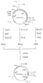
- a Bg1 II- Pvu I fragment (about 1.17 kb) of pSGH1B2, an Nsi I- Pvu I fragment (about 1.80 kb) of pSGH1B2, DNA-18 and DNA-19 were mixed and ligation of the double-stranded DNAs to each other was effected using DNA ligase to thereby construct a plasmid, pSGHE1, containing a gene coding for a polypeptide resulting from binding of glutamic acid and phenylalanine, via methionine, to a polypeptide comprising an aminoterminal methionine and the 1st to 103rd amino acids of sGH (cf. Fig. 1).
- amino acid sequence of the fused polypeptide expressed by pSGHE1 the DNA sequence coding for said polypeptide and the Eco RI site contained therein are shown under SEQ ID NO: 24.
- the linker region is underlined.
- the pSGHE1 constructed in step 2 is cleaved with Eco RI, then treated with bacterial alkaline phosphatase (BAP) and further cleaved with Hin dIII and Pvu I, and an Eco RI- Hin dIII fragment of about 320 bp and an Eco RI- Pvu I fragment of about 1.8 kb are isolated.
- BAP bacterial alkaline phosphatase
- pSGHE1 is cleaved with Hin dIII and Pvu I, and a Hin dIII- Pvu I fragment of about 850 bp is isolated.
- pSGHEC-2 The above DNA fragments and the double-stranded DNA fragments formed from DNA-4 and DNA-5 are subjected to ligation using DNA ligase to give a plasmid named pSGHEC-2 (cf. Fig. 2).
- pSGHEC-2 holds the sGH gene of pSGHE1 and the gene coding for C-2 via a methionine-encoding gene.
- the amino acid sequence of the fused antigen polypeptide expressed by pSGHEC-2 and the DNA sequence coding for said polypeptide are shown under SEQ ID NO: 25. In SEQ ID NO: 25, the C-2 region is underlined.
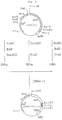
- the pSGHE1 constructed in step 2 is cleaved with Eco RI, then treated with bacterial alkaline phosphatase (BAP) and further cleaved with Hin dIII and Pvu I, and an Eco RI- Hin dIII fragment of about 320 bp and an Eco RI- Pvu I fragment of about 1.8 kb are isolated.
- BAP bacterial alkaline phosphatase
- the DNA fragments mentioned above and the double-stranded DNA fragments formed from DNA-6 through DNA-9 are subjected to ligation using DNA ligase to give a plasmid named pSGHEC-14 (cf. Fig. 3).
- the pSGHEC-14 holds the sGH gene of pSGHE1 and the gene coding for C-14 via a methionine-encoding gene.
- the amino acid sequence of the fused antigen polypeptide expressed by pSGHEC-14 and the DNA sequence coding for said polypeptide are shown under SEQ ID NO: 26. In SEQ ID NO: 26, the C-14 region is underlined.
- the pSGHE1 constructed in step 2 is cleaved with Eco RI, then treated with bacterial alkaline phosphatase (BAP) and further cleaved with Hin dIII and Pvu I, and an Eco RI- Hin dIII fragment of about 320 bp and an Eco RI- Pvu I fragment of about 1.8 kb are isolated.
- BAP bacterial alkaline phosphatase
- pSGHE1 is cleaved with Hin dIII and Pvu I, and a Hin dIII- Pvu I fragment of about 850 bp is isolated.
- the DNA fragments mentioned above and the double-stranded DNA fragments formed from DNA-10 through DNA-17 are subjected to ligation using DNA ligase to give a plasmid named pSGHEC-18 (cf. Fig. 4).
- the pSGHEC-18 holds the sGH gene of pSGHE1 and the gene coding for C-18 via a methionine-encoding gene.
- the amino acid sequence of the fused antigen polypeptide expressed by pSGHEC-18 and the DNA sequence coding for said polypeptide are shown under SEQ ID NO: 27. In SEQ ID NO: 27, the C-18 region is underlined.
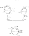
- the pSGHEC-18 is cleaved with Hin dIII and Pvu I and a DNA fragment of about 2.36 kb is isolated.
- the pKYP10 is cleaved with Hin dIII and Pvu I and a DNA fragment of about 1.1 kb is isolated.
- the above DNAs are subjected to ligation using DNA ligase to give pSGHEC-18S (cf. Fig. 5).
- pSGHEC-18S the promoter of pSGHEC-18 has been substituted and pSGHEC-18S holds the gene coding for sGH and the gene coding for C-18 via a methionine-encoding gene.
- the amino acid sequence of the fused antigen polypeptide expressed by pSGHEC-18S and the DNA sequence coding for said polypeptide are shown under SEQ ID NO: 27. In SEQ ID NO: 27, the C-18 region is undelined.
- pSGHEC-14 constructed in step 3 is used.
- pSGHEC-14 is cleaved with Pst I and then partially digested with the restriction enzyme Eco RI, and an Eco RI- Pst I fragment of about 1.40 kb and an Eco RI- Pst I fragment of about 1.85 kb are isolated.
- the above DNA fragments are subjected to ligation using DNA ligase to give a plasmid named pSGHEC-1414 (cf. Fig. 6).
- pSGHEC-1414 holds the sGH-encoding gene of pSGHE1 and a gene coding for two repetitions of C-14 via a methionine-encoding gene.
- SEQ ID NO: 28 The amino acid sequence of the fused antigen polypeptide expressed by pSGHEC-1414 and the DNA sequence coding therefor are shown under SEQ ID NO: 28.
- SEQ ID NO: 28 the region for the two repetitions of C-14 is underlined. This step 5 makes it possible to link antigen genes of different kinds together and can be utilized in producing chimera antigens.
- Step 6 Production, isolation and purification of fused antigen polypeptides in which the chum salmon growth hormone is coupled to an HCV-associated antigen
- the plasmid pSGHE1, pSGHEC-2, pSGHEC-14, pSGHEC-18S or pSGHEC-1414 produced in step 2, step 3, step 4 or step 5 is introduced into Escherichia coli .
- the Escherichia coli transformant obtained is cultured in a medium and the desired fused antigen polypeptide in which the chum salmon growth hormone is coupled to an HCV-associated antigen is recovered from the culture broth.
- the desired fused antigen polypeptide produced intracellularly by Escherichia coli occurs as water-insoluble granules. After cultivation, cells are collected and disrupted, the liquid obtained by cell disruption is centrifuged, and granules containing the fused antigen polypeptide in which the chum salmon growth hormone is coupled to the HCV-associated antigen are separated.
- the fused antigen polypeptides produced in Escherichia coli transformants carrying the plasmids pSGHE1, pSGHEC-2, pSGHEC-14, pSGHEC-18S and pSGHEC-1414, respectively are hereinafter referred to as sGHE1, sGHEC-2, sGHEC-14, sGHEC-18S and sGHEC-1414, respectively.
- DNAs having SEQ ID NO: 29, 30, 31, 32, 33, 34, 35 and 36 are chemically synthesized.
- a plasmid containing a double-stranded DNA fragment having SEQ ID NO: 38 (hereinafter referred to as gene-38) is constructed by subjecting a double-stranded DNA formed from DNA-29 and DNA-30, a double-stranded DNA formed from DNA-31 and DNA-32 and a double-stranded DNA formed from a DNA having SEQ ID NO: 35 (hereinafter referred to as DNA-35) and a DNA having SEQ ID NO: 36 (hereinafter referred to as DNA-36) to ligation using ligase.
- the double-stranded DNA fragments prepared from the synthetic DNAs as mentioned above, namely gene-37 and gene-38, have a base sequence coding for the C-14 peptide and a base sequence coding for a peptide derived from the C-14 peptide by deletion of the 32nd to 36th amino acids, respectively.
- the base sequence has been partly modified without changing the amino acid sequence of C-14; thus, in the base sequence for C-14, the 15th base A has been replaced by G, the 33rd base A by G, the 66th C by T, the 69th C by T, and the 99th base A by G.
- Gene-38 is a synthetic DNA corresponding to gene-37 but missing the 93rd to the 107th bases of the base sequence thereof.
- the peptide encoded by gene-37 or the DNA coding for said peptide is referred to as C-14A
- the peptide encoded by gene-38 or the DNA coding for said peptide as C-14B.
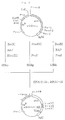
- pSGHE1 constructed in step 2 is cleaved with Eco RI, then treated with bacterial alkaline phosphatase (BAP) and further cleaved with Hin dIII and Pvu I, and an Eco RI- Hin dIII fragment of about 320 bp and an Eco RI- Pvu I fragment of about 1.8 kb are isolated.
- BAP bacterial alkaline phosphatase
- pSGHE1 is cleaved with Hin dIII and Pvu I, and a Hin dIII- Pvu I fragment of about 850 bp is isolated.
- pSGHEC-14A holds the sGH gene of pSGHE1 and the C-14A-encoding gene via a methionine-encoding gene.
- the amino acid sequence of the fused antigen polypeptide expressed by pSGHEC-14A and the DNA sequence coding for said polypeptide are shown under SEQ ID NO: 39. In SEQ ID NO: 39, the C-14A region is underlined.
- pSGHE1 constructed in step 2 is cleaved with Eco RI, then treated with bacterial alkaline phosphatase (BAP) and further cleaved with Hin dIII and Pvu I, and an Eco RI- Hin dIII fragment of about 320 bp and an Eco RI- Pvu I fragment of about 1.8 kb are isolated.
- BAP bacterial alkaline phosphatase
- pSGHE1 is cleaved with Hin dIII and Pvu I, and a Hin dIII- Pvu I fragment of about 850 bp is isolated.
- pSGHEC-14B holds the sGH gene of pSGHE1 and the C-14B-encoding gene via a methionine-encoding gene.
- SEQ ID NO: 40 The amino acid sequence of the fused antigen polypeptide expressed by pSGHEC-14B and the DNA sequence coding for said polypeptide are shown under SEQ ID NO: 40. In SEQ ID NO: 40, the C-14B region is underlined.
- Step 9 Production, isolation and purification of fused antigen polypeptides in which the chum salmon growth hormone is coupled to an HCV-associated antigen (C-14A or C-14B)
- fused antigen polypeptides are produced, and isolated and purified.
- the fused antigen polypeptides encoded by the plasmids pSGHEC-14A and pSGHEC-14B are referred to as sGHEC-14A and sGHEC-14B, respectively.
- the roughly purified fractions can be prepared by an appropriate combination of known methods, preferably in the following manner.
- the granules are collected by the method described in JP-A-60-244259 and dissolved in a buffer (pH 5 to 12) containing a denaturating agent (preferably 4 M or a higher concentration of urea, or 3 M or a higher concentration of guanidine hydrochloride) or in a solubilizing agent solution (preferably 50% or more concentrated formic acid).
- a denaturating agent preferably 4 M or a higher concentration of urea, or 3 M or a higher concentration of guanidine hydrochloride
- a solubilizing agent solution preferably 50% or more concentrated formic acid
- Such purification in the presence of a denaturating or solubilizing agent and removal of the denaturating or solublizing agent can be performed using an appropriate combination of known methods [JP-A-1-131195; Japanese Biochemical Society (ed.): Seikagaku Jikken Koza (Experiments in Biochemistry) 1, Tanpakushitsu no Kagaku (Chemistry of Proteins) II, published by Tokyo Kagaku Dozin, pp. 11-354 (1976); Japanese Biochemical Society (ed.): Seikagaku Jikken Koza, second series, 2, Tanpakushitsu no Kagaku, Book 1, published by Tokyo Kagaku Dozin, pp. 3-12 and pp. 8-186 (1987)].
- the fused antigen polypeptide When the fused antigen polypeptide is produced in an intracellular soluble fraction, cells are disrupted by the method described in JP-A-63-17898, and the supernatant is separated as a roughly purified fraction. When the fused protein is produced in the culture supernatant, the culture supernatant is separated by centrifugation and used as a roughly purified fraction.
- the fused antigen polypeptide can be purified from the roughly purified fraction using an appropriate combination of known methods [Japanese Biochemical Society (ed.): Seikagaku Jikken Koza 1, Tanpakushitsu no Kagaku II, published by Tokyo Kagaku Dozin, pp. 11-354 and pp. 81-186 (1987)].
- the present invention also provides a method of detecting an anti-HCV antibody using the above-mentioned fused antigen polypeptide.
- a serum sample diluted with an appropriate buffer for example buffer A [1/15 M sodium phosphate buffer (pH 7.2) containing 0.15 M NaCl, 0.1% gelatin, 1% bovine serum albumin and 0.1% NaN3], is placed in the wells.
- the plate is allowed to stand at 4 to 37°C for 1 hour to 1 week and then the wells are washed thoroughly with an appropriate buffer, for example buffer B [1/15 M sodium phosphate buffer (pH 7.2) containing 0.05% Tween 20 and 0.15 M NaCl].
- a conjugate prepared by binding a rabbit anti-human immunoglobulin antibody Fab fragment to horseraddish peroxidase by the periodic acid method is diluted with an appropriate buffer, for example buffer B, and the dilution is placed in the wells. After 1 to 24 hours of standing at 4 to 45°C, the plate is washed with the same buffer. Then, an appropriate substrate solution, for example an orthophenylenediamine substrate solution [1/15 M sodium phosphate buffer (pH 7.2) containing 0.02% orthophenylenediamine and 35 mM hydrogen peroxide] is distributed into the wells, and the plate is allowed to stand at 15 to 37°C for 5 to 60 minutes.
- an appropriate substrate solution for example an orthophenylenediamine substrate solution [1/15 M sodium phosphate buffer (pH 7.2) containing 0.02% orthophenylenediamine and 35 mM hydrogen peroxide
- the reaction is terminated with 2 N H2SO4 and, after 1 to 10 minutes of standing at room temperature, optical density (O.D.) measurements are carried out at 410 nm and 600 nm using a photometer for microtiter plates. The value obtained by subtracting the latter value from the former value is taken as the antibody titer.
- O.D. optical density
- anti-HCV antibody assay carried out in the manner of Western blotting is given below.
- a purified fused antigen polypeptide is directly dissolved in Laemmli's sample buffer with heating and electrophoresed on an SDS-polyacrylamide gel.
- the protein on said gel is transferred to a nitrocellulose membrane and Western blotting [Burnett: Anal. Biochem., 112 , 195 (1981)] is performed using a normal human serum and the serum from a hepatitis C patient.
- reaction conditions in the above-mentioned recombinant DNA technology are generally as follows:
- DNA digestion with a restriction enzyme or enzymes is generally carried out in a reaction mixture containing 0.1 to 20 ⁇ g of DNA, 2 to 200 mM (preferably 10 to 40 mM) Tris-HCl (pH 6.0 to 9.5, preferably 7.0 to 8.0), 0 to 200 mM NaCl or KCl, 2 to 30 mM (preferably 5 to 10 mM) MgCl2 and 0 to 20 mM 2-mercaptoethanol at 20 to 70°C (the optimum temperature varies depending on the restriction enzyme) for 15 minutes to 24 hours using 0.1 to 100 units (preferably 1 to 3 units per ⁇ g of DNA) of each restriction enzyme.
- Tris-HCl pH 6.0 to 9.5, preferably 7.0 to 8.0
- 0 to 200 mM NaCl or KCl 2 to 30 mM (preferably 5 to 10 mM) MgCl2 and 0 to 20 mM 2-mercaptoethanol at 20 to 70°C (the optimum temperature varies depending on the restriction enzyme)
- DNA fragments formed upon restriction enzyme digestion are purified by the LGT method or by polyacrylamide gel electrophoresis, for instance.
- Ligation of DNA fragments is carried out in a reaction mixture containing 2 to 200 mM (preferably 10 to 40 mM) Tris-HCl (pH 6.1 to 9.5, preferably 7.0 to 8.0), 2 to 20 mM (preferably 5 to 10 mM) MgCl2, 0.1 to 10 mM (preferably 0.5 to 2.0 mM) ATP and 1 to 50 mM (preferably 5 to 10 mM) dithiothreitol at 1 to 37°C (preferably 3 to 20°C) for 15 minutes to 72 hours (preferably 2 to 20 hours) using 0.3 to 10 units of T4 DNA ligase.
- 2 to 200 mM preferably 10 to 40 mM
- Tris-HCl pH 6.1 to 9.5, preferably 7.0 to 8.0
- 2 to 20 mM preferably 5 to 10 mM
- MgCl2 preferably 5 to 10 mM
- 0.1 to 10 mM preferably 0.5 to 2.0 mM
- ATP preferably 0.5 to 2.0
- the recombinant plasmid DNA formed by the ligation reaction is introduced into Escherichia coli as necessary by the transformation method of Cohen et al. [S. N. Cohen et al.: Proc. Natl. Acad. Sci. U.S.A., 69 , 2110 (1972)].
- the recombinant plasmid DNA can be isolated from Escherichia coli carrying said DNA, for exmaple by cesium chloride-ethidium bromide density gradient ultracentrifugation [D. B. Clewell et al.: Proc. Natl. Acad. Sci. U.S.A., 62 , 1159 (1969)] or the method of Birnboim et al. [H. C. Birnboim et al.: Nucleic Acids Res., 7 , 1513 (1979)].
- the plasmid DNA is digested with a restriction enzyme or enzymes and the cleavage site or sites thereof are examined by agarose gel electrophoresis or polyacrylamide gel electrophoresis. Furthermore, where DNA base sequence determination is necessary, the Maxam-Gilbert method [Proc. Natl. Acad. Sci. U.S.A., 74 , 560 (1977)] or the Sanger method using M13 phage [Sanger et al.: Proc. Natl. Acad. Sci. U.S.A., 74 , 5463 (1977); Amersham's M13 Cloning and Sequencing Handbook] can be used.
- the fused antigen polypeptides of this invention can be produced in the following manner.
- Escherichia coli W3110 strA or HB101 is transformed with the plasmid and a colony of Escherichia coli carrying the plasmid is selected from among ampicillin-resistant colonies.
- the Escherichia coli transformant carrying the plasmid is cultivated in a medium, whereby the fused antigen polypeptide is produced in the culture broth.
- the medium to be used may be a synthetic one or a natural one provided that it is suited for the growth of Escherichia coli and the production of the fused antigen polypeptide.
- Glucose, fructose, lactose, glycerol, mannitol and sorbitol can be used as carbon sources, and NH4Cl, (NH4)2SO4, casamino acids, yeast extract, polypeptone, meat extract, Bacto-tryptone, corn steep liquor and the like as nitrogen sources.
- K2HPO4, KH2PO4, NaCl, MgSO4, vitamin B1, MgCl2 and so forth can be used as other nutrient sources.
- the cultivation is carried out at a pH of 5.5 to 8.5 and a temperature of 18 to 40°C with aeration and stirring.
- the fused antigen polypeptide is found accumulated in cultured cells as water-insoluble granules. Cells are harvested from the culture and disrupted. The dusruption product is subjected to centrifugation and the fused antigen polypeptide is isolated from the sediment by gel filtration column chromatography or HPLC, for instance.
- Said fused antigen polypeptide can be detected by heating cultured cells directly in Laemmli's sample buffer [Laemmli: Nature, 227 , 680 (1970)] for dissolution, applying the solution to an SDS-polyacrylamide gel [method of Laemmli: vide supra.], followed by staining with Coomassie Brilliant Blue (Bio-Rad).
- DNA-4 through DNA-19 were synthesized by the solid-phase phosphoamidite method [S. L. Beaucage et al.: Tetrahedron Letters, 22 , 1859 (1981); L. J. McBrie et al.: ibid ., 24 , 245 (1983)] using an Applied Biosystems model 380A automatic DNA synthesizer, as follows:
- Silica gel was used as the solid carrier.
- a nucleotide was condensed, by the phosphoamidite method, with the 5' hydroxy group of a nucleotide bound to said carrier via its 3' hydroxy group, (2) the phosphite bond of the condensed nucleotide is converted to a phosphate bond by oxidation with iodine and (3) the protective group on the 5' hydroxy group of the condensed nucleotide was removed using trifluoroacetic acid. Then, step (1) was repeated for condensing a next nucleotide in the same manner. Steps (1) to (3) were thus repeated and a DNA was synthesized on the carrier.
- the carrier-bound DNA was allowed to stand in thiophenol solution at room temperature for 1 hour for eliminating the protective group on the phosphoric acid moiety and then allowed to stand in concentrated aqueous ammonia at room temperature for 1 hour, whereby the DNA was released from the carrier.
- the DNA-containing concentrated aqueous ammonia was heated in a sealed tube at 60°C for 12 hours for eliminating the protective groups on the bases.
- DNAs were radiolabeled by the conventional phosphorylation of the 5' hydroxy group using phage T4 polynucleotide kinase and [ ⁇ -32P]ATP [A. M. Maxam et al.: Methods in Enzymology, Vol. 65, Part 1, p. 499, Academic Press (1980)].
- the labeled DNAs were subjected to 12% polyacrylamide gel electrophoresis using Tris-borate buffer containing 7 M urea, whereby the purity and chain length of each DNA were confirmed.
- Example 2 Isolation and purification of the plasmids pKYP10 and pSGH1B2
- a pKYP10-carrying Escherichia coli strain [ Escherichia coli HB101; Bolivar et al.: Gene, 2 , 75 (1977)] and a pSGH1B2-carrying Escherichia coli strain [Escherichia coli HB101] (JP-A-61-93197) were respectively cultured in 10 ml of L medium (1% Bacto-tryptone, 0.5% yeast extract, 1% NaCl, pH 7.5) containing 50 ⁇ g/ml of ampicillin at 37°C for 18 hours.
- L medium 1% Bacto-tryptone, 0.5% yeast extract, 1% NaCl, pH 7.5
- each culture was transferred to 1 liter of L medium containing 50 ⁇ g/ml ampicillin and cultivation was performed at 37°C. After 4 hours, chloramphenicol was added at a final concentration of 170 ⁇ g/ml, and cultivation was further conducted at 37°C for 16 hours.
- Cells were harvested by centrifugation (5,000 rpm, 10 minutes), washed with 0.8% NaCl, and suspended in 20 ml of 50 mM Tris-HCl (pH 8.0). After the suspension was ice-cooled, 8 ml of 10 mg/ml lysozyme was added, and the mixture was allowed to stand on ice for 10 minutes.
- RNA decomposition was carried out at 37°C for 1 hour. Then, 1/5 volume of 5 M NaCl was added, then 1/3 volume of 30% PEG 6000 (Nakalai Tesque) was added, and the mixture was allowed to stand at -20°C for 2 hours. The precipitate was collected by centrifugation and dissolved in 2 ml of a solution containing 10 mM Tris-HCl (pH 7.5) and 1 mM EDTA.
- SDS sodium dodecyl sulfate
- proteinase K Sigma was added at a final concentration of 50 ⁇ g/ml and proteolysis was effected at 37°C for 1 hour.
- pKYP10 The structure of pKYP10 was verified by cleavage with Eco RI, Kpn I, Bam HI, Bgl II and Pst I (Takara Shuzo) followed by agarose gel electrophoresis.
- the structure of pSGH1B2 was confirmed by cleavage with Eco RI, Hind III, Bgl II, Bam HI, Nsi I and Pst I (Takara Shuzo) followed by agarose gel electrophoresis.
- Two micrograms of the plasmid pSGH1B2 obtained in Exmaple 2 was dissolved in 30 ⁇ l of a solution [10 mM Tris-HCl (pH 7.5), 7 mM MgCl2, 6 mM 2-mercaptoethanol, 100 mM Nacl] containing 10 units of the restriction enzyme Pvu I (Takara Shuzo), 10 units of Bgl II (Takara Shuzo) and 10 units of Nsi I (Takara Shuzo), and the digestion reaction was carried out at 37°C for 2 hours.
- a solution [10 mM Tris-HCl (pH 7.5), 7 mM MgCl2, 6 mM 2-mercaptoethanol, 100 mM Nacl] containing 10 units of the restriction enzyme Pvu I (Takara Shuzo), 10 units of Bgl II (Takara Shuzo) and 10 units of Nsi I (Takara Shuzo), and the digestion reaction was
- This reaction mixture was subjected to electrophoresis on an agarose gel containing ethidium bromide, and a gel segment containing a DNA of about 1.17 kb and a gel segment containing a DNA of about 1.80 kb were excised following detection with ultraviolet light (wavelength 302 nm).
- ultraviolet light wavelength 302 nm
- To each gel section was added 0.5 ml of phenol, followed by freezing and thawing. The aqueous layer was washed with chloroform and the DNA was then recovered by ethanol precipitation.
- DNA-18 and DNA-19 (10 picomoles each) were respectively dissolved in 30 ⁇ l of T4 polynucleotide kinase reaction buffer [50 mM Tris-HCl (pH 7.5), 10 mM MgCl2, 5 mM dithiothreitol (hereinafter abbreviated as DTT), 1 mM ATP, 0.1 mM spermidine, 0.1 mM EDTA], 3 units of T4 polynucleotide kinase (Takara Shuzo) was added, and the phosphorylation reaction was conducted at 37°C for 40 minutes. The enzyme was then deactivated by heating at 65°C for 15 minutes.
- T4 polynucleotide kinase reaction buffer 50 mM Tris-HCl (pH 7.5), 10 mM MgCl2, 5 mM dithiothreitol (hereinafter abbreviated as DTT), 1 mM ATP, 0.1 mM
- the reaction mixtures (4 ⁇ l each) were combined, the DNA fragments prepared as described above (0.08 picomole each) were added, and the mixture was adjusted to the composition of T4 ligase reaction buffer [28 mM Tris-HCl (pH 7.5), 9 mM MgCl2, 10 mM DTT, 0.03 mM EDTA, 0.7 mM ATP, 0.03 mM spermidine]. Then, 175 units of T4 DNA ligase (Takara Shuzo) was added. The volume was 30 ⁇ l. The ligation reaction was then carried out at 4°C for 16 hours.
- Escherichia coli HB101 [Bolivar et al.: Gene, 2 , 75 (1977)] was transformed by the method of Cohen et al. [S. N. Cohen et al.: Proc. Natl. Acad. Sci. U.S.A., 69 , 2110 (1972)] to give ampicillin-resistant (Ap r ) colonies.
- the plasmid DNA was recovered from one of the colonies by the alkaline treatment method [Maniatis et al. (ed.): Molecular Cloning, published by Cold Spring Harbor Laboratory, page 368]. pSGHE1 was thus obtained.
- pSGHE1 The structure of pSGHE1 was confirmed by agarose gel electrophoresis following cleavage with Bgl II, Pvu I, Hin dIII, Eco RI and Nsi I.
- the restriction enzyme cleavage reaction was carried out in a solution containing 10 mM Tris-HCl (pH 7.5), 7 mM MgCl2 and 6 mM 2-mercaptoethanol with NaCl added thereto to an optimum concentration (0 to 200 mM) for the enzyme used.
- the plasmid pSGHE1 (2 ⁇ g) obtained in Example 3 was dissolved in 30 ⁇ l of the restriction enzyme reaction buffer (100 mM NaCl) mentioned in Example 3, 10 units of Eco RI (Takara Shuzo) was added, and digestion was effected at 37°C for 2 hours. To this reaction mixture was then added 1 unit of bacterial alkaline phosphatase (Takara Shuzo), and the reaction was carried out at 65°C for 30 minutes. Then, 10 units of Hin dIII (Takara Shuzo) and 10 units of Pvu I (Takara Shuzo) were added, and the digestion reaction was conducted at 37°C for 2 hours. In parallel, pSGHE1 was digested directly with 10 units of Hin dIII (Takara Shuzo) and 10 units of Pvu I (Takara Shuzo) at 37°C for 2 hours.
- reaction mixtures were subjected to electrophoresis on an agarose gel containing ethidium bromide and, using ultraviolet light (302 nm in wavelength) for detection, gel portions containing DNA fragments of about 320 bp, about 850 bp and about 1.8 kb, respectively, were excised. Phenol (0.5 ml) was added to each gel section, the whole was frozen and thawed, and the aqueous layer was washed with chloroform. Each DNA fragment mentioned above was recovered by ethanol precipitation.
- DNA-4 and DNA-5 (10 picomoles each) were respectively dissolved in 30 ⁇ l of T4 polynucleotide kinase reaction buffer [50 mM Tris-HCl (pH 7.5), 10 mM MgCl2, 5 mM DTT, 1 mM ATP, 0.1 mM spermidine, 0.1 mM EDTA] and, after addition of 3 units of T4 polynucleotide kinase (Takara Shuzo) to each solution, the phosphorylation reaction was carried out at 37°C for 40 minutes. The enzyme was then deactivated by heating at 65°C for 15 minutes.
- T4 polynucleotide kinase reaction buffer 50 mM Tris-HCl (pH 7.5), 10 mM MgCl2, 5 mM DTT, 1 mM ATP, 0.1 mM spermidine, 0.1 mM EDTA
- the reaction mixtures (4 ⁇ l each) were combined, the DNA fragments obtained as described above (0.08 picomole each) were added, the mixture was adjusted to the composition of T4 ligase reaction buffer [28 mM Tris-HCl (pH 7.5), 9 mM MgCl2, 10 mM DTT, 0.03 mM EDTA, 0.7 mM ATP, 0.03 mM spermidine], and 175 units of T4 DNA ligase (Takara Shuzo) was added. The volume was 30 ⁇ l. The ligation was then carried out at 4°C for 16 hours.
- Escherichia coli HB101 was transformed by the method of Cohen et al. to give ampicillin-resistant (Ap r ) colonies. Plasmid DNA recovery from one of these colonies by the alkaline treatment method gave pSGHEC-2. The structure of pSGHEC-2 was confirmed by cleavage with Bgl II, Pvu I, Hin dIII and Eco RI followed by agarose gel electrophoresis. The restriction enzyme cleavage reaction was carried out in a solution containing 10 mM Tris-HCl (pH 7.5), 7 mM MgCl2 and 6 mM 2-mercaptoethanol with NaCl added to an optimum concentration (0 to 200 mM) for the enzyme used.
- the Escherichia coli strain carrying pSGHEC-2 has been deposited with the Fermentation Research Institute (FRI), Agency of Industrial Science and Technology as of January 18, 1990 under the Budapest Treaty under the designation and deposit number of Escherichia coli SGHEC2, FERM BP-2732.
- the plasmid pSGHE1 (2 ⁇ g) obtained in Example 3 was dissolved in 30 ⁇ l of the restriction enzyme reaction buffer (100 mM NaCl) mentioned in Example 2, then 10 units of Eco RI (Takara Shuzo) was added, and digestion was effected at 37°C for 2 hours. To this reaction mixture was then added 1 unit of bacterial alkaline phosphatase (Takara Shuzo), and the mixture was incubated at 65°C for 30 minutes. Then, 10 units of Hin dIII (Takara Shuzo) and 10 units of Pvu I (Takara Shuzo) were added, and digestion was effected at 37°C for 2 hours. Separately, pSGHE1 was digested directly with 10 units of Hin dIII (Takara Shuzo) and 10 units of Pvu I (Takara Shuzo) at 37°C for 2 hours.
- reaction mixtures were subjected to electrophoresis on an agarose gel containing ethidium bromide and, using ultraviolet light (302 nm in wavelength) for detection, gel segments containing DNA fragments of about 320 bp, about 850 bp and about 1.8 kb were excised. Phenol (0.5 ml) was added to each gel section, followed by freezing and thawing. The aqueous layer was then washed with chloroform and each DNA fragment mentioned above was recovered by ethanol precipitation.
- DNA-6 through DNA-9 (10 picomoles each) were respectively dissolved in 30 ⁇ l of T4 polynucleotide kinase reaction buffer [50 mM Tris-HCl (pH 7.5), 10 mM MgCl2, 5 mM DTT, 1 mM ATP, 0.1 mM spermidine, 0.1 mM EDTA], then 3 units of T4 polynucleotide kinase (Takara Shuzo) was added to each solution, and the phosphorylation reaction was carried out at 37°C for 40 minutes. The enzyme was then inactivated by heating at 65°C for 15 minutes.
- T4 polynucleotide kinase reaction buffer 50 mM Tris-HCl (pH 7.5), 10 mM MgCl2, 5 mM DTT, 1 mM ATP, 0.1 mM spermidine, 0.1 mM EDTA
- reaction mixtures (4 ⁇ l each) were combined and the DNA fragments obtained as described above (0.08 picomole each) were added.
- the resulting mixture was adjusted to the composition of T4 ligase reaction buffer [28 mM Tris-HCl (pH 7.5), 9 mM MgCl2, 10 mM DTT, 0.03 mM EDTA, 0.7 mM ATP, 0.03 mM spermidine] and 175 units of T4 DNA ligase (Takara Shuzo) was added.
- the final volume was 30 ⁇ l.
- ligation was effected at 4°C for 16 hours.
- Escherichia coli HB101 was transformed by the method of Cohen et al. to give ampicillin-resistant (Ap r ) colonies. Plasmid DNA recovery from one of these colonies by the alkaline treatment method gave pSGHEC-14. The structure of pSGHEC-14 was confirmed by cleavage with Bgl II, Pvu I, Hin dIII and Eco RI followed by agarose gel electrophoresis. The restriction enzyme cleavage reaction was carried out in a solution containing 10 mM Tris-HCl (pH 7.5), 7 mM MgCl2 and 6 mM 2-mercaptoethanol with NaCl added to an optimum concentration (0 to 200 mM) for the enzyme used.
- the Escherichia coli transformant carrying pSGHEC-14 has been deposited with the Fermentation Research Institute (FRI), Agency of Industrial Science and Technology as of January 18, 1990 under the Budapest Treaty under the designation and deposit number of Escherichia coli SGHEC14 and FERM BP-2731, respectively.
- FI Fermentation Research Institute
- the plasmid pSGHE1 (2 ⁇ g) obtained in Example 3 was dissolved in 30 ⁇ l of the restriction enzyme reaction buffer (100 mM NaCl) mentioned in Example 2, 10 units of Eco RI (Takara Shuzo) was added, and the digestion reaction was carried out at 37°C for 2 hours. To this reaction mixture was added 1 unit of bacterial alkaline phosphatase (Takara Shuzo), and the reaction was carried out at 65°C for 30 minutes. Then, 10 units of Hin dIII (Takara Shuzo) and 10 units of Pvu I (Takara Shuzo) were added, and the digestion reaction was conducted at 37°C for 2 hours. Separately, pSGHE1 was digested with 10 units of Hin dIII (Takara Shuzo) and 10 units of Pvu I (Takara Shuzo) at 37°C for 2 hours.
- reaction mixtures were subjected to electrophoresis on an agarose gel containing ethidium bromide and, using ultraviolet light (302 nm in wavelength) for detection, gel portions containing DNA fragments of about 320 bp, about 850 bp and about 1.8 kb were excised. Phenol (0.5 ml) was added to each gel section and, after freezing and thawing, the aqueous layer was separated and washed with chloroform. The DNA fragments mentioned above were then recovered by ethanol precipitation.
- DNA-10 through DNA-17 (10 picomoles each) were respectively dissolved in 30 ⁇ l of T4 polynucleotide kinase reaction buffer [50 mM Tris-HCl (pH 7.5), 10 mM MgCl2, 5 mM DTT, 1 mM ATP, 0.1 mM spermidine, 0.1 mM EDTA], then 3 units of T4 polynucleotide kinase (Takara Shuzo) was added to each solution, and the phosphorylation reaction was conducted at 37°C for 40 minutes. The enzyme was then deactivated by heating at 65°C for 15 minutes.
- T4 polynucleotide kinase reaction buffer 50 mM Tris-HCl (pH 7.5), 10 mM MgCl2, 5 mM DTT, 1 mM ATP, 0.1 mM spermidine, 0.1 mM EDTA
- the reaction mixtures (4 ⁇ l each) were combined, the DNA fragments obtained as described above (0.08 picomole each) were added, the mixture was adjusted to the composition of T4 ligase reaction buffer [28 mM Tris-HCl (pH 7.5), 9 mM MgCl2, 10 mM DTT, 0.03 mM EDTA, 0.7 mM ATP, 0.03 mM spermidine] and 175 units of T4 DNA ligase (Takara Shuzo) was added. The final volume was 30 ⁇ l. Ligation was then carried out at 4°C for 16 hours.
- Escherichia coli HB101 was transformed by the method of Cohen et al. to give ampicillin-resistant (Ap r ) colonies. Plasmid DNA recovery from one of these colonies by the alkaline treatment method gave pSGHEC-18. The structure of pSGHEC-18 was confirmed by cleavage with Bgl II, Pvu I, Hin dIII an Eco RI followed by agarose gel electrophoresis. The restriction enzyme cleavage reaction was carried out in a solution containing 10 mM Tris-HCl (pH 7.5), 7 mM MgCl2 and 6 mM 2-mercaptoethanol with NaCl added to an optimum concentration (0 to 200 mM) for the enzyme used.
- Emample 7 Construction of the plasmid pSGHEC-18S
- the plasmid pSGHEC-18 (2 ⁇ g) obtained in Example 6 and 2 ⁇ g of pKYP10 (JP-A-58-110600) were respectively dissolved in 30 ⁇ l of a solution [10 mM Tris-HCl (pH 7.5), 7 mM MgCl2, 6 mM 2-mercaptoethanol, 100 mM NaCl] containing 10 units of Hin dIII (Takara Shuzo) and 10 units of Pvu I (Takara Shuzo), and the digestion reaction was carried out at 37°C for 2 hours.
- the reaction mixtures were subjected to electrophoresis on an agarose gel containing ethidium bromide and, using ultraviolet light (wavelength 302 nm) for detection, gel portions containing a pSGHEC-18 derived DNA fragment of about 2.36 kb and a pKYP10-derived DNA fragment of about 1.1 kb, respectively, were excised. Phenol (0.5 ml) was added to each gel section and, after freezing and thawing, the aqueous layer was separated and washed with chloroform. The DNA fragments mentioned above were then recovered by ethanol precipitation.
- the DNA fragments obtained as described above (0.08 picomole each) were combined, the mixture was adjusted to the composition of T4 ligase reaction buffer [28 mM Tris-HCl (pH 7.5), 9 mM MgCl2, 10 mM DDT, 0.03 mM EDTA, 0.7 mM ATP, 0.03 mM spermidine] and 175 units of T4 DNA ligase (Takara Shuzo) was added. The final volume was 30 ⁇ l. The ligation reaction was then carried out at 4°C for 16 hours.
- Escherichia coli HB101 was transformed by the method of Cohen et al. to give ampicillin-resistant (Ap r ) colonies. Plasmid DNA recovery from one of these colonies by the alkaline treatment method gave pSGHEC-18S. The structure of pSGHEC-18S was confirmed by cleavage with Bgl II, Pvu I, Hin dIII and Eco RI followed by agarose gel electrophoresis. The reaction enzyme cleavage reaction was conducted in a solution containing 10 mM Tris-HCl (pH 7.5), 7 mM MgCl2 and 6 mM 2-mercaptoethanol with NaCl added to an optimum concentration (0 to 200 mM) for the enzyme used.
- the Escherichia coli transformant carrying pSGHEC-18S has been deposited with the Fermentation Research Institute (FRI), Agency of Industrial Science and Technology as of January 18, 1990 under the Budapest Treaty under the designation and deposit number of Escherichia coli SGHEC18S and FERM BP-2729, respectively.
- FI Fermentation Research Institute
- the plasmid pSGHEC-14 (5 ⁇ g) obtained in Example 5 was dissolved in 30 ⁇ l of the restriction enzyme reaction buffer (100 mM NaCl) mentioned in Example 3, 10 units of Pst I (Takara Shuzo) was added, and the digestion reaction was carried out at 37°C for 2 hour. Then, 1 units of Eco RI (Takara Shuzo) was added to that reaction mixture and partial digestion was effected at 37°C for 1 hour.
- This reaction mixture was subjected to electrophoresis on an agarose gel containing ethidium bromide and, using ultraviolet light (wavelength 302 nm) for detection, gel portions containing DNA fragments of about 1.4 kb and about 1.85 kb, respectively, were excised. Phenol (0.5 ml) was added to each gel section and, after freezing and thawing, the aqueous layer was washed with chloroform. The DNA fragments mentioned above were then recovered by ethanol precipitation.
- the DNA fragments (0.08 picomole each) obtained as described above were combined to make 10 ml, the mixture was adjusted to the composition of T4 ligase reaction buffer [28 mM Tris-HCl (pH 7.5), 9 mM MgCl2, 10 mM DTT, 0.03 mM EDTA, 0.7 mM ATP, 0.03 mM spermidine] and 175 units of T4 DNA ligase (Takara Shuzo) was added. The final volume was 30 ⁇ l. The ligation reaction was then carried out at 4°C for 16 hours.
- Escherichia coli HB101 was transformed by the method of Cohen et al. to give ampicillin-resistant (Ap r ) colonies. Plasmid DNA recovery from one of these colonies by the alkaline treatment method gave pSGHEC-1414. The structure of pSGHEC-1414 was confirmed by cleavage with Bgl II, Pvu I, Hin dIII and Eco RI followed by agarose gel electrophoresis. The restriction enzyme cleavage reaction was conducted in a solution containing 10 mM Tris-HCl (pH 7.5), 7 mM MgCl2 and 6 mM 2-mercaptoethanol with NaCl added to an optimum concentration (0 to 200 mM) for the enzyme used.
- the Escherichia coli transformant carrying pSGHEC-1414 has been deposited with the Fermentation Research Institute (FRI), Agency of Industrial Science and Technology as of January 18, 1990 under the Budapest Treaty under the designation and deposit number of Escherichia coli SGHEC1414 and FERM BP-2730, respectively.
- FI Fermentation Research Institute
- Example 9 Production of sGHE1 in Escherichia coli with the plasmid pSGHE1 introduced thereinto
- Escherichia coli W3110 strA (FERM BP-732) was transformed by the method described in Example 3.
- An Ap r colony obtained was inoculated into 8 ml of LG medium (1% Bacto-tryptone, 0.5% yeast extract, 0.5% NaCl, 0.1% glucose, 50 ⁇ g/ml ampicillin, pH 7.4) and cultured at 30°C for 16 hours.
- LG medium 1% Bacto-tryptone, 0.5% yeast extract, 0.5% NaCl, 0.1% glucose, 50 ⁇ g/ml ampicillin, pH 7.4
- a 200 ⁇ l portion of this culture was inoculated into 10 ml of MCG medium (0.6% Na2HPO4, 0.3% KH2PO4, 0.05% NaCl, 0.1% NH4Cl, 0.5% glucose, 0.5% Casamino acids, 1 mM MgSO4, 0.1mM CaCl2, 4 ⁇ g/ml vitamin B1, pH 7.4) supplemented with 50 ⁇ g/ml ampicillin and cultivation was carried out at 30°C for 24 hours.
- MCG medium 0.6% Na2HPO4, 0.3% KH2PO4, 0.05% NaCl, 0.1% NH4Cl, 0.5% glucose, 0.5% Casamino acids, 1 mM MgSO4, 0.1mM CaCl2, 4 ⁇ g/ml vitamin B1, pH 7.4
- Example 10 Production of sGHEC-2 in Escherichia coli with the plasmid pSGHEC-2 introduced thereinto
- Escherichia coli W3110 strA (FERM BP-732) was transformed by the method described in Example 3.
- An Ap r colony obtained was inoculated into LG medium in the same manner as in Example 9 and cultured at 30°C for 16 hours.
- Example 11 Production of a fused antigen polypeptide in Escherichia coli with the plasmid pSGHEC-14 introduced thereinto.
- Escherichia coli W3110 strA (FERM BP-732) was transformed by the method described in Example 3.
- An Ap r colony obtained was inoculated into LG medium in the same manner as in Example 9 and cultured at 30°C for 16 hours.
- Example 12 Production of a fused antigen polypeptide in Escherichia coli with the plasmid pSGHEC-18S introduced thereinto
- Escherichia coli W3110 strA (FERM BP-732) was transformed by the method described in Example 3.
- An Ap r colony obtained was inoculated into LG medium in the same manner as in Example 9 and cultured at 30°C for 16 hours.
- a 200- ⁇ l portion of this culture was inoculated into 10 ml of MCG medium [0.6% Na2HPO4, 0.3% KH2PO4, 0.5% NaCl, 0.5% Casamino acids, 1 mM MgSO4, 4 ⁇ g/ml vitamin B1, pH 7.4] containing 25 ⁇ g/ml tryptophan and 30 ⁇ g/ml ampicillin and cultured at 30°C for 4 to 8 hours. Then, 3 ⁇ -indoleacrylic acid (hereinafter abbreviated as IAA), a tryptophan inducer, was added to a concentration of 10 ⁇ g/ml and cultivation was further continued for 2 to 12 hours.
- IAA 3 ⁇ -indoleacrylic acid
- Example 13 Production of a fused antigen polypeptide in Escherichia coli with the plasmid pSGHEC-1414 introduced thereinto
- Escherichia coli W3110 strA (FERM BP-732) was transformed by the method described in Example 3.
- An Ap r colony obtained was inoculated into LG medium in the same manner as in Example 9 and cultured at 30°C for 16 hours.
- a 200- ⁇ l portion of this culture was inoculated into 10 ml of MCG medium supplemented with 50 ⁇ g/ml ampicillin and cultured at 30°C for 24 hours. Cells were collect by centrifuging the culture broth at 7,000 rpm for 5 minutes and dissolved in Laemmli's sample buffer.
- the solution was heated and then subjected to SDS-polyacrylamide gel electrophoresis, followed by staining with Coomassie Brilliant Blue, whereupon a polypeptide band was detected at a site corresponding to a molecular weight of about 21,700 (cf. Fig. 7E).
- the pSGHEC-1414-free Escherichia coli W3110 strA strain did not give the corresponding band. This means that the pSGHEC-1414-carrying Escherichia coli W3110 strA strain did produce the fused antigen polypeptide sGHEC-1414 composed of 2 molecules of the HCV-associated C-14 antigen and sGH.
- Example 14 Purification of the sGHEC-14 polypeptide produced in Escherichia coli
- the culture broth obtained in Example 11 was centrifuged at 8,000 rpm for 40 minutes for cell harvesting.
- the cells collected were washed with 30 mM Tris-HCl buffer (pH 7.5) containing 30 mM NaCl.
- sGHEC-14 was expressed in the form of granules intracellularly in Escherichia coli .
- the granular sGHEC-14 was purified from the washed cells to a purity of 90% by the method described in JP-A-60-244259.
- the granules (800 mg) were solubilized in 70% formic acid (final volume 40 ml) and, after 20 minutes of centrifugation at 12,000 rpm, sGHEC-14 was purified by reversed-phase HPLC.
- sGHEC-14 was eluted 26 to 28 minutes after start of gradient application. This eluate fraction (40 ml) was distributed in 1-ml portions into vials and lyophilized to give a standard sample of sGHEC-14. The contents of one vial were dissolved in 1 ml of 0.1% trifluoroacetic acid and analyzed by SDS-polyacrylamide gel electrophoresis using a protein assay kit (Bio-Rad). The protein concentration was 0.2 to 0.6 mg/ml (in the case of sGHEC-14, 0.44 mg/ml, calculated as bovine serum albumin) and the purity was not less than 90%.
- sGHE1, sGHEC-2, sGHEC-18S and sGHEC-1414 can be purified in the same manner.
- the C-14 fused antigen polypeptide purified in Example 14 was used as the HCV-associated fused antigen.
- the purified C-14 fused antigen polypeptide (100 ⁇ g) was electrophoresed on an SDS-polyacrylamide gel and the same gel was subjected to electrical blotting onto a nitrocellulose membrane [Towbin: Proc. Natl. Acad. Sci. U.S.A., 76 , 4350 (1979)]. After blotting, the membrane was immersed in a blotting solution [10 mM phosphate buffer (pH 7.2), 500 mM NaCl, 5% skimmed milk, 1 drop antifoam) and shaken at room temperature for 2 hours. After shaking, the nitrocellulose membrane was cut into strips so that the protein quantity amounted to 3 ⁇ g/strip.
- a blotting solution 10 mM phosphate buffer (pH 7.2), 500 mM NaCl, 5% skimmed milk, 1 drop antifoam
- the color developer system comprises a solution of 60 mg of 4-chloro-1-naphthol (Bio-Rad) in cold methanol and a mixture of 100 ml of PBS and 60 ⁇ l of aqueous hydrogen peroxide.
- the color development reaction was carried out for 30 minutes under protection from light and terminated by washing with water.
- the sera used were as shown below.
- the abbreviation PIB stands for plaque immunoblotting [Arima et al.: Nippon Rinsho, 48 , 27-32 (1990)] and the Chiron method is the method described by G. Kuo et al. in Science, 244 , 362-364 (1989).
- the results of Western blotting for sera (1) to (15) are shown in Table 2.
- the results of Western blotting for sera (1E) to (16E) are shown in Table 3.
- DNA-29 to DNA-36 were synthesized in the same manner as in Example 1.
- the plasmid pSGHE1 (2 ⁇ g) obtained in Example 3 was dissolved in 30 ⁇ l of the restriction enzyme reaction buffer (100 mM NaCl) mentioned in Example 2, 10 units of Eco RI (Takara Shuzo) was added, and digestion was carried out at 37°C for 2 hours. To this reaction mixture was then added 1 unit of bacterial alkaline phosphatase (Takara Shuzo), and the reaction was carried out at 65°C for 30 minutes. Then, 10 units of Hin dIII (Takara Shuzo) and 10 units of Pvu I (Takara Shuzo) were added, and further digestion was effected at 37°C for 2 hours. In parallel, pSGHE1 was digested with 10 units of Hin dIII (Takara Shuzo) and 10 units of Pvu I (Takara Shuzo) at 37°C for 2 hours.
- reaction mixtures were subjected to electrophoresis on an agarose gel containing ethidium bromide and, using ultraviolet light (wavelength 302 nm) for detection, gel portions containing DNA fragments of about 320 bp, about 850 bp and about 1.8 kb were excised.
- Phenol 0.5 ml was added to each gel section and, after freezing and thawing, the aqueous layer was separated and washed with chloroform. The above-mentioned DNA fragments were then recovered by ethanol precipitation.
- DNA-29 to DNA-34 (10 picomoles each) were respectively dissolved in 30 ⁇ l of T4 polynucleotide kinase reaction buffer [50 mM Tris-HCl (pH 7.5), 10 mM MgCl2, 5 mM DTT, 1 mM ATP, 0.1 mM spermidine, 0.1 mM EDTA], 3 units of T4 polynucleotide kinase (Takara Shuzo) was added to each solution, and the phosphorylation reaction was conducted at 37°C for 40 minutes. The enzyme was then inactivated by 15 minutes of heating at 65°C.
- T4 polynucleotide kinase reaction buffer 50 mM Tris-HCl (pH 7.5), 10 mM MgCl2, 5 mM DTT, 1 mM ATP, 0.1 mM spermidine, 0.1 mM EDTA
- the reaction mixture (4 ⁇ l each) were combined, the DNA fragments obtained as described above (0.08 picomole each) were added, the mixture was adjusted to the composition of T4 ligase reaction buffer [28 mM Tris-HCl (pH 7.5), 9 mM MgCl2, 10 mM DTT, 0.03 mM EDTA, 0.7 mM ATP, 0.03 mM spermidine] and 175 units of T4 DNA ligase (Takara Shuzo) was added. The final volume was made 30 ⁇ l. The ligation reaction was carried out at 4°C for 16 hours.
- Escherichia coli HB101 was transformed by the method of Cohen et al. to give ampicillin-resistant (Ap r ) colonies. Plasmid DNA recovery from one of these colonies by the alkaline treatment method gave pSGHEC-14A. The structure of pSGHEC-14A was confirmed by cleavage with Bgl II, Pvu I, Hin dIII and Eco RI followed by agarose gel electrophoresis. The restriction enzyme cleavage reaction was carried out in a solution containing 10 mM Tris-HCl (pH 7.5), 7 mM MgCl2 and 6 mM 2-mercaptoethanol with NaCl added to an optimum concentration (0 to 200 mM) for the enzyme used.
- the pSGHEC-14A-carrying Escherichia coli strain has been deposited with the Fermentation Research Institute (FRI), Agency of Industrial Science and Technology since February 1, 1991 under the Budapest Treaty under the designation and deposit number of Escherichia coli SGHEC14A and FERM BP-3261, respectively.
- FI Fermentation Research Institute
- the plasmid pSGHE1 (2 ⁇ g) obtained in Example 3 was dissolved in 30 ⁇ l of the restriction enzyme reaction buffer (100 mM NaCl) mentioned in Example 2, 10 units of Eco RI (Takara Shuzo) was added, and the digestion reaction was carried out at 37°C for 2 hours. Then, 1 unit of bacterial alkaline phosphatase (Takara Shuzo) was added to this reaction mixture and the reaction was conducted at 65°C for 30 minutes. Then, 10 units of Hin dIII (Takara Shuzo) and 10 units of Pvu I (Takara Shuzo) were added, and the digestion reaction was conducted at 37°C for 2 hours. In parallel, pSGHE1 was digested with 10 units of Hin dIII (Takara Shuzo) and 10 units of Pvu I (Takara Shuzo) at 37°C for 2 hours.
- reaction mixtures were subjected to electrophoresis on an agarose gel containing ethidium bromide and, using ultraviolet light (wavelength 302 nm) for detection, gel portions containing DNA fragments of about 320 bp, about 850 bp and about 1.8 kb, respectively.
- Phenol 0.5 ml was added to each gel section and, after freezing and thawing, the aqeuous layer was taken and washed with chloroform. The DNA fragments mentioned above were then recovered by ethanol precipitation.
- DNA-29 to DNA-32, DNA-35 and DNA-36 (10 picomoles each) were respectively dissolved in 30 ⁇ l of T4 polynucleotide kinase reaction buffer [50 mM Tris-HCl (pH 7.5), 10 mM MgCl2, 5 mM DTT, 1 mM ATP, 0.1 mM spermidine, 0.1 mM EDTA] and 3 units of T4 polynucleotide kinase (Takara Shuzo) was added to each solution, and the phosphorylation reaction was carried out at 37°C for 40 minutes. The enzyme was then inactivated by 15 minutes of heating at 65°C.
- T4 polynucleotide kinase reaction buffer 50 mM Tris-HCl (pH 7.5), 10 mM MgCl2, 5 mM DTT, 1 mM ATP, 0.1 mM spermidine, 0.1 mM EDTA
- reaction mixtures (4 ⁇ l each) were combined, the DNA fragments obtained as described above (0.08 picomole each) were added, the mixture was adjusted to the composition of T4 ligase reaction buffer [28 mM Tris-HCl (pH 7.5), 9 mM MgCl2, 10 mM DTT, 0.03 mM EDTA, 0.7 mM ATP, 0.03 mM spermidine] and 175 units of T4 DNA ligase (Takara Shuzo) was added. The final volume was made 30 ⁇ l. Ligation was carried at 4°C for 16 hours.
- Escherichia coli HB101 was transformed by the method of Cohen et al. to give ampicillin-resistant (Ap r ) colonies. Plasmid DNA recovery from one of these colonies by the alkaline treatment method gave pSGHEC-14B. The structure of pSGHEC-14B was confirmed by cleavage with Bgl III, Pvu I, Hin dIII and Eco RI followed by agarose gel electrophoresis. The restriction enzyme cleavage reaction was carried out in a solution containing 10 mM Tris-HCl (pH 7.5), 7 mM MgCl2 and 6 mM 2-mercaptoethanol with NaCl added to an optimum concentration (0 to 200 mM) for the enzyme used.
- the pSGHEC-14B-carrying Escherichia coli strain has been deposited with the Fermentation Research Institute (FRI), Agency of Industrial Science and Technology as of February 1, 1991 under the Budapest Treaty under the designation and deposit number of Escherichia coli SGHEC14B and FERM BP-3262, respectively.
- FI Fermentation Research Institute
- Example 19 Production of a fused antigen polypeptide in Escherichia coli with the plasmid pSGHEC-14A introduced thereinto
- Escherichia coli W3110 strA (FERM BP-732) was transformed by the method described in Example 3.
- An Ap r colony obtained was inoculated into LG medium in the same manner as in Example 9 and cultured at 30°C for 16 hours.
- a 200- ⁇ l portion of this culture was inoculated into 10 ml of MCG medium supplemented with 50 ⁇ g/ml ampicillin and cultured at 30°C for 24 hours. Cells were collected by centrifuging the culture broth at 7,000 rpm for 5 minutes and dissolved in Laemmli's sample buffer. The solution was heated and then subjected to SDS-polyacrylamide gel electrophoresis, which was followed by staining with Coomassie Brilliant Blue. A polypeptide band was detected at a site corresponding to a molecular weight of about 17,100 (cf. Fig. 10F). With the pSGHEC-14A-free Escherichia coli W3110 strA strain, the corresponding band could not be detected. This indicates that the pSGHEC-14A-carrying Escherichia coli W3110 strA strain did produce the fused antigen polypeptide sGHEC-14A composed of the HCV-associated C-14A antigen and sGH.
- Example 20 Production of a fused antigen polypeptide in Escherichia coli with the plasmid pSGHEC-14B introduced thereinto
- Escherichia coli W3110 strA (FERM BP-732) was transformed by the method described in Example 3.
- An Ap r colony obtained was inoculated into LG medium in the same manner as in Example 9 and cultured at 30°C for 16 hours.
- a 200- ⁇ l portion of this culture was inoculated into 10 ml of MCG medium supplemented with 50 ⁇ g/ml ampicillin and cultured at 30°C for 24 hours. Cells were collected by centrifuging the culture broth at 7,000 rpm for 5 minutes and dissolved in Laemmli's sample buffer. The solution was heated and then subjected to SDS-polyacrylamide gel electrophoresis, which was followed by staining with Coomassie Brilliant Blue. A polypeptide band was detected at a site corresponding to a molecular weight of about 16,500 (cf. Fig. 10G). The pSGHEC-14B-free Escherichia coli W3110 strA strain did not give the corresponding band. This indicates that the pSGHEC-14B-carrying Escherichia coli W31l0 strA strain did produce the fused antigen polypeptide sGHEC-14B composed of the HCV-associated C-14B antigen and sGH.
- Example 21 Purification of the polypeptides sGHEC-14A and sGHEC-14B produced in Escherichia coli
- sGHEC-14A and sGHEC-14B can be purified in the same manner as in Example 14.
- fused antigen polypeptides in which an antigen polypeptide is fused with a heterologous polypeptide can be produced in large quantities.
- fused antigen polypeptides can be used for precise serodiagnosis of hepatitis C virus-infected patients.
Abstract
Applications Claiming Priority (2)
| Application Number | Priority Date | Filing Date | Title |
|---|---|---|---|
| JP8052090A JPH0436185A (ja) | 1990-03-28 | 1990-03-28 | 融合抗原ポリペプチド |
| JP80520/90 | 1990-03-28 |
Publications (2)
| Publication Number | Publication Date |
|---|---|
| EP0480041A1 true EP0480041A1 (fr) | 1992-04-15 |
| EP0480041A4 EP0480041A4 (en) | 1993-06-30 |
Family
ID=13720589
Family Applications (1)
| Application Number | Title | Priority Date | Filing Date |
|---|---|---|---|
| EP19910905886 Withdrawn EP0480041A4 (en) | 1990-03-28 | 1991-03-22 | Fused antigen polypeptide |
Country Status (3)
| Country | Link |
|---|---|
| EP (1) | EP0480041A4 (fr) |
| JP (1) | JPH0436185A (fr) |
| WO (1) | WO1991014705A1 (fr) |
Cited By (9)
| Publication number | Priority date | Publication date | Assignee | Title |
|---|---|---|---|---|
| US5595868A (en) * | 1991-10-15 | 1997-01-21 | Akzo Nobel N.V. | Monoclonal antibodies to hepatitis C virus |
| US9814780B2 (en) | 2010-08-10 | 2017-11-14 | Ecole Polytechnique Federale De Lausanne (Epfl) | Compositions for inducing antigen-specific tolerance |
| US9850296B2 (en) | 2010-08-10 | 2017-12-26 | Ecole Polytechnique Federale De Lausanne (Epfl) | Erythrocyte-binding therapeutics |
| US10046056B2 (en) | 2014-02-21 | 2018-08-14 | École Polytechnique Fédérale De Lausanne (Epfl) | Glycotargeting therapeutics |
| US10392437B2 (en) | 2010-08-10 | 2019-08-27 | École Polytechnique Fédérale De Lausanne (Epfl) | Erythrocyte-binding therapeutics |
| US10821157B2 (en) | 2014-02-21 | 2020-11-03 | Anokion Sa | Glycotargeting therapeutics |
| US10946079B2 (en) | 2014-02-21 | 2021-03-16 | Ecole Polytechnique Federale De Lausanne | Glycotargeting therapeutics |
| US10953101B2 (en) | 2014-02-21 | 2021-03-23 | École Polytechnique Fédérale De Lausanne (Epfl) | Glycotargeting therapeutics |
| US11253579B2 (en) | 2017-06-16 | 2022-02-22 | The University Of Chicago | Compositions and methods for inducing immune tolerance |
Families Citing this family (5)
| Publication number | Priority date | Publication date | Assignee | Title |
|---|---|---|---|---|
| ZA912040B (en) * | 1990-03-30 | 1991-12-24 | Akzo Nv | Peptides immunochemically reactive with antibodies directed against hepatitis non-a,non-b virus |
| WO1992017612A1 (fr) * | 1991-03-26 | 1992-10-15 | Baxter Diagnostics Inc. | Analyse permettant de diagnostiquer l'hepatite non a non b |
| EP0544861A4 (en) * | 1991-06-13 | 1997-06-04 | Baxter Diagnostics Inc | Immunoassay for non-a non-b hepatitis |
| CA2120340A1 (fr) * | 1991-10-02 | 1994-04-15 | Kaichiro Ishibashi | Reactifs et methode de depistage de l'hepatite c |
| ES2316171T3 (es) * | 1997-09-22 | 2009-04-01 | Novartis Vaccines And Diagnostics, Inc. | Tampones para estabilizar antigenos hcv. |
Citations (3)
| Publication number | Priority date | Publication date | Assignee | Title |
|---|---|---|---|---|
| EP0363025A2 (fr) * | 1988-09-30 | 1990-04-11 | The Research Foundation for Microbial Diseases of Osaka University | Peptide antigénique du virus de l'hépatite non-A, non-B |
| JPH0327294A (ja) * | 1989-06-23 | 1991-02-05 | Kyowa Hakko Kogyo Co Ltd | 融合ポリペプチド |
| WO2008094669A1 (fr) * | 2007-01-31 | 2008-08-07 | Advanced Micro Devices, Inc. | Augmentation de fiabilité de structures de métallisation à base de cuivre dans un dispositif à microstructure en utilisant du nitrure d'aluminium |
Family Cites Families (3)
| Publication number | Priority date | Publication date | Assignee | Title |
|---|---|---|---|---|
| SE7811458L (sv) * | 1977-11-08 | 1979-05-09 | Genentech Inc | Forfarande och medel for polypeptidbildning via mikrober |
| SE8505124L (sv) * | 1984-11-07 | 1986-05-08 | Green Bay Packaging Inc | Sett och apparat for behandling av en uppslamning |
| ES2012739T5 (es) * | 1987-11-18 | 2001-12-01 | Chiron Corp | Diagnosticos para nanbv. |
-
1990
- 1990-03-28 JP JP8052090A patent/JPH0436185A/ja active Pending
-
1991
- 1991-03-22 EP EP19910905886 patent/EP0480041A4/en not_active Withdrawn
- 1991-03-22 WO PCT/JP1991/000376 patent/WO1991014705A1/fr not_active Application Discontinuation
Patent Citations (3)
| Publication number | Priority date | Publication date | Assignee | Title |
|---|---|---|---|---|
| EP0363025A2 (fr) * | 1988-09-30 | 1990-04-11 | The Research Foundation for Microbial Diseases of Osaka University | Peptide antigénique du virus de l'hépatite non-A, non-B |
| JPH0327294A (ja) * | 1989-06-23 | 1991-02-05 | Kyowa Hakko Kogyo Co Ltd | 融合ポリペプチド |
| WO2008094669A1 (fr) * | 2007-01-31 | 2008-08-07 | Advanced Micro Devices, Inc. | Augmentation de fiabilité de structures de métallisation à base de cuivre dans un dispositif à microstructure en utilisant du nitrure d'aluminium |
Non-Patent Citations (3)
| Title |
|---|
| GASTROENTEROLOGICA JAPONICA vol. 24, no. 5, October 1989, JAP. SOC. GATROENTEROLOGY, JAPAN; pages 545 - 548 T. ARIMA ET AL. 'A lambda gt11-cDNA clone specific for chronic hepatitis C generated from poooled serum presumably infected by hepatitis C virus' * |
| JAPANESE PATENTS ABSTRACTS (EXAMINED) Section Ch, Week 9111, Derwent Publications Ltd., London, GB; AN 91-078672 & JP-A-3 027 294 (KYOWA HAKKO KOGYO KK) 5 February 1991 * |
| See also references of WO9114705A1 * |
Cited By (24)
| Publication number | Priority date | Publication date | Assignee | Title |
|---|---|---|---|---|
| US5595868A (en) * | 1991-10-15 | 1997-01-21 | Akzo Nobel N.V. | Monoclonal antibodies to hepatitis C virus |
| US11884721B2 (en) | 2010-08-10 | 2024-01-30 | École Polytechnique Fédérale De Lausanne (Epfl) | Erythrocyte-binding therapeutics |
| US9850296B2 (en) | 2010-08-10 | 2017-12-26 | Ecole Polytechnique Federale De Lausanne (Epfl) | Erythrocyte-binding therapeutics |
| US9878048B2 (en) | 2010-08-10 | 2018-01-30 | Ecole Polytechnique Federale De Lausanne (Epfl) | Compositions for generating immune tolerance by targeting erythrocytes |
| US9901646B2 (en) | 2010-08-10 | 2018-02-27 | Ecole Polytechnique Federale De Lausanne (Epfl) | Methods for induction of antigen-specific immune tolerance |
| US9901645B2 (en) | 2010-08-10 | 2018-02-27 | Ecole Polytechnique Fedrale de Lausanne (EPFL) | Methods for reducing immune responses |
| US11246943B2 (en) | 2010-08-10 | 2022-02-15 | École Polytechnique Fédérale De Lausanne (Epfl) | Antigen-specific tolerance and compositions for induction of same |
| US10265416B2 (en) | 2010-08-10 | 2019-04-23 | École Polytechnique Fédérale de Lausanna (EPFL) | Compositions for generation of immune tolerance to specific antigens |
| US10265415B2 (en) | 2010-08-10 | 2019-04-23 | École Polytechnique Fédérale De Lausanne (Epfl) | Compositions for inducing antigen-specific tolerance |
| US10392437B2 (en) | 2010-08-10 | 2019-08-27 | École Polytechnique Fédérale De Lausanne (Epfl) | Erythrocyte-binding therapeutics |
| US10471155B2 (en) | 2010-08-10 | 2019-11-12 | École Polytechnique Fédérale De Lausanne (Epfl) | Antigen-specific tolerance and compositions for induction of same |
| US10800838B2 (en) | 2010-08-10 | 2020-10-13 | École Polytechnique Fédérale De Lausanne (Epfl) | Erythrocyte-binding therapeutics |
| US9814780B2 (en) | 2010-08-10 | 2017-11-14 | Ecole Polytechnique Federale De Lausanne (Epfl) | Compositions for inducing antigen-specific tolerance |
| US10919963B2 (en) | 2010-08-10 | 2021-02-16 | École Polytechnique Fédérale De Lausanne (Epfl) | Erythrocyte-binding therapeutics |
| US10821157B2 (en) | 2014-02-21 | 2020-11-03 | Anokion Sa | Glycotargeting therapeutics |
| US10946079B2 (en) | 2014-02-21 | 2021-03-16 | Ecole Polytechnique Federale De Lausanne | Glycotargeting therapeutics |
| US10953101B2 (en) | 2014-02-21 | 2021-03-23 | École Polytechnique Fédérale De Lausanne (Epfl) | Glycotargeting therapeutics |
| US10940209B2 (en) | 2014-02-21 | 2021-03-09 | École Polytechnique Fédérale De Lausanne (Epfl) | Glycotargeting therapeutics |
| US11654188B2 (en) | 2014-02-21 | 2023-05-23 | Ecole Polytechnique Federale De Lausanne (Epfl) | Glycotargeting therapeutics |
| US11666638B2 (en) | 2014-02-21 | 2023-06-06 | Ecole Polytechnique Federale De Lausanne (Epfl) | Glycotargeting therapeutics |
| US11793882B2 (en) | 2014-02-21 | 2023-10-24 | École Polytechnique Fédérale De Lausanne (Epfl) | Glycotargeting therapeutics |
| US11801305B2 (en) | 2014-02-21 | 2023-10-31 | École Polytechnique Fédérale De Lausanne (Epfl) | Glycotargeting therapeutics |
| US10046056B2 (en) | 2014-02-21 | 2018-08-14 | École Polytechnique Fédérale De Lausanne (Epfl) | Glycotargeting therapeutics |
| US11253579B2 (en) | 2017-06-16 | 2022-02-22 | The University Of Chicago | Compositions and methods for inducing immune tolerance |
Also Published As
| Publication number | Publication date |
|---|---|
| WO1991014705A1 (fr) | 1991-10-03 |
| EP0480041A4 (en) | 1993-06-30 |
| JPH0436185A (ja) | 1992-02-06 |
Similar Documents
| Publication | Publication Date | Title |
|---|---|---|
| US4710463A (en) | Recombinant DNA molecules capable of expressing HBV core and surface antigens | |
| KR100217483B1 (ko) | C형 간염 바이러스 에피토프 | |
| CA1341643C (fr) | Virus non transmissibles | |
| US5082776A (en) | Method of producing an adult t cell leukemia virus fused antigen polypeptide | |
| US7635466B1 (en) | DNA sequences, recombinant DNA molecules and processes for producing human fibroblast interferon-like polypeptides | |
| EP0480041A1 (fr) | Polypeptide a antigene soude | |
| US5919454A (en) | Nucleotide and peptide sequences of a hepatitis C virus isolate, diagnostic and therapeutic applications | |
| EP0363025A2 (fr) | Peptide antigénique du virus de l'hépatite non-A, non-B | |
| CA1216807A (fr) | Peptide, et son emploi | |
| HU203255B (en) | Process for producing hybride interferons | |
| US20030129582A1 (en) | Method for determining early HCV seroconversion | |
| EP0717104B1 (fr) | Immunoessai pour antigènes apparentés au virus de l'hépatite non-A, non-B, anticorps monoclonaux et hybridomes les produisants | |
| US5830691A (en) | Method of producing ectoprotein of hepatitis C virus | |
| EP0182442B2 (fr) | Molécules de l'ADN recombinant et procédé pour leur production | |
| Ooshika et al. | Identification of the 30K protein of TMV by immunoprecipitation with antibodies directed against a synthetic peptide | |
| US6692751B1 (en) | Methods and systems for producing recombinant viral antigens | |
| EP0516859A1 (fr) | Proteine anti-genique du virus de l'hepatite non a et non b | |
| CA1340521C (fr) | Antigene du virus de l'hepatite b et methode pour le produire | |
| EP0267622A2 (fr) | Procédé pour la détection d'anticorps contre le virus de la leucémie des cellules T adultes | |
| US6270955B1 (en) | Pharmaceutical compositions and methods for producing antibodies to hepatitis b virus and kits and methods for detecting antibodies to hepatitis b virus | |
| JPH0445795A (ja) | C型肝炎ウイルス抗原ポリペプチド、その産生方法および抗体の検出方法 | |
| JPH051099A (ja) | 融合抗原ポリペプチド | |
| Howard | The nature of the hepatitis B virus and its mode of replication | |
| JPH04305156A (ja) | C型肝炎ウイルス関連抗体および抗原の測定法 | |
| Howard | The Nature of the Hepatitis B Virus and its Mode of Replication |
Legal Events
| Date | Code | Title | Description |
|---|---|---|---|
| PUAI | Public reference made under article 153(3) epc to a published international application that has entered the european phase |
Free format text: ORIGINAL CODE: 0009012 |
|
| 17P | Request for examination filed |
Effective date: 19911128 |
|
| AK | Designated contracting states |
Kind code of ref document: A1 Designated state(s): AT BE CH DE DK ES FR GB GR IT LI LU NL SE |
|
| A4 | Supplementary search report drawn up and despatched |
Effective date: 19930511 |
|
| AK | Designated contracting states |
Kind code of ref document: A4 Designated state(s): AT BE CH DE DK ES FR GB GR IT LI LU NL SE |
|
| 17Q | First examination report despatched |
Effective date: 19931110 |
|
| STAA | Information on the status of an ep patent application or granted ep patent |
Free format text: STATUS: THE APPLICATION IS DEEMED TO BE WITHDRAWN |
|
| 18D | Application deemed to be withdrawn |
Effective date: 19940322 |