EP0422209B1 - Implantaten für grosse mengen von zellen auf polymerische matrizen - Google Patents
Implantaten für grosse mengen von zellen auf polymerische matrizen Download PDFInfo
- Publication number
- EP0422209B1 EP0422209B1 EP90907948A EP90907948A EP0422209B1 EP 0422209 B1 EP0422209 B1 EP 0422209B1 EP 90907948 A EP90907948 A EP 90907948A EP 90907948 A EP90907948 A EP 90907948A EP 0422209 B1 EP0422209 B1 EP 0422209B1
- Authority
- EP
- European Patent Office
- Prior art keywords
- cells
- matrix
- cell
- group
- use according
- Prior art date
- Legal status (The legal status is an assumption and is not a legal conclusion. Google has not performed a legal analysis and makes no representation as to the accuracy of the status listed.)
- Expired - Lifetime
Links
Images


Classifications
-
- C—CHEMISTRY; METALLURGY
- C12—BIOCHEMISTRY; BEER; SPIRITS; WINE; VINEGAR; MICROBIOLOGY; ENZYMOLOGY; MUTATION OR GENETIC ENGINEERING
- C12N—MICROORGANISMS OR ENZYMES; COMPOSITIONS THEREOF; PROPAGATING, PRESERVING, OR MAINTAINING MICROORGANISMS; MUTATION OR GENETIC ENGINEERING; CULTURE MEDIA
- C12N5/00—Undifferentiated human, animal or plant cells, e.g. cell lines; Tissues; Cultivation or maintenance thereof; Culture media therefor
- C12N5/0068—General culture methods using substrates
-
- A—HUMAN NECESSITIES
- A61—MEDICAL OR VETERINARY SCIENCE; HYGIENE
- A61F—FILTERS IMPLANTABLE INTO BLOOD VESSELS; PROSTHESES; DEVICES PROVIDING PATENCY TO, OR PREVENTING COLLAPSING OF, TUBULAR STRUCTURES OF THE BODY, e.g. STENTS; ORTHOPAEDIC, NURSING OR CONTRACEPTIVE DEVICES; FOMENTATION; TREATMENT OR PROTECTION OF EYES OR EARS; BANDAGES, DRESSINGS OR ABSORBENT PADS; FIRST-AID KITS
- A61F2/00—Filters implantable into blood vessels; Prostheses, i.e. artificial substitutes or replacements for parts of the body; Appliances for connecting them with the body; Devices providing patency to, or preventing collapsing of, tubular structures of the body, e.g. stents
- A61F2/02—Prostheses implantable into the body
- A61F2/022—Artificial gland structures using bioreactors
-
- A—HUMAN NECESSITIES
- A61—MEDICAL OR VETERINARY SCIENCE; HYGIENE
- A61L—METHODS OR APPARATUS FOR STERILISING MATERIALS OR OBJECTS IN GENERAL; DISINFECTION, STERILISATION OR DEODORISATION OF AIR; CHEMICAL ASPECTS OF BANDAGES, DRESSINGS, ABSORBENT PADS OR SURGICAL ARTICLES; MATERIALS FOR BANDAGES, DRESSINGS, ABSORBENT PADS OR SURGICAL ARTICLES
- A61L27/00—Materials for grafts or prostheses or for coating grafts or prostheses
- A61L27/36—Materials for grafts or prostheses or for coating grafts or prostheses containing ingredients of undetermined constitution or reaction products thereof, e.g. transplant tissue, natural bone, extracellular matrix
- A61L27/38—Materials for grafts or prostheses or for coating grafts or prostheses containing ingredients of undetermined constitution or reaction products thereof, e.g. transplant tissue, natural bone, extracellular matrix containing added animal cells
-
- C—CHEMISTRY; METALLURGY
- C12—BIOCHEMISTRY; BEER; SPIRITS; WINE; VINEGAR; MICROBIOLOGY; ENZYMOLOGY; MUTATION OR GENETIC ENGINEERING
- C12N—MICROORGANISMS OR ENZYMES; COMPOSITIONS THEREOF; PROPAGATING, PRESERVING, OR MAINTAINING MICROORGANISMS; MUTATION OR GENETIC ENGINEERING; CULTURE MEDIA
- C12N5/00—Undifferentiated human, animal or plant cells, e.g. cell lines; Tissues; Cultivation or maintenance thereof; Culture media therefor
- C12N5/06—Animal cells or tissues; Human cells or tissues
- C12N5/0602—Vertebrate cells
- C12N5/067—Hepatocytes
- C12N5/0671—Three-dimensional culture, tissue culture or organ culture; Encapsulated cells
-
- C—CHEMISTRY; METALLURGY
- C12—BIOCHEMISTRY; BEER; SPIRITS; WINE; VINEGAR; MICROBIOLOGY; ENZYMOLOGY; MUTATION OR GENETIC ENGINEERING
- C12N—MICROORGANISMS OR ENZYMES; COMPOSITIONS THEREOF; PROPAGATING, PRESERVING, OR MAINTAINING MICROORGANISMS; MUTATION OR GENETIC ENGINEERING; CULTURE MEDIA
- C12N2501/00—Active agents used in cell culture processes, e.g. differentation
- C12N2501/10—Growth factors
- C12N2501/18—Liver cell growth factor (LCGF, Gly-His-Lys)
-
- C—CHEMISTRY; METALLURGY
- C12—BIOCHEMISTRY; BEER; SPIRITS; WINE; VINEGAR; MICROBIOLOGY; ENZYMOLOGY; MUTATION OR GENETIC ENGINEERING
- C12N—MICROORGANISMS OR ENZYMES; COMPOSITIONS THEREOF; PROPAGATING, PRESERVING, OR MAINTAINING MICROORGANISMS; MUTATION OR GENETIC ENGINEERING; CULTURE MEDIA
- C12N2533/00—Supports or coatings for cell culture, characterised by material
- C12N2533/30—Synthetic polymers
- C12N2533/40—Polyhydroxyacids, e.g. polymers of glycolic or lactic acid (PGA, PLA, PLGA); Bioresorbable polymers
Definitions
- This invention generally relates to organ implantation and more specifically relates to a method for implanting large volumes of cells on polymeric matrices into a patient.
- liver failure There are many diseases which cause significant dysfunction of the liver, ultimately causing hepatic failure. There are no artificial support systems for liver failure, so that, in the absence of a successful transplant, liver failure always results in the death of the patient. It has been estimated that 30,000 people die of hepatic failure every year in the United States, at a cost to society of $14 billion dollars annually. Some of these diseases include genetic defects that result in defects of protein metabolism, defects of amino acid metabolism, defects of carbohydrate metabolism, defects of pyrimidine and purine metabolism, defects of lipid metabolism, and defects of mineral metabolism. Another group of patients suffering from liver disease are those with alcohol induced liver disease. At this time, these patients have no options.
- organ implantation has become an increasingly important method for treating organ dysfunction.
- Unfortunately despite the current success in transplantation of a variety of organs, especially the liver, many people die as a result of the critical shortage of donor organs. The only method for treating those patients for which transplantation is an option is to maintain them until a liver becomes available for transplantation.
- WO87/06120 by Marrow-Tech Incorporated describes successfully growing in vitro cells such as bone marrow cells on nylon meshes seeded with stromal cells.
- A.A.Demetriou,et al., Science 233,1190-1192 (1986) describes implantation and function of hepatocytes attached to collagen coated microcarrier beads injected into the peritoneal cavity. Others have directly implanted in vivo pancreatic tissue into diabetic patients.
- An earlier approach which was not successful in achieving long-term benefits was the transplantation of islet cells through injection of isolated clusters of islet cells into the portal circulation, with implantation in the vascular bed of the liver.
- pancreatic beta cells to prevent immune attack by the host and injection of fetal beta cells beneath the capsule of the kidney.
- fetal beta cells beneath the capsule of the kidney.
- WO-A-8803785 entitled "Chimeric Neomorphogenesis of Organs by Controlled Cellular Implantation Using Artificial Matrices” filed by Joseph P. Vacanti and Robert S. Langer discloses methods and matrices that allow cells of a variety of types to be proliferated in vitro prior to implantation in vivo and vascularization.
- the principal element of both the method and the matrices is that the three dimensional support structure provides sufficient spacing between seeded cells for adequate diffusion of nutrients and gas exchange from the surrounding media to occur in the absence of vascularization. This was developed in response to the problem that the inventors had observed empirically when "thick" structures were implanted, where the structures had inadequate interstitial spacing to allow free diffusion of nutrients and gases through the structure to maintain cell viability until vascular ingrowth had occurred.
- This method utilizes structures having a diameter of greater than 300 microns, which are initially cultured in vitro , then implanted in vivo .
- a large mass including cells must be implanted into the patient using surgical procedures creating a wound which can then produce complications. Further, it requires implantation of a large volume of cells for the cells to proliferate and function.
- the present invention is a matrix sheet structure and use of a matrix structure whereby large volumes of cells having a desired function are attached to polymer scaffolding and transferred with minimal wounding and blood loss into a patient at a site appropriate for attachment, growth and function of the cells on the scaffolding, thereby producing a functional organ equivalent.
- One aspect of the invention provides the use of a matrix structure comprising a biocompatible material in a fibrous shape having interstitial spacing in the range of 100 to 200 »m and having viable cells attached thereto in the manufacture of a surgical implant for use in a method of surgery comprising implanting a plurality of matrices between folds of tissue having high surface area and vasculature adjacent the surface of the tissue.
- a further aspect of the invention provides a matrix sheet structure comprising a biocompatible material in a fibrous shaping having interstitial spacing in the range 100 to 200 »m and having viable parenchymal cells attached thereto wherein the thickness of the matrix structure is in the range 200 »m to 2 mm.
- the method involves seeding cells onto a number of similar or different matrices, then implanting the matrices in vivo between tissues so that the implanted cells are provided with adequate nutrition and gas exchange, even in the absence of vascularization, but in cell quantities sufficiently large to provide the required function.
- the method is particularly well suited for growth of endocrine structures, including liver, pancreas, and adrenal gland, but can be used for growth and function of other types of tissue.
- seeded polymer sheets are placed between folds of the mesentery.
- the vascular supply from the portal circulation supplies nutrients and normal metabolic factors to the implanted cells by diffusion until ingrowth of blood vessels following implantation provides for normal feedback mechanisms controlling the soluble products of the implanted cells.
- the preferred material for forming the matrix or support structure is a biodegradable artificial polymer, which is degraded by hydrolysis at a controlled rate and absorbed, alone or in combination with a non-degradable support structure.
- the degradable materials provide the maximum control of degradability, manageability, size and configuration. Further, materials such as angiogenesis factors can be incorporated into degradable matrices for use in preparing the implantation sites prior to, or at the time of, implanting the cells.
- Hepatocyte loading on polymers in culture has varied between 30 and 600 million cells per rat, averaging 60 to 100 million cells per rat.
- a 150 gram rat accepts 36 cm2 of polymer material loaded with cells (eight sheets 1x3 cm x 2 mm thickness seeded to 500,000 cells/cm2, yielding an implant 1x1x3 cm). Engraftment has been achieved in 96% of cases. Histological analysis from 5 days to ten months reveals neovascularization, histologically normal appearing nests and clusters of hepatocytes. Liver specific function has been documented in situ at 62 days using immunofluorescent staining for albumin, and partial replacement of function has been observed in the Gunn rat model of glucuronyl transferase deficiency.
- Figure 1A is a side saggital sectional view of an adult human showing the various elements of the upper and lower gastrointestinal tract, including the small intestines and the mesentery.
- Figure 1B is a frontal prospective view of the mesentery of the small intestine.
- Figure 2 is a view of the veins of the circulatory system associated with the mesentery.
- Figure 3A and 3B are diagrams of the method of the present invention showing implantation of polymer sheets seeded with cells being placed between folds of the mesentery;
- Figure 3A is a prospective view;
- Figure 3B is a cross-sectional view.
- Figure 4 is a diagram of the chimeric structure resulting from the insertion of the polymer sheets, as shown in Figure 3, loosely approximated together to form a chimeric cell matrix structure according to the present invention.
- Figure 5 is a freeze-fracture view of a cross-section of a sponge-like matrix having a thickness in the range 200 to 500 »m thick.
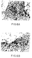
- Figure 6A depicts a low power view of an implantation site.
- the right and left margins are the original mesenteric folds and the cell polymer construct can be seen between the leaves.
- the polymer material shows a giant cell foreign body reaction around each of the fibers.
- Small nests and clusters of viable hepatocytes can be seen throughout the implant, and neovascularization is present throughout.
- Figure 6b is a higher power view documenting that the nests of hepatocytes clearly prefer the edges of the implant closest to the mesenteric fold and the associated blood supply.
- the hepatocytes are also not attached to the polymer fibers, indicating the preferential adhesivity that occurs to each other as well as to the matrix that they have laid down. There is an associated fibroblast reaction with collagen deposition.
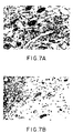
- Figure 7a is a photo of a polymeric matrix containing GHL implanted into the mesentery.
- Figure 7b is a photo of a polymeric matrix not containing GHL implanted into the mesentery.
- a method allowing large volumes of avascular tissue to be simultaneously implanted in a patient with minimal wounding is based on the discovery that multiple seeded polymeric matrices can be juxtaposed with certain tissues and undergo adequate exchange of nutrients and gases to grow and proliferate. These tissues, such as the mesentery and the omentum, have large surface areas and are highly vascular.
- the peritoneum is an extensive serous membrane lining the abdominal cavity and many of the organs in the cavity.
- the "mesentery” is ordinarily used to refer to the mesentery of the small intestine, a double-layered fold of peritoneum suspending it from the posterior abdominal wall.
- the attached border of the mesentery is only about 15 cm in length, and runs from approximately the second lumbar vertebra downward and to the right, crossing part of the duodenum, the aorta, the inferior vena cava, and across towards the right sacroiliac joint.
- the free or unattached border, containing the jejunaileum, is frilled out like an accordion, attaining a length ranging from three to six meters. The distance from the free border to the attached border ranges from 15 to 22 cm in length.
- the superior mesenteric artery and its branches Between the two layers of peritoneum, on the two surfaces of the mesentery, are the superior mesenteric artery and its branches, the accompanying veins, lymphatics, lymph nodes, connective tissue, and varying amounts of adipose tissue.
- the mesentery is shown in cross section in Figures 1A and 1B.
- the mesentery 10 With reference to Figure 1A, the mesentery 10 is connected to the superior 12 and inferior 14 mesenteric arteries, supplying blood to the small intestine 16.
- the mesentery 10 expands outwardly like a fan to the small intestine 16, the superior mesenteric artery 12 draining into the superior mesenteric vein.
- the inferior mesenteric vein 20 drains blood from the splenic 22, coronary and pyloric veins.
- the superior mesenteric vein 18 drains into the portal vein, leading to the liver 26.
- the vascular supply from the portal circulation supplies nutrients and normal metabolic factors to the implanted cells by diffusion until ingrowth of blood vessels following implantation provides for normal feedback mechanisms controlling the soluble products of the implanted cells.
- the omentum is a double fold of peritoneum attached to the stomach and connecting it with some of the other organs, including the intestines.
- Other sections of the peritoneum and isolated tissues having similar characteristics can also be used in the method for implanting large volumes of cells.
- chimeric cell-polymeric structures are formed by seeding biodegradable, biocompatible high surface area matrices with cells, derived from biopsy of the patient or a close relative or from cell culture, and implanting the seeded matrices between folds 32 of the mesentery 16.
- the folds 32 of the mesentery are approximated together to form a chimeric structure 34.
- the preferred material for forming the matrix or support structure is a biodegradable artificial polymer, for example, polylactic acid, polyglycolic acid, polyorthoester, polyanhydride, blends or copolymers thereof, which is degraded by hydrolysis at a controlled rate and absorbed, alone or in combination with non-degradable materials.
- the biodegradable materials provide the maximum control of degradability, manageability, size and configuration.
- other biocompatible polymeric materials including collagen and non-biodegradable materials, can be used to form the structures.
- non-degradable, or non-absorbable, materials include polypropylene, polyethylene terephthalate (Dacron polyester) and other polyesters, Teflon polytetrafluoroethylene; Teflon is a trade mark, ethylene vinyl acetate, nylon and stainless steel.
- Nylon is not preferred because it undergoes slow but constant hydration and degradation.
- Stainless steel filaments are difficult to fabricate.
- polypropylene and polyester are FDA approved as suture materials and as other implant components. They can be easily fabricated into monofilament, yarn or staple with varying fiber diameters. Both materials have very low orders of tissue reaction.
- these materials are overlaid with a second material such as basement membrane components, agar, agarose, gelatin, gum arabic, collagens, fibronectin, laminin, glycosaminoglycans, mixtures thereof, and other materials having properties similar to biological matrix molecules known to those skilled in the art of cell culture to enhance cell attachment.
- a second material such as basement membrane components, agar, agarose, gelatin, gum arabic, collagens, fibronectin, laminin, glycosaminoglycans, mixtures thereof, and other materials having properties similar to biological matrix molecules known to those skilled in the art of cell culture to enhance cell attachment.
- a second material such as basement membrane components, agar, agarose, gelatin, gum arabic, collagens, fibronectin, laminin, glycosaminoglycans, mixtures thereof, and other materials having properties similar to biological matrix molecules known to those skilled in the art of cell culture to enhance cell attachment.
- These materials can also affect longevity and maintenance of several liver-specific
- Attachment site density can be altered by selection or chemical modification of the polymeric support material, or by coating the substrate with a predetermined number of attachment molecules.
- a high pH carbonate buffer can be used to adsorb purified extracellular matrix (ECM) molecules, such as laminin and fibronectin, onto polymer substrates.
- ECM extracellular matrix
- Hepatocytes attached to low densities of purified ECM maintain a round morphology, high secretion rates for hepatocyte specific proteins (albumin, transferrin, fibrinogen), and low levels of DNA synthesis.
- Hepatocytes attached to high densities of purified ECM spread extensively, have lower secretion rates for the hepatocyte specific proteins, and high levels of DNA synthesis. This control is largely independent of the ECM molecule used, although there is a small effect on differentiation depending on the specific ECM molecule used.
- the major advantage of the biodegradable material is that it does not have to be removed once cell growth and formation of a functional mass has occurred.
- Another advantage of the biodegradable material is that compounds may be incorporated into the matrix for slow release during degradation of the matrix. For example, angiogenic compounds, nutrients, growth factors, inducers of differentiation or de-differentiation, products of secretion, immunomodulators, inhibitors of inflammation, regression factors, biologically active compounds which enhance or allow ingrowth of the lymphatic network or nerve fibers, and drugs can be incorporated into the matrix or provided in conjunction with the matrix, in solution or incorporated into a second biodegradable polymer matrix.
- multiple polymeric matrices formed of fibrous sheets are inserted into either a fresh bed or a prevascularized bed in the mesentery to create a structure having overall dimensions between a few microns and several centimeters.
- Matrices can be made in a variety of shapes, taking into consideration the requirements of adequate surface area for attachment of the number of cells required for implantation and formation of a function organ equivalent, and the requirements of adequate spacing between surfaces of attachment for nutrients and gases to diffuse into the interior of the matrices to each attached cell.
- the matrix is fibrous in nature. This can range from a non-woven felt-like mesh to a overlying or entangled single fibers to a sponge-like structure.
- other variables including fiber diameter, fiber density, knitted constructions, and fiber cross-over fusion or entanglement, must be considered.
- These matrices can be characterized in vitro on the basis of dry and wet compressibility, compressibility after absorbable component is removed (in the case of a mixture of absorbable and non-absorbable materials), breaking strength, suture pull-out strength, collagen distribution and microscopy. In vivo , matrices are assessed on the basis of attachment and viability of the cells is determined.
- Fibrous materials are commercially available in the form of sutures and non-woven felt-like materials. Suitable matrices can also be formed using standard techniques known to those skilled in the art such as solvent casting, spin casting or extrusion, or similar methodology. A useful felt-like material can be obtained from Davis & Geck. These types of fabrics made from a variety of biodegrading polymers have been used in many clinical applications and display very good tissue and vascular ingrowth when thinned to approximately 1/3 to 1/2 mm.
- Sponge-like fibrous matrices have been made by solvent casting a polymer solution containing leachable particulates.
- PLA/PLGA polymers have been cast containing NaCl particles sieved to the size range of 75 to 150 »m.
- the matrices were cast from a 10 wt% solution in methylene chloride into glass petri dishes, and the solvent was allowed to evaporate at room temperature under an atmosphere of methylene chloride.
- a 50/50 w/w blend of poly(DL) lactic acid, Mw 50,000 (Polysciences) and Dupont Medisorb 85:15 gave optimum results in terms of uniformity and porosity.
- These matrixes are 200 to 300 »m in thickness.
- a scanning electron micrograph of an example of this type of matrix is shown in Figure 5.
- the pore size for sponge-like structures, or interstitial spacing for fabrics, should be in the range of 100 to 200 »m for optimal ingrowth of vasculature.
- a sponge-like matrix can also be made by dipping the Davis & Geck poly(glycolic acid) mesh into a polymer solution, such as poly(lactic acid) or a copolymer of poly(lactic acid) and poly(glycolic acid), where the polymer is dissolved in a solvent for the second polymer but not the poly(glycolic acid), such as methylene chloride.
- the second polymer greatly increases the compressive strength of the poly(glycolic acid) mesh and forms a "meniscus" between the fibers which harden into flat surfaces for cell attachment.
- the PGA fabric is dipped into a two to twenty weight percent PLA:GA 85:15 solution in methylene chloride, excess polymer solution is blotted away, and the matrix is air dried.
- This method is applicable to any biocompatible polymer or substrate which is not soluble in a second biocompatible, biodegradable polymer solution.
- the surface area of the matrix or matrices to be implanted is determined based on the size of the recipient, as well as the type of cell(s) to be implanted.
- the average cell implant for a rat varies between 60 and 100 million cells per animal. This has been implanted using a surface area of approximately 36 cm2 of polymer material. It is calculated that the raw available surface area of mesentery that could be used for implantation for a 70 kg adult human is 2.68 m2. Modest cell application densities in the range of 700,000 cells per cm2 could theoretically allow implantation of 109 cells per adult human, 10% of the size of an adult human liver.
- a matrix containing one or more of these biologically active compounds is implanted in the tissue prior to implantation of seeded matrices, to prepare the implantation site, for example, using angiogenic compounds to pre-vascularize the site.
- endocrine cells such as hepatocytes, pancreatic cells or cells of the adrenal gland are proliferated on the matrices.
- Other cells such as cells of the nervous system, including hypothalamus and pituitary cells, lymphoid cells, mesodermal cells, such as fibroblasts, endothelial cells, and lymphatic cells, splenic cells, and cells of the genitourinary system, for example, renal endocrine tissues, and sex related endocrine tissues, can also be implanted using this method.
- the method locates these cells within the blood stream in close proximity to the blood supply the organs normally receive, between the portal and systemic systems. This exposes the cells to many of the factors present in the blood that aid in normal growth and proliferation.
- Cells of one or more types can be selected and grown on the matrix.
- the matrix structure and the length of time and conditions under which the cells are cultured in vitro are determined on an individual basis for each type of cell by measuring cell attachment (only viable cells remain attached to the polymers), extent of proliferation, and percent successful engraftment.
- cell attachment only viable cells remain attached to the polymers
- extent of proliferation and percent successful engraftment.
- cell attachment it is not necessary to culture cells in vitro , other than for purposes of attaching the cells to the matrix, prior to implantation if sufficient numbers of cells are available.
- Cells generally attach within a few hours.
- the most efficient technique for attaching cells to the mesh is to place a concentrated suspension of cells on the surface of the polymer, which is hydrophobic, and to allow the cell suspension to wick into the fabric over a period of about 30 min.
- the cells attach to the fibers mostly as individual cells but also in groups of two or three. Within the first twenty-four hours, the cells begin rearranging into clusters; at this point, some cells can be seen interacting a great deal with the fibers by wrapping all the way around an individual fiber. Within three days, the cells are almost completely organized into large clusters and groups of cells, and interact mostly with each other and not the fiber support.
- Cells can be obtained by biopsy, surgical excision from a donor, or from established cell lines. Methods for dissociation of tissue are known but may need to be optimized for cell type and source. For example, hepatocytes are dissociated using enzymes such as collagenase, by mechanical disruption, and/or treatment with chemical agents such as ethylenediamine tetraacetic acid (EDTA) and tetraphenylboron.
- enzymes such as collagenase
- EDTA ethylenediamine tetraacetic acid
- tetraphenylboron ethylenediamine tetraphenylboron.
- an immunosuppressant such as cyclosporine after implantation of the cell-matrix to increase viability of the implanted cells.
- an immunosuppressant such as cyclosporine
- Example 1 Isolation of Hepatocytes for Implantation on a Matrix in the Mesentery.
- Livers are perfused using a pump, autoclavable silastic tubing, a water bath and an air trap.
- the air trap positioned to eliminate air bubbles in the perfusion buffers, is a very important component of the system.
- the liver can be perfused in situ or after removal from the body. If possible, in situ perfusion is preferred. In situ , the liver remains in the abdominal cavity through both steps of the perfusion and only following completion of the collagenase perfusion is surgically removed for dissociation into the primary cell culture. There is no recirculation of the collagenase perfusate.
- Optimal results are obtained using Seglen's HEPES base initial buffer, pH 7.4, for the initial perfusion and the same buffer with 4.8 mmol/L CaCl2 for the subsequent collagenase perfusion.
- the perfusion with the buffer clears blood and calcium from the liver.
- Five to six minutes for a rat liver is sufficient.
- the second step must continue long enough to effect good dissociation of the liver but not so long as to cause excessive damage to the cells.
- five to ten minutes is optimal for the second step using rat liver, with some variation according to the age and weight of the donor and the activity of the collagenase.
- Optimal temperature of the water bath is 38 to 39 °C to produce a cannula output temperature of 35 to 36°C.
- Hepatocytes were obtained from Fischer 34 and Gunn rats by collagenase perfusion. Cells were seeded onto non-woven filamentous sheets of polyglycolic acid 1 x 3 cm in size and 2 mm thick to 500,000 cells/cm2. Recipient animals underwent laparotomy using sterile technique and sheets were placed between leaves of mesentery. Eight sheets were placed per animal and the leaves were approximated, creating a functional implant 1 x 1 x 3 cm.
- Biopsy at day five post implantation revealed neovascularization, moderate inflammatory reaction, and the presence of viable hepatocytes.
- This example demonstrates the successful implantation of large volumes of hepatocytes, cells which do not normally remain viable in the absence of a polymeric support, and which are difficult to proliferate in vivo to a number sufficient to form a functional organ equivalent, using multiple polymeric sheets placed into folds of the mesentery, with minimal trauma and blood loss.
- Example 2 Comparison of Absorbable Polymeric Matrix Materials and Coatings of Attachment Factors.
- the material should be biocompatible; it should also have properties that allow it to be fabricated into porous three dimensional devices with a high surface area/volume ratio to provide a significant surface area for cell attachment, and the resulting devices must have sufficient compressive strength to prevent collapse upon implantation.
- the material should be an adhesive substrate for the cells to be transplanted, and should ideally allow for retention of differentiated function by the cells and possibly for cell growth as well.
- Synthetic polymers can be manufactured reproducibly and have good mechanical properties. Furthermore, use of degradable polymers should preclude long-term infections and foreign body reactions that would prevent integration of the transplanted cells into the proper tissue architecture.
- polyesters in the polylactide (PLA), and polylactide-co-glycolide (PLGA) family have many ideal features.
- the interaction parameters of interest are cell adhesion, longevity, and maintenance of differentiated function. Adhesion is desired because the hepatocytes are anchorage-dependent and because it is unlikely that non-adherent cells will stay localized to the site of the implant. It is also desired that cells remain attached and viable on the substrate; and that the attachment sites not degrade during the time span attachment is desired.
- a substratum that allows retention of function in vitro is the optimal substratum for functional retention in vivo .
- the functional viability of the cells may be altered if necessary by coating the polymer substratum with extracellular matrix proteins.
- Substrates were made in the form of solvent cast films, and films of two different compositions were investigated.
- One set of films was made from poly(DL-lactide-co glycolide) with monomers in the ratio 85:15 lactide:glycolide (DuPont, Medisorb 85:15, weight ave Mw 40-100,000) and another set was made from a blend (50/50 w/w) of Medisorb 85:15 and poly(L-lactide) (Polysciences, Mw 50,000).
- Films were cast in 50 mm diameter glass petri dishes from a freshly made 15% polymer solution in methylene chloride (Mallinkrodt, analytical reagent grade); each film contained 0.4 gm polymer.
- the dishes containing the films were covered with petri dish covers and the solvent evaporated at room temperature for a minimum of 5 hr.
- the films were then placed under vacuum for 24-48 hr to remove residual solvent.
- the films were exposed to UV light for 90 min for purposes of sterilization and were stored desiccated until use.
- the films were prepared for culture by washing once with 5 ml phosphate buffered saline (PBS, pH 7.4) and then once with 5 ml complete culture medium.
- PBS phosphate buffered saline
- Control petri dishes 35 mm bacteriologic, Falcon #1008 were coated with Type I collagen (Vitrogen, Collagen Corp.) by adsorption from a 5 »g/ml solution of Vitrogen in 50 mM carbonate buffer (pH 9.4) for 16-20 hr at 4°C; the resulting surface concentration of collagen was 1 »g/cm2.
- Cells were dispersed in complete chemically defined serum-free culture medium (William's E with 10 ng/ml EGF (Collaborative Research), 20 mU/ml insulin (Gibco), 5 nM dexamethasone (Sigma), 20 mM pyruvate (Gibco), and 100 U/ml penicillin/streptomycin (Gibco)/(McGowan); cell viability following dispersion was 80-90% as determined by trypan blue exclusion. Dead cells and debris were removed by centrifugation in an iso-density Percoll solution (Kreamer, B.L., et al., In Vitro Cell. Dev. Biol. , 22(4), 201-207 (1986)) and the resulting pellet was washed three times with complete medium prior to planting the cells. Viability at plating was 88-98%.
- cells were plated at a concentration of 30,000 viable cells/cm2 culture surface area (300,000 cells/dish for 35 mm control dishes, 600,000 cells dish for 50 mm polymer films; 150,000 cells/ml). Following an attachment period of 2-4 hr (maximum attachment to all substrates occurred within 90 min), the medium was changed to remove unattached cells and then cells were maintained in serum-free medium with daily medium changes.
- Cell attachment to substrates was measured by direct counting or by determining relative protein content.
- direct counting cells were removed from the substrate using 0.05% trypsin/EDTA (Gibco).
- Cells plated on collagen-coated substrates required prolonged (30-45 min) treatment with trypsin for adequate cell dispersion, and plates were inspected visually before counting to ensure all cells had been removed from the substrate.
- quantitative binding and extraction of the dye flavianic acid NYS, Sigma
- the assay was calibrated for cell number by measuring the response of cells plated at two different concentrations (300,000 and 600,000 cells/plate) and counting identical plates seeded at the same concentrations; each point was measured in triplicate.
- the rate of cell attachment was measured by seeding replicate plates at 30,000 cells/cm2 surface area. At each time point, three identical plates were sacrificed for measurement of cell number by the NYS dye-binding assay.
- deoxyribonuclease I (United States Biochemical Corp.) was added to the attachment medium at concentrations 0, 10, 100, and 1000 U/ml. Cells were plated on the standard vitrogen-coated 35 mm polystyrene dishes and attachment was determined on duplicate plates.
- the kinetics of cell attachment to substrates were studied at cell surface concentrations of 30,000 cells/cm2 and below to minimize the role of cell-cell interactions.
- a two dimensional surface concentration is relevant because the cells settle to the surface quite quickly regardless of the total volume, and the surface concentration reflects the degree to which cells may interact at the attachment interface and the relative competition for sites; i.e., for cells 20-25 »m in diameter, total surface coverage with spheres would correspond to 160,000-250,000 cells/cm2, so 30,000 cells/cm2 would represent less than 20% surface coverage.
- the volume concentration is important because attachment may be inhibited by cell-secreted proteins.)
- the rate of attachment to both types of polymer films is similar and maximum attachment is achieved in 60 min, while maximum attachment to the collagen-coated control dishes required about 120 min. Hepatocytes were observed to attach primarily as single cells, and no difference in the kinetics of attachment to collagen-coated dishes was observed at a four-fold lower cell concentration (7500 cells/cm2).
- cell-cell interactions significantly affect the number of cells attaching to the substrate and the pattern of hepatocyte attachment is quite different for the polymer substrates and controls.
- the cells were observed to form aggregates of 2-10 cells, and while these aggregates could be seen to attach to the collagen-coated substrates, they did not appear to interact with the polymer films.
- the formation of cell aggregates was not affected by the presence of DNAase (10-1000 U/ml), as would be expected if the leakage of DNA from dead cells was causing nonspecific aggregation.
- the polymer substrates used for culture are opaque so scanning electron microscopy can be used to observe the morphology of the cells.
- the cells maintained on the collagen-coated polystyrene dishes were in general highly spread and flat; surface microvilli were observed predominantly in the center of the cells.
- the morphology of cells on the polymer substrates was heterogeneous; both highly rounded and spread cells were observed.
- both rounded and spread cells had numerous surface microvilli.
- the growth pattern of hepatocytes maintained on polymer films cast from the blend of PLA and PLGA is similar to that of hepatocytes maintained on the collagen control substrates and the DNA synthesis rates are equivalent. Attrition of cells begins after three days in culture.
- the films cast from pure PLGA 85:15 did not prove to be good substrates for cell longevity at the cell concentration investigated (20,000 cells/cm2). After 3-4 days, the cells detached from the substrate in large wisps or cords. Similar detachment of hepatocytes from the substrate when cells are cultured on fibronectin-coated polystyrene dishes (10 »g/cm2 10,000-30,000 cells/cm2) has been observed. Such detachment of cell sheets or cords from the substratum has also been observed in other systems and has been attributed to cell-cell tensions that overcome the cell-substratum tension.
- the retention of hepatocellular function on polymer blend films was assessed by measuring the rate of albumin secretion.
- the rate of albumin secretion (»g/106 cells/day) by cells maintained on the polymer blend films increased almost twofold over the five days in culture; in contrast, albumin secretion for cells on the collagen control dishes decreased over 60%.
- the decline in albumin synthesis for cells maintained on collagen was the same for collagen surface concentrations of 1 and 10 »g/cm2.
- the secretion rate of albumin by cells on the polymer blend films is in the range of the reported in vivo rate for rats [17.3-19.4 mg/gm liver/24 hr (Peters, T.Jr., and J.C. Peters, J. Biol. Chem. , 247(12), 3858-3863 (1972)) which corresponds to 144-162 »g/106 cells/24 hr based on 120 x 106 hepatocytes/gm liver.
- the maximum cell concentration that could be accommodated on the polymer surface was 50,000 cells/cm2, and the number of cells attached to the substrate declined if the number of cells plated at the surface exceeded 100,000 cells/cm2. While the maximum in cell attachment of 50,000 cells/cm2 may be a sufficient cell number in implant situations where cell growth is desired, higher cell attachment densities may be required if extensive cell-cell contact is necessary for retention of function by the implanted cells or if more efficient use of the polymer surface area is needed.
- coating the polymer substrates with ng-»g /cm2 amounts of extracellular matrix proteins such as collagen (or laminin or fibronectin) may be used to enhance cell adhesion for transplant matrices if high cell surface densities are required.
- Example 3 Prevascularization of a site for subsequent Implantation of a Cell Seeded Matrix.
- FIG. 6a depicts a low power view of an implantation site.
- the right and left margins are the original mesenteric folds and the cell polymer construct can be seen between the leaves.
- the polymer material shows a giant cell foreign body reaction around each of the fibers. There is a modest inflammatory cell infiltrate in the interstices.
- Figure 6b is a higher power view documenting that the nests of hepatocytes clearly prefer the edges of the implant closest to the mesenteric fold and the associated blood supply.
- the hepatocytes are also not attached to the polymer fibers, indicating the preferential adhesivity that occurs to each other as well as to the matrix that they have laid down. There is an associated fibroblast reaction with collagen deposition.
- Prevascularization was accomplished by implantation of polymer loaded with the tripeptide glycine-histidine-lysine (GHL), a known potent angiogenesis agent as well as an hepatotrophic agent.
- GHL tripeptide glycine-histidine-lysine
- Figure 7a is a photo of a polymeric matrix containing GHL implanted into the mesentery.
- Figure 7b is a photo of a polymeric matrix not containing GHL implanted into the mesentery.
- the matrices did not include cells.
- the polymer is in the upper left hand corners.
- the arrows in Figure 7a denote the large, dense vascular network that arose over the five-day implantation period of the GHL containing polymeric matrix.
Claims (24)
- Verwendung einer Matrixstruktur, die ein biokompatibles Material in einer faserigen Form mit interstitiellen Abständen im Bereich von 100 bis 200 »m und mit daran angehefteten lebensfähigen parenchymalen Zellen umfaßt, zur Herstellung eines chirurgischen Implantats zur Verwendung in einem chirurgischen Verfahren, das die Implantation einer Vielzahl von Matrizen zwischen Gewebefalten mit großer Oberfläche und Vaskularisation nahe der Oberfläche des Gewebes umfaßt.
- Verwendung nach Anspruch 1, wobei das Gewebe das Mesenterium, das Omentum oder das Peritoneum ist.
- Verwendung nach Anspruch 1 oder 2, wobei das Material ein biologisch abbaubares Polymer ist, ausgewählt aus der Gruppe Polymilchsäure, Polyglykolsäure, Polyorthoester, Polyanhydrid, Kollagen und Copolymere, Mischungen und Kombinationen daraus.
- Verwendung nach Anspruch 1 oder 2, wobei das Material ein nicht-abbaubares oder nicht-absorbierbares Material, ausgewählt aus der Gruppe Polypropylen, Polyethylenterephthalat und andere Polyester, Polytetrafluorethylen, Ethylenvinylacetat, Nylon, rostfreier Stahl und Kombinationen daraus, ist.
- Verwendung nach einem der Ansprüche 1 bis 4, wobei das Matrixmaterial mit einem Anheftungsfaktor, ausgewählt aus der Gruppe Grundmembrankompopenten, Agar, Agarose, Gelatine, Gummi arabicum, Kollagene, Fibronektin, Laminin, Hyaluronsäure, Glycosaminoglycane, Anheftungspeptide und Mischungen daraus, beschichtet ist.
- Verwendung nach einem der Ansprüche 1 bis 5, wobei die Matrix desweiteren eine Verbindung, ausgewählt aus der Gruppe angiogene Verbindungen, Nährstoffe, Wachstumsfaktoren, Induktoren einer Differenzierung oder Dedifferenzierung, Sekretionsprodukte, Immunmodulatoren, Entzündungshemmer, Regressionsfaktoren, biologisch aktive Verbindungen, die das Einwachsen des lymphatischen Netzwerkes oder der Nervenfasern erhöhen oder ermöglichen, und Mischungen daraus, enthält.
- Verwendung nach einem der Ansprüche 1 bis 6, wobei die Zellen aus der Gruppe Hepatozyten, Pankreaszellen, Zellen der Nebenniere, lymphoide Zellen, Zellen des Nervensystems, Fibroblasten, Endothelzellen, lymphatische Zellen, Milzzellen und Zellen des Geschlechtssystems, ausgewählt sind.
- Verwendung nach einem der Ansprüche 1 bis 7, wobei die Matrixstruktur eine faserige netzartige Form aufweist.
- Verwendung nach einem der Ansprüche 1 bis 7, wobei die Matrixstruktur eine schwammartige Form aufweist.
- Verwendung nach einem der Ansprüche 1 bis 7, wobei die Matrixstruktur die Form einer Lage besitzt.
- Verwendung nach Anspruch 10, wobei die Dicke der Lage im Bereich von 200 »m bis 2 mm liegt.
- Verwendung nach einem der Ansprüche 1 bis 3 oder einem der Ansprüche 5 bis 11, bei Nichtabhängigkeit von Anspruch 4, gebildet durch Beschichten eines biokompatiblen faserigen Netzes mit einer zweiten biokompatiblen, biologisch abbaubaren Polymerlösung, wobei das faserige Netzmaterial in der zweiten Polymerlösung nicht löslich ist.
- Verwendung nach Anspruch 12, wobei das faserige Netz aus Poly(glykolsäure) gebildet ist und das zweite Polymer aus der Gruppe aus Poly(milchsäure) und Poly(milchsäure-Glykolsäure)-Copolymere, ausgewählt ist.
- Die Verwendung nach Anspruch 12 oder 13, wobei das zweite Polymer zwischen den das faserige Netz bildenden Fasern flache Oberflächen ausbildet.
- Matrixlagenstruktur, die ein biokompatibles Material in einer faserigen Form mit interstitiellen Abständen im Bereich von 100 bis 200 »m und mit daran angehefteten lebensfähigen parenchymalen Zellen umfaßt, wobei die Dicke der Matrixstruktur im Bereich von 200 »m bis 2 mm liegt.
- Matrixlagenstruktur nach Anspruch 15, wobei die Dicke der Matrixstruktur im Bereich von 200 bis 300 »m liegt.
- Matrixlagenstruktur nach Anspruch 15, wobei das Material ein biologisch abbaubares Polymer ist, ausgewählt aus der Gruppe Polymilchsäure, Polyglykolsäure, Polyortnoester, Polyanhydrid, Kollagen und Copolymere, Mischungen und Kombinationen daraus.
- Matrixlagenstruktur nach Anspruch 15, wobei das Material ein nicht-abbaubares oder nicht-absorbierbares Material, ausgewählt aus der Gruppe Polypropylen, Polyethylenterephthalat und andere Polyester, Polytetra-fluorethylen, Ethylenvinylacetat, Nylon, rostfreier Stahl und Kombinationen daraus, ist.
- Matrixlagenstruktur nach einem der Ansprüche 15 bis 18, wobei das Matrixmaterial mit einem Anheftungsfaktor, ausgewählt aus der Gruppe Grundmembrankomponenten, Agar, Agarose, Gelatine, Gummi arabicum, Kollagene, Fibronektin, Laminin, Hyaluronsäure, Glycosaminoglycane, Anheftungspeptide und Mischungen daraus, beschichtet ist.
- Matrixlagenstruktur nach einem der Ansprüche 15 bis 19, wobei die Matrix desweiteren eine Verbindung, ausgewählt aus der Gruppe angiogene Verbindungen, Nährstoffe, Wachstumsfaktoren, Induktoren einer Differenzierung oder Dedifferenzierung, Sekretionsprodukte, Immunmodulatoren, Entzündungshemmer, Regressionsfaktoren, biologisch aktive Verbindungen, die das Einwachsen des lymphatischen Netzwerkes oder der Nervenfasern erhöhen oder ermöglichen, und Mischungen daraus, enthält.
- Matrixlagenstruktur nach einem der Ansprüche 15 bis 20, wobei die Zellen aus der Gruppe Hepatozyten, Pankreaszellen, Zellen der Nebenniere, lymphoide Zellen, Zellen des Nervensystems, Fibroblasten, Endothelzellen, lymphatische Zellen, Milzsellen und Zellen des Geschlechtssystems ausgewählt sind.
- Matrixlagenstruktur nach einem der Ansprüche 15 bis 17 oder einem der Ansprüche 19 bis 21 bei Nichtabhängigkeit von Anspruch 18, gebildet durch Beschichten eines biokompatiblen faserige Netzes mit einer zweiten biokompatiblen, biologisch abbaubaren Polymerlösung, wobei das faserige Netzmaterial in der zweiten Polymerlösung nicht löslich ist.
- Matrixlagenstruktur nach Anspruch 22, wobei das faserige Netz aus Poly(glykolsäure) gebildet ist und das zweite Polymer aus der Gruppe Poly(milchsäure) und Poly(milchsäure-Glykolsäure)-Copolymere, ausgewählt ist.
- Matrixlagenstruktur nach Anspruch 22 oder 23, wobei das zweite Polymer zwischen den die das faserige Netz bildenden Fasern flache Oberflächen ausbildet.
Applications Claiming Priority (3)
| Application Number | Priority Date | Filing Date | Title |
|---|---|---|---|
| US34315889A | 1989-04-25 | 1989-04-25 | |
| US343158 | 1989-04-25 | ||
| PCT/US1990/002257 WO1990012604A1 (en) | 1989-04-25 | 1990-04-25 | Method for implanting large volumes of cells on polymeric matrices |
Publications (2)
| Publication Number | Publication Date |
|---|---|
| EP0422209A1 EP0422209A1 (de) | 1991-04-17 |
| EP0422209B1 true EP0422209B1 (de) | 1995-03-15 |
Family
ID=23344941
Family Applications (1)
| Application Number | Title | Priority Date | Filing Date |
|---|---|---|---|
| EP90907948A Expired - Lifetime EP0422209B1 (de) | 1989-04-25 | 1990-04-25 | Implantaten für grosse mengen von zellen auf polymerische matrizen |
Country Status (8)
| Country | Link |
|---|---|
| EP (1) | EP0422209B1 (de) |
| JP (3) | JP3073766B2 (de) |
| AT (1) | ATE119787T1 (de) |
| AU (1) | AU636346B2 (de) |
| CA (1) | CA2031532C (de) |
| DE (1) | DE69017820T2 (de) |
| ES (1) | ES2072434T3 (de) |
| WO (1) | WO1990012604A1 (de) |
Cited By (14)
| Publication number | Priority date | Publication date | Assignee | Title |
|---|---|---|---|---|
| EP0771849A2 (de) | 1995-11-06 | 1997-05-07 | Ethicon, Inc. | Polymermischungen die Polyoxaestern enthalten |
| EP0771832A2 (de) | 1995-11-06 | 1997-05-07 | Ethicon, Inc. | Mischungen von absorbierbaren Polyoxaestern die Amin und/oder Amido-Gruppen enthalten |
| US6210436B1 (en) | 1998-05-18 | 2001-04-03 | Scimed Life Systems Inc. | Implantable members for receiving therapeutically useful compositions |
| US6316522B1 (en) | 1997-08-18 | 2001-11-13 | Scimed Life Systems, Inc. | Bioresorbable hydrogel compositions for implantable prostheses |
| US6866860B2 (en) | 2002-12-19 | 2005-03-15 | Ethicon, Inc. | Cationic alkyd polyesters for medical applications |
| US6872799B2 (en) | 2002-12-18 | 2005-03-29 | Ethicon, Inc. | Functionalized polymers for medical applications |
| US6967234B2 (en) | 2002-12-18 | 2005-11-22 | Ethicon, Inc. | Alkyd-lactone copolymers for medical applications |
| US7005136B2 (en) | 2002-03-29 | 2006-02-28 | Ethicon, Inc. | Bone replacement materials utilizing bioabsorbable liquid polymers |
| US7026374B2 (en) | 2002-06-25 | 2006-04-11 | Aruna Nathan | Injectable microdispersions for medical applications |
| US7030127B2 (en) | 2001-06-29 | 2006-04-18 | Ethicon, Inc. | Composition and medical devices utilizing bioabsorbable polymeric waxes |
| US7034037B2 (en) | 2001-06-29 | 2006-04-25 | Ethicon, Inc. | Compositions and medical devices utilizing bioabsorbable polymeric waxes and rapamycin |
| US7101566B2 (en) | 2002-06-28 | 2006-09-05 | Ethicon, Inc. | Polymer coated microparticles for sustained release |
| US7368125B2 (en) | 2002-06-05 | 2008-05-06 | Ethicon, Inc. | Amphiphilic polymers for medical applications |
| US8623413B2 (en) | 2002-03-29 | 2014-01-07 | Ethicon, Inc. | Compositions and medical devices utilizing bioabsorbable liquid polymers |
Families Citing this family (36)
| Publication number | Priority date | Publication date | Assignee | Title |
|---|---|---|---|---|
| US6309635B1 (en) | 1986-11-20 | 2001-10-30 | Children's Medical Center Corp. | Seeding parenchymal cells into compression resistant porous scaffold after vascularizing in vivo |
| US5376118A (en) * | 1989-05-10 | 1994-12-27 | United States Surgical Corporation | Support material for cell impregnation |
| AU6908591A (en) * | 1989-12-07 | 1991-07-18 | Biosynthesis, Inc. | Hollow viscus prosthesis and method of implantation |
| ES2193132T3 (es) * | 1990-10-19 | 2003-11-01 | Univ New York | Procedimiento para transplantar celulas en el cerebro y utilizaciones terapeuticas del mismo. |
| US5618531A (en) | 1990-10-19 | 1997-04-08 | New York University | Method for increasing the viability of cells which are administered to the brain or spinal cord |
| US5849686A (en) * | 1991-03-11 | 1998-12-15 | Creative Biomolecules, Inc. | Morphogen-induced liver regeneration |
| WO1993007913A1 (en) * | 1991-10-24 | 1993-04-29 | Children's Medical Center Corporation | Neomorphogenesis of urological structures in vivo from cell culture |
| WO1993008850A1 (en) * | 1991-10-30 | 1993-05-13 | Massachusetts Institute Of Technology | Prevascularized polymeric implants for organ transplantation |
| US5610753A (en) | 1991-12-12 | 1997-03-11 | Eastman Kodak Company | Optical design of laser scanner to reduce thermal sensitivity |
| DE4206585C2 (de) * | 1992-03-03 | 1994-11-24 | Augustinus Dr Med Bader | Vorrichtung zur Massenkultur von Zellen |
| ATE208169T1 (de) * | 1992-04-01 | 2001-11-15 | Baxter Int | Systeme zum einpflanzen von gefässbildendem gewebe |
| GB9210574D0 (en) * | 1992-05-18 | 1992-07-01 | Ca Nat Research Council | Biotherapeutic cell-coated microspheres for wound/burn and prothesis implant applications |
| CA2140905A1 (en) * | 1992-07-29 | 1994-01-30 | Keith E. Dionne | Use of pouch of implantation of living cells |
| ATE162078T1 (de) | 1992-09-16 | 1998-01-15 | Creative Biomolecules Inc | Morphogeninduzierte regenerierung der leber |
| US6689608B1 (en) | 1993-02-01 | 2004-02-10 | Massachusetts Institute Of Technology | Porous biodegradable polymeric materials for cell transplantation |
| WO1994025079A1 (en) * | 1993-04-23 | 1994-11-10 | Massachusetts Institute Of Technology | Porous biodegradable polymeric materials for cell transplantation |
| DE938893T1 (de) * | 1993-08-10 | 2000-03-09 | Gore & Ass | Zelleinkapselungsvorrichtung |
| US5549675A (en) * | 1994-01-11 | 1996-08-27 | Baxter International, Inc. | Method for implanting tissue in a host |
| US20020055786A1 (en) | 1994-08-16 | 2002-05-09 | Anthony Atala | Reconstruction of urological structures with polymeric matrices |
| US5855610A (en) | 1995-05-19 | 1999-01-05 | Children's Medical Center Corporation | Engineering of strong, pliable tissues |
| WO1997010807A1 (en) * | 1995-09-22 | 1997-03-27 | Gore Hybrid Technologies, Inc. | Improved cell encapsulation device |
| US5916585A (en) * | 1996-06-03 | 1999-06-29 | Gore Enterprise Holdings, Inc. | Materials and method for the immobilization of bioactive species onto biodegradable polymers |
| ATE248615T1 (de) | 1997-10-31 | 2003-09-15 | Childrens Medical Center | Blasenrekonstruktion |
| DE69922941D1 (de) * | 1998-04-24 | 2005-02-03 | Transkaryotic Therapies Inc | Verabreichung der therapeutischen proteine durch implantation der genetischen modifizierten zellen in das omentum |
| US6245345B1 (en) * | 1998-07-07 | 2001-06-12 | Atrix Laboratories, Inc. | Filamentous porous films and methods for producing the same |
| WO2002030481A1 (en) * | 2000-10-10 | 2002-04-18 | Massachusetts Institute Of Technology | Cell delivery using controllably degradable mesh-gel constructs |
| KR20010044624A (ko) * | 2001-03-12 | 2001-06-05 | 정재호 | 연골조직공학을 위한 연골지지체의 제조방법 및 이연골지지체를 사용하여 제조한 인공연골 |
| US8268361B2 (en) | 2005-10-26 | 2012-09-18 | Ahlfors Jan-Eric W | Acellular bioabsorbable tissue regeneration matrices |
| US10590391B2 (en) | 2007-06-08 | 2020-03-17 | Wake Forest University Health Sciences | Selective cell therapy for the treatment of renal failure |
| AU2008262333B2 (en) | 2007-06-08 | 2014-07-17 | Wake Forest University Health Sciences | Selective cell therapy for the treatment of renal failure |
| CN102271692B (zh) | 2008-11-12 | 2014-05-21 | 坦吉恩股份有限公司 | 分离的肾细胞及其用途 |
| WO2010057015A1 (en) | 2008-11-14 | 2010-05-20 | Wake Forest University Health Sciences | Kidney structures and methods of forming the same |
| WO2011143499A1 (en) | 2010-05-12 | 2011-11-17 | Tengion, Inc. | Bioactive renal cells |
| US9386863B1 (en) | 2010-06-18 | 2016-07-12 | Boba Inc. | Child carrier and methods of use |
| CN103298498B (zh) | 2010-11-10 | 2017-12-08 | 因瑞金公司 | 用于器官增强的注射制剂 |
| US11123372B2 (en) | 2016-07-29 | 2021-09-21 | Prokidney | Bioactive renal cells for the treatment of chronic kidney disease |
Family Cites Families (4)
| Publication number | Priority date | Publication date | Assignee | Title |
|---|---|---|---|---|
| US4637931A (en) * | 1984-10-09 | 1987-01-20 | The United States Of America As Represented By The Secretary Of The Army | Polyactic-polyglycolic acid copolymer combined with decalcified freeze-dried bone for use as a bone repair material |
| CH670760A5 (de) * | 1986-06-02 | 1989-07-14 | Sulzer Ag | |
| US5041138A (en) * | 1986-11-20 | 1991-08-20 | Massachusetts Institute Of Technology | Neomorphogenesis of cartilage in vivo from cell culture |
| CA1340581C (en) * | 1986-11-20 | 1999-06-08 | Joseph P. Vacanti | Chimeric neomorphogenesis of organs by controlled cellular implantation using artificial matrices |
-
1990
- 1990-04-25 AT AT90907948T patent/ATE119787T1/de not_active IP Right Cessation
- 1990-04-25 ES ES90907948T patent/ES2072434T3/es not_active Expired - Lifetime
- 1990-04-25 AU AU55691/90A patent/AU636346B2/en not_active Expired
- 1990-04-25 CA CA002031532A patent/CA2031532C/en not_active Expired - Lifetime
- 1990-04-25 JP JP02507248A patent/JP3073766B2/ja not_active Expired - Lifetime
- 1990-04-25 WO PCT/US1990/002257 patent/WO1990012604A1/en active IP Right Grant
- 1990-04-25 EP EP90907948A patent/EP0422209B1/de not_active Expired - Lifetime
- 1990-04-25 DE DE69017820T patent/DE69017820T2/de not_active Expired - Lifetime
-
1998
- 1998-03-18 JP JP10069123A patent/JPH10263070A/ja active Pending
-
2001
- 2001-05-14 JP JP2001144028A patent/JP2001314498A/ja active Pending
Cited By (18)
| Publication number | Priority date | Publication date | Assignee | Title |
|---|---|---|---|---|
| EP0771832A2 (de) | 1995-11-06 | 1997-05-07 | Ethicon, Inc. | Mischungen von absorbierbaren Polyoxaestern die Amin und/oder Amido-Gruppen enthalten |
| EP0771849A2 (de) | 1995-11-06 | 1997-05-07 | Ethicon, Inc. | Polymermischungen die Polyoxaestern enthalten |
| US6946499B2 (en) | 1997-08-18 | 2005-09-20 | Scimed Life Systems, Inc. | Bioresorbable hydrogel compositions for implantable prostheses |
| US7109255B2 (en) | 1997-08-18 | 2006-09-19 | Scimed Life Systems, Inc. | Bioresorbable hydrogel compositions for implantable prostheses |
| US6316522B1 (en) | 1997-08-18 | 2001-11-13 | Scimed Life Systems, Inc. | Bioresorbable hydrogel compositions for implantable prostheses |
| US6660827B2 (en) | 1997-08-18 | 2003-12-09 | Scimed Life Systems, Inc. | Bioresorbable hydrogel compositions for implantable prostheses |
| US6447542B1 (en) | 1998-05-18 | 2002-09-10 | Scimed Life Systems, Inc. | Implantable members for receiving therapeutically useful compositions |
| US6210436B1 (en) | 1998-05-18 | 2001-04-03 | Scimed Life Systems Inc. | Implantable members for receiving therapeutically useful compositions |
| US7030127B2 (en) | 2001-06-29 | 2006-04-18 | Ethicon, Inc. | Composition and medical devices utilizing bioabsorbable polymeric waxes |
| US7034037B2 (en) | 2001-06-29 | 2006-04-25 | Ethicon, Inc. | Compositions and medical devices utilizing bioabsorbable polymeric waxes and rapamycin |
| US7005136B2 (en) | 2002-03-29 | 2006-02-28 | Ethicon, Inc. | Bone replacement materials utilizing bioabsorbable liquid polymers |
| US8623413B2 (en) | 2002-03-29 | 2014-01-07 | Ethicon, Inc. | Compositions and medical devices utilizing bioabsorbable liquid polymers |
| US7368125B2 (en) | 2002-06-05 | 2008-05-06 | Ethicon, Inc. | Amphiphilic polymers for medical applications |
| US7026374B2 (en) | 2002-06-25 | 2006-04-11 | Aruna Nathan | Injectable microdispersions for medical applications |
| US7101566B2 (en) | 2002-06-28 | 2006-09-05 | Ethicon, Inc. | Polymer coated microparticles for sustained release |
| US6872799B2 (en) | 2002-12-18 | 2005-03-29 | Ethicon, Inc. | Functionalized polymers for medical applications |
| US6967234B2 (en) | 2002-12-18 | 2005-11-22 | Ethicon, Inc. | Alkyd-lactone copolymers for medical applications |
| US6866860B2 (en) | 2002-12-19 | 2005-03-15 | Ethicon, Inc. | Cationic alkyd polyesters for medical applications |
Also Published As
| Publication number | Publication date |
|---|---|
| AU636346B2 (en) | 1993-04-29 |
| CA2031532C (en) | 2003-02-25 |
| DE69017820D1 (de) | 1995-04-20 |
| AU5569190A (en) | 1990-11-16 |
| ATE119787T1 (de) | 1995-04-15 |
| JPH04501080A (ja) | 1992-02-27 |
| JPH10263070A (ja) | 1998-10-06 |
| ES2072434T3 (es) | 1995-07-16 |
| CA2031532A1 (en) | 1990-10-26 |
| DE69017820T2 (de) | 1995-10-05 |
| EP0422209A1 (de) | 1991-04-17 |
| JP3073766B2 (ja) | 2000-08-07 |
| WO1990012604A1 (en) | 1990-11-01 |
| JP2001314498A (ja) | 2001-11-13 |
Similar Documents
| Publication | Publication Date | Title |
|---|---|---|
| EP0422209B1 (de) | Implantaten für grosse mengen von zellen auf polymerische matrizen | |
| EP0299010B1 (de) | Herstellung von körperorganen durch kontroliertes zellwachstum auf künstlicher matrix | |
| US5759830A (en) | Three-dimensional fibrous scaffold containing attached cells for producing vascularized tissue in vivo | |
| Cima et al. | Tissue engineering by cell transplantation using degradable polymer substrates | |
| US5567612A (en) | Genitourinary cell-matrix structure for implantation into a human and a method of making | |
| US5851833A (en) | Neomorphogenesis of urological structures in vivo from cell culture | |
| EP0610423B1 (de) | Vorvaskularisierte polymerimplantate für organtransplantation | |
| US5830507A (en) | Biotherapeutic cell-coated microspheres | |
| Cohen et al. | Design of synthetic polymeric structures for cell transplantation and tissue engineering | |
| US5849588A (en) | Methods of use of a three-dimensional liver cell and tissue culture system | |
| US5942436A (en) | Culturing liver cells | |
| US20110281351A1 (en) | Process for producing laminated high-density cultured artificial tissue, and laminated high-density cultured artificial tissue | |
| US6309635B1 (en) | Seeding parenchymal cells into compression resistant porous scaffold after vascularizing in vivo | |
| CA2589588A1 (en) | Materials and methods for minimally-invasive administration of a cell-containing flowable composition | |
| EP2815773A1 (de) | Matrix und Implantat zur Gewebezüchtung | |
| Cima et al. | Polymers for tissue and organ culture | |
| Chen et al. | High-density culture of hepatocytes in a packed-bed bioreactor using a fibrous scaffold from plant | |
| JP4061487B2 (ja) | 血管新生を目的とする移植材料及びその製造方法 | |
| Ito et al. | Implantation of cell-seeded biodegradable polymers for tissue reconstruction | |
| AU668959C (en) | Biotherapeutic cell-coated microspheres | |
| JPH01170466A (ja) | 生体内インプラント材料 |
Legal Events
| Date | Code | Title | Description |
|---|---|---|---|
| PUAI | Public reference made under article 153(3) epc to a published international application that has entered the european phase |
Free format text: ORIGINAL CODE: 0009012 |
|
| 17P | Request for examination filed |
Effective date: 19901228 |
|
| AK | Designated contracting states |
Kind code of ref document: A1 Designated state(s): AT BE CH DE ES FR GB IT LI NL SE |
|
| RAP1 | Party data changed (applicant data changed or rights of an application transferred) |
Owner name: MASSACHUSETTS INSTITUTE OF TECHNOLOGY Owner name: CHILDREN'S MEDICAL CENTER CORPORATION |
|
| 17Q | First examination report despatched |
Effective date: 19930712 |
|
| GRAA | (expected) grant |
Free format text: ORIGINAL CODE: 0009210 |
|
| AK | Designated contracting states |
Kind code of ref document: B1 Designated state(s): AT BE CH DE ES FR GB IT LI NL SE |
|
| REF | Corresponds to: |
Ref document number: 119787 Country of ref document: AT Date of ref document: 19950415 Kind code of ref document: T |
|
| REF | Corresponds to: |
Ref document number: 69017820 Country of ref document: DE Date of ref document: 19950420 |
|
| ITF | It: translation for a ep patent filed |
Owner name: NOTARBARTOLO & GERVASI S.R.L. |
|
| ET | Fr: translation filed | ||
| REG | Reference to a national code |
Ref country code: ES Ref legal event code: FG2A Ref document number: 2072434 Country of ref document: ES Kind code of ref document: T3 |
|
| PLBE | No opposition filed within time limit |
Free format text: ORIGINAL CODE: 0009261 |
|
| STAA | Information on the status of an ep patent application or granted ep patent |
Free format text: STATUS: NO OPPOSITION FILED WITHIN TIME LIMIT |
|
| 26N | No opposition filed | ||
| REG | Reference to a national code |
Ref country code: GB Ref legal event code: IF02 |
|
| PGFP | Annual fee paid to national office [announced via postgrant information from national office to epo] |
Ref country code: ES Payment date: 20090427 Year of fee payment: 20 |
|
| PGFP | Annual fee paid to national office [announced via postgrant information from national office to epo] |
Ref country code: DE Payment date: 20090429 Year of fee payment: 20 Ref country code: IT Payment date: 20090428 Year of fee payment: 20 Ref country code: SE Payment date: 20090429 Year of fee payment: 20 Ref country code: FR Payment date: 20090417 Year of fee payment: 20 Ref country code: AT Payment date: 20090401 Year of fee payment: 20 Ref country code: NL Payment date: 20090423 Year of fee payment: 20 |
|
| PGFP | Annual fee paid to national office [announced via postgrant information from national office to epo] |
Ref country code: BE Payment date: 20090528 Year of fee payment: 20 |
|
| PGFP | Annual fee paid to national office [announced via postgrant information from national office to epo] |
Ref country code: CH Payment date: 20090427 Year of fee payment: 20 |
|
| PGFP | Annual fee paid to national office [announced via postgrant information from national office to epo] |
Ref country code: GB Payment date: 20090429 Year of fee payment: 20 |
|
| BE20 | Be: patent expired |
Owner name: *MASSACHUSETTS INSTITUTE OF TECHNOLOGY Effective date: 20100425 Owner name: *CHILDREN'S MEDICAL CENTER CORP. Effective date: 20100425 |
|
| REG | Reference to a national code |
Ref country code: CH Ref legal event code: PL |
|
| REG | Reference to a national code |
Ref country code: NL Ref legal event code: V4 Effective date: 20100425 |
|
| REG | Reference to a national code |
Ref country code: GB Ref legal event code: PE20 Expiry date: 20100424 |
|
| EUG | Se: european patent has lapsed | ||
| REG | Reference to a national code |
Ref country code: ES Ref legal event code: FD2A Effective date: 20100426 |
|
| PG25 | Lapsed in a contracting state [announced via postgrant information from national office to epo] |
Ref country code: ES Free format text: LAPSE BECAUSE OF EXPIRATION OF PROTECTION Effective date: 20100426 Ref country code: NL Free format text: LAPSE BECAUSE OF EXPIRATION OF PROTECTION Effective date: 20100425 |
|
| PG25 | Lapsed in a contracting state [announced via postgrant information from national office to epo] |
Ref country code: GB Free format text: LAPSE BECAUSE OF EXPIRATION OF PROTECTION Effective date: 20100424 |
|
| PG25 | Lapsed in a contracting state [announced via postgrant information from national office to epo] |
Ref country code: DE Free format text: LAPSE BECAUSE OF EXPIRATION OF PROTECTION Effective date: 20100425 |