EP0259893A2 - Mittel zur Entziehung von Endprodukten der fortgeschrittenen Glycosylation - Google Patents
Mittel zur Entziehung von Endprodukten der fortgeschrittenen Glycosylation Download PDFInfo
- Publication number
- EP0259893A2 EP0259893A2 EP87113386A EP87113386A EP0259893A2 EP 0259893 A2 EP0259893 A2 EP 0259893A2 EP 87113386 A EP87113386 A EP 87113386A EP 87113386 A EP87113386 A EP 87113386A EP 0259893 A2 EP0259893 A2 EP 0259893A2
- Authority
- EP
- European Patent Office
- Prior art keywords
- animal
- advanced glycosylation
- agent
- cells
- phagocytic cells
- Prior art date
- Legal status (The legal status is an assumption and is not a legal conclusion. Google has not performed a legal analysis and makes no representation as to the accuracy of the status listed.)
- Granted
Links
Images




Classifications
-
- G—PHYSICS
- G01—MEASURING; TESTING
- G01N—INVESTIGATING OR ANALYSING MATERIALS BY DETERMINING THEIR CHEMICAL OR PHYSICAL PROPERTIES
- G01N33/00—Investigating or analysing materials by specific methods not covered by groups G01N1/00 - G01N31/00
- G01N33/48—Biological material, e.g. blood, urine; Haemocytometers
- G01N33/50—Chemical analysis of biological material, e.g. blood, urine; Testing involving biospecific ligand binding methods; Immunological testing
- G01N33/68—Chemical analysis of biological material, e.g. blood, urine; Testing involving biospecific ligand binding methods; Immunological testing involving proteins, peptides or amino acids
- G01N33/6803—General methods of protein analysis not limited to specific proteins or families of proteins
- G01N33/6842—Proteomic analysis of subsets of protein mixtures with reduced complexity, e.g. membrane proteins, phosphoproteins, organelle proteins
-
- A—HUMAN NECESSITIES
- A61—MEDICAL OR VETERINARY SCIENCE; HYGIENE
- A61K—PREPARATIONS FOR MEDICAL, DENTAL OR TOILETRY PURPOSES
- A61K31/00—Medicinal preparations containing organic active ingredients
- A61K31/33—Heterocyclic compounds
- A61K31/395—Heterocyclic compounds having nitrogen as a ring hetero atom, e.g. guanethidine or rifamycins
- A61K31/41—Heterocyclic compounds having nitrogen as a ring hetero atom, e.g. guanethidine or rifamycins having five-membered rings with two or more ring hetero atoms, at least one of which being nitrogen, e.g. tetrazole
- A61K31/4164—1,3-Diazoles
- A61K31/4178—1,3-Diazoles not condensed 1,3-diazoles and containing further heterocyclic rings, e.g. pilocarpine, nitrofurantoin
-
- A—HUMAN NECESSITIES
- A61—MEDICAL OR VETERINARY SCIENCE; HYGIENE
- A61K—PREPARATIONS FOR MEDICAL, DENTAL OR TOILETRY PURPOSES
- A61K38/00—Medicinal preparations containing peptides
- A61K38/16—Peptides having more than 20 amino acids; Gastrins; Somatostatins; Melanotropins; Derivatives thereof
- A61K38/17—Peptides having more than 20 amino acids; Gastrins; Somatostatins; Melanotropins; Derivatives thereof from animals; from humans
- A61K38/19—Cytokines; Lymphokines; Interferons
- A61K38/191—Tumor necrosis factors [TNF], e.g. lymphotoxin [LT], i.e. TNF-beta
-
- A—HUMAN NECESSITIES
- A61—MEDICAL OR VETERINARY SCIENCE; HYGIENE
- A61K—PREPARATIONS FOR MEDICAL, DENTAL OR TOILETRY PURPOSES
- A61K38/00—Medicinal preparations containing peptides
- A61K38/16—Peptides having more than 20 amino acids; Gastrins; Somatostatins; Melanotropins; Derivatives thereof
- A61K38/17—Peptides having more than 20 amino acids; Gastrins; Somatostatins; Melanotropins; Derivatives thereof from animals; from humans
- A61K38/19—Cytokines; Lymphokines; Interferons
- A61K38/20—Interleukins [IL]
- A61K38/2006—IL-1
-
- A—HUMAN NECESSITIES
- A61—MEDICAL OR VETERINARY SCIENCE; HYGIENE
- A61K—PREPARATIONS FOR MEDICAL, DENTAL OR TOILETRY PURPOSES
- A61K38/00—Medicinal preparations containing peptides
- A61K38/16—Peptides having more than 20 amino acids; Gastrins; Somatostatins; Melanotropins; Derivatives thereof
- A61K38/17—Peptides having more than 20 amino acids; Gastrins; Somatostatins; Melanotropins; Derivatives thereof from animals; from humans
- A61K38/19—Cytokines; Lymphokines; Interferons
- A61K38/21—Interferons [IFN]
- A61K38/217—IFN-gamma
-
- A—HUMAN NECESSITIES
- A61—MEDICAL OR VETERINARY SCIENCE; HYGIENE
- A61K—PREPARATIONS FOR MEDICAL, DENTAL OR TOILETRY PURPOSES
- A61K38/00—Medicinal preparations containing peptides
- A61K38/16—Peptides having more than 20 amino acids; Gastrins; Somatostatins; Melanotropins; Derivatives thereof
- A61K38/17—Peptides having more than 20 amino acids; Gastrins; Somatostatins; Melanotropins; Derivatives thereof from animals; from humans
- A61K38/38—Albumins
-
- A—HUMAN NECESSITIES
- A61—MEDICAL OR VETERINARY SCIENCE; HYGIENE
- A61K—PREPARATIONS FOR MEDICAL, DENTAL OR TOILETRY PURPOSES
- A61K39/00—Medicinal preparations containing antigens or antibodies
- A61K39/0005—Vertebrate antigens
-
- A—HUMAN NECESSITIES
- A61—MEDICAL OR VETERINARY SCIENCE; HYGIENE
- A61P—SPECIFIC THERAPEUTIC ACTIVITY OF CHEMICAL COMPOUNDS OR MEDICINAL PREPARATIONS
- A61P3/00—Drugs for disorders of the metabolism
-
- A—HUMAN NECESSITIES
- A61—MEDICAL OR VETERINARY SCIENCE; HYGIENE
- A61P—SPECIFIC THERAPEUTIC ACTIVITY OF CHEMICAL COMPOUNDS OR MEDICINAL PREPARATIONS
- A61P39/00—General protective or antinoxious agents
- A61P39/02—Antidotes
-
- A—HUMAN NECESSITIES
- A61—MEDICAL OR VETERINARY SCIENCE; HYGIENE
- A61P—SPECIFIC THERAPEUTIC ACTIVITY OF CHEMICAL COMPOUNDS OR MEDICINAL PREPARATIONS
- A61P43/00—Drugs for specific purposes, not provided for in groups A61P1/00-A61P41/00
-
- C—CHEMISTRY; METALLURGY
- C07—ORGANIC CHEMISTRY
- C07K—PEPTIDES
- C07K16/00—Immunoglobulins [IG], e.g. monoclonal or polyclonal antibodies
- C07K16/18—Immunoglobulins [IG], e.g. monoclonal or polyclonal antibodies against material from animals or humans
- C07K16/28—Immunoglobulins [IG], e.g. monoclonal or polyclonal antibodies against material from animals or humans against receptors, cell surface antigens or cell surface determinants
-
- G—PHYSICS
- G01—MEASURING; TESTING
- G01N—INVESTIGATING OR ANALYSING MATERIALS BY DETERMINING THEIR CHEMICAL OR PHYSICAL PROPERTIES
- G01N33/00—Investigating or analysing materials by specific methods not covered by groups G01N1/00 - G01N31/00
- G01N33/48—Biological material, e.g. blood, urine; Haemocytometers
- G01N33/50—Chemical analysis of biological material, e.g. blood, urine; Testing involving biospecific ligand binding methods; Immunological testing
- G01N33/5005—Chemical analysis of biological material, e.g. blood, urine; Testing involving biospecific ligand binding methods; Immunological testing involving human or animal cells
- G01N33/5091—Chemical analysis of biological material, e.g. blood, urine; Testing involving biospecific ligand binding methods; Immunological testing involving human or animal cells for testing the pathological state of an organism
-
- G—PHYSICS
- G01—MEASURING; TESTING
- G01N—INVESTIGATING OR ANALYSING MATERIALS BY DETERMINING THEIR CHEMICAL OR PHYSICAL PROPERTIES
- G01N33/00—Investigating or analysing materials by specific methods not covered by groups G01N1/00 - G01N31/00
- G01N33/48—Biological material, e.g. blood, urine; Haemocytometers
- G01N33/50—Chemical analysis of biological material, e.g. blood, urine; Testing involving biospecific ligand binding methods; Immunological testing
- G01N33/53—Immunoassay; Biospecific binding assay; Materials therefor
- G01N33/573—Immunoassay; Biospecific binding assay; Materials therefor for enzymes or isoenzymes
-
- G—PHYSICS
- G01—MEASURING; TESTING
- G01N—INVESTIGATING OR ANALYSING MATERIALS BY DETERMINING THEIR CHEMICAL OR PHYSICAL PROPERTIES
- G01N33/00—Investigating or analysing materials by specific methods not covered by groups G01N1/00 - G01N31/00
- G01N33/48—Biological material, e.g. blood, urine; Haemocytometers
- G01N33/50—Chemical analysis of biological material, e.g. blood, urine; Testing involving biospecific ligand binding methods; Immunological testing
- G01N33/68—Chemical analysis of biological material, e.g. blood, urine; Testing involving biospecific ligand binding methods; Immunological testing involving proteins, peptides or amino acids
-
- G—PHYSICS
- G01—MEASURING; TESTING
- G01N—INVESTIGATING OR ANALYSING MATERIALS BY DETERMINING THEIR CHEMICAL OR PHYSICAL PROPERTIES
- G01N33/00—Investigating or analysing materials by specific methods not covered by groups G01N1/00 - G01N31/00
- G01N33/48—Biological material, e.g. blood, urine; Haemocytometers
- G01N33/50—Chemical analysis of biological material, e.g. blood, urine; Testing involving biospecific ligand binding methods; Immunological testing
- G01N33/68—Chemical analysis of biological material, e.g. blood, urine; Testing involving biospecific ligand binding methods; Immunological testing involving proteins, peptides or amino acids
- G01N33/6893—Chemical analysis of biological material, e.g. blood, urine; Testing involving biospecific ligand binding methods; Immunological testing involving proteins, peptides or amino acids related to diseases not provided for elsewhere
-
- A—HUMAN NECESSITIES
- A61—MEDICAL OR VETERINARY SCIENCE; HYGIENE
- A61K—PREPARATIONS FOR MEDICAL, DENTAL OR TOILETRY PURPOSES
- A61K39/00—Medicinal preparations containing antigens or antibodies
- A61K2039/60—Medicinal preparations containing antigens or antibodies characteristics by the carrier linked to the antigen
- A61K2039/6031—Proteins
- A61K2039/6081—Albumin; Keyhole limpet haemocyanin [KLH]
-
- G—PHYSICS
- G01—MEASURING; TESTING
- G01N—INVESTIGATING OR ANALYSING MATERIALS BY DETERMINING THEIR CHEMICAL OR PHYSICAL PROPERTIES
- G01N2333/00—Assays involving biological materials from specific organisms or of a specific nature
- G01N2333/90—Enzymes; Proenzymes
- G01N2333/914—Hydrolases (3)
- G01N2333/916—Hydrolases (3) acting on ester bonds (3.1), e.g. phosphatases (3.1.3), phospholipases C or phospholipases D (3.1.4)
- G01N2333/922—Ribonucleases (RNAses); Deoxyribonucleases (DNAses)
-
- G—PHYSICS
- G01—MEASURING; TESTING
- G01N—INVESTIGATING OR ANALYSING MATERIALS BY DETERMINING THEIR CHEMICAL OR PHYSICAL PROPERTIES
- G01N2800/00—Detection or diagnosis of diseases
- G01N2800/04—Endocrine or metabolic disorders
- G01N2800/042—Disorders of carbohydrate metabolism, e.g. diabetes, glucose metabolism
Definitions
- the present invention relates generally to the in vivo reaction of animal proteins with glucose, and particularly to the nonenzymatic glycosylation of proteins, and to methods for their in vivo inhibition.
- brown pigments with spectral and fluorescent properties similar to those of late-stage Maillard products have also been observed in vivo in association with several long-lived proteins, such as lens proteins and collagen from aged individuals.
- An age-related linear increase in pigment was observed in human dura collagen between the ages of 20 and 90 years. See Monnier, V.M. and Cerami, A., (1981) SCIENCE, Vol. 211, pp. 491-493; Monnier, V.M. and Cerami, A., (1983) BIOCHEM. BIOPHYS. ACTA., Vol. 760, pp. 97-103; and Monnier, V.M., Kohn, R.R.
- a method and associated agents are disclosed for the inhibition and treatment of protein aging in animals by stimulating the bodies of such animals to increase their recognition of and affinity for advanced glycosylation endproducts.
- phagocytic cells such as monocytes and macrophages are treated with an agent capable of causing the phagocytic cells to increase their activity of recognizing and removing macromolecules such as target proteins.
- the agents of the present invention comprise one or more stimulator compounds in turn, comprising a natural or synthetic advanced glycosylation endproduct alone or bound to a carrier, said carrier including a material selected from carbohydrates, proteins, synthetic polypeptides, lipids, bio-compatible natural and synthetic resins, antigens, and mixtures thereof.
- the stimulator compounds could include other advanced glycosylation endproducts that may be prepared from the reaction between sugars and other macromolecules, and monokines which stimulate phagocytic cells to increase their activity toward advanced glycosylation endproducts.
- the stimulator compound may comprise the compound FFI bound to a protein such as albumin.
- the stimulator compound may comprise a synthetically derived advanced glycosylation endproduct which is prepared, for example, by the reaction of glucose or glucose-6-phosphate with albumin. This reaction product can be used alone or with a carrier in the same fashion as the FFI-albumin complex.
- a monokine that functions as a stimulator compound comprises the protein known as Tumor Necrosis Factor (TNF) and its variant discovered and isolated by one of the inventors herein and named "cachectin". This material may be administered alone or in conjunction with other stimulator compounds.
- TNF Tumor Necrosis Factor
- the stimulator compounds of the present invention may be administered in conjunction with materials identified hereinafter as "co-stimulatory agents".
- co-stimulatory agents include monokines such as Interleukin-1 (IL-1) and gamma-interferon.
- a further alternative embodiment of the method of the present invention and one which may be practiced independently or conjointly with the above recited method, is the ex vivo treatment of the phagocytic cells to expose them to the stimulator compounds.
- a patient may be given an extracorporeal blood treatment in which blood is diverted out of the body from the arterial and venous system and is directed through a device which contains stimulator compounds and/or co-stimulatory agents which are suitably positioned to come in contact with the phagocytic cells within the blood.
- the stimulator compounds and/or co-stimulatory agents may be immobilized or may be allowed to enter the flow of the body fluid.
- the method comprises the in vivo administration of the stimulator compound and/or stimulatory agents
- administration may be accomplished by known techniques, including oral techniques and parenteral techniques such as intradermal, subcutaneous, intravenous, or intraperitoneal injection, catheterization or other conventional means.
- the stimulator compounds or mixtures of them may be prepared in suitable pharmaceutical compositions for such administration.
- phagocytic cells may be stimulated to increase their ability to recognize and remove target macromolecules by adjustment of the insulin level in the body fluid.
- artificial reduction of insulin levels may be achieved by dietary manipulation and/or by the use of pancreatic beta-cell suppression, conducted alone or in combination with the administration of the agents discussed above.
- pancreatic beta-cell suppression conducted alone or in combination with the administration of the agents discussed above.
- the present invention relates to various therapeutic methods, for the treatment of the adverse sequelae of the build-up of advanced glycosylation endproducts in the body.
- pathologies such as age related or diabetes related hardening of the arteries, skin wrinkling, arterial blockage and diabetic retinal and renal damage are all the result of the excessive build-up or trapping that occurs as the presence of advanced glycosylation endproducts increases.
- a therapeutic method in accordance with the present invention generally seeking to avert such pathologies contemplates the administration of the agents of the present invention either directly or in suitable pharmaceutical compositions to stimulate the phagocytic cells to remove advanced glycosylation endproducts from the body with greater speed and efficiency, and to thereby avert the onset of the pathologies recited herein.
- Specific administrative protocols may vary and would be determined upon the specific instruction of qualified medical or veterinary practitioners.
- the present method has particular therapeutic application as the Maillard process acutely affects several of the significant protein masses in the body, among them collagen, elastin, lens proteins, and the kidney glomerular basement membranes. These proteins deteriorate both with age (hence the application of the term "protein aging") and as a result of prolonged exposure to blood sugar and AGE formation. Consequently, the enhanced ability to remove glycosylation endproducts from the animal's system carries the promise of favorably treating the significant adverse effects of numerous pathologies including diabetes and of course, improving the quality and perhaps duration of animal life.
- a further therapeutic application of the present invention lies in the area of immunology.
- advanced glycosylation endproducts generally, and FFI in particular, may be coupled to antigens, and phagocytic cells may then be exposed to this coupled complex to promote the uptake and digestion of this coupled complex by such cells.
- the phagocytic cells would then present the digested complex to the immune system of the animal of origin to elicit the development of antibodies to the particular antigen. In this manner, antigens that might not otherwise elicit an immunologically significant response could be made more immunologically reactive to develop effective defenses to the antigen.
- the coupled complex including the advanced glycosylation endproduct or FFI could either be introduced into the body of the animal, or phagocytic cells could be isolated from the animal in extracorporeal fashion and thereby contacted with the coupled complex, after which the phagocytic cells with their increased receptors could be reintroduced into the animal's body to appropriately stimulate the immune system to develop antibodies.
- a variant to the foregoing protocol contemplates the stimulation of the phagocytic cells to take direct action against the antigen.
- an antibody specific to the target antigen is labeled with or coupled to an advanced glycosylation endproduct such as FFI, and the labeled/coupled material is then introduced in vivo to promote the arousal of the activity of the phagocytes toward this material.
- the labeled/coupled material would bind to the target antigen and the resulting complex would be attacked by the phagocytes that recognize the AGE or FFI. Attack would occur either directly or by the secretion of a monokine such as cachectin to cause the necrosis of the antigen.
- the phagocytes could be preliminarily activated by in vivo or ex vivo means as described earlier.
- This protocol like the one described above, possesses particular utility as a possible treatment for Acquired Immune Deficiency Syndrome (AIDS) and other tumorous or viral infections.
- AIDS Acquired Immune Deficiency Syndrome
- the present invention contemplates certain diagnostic applications, including the development of an assay system for screening of potential drugs effective to act as agents to stimulate the activity of particular phagocytic cells against advanced glycosylation endproducts.
- a prospective test drug could be administered to a macrophage sample to determine its stimulatory effect, with control samples receiving, respectively, a known stimulator compound such as those listed above, and no stimulation whatsoever.
- a particular phagocytic cell sample could be investigated to determine the agents from among those known that are most effective in stimulating such cellular activity, if such stimulation is possible, by the inoculation of a series of identical sample colonies with various of the known agents recited above, with such agents being appropriately labeled by a radioactive indicator or otherwise, to chart the activity or progress in stimulation, by the uptake of such agents by the particular cellular colony.
- the colony exhibiting the greatest uptake and disposal of labeled advanced glycosylation endproducts would identify the corresponding agent that is most effective in this stimulatory capacity.
- cellular colonies capable of stimulation could be determined, and in the instance where such capability is in evidence, the colonies could be further examined to determine whether any discrimination in the particular agent capable of achieving such stimulation is in evidence.
- phagocytic cells such as macrophages may be removed from the animal's body and activated ex vivo by exposure to advanced glycosylation endproducts. These activated phagocytic cells may then be radiolabeled as with Technicium and thereafter reintroduced to the animal and allowed to circulate through the animal's system, while being radioimaged to note the final location of the cells. In this manner, the location of concentrations of advanced glycosylation endproducts in the animal's body could be identified. This technique is particularly useful in identifying undesireable concentrations of advanced glycosylation endproducts, such as atheromatous plaques. In such manner, the location of the systemic malfunction could be identified.
- condition of the system for the removal of advanced glycosylation endproducts from the body could be measured by the preparation of radiolabeled advanced glycosylation endproducts and the administration of these radiolabeled materials to the body to determine the time required for their recognition, uptake and elimination. Such measurement could then be compared against standard measurements determined by testing normal systems under the same parameters.
- the foregoing test could, for example, be performed as a test for diabetes, or other disorders that would adversely effect the operability of the AGE removal system of the body.
- this method may likewise be practiced in extracorporeal fashion by removing phagocytic cells from the body and testing them for their efficiency and rate of operation ex-vivo with radiolabeled advanced glycosylation endproducts.
- a further diagnostic technique could measure the presence of pathology as a function of the state of activation of the phagocytic cells in the body of the animal under investigation.
- macrophage cells could be exposed to particular radiolabeled advanced glycosylation endproducts known to be found in connection with certain pathologies, and the state of stimulation of the phagocytic cells could then be observed, by comparison against suitably developed norms, to determine whether the phagocytic cells are in a state of stimulation, and if so, as to the probable source of such stimulation.
- a composition and associated methods have been developed for enhancing the removal of advanced glycosylation endproducts in animals, to treat protein aging and thereby inhibit the adverse effects thereof.
- phagocytic cells such as monocytes and macrophage cells are exposed to one or more agents or stimulator compounds which enhance the ability of such phagocytic cells to recognize, bind and degrade advanced glycosylation endproducts.
- the present invention is predicated upon the observation that monocytes and macrophage cells have the ability to recognize, remove and degrade macromolecules that have undergone glucose-mediated damage and thus have undergone advanced glycosylation endproduct formation.
- monocytes and macrophages have a specific surface receptor for the glucose-altered macromolecules that allows the cells to perform their recognition, removal and degrading functions with respect thereto.
- glucose may initially react with an amino group on a protein and thereby form what is known as a reversible Schiff base adduct.
- the adduct then rearranges to form a more stable, but still reversible Amadori product.
- the Amadori product which is an initially glycosylated protein may then undergo further reactions, which in one instance, include the reaction with a second glycosylated amino group of a protein, whereby a crosslink is formed.
- FFI This reaction is part of a complex family of advanced glycosylation endproducts that exhibit yellow and fluorescent appearance. The presence of this compound has been confirmed by the in vitro reaction of glucose and proteins, whereby the resulting mass is observed to become yellow to brown in color.
- These advanced glycosylation endproducts form simply from the reaction of sugars with proteins and other macromolecules, including DNA.
- the Amadori product can also react with a second glucose, and the resulting doubly glycosylated derivative can then react directly with the amino group of a non-glycosylated protein to form an advanced glycosylation endproduct (AGE) and to thereby link the proteins together.
- AGE advanced glycosylation endproduct
- This phenomenon has been termed "trapping" and has been demonstrated in vitro by the reaction of collagen, a normally insoluble, structural protein, and low-density lipoprotein, which is a circulating, soluble protein, as well as collagen with albumin and IgG. See Brownlee, M., et al., DIABETES, Vol. 34, pp. 938-941 (1985); Brownlee, M., Pongor, S., and Cerami A., J. EXP. MED., Vol. 158, pp. 1739-1744 (1983).
- Glucose mediated crosslinking and trapping of proteins is a normal process in the body which over time leads to pathology in many tissues and organs. Internal crosslinking of proteins or crosslinking of two adjacent proteins may change the mechanical properties of structural proteins. Changes in the immunologic, enzymatic, physical and other properties as a result of crosslinking and trapping are also known. For example, it has been observed that the abnormally high levels of glucose in the blood of diabetics leads to an abnormal increase in the formation of crosslinks and trapped proteins, and it is postulated that this may be responsible for the increased morbidity and mortality of the disease.
- Trapping leads to an abnormal build up of proteins in abnormal locations which can lead to pathology.
- pathology that can be caused by glucose-mediated crosslinking and trapping includes the attachment of lipoprotein and other plasma proteins to the walls of coronary arteries, and its consequent build up of proteins and cholesterol to cause arterial blockage and heart attacks.
- crosslinking of collagen in the arterial wall can change the mechanical properties of the arterial wall by stiffening its structure and thereby causing circulatory problems.
- Thickening of basement membranes of smaller blood vessels in the body and in the kidney resulting from trapping and crosslinking leads to peripheral vascular disease and thickening of the kidney basement membrane with subsequent loss of kidney function (Nephropathy).
- thickening of vessel walls in the brain leads to reduced blood flow and can contribute to the onset of senility.
- phagocytic cells are capable of recognizing and removing abnormal macromolecules by means of receptors on their surfaces which recognize specific chemical structures and bind to them. Once the abnormal macromolecule is recognized in this way, the phagocytic cell may internalize the macromolecule or particle containing the abnormal macromolecule and may then degrade it. In some instances, the phagocytic cell may in addition secrete enzymes and other factors to help degrade the molecule or particle extracellularly if it cannot be internalized. After the damaged protein is removed, new growth of normal tissue can ensue, and normal function of the affected area may resume.
- Phagocytic cells in the body comprise numerous types of white blood cells.
- One type of white blood cell, the monocyte is produced in the bone marrow, and circulates briefly in the blood and thereafter enters the tissues where it becomes a macrophage. Exposure of the phagocytic cell either as a monocyte or a macrophage, to certain molecules can regulate the appearance on the surface of the cell of receptors for these molecules.
- the present invention is predicated on the discovery that the phagocytic cells including monocytes and macrophages can be modified by exposure to certain agents or stimulator compounds that potentiate the capability of these cells with respect to their recognition and affinity for, and capability to degrade advanced glycosylation endproducts.
- the exposure of these cells to certain stimulator compounds has been found to increase the number of receptors developed on these cells and to thereby increase the capacity and efficiency of these cells with respect to the recognition and degradation of advanced glycosylation endproducts.
- the method of the present invention generally comprises exposing the animal body to certain agents or stimulator compounds, which cause the body, and its phagocytic cells in particular to become activated and to increase its recognition and removal of target macromolecules that have undergone advanced glycosylation.
- Suitable stimulator compounds useful in the present invention comprise materials including advanced glycosylation endproducts either naturally or synthetically formed which may be employed alone or bound to a carrier.
- Suitable stimulator compounds include the compound FFI bound to a carrier protein such as the protein albumin.
- the stimulator compound may also comprise a synthetically derived advanced glycosylation endproduct which is prepared, for example, by the reaction of a protein or other macromolecule with a sugar such as glucose, glucose-6-phosphate, or others. This reaction product could be used alone or could be combined with a carrier in the same fashion as the FFI-albumin complex.
- the carrier may be selected from the group consisting of carbohydrates, proteins, synthetic polypeptides, lipids, bio-compatible natural and synthetic resins, antigens and mixtures thereof.
- the term "antigen” includes various invasive stimuli that may comprise or cause the onset of pathology or other organic disability, such as protein and lipid fragments, bacteria, viruses and/or other organisms of similar origin and effect.
- Stimulator compounds also include other monokines which stimulate phagocytic cells to increase their activities toward advanced glycosylation endproducts.
- a particular monokine that functions as a stimulator compound comprises the protein known as Tumor Necrosis Factor (TNF) and its variant discovered and isolated by one of the inventors herein and named "cachectin". This material may be administered alone or in conjunction with other stimulator compounds.
- TNF Tumor Necrosis Factor
- the stimulator compounds of the present invention may be administered in conjunction with materials identified hereinafter as "co-stimulatory agents".
- co-stimulatory agents include monokines such as Interleukin-1 (IL-1) and gamma-interferon.
- the synthetic compound FFI-hexanoic acid may be used in its preparation.
- a water-soluble carbodiimide is used to attach the acid moiety of the FFI-hexanoic acid to an amino group on the protein.
- This conjugate after purification, is used in vitro to stimulate macrophages. After incubation for 4 to 24 hours, it can be shown that such macrophages will more actively bind, internalize, and degrade AGE-albumin.
- the present invention includes various therapeutic methods seeking to treat the adverse effects of the buildup of advanced glycosylation endproducts in animals.
- Such conditions as age- or diabetes- related hardening of the arteries, skin wrinkling, arterial blockage and diabetic retinal and renal damage all result from the excessive buildup or trapping that occurs as advanced glycosylation endproducts increase in quantity.
- a therapeutic method seeking to avert pathologies caused at least in part by the accumulation of advanced glycosylation endproducts in the body comprises the administration of the agents of the present invention either directly or in suitable pharmaceutical compositions to stimulate the body the increase its activity toward the recognition and removal of such advanced glycosylation endproducts.
- the agents are administered to stimulate the phagocytic cells in the body to increase their activity toward the recognition and removal of advanced glycosylation endproducts so that such removal occurs with greater speed and efficiency.
- Specific administrative protocols would vary and would be determined upon the instruction of qualified medical or veterinary practitioners.
- the present invention also includes suitable pharmaceutical compositions for use in the therapeutic methods of the invention, comprising the agents of the present invention prepared in a suitable pharmaceutically acceptable carrier.
- suitable pharmaceutically acceptable carrier are known and may vary in composition and/or concentration depending upon the manner of administration, i.e. oral, parenteral, etc.
- RBC preparation Human Blood (2.0 ml) from normal, healthy adult volunteers was collected in heparinized tubes. Following removal of plasma and buffy coat, the RBC's were washed four times with 10 vol of Ca2+ and Mg2+ free phosphate-buffered saline (PBS), pH 7.4 and were resuspended in Dulbecco's modified Eagle medium.
- PBS Ca2+ and Mg2+ free phosphate-buffered saline
- Opsonized RBC preparation 0.1 ml of a 15% RBC suspension from a D+ donor was added to 0.5 ml of a high-titer anti-D serum and was incubated at 37°C for 30 minutes. Following incubation, cells were washed three times with PBS (GIBCO) and were resuspended in 3 ml of Dulbecco's modified Eagle medium.
- FFI-RBC preparation The specific advanced glycosylation endproduct (AGE) [2-(2-furoyl)-4(5)-(2-furanyl)-1 H -imidazole]-hexanoic acid (FFI-HA) was prepared as described previously (Pongor et al., PNAS (1984) Vol. 81, pp. 2684-2688). In brief, furylglyoxal hydrate (10 mmol) in 3:1 dioxane/water was treated with 6-amino hexanoic acid (15 mmol) and triethylamine (15 mmol) and stirred at 25°C for 1 hour.
- AGE The specific advanced glycosylation endproduct
- FFI-HA hexanoic acid
- Glycosylated-RBC preparation In order to produce nonenzymatically glycosylated erythrocytes, glucose, glucose-6-phosphate, xylose and arabinose were added to freshly isolated normal human red cells suspended in Dulbecco's modified Eagle medium at 100 mM concentrations and incubated for 48 hours at room temperature. RBC suspensions without sugars added were used as control cells. Following the incubation, the cells were washed three times with PBS and were suspended in RPMI.
- AGE-BSA was prepared by incubating bovine serum albumin (BSA) in 50 mM glucose at 37°C for 6 weeks, in the presence of protease inhibitors (PMSF 1.5 mM, EDTA 0.5 mM) and antibiotics (penicillin 100 U/ml, gentamicin 40 mg/ml) See, Vlassara et al., Supra .)
- BSA bovine serum albumin
- PMSF 1.5 mM, EDTA 0.5 mM protease inhibitors
- antibiotics penicillin 100 U/ml, gentamicin 40 mg/ml
- Human monocyte preparation The buffy coat from 100 ml fresh human blood was diluted two-fold with saline 1 mM EDTA, pH 7.4, and mononuclear cells were separated from other elements of the blood by centrifugation on Ficoll-Paque gradients as described before (Gimelig-Meyling et al. (1950), J. IMMUN. METHODS, Vol. 33, p. 1). The mononuclear cells were washed three times in cold RPMI 1640 (GIBCO) to remove platelets, and the cells were suspended in RPMI made 10% in normal human serum.
- GIBCO cold RPMI 1640
- Percoll purification method described previously was used (Gimelig-Meyling supra .). Percoll was brought to isotonicity by the addition of 0.1 vol. of 10-X concentrated PBS. One ml of normal human serum, 14.7 ml PBS, and 22 ml isotonic Percoll was mixed in sterile 50-ml centrifuge tubes and centrifuged for 25 minutes at 18,000 rpm at 5°C in a Sorvall SS-34 rotor. Five ml of the mononuclear suspension were layered on the resulting gradients and the tubes were centrifuged at 1,500 g for 25 minutes at 5°C in a swinging bucket rotor.
- Phagocytosis assay Before the addition of RBC's the monocyte cultures were washed twice with RPMI 1640. The various red cell suspensions were added to each well at a 100 fold excess to the monocytes and were allowed to incubate at 37°C and 5% CO2 for up to 2 hours. At that point the coverslips were removed from the wells, washed three times with RPMI to remove nonadherent material, placed into clean wells and fixed with 1.25% glutaraldehyde in PBS for 30 minutes. To differentiate surface-attached from ingested erythrocytes, a hypotonic solution (PBS diluted 1:4 in H2O) was added to selected wells for 10 seconds, followed by the addition of fixative.
- PBS diluted 1:4 in H2O hypotonic solution
- duplicate coverslips were counted using 40X phase microscopy. At least 300 monocytes from three or more randomly selected fields were counted per well. The data were expressed as the number of positive monocytes (monocytes with erythrocytes attached or ingested) per 100 monocytes (percent binding). Ingestion index was also determined as the number of ingested or adhered red cells per positive monocyte (Bianco et al. (1976) J. EXP. MED., Vol. 144, p. 1531).
- FFI-RBC 1/2 life assay Balb/c inbred mice were bled by cardiac puncture yielding approximately 2.0 ml of blood. Red cells were washed with large volumes of divalent cation-free PBS and coupled with FFI-HA as described above in the presence of 10 mM CMC. In addition, RBC's incubated in CMC alone or PBS alone were used as controls. Subsequently all cell suspensions were labeled with 51Cr by adding 0.2 mCi Na2[51Cr]O4 to 2 ml of 50% packed RBC in RPM1-1640 medium for 1 hour at 37°C. The labeled cells were washed at least four times to remove unbound isotope. Twelve Balb/c mice were then injected intravenously with 200 ⁇ l RBC suspension. Each sample was administered in three Balb/c mice. At appropriate time intervals the mice were bled (0.2 ml) and radioactivity levels were measured by counting.
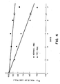
- Advanced glycosylation endproduct (AGE) formation was then induced on red cell surfaces by 48 hour incubation in Dulbecco's modified Eagle medium containing different sugars, such as glucose, glucose-6-phosphate, xylose and arabinose, at 100 mM concentrations, at room temperature. At the end of this period, the media demonstrated no evidence of cell lysis, and red cells themselves appeared microscopically indistinguishable from the controls. As demonstrated by RIA (the method of Chang et al., (1985) J. BIOL. CHEM., Vol. 260, pp. 7970-7974), all cells incubated in sugar underwent formation of a significant amount of advanced glycosylation endproducts on their cell membrane, as compared to the controls.
- mice In order to determine whether autologous red blood cells modified by an AGE such as FFI-HA can be recognized by macrophages in vivo and be removed from the circulation faster than the non-modified cells, presumably by the same AGE-receptor present on hepatic and splenic macrophages, Balb/c inbred mouse red cells were treated either with FFI-HA, as described in Methods, or PBS alone for one hour at room temperature. As an additional control, mouse cells were treated with carbodiimide alone (10 mM). Following 51Chromium-labeling all three groups of cells were reinjected intravenously into syngenetic mice and erythrocyte radioactivity was monitored for 20 days.
- an AGE such as FFI-HA
- Nonenzymatic glycosylation of erythrocyte membrane proteins has been shown previously (Miller et al., (1980) J. CLIN INVEST. Vol. 65, pp. 896-901) to occur primarily at the lysine residues of all the major protein bands without distinction and is enhanced in diabetes, while it is decreased in hemolytic anemia.
- These findings have suggested that the modification of membrane proteins, other than by blood glucose, depends not only on glucose concentration, but also on erythrocyte age.
- the foregoing experiments demonstrate that AGE is present on normal intact red blood cells, which may be responsible in part for daily removal of erythrocytes from the circulation by the monocyte/macrophage AGE-receptor.
- AGE accumulation can be accelerated by exposure to high glucose levels, which in turn leads to increased monocyte AGE-receptor binding and ingestion of glucose-modified red cells, and this may play a role in the moderately shortened erythrocyte survival in diabetics (Peterson, C.M., et al., (1977) ANN. INT. MED., Vol. 86, pp. 425-429).
- AGEs were prepared using the same procedure as disclosed in Example I, above.
- FFI-HA was prepared as described and quantities were bound to both human and bovine albumin.
- a water soluble carbodiimide was used to attach the acid moiety of the FFI-HA to an amino group on the protein.
- the conjugate was purified and then used in vitro to stimulate macrophages, by incubation for from 4 to 24 hours.
- the AGEs that were to be observed for uptake and degradation were appropriately radiolabeled so that they could be traced.
- the stimulated macrophages were tested by exposure to the radiolabeled AGEs following exposure to various agents to measure the effect that these agents had on the ability of the macrophages to take up and degrade the labeled AGEs.
- the above procedures and the studies that follow conform to the protocol employed by Vlassara et al., supra .
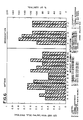
- Figure 6, Part I shows the uptake of AGE-BSA by human monocytes after stimulation with certain agents in the absence of gamma-interferon.
- the FFI-human albumin at 0.1 and 0.2 mg/ml had a stimulatory effect on the uptake system (bars E and F, respectively) as compared to the control (bar A).
- FFI-bovine albumin is also stimulatory (bars E ⁇ , F ⁇ , and G ⁇ ).
- Gamma-interferon alone (bar C), or lipopolysaccharide alone (bar B) or Interleukin-1 alone (bar W) have no stimulatory effect.
- Figure 6 Part II shows the degradation of AGE-BSA by human monocytes in the presence of these agents, in the absence of gamma-interferon.
- the FFI-BSA preparations are slightly stimulatory (bar Y).
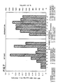
- Figure 7, Part 1 the effect of stimulation on the uptake of AGE-BSA by the same agents in the presence of 10 micrograms/ml of human gamma-interferon is shown.
- gamma-interferon greatly potentiates (up to 8-fold) the stimulation by 0.01, 0.02, and 0.05 mg/ml of FFI-BSA (bars N, O, and P). Degradation is also enhanced in the presence of these agents plus gamma-interferon (Figure 7, bar V).
- monocyte or macrophage cells can also be stimulated by AGE-carrier molecules which result in cells with enhanced ability to bind, internalize and degrade other AGE-molecules.
- AGE-carrier molecules are made, for example, from the reaction of glucose or glucose-6-phosphate with albumin. After purification of the reaction product, the AGE-albumin uptake of AGE-macromolecules demonstrated as in (A) above.
- AGE-BSA prepared from the incubation of glucose-6-phosphate with albumin for 6-8 weeks
- FIG. 7 shows that in the presence of gamma-interferon, 0.5 mg/ml of AGE-BSA made from glucose-6-phosphate, greatly stimulates uptake of AGE-BSA by macrophages. The lower concentrations show a slight stimulatory effect (bars Q and R).
- cachectin tumor necrosis factor
- the structure and properties of cachectin have been previously elucidated by one of the inventors herein. It has been determined that cachectin at a concentration of 20 ng per milliliter of culture fluid stimulates macrophages to express the AGE-receptor and to increase binding, uptake and degradation of AGE-macromolecules.
- agents which stimulate macrophages to increase the ability to internalize and degrade AGEs should not be restrictive.
- Other agents include additional synthetic specific AGEs linked to carrier molecules, other AGEs made from the reaction of sugars with macromolecules, and other monokines which stimulate macrophages to increase their activity toward AGEs.
- the method also includes the coadminstration of these agents and one or more co-stimulatory agent such as Interleukin-1 and gamma interferon to achieve an even greater stimulation of the body's AGE removal system.
- co-stimulatory agent such as Interleukin-1 and gamma interferon
- macrophages could be treated ex vivo with these agents and returned to the body.
- an extracorporeal blood treatment may be performed in which indwelling lines are placed in a patient's arterial and venous system, and blood is taken from the body, passed through a device and then returned to the patient.
- the device is contemplated to contain agents which by contact with or exposure to monocytes/macrophages will stimulate them to increase the activity of the AGE removal system.
- the agents may either be immobilized or may be capable of entering the blood flow.
- Such an extracorporeal process may be performed alone or jointly with the administration to the patient of other agents to enhance the process, such as the gamma-interferon described above.
- An additional aspect of the present invention herein relates to the observation of the effects of insulin on the macrophage AGE clearance system. It has been found that animals with lower than normal levels of insulin in the blood have an enhanced macrophage AGE clearance system. This has been demonstrated in both experimental animals in which insulin-producing pancreas cells are destroyed by injection of the animal with alloxan, or in genetically diabetic animals which have low insulin levels. In both groups, blood glucose levels are higher than normal.
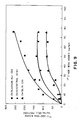
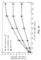
- animals with high levels of insulin in the blood such as genetic hyperinsulinaemic animals (C57BL6, db/db, serum insulin > 300 ⁇ U/ml, Figure 9), have a reduction of about 50% of the activity of binding of AGE-BSA to macrophages compared to normal animals. This suppression of the removal system would have a negative effect since clearance of AGE-macromolecules would be decreased. Degradation of AGE ( Figure 10) shows the same relationship to insulin levels.
- hypoinsulinaemic and hyperinsulinaemic animals had equally abnormal elevations of glucose in the blood.
- one of the therapeutic methods of the present invention comprises providing cachectin or a derivative of cachectin in amounts sufficient to stimulate the removal system.
- the exact quantities of cachectin to be administered may vary, and the amounts employed in the experiments with cachectin set forth herein are representative. Naturally, specific amounts would be determined by the attending physicial or veterinarian administering the treatment.
- a further therapeutic application of the present invention lies in the area of immunology.
- advanced glycosylation endproducts such as FFI
- phagocytic cells facilitate the introduction to those cells of certain antigens against which it is desired to raise an immunity.
- the advanced glycosylation endproduct or FFI could be coupled to an antigen in much the same fashion as disclosed herein for the coupling to any other carrier.
- the particular phagocytic cells that it is desired to stimulate could be exposed to the coupled complex whereupon the cells would recognize and degrade the latter, and would concommitantly develop specific receptors therefor.
- activated phagocytes could then be introduced to the immune system of the animal and would promote the development by the immune system of antibodies to the initial antigen.
- the above method may be practiced ex vivo or in vivo. If ex vivo, the method would include the initial removal of the phagocytes from the body and their exposure to the coupled complex to develop their sensitivity to the particular antigen. Thereafter a sample of cells of the animal that is known to be responsible for raising antibodies and that would have been similarly isolated and removed from the animal, or cells from other sources that may perform the same function, would be exposed to the activated phagocytes for a period of time sufficient to enable antibodies to be raised to the antigen of interest. The antibodies could be administered to animals to avert or alleviate the adverse effects of any pathology that might be caused by the invasion of the antigen.
- ex vivo activated phagocytes could be utilized as a vaccine to innoculate animals against the antigen and to thereby promote the in vivo development of antibodies thereto.
- An in vivo protocol contemplates the preparation of the coupled complex between the AGE/FFI and the antigen and the administration of this coupled complex directly to the animal to promote the in vivo activation of phagocytes and the subsequent development of antibodies by the animal's immune system.
- a further alternative contemplates the formation of a mixture rather than a coupled complex between the AGE/FFI and the antigen. This mixture could be utilized in place of the coupled complex in the above protocols.
- a particular implementation of the ex vivo embodiment of the immunological protocol could include the immobilization of either the coupled complex or the phagocytic cells during all or part of the practice of the particular method.
- either the cells or the coupled complex could be immobilized and the unbound material then circulated therepast to achieve activation. If it is desired to administer the bound material to the animal, it could be released from the substrate after activation by known techniques.
- a further therapeutic protocol is suggested by the foregoing immunological protocols, which is based upon the binding, labeling or other combination of the AGE/FFI in this instance to a particular antigen as defined herein.
- the AGE/FFI could be combined with an antibody specific to the particular antigen, and the resulting associated mixture or combined material then placed in contact with the phagocytic cells of the animal/human host to stimulate such cells to recognize and attack the antigen.
- the manner by which contact between the combined material and the phagocytes occurs may vary.
- a quantity of the combined material may be introduced in vivo to bind with the target antigen.
- the stimulated phagocytes would then be introduced or stimulation of the phagocytes would take place concurrently upon the in vivo introduction of the combined material, whereupon the phagocytes would attack and destroy/degrade the complex formed by the combined material and the target antigen, either directly or by means of the secretion of the monokine cachectin/TNF.
- the phagocytes would act specifically against the target antigen because of their recognition of the particular AGE/FFI now associated therewith by means of the antibody.
- the foregoing therapeutic method may vary as to dosage, manner of administration and periodicity, depending upon the particular host and the attending pathological condition, and is subject to adjustment by the trained physician or veterinarian.
- This therapy offers a potentially effective avenue of treatment for patients suffering from Acquired Immune Deficiency Syndrome (AIDS) in view of the inability of conventional therapies to alleviate the condition.
- AIDS Acquired Immune Deficiency Syndrome
- an assay system may be developed for screening potential drugs effective to act as agents to stimulate the activity of specific phagocytes against advanced glycosylation endproducts.
- a prospective test drug could be administered to a macrophage sample to determine its stimulatory effect and the macrophage sample after incubation with the test drug could be incubated with a fluid or tissue sample having a quantity of appropriately labeled advanced glycosylation endproducts present therein, so that comparative uptake and degradation studies could then be made.
- plural macrophage colonies could be initially incubated with various of the known agents, so that comparative testing of the prospective drug could take place with greater specificity.
- An alternate diagnostic protocol contemplates the investigation of particular phagocytes to determine which of the known agents are most effective in stimulating cellular activity against advanced glycosylation endproducts.
- plural comparable quantities of a particular phagocytic cell colony could be isolated and incubated with several different known agents, and thereafter incubated with identical corresponding samples containing appropriately labeled advanced glycosylation endproducts, so that a comparason of the extent of activation of the phagocytic cells could then be made, and the agent most effective in stimulating the cellular colony thereby identified.
- the colony exhibiting the greatest uptake and disposal of labeled advanced glycosylation endproducts would correspondingly identify the agent that is most effective.
- phagocytic cells such as monocytes and macrophages are removed from the animal's body and are activated ex vivo by exposure to the agents of the present invention. This may be accomplished by an extracorporeal shunt as has been described earlier on herein.
- the phagocytic cells instead of being returned immediately to the body, are appropriately radiolabeled, as with Technicium, and are then returned to the body, whereupon the animal may undergo radioimaging to note the course of travel of the phagocytes and to thereby determine the location of the concentrations of advanced glycosylation endproducts in the body.
- undesireable concentrations of advanced glycosylation endproducts such as atheromatous plaques could be discovered, and the clinical significance of these concentrations accordingly assessed. This procedure would provide additional vital information in attempting to assess the pathological state of the animal or patient.
- a further diagnostic application likewise seeking to provide vital information regarding pathological status contemplates the preparation of radiolabeled agents and their incubation or contact with phagocytes from a particular animal, and the subsequent measurement of the elapsed time before the labeled agents are recognized and degraded. Such time measurements could be compared against standard measurements determined by testing cells taken from normal animals or patients that were tested under the same protocol. This data could then identify the existence of pathologies such as diabetes, or the others mentioned earlier herein, or other disorders that may adversely affect the operability of the AGE removal system of the body.
- This diagnostic procedure can be performed in vivo by administering the labeled agents to the animal or patient, or alternatively, could be conducted ex vivo by the isolation of phagocytic cells of the animal or patient outside the body and the incubation of these cells with the labeled agents.
- An additional diagnostic technique contemplates a further qualitative analysis of the animal's body.
- particular pathologies may be identified by a method wherein the activity of the phagocytic cells of the animal could be measured and compared against the activities found with normal cells.
- radiolabeled agents could be introduced to the body, or phagocytic cellular colonies from the body could be isolated and incubated with the radiolabeled agents, and the level of activity of the phagocytic cells could then be measured with greater specificity as to particular advanced glycosylation endproducts. This is possible because of the specific nature of the cellular receptors that develop on the phagocytic cells as to particular advanced glycosylation endproducts.
- the macrophage or monocytes having greatest involvement with the removal of advanced glycosylation endproducts that would otherwise develop atheromatous plaques could be isolated and tested for their activity, and if their activity appears to be abnormally increased, the development of such plaques may be indicated. Similar testing protocols could be employed to determine the existence of diabetic aging and the like.
- the specific advanced glycosylation endproducts associated with particular conditions would elicit responses from the phagocytes that would suggest any abnormal accumulation of these advanced glycosylation endproducts in the body system.
Landscapes
- Health & Medical Sciences (AREA)
- Life Sciences & Earth Sciences (AREA)
- Engineering & Computer Science (AREA)
- Chemical & Material Sciences (AREA)
- Immunology (AREA)
- General Health & Medical Sciences (AREA)
- Medicinal Chemistry (AREA)
- Molecular Biology (AREA)
- Hematology (AREA)
- Urology & Nephrology (AREA)
- Biomedical Technology (AREA)
- Public Health (AREA)
- Pharmacology & Pharmacy (AREA)
- Veterinary Medicine (AREA)
- Animal Behavior & Ethology (AREA)
- Proteomics, Peptides & Aminoacids (AREA)
- Bioinformatics & Cheminformatics (AREA)
- Physics & Mathematics (AREA)
- Epidemiology (AREA)
- Microbiology (AREA)
- Biochemistry (AREA)
- Organic Chemistry (AREA)
- Biotechnology (AREA)
- General Physics & Mathematics (AREA)
- Analytical Chemistry (AREA)
- Food Science & Technology (AREA)
- Pathology (AREA)
- Cell Biology (AREA)
- Gastroenterology & Hepatology (AREA)
- Zoology (AREA)
- General Chemical & Material Sciences (AREA)
- Chemical Kinetics & Catalysis (AREA)
- Nuclear Medicine, Radiotherapy & Molecular Imaging (AREA)
- Biophysics (AREA)
- Bioinformatics & Computational Biology (AREA)
- Mycology (AREA)
- Toxicology (AREA)
- Diabetes (AREA)
- Obesity (AREA)
- Physiology (AREA)
Applications Claiming Priority (2)
| Application Number | Priority Date | Filing Date | Title |
|---|---|---|---|
| US90774786A | 1986-09-12 | 1986-09-12 | |
| US907747 | 1986-09-12 |
Publications (3)
| Publication Number | Publication Date |
|---|---|
| EP0259893A2 true EP0259893A2 (de) | 1988-03-16 |
| EP0259893A3 EP0259893A3 (de) | 1989-06-14 |
| EP0259893B1 EP0259893B1 (de) | 1997-01-22 |
Family
ID=25424576
Family Applications (1)
| Application Number | Title | Priority Date | Filing Date |
|---|---|---|---|
| EP87113386A Expired - Lifetime EP0259893B1 (de) | 1986-09-12 | 1987-09-14 | Mittel zur Entziehung von Endprodukten der fortgeschrittenen Glycosylation |
Country Status (7)
| Country | Link |
|---|---|
| EP (1) | EP0259893B1 (de) |
| JP (1) | JP2644767B2 (de) |
| AT (1) | ATE147991T1 (de) |
| AU (1) | AU600306B2 (de) |
| DE (1) | DE3752007T2 (de) |
| ES (1) | ES2099059T3 (de) |
| GR (1) | GR3022382T3 (de) |
Cited By (23)
| Publication number | Priority date | Publication date | Assignee | Title |
|---|---|---|---|---|
| EP0359112A3 (de) * | 1988-09-08 | 1990-08-29 | Takeda Chemical Industries, Ltd. | Carbazinsäure-Derivate, ihre Herstellung und Anwendung |
| WO1993013421A1 (en) * | 1991-12-20 | 1993-07-08 | The Rockefeller University | Immunochemical detection of in vivo advanced glycosylation endproducts |
| WO1993016717A1 (en) * | 1992-02-25 | 1993-09-02 | The Rockefeller University | Method and agents for promoting wound healing |
| WO1993023081A1 (en) * | 1992-05-15 | 1993-11-25 | The Rockefeller University | Methods and agents for modulating immune response, and uses thereof |
| WO1995020979A1 (en) * | 1994-02-03 | 1995-08-10 | The Picower Institute For Medical Research | Compositions and methods for advanced glycosylation endproduct-mediated modulation of amyloidosis |
| US5811075A (en) * | 1984-03-19 | 1998-09-22 | The Rockefeller University | Method and agents for removing advanced glycosylation endproducts |
| US6410598B1 (en) | 1994-02-03 | 2002-06-25 | Michael P. Vitek | Compositions and methods for advanced glycosylation endproduct-mediated modulation of amyloidosis |
| CN102478569A (zh) * | 2010-11-25 | 2012-05-30 | 苏州卫生职业技术学院 | 一种西红花酸对AGEs抑制效果的检测方法 |
| US20160152697A1 (en) * | 2008-05-23 | 2016-06-02 | Siwa Corporation | Methods, compositions and apparatuses for facilitating regeneration |
| US10226531B2 (en) | 2010-09-27 | 2019-03-12 | Siwa Corporation | Selective removal of age-modified cells for treatment of atherosclerosis |
| US10358502B2 (en) | 2014-12-18 | 2019-07-23 | Siwa Corporation | Product and method for treating sarcopenia |
| US10584180B2 (en) | 2014-09-19 | 2020-03-10 | Siwa Corporation | Anti-AGE antibodies for treating inflammation and auto-immune disorders |
| US10858449B1 (en) | 2017-01-06 | 2020-12-08 | Siwa Corporation | Methods and compositions for treating osteoarthritis |
| US10919957B2 (en) | 2017-04-13 | 2021-02-16 | Siwa Corporation | Humanized monoclonal advanced glycation end-product antibody |
| US10925937B1 (en) | 2017-01-06 | 2021-02-23 | Siwa Corporation | Vaccines for use in treating juvenile disorders associated with inflammation |
| US10961321B1 (en) | 2017-01-06 | 2021-03-30 | Siwa Corporation | Methods and compositions for treating pain associated with inflammation |
| US10960234B2 (en) | 2010-11-22 | 2021-03-30 | Siwa Corporation | Selective removal of cells having accumulated agents |
| US10995151B1 (en) | 2017-01-06 | 2021-05-04 | Siwa Corporation | Methods and compositions for treating disease-related cachexia |
| US11213585B2 (en) | 2016-06-23 | 2022-01-04 | Siwa Corporation | Vaccines for use in treating various diseases and disorders |
| US11518801B1 (en) | 2017-12-22 | 2022-12-06 | Siwa Corporation | Methods and compositions for treating diabetes and diabetic complications |
| US11833202B2 (en) | 2016-02-19 | 2023-12-05 | Siwa Corporation | Method and composition for treating cancer, killing metastatic cancer cells and preventing cancer metastasis using antibody to advanced glycation end products (AGE) |
| US11872269B2 (en) | 2014-12-18 | 2024-01-16 | Siwa Corporation | Method and composition for treating sarcopenia |
| US11958900B2 (en) | 2016-04-15 | 2024-04-16 | Siwa Corporation | Anti-age antibodies for treating neurodegenerative disorders |
Families Citing this family (2)
| Publication number | Priority date | Publication date | Assignee | Title |
|---|---|---|---|---|
| KR920702230A (ko) * | 1990-08-10 | 1992-09-03 | 오쯔까 아끼히꼬 | 간염예방·치료제 |
| US6750209B1 (en) * | 1995-09-12 | 2004-06-15 | Kansas University Medical Center | Advanced glycation end-product intermediaries and post-amadori inhibition |
Family Cites Families (1)
| Publication number | Priority date | Publication date | Assignee | Title |
|---|---|---|---|---|
| US4665192A (en) * | 1984-03-19 | 1987-05-12 | The Rockefeller University | 2-(2-furoyl)-4(5)-2(furanyl)-1H-imidazole |
-
1987
- 1987-09-11 JP JP62228202A patent/JP2644767B2/ja not_active Expired - Fee Related
- 1987-09-11 AU AU78275/87A patent/AU600306B2/en not_active Ceased
- 1987-09-14 ES ES87113386T patent/ES2099059T3/es not_active Expired - Lifetime
- 1987-09-14 EP EP87113386A patent/EP0259893B1/de not_active Expired - Lifetime
- 1987-09-14 AT AT87113386T patent/ATE147991T1/de not_active IP Right Cessation
- 1987-09-14 DE DE3752007T patent/DE3752007T2/de not_active Expired - Fee Related
-
1997
- 1997-01-28 GR GR970400123T patent/GR3022382T3/el unknown
Cited By (30)
| Publication number | Priority date | Publication date | Assignee | Title |
|---|---|---|---|---|
| US5811075A (en) * | 1984-03-19 | 1998-09-22 | The Rockefeller University | Method and agents for removing advanced glycosylation endproducts |
| EP0359112A3 (de) * | 1988-09-08 | 1990-08-29 | Takeda Chemical Industries, Ltd. | Carbazinsäure-Derivate, ihre Herstellung und Anwendung |
| WO1993013421A1 (en) * | 1991-12-20 | 1993-07-08 | The Rockefeller University | Immunochemical detection of in vivo advanced glycosylation endproducts |
| CN1053964C (zh) * | 1991-12-20 | 2000-06-28 | 洛克菲勒大学 | 体内高级糖基化终产物的免疫化学检测 |
| WO1993016717A1 (en) * | 1992-02-25 | 1993-09-02 | The Rockefeller University | Method and agents for promoting wound healing |
| WO1993023081A1 (en) * | 1992-05-15 | 1993-11-25 | The Rockefeller University | Methods and agents for modulating immune response, and uses thereof |
| WO1995020979A1 (en) * | 1994-02-03 | 1995-08-10 | The Picower Institute For Medical Research | Compositions and methods for advanced glycosylation endproduct-mediated modulation of amyloidosis |
| US5935927A (en) * | 1994-02-03 | 1999-08-10 | The Picower Institute For Medical Research | Compositions and methods for stimulating amyloid removal in amyloidogenic diseases using advanced glycosylation endproducts |
| US6410598B1 (en) | 1994-02-03 | 2002-06-25 | Michael P. Vitek | Compositions and methods for advanced glycosylation endproduct-mediated modulation of amyloidosis |
| US11261241B2 (en) | 2008-05-23 | 2022-03-01 | Siwa Corporation | Methods, compositions and apparatuses for facilitating regeneration |
| US20160152697A1 (en) * | 2008-05-23 | 2016-06-02 | Siwa Corporation | Methods, compositions and apparatuses for facilitating regeneration |
| US10226531B2 (en) | 2010-09-27 | 2019-03-12 | Siwa Corporation | Selective removal of age-modified cells for treatment of atherosclerosis |
| US10960234B2 (en) | 2010-11-22 | 2021-03-30 | Siwa Corporation | Selective removal of cells having accumulated agents |
| CN102478569A (zh) * | 2010-11-25 | 2012-05-30 | 苏州卫生职业技术学院 | 一种西红花酸对AGEs抑制效果的检测方法 |
| US10584180B2 (en) | 2014-09-19 | 2020-03-10 | Siwa Corporation | Anti-AGE antibodies for treating inflammation and auto-immune disorders |
| US12134660B2 (en) | 2014-09-19 | 2024-11-05 | Siwa Corporation | Anti-age antibodies for treating inflammation and auto-immune disorders |
| US10358502B2 (en) | 2014-12-18 | 2019-07-23 | Siwa Corporation | Product and method for treating sarcopenia |
| US11872269B2 (en) | 2014-12-18 | 2024-01-16 | Siwa Corporation | Method and composition for treating sarcopenia |
| US11873345B2 (en) | 2014-12-18 | 2024-01-16 | Siwa Corporation | Product and method for treating sarcopenia |
| US11833202B2 (en) | 2016-02-19 | 2023-12-05 | Siwa Corporation | Method and composition for treating cancer, killing metastatic cancer cells and preventing cancer metastasis using antibody to advanced glycation end products (AGE) |
| US11958900B2 (en) | 2016-04-15 | 2024-04-16 | Siwa Corporation | Anti-age antibodies for treating neurodegenerative disorders |
| US11213585B2 (en) | 2016-06-23 | 2022-01-04 | Siwa Corporation | Vaccines for use in treating various diseases and disorders |
| US10995151B1 (en) | 2017-01-06 | 2021-05-04 | Siwa Corporation | Methods and compositions for treating disease-related cachexia |
| US10961321B1 (en) | 2017-01-06 | 2021-03-30 | Siwa Corporation | Methods and compositions for treating pain associated with inflammation |
| US10925937B1 (en) | 2017-01-06 | 2021-02-23 | Siwa Corporation | Vaccines for use in treating juvenile disorders associated with inflammation |
| US10858449B1 (en) | 2017-01-06 | 2020-12-08 | Siwa Corporation | Methods and compositions for treating osteoarthritis |
| US11542324B2 (en) | 2017-04-13 | 2023-01-03 | Siwa Corporation | Humanized monoclonal advanced glycation end-product antibody |
| US10919957B2 (en) | 2017-04-13 | 2021-02-16 | Siwa Corporation | Humanized monoclonal advanced glycation end-product antibody |
| US12226465B2 (en) | 2017-04-13 | 2025-02-18 | Siwa Corporation | Humanized monoclonal advanced glycation end-product antibody |
| US11518801B1 (en) | 2017-12-22 | 2022-12-06 | Siwa Corporation | Methods and compositions for treating diabetes and diabetic complications |
Also Published As
| Publication number | Publication date |
|---|---|
| ES2099059T3 (es) | 1997-05-16 |
| AU7827587A (en) | 1988-03-17 |
| JP2644767B2 (ja) | 1997-08-25 |
| ATE147991T1 (de) | 1997-02-15 |
| DE3752007D1 (de) | 1997-03-06 |
| EP0259893A3 (de) | 1989-06-14 |
| GR3022382T3 (en) | 1997-04-30 |
| EP0259893B1 (de) | 1997-01-22 |
| AU600306B2 (en) | 1990-08-09 |
| JPS63146833A (ja) | 1988-06-18 |
| DE3752007T2 (de) | 1997-05-28 |
Similar Documents
| Publication | Publication Date | Title |
|---|---|---|
| US4900747A (en) | Method and agents for removing advanced glycosylation endproducts | |
| EP0259893B1 (de) | Mittel zur Entziehung von Endprodukten der fortgeschrittenen Glycosylation | |
| US5811075A (en) | Method and agents for removing advanced glycosylation endproducts | |
| Bennett et al. | Immunosuppression by human plasma alpha 1-acid glycoprotein: importance of the carbohydrate moiety. | |
| CA1208546A (en) | Lipoprotein lipase suppression by endotoxin-induced mediator (shock assay) | |
| US4603106A (en) | Lipoprotein lipase suppression by endotoxin-induced mediator (shock assay) | |
| US4822776A (en) | Lipoprotein lipase suppression by endotoxin-induced mediator (shock assay) | |
| Schmid et al. | Congenital jaundice in rats, due to a defect in glucuronide formation | |
| Steinbrecher et al. | Glucosylation of low-density lipoproteins to an extent comparable to that seen in diabetes slows their catabolism | |
| JPH0667827B2 (ja) | メイラード反応阻害剤 | |
| AU681340B2 (en) | Immunochemical detection of (in vivo) advanced glycosylation endproducts | |
| Cerami et al. | Protein glycosylation and the pathogenesis of atherosclerosis | |
| FEDER | A heme-peptide as an ultrastructural tracer | |
| JP3502125B2 (ja) | 形質転換細胞の抽出および培養ならびにそれらに対する抗体の製造 | |
| US6309640B1 (en) | Lipoprotein lipase suppression by endotoxin-induced mediator (shock assay) | |
| CA1063019A (en) | Extracts of the haemopoietic system | |
| Peter et al. | Cell-mediated cytotoxicity during rejection and enhancement of allogeneic skin grafts in rats | |
| SU1767433A1 (ru) | Способ определени инсулинорезистентности имунного генеза у больных сахарным диабетом I типа | |
| Holtzman et al. | Ceruloplasmin in Wilson's disease | |
| USRE35465E (en) | Method and agents for removing advanced glycosylation endproducts | |
| CA1323565C (en) | Method and agents for removing advanced glycosylation endproducts | |
| Bennett et al. | RNA in the periphery of rapidly proliferating mouse lymphoid cells | |
| Vlassara | Chronic diabetic complications and tissue glycosylation: relevant concern for diabetes-prone black population | |
| JP4319637B2 (ja) | ボケからレクチンを含有するエキスの抽出方法、そのエキス、及びその抗腫瘍用医薬組成物、並びに細胞表面のグリコフォリンaの検出製剤と血液型検査キット | |
| RU2077725C1 (ru) | Способ обнаружения раковоэмбрионального антигена при помощи специального антительного диагностикума |
Legal Events
| Date | Code | Title | Description |
|---|---|---|---|
| PUAI | Public reference made under article 153(3) epc to a published international application that has entered the european phase |
Free format text: ORIGINAL CODE: 0009012 |
|
| AK | Designated contracting states |
Kind code of ref document: A2 Designated state(s): AT BE CH DE ES FR GB GR IT LI LU NL SE |
|
| PUAL | Search report despatched |
Free format text: ORIGINAL CODE: 0009013 |
|
| AK | Designated contracting states |
Kind code of ref document: A3 Designated state(s): AT BE CH DE ES FR GB GR IT LI LU NL SE |
|
| 17P | Request for examination filed |
Effective date: 19891211 |
|
| 17Q | First examination report despatched |
Effective date: 19910624 |
|
| GRAH | Despatch of communication of intention to grant a patent |
Free format text: ORIGINAL CODE: EPIDOS IGRA |
|
| GRAH | Despatch of communication of intention to grant a patent |
Free format text: ORIGINAL CODE: EPIDOS IGRA |
|
| GRAA | (expected) grant |
Free format text: ORIGINAL CODE: 0009210 |
|
| AK | Designated contracting states |
Kind code of ref document: B1 Designated state(s): AT BE CH DE ES FR GB GR IT LI LU NL SE |
|
| REF | Corresponds to: |
Ref document number: 147991 Country of ref document: AT Date of ref document: 19970215 Kind code of ref document: T |
|
| REG | Reference to a national code |
Ref country code: CH Ref legal event code: EP |
|
| REG | Reference to a national code |
Ref country code: CH Ref legal event code: NV Representative=s name: BOVARD AG PATENTANWAELTE |
|
| ET | Fr: translation filed | ||
| ITF | It: translation for a ep patent filed | ||
| REF | Corresponds to: |
Ref document number: 3752007 Country of ref document: DE Date of ref document: 19970306 |
|
| REG | Reference to a national code |
Ref country code: GR Ref legal event code: FG4A Free format text: 3022382 |
|
| REG | Reference to a national code |
Ref country code: ES Ref legal event code: FG2A Ref document number: 2099059 Country of ref document: ES Kind code of ref document: T3 |
|
| PLBE | No opposition filed within time limit |
Free format text: ORIGINAL CODE: 0009261 |
|
| STAA | Information on the status of an ep patent application or granted ep patent |
Free format text: STATUS: NO OPPOSITION FILED WITHIN TIME LIMIT |
|
| 26N | No opposition filed | ||
| PGFP | Annual fee paid to national office [announced via postgrant information from national office to epo] |
Ref country code: GR Payment date: 20010926 Year of fee payment: 15 |
|
| PGFP | Annual fee paid to national office [announced via postgrant information from national office to epo] |
Ref country code: NL Payment date: 20010930 Year of fee payment: 15 Ref country code: AT Payment date: 20010930 Year of fee payment: 15 |
|
| PGFP | Annual fee paid to national office [announced via postgrant information from national office to epo] |
Ref country code: SE Payment date: 20011001 Year of fee payment: 15 Ref country code: GB Payment date: 20011001 Year of fee payment: 15 Ref country code: FR Payment date: 20011001 Year of fee payment: 15 |
|
| PGFP | Annual fee paid to national office [announced via postgrant information from national office to epo] |
Ref country code: BE Payment date: 20011005 Year of fee payment: 15 |
|
| PGFP | Annual fee paid to national office [announced via postgrant information from national office to epo] |
Ref country code: CH Payment date: 20011026 Year of fee payment: 15 |
|
| PGFP | Annual fee paid to national office [announced via postgrant information from national office to epo] |
Ref country code: ES Payment date: 20011030 Year of fee payment: 15 |
|
| PGFP | Annual fee paid to national office [announced via postgrant information from national office to epo] |
Ref country code: LU Payment date: 20011105 Year of fee payment: 15 |
|
| PGFP | Annual fee paid to national office [announced via postgrant information from national office to epo] |
Ref country code: DE Payment date: 20011120 Year of fee payment: 15 |
|
| REG | Reference to a national code |
Ref country code: GB Ref legal event code: IF02 |
|
| PG25 | Lapsed in a contracting state [announced via postgrant information from national office to epo] |
Ref country code: LU Free format text: LAPSE BECAUSE OF NON-PAYMENT OF DUE FEES Effective date: 20020914 Ref country code: GB Free format text: LAPSE BECAUSE OF NON-PAYMENT OF DUE FEES Effective date: 20020914 Ref country code: AT Free format text: LAPSE BECAUSE OF NON-PAYMENT OF DUE FEES Effective date: 20020914 |
|
| PG25 | Lapsed in a contracting state [announced via postgrant information from national office to epo] |
Ref country code: SE Free format text: LAPSE BECAUSE OF NON-PAYMENT OF DUE FEES Effective date: 20020915 Ref country code: ES Free format text: LAPSE BECAUSE OF NON-PAYMENT OF DUE FEES Effective date: 20020915 |
|
| PG25 | Lapsed in a contracting state [announced via postgrant information from national office to epo] |
Ref country code: LI Free format text: LAPSE BECAUSE OF NON-PAYMENT OF DUE FEES Effective date: 20020930 Ref country code: CH Free format text: LAPSE BECAUSE OF NON-PAYMENT OF DUE FEES Effective date: 20020930 Ref country code: BE Free format text: LAPSE BECAUSE OF NON-PAYMENT OF DUE FEES Effective date: 20020930 |
|
| BERE | Be: lapsed |
Owner name: THE *ROCKEFELLER UNIVERSITY Effective date: 20020930 |
|
| PG25 | Lapsed in a contracting state [announced via postgrant information from national office to epo] |
Ref country code: NL Free format text: LAPSE BECAUSE OF NON-PAYMENT OF DUE FEES Effective date: 20030401 Ref country code: DE Free format text: LAPSE BECAUSE OF NON-PAYMENT OF DUE FEES Effective date: 20030401 |
|
| PG25 | Lapsed in a contracting state [announced via postgrant information from national office to epo] |
Ref country code: GR Free format text: LAPSE BECAUSE OF NON-PAYMENT OF DUE FEES Effective date: 20030404 |
|
| EUG | Se: european patent has lapsed | ||
| GBPC | Gb: european patent ceased through non-payment of renewal fee |
Effective date: 20020914 |
|
| REG | Reference to a national code |
Ref country code: CH Ref legal event code: PL |
|
| PG25 | Lapsed in a contracting state [announced via postgrant information from national office to epo] |
Ref country code: FR Free format text: LAPSE BECAUSE OF NON-PAYMENT OF DUE FEES Effective date: 20030603 |
|
| REG | Reference to a national code |
Ref country code: FR Ref legal event code: ST |
|
| REG | Reference to a national code |
Ref country code: ES Ref legal event code: FD2A Effective date: 20031011 |
|
| PG25 | Lapsed in a contracting state [announced via postgrant information from national office to epo] |
Ref country code: IT Free format text: LAPSE BECAUSE OF NON-PAYMENT OF DUE FEES;WARNING: LAPSES OF ITALIAN PATENTS WITH EFFECTIVE DATE BEFORE 2007 MAY HAVE OCCURRED AT ANY TIME BEFORE 2007. THE CORRECT EFFECTIVE DATE MAY BE DIFFERENT FROM THE ONE RECORDED. Effective date: 20050914 |