US11518801B1 - Methods and compositions for treating diabetes and diabetic complications - Google Patents
Methods and compositions for treating diabetes and diabetic complications Download PDFInfo
- Publication number
- US11518801B1 US11518801B1 US16/228,293 US201816228293A US11518801B1 US 11518801 B1 US11518801 B1 US 11518801B1 US 201816228293 A US201816228293 A US 201816228293A US 11518801 B1 US11518801 B1 US 11518801B1
- Authority
- US
- United States
- Prior art keywords
- age
- seq
- antibody
- amino acid
- acid sequence
- Prior art date
- Legal status (The legal status is an assumption and is not a legal conclusion. Google has not performed a legal analysis and makes no representation as to the accuracy of the status listed.)
- Active, expires
Links
Images
Classifications
-
- C—CHEMISTRY; METALLURGY
- C07—ORGANIC CHEMISTRY
- C07K—PEPTIDES
- C07K16/00—Immunoglobulins [IGs], e.g. monoclonal or polyclonal antibodies
- C07K16/18—Immunoglobulins [IGs], e.g. monoclonal or polyclonal antibodies against material from animals or humans
-
- A—HUMAN NECESSITIES
- A61—MEDICAL OR VETERINARY SCIENCE; HYGIENE
- A61P—SPECIFIC THERAPEUTIC ACTIVITY OF CHEMICAL COMPOUNDS OR MEDICINAL PREPARATIONS
- A61P3/00—Drugs for disorders of the metabolism
- A61P3/08—Drugs for disorders of the metabolism for glucose homeostasis
- A61P3/10—Drugs for disorders of the metabolism for glucose homeostasis for hyperglycaemia, e.g. antidiabetics
-
- A—HUMAN NECESSITIES
- A61—MEDICAL OR VETERINARY SCIENCE; HYGIENE
- A61K—PREPARATIONS FOR MEDICAL, DENTAL OR TOILETRY PURPOSES
- A61K39/00—Medicinal preparations containing antigens or antibodies
- A61K39/0003—Invertebrate antigens
-
- A—HUMAN NECESSITIES
- A61—MEDICAL OR VETERINARY SCIENCE; HYGIENE
- A61K—PREPARATIONS FOR MEDICAL, DENTAL OR TOILETRY PURPOSES
- A61K39/00—Medicinal preparations containing antigens or antibodies
- A61K39/0005—Vertebrate antigens
- A61K39/0008—Antigens related to auto-immune diseases; Preparations to induce self-tolerance
-
- A—HUMAN NECESSITIES
- A61—MEDICAL OR VETERINARY SCIENCE; HYGIENE
- A61K—PREPARATIONS FOR MEDICAL, DENTAL OR TOILETRY PURPOSES
- A61K39/00—Medicinal preparations containing antigens or antibodies
- A61K39/12—Viral antigens
-
- A—HUMAN NECESSITIES
- A61—MEDICAL OR VETERINARY SCIENCE; HYGIENE
- A61K—PREPARATIONS FOR MEDICAL, DENTAL OR TOILETRY PURPOSES
- A61K39/00—Medicinal preparations containing antigens or antibodies
- A61K39/395—Antibodies; Immunoglobulins; Immune serum, e.g. antilymphocytic serum
- A61K39/39533—Antibodies; Immunoglobulins; Immune serum, e.g. antilymphocytic serum against materials from animals
- A61K39/3955—Antibodies; Immunoglobulins; Immune serum, e.g. antilymphocytic serum against materials from animals against proteinaceous materials, e.g. enzymes, hormones, lymphokines
-
- A—HUMAN NECESSITIES
- A61—MEDICAL OR VETERINARY SCIENCE; HYGIENE
- A61K—PREPARATIONS FOR MEDICAL, DENTAL OR TOILETRY PURPOSES
- A61K9/00—Medicinal preparations characterised by special physical form
- A61K9/0012—Galenical forms characterised by the site of application
- A61K9/0019—Injectable compositions; Intramuscular, intravenous, arterial, subcutaneous administration; Compositions to be administered through the skin in an invasive manner
-
- C—CHEMISTRY; METALLURGY
- C07—ORGANIC CHEMISTRY
- C07K—PEPTIDES
- C07K16/00—Immunoglobulins [IGs], e.g. monoclonal or polyclonal antibodies
- C07K16/18—Immunoglobulins [IGs], e.g. monoclonal or polyclonal antibodies against material from animals or humans
- C07K16/28—Immunoglobulins [IGs], e.g. monoclonal or polyclonal antibodies against material from animals or humans against receptors, cell surface antigens or cell surface determinants
- C07K16/2803—Immunoglobulins [IGs], e.g. monoclonal or polyclonal antibodies against material from animals or humans against receptors, cell surface antigens or cell surface determinants against the immunoglobulin superfamily
-
- C—CHEMISTRY; METALLURGY
- C07—ORGANIC CHEMISTRY
- C07K—PEPTIDES
- C07K16/00—Immunoglobulins [IGs], e.g. monoclonal or polyclonal antibodies
- C07K16/44—Immunoglobulins [IGs], e.g. monoclonal or polyclonal antibodies against material not provided for elsewhere, e.g. haptens, metals, DNA, RNA, amino acids
-
- A—HUMAN NECESSITIES
- A61—MEDICAL OR VETERINARY SCIENCE; HYGIENE
- A61K—PREPARATIONS FOR MEDICAL, DENTAL OR TOILETRY PURPOSES
- A61K39/00—Medicinal preparations containing antigens or antibodies
- A61K2039/505—Medicinal preparations containing antigens or antibodies comprising antibodies
-
- A—HUMAN NECESSITIES
- A61—MEDICAL OR VETERINARY SCIENCE; HYGIENE
- A61K—PREPARATIONS FOR MEDICAL, DENTAL OR TOILETRY PURPOSES
- A61K39/00—Medicinal preparations containing antigens or antibodies
- A61K2039/545—Medicinal preparations containing antigens or antibodies characterised by the dose, timing or administration schedule
-
- A—HUMAN NECESSITIES
- A61—MEDICAL OR VETERINARY SCIENCE; HYGIENE
- A61K—PREPARATIONS FOR MEDICAL, DENTAL OR TOILETRY PURPOSES
- A61K39/00—Medicinal preparations containing antigens or antibodies
- A61K2039/555—Medicinal preparations containing antigens or antibodies characterised by a specific combination antigen/adjuvant
- A61K2039/55505—Inorganic adjuvants
-
- A—HUMAN NECESSITIES
- A61—MEDICAL OR VETERINARY SCIENCE; HYGIENE
- A61K—PREPARATIONS FOR MEDICAL, DENTAL OR TOILETRY PURPOSES
- A61K39/00—Medicinal preparations containing antigens or antibodies
- A61K2039/57—Medicinal preparations containing antigens or antibodies characterised by the type of response, e.g. Th1, Th2
- A61K2039/575—Medicinal preparations containing antigens or antibodies characterised by the type of response, e.g. Th1, Th2 humoral response
-
- C—CHEMISTRY; METALLURGY
- C07—ORGANIC CHEMISTRY
- C07K—PEPTIDES
- C07K2317/00—Immunoglobulins specific features
- C07K2317/20—Immunoglobulins specific features characterized by taxonomic origin
- C07K2317/24—Immunoglobulins specific features characterized by taxonomic origin containing regions, domains or residues from different species, e.g. chimeric, humanized or veneered
-
- C—CHEMISTRY; METALLURGY
- C07—ORGANIC CHEMISTRY
- C07K—PEPTIDES
- C07K2317/00—Immunoglobulins specific features
- C07K2317/90—Immunoglobulins specific features characterized by (pharmaco)kinetic aspects or by stability of the immunoglobulin
- C07K2317/92—Affinity (KD), association rate (Ka), dissociation rate (Kd) or EC50 value
Definitions
- Diabetes mellitus commonly referred to as diabetes or DM
- diabetes is the general term for diseases and disorders that are characterized by recurrent or persistent elevated levels of blood glucose. Diabetes occurs when beta cells in the pancreas do not produce sufficient insulin and/or when the body cannot effectively use the insulin produced by beta cells.
- Type 1 diabetes also known as juvenile diabetes, juvenile-onset diabetes or insulin-dependent diabetes
- Type 2 diabetes also known as adult-onset diabetes or non-insulin-dependent diabetes.
- Type 1 diabetes is an autoimmune disease in which the body attacks insulin-producing beta cells in the pancreas.
- Type 2 diabetes is a progressive metabolic disease in which the body becomes unable to properly regulate insulin and beta cells become unable to produce sufficient insulin.
- Diabetes is a significant public health problem.
- the World Health Organization estimates that 422 million adults worldwide have diabetes (“Global Report on Diabetes”, World Health Organization, 2016).
- the United States Centers for Disease Control and Prevention (CDC) estimates that 29.1 million people in the U.S. currently have diabetes, which is 9.3% of the total population (“National Diabetes Statistics Report: Estimates of Diabetes and Its Burden in the United States, 2014”, U.S. Department of Health and Human Services, Centers for Disease Control and Prevention, 2014). Diabetes is currently the seventh leading cause of death in the U.S. and the estimated total costs of diabetes in the U.S. is $245 billion.
- the prevalence of diabetes is increasing and the CDC projects that one in three adults in the U.S. will have diabetes by 2050.
- Diabetes is associated with a number of complications, including heart disease such as cardiomyopathy, stroke, diabetic retinopathy, cataracts, glaucoma, kidney disease such as nephropathy, hypoglycemia, hyperglycemic crisis, high blood pressure, high blood LDL cholesterol, nerve disease, diabetic neuropathy (peripheral and autonomic neuropathy), neuropathic pain, cognitive impairment, non-alcoholic fatty liver disease, periodontal (gum) disease, hearing loss, erectile dysfunction, depression, complications with pregnancy, diabetic ketoacidosis, hyperosmolar hyperglycemic state and diabetic coma.
- heart disease such as cardiomyopathy, stroke, diabetic retinopathy, cataracts, glaucoma
- kidney disease such as nephropathy, hypoglycemia, hyperglycemic crisis, high blood pressure, high blood LDL cholesterol, nerve disease, diabetic neuropathy (peripheral and autonomic neuropathy), neuropathic pain, cognitive impairment, non-alcoholic fatty liver disease,
- Diabetic complications may be studied in the mouse strain BTBR with the ob/ob leptin-deficiency mutation, a recently developed animal model of type 2 diabetes.
- the BTBR mouse has been recognized as a good animal model of diabetic neuropathy (O'Brien, P. D. et al., “BTBR ob/ob mice as a novel diabetic neuropathy model: Neurological characterization and gene expression analyses”, Neurobiology of Disease, Vol. 73, pp. 348-355 (2015)).
- the BTBR mouse also serves as an animal model of nephropathy and cardiomyopathy (Alpers, C. E. et al., “Mouse models of diabetic nephropathy”, Current Opinion in Nephrology and Hypertension, Vol.
- Type 1 diabetes is not preventable and blood glucose must be managed with insulin therapy, such as insulin injections or an implantable insulin pump.
- Type 2 diabetes may be prevented, treated or reversed with lifestyle changes, such as maintaining a healthy weight, eating a healthy diet, being physically active and avoiding tobacco use. If lifestyle changes are insufficient, Type 2 diabetics may manage their blood glucose by consuming oral glucose or regulating insulin levels through insulin injections or an implantable insulin pump. Bariatric surgery is a radical treatment option, but has been shown to prevent or even reverse Type 2 diabetes. Pharmacotherapies for treating and/or preventing diabetes and diabetic complications are limited.
- Senescent cells are cells that are partially-functional or non-functional and are in a state of proliferative arrest. Senescence is a distinct state of a cell, and is associated with biomarkers, such as activation of the biomarker p16 Ink4a , and expression of ⁇ -galactosidase. Senescence begins with damage or stress (such as overstimulation by growth factors) of cells.
- AGEs Advanced glycation end-products
- AGEs also referred to as AGE-modified proteins or peptides, or glycation end-products
- AGE-modified proteins or peptides or glycation end-products
- Maho K. et al., Membrane Proteins of Human Erythrocytes Are Modified by Advanced Glycation End Products during Aging in the Circulation, Biochem Biophys Res Commun ., Vol. 258, 123, 125 (1999)
- This process begins with a reversible reaction between the reducing sugar and the amino group to form a Schiff base, which proceeds to form a covalently-bonded Amadori rearrangement product.
- AGEs may also be formed from other processes.
- the advanced glycation end product, N ⁇ -(carboxymethyl)lysine is a product of both lipid peroxidation and glycoxidation reactions.
- AGEs have been associated with several pathological conditions including inflammation, atherosclerosis, stroke, endothelial cell dysfunction, and neurodegenerative disorders (Bierhaus A, “AGEs and their interaction with AGE-receptors in vascular disease and diabetes mellitus. I. The AGE concept,” Cardiovasc Res, Vol. 37(3), 586-600 (1998)).
- AGE-modified proteins are also a marker of senescent cells. This association between AGEs and senescence is well known in the art. See, for example, Gruber, L. (WO 2009/143411, 26 Nov. 2009), Ando, K. et al. (Membrane Proteins of Human Erythrocytes Are Modified by Advanced Glycation End Products during Aging in the Circulation, Biochem Biophys Res Commun ., Vol. 258, 123, 125 (1999)), Ahmed, E. K. et al. (“Protein Modification and Replicative Senescence of WI-38 Human Embryonic Fibroblasts” Aging Cells , vol. 9, 252, 260 (2010)), Vlassara, H. et al.
- glycation end-products are “one of the major causes of spontaneous damage to cellular and extracellular proteins” (Ahmed, E. K. et al., see above, page 353). Accordingly, the accumulation of glycation end-products is associated with senescence and lack of function.
- MG methyl glyoxal
- Damage or stress to mitochondrial DNA also sets off a DNA damage response which induces the cell to produce cell cycle blocking proteins. These blocking proteins prevent the cell from dividing. Continued damage or stress causes mTOR production, which in turn activates protein synthesis and inactivates protein breakdown. Further stimulation of the cells leads to programmed cell death (apoptosis).
- p16 is a protein involved in regulation of the cell cycle, by inhibiting the S phase (synthesis phase). It can be activated during ageing or in response to various stresses, such as DNA damage, oxidative stress or exposure to drugs. p16 is typically considered a tumor suppressor protein, causing a cell to become senescent in response to DNA damage and irreversibly preventing the cell from entering a hyperproliferative state. However, there has been some ambiguity in this regard, as some tumors show overexpression of p16, while others show downregulated expression. Evidence suggests that overexpression of p16 is some tumors results from a defective retinoblastoma protein (“Rb”).
- Rb defective retinoblastoma protein
- p16 acts on Rb to inhibit the S phase, and Rb downregulates p16, creating negative feedback.
- Defective Rb fails to both inhibit the S phase and downregulate p16, thus resulting in overexpression of p16 in hyperproliferating cells (Romagosa, C. et al., p16 Ink4a overexpression in cancer: a tumor suppressor gene associated with senescence and high-grade tumors, Oncogene , Vol. 30, 2087-2097 (2011)).
- Senescent cells are associated with secretion of many factors involved in intercellular signaling, including pro-inflammatory factors; secretion of these factors has been termed the senescence-associated secretory phenotype, or SASP (Freund, A. “Inflammatory networks during cellular senescence: causes and consequences” Trends Mol Med. 2010 May; 16(5):238-46).
- SASP senescence-associated secretory phenotype
- Autoimmune diseases such as Crohn's disease and rheumatoid arthritis, are associated with chronic inflammation (Ferraccioli, G. et al.
- Interleukin-1 ⁇ , and Interleukin-6 in Arthritis Animal Models Roles in the Early Phase of Transition from Acute to Chronic Inflammation and Relevance for Human Rheumatoid Arthritis” Mol Med. 2010 November-December; 16(11-12): 552-557).
- Chronic inflammation may be characterized by the presence of pro-inflammatory factors at levels higher than baseline near the site of pathology, but lower than those found in acute inflammation.
- Senescent cells also upregulate genes with roles in inflammation including IL-1 ⁇ , IL-8, ICAM1, TNFAP3, ESM1 and CCL2 (Burton, D. G. A. et al., “Microarray analysis of senescent vascular smooth muscle cells: a link to atherosclerosis and vascular calcification”, Experimental Gerontology, Vol. 44, No. 10, pp. 659-665 (October 2009)). Because senescent cells produce pro-inflammatory factors, removal of these cells alone produces a profound reduction in inflammation as well as the amount and concentration of pro-inflammatory factors.
- ROS reactive oxygen species
- the p16/Rb pathway leads to the induction of ROS, which in turn activates the protein kinase C delta creating a positive feedback loop that further enhance ROS, helping maintain the irreversible cell cycle arrest; it has even been suggested that exposing cancer cells to ROS might be effective to treat cancer by inducing cell phase arrest in hyperproliferating cells (Rayess, H. et al., Cellular senescence and tumor suppressor gene p16 , Int J Cancer , Vol. 130, 1715-1725 (2012)).
- mice that were treated to induce senescent cell elimination were found to have larger diameters of muscle fibers as compared to untreated mice. Treadmill exercise tests indicated that treatment also preserved muscle function. Continuous treatment of transgenic mice for removal of senescent cells had no negative side effects and selectively delayed age-related phenotypes that depend on cells. This data demonstrates that removal of senescent cells produces beneficial therapeutic effects and shows that these benefits may be achieved without adverse effects.
- Vaccines have been widely used since their introduction by Edward Jenner in the 1770s to confer immunity against a wide range of diseases and afflictions.
- Vaccine preparations contain a selected immunogenic agent capable of stimulating immunity to an antigen.
- antigens are used as the immunogenic agent in vaccines, such as, for example, viruses, either killed or attenuated, and purified viral components.
- Antigens used in the production of cancer vaccines include, for example, tumor-associated carbohydrate antigens (TACAs), dendritic cells, whole cells and viral vectors. Different techniques are employed to produce the desired amount and type of antigen being sought. For example, pathogenic viruses are grown either in eggs or cells. Recombinant DNA technology is often utilized to generate attenuated viruses for vaccines.
- Vaccines may therefore be used to stimulate the production of antibodies in the body and provide immunity against antigens.
- the immune system may destroy or remove cells that express the antigen.
- the invention is a method of treating or preventing the onset of diabetes or diabetic complications, comprising killing senescent cells by administering to a subject a composition comprising an anti-AGE antibody.
- the diabetic complication comprises at least one disease or disorder selected from the group consisting of stroke, diabetic retinopathy, cataracts, glaucoma, kidney disease, hypoglycemia, hyperglycemic crisis, high blood pressure, high blood LDL cholesterol, nerve disease, diabetic neuropathy, neuropathic pain, cognitive impairment, non-alcoholic fatty liver disease, periodontal disease, hearing loss, erectile dysfunction, depression, complications with pregnancy, diabetic ketoacidosis, hyperosmolar hyperglycemic state and diabetic coma.
- diabetes means a disease or disorder characterized by persistent elevated levels of blood glucose in which a subject has an A1C level greater than 5.7% when measured on two separate occasions.
- Diabetes includes Type 1 diabetes, Type 2 diabetes, gestational diabetes, prediabetes, latent autoimmune diabetes of adults, congenital diabetes, monogenic diabetes, maturity-onset diabetes of the young (MODY), cystic fibrosis-related diabetes and idiopathic diabetes.
- peptide means a molecule composed of 2-50 amino acids.
- protein means a molecule composed of more than 50 amino acids.
- AGE end-product refers to modified proteins or peptides that are formed as the result of the reaction of sugars with protein side chains that further rearrange and form irreversible cross-links. This process begins with a reversible reaction between a reducing sugar and an amino group to form a Schiff base, which proceeds to form a covalently-bonded Amadori rearrangement product. Once formed, the Amadori product undergoes further rearrangement to produce AGEs.
- AGE-modified proteins and antibodies to AGE-modified proteins are described in U.S. Pat. No.
- AGEs may be identified by the presence of AGE modifications (also referred to as AGE epitopes or AGE moieties) such as 2-(2-furoyl)-4(5)-(2-furanyl)-1H-imidazole (“FFI”); 5-hydroxymethyl-1-alkylpyrrole-2-carbaldehyde (“Pyrraline”); 1-alkyl-2-formyl-3,4-diglycosyl pyrrole (“AFGP”), a non-fluorescent model AGE; carboxymethyllysine; carboxyethyllysine; and pentosidine.
- AGE modifications also referred to as AGE epitopes or AGE moieties
- FFI 2-(2-furoyl)-4(5)-(2-furanyl)-1H-imidazole
- Pyrraline 5-hydroxymethyl-1-alkylpyrrole-2-carbaldehyde
- AFGP 1-alkyl-2-formyl-3,4-diglycosyl pyrrole
- AGE antigen means a substance that elicits an immune response against an AGE-modified protein or peptide of a cell.
- the immune response against an AGE-modified protein or peptide of a cell does not include the production of antibodies to the non-AGE-modified protein or peptide.
- an antibody that binds to an AGE-modified protein on a cell means an antibody, antibody fragment or other protein or peptide that binds to an AGE-modified protein or peptide which preferably includes a constant region of an antibody, where the protein or peptide which has been AGE-modified is a protein or peptide normally found bound on the surface of a cell, preferably a mammalian cell, more preferably a human, cat, dog, horse, camelid (for example, camel or alpaca), cattle, sheep, or goat cell.
- an antibody that binds to an AGE-modified protein on a cell does not include an antibody or other protein which binds with the same specificity and selectivity to both the AGE-modified protein or peptide, and the same non-AGE-modified protein or peptide (that is, the presence of the AGE modification does not increase binding).
- AGE-modified albumin is not an AGE-modified protein on a cell, because albumin is not a protein normally found bound on the surface of cells.
- “An antibody that binds to an AGE-modified protein on a cell”, “anti-AGE antibody” or “AGE antibody” only includes those antibodies which lead to removal, destruction, or death of the cell.
- antibodies which are conjugated, for example to a toxin, drug, or other chemical or particle Preferably, the antibodies are monoclonal antibodies, but polyclonal antibodies are also possible.
- senescent cell means a cell which is in a state of proliferative arrest and expresses one or more biomarkers of senescence, such as activation of p16 Ink4a or expression of senescence-associated ⁇ -galactosidase. Also included are cells which express one or more biomarkers of senescence, do not proliferate in vivo, but may proliferate in vitro under certain conditions, such as some satellite cells found in the muscles of ALS patients.
- variant means a nucleotide, protein or amino acid sequence different from the specifically identified sequences, wherein one or more nucleotides, proteins or amino acid residues is deleted, substituted or added. Variants may be naturally-occurring allelic variants, or non-naturally-occurring variants. Variants of the identified sequences may retain some or all of the functional characteristics of the identified sequences.
- percent (%) sequence identity is defined as the percentage of amino acid residues in a candidate sequence that are identical to the amino acid residues in a reference polypeptide sequence, after aligning the sequences and introducing gaps, if necessary, to achieve the maximum percent sequence identity, and not considering any conservative substitutions as part of the sequence identity. Alignment for purposes of determining percent amino acid sequence identity can be achieved in various ways using publicly available computer software such as BLAST, BLAST-2, ALIGN or Megalign (DNASTAR) software. Preferably, % sequence identity values are generated using the sequence comparison computer program ALIGN-2. The ALIGN-2 sequence comparison computer program is publicly available from Genentech, Inc.
- ALIGN-2 (South San Francisco, Calif.), or may be compiled from the source code, which has been filed with user documentation in the U.S. Copyright Office and is registered under U.S. Copyright Registration No. TXU510087.
- the ALIGN-2 program should be compiled for use on a UNIX operating system, including digital UNIX V4.0D. All sequence comparison parameters are set by the ALIGN-2 program and do not vary.
- the % sequence identity of a given amino acid sequence A to, with, or against a given amino acid sequence B is calculated as follows: 100 times the fraction X/Y where X is the number of amino acid residues scored as identical matches by the sequence alignment program ALIGN-2 in that program's alignment of A and B, and where Y is the total number of amino acid residues in B.
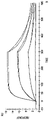
- FIG. 1 is a graph of the response versus time in an antibody binding experiment.
- Elevated blood glucose has long been considered to be the primary cause of diabetes. Recent research into the pathogenesis of diabetes and diabetic complications has revealed that the mechanism is more complex. A number of studies have indicated that cellular senescence is the main proponent in the development and progression of diabetes and diabetic complications.
- Senescent cells have been identified as a contributing factor to the onset and progression of both Type 1 and Type 2 diabetes through the secretion of reactive oxygen species.
- Reactive oxygen species contribute to Type 1 diabetes by activating inflammatory and apoptotic processes involved in beta cell dysfunction (Radoi, V. et al., “Advanced glycation end-products in diabetes mellitus: mechanism of action and focused treatment”, Proceedings of the Bulgarian Academy, Series B, No. 1, p. 9-19 (2012)).
- reactive oxygen species disrupt transmission pathways between the insulin receptor and the glucose transport system, which causes insulin resistance and inactivation of anti-atherosclerotic enzymes.
- the interaction of toxic AGEs, a marker of senescent cells, with their receptors (RAGE) in endothelial and inflammatory cells also leads to intracellular generation of reactive oxygen species.
- Senescent cells have been linked specifically with Type 2 diabetes. Elevated glucose is known to promote premature senescence in vitro in endothelial cells, renal mesangial cells, adipose-derived stem cells and fibroblasts (Palmer, A. K. et al., “Cellular senescence in Type 2 diabetes: a therapeutic opportunity”, Diabetes, Vol 64, pp. 2289-2298 (2015)). Other metabolic and signaling changes seen in diabetes such as altered lipid metabolism and growth hormone axis perturbations also promote senescent cell formation. Inflammatory factors are part of the senescence-associated secretory phenotype (SASP) and are thought to be a major contributor to the development of insulin resistance.
- SASP senescence-associated secretory phenotype
- WO 2016/044252 The association between inflammation and proinflammatory factors and cellular senescence is described in more detail in WO 2016/044252 and in Application No. 62/443,557.
- Cellular senescence may directly contribute to Type 2 diabetes through beta cell senescence.
- Senescent cells may be both a cause and a consequence of Type 2 diabetes as part of a pathogenic feedback loop (Palmer et al.).
- a high proportion of senescent islets of Langerhans which decreases the beta cell number and islet mass to pathological levels, was found in a mouse model of Friedreich's ataxia, a disorder characterized by diabetes (Mollá, B.
- CML Carboxymethyllysine
- AGEs have been associated with diabetic complications including diabetic retinopathy, diabetic nephropathy, diabetic neuropathy, diabetic cardiomyopathy, thrombogenesis, atherosclerosis, stroke, osteoporosis and erectile dysfunction. While the specific mechanism varies depending on the diabetic complication, many diabetic complications result from an increase in proinflammatory factors such as NE- ⁇ B, TNF- ⁇ and IL-6 or an increase in fibrosis by promoting profibrotic proteins such as TGF- ⁇ , both of which are promoted by AGEs and the AGE-RAGE interaction (Radoi et al.). AGEs have also been indirectly implicated in diabetic complications though methylglyoxal, a known cause of AGEs.
- methylglyoxal increases the chronic extremity soreness in diabetic neuropathy (“Methylglyoxal”, available online at en.wikipedia.org/wiki/Methylglyoxal (Jun. 5, 2017)).
- the presence of AGEs in skin, serum, saliva and urine has been correlated with diabetes and diabetic complications (Garay-Sevilla, M. E. et al., “Advanced glycosylation end products in skin, serum, saliva and urine and its association with complications of patients with Type 2 diabetes mellitus”, Journal of Endocrinological Investigation, Vol. 28, No. 5, pp. 223-230 (2005); Yoon, M-S. et al., “Characterisation of advanced glycation endproducts in saliva from patients with diabetes mellitus”, Biochemical and Biophysical Research Communications, Vol. 323, Issue 2, pp. 377-381 (2004)).
- MMPs matrix metalloproteinases
- MMP12 MMP macrophage metalloelastase
- AGEs can be one of the major factors influencing the progression of diabetic nephropathy by modulating the expression of MMPs (Xu, X. et al., “A glimpse of matrix metalloproteinases in diabetic nephropathy”, Current Medicinal Chemistry, Vol. 21, No. 28, p. 3244-3260 (2014)).
- the therapeutic benefits of removing senescent cells has been demonstrated in atherosclerosis and in age-related diseases, such as sarcopenia (Xu, M. et al., “Targeting senescent cells enhances adipogenesis and metabolic function in old age”, eLife (2015)).
- the identification of a link between cellular senescence, either directly or through AGEs, and diabetes and diabetic complications allows for similar treatment possibilities.
- the present invention uses enhanced clearance of cells expressing AGE-modified proteins or peptides (AGE-modified cells) to treat, ameliorate or prevent the onset of diabetes and diabetic complications by removing or killing senescent cells. This may be accomplished by administering anti-AGE antibodies to a subject.
- Vaccination against AGE-modified proteins or peptides of a cell may also be used to control the presence of AGE-modified cells in a subject.
- the continuous and virtually ubiquitous surveillance exercised by the immune system in the body in response to a vaccination allows maintaining low levels of AGE-modified cells in the body.
- Vaccination against AGE-modified proteins or peptides of a cell removes or kills senescent cells.
- the process of senescent cell removal or destruction allows vaccination against AGE-modified proteins or peptides of a cell to be used to treat or prevent the onset of diabetes and diabetic complications.
- an antibody that binds to an AGE-modified protein on a cell (“anti-AGE antibody” or “AGE antibody”) is known in the art. Examples include those described in U.S. Pat. No. 5,702,704 (Bucala) and U.S. Pat. No. 6,380,165 (Al-Abed et al.).
- the antibody may bind to one or more AGE-modified proteins or peptides having an AGE modification such as FFI, pyrraline, AFGP, ALI, carboxymethyllysine, carboxyethyllysine and pentosidine, and mixtures of such antibodies.
- the antibody binds carboxymethyllysine-modified or carboxyethyllysine-modified proteins.
- the antibody is non-immunogenic to the animal in which it will be used, such as non-immunogenic to humans; companion animals including cats, dogs and horses; and commercially important animals, such camels (or alpaca), cattle (bovine), sheep, and goats. More preferably, the antibody has the same species constant region as antibodies of the animal to reduce the immune response against the antibody, such as being humanized (for humans), felinized (for cats), caninized (for dogs), equuinized (for horses), camelized (for camels or alpaca), bovinized (for cattle), ovinized (for sheep), or caperized (for goats).
- the antibody is identical to that of the animal in which it will be used (except for the variable region), such as a human antibody, a cat antibody, a dog antibody, a horse antibody, a camel antibody, a bovine antibody, a sheep antibody or a goat antibody. Details of the constant regions and other parts of antibodies for these animals are described below.
- the antibody may be monoclonal or polyclonal.
- the antibody is a monoclonal antibody.
- Preferred anti-AGE antibodies include those which bind to proteins or peptides that exhibit a carboxymethyllysine or carboxyethyllysine AGE modification.
- Carboxymethyllysine also known as N(epsilon)-(carboxymethyl)lysine, N(6)-carboxymethyllysine, or 2-Amino-6-(carboxymethylamino)hexanoic acid
- carboxyethyllysine also known as N-epsilon-(carboxyethyl)lysine
- CML- and CEL-modified proteins or peptides are recognized by the receptor RAGE which is expressed on a variety of cells.
- CML and CEL have been well-studied and CML- and CEL-related products are commercially available.
- Cell Biolabs, Inc. sells CML-BSA antigens, CML polyclonal antibodies, CML immunoblot kits, and CML competitive ELISA kits (www.cellbiolabs.com/cml-assays) as well as CEL-BSA antigens and CEL competitive ELISA kits (www.cellbiolabs.com/cel-n-epsilon-carboxyethyl-lysine-assays-and-reagents).
- a particularly preferred antibody includes the variable region of the commercially available mouse anti-glycation end-product antibody raised against carboxymethyl lysine conjugated with keyhole limpet hemocyanin, the carboxymethyl lysine MAb (Clone 318003) available from R&D Systems, Inc. (Minneapolis, Minn.; catalog no. MAB3247), modified to have a human constant region (or the constant region of the animal into which it will be administered).
- Commercially-available antibodies such as the carboxymethyl lysine antibody corresponding to catalog no. MAB3247 from R&D Systems, Inc., may be intended for diagnostic purposes and may contain material that is not suited for use in animals or humans.
- commercially-available antibodies are purified and/or isolated prior to use in animals or humans to remove toxins or other potentially-harmful material.
- the anti-AGE antibody has low rate of dissociation from the antibody-antigen complex, or k d (also referred to as k back or off-rate), preferably at most 9 ⁇ 10 ⁇ 3 , 8 ⁇ 10 ⁇ 3 , 7 ⁇ 10 ⁇ 3 or 6 ⁇ 10 ⁇ 3 (sec ⁇ 1 ).
- the anti-AGE antibody has a high affinity for the AGE-modified protein of a cell, which may be expressed as a low dissociation constant K D of at most 9 ⁇ 10 ⁇ 6 , 8 ⁇ 10 ⁇ 6 , 7 ⁇ 10 ⁇ 6 , 6 ⁇ 10 ⁇ 6 , 5 ⁇ 10 ⁇ 6 , 4 ⁇ 10 ⁇ 6 or 3 ⁇ 10 ⁇ 6 (M).
- the binding properties of the anti-AGE antibody are similar to, the same as, or superior to the carboxymethyl lysine MAb (Clone 318003) available from R&D Systems, Inc. (Minneapolis, Minn.; catalog no. MAB3247), illustrated in FIG. 1 .
- the anti-AGE antibody may destroy AGE-modified cells through antibody-dependent cell-mediated cytotoxicity (ADCC).
- ADCC is a mechanism of cell-mediated immune defense in which an effector cell of the immune system actively lyses a target cell whose membrane-surface antigens have been bound by specific antibodies.
- ADCC may be mediated by natural killer (NK) cells, macrophages, neutrophils or eosinophils.
- NK natural killer
- the effector cells bind to the Fc portion of the bound antibody.
- the anti-AGE antibody may also destroy AGE-modified cells through complement-dependent cytotoxicity (CDC). In CDC, the complement cascade of the immune system is triggered by an antibody binding to a target antigen.
- CDC complement-dependent cytotoxicity
- the anti-AGE antibody may be conjugated to an agent that causes the destruction of AGE-modified cells.
- agents may be a toxin, a cytotoxic agent, magnetic nanoparticles, and magnetic spin-vortex discs.
- a toxin such as pore-forming toxins (PFT) (Aroian R. et al., “Pore-Forming Toxins and Cellular Non-Immune Defenses (CNIDs),” Current Opinion in Microbiology, 10:57-61 (2007)) conjugated to an anti-AGE antibody may be injected into a patient to selectively target and remove AGE-modified cells.
- the anti-AGE antibody recognizes and binds to AGE-modified cells. Then, the toxin causes pore formation at the cell surface and subsequent cell removal through osmotic lysis.
- Magnetic nanoparticles conjugated to the anti-AGE antibody may be injected into a patient to target and remove AGE-modified cells.
- the magnetic nanoparticles can be heated by applying a magnetic field in order to selectively remove the AGE-modified cells.
- magnetic spin-vortex discs which are magnetized only when a magnetic field is applied to avoid self-aggregation that can block blood vessels, begin to spin when a magnetic field is applied, causing membrane disruption of target cells.
- Magnetic spin-vortex discs, conjugated to anti-AGE antibodies specifically target AGE-modified cell types, without removing other cells.
- Antibodies are Y-shaped proteins composed of two heavy chains and two light chains.
- the two arms of the Y shape form the fragment antigen-binding (Fab) region while the base or tail of the Y shape forms the fragment crystallizable (Fc) region of the antibody.
- Antigen binding occurs at the terminal portion of the fragment antigen-binding region (the tips of the arms of the Y shape) at a location referred to as the paratope, which is a set of complementarity determining regions (also known as CDRs or the hypervariable region).
- the complementarity determining regions vary among different antibodies and gives a given antibody its specificity for binding to a given antigen.
- the fragment crystallizable region of the antibody determines the result of antigen binding and may interact with the immune system, such as by triggering the complement cascade or initiating antibody-dependent cell-mediated cytotoxicity (ADCC).
- ADCC antibody-dependent cell-mediated cytotoxicity
- a humanized anti-AGE antibody according to the present invention may have the human constant region sequence of amino acids shown in SEQ ID NO: 22.
- the heavy chain complementarity determining regions of the humanized anti-AGE antibody may have one or more of the protein sequences shown in SEQ ID NO: 23 (CDR1H), SEQ ID NO: 24 (CDR2H) and SEQ ID NO: 25 (CDR3H).
- the light chain complementarity determining regions of the humanized anti-AGE antibody may have one or more of the protein sequences shown in SEQ ID NO: 26 (CDR1L), SEQ ID NO: 27 (CDR2L) and SEQ ID NO: 28 (CDR3L).
- the heavy chain of a humanized anti-AGE antibody may have or may include the protein sequence of SEQ ID NO: 1.
- the variable domain of the heavy chain may have or may include the protein sequence of SEQ ID NO: 2.
- the complementarity determining regions of the variable domain of the heavy chain (SEQ ID NO: 2) are shown in SEQ ID NO: 41, SEQ ID NO: 42 and SEQ ID NO: 43.
- the kappa light chain of a humanized anti-AGE antibody may have or may include the protein sequence of SEQ ID NO: 3.
- the variable domain of the kappa light chain may have or may include the protein sequence of SEQ ID NO: 4.
- the arginine (Arg or R) residue at position 128 of SEQ ID NO: 4 may be omitted.
- variable domain of the light chain (SEQ ID NO: 4) are shown in SEQ ID NO: 44, SEQ ID NO: 45 and SEQ ID NO: 46.
- the variable regions may be codon-optimized, synthesized and cloned into expression vectors containing human immunoglobulin G1 constant regions.
- the variable regions may be used in the preparation of non-human anti-AGE antibodies.
- the antibody heavy chain may be encoded by the DNA sequence of SEQ ID NO: 12, a murine anti-AGE immunoglobulin G2b heavy chain.
- the protein sequence of the murine anti-AGE immunoglobulin G2b heavy chain encoded by SEQ ID NO: 12 is shown in SEQ ID NO: 16.
- the variable region of the murine antibody is shown in SEQ ID NO: 20, which corresponds to positions 25-142 of SEQ ID NO: 16.
- the antibody heavy chain may alternatively be encoded by the DNA sequence of SEQ ID NO: 13, a chimeric anti-AGE human immunoglobulin G1 heavy chain.
- the protein sequence of the chimeric anti-AGE human immunoglobulin G1 heavy chain encoded by SEQ ID NO: 13 is shown in SEQ ID NO: 17.
- the chimeric anti-AGE human immunoglobulin includes the murine variable region of SEQ ID NO: 20 in positions 25-142.
- the antibody light chain may be encoded by the DNA sequence of SEQ ID NO: 14, a murine anti-AGE kappa light chain.
- the protein sequence of the murine anti-AGE kappa light chain encoded by SEQ ID NO: 14 is shown in SEQ ID NO: 18.
- the variable region of the murine antibody is shown in SEQ ID NO: 21, which corresponds to positions 21-132 of SEQ ID NO: 18.
- the antibody light chain may alternatively be encoded by the DNA sequence of SEQ ID NO: 15, a chimeric anti-AGE human kappa light chain.
- the protein sequence of the chimeric anti-AGE human kappa light chain encoded by SEQ ID NO: 15 is shown in SEQ ID NO: 19.
- the chimeric anti-AGE human immunoglobulin includes the murine variable region of SEQ ID NO: 21 in positions 21-132.
- a humanized anti-AGE antibody according to the present invention may have or may include one or more humanized heavy chains or humanized light chains.
- a humanized heavy chain may be encoded by the DNA sequence of SEQ ID NO: 30, 32 or 34.
- the protein sequences of the humanized heavy chains encoded by SEQ ID NOs: 30, 32 and 34 are shown in SEQ ID NOs: 29, 31 and 33, respectively.
- a humanized light chain may be encoded by the DNA sequence of SEQ ID NO: 36, 38 or 40.
- the protein sequences of the humanized light chains encoded by SEQ ID NOs: 36, 38 and 40 are shown in SEQ ID NOs: 35, 37 and 39, respectively.
- the humanized anti-AGE antibody maximizes the amount of human sequence while retaining the original antibody specificity.
- a complete humanized antibody may be constructed that contains a heavy chain having a protein sequence chosen from SEQ ID NOs: 29, 31 and 33 and a light chain having a protein sequence chosen from SEQ ID NOs: 35, 37 and 39.
- anti-AGE antibodies may be obtained by humanizing murine monoclonal anti-AGE antibodies.
- Murine monoclonal anti-AGE antibodies have the heavy chain protein sequence shown in SEQ ID NO: 47 (the protein sequence of the variable domain is shown in SEQ ID NO: 52) and the light chain protein sequence shown in SEQ ID NO: 57 (the protein sequence of the variable domain is shown in SEQ ID NO: 62).
- a preferred humanized heavy chain may have the protein sequence shown in SEQ ID NO: 48, SEQ ID NO: 49, SEQ ID NO: 50 or SEQ ID NO: 51 (the protein sequences of the variable domains of the humanized heavy chains are shown in SEQ ID NO: 53, SEQ ID NO: 54, SEQ ID NO: 55 and SEQ ID NO: 56, respectively).
- a preferred humanized light chain may have the protein sequence shown in SEQ ID NO: 58, SEQ ID NO: 59, SEQ ID NO: 60 or SEQ ID NO: 61 (the protein sequences of the variable domains of the humanized light chains are shown in SEQ ID NO: 63, SEQ ID NO: 64, SEQ ID NO: 65 and SEQ ID NO: 66, respectively).
- a humanized anti-AGE monoclonal antibody is composed a heavy chain having a protein sequence selected from the group consisting of SEQ ID NO: 48, SEQ ID NO: 49, SEQ ID NO: 50 and SEQ ID NO: 51 and a light chain having a protein sequence selected from the group consisting of SEQ ID NO: 58, SEQ ID NO: 59, SEQ ID NO: 60 and SEQ ID NO: 61.
- Humanized monoclonal anti-AGE antibodies composed of these protein sequences may have better binding and/or improved activation of the immune system, resulting in greater efficacy.
- the protein sequence of an antibody from a non-human species may be modified to include the variable domain of the heavy chain having the sequence shown in SEQ ID NO: 2 or the kappa light chain having the sequence shown in SEQ ID NO: 4.
- the non-human species may be a companion animal, such as the domestic cat or domestic dog, or livestock, such as cattle, the horse or the camel. Preferably, the non-human species is not the mouse.
- the heavy chain of the horse ( Equus caballus ) antibody immunoglobulin gamma 4 may have or may include the protein sequence of SEQ ID NO: 5 (EMBL/GenBank accession number AY445518).
- the heavy chain of the horse ( Equus caballus ) antibody immunoglobulin delta may have or may include the protein sequence of SEQ ID NO: 6 (EMBL/GenBank accession number AY631942).
- the heavy chain of the dog ( Canis familiaris ) antibody immunoglobulin A may have or may include the protein sequence of SEQ ID NO: 7 (GenBank accession number L36871).
- the heavy chain of the dog ( Canis familiaris ) antibody immunoglobulin E may have or may include the protein sequence of SEQ ID NO: 8 (GenBank accession number L36872).
- the heavy chain of the cat (Fells catus ) antibody immunoglobulin G2 may have or may include the protein sequence of SEQ ID NO: 9 (DDBJ/EMBL/GenBank accession number KF811175).
- camelids Animals of the camelid family, such as camels ( Camelus dromedarius and Camelus bactrianus ), llamas ( Lama glama, Lama pacos and Lama vicugna ), alpacas ( Vicugna pacos) and guanacos ( Lama guanicoe ), have a unique antibody that is not found in other mammals.
- camelids In addition to conventional immunoglobulin G antibodies composed of heavy and light chain tetramers, camelids also have heavy chain immunoglobulin G antibodies that do not contain light chains and exist as heavy chain dimers.
- variable domain of a camelid heavy chain antibody is known as the VHH.
- the camelid heavy chain antibodies lack the heavy chain CH1 domain and have a hinge region that is not found in other species.
- the variable region of the Arabian camel ( Camelus dromedarius ) single-domain antibody may have or may include the protein sequence of SEQ ID NO: 10 (GenBank accession number AJ245148).
- the variable region of the heavy chain of the Arabian camel ( Camelus dromedarius ) tetrameric immunoglobulin may have or may include the protein sequence of SEQ ID NO: 11 (GenBank accession number AJ245184).
- heavy chain antibodies are also found in cartilaginous fishes, such as sharks, skates and rays.
- This type of antibody is known as an immunoglobulin new antigen receptor or IgNAR
- the variable domain of an IgNAR is known as the VNAR.
- the IgNAR exists as two identical heavy chain dimers composed of one variable domain and five constant domains each. Like camelids, there is no light chain.
- the protein sequences of additional non-human species may be readily found in online databases, such as the International ImMunoGeneTics Information System (www.imgt.org), the European Bioinformatics Institute (www.ebi.ac.uk), the DNA Databank of Japan (ddbj.nig.ac.jp/arsa) or the National Center for Biotechnology Information (www.ncbi.nlm.nih.gov).
- An anti-AGE antibody or a variant thereof may include a heavy chain having at least 90%, 91%, 92%, 93%, 94%, 95%, 96%, 97%, 98%, 99%, or 100% sequence identity to the amino acid sequence of SEQ ID NO: 1, SEQ ID NO: 16, SEQ ID NO: 17, SEQ ID NO: 29, SEQ ID NO: 31, SEQ ID NO: 33, SEQ ID NO: 47, SEQ ID NO: 48, SEQ ID NO: 49, SEQ ID NO: 50 or SEQ ID NO: 51, including post-translational modifications thereof.
- a heavy chain having at least 90%, 91%, 92%, 93%, 94%, 95%, 96%, 97%, 98%, or 99% sequence identity may contain substitutions (e.g., conservative substitutions), insertions, or deletions relative to the reference sequence, but an anti-AGE antibody including that sequence retains the ability to bind to AGE.
- An anti-AGE antibody or a variant thereof may include a heavy chain variable region having at least 90%, 91%, 92%, 93%, 94%, 95%, 96%, 97%, 98%, 99%, or 100% sequence identity to the amino acid sequence of SEQ ID NO: 2, SEQ ID NO: 20, SEQ ID NO: 23, SEQ ID NO: 24, SEQ ID NO: 25, SEQ ID NO: 41, SEQ ID NO: 42, SEQ ID NO: 43, SEQ ID NO: 52, SEQ ID NO: 53, SEQ ID NO: 54, SEQ ID NO: 55, or SEQ ID NO: 56, including post-translational modifications thereof.
- variable region having at least 90%, 91%, 92%, 93%, 94%, 95%, 96%, 97%, 98%, or 99% sequence identity may contain substitutions (e.g., conservative substitutions), insertions, or deletions relative to the reference sequence, but an anti-AGE antibody including that sequence retains the ability to bind to AGE.
- substitutions, insertions, or deletions may occur in regions outside the variable region.
- An anti-AGE antibody or a variant thereof may include a light chain having at least 90%, 91%, 92%, 93%, 94%, 95%, 96%, 97%, 98%, 99%, or 100% sequence identity to the amino acid sequence of SEQ ID NO: 3, SEQ ID NO: 18, SEQ ID NO: 19, SEQ ID NO: 35, SEQ ID NO: 37, SEQ ID NO: 39, SEQ ID NO: 57, SEQ ID NO: 58, SEQ ID NO: 59, SEQ ID NO: 60 or SEQ ID NO: 61, including post-translational modifications thereof.
- a light chain having at least 90%, 91%, 92%, 93%, 94%, 95%, 96%, 97%, 98%, or 99% sequence identity may contain substitutions (e.g., conservative substitutions), insertions, or deletions relative to the reference sequence, but an anti-AGE antibody including that sequence retains the ability to bind to AGE.
- substitutions, insertions, or deletions may occur in regions outside the variable region.
- An anti-AGE antibody or a variant thereof may include a light chain variable region having at least 90%, 91%, 92%, 93%, 94%, 95%, 96%, 97%, 98%, 99%, or 100% sequence identity to the amino acid sequence of SEQ ID NO: 4, SEQ ID NO: 21, SEQ ID NO: 26, SEQ ID NO: 27, SEQ ID NO: 28, SEQ ID NO: 44, SEQ ID NO: 45, SEQ ID NO: 46, SEQ ID NO: 62, SEQ ID NO: 63, SEQ ID NO: 64, SEQ ID NO: 65 or SEQ ID NO: 66, including post-translational modifications thereof.
- variable region having at least 90%, 91%, 92%, 93%, 94%, 95%, 96%, 97%, 98%, or 99% sequence identity may contain substitutions (e.g., conservative substitutions), insertions, or deletions relative to the reference sequence, but an anti-AGE antibody including that sequence retains the ability to bind to AGE.
- substitutions, insertions, or deletions may occur in regions outside the variable region.
- the antibody may have the complementarity determining regions of commercially available mouse anti-glycation end-product antibody raised against carboxymethyl lysine conjugated with keyhole limpet hemocyanin (CML-KLH), the carboxymethyl lysine MAb (Clone 318003) available from R&D Systems, Inc. (Minneapolis, Minn.; catalog no. MAB3247).
- CML-KLH keyhole limpet hemocyanin
- CD3 carboxymethyl lysine MAb
- the antibody may have or may include constant regions which permit destruction of targeted cells by a subject's immune system.
- Bi-specific antibodies which are anti-AGE antibodies directed to two different epitopes, may also be used. Such antibodies will have a variable region (or complementary determining region) from those of one anti-AGE antibody, and a variable region (or complementary determining region) from a different antibody.
- Antibody fragments may be used in place of whole antibodies.
- immunoglobulin G may be broken down into smaller fragments by digestion with enzymes.
- Papain digestion cleaves the N-terminal side of inter-heavy chain disulfide bridges to produce Fab fragments.
- Fab fragments include the light chain and one of the two N-terminal domains of the heavy chain (also known as the Fd fragment).
- Pepsin digestion cleaves the C-terminal side of the inter-heavy chain disulfide bridges to produce F(ab′) 2 fragments.
- F(ab′) 2 fragments include both light chains and the two N-terminal domains linked by disulfide bridges.
- Pepsin digestion may also form the Fv (fragment variable) and Fc (fragment crystallizable) fragments.
- the Fv fragment contains the two N-terminal variable domains.
- the Fc fragment contains the domains which interact with immunoglobulin receptors on cells and with the initial elements of the complement cascade.
- Pepsin may also cleave immunoglobulin G before the third constant domain of the heavy chain (C H 3) to produce a large fragment F(abc) and a small fragment pFc′.
- Antibody fragments may alternatively be produced recombinantly. Preferably, such antibody fragments are conjugated to an agent that causes the destruction of AGE-modified cells.
- polyclonal antibodies can be raised in a mammalian host by one or more injections of an immunogen, and if desired, an adjuvant.
- an immunogen and if desired, an adjuvant.
- the immunogen (and adjuvant) is injected in a mammal by a subcutaneous or intraperitoneal injection.
- the immunogen may be an AGE-modified protein of a cell, such as AGE-antithrombin III, AGE-calmodulin, AGE-insulin, AGE-ceruloplasmin, AGE-collagen, AGE-cathepsin B, AGE-albumin such as AGE-bovine serum albumin (AGE-BSA), AGE-human serum albumin and ovalbumin, AGE-crystallin, AGE-plasminogen activator, AGE-endothelial plasma membrane protein, AGE-aldehyde reductase, AGE-transferrin, AGE-fibrin, AGE-copper/zinc SOD, AGE-apo B, AGE-fibronectin, AGE-pancreatic ribose, AGE-apo A-I and II, AGE-hemoglobin, AGE-Na + /K + -ATPase, AGE-plasminogen, AGE-myelin, AGE-lysozyme,
- AGE-modified cells such as AGE-modified erythrocytes, whole, lysed, or partially digested, may also be used as AGE antigens.
- adjuvants include Freund's complete, monophosphoryl Lipid A synthetic-trehalose dicorynomycolate, aluminum hydroxide (alum), heat shock proteins HSP 70 or HSP96, squalene emulsion containing monophosphoryl lipid A, a2-macroglobulin and surface active substances, including oil emulsions, pleuronic polyols, polyanions and dinitrophenol.
- an immunogen may be conjugated to a polypeptide that is immunogenic in the host, such as keyhole limpet hemocyanin (KLH), serum albumin, bovine thyroglobulin, cholera toxin, labile enterotoxin, silica particles or soybean trypsin inhibitor.
- KLH keyhole limpet hemocyanin
- serum albumin serum albumin
- bovine thyroglobulin bovine thyroglobulin
- cholera toxin cholera toxin
- labile enterotoxin silica particles
- silica particles silica particles
- soybean trypsin inhibitor e.g., soybean trypsin inhibitor.
- Monoclonal antibodies may also be made by immunizing a host or lymphocytes from a host, harvesting the mAb-secreting (or potentially secreting) lymphocytes, fusing those lymphocytes to immortalized cells (for example, myeloma cells), and selecting those cells that secrete the desired mAb.
- Other techniques may be used, such as the EBV-hybridoma technique.
- chimeric antibodies that are substantially human (humanized) or substantially “ized” to another animal (such as cat, dog, horse, camel or alpaca, cattle, sheep, or goat) at the amino acid level.
- the mAbs may be purified from the culture medium or ascites fluid by conventional procedures, such as protein A-sepharose, hydroxyapatite chromatography, gel electrophoresis, dialysis, ammonium sulfate precipitation or affinity chromatography.
- human monoclonal antibodies can be generated by immunization of transgenic mice containing a third copy IgG human trans-loci and silenced endogenous mouse Ig loci or using human-transgenic mice. Production of humanized monoclonal antibodies and fragments thereof can also be generated through phage display technologies.
- a “pharmaceutically acceptable carrier” includes any and all solvents, dispersion media, coatings, antibacterial and antifungal agents, isotonic and absorption delaying agents, and the like, compatible with pharmaceutical administration.
- Preferred examples of such carriers or diluents include water, saline, Ringer's solutions and dextrose solution. Supplementary active compounds can also be incorporated into the compositions.
- Solutions and suspensions used for parenteral administration can include a sterile diluent, such as water for injection, saline solution, polyethylene glycols, glycerin, propylene glycol or other synthetic solvents; antibacterial agents such as benzyl alcohol or methyl parabens; antioxidants such as ascorbic acid or sodium bisulfite; buffers such as acetates, citrates or phosphates, and agents for the adjustment of tonicity such as sodium chloride or dextrose.
- the pH can be adjusted with acids or bases, such as hydrochloric acid or sodium hydroxide.
- the parenteral preparation can be enclosed in ampoules, disposable syringes or multiple dose vials made of glass or plastic.
- the antibodies may be administered by injection, such as by intravenous injection or locally, such as by intra-articular injection into a joint.
- Pharmaceutical compositions suitable for injection include sterile aqueous solutions or dispersions for the extemporaneous preparation of sterile injectable solutions or dispersion.
- Suitable carriers include physiological saline, bacteriostatic water, CREMOPHOR EL® (BASF; Parsippany, N.J.) or phosphate buffered saline (PBS). In all cases, the composition must be sterile and should be fluid so as to be administered using a syringe.
- compositions should be stable during manufacture and storage and must be preserved against contamination from microorganisms such as bacteria and fungi.
- Various antibacterial and anti-fungal agents for example, parabens, chlorobutanol, phenol, ascorbic acid, and thimerosal, can contain microorganism contamination.
- Isotonic agents such as sugars, polyalcohols, such as manitol, sorbitol, and sodium chloride can be included in the composition.
- Compositions that can delay absorption include agents such as aluminum monostearate and gelatin.
- Sterile injectable solutions can be prepared by incorporating antibodies, and optionally other therapeutic components, in the required amount in an appropriate solvent with one or a combination of ingredients as required, followed by sterilization. Methods of preparation of sterile solids for the preparation of sterile injectable solutions include vacuum drying and freeze-drying to yield a solid.
- the antibodies may be delivered as an aerosol spray from a nebulizer or a pressurized container that contains a suitable propellant, for example, a gas such as carbon dioxide.
- a suitable propellant for example, a gas such as carbon dioxide.
- Antibodies may also be delivered via inhalation as a dry powder, for example using the iSPERSETM inhaled drug delivery platform (PULMATRIX, Lexington, Mass.).
- the use of anti-AGE antibodies which are chicken antibodies (IgY) may be non-immunogenic in a variety of animals, including humans, when administered by inhalation.
- An appropriate dosage level of each type of antibody will generally be about 0.01 to 500 mg per kg patient body weight.
- the dosage level will be about 0.1 to about 250 mg/kg; more preferably about 0.5 to about 100 mg/kg.
- a suitable dosage level may be about 0.01 to 250 mg/kg, about 0.05 to 100 mg/kg, or about 0.1 to 50 mg/kg. Within this range the dosage may be 0.05 to 0.5, 0.5 to 5 or 5 to 50 mg/kg.
- each type of antibody may be administered on a regimen of 1 to 4 times per day, such as once or twice per day, antibodies typically have a long half-life in vivo. Accordingly, each type of antibody may be administered once a day, once a week, once every two or three weeks, once a month, or once every 60 to 90 days.
- a subject that receives administration of an anti-AGE antibody may be tested to determine if the administration has been effective to treat diabetes or diabetic complications.
- Diabetes may be monitored with a blood test that measures blood glucose such as the fasting plasma glucose (FPG) test, the A1C test (also known as the hemoglobin A1C, HbA1C, glycated hemoglobin, and glycosylated hemoglobin test), the random plasma glucose (RPG) test, the glucose challenge test or the oral glucose tolerance test (OGTT).
- FPG fasting plasma glucose
- A1C test also known as the hemoglobin A1C, HbA1C, glycated hemoglobin, and glycosylated hemoglobin test
- RPG random plasma glucose
- GTT oral glucose tolerance test
- a subject may be considered to have received an effective antibody treatment if he or she demonstrates a reduction in blood glucose between subsequent measurements or over time.
- Diabetic complications may be monitored with a diagnostic test that is suitable for evaluating a given diabetic com
- a reduction in blood pressure a reduction in the development of cataracts, a reduction in neuropathic pain, reduced albumin in urine (diabetic nephropathy) or improved ventricular diastolic function (cardiomyopathy).
- the concentration and/or number of senescent cells may be measured over time. Administration of antibody and subsequent testing may be repeated until the desired therapeutic result is achieved.
- Unit dosage forms can be created to facilitate administration and dosage uniformity.
- Unit dosage form refers to physically discrete units suited as single dosages for the subject to be treated, containing a therapeutically effective quantity of one or more types of antibodies in association with the required pharmaceutical carrier.
- the unit dosage form is in a sealed container and is sterile.
- Vaccines against AGE-modified proteins or peptides contain an AGE antigen, an adjuvant, optional preservatives and optional excipients.
- AGE antigens include AGE-modified proteins or peptides such as AGE-antithrombin III, AGE-calmodulin, AGE-insulin, AGE-ceruloplasmin, AGE-collagen, AGE-cathepsin B, AGE-albumin such as AGE-bovine serum albumin (AGE-BSA), AGE-human serum albumin and ovalbumin, AGE-crystallin, AGE-plasminogen activator, AGE-endothelial plasma membrane protein, AGE-aldehyde reductase, AGE-transferrin, AGE-fibrin, AGE-copper/zinc SOD, AGE-apo B, AGE-fibronectin, AGE-pancreatic ribose, AGE-apo A-I and II, AGE-hemoglobin
- AGE-modified cells such as AGE-modified erythrocytes, whole, lysed, or partially digested, may also be used as AGE antigens.
- Suitable AGE antigens also include proteins or peptides that exhibit AGE modifications (also referred to as AGE epitopes or AGE moieties) such as carboxymethyllysine (CML), carboxyethyllysine (CEL), pentosidine, pyrraline, FFI, AFGP and ALI.
- the AGE antigen may be an AGE-protein conjugate, such as AGE conjugated to keyhole limpet hemocyanin (AGE-KLH). Further details of some of these AGE-modified proteins or peptides and their preparation are described in Bucala.
- Particularly preferred AGE antigens include proteins or peptides that exhibit a carboxymethyllysine or carboxyethyllysine AGE modification.
- Carboxymethyllysine also known as N(epsilon)-(carboxymethyl)lysine, N(6)-carboxymethyllysine, or 2-Amino-6-(carboxymethylamino)hexanoic acid
- carboxyethyllysine also known as N-epsilon-(carboxyethyl)lysine
- proteins or peptides and lipids as a result of oxidative stress and chemical glycation and have been correlated with juvenile genetic disorders.
- CML- and CEL-modified proteins or peptides are recognized by the receptor RAGE which is expressed on a variety of cells.
- CML and CEL have been well-studied and CML- and CEL-related products are commercially available.
- Cell Biolabs, Inc. sells CML-BSA antigens, CML polyclonal antibodies, CML immunoblot kits, and CML competitive ELISA kits (www.cellbiolabs.com/cml-assays) as well as CEL-BSA antigens and CEL competitive ELISA kits (www.cellbiolabs.com/cel-n-epsilon-carboxyethyl-lysine-assays-and-reagents).
- AGE antigens may be conjugated to carrier proteins to enhance antibody production in a subject. Antigens that are not sufficiently immunogenic alone may require a suitable carrier protein to stimulate a response from the immune system.
- suitable carrier proteins include keyhole limpet hemocyanin (KLH), serum albumin, bovine thyroglobulin, cholera toxin, labile enterotoxin, silica particles and soybean trypsin inhibitor.
- KLH keyhole limpet hemocyanin
- serum albumin serum albumin
- bovine thyroglobulin cholera toxin
- labile enterotoxin silica particles
- soybean trypsin inhibitor e.g., the carrier protein is KLH (AGE-KLH).
- KLH has been extensively studied and has been identified as an effective carrier protein in experimental cancer vaccines.
- Preferred AGE antigen-carrier protein conjugates include CML-KLH and CEL-KLH.
- Immunity is a long-term immune response, either cellular or humoral.
- a cellular immune response is activated when an antigen is presented, preferably with a co-stimulator to a T-cell which causes it to differentiate and produce cytokines.
- the cells involved in the generation of the cellular immune response are two classes of T-helper (Th) cells, Th1 and Th2.
- Th1 cells stimulate B cells to produce predominantly antibodies of the IgG2A isotype, which activates the complement cascade and binds the Fc receptors of macrophages, while Th2 cells stimulate B cells to produce IgG1 isotype antibodies in mice, IgG4 isotype antibodies in humans, and IgE isotype antibodies.
- the human body also contains “professional” antigen-presenting cells such as dendritic cells, macrophages, and B cells.
- a humoral immune response is triggered when a B cell selectively binds to an antigen and begins to proliferate, leading to the production of a clonal population of cells that produce antibodies that specifically recognize that antigen and which may differentiate into antibody-secreting cells, referred to as plasma-cells or memory-B cells.
- Antibodies are molecules produced by B-cells that bind a specific antigen.
- the antigen-antibody complex triggers several responses, either cell-mediated, for example by natural killers (NK) or macrophages, or serum-mediated, for example by activating the complement system, a complex of several serum proteins that act sequentially in a cascade that result in the lysis of the target cell.
- Immunological adjuvants are the component(s) of a vaccine which augment the immune response to the immunogenic agent.
- Adjuvants function by attracting macrophages to the immunogenic agent and then presenting the agent to the regional lymph nodes to initiate an effective antigenic response.
- Adjuvants may also act as carriers themselves for the immunogenic agent.
- Adjuvants may induce an inflammatory response, which may play an important role in initiating the immune response.
- Adjuvants include mineral compounds such as aluminum salts, oil emulsions, bacterial products, liposomes, immunostimulating complexes and squalene.
- Aluminum compounds are the most widely used adjuvants in human and veterinary vaccines. These aluminum compounds include aluminum salts such as aluminum phosphate (AIPO 4 ) and aluminum hydroxide (Al(OH) 3 ) compounds, typically in the form of gels, and are generically referred to in the field of vaccine immunological adjuvants as “alum.”
- Aluminum hydroxide is a poorly crystalline aluminum oxyhydroxide having the structure of the mineral boehmite.
- Aluminum phosphate is an amorphous aluminum hydroxyphosphate.
- Negatively charged species can absorb onto aluminum hydroxide gels at neutral pH
- positively charged species can absorb onto aluminum phosphate gels at neutral pH. It is believed that these aluminum compounds provide a depot of antigen at the site of administration, thereby providing a gradual and continuous release of antigen to stimulate antibody production. Aluminum compounds tend to more effectively stimulate a cellular response mediated by Th2, rather than Th1 cells.
- Emulsion adjuvants include water-in-oil emulsions (for example, Freund's adjuvants, such as killed mycobacteria in oil emulsion) and oil-in-water emulsions (for example, MF-59).
- Emulsion adjuvants include an immunogenic component, for example squalene (MF-59) or mannide oleate (Incomplete Freund's Adjuvants), which can induce an elevated humoral response, increased T cell proliferation, cytotoxic lymphocytes and cell-mediated immunity.
- Liposomal or vesicular adjuvants have lipophilic bilayer domains and an aqueous milieu which can be used to encapsulate and transport a variety of materials, for example an antigen.
- Paucilamellar vesicles can be prepared by mixing, under high pressure or shear conditions, a lipid phase comprising a non-phospholipid material (for example, an amphiphile surfactant; see U.S. Pat. Nos.
- a sterol optionally a sterol, and any water-immiscible oily material to be encapsulated in the vesicles (for example, an oil such as squalene oil and an oil-soluble or oil-suspended antigen); and an aqueous phase such as water, saline, buffer or any other aqueous solution used to hydrate the lipids.
- a sterol optionally a sterol, and any water-immiscible oily material to be encapsulated in the vesicles
- an oil such as squalene oil and an oil-soluble or oil-suspended antigen
- an aqueous phase such as water, saline, buffer or any other aqueous solution used to hydrate the lipids.
- Liposomal or vesicular adjuvants are believed to promote contact of the antigen with immune cells, for example by fusion of the vesicle to the immune cell membrane, and preferentially stimulate
- adjuvants include Mycobacterium bovis bacillus Calmette-Guérin (BCG), quill-saponin and unmethylated CpG dinucleotides (CpG motifs). Additional adjuvants are described in U.S. Patent Application Publication Pub. No. US 2010/0226932 (Sep. 9, 2010) and Jiang, Z-H. et al. “Synthetic vaccines: the role of adjuvants in immune targeting”, Current Medicinal Chemistry , Vol. 10(15), pp. 1423-39 (2003). Preferable adjuvants include Freund's complete adjuvant and Freund's incomplete adjuvant.
- the vaccine may optionally include one or more preservatives, such as antioxidants, antibacterial and antimicrobial agents, as well as combinations thereof.
- preservatives such as antioxidants, antibacterial and antimicrobial agents, as well as combinations thereof.
- examples include benzethonium chloride, ethylenediamine-tetraacetic acid sodium (EDTA), thimerosal, phenol, 2-phenoxyethanol, formaldehyde and formalin; antibacterial agents such as amphotericin B, chlortetracycline, gentamicin, neomycin, polymyxin B and streptomycin; antimicrobial surfactants such as polyoxyethylene-9, 10-nonyl phenol (Triton N-101, octoxynol-9), sodium deoxycholate and polyoxyethylated octyl phenol (Triton X-100).
- the production and packaging of the vaccine may eliminate the need for a preservative. For example, a vaccine that has been sterilized and
- vaccines include pharmaceutically acceptable excipients, such as stabilizers, thickening agents, toxin detoxifiers, diluents, pH adjusters, tonicity adjustors, surfactants, antifoaming agents, protein stabilizers, dyes and solvents.
- pharmaceutically acceptable excipients such as stabilizers, thickening agents, toxin detoxifiers, diluents, pH adjusters, tonicity adjustors, surfactants, antifoaming agents, protein stabilizers, dyes and solvents.
- excipients examples include hydrochloric acid, phosphate buffers, sodium acetate, sodium bicarbonate, sodium borate, sodium citrate, sodium hydroxide, potassium chloride, potassium chloride, sodium chloride, polydimethylsilozone, brilliant green, phenol red (phenolsulfon-phthalein), glycine, glycerin, sorbitol, histidine, monosodium glutamate, potassium glutamate, sucrose, urea, lactose, gelatin, sorbitol, polysorbate 20, polysorbate 80 and glutaraldehyde.
- hydrochloric acid phosphate buffers, sodium acetate, sodium bicarbonate, sodium borate, sodium citrate, sodium hydroxide, potassium chloride, potassium chloride, sodium chloride, polydimethylsilozone, brilliant green, phenol red (phenolsulfon-phthalein), glycine, glycerin, sorbitol, histidine, monosodium glutamate, potassium glut
- the vaccine may contain from 1 ⁇ g to 100 mg of at least one AGE antigen, including 10, 20, 30, 40, 50, 60, 70, 80, 90, 100, 200, 400, 800 or 1000 ⁇ g, or 2, 3, 4, 5, 6, 7, 8, 9, 10, 20, 30, 40, 50, 60, 70, 80 or 90 mg.
- the amount used for a single injection corresponds to a unit dosage.
- the vaccine may be provided in unit dosage form or in multidosage form, such as 2-100 or 2-10 doses.
- the unit dosages may be provided in a vial with a septum, or in a syringe with or without a needle.
- the vaccine may be administered intravenously, subdermally or intraperitoneally.
- the vaccine is sterile.
- the vaccine may be administered one or more times, such as 1 to 10 times, including 2, 3, 4, 5, 6, 7, 8 or 9 times, and may be administered over a period of time ranging from 1 week to 1 year, 2-10 weeks or 2-10 months. Furthermore, booster vaccinations may be desirable, over the course of 1 year to 20 years, including 2, 5, 10 and 15 years.
- a subject that receives a vaccine for AGE-modified proteins or peptides of a cell may be tested to determine if he or she has developed an immunity to the AGE-modified proteins or peptides. Suitable tests may include blood tests for detecting the presence of an antibody, such as immunoassays or antibody titers. An immunity to AGE-modified proteins or peptides may also be determined by monitoring the concentration and/or number of senescent cells over time. In addition to testing for the development of an immunity to AGE-modified proteins or peptides, a subject may also be tested to determine if the vaccination has been effective to treat diabetes or diabetic complications.
- a subject may be considered to have received an effective vaccination if he or she demonstrates a reduction in blood glucose or an improvement in diabetic complications between subsequent measurements or over time, or by measuring the concentration and/or number of senescent cells. Vaccination and subsequent testing may be repeated until the desired therapeutic result is achieved.
- the vaccination process may be designed to provide immunity against multiple AGE moieties.
- a single AGE antigen may induce the production of AGE antibodies which are capable of binding to multiple AGE moieties.
- the vaccine may contain multiple AGE antigens.
- a subject may receive multiple vaccines, where each vaccine contains a different AGE antigen.
- Any mammal that could develop diabetes or diabetic complications may be treated by the methods herein described.
- Humans are a preferred mammal for treatment.
- Other mammals that may be treated include mice, rats, goats, sheep, cows, horses and companion animals, such as dogs or cats.
- any of the mammals or subjects identified above may be excluded from the patient population in need of treatment for diabetes or diabetic complications.
- a subject may be identified as in need of treatment based on a diagnosis of diabetes.
- Diabetes may be diagnosed with a blood test that measures blood glucose such as the fasting plasma glucose (FPG) test, the A1C test (also known as the hemoglobin A1C, HbA1C, glycated hemoglobin, and glycosylated hemoglobin test), the random plasma glucose (RPG) test, the glucose challenge test or the oral glucose tolerance test (OGTT).
- FPG fasting plasma glucose
- A1C test also known as the hemoglobin A1C, HbA1C, glycated hemoglobin, and glycosylated hemoglobin test
- RPG random plasma glucose
- OGTT oral glucose tolerance test
- a subject may be diagnosed with diabetes if he or she has an A1C level greater than 5.7% when measured on two separate occasions.
- a subject may be identified as in need of treatment based on a diagnosis of one or more diabetic complications.
- diabetic complications include heart disease such as cardiomyopathy, stroke, diabetic retinopathy, cataracts, glaucoma, kidney disease such as nephropathy, hypoglycemia, hyperglycemic crisis, high blood pressure, high blood LDL cholesterol, nerve disease, diabetic neuropathy (peripheral and autonomic neuropathy), neuropathic pain, cognitive impairment, non-alcoholic fatty liver disease, periodontal (gum) disease, hearing loss, erectile dysfunction, depression, complications with pregnancy, diabetic ketoacidosis, hyperosmolar hyperglycemic state and diabetic coma.
- heart disease such as cardiomyopathy, stroke, diabetic retinopathy, cataracts, glaucoma
- kidney disease such as nephropathy, hypoglycemia, hyperglycemic crisis, high blood pressure, high blood LDL cholesterol, nerve disease, diabetic neuropathy (peripheral and autonomic neuro
- Diabetic complications may be diagnosed with any suitable diagnostic test for a given diabetic complication. For example, measuring blood pressure, examining the eye for cataracts, a liver ultrasound indicating steatosis (non-alcoholic fatty liver disease), urinalysis measuring albumin in urine (diabetic nephropathy) or echocardiography to measure ventricular diastolic function (cardiomyopathy).
- any suitable diagnostic test for a given diabetic complication For example, measuring blood pressure, examining the eye for cataracts, a liver ultrasound indicating steatosis (non-alcoholic fatty liver disease), urinalysis measuring albumin in urine (diabetic nephropathy) or echocardiography to measure ventricular diastolic function (cardiomyopathy).
- Subjects may also be identified as in need of treatment based on detection of advanced glycation end products in a sample obtained from the subject. Suitable samples include blood, skin, serum, saliva and urine. The diagnostic use of anti-AGE antibodies is discussed in more detail in Application No. 62/501,424.
- Positions 16-133 of the above amino acid sequence correspond to SEQ ID NO: 2. Positions 46-50 of the above amino acid sequence correspond to SEQ ID NO: 41. Positions 65-81 of the above amino acid sequence correspond to SEQ ID NO: 42. Positions 114-122 of the above amino acid sequence correspond to SEQ ID NO: 43.
- Positions 16-128 of the above amino acid sequence correspond to SEQ ID NO: 4.
- the arginine (Arg or R) residue at position 128 of SEQ ID NO: 4 may be omitted.
- Positions 39-54 of the above amino acid sequence correspond to SEQ ID NO: 44.
- Positions 70-76 of the above amino acid sequence correspond to SEQ ID NO: 45.
- Positions 109-117 of the above amino acid sequence correspond to SEQ ID NO: 46.
- the alanine residue at position 123 of the above amino acid sequence may optionally be replaced with a serine residue.
- the tyrosine residue at position 124 of the above amino acid sequence may optionally be replaced with a phenylalanine residue.
- Positions 25-142 of the above amino acid sequence correspond to SEQ ID NO: 20.
- SEQ ID NO: 20 may optionally include the substitutions at positions 123 and 124.
- SEQ ID NO: 20 may optionally contain one additional lysine residue after the terminal valine residue.
- Positions 21-132 of the above amino acid sequence correspond to SEQ ID NO: 21.
- the one-letter amino acid sequence that corresponds to SEQ ID NO: 23 is SYTMGVS.
- the one-letter amino acid sequence that corresponds to SEQ ID NO: 24 is TISSGGGSTYYPDSVKG.
- the one-letter amino acid sequence that corresponds to SEQ ID NO: 25 is QGGWLPPFAX, where X may be any naturally occurring amino acid.
- the one-letter amino acid sequence that corresponds to SEQ ID NO: 26 is RASKSVSTSSRGYSYMH.
- the one-letter amino acid sequence that corresponds to SEQ ID NO: 27 is LVSNLES.
- the one-letter amino acid sequence that corresponds to SEQ ID NO: 28 is QHIRELTRS.
- the antibody was administered to the aged CD1(ICR) mouse (Charles River Laboratories), twice daily by intravenous injection, once a week, for three weeks (Days 1, 8 and 15), followed by a 10 week treatment-free period.
- the test antibody was a commercially available mouse anti-glycation end-product antibody raised against carboxymethyl lysine conjugated with keyhole limpet hemocyanin, the carboxymethyl lysine MAb (Clone 318003) available from R&D Systems, Inc. (Minneapolis, Minn.; catalog no. MAB3247).
- a control reference of physiological saline was used in the control animals.
- mice referred to as “young” were 8 weeks old, while mice referred to as “old” were 88 weeks ( ⁇ 2 days) old. No adverse events were noted from the administration of the antibody.
- the different groups of animals used in the study are shown in Table 1.
- p16 INK4a mRNA a marker for senescent cells, was quantified in adipose tissue of the groups by Real Time-qPCR. The results are shown in Table 2.
- the mass of the gastrocnemius muscle was also measured, to determine the effect of antibody administration on sarcopenia.
- the results are provided in Table 3. The results indicate that administration of the antibody increased muscle mass as compared to controls, but only at the higher dosage of 5.0 ⁇ g/gm/BID/week.
- Muscle (%) 1 Mean 0.3291 1.1037 SD 0.0412 0.1473 N 20 20 2 Mean 0.3304 0.7671 SD 0.0371 0.1246 N 20 20 3 Mean 0.3410 0.7706 SD 0.0439 0.0971 N 19 19 5 Mean 0.4074 0.9480 SD 0.0508 0.2049 N 9 9
- Example 1 The affinity and kinetics of the test antibody used in Example 1 were analyzed using N ⁇ ,N ⁇ -bis(carboxymethyl)-L-lysine trifluoroacetate salt (Sigma-Aldrich, St. Louis, Mo.) as a model substrate for an AGE-modified protein of a cell. Label-free interaction analysis was carried out on a BIACORETM T200 (GE Healthcare, Pittsburgh, Pa.), using a Series S sensor chip CM5 (GE Healthcare, Pittsburgh, Pa.), with Fc1 set as blank, and Fc2 immodilized with the test antibody (molecular weigh of 150,000 Da).
- the running buffer was a HBS-EP buffer (10 mM HEPES, 150 mM NaCl, 3 mM EDTA and 0.05% P-20, pH of 7.4), at a temperature of 25° C.
- Software was BIACORETM T200 evaluation software, version 2.0. A double reference (Fc2-1 and only buffer injection), was used in the analysis, and the data was fitted to a Langmuir 1:1 binding model.
- FIG. 1 A graph of the response versus time is illustrated in FIG. 1 .
- Murine and chimeric human anti-AGE antibodies were prepared.
- the DNA sequence of murine anti-AGE antibody IgG2b heavy chain is shown in SEQ ID NO: 12.
- the DNA sequence of chimeric human anti-AGE antibody IgG1 heavy chain is shown in SEQ ID NO: 13.
- the DNA sequence of murine anti-AGE antibody kappa light chain is shown in SEQ ID NO: 14.
- the DNA sequence of chimeric human anti-AGE antibody kappa light chain is shown in SEQ ID NO: 15.
- the gene sequences were synthesized and cloned into high expression mammalian vectors. The sequences were codon optimized. Completed constructs were sequence confirmed before proceeding to transfection.
- HEK293 cells were seeded in a shake flask one day before transfection, and were grown using serum-free chemically defined media.
- the DNA expression constructs were transiently transfected into 0.03 liters of suspension HEK293 cells. After 20 hours, cells were sampled to obtain the viabilities and viable cell counts, and titers were measured (Octet QKe, ForteBio). Additional readings were taken throughout the transient transfection production runs. The cultures were harvested on day 5, and an additional sample for each was measured for cell density, viability and titer.
- the conditioned media for murine and chimeric anti-AGE antibodies were harvested and clarified from the transient transfection production runs by centrifugation and filtration. The supernatants were run over a Protein A column and eluted with a low pH buffer. Filtration using a 0.2 ⁇ m membrane filter was performed before aliquoting. After purification and filtration, the protein concentrations were calculated from the OD280 and the extinction coefficient. A summary of yields and aliquots is shown in Table 5:
- Antibody purity was evaluated by capillary electrophoresis sodium-dodecyl sulfate (CE-SDS) analysis using LabChip® GXII, (PerkinElmer).
- the binding of the murine (parental) and chimeric anti-AGE antibodies described in Example 3 was investigated by a direct binding ELISA.
- An anti-carboxymethyl lysine (CML) antibody (R&D Systems, MAB3247) was used as a control.
- CML was conjugated to KLH (CML-KLH) and both CML and CML-KLH were coated overnight onto an ELISA plate.
- HRP-goat anti-mouse Fc was used to detect the control and murine (parental) anti-AGE antibodies.
- HRP-goat anti-human Fc was used to detect the chimeric anti-AGE antibody.
- the antigens were diluted to 1 ⁇ g/mL in 1 ⁇ phosphate buffer at pH 6.5.
- a 96-well microtiter ELISA plate was coated with 100 ⁇ L/well of the diluted antigen and let sit at 4° C. overnight. The plate was blocked with 1 ⁇ PBS, 2.5% BSA and allowed to sit for 1-2 hours the next morning at room temperature.
- the antibody samples were prepared in serial dilutions with 1 ⁇ PBS, 1% BSA with the starting concentration of 50 ⁇ g/mL. Secondary antibodies were diluted 1:5,000. 100 ⁇ L of the antibody dilutions was applied to each well. The plate was incubated at room temperature for 0.5-1 hour on a microplate shaker. The plate was washed 3 times with 1 ⁇ PBS.
- the OD450 absorbance raw data for the CML and CML-KLH ELISA is shown in the plate map below. 48 of the 96 wells in the well plate were used. Blank wells in the plate map indicate unused wells.
- the OD450 absorbance raw data for the CML-only ELISA is shown in the plate map below. 24 of the 96 wells in the well plate were used. Blank wells in the plate map indicate unused wells.
- control and chimeric anti-AGE antibodies showed binding to both CML and CML-KLH.
- the murine (parental) anti-AGE antibody showed very weak to no binding to either CML or CML-KLH.
- Data from repeated ELISA confirms binding of the control and chimeric anti-AGE to CML. All buffer control showed negative signal.
- Humanized antibodies were designed by creating multiple hybrid sequences that fuse select parts of the parental (mouse) antibody sequence with the human framework sequences. Acceptor frameworks were identified based on the overall sequence identity across the framework, matching interface position, similarly classed CDR canonical positions, and presence of N-glycosylation sites that would have to be removed. Three humanized light chains and three humanized heavy chains were designed based on two different heavy and light chain human acceptor frameworks. The amino acid sequences of the heavy chains are shown in SEQ ID NO: 29, 31 and 33, which are encoded by the DNA sequences shown in SEQ ID NO: 30, 32 and 34, respectively.
- the amino acid sequences of the light chains are shown in SEQ ID NO: 35, 37 and 39, which are encoded by the DNA sequences shown in SEQ ID NO: 36, 38 and 40, respectively.
- the humanized sequences were methodically analyzed by eye and computer modeling to isolate the sequences that would most likely retain antigen binding. The goal was to maximize the amount of human sequence in the final humanized antibodies while retaining the original antibody specificity.
- the light and heavy humanized chains could be combined to create nine variant fully humanized antibodies.
- the three heavy chains and three light chains were analyzed to determine their humanness.
- Antibody humanness scores were calculated according to the method described in Gao, S. H., et al., “Monoclonal antibody humanness score and its applications”, BMC Biotechnology, 13:55 (Jul. 5, 2013).
- the humanness score represents how human-like an antibody variable region sequence looks. For heavy chains a score of 79 or above is indicative of looking human-like; for light chains a score of 86 or above is indicative of looking human-like.
- the humanness of the three heavy chains, three light chains, a parental (mouse) heavy chain and a parental (mouse) light chain are shown below in Table 6:
- variable region sequences were designed by first synthesizing the variable region sequences. The sequences were optimized for expression in mammalian cells. These variable region sequences were then cloned into expression vectors that already contain human Fc domains; for the heavy chain, the IgG1 was used.
- the binding of the humanized antibodies may be evaluated, for example, by dose-dependent binding ELISA or cell-based binding assay.
- Example 6 An AGE-RNAse Containing Vaccine in a Human Subject
- AGE-RNAse is prepared by incubating RNAse in a phosphate buffer solution containing 0.1-3 M glucose, glucose-6-phosphate, fructose or ribose for 10-100 days. The AGE-RNAse solution is dialyzed and the protein content is measured. Aluminum hydroxide or aluminum phosphate, as an adjuvant, is added to 100 ⁇ g of the AGE-RNAse. Formaldehyde or formalin is added as a preservative to the preparation. Ascorbic acid is added as an antioxidant.
- the vaccine also includes phosphate buffer to adjust the pH and glycine as a protein stabilizer. The composition is injected intravenously into a subject with diabetes.
- Example 7 Injection Regimen for an AGE-RNAse Containing Vaccine in a Human Subject
- Example 6 The same vaccine as described in Example 6 is injected intra-articularly into a subject with diabetic neuropathy.
- the titer of antibodies to AGE-RNAse is determined by ELISA after two weeks. Additional injections are performed after three weeks and six weeks, respectively. Further titer determination is performed two weeks after each injection.
- Example 8 An AGE-Hemoglobin Containing Vaccine in a Human Subject
- AGE-hemoglobin is prepared by incubating human hemoglobin in a phosphate buffer solution containing 0.1-3 M glucose, glucose-6-phosphate, fructose or ribose for 10-100 days. The AGE-hemoglobin solution is dialyzed and the protein content is measured. All vaccine components are the same as in Example 6, except AGE-hemoglobin is substituted for AGE-RNAse. Administration is carried out as in Example 6, or as in Example 7.
- Example 9 An AGE-Human Serum Albumin Containing Vaccine in a Human Subject
- AGE-human serum albumin is prepared by incubating human serum albumin in a phosphate buffer solution containing 0.1-3 M glucose, glucose-6-phosphate, fructose or ribose for 10-100 days. The AGE-human serum albumin solution is dialyzed and the protein content is measured. All vaccine components are the same as in Example 6, except AGE-human serum albumin is substituted for AGE-RNAse. Administration is carried out as in Example 6, or as in Example 7.
- Example 10 Carboxymethyllysine-Modified Protein Vaccine for a Human Subject (Prophetic)
- a vaccine is prepared by combining a carboxymethyllysine-modified protein as an AGE antigen, aluminum hydroxide as an adjuvant, formaldehyde as a preservative, ascorbic acid as an antioxidant, a phosphate buffer to adjust the pH of the vaccine and glycine as a protein stabilizer.
- the vaccine is injected subcutaneously into a subject with diabetic cardiomyopathy.
- Example 11 Carboxyethyllysine-Modified Peptide Vaccine for a Human Subject (Prophetic)
- a vaccine is prepared by combining a carboxyethyllysine-modified peptide conjugated to KLH as an AGE antigen, aluminum hydroxide as an adjuvant, formaldehyde as a preservative, ascorbic acid as an antioxidant, a phosphate buffer to adjust the pH of the vaccine and glycine as a protein stabilizer.
- the vaccine is injected subcutaneously into a subject with diabetic nephropathy.
- Example 12 In Vivo Study of the Administration of a Carboxymethyl Lysine Monoclonal Antibody
- mice Female BALB/c mice (BALB/cAnNCrl, Charles River) were eleven weeks old on Day 1 of the study.
- 4T1 murine breast tumor cells (ATCC CRL-2539) were cultured in RPMI 1640 medium containing 10% fetal bovine serum, 2 mM glutamine, 25 ⁇ g/mL gentamicin, 100 units/mL penicillin G Na and 100 ⁇ g/mL streptomycin sulfate. Tumor cells were maintained in tissue culture flasks in a humidified incubator at 37° C. in an atmosphere of 5% CO 2 and 95% air.
- the cultured breast cancer cells were then implanted in the mice.
- 4T1 cells were harvested during log phase growth and re-suspended in phosphate buffered saline (PBS) at a concentration of 1 ⁇ 10 6 cells/mL on the day of implant.
- Tumors were initiated by subcutaneously implanting 1 ⁇ 10 5 4T1 cells (0.1 mL suspension) into the right flank of each test animal. Tumors were monitored as their volumes approached a target range of 80-120 mm 3 .
- Tumor weight was approximated using the assumption that 1 mm 3 of tumor volume has a weight of 1 mg.
- the four treatment groups are shown in Table 8 below:
- An anti-carboxymethyl lysine monoclonal antibody was used as a therapeutic agent.
- 250 mg of carboxymethyl lysine monoclonal antibody was obtained from R&D Systems (Minneapolis, Minn.).
- Dosing solutions of the carboxymethyl lysine monoclonal antibody were prepared at 1 and 0.5 mg/mL in a vehicle (PBS) to provide the active dosages of 10 and 5 ⁇ g/g, respectively, in a dosing volume of 10 mL/kg. Dosing solutions were stored at 4° C. protected from light.
- i.v. dosing was changed to intraperitoneal (i.p.) dosing for those animals that could not be dosed i.v. due to tail vein degradation.
- the dosing volume was 0.200 mL per 20 grams of body weight (10 mL/kg), and was scaled to the body weight of each individual animal.
- % TGI percent tumor growth inhibition
- the ability of the anti-carboxymethyl lysine antibody to inhibit cachexia was determined by comparing the weights of the lungs and gastrocnemius muscles for Groups 1-3. Tissue weights were also normalized to 100 g body weight.
- Treatment efficacy was also evaluated by the incidence and magnitude of regression responses observed during the study. Treatment may cause partial regression (PR) or complete regression (CR) of the tumor in an animal.
- PR partial regression
- CR complete regression
- the tumor volume was 50% or less of its Day 1 volume for three consecutive measurements during the course of the study, and equal to or greater than 13.5 mm 3 for one or more of these three measurements.
- the tumor volume was less than 13.5 mm 3 for three consecutive measurements during the course of the study.
- mice In vivo studies are carried out in mice to study the effect of treatment with anti-AGE antibodies on diabetic complications including diabetic neuropathy and diabetic cardiomyopathy.
- Male and female BTBR ob/ob mice are used for the study.
- the male mice are primarily intended for studying diabetic neuropathy while the female mice are primarily intended for studying diabetic cardiomyopathy. All mice are 6-8 weeks old on Day 1 of the study.
- the mice are separated into four treatment groups of 10-15 mice each: (1) male—control; (2) female—control; (3) male—anti-AGE antibody; and (4) female—anti-AGE antibody.
- Dosing begins on Day 1 of the study.
- Groups 1 and 2 receive phosphate-buffered saline (PBS) delivered intravenously.
- Groups 3 and 4 receive 10 ⁇ g/g of an anti-AGE antibody twice daily for 21 days delivered intravenously.
- the dosing volume is 0.200 mL per 20 grams of body weight (10 mL/kg), and is scaled to the body weight of each individual animal.
- Diabetic neuropathy is measured by tail flick thermal testing. Blood is drawn and percent of glycosylated hemoglobin (A1C) is measured 1 day before Day 1 of the study, at 12 weeks, at 16 weeks and at 24 weeks. Treatment will end at 9-11 weeks (21 days after treatment commences).
- A1C glycosylated hemoglobin
- Diabetic cardiomyopathy is studied by analyzing the left ventricle including measuring myocardial cross-sectional area, myocyte size, fibrosis and macrophage content.
- Diabetic nephropathy is studied by histologically examining the kidneys for loss of podocytes and by measuring the glomerular basement membrane thickness, the amount of mesangial matrix and the presence/absence of mesangial sclerosis.
- Diabetic neuropathy is studied by determining the epidermal nerve fiber density.
- the anti-AGE antibody will specifically bind to cells expressing cell-surface AGEs, such as senescent beta cells, and allow the immune system to destroy those cells. Killing and removing senescent cells will prevent the onset of diabetes and the development of diabetic complications.
Abstract
A method of treating or preventing the onset of diabetes or diabetic complications comprises administering to a subject a composition comprising an anti-AGE antibody. A composition for treating or preventing the onset of diabetes or diabetic complications comprises a first anti-AGE antibody, a second anti-AGE antibody and a pharmaceutically acceptable carrier. The first anti-AGE antibody is different from the second anti-AGE antibody. A method of treating or preventing the onset of diabetes or diabetic complications comprises immunizing a subject in need thereof against AGE-modified proteins or peptides of a cell.
Description
Diabetes mellitus, commonly referred to as diabetes or DM, is the general term for diseases and disorders that are characterized by recurrent or persistent elevated levels of blood glucose. Diabetes occurs when beta cells in the pancreas do not produce sufficient insulin and/or when the body cannot effectively use the insulin produced by beta cells. The most common forms of diabetes are Type 1 diabetes (also known as juvenile diabetes, juvenile-onset diabetes or insulin-dependent diabetes) and Type 2 diabetes (also known as adult-onset diabetes or non-insulin-dependent diabetes). Type 1 diabetes is an autoimmune disease in which the body attacks insulin-producing beta cells in the pancreas. Type 2 diabetes is a progressive metabolic disease in which the body becomes unable to properly regulate insulin and beta cells become unable to produce sufficient insulin.
Diabetes is a significant public health problem. The World Health Organization estimates that 422 million adults worldwide have diabetes (“Global Report on Diabetes”, World Health Organization, 2016). The United States Centers for Disease Control and Prevention (CDC) estimates that 29.1 million people in the U.S. currently have diabetes, which is 9.3% of the total population (“National Diabetes Statistics Report: Estimates of Diabetes and Its Burden in the United States, 2014”, U.S. Department of Health and Human Services, Centers for Disease Control and Prevention, 2014). Diabetes is currently the seventh leading cause of death in the U.S. and the estimated total costs of diabetes in the U.S. is $245 billion. The prevalence of diabetes is increasing and the CDC projects that one in three adults in the U.S. will have diabetes by 2050.
Diabetes is associated with a number of complications, including heart disease such as cardiomyopathy, stroke, diabetic retinopathy, cataracts, glaucoma, kidney disease such as nephropathy, hypoglycemia, hyperglycemic crisis, high blood pressure, high blood LDL cholesterol, nerve disease, diabetic neuropathy (peripheral and autonomic neuropathy), neuropathic pain, cognitive impairment, non-alcoholic fatty liver disease, periodontal (gum) disease, hearing loss, erectile dysfunction, depression, complications with pregnancy, diabetic ketoacidosis, hyperosmolar hyperglycemic state and diabetic coma. These complications can result in severe consequences such as blindness, kidney failure, amputations, infections and even death.
Diabetic complications may be studied in the mouse strain BTBR with the ob/ob leptin-deficiency mutation, a recently developed animal model of type 2 diabetes. The BTBR mouse has been recognized as a good animal model of diabetic neuropathy (O'Brien, P. D. et al., “BTBR ob/ob mice as a novel diabetic neuropathy model: Neurological characterization and gene expression analyses”, Neurobiology of Disease, Vol. 73, pp. 348-355 (2015)). The BTBR mouse also serves as an animal model of nephropathy and cardiomyopathy (Alpers, C. E. et al., “Mouse models of diabetic nephropathy”, Current Opinion in Nephrology and Hypertension, Vol. 20, No. 3, pp. 278-284 (2011); O'Brien, K. D. et al., “Divergent effects of vasodilators on cardiac hypertrophy and inflammation in a murine model of diabetic cardiomyopathy”, Journal of the American College of Cardiology, Vol. 57, Issue 17, p. E193 (2011)). The BTBR mouse has the potential to advance the development of treatments for diabetes and diabetic complications.
Current treatments for diabetes and diabetic complications focus on maintaining blood glucose at normal levels. Type 1 diabetes is not preventable and blood glucose must be managed with insulin therapy, such as insulin injections or an implantable insulin pump. Type 2 diabetes may be prevented, treated or reversed with lifestyle changes, such as maintaining a healthy weight, eating a healthy diet, being physically active and avoiding tobacco use. If lifestyle changes are insufficient, Type 2 diabetics may manage their blood glucose by consuming oral glucose or regulating insulin levels through insulin injections or an implantable insulin pump. Bariatric surgery is a radical treatment option, but has been shown to prevent or even reverse Type 2 diabetes. Pharmacotherapies for treating and/or preventing diabetes and diabetic complications are limited.
Senescent cells are cells that are partially-functional or non-functional and are in a state of proliferative arrest. Senescence is a distinct state of a cell, and is associated with biomarkers, such as activation of the biomarker p16Ink4a, and expression of β-galactosidase. Senescence begins with damage or stress (such as overstimulation by growth factors) of cells.
Advanced glycation end-products (AGEs; also referred to as AGE-modified proteins or peptides, or glycation end-products) arise from a non-enzymatic reaction of sugars with protein side-chains (Ando, K. et al., Membrane Proteins of Human Erythrocytes Are Modified by Advanced Glycation End Products during Aging in the Circulation, Biochem Biophys Res Commun., Vol. 258, 123, 125 (1999)). This process begins with a reversible reaction between the reducing sugar and the amino group to form a Schiff base, which proceeds to form a covalently-bonded Amadori rearrangement product. Once formed, the Amadori product undergoes further rearrangement to produce AGEs. Hyperglycemia and oxidative stress promote this post-translational modification of membrane proteins (Lindsey J B, et al., “Receptor For Advanced Glycation End-Products (RAGE) and soluble RAGE (sRAGE): Cardiovascular Implications,” Diabetes Vascular Disease Research, Vol. 6(1), 7-14, (2009)). AGEs may also be formed from other processes. For example, the advanced glycation end product, Nε-(carboxymethyl)lysine, is a product of both lipid peroxidation and glycoxidation reactions. AGEs have been associated with several pathological conditions including inflammation, atherosclerosis, stroke, endothelial cell dysfunction, and neurodegenerative disorders (Bierhaus A, “AGEs and their interaction with AGE-receptors in vascular disease and diabetes mellitus. I. The AGE concept,” Cardiovasc Res, Vol. 37(3), 586-600 (1998)).
AGE-modified proteins are also a marker of senescent cells. This association between AGEs and senescence is well known in the art. See, for example, Gruber, L. (WO 2009/143411, 26 Nov. 2009), Ando, K. et al. (Membrane Proteins of Human Erythrocytes Are Modified by Advanced Glycation End Products during Aging in the Circulation, Biochem Biophys Res Commun., Vol. 258, 123, 125 (1999)), Ahmed, E. K. et al. (“Protein Modification and Replicative Senescence of WI-38 Human Embryonic Fibroblasts” Aging Cells, vol. 9, 252, 260 (2010)), Vlassara, H. et al. (Advanced Glycosylation Endproducts on Erythrocyte Cell Surface Induce Receptor-Mediated Phagocytosis by Macrophages, J. Exp. Med., Vol. 166, 539, 545 (1987)) and Vlassara et al. (“High-affinity-receptor-mediated Uptake and Degradation of Glucose-modified Proteins: A Potential Mechanism for the Removal of Senescent Macromolecules” Proc. Natl. Acad. Sci. USAI, Vol. 82, 5588, 5591 (1985)). Furthermore, Ahmed, E. K. et al. indicates that glycation end-products are “one of the major causes of spontaneous damage to cellular and extracellular proteins” (Ahmed, E. K. et al., see above, page 353). Accordingly, the accumulation of glycation end-products is associated with senescence and lack of function.
The damage or stress that causes cellular senescence also negatively impacts mitochondrial DNA in the cells to cause them to produce free radicals which react with sugars in the cell to form methyl glyoxal (MG). MG in turn reacts with proteins or lipids to generate advanced glycation end products. In the case of the protein component lysine, MG reacts to form carboxymethyllysine, which is an AGE.
Damage or stress to mitochondrial DNA also sets off a DNA damage response which induces the cell to produce cell cycle blocking proteins. These blocking proteins prevent the cell from dividing. Continued damage or stress causes mTOR production, which in turn activates protein synthesis and inactivates protein breakdown. Further stimulation of the cells leads to programmed cell death (apoptosis).
p16 is a protein involved in regulation of the cell cycle, by inhibiting the S phase (synthesis phase). It can be activated during ageing or in response to various stresses, such as DNA damage, oxidative stress or exposure to drugs. p16 is typically considered a tumor suppressor protein, causing a cell to become senescent in response to DNA damage and irreversibly preventing the cell from entering a hyperproliferative state. However, there has been some ambiguity in this regard, as some tumors show overexpression of p16, while others show downregulated expression. Evidence suggests that overexpression of p16 is some tumors results from a defective retinoblastoma protein (“Rb”). p16 acts on Rb to inhibit the S phase, and Rb downregulates p16, creating negative feedback. Defective Rb fails to both inhibit the S phase and downregulate p16, thus resulting in overexpression of p16 in hyperproliferating cells (Romagosa, C. et al., p16Ink4a overexpression in cancer: a tumor suppressor gene associated with senescence and high-grade tumors, Oncogene, Vol. 30, 2087-2097 (2011)).
Senescent cells are associated with secretion of many factors involved in intercellular signaling, including pro-inflammatory factors; secretion of these factors has been termed the senescence-associated secretory phenotype, or SASP (Freund, A. “Inflammatory networks during cellular senescence: causes and consequences” Trends Mol Med. 2010 May; 16(5):238-46). Autoimmune diseases, such as Crohn's disease and rheumatoid arthritis, are associated with chronic inflammation (Ferraccioli, G. et al. “Interleukin-1β, and Interleukin-6 in Arthritis Animal Models: Roles in the Early Phase of Transition from Acute to Chronic Inflammation and Relevance for Human Rheumatoid Arthritis” Mol Med. 2010 November-December; 16(11-12): 552-557). Chronic inflammation may be characterized by the presence of pro-inflammatory factors at levels higher than baseline near the site of pathology, but lower than those found in acute inflammation. Examples of these factors include TNF, IL-1α, IL-1β, IL-5, IL-6, IL-8, IL-12, IL-23, CD2, CD3, CD20, CD22, CD52, CD80, CD86, C5 complement protein, BAFF, APRIL, IgE, α4β1 integrin and α4β7 integrin. Senescent cells also upregulate genes with roles in inflammation including IL-1β, IL-8, ICAM1, TNFAP3, ESM1 and CCL2 (Burton, D. G. A. et al., “Microarray analysis of senescent vascular smooth muscle cells: a link to atherosclerosis and vascular calcification”, Experimental Gerontology, Vol. 44, No. 10, pp. 659-665 (October 2009)). Because senescent cells produce pro-inflammatory factors, removal of these cells alone produces a profound reduction in inflammation as well as the amount and concentration of pro-inflammatory factors.
Senescent cells secrete reactive oxygen species (“ROS”) as part of the SASP. ROS are believed to play an important role in maintaining senescence of cells. The secretion of ROS creates a bystander effect, where senescent cells induce senescence in neighboring cells: ROS create the very cellular damage known to activate p16 expression, leading to senescence (Nelson, G., A senescent cell bystander effect: senescence-induced senescence, Aging Cell, Vo. 11, 345-349 (2012)). The p16/Rb pathway leads to the induction of ROS, which in turn activates the protein kinase C delta creating a positive feedback loop that further enhance ROS, helping maintain the irreversible cell cycle arrest; it has even been suggested that exposing cancer cells to ROS might be effective to treat cancer by inducing cell phase arrest in hyperproliferating cells (Rayess, H. et al., Cellular senescence and tumor suppressor gene p16, Int J Cancer, Vol. 130, 1715-1725 (2012)).
Recent research demonstrates the therapeutic benefits of removing senescent cells. In vivo animal studies at the Mayo Clinic in Rochester, Minn., found that elimination of senescent cells in transgenic mice carrying a biomarker for elimination delayed age-related disorders associated with cellular senescence. Eliminating senescent cells in fat and muscle tissues substantially delayed the onset of sarcopenia and cataracts and reduced senescence indicators in skeletal muscle and the eye (Baker, D. J. et al., “Clearance of p16Ink4a-positive senescent cells delays ageing-associated disorders”, Nature, Vol. 479, pp. 232-236, (2011)). Mice that were treated to induce senescent cell elimination were found to have larger diameters of muscle fibers as compared to untreated mice. Treadmill exercise tests indicated that treatment also preserved muscle function. Continuous treatment of transgenic mice for removal of senescent cells had no negative side effects and selectively delayed age-related phenotypes that depend on cells. This data demonstrates that removal of senescent cells produces beneficial therapeutic effects and shows that these benefits may be achieved without adverse effects.
Additional In vivo animal studies in mice found that removing senescent cells using senolytic agents treats aging-related disorders and atherosclerosis. Short-term treatment with senolytic drugs in chronologically aged or progeroid mice alleviated several aging-related phenotypes (Zhu, Y. et al., “The Achilles' heel of senescent cells: from transcriptome to senolytic drugs”, Aging Cell, vol. 14, pp. 644-658 (2015)). Long-term treatment with senolytic drugs improved vasomotor function in mice with established atherosclerosis and reduced intimal plaque calcification (Roos, C. M. et al., “Chronic senolytic treatment alleviates established vasomotor dysfunction in aged or atherosclerotic mice”, Aging Cell (2016)). This data further demonstrates the benefits of removing senescent cells.
Vaccines have been widely used since their introduction by Edward Jenner in the 1770s to confer immunity against a wide range of diseases and afflictions. Vaccine preparations contain a selected immunogenic agent capable of stimulating immunity to an antigen. Typically, antigens are used as the immunogenic agent in vaccines, such as, for example, viruses, either killed or attenuated, and purified viral components. Antigens used in the production of cancer vaccines include, for example, tumor-associated carbohydrate antigens (TACAs), dendritic cells, whole cells and viral vectors. Different techniques are employed to produce the desired amount and type of antigen being sought. For example, pathogenic viruses are grown either in eggs or cells. Recombinant DNA technology is often utilized to generate attenuated viruses for vaccines.
Vaccines may therefore be used to stimulate the production of antibodies in the body and provide immunity against antigens. When an antigen is introduced to a subject that has been vaccinated and developed immunity to that antigen, the immune system may destroy or remove cells that express the antigen.
In a first aspect, the invention is a method of treating or preventing the onset of diabetes or diabetic complications, comprising killing senescent cells by administering to a subject a composition comprising an anti-AGE antibody. The diabetic complication comprises at least one disease or disorder selected from the group consisting of stroke, diabetic retinopathy, cataracts, glaucoma, kidney disease, hypoglycemia, hyperglycemic crisis, high blood pressure, high blood LDL cholesterol, nerve disease, diabetic neuropathy, neuropathic pain, cognitive impairment, non-alcoholic fatty liver disease, periodontal disease, hearing loss, erectile dysfunction, depression, complications with pregnancy, diabetic ketoacidosis, hyperosmolar hyperglycemic state and diabetic coma.
The term “diabetes” means a disease or disorder characterized by persistent elevated levels of blood glucose in which a subject has an A1C level greater than 5.7% when measured on two separate occasions. Diabetes includes Type 1 diabetes, Type 2 diabetes, gestational diabetes, prediabetes, latent autoimmune diabetes of adults, congenital diabetes, monogenic diabetes, maturity-onset diabetes of the young (MODY), cystic fibrosis-related diabetes and idiopathic diabetes.
The term “peptide” means a molecule composed of 2-50 amino acids.
The term “protein” means a molecule composed of more than 50 amino acids.
The terms “advanced glycation end-product”, “AGE”, “AGE-modified protein”, “AGE-modified peptide” and “glycation end-product” refer to modified proteins or peptides that are formed as the result of the reaction of sugars with protein side chains that further rearrange and form irreversible cross-links. This process begins with a reversible reaction between a reducing sugar and an amino group to form a Schiff base, which proceeds to form a covalently-bonded Amadori rearrangement product. Once formed, the Amadori product undergoes further rearrangement to produce AGEs. AGE-modified proteins and antibodies to AGE-modified proteins are described in U.S. Pat. No. 5,702,704 to Bucala (“Bucala”) and U.S. Pat. No. 6,380,165 to Al-Abed et al. (“Al-Abed”). Glycated proteins or peptides that have not undergone the necessary rearrangement to form AGEs, such as N-deoxyfructosyllysine found on glycated albumin, are not AGEs. AGEs may be identified by the presence of AGE modifications (also referred to as AGE epitopes or AGE moieties) such as 2-(2-furoyl)-4(5)-(2-furanyl)-1H-imidazole (“FFI”); 5-hydroxymethyl-1-alkylpyrrole-2-carbaldehyde (“Pyrraline”); 1-alkyl-2-formyl-3,4-diglycosyl pyrrole (“AFGP”), a non-fluorescent model AGE; carboxymethyllysine; carboxyethyllysine; and pentosidine. ALI, another AGE, is described in Al-Abed.
The term “AGE antigen” means a substance that elicits an immune response against an AGE-modified protein or peptide of a cell. The immune response against an AGE-modified protein or peptide of a cell does not include the production of antibodies to the non-AGE-modified protein or peptide.
“An antibody that binds to an AGE-modified protein on a cell”, “anti-AGE antibody” or “AGE antibody” means an antibody, antibody fragment or other protein or peptide that binds to an AGE-modified protein or peptide which preferably includes a constant region of an antibody, where the protein or peptide which has been AGE-modified is a protein or peptide normally found bound on the surface of a cell, preferably a mammalian cell, more preferably a human, cat, dog, horse, camelid (for example, camel or alpaca), cattle, sheep, or goat cell. “An antibody that binds to an AGE-modified protein on a cell”, “anti-AGE antibody” or “AGE antibody” does not include an antibody or other protein which binds with the same specificity and selectivity to both the AGE-modified protein or peptide, and the same non-AGE-modified protein or peptide (that is, the presence of the AGE modification does not increase binding). AGE-modified albumin is not an AGE-modified protein on a cell, because albumin is not a protein normally found bound on the surface of cells. “An antibody that binds to an AGE-modified protein on a cell”, “anti-AGE antibody” or “AGE antibody” only includes those antibodies which lead to removal, destruction, or death of the cell. Also included are antibodies which are conjugated, for example to a toxin, drug, or other chemical or particle. Preferably, the antibodies are monoclonal antibodies, but polyclonal antibodies are also possible.
The term “senescent cell” means a cell which is in a state of proliferative arrest and expresses one or more biomarkers of senescence, such as activation of p16Ink4a or expression of senescence-associated β-galactosidase. Also included are cells which express one or more biomarkers of senescence, do not proliferate in vivo, but may proliferate in vitro under certain conditions, such as some satellite cells found in the muscles of ALS patients.
The term “variant” means a nucleotide, protein or amino acid sequence different from the specifically identified sequences, wherein one or more nucleotides, proteins or amino acid residues is deleted, substituted or added. Variants may be naturally-occurring allelic variants, or non-naturally-occurring variants. Variants of the identified sequences may retain some or all of the functional characteristics of the identified sequences.
The term “percent (%) sequence identity” is defined as the percentage of amino acid residues in a candidate sequence that are identical to the amino acid residues in a reference polypeptide sequence, after aligning the sequences and introducing gaps, if necessary, to achieve the maximum percent sequence identity, and not considering any conservative substitutions as part of the sequence identity. Alignment for purposes of determining percent amino acid sequence identity can be achieved in various ways using publicly available computer software such as BLAST, BLAST-2, ALIGN or Megalign (DNASTAR) software. Preferably, % sequence identity values are generated using the sequence comparison computer program ALIGN-2. The ALIGN-2 sequence comparison computer program is publicly available from Genentech, Inc. (South San Francisco, Calif.), or may be compiled from the source code, which has been filed with user documentation in the U.S. Copyright Office and is registered under U.S. Copyright Registration No. TXU510087. The ALIGN-2 program should be compiled for use on a UNIX operating system, including digital UNIX V4.0D. All sequence comparison parameters are set by the ALIGN-2 program and do not vary.
In situations where ALIGN-2 is employed for amino acid sequence comparisons, the % sequence identity of a given amino acid sequence A to, with, or against a given amino acid sequence B (which can alternatively be phrased as a given amino acid sequence A that has or comprises a certain % amino acid sequence identity to, with, or against a given amino acid sequence B) is calculated as follows: 100 times the fraction X/Y where X is the number of amino acid residues scored as identical matches by the sequence alignment program ALIGN-2 in that program's alignment of A and B, and where Y is the total number of amino acid residues in B. Where the length of amino acid sequence A is not equal to the length of amino acid sequence B, the % amino acid sequence identity of A to B will not equal the % amino acid sequence identity of B to A. Unless specifically stated otherwise, all % amino acid sequence identity values used herein are obtained using the ALIGN-2 computer program.
Elevated blood glucose has long been considered to be the primary cause of diabetes. Recent research into the pathogenesis of diabetes and diabetic complications has revealed that the mechanism is more complex. A number of studies have indicated that cellular senescence is the main proponent in the development and progression of diabetes and diabetic complications.
Senescent cells have been identified as a contributing factor to the onset and progression of both Type 1 and Type 2 diabetes through the secretion of reactive oxygen species. Reactive oxygen species contribute to Type 1 diabetes by activating inflammatory and apoptotic processes involved in beta cell dysfunction (Radoi, V. et al., “Advanced glycation end-products in diabetes mellitus: mechanism of action and focused treatment”, Proceedings of the Romanian Academy, Series B, No. 1, p. 9-19 (2012)). In Type 2 diabetes, reactive oxygen species disrupt transmission pathways between the insulin receptor and the glucose transport system, which causes insulin resistance and inactivation of anti-atherosclerotic enzymes. The interaction of toxic AGEs, a marker of senescent cells, with their receptors (RAGE) in endothelial and inflammatory cells also leads to intracellular generation of reactive oxygen species.
Senescent cells have been linked specifically with Type 2 diabetes. Elevated glucose is known to promote premature senescence in vitro in endothelial cells, renal mesangial cells, adipose-derived stem cells and fibroblasts (Palmer, A. K. et al., “Cellular senescence in Type 2 diabetes: a therapeutic opportunity”, Diabetes, Vol 64, pp. 2289-2298 (2015)). Other metabolic and signaling changes seen in diabetes such as altered lipid metabolism and growth hormone axis perturbations also promote senescent cell formation. Inflammatory factors are part of the senescence-associated secretory phenotype (SASP) and are thought to be a major contributor to the development of insulin resistance. The association between inflammation and proinflammatory factors and cellular senescence is described in more detail in WO 2016/044252 and in Application No. 62/443,557. Cellular senescence may directly contribute to Type 2 diabetes through beta cell senescence. Senescent cells may be both a cause and a consequence of Type 2 diabetes as part of a pathogenic feedback loop (Palmer et al.). In addition, a high proportion of senescent islets of Langerhans, which decreases the beta cell number and islet mass to pathological levels, was found in a mouse model of Friedreich's ataxia, a disorder characterized by diabetes (Mollá, B. et al., “Two different pathogenic mechanisms, dying-back axonal neuropathy and pancreatic senescence, are present in the YG8R mouse model of Friedreich's ataxia”, Disease Models & Mechanisms, Vol. 9, pp. 647-657 (2016)).
The relationship between AGEs and diabetes and diabetic complications also implicates cellular senescence. Carboxymethyllysine (CML), the most well-known AGE, induced cell death in human pancreatic beta cells in vitro and was accompanied by an increase in intracellular oxidative stress (Boesten, D. M. P. H. J. et al., “Effect of NE-carboxymethyllysine on oxidative stress and the glutathione system in beta cells”, Toxicology Reports, Vol. 1, pp. 973-980 (2014)). AGEs have been associated with diabetic complications including diabetic retinopathy, diabetic nephropathy, diabetic neuropathy, diabetic cardiomyopathy, thrombogenesis, atherosclerosis, stroke, osteoporosis and erectile dysfunction. While the specific mechanism varies depending on the diabetic complication, many diabetic complications result from an increase in proinflammatory factors such as NE-κB, TNF-α and IL-6 or an increase in fibrosis by promoting profibrotic proteins such as TGF-β, both of which are promoted by AGEs and the AGE-RAGE interaction (Radoi et al.). AGEs have also been indirectly implicated in diabetic complications though methylglyoxal, a known cause of AGEs. For example, methylglyoxal increases the chronic extremity soreness in diabetic neuropathy (“Methylglyoxal”, available online at en.wikipedia.org/wiki/Methylglyoxal (Jun. 5, 2017)). The presence of AGEs in skin, serum, saliva and urine has been correlated with diabetes and diabetic complications (Garay-Sevilla, M. E. et al., “Advanced glycosylation end products in skin, serum, saliva and urine and its association with complications of patients with Type 2 diabetes mellitus”, Journal of Endocrinological Investigation, Vol. 28, No. 5, pp. 223-230 (2005); Yoon, M-S. et al., “Characterisation of advanced glycation endproducts in saliva from patients with diabetes mellitus”, Biochemical and Biophysical Research Communications, Vol. 323, Issue 2, pp. 377-381 (2004)).
AGEs have also been associated with the development of diabetic complications through matrix metalloproteinases (MMPs). MMPs have been implicated in a number of diabetic microvascular complications including diabetic nephropathy, diabetic cardiomyopathy, diabetic peripheral arterial disease, cerebral circulation and stroke volume in diabetes and poor diabetic wound healing (Tsioufis, C. et al., “The role of matrix metalloproteinases in diabetes mellitus”, Current Topics in Medicinal Chemistry, Vol. 12, No. 10, Pages 1159-1165 (2012)). Expression of the MMP macrophage metalloelastase (MMP12) is correlated positively and significantly with insulin resistance, TNF-α expression and the number of CD14+CD206+ macrophages in adipose tissue (Lee, J-T. et al., “Macrophage metalloelastase (MMP12) regulates adipose tissue expansion, insulin sensitivity, and expression of inducible nitric oxide synthase”, Endocrinology, Vol. 155, No. 9, p. 3409-3420 (2014)). AGEs can be one of the major factors influencing the progression of diabetic nephropathy by modulating the expression of MMPs (Xu, X. et al., “A glimpse of matrix metalloproteinases in diabetic nephropathy”, Current Medicinal Chemistry, Vol. 21, No. 28, p. 3244-3260 (2014)).
The recognition of a link between AGEs and diabetes has resulted in research into therapies that target AGEs for treating diabetic complications. Therapies that have been investigated for treating diabetic complications include aminoguanidine, pyridoxamine, benfotiamine, LR-90, soluble AGE receptor molecules, angiotensin-converting-enzyme (ACE) inhibitors, angiotensin II receptor antagonists, superoxide dismutase (SOD)/catalase mimetics, bisphosphonates, minodronate, statins, AGE-breakers, pyridinium (TRC4186), alagebrium (ALT-711) and Janus kinase (JAK) inhibitors (Radoi et al.)). However, none of these therapies targets senescent cells, the root cause of diabetes and diabetic complications.
The therapeutic benefits of removing senescent cells has been demonstrated in atherosclerosis and in age-related diseases, such as sarcopenia (Xu, M. et al., “Targeting senescent cells enhances adipogenesis and metabolic function in old age”, eLife (2015)). The identification of a link between cellular senescence, either directly or through AGEs, and diabetes and diabetic complications allows for similar treatment possibilities. The present invention uses enhanced clearance of cells expressing AGE-modified proteins or peptides (AGE-modified cells) to treat, ameliorate or prevent the onset of diabetes and diabetic complications by removing or killing senescent cells. This may be accomplished by administering anti-AGE antibodies to a subject.
Vaccination against AGE-modified proteins or peptides of a cell may also be used to control the presence of AGE-modified cells in a subject. The continuous and virtually ubiquitous surveillance exercised by the immune system in the body in response to a vaccination allows maintaining low levels of AGE-modified cells in the body. Vaccination against AGE-modified proteins or peptides of a cell removes or kills senescent cells. The process of senescent cell removal or destruction allows vaccination against AGE-modified proteins or peptides of a cell to be used to treat or prevent the onset of diabetes and diabetic complications.
An antibody that binds to an AGE-modified protein on a cell (“anti-AGE antibody” or “AGE antibody”) is known in the art. Examples include those described in U.S. Pat. No. 5,702,704 (Bucala) and U.S. Pat. No. 6,380,165 (Al-Abed et al.). The antibody may bind to one or more AGE-modified proteins or peptides having an AGE modification such as FFI, pyrraline, AFGP, ALI, carboxymethyllysine, carboxyethyllysine and pentosidine, and mixtures of such antibodies. Preferably, the antibody binds carboxymethyllysine-modified or carboxyethyllysine-modified proteins. Preferably, the antibody is non-immunogenic to the animal in which it will be used, such as non-immunogenic to humans; companion animals including cats, dogs and horses; and commercially important animals, such camels (or alpaca), cattle (bovine), sheep, and goats. More preferably, the antibody has the same species constant region as antibodies of the animal to reduce the immune response against the antibody, such as being humanized (for humans), felinized (for cats), caninized (for dogs), equuinized (for horses), camelized (for camels or alpaca), bovinized (for cattle), ovinized (for sheep), or caperized (for goats). Most preferably, the antibody is identical to that of the animal in which it will be used (except for the variable region), such as a human antibody, a cat antibody, a dog antibody, a horse antibody, a camel antibody, a bovine antibody, a sheep antibody or a goat antibody. Details of the constant regions and other parts of antibodies for these animals are described below. The antibody may be monoclonal or polyclonal. Preferably, the antibody is a monoclonal antibody.
Preferred anti-AGE antibodies include those which bind to proteins or peptides that exhibit a carboxymethyllysine or carboxyethyllysine AGE modification. Carboxymethyllysine (also known as N(epsilon)-(carboxymethyl)lysine, N(6)-carboxymethyllysine, or 2-Amino-6-(carboxymethylamino)hexanoic acid) and carboxyethyllysine (also known as N-epsilon-(carboxyethyl)lysine) are found on proteins or peptides and lipids as a result of oxidative stress and chemical glycation. CML- and CEL-modified proteins or peptides are recognized by the receptor RAGE which is expressed on a variety of cells. CML and CEL have been well-studied and CML- and CEL-related products are commercially available. For example, Cell Biolabs, Inc. sells CML-BSA antigens, CML polyclonal antibodies, CML immunoblot kits, and CML competitive ELISA kits (www.cellbiolabs.com/cml-assays) as well as CEL-BSA antigens and CEL competitive ELISA kits (www.cellbiolabs.com/cel-n-epsilon-carboxyethyl-lysine-assays-and-reagents). A particularly preferred antibody includes the variable region of the commercially available mouse anti-glycation end-product antibody raised against carboxymethyl lysine conjugated with keyhole limpet hemocyanin, the carboxymethyl lysine MAb (Clone 318003) available from R&D Systems, Inc. (Minneapolis, Minn.; catalog no. MAB3247), modified to have a human constant region (or the constant region of the animal into which it will be administered). Commercially-available antibodies, such as the carboxymethyl lysine antibody corresponding to catalog no. MAB3247 from R&D Systems, Inc., may be intended for diagnostic purposes and may contain material that is not suited for use in animals or humans. Preferably, commercially-available antibodies are purified and/or isolated prior to use in animals or humans to remove toxins or other potentially-harmful material.
The anti-AGE antibody has low rate of dissociation from the antibody-antigen complex, or kd (also referred to as kback or off-rate), preferably at most 9×10⁻³, 8×10⁻³, 7×10⁻³ or 6×10⁻³ (sec−1). The anti-AGE antibody has a high affinity for the AGE-modified protein of a cell, which may be expressed as a low dissociation constant KD of at most 9×10⁻⁶, 8×10⁻⁶, 7×10⁻⁶, 6×10⁻⁶, 5×10⁻⁶, 4×10⁻⁶ or 3×10⁻⁶ (M). Preferably, the binding properties of the anti-AGE antibody are similar to, the same as, or superior to the carboxymethyl lysine MAb (Clone 318003) available from R&D Systems, Inc. (Minneapolis, Minn.; catalog no. MAB3247), illustrated in FIG. 1 .
The anti-AGE antibody may destroy AGE-modified cells through antibody-dependent cell-mediated cytotoxicity (ADCC). ADCC is a mechanism of cell-mediated immune defense in which an effector cell of the immune system actively lyses a target cell whose membrane-surface antigens have been bound by specific antibodies. ADCC may be mediated by natural killer (NK) cells, macrophages, neutrophils or eosinophils. The effector cells bind to the Fc portion of the bound antibody. The anti-AGE antibody may also destroy AGE-modified cells through complement-dependent cytotoxicity (CDC). In CDC, the complement cascade of the immune system is triggered by an antibody binding to a target antigen.
The anti-AGE antibody may be conjugated to an agent that causes the destruction of AGE-modified cells. Such agents may be a toxin, a cytotoxic agent, magnetic nanoparticles, and magnetic spin-vortex discs.
A toxin, such as pore-forming toxins (PFT) (Aroian R. et al., “Pore-Forming Toxins and Cellular Non-Immune Defenses (CNIDs),” Current Opinion in Microbiology, 10:57-61 (2007)), conjugated to an anti-AGE antibody may be injected into a patient to selectively target and remove AGE-modified cells. The anti-AGE antibody recognizes and binds to AGE-modified cells. Then, the toxin causes pore formation at the cell surface and subsequent cell removal through osmotic lysis.
Magnetic nanoparticles conjugated to the anti-AGE antibody may be injected into a patient to target and remove AGE-modified cells. The magnetic nanoparticles can be heated by applying a magnetic field in order to selectively remove the AGE-modified cells.
As an alternative, magnetic spin-vortex discs, which are magnetized only when a magnetic field is applied to avoid self-aggregation that can block blood vessels, begin to spin when a magnetic field is applied, causing membrane disruption of target cells. Magnetic spin-vortex discs, conjugated to anti-AGE antibodies specifically target AGE-modified cell types, without removing other cells.
Antibodies are Y-shaped proteins composed of two heavy chains and two light chains. The two arms of the Y shape form the fragment antigen-binding (Fab) region while the base or tail of the Y shape forms the fragment crystallizable (Fc) region of the antibody. Antigen binding occurs at the terminal portion of the fragment antigen-binding region (the tips of the arms of the Y shape) at a location referred to as the paratope, which is a set of complementarity determining regions (also known as CDRs or the hypervariable region). The complementarity determining regions vary among different antibodies and gives a given antibody its specificity for binding to a given antigen. The fragment crystallizable region of the antibody determines the result of antigen binding and may interact with the immune system, such as by triggering the complement cascade or initiating antibody-dependent cell-mediated cytotoxicity (ADCC). When antibodies are prepared recombinantly, it is also possible to have a single antibody with variable regions (or complementary determining regions) that bind to two different antigens, with each tip of the Y shape being specific to one of the antigens; these are referred to as bi-specific antibodies.
A humanized anti-AGE antibody according to the present invention may have the human constant region sequence of amino acids shown in SEQ ID NO: 22. The heavy chain complementarity determining regions of the humanized anti-AGE antibody may have one or more of the protein sequences shown in SEQ ID NO: 23 (CDR1H), SEQ ID NO: 24 (CDR2H) and SEQ ID NO: 25 (CDR3H). The light chain complementarity determining regions of the humanized anti-AGE antibody may have one or more of the protein sequences shown in SEQ ID NO: 26 (CDR1L), SEQ ID NO: 27 (CDR2L) and SEQ ID NO: 28 (CDR3L).
The heavy chain of a humanized anti-AGE antibody may have or may include the protein sequence of SEQ ID NO: 1. The variable domain of the heavy chain may have or may include the protein sequence of SEQ ID NO: 2. The complementarity determining regions of the variable domain of the heavy chain (SEQ ID NO: 2) are shown in SEQ ID NO: 41, SEQ ID NO: 42 and SEQ ID NO: 43. The kappa light chain of a humanized anti-AGE antibody may have or may include the protein sequence of SEQ ID NO: 3. The variable domain of the kappa light chain may have or may include the protein sequence of SEQ ID NO: 4. Optionally, the arginine (Arg or R) residue at position 128 of SEQ ID NO: 4 may be omitted. The complementarity determining regions of the variable domain of the light chain (SEQ ID NO: 4) are shown in SEQ ID NO: 44, SEQ ID NO: 45 and SEQ ID NO: 46. The variable regions may be codon-optimized, synthesized and cloned into expression vectors containing human immunoglobulin G1 constant regions. In addition, the variable regions may be used in the preparation of non-human anti-AGE antibodies.
The antibody heavy chain may be encoded by the DNA sequence of SEQ ID NO: 12, a murine anti-AGE immunoglobulin G2b heavy chain. The protein sequence of the murine anti-AGE immunoglobulin G2b heavy chain encoded by SEQ ID NO: 12 is shown in SEQ ID NO: 16. The variable region of the murine antibody is shown in SEQ ID NO: 20, which corresponds to positions 25-142 of SEQ ID NO: 16. The antibody heavy chain may alternatively be encoded by the DNA sequence of SEQ ID NO: 13, a chimeric anti-AGE human immunoglobulin G1 heavy chain. The protein sequence of the chimeric anti-AGE human immunoglobulin G1 heavy chain encoded by SEQ ID NO: 13 is shown in SEQ ID NO: 17. The chimeric anti-AGE human immunoglobulin includes the murine variable region of SEQ ID NO: 20 in positions 25-142. The antibody light chain may be encoded by the DNA sequence of SEQ ID NO: 14, a murine anti-AGE kappa light chain. The protein sequence of the murine anti-AGE kappa light chain encoded by SEQ ID NO: 14 is shown in SEQ ID NO: 18. The variable region of the murine antibody is shown in SEQ ID NO: 21, which corresponds to positions 21-132 of SEQ ID NO: 18. The antibody light chain may alternatively be encoded by the DNA sequence of SEQ ID NO: 15, a chimeric anti-AGE human kappa light chain. The protein sequence of the chimeric anti-AGE human kappa light chain encoded by SEQ ID NO: 15 is shown in SEQ ID NO: 19. The chimeric anti-AGE human immunoglobulin includes the murine variable region of SEQ ID NO: 21 in positions 21-132.
A humanized anti-AGE antibody according to the present invention may have or may include one or more humanized heavy chains or humanized light chains. A humanized heavy chain may be encoded by the DNA sequence of SEQ ID NO: 30, 32 or 34. The protein sequences of the humanized heavy chains encoded by SEQ ID NOs: 30, 32 and 34 are shown in SEQ ID NOs: 29, 31 and 33, respectively. A humanized light chain may be encoded by the DNA sequence of SEQ ID NO: 36, 38 or 40. The protein sequences of the humanized light chains encoded by SEQ ID NOs: 36, 38 and 40 are shown in SEQ ID NOs: 35, 37 and 39, respectively. Preferably, the humanized anti-AGE antibody maximizes the amount of human sequence while retaining the original antibody specificity. A complete humanized antibody may be constructed that contains a heavy chain having a protein sequence chosen from SEQ ID NOs: 29, 31 and 33 and a light chain having a protein sequence chosen from SEQ ID NOs: 35, 37 and 39.
Particularly preferred anti-AGE antibodies may be obtained by humanizing murine monoclonal anti-AGE antibodies. Murine monoclonal anti-AGE antibodies have the heavy chain protein sequence shown in SEQ ID NO: 47 (the protein sequence of the variable domain is shown in SEQ ID NO: 52) and the light chain protein sequence shown in SEQ ID NO: 57 (the protein sequence of the variable domain is shown in SEQ ID NO: 62). A preferred humanized heavy chain may have the protein sequence shown in SEQ ID NO: 48, SEQ ID NO: 49, SEQ ID NO: 50 or SEQ ID NO: 51 (the protein sequences of the variable domains of the humanized heavy chains are shown in SEQ ID NO: 53, SEQ ID NO: 54, SEQ ID NO: 55 and SEQ ID NO: 56, respectively). A preferred humanized light chain may have the protein sequence shown in SEQ ID NO: 58, SEQ ID NO: 59, SEQ ID NO: 60 or SEQ ID NO: 61 (the protein sequences of the variable domains of the humanized light chains are shown in SEQ ID NO: 63, SEQ ID NO: 64, SEQ ID NO: 65 and SEQ ID NO: 66, respectively). Preferably, a humanized anti-AGE monoclonal antibody is composed a heavy chain having a protein sequence selected from the group consisting of SEQ ID NO: 48, SEQ ID NO: 49, SEQ ID NO: 50 and SEQ ID NO: 51 and a light chain having a protein sequence selected from the group consisting of SEQ ID NO: 58, SEQ ID NO: 59, SEQ ID NO: 60 and SEQ ID NO: 61. Humanized monoclonal anti-AGE antibodies composed of these protein sequences may have better binding and/or improved activation of the immune system, resulting in greater efficacy.
The protein sequence of an antibody from a non-human species may be modified to include the variable domain of the heavy chain having the sequence shown in SEQ ID NO: 2 or the kappa light chain having the sequence shown in SEQ ID NO: 4. The non-human species may be a companion animal, such as the domestic cat or domestic dog, or livestock, such as cattle, the horse or the camel. Preferably, the non-human species is not the mouse. The heavy chain of the horse (Equus caballus) antibody immunoglobulin gamma 4 may have or may include the protein sequence of SEQ ID NO: 5 (EMBL/GenBank accession number AY445518). The heavy chain of the horse (Equus caballus) antibody immunoglobulin delta may have or may include the protein sequence of SEQ ID NO: 6 (EMBL/GenBank accession number AY631942). The heavy chain of the dog (Canis familiaris) antibody immunoglobulin A may have or may include the protein sequence of SEQ ID NO: 7 (GenBank accession number L36871). The heavy chain of the dog (Canis familiaris) antibody immunoglobulin E may have or may include the protein sequence of SEQ ID NO: 8 (GenBank accession number L36872). The heavy chain of the cat (Fells catus) antibody immunoglobulin G2 may have or may include the protein sequence of SEQ ID NO: 9 (DDBJ/EMBL/GenBank accession number KF811175).
Animals of the camelid family, such as camels (Camelus dromedarius and Camelus bactrianus), llamas (Lama glama, Lama pacos and Lama vicugna), alpacas (Vicugna pacos) and guanacos (Lama guanicoe), have a unique antibody that is not found in other mammals. In addition to conventional immunoglobulin G antibodies composed of heavy and light chain tetramers, camelids also have heavy chain immunoglobulin G antibodies that do not contain light chains and exist as heavy chain dimers. These antibodies are known as heavy chain antibodies, HCAbs, single-domain antibodies or sdAbs, and the variable domain of a camelid heavy chain antibody is known as the VHH. The camelid heavy chain antibodies lack the heavy chain CH1 domain and have a hinge region that is not found in other species. The variable region of the Arabian camel (Camelus dromedarius) single-domain antibody may have or may include the protein sequence of SEQ ID NO: 10 (GenBank accession number AJ245148). The variable region of the heavy chain of the Arabian camel (Camelus dromedarius) tetrameric immunoglobulin may have or may include the protein sequence of SEQ ID NO: 11 (GenBank accession number AJ245184).
In addition to camelids, heavy chain antibodies are also found in cartilaginous fishes, such as sharks, skates and rays. This type of antibody is known as an immunoglobulin new antigen receptor or IgNAR, and the variable domain of an IgNAR is known as the VNAR. The IgNAR exists as two identical heavy chain dimers composed of one variable domain and five constant domains each. Like camelids, there is no light chain.
The protein sequences of additional non-human species may be readily found in online databases, such as the International ImMunoGeneTics Information System (www.imgt.org), the European Bioinformatics Institute (www.ebi.ac.uk), the DNA Databank of Japan (ddbj.nig.ac.jp/arsa) or the National Center for Biotechnology Information (www.ncbi.nlm.nih.gov).
An anti-AGE antibody or a variant thereof may include a heavy chain having at least 90%, 91%, 92%, 93%, 94%, 95%, 96%, 97%, 98%, 99%, or 100% sequence identity to the amino acid sequence of SEQ ID NO: 1, SEQ ID NO: 16, SEQ ID NO: 17, SEQ ID NO: 29, SEQ ID NO: 31, SEQ ID NO: 33, SEQ ID NO: 47, SEQ ID NO: 48, SEQ ID NO: 49, SEQ ID NO: 50 or SEQ ID NO: 51, including post-translational modifications thereof. A heavy chain having at least 90%, 91%, 92%, 93%, 94%, 95%, 96%, 97%, 98%, or 99% sequence identity may contain substitutions (e.g., conservative substitutions), insertions, or deletions relative to the reference sequence, but an anti-AGE antibody including that sequence retains the ability to bind to AGE.
An anti-AGE antibody or a variant thereof may include a heavy chain variable region having at least 90%, 91%, 92%, 93%, 94%, 95%, 96%, 97%, 98%, 99%, or 100% sequence identity to the amino acid sequence of SEQ ID NO: 2, SEQ ID NO: 20, SEQ ID NO: 23, SEQ ID NO: 24, SEQ ID NO: 25, SEQ ID NO: 41, SEQ ID NO: 42, SEQ ID NO: 43, SEQ ID NO: 52, SEQ ID NO: 53, SEQ ID NO: 54, SEQ ID NO: 55, or SEQ ID NO: 56, including post-translational modifications thereof. A variable region having at least 90%, 91%, 92%, 93%, 94%, 95%, 96%, 97%, 98%, or 99% sequence identity may contain substitutions (e.g., conservative substitutions), insertions, or deletions relative to the reference sequence, but an anti-AGE antibody including that sequence retains the ability to bind to AGE. The substitutions, insertions, or deletions may occur in regions outside the variable region.
An anti-AGE antibody or a variant thereof may include a light chain having at least 90%, 91%, 92%, 93%, 94%, 95%, 96%, 97%, 98%, 99%, or 100% sequence identity to the amino acid sequence of SEQ ID NO: 3, SEQ ID NO: 18, SEQ ID NO: 19, SEQ ID NO: 35, SEQ ID NO: 37, SEQ ID NO: 39, SEQ ID NO: 57, SEQ ID NO: 58, SEQ ID NO: 59, SEQ ID NO: 60 or SEQ ID NO: 61, including post-translational modifications thereof. A light chain having at least 90%, 91%, 92%, 93%, 94%, 95%, 96%, 97%, 98%, or 99% sequence identity may contain substitutions (e.g., conservative substitutions), insertions, or deletions relative to the reference sequence, but an anti-AGE antibody including that sequence retains the ability to bind to AGE. The substitutions, insertions, or deletions may occur in regions outside the variable region.
An anti-AGE antibody or a variant thereof may include a light chain variable region having at least 90%, 91%, 92%, 93%, 94%, 95%, 96%, 97%, 98%, 99%, or 100% sequence identity to the amino acid sequence of SEQ ID NO: 4, SEQ ID NO: 21, SEQ ID NO: 26, SEQ ID NO: 27, SEQ ID NO: 28, SEQ ID NO: 44, SEQ ID NO: 45, SEQ ID NO: 46, SEQ ID NO: 62, SEQ ID NO: 63, SEQ ID NO: 64, SEQ ID NO: 65 or SEQ ID NO: 66, including post-translational modifications thereof. A variable region having at least 90%, 91%, 92%, 93%, 94%, 95%, 96%, 97%, 98%, or 99% sequence identity may contain substitutions (e.g., conservative substitutions), insertions, or deletions relative to the reference sequence, but an anti-AGE antibody including that sequence retains the ability to bind to AGE. The substitutions, insertions, or deletions may occur in regions outside the variable region.
Alternatively, the antibody may have the complementarity determining regions of commercially available mouse anti-glycation end-product antibody raised against carboxymethyl lysine conjugated with keyhole limpet hemocyanin (CML-KLH), the carboxymethyl lysine MAb (Clone 318003) available from R&D Systems, Inc. (Minneapolis, Minn.; catalog no. MAB3247).
The antibody may have or may include constant regions which permit destruction of targeted cells by a subject's immune system.
Mixtures of antibodies that bind to more than one type AGE of AGE-modified proteins may also be used.
Bi-specific antibodies, which are anti-AGE antibodies directed to two different epitopes, may also be used. Such antibodies will have a variable region (or complementary determining region) from those of one anti-AGE antibody, and a variable region (or complementary determining region) from a different antibody.
Antibody fragments may be used in place of whole antibodies. For example, immunoglobulin G may be broken down into smaller fragments by digestion with enzymes. Papain digestion cleaves the N-terminal side of inter-heavy chain disulfide bridges to produce Fab fragments. Fab fragments include the light chain and one of the two N-terminal domains of the heavy chain (also known as the Fd fragment). Pepsin digestion cleaves the C-terminal side of the inter-heavy chain disulfide bridges to produce F(ab′)2 fragments. F(ab′)2 fragments include both light chains and the two N-terminal domains linked by disulfide bridges. Pepsin digestion may also form the Fv (fragment variable) and Fc (fragment crystallizable) fragments. The Fv fragment contains the two N-terminal variable domains. The Fc fragment contains the domains which interact with immunoglobulin receptors on cells and with the initial elements of the complement cascade. Pepsin may also cleave immunoglobulin G before the third constant domain of the heavy chain (CH3) to produce a large fragment F(abc) and a small fragment pFc′. Antibody fragments may alternatively be produced recombinantly. Preferably, such antibody fragments are conjugated to an agent that causes the destruction of AGE-modified cells.
If additional antibodies are desired, they can be produced using well-known methods. For example, polyclonal antibodies (pAbs) can be raised in a mammalian host by one or more injections of an immunogen, and if desired, an adjuvant. Typically, the immunogen (and adjuvant) is injected in a mammal by a subcutaneous or intraperitoneal injection. The immunogen may be an AGE-modified protein of a cell, such as AGE-antithrombin III, AGE-calmodulin, AGE-insulin, AGE-ceruloplasmin, AGE-collagen, AGE-cathepsin B, AGE-albumin such as AGE-bovine serum albumin (AGE-BSA), AGE-human serum albumin and ovalbumin, AGE-crystallin, AGE-plasminogen activator, AGE-endothelial plasma membrane protein, AGE-aldehyde reductase, AGE-transferrin, AGE-fibrin, AGE-copper/zinc SOD, AGE-apo B, AGE-fibronectin, AGE-pancreatic ribose, AGE-apo A-I and II, AGE-hemoglobin, AGE-Na+/K+-ATPase, AGE-plasminogen, AGE-myelin, AGE-lysozyme, AGE-immunoglobulin, AGE-red cell Glu transport protein, AGE-6-N-acetyl hexominase, AGE-apo E, AGE-red cell membrane protein, AGE-aldose reductase, AGE-ferritin, AGE-red cell spectrin, AGE-alcohol dehydrogenase, AGE-haptoglobin, AGE-tubulin, AGE-thyroid hormone, AGE-fibrinogen, AGE-β2-microglobulin, AGE-sorbitol dehydrogenase, AGE-α1-antitrypsin, AGE-carbonate dehydratase, AGE-RNAse, AGE-low density lipoprotein, AGE-hexokinase, AGE-apo C-I, AGE-RNAse, AGE-hemoglobin such as AGE-human hemoglobin, AGE-low density lipoprotein (AGE-LDL) and AGE-collagen IV. AGE-modified cells, such as AGE-modified erythrocytes, whole, lysed, or partially digested, may also be used as AGE antigens. Examples of adjuvants include Freund's complete, monophosphoryl Lipid A synthetic-trehalose dicorynomycolate, aluminum hydroxide (alum), heat shock proteins HSP 70 or HSP96, squalene emulsion containing monophosphoryl lipid A, a2-macroglobulin and surface active substances, including oil emulsions, pleuronic polyols, polyanions and dinitrophenol. To improve the immune response, an immunogen may be conjugated to a polypeptide that is immunogenic in the host, such as keyhole limpet hemocyanin (KLH), serum albumin, bovine thyroglobulin, cholera toxin, labile enterotoxin, silica particles or soybean trypsin inhibitor. A preferred immunogen conjugate is AGE-KLH. Alternatively, pAbs may be made in chickens, producing IgY molecules.
Monoclonal antibodies (mAbs) may also be made by immunizing a host or lymphocytes from a host, harvesting the mAb-secreting (or potentially secreting) lymphocytes, fusing those lymphocytes to immortalized cells (for example, myeloma cells), and selecting those cells that secrete the desired mAb. Other techniques may be used, such as the EBV-hybridoma technique. Techniques for the generation of chimeric antibodies by splicing genes encoding the variable domains of antibodies to genes of the constant domains of human (or other animal) immunoglobulin result in “chimeric antibodies” that are substantially human (humanized) or substantially “ized” to another animal (such as cat, dog, horse, camel or alpaca, cattle, sheep, or goat) at the amino acid level. If desired, the mAbs may be purified from the culture medium or ascites fluid by conventional procedures, such as protein A-sepharose, hydroxyapatite chromatography, gel electrophoresis, dialysis, ammonium sulfate precipitation or affinity chromatography. Additionally, human monoclonal antibodies can be generated by immunization of transgenic mice containing a third copy IgG human trans-loci and silenced endogenous mouse Ig loci or using human-transgenic mice. Production of humanized monoclonal antibodies and fragments thereof can also be generated through phage display technologies.
A “pharmaceutically acceptable carrier” includes any and all solvents, dispersion media, coatings, antibacterial and antifungal agents, isotonic and absorption delaying agents, and the like, compatible with pharmaceutical administration. Preferred examples of such carriers or diluents include water, saline, Ringer's solutions and dextrose solution. Supplementary active compounds can also be incorporated into the compositions. Solutions and suspensions used for parenteral administration can include a sterile diluent, such as water for injection, saline solution, polyethylene glycols, glycerin, propylene glycol or other synthetic solvents; antibacterial agents such as benzyl alcohol or methyl parabens; antioxidants such as ascorbic acid or sodium bisulfite; buffers such as acetates, citrates or phosphates, and agents for the adjustment of tonicity such as sodium chloride or dextrose. The pH can be adjusted with acids or bases, such as hydrochloric acid or sodium hydroxide. The parenteral preparation can be enclosed in ampoules, disposable syringes or multiple dose vials made of glass or plastic.
The antibodies may be administered by injection, such as by intravenous injection or locally, such as by intra-articular injection into a joint. Pharmaceutical compositions suitable for injection include sterile aqueous solutions or dispersions for the extemporaneous preparation of sterile injectable solutions or dispersion. Various excipients may be included in pharmaceutical compositions of antibodies suitable for injection. Suitable carriers include physiological saline, bacteriostatic water, CREMOPHOR EL® (BASF; Parsippany, N.J.) or phosphate buffered saline (PBS). In all cases, the composition must be sterile and should be fluid so as to be administered using a syringe. Such compositions should be stable during manufacture and storage and must be preserved against contamination from microorganisms such as bacteria and fungi. Various antibacterial and anti-fungal agents, for example, parabens, chlorobutanol, phenol, ascorbic acid, and thimerosal, can contain microorganism contamination. Isotonic agents such as sugars, polyalcohols, such as manitol, sorbitol, and sodium chloride can be included in the composition. Compositions that can delay absorption include agents such as aluminum monostearate and gelatin. Sterile injectable solutions can be prepared by incorporating antibodies, and optionally other therapeutic components, in the required amount in an appropriate solvent with one or a combination of ingredients as required, followed by sterilization. Methods of preparation of sterile solids for the preparation of sterile injectable solutions include vacuum drying and freeze-drying to yield a solid.
For administration by inhalation, the antibodies may be delivered as an aerosol spray from a nebulizer or a pressurized container that contains a suitable propellant, for example, a gas such as carbon dioxide. Antibodies may also be delivered via inhalation as a dry powder, for example using the iSPERSE™ inhaled drug delivery platform (PULMATRIX, Lexington, Mass.). The use of anti-AGE antibodies which are chicken antibodies (IgY) may be non-immunogenic in a variety of animals, including humans, when administered by inhalation.
An appropriate dosage level of each type of antibody will generally be about 0.01 to 500 mg per kg patient body weight. Preferably, the dosage level will be about 0.1 to about 250 mg/kg; more preferably about 0.5 to about 100 mg/kg. A suitable dosage level may be about 0.01 to 250 mg/kg, about 0.05 to 100 mg/kg, or about 0.1 to 50 mg/kg. Within this range the dosage may be 0.05 to 0.5, 0.5 to 5 or 5 to 50 mg/kg. Although each type of antibody may be administered on a regimen of 1 to 4 times per day, such as once or twice per day, antibodies typically have a long half-life in vivo. Accordingly, each type of antibody may be administered once a day, once a week, once every two or three weeks, once a month, or once every 60 to 90 days.
A subject that receives administration of an anti-AGE antibody may be tested to determine if the administration has been effective to treat diabetes or diabetic complications. Diabetes may be monitored with a blood test that measures blood glucose such as the fasting plasma glucose (FPG) test, the A1C test (also known as the hemoglobin A1C, HbA1C, glycated hemoglobin, and glycosylated hemoglobin test), the random plasma glucose (RPG) test, the glucose challenge test or the oral glucose tolerance test (OGTT). A subject may be considered to have received an effective antibody treatment if he or she demonstrates a reduction in blood glucose between subsequent measurements or over time. Diabetic complications may be monitored with a diagnostic test that is suitable for evaluating a given diabetic complication. For example, a reduction in blood pressure, a reduction in the development of cataracts, a reduction in neuropathic pain, reduced albumin in urine (diabetic nephropathy) or improved ventricular diastolic function (cardiomyopathy). Alternatively, the concentration and/or number of senescent cells may be measured over time. Administration of antibody and subsequent testing may be repeated until the desired therapeutic result is achieved.
Unit dosage forms can be created to facilitate administration and dosage uniformity. Unit dosage form refers to physically discrete units suited as single dosages for the subject to be treated, containing a therapeutically effective quantity of one or more types of antibodies in association with the required pharmaceutical carrier. Preferably, the unit dosage form is in a sealed container and is sterile.
Vaccines against AGE-modified proteins or peptides contain an AGE antigen, an adjuvant, optional preservatives and optional excipients. Examples of AGE antigens include AGE-modified proteins or peptides such as AGE-antithrombin III, AGE-calmodulin, AGE-insulin, AGE-ceruloplasmin, AGE-collagen, AGE-cathepsin B, AGE-albumin such as AGE-bovine serum albumin (AGE-BSA), AGE-human serum albumin and ovalbumin, AGE-crystallin, AGE-plasminogen activator, AGE-endothelial plasma membrane protein, AGE-aldehyde reductase, AGE-transferrin, AGE-fibrin, AGE-copper/zinc SOD, AGE-apo B, AGE-fibronectin, AGE-pancreatic ribose, AGE-apo A-I and II, AGE-hemoglobin, AGE-Na+/K+-ATPase, AGE-plasminogen, AGE-myelin, AGE-lysozyme, AGE-immunoglobulin, AGE-red cell Glu transport protein, AGE-β-N-acetyl hexominase, AGE-apo E, AGE-red cell membrane protein, AGE-aldose reductase, AGE-ferritin, AGE-red cell spectrin, AGE-alcohol dehydrogenase, AGE-haptoglobin, AGE-tubulin, AGE-thyroid hormone, AGE-fibrinogen, AGE-β2-microglobulin, AGE-sorbitol dehydrogenase, AGE-α1-antitrypsin, AGE-carbonate dehydratase, AGE-RNAse, AGE-low density lipoprotein, AGE-hexokinase, AGE-apo C-I, AGE-RNAse, AGE-hemoglobin such as AGE-human hemoglobin, AGE-low density lipoprotein (AGE-LDL) and AGE-collagen IV. AGE-modified cells, such as AGE-modified erythrocytes, whole, lysed, or partially digested, may also be used as AGE antigens. Suitable AGE antigens also include proteins or peptides that exhibit AGE modifications (also referred to as AGE epitopes or AGE moieties) such as carboxymethyllysine (CML), carboxyethyllysine (CEL), pentosidine, pyrraline, FFI, AFGP and ALI. The AGE antigen may be an AGE-protein conjugate, such as AGE conjugated to keyhole limpet hemocyanin (AGE-KLH). Further details of some of these AGE-modified proteins or peptides and their preparation are described in Bucala.
Particularly preferred AGE antigens include proteins or peptides that exhibit a carboxymethyllysine or carboxyethyllysine AGE modification. Carboxymethyllysine (also known as N(epsilon)-(carboxymethyl)lysine, N(6)-carboxymethyllysine, or 2-Amino-6-(carboxymethylamino)hexanoic acid) and carboxyethyllysine (also known as N-epsilon-(carboxyethyl)lysine) are found on proteins or peptides and lipids as a result of oxidative stress and chemical glycation, and have been correlated with juvenile genetic disorders. CML- and CEL-modified proteins or peptides are recognized by the receptor RAGE which is expressed on a variety of cells. CML and CEL have been well-studied and CML- and CEL-related products are commercially available. For example, Cell Biolabs, Inc. sells CML-BSA antigens, CML polyclonal antibodies, CML immunoblot kits, and CML competitive ELISA kits (www.cellbiolabs.com/cml-assays) as well as CEL-BSA antigens and CEL competitive ELISA kits (www.cellbiolabs.com/cel-n-epsilon-carboxyethyl-lysine-assays-and-reagents).
AGE antigens may be conjugated to carrier proteins to enhance antibody production in a subject. Antigens that are not sufficiently immunogenic alone may require a suitable carrier protein to stimulate a response from the immune system. Examples of suitable carrier proteins include keyhole limpet hemocyanin (KLH), serum albumin, bovine thyroglobulin, cholera toxin, labile enterotoxin, silica particles and soybean trypsin inhibitor. Preferably, the carrier protein is KLH (AGE-KLH). KLH has been extensively studied and has been identified as an effective carrier protein in experimental cancer vaccines. Preferred AGE antigen-carrier protein conjugates include CML-KLH and CEL-KLH.
The administration of an AGE antigen allows the immune system to develop immunity to the antigen. Immunity is a long-term immune response, either cellular or humoral. A cellular immune response is activated when an antigen is presented, preferably with a co-stimulator to a T-cell which causes it to differentiate and produce cytokines. The cells involved in the generation of the cellular immune response are two classes of T-helper (Th) cells, Th1 and Th2. Th1 cells stimulate B cells to produce predominantly antibodies of the IgG2A isotype, which activates the complement cascade and binds the Fc receptors of macrophages, while Th2 cells stimulate B cells to produce IgG1 isotype antibodies in mice, IgG4 isotype antibodies in humans, and IgE isotype antibodies. The human body also contains “professional” antigen-presenting cells such as dendritic cells, macrophages, and B cells.
A humoral immune response is triggered when a B cell selectively binds to an antigen and begins to proliferate, leading to the production of a clonal population of cells that produce antibodies that specifically recognize that antigen and which may differentiate into antibody-secreting cells, referred to as plasma-cells or memory-B cells. Antibodies are molecules produced by B-cells that bind a specific antigen. The antigen-antibody complex triggers several responses, either cell-mediated, for example by natural killers (NK) or macrophages, or serum-mediated, for example by activating the complement system, a complex of several serum proteins that act sequentially in a cascade that result in the lysis of the target cell.
Immunological adjuvants (also referred to simply as “adjuvants”) are the component(s) of a vaccine which augment the immune response to the immunogenic agent. Adjuvants function by attracting macrophages to the immunogenic agent and then presenting the agent to the regional lymph nodes to initiate an effective antigenic response. Adjuvants may also act as carriers themselves for the immunogenic agent. Adjuvants may induce an inflammatory response, which may play an important role in initiating the immune response.
Adjuvants include mineral compounds such as aluminum salts, oil emulsions, bacterial products, liposomes, immunostimulating complexes and squalene. Aluminum compounds are the most widely used adjuvants in human and veterinary vaccines. These aluminum compounds include aluminum salts such as aluminum phosphate (AIPO4) and aluminum hydroxide (Al(OH)3) compounds, typically in the form of gels, and are generically referred to in the field of vaccine immunological adjuvants as “alum.” Aluminum hydroxide is a poorly crystalline aluminum oxyhydroxide having the structure of the mineral boehmite. Aluminum phosphate is an amorphous aluminum hydroxyphosphate. Negatively charged species (for example, negatively charged antigens) can absorb onto aluminum hydroxide gels at neutral pH, whereas positively charged species (for example, positively charged antigens) can absorb onto aluminum phosphate gels at neutral pH. It is believed that these aluminum compounds provide a depot of antigen at the site of administration, thereby providing a gradual and continuous release of antigen to stimulate antibody production. Aluminum compounds tend to more effectively stimulate a cellular response mediated by Th2, rather than Th1 cells.
Emulsion adjuvants include water-in-oil emulsions (for example, Freund's adjuvants, such as killed mycobacteria in oil emulsion) and oil-in-water emulsions (for example, MF-59). Emulsion adjuvants include an immunogenic component, for example squalene (MF-59) or mannide oleate (Incomplete Freund's Adjuvants), which can induce an elevated humoral response, increased T cell proliferation, cytotoxic lymphocytes and cell-mediated immunity.
Liposomal or vesicular adjuvants (including paucilamellar lipid vesicles) have lipophilic bilayer domains and an aqueous milieu which can be used to encapsulate and transport a variety of materials, for example an antigen. Paucilamellar vesicles (for example, those described in U.S. Pat. No. 6,387,373) can be prepared by mixing, under high pressure or shear conditions, a lipid phase comprising a non-phospholipid material (for example, an amphiphile surfactant; see U.S. Pat. Nos. 4,217,344; 4,917,951; and 4,911,928), optionally a sterol, and any water-immiscible oily material to be encapsulated in the vesicles (for example, an oil such as squalene oil and an oil-soluble or oil-suspended antigen); and an aqueous phase such as water, saline, buffer or any other aqueous solution used to hydrate the lipids. Liposomal or vesicular adjuvants are believed to promote contact of the antigen with immune cells, for example by fusion of the vesicle to the immune cell membrane, and preferentially stimulate the Th1 sub-population of T-helper cells.
Other types of adjuvants include Mycobacterium bovis bacillus Calmette-Guérin (BCG), quill-saponin and unmethylated CpG dinucleotides (CpG motifs). Additional adjuvants are described in U.S. Patent Application Publication Pub. No. US 2010/0226932 (Sep. 9, 2010) and Jiang, Z-H. et al. “Synthetic vaccines: the role of adjuvants in immune targeting”, Current Medicinal Chemistry, Vol. 10(15), pp. 1423-39 (2003). Preferable adjuvants include Freund's complete adjuvant and Freund's incomplete adjuvant.
The vaccine may optionally include one or more preservatives, such as antioxidants, antibacterial and antimicrobial agents, as well as combinations thereof. Examples include benzethonium chloride, ethylenediamine-tetraacetic acid sodium (EDTA), thimerosal, phenol, 2-phenoxyethanol, formaldehyde and formalin; antibacterial agents such as amphotericin B, chlortetracycline, gentamicin, neomycin, polymyxin B and streptomycin; antimicrobial surfactants such as polyoxyethylene-9, 10-nonyl phenol (Triton N-101, octoxynol-9), sodium deoxycholate and polyoxyethylated octyl phenol (Triton X-100). The production and packaging of the vaccine may eliminate the need for a preservative. For example, a vaccine that has been sterilized and stored in a sealed container may not require a preservative.
Other components of vaccines include pharmaceutically acceptable excipients, such as stabilizers, thickening agents, toxin detoxifiers, diluents, pH adjusters, tonicity adjustors, surfactants, antifoaming agents, protein stabilizers, dyes and solvents. Examples of such excipients include hydrochloric acid, phosphate buffers, sodium acetate, sodium bicarbonate, sodium borate, sodium citrate, sodium hydroxide, potassium chloride, potassium chloride, sodium chloride, polydimethylsilozone, brilliant green, phenol red (phenolsulfon-phthalein), glycine, glycerin, sorbitol, histidine, monosodium glutamate, potassium glutamate, sucrose, urea, lactose, gelatin, sorbitol, polysorbate 20, polysorbate 80 and glutaraldehyde. A variety of these components of vaccines, as well as adjuvants, are described in www.cdc.gov/vaccines/pubs/pinkbook/downloads/appendices/B/excipient-table-2.pdf and Vogel, F. R. et al., “A compendium of vaccine adjuvants and excipients”, Pharmaceutical Biotechnology, Vol. 6, pp. 141-228 (1995).
The vaccine may contain from 1 μg to 100 mg of at least one AGE antigen, including 10, 20, 30, 40, 50, 60, 70, 80, 90, 100, 200, 400, 800 or 1000 μg, or 2, 3, 4, 5, 6, 7, 8, 9, 10, 20, 30, 40, 50, 60, 70, 80 or 90 mg. The amount used for a single injection corresponds to a unit dosage.
The vaccine may be provided in unit dosage form or in multidosage form, such as 2-100 or 2-10 doses. The unit dosages may be provided in a vial with a septum, or in a syringe with or without a needle. The vaccine may be administered intravenously, subdermally or intraperitoneally. Preferably, the vaccine is sterile.
The vaccine may be administered one or more times, such as 1 to 10 times, including 2, 3, 4, 5, 6, 7, 8 or 9 times, and may be administered over a period of time ranging from 1 week to 1 year, 2-10 weeks or 2-10 months. Furthermore, booster vaccinations may be desirable, over the course of 1 year to 20 years, including 2, 5, 10 and 15 years.
A subject that receives a vaccine for AGE-modified proteins or peptides of a cell may be tested to determine if he or she has developed an immunity to the AGE-modified proteins or peptides. Suitable tests may include blood tests for detecting the presence of an antibody, such as immunoassays or antibody titers. An immunity to AGE-modified proteins or peptides may also be determined by monitoring the concentration and/or number of senescent cells over time. In addition to testing for the development of an immunity to AGE-modified proteins or peptides, a subject may also be tested to determine if the vaccination has been effective to treat diabetes or diabetic complications. A subject may be considered to have received an effective vaccination if he or she demonstrates a reduction in blood glucose or an improvement in diabetic complications between subsequent measurements or over time, or by measuring the concentration and/or number of senescent cells. Vaccination and subsequent testing may be repeated until the desired therapeutic result is achieved.
The vaccination process may be designed to provide immunity against multiple AGE moieties. A single AGE antigen may induce the production of AGE antibodies which are capable of binding to multiple AGE moieties. Alternatively, the vaccine may contain multiple AGE antigens. In addition, a subject may receive multiple vaccines, where each vaccine contains a different AGE antigen.
Any mammal that could develop diabetes or diabetic complications may be treated by the methods herein described. Humans are a preferred mammal for treatment. Other mammals that may be treated include mice, rats, goats, sheep, cows, horses and companion animals, such as dogs or cats. Alternatively, any of the mammals or subjects identified above may be excluded from the patient population in need of treatment for diabetes or diabetic complications.
A subject may be identified as in need of treatment based on a diagnosis of diabetes. Diabetes may be diagnosed with a blood test that measures blood glucose such as the fasting plasma glucose (FPG) test, the A1C test (also known as the hemoglobin A1C, HbA1C, glycated hemoglobin, and glycosylated hemoglobin test), the random plasma glucose (RPG) test, the glucose challenge test or the oral glucose tolerance test (OGTT). For example, a subject may be diagnosed with diabetes if he or she has an A1C level greater than 5.7% when measured on two separate occasions.
A subject may be identified as in need of treatment based on a diagnosis of one or more diabetic complications. Examples of diabetic complications include heart disease such as cardiomyopathy, stroke, diabetic retinopathy, cataracts, glaucoma, kidney disease such as nephropathy, hypoglycemia, hyperglycemic crisis, high blood pressure, high blood LDL cholesterol, nerve disease, diabetic neuropathy (peripheral and autonomic neuropathy), neuropathic pain, cognitive impairment, non-alcoholic fatty liver disease, periodontal (gum) disease, hearing loss, erectile dysfunction, depression, complications with pregnancy, diabetic ketoacidosis, hyperosmolar hyperglycemic state and diabetic coma. Diabetic complications may be diagnosed with any suitable diagnostic test for a given diabetic complication. For example, measuring blood pressure, examining the eye for cataracts, a liver ultrasound indicating steatosis (non-alcoholic fatty liver disease), urinalysis measuring albumin in urine (diabetic nephropathy) or echocardiography to measure ventricular diastolic function (cardiomyopathy).
Subjects may also be identified as in need of treatment based on detection of advanced glycation end products in a sample obtained from the subject. Suitable samples include blood, skin, serum, saliva and urine. The diagnostic use of anti-AGE antibodies is discussed in more detail in Application No. 62/501,424.
The one-letter amino acid sequence that corresponds to SEQ ID NO: 1 is shown below:
| 10 20 30 40 50 | ||
| MNLLLILTFV AAAVAQVQLL QPGAELVKPG ASVKLACKAS GYLFTTYWMH | ||
| 60 70 80 90 | ||
| WLKQRPGQGL |
||
| 100 110 120 130 | ||
| SNTAYMQLSS LTSEASAVYY CARAYGNYEF AYWGQGTLVT | ||
| 140 150 160 170 | ||
| VSVASTKGPS VFPLAPSSKS TSGGTAALGC LVKDYFPEPV | ||
| 180 190 200 210 220 | ||
| TVSWNSGALT SGVHTFPAVL QSSGLYSLSS VVTVPSSSLG TQTYICNVNH | ||
| 230 240 250 260 | ||
| KPSNTKVDKK VEPKSCDKTH TCPPCPAPEL LGGPSVFLFP | ||
| 270 280 290 300 | ||
| PKPKDTLMIS RTPEVTCVVV DVSHEDPEVK FNWYVDGVEV | ||
| 310 320 330 340 | ||
| HNAKTKPREE QYNSTYRVVS VLTVLHQDWL NGKEYKCKVS | ||
| 350 360 370 380 390 | ||
| NKALPAPIEK TISKAKGQPR |
||
| 400 410 420 430 | ||
| SDIAVEWESN GQPENNYKTT PPVLDSDGSF FLYSKUTVDK | ||
| 440 450 460 | ||
| SRWQQGNVFS CSVMHEALHN HYTQKSLSLS PGK |
Positions 16-133 of the above amino acid sequence correspond to SEQ ID NO: 2. Positions 46-50 of the above amino acid sequence correspond to SEQ ID NO: 41. Positions 65-81 of the above amino acid sequence correspond to SEQ ID NO: 42. Positions 114-122 of the above amino acid sequence correspond to SEQ ID NO: 43.
The one-letter amino acid sequence that corresponds to SEQ ID NO: 3 is shown below:
| 10 20 30 40 50 | ||
| MNLLLILTFV AAAVADVVMT QTPLSLPVSL GDQASISCRS RQSLVNSNGN | ||
| 60 70 80 90 100 | ||
| TFLQWYLQKP GQSPKLLIYK VSLRFSGVPD RFSGSGSGTD FTLKISRVEA | ||
| 110 120 130 140 150 | ||
| EDLGLYFCSQ STHVPPTFGG GTKLEIKRTV AAPSVFIFPP SDEQLKSGTA | ||
| 160 170 180 190 | ||
| SVVCLLNNFY |
||
| 200 210 220 230 | ||
| STYSLSSTLT LSKADYEKHK VYACEVTHQG LSSPVTKSFN RGEC |
Positions 16-128 of the above amino acid sequence correspond to SEQ ID NO: 4. Optionally, the arginine (Arg or R) residue at position 128 of SEQ ID NO: 4 may be omitted. Positions 39-54 of the above amino acid sequence correspond to SEQ ID NO: 44. Positions 70-76 of the above amino acid sequence correspond to SEQ ID NO: 45. Positions 109-117 of the above amino acid sequence correspond to SEQ ID NO: 46.
The DNA sequence that corresponds to SEQ ID NO: 12 is shown below:
| ATGGACCCCAAGGGCAGCCTGAGCTGGAGAATCCTGCTGTTCCTGAGCC |
| TGGCCTTCGAGCTGAGCTACGGCCAGGTGCAGCTGCTGCAGCCAGGTGC |
| CGAGCTCGTGAAACCTGGCGCCTCTGTGAAGCTGGCCTGCAAGGCTTCC |
| GGCTACCTGTTCACCACCTACTGGATGCACTGGCTGAAGCAGAGGCCAG |
| GCCAGGGCCTGGAATGGATCGGCGAGATCTCCCCCACCAACGGCAGAGC |
| CTACTACAACGCCCGGTTCAAGTCCGAGGCCACCCTGACCGTGGACAAG |
| TCCTCCAACACCGCCTACATGCAGCTGTCCTCCCTGACCTCTGAGGCCT |
| CCGCCGTGTACTACTGCGCCAGAGCTTACGGCAACTACGAGTTCGCCTA |
| CTGGGGCCAGGGCACCCTCGTGACAGTGTCTGTGGCTAAGACCACCCCT |
| CCCTCCGTGTACCCTCTGGCTCCTGGCTGTGGCGACACCACCGGATCCT |
| CTGTGACCCTGGGCTGCCTCGTGAAGGGCTACTTCCCTGAGTCCGTGAC |
| CGTGACCTGGAACTCCGGCTCCCTGTCCTCCTCCGTGCACACCTTTCCA |
| GCCCTGCTGCAGTCCGGCCTGTACACCATGTCCTCCAGCGTGACAGTGC |
| CCTCCTCCACCTGGCCTTCCCAGACCGTGACATGCTCTGTGGCCCACCC |
| TGCCTCTTCCACCACCGTGGACAAGAAGCTGGAACCCTCCGGCCCCATC |
| TCCACCATCAACCCTTGCCCTCCCTGCAAAGAATGCCACAAGTGCCCTG |
| CCCCCAACCTGGAAGGCGGCCCTTCCGTGTTCATCTTCCCACCCAACAT |
| CAAGGACGTGCTGATGATCTCCCTGACCCCCAAAGTGACCTGCGTGGTG |
| GTGGACGTGTCCGAGGACGACCCTGACGTGCAGATCAGTTGGTTCGTGA |
| ACAACGTGGAAGTGCACACCGCCCAGACCCAGACACACAGAGAGGACTA |
| CAACAGCACCATCAGAGTGGTGTCTACCCTGCCCATCCAGCACCAGGAC |
| TGGATGTCCGGCAAAGAATTCAAGTGCAAAGTGAACAACAAGGACCTGC |
| CCAGCCCCATCGAGCGGACCATCTCCAAGATCAAGGGCCTCGTGCGGGC |
| TCCCCAGGTGTACATTCTGCCTCCACCAGCCGAGCAGCTGTCCCGGAAG |
| GATGTGTCTCTGACATGTCTGGTCGTGGGCTTCAACCCCGGCGACATCT |
| CCGTGGAATGGACCTCCAACGGCCACACCGAGGAAAACTACAAGGACAC |
| CGCCCCTGTGCTGGACTCCGACGGCTCCTACTTCATCTACTCCAAGCTG |
| AACATGAAGACCTCCAAGTGGGAAAAGACCGACTCCTTCTCCTGCAACG |
| TGCGGCACGAGGGCCTGAAGAACTACTACCTGAAGAAAACCATCTCCCG |
| GTCCCCCGGCTAG |
The DNA sequence that corresponds to SEQ ID NO: 13 is shown below:
| ATGGACCCCAAGGGCAGCCTGAGCTGGAGAATCCTGCTGTTCCTGAGCC |
| TGGCCTTCGAGCTGAGCTACGGCCAGGTGCAGCTGCTGCAGCCAGGTGC |
| CGAGCTCGTGAAACCTGGCGCCTCTGTGAAGCTGGCCTGCAAGGCTTCC |
| GGCTACCTGTTCACCACCTACTGGATGCACTGGCTGAAGCAGAGGCCAG |
| GCCAGGGCCTGGAATGGATCGGCGAGATCTCCCCCACCAACGGCAGAGC |
| CTACTACAACGCCCGGTTCAAGTCCGAGGCCACCCTGACCGTGGACAAG |
| TCCTCCAACACCGCCTACATGCAGCTGTCCTCCCTGACCTCTGAGGCCT |
| CCGCCGTGTACTACTGCGCCAGAGCTTACGGCAACTACGAGTTCGCCTA |
| CTGGGGCCAGGGCACCCTCGTGACAGTGTCTGTGGCTAGCACCAAGGGC |
| CCCAGCGTGTTCCCTCTGGCCCCCAGCAGCAAGAGCACCAGCGGCGGAA |
| CCGCCGCCCTGGGCTGCCTGGTGAAGGACTACTTCCCCGAGCCCGTGAC |
| CGTGTCCTGGAACAGCGGCGCTCTGACCAGCGGAGTGCACACCTTCCCT |
| GCCGTGCTGCAGAGCAGCGGCCTGTACTCCCTGAGCAGCGTGGTGACCG |
| TGCCCAGCAGCAGCCTGGGCACCCAGACCTACATCTGCAACGTGAACCA |
| CAAGCCCTCCAACACCAAGGTGGACAAGAAGGTGGAGCCTAAGAGCTGC |
| GACAAGACCCACACCTGCCCTCCCTGCCCCGCCCCCGAGCTGCTGGGCG |
| GACCCAGCGTGITCCTGITCCCTCCCAAGCCCAAGGACACCCTGATGAT |
| CAGCCGCACCCCCGAGGTGACCTGCGTGGTGGTGGACGTGAGCCACGAG |
| GACCCCGAGGTGAAGT1CAACTGGTACGTGGACGGCGTGGAGGTGCACA |
| ACGCCAAGACCAAGCCTCGGGAGGAGCAGTACAACTCCACCTACCGCGT |
| GGTGAGCGTGCTGACCGTGCTGCACCAGGACTGGCTGAACGGCAAGGAG |
| TACAAGTGCAAGGTGAGCAACAAGGCCCTGCCCGCTCCCATCGAGAAGA |
| CCATCAGCAAGGCCAAGGGCCAGCCCCGGGAGCCTCAGGTGTACACCCT |
| GCCCCCCAGCCGCGACGAGCTGACCAAGAACCAGGTGAGCCTGACCTGC |
| CTGGTGAAGGGCTTCTACCCCTCCGACATCGCCGTGGAGTGGGAGAGCA |
| ACGGCCAGCCTGAGAACAACTACAAGACCACCCCTCCCGTGCTGGACAG |
| CGACGGCAGCTTCTTCCTGTACAGCAAGCTGACCGTGGACAAGTCCCGG |
| TGGCAGCAGGGCAACGTGTTCAGCTGCAGCGTGATGCACGAGGCCCTGC |
| ACAACCACTACACCCAGAAGAGCCTGAGCCTGAGCCCCGGATAG |
The DNA sequence that corresponds to SEQ ID NO: 14 is shown below:
| ATGGAGACCGACACCCTGCTGCTCTGGGTGCTGCTGCTCTGGGTGCCCG |
| GCTCCACCGGAGACGTCGTGATGACCCAGACCCCTCTGTCCCTGCCTGT |
| GTCTCTGGGCGACCAGGCCTCCATCTCCTGCCGGTCTAGACAGTCCCTC |
| GTGAACTCCAACGGCAACACCTTCCTGCAGTGGTATCTGCAGAAGCCCG |
| GCCAGTCCCCCAAGCTGCTGATCTACAAGGTGTCCCTGCGGTTCTCCGG |
| CGTGCCCGACAGATTTTCCGGCTCTGGCTCTGGCACCGACTTCACCCTG |
| AAGATCTCCCGGGTGGAAGCCGAGGACCTGGGCCTGTACTTCTGCAGCC |
| AGTCCACCCACGTGCCCCCTACATTTGGCGGAGGCACCAAGCTGGAAAT |
| CAAACGGGCAGATGCTGCACCAACTGTATCCATCTTCCCACCATCCAGT |
| GAGCAGTTAACATCTGGAGGTGCCTCAGTCGTGTGCTTCTTGAACAACT |
| TCTACCCCAAAGACATCAATGTCAAGTGGAAGATTGATGGCAGTGAACG |
| ACAAAATGGCGTCCTGAACAGTTGGACTGATCAGGACAGCAAAGACAGC |
| ACCTACAGCATGAGCAGCACCCTCACGTTGACCAAGGACGAGTATGAAC |
| GACATAACAGCTATACCTGTGAGGCCACTCACAAGACATCAACTTCACC |
| CATTGTCAAGAGCTTCAACAGGAATGAGTGTTGA |
The DNA sequence that corresponds to SEQ ID NO: 15 is shown below:
| ATGGAGACCGACACCCTGCTGCTCTGGGTGCTGCTGCTCTGGGTGCCCG |
| GCTCCACCGGAGACGTCGTGATGACCCAGACCCCTCTGTCCCTGCCTGT |
| GTCTCTGGGCGACCAGGCCTCCATCTCCTGCCGGTCTAGACAGTCCCTC |
| GTGAACTCCAACGGCAACACCTTCCTGCAGTGGTATCTGCAGAAGCCCG |
| GCCAGTCCCCCAAGCTGCTGATCTACAAGGTGTCCCTGCGGTTCTCCGG |
| CGTGCCCGACAGATTTTCCGGCTCTGGCTCTGGCACCGACTTCACCCTG |
| AAGATCTCCCGGGTGGAAGCCGAGGACCTGGGCCTGTACTTCTGCAGCC |
| AGTCCACCCACGTGCCCCCTACATTIGGCGGAGGCACCAAGCTGGAAAT |
| CAAGCGGACCGTGGCCGCCCCCAGCGTGTTCATCTTCCCTCCCAGCGAC |
| GAGCAGCTGAAGTCTGGCACCGCCAGCGTGGTGTGCCTGCTGAACAACT |
| TCTACCCCCGCGAGGCCAAGGTGCAGTGGAAGGTGGACAACGCCCTGCA |
| GAGCGGCAACAGCCAGGAGAGCGTGACCGAGCAGGACTCCAAGGACAGC |
| ACCTACAGCCTGAGCAGCACCCTGACCCTGAGCAAGGCCGACTACGAGA |
| AGCACAAGGTGTACGCCTGCGAGGTGACCCACCAGGGACTGTCTAGCCC |
| CGTGACCAAGAGCTTCAACCGGGGCGAGTGCTAA |
The one-letter amino acid sequence that corresponds to SEQ ID NO: 16 is shown below:
| MDPKGSLSWRILLFLSLAFELSYGQVQLLQPGAELVKPGASVKLACKAS |
| GYLFTTYWMHWLKQRPGQGLEWIGEISPTNGRAYYNARFKSEATLTVDK |
| SSNTAYMQLSSLTSEASAVYYCARAYGNYEFAYWGQGTLVTVSVAKTTP |
| PSVYPLAPGCGDTTGSSVTLGCLVKGYFPESVTVTWNSGSLSSSVHTFP |
| ALLQSGLYTMSSSVTVPSSTWPSQTVTCSVAHPASSTTVDKKLEPSGP |
| ISTINPCPPCKECHKCPAPNLEGGPSVFIFPPNIKDVLMISLTPKVTCV |
| VVDVSEDDPDVQISWFVNNVEVHTAQTQTHREDYNSTIRVVSTLPIQHQ |
| DWMSGKEFKCKVNNKDLPSPIERTISKIKGLVRAPQVYILPPPAEQLSR |
| KDVSLTCLVVGFNPGDISVEWTSNGHTEENYKDTAPVLDSDGSYFIYS |
| KLNMKTSKWEKTDSFSCNVRHEGLKNYYLKKTISRSPG* |
The alanine residue at position 123 of the above amino acid sequence may optionally be replaced with a serine residue. The tyrosine residue at position 124 of the above amino acid sequence may optionally be replaced with a phenylalanine residue. Positions 25-142 of the above amino acid sequence correspond to SEQ ID NO: 20. SEQ ID NO: 20 may optionally include the substitutions at positions 123 and 124. SEQ ID NO: 20 may optionally contain one additional lysine residue after the terminal valine residue.
The one-letter amino acid sequence that corresponds to SEQ ID NO: 17 is shown below:
| MDPKGSLSWRILLFLSLAFELSYGQVQLLQPGAELVKPGASVKLACKAS |
| GYLFTTYWMHWLKQRPGQGLEWIGEISPTNGRAYYNARFKSEATLTVDK |
| SSNTAYMQLSSLTSEASAVYYCARAYGNYEFAYWGQGTLVIVSVASTKG |
| PSVFPLAPSSKSTSGGTAALGCLVKDYFPEPVTVSWNSGALTSGVHTFP |
| AVLQSSGLYSLSSVVTVPSSSLGTQTYICNVNHKPSNTKVDKKVEPKSC |
| DKTHTCPPCPAPELLGGPSVFLFPPKPKDTLMISRTPEVTCVVVDVSHE |
| DPEVKFNWYVDGVEVHNAKTKPREEQYNSTYRVVSVLWLHQDWLNGKEY |
| KCKVSNKALPAPIEKTISKAKGQPREPQVYTLPPSRDELTKNQVSLTCL |
| VKGFYPSDIAVEWESNGQPENNYKTTPPVLDSDGSFFLYSKLTVDKSRW |
| QQGNVFSCSVMHEALHNHYTQKSLSLSPG* |
The one-letter amino acid sequence that corresponds to SEQ ID NO: 18 is shown below:
| METDTLLLWVLLLWVPGSTGDVVMTQTPLSLPVSLGDQASISCRSRQ |
| SLVNSNGNTFLQWYLQKPGQSPKLLIYKVSLRFSGVPDRFSGSGSGTDF |
| TLKISRVEAEDLGLYFCSQSTHVPPTFGGGTKLEIKRADAAPTVSIFPP |
| SSEQLTSGGASVVCFLNNFYPKDINVKWKIDGSERQNGVLNSWTDQDS |
| KDSTYSMSSTLTLTKDEYERHNSYTCEATHKTSTSPIVKSFNRNEC* |
Positions 21-132 of the above amino acid sequence correspond to SEQ ID NO: 21.
The one-letter amino acid sequence that corresponds to SEQ ID NO: 19 is shown below:
| METDTLLLWVLLLWVPGSTGDVVMTQTPLSLPVSLGDQASISCRSRQ |
| SLVNSNGNTFLQWYLQKPGQSPKWYKVSLRFSGVPDRFSGSGSGTDFT |
| LKISRVEAEDLGLYFCSQSTHVPPTFGGGTKLEIKRTVAAPSVFIFPPS |
| DEQLKSGTASVVCLLNNFYPREAKVQWKVDNALQSGNSQESVTEQDSKD |
| STYSLSSTLTLSKADYEKHKVYACEVTHQGLSSPVTKSFNRGEC* |
The one-letter amino acid sequence that corresponds to SEQ ID NO: 22 is shown below:
| 10 20 30 40 50 | ||
| ASTKGPSVFP LAPCSRSTSE STAALGCLVK DYFPEPVTVS WNSGALTSGV | ||
| 60 70 80 90 100 | ||
| HTFPAVLQSS GLYSLSSVVT VPSSNFGTQT YTCNVDHKPS NTKVDKTVER | ||
| 110 120 130 140 150 | ||
| KCCVECPPCP APPVAGPSVF LFPPKPKDTL MISRTPEVTC VVVDVSHEDP | ||
| 160 170 180 190 | ||
| EVQFNWYVDG |
||
| 200 210 220 230 240 | ||
| DWLNGKEYKC KVSNKGLPAP IEKTISKTKG QPREPQVYTL PPSREEMTKN | ||
| 250 260 270 280 290 | ||
| QVSLTCLVKG FYPSDISVEW |
||
| 300 310 320 | ||
| VDKSRWQQGN VFSCSVMHEA LHNHYTQKSL SLSPGK |
The one-letter amino acid sequence that corresponds to SEQ ID NO: 23 is SYTMGVS.
The one-letter amino acid sequence that corresponds to SEQ ID NO: 24 is TISSGGGSTYYPDSVKG.
The one-letter amino acid sequence that corresponds to SEQ ID NO: 25 is QGGWLPPFAX, where X may be any naturally occurring amino acid.
The one-letter amino acid sequence that corresponds to SEQ ID NO: 26 is RASKSVSTSSRGYSYMH.
The one-letter amino acid sequence that corresponds to SEQ ID NO: 27 is LVSNLES.
The one-letter amino acid sequence that corresponds to SEQ ID NO: 28 is QHIRELTRS.
The one-letter amino acid sequence that corresponds to SEQ ID NO: 29 is
| MDPKGSLSWRILLFLSLAFELSYGQVQLVQSGAEVKKPGASVKVSCKAS |
| GYLFTTYWMHVVVRQAPGQGLEWMGEISPTNGRAYYNQKFQGRVTMTVD |
| KSTNTVYMELSSLRSEDTAVYYCARAYGNYFAYWGQGTLVTVSSASTKG |
| PSVFPLAPSSKSTSGGTAALGCLVKDYFPEPVWSWNSGALTSGVHTFPA |
| VLQSSGLYSLSSVVIVPSSSLGTQTYICNVNHKPSNTKVDKKVEPKSCD |
| KTHTCPPCPPELLGGPSVFLFPPKPKDTLMISRTPEVTCVVVDVSHEDP |
| EVKFNWYVDGVEVHNAKTKPREEQYNSTYRVVSVUTVLHQDWLNGKEYK |
| CKVSNKALPAPIEKTISKAKGQPREPQVYTLPPSRDELKNQVSLTCLVK |
| GFYPSDIAVEWESNGQPENNYKTTPPVLDSDGSFFLYSKLTVDKSRWQQ |
| GNVFSCSVMHEALHNHYTQKSLSLSPG. |
The DNA sequence that corresponds to SEQ ID NO: 30 is
| ATGGACCCCAAGGGCAGCCTGAGCTGGAGAATCCTGCTGTTCCTGAGCC |
| TGGCCTTCGAGCTGAGCTACGGCCAGGTGCAGCTGGTGCAGTCTGGCGC |
| CGAAGTGAAGAAACCTGGCGCCTCCGTGAGGTGTCCTGCAAGGCTTCCG |
| GCTACCTGTTCACCACCTACTGGATGCACTGGGTGCGACAGGCCCCTGG |
| ACAGGGCCTGGAATGGATGGGCGAGATCTCCCCTACCAACGGCAGAGCC |
| TACTACAACAGAAATTCCAGGGCAGAGTGACCATGACCGTGGACAAGTC |
| CACCAACACCGTGTACATGGAACTGTCCTCCCTGCGGAGCGAGGACACC |
| GCCGTGTACTACTGCGCTAGAGCCTACGGCAACTACGATTCGCCTACTG |
| GGGCCAGGGCACCCTCGTGACAGTGTCCTCTGCTAGCACCAAGGGCCCC |
| AGCGTGTTCCCTCTGGCCCCCAGCAGCAAGAGCACCAGCGGCGGAACCG |
| CCGCCCTGGGCTGCCTGGGAAGGACTACTTCCCCGAGCCCGTGACCGTG |
| TCCTGGAACAGCGGCGCTCTGACCAGCGGAGTGCACACCTTCCCTGCCG |
| TGCTGCAGAGCAGCGGCCTGTACTCCCTGAGCAGCGTGGTGACCGTGCC |
| AGCAGCAGCCTGGGCACCCAGACCTACATCTGCAACGTGAACCACAAGC |
| CCTCCAACACCAAGGTGGACAAGAAGGTGGAGCCTAAGAGCTGCGACAA |
| GACCCACACCTGCCCTCCCTGCCCCGCCCCGAGCTGCTGGGCGGACCCA |
| GCGTGTTCCTGTTCCCTCCCAAGCCCAAGGACACCCTGATGATCAGCCG |
| CACCCCCGAGGTGACCTGCGTGGTGGTGGACGTGAGCCACGAGGACCCC |
| GAGGTGAGTTCAACTGGTACGTGGACGGCGTGGAGGTGCACAACGCCAA |
| GACCAAGCCTCGGGAGGAGCAGTACAACTCCACCTACCGCGTGGTGAGC |
| GTGCTGACCGTGCTGCACCAGGACTGGCTGAACGGCAGGAGTACAAGTG |
| CAAGGTGAGCAACAAGGCCCTGCCCGCTCCCATCGAGAAGACCATCAGC |
| AAGGCCAAGGGCCAGCCCCGGGAGCCTCAGGTGTACACCCTGCCCCCCA |
| GCCGCGACGAGCTGACAAGAACCAGGTGAGCCTGACCTGCCTGGTGAAG |
| GGCTTCTACCCCTCCGACATCGCCGTGGAGTGGGAGAGCAACGGCCAGC |
| CTGAGAACAACTACAAGACCACCCCTCCCGTGCTGGACAGCGACGCAGC |
| TTCTTCCTGTACAGCAAGCTGACCGTGGACAAGTCCCGGTGGCAGCAGG |
| GCAACGTGTTCAGCTGCAGCGTGATGCACGAGGCCCTGCACAACCACTA |
| CACCCAGAAGAGCCTGAGCCTGAGCCCGGATAGTAA. |
The one-letter amino acid sequence that corresponds to SEQ ID NO: 31 is
| MDPKGSLSWRILLFLSLAFELSYGQVQLVQSGAEVKKPGASVKVSCKASG |
| YLFTTYWMHVVVRQAPGQGLEWMGEISPTNGRAYYNAKFQGRVTMTVDKS |
| TNTAYMELSSLRSEDTAVYYCARAYGNYFAYWGQGTLVTVSSASTKGPSV |
| FPLAPSSKSTSGGTAALGCLVKDYFPEPVTVSWNSGALTSGVHTFPAVLQ |
| SSGLYSLSSVVIVPSSSLGTQTYICNVNHKPSNTKVDKKVEPKSCDKTHT |
| CPPCPPELLGGPSVFLFPPKPKDTLMISRTPEVTCWVDVSHEDPEVKFNW |
| YVDGVEVHNAKTKPREEQYNSTYRVVSVLTVLHQDWLNGKEYKCKVSNKA |
| LPAPIEKTISMKGQPREPQVYTLPPSRDELKNQVSLTCLVKGFYPSDIAV |
| EWESNGQPENNYKTTPPVLDSDGSFFLYSKLTVDKSRWQQGNVFSCSVMH |
| EALHNHYTQKSLSLSPG. |
The DNA sequence that corresponds to SEQ ID NO: 32 is
| ATGGACCCCAAGGGCAGCCTGAGCTGGAGAATCCTGCTGTTCCTGAGCCT |
| GGCCTTCGAGCTGAGCTACGGCCAGGTGCAGCTGGTGCAGTCTGGCGCCG |
| AAGTGAAGAAACCTGGCGCCTCCGTGAGGTGTCCTGCAAGGCTTCCGGCT |
| ACCTGTTCACCACCTACTGGATGCACTGGGTGCGACAGGCCCCTGGACAG |
| GGCCTGGAATGGATGGGCGAGATCTCCCCTACCAACGGCAGAGCCTACTA |
| CAACCAAAATTCCAGGGCAGAGTGACCATGACCGTGGACAAGTCCACCAA |
| CACCGCTTACATGGAACTGTCCTCCCTGCGGAGCGAGGACACCGCCGTGT |
| ACTACTGCGCTAGAGCCTACGGCAACTACGATTCGCCTACTGGGGCCAGG |
| GCACCCTCGTGACAGTGTCCTCTGCTAGCACCAAGGGCCCCAGCGTGTTC |
| CCTCTGGCCCCCAGCAGCAAGAGCACCAGCGGCGGAACCGCCGCCCTGGG |
| CTGCCTGGGAAGGACTACTTCCCCGAGCCCGTGACCGTGTCCTGGAACAG |
| CGGCGCTCTGACCAGCGGAGTGCACACCTTCCCTGCCGTGCTGCAGAGCA |
| GCGGCCTGTACTCCCTGAGCAGCGTGGTGACCGTGCCAGCAGCAGCCTGG |
| GCACCCAGACCTACATCTGCAACGTGAACCACAAGCCCTCCAACACCAAG |
| GTGGACAAGAAGGTGGAGCCTAAGAGCTGCGACAAGACCCACACCTGCCC |
| TCCCTGCCCCGCCCCGAGCTGCTGGGCGGACCCAGCGTGTTCCTGTTCCC |
| TCCCAAGCCCAAGGACACCCTGATGATCAGCCGCACCCCCGAGGTGACCT |
| GCGTGGTGGTGGACGTGAGCCACGAGGACCCCGAGGTGAGTTCAACTGGT |
| ACGTGGACGGCGTGGAGGTGCACAACGCCAAGACCAAGCCTCGGGAGGAG |
| CAGTACAACTCCACCTACCGCGTGGTGAGCGTGCTGACCGTGCTGCACCA |
| GGACTGGCTGAACGGCAGGAGTACAAGTGCAAGGTGAGCAACAAGGCCCT |
| GCCCGCTCCCATCGAGAAGACCATCAGCAAGGCCAAGGGCCAGCCCCGGG |
| AGCCTCAGGTGTACACCCTGCCCCCCAGCCGCGACGAGCTGACAAGAACC |
| AGGTGAGCCTGACCTGCCTGGTGAAGGGCTTCTACCCCTCCGACATCGCC |
| GTGGAGTGGGAGAGCAACGGCCAGCCTGAGAACAACTACAAGACCACCCC |
| TCCCGTGCTGGACAGCGACGCAGCTICTTCCIGTACAGCAAGCTGACCGT |
| GGACAAGTCCCGGTGGCAGCAGGGCAACGTGTTCAGCTGCAGCGTGATGC |
| ACGAGGCCCTGCACAACCACTACACCCAGAAGAGCCTGAGCCTGAGCCCG |
| GATAGTAA. |
The one-letter amino acid sequence that corresponds to SEQ ID NO: 33 is
| MDPKGSLSWRILLFLSLAFELSYGQVQLVQSGAEVKKPGASVKVSCKASG |
| YLFTTYWMHVVVRQAPGQGLEWMGEISPTNGRAYYNAKFQGRVTMTVDKS |
| INTAYMELSRLRSDDTAVYYCARAYGNYFAYWGQGTLVTVSSASTKGPSV |
| FPLAPSSKSTSGGTAALGCLVKDYFPEPVTVSWNSGALTSGVHTFPAVLQ |
| SSGLYSLSSVVTVPSSSLGTQTYICNVNHKPSNTKVDKKVEPKSCDKTHT |
| CPPCPPELLGGPSVFLFPPKPKDTLMISRTPEVTCVVVDVSHEDPEVKFN |
| INYVDGVEVHNAKTKPREEQYNSTYRWSVLTVLHQDWLNGKEYKCKVSNK |
| ALPAPIEKTISKAKGQPREPQVYTLPPSRDELKNQVSLTCLVKGFYPSDI |
| AVEWESNGQPENNYKTTPPVLDSDGSFFLYSKLTVDKSRWQQGNVFSCSV |
| MHEALHNHYTQKSLSLSPG. |
The DNA sequence that corresponds to SEQ ID NO: 34 is
| ATGGACCCCAAGGGCAGCCTGAGCTGGAGAATCCTGCTGTTCCTGAGCCT |
| GGCCTTCGAGCTGAGCTACGGCCAGGTGCAGCTGGTGCAGTCTGGCGCCG |
| AAGTGAAGAAACCTGGCGCCTCCGTGAGGTGTCCTGCAAGGCTTCCGGCT |
| ACCTGTTCACCACCTACTGGATGCACTGGGTGCGACAGGCCCCTGGACAG |
| GGCCTGGAATGGATGGGCGAGATCTCCCCTACCAACGGCAGAGCCTACTA |
| CAACCAAAATTCCAGGGCAGAGTGACCATGACCGTGGACAAGTCCATCAA |
| CACCGCTTACATGGAACTGTCCAGACTGCGGAGCGATGACACCGCCGTGT |
| ACTACTGCGCTAGAGCCTACGGCAACTACGATTCGCCTACTGGGGCCAGG |
| GCACCCTCGTGACAGTGTCCTCTGCTAGCACCAAGGGCCCCAGCGTGTTC |
| CCTCTGGCCCCCAGCAGCAAGAGCACCAGCGGCGGAACCGCCGCCCTGGG |
| CTGCCTGGGAAGGACTACTTCCCCGAGCCCGTGACCGTGTCCTGGAACAG |
| CGGCGCTCTGACCAGCGGAGTGCACACCTTCCCTGCCGTGCTGCAGAGCA |
| GCGGCCTGTACTCCCTGAGCAGCGTGGTGACCGTGCCAGCAGCAGCCTGG |
| GCACCCAGACCTACATCTGCAACGTGAACCACAAGCCCTCCAACACCAAG |
| GTGGACAAGAAGGTGGAGCCTAAGAGCTGCGACAAGACCCACACCTGCCC |
| TCCCTGCCCCGCCCCGAGCTGCTGGGCGGACCCAGCGTGTTCCTGTTCCC |
| TCCCAAGCCCAAGGACACCCTGATGATCAGCCGCACCCCCGAGGTGACCT |
| GCGTGGTGGTGGACGTGAGCCACGAGGACCCCGAGGTGAGTTCAACTGGT |
| ACGTGGACGGCGTGGAGGTGCACAACGCCAAGACCAAGCCTCGGGAGGAG |
| CAGTACAACTCCACCTACCGCGTGGTGAGCGTGCTGACCGTGCTGCACCA |
| GGACTGGCTGAACGGCAGGAGTACAAGTGCAAGGTGAGCAACAAGGCCCT |
| GCCCGCTCCCATCGAGAAGACCATCAGCAAGGCCAAGGGCCAGCCCCGGG |
| AGCCTCAGGTGTACACCCTGCCCCCCAGCCGCGACGAGCTGACAAGAACC |
| AGGTGAGCCTGACCTGCCTGGTGAAGGGCTTCTACCCCTCCGACATCGCC |
| GTGGAGTGGGAGAGCAACGGCCAGCCTGAGAACAACTACAAGACCACCCC |
| TCCCGTGCTGGACAGCGACGCAGCTTCTTCCTGTACAGCAAGCTGACCGT |
| GGACAAGTCCCGGTGGCAGCAGGGCAACGTGTTCAGCTGCAGCGTGATGC |
| ACGAGGCCCTGCACAACCACTACACCCAGAAGAGCCTGAGCCTGAGCCCG |
| GATAGTAA. |
The one-letter amino acid sequence that corresponds to SEQ ID NO: 35 is
| METDTLLLVVVLLLVVVPGSTGDWMTQSPLSLPVTLGQPASISCRSSQSL |
| VNSNGNTFLQVVYQQRPGQSPRWYKVSLRFSGVPDRFSGSGSGTDFTLKI |
| SRVEAEDVGVYYCSQSTHVPPTFGGGTVEIKRTVAAPSVFIFPPSDEQLK |
| SGTASWCLLNNFYPREAKVQWKVDNALQSGNSQESVTEQDSKDSTYSLSS |
| TLTLSKADYEKHKVYACEVTHQGLSSPVTKSFNRGEC. |
The DNA sequence that corresponds to SEQ ID NO: 36 is
| ATGGAGACCGACACCCTGCTGCTCTGGGTGCTGCTGCTCTGGGTGCCCGG |
| CTCCACCGGAGACGTCGTGATGACCCAGTCCCCTCTGTCCCTGCCTGTGA |
| CCCTGGGACAGCCTGCCTCCATCTCCTCAGATCCTCCCAGTCCCTCGTGA |
| ACTCCAACGGCAACACCTTCCTGCAGTGGTATCAGCAGCGGCCTGGCCAG |
| AGCCCCAGACTGCTGATCTACAAGGTGTCCCTGCGGTTCTCCGGCGTGCC |
| CGACGATTTTCCGGCTCTGGCTCTGGCACCGACTTCACCCTGAAGATCTC |
| CCGGGTGGAAGCCGAGGACGTGGGCGTGTACTACTGCTCCCAGAGCACCC |
| ACGTGCCCCCTACATTTGGCGGAGGCACCAAGTGGAAATCAAGCGGACCG |
| TGGCCGCCCCCAGCGTGTTTCATCTTCCCTCCCAGCGACGAGCAGCTGAA |
| GTCTGGCACCGCCAGCGTGGTGTGCCTGCTGAACAACTTCTACCCCCGCG |
| AGGCCAAGGGCAGTGGAAGGTGGACAACGCCCTGCAGAGCGGCAACAGCC |
| AGGAGAGCGTGACCGAGCAGGACTCCAAGGACAGCACCTACAGCCTGAGC |
| AGCACCCTGACCCTGAGCAAGGCCGACTACGAGAAGACAAGGTGTACGCC |
| TGCGAGGTGACCCACCAGGGACTGTCTAGCCCCGTGACCAAGAGCTTCAA |
| CCGGGGCGAGTGCTAA. |
The one-letter amino acid sequence that corresponds to SEQ ID NO: 37 is
| METDTLLLVVVLLLVVVPGSTGDVVMTQSPLSLPVTLGQPASISCRSRQS |
| LVNSNGNTFLQVVYQQRPGQSPRLLIYKVSLRFSGVPDRFSGSGSGTDFT |
| LKISRVEAEDVGVYYCSQSTHVPPTFGGGTVEIKRTVAAPSVFIFPPSDE |
| QLKSGTASVVCLLNNFYPREAKVQWKVDNALQSGNSQESVTEQDSKDSTY |
| SLSSTLTLSKADYEKHKVYACEVTHQGLSSPVTKSFNRGEC. |
The DNA sequence that corresponds to SEQ ID NO: 38 is
| ATGGAGACCGACACCCTGCTGCTCTGGGTGCTGCTGCTCTGGGTGCCCGG |
| CTCCACCGGAGACGTCGTGATGACCCAGTCCCCTCTGTCCCTGCCTGTGA |
| CCCTGGGACAGCCTGCCTCCATCTCCTCAGATCCAGGCAGTCCCTCGTGA |
| ACTCCAACGGCAACACCTTCCTGCAGTGGTATCAGCAGCGGCCTGGCCAG |
| AGCCCCAGACTGCTGATCTACAAGGTGTCCCTGCGGTTCTCCGGCGTGCC |
| CGACGATTTTCCGGCTCTGGCTCTGGCACCGACTTCACCCTGAAGATCTC |
| CCGGGTGGAAGCCGAGGACGTGGGCGTGTACTACTGCTCCCAGAGCACCC |
| ACGTGCCCCCTACATTTGGCGGAGGCACCAAGTGGAAATCAAGCGGACCG |
| TGGCCGCCCCCAGCGTGTTCATCTTCCCTCCCAGCGACGAGCAGCTGAAG |
| TCTGGCACCGCCAGCGTGGTGTGCCTGCTGAACAACTTCTACCCCCGCGA |
| GGCCAAGGGCAGTGGAAGGTGGACAACGCCCTGCAGAGCGGCAACAGCCA |
| GGAGAGCGTGACCGAGCAGGACTCCAAGGACAGCACCTACAGCCTGAGCA |
| GCACCCTGACCCTGAGCAAGGCCGACTACGAGAAGACAAGGTGTACGCCT |
| GCGAGGTGACCCACCAGGGACTGTCTAGCCCCGTGACCAAGAGCTTCAAC |
| CGGGGCGAGTGCTAA. |
The one-letter amino acid sequence that corresponds to SEQ ID NO: 39 is
| METDTLLLVVVLLLVVVPGSTGDVVMTQSPLSSPVTLGQPASISCRSSQS |
| LVNSNGNTFLQWYHQRPGQPPRLLIYKVSLRFSGVPDRFSGSGAGKDFTL |
| KISRVEAEDVGVYYCSQSTHVPPTFGQGTLEIKRTVAAPSVFIFPPSDEQ |
| LKSGTASVVGLLNNFYPREAKVQWKVDNALQSGNSQESVTEQDSKDSTYS |
| LSSTLTLSKADYEKHKVYACEVTHQGLSSPVTKSFNRGEC. |
The DNA sequence that corresponds to SEQ ID NO: 40 is
| ATGGAGACCGACACCCTGCTGCTCTGGGTGCTGCTGCTCTGGGTGCCCGG |
| CTCCACCGGAGACGTCGTGATGACCCAGTCCCCTCTGTCCAGTCCTGTGA |
| CCCTGGGACAGCCTGCCTCCATCTCCTCAGATCCTCCCAGTCCCTCGTGA |
| ACTCCAACGGCAACACCTTCCTGCAGTGGTATCACCAGCGGCCTGGCCAG |
| CCTCCCAGACTGCTGATCTACAAGGTGTCCCTGCGGTTCTCCGGCGTGCC |
| CGACGATTTTCCGGCTCTGGCGCTGGCAAGGACTTCACCCTGAAGATCTC |
| CCGGGTGGAAGCCGAGGACGTGGGCGTGTACTACTGCTCCCAGAGCACCC |
| ACGTGCCCCCTACATTTGGCCAGGGCACCAACTGGAAATCAAGCGGACCG |
| TGGCCGCCCCCAGCGTGTTCATCTTCCCTCCCAGCGACGAGCAGCTGAAG |
| TCTGGCACCGCCAGCGTGGTGTGCCTGCTGAACAACTTCTACCCCCGCGA |
| GGCCAAGGGCAGTGGAAGGTGGACAACGCCCTGCAGAGCGGCAACAGCCA |
| GGAGAGCGTGACCGAGCAGGACTCCAAGGACAGCACCTACAGCCTGAGCA |
| GCACCCTGACCCTGAGCAAGGCCGACTACGAGAAGACAAGGTGTACGCCT |
| GCGAGGTGACCCACCAGGGACTGTCTAGCCCCGTGACCAAGAGCTTCAAC |
| CGGGGCGAGTGCTAA. |
The one-letter amino acid sequence that corresponds to SEQ ID NO: 47 is
| MGINTLVFLFLLSVTAGVHSQVQLLQPGAELVKPGASVKLACKASGYLFT |
| TYWMHWLKQRPGQGLEWIGEISPTNGRAYYNARFKSEATLTVDKSSNTAY |
| MQLSSLTSEASAVYYCARSFGNYEFAYWGQGTLVTVSVASTKGPSVFPLA |
| PSSKSTSGGTAALGCLVKDYFPEPVTVSWNSGALTSGVHTFPAVLQSSGL |
| YSLSSVVTVPSSSLGTQTYICNVNHKPSNTKVDKKVEPKSCDKTHTCPPC |
| PAPELLGGPSVFLFPPKPKDTLMISRTPEVTCVVVDVSHEDPEVKFNINY |
| VDGVEVHNAKTKPREEQYNSTYRVVSVLTVLHQDWLNGKEYKCKVSNKAL |
| PAPIEKTISKAKGQPREPQVYTLPPSRDELTKNQVSLTCLVKGFYPSDIA |
| VEWESNGQPENNYKTTPPVLDSDGSFFLYSKLTVDKSRWQQGNVFSCSVM |
| HEALHNHYTQKSLSLSPGK. |
The one-letter amino acid sequence that corresponds to SEQ ID NO: 48 is
| MGVVTLVFLFLLSVTAGVHSEVQLLESGAEAKKPGASVKLSCKASGYLFT |
| TYWMHVVVHQAPGQRLEWMGEISPTNGRAYYNARFKSRVTITVDKSASTA |
| YMELSSLRSEDTAVYYCARSFGNYEFAYWGQGTLVTVSSASTKGPSVFPL |
| APSSKSTSGGTAALGCLVKDYFPEPVTVSWNSGALTSGVHTFPAVLQSSG |
| LYSLSSVVTVPSSSLGTQTYICNVNHKPSNTKVDKKVEPKSCDKTHTCPP |
| CPAPELLGGPSVFLFPPKPKDTLMISRTPEVTCVVVDVSHEDPEVKFNVV |
| YVDGVEVHNAKTKPREEQYNSTYRVVSVLTVLHQDWLNGKEYKCKVSNKA |
| LPAPIEKTISKAKGQPREPQVYTLPPSRDELTKNQVSLTCLVKGFYPSDI |
| AVEWESNGQPENNYKTTPPVLDSDGSFFLYSKLTVDKSRWQQGNVFSCSV |
| MHEALHNHYTQKSLSLSPGK. |
The one-letter amino acid sequence that corresponds to SEQ ID NO: 49 is
| MGWTLVFLFLLSVTAGVHSQVQLVQSGAEVKKPGASVKVSCKASGYLFTT |
| YWMHVVVRQAPGQRLEWIGEISPTNGRAYYNARFKSRVTITRDTSASTAY |
| MELSSLRSEDTAVYYCARSFGNYEFAYWGQGTLVTVSSASTKGPSVFPLA |
| PSSKSTSGGTAALGCLVKDYFPEPVTVSWNSGALTSGVHTFPAVLQSSGL |
| YSLSSVVIVPSSSLGTQTYICNVNHKPSNTKVDKKVEPKSCDKTHTCPPC |
| PAPELLGGPSVFLFPPKPKDTLMISRTPEVTCVVVDVSHEDPEVKFNWYV |
| DGVEVHNAKTKPREEQYNSTYRWSVLTVLHQDWLNGKEYKCKVSNKALPA |
| PIEKTISKAKGQPREPQVYTLPPSRDELTKNQVSLTCLVKGFYPSDIAVE |
| WESNGQPENNYKTIPPVLDSDGSFFLYSKUTVDKSRWQQGNVFSCSVMHE |
| ALHNHYTQKSLSLSPGK. |
The one-letter amino acid sequence that corresponds to SEQ ID NO: 50 is
| MGVVTLVFLFLLSVTAGVHSQVQLVQSGAEVKKPGSSVKVSCKASGYLFT |
| TYWMHVVVRQAPGQGLEWMGEISPTNGRAYYNARFKSRVTITADKSTSTA |
| YMELSSLRSEDTAVYYCARSFGNYEFAYWGQGTLVTVSSASTKGPSVFPL |
| APSSKSTSGGTAALGCLVKDYFPEPVTVSWNSGALTSGVHTFPAVLQSSG |
| LYSLSSVVTVPSSSLGTQTYICNVNHKPSNTKVDKKVEPKSCDKTHTCPP |
| CPAPELLGGPSVFLFPPKPKDTLMISRTPEVTCWVDVSHEDPEVKFNWYV |
| DGVEVHNAKTKPREEQYNSTYRVVSVLTVLHQDWLNGKEYKCKVSNKALP |
| APIEKTISKAKGQPREPQVYTLPPSRDELTKNQVSLTCLVKGFYPSDIAV |
| EWESNGQPENNYKTTPPVLDSDGSFFLYSKLTVDKSRWQQGNVFSCSVMH |
| EALHNHYTQKSLSLSPGK. |
The one-letter amino acid sequence that corresponds to SEQ ID NO: 51 is
| MGWTLVFLFLLSVTAGVHSQVQLVQSGAEVKKPGASVKVSCEASGYLFTT |
| YWMHVVVRQAPGQGLEWMGEISPTNGRAYYNARFKSRVTITRDTSINTAY |
| MELSRLRSDDTAVYYCARSFGNYEFAYWGQGTLVTVSSASTKGPSVFPLA |
| PSSKSTSGGTAALGCLVKDYFPEPVTVSWNSGALTSGVHTFPAVLQSSGL |
| YSLSSVVTVPSSSLGTQTYICNVNHKPSNTKVDKKVEPKSCDKTHTCPPC |
| PAPELLGGPSVFLFPPKPKDTLMISRTPEVTCVVVDVSHEDPEVKFNWYV |
| DGVEVHNAKTKPREEQYNSTYRWSVLTVLHQDWLNGKEYKCKVSNKALPA |
| PIEKTISKAKGQPREPQVYTLPPSRDELTKNQVSLTCLVKGFYPSDIAVE |
| WESNGQPENNYKTTPPVLDSDGSFFLYSKLTVDKSRWQQGNVFSCSVMHE |
| ALHNHYTQKSLSLSPGK. |
The one-letter amino acid sequence that corresponds to SEQ ID NO: 52 is
| QVQLLQPGAELVKPGASVKLACKASGYLFTTYWMHWLKQRPGQ | |
| GLEWIGEISPTNGRAYYNARFKSEATLTVDKSSNTAYMQLSSL | |
| TSEASAVYYCARSFGNYEFAYWGQGTLVTVSV. |
The one-letter amino acid sequence that corresponds to SEQ ID NO: 53 is
| EVQLLESGAEAKKPGASVKLSCKASGYLFTTYWMHVVVHQAPGQ | |
| RLEWMGEISPTNGRAYYNARFKSRVTITVDKSASTAYMELSSLR | |
| SEDTAVYYCARSFGNYEFAYWGQGTLVTVSS. |
The one-letter amino acid sequence that corresponds to SEQ ID NO: 54 is
| QVQLVQSGAEVKKPGASVKVSCKASGYLFTTYWMHVVVRQAPGQRLE |
| WIGEISPTNGRAYYNARFKSRVTITRDTSASTAYMELSSLRSEDTAV |
| YYCARSFGNYEFAYWGQGTLVTVSS. |
The one-letter amino acid sequence that corresponds to SEQ ID NO: 55 is
| QVQLVQSGAEVKKPGSSVKVSCKASGYLFTTYWMHINVRQAPGQG | |
| LEWMGEISPTNGRAYYNARFKSRVTITADKSTSTAYMELSSLRSE | |
| DTAVYYCARSFGNYEFAYWGQGTLVTVSS. |
The one-letter amino acid sequence that corresponds to SEQ ID NO: 56 is
| QVQLVQSGAEVKKPGASVKVSCEASGYLFTTYWMHWVRQAPGQ | |
| GLEWMGEISPTNGRAYYNARFKSRVTITRDTSINTAYMELSRL | |
| RSDDTAVYYCARSFGNYEFAYWGQGTLVTVSS. |
The one-letter amino acid sequence that corresponds to SEQ ID NO: 57 is
| MVSSAQFLGLLLLCFQGTRCDVVMTQTPLSLPVSLGDQASISCR |
| SRQSLVNSNGNTFLQWYLQKPGQSPK |
| LLIWYKVSLRFSGVPDRFSGSGSGTDFTLKISRVEAEDLGLYFCSQS |
| THVPPTFGGGTKLEIKRTVAAPSVFIFPPSDEQLKSGTASVVCLLNN |
| FYPREAKVQWKVDNALQSGNSQESVTEQDSKDSTYSLSSTLTLSKAD |
| YEKHKVYACEVTHQGLSSPVTKSFNRGEC. |
The one-letter amino acid sequence that corresponds to SEQ ID NO: 58 is
| MVSSAQFLGLLLLCFQGTRCDIVMTQTPLSLPV | |
| TLGQPASISCRSRQSLVNSNGNTFLQWYLQKPG | |
| QSPQLLIYKVSLRFSGVPDRFSGSGGTDFTLTL | |
| KISRVEPEDVGYYCSQSTHVPPTFGQGTKVEIK | |
| RTVAAPSVFIFPPSDEQLKSGTASVVCLLNNFYPRE | |
| AKVQWKVDNALQSGNSQESVTEQDSKDSTYSLSSTL | |
| TLSKADYEKHKVYACEVTHQGLSSPVTKSFNRGEC. |
The one-letter amino acid sequence that corresponds to SEQ ID NO: 59 is
| MVSSAQFLGLLLLCFQGTRCDIVMTQTPLSLSVTPGQPASISCRSRQSL |
| VNSNGNTFLQWYLQKPGQSPQLLIYKVSLRFSGVPDRFSGSGSGTDFTL |
| KISRVEPEDVGVYYCSQSTHVPPTFGGGTKVEVKRTVAAPSVFIFPPSD |
| EQLKSGTASVVCLLNNFYPREAKVQWKVDNALQSGNSQESVTEQDSKDS |
| TYSLSSTLTLSKADYEKHKVYACEVTHQGLSSPVTKSFNRGEC. |
The one-letter amino acid sequence that corresponds to SEQ ID NO: 60 is
| MVSSAQFLGLLLLCFQGTRCDVVMTQSPLSLPVTLGQPASISCRSRQS |
| LVNSNGNTFLQWFQQRPGQSPRRLIYKVSLRFSGVPDRFSGSGSDTDF |
| TLRISRVEAEDVGLYYCSQSTHVPPTFGQGTKLEIKRTVAAPSVFIFP |
| PSDEQLKSGTASVVCLLNNFYPREAKVQWKVDNALQSGNSQESVTEQD |
| SKDSTYSLSSTLTLSKADYEKHKVYACEVTHQGLSSPVTKSFNRGEC. |
The one-letter amino acid sequence that corresponds to SEQ ID NO: 61 is
| MVSSAQFLGLLLLCFQGTRCDIVMTQTPLSLSVTPGQPASISCRSR | |
| QSLVNSNGNTFLQWLLQKPGQPPQLLIYKVSLRFSGVPNRFSGSGS | |
| GTDFTLKISRVEAEDVGLYYCSQSTHVPPTFGGGTKVEIKRTVAAP | |
| SVFIFPPSDEQLKSGTASVVCLLNNFYPREAKVQWKVDNALQSGNS | |
| QESVTEQDSKDSTYSLSSTLTLSKADYEKHKVYACEVTHQGLSSPV | |
| TKSFNRGEC. |
The one-letter amino acid sequence that corresponds to SEQ ID NO: 62 is
| DVVMTQTPLSLPVSLGDQASISCRSRQSLVNSNGNTFLQVVY | |
| LQKPGQSPKWYKVSLRFSGVPDRFSGSGSGTDFTLKISRVEA | |
| EDLGLYFCSQSTHVPPTFGGGTKLEIK. |
The one-letter amino acid sequence that corresponds to SEQ ID NO: 63 is
| DIVMTQTPLSLPVTLGQPASISCRSRQSLVNSNGNTFLQ | |
| WLQQRPGQPPRLLIYKVSLRFSGVPDRFSGSGAGTDFTL | |
| TISRVEAEDVGIYFCSQSTHVPPTFGQGTKVEIK. |
The one-letter amino acid sequence that corresponds to SEQ ID NO: 64 is
| DIVMTQTPLSLSVTPGQPASISCRSRQSLVNSN | |
| GNTFLQWYLQKPGQSPQLLIYKVSLRFSGVPDR | |
| FSGSGSGTDFTLKISRVEPEDVGVYYCSQSTHV | |
| PPTFGGGTKVEVK. |
The one-letter amino acid sequence that corresponds to SEQ ID NO: 65 is
| DVVMTQSPLSLPVTLGQPASISCRSRQSLVNSNGN | |
| TFLQWFQQRPGQSPRRLIYKVSLRFSGVPDRFSGS | |
| GSDTDFTLRISRVEAEDVGLYYCSQSTHVPPTFGQGTKLEIK. |
The one-letter amino acid sequence that corresponds to SEQ ID NO: 66 is
| DIVMTQTPLSLSVTPGQPASISCRSRQSLVNSNGNT | |
| FLQWLLQKPGQPPQLLIYKVSLRFSGVPNRFSGSGS | |
| GTDFTLKISRVEAEDVGLYYCSQSTHVPPTFGGGTKVEIK. |
To examine the effects of an anti-glycation end-product antibody, the antibody was administered to the aged CD1(ICR) mouse (Charles River Laboratories), twice daily by intravenous injection, once a week, for three weeks (Days 1, 8 and 15), followed by a 10 week treatment-free period. The test antibody was a commercially available mouse anti-glycation end-product antibody raised against carboxymethyl lysine conjugated with keyhole limpet hemocyanin, the carboxymethyl lysine MAb (Clone 318003) available from R&D Systems, Inc. (Minneapolis, Minn.; catalog no. MAB3247). A control reference of physiological saline was used in the control animals.
Mice referred to as “young” were 8 weeks old, while mice referred to as “old” were 88 weeks (±2 days) old. No adverse events were noted from the administration of the antibody. The different groups of animals used in the study are shown in Table 1.
| TABLE 1 |
| The different groups of animals used in the study |
| Number of Animals |
| Main | Treatment- | ||||
| Group | Dose Level | Study | Free | ||
| No. | Test Material | Mice | (μg/gm/BID/week) | Females | Females |
| 1 | Saline | young | 0 | 20 | — |
| 2 | Saline | old | 0 | 20 | 20 |
| 3 | Antibody | old | 2.5 | 20 | 20 |
| 4 | None | old | 0 | 20 | pre |
| 5 | Antibody | old | 5.0 | 20 | 20 |
| — = Not Applicable, Pre = Subset of animals euthanized prior to treatment start for collection of adipose tissue. | |||||
p16INK4a mRNA, a marker for senescent cells, was quantified in adipose tissue of the groups by Real Time-qPCR. The results are shown in Table 2. In the table ΔΔCt=ΔCt mean control Group (2)−ΔCt mean experimental Group (1 or 3 or 5); Fold Expression=2−ΔΔCt.
| TABLE 2 |
| P16INK4a mRNA quantified in adipose tissue |
| Calculation ( | Group | 2 vs Group 1 | |
|
| to Group 4: 5.59) | |
Group 1 | |
Group 3 | |
Group 5 |
| Mean ΔCt | 5.79 | 7.14 | 5.79 | 6.09 | 5.79 | 7.39 |
| ΔΔCt | −1.35 | −0.30 | −1.60 |
| Fold Expression | 2.55 | 1.23 | 3.03 |
The table above indicates that untreated old mice (Control Group 2) express 2.55-fold more p16Ink4a mRNA than the untreated young mice (Control Group 1), as expected. This was observed when comparing Group 2 untreated old mice euthanized at end of recovery Day 85 to Group 1 untreated young mice euthanized at end of treatment Day 22. When results from Group 2 untreated old mice were compared to results from Group 3 treated old mice euthanized Day 85, it was observed that p16Ink4a mRNA was 1.23-fold higher in Group 2 than in Group 3. Therefore, the level of p16Ink4a mRNA expression was lower when the old mice were treated with 2.5 μg/gram/BID/week of antibody.
When results from Group 2 (Control) untreated old mice were compared to results from Group 5 (5 μg/gram) treated old mice euthanized Day 22, it was observed that p16Ink4a mRNA was 3.03-fold higher in Group 2 (controls) than in Group 5 (5 μg/gram). This comparison indicated that the Group 5 animals had lower levels of p16Ink4a mRNA expression when they were treated with 5.0 μg/gram/BID/week, providing p16Ink4a mRNA expression levels comparable to that of the young untreated mice (i.e. Group 1). Unlike Group 3 (2.5 μg/gram) mice that were euthanized at end of recovery Day 85, Group 5 mice were euthanized at end of treatment Day 22.
These results indicate the antibody administration resulted in the killing of senescent cells.
The mass of the gastrocnemius muscle was also measured, to determine the effect of antibody administration on sarcopenia. The results are provided in Table 3. The results indicate that administration of the antibody increased muscle mass as compared to controls, but only at the higher dosage of 5.0 μg/gm/BID/week.
| TABLE 3 |
| Effect of antibody administration on mass of the gastrocnemius muscle |
| Weight relative | |||
| Absolute weight | to body mass of | ||
| Summary | of Gastrocnemius | Gastrocnemius | |
| Group | Information | Muscle (g) | Muscle (%) |
| 1 | Mean | 0.3291 | 1.1037 |
| SD | 0.0412 | 0.1473 | |
| N | 20 | 20 | |
| 2 | Mean | 0.3304 | 0.7671 |
| SD | 0.0371 | 0.1246 | |
| N | 20 | 20 | |
| 3 | Mean | 0.3410 | 0.7706 |
| SD | 0.0439 | 0.0971 | |
| N | 19 | 19 | |
| 5 | Mean | 0.4074 | 0.9480 |
| SD | 0.0508 | 0.2049 | |
| N | 9 | 9 | |
These results demonstrate that administration of antibodies that bind to AGEs of a cell resulted in a reduction of cells expressing p16Ink4a, a biomarker of senescence. The data show that reducing senescent cells leads directly to an increase in muscle mass in aged mice. These results indicate that the loss of muscle mass, a classic sign of sarcopenia, can be treated by administration of antibodies that bind to AGEs of a cell. The results suggest that administration of the antibodies would be effective in treating or preventing the onset of diabetes and diabetic complications by removing senescent cells.
The affinity and kinetics of the test antibody used in Example 1 were analyzed using Nα,Nα-bis(carboxymethyl)-L-lysine trifluoroacetate salt (Sigma-Aldrich, St. Louis, Mo.) as a model substrate for an AGE-modified protein of a cell. Label-free interaction analysis was carried out on a BIACORE™ T200 (GE Healthcare, Pittsburgh, Pa.), using a Series S sensor chip CM5 (GE Healthcare, Pittsburgh, Pa.), with Fc1 set as blank, and Fc2 immodilized with the test antibody (molecular weigh of 150,000 Da). The running buffer was a HBS-EP buffer (10 mM HEPES, 150 mM NaCl, 3 mM EDTA and 0.05% P-20, pH of 7.4), at a temperature of 25° C. Software was BIACORE™ T200 evaluation software, version 2.0. A double reference (Fc2-1 and only buffer injection), was used in the analysis, and the data was fitted to a Langmuir 1:1 binding model.
| TABLE 4 |
| Experimental set-up of affinity and kinetics analysis |
| Association and dissociation |
| Flow path | Fc1 and Fc2 |
| Flow rate (μl/min.) | 30 |
| Association time (s) | 300 |
| Dissociation time (s) | 300 |
| Sample concentration (μM) | 20 − 5 − 1.25 (×2) − 0.3125 − 0.078 − 0 |
A graph of the response versus time is illustrated in FIG. 1 . The following values were determined from the analysis: ka (1/Ms)=1.857×103; kd (1/s)=6.781×10−3; KD (M)=3.651×10⁻⁶; Rmax (RU)=19.52; and Chi2=0.114. Because the Chi2 value of the fitting is less than 10% of Rmax, the fit is reliable.
Murine and chimeric human anti-AGE antibodies were prepared. The DNA sequence of murine anti-AGE antibody IgG2b heavy chain is shown in SEQ ID NO: 12. The DNA sequence of chimeric human anti-AGE antibody IgG1 heavy chain is shown in SEQ ID NO: 13. The DNA sequence of murine anti-AGE antibody kappa light chain is shown in SEQ ID NO: 14. The DNA sequence of chimeric human anti-AGE antibody kappa light chain is shown in SEQ ID NO: 15. The gene sequences were synthesized and cloned into high expression mammalian vectors. The sequences were codon optimized. Completed constructs were sequence confirmed before proceeding to transfection.
HEK293 cells were seeded in a shake flask one day before transfection, and were grown using serum-free chemically defined media. The DNA expression constructs were transiently transfected into 0.03 liters of suspension HEK293 cells. After 20 hours, cells were sampled to obtain the viabilities and viable cell counts, and titers were measured (Octet QKe, ForteBio). Additional readings were taken throughout the transient transfection production runs. The cultures were harvested on day 5, and an additional sample for each was measured for cell density, viability and titer.
The conditioned media for murine and chimeric anti-AGE antibodies were harvested and clarified from the transient transfection production runs by centrifugation and filtration. The supernatants were run over a Protein A column and eluted with a low pH buffer. Filtration using a 0.2 μm membrane filter was performed before aliquoting. After purification and filtration, the protein concentrations were calculated from the OD280 and the extinction coefficient. A summary of yields and aliquots is shown in Table 5:
| TABLE 5 |
| Yields and aliquots |
| Concentration | Volume | No. of | Total | |
| Protein | (mg/mL) | (mL) | vials | Yield (mg) |
| Murine anti-AGE | 0.08 | 1.00 | 3 | 0.24 |
| Chimeric anti-AGE | 0.23 | 1.00 | 3 | 0.69 |
Antibody purity was evaluated by capillary electrophoresis sodium-dodecyl sulfate (CE-SDS) analysis using LabChip® GXII, (PerkinElmer).
The binding of the murine (parental) and chimeric anti-AGE antibodies described in Example 3 was investigated by a direct binding ELISA. An anti-carboxymethyl lysine (CML) antibody (R&D Systems, MAB3247) was used as a control. CML was conjugated to KLH (CML-KLH) and both CML and CML-KLH were coated overnight onto an ELISA plate. HRP-goat anti-mouse Fc was used to detect the control and murine (parental) anti-AGE antibodies. HRP-goat anti-human Fc was used to detect the chimeric anti-AGE antibody.
The antigens were diluted to 1 μg/mL in 1× phosphate buffer at pH 6.5. A 96-well microtiter ELISA plate was coated with 100 μL/well of the diluted antigen and let sit at 4° C. overnight. The plate was blocked with 1×PBS, 2.5% BSA and allowed to sit for 1-2 hours the next morning at room temperature. The antibody samples were prepared in serial dilutions with 1×PBS, 1% BSA with the starting concentration of 50 μg/mL. Secondary antibodies were diluted 1:5,000. 100 μL of the antibody dilutions was applied to each well. The plate was incubated at room temperature for 0.5-1 hour on a microplate shaker. The plate was washed 3 times with 1×PBS. 100 μL/well diluted HRP-conjugated goat anti-human Fc secondary antibody was applied to the wells. The plate was incubated for 1 hour on a microplate shaker. The plate was then washed 3 times with 1×PBS. 100 μL HRP substrate TMB was added to each well to develop the plate. After 3-5 minutes elapsed, the reaction was terminated by adding 100 μL of 1N HCl. A second direct binding ELISA was performed with only CML coating. The absorbance at OD450 was read using a microplate reader.
The OD450 absorbance raw data for the CML and CML-KLH ELISA is shown in the plate map below. 48 of the 96 wells in the well plate were used. Blank wells in the plate map indicate unused wells.
| Conc. | |||||||
| (μg/mL) | 1 | 2 | 3 | 4 | 5 | 6 | 7 |
| 50 | 0.462 | 0.092 | 0.42 | 1.199 | 0.142 | 1.852 | |
| 16.67 | 0.312 | 0.067 | 0.185 | 0.31 | 0.13 | 0.383 | |
| 5.56 | 0.165 | 0.063 | 0.123 | 0.19 | 0.115 | 0.425 | |
| 1.85 | 0.092 | 0.063 | 0.088 | 0.146 | 0.099 | 0.414 | |
| 0.62 | 0.083 | 0.072 | 0.066 | 0.108 | 0.085 | 0.248 | |
| 0.21 | 0.075 | 0.066 | 0.09 | 0.096 | 0.096 | 0.12 | |
| 0.07 | 0.086 | 0.086 | 0.082 | 0.098 | 0.096 | 0.098 | |
| 0 | 0.09 | 0.085 | 0.12 | 0.111 | 0.083 | 0.582 | |
| R&D | Parental | Chimeric | R&D | Parental | Chimeric | ||
| Positive | Anti- | Anti- | Positive | Anti- | Anti- | ||
| Control | AGE | AGE | Control | AGE | AGE |
| CML-KLH Coat | CML Coat | ||
The OD450 absorbance raw data for the CML-only ELISA is shown in the plate map below. 24 of the 96 wells in the well plate were used. Blank wells in the plate map indicate unused wells.
| Conc. | |||||||
| (μg/mL) | 1 | 2 | 3 | 4 | 5 | 6 | 7 |
| 50 | 1.913 | 0.165 | 0.992 | ||||
| 16.66667 | 1.113 | 0.226 | 0.541 | ||||
| 5.555556 | 0.549 | 0.166 | 0.356 | ||||
| 1.851852 | 0.199 | 0.078 | 0.248 | ||||
| 0.617284 | 0.128 | 0.103 | 0.159 | ||||
| 0.205761 | 0.116 | 0.056 | 0.097 | ||||
| 0.068587 | 0.073 | 0.055 | 0.071 | ||||
| 0 | 0.053 | 0.057 | 0.06 | ||||
| R&D | Parental | Chimeric | |||||
| Positive | Anti- | Anti- | |||||
| Control | AGE | AGE | |||||
The control and chimeric anti-AGE antibodies showed binding to both CML and CML-KLH. The murine (parental) anti-AGE antibody showed very weak to no binding to either CML or CML-KLH. Data from repeated ELISA confirms binding of the control and chimeric anti-AGE to CML. All buffer control showed negative signal.
Humanized antibodies were designed by creating multiple hybrid sequences that fuse select parts of the parental (mouse) antibody sequence with the human framework sequences. Acceptor frameworks were identified based on the overall sequence identity across the framework, matching interface position, similarly classed CDR canonical positions, and presence of N-glycosylation sites that would have to be removed. Three humanized light chains and three humanized heavy chains were designed based on two different heavy and light chain human acceptor frameworks. The amino acid sequences of the heavy chains are shown in SEQ ID NO: 29, 31 and 33, which are encoded by the DNA sequences shown in SEQ ID NO: 30, 32 and 34, respectively. The amino acid sequences of the light chains are shown in SEQ ID NO: 35, 37 and 39, which are encoded by the DNA sequences shown in SEQ ID NO: 36, 38 and 40, respectively. The humanized sequences were methodically analyzed by eye and computer modeling to isolate the sequences that would most likely retain antigen binding. The goal was to maximize the amount of human sequence in the final humanized antibodies while retaining the original antibody specificity. The light and heavy humanized chains could be combined to create nine variant fully humanized antibodies.
The three heavy chains and three light chains were analyzed to determine their humanness. Antibody humanness scores were calculated according to the method described in Gao, S. H., et al., “Monoclonal antibody humanness score and its applications”, BMC Biotechnology, 13:55 (Jul. 5, 2013). The humanness score represents how human-like an antibody variable region sequence looks. For heavy chains a score of 79 or above is indicative of looking human-like; for light chains a score of 86 or above is indicative of looking human-like. The humanness of the three heavy chains, three light chains, a parental (mouse) heavy chain and a parental (mouse) light chain are shown below in Table 6:
| TABLE 6 |
| Antibody humanness |
| Humanness | |||
| Antibody | (Framework + CDR) | ||
| Parental (mouse) heavy chain | 63.60 | ||
| Heavy chain 1 (SEQ ID NO: 29) | 82.20 | ||
| Heavy chain 2 (SEQ ID NO: 31) | 80.76 | ||
| Heavy chain 3 (SEQ ID NO: 33) | 81.10 | ||
| Parental (mouse) light chain | 77.87 | ||
| Light chain 1 (SEQ ID NO: 35) | 86.74 | ||
| Light chain 2 (SEQ ID NO: 37) | 86.04 | ||
| Light chain 3 (SEQ IN NO: 39) | 83.57 | ||
Full-length antibody genes were constructed by first synthesizing the variable region sequences. The sequences were optimized for expression in mammalian cells. These variable region sequences were then cloned into expression vectors that already contain human Fc domains; for the heavy chain, the IgG1 was used.
Small scale production of humanized antibodies was carried out by transfecting plasmids for the heavy and light chains into suspension HEK293 cells using chemically defined media in the absence of serum. Whole antibodies in the conditioned media were purified using MabSelect SuRe Protein A medium (GE Healthcare).
Nine humanized antibodies were produced from each combination of the three heavy chains having the amino acid sequences shown in SEQ ID NO: 29, 31 and 33 and three light chains having the amino acid sequences shown in SEQ ID NO: 35, 37 and 39. A comparative chimeric parental antibody was also prepared. The antibodies and their respective titers are shown below in Table 7:
| TABLE 7 |
| Antibody titers |
| Antibody | Titer (mg/L) | ||
| Chimeric parental | 23.00 | ||
| SEQ ID NO: 29 + SEQ ID NO: 35 | 24.67 | ||
| SEQ ID NO: 29 + SEQ ID NO: 37 | 41.67 | ||
| SEQ ID NO: 29 + SEQ ID NO: 39 | 29.67 | ||
| SEQ ID NO: 31 + SEQ ID NO: 35 | 26.00 | ||
| SEQ ID NO: 31 + SEQ ID NO: 37 | 27.33 | ||
| SEQ ID NO: 31 + SEQ ID NO: 39 | 35.33 | ||
| SEQ ID NO: 33 + SEQ ID NO: 35 | 44.00 | ||
| SEQ ID NO: 33 + SEQ ID NO: 37 | 30.33 | ||
| SEQ ID NO: 33 + SEQ ID NO: 39 | 37.33 | ||
The binding of the humanized antibodies may be evaluated, for example, by dose-dependent binding ELISA or cell-based binding assay.
AGE-RNAse is prepared by incubating RNAse in a phosphate buffer solution containing 0.1-3 M glucose, glucose-6-phosphate, fructose or ribose for 10-100 days. The AGE-RNAse solution is dialyzed and the protein content is measured. Aluminum hydroxide or aluminum phosphate, as an adjuvant, is added to 100 μg of the AGE-RNAse. Formaldehyde or formalin is added as a preservative to the preparation. Ascorbic acid is added as an antioxidant. The vaccine also includes phosphate buffer to adjust the pH and glycine as a protein stabilizer. The composition is injected intravenously into a subject with diabetes.
The same vaccine as described in Example 6 is injected intra-articularly into a subject with diabetic neuropathy. The titer of antibodies to AGE-RNAse is determined by ELISA after two weeks. Additional injections are performed after three weeks and six weeks, respectively. Further titer determination is performed two weeks after each injection.
AGE-hemoglobin is prepared by incubating human hemoglobin in a phosphate buffer solution containing 0.1-3 M glucose, glucose-6-phosphate, fructose or ribose for 10-100 days. The AGE-hemoglobin solution is dialyzed and the protein content is measured. All vaccine components are the same as in Example 6, except AGE-hemoglobin is substituted for AGE-RNAse. Administration is carried out as in Example 6, or as in Example 7.
AGE-human serum albumin is prepared by incubating human serum albumin in a phosphate buffer solution containing 0.1-3 M glucose, glucose-6-phosphate, fructose or ribose for 10-100 days. The AGE-human serum albumin solution is dialyzed and the protein content is measured. All vaccine components are the same as in Example 6, except AGE-human serum albumin is substituted for AGE-RNAse. Administration is carried out as in Example 6, or as in Example 7.
A vaccine is prepared by combining a carboxymethyllysine-modified protein as an AGE antigen, aluminum hydroxide as an adjuvant, formaldehyde as a preservative, ascorbic acid as an antioxidant, a phosphate buffer to adjust the pH of the vaccine and glycine as a protein stabilizer. The vaccine is injected subcutaneously into a subject with diabetic cardiomyopathy.
A vaccine is prepared by combining a carboxyethyllysine-modified peptide conjugated to KLH as an AGE antigen, aluminum hydroxide as an adjuvant, formaldehyde as a preservative, ascorbic acid as an antioxidant, a phosphate buffer to adjust the pH of the vaccine and glycine as a protein stabilizer. The vaccine is injected subcutaneously into a subject with diabetic nephropathy.
The effect of a carboxymethyl lysine antibody on tumor growth, metastatic potential and cachexia was investigated. In vivo studies were carried out in mice using a murine breast cancer tumor model. Female BALB/c mice (BALB/cAnNCrl, Charles River) were eleven weeks old on Day 1 of the study.
4T1 murine breast tumor cells (ATCC CRL-2539) were cultured in RPMI 1640 medium containing 10% fetal bovine serum, 2 mM glutamine, 25 μg/mL gentamicin, 100 units/mL penicillin G Na and 100 μg/mL streptomycin sulfate. Tumor cells were maintained in tissue culture flasks in a humidified incubator at 37° C. in an atmosphere of 5% CO2 and 95% air.
The cultured breast cancer cells were then implanted in the mice. 4T1 cells were harvested during log phase growth and re-suspended in phosphate buffered saline (PBS) at a concentration of 1×10⁶ cells/mL on the day of implant. Tumors were initiated by subcutaneously implanting 1×10⁵ 4T1 cells (0.1 mL suspension) into the right flank of each test animal. Tumors were monitored as their volumes approached a target range of 80-120 mm3. Tumor volume was determined using the formula: tumor volume=(tumor width)2(tumor length)/2. Tumor weight was approximated using the assumption that 1 mm3 of tumor volume has a weight of 1 mg. Thirteen days after implantation, designated as Day 1 of the study, mice were sorted into four groups (n=15/group) with individual tumor volumes ranging from 108 to 126 mm3 and a group mean tumor volume of 112 mm3. The four treatment groups are shown in Table 8 below:
| TABLE 8 |
| Treatment groups |
| Group | Description | Agent | Dosing (μg/g) | |
| 1 | Control | phosphate buffered | N/A | |
| saline (PBS) | ||||
| 2 | Low-dose | carboxymethyl lysine | 5 | |
| monoclonal antibody | ||||
| 3 | High- | carboxymethyl lysine | 10 | |
| monoclonal antibody | ||||
| 4 | Observation only | None | N/A | |
An anti-carboxymethyl lysine monoclonal antibody was used as a therapeutic agent. 250 mg of carboxymethyl lysine monoclonal antibody was obtained from R&D Systems (Minneapolis, Minn.). Dosing solutions of the carboxymethyl lysine monoclonal antibody were prepared at 1 and 0.5 mg/mL in a vehicle (PBS) to provide the active dosages of 10 and 5 μg/g, respectively, in a dosing volume of 10 mL/kg. Dosing solutions were stored at 4° C. protected from light.
All treatments were administered intravenously (i.v.) twice daily for 21 days, except on Day 1 of the study where the mice were administered one dose. On Day 19 of the study, i.v. dosing was changed to intraperitoneal (i.p.) dosing for those animals that could not be dosed i.v. due to tail vein degradation. The dosing volume was 0.200 mL per 20 grams of body weight (10 mL/kg), and was scaled to the body weight of each individual animal.
The study continued for 23 days. Tumors were measured using calipers twice per week. Animals were weighed daily on Days 1-5, then twice per week until the completion of the study. Mice were also observed for any side effects. Acceptable toxicity was defined as a group mean body weight loss of less than 20% during the study and not more than 10% treatment-related deaths. Treatment efficacy was determined using data from the final day of the study (Day 23).
The ability of the anti-carboxymethyl lysine antibody to inhibit tumor growth was determined by comparing the median tumor volume (MTV) for Groups 1-3. Tumor volume was measured as described above. Percent tumor growth inhibition (% TGI) was defined as the difference between the MTV of the control group (Group 1) and the MN of the drug-treated group, expressed as a percentage of the MTV of the control group. % TGI may be calculated according to the formula: % TGI=(1−MTVtreated/MTVcontrol)×100.
The ability of the anti-carboxymethyl lysine antibody to inhibit cancer metastasis was determined by comparing lung cancer foci for Groups 1-3. Percent inhibition (% Inhibition) was defined as the difference between the mean count of metastatic foci of the control group and the mean count of metastatic foci of a drug-treated group, expressed as a percentage of the mean count of metastatic foci of the control group. % Inhibition may be calculated according to the following formula: % Inhibition=(1−Mean Count of Focitreated/Mean Count of Focicontrol)×100.
The ability of the anti-carboxymethyl lysine antibody to inhibit cachexia was determined by comparing the weights of the lungs and gastrocnemius muscles for Groups 1-3. Tissue weights were also normalized to 100 g body weight.
Treatment efficacy was also evaluated by the incidence and magnitude of regression responses observed during the study. Treatment may cause partial regression (PR) or complete regression (CR) of the tumor in an animal. In a PR response, the tumor volume was 50% or less of its Day 1 volume for three consecutive measurements during the course of the study, and equal to or greater than 13.5 mm3 for one or more of these three measurements. In a CR response, the tumor volume was less than 13.5 mm3 for three consecutive measurements during the course of the study.
Statistical analysis was carried out using Prism (GraphPad) for Windows 6.07. Statistical analyses of the differences between Day 23 mean tumor volumes (MTVs) of two groups were accomplished using the Mann-Whitney U test. Comparisons of metastatic foci were assessed by ANOVA-Dunnett. Normalized tissue weights were compared by ANOVA. Two-tailed statistical analyses were conducted at significance level P=0.05. Results were classified as statistically significant or not statistically significant.
The results of the study are shown below in Table 9:
| TABLE 9 |
| Results |
| Gastroc. | Lung | |||||||
| weight/ | weight/ | |||||||
| MTV | Lung | normalized | normalized | |||||
| Group | (mm3) | % TGI | foci | % Inhibition | PR | CR | (mg) | (mg) |
| 1 | 1800 | N/A | 70.4 | N/ |
0 | 0 | 353.4/19.68 | 2799.4/292.98 |
| 2 | 1568 | 13% | 60.3 | 14% | 0 | 0 | 330.4/21.62 | 2388.9/179.75 |
| 3 | 1688 | 6% | 49.0 | 30% | 0 | 0 | 398.6/24.91 | 2191.6/214.90 |
All treatment regimens were acceptably tolerated with no treatment-related deaths. The only animal deaths were non-treatment-related deaths due to metastasis. The % TGI trended towards significance (P >0.05, Mann-Whitney) for the 5 μg/g (Group 2) or 10 μg/g treatment group (Group 3). The % Inhibition trended towards significance (P >0.05, ANOVA-Dunnett) for the 5 μg/g treatment group. The % Inhibition was statistically significant (P≤0.01, ANOVA-Dunnett) for the 10 μg/g treatment group. The ability of the carboxymethyl lysine antibody to treat cachexia trended towards significance (P >0.05, ANOVA) based on a comparison of the organ weights of the lung and gastrocnemius between treatment groups and the control group. The results indicate that administration of an anti-carboxymethyl lysine monoclonal antibody is able to reduce cancer metastases. This data provides additional evidence that in vivo administration of anti-AGE antibodies can provide therapeutic benefits safely and effectively.
In vivo studies are carried out in mice to study the effect of treatment with anti-AGE antibodies on diabetic complications including diabetic neuropathy and diabetic cardiomyopathy. Male and female BTBR ob/ob mice are used for the study. The male mice are primarily intended for studying diabetic neuropathy while the female mice are primarily intended for studying diabetic cardiomyopathy. All mice are 6-8 weeks old on Day 1 of the study. The mice are separated into four treatment groups of 10-15 mice each: (1) male—control; (2) female—control; (3) male—anti-AGE antibody; and (4) female—anti-AGE antibody.
Dosing begins on Day 1 of the study. Groups 1 and 2 receive phosphate-buffered saline (PBS) delivered intravenously. Groups 3 and 4 receive 10 μg/g of an anti-AGE antibody twice daily for 21 days delivered intravenously. The dosing volume is 0.200 mL per 20 grams of body weight (10 mL/kg), and is scaled to the body weight of each individual animal.
All Groups are monitored daily for morbidity/mortality. Diabetic neuropathy is measured by tail flick thermal testing. Blood is drawn and percent of glycosylated hemoglobin (A1C) is measured 1 day before Day 1 of the study, at 12 weeks, at 16 weeks and at 24 weeks. Treatment will end at 9-11 weeks (21 days after treatment commences).
Half of the animals are sacrificed at 16 weeks. The remaining animals are sacrificed at 24 weeks. The sacrificed animals are analyzed for p16INK4a qRT PCR measured in inguinal adipose tissue. The p16INK4a qRT PCR is preserved for qRT PCR analysis. Diabetic cardiomyopathy is studied by analyzing the left ventricle including measuring myocardial cross-sectional area, myocyte size, fibrosis and macrophage content. Diabetic nephropathy is studied by histologically examining the kidneys for loss of podocytes and by measuring the glomerular basement membrane thickness, the amount of mesangial matrix and the presence/absence of mesangial sclerosis. Diabetic neuropathy is studied by determining the epidermal nerve fiber density.
The anti-AGE antibody will specifically bind to cells expressing cell-surface AGEs, such as senescent beta cells, and allow the immune system to destroy those cells. Killing and removing senescent cells will prevent the onset of diabetes and the development of diabetic complications.
- 1. American Diabetes Association, “Diagnosis and Classification of Diabetes”, Diabetes Care, Vol. 31, Supp. 1, pp. S62-S67 (2008).
- 2. “Global Report on Diabetes”, World Health Organization, 2016.
- 3. “National Diabetes Statistics Report: Estimates of Diabetes and Its Burden in the United States, 2014”, U.S. Department of Health and Human Services, Centers for Disease Control and Prevention, 2014.
- 4. O'Brien, P. D. et al., “Mouse models of diabetic neuropathy”, Institute for
Laboratory Animal Research Journal, Vol. 54, No. 3, (2014).
- 5. O'Brien, P. D. et al., “BTBR ob/ob mice as a novel diabetic neuropathy model: Neurological characterization and gene expression analyses”, Neurobiology of Disease, Vol. 73, pp. 348-355 (2015).
- 6. Alpers, C. E. et al., “Mouse models of diabetic nephropathy”, Current Opinion in Nephrology and Hypertension, Vol. 20, No. 3, pp. 278-284 (2011).
- 7. Hudkins, K. L et al., “BTBR ob/ob mutant mice model progressive diabetic nephropathy”, Journal of the American Society of Nephrology, Vol. 21, pp. 1533-1542 (2010).
- 8. O'Brien, K. D. et al., “Divergent effects of vasodilators on cardiac hypertrophy and inflammation in a murine model of diabetic cardiomyopathy”, Journal of the American College of Cardiology, Vol. 57, Issue 17, p. E193 (2011).
- 9. Lee, J-T. et al., “Macrophage metalloelastase (MMP12) regulates adipose tissue expansion, insulin sensitivity, and expression of inducible nitric oxide synthase”, Endocrinology, Vol. 155, No. 9, p. 3409-3420 (2014).
- 10. Xu, X. et al., “A glimpse of matrix metalloproteinases in diabetic nephropathy”, Current Medicinal Chemistry, Vol. 21, No. 28, p. 3244-3260 (2014).
- 11. Tsioufis, C. et al., “The role of matrix metalloproteinases in diabetes mellitus”, Current Topics in Medicinal Chemistry, Vol. 12, No. 10, Pages 1159-1165 (2012).
- 12. Watkins, L. R. et al., “Mechanisms of tumor necrosis factor-α (TNF-α) hyperalgesia”, Brain Research, Vol. 692, No. 1-2, p. 244-250 (1995).
- 13. Pechhold, K. et al., “Blood Glucose Levels Regulate Pancreatic β-Cell Proliferation during Experimentally-Induced and Spontaneous Autoimmune Diabetes in Mice”, PLoS One, Vol. 4, No. 3, e4827 (2009).
- 14. Oh, K-J. et al., “Metabolic Adaptation in Obesity and Type II Diabetes: Myokines, Adipokines and Hepatokines”, International Journal of Molecular Sciences, Vol. 18, No. 1, Article 8 (2017).
- 15. Micov, A. et al., “Levetiracetam synergises with common analgesics in producing antinociception in a mouse model of painful diabetic neuropathy”, Pharmacological Research, Vol. 97, pp. 131-142 (2015).
- 16. Feldman, E., “Tail flick assay”, Animal Models of Diabetic Complications Consortium (2004).
- 17. Bratwur, W., “ABT 263 was formulated in 10 ethano”, available online at www.selleckchem.com/blog/ABT-263-was-formulated-in-10-ethano.html (2013).
- 18. Kohnert, K. D. et al., “Destruction of pancreatic beta cells in rats by complete Freund's adjuvant combined with non-diabetogenic doses of streptozotocin”, Diabetes Research, Vol. 5, No. 1, pp. 1-11 (1987).
- 19. Xu, M. et al., “Targeting senescent cells enhances adipogenesis and metabolic function in old age”, eLife (2015).
- 20. Larsen, S. A. et al., “Glucose metabolite glyoxal induces senescence in telomerase-immortalized human mesenchymal stem cells”, Chemistry Central Journal, Vol. 6, No. 18 (2012).
- 21. “Beta cell dysfunction”, Diabetes and the Environment, available online at www.diabetesandenvironment.org/home/mech/betacells (accessed Mar. 28, 2017).
- 22. Edelman, D., “Understanding beta cell exhaustion in
Type 2 diabetics”, Diabetes Daily, available online at www.diabetesdaily.com/blog/2008/06/podcast-understanding-beta-cell-exhaustion-in-type-2-diabetics (2008). - 23. Cao, Y. et al., “Mechanisms of endothelial to mesenchymal transition in the retina in diabetes”, Investigative Ophthalmology & Visual Science, Vol. 55, p. 7321-7331 (2014).
- 24. Radoi, V. et al., “Advanced glycation end-products in diabetes mellitus: mechanism of action and focused treatment”, Proceedings of the Romanian Academy, Series B, No. 1, p. 9-19 (2012).
- 25. Palmer, A. K. et al., “Cellular senescence in
Type 2 diabetes: a therapeutic opportunity”, Diabetes, Vol 64, pp. 2289-2298 (2015). - 26. Cummings, B. P. et al., “Maternal Heal interposition surgery confers metabolic improvements to offspring independent of effects on maternal body weight in UCD-T2DM rats”, Obesity Surgery, Vol. 23, No. 12, pp. 2042-2049 (2013).
- 27. Cummings, B. P. et al., “Development and characterization of a novel rat model of
type 2 diabetes mellitus: theUC Davis type 2 diabetes mellitus UCD-T2DM rat”, American Journal of Physiology Regulatory, Integrative and Comparative Physiology, Vol. 295, pp. R1782-R1793 (2008). - 28. Cummings, B. P. et al., “Bile-acid-mediated decrease in endoplasmic reticulum stress: a potential contributor to the metabolic benefits of ileal interposition surgery in UCD-T2DM rats”, Disease Models & Mechanisms, Vol. 6. No. 2, pp. 443-456.
- 29. Cummings, B. P., et al., “Vertical sleeve gastrectomy improves glucose and lipid metabolism and delays diabetes onset in UCD-T2DM rats”, Endocrinology, Vol. 153. No. 8, pp. 3620-3632 (2012).
- 30. Cummings, B. P. et al., “Ileal interposition surgery improves glucose and lipid metabolism and delays diabetes onset in the UCD-T2DM rat”, Gastroenterology, Vol. 138, pp. 2437-2446 (2010).
- 31. American Diabetes Association, “Standards of medical care in diabetes—2016 abridged for primary care providers”, Diabetes, Vol. 34, No. 1, pp. 3-21 (2016).
- 32. Yoon, M-S. et al., “Characterisation of advanced glycation endproducts in saliva from patients with diabetes mellitus”, Biochemical and Biophysical Research Communications, Vol. 323,
Issue 2, pp. 377-381 (2004). - 33. “Methylglyoxal”, available online at en.wikipedia.org/wiki/Methylglyoxal
(Jun. 5, 2017).
- 34. Boesten, D. M. P. H. J. et al., “Effect of NE-carboxymethyllysine on oxidative stress and the glutathione system in beta cells”, Toxicology Reports, Vol. 1, pp. 973-980 (2014).
- 35. Moll& B. et al., “Two different pathogenic mechanisms, dying-back axonal neuropathy and pancreatic senescence, are present in the YG8R mouse model of Friedreich's ataxia”, Disease Models & Mechanisms, Vol. 9, pp. 647-657 (2016).
- 36. Kender, Z. et al., “Effect of metformin on methylglyoxal metabolism in patients with
type 2 diabetes”, Experimental and Clinical Endocrinology & Diabetes, Vol. 122, No. 5, pp. 316-319 (2014). - 37. Brown, J. N. et al., “Class effect of erythropoietin therapy on hemoglobin A1C in a patient with diabetes mellitus and chronic kidney disease not undergoing hemodialysis”, Pharmacotherapy: The Journal of Human Pharmacology and Drug Therapy, Vol. 29, No. 4, pp. 468-472 (2009).
- 38. Garay-Sevilla, M. E. et al., “Advanced glycosylation end products in skin, serum, saliva and urine and its association with complications of patients with
Type 2 diabetes mellitus”, Journal of Endocrinological Investigation, Vol. 28, No. 5, pp. 223-230 (2005).
Claims (20)
1. A method of treating diabetes or diabetic complications, comprising killing senescent cells by administering to a subject a composition comprising an anti-AGE antibody,
wherein the diabetic complication comprises at least one disease or disorder selected from the group consisting of stroke, diabetic retinopathy, cataracts, glaucoma, kidney disease, hypoglycemia, hyperglycemic crisis, high blood pressure, high blood LDL cholesterol, nerve disease, diabetic neuropathy, neuropathic pain, cognitive impairment, non-alcoholic fatty liver disease, periodontal disease, hearing loss, erectile dysfunction, depression, complications with pregnancy, diabetic ketoacidosis, hyperosmolar hyperglycemic state and diabetic coma.
2. The method of claim 1 , wherein the subject is selected from the group consisting of humans, goats, sheep, cows, horses, dogs and cats.
3. The method of claim 1 , wherein the anti-AGE antibody is non-immunogenic to a species selected from the group consisting of humans, cats, dogs, horses, camels, alpaca, cattle, sheep, and goats.
4. The method of claim 1 , wherein the anti-AGE antibody binds an AGE antigen comprising at least one protein or peptide that exhibits AGE modifications selected from the group consisting of FFI, pyrraline, AFGP, ALI, carboxymethyllysine, carboxyethyllysine and pentosidine.
5. The method of claim 1 , wherein the antibody is a humanized antibody.
6. The method of claim 1 , wherein the antibody is monoclonal.
7. The method of claim 1 , wherein the anti-AGE antibody includes a first complementarity determining region comprising the amino acid sequence of SEQ ID NO: 23,
a second complementarity determining region comprising the amino acid sequence of SEQ ID NO: 24,
a third complementarity determining region comprising the amino acid sequence of SEQ ID NO: 25,
a fourth complementarity determining region comprising the amino acid sequence of SEQ ID NO: 26,
a fifth complementarity determining region comprising the amino acid sequence of SEQ ID NO: 27, and
a sixth complementarity determining region comprising the amino acid sequence of SEQ ID NO: 28.
8. The method of claim 1 , wherein the subject is human,
the antibody is a humanized monoclonal antibody, and
the anti-AGE antibody binds a carboxymethyllysine-modified protein.
9. The method of claim 1 , wherein the anti-AGE antibody binds a carboxymethyllysine-modified protein.
10. The method of claim 1 , wherein the killing is through antibody-dependent cell-mediated cytotoxicity (ADCC).
11. The method of claim 1 , wherein the killing is through complement-dependent cytotoxicity (CDC).
12. A method of treating a subject with diabetes or diabetic complications, comprising:
killing senescent cells by a first administering of an anti-AGE antibody; followed by
testing the subject for effectiveness of the first administration at treating diabetes or diabetic complications; followed by
a second administering of the anti-AGE antibody,
wherein the diabetic complication comprises at least one disease or disorder selected from the group consisting of stroke, diabetic retinopathy, cataracts, glaucoma, kidney disease, hypoglycemia, hyperglycemic crisis, high blood pressure, high blood LDL cholesterol, nerve disease, diabetic neuropathy, neuropathic pain, cognitive impairment, non-alcoholic fatty liver disease, periodontal disease, hearing loss, erectile dysfunction, depression, complications with pregnancy, diabetic ketoacidosis, hyperosmolar hyperglycemic state and diabetic coma.
13. The method of claim 12 , wherein the anti-AGE antibody includes a first complementarity determining region comprising the amino acid sequence of SEQ ID NO: 23,
a second complementarity determining region comprising the amino acid sequence of SEQ ID NO: 24,
a third complementarity determining region comprising the amino acid sequence of SEQ ID NO: 25,
a fourth complementarity determining region comprising the amino acid sequence of SEQ ID NO: 26,
a fifth complementarity determining region comprising the amino acid sequence of SEQ ID NO: 27, and
a sixth complementarity determining region comprising the amino acid sequence of SEQ ID NO: 28.
14. The method of claim 12 , wherein the subject is selected from the group consisting of humans, goats, sheep, cows, horses, dogs and cats.
15. The method of claim 12 , wherein the anti-AGE antibody binds an AGE antigen comprises at least one protein or peptide that exhibits AGE modifications selected from the group consisting of FFI, pyrraline, AFGP, ALI, carboxymethyllysine, carboxyethyllysine and pentosidine.
16. The method of claim 12 , wherein the subject is human,
the antibody is a humanized monoclonal antibody, and
the anti-AGE antibody binds a carboxymethyllysine-modified protein.
17. The method of claim 12 , wherein the anti-AGE antibody is non-immunogenic to a species selected from the group consisting of humans, cats, dogs, horses, camels, alpaca, cattle, sheep, and goats.
18. The method of claim 12 , wherein the anti-AGE antibody binds a carboxymethyllysine-modified protein.
19. The method of claim 12 , wherein the killing is through antibody-dependent cell-mediated cytotoxicity (ADCC).
20. The method of claim 12 , wherein the killing is through complement-dependent cytotoxicity (CDC).
Priority Applications (1)
| Application Number | Priority Date | Filing Date | Title |
|---|---|---|---|
| US16/228,293 US11518801B1 (en) | 2017-12-22 | 2018-12-20 | Methods and compositions for treating diabetes and diabetic complications |
Applications Claiming Priority (2)
| Application Number | Priority Date | Filing Date | Title |
|---|---|---|---|
| US201762610034P | 2017-12-22 | 2017-12-22 | |
| US16/228,293 US11518801B1 (en) | 2017-12-22 | 2018-12-20 | Methods and compositions for treating diabetes and diabetic complications |
Publications (1)
| Publication Number | Publication Date |
|---|---|
| US11518801B1 true US11518801B1 (en) | 2022-12-06 |
Family
ID=84324927
Family Applications (1)
| Application Number | Title | Priority Date | Filing Date |
|---|---|---|---|
| US16/228,293 Active 2039-09-14 US11518801B1 (en) | 2017-12-22 | 2018-12-20 | Methods and compositions for treating diabetes and diabetic complications |
Country Status (1)
| Country | Link |
|---|---|
| US (1) | US11518801B1 (en) |
Cited By (4)
| Publication number | Priority date | Publication date | Assignee | Title |
|---|---|---|---|---|
| US11833202B2 (en) | 2016-02-19 | 2023-12-05 | Siwa Corporation | Method and composition for treating cancer, killing metastatic cancer cells and preventing cancer metastasis using antibody to advanced glycation end products (AGE) |
| US11873345B2 (en) | 2014-12-18 | 2024-01-16 | Siwa Corporation | Product and method for treating sarcopenia |
| US11872269B2 (en) | 2014-12-18 | 2024-01-16 | Siwa Corporation | Method and composition for treating sarcopenia |
| US11958900B2 (en) | 2016-04-15 | 2024-04-16 | Siwa Corporation | Anti-age antibodies for treating neurodegenerative disorders |
Citations (169)
| Publication number | Priority date | Publication date | Assignee | Title |
|---|---|---|---|---|
| US4217344A (en) | 1976-06-23 | 1980-08-12 | L'oreal | Compositions containing aqueous dispersions of lipid spheres |
| EP0259893A2 (en) | 1986-09-12 | 1988-03-16 | The Rockefeller University | Agents for removing advanced glycosylation endproducts |
| US4900747A (en) | 1984-03-19 | 1990-02-13 | The Rockefeller University | Method and agents for removing advanced glycosylation endproducts |
| US4911928A (en) | 1987-03-13 | 1990-03-27 | Micro-Pak, Inc. | Paucilamellar lipid vesicles |
| US4917951A (en) | 1987-07-28 | 1990-04-17 | Micro-Pak, Inc. | Lipid vesicles formed of surfactants and steroids |
| US4965288A (en) | 1988-02-25 | 1990-10-23 | Merrell Dow Pharmaceuticals Inc. | Inhibitors of lysyl oxidase |
| WO1993013421A1 (en) | 1991-12-20 | 1993-07-08 | The Rockefeller University | Immunochemical detection of in vivo advanced glycosylation endproducts |
| WO1995020979A1 (en) | 1994-02-03 | 1995-08-10 | The Picower Institute For Medical Research | Compositions and methods for advanced glycosylation endproduct-mediated modulation of amyloidosis |
| US5494791A (en) | 1992-06-26 | 1996-02-27 | Exocell, Inc. | Monoclonal antibodies against glycated low density lipoprotein |
| US5518720A (en) | 1992-12-30 | 1996-05-21 | Exocell, Inc. | Treatment of complications of diabetes with substances reactive with the fructosyl-lysine structure in glycated albumin |
| WO1996020958A1 (en) | 1994-12-30 | 1996-07-11 | Alteon, Inc. | Monoclonal antibodies specific for advanced glycosylation endproducts in biological samples |
| US5601526A (en) | 1991-12-20 | 1997-02-11 | Technomed Medical Systems | Ultrasound therapy apparatus delivering ultrasound waves having thermal and cavitation effects |
| WO1997007803A1 (en) | 1995-08-25 | 1997-03-06 | Case Western Reserve University | Process for detecting pentosidine and for assessing the biological age of a biological sample |
| US5620409A (en) | 1995-09-15 | 1997-04-15 | The Research Foundation Of State University Of New York | Method for inhibiting clot formation |
| US5620479A (en) | 1992-11-13 | 1997-04-15 | The Regents Of The University Of California | Method and apparatus for thermal therapy of tumors |
| JPH09178740A (en) | 1995-10-24 | 1997-07-11 | Tokuyama Corp | Marker compound for diabetes and complication thereof |
| US5664570A (en) | 1996-02-20 | 1997-09-09 | Svc | Apparatus for applying high-intensity ultrasonic waves to a target volume within a human or animal body |
| US5693762A (en) | 1988-12-28 | 1997-12-02 | Protein Design Labs, Inc. | Humanized immunoglobulins |
| WO1997049429A1 (en) | 1996-06-27 | 1997-12-31 | Exocell, Inc. | Genetically engineered immunoglobulins with specificity for glycated albumin |
| US5766590A (en) * | 1994-12-30 | 1998-06-16 | Alteon Inc. | Therapeutic methods and pharmaceutical compositions |
| US5811075A (en) | 1984-03-19 | 1998-09-22 | The Rockefeller University | Method and agents for removing advanced glycosylation endproducts |
| US5817771A (en) | 1993-04-28 | 1998-10-06 | Worcester Foundation For Experimental Biology | Cell-targeted lytic pore-forming agents |
| WO1999007893A1 (en) | 1997-08-08 | 1999-02-18 | University Of Washington | ISOLATION OF A NOVEL SENESCENCE-FACTOR GENE, p23 |
| WO1999014587A1 (en) | 1997-09-19 | 1999-03-25 | The Picower Institute For Medical Research | Methods for measuring and eliminating advanced glycation endproducts |
| JPH11246599A (en) | 1998-02-26 | 1999-09-14 | Nof Corp | Monoclonal antibody, hybrid cell, and production of monoclonal antibody |
| US5984882A (en) | 1996-08-19 | 1999-11-16 | Angiosonics Inc. | Methods for prevention and treatment of cancer and other proliferative diseases with ultrasonic energy |
| WO1999064463A1 (en) | 1998-06-09 | 1999-12-16 | Alteon Inc. | Monoclonal antibodies specific for guanidino group-derived advanced glycosylation endproducts in biological samples |
| WO2000020458A1 (en) | 1998-10-05 | 2000-04-13 | The Trustees Of Columbia University In The City Of New York | Methods for determining whether a compound is capable of inhibiting the interaction of a peptide with rage |
| US6067859A (en) | 1999-03-04 | 2000-05-30 | The Board Of Regents, The University Of Texas System | Optical stretcher |
| US6090382A (en) | 1996-02-09 | 2000-07-18 | Basf Aktiengesellschaft | Human antibodies that bind human TNFα |
| WO2001000245A2 (en) | 1999-06-25 | 2001-01-04 | Genentech, Inc. | HUMANIZED ANTI-ErbB2 ANTIBODIES AND TREATMENT WITH ANTI-ErbB2 ANTIBODIES |
| US6176842B1 (en) | 1995-03-08 | 2001-01-23 | Ekos Corporation | Ultrasound assembly for use with light activated drugs |
| US6245318B1 (en) | 1997-05-27 | 2001-06-12 | Mallinckrodt Inc. | Selectively binding ultrasound contrast agents |
| WO2001077342A1 (en) | 2000-04-11 | 2001-10-18 | Genentech, Inc. | Multivalent antibodies and uses therefor |
| US6309355B1 (en) | 1998-12-22 | 2001-10-30 | The Regents Of The University Of Michigan | Method and assembly for performing ultrasound surgery using cavitation |
| US6387373B1 (en) | 1993-01-15 | 2002-05-14 | Novavax, Inc. | Vaccines containing paucilsmellar lipid vesicles as immunological adjuvants |
| US6410598B1 (en) | 1994-02-03 | 2002-06-25 | Michael P. Vitek | Compositions and methods for advanced glycosylation endproduct-mediated modulation of amyloidosis |
| EP1219639A1 (en) | 1999-09-08 | 2002-07-03 | Toray Industries, Inc. | Materials for extracorporeal circulation, adsorbents for diabetic complication factors, containers for eliminating diabetic complication factors and method of eliminating diabetic complication factors |
| US20020122799A1 (en) | 1996-11-22 | 2002-09-05 | Stern David M. | Methods for treating inflammation |
| US20020193784A1 (en) | 2001-03-07 | 2002-12-19 | Mchale Anthony Patrick | Ultrasound therapy for selective cell ablation |
| US20030073138A1 (en) | 2001-03-22 | 2003-04-17 | Rosemarie Kientsch-Engel | Reagents and solid phase components in specific binding assays free of advanced glycosylation endproducts |
| JP2003160599A (en) | 2001-11-22 | 2003-06-03 | Transgenic Inc | Antibody against carboxymethylated peptide |
| US20030115614A1 (en) | 2000-10-06 | 2003-06-19 | Yutaka Kanda | Antibody composition-producing cell |
| US20030170173A1 (en) | 2000-05-23 | 2003-09-11 | Jo Klaveness | Contrast agents |
| US20030229283A1 (en) | 2000-11-17 | 2003-12-11 | Craig Roger Kingdon | Ultrasound therapy |
| US6670136B2 (en) | 1998-10-06 | 2003-12-30 | The Trustees Of Columbia University In The City Of New York | Extracellular novel RAGE binding protein (EN-RAGE) and uses thereof |
| US6676963B1 (en) | 2000-10-27 | 2004-01-13 | Barnes-Jewish Hospital | Ligand-targeted emulsions carrying bioactive agents |
| WO2004011460A2 (en) | 2002-07-24 | 2004-02-05 | Qlt, Inc. | Pyrazolylbenzothiazole derivatives and their use as therapeutic agents |
| US20040039416A1 (en) | 2000-08-25 | 2004-02-26 | Gunnar Myhr | Apparatus for selective sell and virus destruction within a living organism |
| WO2004016229A2 (en) | 2002-08-16 | 2004-02-26 | Wyeth | Compositions and methods for treating rage-associated disorders |
| EP1415997A1 (en) | 2001-07-19 | 2004-05-06 | Mitsubishi Pharma Corporation | Polypeptides relating to signal transfer of advanced glycation end product receptor |
| US20040141922A1 (en) | 1996-10-28 | 2004-07-22 | Nycomed Imaging As | Diagnostic/therapeutic agents |
| WO2004076677A2 (en) | 2003-02-26 | 2004-09-10 | Institute For Research In Biomedicine | Monoclonal antibody production by ebv transformation of b cells |
| US20040208826A1 (en) | 1990-04-02 | 2004-10-21 | Bracco International B.V. | Ultrasound contrast agents and methods of making and using them |
| US6818215B2 (en) | 1991-12-16 | 2004-11-16 | James R. Smith | Antibodies to senescent cell-derived inhibiters of DNA synthesis |
| US20050084538A1 (en) | 2003-08-27 | 2005-04-21 | The Regents Of The University Of California, A California Corporation | Ultrasonic concentration of drug delivery capsules |
| US20050283098A1 (en) | 1998-02-06 | 2005-12-22 | Conston Stanley R | Method for ultrasound triggered drug delivery using hollow microbubbles with controlled fragility |
| WO2006012415A2 (en) | 2004-07-20 | 2006-02-02 | Critical Therapeutics, Inc. | Rage protein derivatives |
| WO2006017647A1 (en) | 2004-08-03 | 2006-02-16 | Transtech Pharma, Inc. | Rage fusion proteins and methods of use |
| US20060078501A1 (en) | 2004-01-20 | 2006-04-13 | Goertz David E | High frequency ultrasound imaging using contrast agents |
| WO2006040597A1 (en) | 2004-10-11 | 2006-04-20 | Dermaglaia Ltd | Apparatus for treatment of dermatological conditions |
| US7033574B1 (en) | 1990-04-02 | 2006-04-25 | Bracco International B.V. | Stable microbubbles suspensions injectable into living organisms |
| US20060122543A1 (en) | 2003-07-31 | 2006-06-08 | Woodwelding Ag | Method for promoting tissue regeneration on wound surfaces as device and treatment instrument or implant for carrying out method |
| US20060188883A1 (en) | 2003-03-08 | 2006-08-24 | Murray Graeme I | Markers for colorectal cancer |
| US7101838B2 (en) | 1997-08-05 | 2006-09-05 | The Trustees Of Columbia University In The City Of New York | Method to prevent accelerated atherosclerosis using (sRAGE) soluble receptor for advanced glycation endproducts |
| JP2006249015A (en) | 2005-03-11 | 2006-09-21 | Mochida Pharmaceut Co Ltd | Cellular senescence inhibitor |
| US20060222646A1 (en) | 1999-03-02 | 2006-10-05 | George Treacy | Anti-TNFalpha antibodies in therapy of asthma |
| US20060241524A1 (en) | 2005-03-11 | 2006-10-26 | Qi Yu | Intravascular ultrasound catheter device and method for ablating atheroma |
| US20070059247A1 (en) | 2005-08-30 | 2007-03-15 | Lindner Jonathan R | Deposit contrast agents and related methods thereof |
| US20070065443A1 (en) | 2004-02-17 | 2007-03-22 | Annette Tobia | Fructoseamine 3 kinase and the formation of collagen and elastin |
| US20070065415A1 (en) | 2005-09-16 | 2007-03-22 | Kleinsek Donald A | Compositions and methods for the augmentation and repair of defects in tissue |
| US20070071675A1 (en) | 2005-08-19 | 2007-03-29 | Chengbin Wu | Dual variable domain immunoglobulin and uses thereof |
| US20070078290A1 (en) | 2005-09-30 | 2007-04-05 | Esenaliev Rinat O | Ultrasound-based treatment methods for therapeutic treatment of skin and subcutaneous tissues |
| US20070083120A1 (en) | 2005-09-22 | 2007-04-12 | Cain Charles A | Pulsed cavitational ultrasound therapy |
| US20070129633A1 (en) | 2005-11-23 | 2007-06-07 | Warren Lee | Ablation array having independently activated ablation elements |
| US20070128117A1 (en) | 2003-02-04 | 2007-06-07 | Bracco International B.V. | Ultrasound contrast agents and process for the preparation thereof |
| JP2007163407A (en) | 2005-12-16 | 2007-06-28 | Tohoku Univ | Post-operative prognosis testing method for early-stage lung cancer |
| US7256273B2 (en) | 2002-03-12 | 2007-08-14 | Elan Pharma International Limited | Humanized antibodies that recognize beta amyloid peptide |
| US20070225242A1 (en) | 2005-06-21 | 2007-09-27 | The Board Of Trustees Of The Leland Stanford Junior University | Method and composition for treating and preventing tumor metastasis in vivo |
| JP2007277263A (en) | 2007-07-13 | 2007-10-25 | Transgenic Inc | Antibody to carboxymethylated protein |
| EP1867659A1 (en) | 2005-04-05 | 2007-12-19 | JMS Co., Ltd. | Antibody reactive specifically to age derived from 3,4-dge |
| US20080019986A1 (en) | 1999-08-13 | 2008-01-24 | The Trustees Of Columbia University In The City Of New York | Methods of inhibiting binding of beta-sheet fibril to rage and consequences thereof |
| US20080063603A1 (en) | 1990-04-02 | 2008-03-13 | Bracco International B.V. | Ultrasound contrast agents and methods of making and using them |
| US7347855B2 (en) | 2001-10-29 | 2008-03-25 | Ultrashape Ltd. | Non-invasive ultrasonic body contouring |
| US7367988B1 (en) | 2000-02-02 | 2008-05-06 | The Catholic University Of America | Use of electromagnetic fields in cancer and other therapies |
| US20080139942A1 (en) | 2005-03-03 | 2008-06-12 | Emmanuel Gaud | Medical Imaging System Based on a Targeted Contrast Agent |
| US20080160506A1 (en) | 2000-10-09 | 2008-07-03 | Yung-Hsiang Liu | Immunological analytical method and device for the determination of advanced glycosylation end products (AGEs) |
| US20090022659A1 (en) | 2007-05-21 | 2009-01-22 | Katie Olson | Antibodies to TNF alpha and use thereof |
| DE102008009461A1 (en) | 2008-02-15 | 2009-08-20 | Beiersdorf Ag | Reducing the wrinkle, comprises applying a cosmetic preparation on the skin to be treated and subsequently working up by means of an ultrasonic applicator |
| WO2009136382A2 (en) | 2008-05-09 | 2009-11-12 | Abbott Gmbh & Co. Kg | Antibodies to receptor of advanced glycation end products (rage) and uses thereof |
| AU2009248945A1 (en) | 2008-05-23 | 2009-11-26 | Siwa Corporation | Methods, compositions and apparatuses for facilitating regeneration |
| US20090306552A1 (en) | 2008-06-04 | 2009-12-10 | Japan Health Sciences Foundation | Ultrasonic medical apparatus |
| WO2010005531A2 (en) | 2008-06-30 | 2010-01-14 | The Johns Hopkins University | Methods for the detection of advanced glycation endproducts and markers for disease |
| US7751057B2 (en) | 2008-01-18 | 2010-07-06 | The Board Of Trustees Of The University Of Illinois | Magnetomotive optical coherence tomography |
| US20100226932A1 (en) | 2006-02-22 | 2010-09-09 | Novavax, Inc. | Adjuvant and Vaccine Compositions |
| US20100249038A1 (en) | 2007-06-12 | 2010-09-30 | Board Of Regents, University Of Texas System | Antagonists of the receptor for advanced glycation end-products (rage) |
| US7815570B2 (en) | 2001-01-03 | 2010-10-19 | Ultrashape Ltd. | Non-invasive ultrasonic body contouring |
| WO2011032633A1 (en) | 2009-09-18 | 2011-03-24 | Ucb Pharma, S.A. | Treatment of autoimmune and inflammatory diseases with epratuzumab |
| JP2011511734A (en) | 2008-02-12 | 2011-04-14 | ティーケー ホールディングス インク. | Airbag |
| WO2012047629A2 (en) | 2010-09-27 | 2012-04-12 | Siwa Corporation | Selective removal of age-modified cells for treatment of ather0sclerosis |
| US20120130287A1 (en) | 2010-11-22 | 2012-05-24 | Lewis Gruber | Selective removal of cells having accumulated agents |
| US20120135918A1 (en) | 2009-05-12 | 2012-05-31 | Mcgill University | Methods of inhibiting the ghrelin/growth hormone secretatogue receptor pathway and uses thereof |
| US20120156134A1 (en) | 2007-12-20 | 2012-06-21 | Shayne Squires | Compositions and methods for detecting or eliminating senescent cells to diagnose or treat disease |
| WO2012135616A1 (en) | 2011-03-31 | 2012-10-04 | Siwa Corporation | Vaccination against advanced glycation end-products |
| US8318164B2 (en) | 2005-01-28 | 2012-11-27 | Janssen Alzheimer Immunotherapy | Anti A beta antibody formulation |
| US8343420B2 (en) | 2009-09-17 | 2013-01-01 | Sanuwave, Inc. | Methods and devices for cleaning and sterilization with shock waves |
| WO2013009785A2 (en) | 2011-07-10 | 2013-01-17 | Guided Therapy Systems, Llc. | Systems and methods for improving an outside appearance of skin using ultrasound as an energy source |
| US20130058921A1 (en) | 2009-10-30 | 2013-03-07 | Frits VAN RHEE | Use of autologous effector cells and antibodies for treatment of multiple myeloma |
| US8398977B2 (en) | 2007-06-14 | 2013-03-19 | Galactica Pharmaceuticals, Inc. | Rage fusion proteins |
| WO2013043161A1 (en) | 2011-09-19 | 2013-03-28 | Biotalk Technologies, Inc. | Apparatus and method for rejuvenating skin |
| WO2013070468A1 (en) | 2011-11-08 | 2013-05-16 | The Trustees Of The University Of Pennsylvania | Glypican-3-specific antibody and uses thereof |
| US20130131006A1 (en) | 2010-05-11 | 2013-05-23 | Gachon University Of Industry-Academic Cooperation Foundation | Method for inhibiting the induction of cell death by inhibiting the synthesis or secretion of age-albumin in cells of the mononuclear phagocyte system |
| US20130288980A1 (en) | 2012-04-02 | 2013-10-31 | Buck Institute For Research On Aging | Targeting senescent and cancer cells for selective killing by interference with foxo4 |
| AU2014202548A1 (en) | 2008-05-23 | 2014-05-29 | Siwa Corporation | Methods, compositions and apparatuses for facilitating regeneration |
| WO2014090991A1 (en) | 2012-12-14 | 2014-06-19 | Tissue Med Biosciences Forschungs- Und Entwicklungsgesellschaft Mbh | Medicament for atrophy treatment or increasing cell growth |
| US20140234343A1 (en) | 2011-11-10 | 2014-08-21 | Gachon University Of Medicine Science Industry-Academic Cooperation Foundation | Composition for prevention or treatment of ischemic cardiac disease, comprising inhibitor against age-albumin synthesis or release of mononuclear phagocyte system cells as active ingredient |
| US20140234339A1 (en) | 2011-09-23 | 2014-08-21 | Knut Ohlsen | Peptide or arrangement of peptides forming a staphylococcus aureus epitope binding site |
| WO2014136114A1 (en) | 2013-03-06 | 2014-09-12 | Protalix Ltd. | TNF alpha INHIBITOR POLYPEPTIDES, POLYNUCLEOTIDES ENCODING SAME, CELLS EXPRESSING SAME AND METHODS OF PRODUCING SAME |
| WO2014164693A2 (en) | 2013-03-12 | 2014-10-09 | Molecular Templates, Inc. | Cytotoxic proteins comprising cell-targeting binding regions and shiga toxin a subunit regions for selective killing of specific cell types |
| US8977361B2 (en) | 2010-02-22 | 2015-03-10 | Universite Pierre Et Marie Curie (Paris 6) | Apparatus for the treatment of brain affections and method implementing thereof |
| US8981112B2 (en) | 2012-03-05 | 2015-03-17 | Vitae Pharmaceuticals, Inc. | Inhibitors of β-secretase |
| JP2015076575A (en) | 2013-10-11 | 2015-04-20 | 株式会社日本自動車部品総合研究所 | Cooler |
| WO2015112835A1 (en) | 2014-01-24 | 2015-07-30 | COLE Research & Design, Inc. | Oral suction device |
| WO2015116740A1 (en) | 2014-01-28 | 2015-08-06 | Buck Institute For Research On Aging | Methods and compositions for killing senescent cells and for treating senescence-associated diseases and disorders |
| US9155805B2 (en) | 2009-02-20 | 2015-10-13 | Perseus Proteomics Inc. | Monoclonal antibody, and use thereof |
| US20150376279A1 (en) | 2014-06-25 | 2015-12-31 | Yale University | Cell penetrating nucleolytic antibody based cancer therapy |
| CA2961603A1 (en) | 2014-09-19 | 2016-03-24 | Siwa Corporation | Anti-age antibodies for treating inflammation and auto-immune disorders |
| US20160083437A1 (en) | 2012-11-30 | 2016-03-24 | Industry Foundation Of Chonnam National University | Composition comprising recombinant fusion protein of pathogenic antigen protein and flagellin of vibrio vulnificus for preventing, alleviating, or treating aging |
| US20160091410A1 (en) | 2012-08-29 | 2016-03-31 | Inguran, Llc | Cell processing using magnetic particles |
| JP2016098558A (en) | 2014-11-21 | 2016-05-30 | 株式会社大林組 | Free surface forming method |
| US20160175413A1 (en) | 2014-12-18 | 2016-06-23 | Siwa Corporation | Method and composition for treating sarcopenia |
| US20160193358A1 (en) | 2011-05-27 | 2016-07-07 | Glaxo Group Limited | Antigen binding proteins |
| AU2016204196A1 (en) | 2008-05-23 | 2016-07-14 | Siwa Corporation | Methods, compositions and apparatuses for facilitating regeneration |
| US20160215043A1 (en) | 2014-12-18 | 2016-07-28 | Siwa Corporation | Product and method for treating sarcopenia |
| US20160279261A1 (en) | 2013-12-16 | 2016-09-29 | Genentech, Inc. | Peptidomimetic compounds and antibody-drug conjugates thereof |
| US20160311907A1 (en) | 2013-12-20 | 2016-10-27 | Jennifer Brogdon | Regulatable chimeric antigen receptor |
| US9493567B2 (en) | 2006-02-28 | 2016-11-15 | Biogen Inc. | Methods of treating inflammatory and autoimmune diseases with natalizumab |
| US9493566B2 (en) | 2012-02-17 | 2016-11-15 | Seattle Genetics, Inc. | Antibodies to integrin AVB6 and use of same to treat cancer |
| US20160340418A1 (en) | 2014-01-15 | 2016-11-24 | The United States Of America, As Represented By The Secretary, Department Of Health And Human Serv | Cartilage targeting agents and their use |
| WO2017065837A1 (en) | 2015-10-13 | 2017-04-20 | Siwa Corporation | Anti-age antibodies and methods of use thereof |
| RU2617313C1 (en) | 2016-07-18 | 2017-04-24 | Государственное автономное учреждение здравоохранения Свердловской области "Центр специализированных видов медицинской помощи "Институт медицинских клеточных технологий" | Method for biological age determination for men |
| JP2017086871A (en) | 2015-11-09 | 2017-05-25 | 三星電子株式会社Samsung Electronics Co.,Ltd. | Standing-up assistance method and apparatus |
| JP2017515740A (en) | 2014-05-20 | 2017-06-15 | イーシーエス エンジニアード コントロール システムズ アクチエンゲゼルシャフト | Shift device |
| US20170216286A1 (en) | 2014-01-28 | 2017-08-03 | Mayo Foundation For Medical Education And Research | Killing senescent cells and treating senescence-associated conditions using a src inhibitor and a flavonoid |
| CA3021150A1 (en) | 2016-02-19 | 2017-08-24 | Siwa Corporation | Method and composition for treating cancer, killing metastatic cancer cells and preventing cancer metastasis using antibody to advanced glycation end products (age) |
| US20170240632A1 (en) | 2014-10-16 | 2017-08-24 | The Broad Institute Inc. | Compositions and methods for identifying and treating cachexia or pre-cachexia |
| US20170259086A1 (en) | 2016-03-11 | 2017-09-14 | Carthera | Method for transiently disrupting a region of the blood-brain barrier of a human |
| WO2017181116A1 (en) | 2016-04-15 | 2017-10-19 | Siwa Corporation | Anti-age antibodies for treating neurodegenerative disorders |
| WO2017222535A1 (en) | 2016-06-23 | 2017-12-28 | Siwa Corporation | Vaccines for use in treating various diseases and disorders |
| US20180036558A1 (en) | 2013-03-11 | 2018-02-08 | Vermon S.A. | Interstitial ultrasonic disposable applicator and method for tissue thermal conformal volume ablation and monitoring the same |
| JP2018519727A (en) | 2015-11-06 | 2018-07-19 | テンセント・テクノロジー・(シェンジェン)・カンパニー・リミテッド | Method and device for displaying information on a video image |
| US20180298087A1 (en) | 2017-04-13 | 2018-10-18 | Siwa Corporation | Humanized monoclonal advanced glycation end-product antibody |
| US20180312577A1 (en) | 2015-10-13 | 2018-11-01 | Siwa Corporation | Anti-age antibodies and methods of use thereof |
| WO2018204679A1 (en) | 2017-05-04 | 2018-11-08 | Siwa Corporation | Diagnostic advanced glycation end-product antibodies |
| AU2018251183A1 (en) | 2016-02-19 | 2019-10-31 | Siwa Corporation | Humanized monoclonal advanced glycation end-product antibody |
| WO2020023532A1 (en) | 2018-07-23 | 2020-01-30 | Siwa Corporation | Methods and compositions for treating chronic effects of radiation and chemical exposure |
| US20200054682A1 (en) | 2018-08-14 | 2020-02-20 | Imel Biotherapeutics, Inc. | Methods and compositions for treating mitochondrial disease or disorders and heteroplasmy |
| WO2020041625A1 (en) | 2018-08-23 | 2020-02-27 | Siwa Corporation | Anti carboxymethyl lysine antibodies and ultrasound for removing age-modified cells |
| JP2020085180A (en) | 2018-11-29 | 2020-06-04 | 大日本印刷株式会社 | External packaging material for vacuum heat insulating material, vacuum heat insulating material, and product with vacuum heat insulating material |
| JP2020106264A (en) | 2018-12-27 | 2020-07-09 | アクア株式会社 | refrigerator |
| US10858449B1 (en) | 2017-01-06 | 2020-12-08 | Siwa Corporation | Methods and compositions for treating osteoarthritis |
| US10925937B1 (en) | 2017-01-06 | 2021-02-23 | Siwa Corporation | Vaccines for use in treating juvenile disorders associated with inflammation |
| US20210087297A1 (en) | 2017-01-06 | 2021-03-25 | Siwa Corporation | Methods and compositions for treating pain associated with inflammation |
| US10995151B1 (en) | 2017-01-06 | 2021-05-04 | Siwa Corporation | Methods and compositions for treating disease-related cachexia |
| JP2021155024A (en) | 2020-02-11 | 2021-10-07 | トヨタ リサーチ インスティテュート,インコーポレイティド | System and method for predicting movement of pedestrians |
| WO2021222758A1 (en) | 2020-05-01 | 2021-11-04 | Siwa Corporation | Methods of treating infections |
| WO2021247397A2 (en) | 2020-06-04 | 2021-12-09 | Siwa Corporation | Methods and compositions for enhancing the immune system |
| WO2022093195A1 (en) | 2020-10-27 | 2022-05-05 | Siwa Corporation | Methods and compositions for treating osteoarthritis using anti-age antibodies or age antigens |
| JP2022128911A (en) | 2021-02-24 | 2022-09-05 | 三菱瓦斯化学株式会社 | Polymerizable composition and resin made by polymerizing and curing the same |
-
2018
- 2018-12-20 US US16/228,293 patent/US11518801B1/en active Active
Patent Citations (244)
| Publication number | Priority date | Publication date | Assignee | Title |
|---|---|---|---|---|
| US4217344A (en) | 1976-06-23 | 1980-08-12 | L'oreal | Compositions containing aqueous dispersions of lipid spheres |
| US4900747A (en) | 1984-03-19 | 1990-02-13 | The Rockefeller University | Method and agents for removing advanced glycosylation endproducts |
| US5811075A (en) | 1984-03-19 | 1998-09-22 | The Rockefeller University | Method and agents for removing advanced glycosylation endproducts |
| EP0259893A2 (en) | 1986-09-12 | 1988-03-16 | The Rockefeller University | Agents for removing advanced glycosylation endproducts |
| US4911928A (en) | 1987-03-13 | 1990-03-27 | Micro-Pak, Inc. | Paucilamellar lipid vesicles |
| US4917951A (en) | 1987-07-28 | 1990-04-17 | Micro-Pak, Inc. | Lipid vesicles formed of surfactants and steroids |
| US4965288A (en) | 1988-02-25 | 1990-10-23 | Merrell Dow Pharmaceuticals Inc. | Inhibitors of lysyl oxidase |
| US5693762A (en) | 1988-12-28 | 1997-12-02 | Protein Design Labs, Inc. | Humanized immunoglobulins |
| US7033574B1 (en) | 1990-04-02 | 2006-04-25 | Bracco International B.V. | Stable microbubbles suspensions injectable into living organisms |
| US20040208826A1 (en) | 1990-04-02 | 2004-10-21 | Bracco International B.V. | Ultrasound contrast agents and methods of making and using them |
| US20080063603A1 (en) | 1990-04-02 | 2008-03-13 | Bracco International B.V. | Ultrasound contrast agents and methods of making and using them |
| US6818215B2 (en) | 1991-12-16 | 2004-11-16 | James R. Smith | Antibodies to senescent cell-derived inhibiters of DNA synthesis |
| US5702704A (en) | 1991-12-20 | 1997-12-30 | The Rockefeller University | Antibodies to in vivo advanced glycosylation endproducts |
| US5601526A (en) | 1991-12-20 | 1997-02-11 | Technomed Medical Systems | Ultrasound therapy apparatus delivering ultrasound waves having thermal and cavitation effects |
| WO1993013421A1 (en) | 1991-12-20 | 1993-07-08 | The Rockefeller University | Immunochemical detection of in vivo advanced glycosylation endproducts |
| US5494791A (en) | 1992-06-26 | 1996-02-27 | Exocell, Inc. | Monoclonal antibodies against glycated low density lipoprotein |
| US5620479A (en) | 1992-11-13 | 1997-04-15 | The Regents Of The University Of California | Method and apparatus for thermal therapy of tumors |
| US5518720A (en) | 1992-12-30 | 1996-05-21 | Exocell, Inc. | Treatment of complications of diabetes with substances reactive with the fructosyl-lysine structure in glycated albumin |
| US6387373B1 (en) | 1993-01-15 | 2002-05-14 | Novavax, Inc. | Vaccines containing paucilsmellar lipid vesicles as immunological adjuvants |
| US5817771A (en) | 1993-04-28 | 1998-10-06 | Worcester Foundation For Experimental Biology | Cell-targeted lytic pore-forming agents |
| US6410598B1 (en) | 1994-02-03 | 2002-06-25 | Michael P. Vitek | Compositions and methods for advanced glycosylation endproduct-mediated modulation of amyloidosis |
| WO1995020979A1 (en) | 1994-02-03 | 1995-08-10 | The Picower Institute For Medical Research | Compositions and methods for advanced glycosylation endproduct-mediated modulation of amyloidosis |
| WO1996020958A1 (en) | 1994-12-30 | 1996-07-11 | Alteon, Inc. | Monoclonal antibodies specific for advanced glycosylation endproducts in biological samples |
| US5766590A (en) * | 1994-12-30 | 1998-06-16 | Alteon Inc. | Therapeutic methods and pharmaceutical compositions |
| US20040229830A1 (en) | 1995-03-05 | 2004-11-18 | Katsuro Tachibana | Delivery of therapeutic compositions using ultrasound |
| US6176842B1 (en) | 1995-03-08 | 2001-01-23 | Ekos Corporation | Ultrasound assembly for use with light activated drugs |
| WO1997007803A1 (en) | 1995-08-25 | 1997-03-06 | Case Western Reserve University | Process for detecting pentosidine and for assessing the biological age of a biological sample |
| US5620409A (en) | 1995-09-15 | 1997-04-15 | The Research Foundation Of State University Of New York | Method for inhibiting clot formation |
| JPH09178740A (en) | 1995-10-24 | 1997-07-11 | Tokuyama Corp | Marker compound for diabetes and complication thereof |
| US6090382A (en) | 1996-02-09 | 2000-07-18 | Basf Aktiengesellschaft | Human antibodies that bind human TNFα |
| US5664570A (en) | 1996-02-20 | 1997-09-09 | Svc | Apparatus for applying high-intensity ultrasonic waves to a target volume within a human or animal body |
| WO1997049429A1 (en) | 1996-06-27 | 1997-12-31 | Exocell, Inc. | Genetically engineered immunoglobulins with specificity for glycated albumin |
| US5984882A (en) | 1996-08-19 | 1999-11-16 | Angiosonics Inc. | Methods for prevention and treatment of cancer and other proliferative diseases with ultrasonic energy |
| US20040141922A1 (en) | 1996-10-28 | 2004-07-22 | Nycomed Imaging As | Diagnostic/therapeutic agents |
| US20020122799A1 (en) | 1996-11-22 | 2002-09-05 | Stern David M. | Methods for treating inflammation |
| US6245318B1 (en) | 1997-05-27 | 2001-06-12 | Mallinckrodt Inc. | Selectively binding ultrasound contrast agents |
| US7101838B2 (en) | 1997-08-05 | 2006-09-05 | The Trustees Of Columbia University In The City Of New York | Method to prevent accelerated atherosclerosis using (sRAGE) soluble receptor for advanced glycation endproducts |
| WO1999007893A1 (en) | 1997-08-08 | 1999-02-18 | University Of Washington | ISOLATION OF A NOVEL SENESCENCE-FACTOR GENE, p23 |
| WO1999014587A1 (en) | 1997-09-19 | 1999-03-25 | The Picower Institute For Medical Research | Methods for measuring and eliminating advanced glycation endproducts |
| US6380165B1 (en) | 1997-09-19 | 2002-04-30 | The Picower Institute For Medical Research | Immunological advanced glycation endproduct crosslink |
| US20050283098A1 (en) | 1998-02-06 | 2005-12-22 | Conston Stanley R | Method for ultrasound triggered drug delivery using hollow microbubbles with controlled fragility |
| JPH11246599A (en) | 1998-02-26 | 1999-09-14 | Nof Corp | Monoclonal antibody, hybrid cell, and production of monoclonal antibody |
| WO1999064463A1 (en) | 1998-06-09 | 1999-12-16 | Alteon Inc. | Monoclonal antibodies specific for guanidino group-derived advanced glycosylation endproducts in biological samples |
| WO2000020458A1 (en) | 1998-10-05 | 2000-04-13 | The Trustees Of Columbia University In The City Of New York | Methods for determining whether a compound is capable of inhibiting the interaction of a peptide with rage |
| US20040142391A1 (en) | 1998-10-05 | 2004-07-22 | The Trustees Of Columbia University In The City Of New York | Methods for determining whether a compound is capable of inhibiting the interaction of a peptide with RAGE |
| US6670136B2 (en) | 1998-10-06 | 2003-12-30 | The Trustees Of Columbia University In The City Of New York | Extracellular novel RAGE binding protein (EN-RAGE) and uses thereof |
| US6309355B1 (en) | 1998-12-22 | 2001-10-30 | The Regents Of The University Of Michigan | Method and assembly for performing ultrasound surgery using cavitation |
| US20060222646A1 (en) | 1999-03-02 | 2006-10-05 | George Treacy | Anti-TNFalpha antibodies in therapy of asthma |
| US6067859A (en) | 1999-03-04 | 2000-05-30 | The Board Of Regents, The University Of Texas System | Optical stretcher |
| WO2001000245A2 (en) | 1999-06-25 | 2001-01-04 | Genentech, Inc. | HUMANIZED ANTI-ErbB2 ANTIBODIES AND TREATMENT WITH ANTI-ErbB2 ANTIBODIES |
| RU2270029C2 (en) | 1999-06-25 | 2006-02-20 | Джинентех, Инк. | HUMANIZED ANTIBODY THAT SHOWS CAPACITY TO BIND ErbB2 AND BLOCK ACTIVATION OF ErbB2 RECEPTOR BY LIGAND (VARIANTS) AND COMPOSITION FOR USING IN CANCER TREATMENT COMPRISING THIS ANTIBODY |
| US20080019986A1 (en) | 1999-08-13 | 2008-01-24 | The Trustees Of Columbia University In The City Of New York | Methods of inhibiting binding of beta-sheet fibril to rage and consequences thereof |
| EP1219639A1 (en) | 1999-09-08 | 2002-07-03 | Toray Industries, Inc. | Materials for extracorporeal circulation, adsorbents for diabetic complication factors, containers for eliminating diabetic complication factors and method of eliminating diabetic complication factors |
| US7367988B1 (en) | 2000-02-02 | 2008-05-06 | The Catholic University Of America | Use of electromagnetic fields in cancer and other therapies |
| WO2001077342A1 (en) | 2000-04-11 | 2001-10-18 | Genentech, Inc. | Multivalent antibodies and uses therefor |
| US20030170173A1 (en) | 2000-05-23 | 2003-09-11 | Jo Klaveness | Contrast agents |
| US20040039416A1 (en) | 2000-08-25 | 2004-02-26 | Gunnar Myhr | Apparatus for selective sell and virus destruction within a living organism |
| US20030115614A1 (en) | 2000-10-06 | 2003-06-19 | Yutaka Kanda | Antibody composition-producing cell |
| US20080160506A1 (en) | 2000-10-09 | 2008-07-03 | Yung-Hsiang Liu | Immunological analytical method and device for the determination of advanced glycosylation end products (AGEs) |
| US6676963B1 (en) | 2000-10-27 | 2004-01-13 | Barnes-Jewish Hospital | Ligand-targeted emulsions carrying bioactive agents |
| US20030229283A1 (en) | 2000-11-17 | 2003-12-11 | Craig Roger Kingdon | Ultrasound therapy |
| US7815570B2 (en) | 2001-01-03 | 2010-10-19 | Ultrashape Ltd. | Non-invasive ultrasonic body contouring |
| US6821274B2 (en) | 2001-03-07 | 2004-11-23 | Gendel Ltd. | Ultrasound therapy for selective cell ablation |
| US20020193784A1 (en) | 2001-03-07 | 2002-12-19 | Mchale Anthony Patrick | Ultrasound therapy for selective cell ablation |
| US20030073138A1 (en) | 2001-03-22 | 2003-04-17 | Rosemarie Kientsch-Engel | Reagents and solid phase components in specific binding assays free of advanced glycosylation endproducts |
| US20040210042A1 (en) | 2001-07-19 | 2004-10-21 | Tsuchida Jun-Ichi | Polypeptides relating to signal transfer of advanced glycation end product receptor |
| EP1415997A1 (en) | 2001-07-19 | 2004-05-06 | Mitsubishi Pharma Corporation | Polypeptides relating to signal transfer of advanced glycation end product receptor |
| US7347855B2 (en) | 2001-10-29 | 2008-03-25 | Ultrashape Ltd. | Non-invasive ultrasonic body contouring |
| JP2003160599A (en) | 2001-11-22 | 2003-06-03 | Transgenic Inc | Antibody against carboxymethylated peptide |
| US7256273B2 (en) | 2002-03-12 | 2007-08-14 | Elan Pharma International Limited | Humanized antibodies that recognize beta amyloid peptide |
| WO2004011460A2 (en) | 2002-07-24 | 2004-02-05 | Qlt, Inc. | Pyrazolylbenzothiazole derivatives and their use as therapeutic agents |
| WO2004016229A2 (en) | 2002-08-16 | 2004-02-26 | Wyeth | Compositions and methods for treating rage-associated disorders |
| US20070128117A1 (en) | 2003-02-04 | 2007-06-07 | Bracco International B.V. | Ultrasound contrast agents and process for the preparation thereof |
| WO2004076677A2 (en) | 2003-02-26 | 2004-09-10 | Institute For Research In Biomedicine | Monoclonal antibody production by ebv transformation of b cells |
| US20060188883A1 (en) | 2003-03-08 | 2006-08-24 | Murray Graeme I | Markers for colorectal cancer |
| US20060122543A1 (en) | 2003-07-31 | 2006-06-08 | Woodwelding Ag | Method for promoting tissue regeneration on wound surfaces as device and treatment instrument or implant for carrying out method |
| US20050084538A1 (en) | 2003-08-27 | 2005-04-21 | The Regents Of The University Of California, A California Corporation | Ultrasonic concentration of drug delivery capsules |
| US7358226B2 (en) | 2003-08-27 | 2008-04-15 | The Regents Of The University Of California | Ultrasonic concentration of drug delivery capsules |
| US20060078501A1 (en) | 2004-01-20 | 2006-04-13 | Goertz David E | High frequency ultrasound imaging using contrast agents |
| US20070065443A1 (en) | 2004-02-17 | 2007-03-22 | Annette Tobia | Fructoseamine 3 kinase and the formation of collagen and elastin |
| US7470521B2 (en) | 2004-07-20 | 2008-12-30 | Critical Therapeutics, Inc. | RAGE protein derivatives |
| WO2006012415A2 (en) | 2004-07-20 | 2006-02-02 | Critical Therapeutics, Inc. | Rage protein derivatives |
| WO2006017647A1 (en) | 2004-08-03 | 2006-02-16 | Transtech Pharma, Inc. | Rage fusion proteins and methods of use |
| US20080051680A1 (en) | 2004-10-11 | 2008-02-28 | Peter Luebcke | Apparatus for Treatment of Dermatological Conditions |
| WO2006040597A1 (en) | 2004-10-11 | 2006-04-20 | Dermaglaia Ltd | Apparatus for treatment of dermatological conditions |
| US8318164B2 (en) | 2005-01-28 | 2012-11-27 | Janssen Alzheimer Immunotherapy | Anti A beta antibody formulation |
| US20080139942A1 (en) | 2005-03-03 | 2008-06-12 | Emmanuel Gaud | Medical Imaging System Based on a Targeted Contrast Agent |
| JP2006249015A (en) | 2005-03-11 | 2006-09-21 | Mochida Pharmaceut Co Ltd | Cellular senescence inhibitor |
| US20060241524A1 (en) | 2005-03-11 | 2006-10-26 | Qi Yu | Intravascular ultrasound catheter device and method for ablating atheroma |
| EP1867659A1 (en) | 2005-04-05 | 2007-12-19 | JMS Co., Ltd. | Antibody reactive specifically to age derived from 3,4-dge |
| US20070225242A1 (en) | 2005-06-21 | 2007-09-27 | The Board Of Trustees Of The Leland Stanford Junior University | Method and composition for treating and preventing tumor metastasis in vivo |
| US20070071675A1 (en) | 2005-08-19 | 2007-03-29 | Chengbin Wu | Dual variable domain immunoglobulin and uses thereof |
| US20070059247A1 (en) | 2005-08-30 | 2007-03-15 | Lindner Jonathan R | Deposit contrast agents and related methods thereof |
| US20070065415A1 (en) | 2005-09-16 | 2007-03-22 | Kleinsek Donald A | Compositions and methods for the augmentation and repair of defects in tissue |
| US20070083120A1 (en) | 2005-09-22 | 2007-04-12 | Cain Charles A | Pulsed cavitational ultrasound therapy |
| US20070078290A1 (en) | 2005-09-30 | 2007-04-05 | Esenaliev Rinat O | Ultrasound-based treatment methods for therapeutic treatment of skin and subcutaneous tissues |
| US20070129633A1 (en) | 2005-11-23 | 2007-06-07 | Warren Lee | Ablation array having independently activated ablation elements |
| US20090076390A1 (en) | 2005-11-23 | 2009-03-19 | Warren Lee | Integrated ultrasound imaging and ablation probe |
| JP2007163407A (en) | 2005-12-16 | 2007-06-28 | Tohoku Univ | Post-operative prognosis testing method for early-stage lung cancer |
| US20100226932A1 (en) | 2006-02-22 | 2010-09-09 | Novavax, Inc. | Adjuvant and Vaccine Compositions |
| US9493567B2 (en) | 2006-02-28 | 2016-11-15 | Biogen Inc. | Methods of treating inflammatory and autoimmune diseases with natalizumab |
| US20090022659A1 (en) | 2007-05-21 | 2009-01-22 | Katie Olson | Antibodies to TNF alpha and use thereof |
| US20100249038A1 (en) | 2007-06-12 | 2010-09-30 | Board Of Regents, University Of Texas System | Antagonists of the receptor for advanced glycation end-products (rage) |
| US8398977B2 (en) | 2007-06-14 | 2013-03-19 | Galactica Pharmaceuticals, Inc. | Rage fusion proteins |
| JP2007277263A (en) | 2007-07-13 | 2007-10-25 | Transgenic Inc | Antibody to carboxymethylated protein |
| US20120156134A1 (en) | 2007-12-20 | 2012-06-21 | Shayne Squires | Compositions and methods for detecting or eliminating senescent cells to diagnose or treat disease |
| US7751057B2 (en) | 2008-01-18 | 2010-07-06 | The Board Of Trustees Of The University Of Illinois | Magnetomotive optical coherence tomography |
| JP2011511734A (en) | 2008-02-12 | 2011-04-14 | ティーケー ホールディングス インク. | Airbag |
| DE102008009461A1 (en) | 2008-02-15 | 2009-08-20 | Beiersdorf Ag | Reducing the wrinkle, comprises applying a cosmetic preparation on the skin to be treated and subsequently working up by means of an ultrasonic applicator |
| US20100028359A1 (en) | 2008-05-09 | 2010-02-04 | Abbott Gmbh & Co. Kg | Antibodies to receptor of advanced glycation end products (rage) and uses thereof |
| US8323651B2 (en) | 2008-05-09 | 2012-12-04 | Abbott Laboratories | Antibodies to receptor of advanced glycation end products (RAGE) and uses thereof |
| WO2009136382A2 (en) | 2008-05-09 | 2009-11-12 | Abbott Gmbh & Co. Kg | Antibodies to receptor of advanced glycation end products (rage) and uses thereof |
| RU2010152693A (en) | 2008-05-23 | 2012-06-27 | Сива Корпорейшн (Us) | METHODS, COMPOSITIONS AND DEVICES FOR REDUCING REGENERATION |
| CA2724886A1 (en) | 2008-05-23 | 2009-11-26 | Siwa Corporation | Methods, compositions and apparatuses for facilitating regeneration |
| EP2294178A2 (en) | 2008-05-23 | 2011-03-16 | Siwa Corporation | Methods, compositions and apparatus for facilitating regeneration |
| US20110105961A1 (en) | 2008-05-23 | 2011-05-05 | Gruber Lewis S | Methods, compositions and apparatuses for facilitating regeneration |
| AU2009248945A1 (en) | 2008-05-23 | 2009-11-26 | Siwa Corporation | Methods, compositions and apparatuses for facilitating regeneration |
| US20160152697A1 (en) | 2008-05-23 | 2016-06-02 | Siwa Corporation | Methods, compositions and apparatuses for facilitating regeneration |
| US11261241B2 (en) | 2008-05-23 | 2022-03-01 | Siwa Corporation | Methods, compositions and apparatuses for facilitating regeneration |
| RU2015114990A (en) | 2008-05-23 | 2015-11-27 | Сива Корпорейшн | METHODS, COMPOSITIONS AND DEVICES FOR REGENERATION FACILITATION |
| US9161810B2 (en) | 2008-05-23 | 2015-10-20 | Siwa Corporation | Methods, compositions and apparatuses for facilitating regeneration |
| US20180044411A1 (en) | 2008-05-23 | 2018-02-15 | Siwa Corporation | Methods, compositions and apparatuses for facilitating regeneration |
| MX2010012473A (en) | 2008-05-23 | 2011-02-23 | Siwa Corp | Methods, compositions and apparatuses for facilitating regeneration. |
| US20180111982A2 (en) | 2008-05-23 | 2018-04-26 | Siwa Corporation | Methods, compositions and apparatuses for facilitating regeneration |
| US20120183534A1 (en) | 2008-05-23 | 2012-07-19 | Lewis Gruber | Methods, compositions and apparatuses for facilitating regeneration |
| AU2014202548A1 (en) | 2008-05-23 | 2014-05-29 | Siwa Corporation | Methods, compositions and apparatuses for facilitating regeneration |
| AU2016204196A1 (en) | 2008-05-23 | 2016-07-14 | Siwa Corporation | Methods, compositions and apparatuses for facilitating regeneration |
| WO2009143411A2 (en) | 2008-05-23 | 2009-11-26 | Siwa Corporation | Methods, compositions and apparatus for facilitating regeneration |
| US20090306552A1 (en) | 2008-06-04 | 2009-12-10 | Japan Health Sciences Foundation | Ultrasonic medical apparatus |
| WO2010005531A2 (en) | 2008-06-30 | 2010-01-14 | The Johns Hopkins University | Methods for the detection of advanced glycation endproducts and markers for disease |
| US20110319499A1 (en) | 2008-06-30 | 2011-12-29 | The Johns Hopkins University | Methods for the Detection of Advanced Glycation Endproducts and Markers for Disease |
| US9155805B2 (en) | 2009-02-20 | 2015-10-13 | Perseus Proteomics Inc. | Monoclonal antibody, and use thereof |
| US20120135918A1 (en) | 2009-05-12 | 2012-05-31 | Mcgill University | Methods of inhibiting the ghrelin/growth hormone secretatogue receptor pathway and uses thereof |
| US8343420B2 (en) | 2009-09-17 | 2013-01-01 | Sanuwave, Inc. | Methods and devices for cleaning and sterilization with shock waves |
| WO2011032633A1 (en) | 2009-09-18 | 2011-03-24 | Ucb Pharma, S.A. | Treatment of autoimmune and inflammatory diseases with epratuzumab |
| US20130058921A1 (en) | 2009-10-30 | 2013-03-07 | Frits VAN RHEE | Use of autologous effector cells and antibodies for treatment of multiple myeloma |
| US8977361B2 (en) | 2010-02-22 | 2015-03-10 | Universite Pierre Et Marie Curie (Paris 6) | Apparatus for the treatment of brain affections and method implementing thereof |
| US20130131006A1 (en) | 2010-05-11 | 2013-05-23 | Gachon University Of Industry-Academic Cooperation Foundation | Method for inhibiting the induction of cell death by inhibiting the synthesis or secretion of age-albumin in cells of the mononuclear phagocyte system |
| US10226531B2 (en) | 2010-09-27 | 2019-03-12 | Siwa Corporation | Selective removal of age-modified cells for treatment of atherosclerosis |
| US20130243785A1 (en) | 2010-09-27 | 2013-09-19 | Lewis Gruber | Selective removal of age-modified cells for treatment of atherosclerosis |
| US9649376B2 (en) | 2010-09-27 | 2017-05-16 | Siwa Corporation | Selective removal of age-modified cells for treatment of atherosclerosis |
| WO2012047629A2 (en) | 2010-09-27 | 2012-04-12 | Siwa Corporation | Selective removal of age-modified cells for treatment of ather0sclerosis |
| US20190328876A1 (en) | 2010-09-27 | 2019-10-31 | Siwa Corporation | Selective removal of age-modified cells for treatment of atherosclerosis |
| US20170216435A1 (en) | 2010-09-27 | 2017-08-03 | Siwa Corporation | Selective removal of age-modified cells for treatment of atherosclerosis |
| US20120130287A1 (en) | 2010-11-22 | 2012-05-24 | Lewis Gruber | Selective removal of cells having accumulated agents |
| US8721571B2 (en) | 2010-11-22 | 2014-05-13 | Siwa Corporation | Selective removal of cells having accumulated agents |
| US20160101299A1 (en) | 2010-11-22 | 2016-04-14 | Siwa Corporation | Selective removal of cells having accumulated agents |
| AU2011332143A1 (en) | 2010-11-22 | 2013-07-04 | Siwa Corporation | Selective removal of cells having accumulated agents |
| US10960234B2 (en) | 2010-11-22 | 2021-03-30 | Siwa Corporation | Selective removal of cells having accumulated agents |
| US20210236860A1 (en) | 2010-11-22 | 2021-08-05 | Siwa Corporation | Selective removal of cells having accumulated agents |
| US20140303526A1 (en) | 2010-11-22 | 2014-10-09 | Siwa Corporation, Inc. | Selective removal of cells having accumulated agents |
| US9320919B2 (en) | 2010-11-22 | 2016-04-26 | Siwa Corporation | Selective removal of cells having accumulated agents |
| WO2012071269A2 (en) | 2010-11-22 | 2012-05-31 | Siwa Corporation | Selective removal of cells having accumulated agents |
| CA2818647A1 (en) | 2010-11-22 | 2012-05-31 | Siwa Corporation | Selective removal of cells having accumulated agents |
| WO2012135616A1 (en) | 2011-03-31 | 2012-10-04 | Siwa Corporation | Vaccination against advanced glycation end-products |
| US20160193358A1 (en) | 2011-05-27 | 2016-07-07 | Glaxo Group Limited | Antigen binding proteins |
| WO2013009785A2 (en) | 2011-07-10 | 2013-01-17 | Guided Therapy Systems, Llc. | Systems and methods for improving an outside appearance of skin using ultrasound as an energy source |
| WO2013043161A1 (en) | 2011-09-19 | 2013-03-28 | Biotalk Technologies, Inc. | Apparatus and method for rejuvenating skin |
| US20140234339A1 (en) | 2011-09-23 | 2014-08-21 | Knut Ohlsen | Peptide or arrangement of peptides forming a staphylococcus aureus epitope binding site |
| WO2013070468A1 (en) | 2011-11-08 | 2013-05-16 | The Trustees Of The University Of Pennsylvania | Glypican-3-specific antibody and uses thereof |
| US20140234343A1 (en) | 2011-11-10 | 2014-08-21 | Gachon University Of Medicine Science Industry-Academic Cooperation Foundation | Composition for prevention or treatment of ischemic cardiac disease, comprising inhibitor against age-albumin synthesis or release of mononuclear phagocyte system cells as active ingredient |
| US9493566B2 (en) | 2012-02-17 | 2016-11-15 | Seattle Genetics, Inc. | Antibodies to integrin AVB6 and use of same to treat cancer |
| US8981112B2 (en) | 2012-03-05 | 2015-03-17 | Vitae Pharmaceuticals, Inc. | Inhibitors of β-secretase |
| US20130288980A1 (en) | 2012-04-02 | 2013-10-31 | Buck Institute For Research On Aging | Targeting senescent and cancer cells for selective killing by interference with foxo4 |
| US20160091410A1 (en) | 2012-08-29 | 2016-03-31 | Inguran, Llc | Cell processing using magnetic particles |
| US20160083437A1 (en) | 2012-11-30 | 2016-03-24 | Industry Foundation Of Chonnam National University | Composition comprising recombinant fusion protein of pathogenic antigen protein and flagellin of vibrio vulnificus for preventing, alleviating, or treating aging |
| WO2014090991A1 (en) | 2012-12-14 | 2014-06-19 | Tissue Med Biosciences Forschungs- Und Entwicklungsgesellschaft Mbh | Medicament for atrophy treatment or increasing cell growth |
| WO2014136114A1 (en) | 2013-03-06 | 2014-09-12 | Protalix Ltd. | TNF alpha INHIBITOR POLYPEPTIDES, POLYNUCLEOTIDES ENCODING SAME, CELLS EXPRESSING SAME AND METHODS OF PRODUCING SAME |
| US20180036558A1 (en) | 2013-03-11 | 2018-02-08 | Vermon S.A. | Interstitial ultrasonic disposable applicator and method for tissue thermal conformal volume ablation and monitoring the same |
| WO2014164693A2 (en) | 2013-03-12 | 2014-10-09 | Molecular Templates, Inc. | Cytotoxic proteins comprising cell-targeting binding regions and shiga toxin a subunit regions for selective killing of specific cell types |
| JP2015076575A (en) | 2013-10-11 | 2015-04-20 | 株式会社日本自動車部品総合研究所 | Cooler |
| US20160279261A1 (en) | 2013-12-16 | 2016-09-29 | Genentech, Inc. | Peptidomimetic compounds and antibody-drug conjugates thereof |
| US20160311907A1 (en) | 2013-12-20 | 2016-10-27 | Jennifer Brogdon | Regulatable chimeric antigen receptor |
| US20160340418A1 (en) | 2014-01-15 | 2016-11-24 | The United States Of America, As Represented By The Secretary, Department Of Health And Human Serv | Cartilage targeting agents and their use |
| WO2015112835A1 (en) | 2014-01-24 | 2015-07-30 | COLE Research & Design, Inc. | Oral suction device |
| US20160339019A1 (en) | 2014-01-28 | 2016-11-24 | Buck Institute For Research On Aging | Methods and compositions for killing senescent cells and for treating senescence-associated diseases and disorders |
| US20170216286A1 (en) | 2014-01-28 | 2017-08-03 | Mayo Foundation For Medical Education And Research | Killing senescent cells and treating senescence-associated conditions using a src inhibitor and a flavonoid |
| WO2015116740A1 (en) | 2014-01-28 | 2015-08-06 | Buck Institute For Research On Aging | Methods and compositions for killing senescent cells and for treating senescence-associated diseases and disorders |
| JP2017515740A (en) | 2014-05-20 | 2017-06-15 | イーシーエス エンジニアード コントロール システムズ アクチエンゲゼルシャフト | Shift device |
| US20150376279A1 (en) | 2014-06-25 | 2015-12-31 | Yale University | Cell penetrating nucleolytic antibody based cancer therapy |
| MX2017003565A (en) | 2014-09-19 | 2017-07-14 | Siwa Corp | Anti-age antibodies for treating inflammation and auto-immune disorders. |
| CA2961603A1 (en) | 2014-09-19 | 2016-03-24 | Siwa Corporation | Anti-age antibodies for treating inflammation and auto-immune disorders |
| WO2016044252A2 (en) | 2014-09-19 | 2016-03-24 | Siwa Corporation | Anti-age antibodies for treating inflammation and auto-immune disorders |
| AU2015318036A1 (en) | 2014-09-19 | 2017-04-06 | Siwa Corporation | Anti-AGE antibodies for treating inflammation and auto-immune disorders |
| US20170247472A1 (en) | 2014-09-19 | 2017-08-31 | Siwa Corporation | Anti-age antibodies for treating inflammation and auto-immune disorders |
| RU2017113349A (en) | 2014-09-19 | 2018-10-19 | Сива Корпорейшн | ANTI-AGE ANTIBODIES FOR TREATMENT OF INFLAMMATION AND AUTOIMMUNE DISORDERS |
| US20200231706A1 (en) | 2014-09-19 | 2020-07-23 | Siwa Corporation | Anti-age antibodies for treating inflammation and auto-immune disorders |
| US10584180B2 (en) | 2014-09-19 | 2020-03-10 | Siwa Corporation | Anti-AGE antibodies for treating inflammation and auto-immune disorders |
| US20170240632A1 (en) | 2014-10-16 | 2017-08-24 | The Broad Institute Inc. | Compositions and methods for identifying and treating cachexia or pre-cachexia |
| JP2016098558A (en) | 2014-11-21 | 2016-05-30 | 株式会社大林組 | Free surface forming method |
| US9993535B2 (en) | 2014-12-18 | 2018-06-12 | Siwa Corporation | Method and composition for treating sarcopenia |
| US20160215043A1 (en) | 2014-12-18 | 2016-07-28 | Siwa Corporation | Product and method for treating sarcopenia |
| US20200055957A1 (en) | 2014-12-18 | 2020-02-20 | Siwa Corporation | Product and method for treating sarcopenia |
| US20160175413A1 (en) | 2014-12-18 | 2016-06-23 | Siwa Corporation | Method and composition for treating sarcopenia |
| US10358502B2 (en) | 2014-12-18 | 2019-07-23 | Siwa Corporation | Product and method for treating sarcopenia |
| US20180326026A1 (en) | 2014-12-18 | 2018-11-15 | Siwa Corporation | Method and composition for treating sarcopenia |
| US10889634B2 (en) | 2015-10-13 | 2021-01-12 | Siwa Corporation | Anti-age antibodies and methods of use thereof |
| CA3000815A1 (en) | 2015-10-13 | 2017-04-20 | Siwa Corporation | Anti-age antibodies and methods of use thereof |
| RU2018110885A (en) | 2015-10-13 | 2019-11-19 | Сива Корпорейшн | ANTIBODIES TO AGE AND WAYS OF THEIR APPLICATION |
| WO2017065837A1 (en) | 2015-10-13 | 2017-04-20 | Siwa Corporation | Anti-age antibodies and methods of use thereof |
| US20180312577A1 (en) | 2015-10-13 | 2018-11-01 | Siwa Corporation | Anti-age antibodies and methods of use thereof |
| JP2018519727A (en) | 2015-11-06 | 2018-07-19 | テンセント・テクノロジー・(シェンジェン)・カンパニー・リミテッド | Method and device for displaying information on a video image |
| JP2017086871A (en) | 2015-11-09 | 2017-05-25 | 三星電子株式会社Samsung Electronics Co.,Ltd. | Standing-up assistance method and apparatus |
| IL261006B (en) | 2016-02-19 | 2021-06-30 | Siwa Corp | Method and composition for treating cancer, killing metastatic cancer cells and preventing cancer metastasis using antibody to advanced glycation end products (age) |
| MX2018009988A (en) | 2016-02-19 | 2018-12-17 | Siwa Corp | Method and composition for treating cancer, killing metastatic cancer cells and preventing cancer metastasis using antibody to advanced glycation end products (age). |
| WO2017143073A1 (en) | 2016-02-19 | 2017-08-24 | Siwa Corporation | Method and composition for treating cancer, killing metastatic cancer cells and preventing cancer metastasis using antibody to advanced glycation end products (age) |
| RU2018132998A (en) | 2016-02-19 | 2020-03-19 | Сива Корпорейшн | Method and composition for the treatment of cancer, the destruction of metastatic cancer cells and the prevention of cancer metastases using antibodies to end products of high glycation (AGE) |
| AU2018251183A1 (en) | 2016-02-19 | 2019-10-31 | Siwa Corporation | Humanized monoclonal advanced glycation end-product antibody |
| US20190031781A1 (en) | 2016-02-19 | 2019-01-31 | Siwa Corporation | Method and composition for treating cancer, killing metastatic cancer cells and preventing cancer metastasis using antibody to advanced glycation end products (age) |
| CA3021150A1 (en) | 2016-02-19 | 2017-08-24 | Siwa Corporation | Method and composition for treating cancer, killing metastatic cancer cells and preventing cancer metastasis using antibody to advanced glycation end products (age) |
| AU2017219749A1 (en) | 2016-02-19 | 2018-08-23 | Siwa Corporation | Method and composition for treating cancer, killing metastatic cancer cells and preventing cancer metastasis using antibody to advanced glycation end products (AGE) |
| US20170259086A1 (en) | 2016-03-11 | 2017-09-14 | Carthera | Method for transiently disrupting a region of the blood-brain barrier of a human |
| US20190119371A1 (en) | 2016-04-15 | 2019-04-25 | Siwa Corporation | Anti-age antibodies for treating neurodegenerative disorders |
| WO2017181116A1 (en) | 2016-04-15 | 2017-10-19 | Siwa Corporation | Anti-age antibodies for treating neurodegenerative disorders |
| WO2017222535A1 (en) | 2016-06-23 | 2017-12-28 | Siwa Corporation | Vaccines for use in treating various diseases and disorders |
| US11213585B2 (en) | 2016-06-23 | 2022-01-04 | Siwa Corporation | Vaccines for use in treating various diseases and disorders |
| US20190328873A1 (en) | 2016-06-23 | 2019-10-31 | Siwa Corporation | Vaccines for use in treating various diseases and disorders |
| US20220160869A1 (en) | 2016-06-23 | 2022-05-26 | Siwa Corporation | Vaccines for use in treating various diseases and disorders |
| RU2617313C1 (en) | 2016-07-18 | 2017-04-24 | Государственное автономное учреждение здравоохранения Свердловской области "Центр специализированных видов медицинской помощи "Институт медицинских клеточных технологий" | Method for biological age determination for men |
| US20210253737A1 (en) | 2017-01-06 | 2021-08-19 | Siwa Corporation | Methods and compositions for treating disease-related cachexia |
| US10858449B1 (en) | 2017-01-06 | 2020-12-08 | Siwa Corporation | Methods and compositions for treating osteoarthritis |
| US10925937B1 (en) | 2017-01-06 | 2021-02-23 | Siwa Corporation | Vaccines for use in treating juvenile disorders associated with inflammation |
| US20210087297A1 (en) | 2017-01-06 | 2021-03-25 | Siwa Corporation | Methods and compositions for treating pain associated with inflammation |
| US10961321B1 (en) | 2017-01-06 | 2021-03-30 | Siwa Corporation | Methods and compositions for treating pain associated with inflammation |
| US10995151B1 (en) | 2017-01-06 | 2021-05-04 | Siwa Corporation | Methods and compositions for treating disease-related cachexia |
| US20210208533A1 (en) | 2017-04-13 | 2021-07-08 | Siwa Corporation | Humanized monoclonal advanced glycation end-product antibody |
| WO2018191718A1 (en) | 2017-04-13 | 2018-10-18 | Siwa Corporation | Humanized monoclonal advanced glycation end-product antibody |
| US10919957B2 (en) | 2017-04-13 | 2021-02-16 | Siwa Corporation | Humanized monoclonal advanced glycation end-product antibody |
| US20180298087A1 (en) | 2017-04-13 | 2018-10-18 | Siwa Corporation | Humanized monoclonal advanced glycation end-product antibody |
| US20200150131A1 (en) | 2017-05-04 | 2020-05-14 | Siwa Corporation | Diagnostic advanced glycation end-product antibodies |
| RU2019139256A (en) | 2017-05-04 | 2021-06-07 | Сива Корпорейшн | DIAGNOSTIC ANTIBODIES TO DEEP GLYCATION FINAL PRODUCTS |
| WO2018204679A1 (en) | 2017-05-04 | 2018-11-08 | Siwa Corporation | Diagnostic advanced glycation end-product antibodies |
| WO2020023532A1 (en) | 2018-07-23 | 2020-01-30 | Siwa Corporation | Methods and compositions for treating chronic effects of radiation and chemical exposure |
| US20220175916A1 (en) | 2018-07-23 | 2022-06-09 | Siwa Corporation | Methods and compositions for treating chronic effects of radiation and chemical exposure |
| US20200054682A1 (en) | 2018-08-14 | 2020-02-20 | Imel Biotherapeutics, Inc. | Methods and compositions for treating mitochondrial disease or disorders and heteroplasmy |
| WO2020041625A1 (en) | 2018-08-23 | 2020-02-27 | Siwa Corporation | Anti carboxymethyl lysine antibodies and ultrasound for removing age-modified cells |
| US20210253739A1 (en) | 2018-08-23 | 2021-08-19 | Siwa Corporation | Anticarboxymethyl lysine antibodies and ultrasound for removing age-modified cells |
| JP2020085180A (en) | 2018-11-29 | 2020-06-04 | 大日本印刷株式会社 | External packaging material for vacuum heat insulating material, vacuum heat insulating material, and product with vacuum heat insulating material |
| JP2020106264A (en) | 2018-12-27 | 2020-07-09 | アクア株式会社 | refrigerator |
| JP2021155024A (en) | 2020-02-11 | 2021-10-07 | トヨタ リサーチ インスティテュート,インコーポレイティド | System and method for predicting movement of pedestrians |
| WO2021222758A1 (en) | 2020-05-01 | 2021-11-04 | Siwa Corporation | Methods of treating infections |
| WO2021247397A2 (en) | 2020-06-04 | 2021-12-09 | Siwa Corporation | Methods and compositions for enhancing the immune system |
| WO2022093195A1 (en) | 2020-10-27 | 2022-05-05 | Siwa Corporation | Methods and compositions for treating osteoarthritis using anti-age antibodies or age antigens |
| JP2022128911A (en) | 2021-02-24 | 2022-09-05 | 三菱瓦斯化学株式会社 | Polymerizable composition and resin made by polymerizing and curing the same |
Non-Patent Citations (1074)
| Title |
|---|
| ""Shelf life" of blood? Shorter than we think", Johns Hopkins Medicine, pp. 1-2 found at www.hopkinsmedicine.org/news/media/releases/shelf_life_of_blood_shorter_than_we_think, (2013). |
| "About PF", The Pulmonary Fibrosis Foundation, pp. 1-9, printed on Jul. 18, 2017. |
| "AGEs (all species) antibody—Product Details", Antibodies Online, 4 pages, found at www.web.archive.org/web/20081229071154/http://www.antibodies-online.com/antibody/289931/AGEs+All+Species/, printed on Dec. 10, 2014. |
| "ALSUntangled No. 23: The Rife Machine and retroviruses", Amyotrophic Lateral Sclerosis and Frontotemporal Degeneration, vol. 15, pp. 157-159, (2014). |
| "Alzheimer basics: Plaques and tangles", ALZ.org, pp. 1-2, found at www.alz.org/norcal/in_my_community_20545.asp, printed on Nov. 17, 2015. |
| "Antibody Engineering", Fusion Antibodies, 2 pages, found at www.web.archive.org/web/20080628225818/http://www.fusionantibodies.com/index.cfm/area/information/page/engineering?, printed on Dec. 16, 2014. |
| "Astrocytes as a novel target in Alzheimer's disease", Expertsvar, pp. 1-2, (2012). |
| "At a glance 2016, Arthritis, Improving the quality of life for people with arthritis", National Center for Chronic Disease Prevention and Health Promotion, pp. 1-4, (2016). |
| "Beta cell dysfunction", Diabetes and the Environment, found at www.diabetesandenvironment.org/home/mech/betacells, pp. 1-7, printed on Feb. 27, 2019. |
| "Covid-19: novel coronavirus, tackling coronavirus with WP1122", Moleculin, pp. 1-10, found at www.moleculin.com/covid-19/, (2020). |
| "Diabetic glycation and inflammation—what diabetes does to your coronary arteries", www.rebelheartsurgeon-antioxidants.net/diabetic-glycation.html, pp. 1-9, downloaded Aug. 17, 2010. |
| "Global Arthritis Research Network: 4th World Congress on Arthritis in Montreal", Arthritis Research & Therapy, vol. 6, supplement 3, meeting abstracts, pp. S1-S41, Sep. 20-22, 2004. |
| "Global report on diabetes", World Health Organization, pp. 1-88, (2016). |
| "Glycation: How eating sugar causes wrinkles", www.brighthub.com/health/diet-nutrition/articles/18410.aspx, 1 page, published Oct. 8, 2009. |
| "Grants for Health Science of the Ministry of Health and Welfare, Comprehensive Research Project on Longevity Science", Long-Term Longitudinal Epidemiology, vol. 5, pp. 223-227, (1998). |
| "IASP Taxonomy", International Association for the Study of Pain, found at www.iasp-pain.org/Taxonomy, pp. 1-9, (2014). |
| "Low back pain", U.S. Department of Health and Human Services, Public Health Service National Institutes of Health, 1-28, (2014). |
| "MMP13 gene", NIH U.S. National Library of Medicine, found at ghr.nlm.nih.gov/gene/MMP13, 4 pages, (2016). |
| "National diabetes statistics report, 2017: Estimates of diabetes and its burden in the United States", U.S. Department of Health and Human Services, Centers for Disease Control and Prevention, pp. 1-20, (2017). |
| "New biomarker for the prevention of arteriosclerosis", Atherosclerosis Prevention, vol. 14, No. 1, pp. 22-27, (2015). |
| "Oxidative stress and cellular senescence in age-related thymic involution", Fight Aging!, pp. 1-4, found at www.fightaging.org/archives/2020/03/oxidative-stress-and-cellular-senescence-in-age-related-thymic-involution/,(2020). |
| "Pain: Hope through research", National Institute of Neurological Disorders and Stroke, National Institutes of Health, pp. 1-46, (2014). |
| "Prion Diseases", National Institute of Allergy and Infectious Diseases, pp. 1-2, found at www.niaid.nih.gov/diseases-conditions/prion-diseases, printed on Oct. 30, 2017. |
| "Pulmatrix demonstrates iSPERSE capabilities for inhaled dry powder delivery of antibiotics and antibodies", data presented at Respiratory Drug Delivery 2012, 3 pages, printed on Sep. 4, 2014, found at http://businesswire.com/news/home/20120515005279/en/Pulmatrix-Demonstrates-iSPERSE-Capabilities-Inhaled-Dry-Powder#.VEgU4hauNbs. |
| "Sarcopenia with aging", Webmd, 2 pages, found at www.webmd.com/healthy-aging/sarcopenia-with-aging, (2014). |
| "Scleroderma, What is it?", National Institute of Arthritis and Musculoskeletal and Skin Diseases, pp. 1-8, printed on Feb. 26, 2018. |
| "Senescence promotes chemotherapy side effects and cancer relapse", Medical Xpress, pp. 1-4, found at https://m.medicalxpress.com/news/2017-01-senescence-chemotherapy-side-effects-cancer.html, (2017). |
| "Senescent cells drive plaque formation in animal models of atherosclerosis, research shows", Mayo Clinic, pp. 1-2, (2016), found at www.news-medical.net/news/20161027/Senescent-cells-drive-plaque-formation-in-animal-models-of-atherosclerosis-research-shows.aspx. |
| "The basic guide to magnetic bead cell separation", Sepmag.eu, pp. 1-15, found at www.sepmag.eu/free-basic-guide-magnetic-bead-cell-separation, (2017). |
| "What are chondrocytes?", wiseGeek, found at www.wisegeek.org/what-are-chondrocytes.htm, 1 page, printed on Nov. 29, 2016. |
| "What is ALS?", ALSA.org, found atwww.alsa.org/2015-non-responsive-pages/about-als/what-is-als.html, printed on Mar. 31, 2016. |
| "What is osteoarthritis?", NIH National Institute of Arthritis and Musculoskeletal and Skin Diseases, pp. 1-4, (2014). |
| "What is Sarcopenia?", International Osteoporosis Foundation, 2 pages, found at www.iofbonehealth.org/what-sarcopenia, (2014). |
| AbbVie, Inc., "How Humira (adalimumab) works video transcript", found at https://www.humira.com/rheumatoid-arthritis/how-humira-works-video-transcript, 5 pages, printed on Aug. 11, 2014. |
| AbbVie, Inc., "Humira adalimumab: Learn about Humira", found at https://www.humira.com/rheumatoid-arthritis, 7 pages, printed on Aug. 11, 2014. |
| AbbVie, Inc., "Humira and methotrexate—a combination that has demonstrated results", found at https://www.humira.com/rheumatoid-arthritis/humira-and-methotrexate, 7 pages, printed on Aug. 11, 2014. |
| AbbVie, Inc., "Humira: A biologic that targets and helps block TNF-alpha", found at https://www.humira.com/rheumatoid-arthritis/how-humira-works-for-ra, 8 pages, printed on Aug. 11, 2014. |
| AbbVie, Inc., "Medication Guide for Humira", found at https://www.humira.com/rheumatoid-arthritis, 9 pages, printed on Aug. 11, 2014. |
| Abe, H. et al., "Targeted sonodynamic therapy of cancer using a photosensitizer conjugated with antibody against carcinoembryonic antigen", Anticancer Research, vol. 22, No. 3, pp. 1575-1580, (2002). |
| Abe, R. et al. "Regulation of human melanoma growth and metastasis by AGE-AGE receptor interactions", Journal of Investigative Dermatology, vol. 122, No. 2, pp. 461-467, (2004). |
| Agostini, A. et al., "Targeted cargo delivery in senescent cells using capped mesoporous silica nanoparticles", Angewandte Chemie International Edition, vol. 51, pp. 10556-10560, (2012). |
| Ahmad, S. et al., "Preferential recognition of epitopes on AGE-IgG by the autoantibodies in rheumatoid arthritis patients", Human Immunology, vol. 74, No. 1, pp. 23-27, (2013). |
| Ahmed, E. K. et al., "Protein modification and replicative senescence of WI-38 human embryonic fibroblasts", Aging Cell, vol. 9, pp. 252-272, (2010). |
| Ahmed, M.U. et al., "Nε-(carboxymethyl)lysine, a product of the chemical modification of proteins by methylglyoxal, increases with age in human lens proteins", Biochemical Journal, vol. 324, pp. 565-570, (1997). |
| Akbulut, H. et al., "Chemotherapy targeted to cancer tissue potentiates antigen-specific immune response induced by vaccine for in vivo antigen loading and activation of dendritic cells", Molecular Therapy, vol. 16, No. 10, pp. 1753-1760, (2008). |
| Ali, S. et al., "Sarcopenia, cachexia and aging: Diagnosis, mechanisms and therapeutic options", Gerontology, vol. 60, No. 4, pp. 294-305, (2014). |
| Allen, K.D. et al., "Evaluating intra-articular drug delivery for the treatment of osteoarthritis in a rat model", Tissue Engineering: Part B, vol. 16, No. 1, pp. 81-92, (2010). |
| Allen, T.M. et al., "Humanized immune system mouse models: progress, challenges and opportunities", Nature Immunology, vol. 20, No. 7, pp. 770-774, (2019). |
| Almeida, S. et al., "Fertility and sperm quality in the aging male", Current Pharmaceutical Design, vol. 23, issue 30, pp. 4429-4437, (2017). Abstract Only. |
| Al-Motawa, M. et al., "Vulnerabilities of the SARS-CoV-2 virus to proteotoxicity—opportunity for repurposed chemotherapy of COVID-19 infection", Cell—Reports, 43 pages, found at www.ssrn.com/abstract=3582068, (2020). |
| Alpers, C.E. et al., "Mouse models of diabetic nephropathy", Current Opinion in Nephrology and Hypertension, vol. 20, No. 3, pp. 278-284, (2011). |
| Ameli, S. et al., "Effect of immunization with homologous LDL and oxidized LDL on early atherosclerosis in hypercholesterolemia rabbits", Arteriosclerosis, Thrombosis, and Vascular Biology, vol. 16, pp. 1074-1079, (1996). |
| American Diabetes Association, "Diagnosis and classification of diabetes mellitus". Diabetes Care, vol. 31, supp. 1, pp. S55-S60, (2008). |
| American Diabetes Association, "Standards of medical care in diabetes—2016 abridged for primary care providers", Diabetes, vol. 34, No. 1, pp. 3-21, (2016). |
| Anderson, J.L. et al., "Brain function in Duchenne muscular dystrophy", Brain, vol. 125, pp. 4-13, (2002). |
| Ando, K. et al., "Membrane proteins of human erythrocytes are modified by advanced glycation end products during aging in the circulation", Biochemical and Biophysical Research Communications, vol. 258, pp. 123-127, (1999). |
| Ang, D.C. et al., "MCP-1 and IL-8 as pain biomarkers in fibromyalgia: A pilot study", Pain Medicine, vol. 12, pp. 1154-1161, (2011). |
| Angelini, P.D. et al., "Constitutive HER2 signaling promotes breast cancer metastasis through cellular senescence", Cancer Research, vol. 73, No. 1, pp. 450-458, (2013). |
| Ansari, N.A. et al., "Glycated lysine residues: A marker for non-enzymatic protein glycation in age-related diseases", Disease Markers, vol. 30, pp. 317-324, (2011). |
| Apple, S., "An old idea, revived: Starve cancer to death", NYTimes.com, pp. 1-15, (2016). |
| Apr. 1, 2015, U.S. Appl. No. 13/332,976. |
| Apr. 1, 2021, U.S. Appl. No. 16/228,293. |
| Apr. 1, 2022, U.S. Appl. No. 16/077,713. |
| Apr. 10, 2019, U.S. Appl. No. 15/863,741. |
| Apr. 11, 2022, U.S. Appl. No. 16/228,293. |
| Apr. 16, 2020, U.S. Appl. No. 15/863,828. |
| Apr. 20, 2020, U.S. Appl. No. 15/863,741. |
| Apr. 23, 2015, U.S. Appl. No. 13/332,976. |
| Apr. 8, 2022, U.S. Appl. No. 16/077,713. |
| Arai, Y. et al., "Inflammation, but not telomere length, predicts successful ageing at extreme old age: A longitudinal study of semi-supercentenarians", EBioMedicine, vol. 2, pp. 1549-1558, (2015). |
| Aranow, C., "Vitamin D and the immune system", Journal of Investigative Medicine, vol. 59, No. 6, pp. 881-886, (2011). |
| Arichika, S. et al., "Correlation of retinal arterial wall thickness with atherosclerosis predictors in type 2 diabetes without clinical retinopathy", British Journal of Ophthalmology, vol. 101, pp. 69-74, (2017). |
| Aroian, R. et al., "Pore-forming toxins and cellular non-immune defenses (CNIDs)", Current Opinion in Microbiology, vol. 10, pp. 57-61, (2007). |
| Attur, M.G. et al., "Autocrine production of IL-1 beta by human osteoarthritis-affected cartilage and differential regulation of endogenous nitric oxide, IL-6, prostaglandin E2, and IL-8", Proceeding of the Association of American Physicians, vol. 110, No. 1, pp. 65-72, (1998). Abstract Only. |
| Aug. 12, 2020, U.S. Appl. No. 16/077,713. |
| Aug. 12, 2021, U.S. Appl. No. 15/977,587. |
| Aug. 15, 2019, U.S. Appl. No. 14/920,737. |
| Aug. 18, 2022, U.S. Appl. No. 16/092,743. |
| Aug. 2, 2022, U.S. Appl. No. 16/228,293. |
| Aug. 26, 2020, U.S. Appl. No. 15/768,425. |
| Aug. 30, 2021, U.S. Appl. No. 16/077,713. |
| Aug. 4, 2020, U.S. Appl. No. 14/920,737. |
| Aug. 6, 2020, U.S. Appl. No. 15/953,244. |
| Aug. 6, 2021, U.S. Appl. No. 16/077,713. |
| Aviles Jr., F., "Contact low-frequency ultrasound used to accelerate granulation tissue proliferation and rapid removal of nonviable tissue in colonized wounds: A case study", Wound Management and Prevention, pp. 1-6, (2011). |
| Aw, D. et al., "Immunosenescence: emerging challenges for an ageing population", Immunology, vol. 120, pp. 435-446, (2007). |
| Awwad, S. et al., "Overview of antibody drug delivery", Pharmaceutics, vol. 10, No. 83, pp. 1-24, (2018). |
| Baar, M.P. et al., "Targeted apoptosis of senescent cells restores tissue homeostasis in response to chemotoxicity and aging", Cell, vol. 169, pp. 132-147, (2017). |
| Baarine, M. et al., "ABCD1 deletion-induced mitochondrial dysfunction is corrected by SAHA: implication for adrenoleukodystrophy", Journal of Neurochemistry, vol. 133, pp. 380-396, (2015). |
| Babic, I. et al., "Pritumumab, the first therapeutic antibody for glioma patients", Human Antibodies, vol. 26, No. 2, pp. 95-101, (2017). Abstract Only. |
| Bach, B. "New drug promises relief for inflammatory pain, scientists say", News Center, Stanford Medicine PASiN, found at med.stanford.edu/news/all-news/2014/08/new-drug-promises-relief-for-inflammatory-pain--scientists-say.html, 3 pages, (2014). |
| Bachmeier, B.E. et al., "Maillard products as biomarkers in cancer", Annals of the New York Academy of Sciences, vol. 1126, No. 1, pp. 283-287, (2008). Abstract Only. |
| Bakala, H. et al., "Changes in rat liver mitochondria with aging Ion protease-like activity and Nε-carboxymethyllysine accumulation in the matrix", European Journal of Biochemistry, vol. 270, No. 10, pp. 2295-2302, (2003). |
| Baker, D.J. et al., "Clearance of p16Ink4a-positive senescent cells delays ageing-associated disorders", Nature, vol. 479, pp. 232-236, (2011). |
| Baker, D.J. et al., "Naturally occurring p16ink4a-positive cells shorten healthy lifespan", Nature, vol. 530, issue 7589, pp. 184-189, (2016). |
| Banerjee, N. et al., "Arsenic-induced mitochondrial instability leading to programmed cell death in the exposed individuals", Toxicology, vol. 246, No. 2, pp. 101-111, (2008). Abstract Only. |
| Bao, Z. et al., "Nε-carboxymethyl-lysine negatively regulates foam cell migration via the vav1/rac1 pathway", Journal of Immunology Research, vol. 2020, pp. 1-10, (2020). |
| Baraibar, M.A. et al., "Proteomic quantification and identification of carbonylated proteins upon oxidative stress and during cellular aging", Journal of Proteomics, vol. 92, pp. 63-70, (2013). Abstract Only. |
| Barbouti, A. et al., "Implications of oxidative stress and cellular senescence in age-related thymus involution", Oxidative Medicine and Cellular Longevity, vol. 2020, article ID 7986071, pp. 1-14, (2020). |
| Bardeesy, N. et al., "Both p16Ink4a and the p19Arf-p53 pathway constrain progression of pancreatic adenocarcinoma in the mouse", PNAS, vol. 103, No. 15, pp. 5947-5952, (2006). |
| Barja, G., "Aging in vertebrates, and the effect of caloric restriction: a mitochondrial free radical production-DNA damage mechanism?", Biological Reviews, vol. 79, No. 2, pp. 235-251, (2004). Abstract Only. |
| Bassirat, M. et al., "Short- and long-term modulation of microvascular responses in streptozotocin-induced diabetic rats by glycosylated products", Journal of Diabetes and its Complications, vol. 24, pp. 64-72, (2010). |
| Bataller, R. et al., "Liver fibrosis", The Journal of Clinical Investigation, vol. 115, No. 2, pp. 209-218, (2005). |
| Baxevanis, C.N. "Antibody-based cancer therapy", Expert Opinion Drug Discovery, vol. 3, No. 4, pp. 441-452, (2008). |
| Beaulieu, L-P. et al., "Inhibitory effect of the cree traditional medicine wiishichimanaanh (vaccinium vitis-idaea) on advanced glycation endproduct formation: identification of active principles", Phytotherapy Research, vol. 24, pp. 741-747, (2010). |
| Beausejour, C.M. et al., "Reversal of human cellular senescence: roles of the p53 and p16 pathways", The EMBO Journal, vol. 22, No. 16, pp. 4212-4222, (2003). |
| Bedard, N. et al., "Inactivation of the ubiquitin-specific protease 19 deubiquitinating enzyme protects against muscle wasting". The FASEB Journal, vol. 29, No. 9, pp. 3889-3898, (2016). |
| Beier, K.C., "Master switches of T-cell activation and differentiation", European Respiratory Journal, vol. 29, pp. 804-812, (2007). |
| Berens, M.E. et al., "". . . those left behind." Biology and oncology of invasive glioma cells", Neoplasia, vol. 1, No. 3, pp. 208-219, (1999). |
| Berg, T.J. et al., "The advanced glycation end product Nε-(carboxymethyl)lysine is increased in serum from children and adolescents with type 1 diabetes", Diabetes Care, vol. 21, No. 11, pp. 1997-2002, (1998). |
| Bhat, R. et al., "Astrocyte senescence as a component of Alzheimer's Disease", PLOS One, vol. 7, No. 9, pp. 1-10, (2012). |
| Bhatt A.N. et al., "Transient elevation of glycolysis confers radio-resistance by facilitating DNA repair in cells", BMC Cancer, vol. 15, Article 335, pp. 1-12, (2015). |
| Bian, C. et al., "Clinical outcome and expression of mutant P53, P16, and Smad4 in lung adenocarcinoma: a prospective study", World Journal of Surgical Oncology, vol. 13, No. 128, pp. 1-8, (2015). |
| Bicer, F. "CCL2 (MCP-1) mediates chronic pelvic pain through mast cells in experimental autoimmune cystitis", ETD Archive, pp. 1-120, (2012). |
| Bierhaus, A. et al., "AGEs and their interaction with AGE-receptors in vascular disease and diabetes mellitus. I. the AGE concept", Cardiovascular Research, vol. 37, No. 3, pp. 586-600, (1998). |
| Biran, A. et al., "Senescent cells communicate via intercellular protein transfer", Genes & Development, vol. 29, pp. 791-802, (2015). |
| Bitterman, P.B. et al., "Fibroproliferative disorders", Chest, vol. 99, No. 3, pp. 81S-84S, (1991). |
| Blagosklonny, M.V. et al., "Cancer and aging", Cell Cycle, vol. 7, No. 17, pp. 2615-2618, (2008). |
| Blasko, I. et al., "Glial cells: Astrocytes and oligodendrocytes during normal brain aging", Encyclopedia of Neuroscience, pp. 743-747, (2009). |
| Boesten, D.M.P.H.J. et al., "Effect of Nε-carboxymethyllysine on oxidative stress and the glutathione system in beta cells", Toxicology Reports, vol. 1, pp. 973-980, (2014). |
| Book Reviews, International Microbiology, vol. 7, pp. 291-295, (2004). |
| Boquio, A. et al., "Reversible cell cycle inhibition and premature aging features imposed by conditional expression of p16ink4a", Aging Cell, vol. 14, pp. 139-147, (2015). |
| Boskovitz, A. et al., "Monoclonal antibodies for brain tumour treatment", Expert Opinion on Biological Therapy, vol. 4, No. 9, pp. 1453-1471, (2004). |
| Bouchard, H. et al., "Antibody-drug conjugates—A new wave of cancer drugs", Bioorganic & Medicinal Chemistry Letters, vol. 24, pp. 5357-5363, (2014). |
| Bourke, C.D. et al., "Immune dysfunction as a cause and consequence of malnutrition", Trends in Immunology, vol. 3 7, No. 6, pp. 386-398, (2016). |
| Bratwur, W., "ABT 263 was formulated in 10 ethano", found at www.selleckchem.com/blog/ABT-263-was-formulated-in-10-ethano.html, (2013). Abstract Only. |
| Breyer, V. et al., "Intracellular glycation of nuclear DNA, mitochondrial DNA, and cytosolic proteins during senescence-like growth arrest", DNA Cell Biology, vol. 30, No. 9, pp. 681-689, (2011). |
| Bristol-Myers Squibb, "Safety evaluation of Dasatinib in subjects with scleroderma pulmonary fibrosis", Clinical Trials, 12 pages, (2012). |
| Bro, S. et al., "A neutralizing antibody against receptor for advanced glycation end products (RAGE) reduces atherosclerosis in uremic mice", Atherosclerosis, vol. 201, pp. 274-280, (2008). |
| Brown, J.N. et al., "Class effect of erythropoietin therapy on hemoglobin A1c in a patient with diabetes mellitus and chronic kidney disease not undergoing hemodialysis", Pharmacotherapy, The Journal of Human Pharmacology and Drug Therapy, vol. 29, No. 4, pp. 468-472, (2009). Abstract Only. |
| Brundin, P. et al., "Prion-like transmission of protein aggregates in neurodegenerative diseases", Nature Reviews Molecular Cell Biology, vol. 11, No. 4, pp. 301-307, (2010). |
| Bryant, P.A. et al., "Sick and tired: does sleep have a vital role in the immune system?", Nature Reviews Immunology, vol. 4, pp. 457-467, (2004). |
| Bumol, T.F. et al., "Monoclonal antibody and an antibody-toxin conjugate to a cell surface proteoglycan of melanoma cells suppress in vivo tumor growth", Proceeding of the National Academy of Science, vol. 80, pp. 529-533, (1983). |
| Burton, D.G.A. et al., "Microarray analysis of senescent vascular smooth muscle cells: A link to atherosclerosis and vascular calcification", Experimental Gerontology, vol. 44, issue 10, pp. 659-665, (2009). |
| Buskas, T. et al., "Immunotherapy for cancer: Synthetic carbohydrate-based vaccines", Chemical Communications, Issue 36, pp. 5335-5349, (2009). |
| Bussian, T.J. et al., "Clearance of senescent glial cells prevents tau-dependent pathology and cognitive decline", Nature Letters, vol. 562, pp. 578-582, (2018). |
| Cabezas, I.L. et al., "The role of glial cells in Alzheimer disease: potential therapeutic implications", Neurologia, vol. 29, No. 5, pp. 305-309, (2014). |
| Calhoun, C. et al., "Senescent cells contribute to the physiological remodeling of aged lungs", Journals of Gerontology: Biological Sciences, vol. 71, No. 2, pp. 153-160, (2016). |
| Callier, V., "Cancer cells can't proliferate and invade at the same time", Scientific American, pp. 1-5, (2016), found at www.scientificamerican.com/article/cancer-cells-can-t-proliferate-and-invade-at-the-same-time. |
| Campisi, J. et al., "Cellular senescence: when bad things happen to good cells", Nature Reviews: Molecular Cell Biology, vol. 8, pp. 729-749, (2007). |
| Cannizzo, E.S. et al., "Oxidative stress, inflamm-aging and immunosenescence", Journal of Proteomics, vol. 74, issue 11, pp. 2313-2323, (2011). |
| Cao, Y. et al., "Mechanisms of endothelial to mesenchymal transition in the retina in diabetes", Investigative Ophthalmology & Visual Science, vol. 55, pp. 7321-7331, (2014). |
| Capparelli, C. et al., "Autophagy and senescence in cancer-associated fibroblasts metabolically supports tumor growth and metastasis via glycolysis and ketone production", Cell Cycle, vol. 11, No. 12, pp. 2285-2302, (2012). |
| Carstensen, E.L. et al., "Lysis of erythrocytes by exposure to cw ultrasound", Ultrasound in Medicine and Biology, vol. 19, No. 2, pp. 147-165, (1993). |
| Casset, F. et al., "A peptide mimetic of an anti-CD4 monoclonal antibody by rational design", Biochemical and Biophysical Research Communications, vol. 307, pp. 198-205, (2003). |
| Cell Biolabs, Inc., "CML (N-epsilon-(Caboxymethyl)Lysine) Assays and Reagents", 1 page, found at http://www.cellbiolabs.com/cml-assays, (2014). |
| Cell Biolabs, Inc., "CML-BSA Product Data Sheet", 3 pages, found at http://www.cellbiolabs.com/sites/default/files/STA-314-cml-bsa.pdf, (2010). |
| Centers for Disease Control and Prevention, "Vaccine excipient and media summary", 4 pages, found at www.cdc.gov/vaccines/pubs/pinkbook/downloads/appendices/B/excipient-table-2.pdf?utm_content=buffer4538f&utm_medium=social&utm_source=linkedin.com&utm_campaign=buffer, (2015). |
| Chan, A.C. et al., "Therapeutic antibodies for autoimmunity and inflammation", Nature Reviews Immunology, vol. 10, pp. 301-316, (2010). |
| Chang, J. et al., "Clearance of senescent cells by ABT263 rejuvenates aged hematopoietic stem cells in mice", Nature Medicine, vol. 22, No. 1, pp. 78-83, (2016). |
| Chatterjea, D. "Mast cells and pain", Mastcell Basophil, pp. 1-5, found at www.mastcell-basophil.net/wiki/wiki-start/mast-cells-and-pain/, (2013). |
| Chaudhary, M.K. et al., "Redox imbalance in a model of rat mimicking Hutchinson-Gilford progeria syndrome", Biochemical and Biophysical Research Communications, vol. 491, No. 2, pp. 361-367, (2017). Abstract Only. |
| Chaudhuri, J. et al., "A Caenorhabditis elegans model elucidates a conserved role for TRPA1-Nrf signaling in reactive α-dicarbonyl detoxification", Current Biology, vol. 26, pp. 3014-3025, (2016). |
| Cheadle, E.J., "Bugs as drugs for cancer", Immunology, vol. 107, pp. 10-19, (2002). |
| Chen, H. et al., "Advanced glycation end products increase carbohydrate responsive element binding protein expression and promote cancer cell proliferation", Molecular and Cellular Endocrinology, vol. 395, No. 1-2, pp. 69-78, (2014). |
| Chen, K.S et al., "Monoclonal antibody therapy for malignant glioma", Glioma: Immunotherapeutic Approaches, chapter 10, pp. 121-141, (2012). |
| Chen, L. et al., "Cytolysis of human erythrocytes by a covalent antibody-selenium immunoconjugate", Free Radical Biology & Medicine, vol. 19, No. 6, pp. 713-724, (1995). |
| Chen, R. et al., "Ultrasound-accelerated immunoassay, as exemplified by enzyme immunoassay of choriogonadotropin", Clinical Chemistry, vol. 30, No. 9, pp. 1446-1451, (1984). |
| Chen, Z. et al., "Senescent cells re-engineered to express soluble programmed death receptor-1 for inhibiting programmed death receptor-1/programmed death ligand-1 as a vaccination approach against breast cancer", Cancer Science, vol. 109, pp. 1753-1763, (2018). |
| Chikazawa, M. et al., "Multispecificity of immunoglobulin M antibodies raised against advanced glycation end products", The Journal of Biological Chemistry, vol. 288, No. 19, pp. 13204-13214, (2013). |
| Childs, B.G. et al., "Senescent intimal foam cells are deleterious at all stages of atherosclerosis", Science, vol. 354, No. 6311, pp. 472-477, (2016). |
| Chilelli, N.C. et al., "AGEs, rather than hyperglycemia, are responsible for microvascular complications in diabetes: a "glycoxidation-centric" point of view", Nutrition, Metabolism & Cardiovascular Diseases, vol. 23, issue 10, pp. 913-919, (2013). |
| Chilosi, M. et al., "Premature lung aging and cellular senescence in the pathogenesis of idiopathic pulmonary fibrosis and COPD/emphysema", Translational Research, vol. 162, issue 3, pp. 156-173, (2013). Abstract Only. |
| Chinta, S.J. et al., "Environmental stress, ageing and glial cell senescence: a novel mechanistic link to Parkinson's disease?", Journal of Internal Medicine, vol. 273, pp. 429-436, (2013). |
| Choi, Y-G. et al., "Characterization of anti-advanced glycation end product antibodies to nonenzymatically lysine-derived and arginine-derived glycated products", Journal of Immunoassay and Immunochemistry, vol. 30, No. 4, pp. 386-399, (2009). |
| Choi, Y-G. et al., "Nε-carboxymethyl modification of lysine residues in pathogenic prion isoforms", Molecular Neurobiology, vol. 53, pp. 3102-3112, (2016). |
| Chow, H-M. et al., "Senescent neurons in the alzheimer's brain kill nearby healthy neurons by blocking their WNT lifeline: The continuing saga of the zombie apocalypse", Alzheimer's & Dementia, vol. 12. No. 7(S), p. P658. (2016). |
| Christian, S. et al., "Nucleolin expressed at the cell surface is a marker of endothelial cells in angiogenic blood vessels", Journal of Cell Biology, vol. 163, No. 4, pp. 871-878, (2003). |
| Ciccone, T.G. et al., "Reversing OA-new treatment on the horizon", Practical Pain Management, pp. 1-5, found at www.practicalpainmanagement.com/resources/news-and-research/reversing-oa-new-treatment-horizon, printed on Aug. 17, 2017. |
| Cirocchi, R. et al., "Meta-analysis of thyroidectomy with ultrasonic dissector versus conventional clamp and tie", World Journal of Surgical Oncology, vol. 8, No. 112, pp. 1-7, (2010). |
| ClinicalTrials.gov, "A study evaluating the safety of ABT-263 in combination with etoposide/cisplatin in subjects with cancer", ClinicalTrials.gov, 4 pages, found at https://clinicaltrials.gov/ct2/show/NCT00878449?term=A+study+evaluating+the+safety+of+ABT-263+in+combination+with+etoposide%2Fcisplatin+in+subjects+with+cancer&rank=1, printed on Aug. 4, 2016. |
| Cohen, M.P. et al., "Amelioration of diabetic nephropathy by treatment with monoclonal antibodies against glycated albumin", Kidney International, vol. 45, pp. 1673-1679, (1994). |
| Cohen, M.P. et al., "Prevention of diabetic nephropathy in db/db mice with glycated albumin antagonists: A novel treatment strategy", The Journal of Clinical Investigation, vol. 95, pp. 2338-2345, (1995). |
| Coler, R.N. et al., "Development and characterization of synthetic glucopyranosyl lipid adjuvant system as a vaccine adjuvant", PLoS One, vol. 6, No. 1, e16333, pp. 1-12, (2011). |
| Company Information on "NantKwest", pp. 1-4, found at www.nantkwest.com, printed on Apr. 1, 2016. |
| Cook, L.S., "Learning about blood component therapy", Nursing, vol. 39, No. 4, pp. 30-33, (2009). |
| Cruz-Jentoft, A.J. et al., "Sarcopenia: European consensus on definition and diagnosis", Age and Ageing, vol. 39, pp. 412-423, (2010). |
| Cui, J.H. et al., "Effects of low-intensity ultrasound on chondrogenic differentiation of mesenchymal stem cells embedded in polyglycolic acid: an in vivo study", Tissue Engineering, vol. 12, No. 1, pp. 75-82, (2006). |
| Cummings, B.P. et al., "Bile-acid-mediated decrease in endoplasmic reticulum stress: a potential contributor to the metabolic benefits of ileal interposition surgery in UCD-T2DM rats", Disease Models & Mechanisms, vol. 6, No. 2, pp. 443-456, (2013). |
| Cummings, B.P. et al., "Development and characterization of a novel rat model of type 2 diabetes mellitus: the UC Davis type 2 diabetes mellitus UCD-T2DM rat", American Journal of Physiology Regulatory, Integrative and Comparative Physiology, vol. 295, pp. R1782-R1793, (2008). |
| Cummings, B.P. et al., "Ileal interposition surgery improves glucose and lipid metabolism and delays diabetes onset in the UCD-T2DM rat", Gastroenterology, vol. 138, pp. 2437-2446, (2010). |
| Cummings, B.P. et al., "Maternal ileal interposition surgery confers metabolic improvements to offspring independent of effects on maternal body weight in UCD-T2DM rats", Obesity Surgery, vol. 23, No. 12, pp. 2042-2049, (2013). |
| Cummings, B.P. et al., "Vertical sleeve gastrectomy improves glucose and lipid metabolism and delays diabetes onset in the UCD-T2DM rats", Endocrinology, vol. 153, No. 8, pp. 3620-3632, (2012). |
| Cupit-Link, M.C. et al., "Biology of premature ageing in survivors of cancer", ESMO Open Cancer Horizons, vol. 2, pp. 1-8, (2017). |
| Da Rocha, O.M. et al., "Sarcopenia in rheumatoid cachexia: definition, mechanisms, clinical consequences and potential therapies", Revista Brasileira de Reumatologia, vol. 49, No. 3, pp. 294-301, (2009). |
| D'Adda di Fagagna, F., "Living on a break: cellular senescence as a DNA-damage response", Nature Reviews Cancer, vol. 8, pp. 512-522, (2008). |
| Daly, C. et al., "Monocyte chemoattractant protein-1 (CCL2) in inflammatory disease and adaptive immunity: Therapeutic opportunities and controversies", Microcirculation, vol. 10, issue 3-4, pp. 247-257, (2003). |
| Daly, C. et al., "Monocyte chemoattractant protein-1 (CCL2) in inflammatory disease and adaptive immunity: Therapeutic opportunities and controversies", Microcirculation, vol. 10, pp. 247-257, (2003). |
| Danno, D. et al., "Effects of ultrasound on apoptosis induced by anti-CD20 antibody in CD20-positive B lymphoma cells", Ultrasonics Sonochemistry, vol. 15, pp. 463-471, (2008). abstract only. |
| Danysz, W. et al., "Alzheimer's disease, β-amyloid, glutamate, NMDA receptors and memantine-searching for the connections", British Journal of Pharmacology, vol. 167, pp. 324-352, (2012). |
| Das, M.M. et al., "Astrocytes show reduced support of motor neurons with aging that is accelerated in a rodent model of ALS", Neurobiology of Aging, vol. 36, pp. 1130-1139, (2015). |
| Davalos, A.R. et al., "Senescent cells as a source of inflammatory factors for tumor progression", Cancer Metastasis Reviews, vol. 29, pp. 273-283, (2010). |
| Davis, P.J. et al., "How can thermal processing modify the antigenicity of proteins?", Allergy, vol. 56, supplemental 67, pp. 56-60, (2001). |
| De Cauninck, F. et al., "Bearing arms against osteoarthritis and sarcopenia: When cartilage and skeletal muscle find common interest in talking together", Drug Discovery Today, vol. 19, issue 3, pp. 305-311, (2014). Abstract Only. |
| De Genst, E. et al., "Antibody repertoire development in camelids". Developmental and Comparative Immunology, vol. 30, pp. 187-198, (2006). |
| De Groot, K. et al., "Vascular endothelial damage and repair in antineutrophil cytoplasmic antibody—associated vasculitis", Arthritis & Rheumatism, vol. 56, No. 11, pp. 3847-3853, (2007). |
| De Pascalis, R. et al., "Grafting of "abbreviated" complementarity-determining regions containing specificity-determining residues essential for ligand contact to engineer a less immunogenic humanized monoclonal antibody", The Journal of Immunology, vol. 169, pp. 3076-3084, (2002). |
| De Vriese, A.S. et al., "Inhibition of the interaction of AGE-RAGE prevents hyperglycemia-induced fibrosis of the peritoneal membrane", Journal of the American Society of Nephrology, vol. 14, pp. 2109-2118, (2003). |
| Dec. 11, 2019, U.S. Appl. No. 15/977,587. |
| Dec. 13, 2018, U.S. Appl. No. 14/932,200. |
| Dec. 16, 2021, U.S. Appl. No. 16/383,348. |
| Dec. 17, 2020, U.S. Appl. No. 15/863,741. |
| Dec. 17, 2020, U.S. Appl. No. 15/863,784. |
| Dec. 17, 2020, U.S. Appl. No. 15/863,828. |
| Dec. 17, 2021, U.S. Appl. No. 15/720,912. |
| Dec. 20, 2013, U.S. Appl. No. 12/951,768. |
| Dec. 20, 2019, U.S. Appl. No. 15/863,828. |
| Dec. 3, 2020, U.S. Appl. No. 15/863,784. |
| Dec. 5, 2019, U.S. Appl. No. 15/863,741. |
| Dec. 8, 2021, U.S. Appl. No. 16/092,743. |
| Dec. 9, 2020, U.S. Appl. No. 15/768,425. |
| Definition of "Allodynia" printed from Wikipedia, the free encyclopedia, found at en.wikipedia.org/wiki/Allodynia, Dec. 13, 2016. |
| Definition of "Antibody" printed from Wikipedia, the free encyclopedia on Sep. 21, 2015 found at http://en.wikipedia.org/wiki/Antibody. |
| Definition of "Antibody-dependent cell-mediated cytotoxicity" printed from Wikipedia, the free encyclopedia on Dec. 28, 2015 found at http://en.wikipedia.org/wiki/Antibody-dependent_cell-mediated_cytotoxicity. |
| Definition of "Astrocyte" printed from Wikipedia, the free encyclopedia on Oct. 30, 2017 found at http://en.wikipedia.org/wiki/Astrocyte. |
| Definition of "Blocking antibody" printed from Wikipedia, the free encyclopedia on Dec. 28, 2015 found at http://en.wikipedia.org/wiki/Blocking_antibody. |
| Definition of "Cachexia" printed from Wikipedia, the free encyclopedia on Dec. 28, 2015 found at http://en.wikipedia.org/wiki/Cachexia. |
| Definition of "Complement system" printed from Wikipedia, the free encyclopedia on Aug. 4, 2015 found at http://en.wikipedia.org/wiki/Complement_system. |
| Definition of "Dissociation constant" printed from Wikipedia, the free encyclopedia on Sep. 17, 2014 found at http://en.wikipedia.org/wiki/Dissociation_constant. |
| Definition of "Epithelial-mesenchymal transition" printed from Wikipedia, the free encyclopedia on Apr. 4, 2010 found at http://en.wikipedia.org/wiki/Epithelial-mesenchymal_transition. |
| Definition of "Etanercept", printed from Wikipedia, the free encyclopedia on Aug. 24, 2014, 6 pages, found at http://en.wikipedia.org/wiki/Etanercept?oldid=622648157. |
| Definition of "Fc receptor" printed from Wikipedia, the free encyclopedia on Dec. 28, 2015 found at http://en.wikipedia.org/wiki/Fc_receptor. |
| Definition of "Fragment crystallizable region" printed from Wikipedia, the free encyclopedia on Dec. 28, 2015 found at http://en.wikipedia.org/wiki/Fragment_crystallizable_region. |
| Definition of "Idiopathic pulmonary fibrosis" printed from Wikipedia, the free encyclopedia on Aug. 4, 2014 found at http://en.wikipedia.org/wiki/Idiopathic_pulmonary_fibrosis. |
| Definition of "Intrathecal administration" printed from Wikipedia, the free encyclopedia on Oct. 30, 2017 found at http://en.wikipedia.org/wiki/Intrathecal_administration. |
| Definition of "Keyhole limpet hemocyanin", printed from Wikipedia, the free encyclopedia on Jul. 25, 2016, 4 pages, found at https://en.wikipedia.org/wiki/Keyhole_limpet_hemocyanin. |
| Definition of "Lewy body" printed from Wikipedia, the free encyclopedia on Nov. 17, 2015 found at http://en.wikipedia.org/wiki/Lewy_body. |
| Definition of "Lysine", printed from Wikipedia, the free encyclopedia on Dec. 8, 2013, 1 page, found at http://en.wikipedia.org/wiki/Lysine. |
| Definition of "Methylglyoxal" printed from Wikipedia, the free encyclopedia, found at en.wikipedia.org/wiki/Methylglyoxal, Jun. 5, 2017. |
| Definition of "Microglia" printed from Wikipedia, the free encyclopedia on Oct. 30, 2017 found at http://en.wikipedia.org/wiki/Microglia. |
| Definition of "Myocyte" printed from Wikipedia, the free encyclopedia on Nov. 17, 2015 found at http://en.wikipedia.org/wiki/Myocyte. |
| Definition of "Myosatellite cell" printed from Wikipedia, the free encyclopedia on Nov. 17, 2015 found at http://en.wikipedia.org/wiki/Myosatellite_cell. |
| Definition of "N(6)-Carboxymethyllysine", printed from Wikipedia, the free encyclopedia on Dec. 8, 2013, 1 page, found at http://en.wikipedia.org/wiki/N(6)-Carboxymethyllysine. |
| Definition of "Neutralizing antibody" printed from Wikipedia, the free encyclopedia on Dec. 28, 2015 found at http://en.wikipedia.org/wiki/Neutralizing_antibody. |
| Definition of "Osteoarthritis" printed from Wikipedia, the free encyclopedia, found at en.wikipedia.org/wiki/Osteoarthritis, Dec. 13, 2016. |
| Definition of "Prion" printed from Wikipedia, the free encyclopedia on Nov. 17, 2015 found at http://en.wikipedia.org/wiki/Prion. |
| Definition of "Progeroid syndromes" printed from Wikipedia, the free encyclopedia on May 25, 2022 found at http://en.wikipedia.org/wiki/Progeroid_syndromes. |
| Definition of "Pulmonary fibrosis" printed from Wikipedia, the free encyclopedia on Mar. 27, 2022 found at http://en.wikipedia.org/wiki/Pulmonary_fibrosis. |
| Definition of "Sarcopenia", printed from Wikipedia, the free encyclopedia on Jul. 25, 2016, 5 pages, found at http://en.wikipedia.org/wiki/Sarcopenia. |
| Definition of "TNF inhibitor", printed from Wikipedia, the free encyclopedia on Oct. 4, 2014, 4 pages, found at http://en.wikipedia.org/wiki/TNF_inhibitor?oldid=628250399. |
| Definition of "Ventricular system" printed from Wikipedia, the free encyclopedia on Oct. 30, 2017 found at http://en.wikipedia.org/wiki/Ventricular_system. |
| Degenhardt, T.P. et al., "Chemical modification of proteins by methylglyoxal", Cellular and Molecular Biology (Noisy-le-Grand, France), vol. 44, No. 7, pp. 1139-1145, (1998) Abstract Only. |
| Degenhardt, T.P. et al., "The serum concentration of the advanced glycation end-product Nε-(carboxymethyl)lysine is increased in uremia", Kidney International, vol. 52, pp. 1064-1067, (1997). |
| Demaria, M. et al., "Cellular senescence promotes adverse effects of chemotherapy and cancer relapse", Cancer Discovery, vol. 7, pp. 165-176, (2017). |
| DeNardo, S.J. et al., "Development of tumor targeting bioprobes (111in-chimeric L6 monoclonal antibody nanoparticles) for alternating magnetic field cancer therapy", Clinical Cancer Research, vol. 11, 19 supplemental, pp. 7087s-7092s, (2005). |
| Desai, S. et al., "Early immune senescence in HIV disease", Current HIV/AIDS Reports, vol. 7, No. 1, pp. 4-10, (2010). |
| Di, G-H. et al., "IL-6 secreted from senescent mesenchymal stem cells promotes proliferation and migration of breast cancer cells", PloS one, vol. 9, No. 11, pp. 1-15, (2014). |
| DiCarlo, A.L. et al., "Aging in the context of immunological architecture, function and disease outcomes", Trends in Immunology, vol. 30, No. 7, pp. 293-294, (2009). |
| Dillon, P., "Focused ultrasound and pembrolizumab in metastatic breast cancer (breast-48)", ClinicalTrials.gov, pp. 1-7. (2017). |
| Dobson, D.M., "The structural basis of protein folding and its links with human disease", Philosophical Transactions of the Royal Society of London B: Biological Sciences, vol. 356, No. 1406, pp. 133-145, (2001). |
| Dobson, J., "A twist on tumour targeting", Nature Materials, vol. 9, pp. 95-96, (2010). |
| Dock, J.N. et al., "Role of CD8 T cell replicative senescence in human aging and in HIV-mediated immunosenescence", Aging and Disease, vol. 2, No. 5, pp. 382-397, (2011). |
| Dominaitiene, R. et al., "Effects of differently oxidized LDL on the expression of pro-inflammatory molecules in human monocytes in vitro", In Vitro and Molecular Toxicology, vol. 14, No. 2, pp. 83-97, (2001). |
| D'Orazio, J. et al., "UV radiation and the skin", International Journal of Molecular Sciences, vol. 14, pp. 12222-12248, (2013). |
| Dorr, J.R. et al., "Synthetic lethal metabolic targeting of cellular senescence in cancer therapy", Nature, vol. 501, No. 7467, pp. 421-425, (2013). |
| Draber, P. et al., "Stability of monoclonal IgM antibodies freeze-dried in the presence of trehalose", Journal of Immunological Methods, vol. 181, issue 1, pp. 37-43, (1995). |
| Drevin, V.E. et al., "Biological age and methods for its determination", Volgograd State Agrarian University, Monograph, Volgograd, pp. 1-143, (2015). |
| Drews, G. et al., "Oxidative stress and beta-cell dysfunction", European Journal of Physiology, vol. 460, pp. 703-718, (2010). |
| Drinda, S. et al., "Identification of the advanced glycation end products N-carboxymethyllysine in the synovial tissue of patients with rheumatoid arthritis", Annals of the Rheumatic Diseases, vol. 61, No. 6. pp. 488-492, (2002). |
| Dubey, N.K et al., "Adipose-derived stem cells attenuates diabetic osteoarthritis via inhibition of glycation-mediated inflammatory cascade", Aging and Disease, vol. 10, No. 3, pp. 483-496, (2019). |
| Duffield, J.S., "Cellular and molecular mechanisms in kidney fibrosis", The Journal of Clinical Investigation, vol. 124, No. 6, pp. 2299-2306, (2014). |
| Duggal, N.A. et al., "Major features of immunesenescence, including reduced thymic output, are ameliorated by high levels of physical activity in adulthood", Aging Cell, vol. 17, No. 2, pp. 1-13, (2018). |
| Duke Health News & Media, "Duke team finds missing immune cells that could fight lethal brain tumors", Duke University School of Medicine, pp. 1-5, (2018). |
| Dunbar, J. et al., "SAbPred: a structure-based antibody prediction server", Nucleic Acids Research, vol. 44, Web Server Issue, pp. W474-W478, (2016). |
| Dunn, G.P. et al., "Principles of immunology and its nuances in the central nervous system", Neuro-Oncology, vol. 17, pp. vii3-vii8, (2015). |
| Dunn, J.A. et al., "Age-dependent accumulation of Nε-(Carboxymethyl)lysine and Nε-(Carboxymethyl)hydroxylysine in human skin collagen", Biochemistry, vol. 30, pp. 1205-1210, (1991). |
| Dunn, J.A. et al., "Oxidation of glycated proteins: Age-dependent accumulation of Nε-(Carboxymethyl)lysine in lensproteins". Biochemistry, vol. 28, No. 24, pp. 9464-9468, (1989). |
| Durieu, I. et al., "Subepithelial fibrosis and degradation of the bronchial extracellular matrix in cystic fibrosis", American Journal of Respiratory and Critical Care Medicine, vol. 158, No. 2, pp. 580-588, (1998). |
| Dvergsten, J. et al., "Prevalence of functionally active, senescent T cells in juvenile idiopathic arthritis", Abstracts of the American College of Rheumatology/Association of Rheumatology Health Professionals, Annual Scientific Meeting, Philadelphia, Oct. 16-21, 2009, Arthritis & Rheumatism, vol. 60, supplement 10, p. 1313, (2009), found at http://blackwellpublishing.com/acrmeeting/abstract.asp?MeetingID=761&id=80937. |
| Dvorakova, E. et al., "Development of monoclonal antibodies specific for glycated prion protein", Journal of Toxicology and Environmental Health, Part A, vol. 74, pp. 1469-1475, (2011). |
| Dziarski, R., "Cell-bound albumin is the 70-kDa peptidoglycan-, lipopolysaccharide-, and lipoteichoic acid-binding protein on lymphocytes and macrophages", The Journal of Biological Chemistry, vol. 269, No. 32, pp. 20431-20436, (1994). |
| Edelman, D., "Understanding beta cell exhaustion in Type 2 diabetics", Diabetes Daily, found at www.diabetesdaily.com/blog/2008/06/podcast-understanding-beta-cell-exhaustion-in-type-2-diabetics, pp. 1-6, (2008). |
| Edwards, B.M. et al., "The remarkable flexibility of the human antibody repertoire; Isolation of over one thousand different antibodies to a single protein, BLyS", The Journal of Molecular Biology, vol. 334, pp. 103-118, (2003). |
| Egberts, J-H. et al., "Anti-tumor necrosis factor therapy inhibits pancreatic tumor growth and metastasis", Cancer Research, vol. 68, pp. 1443-1450, (2008). |
| Eguchi, K. et al., "Whole-brain low-intensity pulsed ultrasound therapy markedly improves cognitive dysfunctions in mouse models of dementia—crucial roles of endothelial nitric oxide synthase", Brain Stimulation, vol. 11, pp. 959-973, (2018). |
| Ehrenmann, F. et al., "IMGT/3Dstructure-DB and IMGT/DomainGapAlign: a database and a tool for immunoglobulins or antibodies, T cell receptors, MHC, IgSF and MhcSF", Nucleic Acids Research, vol. 38, pp. D301-D307, (2010). |
| Eisenreich, W. et al., "How viral and intracellular bacterial pathogens reprogram the metabolism of host cells to allow their intracellular replication", Frontiers in Cellular and Infection Microbiology, vol. 9, article 42, pp. 1-24, (2019). |
| Ellis, G., "The myth of the glycemic index and its child: good carbs-bad carbs", Targeted Body Systems, www.targetedbodysystems.com/tag/low-carb-diet-plans/, pp. 1-5, published Feb. 16, 2009. |
| El-Taieb, M.A. et al., "Oxidative stress and acrosomal morphology: A cause of infertility in patients with normal semen parameters", Middle East Fertility Society Journal, vol. 20, pp. 79-85, (2015). |
| El-Torky, M. et al., "Collagens in scar carcinoma of the lung", The American Journal of Pathology, vol. 121 No. 2, pp. 322-326, (1985). |
| Endo, S. et al., "Porphyrin derivatives-mediated sonodynamic therapy for malignant gliomas in vitro", Ultrasound in Medicine and Biology, vol. 41, issue 9, pp. 2458-2465, (2015). |
| Esquivel, R.O. et al., "Decoding the building blocks of life from the perspective of quantum information", Advances in Quantum Mechanics, chapter 27, pp. 641-669, (2013). |
| Etame, A.B. et al., "Focused ultrasound disruption of the blood brain barrier: a new frontier for therapeutic delivery in molecular neuro-oncology", Neurosurg Focus, vol. 32, No. 1, pp. 1-17, (2012). |
| Eto, H. et al., "Selective imaging of malignant ascites in a mouse model of peritoneal metastasis using in vivo dynamic nuclear polarization-magnetic resonance imaging", Analytical Chemistry, vol. 88, pp. 2021-2027, (2016). |
| European Search Report dated Feb. 21, 2017 for EP application No. 16198527.0. |
| European Search Report dated Nov. 8, 2011 for PCT application No. PCT/US2009/044951. |
| European Search Report dated Sep. 12, 2014 for EP application No. EP14170802.4-1408. |
| Evans, A. et al., "Differentiating benign from malignant solid breast masses: value of shear wave elastography according to lesion stiffness combined with greyscale ultrasound according to BI-RADS classification", British Journal of Cancer, vol. 107, pp. 224-229, (2012). |
| Extended European Search Report dated May 29, 2020 for European application No. 19210193.9-1111, 8 pages. |
| Extended European Search Report dated Sep. 2, 2022 for European application No. 22157145.8-1111, 22 pages. |
| Eyman, D. et al., "CCL5 secreted by senescent aged fibroblasts induces proliferation of prostate epithelial cells and expression of genes that modulate angiogenesis", Journal of Cellular Physiology, vol. 220, No. 2, pp. 376-381, (2009). |
| Fact Sheet, "Targeted Cancer Therapies", www.cancer.gov/cancertopics/factsheet/Therapy/Fs7_49.pdf, pp. 1-8, (2012). |
| Farboud, B. et al., "Development of a polyclonal antibody with broad epitope specificity for advanced glycation endproducts and localization of these epitopes in bruch's membrane of the aging eye", Molecular Vision, vol. 5, No. 11, 6 pages, (1999). |
| Farr, J.N. et al., "Targeting cellular senescence prevents age-related bone loss in mice", Nature Medicine, vol. 23, No. 9, pp. 1072-1079, (2017). |
| Feb. 1, 2022, U.S. Appl. No. 15/720,912. |
| Feb. 10, 2021, U.S. Appl. No. 14/920,737. |
| Feb. 11, 2019, U.S. Appl. No. 15/863,784. |
| Feb. 11, 2020, U.S. Appl. No. 15/863,181. |
| Feb. 13, 2017, U.S. Appl. No. 14/974,095. |
| Feb. 13, 2020, U.S. Appl. No. 15/863,741. |
| Feb. 14, 2019, U.S. Appl. No. 15/511,731. |
| Feb. 2, 2015, U.S. Appl. No. 14/247,081. |
| Feb. 21, 2018, U.S. Appl. No. 14/932,200. |
| Feb. 23, 2021, U.S. Appl. No. 16/077,713. |
| Feb. 26, 2013, U.S. Appl. No. 12/994,421. |
| Feb. 28, 2022, U.S. Appl. No. 16/228,293. |
| Feb. 3, 2021, U.S. Appl. No. 15/977,587. |
| Feb. 4, 2019, U.S. Appl. No. 15/863,811. |
| Feb. 6, 2019, U.S. Appl. No. 14/974,561. |
| Feb. 7, 2020, U.S. Appl. No. 15/863,784. |
| Feb. 8, 2018, U.S. Appl. No. 14/974,561. |
| Fehrenbach, H. et al., "Up-regulated expression of the receptor for advanced glycation end products in cultured rat hepatic stellate cells during transdifferentiation to myofibroblasts", Hepatology, vol. 34, No. 5, pp. 943-952, (2001). |
| Feige, M.J. et al., "The structural analysis of shark IgNAR antibodies reveals evolutionary principles of immunoglobulins", Proceedings of the National Academy of Sciences, vol. 111, No. 22, pp. 8155-8160, (2014). |
| Feldman, E., "Tail flick assay", Animal Models of Diabetic Complications Consortium, pp. 1-3, (2004). |
| Feldmann, M. et al., "Anti-TNFalpha therapy of rheumatoid arthritis: What have we learned?", Annual Review of Immunology, vol. 19, pp. 163-196, (2001). |
| Ferraccioli, G. et al., "Interleukin-1β and Interleukin-6 in arthritis animal models: Roles in the early phase of transition from acute to chronic inflammation and relevance for human rheumatoid arthritis", Molecular Medicine, vol. 16, issue 11-12, pp. 552-557, (2010). |
| Fessler, J. et al., "Senescent T cells promote bone loss in rheumatoid arthritis", Abstracts of the American College of Rheumatology/Association of Rheumatology Health Professionals, Annual Scientific Meeting, Washington, DC, Nov. 9-14, 2012, Arthritis & Rheumatism, vol. 64, supplement 10, p. 2312, (2012) found at http://blackwellpublishing.com/acrmeeting/abstract.asp?MeetingID=789&id=103040. |
| Fielding, R.A. et al., "Sarcopenia: An undiagnosed condition in older adults. Current consensus definition: Prevalence, etiology, and consequences", Journal of the American Medical Directors Association, vol. 12, No. 4, pp. 249-256, (2011). |
| Figueroa-Clarevega, A. et al., "Malignant Drosophila tumors interrupt insulin signaling to induce cachexia-like wasting", Developmental Cell, vol. 33, pp. 47-55, (2015). |
| Finco, A.B. et al., "Generation and characterization of monoclonal antibody against advanced glycation end products in chronic kidney disease", Biochemistry and Biophysics Reports, vol. 6, pp. 142-148, (2016). |
| Fischer, B.M. et al., "Increased expression of senescence markers in cystic fibrosis airways", American Journal of Physiology Lung Cellular and Molecular Physiology, vol. 304, No. 6, pp. L394-L400, (2013). |
| Flament, F. et al., "Effect of the sun on visible clinical signs of aging in Caucasian skin", Clinical, Cosmetic and Investigational Dermatology, vol. 6, pp. 221-232, (2013). |
| Flanary, B.E. et al., "Evidence that aging and amyloid promote microglial cell senescence", Rejuvenation Research, vol. 10, No. 1, pp. 61-74, (2007). |
| Fleurence, J. et al., "Targeting and killing glioblastoma with monoclonal antibody to O-acetyl GD2 ganglioside", Oncotarget, vol. 7, No. 27, pp. 41172-41185, (2016). |
| Forbes, J.M. et al., "Below the radar: Advanced glycation end products that detour "around the side"", Clinical Biochemist Reviews, vol. 26, pp. 123-134, (2005). |
| Forliti, M., "Mayo clinic researchers link senescent cells to most common form of arthritis", Mayo Clinic, pp. 1-2, found at www.eurekalert.org/pub_releases/2016-08/mc-mcr081016.php, (2016). |
| Formenti, S.C. et al., "Combining radiotherapy and cancer immunotherapy: A paradigm shift", Journal of the National Cancer Institute, vol. 105, issue 4, pp. 256-265, (2013). |
| Foster, D. et al., "AGE metabolites: A biomarker linked to cancer disparity?" Cancer Epidemiology, Biomarkers and Prevention, vol. 23, No. 10, pp. 2186-2191, (2014). |
| Franke-Fayard, B. et al., "Sequestration and tissue accumulation of human malaria parasites: Can we learn anything from rodent models of malaria?", PLoS Pathogens, vol. 6, issue 9, pp. 1-10, e1001032, (2010). |
| Freise, A.C. et al., "In vivo imaging with antibodies and engineered fragments", Molecular Immunology, vol. 67, issue 2, pp. 142-152, (2015). |
| Freitag, H. et al., "Ethylmercurithiosalicylate—a new reagent for the study of phosphate transport in mitochondria", FEBS Letters, vol. 114, No. 2, pp. 295-298, (1980). Citation Only. |
| Freitag, H. et al., "Inhibition of malate transport and activation of phosphate transport in mitochondria by ethylmercurithiosalicylate", FEBS Letters, vol. 117, No. 1, pp. 149-151, (1980). Citation Only. |
| Frescas, D. et al., "Senescent cells expose and secrete an oxidized form of membrane-bound vimentin as revealed by a natural polyreactive antibody", PNAS, vol. 114, No. 9, pp. E1668-E1677, (2017). |
| Freund, A. et al., "Inflammatory networks during cellular senescence: causes and consequences", Trends in Molecular Medicine, vol. 16, No. 5, pp. 238-246, (2010). |
| Fu, M-X. et al., "The advanced glycation end product, N-(Carboxymethyl)lysine, is a product of both lipid peroxidation and glycoxidation reactions", The Journal of Biological Chemistry, vol. 271, No. 17, pp. 9982-9986, (1996). |
| Fuijkschot, W.W. et al., "Prevention of age-induced N(ε)-(carboxymethyl)lysine accumulation in the microvasculature", European Journal of Clinical Investigation, vol. 46, issue 4, pp. 334-341, (2016). (Abstract Only). |
| Gaens, K.H.J. et al., "Nε-(carboxymethyl)lysine-receptor for advanced glycation end product axis is a key modulator of obesity-induced dysregulation of adipokine expression and insulin resistance", Arteriosclerosis, Thrombosis, and Vascular Biology, vol. 34, issue 6, pp. 1199-1208, pp. s1-s9, (2014). |
| Gao, S.H. et al., "Monoclonal antibody humanness score and its applications", BMC Biotechnology, vol. 13, No. 1, pp. 1-12, (2013). |
| Garay-Sevilla, M.E. et al., "Advanced glycosylation end products in skin, serum, saliva and urine and its association with complications of patients with Type 2 diabetes mellitus", Journal of Endocrinological Investigation, vol. 28, No. 5, pp. 223-230, (2005). |
| Garcia-Matas, S. et al., "Dysfunction of astrocytes in senescence-accelerated mice SAMP8 reduces their neuroprotective capacity", Aging Cell, vol. 7, pp. 630-640, (2008). |
| Gash, D.M. et al., "Trichloroethylene: Parkinsonism and complex 1 mitochondrial neurotoxicity", Annals of neurology, vol. 63, No. 2, pp. 184-192, (2008). Abstract Only. |
| Ge, J. et al., "Advanced glycosylation end products might promote atherosclerosis through inducing the immune maturation of dendritic cells", Arteriosclerosis, Thrombosis, and Vascular Biology, vol. 25, pp. 2157-2163, (2005). |
| Geiger, H., "Depleting senescent cells to combat aging", Nature Medicine, vol. 22, No. 1, pp. 23-24, (2016). |
| Giacconi, R. et al., "Cellular senescence and inflammatory burden as determinants of mortality in elderly people until the extreme old age", EBioMedicine, vol. 2, pp. 1316-1317, (2015). |
| Glenn, J.V. et al., "The role of advanced glycation end products in retinal ageing and disease", Biochimica Et Biophysica Acta, vol. 1790, No. 10. pp. 1109-1116, (2009). |
| Glover, A., "Of mice and men", European Biophamaceutical Review, pp. 30-34, (2016). |
| Golay, J. et al., "Mechanism of action of therapeutic monoclonal antibodies: Promises and pitfalls of in vitro and in vivo assays", Archives of Biochemistry and Biophysics, vol. 526, pp. 146-153, (2012). |
| Golde, T.E. et al., "Proteinopathy-induced neuronal senescence: a hypothesis for brain failure in Alzheimer's and other neurodegenerative diseases", Alzheimer's Research & Therapy, vol. 1, No. 2, pp. 1-12, (2009). |
| Goldring, M.B., "The role of the chondrocyte in osteoarthritis", Arthritis & Rheumatism, vol. 43, No. 9, pp. 1916-1926, (2000). |
| Gordon, R.R. et al., "Cellular senescence and cancer chemotherapy resistance", Drug Resistance Updates, vol. 15, No. 1-2, pp. 123-131, (2012). |
| Graham, F.L et al., "Characteristics of a human cell line transformed by DNA from human adenovirus type 5", Journal of General Virology, vol. 36, issue 1, pp. 59-74, (1977). |
| Greenfieldboyce, N., "Boosting life span by clearing out cellular clutter", npr.org, 4 pages, found at www.npr.org/sections/health-shots/2016/02/03/465354874/boosting-lifespan-by-clearing-out-cellular-clutter, printed on Feb. 4, 2016. |
| Griffin, L.M. et al., "Analysis of hevy and light chain sequences of conventional camelid antibodies from Camelus dromedarius and Camelus bactrianus species", Journal of Immunological Methods, vol. 405, pp. 35-46, (2014). |
| Grossin, N. et al., "Red blood cell adhesion in diabetes mellitus is mediated by advanced glycation end product receptor and is modulated by nitric oxide", Biorheology, vol. 46, No. 1, pp. 63-72, (2009). |
| Guan, Z. et al., "Contemporary views on inflammatory pain mechanisms: TRPing over innate and microglial pathways", F1000Research, vol. 5, pp. 1-11, (2016). |
| Gudkov, A., "Andrei Gudkov taped an expanded presentation of the slides he presented at 2017 Biology of Aging conference at Scripps, Florida, Jan. 22-27", Everon Biosciences, found at everonbio.com/Andrei-gudkov-taped-an-expanded-presentation-of-the-slides-he-presented-at-2017-biology-of-aging-conference-at-scripps-florida-22-27-january, 2 pages, Mar. 21, 2017. Abstract Only. |
| Gugliucci, A. et al., "Circulating advanced glycation peptides in streptozotocin-induced diabetic rats: evidence for preferential modification of IgG light chains", Life Sciences, vol. 62, No. 23, pp. 2141-2150, (1998). |
| Gunawan, M. et al., "A novel human systemic lupus erythematosus model in humanised mice", Nature Scientific Reports, vol. 7, pp. 1-11, (2017). |
| Gupta, R.K., "Aluminum compounds as vaccine adjuvants", Advanced Drug Delivery Review, vol. 32, No. 3, pp. 155-172, (1998), Abstract Only. |
| Gutensohn, K. et al., "Extracorporeal plateletpheresis induces the interaction of activated platelets with white blood cells", Vox Sanguinis, vol. 78, No. 2, pp. 101-105, (2000). |
| Gutierrez-Reyes, G. et al., "Cellular senescence in livers from children with end stage liver disease", Plos One, vol. 5, issue 4, pp. 1-5, (2010). |
| Habets, K.L.L. et al., "Vaccination using oxidized low-density lipoprotein-pulsed dendritic cells reduces atherosclerosis in LDL receptor-deficient mice", Cardiovascular Research, vol. 85, pp. 622-630, (2010). |
| Haddadi, G.H. et al., "Hesperidin as radioprotector against radiation-induced lung damage in rat: A histopathological study", Journal of Medical Physics, vol. 42, No. 1, pp. 25-32, (2017). |
| Hadrabová, J. et al., "Chicken immunoglobulins for prophylaxis: Effect of inhaled antibodies on inflammatory parameters in rat airways", Journal of Applied Biomedicine, 4 pages, Available online May 5, 2014. |
| Hall, B.M. et al., "Aging of mice is associated with p16(Ink4a)- and β-galactosidase-positive macrophage accumulation that can be induced in young mice by senescent cells", Aging, vol. 8, No. 7, pp. 1-18, (2016). |
| Hall-Stoodley, L. et al., "Direct detection of bacterial biofilms on the middle-ear mucosa of children with chronic otitis media", JAMA, vol. 296, No. 2, pp. 202-211, (2006). |
| Hamers-Casterman, C. et al., "Naturally occurring antibodies devoid of light chains", Nature, vol. 363, pp. 446-448, (1993). |
| Hansel, T.T. et al., "The safety and side effects of monoclonal antibodies", Nature Reviews, vol. 9, pp. 325-337, (2010). |
| Hansen, K. et al., "Microneedle enabled intradermal delivery of biologics", 3M Drug Delivery Systems, 1 page, printed on Jul. 25, 2018. |
| Hanssen, N.M.J. et al., "Higher levels of advanced glycation endproducts in human carotid atherosclerotic plaques are associated with a rupture-prone phenotype", European Heart Journal, vol. 35, pp. 1137-1146, (2014). |
| Hanzel, C.E. et al., "Thallium induces hydrogen peroxide generation by impairing mitochondrial function", Toxicology and Applied Pharmacology, vol. 216, No. 3, pp. 485-492, (2006). Abstract Only. |
| Hargreaves, R.E.G. et al., "Selective depletion of activated T cells: the CD40L-specific antibody experience", Trends in Molecular Medicine, vol. 10, No. 3, pp. 130-135, (2004). |
| Harja, E. et al., "Vascular and inflammatory stresses mediate atherosclerosis via RAGE and its ligands in apoE-/- mice", The Journal of Clinical Investigation, vol. 118, No. 1, pp. 183-194, (2008). |
| Harris, I.D. et al., "Fertility and the aging male", Reviews in Urology, vol. 13, No. 4, pp. e184-e190, (2011). |
| Harris, W.T. et al., "Myofibroblast differentiation and enhanced Tgf-B signaling in cystic fibrosis lung disease", PLOS ONE, vol. 8, issue 8, pp. 1-8, (2013). |
| Hashimoto, M. et al., "Elimination of p19ARF-expressing cells enhances pulmonary function in mice", JCI Insight, vol. 1, No. 12, pp. 1-15, (2016). |
| Haslbeck, K.M. et al., "The RAGE pathway in inflammatory myopathies and limb girdle muscular dystrophy", Acta Neuropathologica, vol. 110, issue 3, pp. 247-254, (2005). |
| Haus, J.M. et al., "Collagen, cross-linking, and advanced glycation end products in aging human skeletal muscle", Journal of Applied Physiology, vol. 103, pp. 2068-2076, (2007). |
| Hause F. et al., "Accumulation of glycated proteins suggesting premature ageing in lamin B receptor deficient mice", Biogerontology, vol. 19, No. 1, pp. 95-100, (2017). Abstract Only. |
| Hayami, T. et al., "MMP-1 (Collagenase-1) and MMP-13 (Collagenase-3) differentially regulate blastic differentiation in osteogenic cells", Matrix Biology, vol. 27, issue 8, pp. 682-692, (2008). |
| Hayase, F. et al., "Aging of proteins: Immunological detection of a glucose-derived pyrrole formed during maillard reaction in vivo", The Journal of Biological Chemistry, vol. 263, No. 7, pp. 3758-3764, (1989). |
| Hein, G. et al., "Are advanced glycation end-product-modified proteins of pathogenetic importance in fibromyalgia?" Rheumatology, vol. 41, pp. 1163-1167, (2002). |
| Henkel, R.R. et al., "Sperm preparation for ART", Reproductive Biology and Endocrinology, vol. 1, pp. 1-22, (2003). |
| Henrich, C.J. et al., "Isolation and characterization of a glycopeptide from human senescent erythrocytes", Carbohydrate Research, vol. 120, pp. 55-66, (1983). |
| Hipkiss, A.R. "Aging, proteotoxicity, mitochondria, glycation, NAD+ and carnosine: possible inter-relationships and resolution of the oxygen paradox", Frontiers in Aging Neuroscience, vol. 2, article 10, pp. 1-6, (2010). |
| Hirose, J. et al., "Immunohistochemical distribution of advanced glycation end products (AFEs) in human osteoarthritic cartilage", Acta Histochemica, vol. 113, No. 6, pp. 613-618, (2011). |
| Hoenicke, L. et al., "Immune surveillance of senescent cells—biological significance in cancer- and non-cancer pathologies", Carcinogenesis, vol. 33, No. 6, pp. 1123-1126, (2012). |
| Hoke, Z. "Belgian researchers discover way to block cancer metastasis", VOZ News, pp. 1-3, found at www.voanews.com/a/belgian-researchers-discover-way-to-block-cancer-metastasis/2453790.html, (2014). |
| Horie, K. et al., "Immunohistochemical colocalization of glycoxidation products and lipid peroxidation products in diabetic renal glomerular lesions. Implication for glycoxidative stress in the pathogenesis of diabetic nephropathy", The Journal of Clinical Investigation, vol. 100, No. 12, pp. 2995-3004, (1997). |
| Horiuchi, S. et al., "Immunochemical approach to characterize advanced glycation end products of the maillard reaction", The Journal of Biological Chemistry, vol. 266, No. 12, pp. 7329-7332, (1991). |
| Houston-Edwards, K., "Wave of the Future?", PBS.org, pp. 1-6, found at www.pbs.org/wgbh/nova/article/hifu/, (2016). |
| Hsia, T-C. et al., "Carboxymethyllysine, an advanced glycation end-product, promotes the invasion and migration of lung cancer A549 cells", Clinical Medicine Research, vol. 6, No. 5, pp. 149-156, (2017). |
| Huang, C-C. et al., "Glycolytic inhibitor 2-deoxyglucose simultaneously targets cancer and endothelial cells to suppress neuroblastoma growth in mice", Disease Models and Mechanisms, vol. 8, pp. 1247-1254, (2015). |
| Huang, L-W. et al., "P16ink4a overexpression predicts lymph node metastasis in cervical carcinomas". Journal of Clinical Pathology, vol. 65, pp. 117-121, (2012). |
| Hubert, P. et al., "Antibody-dependent cell cytotoxicity in monoclonal antibody-mediated tumor immunotherapy", OncoImmunology, vol. 1, issue 1, pp. 103-105, (2012). |
| Hudkins, K.L. et al., "BTBR ob/ob mutant mice model progressive diabetic nephropathy", Journal of the American Society of Nephrology, vol. 21, pp. 1533-1542, (2010). |
| Humphrey, M.L. et al., "Mitochondrial mediated thimerosal-induced apoptosis in a human neuroblastoma cell line (SK-N-SH)", Neurotoxicology, vol. 26, No. 3, pp. 407-416, (2005). Abstract Only. |
| Hung, L-F. et al., "Advanced glycation end products induce T cell apoptosis: Involvement of oxidative stress, capsase and the mitochondrial pathway", Mechanisms of Ageing and Development, vol. 131, pp. 682-691, (2010). |
| Hutter-Saunders, J.A.L. et al., "Pathways towards an effective immunotherapy for Parkinson's disease", Expert Reviews in Neurotherapeutics, vol. 11, No. 12, pp. 1703-1715, (2011). |
| Idbaih, A. et al., "Phase I/II study of an implantable device delivering low intensity pulsed ultrasound (LIPU) to disrupt the blood-brain barrier (BBB) followed by intravenous carboplatin chemotherapy in patients with recurrent glioblastoma (GBM)", Journal of Clinical Oncology, vol. 35, issue 15, supplemental 2034, pp. 1-6, (2017). abstract only. |
| Ihssen, J. et al., "Production of glycoprotein vaccines in Escherichia coli", Microbial Cell Factories, vol. 9, No. 61, pp. 1-13, (2010). |
| Iizuka, K. et al., "Dasatinib improves insulin sensitivity and affects lipid metabolism in a patient with chronic myeloid leukaemia", BMJ Case Rep, pp. 1-3, (2016). |
| Ikeda, K. et al., "Immunochemical approaches to AGE-structures: characterization of anti-AGE antibodies", Journal of Immunological Methods, vol. 215, No. 1-2, pp. 95-104, (1998). |
| Ikeda, K. et al., "Immunochemical approaches to AGE-structures: characterization of anti-AGE antibodies", The Maillard Reaction in Foods and Medicine, pp. 310-315, (1998). |
| Ikeda, K. et al., "Nε-(Carboxymethyl)lysine protein adduct is a major immunological epitope in proteins modified with advanced glycation end products of the maillard reaction", Biochemistry, vol. 35, No. 24, pp. 8075-8083, (1996). |
| Imani, F. et al., "Advanced glycosylation endproduct-specific receptors on human and rat t-lymphocytes mediate synthesis of interferon γ: role in tissue remodeling", J. Exp. Med., vol. 178, pp. 2165-2172, (1993). |
| Immuno, Catalog No. 637061, 637062, "Mouse, anti-age (advanced glycation end products), monoclonal antibody", http://www.mpbio.com/detailed_info.php?family_key=0863706, 2 pages, accessed Jul. 26, 2012. |
| International Search Report and Written Opinion dated Aug. 10, 2016 for PCT application No. PCT/US2016/034880. |
| International Search Report and Written Opinion dated Aug. 7, 2018 for PCT application No. PCT/US2018/027653. |
| International Search Report and Written Opinion dated Dec. 2, 2016 for PCT application No. PCT/US2016/039076. |
| International Search Report and written opinion dated Jul. 16, 2021 for PCT application No. PCT/US2020/057539. |
| International Search Report and Written Opinion dated Mar. 31, 2016 for PCT application No. PCT/US2015/050154. |
| International Search Report and Written Opinion dated May 17, 2017 for PCT application No. PCT/US2017/018185. |
| International Search Report and written opinion dated Nov. 24, 2021 for PCT application No. PCT/US2021/034777. |
| International Search Report and Written Opinion dated Nov. 25, 2019 for PCT application No. PCT/US2019/047762. |
| International Search Report and Written Opinion dated Nov. 27, 2019 for PCT application No. PCT/US2019/043071. |
| International Search Report and written opinion dated Oct. 18, 2021 for PCT application No. PCT/US2021/030184. |
| International Search Report and Written Opinion dated Sep. 10, 2018 for PCT application No. PCT/US2018/030931. |
| International Search Report and Written Opinion dated Sep. 29, 2017 for PCT application No. PCT/US2017/027773. |
| International Search Report dated Apr. 26, 2012 for PCT application No. PCT/US2011/053399. |
| International Search Report dated Jul. 21, 2009 for PCT application No. PCT/US2009/44951. |
| International Search Report dated Jun. 13, 2012 for PCT application No. PCT/US2011/061387. |
| International Search Report dated Jun. 27, 2012 for PCT application No. PCT/US2012/031446. |
| Inui, H. et al., "A scFv antibody-based immunoaffinity chromatography column for clean-up of bisphenol a-contaminated water samples", Journal of Agricultural and Food Chemistry, vol. 57, No. 2, pp. 353-358, (2009). Abstract Only. |
| Invitation to Pay Additional Fee and Partial International Search Report dated Aug. 13, 2021 PCT application No. PCT/US2021/030184. |
| Invitation to Pay Additional Fees and Partial International Search Report dated Jan. 13, 2016 for PCT application No. PCT/US2015/050154. |
| Isoda, K. et al., "Glycated LDL increases monocyte CC chemokine receptor 2 expression and monocyte chemoattractant protein-1-mediated chemotaxis", Atherosclerosis, vol. 198, No. 2, pp. 307-312, (2008). |
| Iwata, H. et al., "Effect of carbonyl compounds on red blood cells deformability", Biochemical and Biophysical Research Communications vol. 321, pp. 700-706, (2004). |
| James, E.L. et al., "Senescent human fibroblasts show increased glycolysis and redox homeostasis with extracellular metabolomes that overlap with those of irreparable DNA damage, aging, and disease", Journal of Proteome Research, vol. 14, pp. 1854-1871, (2015). |
| James, P.E. et al., "Vasorelaxation by red blood cells and impairment in diabetes: Reduced nitric oxide and oxygen delivery by glycated hemoglobin", Circulation Research, vol. 94, pp. 976-983, (2004). |
| Jan. 11, 2018, U.S. Appl. No. 14/974,095. |
| Jan. 12, 2022, U.S. Appl. No. 16/228,293. |
| Jan. 13, 2015, U.S. Appl. No. 14/247,081. |
| Jan. 13, 2020, U.S. Appl. No. 14/920,737. |
| Jan. 13, 2021, U.S. Appl. No. 15/863,741. |
| Jan. 18, 2022, U.S. Appl. No. 17/089,999. |
| Jan. 19, 2016, U.S. Appl. No. 12/994,421. |
| Jan. 23, 2019, U.S. Appl. No. 15/489,624. |
| Jan. 27, 2020, U.S. Appl. No. 15/511,731. |
| Jan. 28, 2021, U.S. Appl. No. 14/920,737. |
| Jan. 29, 2021, U.S. Appl. No. 16/092,743. |
| Jan. 30, 2018, U.S. Appl. No. 14/974,095. |
| Jan. 5, 2017, U.S. Appl. No. 13/876,157. |
| Jan. 6, 2021, U.S. Appl. No. 15/720,912. |
| Jandeleit-Dahm, K. et al., "The AGE/RAGE axis in diabetes-accelerated atherosclerosis", Clinical and Experimental Pharmacology and Physiology, vol. 35, pp. 329-334, (2008). |
| Janeway, C.A. Jr. et al., "Appendix I. Immunologists' toolbox", Immunobiology: The immune system in health and disease, 5th edition, Garland Science, (2001), found at www.ncbi.nim.nih.gov/books/NBK10755/, (2001). |
| Jarius, S. et al., "AQP4 antibodies in neuromyelitis optica: diagnostic and pathogenetic relevance", Nature Reviews, vol. 6, pp. 383-392, (2010). |
| Jarvis, L.M., "Rethinking antibody-drug conjugates", Chemical & Engineering News, vol. 90, issue 25, pp. 12-18, (2012). |
| Jeon O.H. et al., "Senescent cells and osteoarthritis: a painful connection", The Journal of Clinical Investigation, vol. 128, No. 4, pp. 1229-1237, (2018). |
| Jeon, O.H. et al., "Local clearance of senescent cells attenuates the development of post-traumatic osteoarthritis and creates a pro-regenerative environment", Nature Medicine, vol. 23, pp. 775-781, (2017). Abstract Only. |
| Jiang, Z-H. et al., "Synthetic vaccines: the role of adjuvants in immune targeting", Current Medicinal Chemistry, vol. 10, No. 15, pp. 1423-1439, (2003). |
| Jin, H. et al., "Protein modifications as potential biomarkers in breast cancer", Biomarker Insights, vol. 4, pp. 191-200, (2009). |
| Johmura, Y. et al., "Senolysis by glutaminolysis inhibition ameliorates various age-associated disorders", Science, vol. 371, pp. 265-270, (2021). |
| Johns, L.D., "Nonthermal effects of therapeutic ultrasound: The frequency resonance hypothesis", Journal of Athletic Training, vol. 37, No. 3, pp. 293-299, (2002). |
| Jordao, J.F. et al., "Antibodies targeted to the brain with image-guided focused ultrasound reduces amyloid-β plaque load in the TgCRND8 mouse model of alzheimer's disease", Plos One, vol. 5, issue 5, pp. 1-8, (2010). |
| Jorgensen, L. et al., "The relationship between atherosclerosis of the thoracic aorta and renal scarring in an autopsy material", Acta Pathol Microbiol Immunol Scand A., vol. 93, No. 5, pp. 251-255, (1985) Abstract Only. |
| Joyal, S.V., "Aging and Glycation", Life Extension Magazine, issue 4, pp. 1-7, found at www.lifeextension.com/Magazine/2008/4/Aging-And-Glycation/Page-01, (2008). |
| Jul. 10, 2015, U.S. Appl. No. 14/247,081. |
| Jul. 18, 2013, U.S. Appl. No. 12/994,421. |
| Jul. 2, 2012, U.S. Appl. No. 12/951,768. |
| Jul. 20, 2012, U.S. Appl. No. 12/994,421. |
| Jul. 20, 2020, U.S. Appl. No. 15/863,811. |
| Jul. 21, 2015, U.S. Appl. No. 14/278,081. |
| Jul. 23, 2020, U.S. Appl. No. 15/720,912. |
| Jul. 24, 2020, U.S. Appl. No. 15/863,784. |
| Jul. 29, 2013, U.S. Appl. No. 12/951,768. |
| Jul. 29, 2022, U.S. Appl. No. 17/089,999. |
| Jul. 6, 2022, U.S. Appl. No. 16/440,747. |
| Jul. 7, 2021, U.S. Appl. No. 15/720,912. |
| Jun. 11, 2015, U.S. Appl. No. 13/332,976. |
| Jun. 13, 2017, U.S. Appl. No. 14/974,561. |
| Jun. 13, 2022, U.S. Appl. No. 17/089,999. |
| Jun. 14, 2012, U.S. Appl. No. 12/994,421. |
| Jun. 17, 2021, U.S. Appl. No. 16/311,149. |
| Jun. 18, 2020, U.S. Appl. No. 15/863,784. |
| Jun. 2, 2020, U.S. Appl. No. 16/077,713. |
| Jun. 22, 2022, U.S. Appl. No. 16/228,293. |
| Jun. 23, 2020, U.S. Appl. No. 14/920,737. |
| Jun. 23, 2022, U.S. Appl. No. 15/977,587. |
| Jun. 27, 2017, U.S. Appl. No. 14/974,095. |
| Jun. 27, 2019, U.S. Appl. No. 15/511,731. |
| Jun. 27, 2022, U.S. Appl. No. 16/383,348. |
| Jun. 29, 2021, U.S. Appl. No. 151720,912. |
| Jun. 30, 2022, U.S. Appl. No. 16/265,875. |
| Jun. 7, 2019, U.S. Appl. No. 14/932,200. |
| Kajstura J. et al., "Myocyte turnover in the aging human heart", Circulation Research, vol. 107, pp. 1374-1386, (2010). |
| Karachalias, N. et al., "Accumulation of fructosyl-lysine and advanced glycation end products in the kidney, retina and peripheral nerve of streptozotocin-induced diabetic rats", Biochemical Society Transactions, vol. 31, pp. 1423-1425, (2003). |
| Kared, H. et al., "CD57 in human natural killer cells and T-lymphocytes", Cancer Immunology, Immunotherapy, vol. 65, issue 4, pp. 441-452, (2016). |
| Kasper, M. et al., "Age-related changes in cells and tissues due to advanced glycation end products (AGEs)", Archives of Gerontology and Geriatrics, vol. 32, issue 3, pp. 233-243, (2001). |
| Katcher, H.L., "Studies that shed new light on aging", Biochemistry (Moscow), vol. 78, No. 9, pp. 1061-1070, (2013). |
| Kato, M. et al., "Dasatinib suppresses TGβ-induced epithelial mesenchymal transition and inhibits pulmonary fibrosis", European Respiratory Journal, vol. 44, pp. 1-4, (2014). |
| Kato, S. et al., "Advanced glycation end product-modified superoxide dismutase-1 (SOD1)-positive inclusions are common to familial amyotrophic lateral sclerosis patients with SOD1 gene mutations and transgenic mice expressing human SOD1 with a G85R mutation", Acta Neuropathologica, vol. 100, pp. 490-505, (2000). |
| Kawaguchi, M. et al., "Glyoxal inactivates glutamate transporter-1 in cultured rat astrocytes", Neuropathology, vol. 25, pp. 27-36, (2005). |
| Kay, M.M. "Generation of senescent cell antigen on old cells initiates IgG binding to a neoantigen", Cellular and Molecular Biology (Noisy-le-Grand, France), vol. 39, No. 2, pp. 131-153, (1993), Abstract Only. |
| Keating, D.J. "Mitochondrial dysfunction, oxidative stress, regulation of exocytosis and their relevance to neurodegenerative diseases", vol. 104, No. 2, pp. 298-305, (2008). Abstract Only. |
| Kehm, R. et al., "age-related oxidative changes in pancreatic islets are predominantly located in the vascular system", Redox Biology, vol. 15, pp. 387-393, (2018). |
| Kemmler, W. et al., "Prevalence of sarcopenia in Germany and the corresponding effect of osteoarthritis in females 70 years and older living in the community: results of the FORMoSA study", Clinical Interventions in Aging, vol. 10, pp. 1565-1573, (2015). |
| Kender, Z. et al., "Effect of metformin on methylglyoxal metabolism in patients with type 2 diabetes", Experimental and Clinical Endocrinology & Diabetes, vol. 122, No. 5, pp. 316-319, (2014). Abstract Only. |
| Kesari, S. et al., "Pritumumab binding to glioma cells induces ADCC and inhibits tumor growth", Journal of Clinical Oncology, vol. 35, No. 15 Supplemental, e14004-e14004, (2017). Abstract Only. |
| Khaibullina, A. et al., "Pulsed high-intensity focused ultrasound enhances uptake of radiolabeled monoclonal antibody to human epidermoid tumor in nude mice", The Journal of Nuclear Medicine, vol. 49, pp. 295-302, (2008). |
| Khaw, K-T. et al., "Association of hemoglobin A1c with cardiovascular disease and mortality in adults: The European prospective investigation into cancer in Norfolk", Annals of Internal Medicine, vol. 141, pp. 413-420, (2004). |
| Kidd, B.L. et al., "Mechanisms of inflammatory pain", British Journal of Anesthesia, vol. 87, No. 1, pp. 3-11, (2001). |
| Kidd, S.A. et al., "Effects of male age on semen quality and fertility: a review of the literature", Fertility and Sterility, vol. 75, No. 2, pp. 237-248, (2001). |
| Kim, Y.H. et al., "Senescent tumor cells lead the collective invasion in thyroid cancer", Nature Communications, pp. 1-14, (2017). |
| King, O.D., et al., "The tip of the iceberg: RNA-binding proteins with prion-like domains in neurodegenerative disease", Brain Research, vol. 1462, pp. 61-80, (2012). |
| Kinoshita, M. et al., "Noninvasive localized delivery of Herceptin to the mouse brain by MRI-guided focused ultrasound-induced blood-brain barrier disruption", Proceedings of the National Academy of Science, vol. 103, No. 31, pp. 11719-11723, (2006). |
| Kirkland, J.L. et al., "Senolytic drugs: from discovery to translation", Journal of Internal Medicine, vol. 288, No. 5, pp. 518-536, (2020). |
| Kirstein, et al., "Advanced protein glycosylation induces transendothelial human monocyte chemotaxis and secretion of platelet-derived growth factor: roll in vascular disease of diabetes and aging", PNAS, vol. 87, No. 22, pp. 9010-9014, (1990). |
| Kirstein, M. et al., "Receptor-specific induction of insulin-like growth factor I in human monocytes by advanced glycosylation end product-modified proteins", J. Clin. Invest., vol. 90, pp. 439-446, (1992). |
| Kislinger, T. et al., "Nε-(Carboxymethyl)Lysine adducts of proteins are ligands for receptor for advanced glycation end products that activate cell signaling pathways and modulate gene expression", The Journal of Biological Chemistry, vol. 274, No. 44, pp. 31740-31749, (1999). |
| Kitada, K. et al., "Aldosterone induces p21-regulated apoptosis via increased synthesis and secretion of tumour necrosis factor-α in human proximal tubular cells", Clinical and Experimental Pharmacology and Physiology, vol. 39, No. 10, pp. 858-863, (2012). |
| Kiyoshi M. et al., "Affinity improvement of a therapeutic antibody by structure-based computational design: generation of electrostatic interactions in the transition state stabilizes the antibody-antigen complex", PLOS ONE, vol. 9, issue 1, pp. 1-9, (2014). |
| Ko, S-Y. et al., "Cell migration is regulated by AGE-RAGE interaction in human oral cancer cells in vitro", PLOS One, vol. 9, No. 10, pp. 1-9, e0110542, (2014). |
| Kobayashi, S. et al., "Nε-(Carboxymethyl)lysine-induced choroidal angiogenic potential facilitates retinal neovascularization in advanced-diabetic rat in vitro", The Open Pharmacology Journal vol. 2, pp. 79-85, (2008). |
| Kobayashi, S. et al., "Overproduction of N(epsilon)-(carboxymethyl) lysine-induced neovascularization in cultured choroidal explant of aged rat", Biological & Pharmaceutical Bulletin, vol. 30, No. 1, pp. 133-138, (2007). |
| Kobus, T. et al., "Growth inhibition in a brain metastasis model by antibody delivery using focused ultrasound-mediated blood-brain barrier disruption", Journal of Controlled Release, vol. 238, pp. 281-288, (2016). |
| Koga, M. et al. "Clinical impact of glycated albumin as another glycemic control marker", Endocrine Journal, vol. 57, No. 9, pp. 751-762, (2010). |
| Kohnert, K.D. et al., "Destruction of pancreatic beta cells in rats by complete Freund's adjuvant combined with non-diabetogenic doses of streptozotocin". Diabetes Research, vol. 5, No. 1, pp. 1-11, (1987). Abstract Only. |
| Kohrman, A.Q. et al., "Divide or conquer: Cell cycle regulation of invasive behavior", Trends in Cell Biology, vol. 27, issue 1, pp. 12-25, (2017). |
| Koito, W. et al., "Conventional antibody against Nε-(Carboxymethyl)Lysine (CML) shows cross-reaction to Nε-(Carboxyethyl)Lysine (CEL): Immunochemical quantification of CML with a specific antibody", The Journal of Biochemistry, vol. 135, No. 6, pp. 831-837, (2004). |
| Konttinen, Y.T. et al., "Chondrocyte-mediated collagenolysis correlates with cartilage destruction grades in osteoarthritis", Clinical and Experimental Rheumatology, vol. 23, pp. 19-26, (2005). |
| Korant, B.D. et al., "Inhibition by zinc of rhinovirus protein cleavage: interaction of zinc with capsid polypeptides", Journal of Virology, vol. 18, No. 1, pp. 298-306, (1976). |
| Krautwald, M. et al., "Advanced glycation end products as biomarkers and gerontotoxins—a basis to explore methylglyoxal-lowering agents for Alzheimer's disease?", Experimental Gerontology, vol. 45, issue 10, pp. 744-751, (2010). Abstract Only. |
| Krtolica, A. et al., "Senescent fibroblasts promote epithelial cell growth and tumorigenesis: A link between cancer and aging", Proceedings of the National Academy of Science, vol. 98, No. 21, pp. 12072-12077, (2001). |
| Kudryashova, E. et al., "Satellite cell senescence underlies myopathy in a mouse model of limb-girdle muscular dystrophy 2H", The Journal of Clinical Investigation, vol. 122, No. 5, pp. 1764-1776, (2012). |
| Kuilman, T. et al., "The essence of senescence", Genes & Development, vol. 24, pp. 2463-2479, (2010). |
| Kumar, S. et al., "Molecular cloning and expression of the fabs of human autoantibodies in Escherichia coli", The Journal of Biological Chemistry, vol. 275, No. 45, pp. 35129-35136, (2000). |
| Kume, S. et al., "Immunohistochemical and ultrastructural detection of advanced glycation end products in atherosclerotic lesions of human aorta with a novel specific monoclonal antibody", American Journal of Pathology, vol. 147, No. 3, pp. 654-667, (1995). |
| Kushiro, M. et al., "Accumulation of Nsigma-(carboxy-methyl)lysine and changes in glomerular extracellular matrix components in Otsuka long-evans tokushima fatty rat: A model of spontaneous NIDDM", Nephron, vol. 79, No. 4, pp. 458-468, (1998). Abstract Only. |
| Kvell, K., "Thymic senescence", In tech open, pp. 1-11, found at http://dx.doi.org/10.5772/intechopen.87063, (2019). |
| Kwak, T. et al., "Targeting of RAGE-ligand signaling impairs breast cancer cell invasion and metastasis", Oncogene, vol. 11, pp. 1559-1572, (2017). Abstract Only. |
| Kyte, J.A. et al., "Third international conference on cancer vaccines/adjuvants/delivery for the next decade (CVADD 2009)", Expert Reviews Vaccines, vol. 9, No. 2, pp. 119-123, (2010). |
| Laberge, R-M. et al., "Epithelial-mesenchymal transition induced by senescent fibroblasts", Cancer Microenvironment, vol. 5, pp. 39-44, (2012). |
| Lamminmaki, U. et al., "Crystal structure of a recombinant anti-estradiol fab fragment in complex with 17β-estradiol", The Journal of Biological Chemistry, vol. 276, No. 39, pp. 36687-36694, (2001). |
| Landesberg, R. et al., "The expression of the receptor for glycation endproducts (RAGE) in oral squamous cell carcinomas", Oral Surgery Oral Medicine Oral Pathology Oral Radiology, vol. 105, issue 5, pp. 617-624, (2008). |
| LaPak, K.M. et al., "The molecular balancing act of p16INK4a in cancer and aging", Molecular Cancer Research, vol. 12, No. 2, pp. 167-183, (2013). |
| Larsen, S.A. et al., "Glucose metabolite glyoxal induces senescence in telomerase-immortalized human mesenchymal stem cells", Chemistry Central Journal, vol. 6, No. 18, pp. 1-13, (2012). |
| Larson, R.A. et al., "Tumor lysis syndrome: Definition, pathogenesis, clinical manifestations, etiology and risk factors", found at www.uptodate.com/contents/tumor-lysis-syndrome-definition-pathogenesis-clinical-manifestations-etiology-and-risk-factors?detectedLanguage=en&source=search_result&search=tumor+lysis+syndrome&selectedTitle=2˜69&provider=noProvider, pp. 1-4, printed on Jun. 11, 2013. |
| Lasfer, M. et al., "Cadmium induces mitochondria-dependent apoptosis of normal human hepatocytes", Cell Biology and Toxicology, vol. 24, No. 1, pp. 55-62, (2008). Abstract Only. |
| Le Grand, F. et al., "Skeletal muscle satellite cells and adult myogenesis", Curr. Opin. Cell Biology, vol. 19, No. 6, pp. 628-633, (2007). |
| Leclerc, E., "Development of monoclonal antibodies to inhibit RAGE activation in pancreatic tumors", North Dakota State University, Center for diagnostic and therapeutic strategies in pancreatic cancer, 1 page, (2019), found at www.ndsu.edu/centers/pancreaticcancer/former_investigators/leclerc_project/. |
| Lee, J-T. et al., "Macrophage metalloelastase (MMP12) regulates adipose tissue expansion, insulin sensitivity, and expression of inducible nitric oxide synthase", Endocrinology, vol. 155, No. 9, pp. 3409-3420, (2014). |
| Lee, S.T. et al., "Decreased number and function of endothelial progenitor cells in patients with migraine", Neurology, vol. 70, No. 17, pp. 1510-1517, (2008). Abstract Only. |
| Lee, S-J. et al., "Treating cancer cachexia to treat cancer", Skeletal Muscle, vol. 1, No. 2, pp. 1-5, (2011). |
| Leeman, K.T. et al., "Lung stem and progenitor cells in tissue homeostasis and disease", Current Topics in Developmental Biology, vol. 107, pp. 207-233, (2014). |
| Leinenga, G. et al., "Scanning ultrasound removes amyloid-β and restores memory in an Alzheimer's disease mouse model", Science Translational Medicine, vol. 7, issue 278, pp. 1-11, (2015). |
| Lejbkowicz, F. et al., "Distinct sensitivity of normal and malignant cells to ultrasound in vitro", Environmental Health Perspectives, vol. 105, supplement 6, pp. 1575-1578, (1997). |
| Leonard, B.W. et al., "Subventricular zone neural progenitors from rapid brain autopsies of elderly subjects with and without neurodegenerative disease", The Journal of Comparative Neurology, vol. 515, pp. 269-294, (2009). |
| Leontieva, O.V. et al., "Yeast-like chronological senescence in mammalian cells: phenomenon, mechanism and pharmacological suppression", Aging, vol. 3, No. 11, pp. 1-14, (2011). |
| Leslie, M. "Suicide of aging cells prolongs life span in mice", Sciencemag.org, pp. 1-4, found at www.sciencemag.orq/news/2016/02/suicide-aging-cells-prolongs-life-span-mice, (2016). |
| Leung, L. et al., "TNF-α and neuropathic pain—a review", Journal of Neuroinflammation, vol. 7, No. 27, pp. 1-11, (2010). |
| Li, M. et al., "Glycan changes: cancer metastasis and anti-cancer vaccines", Journal of Biosciences, vol. 35, No. 4, pp. 665-673, (2010). |
| Li, Y. M et al.. "Prevention of cardiovascular and renal pathology of aging by the advanced glycation inhibitor aminoguanidine", Proc. Natl. Acad. Sci. USA, Medical Sciences, vol. 93, pp. 3902-3907, (1996). |
| Li, Y.M. et al., "Glycation products in aged thioglycollate medium enhance the elicitation of peritoneal macrophages", Jounal of Immunological Methods, vol. 201, issue 2, pp. 183-188, (1997). |
| Li, Y-M. et al., "Effects of high glucose on mesenchymal stem cell proliferation and differentiation", Biochemical and Biophysical Research Communications, vol. 363, pp. 209-215, (2007). |
| Li, Z. et al., "Cdkn2a suppresses metastasis in squamous cell carcinomas induced by the gain-of-function mutant p53R172H", The Journal of Pathology, vol. 240, issue 2, pp. 224-234, (2016). (Abstract Only). |
| Liang, Y. et al., "Rituximab for children with immune thrombocytopenia: A systematic review", PLoS ONE, vol. 7, issue 1, pp. 1-11, (2012). |
| Liao, A-H. et al., "Enhanced therapeutic epidermal growth factor receptor (EGFR) antibody delivery via pulsed ultrasound with targeting microbubbles for glioma treatment", Journal of Medical and Biological Engineering, vol. 35, pp. 156-164, (2015). |
| LifeExtension, "Chronic Pain", Lifeextension.com, pp. 1-18, found at www.lifeextension.com/protocols/health-concerns/chronic-pain/page-03, (2016). |
| Lilienthal, G-M, et al., "Potential of murine IgG1 and human IgG4 to inhibit the classical complement and Fcy receptor activation pathways", Frontiers in Immunology, vol. 9, article 958, pp. 1-9, (2018). |
| Lim, M., "Acute immunology, temporal lobe epilepsy and other disorders", YoungEpilepsy.Org, pp. 1-70, found at http://youngepilepsy.org.uk/dmdocuments/MIND-THE-GAP2015_Ming%20Lim.pdf, (2015). |
| Liman, J. et al., "Transcranial ultrasound in adults and children with movement disorders", Perspectives in Medicine, vol. 1, pp. 349-352, (2012). |
| Lin, C-H. et al., "Advanced glycosylation end products induce nitric oxide synthase expression in C6 glioma cells involvement of a p38 MAP kinase-dependent mechanism", Life Sciences, vol. 69, pp. 2503-2515, (2001). |
| Lin, H-T. et al., "Stem cell therapy: an exercise in patience and prudence", Philosophical Transactions of the Royal Society B: Biological Sciences 368, (2013). |
| Lin, J-A., et al., "Glycative stress from advanced glycation end products (AGEs) and dicarbonyls: An emerging biological factor in cancer onset and progression", Molecular Nutrition & Food Research, vol. 60, No. 8, pp. 1850-1864, (2016). |
| Lin, Z. et al., "Vaccination against AGE-LDL significant attenuates atherosclerosis in diabetic apoe mice", Heart, vol. 97, No. 21, supplement 3, p. A18, (2011) Abstract Only. |
| Lindsey, J.B. et al., "Receptor for advanced glycation end-products (RAGE) and soluble RAGE (sRAGE): Cardiovascular implications", Diabetes Vascular Disease Research, vol. 6, No. 1, pp. 7-14, (2009). |
| Linetsky, M. et al., "UVA light-excited kynurenines oxidize ascorbate and modify lens proteins through the formation of advanced glycation end products, Implications for Human Lens Aging and Cataract Formation", Journal of Biological Chemistry, vol. 289, No. 24, pp. 17111-17123, (2014). |
| Lingeman, J.E. et al., "Current perspective on adverse effects in shock wave lithotripsy", White Paper, American Urological Association Education and Research, found at www.auanet.org/content/guidelines-and-quality-care/clinical-guidelines/main-reports/whitepaper.pdf, 17 pages, (2009). |
| Liu, D. et al., "Senescent human fibroblasts increase the early growth of xenograft tumors via matrix metalloproteinase secretion", Cancer Research, vol. 67, No. 7, pp. 3117-3126, (2007). |
| Liu, D.Y. et al., "Defective sperm-zona pellucida interaction: a major cause of failure of fertilization in clinical in-vitro fertilization", Human Reproduction, vol. 15, No. 3, pp. 702-708, (2000). |
| Liu, H. et al., "Abstract 154: Vaccination using advanced glycation end product of low-density lipoprotein pulsed dendritic cells reduces atherosclerosis in diabetic apoe-/- mice", Arteriosclerosis, Thrombosis, and Vascular Biology, pp. 1-4, (2012). |
| Liu, H. et al., "Abstract 154: Vaccination using advanced glycation end product of low-density lipoprotein pulsed dendritic cells reduces atherosclerosis in diabetic apoe-/- mice", Arteriosclerosis, Thrombosis, and Vascular Biology, pp. 1-4, (2012). |
| Liu, H-L. et al., "Blood-brain barrier disruption with focused ultrasound enhances delivery of chemotherapeutic drugs for glioblastoma treatment", Radiology, vol. 255, No. 2, pp. 415-425, (2010). |
| Liu, H-L. et al., "Focused ultrasound enhances central nervous system delivery of bevacizumab for malignant glioma treatment", Radiology, vol. 281, No. 1, pp. 99-108, (2016). |
| Liu, J. et al., "Accelerated senescence of renal tubular epithelial cells is associated with disease progression of patients with immunoglobulin A (IgA) nephropathy", Translational Research, vol. 159, issue 6, pp. 454-463, (2012). Abstract Only. |
| Lloyd, C. et al., "Modelling the human immune response: performance of a 1011 human antibody repertoire against a broad panel of therapeutically relevant antigens", Protein Engineering, Design & Selection, vol. 22, No. 3, pp. 159-168, (2009). |
| Loaiza, N. et al., "Cellular senescence and tumor promotion: Is aging the key?", Biochimica et Biophysica Acta, vol. 1865, pp. 155-167, (2016). |
| Loeser, R.F. "Aging and osteoarthritis: The role of chondrocyte senescence and aging changes in the cartilage matrix", Osteoarthritis and Cartilage, vol. 17, No. 8, pp. 971-979, (2009). |
| Lok, C., "The last illness, researchers are gaining insight into the causes of Cachexia—a devastating form of muscle wasting that is often the final stage of cancer and other diseases", Nature, vol. 528, pp. 182-183, (2015). |
| Loo, T.W. et al., "Identification of residues in the drug translocation pathway of the human multidrug resistance P-glycoprotein by arginine mutagenesis", Journal of Biological Chemistry, vol. 284, No. 36, pp. 24074-24087, (2009). |
| Louveau, A. et al., "Structural and functional features of central nervous system lymphatic vessels", Nature, vol. 523, issue 7560, pp. 337-341, (2015). |
| Lowe, R. et al., "Buccals are likely to be a more informative surrogate tissue than blood for epigenome-wide association studies", Epigenetics, vol. 8, No. 4, pp. 445-454, (2013). |
| Luessi, F. et al., "Neurodegeneration in multiple sclerosis: novel treatment strategies", Expert Review of Neurotherapeutics, vol. 12, No. 9, pp. 1061-1077, (2012). |
| Lv, Y. et al., "Low-intensity ultrasound combined with 5-aminolevulinic acid administration in the treatment of human tongue squamous carcinoma", Cellular Physiology and Biochemistry, vol. 30, pp. 321-333, (2012). |
| Ma, Y. et al., "NS3 helicase domains involved in infectious intracellular hepatitis C virus particle assembly", Journal of Virology, vol. 82, No. 15, pp. 7624-7639, (2008). |
| Maass, D.R. et al., "Alpaca (Lama pacos) as a convenient source of recombinant camelid heavy chain antibodies (VHHs)", Journal of Immunology Methods, vol. 324, issues 1-2, pp. 13-25, (2007). |
| Madhur, M.S. et al., "Senescent T cells and hypertension: New ideas about old cells", Hypertension, vol. 62, pp. 13-15, (2013). |
| Makani, S. et al., "Biochemical and molecular basis of thimerosal-induced apoptosis in T cells: a major role of mitochondrial pathway", Genes and Immunity, vol. 3, No. 5, pp. 270-278, (2002). Abstract Only. |
| Makita, A. et al., "Immunochemical detection of advanced glycosylation end products in vivo", The Journal of Biological Chemistry, vol. 267, No. 8, pp. 5133-5138, (1992). |
| Malatesta, M. "skeletal muscle features in myotonic dystrophy and sarcopenia: do similar nuclear mechanisms lead to skeletal wasting?", European Journal of Histochemistry, vol. 56, pp. 228-230, (2012). |
| Mallidis, C. et al., "Advanced glycation end products accumulate in the reproductive tract of men with diabetes", International Journal of Andrology, vol. 32, pp. 295-305, (2008). |
| Manesso, E. et al., "Dynamics of β-cell turnover: evidence for β-cell turnover and regeneration from sources of β-cells other than β-cell replication in the HIP rat", American Journal of Physiology—Endocrinology and Metabolism, vol. 297, pp. E323-E330, (2009). |
| Manestar-Blazic, T. et al., "The dynamic of senescent cells accumulation can explain the age-specific incidence of autoimmune diseases", Medical Hypotheses, vol. 73, pp. 667-669, (2009). |
| Mar. 12, 2019, U.S. Appl. No. 14/974,561. |
| Mar. 12, 2021, U.S. Appl. No. 15/863,784. |
| Mar. 13, 2015, U.S. Appl. No. 12/994,421. |
| Mar. 13, 2015, U.S. Appl. No. 13/332,976. |
| Mar. 15, 2021, U.S. Appl. No. 16/311,149. |
| Mar. 17, 2020, U.S. Appl. No. 15/768,425. |
| Mar. 18, 2020, U.S. Appl. No. 16/092,743. |
| Mar. 18, 2022, U.S. Appl. No. 17/089,999. |
| Mar. 20, 2019, U.S. Appl. No. 15/863,828. |
| Mar. 20, 2020, U.S. Appl. No. 15/953,244. |
| Mar. 21, 2013, U.S. Appl. No. 12/951,768. |
| Mar. 21, 2022, U.S. Appl. No. 16/228,293. |
| Mar. 21, 2022, U.S. Appl. No. 16/610,473. |
| Mar. 25, 2021, U.S. Appl. No. 15/863,784. |
| Mar. 26, 2019, U.S. Appl. No. 15/720,912. |
| Mar. 27, 2013, U.S. Appl. No. 12/951,768. |
| Mar. 27, 2015, U.S. Appl. No. 12/994,421. |
| Mar. 30, 2012, U.S. Appl. No. 12/951,768. |
| Mar. 30, 2016, U.S. Appl. No. 13/876,157. |
| Mar. 31, 2020, U.S. Appl. No. 14/920,737. |
| Mar. 4, 2019, U.S. Appl. No. 14/920,737. |
| Martin, J.A. et al., "Aging, articular cartilage chondrocyte senescence and osteoarthritis", Biogerontology, vol. 3, pp. 257-264, (2002). |
| Martin, J.A. et al., "Effects of oxidative damage and telomerase activity on human articular cartilage chondrocyte senescence", Journal of Gerontology: Biological Sciences, vol. 59A, No. 4, pp. 324-337, (2004). |
| Martin, J.A. et al., "Human chondrocyte senescence and osteoarthritis", Biorheology, vol. 39, No. 1,2, pp. 145-152, (2002). Abstract Only. |
| Martin, J.A. et al., "Post-traumatic osteoarthritis: the role of accelerated chondrocyte senescence", Biorheology, vol. 41, pp. 479-491, (2004). |
| Mashitah, M.W. et al., "Immunization of AGE-modified albumin inhibits diabetic nephropathy progression in diabetic mice", Diabetes, Metabolic Syndrome and Obesity: Targets and Therapy, vol. 8, pp. 347-355, (2015). |
| Masui, T. et al., "Low-intensity ultrasound enhances the anticancer activity of cetuximab in human head and neck cancer cells", Experimental and Therapeutic Medicine, vol. 5, pp. 11-16, (2013). |
| Matias-Guiu J.A. et al., "Amyloid proteins and their role in multiple sclerosis. Considerations in the use of amyloid-PET imaging",Frontiers in Neurology, vol. 7, article 53, pp. 1-7, (2016). |
| Matus, D.Q. et al., "Invasive cell fate requires G1 cell-cycle arrest and histone deacetylase-mediated changes in gene expression", Developmental Cell, vol. 35, pp. 162-174, (2015). |
| May 1, 2015, U.S. Appl. No. 13/332,976. |
| May 11, 2018, U.S. Appl. No. 14/974,095. |
| May 14, 2018, U.S. Appl. No. 14/920,737. |
| May 18, 2020, U.S. Appl. No. 15/720,912. |
| May 19, 2020, U.S. Appl. No. 14/932,200. |
| May 21, 2013, U.S. Appl. No. 12/994,421. |
| May 21, 2018, U.S. Appl. No. 15/489,624. |
| May 21, 2018, U.S. Appl. No. 15/511,731. |
| May 28, 2020, U.S. Appl. No. 15/977,587. |
| May 29, 2018, U.S. Appl. No. 14/974,561. |
| May 6, 2015 U.S. Appl. No. 14/247,081. |
| May Jr. K.F. et al., "Anti-human CTLA-4 monoclonal antibody promotes T-cell expansion and immunity in a hu-PBL-SCID model: a new method for preclinical screening of costimulatory monoclonal antibodies", Blood, vol. 105, pp. 1114-1120, (2005). |
| Mayer, K.A. et al., "Hijacking the supplies: Metabolism as a novel facet of virus-host interaction", Frontiers in Immunology, vol. 10, article 1533, pp. 1-9, (2019). |
| Mayo Clinic Staff, "Myelofibrosis", Mayo Clinic, pp. 1-2, (2017). |
| Meerwaldt, R. et al., "Skin autofluorescence is a strong predictor of cardiac mortality in diabetes", Diabetes Care, vol. 30, No. 1, pp. 107-112, (2007). |
| Meng, A. et al., "Ionizing radiation and busulfan induce premature senescence in murine bone marrow hematopoietic cells", Cancer Research, vol. 63, pp. 5414-5419, (2003). |
| Menini, S. et al., "The advanced glycation end-product Nε-carboxymethyllysine promotes progression of pancreatic cancer: implications for diabetes-associated risk and its prevention", Journal of Pathology, vol. 245, pp. 197-208, (2018). |
| Mera, K. et al., "An autoantibody against Nε-(carboxyethyl)lysine (CEL): Possible involvement in the removal of CEL-modified proteins by macrophages", Biochemical and Biophysical Research Communications, vol. 407, pp. 420-425, (2011). |
| Mercado-Pimentel, M.E. et al., "The S100P/RAGE signaling pathway regulates expression of microRNA-21 in colon cancer cells", FEBS Letters, vol. 589, No. 18, pp. 2388-2393, (2015). |
| Meuter, A. et al., "Markers of cellular senescence are elevated in murine blastocysts cultured in vitro: molecular consequences of culture in atmospheric oxygen", Journal of Assisted Reproduction and Genetics, vol. 31, issue 10, pp. 1259-1267, (2014). |
| Mi, Y. et al., "Apoptosis in leukemia cells is accompanied by alterations in the levels and localization of nucleolin", Journal of Biological Chemistry, vol. 278, pp. 8572-8579, (2003). |
| Micov, A. et al., "Levetiracetam synergises with common analgesics in producing antinociception in a mouse model of painful diabetic neuropathy", Pharmacological Research, vol. 97, pp. 131-142, (2015). Abstract Only. |
| Midgley, A.C. et al., "Transforming growth factor-β1 (TGF-⊕1)-stimulated fibroblast to myofibroblast differentiation is mediated by hyaluronan (HA)-facilitated epidermal growth factor receptor (EGFR) and CD44 co-localization in lipid rafts", The Journal of Biological Chemistry, vol. 288, No. 21, pp. 14824-14838, (2013). |
| Miller, D. et al., "Overview of therapeutic ultrasound applications and safety considerations", Journal of Ultrasound in Medicine, vol. 31, No. 4, pp. 623-634, (2012). |
| Miller, M.W. et al., "Comparative sensitivity of human erythrocytes and lymphocytes to sonolysis by 1-MHz ultrasound", Ultrasound in Medicine and Biology, vol. 23, No. 4, pp. 635-638, (1997). |
| Miller, R.E. et al., "Osteoarthritis joint pain: The cytokine connection", Cytokine, vol. 70, No. 2, pp. 185-193, (2014). |
| Minamino, T. et al., "Endothelial cell senescence in human atherosclerosis: Role of telomere in endothelial dysfunction", Circulation, vol. 105, issue 13, pp. 1541-1544, (2002). |
| Minamino, T. et al., "Vascular cell senescence contribution to Atherosclerosis", Circulation Research, vol. 100, pp. 15-26, (2007). |
| Mir, A.R. et al., "Structural changed in histone H2A by methylglyoxal generate highly immunogenic amorphous aggregates with implications in auto-immune response in cancer", Glycobiology, vol. 26, No. 2, pp. 129-141, (2016). |
| Mironova, R. et al., "Glycation and post-translational processing of human interferon-γ expressed in Escherichia coli", The Journal of Biological Chemistry, vol. 278, No. 51, pp. 51068-51074, (2003). |
| Misur, I. et al., "Advanced glycation endproducts in peripheral nerve in type 2 diabetes with neuropathy", Acta Diabetol, vol. 41, pp. 158-166, (2004). |
| Miyata, T. et al., "Increased pentosidine, an advanced glycation end product, in plasma and synovial fluid from patients with rheumatoid arthritis and its relation with inflammatory markers", Biochemical and Biophysical Research Communications, vol. 244, pp. 45-49, (1998). |
| Mobasheri, A. et al., "Chondrocyte and mesenchymal stem cell-based therapies for cartilage repair in osteoarthritis and related orthopaedic conditions", Maturitas, vol. 78, pp. 188-198, (2014). |
| Moghaddam, A.E. et al., "Reactive carbonyls are a major Th2-inducing damage-associated molecular pattern generated by oxidative stress", The Journal of Immunology, vol. 187, pp. 1626-1633, (2011). |
| Mohamed, M.M. et al., "Human monocytes augment invasiveness and proteolytic activity of inflammatory breast cancer", Biological Chemistry, vol. 389, No. 8, pp. 1117-1121, (2008). |
| Molla, B. et al., "Two different pathogenic mechanisms, dying-back axonal neuropathy and pancreatic senescence, are present in the YG8R mouse model of Friedreich ataxia", Disease Models & Mechanisms, vol. 9, pp. 647-657, (2016). |
| Monograph series, World Health Organization, "Methods of Vaccine Production", part 4, chapters 18-29, pp. 189-267, (1973). |
| Montecino-Rodriguez, E. et al., "Causes, consequences, and reversal of immune system aging", The Journal of Clinical Investigation, vol. 123, No. 3, pp. 958-965, (2013). |
| Morales, T.I., "Chondrocyte moves: clever strategies?", OsteoArthritis and Cartilage, vol. 15, pp. 861-871, (2007). |
| Moreira, P.I. et al., "Lipoic acid and N-acetyl cysteine decrease mitochondrial-related oxidative stress in Alzheimer disease patient fibroblasts", Journal of Alzheimer's Disease, vol. 12, No. 2, pp. 195-206, (2007). Abstract Only. |
| Mori, M., "The Parkinsonian Brain: Cellular senescence and neurodegeneration", SAGE, found at sage.buckinstitute.org/the-parkinsonian-brain-cellular-senescence-and-neurodegeneration, (2015). |
| Morton, J.P. et al., "Mutant p53 drives metastasis and overcomes growth arrest/senescence in pancreatic cancer", PNAS, vol. 107, No. 1, pp. 246-251, (2010). |
| Moser, A.C. et al., "Immunoaffinity chromatography: an introduction to applications and recent developments", Bioanalysis, vol. 2, No. 4, pp. 769-790, (2010). |
| Moskowitz, S.M. et al., "The role of pseudomonas lipopolysaccharide in cystic fibrosis airway Infection", Subcell Biochemistry, vol. 53, pp. 241-253, (2010). |
| Muhlfeld, J. et al., "Influence of ultrasonic waves and enzymes on antigenic properties of human erythrocytes. I. Ultrasonic waves", Blut., vol. 30, No. 5, pp. 349-352, (1975). Abstract Only. |
| Muller, S. et al., "Analysis of senescence markers in rodent pancreatic stellate cells", The Pancreapedia, pp. 1-8, (2013). |
| Mullin, R., "Cell-free approach to antibody-drug conjugates", Chemical & Engineering News, vol. 91, issue 44, 2 pages, (2013). |
| Mulrennan, S. et al., "The role of receptor for advanced glycation end products in airway inflammation in CF and CF related diabetes", Scientific Reports, vol. 5, No. 8931, pp. 1-9, (2015). |
| Munch, G. et al., "Advanced glycation endproducts and their pathogenic roles in neurological disorders", Amino Acids, vol. 42, No. 4, pp. 1221-1236, (2010). |
| Murphy, J.F. "Trends in cancer immunotherapy", Clinical Medicine Insights: Oncology, vol. 4, pp. 67-80, (2010). |
| Murugavel, P. et al., "Cadmium induced mitochondrial injury and apoptosis in vero cells: protective effect of diallyl tetrasufide from garlic", The International Journal of Biochemistry & Cell Biology, vol. 39, No. 1, pp. 161-170, (2007). Abstract Only. |
| Musi, N. et al., "Tau protein aggregation is associated with cellular senescence in the brain", Aging Cell, vol. 17, pp. 1-13, (2018). |
| Muyldermans, S. et al., "Distinct antibody species: Structural differences creating therapeutic opportunities", Current Opinion in Immunology, vol. 40, pp. 7-13, (2016). |
| Muyldermans, S. et al., "Sequence and structure of VH domain from naturally occurring camel heavy chain immunoglobulins lacking light chains", Protein Engineering, vol. 7, No. 9, pp. 1129-1135, (1994). |
| Myers, R. et al., "Ultrasound-mediated cavitation does not decrease the activity of small molecule, antibody or viral-based medicines", International Journal of Nanomedicine, vol. 13, pp. 337-349, (2018). |
| Myrianthopoulos, V. et al., "Senescence and senotherapeutics: a new field in cancer therapy", Pharmacology & Therapeutics, vol. 193, pp. 31-49, (2019). |
| Myslinski, N., "Alzheimer's disease and the blood-brain barrier", Today's Geriatric Medicine, vol. 7, No. 1, pp. 1-10, (2014). |
| Nagai, R. et al., "Antibody-based detection of advanced glycation end-products: promises vs. limitations", Glycoconjugate Journal, vol. 33, No. 4, pp. 545-552, (2016). |
| Nagai, R. et al., "Prevention of diabetic complication by AGE inhibitors", Progress of Medicine, vol. 207, No. 9, pp. 663-667, (2003). |
| Nagal, R. et al., "Application of monoclonal antibody libraries for the measurement of glycation adducts", Biochemical Society Transactions, vol. 31, part 6, pp. 1438-1440, (2003). |
| Naka, Y. et al., "RAGE Axis, Animal models and novel insights into the vascular complications of diabetes", Arteriosclerosis, Thrombosis, and Vascular Biology, vol. 24, pp. 1342-1349, (2004). |
| Nakayama, H. et al., "Production and characterization of antibodies to advanced glycation products on proteins", Biochemical and Biophysical Research Communications, vol. 162, No. 2, pp. 740-745, (1989). |
| Nande, R. et al., "Ultrasound-mediated oncolytic virus delivery and uptake for increased therapeutic efficacy: state of art", Oncolytic Virotherapy, vol. 4, pp. 193-205, (2015). |
| Nass, N. et al., "Advanced glycation end products, diabetes and ageing", Zeitschrift fur Gerontologie und Geriatrie, vol. 40, issue 5, pp. 349-356, (2007). |
| Naylor, R.M. et al., "Senescent Cells: A novel therapeutic target for aging and age-related diseases", Clinical Pharmacology & Therapeutics, vol. 93, No. 1, pp. 105-116, (2013). |
| Neergaard, L., "Ultrasound opens brain barrier, a step to better care", AZCentral.com, pp. 1, (2018). |
| Nelson, G. et al., "A senescent cell bystander effect: senescence-induced senescence", Aging Cell, vol. 11, pp. 345-349, (2012). |
| Nerlich, A.G. et al., "Nε-(carboxymethyl)lysine in atherosclerotic vascular lesions as a marker for local oxidative stress", Atherosclerosis, vol. 144, issue 1, pp. 41-47, (1999). Abstract Only. |
| Ng, K.K. et al., "Sperm output of older men", Human Reproduction, vol. 19, No. 8, pp. 1811-1815, (2004). |
| Nguyen, D.X. et al., "Metastasis: from dissemination to organ-specific colonization", Nature Reviews Cancer, vol. 9, No. 4, pp. 274-284, (2009). |
| Nguyen, V. K. et al., "Camel heavy-chain antibodies: diverse germline VHH and specific mechanisms enlarge the antigen-binding repertoire", The EMBO Journal, vol. 19, No. 5, pp. 921-930, (2000). |
| Ni, J. et al., "Plasma protein pentosidine and carboxymethyllysine, biomarkers for age-related macular degeneration", Molecular & Cellular Proteomics, vol. 8, No. 8, pp. 1921-1933, (2009). |
| Nie, H et al., "Impaired glial glutamate uptake induces extrasynaptic glutamate spillover in the spinal sensory synapses of neuropathic rats", Journal of Neurophysiology, vol. 103, pp. 2570-2580, (2010). |
| Nilsson, J. et al., "Inflammation and immunity in diabetic vascular complications", Current Opinion in Lipidology, vol. 19, issue 5, pp. 519-524, (2008). |
| Ninomiya, K. et al., "Targeted sonodynamic therapy using protein-modified TiO2 nanoparticles", Ultrasonics Sonochemistry, vol. 19, pp. 607-614, (2012). |
| Nisbet, R.M. et al., "Combined effects of scanning ultrasound and a tau-specific single chain antibody in a tau transgenic mouse model", Brain, A Journal of Neurology, vol. 140, pp. 1220-1230, (2017). |
| Niu, L. et al., "Free and protein-bound Nε-carboxymethyllysine and Nε-carboxyethyllysine in fish muscle: Biological variation and effects of heat treatment", Journal of Food Composition and Analysis, vol. 57, pp. 56-63, (2017). |
| Niwa, H. et al., "Accelerated formation of Nε-(carboxymethyl) lysine, an advanced glycation end product, by glyoxal and 3-deoxyglucosone in cultured rat sensory neurons", Biochemical and Biophysical Research Communications, vol. 248, issue 1, pp. 93-97, (1998). Abstract Only. |
| Niwa, T. et al., "Immunohistochemical detection of advanced glycation end products in dialysis-related amyloidosis", Kidney International, vol. 48, pp. 771-778, (1995). |
| Nov. 1, 2019, U.S. Appl. No. 15/863,811. |
| Nov. 12, 2020, U.S. Appl. No. 15/953,244. |
| Nov. 15, 2013, U.S. Appl. No. 12/951,768. |
| Nov. 15, 2017, U.S. Appl. No. 14/974,561. |
| Nov. 15, 2018, U.S. Appl. No. 15/511,731. |
| Nov. 15, 2021, U.S. Appl. No. 17/089,999. |
| Nov. 18, 2014, U.S. Appl. No. 12/994,421. |
| Nov. 18, 2014, U.S. Appl. No. 13/332,976. |
| Nov. 18, 2021, U.S. Appl. No. 16/311,149. |
| Nov. 19, 2020, U.S. Appl. No. 16/228,293. |
| Nov. 20, 2019, U.S. Appl. No. 14/932,200. |
| Nov. 21, 2019, U.S. Appl. No. 15/863,784. |
| Nov. 24, 2020, U.S. Appl. No. 15/768,425. |
| Nov. 28, 2018, U.S. Appl. No. 15/720,912. |
| Nov. 30, 2017, U.S. Appl. No. 14/932,200. |
| Nov. 5, 2012, U.S. Appl. No. 12/951,768. |
| Nov. 5, 2020, U.S. Appl. No. 15/863,741. |
| Nov. 6, 2020, U.S. Appl. No. 16/092,743. |
| Nov. 9, 2020, U.S. Appl. No. 14/920,737. |
| Nowotny, K. et al., "Advanced glycation end products and oxidative stress in type 2 diabetes mellitus", Biomolecules, vol. 5, pp. 194-222, (2015). |
| O'Brien, K.D. et al., "Divergent effects of vasodilators on cardiac hypertrophy and inflammation in a murine model of diabetic cardiomyopathy", Journal of the American College of Cardiology, vol. 57, issue 17, p. e193, (2011). Abstract Only. |
| O'Brien, P.D. et al., "BTBR ob/ob mice as a novel diabetic neuropathy model: Neurological characterization and gene expression analyses", Neurobiology of Disease, vol. 73, pp. 348-355, (2015). |
| O'Brien, P.D. et al., "Mouse models of diabetic neuropathy", Institute for Laboratory Animal Research Journal, vol. 54, No. 3, pp. 259-272, (2014). |
| Oct. 11, 2019, U.S. Appl. No. 15/768,425. |
| Oct. 11, 2019, U.S. Appl. No. 15/953,244. |
| Oct. 12, 2022, U.S. Appl. No. 16/228,293. |
| Oct. 12, 2022, U.S. Appl. No. 17/089,999. |
| Oct. 13, 2020, U.S. Appl. No. 15/863,741. |
| Oct. 14, 2022, U.S. Appl. No. 15/977,587. |
| Oct. 15, 2019, U.S. Appl. No. 16/092,743. |
| Oct. 17, 2016, U.S. Appl. No. 13/876,157. |
| Oct. 20, 2021, U.S. Appl. No. 15/977,587. |
| Oct. 21, 2019, U.S. Appl. No. 15/511,731. |
| Oct. 21, 2020, U.S. Appl. No. 16/311,149. |
| Oct. 22, 2020, U.S. Appl. No. 15/863,811. |
| Oct. 23, 2018, U.S. Appl. No. 15/489,624. |
| Oct. 26, 2021, U.S. Appl. No. 15/720,912. |
| Oct. 27, 2020, U.S. Appl. No. 15/863,741. |
| Oct. 27, 2020, U.S. Appl. No. 15/863,828. |
| Oct. 28, 2021, U.S. Appl. No. 16/077,713. |
| Oct. 28, 2021, U.S. Appl. No. 16/440,747. |
| Oct. 30, 2019, U.S. Appl. No. 15/720,912. |
| Oct. 7, 2019, U.S. Appl. No. 15/863,828. |
| Oct. 7, 2021, U.S. Appl. No. 16/228,293. |
| Office Action dated Dec. 21, 2020 for Japanese application No. 2019-230026. |
| Oh, J. et al., "Local clearance of senescent cells attenuates the development of post-traumatic osteoarthritis and creates a pro-regenerative environment", Nature Medicine, vol. 23, No. 6, pp. 1-9, (2017). |
| Oh, K-J. et al., "Metabolic adaptation in obesity and type II diabetes: myokines, adipokines and hepatokines", International Journal of Molecular Sciences, vol. 18, No. 1, article 8, pp. 1-31, (2017). |
| Okamoto, T. et al., "Advanced glycation end products induce angiogenesis in vivo", Microvascular Research, vol. 63, pp. 186-195, (2002). |
| Oldfield, M.D. et al., "Advanced glycation end products cause epithelial-myofibroblast transdifferentiation via the receptor for advanced glycation end products (RAGE)", The Journal of Clinical Investigation, vol. 108, No. 12, pp. 1853-1863, (2001). |
| Ooe, H. et al., "Induction of reactive oxygen species by bisphenol A and abrogation of bisphenol A-induced cell injury by DJ-1", Toxicological Sciences, vol. 88, No. 1, pp. 114-126, (2005). Abstract Only. |
| Oren, M. et al., "Mutant p53 gain-of-function in cancer", Cold Spring Harbor Perspectives in Biology, vol. 2, pp. 1-15, (2010). |
| Ott, C. et al., "Role of advanced glycation end products in cellular signaling", Redox Biology, vol. 2, pp. 411-429, (2014). |
| Ott, M. et al., "Mitochondria, oxidative stress and cell death", Apoptosis, vol. 12, No. 5, pp. 913-922, (2007). Abstract Only. |
| Ouchi, R. et al., "Senescence from glioma stem cell differentiation promotes tumor growth", Biochemical and Biophysical Research Communications, vol. 470, No. 2, pp. 275-281, (2016). |
| Ouroboros, "A role for microglial senescence in Alzheimer's?", Research in the Biology of Aging, pp. 1-3, found at https://ouroboros.wordpress.com/?s=a+role+for+microglial, (2007). |
| Ouroboros, "Sweet madness: Sporadic prion disease and age-related changes in protein glycosylation", Research in the Biology of Aging, pp. 1-4, found at https://ouroboros.wordpress.com/2006/12/14/sweet-madness-sporadic-prion-disease-and-age-related-changes-in-protein-glycosylation/, (2006). |
| Oya, T. et al., "Methylglyoxal modification of protein", The Journal of Biological Chemistry, vol. 274, No. 26, pp. 18492-18502, (1999). |
| Padlan, E.A et al., "Structure of an antibody-antigen complex: Crystal structure of the hyhel-10 fab-lysozyme complex", Proceedings of the National Academy of Science, fol. 86, pp. 5938-5942, (1989). |
| Palmer, A.K. et al., "Cellular senescence in Type 2 diabetes: a therapeutic opportunity", Diabetes, vol. 64, pp. 2289-2298, (2015). |
| Palmer, A.K. et al., "Targeting senescent cells alleviates obesity-induced metabolic dysfunction", Aging Cell, vol. 18, pp. 1-15, (2019). |
| Palmer, J.L. et al., "Combined radiation and burn injury results in exaggerated early pulmonary inflammation", Radiation Research, vol. 180, No. 3, pp. 276-283, (2013). |
| Palmer, S. et al., "Thymic involution and rising disease incidence with age", Proceedings of the National Academy of Science, vol. 115, No. 8, pp. 1883-1888, (2018). |
| Pamplona, R. et al., "Aging increases nepsilon-(carboxymethyl)lysine and caloric restriction decreases nepsilon-(carboxyethyl)lysine and nepsilon-(malondialdehyde)lysine in rat heart mitochondrial proteins", Free Radical Research, vol. 36, No. 1, pp. 47-54, (2002). Abstract Only. |
| Pan, J. et al., "Inhibition of Bcl-2/xl with ABT-263 selectively kills senescent type II pneumocytes and reverses persistent pulmonary fibrosis induced by ionizing radiation in mice", International Journal of Radiation Oncology, vol. 99, No. 2, pp. 353-361, (2017). |
| Pan, Q., et al., "Blocking neuropilin-1 function has an additive effect with anti-VEGF to inhibit tumor growth", Cancer Cell, vol. 11, pp. 53-67, (2007). |
| Papanastasiou, P. et al., "Immunological quantification of advanced glycosylation end-products in the serum of patients on hemodialysis of CAPD", Kidney International, vol. 46, pp. 216-222, (1994). |
| Pare, R. et al., "The significance of the senescence pathway in breast cancer progression", Journal of Clinical Pathology, vol. 66, pp. 491-495, (2013). Abstract Only. |
| Partridge, M.A. et al., "Arsenic induced mitochondrial DNA damage and altered mitochondrial oxidative function: Implication for genotoxic mechanisms in mammalian cells", Cancer Research, vol. 67, No. 11, pp. 5239-5247, (2007). Abstract Only. |
| Patel, M. et al., "Sequence of the dog immunoglobulin alpha and epsilon constant region genes", Immunogenetics, vol. 41, issue 5, pp. 282-286, (1995). |
| Paul, W.E., "Fundamental immunology, third edition", Raven Press New York, chapter 9, pp. 292-295, (1993). |
| Pavlides, S. et al., "The reverse Warburg effect: Aerobic glycolysis in cancer associated fibroblasts and the tumor stroma", Cell Cycle, vol. 8, No. 23, pp. 3984-4001, (2009). |
| Pechhold, K. et al., "Blood glucose levels regulate pancreatic β-cell proliferation during experimentally-induced and spontaneous autoimmune diabetes in mice", PLoS One, vol. 4, No. 3, pp. e4827, (2009). |
| Pedotti, M. et al., "Computational docking of antibody-antigen complexes, opportunities and pitfalls illustrated by influenza hemagglutinin", International Journal of Molecular Sciences, vol. 12, pp. 226-251, (2011). |
| Penney, J. et al., "Senescence mediates neurodegeneration", Nature, vol. 562, pp. 503-504, (2018). |
| Peppa, M. et al., "Glucose, advanced glycation end products, and diabetes complications: What is new and what works", Clinical Diabetes, vol. 21, No. 4, pp. 186-187, (2003). |
| Peppa, M. et al., "The role of advanced glycation end products in the development of atherosclerosis", Current Diabetes Reports, vol. 4, pp. 31-36, (2004). |
| Pereira, D. et al., "The effect of osteoarthritis definition on prevalence and incidence estimates: a systematic review", Osteoarthritis and Cartilage, vol. 19, pp. 1270-1285, (2011). |
| Perez, C. et al., "Translational control of the abundance of cytoplasmic poly(A) binding protein in human cytomegalovirus-infected cells", Journal of Virology, vol. 85, No. 1, pp. 156-164, (2011). |
| Persson, J. et al., "Interleukin-Ibeta and tumour necrosis factor-alpha impede neutral lipid turnover in macrophage-derived foam cells", BMC Immunology, vol. 9, No. 70, pp. 1-11, (2008). |
| Peters Jr. T.,"5-Metabolism: Albumin in the body", All About Albumin Biochemistry, Genetics, and Medical Applications, Chapter 5, pp. 188-250, (1995). |
| Philipot, D. et al.,"p16INK4a and its regulator miR-24 link senescence and chondrocyte terminal differentiation-associated matrix remodeling in osteoarthritis", Arthritis Research & Therapy, vol. 16, No. 1, pp. 1-12, (2014). |
| Pinto, N.I. et al., "Cancer as a proinflammatory environment: Metastasis and cachexia", Mediators of Inflammation, vol. 2015, pp. 1-13, (2015). |
| Poggioli, S. et al., "Age-related increase of protein glycation in peripheral blood lymphocytes is restricted to preferential target proteins", Experimental Gerontology, vol. 37, issue 10-11, pp. 1207-1215, (2002). |
| Poggioli, S. et al., "Evidence of preferential protein targets for age-related modifications in peripheral blood lymphocytes", Annals of the New York Academy of Sciences, vol. 1019, issue 1, pp. 211-214,(2004). |
| Porporato, P.E. et al., "A mitochondrial switch promotes tumor metastasis", Cell Reports, vol. 8, pp. 754-766, (2014). |
| Pradat, P.F. et al., "Abnormalities of satellite cells function in amyotrophic lateral sclerosis", Amyotrophic Lateral Sclerosis, vol. 12, No. 4, pp. 264-271, (2011). |
| Price, J.S. et al., "The role of chondrocyte senescence in osteoarthritis", Aging Cell, vol. 1, pp. 57-65, (2002). |
| Product description for "Carboxymethyl Lysine (CML) ELISA", Kamiya Biomedical Company, pp. 1-7, found at www.k-assay.com/pdf/KT-32428.pdf, printed on Aug. 16, 2017. |
| Product description for "CD8+CD57+ T Cell Isolation Kit, human", Miltenyi Biotec, pp. 1-4, found at www.miltenyibiotec.com/en/products-and-services/macs-cell-separation/cell-separation-reagents/t-cells/cd8-cd57-t-cell-isolation-kit-human.aspx, printed on Aug. 16, 2017. |
| Product Description of "Anti-Advanced Glycation End Products (AGE), Carboxy-Methyl Lysine (CML) [6C7] Antibody", Kerafast, www.kerafast.com/product/1779/anti-advanced-glycation-end-products-age-carboxy-methyl-lysine-cml-6c7-antibody, printed on Feb. 2, 2017. |
| Product Description of "US 1000 3rd Edition Portable Ultrasound Unit 1-mHz", TENSpros, found at www.tenspros.com/US-1000-3rd-edition-portable-ultrasound-du1025.html, printed on Oct. 24, 2018. |
| Product description, "Carboxymethyl Lysine Antibody", R&D Systems, a biotechne brand, catalog No. MAB3247,1 page, found at https://resources.rndsystems.com/pdfs/datasheets/mab3247.pdf, (2015). |
| Prosser, C.G. et al., "Nε-carboxymethyllysine in nutritional milk formulas for infants", Food Chemistry, vol. 274, pp. 886-890, (2019). |
| Protocols for "Isolation of untouched human T cells from peripheral blood mononuclear cells (PBMC)", Thermo Fisher Scientific, pp. 1-4, found at www.thermofisher.com/us/en/home/references/protocols/proteins-expression-isolation-and-analysis/cell-separation-methods/human-cell-separation-protocols/isolation-of-untouched-human-t-cells-.html, printed on Aug. 17, 2017. |
| Pullerits, R. et al., "Synovial fluid expression of autoantibodies specific for RAGE relates to less erosive course of rheumatoid arthritis", Rheumatology, vol. 46, pp. 1367-1371, (2007). |
| Purkayastha, S. et al., "Transcranial doppler ultrasound: Technique and application", Seminars in Neurology, vol. 32, No. 4, pp. 411-420, (2012). |
| Quadras, A.U. et al., "Dynamic weight bearing is an efficient and predictable method for evaluation of arthritic nociception and its pathophysiological mechanisms in mice", Nature, Scientific Reports, pp. 1-11, (2015). |
| R&D Systems, a biotechne brand, product specification of "Carboxymethyl Lysine Antibody", found at https://www.rndsystems.com/products/carboxymethyl-lysine-antibody-318003_mab3247, 1 page, (2015). |
| Radoi, V. et al., "Advanced glycation end products in diabetes mellitus: Mechanism of action and focused treatment". Proceedings of the Romanian Academy, Series B, vol. 1, pp. 9-19, (2012). |
| Ralph, A. et al., "P16 and HPV discordance in metastatic carcinoma of cervical lymph nodes of unknown primary", Clinical Case Reports, vol. 3, No. 10, pp. 817-818, (2015). |
| Ramunas, J. et al., "Transient delivery of modified mRNA encoding TERT rapidly extends telomeres in human cells", The FASEB Journal, vol. 29, No. 5, pp. 1930-1939, (2015). |
| Raquib, R., "The key to youth via senescent cell removal", Young Investigators Review, pp. 1-4, (2017), found at sbyireview.com/2017/01/23/the-key-to-youth-via-senescent-cell-removal. |
| Rasheed, Z.A. et al., "Pathology of pancreatic stroma in PDAC", Pancreatic Cancer and Tumor Microenvironment, pp. 1-10, (2012). |
| Ratelade, J. et al., "Neuromyelitis optica IgG and natural killer cells produce NMO lesions in mice without myelin loss", Acta Neuropathologica, vol. 123, issue 6, pp. 861-872, (2012). |
| Ratliff, M. et al., "In senescence, age-associated B cells secrete TNFα and inhibit survival of B-cell precursors", Aging Cell, vol. 12, pp. 303-311, (2013). |
| Ravelojaona, V. et al., "Expression of senescence-associated beta-galactosidase (SA-beta-Gal) by human skin fibroblasts, effect of advanced glycation end-products and fucose or rhamnose-rich polysaccharides", Archives of Gerontology and Geriatrics, vol. 48, issue 2, pp. 151-154, (2009). |
| Rayavarapu, S. et al., "Idiopathic inflammatory myopathies: pathogenic mechanisms of muscle weakness", Skeletal Muscle, vol. 3, No. 13, pp. 1-13, (2013). |
| Rayess, H. et al., "Cellular senescence and tumor suppressor gene p16", International Journal of Cancer, vol. 130, No. 8, pp. 1715-1725, (2012). |
| Reardon, S., "Alzheimer's drug sneaks through blood-brain barrier", Nature News, pp. 1-4, (2014). |
| Reddy, S. et al., "Nε-(Carboxymethyl)lysine is a dominant advanced glycation end product (AGE) antigen in tissue proteins", Biochemistry, vol. 34, pp. 10872-10878, (1995). |
| Reiser, J., "A humanized mouse model of FSGS", Rush University Medical Center, pp. 1-3, printed on Apr. 10, 2020. |
| Rettig, M.P. et al., "Evaluation of biochemical changes during in vivo erythrocyte senescence in the dog", Blood, vol. 93, No. 1, pp. 376-384, (1999). |
| Ribas, J. et al., "Biomechanical strain exacerbates inflammation on a progeria-on-a-chip model", Small, vol. 13, issue 15, pp. 1-3, (2017). Abstract Only. |
| Ribeiro, C.M.P., "The role of intracellular calcium signals in inflammatory responses of polarized cystic fibrosis human airway epithelia", Drugs in R&D, vol. 7, issue 1, pp. 17-31, (2006), Abstract Only. |
| Richardson, R.B. et al., "Ionizing radiation and aging: rejuvenating an old idea", Aging, vol. 1, No. 11, pp. 887-902, (2009). |
| Riva, P. et al., "Treatment of intracranial human glioblastoma by direct intratumoral administration of 131I-labelled anti-tenascin monoclonal antibody BC-2", International Journal of Cancer, vol. 51, No. 1, pp. 7-13, (1992). Abstract Only. |
| Robbins, P. et al., "Scripps research, Mayo Clinic scientists find new class of drugs that dramatically increases healthy lifespan", The Scripps Research Institute, pp. 1-3, found at www.scripps.edu/news/press/2015/20150309agingcell.html, printed on Mar. 14, 2015. |
| Rodier, F. et al., "Four faces of cellular senescence", The Journal of Cell Biology, vol. 192, No. 4, pp. 547-556, (2011). |
| Rohani, A. et al., "A case control study of cardiovascular health in chemical war disabled Iranian victims", Indian Journal of Critical Care Medicine, vol. 14, No. 3, pp. 109-112, (2010). |
| Roll, P. et al., "Anti-CD20 therapy in patients with rheumatoid arthritis", Arthritis & Rheumatism, vol. 58, No. 6, pp. 1566-1575, (2008). |
| Rolland, Y. et al., "Sarcopenia: Its assessment, etiology, pathogenesis, consequences and future perspectives", The Journal of Nutrition, Health & Aging, vol. 12, No. 7, pp. 433-450, (2008). |
| Romagosa, C. et al., "P16ink4a overexpression in cancer: a tumor suppressor gene associated with senescence and high-grade tumors", Oncogene, vol. 30, pp. 2087-2097, (2011). |
| Romanick, M. et al., "Murine models of atrophy, cachexia, and sarcopenia in skeletal muscle", Biochimica et Biophysics Acta—Molecular Basis of Disease, vol. 1832, issue 9, pp. 1410-1420, (2013). |
| Roninson, I.B., "Tumor cell senescence in cancer treatment", Cancer Research, vol. 63, pp. 2705-2715, (2003). |
| Roos, C.M. et al., "Chronic senolytic treatment alleviates established vasomotor dysfunction in aged or atherosclerotic mice", Aging Cell, 8 pages, (2016). |
| Rosenberg, C. et al., "Age is an important determinant in humoral and T cell responses to immunization with hepatitis B surface antigen", Human Vaccines & Immunotherapeutics, vol. 9, No. 7, pp. 1466-1476, (2013). |
| Rosenfeld, E. et al., "Positive and negative effects of diagnostic intensities of ultrasound on erythrocyte blood group markers", Ultrasonics, vol. 28, issue 3, pp. 155-158, (1990). Abstract Only. |
| Roubenoff, R., "Sarcopenic obesity: Does muscle loss cause fat gain? Lessons from Rheumatoid arthritis and osteoarthritis", Annals of the New York Academy of Sciences, vol. 904, pp. 553-557, (2000). Abstract Only. |
| Rouger, K. et al., "Systemic delivery of allogenic muscle stem cells induces long-term muscle repair and clinical efficacy in Duchenne muscular dystrophy dogs", The American Journal of Pathology, vol. 179, No. 5, pp. 2501-2518, (2011). |
| Roylance, D., "Mechanical properties of materials", pp. 1-128, (2008), available at www.web.mit.edu/course/3/3.225/book.pdf. |
| Rudikoff, S. et al., "Single amino acid substitution altering antigen-binding specificity", Proceedings of the National Academy of Science USA, vol. 79, pp. 1979-1983, (1982). |
| Rush University Medical Center, "Scientists home in on cause of osteoarthritis pain". Science Daily, found at www.sciencedaily.com/releases/2012/12/121227173053.htm, pp. 1-4, (2012). |
| Ruster, M. et al., "Detection of elevated Nε-carboxymethyllysine levels in muscular tissue and in serum of patients with fibromyalgia", Scandinavian Journal of Rheumatology, vol. 34, issue 6, pp. 460-463, (2005). Abstract Only. |
| Saito, H. et al., "Regulation of a novel gene encoding a lysyl oxidase-related protein in cellular adhesion and senescence", The Journal of Biological Chemistry, vol. 272, No. 13, pp. 8157-8160, (1997). |
| Sakata, N. et al., "Immunohistochemical localization of different epitopes of advanced glycation end products in human atherosclerotic lesions", Atherosclerosis, vol. 141, pp. 61-75, (1998). |
| Salahuddin, P. et al., "The role of advanced glycation end products in various types of neurodegenerative disease: A therapeutic approach", Cellular & Molecular Biology Letters, vol. 19, pp. 407-437, (2014). |
| Saleh, T. et al., "Reversibility of chemotherapy-induced senescence is independent of autophagy and a potential model for tumor dormancy and cancer recurrence", bioRxiv, pp. 1-29, 5 figures, (2017). |
| Saltykov, B.B., "Mechanisms of development of diabetic macroangiopathy", Arkh Patol., vol. 63, No. 2, pp. 21-26, (2001), Abstract Only. |
| Santra, A. et al., "Arsenic induces apoptosis in mouse liver is mitochondria dependent and is abrogated by N-acetylcysteine", Toxicology and Applied Pharmacology, vol. 220, No. 2, pp. 146-155, (2007). Abstract Only. |
| Sas, K. et al., "Mitochondria, metabolic disturbances, oxidative stress and the kynurenine system, with focus on neurodegenerative disorders", Journal of the neurological sciences, vol. 257, No. 1, pp. 221-239, (2007). Abstract Only. |
| Sasaki, M. et al., "Mesenchymal stem cells are recruited into wounded skin and contribute to wound repair by transdifferentiation into multiple skin cell type". The Journal of Immunology, vol. 180, pp. 2581-2587, (2008). |
| Sawai, Y. et al., "Effects of low-intensity pulsed ultrasound on osteosarcoma and cancer cells", Oncology Reports, vol. 28, pp. 481-486, (2012). |
| Sayej, W.N. et al., "Advanced glycation end products induce obesity and hepatosteatosis in CD-1 wild-type mice", BioMed Research International, vol. 6, No. 39, pp. 1-12, (2016). |
| Schafer, M.J. et al., "Cellularsenescence mediated fibrotic pulmonary disease", Nature Communications, vol. 8, pp. 1-11, (2017). |
| Schafers, M. et al., "Tumor necrosis factor-α induces mechanical allodynia after spinal nerve ligation by activation of p38 MAPK in primary sensory neurons", The Journal of Neuroscience, vol. 23, No. 7, pp. 2517-2521, (2003). |
| Schalkwijk, C.G. et al., "Increased accumulation of the glycoxidation product Nε-(carboxymethyl)lysine in hearts of diabetic patients: generation and characterization of a monoclonal anti-CML antibody", Biochimica et Biophysica Acta (BBA)—Molecular and Cell Biology of Lipids, vol. 1636, No. 2, pp. 82-89, (2004). |
| Schiopu, A. et al., "Recombinant antibodies to an oxidized low-density lipoprotein epitope induce rapid regression of atherosclerosis in apobec-1-/-/low-density lipoprotein receptor-/--mice", Journal of the American College of Cardiology, vol. 50, No. 24, pp. 2313-2318, (2007). |
| Schiopu, A. et al., "Recombinant human antibodies against aldehyde-modified apolipoprotein B-100 peptide sequences inhibit atherosclerosis", Circulation, vol. 110, pp. 2047-2052, (2004). |
| Schleicher, E.D. et al., "Increased accumulation of the glycoxidation product N(epsilon)-(carboxymethyl)lysine in human tissues in diabetes and aging", The Journal of Clinical Investigation, vol. 99, No. 3, pp. 457-468, (1997). |
| Schmidlin, H., "New insights in the regulation of human B cell differentiation", Trends in Immunology, vol. 30, No. 6, pp. 277-285, (2009). |
| Schmidt, A.M. et al., "The biology of the receptor for advanced glycation end products and its ligands", Biochimica et Biophysica Acta, vol. 1498, pp. 99-111, (2000). |
| Schmitt, A. et al., "The binding of advanced glycation end products to cell surfaces can be measured using bead-reconstituted cellular membrane proteins", Biochimica et Biophysica Acta, vol. 1768, pp. 1389-1399, (2007). |
| Schmitt, C.A. "Cellular senescence and cancer treatment", Biochimica et Biophysics Acta—Reviews on Cancer, vol. 1775, No. 1, pp. 5-20, (2007). |
| Schosserer, M. et al., "The dual role of cellular senescence in developing tumors and their response to cancer therapy", Frontiers in Oncology, vol. 7, article 278, pp. 1-13, (2017). |
| Schwab, W. et al., "Immunohistochemical demonstration of Nε-(carboxymethyl)lysine protein adducts in normal and osteoarthritic cartilage", Histochemistry and Cell Biology, vol. 117, issue 6, pp. 541-546, (2002). |
| Scicchitano, B.M. et al., "Counteracting muscle wasting in aging and neuromuscular diseases: the critical role of IGF-1", Aging, vol. 1, No. 5, pp. 451-457, (2009). |
| Scleroderma Research Foundation, "For patients, current treatments available for scleroderma patients", Scleroderma Research Foundation, pp. 1-2, printed on Feb. 26, 2018. |
| Scott, A.M. et al., "Antibody therapy of cancer", Nature Reviews, vol. 12, pp. 278-287, (2012). |
| Search Results for "Carboxy Methyl Lysine Anitbody", 7 pages, antibodies-online.com, (2018). |
| Self-Medlin, Y. et al., "Glucose promotes membrane cholesterol crystalline domain formation by lipid peroxidation", Biochimica et Biophysica Acta, vol. 1788, pp. 1398-1403, (2009). |
| Sellegounder, D. et al., "Advanced glycation end products (AGEs) and its receptor, RAGE, modulate age-dependent COVID-19 morbidity and mortality. A review and hypothesis", International Immunopharmacology, vol. 98, pp. 107806-1-107806-8, (2021). |
| Semba, R.D. et al., "Relationship of an advanced glycation end product, plasma carboxymethyl-lysine, with slow walking speed in older adults: the inCHIANTI study", European Journal of Applied Physiology, vol. 108, No. 1, pp. 191-195, (2010). |
| Sep. 10, 2012, U.S. Appl. No. 12/994,421. |
| Sep. 10, 2015, U.S. Appl. No. 13/876,157. |
| Sep. 12, 2018, U.S. Appl. No. 14/920,737. |
| Sep. 16, 2020, U.S. Appl. No. 15/863,784. |
| Sep. 16, 2020, U.S. Appl. No. 15/953,244. |
| Sep. 17, 2020, U.S. Appl. No. 15/863,828. |
| Sep. 2, 2015, U.S. Appl. No. 12/994,421. |
| Sep. 21, 2022, U.S. Appl. No. 16/610,473. |
| Sep. 22, 2016, U.S. Appl. No. 14/974,095. |
| Sep. 22, 2017, U.S. Appl. No. 14/974,095. |
| Sep. 24, 2021, U.S. Appl. No. 16/311,149. |
| Sep. 25, 2018, U.S. Appl. No. 14/974,561. |
| Sep. 25, 2019, U.S. Appl. No. 15/863,811. |
| Sep. 29, 2020, U.S. Appl. No. 15/768,425. |
| Sep. 29, 2020, U.S. Appl. No. 15/863,811. |
| Sep. 3, 2014, U.S. Appl. No. 13/332,976. |
| Sep. 30, 2019, U.S. Appl. No. 15/863,784. |
| Sep. 5, 2018, U.S. Appl. No. 14/932,200. |
| Sep. 9, 2014, U.S. Appl. No. 14/247,081. |
| Sep. 9, 2020, U.S. Appl. No. 16/440,747. |
| Shapiro, B.L. et al., "Premature senescence in cultured skin fibroblasts from subjects with cystic fibrosis", Science, vol. 203, issue 4386, pp. 1251-1253, (1979). Abstract Only. |
| Sharpless, N.E. et al., "The differential impact of P16Ink4a or p19ARF deficiency on cell growth and tumorigenesis", Oncogene, vol. 23, pp. 379-385, (2004). |
| Shaw, J.N. et al., "Nϵ-(carbosymethyl)lysine (CML) as a biomarker of oxidative stress in long-lived tissue proteins", Methods in Molecular Biology, vol. 186, pp. 129-137, (2002). |
| Shaw, T.J. et al., "Wound-associated skin fibrosis: mechanisms and treatments based on modulating the inflammatory response", Endocrine, Metabolic & Immune Disorders—Drug Targets, vol. 10, No. 4, pp. 320-330, (2010). Abstract Only. |
| Shay, J.W. et al., "Hallmarks of senescence in carcinogenesis and cancer therapy". Oncogene, vol. 23, pp. 2919-2933, (2004). |
| Shcheglova, T. et al., "Reactive immunization suppresses advanced glycation and mitigates diabetic nephropathy", Journal of the American Society of Nephrology, vol. 20, No. 5, pp. 1012-1019, (2009). |
| Shi, M. et al., "Low intensity-pulsed ultrasound induced apoptosis of human hepatocellular carcinoma cells in vitro", Ultrasonics, vol. 64, pp. 43-53, (2016). abstract only. |
| Shibayama, R. et al., "Autoantibody against N(epsilon)-(carboxymethyl)lysine: an advanced glycation end product of the Maillard reaction", Diabetes, vol. 48, No. 9, pp. 1842-1849, (1999). |
| Sieben, C.J. et al., "Two-step senescence-focused cancer therapies", Trends in Cell Biology, pp. 1-15, (2018). |
| Siegel, R. J. et al., "Ultrasonic plaque ablation: A new method for recanalization of partially or totally occluded arteries", Circulation, vol. 78, No. 6, pp. 1443-1448, (1988). |
| Sigma-Aldrich product specification of "Nα,Nα-Bis(carboxymethyl)-L-lysine trifluoroacetate salt ≥95% (TLC)", found at http://sigmaaldrich.com/catalog/product/sigma/c3205?lang=en®ion=US, printed on Sep. 17, 2014. |
| Simpson, R.J., "Aging, persistent viral infections, and immunosenescence: Can exercise "make space"?", Exercise and Sport Sciences Reviews, vol. 39, No. 1, pp. 23-33, (2011). |
| Singh, N. et al., "The PPAR-γ activator, rosiglitazone, inhibits actin polymerisation in monocytes: involvement of Aktand intracellular calcium", Biochemical and Biophysical Research Communications, vol. 333, pp. 455-462, (2005). |
| Smit, M.A. et al., "Deregulating EMT and senescence: Double impact by a single twist", Cancer Cell, pp. 5-7, (2008). |
| Smith, R.L. et al., "Premature and accelerated aging: HIV or HAART?", Frontiers in Genetics, vol. 3, article 328, pp. 1-10, (2013). |
| Smith-Gill, S.J. et al., "Contributions of immunoglobulin heavy and light chains to antibody specificity for lysozyme and two haptens" The Journal of Immunology, vol. 139, No. 12, pp. 4135-4144, (1987). |
| Soetanto, K. et al., "Fundamental examination of cattle red blood cells damage with ultrasound exposure microscopic system (UEMS)", Japanese Journal of Applied Physics, vol. 37, part 1, No. 5B, pp. 3070-3073, (1998). |
| Son, S. et al., "Advanced glycation end products impair NLRP3 inflammasome-mediated innate immune responses in macrophages", Journal of Biological Chemistry, vol. 292, No. 50, pp. 20437-20448, (2017). |
| Song, M-K, et al., "Light chain of natural antibody plays a dominant role in protein antigen binding", Biochemical and Biophysical Research Communications, vol. 268, pp. 390-394, (2000). |
| Soreide, K. et al., "Epidemiological-molecular evidence of metabolic reprogramming on proliferation, autophagy and cell signaling in pancreas cancer", Cancer Letters, vol. 356, issue 2, part A, pp. 281-288, (2015) Abstract Only. |
| Southern, L. et al., "Immunohistochemical study of N-epsilon-carboxymethyl lysine (CML) in human brain: relation to vascular dementia", BMC Neuology, vol. 7, article No. 35, pp. 1-8, (2007). |
| Srikanth, V. et al., "Advanced glycation endproducts and their receptor RAGE in alzheimer's disease", Neurobiology of Aging, vol. 32. No. 5, pp. 763-777, (2011). |
| Staud, R., "Fibromyalgia pain: do we know the source?", Current Opinion in Rheumatology, vol. 16, issue 2, pp. 157-163, (2004). Abstract Only. |
| Stepanov, A.V. et al., "Design of targeted B cell killing agents", PLoS ONE, vol. 6, issue 6, e20991, pp. 1-10, (2011). |
| Sternberg, Z. et al., "AGE-RAGE in multiple sclerosis brain", Immunological Investigations, vol. 40, issue 2, pp. 197-205, (2011). Abstract Only. |
| Sternberg, Z. et al., "Diagnostic potential of plasma carbxymethyllysine and carboxyethyllysine in multiple sclerosis", Journal of Neuroinflammation, vol. 7, No. 72, pp. 1-8, (2010). |
| Stoczynska-Fidelus, E. et al., "Spontaneous in vitro senescence of glioma cells confirmed by an antibody against IDH1R132H" Anticancer Research, vol. 34, pp. 2859-2868, (2014). |
| Stony Brook University, "Targeting invasive cells not dividing cells to halt cancer, study suggests", ScienceDaily, pp. 1-2, found at www.sciencedaily.com/releases/2015/10/151026181610.htm, (2015). |
| Stortz, J.A. et al., "Old mice demonstrate organ dysfunction as well as prolonged inflammation, Immunosuppression, and weight loss in a modified surgical sepsis model", Critical Care Medicine, vol. 47, No. 11, pp. e919-e929, (2019). |
| Strietzel, C.J. et al., "In vitro functional characterization of feline IgGs", Veterinary Immunology and Immunopathology, vol. 158, issues 3-4, pp. 214-223, (2014). |
| Su, W-S. et al., "Controllable permeability of blood-brain barrier and reduced brain injury through low-intensity pulsed ultrasound stimulation", Oncotarget, vol. 6, No. 39, pp. 42290-42299, (2015). |
| Sugimoto, K. et al., "The application of life style diseases—animal models to the research for sarcopenia", Clinical Calcium, vol. 24, No. 10, pp. 51-58, (2014). |
| Sun, J.L et al., "CX3CL1/CX3CR1 regulates nerve injury-induced pain hypersensitivity through the ERK5 signaling pathway", Journal of Neuroscience Research, vol. 91, No. 4, pp. 545-553, (2013). Abstract Only. |
| Sun, K. et al., "Elevated serum carboxymethyl-Lysine, an advanced glycation end product, predicts severe walking disability in older women: The women's health and aging study I", Journal of Aging Research, vol. 2012, pp. 1-8, (2012). |
| Sun, T. et al., "Closed-loop control of targeted ultrasound drug delivery across the blood-brain/tumor barriers in a rat glioma model", Proceedings of the National Academy of Science, pp. e10281-e10290, (2017). |
| Suzuki, D. et al., "Immunohistochemical evidence for an increased oxidative stress and carbonyl modification of proteins in diabetic glomerular lesions", Journal of the American Society of Nephrology, vol. 10, pp. 822-832, (1999). |
| Taguchi, A. et al., "Blockade of RAGE-amphoterin signalling suppresses tumour growth and metastases", Nature, vol. 405, pp. 354-360, (2000). |
| Takami, A. et al., "Treatment of primary central nervous system lymphoma with induction of complement-dependent cytotoxicity by intraventricular administration of autologous-serum-supplemented rituximab", Cancer Science, vol. 97, No. 1, pp. 80-83, (2006). |
| Takata, K. et al., "Endocytic uptake of nonenzymatically glycosylated proteins is mediated by a scavenger receptor for aldehyde-modified proteins", The Journal of Biological Chemistry, vol. 263, No. 29, pp. 14819-14825, (1988). |
| Takeda, A. et al., "Advanced glycation end products co-localize with astrocytes and microglial cells in Alzheimer's disease brain", Acta Neuropathologica, vol. 95, pp. 555-558, (1998). |
| Takeuchi, M. et al., "Detection of noncarboxymethyllysine and carboxymethyllysine advanced glycation end products (AGE) in serum of diabetic patients", Molecular Medicine, vol. 5, pp. 393-405, (1999). |
| Takeuchi, M. et al., "Immunological detection of a novel advanced glycation end-product", Molecular Medicine, vol. 7, No. 11, pp. 783-791, (2001). |
| Takeuchi, R. et al., "Low-intensity pulsed ultrasound activates the phosphatidylinositol 3 kinase/Akt pathway and stimulates the growth of chondrocytes in three-dimensional cultures: a basic science study", Arthritis Research & Therapy, vol. 10, pp. 1-11, (2008). |
| Takino, J-I. et al., "Cancer malignancy is enhanced by glyceraldehyde-derived advanced glycation end-products", Journal of Oncology, vol. 2010, pp. 1-8, (2010). |
| Tamemoto, H. et al., "AGE inhibitor-recent development". Diabetes Frontier, vol. 16, No. 5, pp. 541-546, (2005). |
| Tamura, M. et al., "Structural correlates of an anticarcinoma antibody: Identification of specificity-determining residues (SDRs) and development of a minimally immunogenic antibody variant by retention of SDRs only", The Journal of Immunology, vol. 164, No. 3, pp. 1432-1441, (2000). |
| Tanaka, K. et al., "Nε-(carboxymethyl) lysine represses hair follicle formation by inhibiting sonic hedgehog expression in a NF-κB-independent manner", International Journal of Dermatology and Clinical Research, vol. 5, No. 1, pp. 006-011, (2019). |
| Taneja, V., "Sex hormones determine immune response", Frontiers in Immunology, vol. 9, article 1931, pp. 1-5, (2018). |
| Tang, S-S. et al., "Reaction of aortic lysyl oxidase with β-Aminopropionitrile", The Journal of Biological Chemistry, vol. 258, No. 7, pp. 4331-4338, (1983). |
| Tanji, N. et al., "Expression of advanced glycation end product and their cellular receptor RAGE in diabetic nephropathy and nondiabetic renal disease", Journal of the American Society of Nephrology, vol. 11, No. 9, pp. 1656-1666, (2000). |
| Tape, C.J. et al., "Oncogenic KRAS regulates tumor cell signaling via stromal reciprocation", Cell, vol. 165, pp. 910-920, (2016). |
| Tchkonia, T. et al., "Cellular senescence and the senescent secretory phenotype: therapeutic opportunities", The Journal of Clinical Investigation, vol. 123, No. 3, pp. 966-972, (2013). |
| Tchkonia, T. et al., "Fat tissue, aging, and cellular senescence", Aging Cell, vol. 9, pp. 667-684, (2010). |
| Teodorowicz, M. et al., Immunomodulation by processed animal feed: The role of maillard reaction products and advanced glycation end-products (AGEs), Frontiers in Immunology, vol. 9, article 2088, pp. 1-15, (2018). |
| Terman, A. et al., "Mitochondrial turnover and aging of long-lived postmitotic cells: The mitochondrial-lysosomal axis theory of aging", Antioxidants & Redox Signaling, vol. 12, No. 4, pp. 503-535, (2010). |
| Tesarova, P. et al., "Carbonyl and oxidative stress in patients with breast cancer—is there a relation to the stage of the disease?", Neoplasma, vol. 54, No. 3, pp. 219-224, (2007). |
| Thayer, A.M., "Building antibody-drug conjugates", Chemical & Engineering News, vol. 92, issue 3, pp. 13-21, (2014). |
| The Pink Book, Epidemiology and Prevention of vaccine preventable diseases, 11th Ed., 4 pages, (2009), Found at www.cdc.gov/vaccines/pubs/pinkbook/downloads/appendices/b/excipient-table-2.pdf. |
| The Pink Book, Epidemiology and Prevention of vaccine preventable diseases, 11th Ed., pp. B7-B13, (2009), Found at www.cdc.gov/vaccines/pubs/pinkbook/downloads/appendices/b/excipient-table-1.pdf. |
| Thom, M. et al., "An investigation of the expression of G1-phase cell cycle proteins in focal cortical dysplasia type IIB", Journal of Neuropathology and Experimental Neurology, vol. 66, No. 11, pp. 1045-1055, (2007). |
| Thompson, L.V., "Age-related muscle dysfunction", Experimental Gerontology, vol. 44, pp. 106-111,(2009). |
| Thompson, P.J. et al., "Targeted elimination of senescent beta cells prevents type 1 diabetes", Cell Metabolism, vol. 29, pp. 1045-1060, (2019). |
| Tiner, S., "Mayo clinic research links senescent cells and atherosclerosis progression", Mayo Clinic News Network, pp. 1-3, (2016), found at newsnetwork.mayoclinic.org/discussion/mayo-clinic-research-links-senescent-cells-and-atherosclerosis-progression. |
| Tisdale, M.J., "Biology of Cachexia", Journal of the National Cancer Institute, vol. 89, No. 23, pp. 1763-1773, (1997). |
| Torgan, C., "Lymphatic vessels discovered in central nervous system", NIH Research Matters, pp. 1-4, found at www.nih.gov/news-events/nih-research-matters/lymphatic-vessels-discovered-central-nervous-system, Jun. 15, 2015. |
| Toullec, A. et al., "Oxidative stress promotes myofibroblast differentiation and tumour spreading", EMBO Molecular Medicine, vol. 2, pp. 211-230, (2010). |
| Tracy, J.M. et al., "Preservatives for poliomyelitis (Salk) vaccine II: Formaldehyde and esters of p-hydroxybenzoic acid", Journal of Pharmaceutical Sciences, vol. 53, Issue 6, pp. 659-663, (1964), Abstract Only. |
| Traore, K. et al., "Do advanced glycation end-products play a role in malaria susceptibility?", Parasite, vol. 23, No. 15, pp. 1-10, (2016). |
| Trivedi, P.M. et al., "Repurposed JAK1/JAK2 inhibitor reverses established autoimmune insulitis in NOD mice", Diabetes, vol. 66, p. 1650-1660, (2017). |
| Trushina, E. et al., "Oxidative stress and mitochondrial dysfunction in neurodegenerative diseases", Neuroscience, vol. 145, No. 4, pp. 1233-1248, (2007). Abstract Only. |
| Tsai, K.K.C. et al., "Low-dose radiation-induced senescent stromal fibroblasts render nearby breast cancer cells radioresistant". Radiation Research, vol. 172, pp. 306-313, (2009). |
| Tseng, Y-C., et al., "Preclinical investigation of the novel histone deacetylase inhibitor AR-42 in the treatment of cancer-induced cachexia", Journal of the National Cancer Institute, vol. 107, No. 12, pp. 1-14, (2015). |
| Tsioufis, C. et al., "The role of matrix metalloproteinases in diabetes mellitus", Current Topics in Medicinal Chemistry, vol. 12, No. 10, pp. 1159-1165, (2012). Abstract Only. |
| Turk, Z. et al., "Detection of autoantibodies against advanced glycation endproducts and AGE-immune complexes in serum of patients with diabetes mellitus", Clinica Chimica Acta, vol. 303, pp. 105-115, (2001). |
| U.S. Appl. No. 15/720,912, filed Sep. 29, 2017. |
| U.S. Appl. No. 15/977,587, filed May 11, 2018. |
| U.S. Appl. No. 16/077,713, filed Feb. 16, 2017. |
| U.S. Appl. No. 16/092,743, filed Apr. 14, 2017. |
| U.S. Appl. No. 16/265,875, filed Feb. 1, 2019. |
| U.S. Appl. No. 16/383,348, filed Apr. 12, 2019. |
| U.S. Appl. No. 16/440,747, filed Jun. 13, 2019. |
| U.S. Appl. No. 16/610,473, filed May 3, 2018. |
| U.S. Appl. No. 16/779,369, filed Jan. 31, 2020. |
| U.S. Appl. No. 17/089,999, filed Nov. 5, 2020. |
| U.S. Appl. No. 17/177,140, filed Feb. 16, 2021. |
| U.S. Appl. No. 17/209,554, filed Mar. 23, 2021. |
| U.S. Appl. No. 17/262,684, filed Jan. 22, 2021, Jul. 23, 2019. |
| U.S. Appl. No. 17/268,413, filed Aug. 22, 2019. |
| U.S. Appl. No. 17/544,636, filed Dec. 7, 2021. |
| Udroiu, I., "Ultrasonic drug delivery in oncology", Journal of the Balkan Union of Oncology, vol. 20, No. 2, pp. 381-390, (2015). |
| Uhler, M.L., "Sperm morphology", Fertility Centers of Illinois, pp. 1-2, printed on Apr. 12, 2018. |
| Ulrich, P. et al., "Protein glycation, diabetes, and aging". Recent Progress in Hormone Research, vol. 56, pp. 1-21, (2000). |
| University of Michigan, "Beneficial effects of quercetin in chronic obstructive pulmonary disease (COPP) (Quercetin)", Clinical Trials, 14 pages, (2012). |
| Upton, J.H. et al., "Oxidative stress-associated senescence in dermal papilla cells of men with androgenetic alopecia", Journal of Investigative Dermatology, vol. 135, pp. 1244-1252, (2015). |
| Urushitani, M., "Future perspectives of immunotherapy against ALS", Rinsho Shinkeigaku, vol. 49, No. 11, pp. 818-820, (2009). (Abstract Only). |
| Valdivieso, A.G. et al., "CFTR activity and mitochondrial function", Redox Biology, vol. 1, pp. 190-202, (2013). |
| Valentijn, F.A. et al., "Cellular senescence in the aging and diseased kidney", Journal of Cell Communication and Signaling, vol. 12, No. 1, pp. 69-82, (2018). |
| Van Heijst, J.W.J. et al., "Advanced glycation end products in human cancer tissues: detection of Nε-(carboxymethyl)lysine and argpyrimidine", Annals of the New York Academy of Sciences, vol. 1043, pp. 725-733, (2005). |
| Vaysse, J. et al., "Adhesion and erythrophagocytosis of human senescent erythrocytes by autologous monocytes and their inhibition by β-galactosyl derivatives", Proc. Natl. Acad. Sci. USA, Cell Biology, vol. 83, pp. 1339-1343, (1986). |
| Velarde, M.C. et al., "Senescent cells and their secretory phenotype as targets for cancer therapy", Interdisciplinary Topics in Gerontology, vol. 38, pp. 17-27, (2013). |
| Velisek L. et al., "Aging: effects of aging on seizures and epilepsy", Encyclopedia of Basic Epilepsy Research, pp. 37-40, (2009). Abstract Only. |
| Vergne, I. et al., "Cell biology of mycobacterium tuberculosis phagosome", Annu. Rev. Cell Dev. Biology, vol. 20, pp. 367-394, (2004). |
| Verzijl, N. et al., "AGEing and osteoarthritis: a different perspective", Current Opinion in Rheumatology, vol. 15, issue 5, pp. 616-622, (2003). |
| Vidarsson, G. et al., "IgG subclasses and allotypes: from structure to effector functions", Frontiers in Immunology, vol. 5, article 520, pp. 1-17, (2014). |
| Vincent, T. et al., "Functional consequences of neuromyelitis optica-IgG astrocyte interactions on blood-brain barrier permeability and granulocyte recruitment", The Journal of Immunology, vol. 181, pp. 5730-5737, (2008). |
| Virella, G. et al., "Autoimmune response to advanced glycosylation end-products of human LDL", Journal of Lipid Research, vol. 44, pp. 487-493, (2003). |
| Virella, G. et al., "Development of capture assays for different modifications of human low-density lipoprotein", Clinical and Diagnostic Laboratory Immunology, vol. 12, No. 1, pp. 68-75, (2005). |
| Vistoli, G. et al., "Advanced glycoxidation and lipoxidation end products (AGEs and ALEs): an overview of their mechanisms of formation", Free Radical Research, vol. 47, supple. 1, pp. 3-27, (2013). |
| Vivier, E., et al., "Innate or adaptive immunity? The example of natural killer cells", Science, vol. 331, No. 6013, pp. 44-49, (2011). |
| Vlassara, H. et al., "Advanced glycation endproducts promote adhesion molecule (VCAM-1, ICAM-1) expression and atheroma formation in normal rabbits", Molecular Medicine, vol. 1, No. 4, pp. 447-456,(1995). |
| Vlassara, H. et al., "Advanced glycosylation endproducts on erythrocyte cell surface induce receptor-mediated phagocytosis by macrophages", J. Exp. Med., The Rockefeller University Press, vol. 166, pp. 539-549, (1987). |
| Vlassara, H. et al., "High-affinity-receptor-mediated uptake and degradation of glucose-modified proteins: A potential mechanism for the removal of senescent macromolecules", Proceeding of the National Academy of Science, USA, Biochemistry, vol. 82, pp. 5588-5592, (1985). |
| Vogel, F.R. et al., "A compendium of vaccine adjuvants and excipients", Pharmaceutical Biotechnology, vol. 6, pp. 141-228, (1995). |
| Waaijer, M.E.C. et al., "P16INK4a positive cells in human skin are indicative of local elastic fiber morphology, facial wrinkling, and perceived age", Journals of Gerontology: Biological Sciences, vol. 71, No. 8, pp. 1022-1028, (2016). |
| Wade, N., "Purging cells in mice is found to combat aging ills", New York Times, found at NYTimes.com, pp. 1-3, (2011). |
| Wagner, B. et al., "The complete map of the Ig heavy chain constant gene region reveals evidence for seven IgG isotypes and for IgD in the horse", Journal of Immunology, vol. 173, No. 5, pp. 3230-3242, (2004). |
| Wake Forest University Health Sciences, "Targeting pro-inflammatory cells in idiopathic pulmonary fibrosis: a human trial (IPF)", Clinical Trials, 7 pages, (2017). |
| Waldmann, T.A., "lmmunotherapy:past, present and future", Nature Medicine, vol. 9, No. 3, pp. 269-277, (2003). |
| Walen, K.H., "Normal human cell conversion to 3-D cancer-like growth: Genome damage, endopolyploidy, senescence escape, and cell polarity change/loss", Journal of Cancer Therapy, vol. 2, pp. 181-189, (2011). |
| Wang, B-L. et al., "Identification of monoclonal antibody of advanced glycation end products", Chinese Journal of Arteriosclerosis, vol. 14, No. 5, pp. 409-412, (2006). |
| Wang, C. et al., "DNA damage response and cellular senescence in tissues of aging mice", Aging Cell, vol. 8, pp. 311-323, (2009). |
| Wang, J. et al., "Oxidative stress in pancreatic beta cell regeneration", Oxidative Medicine and Cellular Longevity, vol. 2017, Article id 1930261, pp. 1-9, (2017). |
| Wang, J.C. et al., "Aging and Atherosclerosis mechanisms, functional consequences, and potential therapeutics for cellular senescence", Circulation Research, vol. 111, pp. 245-259, (2012). |
| Wang, P. et al., "Membrane damage effect of continuous wave ultrasound on K562 human leukemia cells", Journal of Ultrasound in Medicine, vol. 31, pp. 1977-1986, (2012). |
| Wang, S. et al., "Characterization of IGFBP-3, PAI-1 and SPARC mRNA expression in senescent fibroblasts", Mechanisms of Ageing and Development, vol. 92, issues 2-3, pp. 121-132, (1996). Abstract Only. |
| Wang, S. et al., "Pulsed high intensity focused ultrasound increases penetration and therapeutic efficacy of monoclonal antibodies in murine xenograft tumors", Journal of Controlled Release, vol. 162, No. 1, pp. 218-224, (2012). |
| Wang, W-J. et al., "Cellular senescence, senescence-associated secretory phenotype, and chronic kidney disease", Oncotarget, vol. 8, No. 38, pp. 64520-64533, (2017). |
| Wang, Z. et al., "Advanced glycation end-product Nε-carboxymethyl-Lysine accelerates progression of atherosclerotic calcification in diabetes", Atherosclerosis, vol. 221, issue 2, pp. 387-396, (2012). Abstract Only. |
| Wang, Z. et al., "CML/RAGE signal induces calcification cascade in diabetes", Diabetology & Metabolic Syndrome, vol. 8, No. 83, pp. 1-12, (2016). |
| Ward, E.S. et al., "Binding activities of a repertoire of single immunoglobulin variable domains secreted from Escherichia coli", Nature, vol. 341, pp. 544-546, (1989. |
| Warrington, K.J. et al. "CD28 loss in senescent CD4+T cells: reversal by interleukin-12 stimulation", Blood, vol. 101, No. 9, pp. 3543-3549, (2003). |
| Watkins, L.R et al., "Mechanisms of tumor necrosis factor-α (TNF-α) hyperalgesia", Brain Research, vol. 692, issues 1-2, pp. 244-250, (1995). Abstract Only. |
| Watson, K.D. et al., "Ultrasound increases nanoparticle delivery by reducing intratumoral pressure and increasing transport in epithelial and epithelial-mesenchymal transition tumors", Cancer Research, vol. 72, No. 6, pp. 1485-1493, (2012). |
| Wautier, J.-L. et al., "Advanced glycation end products (AGEs) on the surface of diabetic erythrocytes bind to the vessel wall via a specific receptor inducing oxidant stress in the vasculature: A link between surface-associated AGEs and diabetic complications", Proc. Natl. Acad. Sci. USA, vol. 91, No. 16, pp. 7742-7746, (1994). |
| Wautier, J-L. et al., Protein Glycation: "A firm link to endothelial cell dysfunction", Circulation Research, Journal of the American Heart Association, vol. 95, pp. 233-238, (2004). |
| Weber, K. et al., "Distribution of advanced glycation end products in the cerebellar neurons of dogs", Brain Research, vol. 791, pp. 11-17, (1998). |
| Wei, C. et al., "Circulating CD40 autoantibody and suPAR synergy drives glomerular injury". Annals of Translational Medicine, vol. 3, No. 19, pp. 1-5, (2015). |
| Wei, C. et al., "uPAR isoform 2 forms a dimer and induces severe kidney disease in mice", The Journal of Clinical Investigation, vol. 129, No. 5, pp. 1946-1959, (2019). |
| Weiler-Normann, C. et al., "Mouse models of liver fibrosis", Z Gastroenterol, vol. 45, pp. 43-50, (2007). |
| Wendel, U et al., "A novel monoclonal antibody targeting carboxymethyllysine, an advanced glycation end product in atherosclerosis and pancreatic cancer", PLoS One, vol. 13, No. 2, pp. 1-22, (2018). |
| Wertheimer, T. et al., "Production of BMP4 by endothelial cells is crucial for endogenous thymic regeneration", Science Immunology, vol. 3, No. 19, pp. 1-11, (2018). |
| Wesolowski, J. et al., "Single domain antibodies: promising experimental and therapeutic tools in infection and immunity", Medical Microbiology and Immunology, vol. 198, pp. 157-174, (2009). |
| Weyand, C.M. et al., Abstract of "T-cell aging in rheumatoid arthritis", Current Opinion in Rheumatology, vol. 26, No. 1, pp. 93-100, (2014) found at http://www.ncbi.nlm.nih.gov/rn/pubmed/24296720/. |
| White S.S. et al., "An overview of the effects of dioxins and dioxin-like compounds on vertebrates, as documented in human and ecological epidemiology", Journal of Environmental Science and Health, Part C, Environmental Carcinogenesis and Ecotoxicology Reviews, vol. 27, No. 4, pp. 197-211, (2009). |
| Wiley, C., "Aging Fundamentals: Cellular senescence", Science of Aging Blog at the Buck Institute, pp. 1-4, (2015), found at sage.buckinstitute.org/aging-fundamentals-cellular-senescence. |
| Wilgus, T.A. et al., "Regulation of scar formation by vascular endothelial growth factor", Laboratory Investigation, vol. 88, pp. 579-590, (2008). |
| Windham, G.C. et al., "Autism spectrum disorders in relation to distribution of hazardous air pollutants in the San Francisco bay area", Environmental Health Perspectives, pp. 1438-1444, (2006). Citation Only. |
| Wingerchuk, D.M. et al., "Multiple sclerosis: Current and emerging disease-modifying therapies and treatment strategies", Mayo Clinic Proceedings, vol. 89, No. 2, pp. 225-240, (2014). |
| Wood, A.K.W. et al., "A review of low-intensity ultrasound for cancer therapy", Ultrasound in Medicine and Biology, vol. 41, No. 4, pp. 905-928, (2015). |
| Woolf, A.D. et al., "Burden of major musculoskeletal conditions", Bulletin of the World Health Organization, vol. 81, No. 9, pp. 646-656, (2003). |
| Wright, W.E., "Myoblast senescence in Muscular Dystrophy", Experimental Cell Research, vol. 157, pp. 343-354, (1985). |
| Wu, H. et al., "Humanization of a murine monoclonal antibody by simultaneous optimization of framework and CDR residues", Journal of Molecular Biology, vol. 294, pp. 151-162, (1999). |
| Wu, J. et al., "Sonoporation, anti-cancer drug and antibody delivery using ultrasound", Ultrasonics, vol. 44, supplement, pp. e21-e25, (2006). Abstract Only. |
| Wynn, T.A., "Fibrotic disease and the TH1/TH2 paradigm", Nature Reviews Immunology, vol. 4, No. 8, pp. 583-594, (2004). |
| Xellbiogene, "Amyotrophic lateral sclerosis, immunotherapy is offering some hope", Xellbiogene.com, pp. 1-3, (2014). |
| Xu, M. et al., "Targeting senescent cells enhances adipogenesis and metabolic function in old age", eLife, vol. 4, pp. 1-20, (2015). |
| Xu, M. et al., "Transplanted senescent cells induce an osteoarthritis-like condition in mice", The Journals of Gerontology Series A: Biological Sciences and Medical Sciences, pp. 1-6, (2016). |
| Xu, X. et al., "A glimpse of matrix metalloproteinases in diabetic nephropathy", Current Medicinal Chemistry, vol. 21, No. 28, pp. 3244-3260, (2014). |
| Xu, Y-K. et al., "The role of MCP-1-CCR2 ligand-receptor axis in chondrocyte degradation and disease progress in knee osteoarthritis", Biological Research, vol. 48, No. 64, pp. 1-8, (2015). |
| Xue, J. et al., "Advanced glycation end product (AGE) recognition by the receptor for AGEs (RAGE)", Structure, vol. 19, No. 5, pp. 722-732, (2011). |
| Yabuuchi, J. et al., "Association of advanced glycation end products with sarcopenia and frailty in chronic kidney disease", Scientific Reports, pp. 1-12, (2020). |
| Yagmur, E. et al., "Elevation of Nϵ-(carboxymethyl)lysine-modified advanced glycation end products in chronic liver disease is an indicator of liver cirrhosis", Clinical Biochemistry, vol. 39, pp. 39-45, (2006). |
| Yamagishi, S-I., et al., "DNA-aptamers raised against AGEs as a blocker of various aging-related disorders", Glycoconjugate Journal, vol. 33, pp. 683-690, (2016). |
| Yamamoto, Y. et al., "Advanced glycation endproducts-receptor interactions stimulate the growth of human pancreatic cancer cells through the induction of platelet-derived growth factor-B", Biochemical and Biophysical Research Communications, vol. 222, pp. 700-705, (1996). |
| Yamashita, K. et al., "Kotai antibody builder: automated high-resolution structural modeling of antibodies", Bioinofrmatics, vol. 30, No. 22, pp. 3279-3280, (2014). |
| Yan, S.F. et al., "Soluble RAGE: Therapy & biomarker in unraveling the RAGE axis in chronic disease and aging", Biochemical Pharmacology, vol. 79, No. 10, pp. 1379-1386, (2010). |
| Yang, H.M. et al., "Doxorubicin conjugated with a monoclonal antibody directed to a human melanoma-associated proteoglycan suppresses the growth of established tumor xenografts in nude mice", Proceeding of the National Academy of Science, vol. 85, pp. 1189-1193, (1988). |
| Yang, S. et al., "Impact of oxidative stress biomarkers and carboxymethyllysine (an advanced glycation end product) on prostate cancer: A prospective study", Clinical Genitourinary Cancer, vol. 13, issue 5, pp. e347-e351, (2015). |
| Yang, Z. et al., "Two novel rat liver membrane proteins that bind advanced glycosylation endproducts: Relationship to macrophage receptor for glucose-modified proteins", J. Exp. Med., The Rockefeller University Press, vol. 174, pp. 515-524, (1991). |
| Ye, J. et al., "IgBLAST: an immunoglobulin variable domain sequence analysis tool", Nucleic Acids Research, vol. 41, pp. W34-W40, (2013). |
| Yel, L. et al., "Thimerosal induces neuronal cell apoptosis by causing cytochrome c and apoptosis-inducing factor release from mitochondria", International Journal of Molecular Medicine, vol. 16, No. 6, pp. 971-977, (2005). Abstract Only. |
| Yi, H-S. et al., "T-cell senescence contributes to abnormal glucose homeostasis in humans and mice", Cell Death and Disease, vol. 10, No. 249, pp. 1-15, (2019). |
| Yilmaz, O. et al., "Dasatinib attenuated bleomycin-induced pulmonary fibrosis in mice", Growth Factors, pp. 1-10, (2015). |
| Yoon, M-S. et al., "Characterisation of advanced glycation endproducts in saliva from patients with diabetes mellitus", Biochemical and Biophysical Research Communications, vol. 323, issue 2, pp. 377-381, (2004). |
| Yu, T. et al., "Ultrasound: A chemotherapy sensitizer", Technology in Cancer Research and Treatment, vol. 5, No. 1, pp. 51-60, (2006). |
| Yuan, Y. et al., "Advanced glycation end products (AGEs) increase human mesangial foam cell formation by increasing Golgi SCAP glycosylation in vitro", American Journal of Physiology—Renal Physiology, vol. 301.1, pp. F236-F243, (2011). |
| Yun, M.H. et al., "Recurrent turnover of senescent cells during regeneration of a complex structure". eLIFE, elifesciences.org, pp. 1-16, (2015). |
| Yun, M.H., "Cellular senescence in regeneration", The Node, pp. 1-8, found at httD://thenode.biologists.com/cellular-senescence-in-regeneration/research/, Jun. 28, 2015. |
| Zhang, C. et al., "FOXO1 mediates advanced glycation end products induced mouse osteocyte-like MLO-Y4 cell apoptosis and dysfunctions", Journal of Diabetes Research, vol. 2019, pp. 1-11, (2019). |
| Zhang, J-M. et al., "Cytokines, Inflammation and Pain", International Anesthesiology Clinics, vol. 45, No. 2, pp. 27-37, (2007). |
| Zhang, S. et al., "Delineation of diverse macrophage activation programs in response to intracellular parasites and cytokines", PLoS Neglected Tropical Diseases, vol. 4, No. 3, e648 (2010). |
| Zhang, Z. et al., "Low frequency and intensity ultrasound induces apoptosis of brain glioma in rats mediated by caspase-3, Bcl-2, and survivin". Brain Research, vol. 1473, pp. 25-34, (2012). Abstract Only. |
| Zhang, Z. et al., "Low intensity ultrasound promotes the sensitivity of rat brain glioma to doxorubicin by down-regulating the expressions of P-Glucoprotein and multidrug resistance protein 1 in vitro and in vivo", PLOS One, vol. 8, issue 8, pp. 1-13, (2013). |
| Zhao, B. et al., "Blood-brain barrier disruption induced by diagnostic ultrasound combined with microbubbles in mice", Oncotarget, vol. 9, No. 4, pp. 4897-4914, (2018). |
| Zhao, Y. et al., "The bovine antibody repertoire", Developmental & Comparative Immunology, vol. 30, issues 1-2, pp. 175-186, (2006). |
| Zhou, H.W., "Recovery of function in osteoarthritic chondrocytes induced by p16NK4a-specific siRNA in vitro", Rheumatology, vol. 43, pp. 555-568, (2004). |
| Zhou, H-W. et al., "Expressions of p16INK4a in healthy and osteoarthritic human articular cartilage and difference analysis", Research Gate, pp. 2148-2149, found at www.researchgate.net/publication/290275008_Expressions_of_p16INK4a_in_healthy_and_osteoarthritic_human_articular_cartilage_and_difference_analysis, (2004). Abstract Only. |
| Zhu, L. et al., "Immunization with advanced glycation end products modified low density lipoprotein inhibits atherosclerosis progression in diabetic apoE and LDLR null mice", Cardiovascular Diabetology, vol. 13, No. 151, pp. 1-12, (2014). |
| Zhu, Y. et al., "The achilles' heel of senescent cells: from transcriptome to senolytic drugs", Aging Cell, vol. 14, pp. 644-658, (2015). |
| Zota, A.R. et al., "Associations of cadmium and lead exposure with leukocyte telomere length: Finding from National Health and Nutrition Examination Survey, 1999-2002", American Journal of Epidemiology, vol. 181, No. 2, pp. 127-136, (2015). |
Cited By (4)
| Publication number | Priority date | Publication date | Assignee | Title |
|---|---|---|---|---|
| US11873345B2 (en) | 2014-12-18 | 2024-01-16 | Siwa Corporation | Product and method for treating sarcopenia |
| US11872269B2 (en) | 2014-12-18 | 2024-01-16 | Siwa Corporation | Method and composition for treating sarcopenia |
| US11833202B2 (en) | 2016-02-19 | 2023-12-05 | Siwa Corporation | Method and composition for treating cancer, killing metastatic cancer cells and preventing cancer metastasis using antibody to advanced glycation end products (AGE) |
| US11958900B2 (en) | 2016-04-15 | 2024-04-16 | Siwa Corporation | Anti-age antibodies for treating neurodegenerative disorders |
Similar Documents
| Publication | Publication Date | Title |
|---|---|---|
| US10858449B1 (en) | Methods and compositions for treating osteoarthritis | |
| US20210253737A1 (en) | Methods and compositions for treating disease-related cachexia | |
| US11873345B2 (en) | Product and method for treating sarcopenia | |
| US20220175916A1 (en) | Methods and compositions for treating chronic effects of radiation and chemical exposure | |
| US10961321B1 (en) | Methods and compositions for treating pain associated with inflammation | |
| US11518801B1 (en) | Methods and compositions for treating diabetes and diabetic complications | |
| AU2017219749B2 (en) | Method and composition for treating cancer, killing metastatic cancer cells and preventing cancer metastasis using antibody to advanced glycation end products (AGE) | |
| US20230181730A1 (en) | Methods of treating infections | |
| US20210253739A1 (en) | Anticarboxymethyl lysine antibodies and ultrasound for removing age-modified cells | |
| WO2022093195A1 (en) | Methods and compositions for treating osteoarthritis using anti-age antibodies or age antigens | |
| AU2016336959A1 (en) | Anti-age antibodies and methods of use thereof | |
| WO2021247397A2 (en) | Methods and compositions for enhancing the immune system | |
| US20240000930A1 (en) | Methods and compositions for treating kidney diseases | |
| WO2023023654A1 (en) | Methods and compositions for treating fibrotic diseases | |
| KR20210089214A (en) | Method of treatment of Graves' ophthalmopathy using anti-FcRn antibody | |
| KR20220100880A (en) | Method for treating thermal autoimmune hemolytic anemia using anti-FCRN antibody |
Legal Events
| Date | Code | Title | Description |
|---|---|---|---|
| FEPP | Fee payment procedure |
Free format text: ENTITY STATUS SET TO UNDISCOUNTED (ORIGINAL EVENT CODE: BIG.); ENTITY STATUS OF PATENT OWNER: SMALL ENTITY |
|
| FEPP | Fee payment procedure |
Free format text: ENTITY STATUS SET TO SMALL (ORIGINAL EVENT CODE: SMAL); ENTITY STATUS OF PATENT OWNER: SMALL ENTITY |
|
| STCF | Information on status: patent grant |
Free format text: PATENTED CASE |