EP0206025A2 - Beschichtung für Prothesen - Google Patents
Beschichtung für Prothesen Download PDFInfo
- Publication number
- EP0206025A2 EP0206025A2 EP86107600A EP86107600A EP0206025A2 EP 0206025 A2 EP0206025 A2 EP 0206025A2 EP 86107600 A EP86107600 A EP 86107600A EP 86107600 A EP86107600 A EP 86107600A EP 0206025 A2 EP0206025 A2 EP 0206025A2
- Authority
- EP
- European Patent Office
- Prior art keywords
- endothelial cells
- cells
- collagen
- human
- graft
- Prior art date
- Legal status (The legal status is an assumption and is not a legal conclusion. Google has not performed a legal analysis and makes no representation as to the accuracy of the status listed.)
- Granted
Links
- 239000011248 coating agent Substances 0.000 title description 7
- 238000000576 coating method Methods 0.000 title description 7
- 210000002889 endothelial cell Anatomy 0.000 claims abstract description 236
- 241000282414 Homo sapiens Species 0.000 claims abstract description 124
- 102000008186 Collagen Human genes 0.000 claims abstract description 91
- 108010035532 Collagen Proteins 0.000 claims abstract description 91
- 229920001436 collagen Polymers 0.000 claims abstract description 91
- 230000002792 vascular Effects 0.000 claims abstract description 42
- 238000002513 implantation Methods 0.000 claims abstract description 34
- 239000010410 layer Substances 0.000 claims abstract description 34
- 239000000758 substrate Substances 0.000 claims abstract description 33
- 239000000463 material Substances 0.000 claims abstract description 26
- 210000004925 microvascular endothelial cell Anatomy 0.000 claims abstract description 24
- 239000007943 implant Substances 0.000 claims abstract description 23
- 239000002344 surface layer Substances 0.000 claims abstract description 16
- SXRSQZLOMIGNAQ-UHFFFAOYSA-N Glutaraldehyde Chemical compound O=CCCCC=O SXRSQZLOMIGNAQ-UHFFFAOYSA-N 0.000 claims abstract description 14
- 238000000034 method Methods 0.000 claims description 86
- 102000004169 proteins and genes Human genes 0.000 claims description 24
- 108090000623 proteins and genes Proteins 0.000 claims description 24
- 239000003795 chemical substances by application Substances 0.000 claims description 6
- 210000003711 chorioallantoic membrane Anatomy 0.000 claims description 3
- 125000003172 aldehyde group Chemical group 0.000 claims description 2
- 230000001747 exhibiting effect Effects 0.000 claims description 2
- 210000004027 cell Anatomy 0.000 abstract description 172
- 239000002356 single layer Substances 0.000 abstract description 28
- 238000001356 surgical procedure Methods 0.000 abstract description 15
- 210000000577 adipose tissue Anatomy 0.000 abstract description 4
- KDXKERNSBIXSRK-UHFFFAOYSA-N Lysine Natural products NCCCCC(N)C(O)=O KDXKERNSBIXSRK-UHFFFAOYSA-N 0.000 abstract description 3
- 239000004472 Lysine Substances 0.000 abstract description 3
- 239000003431 cross linking reagent Substances 0.000 abstract description 3
- 108090000765 processed proteins & peptides Proteins 0.000 abstract description 3
- 239000005020 polyethylene terephthalate Substances 0.000 description 81
- 229920004934 Dacron® Polymers 0.000 description 80
- 210000001691 amnion Anatomy 0.000 description 52
- 238000010899 nucleation Methods 0.000 description 52
- 210000001519 tissue Anatomy 0.000 description 48
- 210000002381 plasma Anatomy 0.000 description 34
- 210000002469 basement membrane Anatomy 0.000 description 33
- 210000004623 platelet-rich plasma Anatomy 0.000 description 26
- 230000001464 adherent effect Effects 0.000 description 22
- 210000004088 microvessel Anatomy 0.000 description 21
- 230000003511 endothelial effect Effects 0.000 description 20
- 239000001963 growth medium Substances 0.000 description 17
- 238000011534 incubation Methods 0.000 description 17
- 238000012360 testing method Methods 0.000 description 17
- 210000003038 endothelium Anatomy 0.000 description 16
- 238000004626 scanning electron microscopy Methods 0.000 description 15
- 210000004379 membrane Anatomy 0.000 description 14
- 239000012528 membrane Substances 0.000 description 14
- 230000003993 interaction Effects 0.000 description 13
- 238000002955 isolation Methods 0.000 description 13
- QTBSBXVTEAMEQO-UHFFFAOYSA-N Acetic acid Chemical compound CC(O)=O QTBSBXVTEAMEQO-UHFFFAOYSA-N 0.000 description 12
- 230000001413 cellular effect Effects 0.000 description 12
- LOKCTEFSRHRXRJ-UHFFFAOYSA-I dipotassium trisodium dihydrogen phosphate hydrogen phosphate dichloride Chemical compound P(=O)(O)(O)[O-].[K+].P(=O)(O)([O-])[O-].[Na+].[Na+].[Cl-].[K+].[Cl-].[Na+] LOKCTEFSRHRXRJ-UHFFFAOYSA-I 0.000 description 12
- 239000002953 phosphate buffered saline Substances 0.000 description 12
- 210000000056 organ Anatomy 0.000 description 11
- 239000008188 pellet Substances 0.000 description 11
- 238000011160 research Methods 0.000 description 11
- 239000000243 solution Substances 0.000 description 11
- 230000021164 cell adhesion Effects 0.000 description 10
- 238000005119 centrifugation Methods 0.000 description 10
- 230000012010 growth Effects 0.000 description 10
- 238000005406 washing Methods 0.000 description 10
- 210000004369 blood Anatomy 0.000 description 9
- 239000008280 blood Substances 0.000 description 9
- 238000012258 culturing Methods 0.000 description 9
- 230000000694 effects Effects 0.000 description 9
- 210000004924 lung microvascular endothelial cell Anatomy 0.000 description 9
- 230000008569 process Effects 0.000 description 9
- 238000011282 treatment Methods 0.000 description 9
- 210000003606 umbilical vein Anatomy 0.000 description 9
- 241000282472 Canis lupus familiaris Species 0.000 description 8
- 102000009123 Fibrin Human genes 0.000 description 8
- 108010073385 Fibrin Proteins 0.000 description 8
- BWGVNKXGVNDBDI-UHFFFAOYSA-N Fibrin monomer Chemical compound CNC(=O)CNC(=O)CN BWGVNKXGVNDBDI-UHFFFAOYSA-N 0.000 description 8
- 239000000872 buffer Substances 0.000 description 8
- 239000002775 capsule Substances 0.000 description 8
- 229950003499 fibrin Drugs 0.000 description 8
- 238000000338 in vitro Methods 0.000 description 8
- 238000002372 labelling Methods 0.000 description 8
- 230000001453 nonthrombogenic effect Effects 0.000 description 8
- 229920000728 polyester Polymers 0.000 description 8
- 241000283690 Bos taurus Species 0.000 description 7
- 102000029816 Collagenase Human genes 0.000 description 7
- 108060005980 Collagenase Proteins 0.000 description 7
- 102000004142 Trypsin Human genes 0.000 description 7
- 108090000631 Trypsin Proteins 0.000 description 7
- 238000004458 analytical method Methods 0.000 description 7
- 230000010261 cell growth Effects 0.000 description 7
- 230000008614 cellular interaction Effects 0.000 description 7
- 229910052738 indium Inorganic materials 0.000 description 7
- APFVFJFRJDLVQX-UHFFFAOYSA-N indium atom Chemical compound [In] APFVFJFRJDLVQX-UHFFFAOYSA-N 0.000 description 7
- 239000011159 matrix material Substances 0.000 description 7
- 230000002123 temporal effect Effects 0.000 description 7
- 239000012588 trypsin Substances 0.000 description 7
- CSCPPACGZOOCGX-UHFFFAOYSA-N Acetone Chemical compound CC(C)=O CSCPPACGZOOCGX-UHFFFAOYSA-N 0.000 description 6
- 102000016359 Fibronectins Human genes 0.000 description 6
- 108010067306 Fibronectins Proteins 0.000 description 6
- FAPWRFPIFSIZLT-UHFFFAOYSA-M Sodium chloride Chemical compound [Na+].[Cl-] FAPWRFPIFSIZLT-UHFFFAOYSA-M 0.000 description 6
- IQFYYKKMVGJFEH-XLPZGREQSA-N Thymidine Chemical compound O=C1NC(=O)C(C)=CN1[C@@H]1O[C@H](CO)[C@@H](O)C1 IQFYYKKMVGJFEH-XLPZGREQSA-N 0.000 description 6
- 239000007975 buffered saline Substances 0.000 description 6
- 229960002424 collagenase Drugs 0.000 description 6
- 238000002474 experimental method Methods 0.000 description 6
- 239000002609 medium Substances 0.000 description 6
- 239000004033 plastic Substances 0.000 description 6
- 229920003023 plastic Polymers 0.000 description 6
- 230000035755 proliferation Effects 0.000 description 6
- 108091003079 Bovine Serum Albumin Proteins 0.000 description 5
- 108090000386 Fibroblast Growth Factor 1 Proteins 0.000 description 5
- 102100031706 Fibroblast growth factor 1 Human genes 0.000 description 5
- 241001465754 Metazoa Species 0.000 description 5
- 210000001043 capillary endothelial cell Anatomy 0.000 description 5
- 210000002919 epithelial cell Anatomy 0.000 description 5
- 238000011156 evaluation Methods 0.000 description 5
- 210000003111 iliac vein Anatomy 0.000 description 5
- 238000002360 preparation method Methods 0.000 description 5
- 210000003752 saphenous vein Anatomy 0.000 description 5
- 239000006228 supernatant Substances 0.000 description 5
- 239000003104 tissue culture media Substances 0.000 description 5
- XLYOFNOQVPJJNP-UHFFFAOYSA-N water Substances O XLYOFNOQVPJJNP-UHFFFAOYSA-N 0.000 description 5
- 208000007204 Brain death Diseases 0.000 description 4
- 241000282465 Canis Species 0.000 description 4
- LFQSCWFLJHTTHZ-UHFFFAOYSA-N Ethanol Chemical compound CCO LFQSCWFLJHTTHZ-UHFFFAOYSA-N 0.000 description 4
- WZUVPPKBWHMQCE-UHFFFAOYSA-N Haematoxylin Chemical compound C12=CC(O)=C(O)C=C2CC2(O)C1C1=CC=C(O)C(O)=C1OC2 WZUVPPKBWHMQCE-UHFFFAOYSA-N 0.000 description 4
- 241000282412 Homo Species 0.000 description 4
- 238000007792 addition Methods 0.000 description 4
- 238000013019 agitation Methods 0.000 description 4
- 150000001299 aldehydes Chemical class 0.000 description 4
- 210000001367 artery Anatomy 0.000 description 4
- 230000006399 behavior Effects 0.000 description 4
- 230000015572 biosynthetic process Effects 0.000 description 4
- 239000013553 cell monolayer Substances 0.000 description 4
- 230000004663 cell proliferation Effects 0.000 description 4
- 239000006285 cell suspension Substances 0.000 description 4
- 230000006870 function Effects 0.000 description 4
- 239000000499 gel Substances 0.000 description 4
- 230000035876 healing Effects 0.000 description 4
- 230000010247 heart contraction Effects 0.000 description 4
- 230000000877 morphologic effect Effects 0.000 description 4
- 229920000642 polymer Polymers 0.000 description 4
- 239000000725 suspension Substances 0.000 description 4
- 230000002885 thrombogenetic effect Effects 0.000 description 4
- 238000007631 vascular surgery Methods 0.000 description 4
- 210000003462 vein Anatomy 0.000 description 4
- IQFYYKKMVGJFEH-OFKYTIFKSA-N 1-[(2r,4s,5r)-4-hydroxy-5-(tritiooxymethyl)oxolan-2-yl]-5-methylpyrimidine-2,4-dione Chemical compound C1[C@H](O)[C@@H](CO[3H])O[C@H]1N1C(=O)NC(=O)C(C)=C1 IQFYYKKMVGJFEH-OFKYTIFKSA-N 0.000 description 3
- DWRXFEITVBNRMK-UHFFFAOYSA-N Beta-D-1-Arabinofuranosylthymine Natural products O=C1NC(=O)C(C)=CN1C1C(O)C(O)C(CO)O1 DWRXFEITVBNRMK-UHFFFAOYSA-N 0.000 description 3
- 102000004506 Blood Proteins Human genes 0.000 description 3
- 108010017384 Blood Proteins Proteins 0.000 description 3
- 102000012422 Collagen Type I Human genes 0.000 description 3
- 108010022452 Collagen Type I Proteins 0.000 description 3
- LYCAIKOWRPUZTN-UHFFFAOYSA-N Ethylene glycol Chemical compound OCCO LYCAIKOWRPUZTN-UHFFFAOYSA-N 0.000 description 3
- 102000010834 Extracellular Matrix Proteins Human genes 0.000 description 3
- 108010037362 Extracellular Matrix Proteins Proteins 0.000 description 3
- 108010010803 Gelatin Proteins 0.000 description 3
- HTTJABKRGRZYRN-UHFFFAOYSA-N Heparin Chemical compound OC1C(NC(=O)C)C(O)OC(COS(O)(=O)=O)C1OC1C(OS(O)(=O)=O)C(O)C(OC2C(C(OS(O)(=O)=O)C(OC3C(C(O)C(O)C(O3)C(O)=O)OS(O)(=O)=O)C(CO)O2)NS(O)(=O)=O)C(C(O)=O)O1 HTTJABKRGRZYRN-UHFFFAOYSA-N 0.000 description 3
- 102000004270 Peptidyl-Dipeptidase A Human genes 0.000 description 3
- 108090000882 Peptidyl-Dipeptidase A Proteins 0.000 description 3
- HEMHJVSKTPXQMS-UHFFFAOYSA-M Sodium hydroxide Chemical compound [OH-].[Na+] HEMHJVSKTPXQMS-UHFFFAOYSA-M 0.000 description 3
- 238000000692 Student's t-test Methods 0.000 description 3
- 208000007536 Thrombosis Diseases 0.000 description 3
- 230000002965 anti-thrombogenic effect Effects 0.000 description 3
- 210000002403 aortic endothelial cell Anatomy 0.000 description 3
- 230000002238 attenuated effect Effects 0.000 description 3
- IQFYYKKMVGJFEH-UHFFFAOYSA-N beta-L-thymidine Natural products O=C1NC(=O)C(C)=CN1C1OC(CO)C(O)C1 IQFYYKKMVGJFEH-UHFFFAOYSA-N 0.000 description 3
- 230000017531 blood circulation Effects 0.000 description 3
- 239000007978 cacodylate buffer Substances 0.000 description 3
- 210000001736 capillary Anatomy 0.000 description 3
- 238000004113 cell culture Methods 0.000 description 3
- 230000008021 deposition Effects 0.000 description 3
- 238000011161 development Methods 0.000 description 3
- 238000010586 diagram Methods 0.000 description 3
- 201000010063 epididymitis Diseases 0.000 description 3
- 210000002744 extracellular matrix Anatomy 0.000 description 3
- 239000008273 gelatin Substances 0.000 description 3
- 229920000159 gelatin Polymers 0.000 description 3
- 235000019322 gelatine Nutrition 0.000 description 3
- 235000011852 gelatine desserts Nutrition 0.000 description 3
- BBKFSSMUWOMYPI-UHFFFAOYSA-N gold palladium Chemical compound [Pd].[Au] BBKFSSMUWOMYPI-UHFFFAOYSA-N 0.000 description 3
- 229960002897 heparin Drugs 0.000 description 3
- 229920000669 heparin Polymers 0.000 description 3
- APFVFJFRJDLVQX-AHCXROLUSA-N indium-111 Chemical compound [111In] APFVFJFRJDLVQX-AHCXROLUSA-N 0.000 description 3
- 230000008611 intercellular interaction Effects 0.000 description 3
- 238000012417 linear regression Methods 0.000 description 3
- -1 plasma Proteins 0.000 description 3
- 229920002635 polyurethane Polymers 0.000 description 3
- 239000004814 polyurethane Substances 0.000 description 3
- 230000001850 reproductive effect Effects 0.000 description 3
- 210000002966 serum Anatomy 0.000 description 3
- 241000894007 species Species 0.000 description 3
- 230000007480 spreading Effects 0.000 description 3
- 238000003892 spreading Methods 0.000 description 3
- 238000010186 staining Methods 0.000 description 3
- 238000007619 statistical method Methods 0.000 description 3
- 229940104230 thymidine Drugs 0.000 description 3
- 108010088751 Albumins Proteins 0.000 description 2
- 102000009027 Albumins Human genes 0.000 description 2
- UXVMQQNJUSDDNG-UHFFFAOYSA-L Calcium chloride Chemical compound [Cl-].[Cl-].[Ca+2] UXVMQQNJUSDDNG-UHFFFAOYSA-L 0.000 description 2
- 241000252203 Clupea harengus Species 0.000 description 2
- 206010028980 Neoplasm Diseases 0.000 description 2
- 229930182555 Penicillin Natural products 0.000 description 2
- JGSARLDLIJGVTE-MBNYWOFBSA-N Penicillin G Chemical compound N([C@H]1[C@H]2SC([C@@H](N2C1=O)C(O)=O)(C)C)C(=O)CC1=CC=CC=C1 JGSARLDLIJGVTE-MBNYWOFBSA-N 0.000 description 2
- 239000004793 Polystyrene Substances 0.000 description 2
- 150000001412 amines Chemical class 0.000 description 2
- 150000001413 amino acids Chemical class 0.000 description 2
- 125000003277 amino group Chemical group 0.000 description 2
- APKFDSVGJQXUKY-INPOYWNPSA-N amphotericin B Chemical compound O[C@H]1[C@@H](N)[C@H](O)[C@@H](C)O[C@H]1O[C@H]1/C=C/C=C/C=C/C=C/C=C/C=C/C=C/[C@H](C)[C@@H](O)[C@@H](C)[C@H](C)OC(=O)C[C@H](O)C[C@H](O)CC[C@@H](O)[C@H](O)C[C@H](O)C[C@](O)(C[C@H](O)[C@H]2C(O)=O)O[C@H]2C1 APKFDSVGJQXUKY-INPOYWNPSA-N 0.000 description 2
- 230000003466 anti-cipated effect Effects 0.000 description 2
- 210000000702 aorta abdominal Anatomy 0.000 description 2
- 238000013459 approach Methods 0.000 description 2
- 210000002565 arteriole Anatomy 0.000 description 2
- 230000008901 benefit Effects 0.000 description 2
- 239000012620 biological material Substances 0.000 description 2
- 239000007853 buffer solution Substances 0.000 description 2
- 239000001110 calcium chloride Substances 0.000 description 2
- 229910001628 calcium chloride Inorganic materials 0.000 description 2
- 201000011510 cancer Diseases 0.000 description 2
- 238000013130 cardiovascular surgery Methods 0.000 description 2
- 210000001136 chorion Anatomy 0.000 description 2
- 230000035602 clotting Effects 0.000 description 2
- 230000001186 cumulative effect Effects 0.000 description 2
- 230000003247 decreasing effect Effects 0.000 description 2
- 230000029087 digestion Effects 0.000 description 2
- 239000012153 distilled water Substances 0.000 description 2
- 210000003989 endothelium vascular Anatomy 0.000 description 2
- 229920000295 expanded polytetrafluoroethylene Polymers 0.000 description 2
- 239000012091 fetal bovine serum Substances 0.000 description 2
- 239000012894 fetal calf serum Substances 0.000 description 2
- 239000000835 fiber Substances 0.000 description 2
- 239000003102 growth factor Substances 0.000 description 2
- 235000019514 herring Nutrition 0.000 description 2
- 210000003090 iliac artery Anatomy 0.000 description 2
- 238000001727 in vivo Methods 0.000 description 2
- 238000011835 investigation Methods 0.000 description 2
- 210000004962 mammalian cell Anatomy 0.000 description 2
- 238000007431 microscopic evaluation Methods 0.000 description 2
- 238000000386 microscopy Methods 0.000 description 2
- 239000000203 mixture Substances 0.000 description 2
- 230000007935 neutral effect Effects 0.000 description 2
- 210000002747 omentum Anatomy 0.000 description 2
- 229940049954 penicillin Drugs 0.000 description 2
- 230000002093 peripheral effect Effects 0.000 description 2
- 229920002223 polystyrene Polymers 0.000 description 2
- 238000002203 pretreatment Methods 0.000 description 2
- 238000004393 prognosis Methods 0.000 description 2
- 230000002035 prolonged effect Effects 0.000 description 2
- 230000004044 response Effects 0.000 description 2
- 229920000260 silastic Polymers 0.000 description 2
- 210000000329 smooth muscle myocyte Anatomy 0.000 description 2
- 238000001179 sorption measurement Methods 0.000 description 2
- UCSJYZPVAKXKNQ-HZYVHMACSA-N streptomycin Chemical compound CN[C@H]1[C@H](O)[C@@H](O)[C@H](CO)O[C@H]1O[C@@H]1[C@](C=O)(O)[C@H](C)O[C@H]1O[C@@H]1[C@@H](NC(N)=N)[C@H](O)[C@@H](NC(N)=N)[C@H](O)[C@H]1O UCSJYZPVAKXKNQ-HZYVHMACSA-N 0.000 description 2
- 210000004003 subcutaneous fat Anatomy 0.000 description 2
- 239000000126 substance Substances 0.000 description 2
- 230000036962 time dependent Effects 0.000 description 2
- 210000000264 venule Anatomy 0.000 description 2
- 108010047303 von Willebrand Factor Proteins 0.000 description 2
- 102100036537 von Willebrand factor Human genes 0.000 description 2
- WCDDVEOXEIYWFB-VXORFPGASA-N (2s,3s,4r,5r,6r)-3-[(2s,3r,5s,6r)-3-acetamido-5-hydroxy-6-(hydroxymethyl)oxan-2-yl]oxy-4,5,6-trihydroxyoxane-2-carboxylic acid Chemical compound CC(=O)N[C@@H]1C[C@H](O)[C@@H](CO)O[C@H]1O[C@@H]1[C@@H](C(O)=O)O[C@@H](O)[C@H](O)[C@H]1O WCDDVEOXEIYWFB-VXORFPGASA-N 0.000 description 1
- UUUHXMGGBIUAPW-UHFFFAOYSA-N 1-[1-[2-[[5-amino-2-[[1-[5-(diaminomethylideneamino)-2-[[1-[3-(1h-indol-3-yl)-2-[(5-oxopyrrolidine-2-carbonyl)amino]propanoyl]pyrrolidine-2-carbonyl]amino]pentanoyl]pyrrolidine-2-carbonyl]amino]-5-oxopentanoyl]amino]-3-methylpentanoyl]pyrrolidine-2-carbon Chemical compound C1CCC(C(=O)N2C(CCC2)C(O)=O)N1C(=O)C(C(C)CC)NC(=O)C(CCC(N)=O)NC(=O)C1CCCN1C(=O)C(CCCN=C(N)N)NC(=O)C1CCCN1C(=O)C(CC=1C2=CC=CC=C2NC=1)NC(=O)C1CCC(=O)N1 UUUHXMGGBIUAPW-UHFFFAOYSA-N 0.000 description 1
- IJRKANNOPXMZSG-SSPAHAAFSA-N 2-hydroxypropane-1,2,3-tricarboxylic acid;(2r,3s,4r,5r)-2,3,4,5,6-pentahydroxyhexanal Chemical compound OC[C@@H](O)[C@@H](O)[C@H](O)[C@@H](O)C=O.OC(=O)CC(O)(C(O)=O)CC(O)=O IJRKANNOPXMZSG-SSPAHAAFSA-N 0.000 description 1
- REIDAMBAPLIATC-UHFFFAOYSA-N 4-methoxycarbonylbenzoic acid Chemical compound COC(=O)C1=CC=C(C(O)=O)C=C1 REIDAMBAPLIATC-UHFFFAOYSA-N 0.000 description 1
- 206010002329 Aneurysm Diseases 0.000 description 1
- 206010053567 Coagulopathies Diseases 0.000 description 1
- 102000012432 Collagen Type V Human genes 0.000 description 1
- 108010022514 Collagen Type V Proteins 0.000 description 1
- 229920002307 Dextran Polymers 0.000 description 1
- 239000012591 Dulbecco’s Phosphate Buffered Saline Substances 0.000 description 1
- KCXVZYZYPLLWCC-UHFFFAOYSA-N EDTA Chemical compound OC(=O)CN(CC(O)=O)CCN(CC(O)=O)CC(O)=O KCXVZYZYPLLWCC-UHFFFAOYSA-N 0.000 description 1
- 108060003393 Granulin Proteins 0.000 description 1
- 208000030984 MIRAGE syndrome Diseases 0.000 description 1
- GHAZCVNUKKZTLG-UHFFFAOYSA-N N-ethyl-succinimide Natural products CCN1C(=O)CCC1=O GHAZCVNUKKZTLG-UHFFFAOYSA-N 0.000 description 1
- HDFGOPSGAURCEO-UHFFFAOYSA-N N-ethylmaleimide Chemical compound CCN1C(=O)C=CC1=O HDFGOPSGAURCEO-UHFFFAOYSA-N 0.000 description 1
- 208000012868 Overgrowth Diseases 0.000 description 1
- 229910019142 PO4 Inorganic materials 0.000 description 1
- 102000057297 Pepsin A Human genes 0.000 description 1
- 108090000284 Pepsin A Proteins 0.000 description 1
- 102000035195 Peptidases Human genes 0.000 description 1
- 108091005804 Peptidases Proteins 0.000 description 1
- NBIIXXVUZAFLBC-UHFFFAOYSA-N Phosphoric acid Chemical compound OP(O)(O)=O NBIIXXVUZAFLBC-UHFFFAOYSA-N 0.000 description 1
- 102000016611 Proteoglycans Human genes 0.000 description 1
- 108010067787 Proteoglycans Proteins 0.000 description 1
- DBMJMQXJHONAFJ-UHFFFAOYSA-M Sodium laurylsulphate Chemical compound [Na+].CCCCCCCCCCCCOS([O-])(=O)=O DBMJMQXJHONAFJ-UHFFFAOYSA-M 0.000 description 1
- 206010060872 Transplant failure Diseases 0.000 description 1
- 206010064390 Tumour invasion Diseases 0.000 description 1
- 241000700605 Viruses Species 0.000 description 1
- 230000003187 abdominal effect Effects 0.000 description 1
- 230000002159 abnormal effect Effects 0.000 description 1
- 239000000853 adhesive Substances 0.000 description 1
- 230000001070 adhesive effect Effects 0.000 description 1
- 210000004100 adrenal gland Anatomy 0.000 description 1
- 230000002411 adverse Effects 0.000 description 1
- 238000010171 animal model Methods 0.000 description 1
- 239000003146 anticoagulant agent Substances 0.000 description 1
- 229940127219 anticoagulant drug Drugs 0.000 description 1
- 230000010100 anticoagulation Effects 0.000 description 1
- 239000000427 antigen Substances 0.000 description 1
- 108091007433 antigens Proteins 0.000 description 1
- 102000036639 antigens Human genes 0.000 description 1
- 210000000709 aorta Anatomy 0.000 description 1
- 239000007864 aqueous solution Substances 0.000 description 1
- 230000003143 atherosclerotic effect Effects 0.000 description 1
- 239000012298 atmosphere Substances 0.000 description 1
- 230000001580 bacterial effect Effects 0.000 description 1
- 230000009286 beneficial effect Effects 0.000 description 1
- 230000005540 biological transmission Effects 0.000 description 1
- 230000002051 biphasic effect Effects 0.000 description 1
- 210000000601 blood cell Anatomy 0.000 description 1
- 238000009530 blood pressure measurement Methods 0.000 description 1
- 229940098773 bovine serum albumin Drugs 0.000 description 1
- 210000004556 brain Anatomy 0.000 description 1
- 239000008366 buffered solution Substances 0.000 description 1
- 239000006172 buffering agent Substances 0.000 description 1
- 230000009400 cancer invasion Effects 0.000 description 1
- 150000001768 cations Chemical class 0.000 description 1
- 238000011072 cell harvest Methods 0.000 description 1
- 230000003833 cell viability Effects 0.000 description 1
- 230000008859 change Effects 0.000 description 1
- 210000000038 chest Anatomy 0.000 description 1
- 208000020832 chronic kidney disease Diseases 0.000 description 1
- 208000022831 chronic renal failure syndrome Diseases 0.000 description 1
- 238000000254 composite pulse decoupling sequence Methods 0.000 description 1
- 239000007859 condensation product Substances 0.000 description 1
- 230000001143 conditioned effect Effects 0.000 description 1
- 238000011109 contamination Methods 0.000 description 1
- 210000004351 coronary vessel Anatomy 0.000 description 1
- 238000012136 culture method Methods 0.000 description 1
- 210000004748 cultured cell Anatomy 0.000 description 1
- 230000003111 delayed effect Effects 0.000 description 1
- 229940009976 deoxycholate Drugs 0.000 description 1
- KXGVEGMKQFWNSR-LLQZFEROSA-N deoxycholic acid Chemical compound C([C@H]1CC2)[C@H](O)CC[C@]1(C)[C@@H]1[C@@H]2[C@@H]2CC[C@H]([C@@H](CCC(O)=O)C)[C@@]2(C)[C@@H](O)C1 KXGVEGMKQFWNSR-LLQZFEROSA-N 0.000 description 1
- 230000001419 dependent effect Effects 0.000 description 1
- 238000013461 design Methods 0.000 description 1
- 238000010790 dilution Methods 0.000 description 1
- 239000012895 dilution Substances 0.000 description 1
- 201000010099 disease Diseases 0.000 description 1
- 208000037265 diseases, disorders, signs and symptoms Diseases 0.000 description 1
- 238000002224 dissection Methods 0.000 description 1
- 238000011833 dog model Methods 0.000 description 1
- 238000001035 drying Methods 0.000 description 1
- 230000002900 effect on cell Effects 0.000 description 1
- 229920001971 elastomer Polymers 0.000 description 1
- 239000003792 electrolyte Substances 0.000 description 1
- 230000006862 enzymatic digestion Effects 0.000 description 1
- 239000000284 extract Substances 0.000 description 1
- 239000004744 fabric Substances 0.000 description 1
- 210000001105 femoral artery Anatomy 0.000 description 1
- 210000002950 fibroblast Anatomy 0.000 description 1
- 239000012847 fine chemical Substances 0.000 description 1
- 238000005206 flow analysis Methods 0.000 description 1
- 102000034287 fluorescent proteins Human genes 0.000 description 1
- 108091006047 fluorescent proteins Proteins 0.000 description 1
- 239000012634 fragment Substances 0.000 description 1
- 230000004077 genetic alteration Effects 0.000 description 1
- 231100000118 genetic alteration Toxicity 0.000 description 1
- 239000011521 glass Substances 0.000 description 1
- 238000001631 haemodialysis Methods 0.000 description 1
- 238000003306 harvesting Methods 0.000 description 1
- 230000036541 health Effects 0.000 description 1
- 210000002216 heart Anatomy 0.000 description 1
- 210000003709 heart valve Anatomy 0.000 description 1
- 238000007490 hematoxylin and eosin (H&E) staining Methods 0.000 description 1
- 230000000322 hemodialysis Effects 0.000 description 1
- 210000003630 histaminocyte Anatomy 0.000 description 1
- 229940014041 hyaluronate Drugs 0.000 description 1
- 238000005286 illumination Methods 0.000 description 1
- 238000010874 in vitro model Methods 0.000 description 1
- 238000011081 inoculation Methods 0.000 description 1
- NTHXOOBQLCIOLC-UHFFFAOYSA-N iohexol Chemical compound OCC(O)CN(C(=O)C)C1=C(I)C(C(=O)NCC(O)CO)=C(I)C(C(=O)NCC(O)CO)=C1I NTHXOOBQLCIOLC-UHFFFAOYSA-N 0.000 description 1
- 208000028867 ischemia Diseases 0.000 description 1
- 230000000302 ischemic effect Effects 0.000 description 1
- 238000011031 large-scale manufacturing process Methods 0.000 description 1
- 210000004185 liver Anatomy 0.000 description 1
- 230000007774 longterm Effects 0.000 description 1
- 238000000464 low-speed centrifugation Methods 0.000 description 1
- 210000003141 lower extremity Anatomy 0.000 description 1
- 210000004072 lung Anatomy 0.000 description 1
- 238000012423 maintenance Methods 0.000 description 1
- 238000004519 manufacturing process Methods 0.000 description 1
- 230000035800 maturation Effects 0.000 description 1
- 238000005259 measurement Methods 0.000 description 1
- 230000001617 migratory effect Effects 0.000 description 1
- 230000003278 mimic effect Effects 0.000 description 1
- 238000002156 mixing Methods 0.000 description 1
- 230000004048 modification Effects 0.000 description 1
- 238000012986 modification Methods 0.000 description 1
- 238000011206 morphological examination Methods 0.000 description 1
- 210000003205 muscle Anatomy 0.000 description 1
- 238000013059 nephrectomy Methods 0.000 description 1
- 230000001537 neural effect Effects 0.000 description 1
- 238000010606 normalization Methods 0.000 description 1
- 239000012188 paraffin wax Substances 0.000 description 1
- 230000007170 pathology Effects 0.000 description 1
- CWEFIMQKSZFZNY-UHFFFAOYSA-N pentyl 2-[4-[[4-[4-[[4-[[4-(pentoxycarbonylamino)phenyl]methyl]phenyl]carbamoyloxy]butoxycarbonylamino]phenyl]methyl]phenyl]acetate Chemical compound C1=CC(CC(=O)OCCCCC)=CC=C1CC(C=C1)=CC=C1NC(=O)OCCCCOC(=O)NC(C=C1)=CC=C1CC1=CC=C(NC(=O)OCCCCC)C=C1 CWEFIMQKSZFZNY-UHFFFAOYSA-N 0.000 description 1
- 229940111202 pepsin Drugs 0.000 description 1
- 230000010412 perfusion Effects 0.000 description 1
- 210000003200 peritoneal cavity Anatomy 0.000 description 1
- 238000002135 phase contrast microscopy Methods 0.000 description 1
- 239000010452 phosphate Substances 0.000 description 1
- 125000002467 phosphate group Chemical group [H]OP(=O)(O[H])O[*] 0.000 description 1
- 230000035479 physiological effects, processes and functions Effects 0.000 description 1
- 229920000139 polyethylene terephthalate Polymers 0.000 description 1
- 229920001343 polytetrafluoroethylene Polymers 0.000 description 1
- 239000004810 polytetrafluoroethylene Substances 0.000 description 1
- 210000003137 popliteal artery Anatomy 0.000 description 1
- 239000002244 precipitate Substances 0.000 description 1
- TVLSRXXIMLFWEO-UHFFFAOYSA-N prochloraz Chemical compound C1=CN=CN1C(=O)N(CCC)CCOC1=C(Cl)C=C(Cl)C=C1Cl TVLSRXXIMLFWEO-UHFFFAOYSA-N 0.000 description 1
- 230000002062 proliferating effect Effects 0.000 description 1
- 150000003180 prostaglandins Chemical class 0.000 description 1
- 239000012460 protein solution Substances 0.000 description 1
- 210000001147 pulmonary artery Anatomy 0.000 description 1
- 210000003492 pulmonary vein Anatomy 0.000 description 1
- 238000000746 purification Methods 0.000 description 1
- 238000004445 quantitative analysis Methods 0.000 description 1
- 230000002285 radioactive effect Effects 0.000 description 1
- 238000000163 radioactive labelling Methods 0.000 description 1
- 108700022737 rat Fat1 Proteins 0.000 description 1
- 230000008439 repair process Effects 0.000 description 1
- 210000001525 retina Anatomy 0.000 description 1
- 238000000926 separation method Methods 0.000 description 1
- 239000012679 serum free medium Substances 0.000 description 1
- 238000007873 sieving Methods 0.000 description 1
- 210000003491 skin Anatomy 0.000 description 1
- 230000011273 social behavior Effects 0.000 description 1
- 229910000030 sodium bicarbonate Inorganic materials 0.000 description 1
- 239000011780 sodium chloride Substances 0.000 description 1
- 230000003381 solubilizing effect Effects 0.000 description 1
- 230000002269 spontaneous effect Effects 0.000 description 1
- 230000006641 stabilisation Effects 0.000 description 1
- 238000011105 stabilization Methods 0.000 description 1
- 238000010972 statistical evaluation Methods 0.000 description 1
- 229960005322 streptomycin Drugs 0.000 description 1
- 238000013337 sub-cultivation Methods 0.000 description 1
- 238000004381 surface treatment Methods 0.000 description 1
- 230000004083 survival effect Effects 0.000 description 1
- 238000003786 synthesis reaction Methods 0.000 description 1
- 229920002994 synthetic fiber Polymers 0.000 description 1
- 229920001059 synthetic polymer Polymers 0.000 description 1
- 230000009897 systematic effect Effects 0.000 description 1
- 239000008399 tap water Substances 0.000 description 1
- 235000020679 tap water Nutrition 0.000 description 1
- WOZVHXUHUFLZGK-UHFFFAOYSA-N terephthalic acid dimethyl ester Natural products COC(=O)C1=CC=C(C(=O)OC)C=C1 WOZVHXUHUFLZGK-UHFFFAOYSA-N 0.000 description 1
- 238000002560 therapeutic procedure Methods 0.000 description 1
- 210000000115 thoracic cavity Anatomy 0.000 description 1
- 239000003053 toxin Substances 0.000 description 1
- 231100000765 toxin Toxicity 0.000 description 1
- 108700012359 toxins Proteins 0.000 description 1
- AEGSYIKLTCZUEZ-FZTWWWDYSA-K tri(quinolin-8-yloxy)indigane Chemical compound [111In+3].C1=CN=C2C([O-])=CC=CC2=C1.C1=CN=C2C([O-])=CC=CC2=C1.C1=CN=C2C([O-])=CC=CC2=C1 AEGSYIKLTCZUEZ-FZTWWWDYSA-K 0.000 description 1
- 238000003211 trypan blue cell staining Methods 0.000 description 1
- 208000019553 vascular disease Diseases 0.000 description 1
- 210000003556 vascular endothelial cell Anatomy 0.000 description 1
- 239000002550 vasoactive agent Substances 0.000 description 1
- 238000009941 weaving Methods 0.000 description 1
Images


Classifications
-
- A—HUMAN NECESSITIES
- A61—MEDICAL OR VETERINARY SCIENCE; HYGIENE
- A61L—METHODS OR APPARATUS FOR STERILISING MATERIALS OR OBJECTS IN GENERAL; DISINFECTION, STERILISATION OR DEODORISATION OF AIR; CHEMICAL ASPECTS OF BANDAGES, DRESSINGS, ABSORBENT PADS OR SURGICAL ARTICLES; MATERIALS FOR BANDAGES, DRESSINGS, ABSORBENT PADS OR SURGICAL ARTICLES
- A61L27/00—Materials for grafts or prostheses or for coating grafts or prostheses
- A61L27/36—Materials for grafts or prostheses or for coating grafts or prostheses containing ingredients of undetermined constitution or reaction products thereof, e.g. transplant tissue, natural bone, extracellular matrix
- A61L27/38—Materials for grafts or prostheses or for coating grafts or prostheses containing ingredients of undetermined constitution or reaction products thereof, e.g. transplant tissue, natural bone, extracellular matrix containing added animal cells
- A61L27/3839—Materials for grafts or prostheses or for coating grafts or prostheses containing ingredients of undetermined constitution or reaction products thereof, e.g. transplant tissue, natural bone, extracellular matrix containing added animal cells characterised by the site of application in the body
- A61L27/3843—Connective tissue
-
- A—HUMAN NECESSITIES
- A61—MEDICAL OR VETERINARY SCIENCE; HYGIENE
- A61L—METHODS OR APPARATUS FOR STERILISING MATERIALS OR OBJECTS IN GENERAL; DISINFECTION, STERILISATION OR DEODORISATION OF AIR; CHEMICAL ASPECTS OF BANDAGES, DRESSINGS, ABSORBENT PADS OR SURGICAL ARTICLES; MATERIALS FOR BANDAGES, DRESSINGS, ABSORBENT PADS OR SURGICAL ARTICLES
- A61L27/00—Materials for grafts or prostheses or for coating grafts or prostheses
- A61L27/28—Materials for coating prostheses
- A61L27/34—Macromolecular materials
-
- A—HUMAN NECESSITIES
- A61—MEDICAL OR VETERINARY SCIENCE; HYGIENE
- A61L—METHODS OR APPARATUS FOR STERILISING MATERIALS OR OBJECTS IN GENERAL; DISINFECTION, STERILISATION OR DEODORISATION OF AIR; CHEMICAL ASPECTS OF BANDAGES, DRESSINGS, ABSORBENT PADS OR SURGICAL ARTICLES; MATERIALS FOR BANDAGES, DRESSINGS, ABSORBENT PADS OR SURGICAL ARTICLES
- A61L27/00—Materials for grafts or prostheses or for coating grafts or prostheses
- A61L27/50—Materials characterised by their function or physical properties, e.g. injectable or lubricating compositions, shape-memory materials, surface modified materials
- A61L27/507—Materials characterised by their function or physical properties, e.g. injectable or lubricating compositions, shape-memory materials, surface modified materials for artificial blood vessels
Definitions
- the present invention relates to the field of implantable prosthetic devices for implantation into humans, and more particularly to synthetic implants such as vascular grafts which are now commonly used to replace the large veins or arteries of human patients. It further relates to treatments provided to such grafts to improve endothelial cell adhesion and/or proliferation thereon.
- grafts Over the past three decades artificial grafts have been used to provide immediate restoration of blood flow to areas of ischemia as a result of atherosclerotic vascular disease. In addition, they have been used to provide vascular access for hemodialysis in patients with chronic renal failure, and in the repair of arterial aneurysms. Although initially successful at restoring perfusion to ischemic tissues, the long-term prognosis for these grafts is not encouraging. Over an extended period, grafts less than 4 mm in diameter lose their patency as they become occluded via fibrin deposition and cellular adhesion. Dilley supra.
- grafts in dogs have been shown to be less thrombogenic as measured by platelet re-activity, to be more resistant to inoculation from blood-born bacterial challenge, and to have prolonged patency of small-caliber vascular grafts.
- Kern et al report on the isolation of human microvascular endothelial cells, and indicate they may be cultured and used in .functional studies. Kern et al, J. Clin. Invest., 71:1822-1829 (1983).
- Eskin et al explain that endothelial cells cultured on biomaterial substrates are nonthrombogenic when implanted as blood-contacting surfaces, but that this technique has not yet proved feasible for clinical use because the two surgical procedures required (one for cell harvest, and second for cell implantation, with an intervening period for in vitro cell growth) and because the cells, cultured in a stationary environment, are at least partly removed when they are exposed to the flowing blood.
- Eskin et al cite "more recent studies', with grafts preclotted with blood containing freshly harvested . autologous endothelial cells showing greater patency than those preclotted with blood alone. This is said to demonstrate that cell harvesting and implantation can be done in one operation, without an intervening period for culturing the cells, making clinical use of the technique feasible as a means of producing a nonthrombogenic surface.
- High blood flow and anticoagulant therapy are suggested as preventing occlusion due to further thrombosis formation on the graft surface, notwithstanding the fact that such large diameter grafts are ususally preclotted with blood to prevent leakage, leaving a rather thrombogenic surface.
- Clinical results with small diameter grafts are said to be “disappointing", mainly because of "immediate occlusion of the grafts'.
- seeding of endothelial cells onto both large and small diameter grafts have been shown to result in a complete endothelial lining between one and four months.
- vascular endothelium is said to represent a unique non-thrombogenic surface
- endothelial cells are reported to be "the first logical choice for lining small diameter vascular grafts..
- a systematic study of the interaction of endothelial cells and polymers with different surface properties is hypothesized as being able to lead to the "development of grafts which promote overgrowth of endothelial cells'.
- van Wachem et al have considered the surface wettability of certain materials which are said to influence adhesion and proliferation of different types of mammalian cells, cell adhesion occurring preferentially to water wettable surfaces.
- cell adhesion to wettable substrates is suggested as being influenced by the adsorption of serum proteins into the substrates. If cell adhesion is studied in serum-free medium, the adsorption of proteins originating from the cells on to wettable substrates may be of importance.
- van Wachem et al note that endothelial cells can be cultured on glass and wettable tissue culture polystyrene, which is a glow discharge treated polystyrene. Wachem et al thus report and suggest the examination of the adhesion and proliferation of human endothelial cells on a number of polymers with different wettabilities in culture medium containing serum.
- Azizkhan et al is of interest for its disclosure relating to in vitro bovine capillary endothelial cells and their migratory response to a factor released from mast cells.
- Roblin et al and Yang et al are of interest for their disclosures of factors effecting the growth of certain mammalian cell cultures.
- T horton et al is of interest for its disclosure of the effect of heparin on human endothelial cell growth involving the culturing of human umbilical vein endothelial cells.
- Thorton et al teach that the described procedures for serial subcultivation can increase the yield of HUVE cells by 10 8 -fold and of adult vessel endothelial cells by 10 12 -fold over previously published methods. This is said to permit minimal amounts of human vascular tissue to be used for the generation of large numbers of cultured endothelial cells, thus permitting problems of human pathology involving the endothelium to be approached directly by means of a human endothelial cell model.
- the cell system is described as proving valuable for various clinical applications, such as in vitro testing of vasoactive agents and a coating of artificial graft materials.
- Maciag et al (1979) is of interest for its description of a human ' endothelial-cell mytogen obtained from extracts of bovine hypothalmus prepared at neutral pH.
- the neural-derived endothelial-cell growth factor (ECGF) is said to have the ability to stimulate quiescent human umbilical vein endothelial cells to grow in culture.
- This invention provides a novel method of treating an implant intended for implantation in a human patient, comprising the steps of providing a synthetic substrate material and treating that material with Type IV/V collagen to improve human endothelial cell adhesion, proliferation and morphology.
- such endothelial cells are derived from the human microvascular endothelial cell rich tissue of that patient, which is separated from that tissue and applied to the Type IV/V collagen surface of that implant to provide at least about 50% or greater confluence of said cells on the surface of said implant to be treated.
- the invention thus provides an implant having a bound Type IV/V collagen surface layer which is well adapted to promote the adhesion and proliferation of the patient's microvascular endothelial cells when seeded at high densities shortly prior to implantation.
- the preferred implant comprises a synthetic substrate, one or more immediate layers of Type I/III collagen and a Type IV/V collagen surface layer.
- the subject graft thus comprises a substrate onto which is applied a laminate comprising at least Type IV/V collagen top surface and a Type I/III collagen underlayer.
- This collagen laminate is acellular laminate preferably derived from human amnion.
- the preferred implant is prepared as follows. A synthetic substrate such a polymeric for example a poly extruded a Paty (Dacron) substrate is treated using a glow discharge plasma cleaner to prepare the graft surface for collagen coating. The glow discharge plasma created by this device etches the graft surface and creates a stronger association between the collagen and the graft.
- the surface of this graft is then treated with a mixture of collagen I and/or III prepared from bovine, or preferably human, sources using conventional procedures, such as those reported in Madri, "The Immunochemistry of Extracellular M atrix', Boca Raton, Florida, CRC Press, (1982) Vol. 1:75-90.
- the resulting collagen is separated from contaminating proteins by its solubility in acetic acid, and separated from other matrix proteins by its differential solubility in high sodium chloride concentrations.
- the graft substrate is then treated with the aforementioned solution of collagen still dissolved in acetic acid and the collagen is polymerized on the surface and within the graft by raising the pH of the solution with the addition of a neutral buffer. At 37°C a gel of collagen forms, which is then crosslinked with glutaraldehyde. This stabilizes the gel and additionally creates an aldehyde activated surface.
- the graft is then ready to receive a collagen laminate which is derived from human amnion.
- This amnion is derived from human placentae prepared in accordance with procedures of Liotta et al, Cancer Letters, 11:141-152 (1980).
- the amnion is physically pulled away from the chorion and chemically treated.
- the amnionic epithelial cells are then physically stripped away from the amnion surface leaving acellular material with basement membrane collagen (Types IV/V) on one side and interstitial collagen (Types I/III) on the other.
- the amnion is soaked in phosphate buffered saline before its application to the aforementioned treated graft material.
- the prepared collagen laminate is subsequently placed on the aldehyde activated surface of the graft material, with its collagen I/III towards the graft.
- the amnion layer surface is permitted to interact and bind covalently. Any remaining free aldehyde groups are then inactivated by treating the graft with an amine, amino acid or a peptide with an aldehyde active amine group. Lysine is presently preferred due to its solubility in phosphate buffered saline.
- the basement membrane surface of the amnion is now oriented away from the graft and can be subsequently treated with human microvascular endothelial cells to create a monolayer.
- the resulting graft may be sterilized by irradiation or other suitable techniques and stored until needed for use. Its Type IV/V collagen surface is ready to receive a high density seeding of endothelial cells. Such seeding leads to the rapid (within 2 hour) formation of a shear resistant endothelial cell monolayer which exhibits a cobblestone morphology of natural appearance.
- a graft prepared in accordance with the present procedures has been placed in a dog to replace the vena cava. Under normal circumstances, an untreated graft will always exhibit rapid clot formation, and will frequently occlude. A significant percentage, perhaps a majority of such dogs die from such grafts, often within twenty minutes of implantation. In the animal tested, the vena cava prepared in accordance with the herein described techniques was removed after two days and showed no signs inconsistent with indefinite patency.
- microvascular endothelial cells that is, the cells which are derived from capillaries, arterioles, and venules, will function suitably in place of large vessel cells even though there are morphological and functional differences between large vessel endothelial cells and microvascular endothelial cells in their native tissues.
- microvascular endothelial cells are present in an abundant supply in body tissue, most notably in fat tissue, and may be used to establish a degree of pre-implantation confluence (i.e., at least 50% confluence) which should dramatically improve the prognosis of most implants.
- fat tissue is designated as the exemplary source of microvascular endothelial cells, but it is to be recognized that endothelial cells from other tissue sources may be used as well.
- a vascular graft or other implant is treated to confluence using microvascular endothelial cells which are separated from fat which is obtained at the beginning of an uninterrupted surgical procedure.
- Fat tissue is removed from the patient after sterile conditions have been established.
- Microvascular endothelial cells in that fat are then quickly separated from their related tissue by enzymatic digestion and centrifugation, and are used to treat a surface which is then implanted in the patient during the latter stages of the same operation. This procedure obviates any need to culture adult endothelial cells to increase their numbers, and permits a patient to receive a graft which has been treated up to or above confluence with his own fresh, "healthy" endothelial cells.
- the microvascular rich tissue obtained is perinephric fat, subcutaneous fat, omentum, or fat associated with the thoracic or peritoneal cavity.
- This tissue is then subjected to digestion using a proteolytic enzyme, such as a collagenase comprising caseanase and trypsin, which is incubated with the tissue until the tissue mass disperses to produce a tissue digest.
- the microvascular endothelial cells are then separated from the digest using low speed centrifugation to produce an endothelial cell rich pellet.
- the pellet is washed witn a' " buffered saline solution, and may be further purified using a continuous gradient centrifugation process or by use of selective sieving.
- microvascular endothelial cells are then preferably suspended in a buffered saline solution containing plasma protein, preferably about 1% plasma protein.
- This suspension which comprises, on a volumetric basis, a pellet to solution ratio of 1:5 to 1:15, or preferably about 1:10, is then used to treat the surface by incubating cells with that surface until sufficient adherence of the microvascular endothelial cells to that surface occurs to provide at least 50% confluence.
- an improved graft or implant is provided having endothelialized surfaces which are either confluent, or which will reach confluence quite rapidly (within one population doubling) following implantation.
- the initial percentages of endothelial cell adherence are not generally as high using prostheses having Type IV/V surface layers, the morphology of the resulting endothelial cell layer is far superior to that obtainable using other prosthetic surfaces and/or surface pretreatments.
- the use of microvascular endothelial cells thus allows for higher density seeding to compensate for lower adhesive yield.
- a primary object of the present invention is the provision of a process for improving endothelial cell coverage of vascular grafts and other implants.
- a further object of the present invention is the provision of an improved synthetic or naturally occurring implant or graft, particularly an improved vascular graft, which may be endothelialized with microvascular endothelial cells.
- the preferred method of the present invention stems from work to investigate the function and characteristics of different types of endothelial cells.
- the method described herein permits the isolation of large quantities of microvascular endothelial cells from human microvascularized tissue (perinephric fat, omentum, or subcutaneous fat) under sterile conditions (e.g., the operating room). Procurement of large quantities of cells does not require tissue culturing subsequent to their isolation. These procedures are related to those developed during investigations concerning the isolation of non-human (rat) microvessel endothelial cells using rat epididymal fat as a source of tissue.
- the present invention provides a novel method of using isolated microvascular endothelial cells for producing an endothelial cell lining on intravascular implants.
- implants include but are not limited to, for example, intravascular devices such as artificial vascular prostheses, artificial hearts, and heart valves. It is anticipated that the herein described procedures may lead to the development of other artificial organs or devices. These organs and devices will receive circulating blood either following implantation or in an extracorporeal circuit, and the present procedures provide a non-thrombogenci or anti-thrombogenic interface between the blood and the implanted surface.
- the immediate objective of the present invention is the use of the herein disclosed methods for endothelializing surfaces composed of known synthetic materials, such as polyester and polytetrafluoroethylene, or naturally occurring materials, such as an umbilical vein, saphenous vein, and native bovine artery.
- the present invention provides a method of treating an implant intended for implantation in a human patient comprising: obtaining human microvascular rich tissue from that patient: separating microvascular endothelial cells from that tissue; and placing said microvascular endothelial cells onto said implant to provide at least about fifty percent (50%) confluence of said cells on the surface of said implant to be treated.
- This method is quick and relatively simple, and facilitates the implantation of a prosthesis or surface which has been treated with the patient's own 'fresh * (uncultured) endothelial cells. Since the surgical procedure may be performed in its entirety in a single sterile environment, the likelihood of contaminating the endothelialized graft is minimized.
- the method of the present invention provides for the isolation of large quantities of endothelial cells without the need for tissue culturing. Yet, the procedures involved may be readily performed in an operating room.
- a general flow diagram of the procedure for separating microvascular endothelial cells from a patient's tissue is illustrated in Figure 1. While these procedures may also be used for the isolation of endothelial cells from tissues other than fat, such as brain, lung, retina, adrenal glands, liver and muscle, the use of fat tissue as the source for the cells is preferred due to its abundance and availability, and due to the fact that its removal should not adversely affect the patient being treated. Accordingly, as shown in Figure. 1, an amount of human microvascularized fat (A) may be procured from a number of sources.
- the donated tissue is then immediately transferred to ice cold buffered saline (pH 7.4) wherein the buffering agent is preferably a phosphate, i.e., a phosphate buffered saline (PBS).
- PBS phosphate buffered saline
- the tissue is minced (Step B) with fine scissors and the buffer decanted.
- the proteolytic enzyme collagenase, containing caseanase and trypsin, is added to the tissue and incubated at 37°C. until the tissue mass disperses.
- This digestion occurs within thirty (30) minutes, and generally should be less than twenty (20) minutes.
- the digest is transferred to a sterile test tube and centrifuged (Step C) at low speed (700 x g) in a table top centrifuge for five (5) minutes at room temperature.
- the pellet of cells thus formed consists of greater than ninety-five percent (95%) endothelial cells.
- endothelial cells are described herein as microvascular endothelial.cells (MEC) since they originate from the arterioles, capillaries and venules, all elements of the microvasculature.
- This MEC pellet is washed one time by centrifugation with buffered saline, preferably PBS, and can be used directly without further purification in the treatment (application) step described herein.
- these microvascular endothelial cells may be further purified by centrifuging the cells with a continuous gradient (Step D of Figure 1).
- This gradient can be formed from a number of large molecular weight solutes, including albumin, dextran, or commercially available density gradient materials, such as Percoll (Pharmacia Inc., Piscataway, N.J.) or Nycodenz (Nyegaard and Company, Norway). Gradient centrifugation is used to remove red cells, white cells and smooth muscle cells. 'A forty-five percent (45%) solution of Percoll has routinely been used in the studies reported herein. Cells are layered on the surface of the Percoll solution and centrifuged at 13,000 x g for twenty (20) minutes.
- cells are layered on a performed Percoll gradient, and centrifuged at 400 x g for five minutes at room temperature. A thick band of endothelial cells results at the upper end of the gradient. These cells are removed with a pipette and washed one time by centrifugation with phosphate-buffered saline.
- microvascular endothelial cells derived from human microvascularized tissue may then be used directly in the seeding step of the present invention without further treatment or culturing for the application to vascular prosthetic surfaces.
- a major advantage of this procedure is the procurement of large quantities of endothelial cells from human tissue for the coating of vascular grafts. In addition, these cells can be obtained from the donor who will receive the prosthetic implant. This methodology thus permits treatment of implantable surfaces with autologous endothelial cells.
- the prosthetic surfaces to receive the MEC can be used directly, without any pretreatment, in the condition in which they are packaged by the manufacturers. It may, however, be advantageous to at least pre-wet those surfaces with an aqueous solution.
- the prosthetic surface should be pretreated. Pretreatment is used to accelerate the adherence, spreading and growth of endothelial cells on the surface.
- isolated human microvascular endothelial cells are suspended in a buffered saline which contains TJU-81 plasma-derived protein from the patient.
- This protein solution is prepared by mixing six parts buffered solution with one part plasma to produce a solution which contains approximately one percent (1%) protein.
- the data set forth in Table 1 indicates that endothelial attachment is affected by protein concentration in the suspension. As the data in Table 1 illustrates, the optimum protein concentration is about one percent (1%), and indicates the need for protein during surface treatment.
- Albumin is the preferred source of the protein, but non-plasma sources of protein can be used.
- microvascularized endothelial cell suspension is then preferably pelletized by centrifugation (200 x g) and the pellet resuspended with protein-containing buffer solution. This resuspension should be performed at a ratio of approximately 1:5 to 1:15 or about 1:10 volumes of packed microvascular endothelial cells to buffer solution.
- the cell suspension is added to tubular grafts and the ends clamped, or the cells are layered upon the surface to be treated. Optimum periods for cell interaction have not yet been defined with precision, and vary depending upon the material of the prostheses, the nature of any pretreatments it may have received and whether the surface of the prostheses has been modified to improve its acceptance of the microvascular endothelial cells.
- endothelial cells require two hours on an untreated polyester graft surface, and less than ten minutes on similar surfaces pretreated with protein.
- This adhesion behavior has been confirmed by scanning electron micrografts of human microvessel endothelial cells (MEC) on plain, untreated Dacron grafts. Following incubation for a sufficient time to permit adherence of the endothelial cells with the graft surface, the surface is washed with a protein containing buffer. The prosthesis can now be implanted in its normal manner.
- MEC human microvessel endothelial cells
- endothelial cells were tested under shear stress in order to simulate conditions which would exist when an endothelial cell seeded graft is objected to arterial flow following implantation.
- Human adult microvascular endothelial cells were isolated from human peri-nephric or omental fat which was obtained from brain-dead, heart-beating cadaver organ donors or patients undergoing unrelated surgical procedures in accordance with IRB protocol.
- the fat was mechanically minced and placed in sterile 50 ml screw cap Erlenmeyer flasks containing 10 ml of Dulbecco's Cation Free (DCF) buffer, pH 7.4, with collagenase (Worthington Type I; Cooper, Biomedial, Malvern, PA) 4 mg/ml and bovine serum albumin (Sigma Type V; Sigma Chemical Co., St. Louis, MO) 4 mg/ml.
- DCF Dulbecco's Cation Free
- the flasks were incubated for 25 minutes at 37°C with gentle agitation.
- the contents of the flask were centrifuged at 200 x g for 7 minutes.
- the pellet was washed twice in DCF buffer containing 0.1% BSA and spun for 3 minutes at 200 x g.
- the resultant pellet was resuspended in 45% Percoll (Pharmacia Fine Chemicals, Piscataway, N.J.) in DCF and centrifuged at 20,000 x g for 20 minutes at 4° C .
- the tufts of capillary endothelial cells were in a milky-white layer at the top of the density gradient with vessel fragments and cellular debri in the pellet.
- the capillary endothelial cells were washed twice in DCF - B SA buffer at 200 x g for 3 minutes.
- the tufts were resuspandad in medium 199 with 20% fetal calf serum (Hazeltown Research Labs, Denver, PA).
- endothelial cells in the primary isolate is based primarily on morphological examination by phase contrast microscopy.
- ACE angiotensin-I-converting enzyme
- the graft surface was prepared using the following procedure. Cooley Graft Woven Porosity Dacron Fabric (supplied by M eadox Medicals, Oakland, NJ) was serially washed in acetone, 8.5% H 3 PO 4' and 1 N NaOH; this was followed by extensive washing with double distilled H 2 0. After drying, it was placed in a Harrick plasma cleaner (Harrick Industry, Ossining, NY) for 10 minutes at 10 -4 torr in an air atmosphere. A 2.5 x 5 cm segment of the graft material was prepared for substrate coating and endothelial cell seeding.
- the Dacron graft material was then treated with platelet rich plasma.
- Platelet rich plasma PRP
- ACD freshly anticoagulated whole blood from normal human donors.
- the PRP was mixed with 50 mM CaCl 2 just prior to graft treatment.
- the PRP was then placed onto the graft material immobilized in a seeding chamber. Once treated with PRP, a fibrin clot was permitted to form at 37°C on the graft surface. The excess clot was then removed, and the graft surface was washed with culture medium prior to EC seeding.
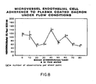
- the woven Dacron graft immobilized in the seeding chamber and coated with PRP was seeded with 5 x 10 5 EC in 0.5 cc of culture medium placed in the seeding well (1 cm 2 area) to allow incubation to occur over 1 hour in a 37°C incubator. Following incubation, the supernatant was removed, and the graft surface was lightly washed with culture medium. Control chambers were filled with 0.5 cc of fresh culture medium and placed in an in vitro circulatory loop. The EC were then exposed to a single shear stress between 0 and 80 dynes /cm 2 for 2 hours at 37°C using recirculated culture medium. During flow, negligible changes in pressure gradients, pH and electrolyte concentrations were observed.
- Indium labelling of endothelial cells was also conducted. Endothelial cells were isolated from the microvascular as described above. Cells were pelleted by centrifugation for 3 minutes at 100 x g, and washed once with PBS (pH 7.4). The cells were resuspended prior to labelling in 0.5 ml of PBS. The cell concentration was adjusted to 2.5 x 10 cells /ml. 20 microcuries of Indium 111 (as Indium 111 oxine, Medi-Physics, E meryville, CA) were added to the cell suspension, and the cells were permitted to label for up to 30 minutes were undergoing general agitation. Just prior to washing, a 5 ul sample was removed to permit final analysis of labelling efficiency. Labelled cells were washed 3 times by centrifugation using complete tissue culture medium. The final pellet was resuspended in complete culture medium to a final concentration of 2.5 x 10 5 cells/ml.
- EC counts on the flow and control slides were made with a Micro-Comp Grain counter supported by an IBM PC AT and Frame Grabber.
- the data obtained during the flow analysis was evaluated in two ways. Firstly EC adherence is expressed as the percentage of cells that remained adhered after flow compared to the control slides. Each point represents at least 4 observations and in some as much as 8. This was plotted versus shear stress and linear regression analysis was performed on this curve to determine statistical significance. Secondly, comparisons of initial adherences were made using the Student's t-test.
- EC adherence is determined by Indium labelling was plotted against time. Each data point represents the mean of two separate samples.
- adipose tissue was obtained from 13 individual donors and included perinephric and omental fat sources.
- E C were successfully isolated from all 13 donors.
- Elapsed time for the 3 stages of EC isolation were 29.9 + 3 (mean + standard error of the mean) minutes for collagenase, 20 minutes for Percoll and 30 minutes for washes and handling for cell counts.
- Mean EC yield per gram of wet fat was 1.25 + 0.45 x 10 6 cells. Cell viability as determined by Trypan Blue dye exclusion exceeded 95% for all isolations.
- Adherence was measured as percent of confluence. This was determined by counting the number of adherent endothelial cells following a 1 hour incubation and dividing it by 10 5 , which is the maximum number of EC present in a confluent monolayer regardless of the cell seeding density. Four separate adherence measurements were made per donor for statistical analysis.
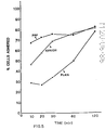
- the temporal sequence of microvessel endothelial cell adherence to plasma treated Dacron was also investigated in this test.
- Microvessel endothelial cell radiolabelled with Indium 111 were plated onto plasma treated Dacron and cell adhesion analyzed . ⁇ over a 120 minute period (see Figure 7).
- the left y-axis represents the number of EC adherent at a given time point divided by the number of adherent EC after a two hour incubation (maximal adherence) expressed as a percentage.
- the right y-axis represents the number of EC adherent at a given time point divided by the number of initially seeded EC (10 EC/cm ) expressed as a percentage of confluence.
- a biphasic rate of adherence was observed, with an initial rapid rate of adherence during the first 60 minutes followed by a slower rate until the final time point of 120 minutes. Significantly, although limited, adherence was observed at 10 minutes, the earliest time evaluated.
- the qualitative evaluation of EC adherence was conducted by scanning electron microscopy. When cells were permitted to , associate with the surface for 1 hour followed by shear for 2 hours, low power observations revealed areas with variable densities of EC.
- the Dacron surface was uniformly covered by the plasma clot and cells were observed to adhere to areas of plasma clot which overly both the peaks (warp) and valleys (weft) created during the weaving process.
- Higher magnification of areas considered by low power observation to display more limited cell association revealed the presence of EC in various stages of surface association.
- Cell morphology varied from flattened cells which exhibited a dramatic increase in cell surface area to cells which remained round with only focal attachment. All of the cells were resistant to shear.
- This test thus supports the proposition that it is possible to generate an endothelium upon a prosthetic surface to avoid complications stemming from the thrombogenicity of prior art prosthetic surfaces.
- This test further demonstrates the feasibility of providing a prosthetic graft that is either completely endothelialized at the time of implantation without precedent EC culture.
- This approach avoids the problem that only limited number of large vessels are available as EC donors whereas microvessels are universally present in high density in almost all tissues.
- These EC are relatively easily isolated from adipose tissue, yielding a high number of EC per gram of tissue in contrast to large vessels. In tissue culture these EC demonstrate many of the functional and morphological characteristics of large vessel EC.
- M icrovessel endothelial cells fulfill many of the requirements of a cell capable of rapidly endothelializing a graft.
- the cells are universally present in tissue, being present in adipose tissue, a donor tissue that can be removed in large quantities without significant surgical effort and with minmal risk to the patient.
- Such cells are easily and reliably isolated from most patients in 60 to 90 minutes, capable of producing large quantities of endothelial cells that are free of contaminating smooth muscle cells, quickly able to become firmly adherent to Dacron pretreated with PRP or other materials, such as basement membrane. They are further able to establish areas of confluent cells, able to withstand physiological shear stresses after only 1 or 2 hours of incubation, and are autologous to the donor.
- Another important property is that freshly isolated and seeded EC be able to rapidly from complete cell to cell interactions. Undoubtedly these interactions additionally protect the EC from shear stresses to help prevent the cells from being pulled off the surface. While cell to cell associations do occur on plasma coated Dacron to a limited degree, in the tests described hereafter more optimal surfaces are provided to promote such cell to cell interactions.
- the creation of the preferred confluent layer of endothelial cells on prosthetic surfaces is now believed to be dependent on at least three major variables.
- the initial adherence of cells should be sufficient to provide at least about fifty percent (50%) initial surface coverage.
- Procurement of large vessel endothelial cells to provide at least about fifty percent (50%) coverage is extremely difficult, if not impossible, since the only available source of cells is the patient's own large vessels.
- large vessel cells can be isolated and cultured to provide a large number of cells, the obvious problems associated with tissue culture media would then be presented.
- Microvascularized fat provides a rich source of endothelial cells for seeding.
- Twenty grams of the patient's fat will provide ample endothelial cells to seed a surface area of one hundred and eighty square centimeters (180 cm 2 ), the surface area represented by a typical femoral artery to popliteal artery bypass graft.
- a second variable to be considered is the ability of endothelial cells to proliferate (grow) on a prosthetic surface.
- Application at fifty percent (50%) confluence requires the cells to duplicate one time to create a confluent cell layer.
- Table 2 shows that on the preferred protein coated surface (coated with platelet rich plasma), the cells will duplicate at least once in tissue culture media which contains growth factor. In the body, however, these growth factors would presumably not be present, and therefore, the ability to treat surfaces at or in excess of confluence is advantageous.
- the availability of human MEC in large quantities permits the application of endothelial cells on a surface at densities capable of establishing a confluent monolayer or near confluent monolayer at the time of implantation.
- endothelial cells on prosthetic surfaces pretreated with proteins, as mentioned above, or upon surfaces which have been modified to emulate protein surfaces.
- modified surfaces are well-known to the endothelial cell tissue culture art.
- the endothelial cells may be 'preclotted' into a fibrin (protein) gel which forms within and around the graft.
- data indicate that human microvascular endothelial cells can be gelled within a protein meshwork, and following incubation in culture media, will migrate to the surface of the gel. This has been confirmed from scanning electron micrografts which show human microvascular endothelial cells forming a confluent monolayer on the surface of a Dacron polyester graft after these cells were preclotted in human plasma.
- a third important variable is the effect which the " " technique and underlying surface have upon the functional characteristics of EC monolayer, including its morphology, resistance to shear, and antithrombogenic characteristics.
- HAEC human adult endothelial cells
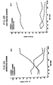
- Scanning electron microscopic evaluation reveals that HAEC adhere rapidly to both the basement surface (collagen IV/V) and interstitial surface (collagen I/III) of amnion.
- the adherence of cells is significantly greater on the basement membrane surface.
- HAEC rapidly form close cell-cell interactions on basement membrane as compared to cells seeded on to the interstitial surface.
- Human amnionic membrane taken from fresh human placentae, were prepared by a modification of the method described by Liotta et al. 'New Method For Preparing Large Surfaces of Intact Human Basement Membrane For Tumor Invasion Studies ⁇ , Cancer Letter, 11:141-152 (1980). All procedures were preformed under sterile conditions. The inner amnionic membrane was gently bluntly dissected away from the chorion, and was then washed twice in ice-cold phosphate-buffered saline with 100 units per milliliter penicillin and 0.25 mcg/ml Fungizone.
- the membrane was washed once in Dulbecco's minimal essential media at 4°C, rinsed once with distilled water with one mM N-ethylmaleimide for 1 hour at 4°C. The amnion was then incubated for 2 hours at 20°C in 4% deoxycholate solution, thus loosening the epithelial cells without damaging the structure of the underlying basement membrane.
- gentle agitation with a rubber policeman denuded the epithelial cells from the basement membrane.
- the integrity of the basement membrane was then verified using India ink staining. The removal of the epithelial cells was verified morphologically.
- amnionic membrane prepared and deepithialized as described above, was then immobilized in plastic capsules similar to those used by Williams et al in "Adult Human Endothelial Cell Compatibility with Prosthetic Graft Material x , J.Surg. Res., supra. This provided a stable, well-defined surface area of amnion (0.5 2 cm) for subsequent seeding with and proliferation of endothelial cells (EC). Both basement membrane and interstitial collagen sides of the amnion were prepared for cell seeding. Prior to tissue cultural studies, the capsules with amnion were soaked overnight at 4° C in complete media with 50 mcg/ml penicillin/streptomycin and 0.25 mcg/ml fungizone.
- Human adult endothelial cells were isolated from vascular tissue procured from brain-dead, heart-beating cadaver renal donors and were cultured according to the published procedures referenced above. In this study, EC from adult human iliac vein were used. Briefly cells were isolated from a fresh iliac vein by treating the luminal surface with collagenase (Worthington Type I, Worthington Diagnostic Systems, Inc., Freehold, N.J.) and grown in 25 cm 2 tissue culture flasks precoated with gelatin (1%) in culture medium (medium 199, 20% heat-inactivated fetal calf serum, 90 ug/ml heparin (procine), and 20 ug/ml endothelial cell growth factor.
- collagenase Wide Type I, Worthington Diagnostic Systems, Inc., Freehold, N.J.
- PD log (number of cells harvested)/(number of cells seeded x attachment efficiency) and summed to give the cumulative population doubling (CPDs).
- the EC identity of these cells has been previously reported and included positive staining for factor VIII related antigen, cobblestone morphology and the expression of E C specific prostaglandin and angiotensin-converting enzyme activity.
- Endothelial cell-seeded amnion was fixed with 2% glutaraldehyde overnight, then formation fixed, paraffin embedded, and sectioned for subsequent hematoxylin and eosin staining. The stained sections were then examined under brightfield illumination in a Nikon diaphot microscope.
- the EC-seeded amnion was fixed with 1% glutaraldehyde for 1 hour, 2% glutaraldehyde for 2 hours, and then washed four times (20 minutes each) in Tyrodes cacodylate buffer pH - 7.4. The amnion was then dehydrated in a grated series of acetone, critical point dried, and coated with gold-palladium. At this point, the plastic capsules were removed from the seeded amnion samples. These were then mounted and examined in a Phillips scanning electron microscope.
- Human amnionic membrane prepared according to the methods described above, was able to withstand the denudation procedures involved. Maintenance of the basic basement membrane structure was established by scanning with India ink. Integrity of the membrane was also evidenced by the observation of intact amnion surfaces-i.e., surfaces devoid of damage, rips or tears, when samples were examined using light and scanning electron microscopy. In addition, the efficacy of the deepithelialization procedure was demonstrated when unseeded, denuded, control amnion remained free of EC.
- HIVE Human iliac vein endothelial cells
- Endothelial cells seeded on plain Dacron exhibited limited adherence, while cells on plasma treated Dacron exhibited limited cell to cell associations.
- the temporal sequence of events to establish confluence using autologous seeding is of particular importance in improving the short-term patency of small caliber grafts.
- a period of 4-6 weeks following implantation would be required for a significant percentage of the graft to be spontaneously endothelialized.
- this time frame is compared with most human lower extremity prosthetic graft clinical series, it is noted that a large percentage of graft failures due to thrombosis occurs within the first month following implantation.
- establishment of an intact endothelium upon a graft at or near the time of implantation might be necessary, or would at least be desirable, before a significant effect on short term patency could be seen.
- the prosthetic surface substrate should be selected to be receptive to endothelial cells, and the procedure should be conducted with knowledge of the attachment requirements and temporal parameters necessary to allow both a functional as well as a shear resistance monolayer to reliably form. Accordingly, the following tests have been conducted to demonstrate that the temporal sequence of events from endothelial cell seeding upon a prosthetic surface to generation of a confluent monolayer is such that attainment of confluence is sufficient swift to permit pre-endothelialization at the time of implantation.
- endothelial cells were isolated from vascular tissue procured from brain-dead, heart-beating cadaver renal donors, and were cultured using the procedures described above.
- endothelial cells from adult human iliac vein, isolated as described above, were used.
- Preparation of human basement membrane was conducted, also in accordance with the above-described procedure.
- Heterologous interstitial collagen Types I/III were prepared from human placentae following the procedures of Madri, 'The Preparation for Type V Collagen" In: H. Furthmayr, ed. the immunochemistry of extracellular matrix, Boca Raton, Florida: CRC Press, 1982 (1):75-90, which paper is hereby incorporated by reference.
- the surface of the graft was prepared as follows.
- the surface of woven Dacron graft was coated with heterologous interstitial collagens Types I and III prepared as described above. Following the stabilization of that collagen in 0.0174 M acetic acid and dilution to 0.32% collagen with ice-cold medium 199 and NaHC0 3 . Deposition was promoted by allowing grafts to sit overnight at 20°C. The surface was then covered with 0.2% glutaraldehyde for 1 minute and rinsed with plain medium. Prepared amnion was then overlayed onto the graft surface with the basement membrane surface oriented away from the Dacron surface.
- P R P Platelet rich plasma
- Anti-coagulation acid-citrate dextrose
- CaCl 2 50 mM CaCl 2 just prior to graft treatment.
- Grafts were treated with PRP and a fibrin clot was permitted to form at 37°C. The clot was washed with cultured media prior to EC seeding.
- EC derived from iliac veins were grown to to confluence in 25 cm 2 flasks and used for cell seeding after two cell passages at a 1:4 split ratio. EC were briefly (1.5 minutes) washed with trypsin solution (0.25% trypsin with 0.09% EDTA in normal saline), washed once with culture media, and resuspended in complete culture media prior to graft surface seeding. EC were seeded at a cell concentration sufficient to provide a 100% confluent monolayer of cells on gelatin coated plastic surfaces. This density is equal to 1 x 10 cells/cm.
- the scanning electron microscopy was performed after fixing with 1% glutaraldehyde for 1 hour, 2% glutaraldehyde for 2 hours, 3 washings (20 minute period) with tyrodes cacodylate buffer (pH 7.4) and dehydrated in a graded series of acetone.
- the grafts, still immobilized within plastic rings, were then critical point dried and coated with gold palladium. Mounted samples were examined in a Phillips scanning electron microscope.
- Endothelial cells were radiolabelled for this test using two separate procedures. Thymidine labelling was performed by treating confluent EC in a T-25 flask with a trypsin solution. These cells were then washed with culture medium, counted and 4 2 replated onto a T-75 flask at 10 EC/cm. After 24 hours of growth, 0.5 uCi of tritiated thymidine (Amersham, Arlington Heights, ILL) was added to the flask and incubated for 24 hours. The radioactive supernatant was removed and incubated for 24 hours. The EC were treated with trypsin solution and counted on a scintillation counter.
- Indium labelling was performed on endothelial cells grown to confluence in 25 cm 2 flasks. The cells were briefly trypsinized. Released cells were pelleted by centrifugation (100 x g; 3 min) and washed once with phosphate buffered saline (pH 7.4). The cells were resuspended just prior to labelling in 0.5 ml of phosphate buffered saline. The cell concentration was adjusted to 2.5 x 10 cells/ml. Twenty microcuries of Indium (Medi Physics, Emeryville, CA) were added to the cell suspension and cells were permitted to label for 30 minutes with gentle agitation. Just prior to washing a 5 ul sample was removed to permit final analysis of labelling efficiency. Labelled cells were washed 3 times by centrifugation using complete tissue culture medium. The final pellet was resuspended in complete tissue culture medium to a final concentration of 2.5 x 10 5 cells/ml.
- the resulting radiolabelled endothelial cells were seeded and their adherence quantitated as follows.
- the endothelial cells were seeded on matrix coated Dacron immobilized in Beem capsules and incubated for specified time intervals (t- 1, 5, 10, 20, 30, 60, 120 minutes). At each time interval, the following samples were obtained.
- the first sample designated the "supernatant” was obtained by pipetting off the supernatant from the Beem capsule.
- the second sample designated 'loosely adhered', was obtained by vigorously washing the graft surface by forcefully pipetting culture medium three times onto the surface. the medium used to perform these washings was pooled and the entire specimen formed the second sample.
- the third sample designate "adhered" was obtained by removing all adherent EC from the graft surface. This was performed by solubilizing EC samples in triplicate in 0.2 ml of 0.3% sodium dodecyl sulfate and transferring the resulting solution to filter paper. Each filter paper was transferred to 10% ice cold TCA and precipitated material was counted. The individual sample counts were normalized to percentages of the total number of counts in all three samples and plotted at % EC in each fraction versus time in minutes. EC labelled with tritiated thymidine were counted in a scintillation counter and EC labelled with Indium were counted in a gamma counter.
- the number of cells adhered to platelet rich plasma increased to a maximum of 80 % by 2 hours.
- the initial adherence of human adult endothelial cells to human basement membrane (amnion) coated Dacron was intermediate between untreated Dacron and PRP treated Dacron ( Figure 5).
- the number of cells adhered increased with the time until the number of cells adhered to amnion equaled that adhered to PRP treated Dacron sometime between 30 and 60 minutes. As seen in Figure 5, all of the surfaces exhibited approximately the same number of adherent cells following two hours of incubation.
- endothelial cells The adherence of endothelial cells to the various graft surfaces was evaluated morphologically.
- the quantitative analysis of endothelial cell-graft interaction provided an analysis of the rate of endothelial cell adherence to the surface, but questions remained as to the form of interaction of endothelial cells with graft surfaces.
- the temporal sequence of human adult endothelial cell adherence to basement membrane treated graft was determined by examining scanning electron micrografts. Following the addition of endothelial cells, graft surfaces were washed at several intervals between 1 and 120 minutes and evaluated by scanning electron microscopy.
- endothelial cells seeded amnion surface A complete morphological maturation of the endothelial cells seeded amnion surface is observed one hour after the onset of cell association. Endothelial cells have covered the amnion surface and the loss of membrane ruffles results in a smooth endothelial cell monolayer surface. The close association of endothelial cells makes the identification of cellular borders difficult, however, the occasional presence of incompletely attenuated cells provide a point of reference for the evaluation of the cellular nature of this monolayer due to the topology of the underlying Dacron fibers.
- the morphology of cells adhered to platelet rich plasma and untreated Dacron was also evaluated after 60 minutes of adherence. Endothelial cell adherence to PRP treated Dacron was observed to involve areas both devoid of cells and areas where cells exhibited endothelial cell characteristic cell to cell interaction. Also of interest is the observed apparent deposition of fibrin on the surface of flattened endothelial cells. Since this fibrin layer was formed prior to seeding, we suggest that endothelial cells are exhibiting the ability to partially migrate under the fibrin lining prior to their complete adherence and flattening. Most importantly 4 is the common observance of areas which totally lack endothelium and therefore expose the fibrin layer.
- vascular graft endothelialization could be produced by low density endothelial cell seeding following by EC proliferation, high density EC seeding or spontaneous ingrowth of EC onto a surface following implantation.
- High density EC seeding with establishment of a confluent monolayer at the time of implantation offers the best possibility of a non-thrombogenic graft in the first several weeks following surgery when the risk of thrombosis is present. The aforementioned study was thus untaken to examine whether high density EC seeding was capable of producing a morphologically normal appearing endothelial monolayer within time parameters compatible with an operating room vascular procedure. Experimental conditions were chosen based on our previous observations on EC-graft interactions.
- endothelial cells at 60 minutes demonstrate few cell to cell interactions, and have a "stellate" morphology rather than a a cobblestone o morphology. Although the non-thrombogenic characteristics of this surface have not been examined, the abnormal morphology suggests that these endothelial cells are not experiencing ideal conditions and may not tolerate the effects of flow. Endothelial cell adherence to the amnion collagen-coated Dacron graft was slower than for the plasma coated, but the attainment of confluence was markedly different.
- Confluence in this study is defined as complete EC coverage of the prosthetic surface as seen on scanning electron micrografts.
- Cell to cell associations appear normal, but further studies with transmission electron micrografts may determine the type of junctions present as well as the type of association between endothelial cells and graft substrate combinations.
- Substrate is known to have an effect on cell morphology and function and will have to be examined in an experimental setting before concrete conclusions can be drawn. Additionally, more will be learned by studying the use of amnion as a graft substrate. This biologically derived material is not currently available for widespread clinical use.
- the amnion contains the cellular attachment factors of fibronectin and laminin.
- vascular grafts For large scale production of vascular grafts, it may be desirable to reconstitute a laminate comprised of an intermediate layer of Type I/III collagen and a top surface layer of Type IV/V collagen which mimics the natural basement membrane. If desired, that membrane may further be mimicked by the additions of cellular attachment factors including fibronectin and/or laminin. Whether the basement membrane used is from natural or synthetic origins, it should be noted that such membranes are entirely acellular, may be sterilized by irradiation, and stored and/or shipped for subsequent use. ⁇ - Alternatively, it may be possible to chemically mimic or emulate the desired Type IV/V collagen on the surface of a synthetic prosthesis.
- the EC monolayer derived from the aforementioned test appears normal morphologically, additional tests (described hereafter) were conducted to investigate whether it possesses other functional characteristics of normal endothelium, particularly with reference to nonthrombogenicity. It is desirable, for example, to show that the monolayer is able to withstand physiological arterial shear stresses and maintain contained adherence. This has been demonstrated not only for the EC-basement membrane adhesion, but also the basement membrane-vascular graft interaction as well as endothelial cell to endothelial cell attachments.
- the present application discloses a novel method of treating an implant intended for implantation in a human patient, comprising providing a synthetic substrate material and treating that material with Type IV/V collagen to improve human endothelial cell adhesion, proliferation and morphology thereon.
- the Type IV/V collagen is bound as a surface layer for receiving endothelial cells.
- the Type IV/V collagen surface layer is applied as a laminate having a Type I/III collagen underlayer adherent to it.
- the aforementioned collagen laminate is derived from human chorioallantoic membrane, and comprises acellular human amnion.
- This collagen laminate (amnion) is applied to a substrate having a bound interstitial collagen base layer formed thereon.
- the base layer is adhered to the substrate through the use of a cross linking agent, such as glutaraldehyde, which further activates the surface of the base layer to permit covalent binding of the collagen laminate to that base layer.
- the linking agent is then safely deactivated using an amine, amino acid or peptide with an aldehyde active amine group, such as lysine, which is soluble in buffered saline. Following washing to remove the deactivating agent and residual linking agent, the graft is ready to receive high density seeding of microvessel endothelial cells.
- microvessel 4 endothelial cells are seeded at a density of at least 5 x 10 4 , 4 2 preferably at least 7.7 x 10 4 , cells per cm to form a confluent monolayer on the graft surface within two hours from the tine of seeding. It is currently preferred to seed in range of 1-3, or 5 2 about 2 x 10 cells per em. While microvessel endothelial cells are preferred due to their uncultured, autologous nature, it is within the scope of the present invention to use human adult endothelial cells which have been briefly cultured for two or less passages, in those instances where such cells are readily available.
- the resulting graft possesses an endothelial lining of autologous endothelial cells, its patency, particularly during the critical early period after implantation, may be expected to be marketly improved.
- Use of the subject grafts for venous implants, and in vessels having diameters of 4 mm or less (small caliber grafts), where patency rates have otherwise been disappointingly low is thus anticipated.
- Diacron is a trademark of E.I. duPont de Nemours and Company of Wilmington, Delaware, which is used to identify a particular polyethylene terephthalate polyester which is a condensation product of methyl terephthalate and ethylene glycol.
- cobblestone refers to both the typical symmetrical endothelial cell-cell morphology exhibited, for example, by bovine aortic endothelial cells in culture (sometimes referred to in the art as 'true cobblestone") as well as cellular morphologies wherein the cells are generally round but have some projections or other asymmetrical portions.
- cobblestone refers to populations of endothelial cells which form tight cell to cell associations, i.e., those which attenuate to maximally cover the underlying surface.
Landscapes
- Health & Medical Sciences (AREA)
- Life Sciences & Earth Sciences (AREA)
- Chemical & Material Sciences (AREA)
- Public Health (AREA)
- Engineering & Computer Science (AREA)
- Transplantation (AREA)
- Epidemiology (AREA)
- Medicinal Chemistry (AREA)
- Animal Behavior & Ethology (AREA)
- General Health & Medical Sciences (AREA)
- Dermatology (AREA)
- Veterinary Medicine (AREA)
- Oral & Maxillofacial Surgery (AREA)
- Biomedical Technology (AREA)
- Vascular Medicine (AREA)
- Cell Biology (AREA)
- Zoology (AREA)
- Botany (AREA)
- Chemical Kinetics & Catalysis (AREA)
- Prostheses (AREA)
- Materials For Medical Uses (AREA)
- Dental Preparations (AREA)
- Paints Or Removers (AREA)
Applications Claiming Priority (4)
| Application Number | Priority Date | Filing Date | Title |
|---|---|---|---|
| US742086 | 1985-06-06 | ||
| US06/742,086 US4820626A (en) | 1985-06-06 | 1985-06-06 | Method of treating a synthetic or naturally occuring surface with microvascular endothelial cells, and the treated surface itself |
| US84845386A | 1986-04-04 | 1986-04-04 | |
| US848453 | 1986-04-04 |
Related Child Applications (1)
| Application Number | Title | Priority Date | Filing Date |
|---|---|---|---|
| EP92114514.0 Division-Into | 1992-08-25 |
Publications (3)
| Publication Number | Publication Date |
|---|---|
| EP0206025A2 true EP0206025A2 (de) | 1986-12-30 |
| EP0206025A3 EP0206025A3 (en) | 1988-03-02 |
| EP0206025B1 EP0206025B1 (de) | 1994-11-09 |
Family
ID=27113964
Family Applications (2)
| Application Number | Title | Priority Date | Filing Date |
|---|---|---|---|
| EP86107600A Expired - Lifetime EP0206025B1 (de) | 1985-06-06 | 1986-06-04 | Beschichtung für Prothesen |
| EP19920114514 Withdrawn EP0518389A3 (en) | 1985-06-06 | 1986-06-04 | Coating for prosthetic devices |
Family Applications After (1)
| Application Number | Title | Priority Date | Filing Date |
|---|---|---|---|
| EP19920114514 Withdrawn EP0518389A3 (en) | 1985-06-06 | 1986-06-04 | Coating for prosthetic devices |
Country Status (10)
| Country | Link |
|---|---|
| EP (2) | EP0206025B1 (de) |
| JP (1) | JP2686930B2 (de) |
| AT (1) | ATE113850T1 (de) |
| AU (1) | AU590573B2 (de) |
| BR (1) | BR8602659A (de) |
| CA (1) | CA1293700C (de) |
| DE (2) | DE206025T1 (de) |
| ES (1) | ES8900149A1 (de) |
| IL (1) | IL78950A (de) |
| MX (1) | MX165125B (de) |
Cited By (11)
| Publication number | Priority date | Publication date | Assignee | Title |
|---|---|---|---|---|
| EP0290642A1 (de) * | 1987-05-12 | 1988-11-17 | Mittermayer, Christian, Dr. med. | Verfahren zur Besiedlung einer Polymeroberfläche mit menschlichen Gefässinnenhautzellen |
| WO1989005345A1 (en) * | 1987-12-11 | 1989-06-15 | Whitehead Institute For Biomedical Research | Genetic modification of endothelial cells |
| WO1989007425A2 (en) * | 1988-02-17 | 1989-08-24 | Genethics Limited | Clinical developments using amniotic cells |
| GB2215614A (en) * | 1988-02-16 | 1989-09-27 | Allergan Inc | Ocular device |
| US4927676A (en) * | 1988-07-01 | 1990-05-22 | Becton, Dickinson And Company | Method for rapid adherence of endothelial cells onto a surface and surfaces prepared thereby |
| EP0504262A1 (de) * | 1989-12-07 | 1992-09-23 | Biosynthesis, Inc. | Hohle viszeraprothese sowie verfahren zur implantation |
| US5192316A (en) * | 1988-02-16 | 1993-03-09 | Allergan, Inc. | Ocular device |
| US5401508A (en) * | 1992-01-15 | 1995-03-28 | Allergan, Inc. | Hydrogel compositions and structures made from same |
| US5415619A (en) * | 1989-12-13 | 1995-05-16 | Korea Research Institute Of Chemical Tech. | Method of manufacturing a vascular graft impregnated with polysaccharide derivatives |
| WO2002052936A1 (fr) * | 2000-12-28 | 2002-07-11 | Japan Science And Technology Corporation | Agents de conservation destines a des cellules lyophilisees |
| US9205177B2 (en) | 2009-03-04 | 2015-12-08 | Peytant Solutions, Inc. | Stents modified with material comprising amnion tissue and corresponding processes |
Families Citing this family (25)
| Publication number | Priority date | Publication date | Assignee | Title |
|---|---|---|---|---|
| US4795475A (en) * | 1985-08-09 | 1989-01-03 | Walker Michael M | Prosthesis utilizing salt-forming oxyacids for bone fixation |
| US5018872A (en) * | 1988-11-01 | 1991-05-28 | Diatek, Inc. | Probe assembly for infrared thermometer |
| JPH062155B2 (ja) * | 1989-09-06 | 1994-01-12 | 住友ベークライト株式会社 | 人工血管の製造方法 |
| US5549674A (en) * | 1992-03-02 | 1996-08-27 | The Regents Of The University Of Michigan | Methods and compositions of a bioartificial kidney suitable for use in vivo or ex vivo |
| AUPN174495A0 (en) * | 1995-03-15 | 1995-04-06 | Ketharanathan, Vettivetpillai | Surgical prostheses |
| US5766584A (en) * | 1995-06-02 | 1998-06-16 | Massachusetts Institute Of Technology | Inhibition of vascular smooth muscle cell proliferation with implanted matrix containing vascular endothelial cells |
| US5869037A (en) * | 1996-06-26 | 1999-02-09 | Cornell Research Foundation, Inc. | Adenoviral-mediated gene transfer to adipocytes |
| AU4351000A (en) * | 1999-04-15 | 2000-11-02 | Research Foundation Of State University Of New York, The | Cellular matrix system and use thereof |
| KR20050044507A (ko) * | 2001-11-16 | 2005-05-12 | 칠드런'즈 메디컬 센터 코포레이션 | 장기 기능의 확장 |
| US20030187515A1 (en) * | 2002-03-26 | 2003-10-02 | Hariri Robert J. | Collagen biofabric and methods of preparing and using the collagen biofabric |
| US9144583B2 (en) | 2002-03-29 | 2015-09-29 | Tissue Genesis, Inc. | Cell separation apparatus and methods of use |
| ATE450276T1 (de) * | 2004-12-08 | 2009-12-15 | Pervasis Therapeutics Inc | Materialien und verfahren zur minimal invasiven abgabe einer zellhaltigen fliessfähigen zusammensetzung |
| ES2569494T3 (es) * | 2004-12-24 | 2016-05-11 | Admedus Regen Pty Ltd. | Un biomaterial implantable y un método de producción del mismo |
| CA2605080A1 (en) | 2005-04-21 | 2006-11-02 | Massachusetts Institute Of Technology | Materials and methods for altering an immune response to exogenous and endogenous immunogens, including syngeneic and non-syngeneic cells, tissues or organs |
| EP2361591A3 (de) | 2005-06-21 | 2011-12-21 | Pervasis Therapeutics, Inc. | Verfahren und Zusammensetzungen zur Verbesserung des vaskulären Zugangs |
| AU2007297776A1 (en) * | 2006-09-21 | 2008-03-27 | Tissue Genesis, Inc. | Cell delivery matrices |
| US11174458B2 (en) | 2007-04-23 | 2021-11-16 | Koligo Therapeutics, Inc. | Cell separation apparatus and methods of use |
| US10130736B1 (en) | 2010-05-14 | 2018-11-20 | Musculoskeletal Transplant Foundation | Tissue-derived tissuegenic implants, and methods of fabricating and using same |
| US8883210B1 (en) | 2010-05-14 | 2014-11-11 | Musculoskeletal Transplant Foundation | Tissue-derived tissuegenic implants, and methods of fabricating and using same |
| US9352003B1 (en) | 2010-05-14 | 2016-05-31 | Musculoskeletal Transplant Foundation | Tissue-derived tissuegenic implants, and methods of fabricating and using same |
| EP3027235A1 (de) | 2013-07-30 | 2016-06-08 | Musculoskeletal Transplant Foundation | Aus einem azellulären weichgewebe gewonnene matrizen und verfahren zur herstellung davon |
| WO2016187413A1 (en) | 2015-05-21 | 2016-11-24 | Musculoskeletal Transplant Foundation | Modified demineralized cortical bone fibers |
| US11052175B2 (en) | 2015-08-19 | 2021-07-06 | Musculoskeletal Transplant Foundation | Cartilage-derived implants and methods of making and using same |
| CZ308556B6 (cs) * | 2017-07-26 | 2020-11-25 | Vseobecna Fakultni Nemocnice V Praze | Kompozitní cévní náhrada a způsob její výroby |
| WO2022192967A1 (en) * | 2021-03-18 | 2022-09-22 | The University Of Sydney | Bioengineered endothelial constructs |
Citations (5)
| Publication number | Priority date | Publication date | Assignee | Title |
|---|---|---|---|---|
| US3276448A (en) * | 1962-12-14 | 1966-10-04 | Ethicon Inc | Collagen coated fabric prosthesis |
| CH593676A5 (en) * | 1975-12-16 | 1977-12-15 | Intermedicat Gmbh | Sealing of blood vessel implants of velour-coated fabric - by impregnating with organic colloidal solns. and drying |
| EP0049341A2 (de) * | 1980-10-07 | 1982-04-14 | Theurer, Karl Eugen, Prof.Dr.med. | Verfahren zur Vorbereitung von alloplastischen Implantationen und Organtransplantationen |
| EP0080956A2 (de) * | 1981-11-26 | 1983-06-08 | Fondation Merieux | Verfahren zur Herstellung von Kollagen aus Placenta; Humankollagen und ihre Verwendung als Biomaterialien |
| WO1983003536A1 (en) * | 1982-04-19 | 1983-10-27 | Massachusetts Inst Technology | A multilayer bioreplaceable blood vessel prosthesis |
Family Cites Families (1)
| Publication number | Priority date | Publication date | Assignee | Title |
|---|---|---|---|---|
| US4795475A (en) * | 1985-08-09 | 1989-01-03 | Walker Michael M | Prosthesis utilizing salt-forming oxyacids for bone fixation |
-
1986
- 1986-05-28 IL IL78950A patent/IL78950A/xx not_active IP Right Cessation
- 1986-06-02 JP JP61127858A patent/JP2686930B2/ja not_active Expired - Fee Related
- 1986-06-03 AU AU58305/86A patent/AU590573B2/en not_active Ceased
- 1986-06-04 DE DE198686107600T patent/DE206025T1/de active Pending
- 1986-06-04 MX MX2704A patent/MX165125B/es unknown
- 1986-06-04 DE DE3650134T patent/DE3650134T2/de not_active Expired - Fee Related
- 1986-06-04 AT AT86107600T patent/ATE113850T1/de not_active IP Right Cessation
- 1986-06-04 EP EP86107600A patent/EP0206025B1/de not_active Expired - Lifetime
- 1986-06-04 EP EP19920114514 patent/EP0518389A3/en not_active Withdrawn
- 1986-06-05 ES ES555739A patent/ES8900149A1/es not_active Expired
- 1986-06-05 CA CA000510929A patent/CA1293700C/en not_active Expired - Lifetime
- 1986-06-06 BR BR8602659A patent/BR8602659A/pt not_active IP Right Cessation
Patent Citations (5)
| Publication number | Priority date | Publication date | Assignee | Title |
|---|---|---|---|---|
| US3276448A (en) * | 1962-12-14 | 1966-10-04 | Ethicon Inc | Collagen coated fabric prosthesis |
| CH593676A5 (en) * | 1975-12-16 | 1977-12-15 | Intermedicat Gmbh | Sealing of blood vessel implants of velour-coated fabric - by impregnating with organic colloidal solns. and drying |
| EP0049341A2 (de) * | 1980-10-07 | 1982-04-14 | Theurer, Karl Eugen, Prof.Dr.med. | Verfahren zur Vorbereitung von alloplastischen Implantationen und Organtransplantationen |
| EP0080956A2 (de) * | 1981-11-26 | 1983-06-08 | Fondation Merieux | Verfahren zur Herstellung von Kollagen aus Placenta; Humankollagen und ihre Verwendung als Biomaterialien |
| WO1983003536A1 (en) * | 1982-04-19 | 1983-10-27 | Massachusetts Inst Technology | A multilayer bioreplaceable blood vessel prosthesis |
Non-Patent Citations (4)
| Title |
|---|
| American Journal of Surgery, Vol. 150, August 1985, pages 197-200, US; K.S. BAKER et al.: "Endothelialization of human collagen surfaces with human adult endothelial cells", presented at the 13th Annual Meeting of the Soc. for Clinical Vascular Surgeons, Rancho Mirage, Ca., April 10-14, 1984, whole article. * |
| Die Angewandte Makromolekulare Chemie, Vol. 111, No. 1701, January 1983, pages 107-122, Huethig & Wepf Verlag, Heidelberg, DE; M.Z. ABEDIN et al.: "Collagen heterogeneity and its functional significance", page 110. * |
| M. SZYCHER: "Biocomptable polymers, metals and composites", 1983, pages 165-178, Technomic, Lancaster, US; W.E. BURKEL: "The development of cellular linings in artificial vascular protheses", page 165; page 169, lines 26,27. * |
| Transactions American Society for Artificial Internal Organs, Vol. 28, April 14-16, 1982, pages 173-184, Chicago, US; T.A. BELDEN et al.: "Endothelial cell seeding of small-diameter vascular grafts", whole document. * |
Cited By (17)
| Publication number | Priority date | Publication date | Assignee | Title |
|---|---|---|---|---|
| EP0290642A1 (de) * | 1987-05-12 | 1988-11-17 | Mittermayer, Christian, Dr. med. | Verfahren zur Besiedlung einer Polymeroberfläche mit menschlichen Gefässinnenhautzellen |
| WO1989005345A1 (en) * | 1987-12-11 | 1989-06-15 | Whitehead Institute For Biomedical Research | Genetic modification of endothelial cells |
| GB2215614A (en) * | 1988-02-16 | 1989-09-27 | Allergan Inc | Ocular device |
| GB2215614B (en) * | 1988-02-16 | 1992-10-21 | Allergan Inc | Ocular device |
| US5192316A (en) * | 1988-02-16 | 1993-03-09 | Allergan, Inc. | Ocular device |
| US5612028A (en) * | 1988-02-17 | 1997-03-18 | Genethics Limited | Method of regenerating or replacing cartilage tissue using amniotic cells |
| WO1989007425A2 (en) * | 1988-02-17 | 1989-08-24 | Genethics Limited | Clinical developments using amniotic cells |
| EP0333328A2 (de) * | 1988-02-17 | 1989-09-20 | Genethics Limited | Klinische Entwicklungen mit Amnionzellen |
| WO1989007425A3 (en) * | 1988-02-17 | 1989-11-02 | Genethics Ltd | Clinical developments using amniotic cells |
| EP0333328A3 (en) * | 1988-02-17 | 1989-12-06 | Genethics Limited | Clinical developments using amniotic cells |
| US4927676A (en) * | 1988-07-01 | 1990-05-22 | Becton, Dickinson And Company | Method for rapid adherence of endothelial cells onto a surface and surfaces prepared thereby |
| EP0504262A1 (de) * | 1989-12-07 | 1992-09-23 | Biosynthesis, Inc. | Hohle viszeraprothese sowie verfahren zur implantation |
| EP0504262A4 (en) * | 1989-12-07 | 1993-06-30 | Biosynthesis, Inc. | Hollow viscus prosthesis and method of implantation |
| US5415619A (en) * | 1989-12-13 | 1995-05-16 | Korea Research Institute Of Chemical Tech. | Method of manufacturing a vascular graft impregnated with polysaccharide derivatives |
| US5401508A (en) * | 1992-01-15 | 1995-03-28 | Allergan, Inc. | Hydrogel compositions and structures made from same |
| WO2002052936A1 (fr) * | 2000-12-28 | 2002-07-11 | Japan Science And Technology Corporation | Agents de conservation destines a des cellules lyophilisees |
| US9205177B2 (en) | 2009-03-04 | 2015-12-08 | Peytant Solutions, Inc. | Stents modified with material comprising amnion tissue and corresponding processes |
Also Published As
| Publication number | Publication date |
|---|---|
| DE3650134D1 (de) | 1994-12-15 |
| ES555739A0 (es) | 1989-03-01 |
| ES8900149A1 (es) | 1989-03-01 |
| EP0518389A2 (de) | 1992-12-16 |
| BR8602659A (pt) | 1987-02-03 |
| JPS6249857A (ja) | 1987-03-04 |
| CA1293700C (en) | 1991-12-31 |
| IL78950A0 (en) | 1986-09-30 |
| EP0518389A3 (en) | 1994-05-18 |
| AU5830586A (en) | 1986-12-11 |
| IL78950A (en) | 1991-12-15 |
| EP0206025A3 (en) | 1988-03-02 |
| DE3650134T2 (de) | 1995-06-22 |
| AU590573B2 (en) | 1989-11-09 |
| DE206025T1 (de) | 1987-10-15 |
| JP2686930B2 (ja) | 1997-12-08 |
| MX165125B (es) | 1992-10-28 |
| ATE113850T1 (de) | 1994-11-15 |
| EP0206025B1 (de) | 1994-11-09 |
Similar Documents
| Publication | Publication Date | Title |
|---|---|---|
| US5131907A (en) | Method of treating a synthetic naturally occurring surface with a collagen laminate to support microvascular endothelial cell growth, and the surface itself | |
| EP0206025B1 (de) | Beschichtung für Prothesen | |
| US5230693A (en) | Implantable prosthetic device for implantation into a human patient having a surface treated with microvascular endothelial cells | |
| US4820626A (en) | Method of treating a synthetic or naturally occuring surface with microvascular endothelial cells, and the treated surface itself | |
| Williams et al. | Adult human endothelial cell compatibility with prosthetic graft material | |
| CA1340581C (en) | Chimeric neomorphogenesis of organs by controlled cellular implantation using artificial matrices | |
| US5716394A (en) | Blood contact surfaces using extracellular matrix synthesized in vitro | |
| Vara et al. | Cardiovascular tissue engineering: state of the art | |
| US5891558A (en) | Biopolymer foams for use in tissue repair and reconstruction | |
| US5879383A (en) | Blood contact surfaces using endothelium on a subendothelial matrix | |
| US6375680B1 (en) | Substrates for forming synthetic tissues | |
| US5628781A (en) | Implant materials, methods of treating the surface of implants with microvascular endothelial cells, and the treated implants themselves | |
| Kristofik et al. | Improving in vivo outcomes of decellularized vascular grafts via incorporation of a novel extracellular matrix | |
| Jarrell et al. | Use of an endothelial monolayer on a vascular graft prior to implantation. Temporal dynamics and compatibility with the operating room. | |
| Matsuda et al. | A hybrid vascular model biomimicking the hierarchic structure of arterial wall: neointimal stability and neoarterial regeneration process under arterial circulation | |
| Baker et al. | Endothelialization of human collagen surfaces with human adult endothelial cells | |
| LEE et al. | Endothelial cell seeding onto the extracellular matrix of fibroblasts for the development of a small diameter polyurethane vessel | |
| Sigot‐Luizard et al. | Cytocompatibility of albuminated polyester fabrics | |
| Wigod et al. | Quantification of in vivo endothelial cell adhesion to vascular graft material | |
| Noishiki et al. | Succinylated collagen crosslinked by thermal treatment for coating vascular prostheses | |
| Bellon et al. | Endothelial cell seeding of polytetrafluoroethylene vascular prostheses coated with a fibroblastic matrix | |
| Noishiki et al. | A new cardiac wall substitute with high affinity for fibroblasts that can induce an endothelial cell lining | |
| JPH01170466A (ja) | 生体内インプラント材料 | |
| WO1997046266A1 (en) | Improved blood contact surfaces using extracellular matrix synthesized in vitro having bioactive species immobilized thereto | |
| Williams et al. | Electron Microscopy of Endothelial Cell-Biopolymer Interaction |
Legal Events
| Date | Code | Title | Description |
|---|---|---|---|
| PUAI | Public reference made under article 153(3) epc to a published international application that has entered the european phase |
Free format text: ORIGINAL CODE: 0009012 |
|
| AK | Designated contracting states |
Kind code of ref document: A2 Designated state(s): AT BE CH DE FR GB IT LI LU NL SE |
|
| ITCL | It: translation for ep claims filed |
Representative=s name: SOCIETA' ITALIANA BREVETTI S.P.A. |
|
| TCAT | At: translation of patent claims filed | ||
| TCNL | Nl: translation of patent claims filed | ||
| EL | Fr: translation of claims filed | ||
| DET | De: translation of patent claims | ||
| PUAL | Search report despatched |
Free format text: ORIGINAL CODE: 0009013 |
|
| AK | Designated contracting states |
Kind code of ref document: A3 Designated state(s): AT BE CH DE FR GB IT LI LU NL SE |
|
| 17P | Request for examination filed |
Effective date: 19880707 |
|
| 17Q | First examination report despatched |
Effective date: 19900514 |
|
| GRAA | (expected) grant |
Free format text: ORIGINAL CODE: 0009210 |
|
| AK | Designated contracting states |
Kind code of ref document: B1 Designated state(s): AT BE CH DE FR GB IT LI LU NL SE |
|
| REF | Corresponds to: |
Ref document number: 113850 Country of ref document: AT Date of ref document: 19941115 Kind code of ref document: T |
|
| REF | Corresponds to: |
Ref document number: 3650134 Country of ref document: DE Date of ref document: 19941215 |
|
| ITF | It: translation for a ep patent filed | ||
| ET | Fr: translation filed | ||
| PG25 | Lapsed in a contracting state [announced via postgrant information from national office to epo] |
Ref country code: LU Free format text: LAPSE BECAUSE OF NON-PAYMENT OF DUE FEES Effective date: 19950630 |
|
| PLBE | No opposition filed within time limit |
Free format text: ORIGINAL CODE: 0009261 |
|
| STAA | Information on the status of an ep patent application or granted ep patent |
Free format text: STATUS: NO OPPOSITION FILED WITHIN TIME LIMIT |
|
| 26N | No opposition filed | ||
| PGFP | Annual fee paid to national office [announced via postgrant information from national office to epo] |
Ref country code: CH Payment date: 19960610 Year of fee payment: 11 |
|
| PGFP | Annual fee paid to national office [announced via postgrant information from national office to epo] |
Ref country code: NL Payment date: 19960617 Year of fee payment: 11 Ref country code: AT Payment date: 19960617 Year of fee payment: 11 |
|
| PGFP | Annual fee paid to national office [announced via postgrant information from national office to epo] |
Ref country code: SE Payment date: 19960701 Year of fee payment: 11 |
|
| PGFP | Annual fee paid to national office [announced via postgrant information from national office to epo] |
Ref country code: BE Payment date: 19960708 Year of fee payment: 11 |
|
| PG25 | Lapsed in a contracting state [announced via postgrant information from national office to epo] |
Ref country code: AT Effective date: 19970604 |
|
| PG25 | Lapsed in a contracting state [announced via postgrant information from national office to epo] |
Ref country code: SE Effective date: 19970605 |
|
| PG25 | Lapsed in a contracting state [announced via postgrant information from national office to epo] |
Ref country code: LI Free format text: LAPSE BECAUSE OF NON-PAYMENT OF DUE FEES Effective date: 19970630 Ref country code: CH Free format text: LAPSE BECAUSE OF NON-PAYMENT OF DUE FEES Effective date: 19970630 Ref country code: BE Effective date: 19970630 |
|
| BERE | Be: lapsed |
Owner name: THOMAS JEFFERSON UNIVERSITY Effective date: 19970630 |
|
| PG25 | Lapsed in a contracting state [announced via postgrant information from national office to epo] |
Ref country code: NL Effective date: 19980101 |
|
| REG | Reference to a national code |
Ref country code: CH Ref legal event code: PL |
|
| EUG | Se: european patent has lapsed |
Ref document number: 86107600.8 |
|
| NLV4 | Nl: lapsed or anulled due to non-payment of the annual fee |
Effective date: 19980101 |
|
| REG | Reference to a national code |
Ref country code: GB Ref legal event code: IF02 |
|
| PGFP | Annual fee paid to national office [announced via postgrant information from national office to epo] |
Ref country code: GB Payment date: 20020624 Year of fee payment: 17 |
|
| PGFP | Annual fee paid to national office [announced via postgrant information from national office to epo] |
Ref country code: FR Payment date: 20020719 Year of fee payment: 17 |
|
| PG25 | Lapsed in a contracting state [announced via postgrant information from national office to epo] |
Ref country code: GB Free format text: LAPSE BECAUSE OF NON-PAYMENT OF DUE FEES Effective date: 20030604 |
|
| GBPC | Gb: european patent ceased through non-payment of renewal fee |
Effective date: 20030604 |
|
| PG25 | Lapsed in a contracting state [announced via postgrant information from national office to epo] |
Ref country code: FR Free format text: LAPSE BECAUSE OF NON-PAYMENT OF DUE FEES Effective date: 20040227 |
|
| REG | Reference to a national code |
Ref country code: FR Ref legal event code: ST |
|
| PGFP | Annual fee paid to national office [announced via postgrant information from national office to epo] |
Ref country code: DE Payment date: 20040624 Year of fee payment: 19 |
|
| PG25 | Lapsed in a contracting state [announced via postgrant information from national office to epo] |
Ref country code: IT Free format text: LAPSE BECAUSE OF NON-PAYMENT OF DUE FEES;WARNING: LAPSES OF ITALIAN PATENTS WITH EFFECTIVE DATE BEFORE 2007 MAY HAVE OCCURRED AT ANY TIME BEFORE 2007. THE CORRECT EFFECTIVE DATE MAY BE DIFFERENT FROM THE ONE RECORDED. Effective date: 20050604 |
|
| PG25 | Lapsed in a contracting state [announced via postgrant information from national office to epo] |
Ref country code: DE Free format text: LAPSE BECAUSE OF NON-PAYMENT OF DUE FEES Effective date: 20060103 |