EP0179127B1 - Leukoregulin, an antitumor lymphokine, and its therapeutic uses - Google Patents
Leukoregulin, an antitumor lymphokine, and its therapeutic uses Download PDFInfo
- Publication number
- EP0179127B1 EP0179127B1 EP85902248A EP85902248A EP0179127B1 EP 0179127 B1 EP0179127 B1 EP 0179127B1 EP 85902248 A EP85902248 A EP 85902248A EP 85902248 A EP85902248 A EP 85902248A EP 0179127 B1 EP0179127 B1 EP 0179127B1
- Authority
- EP
- European Patent Office
- Prior art keywords
- cells
- leukoregulin
- cell
- human
- lymphokine
- Prior art date
- Legal status (The legal status is an assumption and is not a legal conclusion. Google has not performed a legal analysis and makes no representation as to the accuracy of the status listed.)
- Expired
Links
- 108010002060 leukoregulin Proteins 0.000 title claims abstract description 198
- 230000000259 anti-tumor effect Effects 0.000 title description 10
- 230000001225 therapeutic effect Effects 0.000 title description 2
- 210000004027 cell Anatomy 0.000 claims abstract description 178
- 210000004881 tumor cell Anatomy 0.000 claims abstract description 77
- 230000012010 growth Effects 0.000 claims abstract description 23
- 238000000034 method Methods 0.000 claims abstract description 22
- 238000001155 isoelectric focusing Methods 0.000 claims abstract description 18
- 102000005348 Neuraminidase Human genes 0.000 claims abstract description 17
- 108010006232 Neuraminidase Proteins 0.000 claims abstract description 17
- 108091005804 Peptidases Proteins 0.000 claims abstract description 15
- 239000004365 Protease Substances 0.000 claims abstract description 15
- 108090000623 proteins and genes Proteins 0.000 claims abstract description 15
- 230000035699 permeability Effects 0.000 claims abstract description 11
- FAPWRFPIFSIZLT-UHFFFAOYSA-M Sodium chloride Chemical class [Na+].[Cl-] FAPWRFPIFSIZLT-UHFFFAOYSA-M 0.000 claims abstract description 8
- 201000011510 cancer Diseases 0.000 claims abstract description 7
- 210000004408 hybridoma Anatomy 0.000 claims abstract description 7
- 108010001857 Cell Surface Receptors Proteins 0.000 claims abstract description 6
- 230000004663 cell proliferation Effects 0.000 claims abstract description 6
- 102000006240 membrane receptors Human genes 0.000 claims abstract description 6
- 238000005571 anion exchange chromatography Methods 0.000 claims abstract description 4
- 210000001185 bone marrow Anatomy 0.000 claims abstract description 4
- 230000000711 cancerogenic effect Effects 0.000 claims abstract description 4
- 210000001519 tissue Anatomy 0.000 claims abstract description 4
- 230000009466 transformation Effects 0.000 claims abstract description 4
- 210000002798 bone marrow cell Anatomy 0.000 claims abstract description 3
- 238000010494 dissociation reaction Methods 0.000 claims abstract description 3
- 230000005593 dissociations Effects 0.000 claims abstract description 3
- 230000035479 physiological effects, processes and functions Effects 0.000 claims abstract description 3
- 102100037486 Reverse transcriptase/ribonuclease H Human genes 0.000 claims abstract 2
- 231100000315 carcinogenic Toxicity 0.000 claims abstract 2
- 230000000694 effects Effects 0.000 claims description 74
- 239000002609 medium Substances 0.000 claims description 37
- 238000003556 assay Methods 0.000 claims description 28
- 239000006146 Roswell Park Memorial Institute medium Substances 0.000 claims description 17
- 238000011282 treatment Methods 0.000 claims description 16
- 102000008072 Lymphokines Human genes 0.000 claims description 14
- 108010074338 Lymphokines Proteins 0.000 claims description 14
- 238000004519 manufacturing process Methods 0.000 claims description 14
- 108010047620 Phytohemagglutinins Proteins 0.000 claims description 13
- 230000001885 phytohemagglutinin Effects 0.000 claims description 13
- 230000029087 digestion Effects 0.000 claims description 11
- 241001529936 Murinae Species 0.000 claims description 10
- 102000004169 proteins and genes Human genes 0.000 claims description 10
- 238000002264 polyacrylamide gel electrophoresis Methods 0.000 claims description 9
- 230000035755 proliferation Effects 0.000 claims description 9
- 206010035226 Plasma cell myeloma Diseases 0.000 claims description 8
- 230000003013 cytotoxicity Effects 0.000 claims description 8
- 231100000135 cytotoxicity Toxicity 0.000 claims description 8
- 201000000050 myeloid neoplasm Diseases 0.000 claims description 8
- 201000009030 Carcinoma Diseases 0.000 claims description 7
- 210000000170 cell membrane Anatomy 0.000 claims description 6
- 238000002523 gelfiltration Methods 0.000 claims description 6
- 229920001817 Agar Polymers 0.000 claims description 5
- 206010039491 Sarcoma Diseases 0.000 claims description 5
- 239000008272 agar Substances 0.000 claims description 5
- 230000006870 function Effects 0.000 claims description 5
- 239000001963 growth medium Substances 0.000 claims description 5
- 208000032839 leukemia Diseases 0.000 claims description 5
- 229920001184 polypeptide Polymers 0.000 claims description 5
- 102000004196 processed proteins & peptides Human genes 0.000 claims description 5
- 108090000765 processed proteins & peptides Proteins 0.000 claims description 5
- 230000010261 cell growth Effects 0.000 claims description 4
- 230000001105 regulatory effect Effects 0.000 claims description 4
- 230000035945 sensitivity Effects 0.000 claims description 4
- 238000000926 separation method Methods 0.000 claims description 4
- 239000007787 solid Substances 0.000 claims description 4
- 238000012258 culturing Methods 0.000 claims description 3
- 206010053759 Growth retardation Diseases 0.000 claims description 2
- 231100000001 growth retardation Toxicity 0.000 claims description 2
- 210000003819 peripheral blood mononuclear cell Anatomy 0.000 claims description 2
- 239000000825 pharmaceutical preparation Substances 0.000 claims 6
- 239000000203 mixture Substances 0.000 claims 3
- 241001465754 Metazoa Species 0.000 claims 2
- 210000000628 antibody-producing cell Anatomy 0.000 claims 2
- 150000003904 phospholipids Chemical class 0.000 claims 2
- 230000036978 cell physiology Effects 0.000 claims 1
- 230000000295 complement effect Effects 0.000 claims 1
- 239000003085 diluting agent Substances 0.000 claims 1
- 230000003053 immunization Effects 0.000 claims 1
- 239000004615 ingredient Substances 0.000 claims 1
- 230000000153 supplemental effect Effects 0.000 claims 1
- 206010028980 Neoplasm Diseases 0.000 abstract description 19
- 230000001404 mediated effect Effects 0.000 abstract description 13
- 230000009089 cytolysis Effects 0.000 abstract description 12
- 210000003719 b-lymphocyte Anatomy 0.000 abstract description 8
- 210000000822 natural killer cell Anatomy 0.000 abstract description 6
- 210000005259 peripheral blood Anatomy 0.000 abstract description 6
- 239000011886 peripheral blood Substances 0.000 abstract description 6
- 229960005486 vaccine Drugs 0.000 abstract 1
- 108090000542 Lymphotoxin-alpha Proteins 0.000 description 84
- 102000004083 Lymphotoxin-alpha Human genes 0.000 description 84
- 238000002360 preparation method Methods 0.000 description 24
- 241000699800 Cricetinae Species 0.000 description 22
- 102000014150 Interferons Human genes 0.000 description 21
- 108010050904 Interferons Proteins 0.000 description 21
- 229940079322 interferon Drugs 0.000 description 21
- 210000000265 leukocyte Anatomy 0.000 description 21
- 108091003079 Bovine Serum Albumin Proteins 0.000 description 20
- 239000012091 fetal bovine serum Substances 0.000 description 19
- 238000004128 high performance liquid chromatography Methods 0.000 description 16
- CHADEQDQBURGHL-UHFFFAOYSA-N (6'-acetyloxy-3-oxospiro[2-benzofuran-1,9'-xanthene]-3'-yl) acetate Chemical compound O1C(=O)C2=CC=CC=C2C21C1=CC=C(OC(C)=O)C=C1OC1=CC(OC(=O)C)=CC=C21 CHADEQDQBURGHL-UHFFFAOYSA-N 0.000 description 15
- 230000003217 anti-cancerogenic effect Effects 0.000 description 15
- XJMOSONTPMZWPB-UHFFFAOYSA-M propidium iodide Chemical compound [I-].[I-].C12=CC(N)=CC=C2C2=CC=C(N)C=C2[N+](CCC[N+](C)(CC)CC)=C1C1=CC=CC=C1 XJMOSONTPMZWPB-UHFFFAOYSA-M 0.000 description 15
- 102000035195 Peptidases Human genes 0.000 description 13
- 230000001461 cytolytic effect Effects 0.000 description 13
- 239000012528 membrane Substances 0.000 description 12
- 229920001223 polyethylene glycol Polymers 0.000 description 11
- 230000001085 cytostatic effect Effects 0.000 description 10
- 239000002158 endotoxin Substances 0.000 description 10
- 230000005764 inhibitory process Effects 0.000 description 10
- 210000004698 lymphocyte Anatomy 0.000 description 10
- 239000000499 gel Substances 0.000 description 9
- 230000009036 growth inhibition Effects 0.000 description 9
- 230000002401 inhibitory effect Effects 0.000 description 9
- 235000019419 proteases Nutrition 0.000 description 9
- 102000008070 Interferon-gamma Human genes 0.000 description 8
- 108010074328 Interferon-gamma Proteins 0.000 description 8
- IQFYYKKMVGJFEH-XLPZGREQSA-N Thymidine Chemical compound O=C1NC(=O)C(C)=CN1[C@@H]1O[C@H](CO)[C@@H](O)C1 IQFYYKKMVGJFEH-XLPZGREQSA-N 0.000 description 8
- 229940044627 gamma-interferon Drugs 0.000 description 8
- 210000002540 macrophage Anatomy 0.000 description 8
- 102000005962 receptors Human genes 0.000 description 8
- 108020003175 receptors Proteins 0.000 description 8
- 102000006992 Interferon-alpha Human genes 0.000 description 7
- 108010047761 Interferon-alpha Proteins 0.000 description 7
- 102000015696 Interleukins Human genes 0.000 description 7
- 108010063738 Interleukins Proteins 0.000 description 7
- 238000002474 experimental method Methods 0.000 description 7
- 238000001727 in vivo Methods 0.000 description 7
- 229940047122 interleukins Drugs 0.000 description 7
- DBMJMQXJHONAFJ-UHFFFAOYSA-M Sodium laurylsulphate Chemical compound [Na+].CCCCCCCCCCCCOS([O-])(=O)=O DBMJMQXJHONAFJ-UHFFFAOYSA-M 0.000 description 6
- 230000004071 biological effect Effects 0.000 description 6
- 231100000504 carcinogenesis Toxicity 0.000 description 6
- 230000001965 increasing effect Effects 0.000 description 6
- 230000004565 tumor cell growth Effects 0.000 description 6
- 241000699673 Mesocricetus auratus Species 0.000 description 5
- 108010059712 Pronase Proteins 0.000 description 5
- 230000003213 activating effect Effects 0.000 description 5
- 230000008859 change Effects 0.000 description 5
- 238000002512 chemotherapy Methods 0.000 description 5
- 208000029742 colonic neoplasm Diseases 0.000 description 5
- 230000001472 cytotoxic effect Effects 0.000 description 5
- 239000012636 effector Substances 0.000 description 5
- 230000005855 radiation Effects 0.000 description 5
- 239000000126 substance Substances 0.000 description 5
- 239000006228 supernatant Substances 0.000 description 5
- 238000012360 testing method Methods 0.000 description 5
- 238000002560 therapeutic procedure Methods 0.000 description 5
- DWRXFEITVBNRMK-UHFFFAOYSA-N Beta-D-1-Arabinofuranosylthymine Natural products O=C1NC(=O)C(C)=CN1C1C(O)C(O)C(CO)O1 DWRXFEITVBNRMK-UHFFFAOYSA-N 0.000 description 4
- 208000005623 Carcinogenesis Diseases 0.000 description 4
- 102000004190 Enzymes Human genes 0.000 description 4
- 108090000790 Enzymes Proteins 0.000 description 4
- 102000000589 Interleukin-1 Human genes 0.000 description 4
- 108010002352 Interleukin-1 Proteins 0.000 description 4
- 108010002350 Interleukin-2 Proteins 0.000 description 4
- 102000000588 Interleukin-2 Human genes 0.000 description 4
- 241000699666 Mus <mouse, genus> Species 0.000 description 4
- 239000012980 RPMI-1640 medium Substances 0.000 description 4
- 108090000631 Trypsin Proteins 0.000 description 4
- 102000004142 Trypsin Human genes 0.000 description 4
- 238000002835 absorbance Methods 0.000 description 4
- 238000004458 analytical method Methods 0.000 description 4
- IQFYYKKMVGJFEH-UHFFFAOYSA-N beta-L-thymidine Natural products O=C1NC(=O)C(C)=CN1C1OC(CO)C(O)C1 IQFYYKKMVGJFEH-UHFFFAOYSA-N 0.000 description 4
- 239000000872 buffer Substances 0.000 description 4
- 230000036952 cancer formation Effects 0.000 description 4
- 231100000433 cytotoxic Toxicity 0.000 description 4
- 230000006378 damage Effects 0.000 description 4
- 230000003247 decreasing effect Effects 0.000 description 4
- 230000002708 enhancing effect Effects 0.000 description 4
- 229940088598 enzyme Drugs 0.000 description 4
- 238000000684 flow cytometry Methods 0.000 description 4
- 230000004927 fusion Effects 0.000 description 4
- 239000005556 hormone Substances 0.000 description 4
- 229940088597 hormone Drugs 0.000 description 4
- FDGQSTZJBFJUBT-UHFFFAOYSA-N hypoxanthine Chemical compound O=C1NC=NC2=C1NC=N2 FDGQSTZJBFJUBT-UHFFFAOYSA-N 0.000 description 4
- 238000011534 incubation Methods 0.000 description 4
- 238000004255 ion exchange chromatography Methods 0.000 description 4
- 230000002101 lytic effect Effects 0.000 description 4
- PHEDXBVPIONUQT-RGYGYFBISA-N phorbol 13-acetate 12-myristate Chemical compound C([C@]1(O)C(=O)C(C)=C[C@H]1[C@@]1(O)[C@H](C)[C@H]2OC(=O)CCCCCCCCCCCCC)C(CO)=C[C@H]1[C@H]1[C@]2(OC(C)=O)C1(C)C PHEDXBVPIONUQT-RGYGYFBISA-N 0.000 description 4
- 238000003127 radioimmunoassay Methods 0.000 description 4
- 238000001959 radiotherapy Methods 0.000 description 4
- 238000011160 research Methods 0.000 description 4
- 239000011550 stock solution Substances 0.000 description 4
- 229940104230 thymidine Drugs 0.000 description 4
- 239000012588 trypsin Substances 0.000 description 4
- 241000283707 Capra Species 0.000 description 3
- 206010057248 Cell death Diseases 0.000 description 3
- 206010009944 Colon cancer Diseases 0.000 description 3
- 102000036364 Cullin Ring E3 Ligases Human genes 0.000 description 3
- 108091007045 Cullin Ring E3 Ligases Proteins 0.000 description 3
- 108060003951 Immunoglobulin Proteins 0.000 description 3
- YXFVVABEGXRONW-UHFFFAOYSA-N Toluene Chemical compound CC1=CC=CC=C1 YXFVVABEGXRONW-UHFFFAOYSA-N 0.000 description 3
- 230000004913 activation Effects 0.000 description 3
- 230000001464 adherent effect Effects 0.000 description 3
- 230000001093 anti-cancer Effects 0.000 description 3
- SQVRNKJHWKZAKO-UHFFFAOYSA-N beta-N-Acetyl-D-neuraminic acid Natural products CC(=O)NC1C(O)CC(O)(C(O)=O)OC1C(O)C(O)CO SQVRNKJHWKZAKO-UHFFFAOYSA-N 0.000 description 3
- 239000006285 cell suspension Substances 0.000 description 3
- 230000001413 cellular effect Effects 0.000 description 3
- 238000004587 chromatography analysis Methods 0.000 description 3
- 238000010367 cloning Methods 0.000 description 3
- 239000000824 cytostatic agent Substances 0.000 description 3
- 201000010099 disease Diseases 0.000 description 3
- 208000037265 diseases, disorders, signs and symptoms Diseases 0.000 description 3
- 230000005284 excitation Effects 0.000 description 3
- 230000001605 fetal effect Effects 0.000 description 3
- GNBHRKFJIUUOQI-UHFFFAOYSA-N fluorescein Chemical compound O1C(=O)C2=CC=CC=C2C21C1=CC=C(O)C=C1OC1=CC(O)=CC=C21 GNBHRKFJIUUOQI-UHFFFAOYSA-N 0.000 description 3
- 102000018358 immunoglobulin Human genes 0.000 description 3
- 238000000338 in vitro Methods 0.000 description 3
- 239000002502 liposome Substances 0.000 description 3
- 239000000463 material Substances 0.000 description 3
- 230000017095 negative regulation of cell growth Effects 0.000 description 3
- 229920002401 polyacrylamide Polymers 0.000 description 3
- 239000000047 product Substances 0.000 description 3
- 239000012679 serum free medium Substances 0.000 description 3
- SQVRNKJHWKZAKO-OQPLDHBCSA-N sialic acid Chemical compound CC(=O)N[C@@H]1[C@@H](O)C[C@@](O)(C(O)=O)OC1[C@H](O)[C@H](O)CO SQVRNKJHWKZAKO-OQPLDHBCSA-N 0.000 description 3
- 239000011780 sodium chloride Substances 0.000 description 3
- 241000894007 species Species 0.000 description 3
- 239000012224 working solution Substances 0.000 description 3
- TVZGACDUOSZQKY-LBPRGKRZSA-N 4-aminofolic acid Chemical compound C1=NC2=NC(N)=NC(N)=C2N=C1CNC1=CC=C(C(=O)N[C@@H](CCC(O)=O)C(O)=O)C=C1 TVZGACDUOSZQKY-LBPRGKRZSA-N 0.000 description 2
- CSCPPACGZOOCGX-UHFFFAOYSA-N Acetone Chemical compound CC(C)=O CSCPPACGZOOCGX-UHFFFAOYSA-N 0.000 description 2
- XKRFYHLGVUSROY-UHFFFAOYSA-N Argon Chemical compound [Ar] XKRFYHLGVUSROY-UHFFFAOYSA-N 0.000 description 2
- FMMWHPNWAFZXNH-UHFFFAOYSA-N Benz[a]pyrene Chemical compound C1=C2C3=CC=CC=C3C=C(C=C3)C2=C2C3=CC=CC2=C1 FMMWHPNWAFZXNH-UHFFFAOYSA-N 0.000 description 2
- IAZDPXIOMUYVGZ-UHFFFAOYSA-N Dimethylsulphoxide Chemical compound CS(C)=O IAZDPXIOMUYVGZ-UHFFFAOYSA-N 0.000 description 2
- 101001002657 Homo sapiens Interleukin-2 Proteins 0.000 description 2
- UGQMRVRMYYASKQ-UHFFFAOYSA-N Hypoxanthine nucleoside Natural products OC1C(O)C(CO)OC1N1C(NC=NC2=O)=C2N=C1 UGQMRVRMYYASKQ-UHFFFAOYSA-N 0.000 description 2
- KFZMGEQAYNKOFK-UHFFFAOYSA-N Isopropanol Chemical compound CC(C)O KFZMGEQAYNKOFK-UHFFFAOYSA-N 0.000 description 2
- 108010048043 Macrophage Migration-Inhibitory Factors Proteins 0.000 description 2
- 102100037791 Macrophage migration inhibitory factor Human genes 0.000 description 2
- 241000699670 Mus sp. Species 0.000 description 2
- 239000002202 Polyethylene glycol Substances 0.000 description 2
- 241000473945 Theria <moth genus> Species 0.000 description 2
- 239000007983 Tris buffer Substances 0.000 description 2
- 108060008682 Tumor Necrosis Factor Proteins 0.000 description 2
- 239000008351 acetate buffer Substances 0.000 description 2
- 239000002671 adjuvant Substances 0.000 description 2
- 238000013019 agitation Methods 0.000 description 2
- 229960003896 aminopterin Drugs 0.000 description 2
- 230000000840 anti-viral effect Effects 0.000 description 2
- 239000000427 antigen Substances 0.000 description 2
- 102000036639 antigens Human genes 0.000 description 2
- 108091007433 antigens Proteins 0.000 description 2
- 239000002246 antineoplastic agent Substances 0.000 description 2
- 238000013459 approach Methods 0.000 description 2
- 238000004166 bioassay Methods 0.000 description 2
- 210000004369 blood Anatomy 0.000 description 2
- 239000008280 blood Substances 0.000 description 2
- 230000000112 colonic effect Effects 0.000 description 2
- 229940127089 cytotoxic agent Drugs 0.000 description 2
- 238000011161 development Methods 0.000 description 2
- 238000007865 diluting Methods 0.000 description 2
- 238000010790 dilution Methods 0.000 description 2
- 239000012895 dilution Substances 0.000 description 2
- 238000011156 evaluation Methods 0.000 description 2
- 210000002950 fibroblast Anatomy 0.000 description 2
- 238000001943 fluorescence-activated cell sorting Methods 0.000 description 2
- 210000003953 foreskin Anatomy 0.000 description 2
- 238000005194 fractionation Methods 0.000 description 2
- 238000001641 gel filtration chromatography Methods 0.000 description 2
- 102000055277 human IL2 Human genes 0.000 description 2
- 238000009396 hybridization Methods 0.000 description 2
- 208000015181 infectious disease Diseases 0.000 description 2
- 238000002347 injection Methods 0.000 description 2
- 239000007924 injection Substances 0.000 description 2
- 229920006008 lipopolysaccharide Polymers 0.000 description 2
- 238000005259 measurement Methods 0.000 description 2
- 210000001616 monocyte Anatomy 0.000 description 2
- 230000010309 neoplastic transformation Effects 0.000 description 2
- 210000005105 peripheral blood lymphocyte Anatomy 0.000 description 2
- 229920003023 plastic Polymers 0.000 description 2
- 239000004033 plastic Substances 0.000 description 2
- 239000002244 precipitate Substances 0.000 description 2
- 230000002829 reductive effect Effects 0.000 description 2
- 239000012898 sample dilution Substances 0.000 description 2
- 238000007873 sieving Methods 0.000 description 2
- 210000001626 skin fibroblast Anatomy 0.000 description 2
- 230000004936 stimulating effect Effects 0.000 description 2
- 231100000331 toxic Toxicity 0.000 description 2
- 230000002588 toxic effect Effects 0.000 description 2
- LENZDBCJOHFCAS-UHFFFAOYSA-N tris Chemical compound OCC(N)(CO)CO LENZDBCJOHFCAS-UHFFFAOYSA-N 0.000 description 2
- 102000003390 tumor necrosis factor Human genes 0.000 description 2
- 230000003612 virological effect Effects 0.000 description 2
- WFIYPADYPQQLNN-UHFFFAOYSA-N 2-[2-(4-bromopyrazol-1-yl)ethyl]isoindole-1,3-dione Chemical compound C1=C(Br)C=NN1CCN1C(=O)C2=CC=CC=C2C1=O WFIYPADYPQQLNN-UHFFFAOYSA-N 0.000 description 1
- -1 4,5 - dimethylthiazolyl Chemical group 0.000 description 1
- QTBSBXVTEAMEQO-UHFFFAOYSA-M Acetate Chemical compound CC([O-])=O QTBSBXVTEAMEQO-UHFFFAOYSA-M 0.000 description 1
- HRPVXLWXLXDGHG-UHFFFAOYSA-N Acrylamide Chemical compound NC(=O)C=C HRPVXLWXLXDGHG-UHFFFAOYSA-N 0.000 description 1
- ATRRKUHOCOJYRX-UHFFFAOYSA-N Ammonium bicarbonate Chemical compound [NH4+].OC([O-])=O ATRRKUHOCOJYRX-UHFFFAOYSA-N 0.000 description 1
- 102000013142 Amylases Human genes 0.000 description 1
- 108010065511 Amylases Proteins 0.000 description 1
- 208000023275 Autoimmune disease Diseases 0.000 description 1
- TXVHTIQJNYSSKO-UHFFFAOYSA-N BeP Natural products C1=CC=C2C3=CC=CC=C3C3=CC=CC4=CC=C1C2=C34 TXVHTIQJNYSSKO-UHFFFAOYSA-N 0.000 description 1
- 241000283690 Bos taurus Species 0.000 description 1
- 208000011691 Burkitt lymphomas Diseases 0.000 description 1
- 102000014914 Carrier Proteins Human genes 0.000 description 1
- 108010078791 Carrier Proteins Proteins 0.000 description 1
- 108090000317 Chymotrypsin Proteins 0.000 description 1
- 102000029816 Collagenase Human genes 0.000 description 1
- 108060005980 Collagenase Proteins 0.000 description 1
- 108010062580 Concanavalin A Proteins 0.000 description 1
- GUBGYTABKSRVRQ-WFVLMXAXSA-N DEAE-cellulose Chemical compound OC1C(O)C(O)C(CO)O[C@H]1O[C@@H]1C(CO)OC(O)C(O)C1O GUBGYTABKSRVRQ-WFVLMXAXSA-N 0.000 description 1
- 102000016911 Deoxyribonucleases Human genes 0.000 description 1
- 108010053770 Deoxyribonucleases Proteins 0.000 description 1
- 206010061818 Disease progression Diseases 0.000 description 1
- 239000006144 Dulbecco’s modified Eagle's medium Substances 0.000 description 1
- 238000012286 ELISA Assay Methods 0.000 description 1
- 239000006145 Eagle's minimal essential medium Substances 0.000 description 1
- 206010049119 Emotional distress Diseases 0.000 description 1
- 208000031637 Erythroblastic Acute Leukemia Diseases 0.000 description 1
- 208000036566 Erythroleukaemia Diseases 0.000 description 1
- 201000003741 Gastrointestinal carcinoma Diseases 0.000 description 1
- 108090000288 Glycoproteins Proteins 0.000 description 1
- 102000003886 Glycoproteins Human genes 0.000 description 1
- 241000282412 Homo Species 0.000 description 1
- 101000633984 Homo sapiens Influenza virus NS1A-binding protein Proteins 0.000 description 1
- 101000651236 Homo sapiens NCK-interacting protein with SH3 domain Proteins 0.000 description 1
- 101000611183 Homo sapiens Tumor necrosis factor Proteins 0.000 description 1
- 108010001336 Horseradish Peroxidase Proteins 0.000 description 1
- 241000701044 Human gammaherpesvirus 4 Species 0.000 description 1
- 206010062016 Immunosuppression Diseases 0.000 description 1
- 102100029241 Influenza virus NS1A-binding protein Human genes 0.000 description 1
- 102000003996 Interferon-beta Human genes 0.000 description 1
- 108090000467 Interferon-beta Proteins 0.000 description 1
- 102000013967 Monokines Human genes 0.000 description 1
- 108010050619 Monokines Proteins 0.000 description 1
- 241000711408 Murine respirovirus Species 0.000 description 1
- 206010028851 Necrosis Diseases 0.000 description 1
- 239000004677 Nylon Substances 0.000 description 1
- 108010089814 Plant Lectins Proteins 0.000 description 1
- 206010037660 Pyrexia Diseases 0.000 description 1
- 229920002684 Sepharose Polymers 0.000 description 1
- 210000001744 T-lymphocyte Anatomy 0.000 description 1
- 239000004809 Teflon Substances 0.000 description 1
- 229920006362 Teflon® Polymers 0.000 description 1
- GLNADSQYFUSGOU-GPTZEZBUSA-J Trypan blue Chemical compound [Na+].[Na+].[Na+].[Na+].C1=C(S([O-])(=O)=O)C=C2C=C(S([O-])(=O)=O)C(/N=N/C3=CC=C(C=C3C)C=3C=C(C(=CC=3)\N=N\C=3C(=CC4=CC(=CC(N)=C4C=3O)S([O-])(=O)=O)S([O-])(=O)=O)C)=C(O)C2=C1N GLNADSQYFUSGOU-GPTZEZBUSA-J 0.000 description 1
- 241000607626 Vibrio cholerae Species 0.000 description 1
- 241000700605 Viruses Species 0.000 description 1
- 208000021841 acute erythroid leukemia Diseases 0.000 description 1
- 239000000654 additive Substances 0.000 description 1
- 230000000996 additive effect Effects 0.000 description 1
- 230000004075 alteration Effects 0.000 description 1
- 239000001099 ammonium carbonate Substances 0.000 description 1
- 235000012501 ammonium carbonate Nutrition 0.000 description 1
- 208000007502 anemia Diseases 0.000 description 1
- 238000005349 anion exchange Methods 0.000 description 1
- 230000003042 antagnostic effect Effects 0.000 description 1
- 229910052786 argon Inorganic materials 0.000 description 1
- 238000003149 assay kit Methods 0.000 description 1
- 239000012298 atmosphere Substances 0.000 description 1
- 238000000376 autoradiography Methods 0.000 description 1
- 230000008901 benefit Effects 0.000 description 1
- 230000033228 biological regulation Effects 0.000 description 1
- 229960000074 biopharmaceutical Drugs 0.000 description 1
- 150000001642 boronic acid derivatives Chemical class 0.000 description 1
- 229940098773 bovine serum albumin Drugs 0.000 description 1
- 239000012830 cancer therapeutic Substances 0.000 description 1
- 231100000357 carcinogen Toxicity 0.000 description 1
- 239000003183 carcinogenic agent Substances 0.000 description 1
- 230000006037 cell lysis Effects 0.000 description 1
- 231100000147 cell transformation assay Toxicity 0.000 description 1
- 238000005119 centrifugation Methods 0.000 description 1
- 239000003795 chemical substances by application Substances 0.000 description 1
- 230000000973 chemotherapeutic effect Effects 0.000 description 1
- VDQQXEISLMTGAB-UHFFFAOYSA-N chloramine T Chemical compound [Na+].CC1=CC=C(S(=O)(=O)[N-]Cl)C=C1 VDQQXEISLMTGAB-UHFFFAOYSA-N 0.000 description 1
- 229960002376 chymotrypsin Drugs 0.000 description 1
- 238000009643 clonogenic assay Methods 0.000 description 1
- 231100000096 clonogenic assay Toxicity 0.000 description 1
- 229960002424 collagenase Drugs 0.000 description 1
- 238000004440 column chromatography Methods 0.000 description 1
- 230000001276 controlling effect Effects 0.000 description 1
- 210000004748 cultured cell Anatomy 0.000 description 1
- 238000007822 cytometric assay Methods 0.000 description 1
- 239000003145 cytotoxic factor Substances 0.000 description 1
- 238000002784 cytotoxicity assay Methods 0.000 description 1
- 231100000263 cytotoxicity test Toxicity 0.000 description 1
- 230000001419 dependent effect Effects 0.000 description 1
- 238000001514 detection method Methods 0.000 description 1
- 238000007435 diagnostic evaluation Methods 0.000 description 1
- 238000010586 diagram Methods 0.000 description 1
- 230000004069 differentiation Effects 0.000 description 1
- 208000018554 digestive system carcinoma Diseases 0.000 description 1
- 238000003113 dilution method Methods 0.000 description 1
- 230000005750 disease progression Effects 0.000 description 1
- 230000009429 distress Effects 0.000 description 1
- 231100000673 dose–response relationship Toxicity 0.000 description 1
- 239000003814 drug Substances 0.000 description 1
- 229940079593 drug Drugs 0.000 description 1
- 238000001962 electrophoresis Methods 0.000 description 1
- 230000006862 enzymatic digestion Effects 0.000 description 1
- 239000012847 fine chemical Substances 0.000 description 1
- 239000012530 fluid Substances 0.000 description 1
- 230000008014 freezing Effects 0.000 description 1
- 238000007710 freezing Methods 0.000 description 1
- 230000002496 gastric effect Effects 0.000 description 1
- 230000009422 growth inhibiting effect Effects 0.000 description 1
- 230000002008 hemorrhagic effect Effects 0.000 description 1
- 238000000265 homogenisation Methods 0.000 description 1
- 102000051179 human NCKIPSD Human genes 0.000 description 1
- 102000057041 human TNF Human genes 0.000 description 1
- 210000005260 human cell Anatomy 0.000 description 1
- 230000001900 immune effect Effects 0.000 description 1
- 210000000987 immune system Anatomy 0.000 description 1
- 230000001506 immunosuppresive effect Effects 0.000 description 1
- 239000003018 immunosuppressive agent Substances 0.000 description 1
- 229940124589 immunosuppressive drug Drugs 0.000 description 1
- 239000000411 inducer Substances 0.000 description 1
- 230000006698 induction Effects 0.000 description 1
- 230000008595 infiltration Effects 0.000 description 1
- 238000001764 infiltration Methods 0.000 description 1
- 238000001802 infusion Methods 0.000 description 1
- 238000011221 initial treatment Methods 0.000 description 1
- 230000003834 intracellular effect Effects 0.000 description 1
- 230000010189 intracellular transport Effects 0.000 description 1
- 230000002601 intratumoral effect Effects 0.000 description 1
- 238000001990 intravenous administration Methods 0.000 description 1
- 238000011835 investigation Methods 0.000 description 1
- 108010034897 lentil lectin Proteins 0.000 description 1
- 201000002364 leukopenia Diseases 0.000 description 1
- 231100001022 leukopenia Toxicity 0.000 description 1
- 230000000670 limiting effect Effects 0.000 description 1
- 150000002632 lipids Chemical class 0.000 description 1
- 238000000464 low-speed centrifugation Methods 0.000 description 1
- 230000002934 lysing effect Effects 0.000 description 1
- 238000012423 maintenance Methods 0.000 description 1
- 230000036210 malignancy Effects 0.000 description 1
- 239000003550 marker Substances 0.000 description 1
- 241001515942 marmosets Species 0.000 description 1
- 230000007246 mechanism Effects 0.000 description 1
- 230000010534 mechanism of action Effects 0.000 description 1
- 239000003226 mitogen Substances 0.000 description 1
- 230000002297 mitogenic effect Effects 0.000 description 1
- 239000002808 molecular sieve Substances 0.000 description 1
- 210000005087 mononuclear cell Anatomy 0.000 description 1
- 125000001419 myristoyl group Chemical group O=C([*])C([H])([H])C([H])([H])C([H])([H])C([H])([H])C([H])([H])C([H])([H])C([H])([H])C([H])([H])C([H])([H])C([H])([H])C([H])([H])C([H])([H])C([H])([H])[H] 0.000 description 1
- VMGAPWLDMVPYIA-HIDZBRGKSA-N n'-amino-n-iminomethanimidamide Chemical compound N\N=C\N=N VMGAPWLDMVPYIA-HIDZBRGKSA-N 0.000 description 1
- 230000031942 natural killer cell mediated cytotoxicity Effects 0.000 description 1
- 230000017074 necrotic cell death Effects 0.000 description 1
- 210000005170 neoplastic cell Anatomy 0.000 description 1
- 230000001613 neoplastic effect Effects 0.000 description 1
- 108020004707 nucleic acids Proteins 0.000 description 1
- 102000039446 nucleic acids Human genes 0.000 description 1
- 150000007523 nucleic acids Chemical class 0.000 description 1
- 235000015097 nutrients Nutrition 0.000 description 1
- 229920001778 nylon Polymers 0.000 description 1
- 230000003287 optical effect Effects 0.000 description 1
- 239000005304 optical glass Substances 0.000 description 1
- 230000008816 organ damage Effects 0.000 description 1
- 230000004768 organ dysfunction Effects 0.000 description 1
- 201000008968 osteosarcoma Diseases 0.000 description 1
- 239000008188 pellet Substances 0.000 description 1
- 238000009520 phase I clinical trial Methods 0.000 description 1
- 238000009521 phase II clinical trial Methods 0.000 description 1
- 239000008363 phosphate buffer Substances 0.000 description 1
- 239000003726 plant lectin Substances 0.000 description 1
- 230000001855 preneoplastic effect Effects 0.000 description 1
- 230000002797 proteolythic effect Effects 0.000 description 1
- 235000021251 pulses Nutrition 0.000 description 1
- 238000000163 radioactive labelling Methods 0.000 description 1
- 230000003439 radiotherapeutic effect Effects 0.000 description 1
- 230000007115 recruitment Effects 0.000 description 1
- BOLDJAUMGUJJKM-LSDHHAIUSA-N renifolin D Natural products CC(=C)[C@@H]1Cc2c(O)c(O)ccc2[C@H]1CC(=O)c3ccc(O)cc3O BOLDJAUMGUJJKM-LSDHHAIUSA-N 0.000 description 1
- 230000004044 response Effects 0.000 description 1
- 230000004043 responsiveness Effects 0.000 description 1
- 230000000979 retarding effect Effects 0.000 description 1
- 230000003248 secreting effect Effects 0.000 description 1
- 230000001235 sensitizing effect Effects 0.000 description 1
- 210000002966 serum Anatomy 0.000 description 1
- 238000001542 size-exclusion chromatography Methods 0.000 description 1
- 239000011734 sodium Substances 0.000 description 1
- 239000007974 sodium acetate buffer Substances 0.000 description 1
- URGAHOPLAPQHLN-UHFFFAOYSA-N sodium aluminosilicate Chemical compound [Na+].[Al+3].[O-][Si]([O-])=O.[O-][Si]([O-])=O URGAHOPLAPQHLN-UHFFFAOYSA-N 0.000 description 1
- PXLIDIMHPNPGMH-UHFFFAOYSA-N sodium chromate Chemical compound [Na+].[Na+].[O-][Cr]([O-])(=O)=O PXLIDIMHPNPGMH-UHFFFAOYSA-N 0.000 description 1
- 238000002415 sodium dodecyl sulfate polyacrylamide gel electrophoresis Methods 0.000 description 1
- 239000001488 sodium phosphate Substances 0.000 description 1
- 229910000162 sodium phosphate Inorganic materials 0.000 description 1
- 239000012064 sodium phosphate buffer Substances 0.000 description 1
- 239000000243 solution Substances 0.000 description 1
- 210000000952 spleen Anatomy 0.000 description 1
- 210000004989 spleen cell Anatomy 0.000 description 1
- 230000002269 spontaneous effect Effects 0.000 description 1
- 238000010186 staining Methods 0.000 description 1
- 230000000638 stimulation Effects 0.000 description 1
- 208000003265 stomatitis Diseases 0.000 description 1
- 239000007929 subcutaneous injection Substances 0.000 description 1
- 238000010254 subcutaneous injection Methods 0.000 description 1
- 239000000758 substrate Substances 0.000 description 1
- 238000001356 surgical procedure Methods 0.000 description 1
- 230000002195 synergetic effect Effects 0.000 description 1
- 229940124597 therapeutic agent Drugs 0.000 description 1
- 230000001988 toxicity Effects 0.000 description 1
- 231100000419 toxicity Toxicity 0.000 description 1
- 230000001296 transplacental effect Effects 0.000 description 1
- 230000032258 transport Effects 0.000 description 1
- 238000011269 treatment regimen Methods 0.000 description 1
- 150000003626 triacylglycerols Chemical class 0.000 description 1
- RYFMWSXOAZQYPI-UHFFFAOYSA-K trisodium phosphate Chemical compound [Na+].[Na+].[Na+].[O-]P([O-])([O-])=O RYFMWSXOAZQYPI-UHFFFAOYSA-K 0.000 description 1
- 230000004614 tumor growth Effects 0.000 description 1
- 231100000588 tumorigenic Toxicity 0.000 description 1
- 230000000381 tumorigenic effect Effects 0.000 description 1
- 238000005199 ultracentrifugation Methods 0.000 description 1
- 208000005925 vesicular stomatitis Diseases 0.000 description 1
- 230000035899 viability Effects 0.000 description 1
- 230000009385 viral infection Effects 0.000 description 1
- 239000013603 viral vector Substances 0.000 description 1
- 230000004735 virus-associated carcinogenesis Effects 0.000 description 1
Images


Classifications
-
- C—CHEMISTRY; METALLURGY
- C07—ORGANIC CHEMISTRY
- C07K—PEPTIDES
- C07K14/00—Peptides having more than 20 amino acids; Gastrins; Somatostatins; Melanotropins; Derivatives thereof
- C07K14/435—Peptides having more than 20 amino acids; Gastrins; Somatostatins; Melanotropins; Derivatives thereof from animals; from humans
- C07K14/52—Cytokines; Lymphokines; Interferons
-
- A—HUMAN NECESSITIES
- A61—MEDICAL OR VETERINARY SCIENCE; HYGIENE
- A61P—SPECIFIC THERAPEUTIC ACTIVITY OF CHEMICAL COMPOUNDS OR MEDICINAL PREPARATIONS
- A61P35/00—Antineoplastic agents
-
- C—CHEMISTRY; METALLURGY
- C07—ORGANIC CHEMISTRY
- C07K—PEPTIDES
- C07K16/00—Immunoglobulins [IGs], e.g. monoclonal or polyclonal antibodies
- C07K16/18—Immunoglobulins [IGs], e.g. monoclonal or polyclonal antibodies against material from animals or humans
- C07K16/28—Immunoglobulins [IGs], e.g. monoclonal or polyclonal antibodies against material from animals or humans against receptors, cell surface antigens or cell surface determinants
- C07K16/2866—Immunoglobulins [IGs], e.g. monoclonal or polyclonal antibodies against material from animals or humans against receptors, cell surface antigens or cell surface determinants against receptors for cytokines, lymphokines, interferons
-
- A—HUMAN NECESSITIES
- A61—MEDICAL OR VETERINARY SCIENCE; HYGIENE
- A61K—PREPARATIONS FOR MEDICAL, DENTAL OR TOILETRY PURPOSES
- A61K38/00—Medicinal preparations containing peptides
Definitions
- This invention relates to our discovery of a new human lymphokine which is able to directly lyse and suppress the proliferation of human tumor cells, and to enhance their susceptibility to lysis mediated by natural killer lymphocytes.
- These antitumour activities have in the past been attributed to lymphotoxin.
- human lymphokine preparations having these antitumour cell activities are biochemically separable from lymphotoxin and other previously isolated lymphokines, including interferon, interleukins 1 and 2, and macrophage activating factor activities.
- the figures illustrate the identification of leukoregulin as a unique and previously unknown lymphokine an demonstrate its efficacy in controlling the growth of human tumor cells.
- Lymphokines the hormones of the immune system, have the potential to influence the development of cancer, to suppress the growth of tumorigenic cells, and even to destroy tumors. These hormones are glycoproteins, produced and secreted by certain types of lymphoid cells and targeted to interact with other specific cells. Particular lymphokines are known to be produced by and able to interact with more than one type of cell. Similarly, the activities of most lymphokines are known to be multifaceted; a particular lymphokine displaying inhibiting, stimulating, enhancing, contravening and other activities with respect to various target cells and conditions. Moreover, a particular lymphokine may act to both inhibit and enhance carcinogenesis under different circumstances. For example, interferon is known to inhibit viral carcinogenesis but enhance radiation carcinogenesis. Similarly, interferon exhibits the antagonistic activities of increasing the NK cell lysis of tumor cells, while increasing tumor cell resistance to NK cell activity; the former occurring earlier in time after exposure to interferon than the latter.
- Lymphotoxin is a term introduced in 1968 to denote the soluble product of antigen or mitogen stimulated lymphocytes that mediate the cytolytic destruction of mouse L cells (9).
- lymphokine preparations containing lymphotoxin had direct-acting cytolytic and cytostatic activities against a variety of tumor cells (7,30,31), anti-carcinogenic activity toward cells undergoing neoplastic transformation (6, 25, 26), and the ability to enhance the sensitivity of preneoplastic and neoplastic cells to NK-mediated destruction (23, 27).
- An object of this invention was to biochemically and biologically characterize and purity the human lymphokine according to the present invention.
- Biologically this novel lymphokine is characterized by its ability to regulate malignant tumor physiology and growth without affecting the growth of normal cells, its ability to directly lyse human HT-29 and RPMI 2650 tumor cells but not to lyse murine aL929 cells, and its ability to suppress proliferation of the human tumor cells K562 and RPMI 2650, these activities being sensitive to protease but not to neuraminidase digestion of the polypeptide.
- leukoregulin a polypeptide according to the present invention, termed leukoregulin, has been identified and isolated, which is a lymphokine of about 120,000 to 140,000 molecular weight, with subunits on dissociation of about 30,000 to 35,000 molecular weight, that is purified by isoelectric focusing at pH's of between about 4.8 and 5.5 or between about 7.5 and 8.3 and elutes under anion exchange chromatography at between 0.07 and 0.12 mol/I NaCl concentration.
- Another object was to identify the cellular sources of leukoregulin and develop procedures for stimulating its production.
- peripheral blood leukocytes a source of leukoregulin
- Other inducers were also found to enhance luekoregulin production, for example, tetradecanoyl phorbol acetate, concanavalin A, and other plant lectins that are mitogenic.
- a further object of the invention was to provide a rapid assay for cell susceptibility to leukoregulin, which was accomplished by preparing hybridomas or transformed leukocytes producing monoclonal antibodies specific for leukoregulin cell surface receptors and quantitating the binding of the antibodies to the target cell being examined.
- human lymphokine preparations contain a unique lymphokine with the ability to inhibit the growth of human tumor cells by direct lysis, inhibit cellular proliferation, and enhance the susceptibility of tumor cells to natural killer cell (NK) mediated destruction.
- NK natural killer cell
- leukoregulin for this lymphokine because it is a leukocyte product and a cell growth regulatory substance that is molecularly and biologically distinct from lymphotoxin, interferon, interleukins 1 and 2, and macrophage activating factor activities.
- Leukoregulin has a molecular weight of about 120,000 to 140,000 as determined by gradient polyacrylamide gel electrophoresis and gel filtration chromatography, with subunits on dissoccation of about 30,000 to 35,000 molecular weight. It purifies by isoelectric focusing at pH's of 4.8 to 5.5 or 7.5 to 8.3. Leukoregulin fractionated according to molecular wight and isoelectric focusing is obtained free of detectable lymphotoxin, interferon, interleukins 1 and 2, and macrophage activating factor activities.
- lymphotoxin from human peripheral blood leukocytes and highly purified lymphotoxin from RPMI 1788 human lymphoblastoid cells, while readily lysing murine alpha L929 tumor cells, does not possess any detectable antihuman tumor cell activities; 2) leukoregulin activity decreased after protease but not neuraminidase digestion, while lymphotoxin activity was decreased after both protease and neuraminidase treatment; and 3) leukoregulin could be separated from lymphotoxin by isoelectric focusing into species which caused the lysis, growth inhibition and enhancement of human tumor cells to cytolysis by natural killer cells but which did not lyse murine L cells (the activity of lymphotoxin).
- leukoregulin is distinct from lymphotoxin.
- Leukoregulin is also distinct from interferon, another immunologic hormone able to inhibit cellular proliferation. Interferon has been shown to inhibit the growth of some normal and tumor cells (2, 18). However, alpha and gamma interferon did not affect the proliferation of any of the leukoregulin sensitive tumor cells described herein. Although some human lymphokine preparations contain both leukoregulin and interferon, the two hormones can be separated by molecular sieving. Interferon enriched fractions, moreover, do not display leukoregulin activity.
- NK-mediated cytotoxicity In addition to the inhibition of tumor cell proliferation, purified fractions enriched in leukoregulin have also been shown to enhance the susceptibility of tumor cells to NK-mediated cytotoxicity. This activity is opposite to that of interferon (33), and is unique for leukoregulin. The enhancement of NK-mediated cytotoxicity is thought to be an important means whereby leukoregulin controls the growth of tumors in vivo (27).
- leukoregulin activity is its ability to alter the cell surface membrane conformation and the permeability of tumor cells.
- Flow cytometric analysis was used to measure these changes within minutes following target cell exposure to leukoregulin. This method offers great potential for investigations requiring short assay periods such as detecting monoclonal antibodies to leukoregulin or to the leukoregulin receptor.
- leukoregulin and tumor necrosis factor, another tumor cell inhibitory lymphokine, was also investigated.
- Human tumor necrosis factor preparations induce hemorrhagic necrosis of some mouse sarcomas in vivo and have the same in vitro activity as lymphotoxin (34).
- lymphotoxin 34
- purified leukoregulin does not mediate the same biological activity as lymphotoxin, leukoregulin is distinct from tumor necrosis factor.
- Hamster lymphokine preparations contain lymphotoxin, a leukoregulin activity (growth inhibition of hamster tumor cells but not normal hamster cells) and an anticarcinogenic activity.
- the leukoregulin activity and the anticarcinogenic activity copurify; however, these two activities have two isoelectric pH's, one which is distinct from hamster lymphotoxin and one which is identical to hamster lymphotoxin (24).
- Hamster lymphotoxin is degraded by neuraminidase while the leukoregulin and anticarcinogenic activities are unaffected.
- Hamster leukoregulin and anticarcinogenic activities are, however, similarly susceptible to protease digestion.
- hamster leukoregulin also possesses anticarcinogenic activity.
- human leukoregulin has demonstrated the same biological and biochemical characteristics as hamster leukoregulin, it should also be anticarcinogenic.
- Phase II clinical trials will focus on evaluation of leukoregulin in patients with limited disease, with whom therapeutic benefits will be most apparent.
- Initial trials will use leukoregulin as an adjuvant to primary surgical, chemo- or radiotherapy, later trials may evaluate leukoregulin as a primary treatment modality.
- Leukoregulin will be administered systematically by the intravenous or intralymphatic route, locally by intratumoral injection or peritoneal lavage, or as ex-vivo therapy.
- An example of the last is where bone marrow cells are removed from the patient, treated with leukoregulin to remove tumor cells, and used to reconstitute the bone marrow of the patients after their bone marrow has received very high doses of chemo- or radiation therapy.
- Leukoregulin will be administered as a continuous infusion or as individual injections, as required to maintain the necessary levels of leukoregulin within the circulatory systems and tissues.
- cloning leukoregulin encoding genes into human lymphoid cells rendered incapable of proliferation would provide an approach to maintaining consistent in vivo leukoregulin concentration through in vivo production.
- Use of cells with a limited in vivo lifespan would insure that leukoregulin levels would be controllable over relatively short periods.
- leukoregulin encoding genes into a viral genome and using the virus to carry the genes into a specific group of cells which translate the genes into leukoregulin, circumventing normal regulatory products that would otherwise limit leukoregulin production.
- the Epstein-Barr virus which infects only B lymphocytes and usually produces a limited, rather mild infection, could function as an appropriate viral vector for leukoregulin genes if the viral genome is altered to prevent disease progression due to the virus infection.
- Treatment of solid tumors will be most effectively performed by maximizing the leukoregulin concentration in the immediate environment of the tumor, rather than trying to maintain a very high concentration throughout the body.
- Human monoclonal antibodies with qualitative or quantitative specificity for tumor cell surface antigens will provide an appropriate "hook" to which leukoregulin can be coupled to insure high concentrations within tumors.
- Another approach is to encapsulate leukoregulin within liposomes, which may have tumor specific monoclonal antibodies on their surfaces.
- Appropriate selection of liposomal triglycerides provides for liposomes which are resistant to circulating amylase enzyme and are capable of fusing with tumor cell membranes for intracellular transport of their leukoregulin contents.
- Lipophilic carrier substances Dimethyl sulfoxide, boronic acid derivatives
- Leukoregulin appears to augment the anti-tumor activities of chemotherapeutic agents (19) without adding to their toxic side-effects, thus producing a much higher effective dose level. Leukoregulin will, therefore, be applied in treatment regimens with various chemotherapeutic substances and radiation therapy to evaluate this anti-tumor enhancing activity.
- Leukoregulin's anti-carcinogenic activities are as important as its anti-tumor activity when used with cytotoxic chemotherapeutic agents or radiotherapeutic procedures.
- a particularly insidious sequence associated with radiation exposure and chemotherapy is the occurrence of new neoplasms unrelated to the disease being treated.
- With the concommitant administration of leukoregulin much higher dosages may be used in chemotherapy or radiation treatment because leukoregulin will block the carcinogenic effects of these agents.
- Leukoregulin will be useful in a similar way when used with immunosuppressive drugs in the treatment of patients having autoimmune diseases and transplant recipients.
- Leukoregulin is a terminal component of a cytokine-lymphokine cascade that includes the recruitment and immobilization of lymphocytes and monocytes, their activation or differentiation to express various effector functions, and the production of factors, such as leukoregulin, which execute the cytotoxic functions.
- lymphokines used as a cocktail or, more probably, administered in an appropriate sequence should enhance lymphoid cell infiltration of a tumor (macrophage migration inhibitory factor, interleukin-1), stimulate their cytotoxic function (macrophage activating factor, interleukins 1 and 2, leukoregulin, and interferon), directly attack tumor cells (leukoregulin, NK cytotoxic factor, and interferon), and render tumor target cells more susceptible to the cytotoxic effector cells (leukoregulin).
- leukoregulin will have a very important role.
- leukoregulin In addition to tumor cell growth regulation and NK cell mediated cytotoxicity enhancement activities, which together stop the growth of and begin to reduce tumor masses, leukoregulin also exhibits anticarcinogenic characteristics that willbe especially useful for use in conjunction with materials used in biomedical diagnostic evaluations and in radiation or chemotherapy, which themselves can induce carginogenesis (26). Treatment with leukoregulin during procedures which have these possible side effects will prevent carcinogenesis during the term of the tests or therapy (20).
- patients can be evaluated for their level of immunosuppression and potential risk for forming malignancies by measuring their PBL leukoregulin production after stimulation.
- Culturing a patient's tumor cells in the presence of leukoregulin containing, semi-solid medium is a means of quantitating the amount of leukoregulin which is necessary to completely inhibit the growth of the tumor cells.
- a more rapid assay is one in which the responsiveness of the tumor cells is determined by quantitating the amount of leukoregulin cell surface receptor that is expressed on the tumor cell. This is done by measuring the amount of monoclonal antibody directed to the leukoregulin receptor that binds to the tumor cells using an enzyme linked immunosorbant assay, radioimmuno assay or immunofluorescent assay.
- PBS 0.14 M NaCI, 0.1 M sodium phosphate, pH 7.4
- PEG polyethyleneglycol, molecular weight 4000
- FBS fetal bovine serum
- IEF isoelectric focusing
- HPLC high performance liquid chromatography
- NK natural killer lymphocyte
- dThd thymidine
- FDA fluorescein diacetate
- VCN Vibrio cholera neuraminidase
- PHA phytohemagglutinin
- PBL peripheral blood leukocyte
- TPA tetradecanoyl phorbol acetate
- ELISA enzyme linked immunosorbant assay
- RIA radioimmunoassay.
- the lymphocyte culture medium containing the secreted lymphokines was collected and centrifuged for 10 minutes at 1000xg to remove cells.
- the lymphokine medium was concentrated 40-fold over a YM10 membrane in an Amicon cell (Amicon, Inc., Danvers, MA) and diafiltered against PBS-0.1 % PEG.
- the concentrated lymphokine was filter sterilized, aliquoted, frozen at -30°, and used within 1 month after freezing. (See Table 1).
- lymphoblastoid cell line RPMI 1788 Highly purified human lymphotoxin produced by the lymphoblastoid cell line RPMI 1788 (1) was generously provided by Dr. Bharat B. Aggarwal (Genentech, Inc., San Fransisco, CA).
- the 1788 cell line lymphotoxin was purified through sequential DEAE cellulose chromatography, preparative isoelectric focusing, lentil lectin Sepharose chromatography and preparative polyacrylamide gel electrophoresis (1).
- the purified lymphotoxin had a molecular weight of 20,000, an isoelectric point of 5.8, and a specific activity of 4 ⁇ 10 7 units/mg protein.
- K562 Human erythroleukemia cells were provided by Dr. Julie Djeu (Bureau of Biologics, Food and Drug Administration, Bethesda, MD).
- OST cells established in culture in our laboratory were derived from a freshly excised human osteosarcoma, kindly provided by Dr. Elizabeth Grimm (NIH Clinical Center, Bethesda, MD).
- Normal human skin fibroblasts, CRL 1457 (20 yr. female), CRL 1505 (21 yr. male), CRL 1537 (14 yr. male), normal colonic mucosal cells, and all other human tumor cell lines were obtained from the American Type Culture Collection (Rockville, MD).
- Murine alpha L929 cells were a gift from Dr. Gale Granger (USC, Irvine, CA) and are used as a target for lymphotoxin (7).
- 7997 cells are a benzo(a) - pyrene - induced tumor cell line of Syrian golden hamsters (8).
- Lymphotoxin activity was measured as the lysis of murine alpha L929 cells using a radionuclide release assay (7).
- the number of cytolytic lymphotoxin units in a preparation was determined by plotting the regression line of the log of the reciprocal of the sample dilution which caused a 50% release of the ( 3 H]-dThd label in 1 X 10 4 alpha L929 cells during 3 days incubation. (See Table 2).
- Human leukoregulin activity was measured as the growth inhibition (cytostatic activity) of human tumor cells.
- cytostatic activity 10 4 tumor cells in 0.5 ml of appropriate culture medium were plated in 24-well culture plates (Costar, Inc, Cambridge, MA). Either medium or test samples diluted over a hundred-fold range ip 0.5 ml were added in triplicate and the cells incubated at 37°C in a 5% C0 2 :95% air, humidified chamber for 3 days. Nonadherent cells were quantitated by suspending in 9 ml of Hematal and counting with a model ZBI Coulter Counter (Coulter Instruments, Inc., Hialeah, FL). Adherent cells were detached by incubation at 37°C for 1 min. in 0.02% trypsin in PBS before counting as with adherent cells. (See Table 2).
- inhibition of cell growth was determined using a micro assay.
- 2x 10 3 cells in 100 pl of RPMI 1640-10% FBS were plated into 96-well microtiter plates.
- 100 pl of medium, 0.5% SDS, or lymphokine sample were added in quadruplicate and the plates incubated at 37°C in a humdified 95% air:5% C0 2 chamber for 3 days.
- Flat-bottom plates were used for adherent cells and round bottom for K562 nonadherent cells.
- Twenty pl of MTT (Sigma) (5 mg/ml PBS ⁇ dissolved with vigorous agitation and freshly prepared before use) were added to each well.
- the plates were incubated an additional 4 hours at 37°C and the media removed by gentle aspiration with an 18 g needle.
- the reduced MTT a purple formazan precipitate
- the absorbance of the dye was read at 540 nm with an ARTEK automated vertical beam reader (ARTEK Systems, Inc., Farmingdale, New York).
- the sample well containing the SDS was used as a blank and was subtracted from all other sample readings.
- cells were seeded at varying densities, and the amount of reduced precipitate formed was found to be directly proportional to the number of cells per well.
- the microassay was comparable in sensitivity to the cell count assay.
- the percent inhibition of cell growth in either assay was calculated as the ratio of the mean number of cells or absorbance units of test sample divided by the number of cells or absorbance units in medium control wells, minus 1, times 100%.
- Leukoregulin cytostatic units were determined by plotting the regression line of the log of the reciprocal of the sample dilution vs. the % growth inhibition, and equalled the reciprocal of the dilution which caused a 50% inhibition of cell growth.
- the human colon carcinoma cell line HT-29 is highly susceptible to the growth inhibitory activity of leukoregulin and was used as the standard for measuring leukoregulin, against which other cells were compared.
- tumor cells were prelabelled with [ 3 H]-dThd (7) and plated at 10 4 cells in 0.5 ml/well in 24-well plates.
- One half ml of medium, 0.5% sodium dodecyl sulfate (SDS), or test sample diluted over a hundred-fold range were added in triplicate and the cells incubated for 3 days.
- SDS sodium dodecyl sulfate
- the plates were centrifuged at 200xg for 10 min. and 200 ⁇ l aliquots of the supernatants were removed, suspended in 2.8 ml of Ultrafluor (NEW, Boston, MA), and counted in an LKB Scintillation counter (LKB Instruments, Inc., Rockville, MD).
- the percent specific [ 3 H]-dThd released was calculated as follows: The number of cytolytic leukoregulin units were calculated in the same manner as lymphotoxin cytolytic units.
- Interferon was assayed by Biofluids, Inc. (Rockville, MD) by determining the inhibition of the semi-microcytopathic effect caused by bovine vesicular stomatitis infection of human neonatal foreskin (fibroblasts or WISH cells.
- the human WISH cell line is 5- to 10-fold more sensitive to gamma interferon than are human foreskin cells.
- the limit of detection in this assay is 1 unit of antiviral activity which is standardized against NIH reference human fibroblast interferon. (See Table 4).
- Alphainterferon (purchased from Sigma) was produced in Burkitt lymphoma cells (Namalva) by induction with Sendai virus and had a specific activity of 1.1 x10 6 International Reference units/mg protein.
- Gamma interferon with a specific activity of 10 5 antiviral units/ml was kindly provided by Dr. Gary Thurman (NCI, Frederick Cancer Research Facility, Frederick MD) and was prepared by Flow Laboratories, Inc. (McLean, VA) and Meloy Laboratories (Springfield, VA) for the Biological Response Modifiers Program (NCI Frederick Cancer Research Facility, Frederick, MD). (See Table 5).
- Interleukin 1 activity was determined by measuring the enhancement by lymphokine of the proliferation of C3H/HeJ murine thymocytes responding to PHA (18). Thymocyte proliferation may also reflect the presence of interleukin 2; therefore, lymphokine was also tested for interleukin 2 using a standard microassay (3, 22) based on the interleukin 2 dependent proliferation of the OH-1 marmoset continuous T cell line sensitive to human interleukin 2.
- a human interleukin 2 standard (provided by Dr. P. Jagannath, Litton Bionetics, Inc., Kensington, MD) had a half maximal proliferation enhancement at a dilution of 1:1500.
- a human interleukin 1 standard was prepared in our laboratory using the lymphokine production method described herein. (See Table 4.
- Endotoxin chromogenic assay kit M.A. Bioproducts, Walkersville, MD
- Endotoxin lipopolysaccharide
- serotype numbers 055:B5 and 0111:B4 Sigma was added to the tumor cell cytostatic assay at concentrations of 0.01, 0.1, 1.0, 10.0 and 20 ng/ml.
- the effect on tumor cell growth was measured as above. (See Table 4).
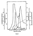
- Preparative column isoelectric focusing of the concentrated lymphokine samples was performed within a pH 3.5-10 ampholine (LKB, Rockville, MD) gradient in a 110 ml isoelectric focusing column (LKB) (28). Three ml fractions were collected and the pH of each fraction measured. Selected fractions were pooled, diafiltered against PBS ⁇ 0.1% PEG, filter sterilized with a 0.22 ⁇ Millex filter (Millipore Corp., Bedford, MA) and assayed for biological activity. (See Table 4 and Figure 8).
- HPLC separation based on molecular charge was performed in a Toyasoda DEAE-545 analytical anion exchange column. Samples in 20 mM Tris HCI, pH 7.4 with 0.1% PEG were eluted with a linear 1 hr. gradient of 0.5 m NaCl at a flow rate of 0.75 ml/min. Two minute fractions were collected, filter sterilzed and assayed. (See Figure 9).
- Concentrated leukoregulin samples were electrophoresed on 4-30% linear gradient polyacrylamide gels (16). After electrophoresis, the gels were sliced into 0.25 mm segments and the slices eluted overnight in medium-10% FBS. Eluates were filter sterilized and assayed. (See Figure 5).
- lymphokine was added to 0.5 ml of the acetate buffer, 0.5 ml of the acetate buffer containing 50 pl of VCN and 50 pl of the 0.2 M sialic acid (Sigma) (this concentration of sialic acid inactivates the enzyme, or 0.5 ml of medium.
- Samples were incubated for 60 minutes at 37°C, dialyzed against PBS-PEG in an Amicon cell with a YM10 membrane, and sample volumes adjusted to 3 ml.
- lymphokine samples 1 ml was added to 1 ml of trypsin in PBS (32 units/ml, specific acvivity 195 units/mg, Worthington Enzymes Inc., Freehold, NJ), 1 ml of chymotrypsin in PBS (6 units/ml, specific activity 59 units/mg, Sigma), or 1 ml of pronase (6 units/ml, specific activity 6 units/mg, Sigma) in 0.1 M Tris base with 3 mM CaCI 2 and 3% toluene.
- PBS 32 units/ml, specific acvivity 195 units/mg, Worthington Enzymes Inc., Freehold, NJ
- chymotrypsin in PBS 6 units/ml, specific activity 59 units/mg, Sigma
- pronase 6 units/ml, specific activity 6 units/mg, Sigma
- Pronase was activated by incubating 10 mg pronase/ml 0.1 M Tris-15 mM CaC1 2 , pH 7.8 for 30 minutes at 37°C just before adding to the sample. Samples with proteases or protease buffer controls were incubated 60 min at 37°C. One ml of FBS was added to each sample and samples were diluted over a hundred-fold range in medium for assay. (See Table 3).
- Enhancement of target cell susceptibility after leukoregulin treatment to NK-mediated cytotoxicity was determined essentially as previously described (23). Normal human peripheral blood mononuclear cells isolated by LSM gradient centrifugation were passaged through nylon wool to remove macrophages and B cells while enriching for NK cells. K562 target cells were labelled 18 hours by addition of 100 u Ci 5 'Cr as sodium chromate (NEN, Boston, MA). Target cells were washed 3 times with RPMI 1640-10% FBS and suspended to 2.5 ⁇ 10 5 cells/ml of medium or lymphokine sample diluted in medium.
- the target cells were then incubated 30 min at 37°C, centrifuged at 280xg for 5 min and resuspended in RPMI 1640-10% FBS at a concentration of 10 5 cells/ml.
- One hundred pl of cells were added to tubes containing 100 pl of medium or effector cells at 25:1, 10:1 or 2.5:1 effector to target cell ratios.
- a 4-hour 5 'Cr-release assay was then performed (23).
- Spontaneous 51 Cr release ranged from 15 to 20% whether the cells were preincubated in medium or lymphokine.
- the degree of lymphokine enhancement was determined by comparing lytic units of NK cytotocixity of target cells incubated in medium to target cells incubated in lymphokine.
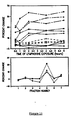
- One lytic unit defined as the number of lymphocytes causing a specific release of 15% of 5 1 Cr from the target cells, was calculated using the Van Krough equation (21, 32). (See Table 6 and Figure 3).
- K562 cells were resuspended at 4x 10 6 /ml in RPMI 1640-10% FBS.
- RPMI 1640-10% FBS RPMI 1640-10% FBS.
- 250 ⁇ l of cell suspension was dispensed with the addition of 250 pl of lymphokine sample.
- the tubes were incubated at 37°C in a 5% C0 2 :95% humidified air atmosphere on a platform rocker (Bellco Glass Co., Vineland, NJ) at 6 cycles/minute for 30 minutes to 6 hours.
- FDA Fluorochromasia FDA was used to measure cell membrane permeability (5).
- the stock solution of FDA (Polyscience, Inc., Warrington, PA) was made by dissolving crystalline FDA in acetone to a concentration of 5 mg FDA/ml.
- a working solution was prepared by diluting the stock solution in RPMI 1640-10% FBS to give final concentration of 6.25 pg FDA/ml.
- lymphokine treatment of K562 cells the cells were washed once in RPMI 1640-10% FBS and resuspended in 1 ml of the working FDA solution at 6.25 ⁇ g/ml, incubated at room temperature for 5 minutes, then analyzed on the flow cytometer.
- Propidium iodide fluorochromasia Propidium iodide was also used to measure cell membrane permeability.
- a stock solution of propidium iodide (Calbiochem-Behring Corp, La Jolla, CA) was made by dissolving propidium iodide in PBS, pH 7.4, at a concentration of 500 ⁇ g/ml.
- a working solution was prepared by diluting the stock solution in PBS to give a final concentration of 0.2 ⁇ g propidium iodide/ml.
- the cells were washed once in RPMI 1640-10% FBS and resuspended in 1 ml of the working solution of propidium iodide, incubated for 5 minutes at room temperature, then analyzed on the flow cytometer.
- Fluorescence measurements The excitation wavelength used for both FDA and propidium iodide was the same, 488 nm at 400 mW. Long pass 515 and 520 nm optical glass filters were placed in front of the fluorescence detector photomultiplier tube. When analyzing propidium iodide labelled samples, an additional red additive dichroic filter (Corion Corp, Hollister, MA) was placed in front of the photomultiplier tube. Medium treated K562 cells were used to fine tune the flow cytometer prior to each experiment. The gain controls and the photomultiplier tube voltage were adjusted so 90% of 2 ⁇ 104 cells analyzed would fall within a predetermined range of channels within an entire scale of 256 channels.
- Cancer patients were immunized intradermally with their own dissociated tumor cells that had been irradiated with 20,000 rads and were mixed with 10 7 viable BCG.
- Peripheral blood lymphocytes prepared from the venous blood of these patients were mixed with murine NS-1 myeloma cells, at a ratio of 3 PBL per 1 myeloma, centrifuged and resuspended in 100 pl serum free medium.
- One ml of polyethylene glycol (50% w/v) prewarmed to 37°C was added dropwise to the cell pellet over the course of one minute with constant agitation of the tube.
- co-cultivation of PBL with myeloma cells may be used to generate transformed diploid B-cells.
- PBL and myeloma cells were mixed (at a ratio of 3:1), pelleted at 800 RPM and selected in HAT medium, as described above.
- Supernatants from the hybridomas or transformed diploid B cells were tested for secreted leukoregulin using the microassay with HT-29 cells as targets.
- Cells producing leukoregulin were expanded in MEM-10% FBS, and when sufficient numbers were obtained, experiments were performed to determine the optimum conditions for leukoregulin production in serum free medium.
- Cells were suspended at a concentration of 2x10 5 /ml RPMI 1640 and incubated for 3 days at 37°C. In some experiments, 10 ng tetradecanoyl phorbol acetate/ml was added to the cultures to stimulate leukoregulin production. The supernatants were collected, centrifuged at 800xg and assayed for leukoregulin activity.
- mice were immunized by 3 subcutaneous injections of K562 membranes spaced two weeks apart. Each mouse received membranes equivalent to 10 7 cells prepared by homogenization with a motor driven Teflon homogenizer, clarified by low speed centrifugation and then collected by ultracentrifugation at 100,000xg for 1 hr. Membranes were mixed with complete Feund's adjuvant for each of the boosts. Three days prior to hybridization, the mice were injected intraperitoneally with the K562 cell membranes in PBS. On the day of the fusion, the spleens were removed and single cells were obtained and fused at a ratio of 3 spleen cells to 1 myeloma cell with PEG.
- hybrids were selected by culturing in hypoxanthine, aminopterin, and thymidine containing medium (10). Colonies producing antibodies to the leukoregulin receptor were cloned by a limiting dilution method.
- a biological assay measuring the inhibition of leukoregulin directed growth retardation of HT-29 cells was used to detect antibodies to the leukoregulin receptor.
- Two thousand HT-29 cells were plated in 0.1 ml MEM-10% FBS in wells of a 96-well plate.
- Sufficient leukoregulin in 0.1 ml to cause a 50% inhibition of HT-29 cell growth was added, followed by 25 ⁇ l of the test antibody containing supernatant. After 3 days' incubation, inhibition was measured by quantitation of cells present by staining with MTT as described earlier. Any sample displaying a 25% or greater inhibition of the leukoregulin activity was considered positive.
- the rationale behind this assay is that the monoclonal antibody will bind to the leukoregulin receptor which thereby inhibits leukoregulin binding and its subsequent growth inhibitory activity.
- Quantitation of leukoregulin receptor expression was measured three ways, all of which quantitated the percentage of monoclonal antibody directed to the leukoregulin receptor.
- 2 ⁇ 10 5 cells were incubated with the monoclonal antibody for 1 hr. at 37°C.
- the cells were washed 2 times, a fluorescent conjugated goat anti-mouse immunoglobulin (Kirkegard and Peiry Labs, Rockville, MD) was added and incubated for 30 min. at 4°C.
- the cells were washed, resuspended in 0.5 ml of PBS and analyzed on a EPICS 5 flow cytometer (Coulter Instruments, Hialeah, FL) using the 488 laser line to excite the green fluorescence.
- ELISA and RIA were performed in a similar manner except that the conjugate used in ELISA was a horseradish peroxidase goat anti-mouse immunoglobulin and that used in the RIA was 125 I-labelled goat anti-mouse immunoglobulin.
- the ELISA assay was quantitated colorimetrically by measuring the amount of substrate hydrolyzed on an ARTEK automated reader.
- the RIA was quantitated by measuring 125I-label bound using a LKB gamma counter (LKB Instruments Rockville, MD). (See Table 8).
- Tumor cells have the unique ability over normal cells to grow in semi-solid medium. This characteristic of tumor cells provides a means for quantitating the effect of cancer therapeutic agents on freshly dissociated cells of excised human tumors. Colon tumors obtained at surgery were minced and dissociated with collagenase and DNAase to obtain single cell suspension (11). The cells were suspended in 0.15% agar in medium according to the procedure of Kern (12), except that leukoregulin was added at varying concentrations. The development of tumor cell colonies growing suspended in the agar medium was quantitated after 10-14 days' incubation. (See Table 9).
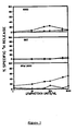
- Human lymphokine and lymphotoxin had little or no cytolytic activity towards human leukemia, sarcoma, and carcinoma cells. A maximum of 50% lysis of the RPMI 2650 carcinoma cells was observed with a lymphokine sample containing 1000 units of lymphotoxin/ml.
- the highly purified 1788 cell line lymphotoxin failed to lyse each of the three types of tumor cells. Despite low lytic activity toward human tumor cells, human lymphokine caused significant growth inhibition of human tumor cells.
- the purified 1788 cell line lymphotoxin displayed significant growth inhibition only at concentrations of 500 and 1000 lymphotoxin units/ml. The growth inhibition at these concentrations, moreover, could well have been due to the growth retarding influence of the pH 8.4 ammonium carbonate buffer in which the purified lymphotoxin was prepared.
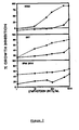
- Human lymphokine Enhancement of target cell susceptibility to NK-mediated cytotoxicity by human lymphokine containing leukoregulin but not by purified human lymphotoxin.
- human lymphokine In addition to inhibiting tumor cell proliferation, human lymphokine, but not purified 1788 lymphotoxin, enhanced the susceptibility of human carcinoma, leukemia, and sarcoma cells to NK-mediated cytotoxicity. Human lymphokine even caused lysis of RPMI 2650 carcinoma cells which are resistant to NK lysis. Purified 1788 lymphotoxin failed to enhance the susceptibility of carcinoma, leukemia, or sarcoma cells to lysis by NK. The divergent biological activities of unfractionated human PBL lymphotoxin containing lymphokine and purified 1788 cell line lymphotoxin suggest that a lymphokine other than lymphotoxin mediates the anti-tumor cell activities.
- the apparent molecular weight of human PBL lymphotoxin and leukoregulin was examined by HPLC molecular sieve chromatography. The majority of the lymphotoxin activity eluted in fractions within the 30,000-40,000 molecular weight range. There was, however, some variation in samples from different individuals with some also having lymphotoxin activity within the 50,000-70,000 and 12,000-20,000 molecular weight ranges. Leukoregulin activity eluted in fractions within the 50,000-70,000 molecular weight range with a minor component in the 10,000-15,000 molecular weight range.
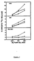
- Linear gradient polyacrylamide gel electrophoresis of leukoregulin A more accurate estimation of the molecular weight of leukoregulin was determined by gradient polyacrylamide gel electrophoresis.
- One ml of a human leukoregulin preparation was electrophoresed overnight. The gel was then sliced and the slices were eluted overnight with MEM-10% FBS at 37°C.
- Leukoregulin activity (open circles) was measured using the microassay with HT-29 carcinoma cells as the targets. Leukoregulin migrated with proteins of molecular weights of 110,000-140,000. Also shown in this figure is the electrophoretic pattern of 125 I-labelled (closed circles) leukoregulin purified by sequential HPLC gel filtration, ion exchange chromatography, and linear gradient polyacrylamide gel electrophoresis.
- Sodium dodecyl sulfate polyacrylamide gel electrophoresis Purified leukoregulin labelled with 125 1 was electrophoresed on a sodium dodecyl sulfate polyacrylamide gel to determine whether the native protein would dissociate into subunits when subjected to denaturing conditions. Leukoregulin electrophoresed with 30,000-35,000 molecular weight molecules, as shown by this autoradiograph of the gel.
- Lymphotoxin had an isoelectric pH between 6.5 and 7.2.
- Leukoregulin had two isoelectric pH's: one between 5.0 and 5.8, the second between 7.5 and 8.3.
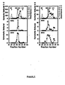
- Flow cytometric analysis of leukoregulin activity Another biological activity of leukoregulin which may be related to its mechanism of action was assessed by flow cytometic analysis. Changes in K562 cell volume or membrane permeability detectable by alterations in narrow angle forward light scatter (0) or by fluorescein diacetate (0) or propidium iodide ( ⁇ ) fluorochromasia were assessed using 488 nm argon laser line excitation in a FACS IV flow cytometer.
- K562 cells were treated with a lymphokine preparation containing 10 (---), 40 (-...-), or 100 (- ⁇ -) units of diafiltered lymphotoxin or 500 (-) units of alpha interferon for 0-6 hours to establish the response of the cells to the lymphokine.
- the narrow angle forward light scatter from the cells decreased propidium iodide fluorochromasia increased, and FDA fluorochromasia decreased.
- the change in narrow angle forward light scatter reflected a change in cell shape and/or size with an increase in cell volume confirmed by cell volume analysis on a Model ZB1 Coulter Counter with added Accucomp cell volume analysis programming (Coulter Instruments, Inc., Hialeah, FL).
- the decrease in FDA fluorochromasia reflected increased membrane permeability, as the fluorescent intracellular fluorescein escapes from the cell.
- Lymphokine cell conformation and membrane permeability activity were present only in HPLC and isoelectric focusing purified leukoregulin fractions. Fractions enriched in lymphotoxin or in the interleukins and interferon produced no detectable cellular conformation or plasma membrane permeability changes.
- lymphotoxin alpha L929 cytolytic activity
- human tumor cell cytostatic activity varied independently in the preparations, indicating that the two biological activities were mediated by distinctly different entities.
- Concentrated lymphokines from three individuals' leukocytes that were not stimulated with PHA or had PHA added for the last half hour of culture did not lyse any tumor cells and had no growth inhibitory activity.
- the same leukocytes were stimulated with PHA for 24 hours they secreted significant amounts of lytic and growth inhibitory activities.
- the antitumor cell activities were not due to PHA, buffer, or a depletion of medium nutrients by the 24-hour cultured cells.
- leukoregulin distinct from lymphotoxin
- human cytolytic lymphokine activity for alpha L929 cells was diminised 49 and 92% by 0.2 and 0.4 units of neuraminidase without any significant inhibition of the lymphokine tumor cell growth inhibitory (leukoregulin) activity.
- Neuraminidase digestion in the presence of 10 mM sialic acid had no effect on either activity, ruling out the possibility of the effect on lymphotoxin being caused by contaminating proteases in the neuraminidase.
- both lymphotoxin and leukoregulin activities were inhibited by proteases, although to differing degrees.
- Six units of pronase completely destroyed all of the lymphotoxin activity while 52% of the leukoregulin activity remained in the sample.
- pronase digestion rendered an active human leukoregulin sample lymphotoxin free.
- lymphotoxin activity was found in the high (45,000-74,000), middle (30,000-38,000) and low (17,000-20,000) molecular weight fractions and was enriched in the pH 6.0-6.8 fraction after focusing.
- Leukoregulin was present only in the high molecular weight material and was separated into two species with isoelectric pH's at 4.2-5.6 and 7.1-8.4.
- leukoregulin enriched fractions contained some lymphotoxin activity, fractions 1 and 3 contained greater than 170 lymphotoxin units but no leukoregulin activity. Therefore, a lymphotoxin activity derived from stimulated human PBL can be separated from leukoregulin.
- the low molecular weight fraction contained all of the interleukin 1 and interleukin 2 and the majority of the interferon activities. No macrophage activating activity was detected in any fraction. All of the fractions also contained endotoxin. The amount of endotoxin did not correlate with the leukoregulin activity.
- leukoregulin enriched fractions contained low levels of interferon an additional experiment was performed to determine to what extent interferon might contribute to the inhibition of tumor cell growth.
- Gamma interferon from one source (ML) neither lysed alpha L929 cells nor inhibited the growth of human K562 tumor cells.
- Gamma interferon from another source (FL) contained 2000 lymphotoxin units/ml but did not contain leukoregulin activity.
- Alpha interferon contained neither lyphotoxin nor leukoregulin. Leukoregulin activity therefore is not mediated by alpha or gamma interferon nor is it a result of the synergistic action of lymphotoxin and gamma interferon.
- Unfractioned lymphokine preparations contain an activity which enhances the susceptibility of tumor cells to NK-mediated cytotoxicity.
- the relationship of the target cell sensitizing activity to leukoregulin and to lymphotoxin was examined following HPLC and isoelectric focusing sequential fractionation of human lymphokine.
- Leukoregulin enriched fractions contained significant tumor cell NK-cytotoxicity enhancing activity.
- Fractions enriched in lymphotoxin and those containing the lower molecular weight lymphokines such as interferon did not enhance the susceptibility of K562 leukemia cells to NK-mediated cytotoxicity.
- NK enhancing activity copurifies with or is identical to leukoregulin.
- Gastro-intestinal carcinomas as a group were found to be highly sensitive to leukoregulin.
- Leukoregulin did not affect the growth of either the normal colonic mucosal cells or the skin fibroblasts.
- leukoregulin In order to determine the potential in vivo, effectiveness of leukoregulin on human cancers, freshly dissociated colon tumor cells were prepared and cultured in semi-solid agar medium containing leukoregulin. Colon cancer was targeted for evaluation because of the high susceptibility of the colon cancer cell lines to leukoregulin. Tumor cells from all four patients tested were growth inhibited in a dose dependent manner to leukoregulin, although to varying degrees. One patient's cells were inhibited 98% by 50 units of leukoregulin while another patient's were inhibited 42% with 4,000 units of leukoregulin. This suggests that leukoregulin has the potential to control the growth of human colon cancers.
Landscapes
- Health & Medical Sciences (AREA)
- Chemical & Material Sciences (AREA)
- Organic Chemistry (AREA)
- Life Sciences & Earth Sciences (AREA)
- Medicinal Chemistry (AREA)
- General Health & Medical Sciences (AREA)
- Biochemistry (AREA)
- Immunology (AREA)
- Proteomics, Peptides & Aminoacids (AREA)
- Molecular Biology (AREA)
- Genetics & Genomics (AREA)
- Biophysics (AREA)
- Pharmacology & Pharmacy (AREA)
- Nuclear Medicine, Radiotherapy & Molecular Imaging (AREA)
- Zoology (AREA)
- Gastroenterology & Hepatology (AREA)
- Veterinary Medicine (AREA)
- Public Health (AREA)
- Animal Behavior & Ethology (AREA)
- Toxicology (AREA)
- General Chemical & Material Sciences (AREA)
- Chemical Kinetics & Catalysis (AREA)
- Preparation Of Compounds By Using Micro-Organisms (AREA)
- Medicines That Contain Protein Lipid Enzymes And Other Medicines (AREA)
- Peptides Or Proteins (AREA)
- Medicines Containing Material From Animals Or Micro-Organisms (AREA)
- Micro-Organisms Or Cultivation Processes Thereof (AREA)
- Medicines Containing Antibodies Or Antigens For Use As Internal Diagnostic Agents (AREA)
Priority Applications (1)
| Application Number | Priority Date | Filing Date | Title |
|---|---|---|---|
| AT85902248T ATE48617T1 (de) | 1984-04-13 | 1985-04-11 | Leukoregulin, ein antitumor-lymphokin und dessen verwendung als heilmittel. |
Applications Claiming Priority (2)
| Application Number | Priority Date | Filing Date | Title |
|---|---|---|---|
| US60030384A | 1984-04-13 | 1984-04-13 | |
| US600303 | 1984-04-13 |
Publications (3)
| Publication Number | Publication Date |
|---|---|
| EP0179127A1 EP0179127A1 (en) | 1986-04-30 |
| EP0179127A4 EP0179127A4 (en) | 1986-08-21 |
| EP0179127B1 true EP0179127B1 (en) | 1989-12-13 |
Family
ID=24403068
Family Applications (1)
| Application Number | Title | Priority Date | Filing Date |
|---|---|---|---|
| EP85902248A Expired EP0179127B1 (en) | 1984-04-13 | 1985-04-11 | Leukoregulin, an antitumor lymphokine, and its therapeutic uses |
Country Status (9)
| Country | Link |
|---|---|
| EP (1) | EP0179127B1 (no) |
| JP (2) | JP2562014B2 (no) |
| AT (1) | ATE48617T1 (no) |
| AU (2) | AU592529B2 (no) |
| DE (1) | DE3574710D1 (no) |
| DK (2) | DK170781B1 (no) |
| FI (1) | FI85867C (no) |
| NO (1) | NO170423C (no) |
| WO (1) | WO1985004662A1 (no) |
Families Citing this family (5)
| Publication number | Priority date | Publication date | Assignee | Title |
|---|---|---|---|---|
| DE3574710D1 (de) * | 1984-04-13 | 1990-01-18 | Litton Bionetics Inc | Leukoregulin, ein antitumor-lymphokin und dessen verwendung als heilmittel. |
| US5683688A (en) * | 1984-05-31 | 1997-11-04 | Genentech, Inc. | Unglycosylated recombinant human lymphotoxin polypeptides and compositions |
| US6087166A (en) | 1997-07-03 | 2000-07-11 | Basf Aktiengesellschaft | Transcriptional activators with graded transactivation potential |
| JP2009537139A (ja) | 2006-05-15 | 2009-10-29 | パラテック ファーマシューティカルズ インコーポレイテッド | 置換されたテトラサイクリン化合物を用いて、遺伝子または遺伝子産物の発現を制御する方法 |
| WO2012156535A1 (en) | 2011-05-19 | 2012-11-22 | Fundación Progreso Y Salud | Highly inducible dual-promoter lentiviral tet-on system |
Family Cites Families (9)
| Publication number | Priority date | Publication date | Assignee | Title |
|---|---|---|---|---|
| US4394448A (en) * | 1978-02-24 | 1983-07-19 | Szoka Jr Francis C | Method of inserting DNA into living cells |
| US4403035A (en) * | 1981-06-19 | 1983-09-06 | Regents Of The University Of Minnesota | In vitro DNA-Protein viral assembly and gene cloning system |
| DE3138230A1 (de) * | 1981-09-25 | 1983-04-07 | Max-Planck-Gesellschaft zur Förderung der Wissenschaften e.V., 3400 Göttingen | Verfahren zur gewinnung von makrophagen- aktivierungsfaktor (maf) |
| JPS6011889B2 (ja) * | 1981-10-28 | 1985-03-28 | 電気化学工業株式会社 | 継代可能なリンホカイン産生ヒトt細胞融合株及びその取得方法 |
| US4464465A (en) * | 1982-04-05 | 1984-08-07 | Genetic Systems Corporation | Cell-driven viral transfer in eukaryotes |
| US4443427A (en) * | 1982-06-21 | 1984-04-17 | Sidney Farber Cancer Institute, Inc. | Monoclonal antibody |
| ZA834976B (en) * | 1982-07-30 | 1984-08-29 | Genentech Inc | Human lymphotoxin |
| EP0135797A3 (de) * | 1983-08-30 | 1987-11-11 | BOEHRINGER INGELHEIM INTERNATIONAL GmbH | Verfahren zur Herstellung von Lymphotoxin und von Lymphotoxin-mRNA |
| DE3574710D1 (de) * | 1984-04-13 | 1990-01-18 | Litton Bionetics Inc | Leukoregulin, ein antitumor-lymphokin und dessen verwendung als heilmittel. |
-
1985
- 1985-04-11 DE DE8585902248T patent/DE3574710D1/de not_active Expired - Fee Related
- 1985-04-11 EP EP85902248A patent/EP0179127B1/en not_active Expired
- 1985-04-11 AT AT85902248T patent/ATE48617T1/de not_active IP Right Cessation
- 1985-04-11 AU AU42341/85A patent/AU592529B2/en not_active Ceased
- 1985-04-11 WO PCT/US1985/000626 patent/WO1985004662A1/en active IP Right Grant
- 1985-04-11 JP JP60501862A patent/JP2562014B2/ja not_active Expired - Lifetime
- 1985-12-04 FI FI854803A patent/FI85867C/fi not_active IP Right Cessation
- 1985-12-04 DK DK562285A patent/DK170781B1/da active
- 1985-12-12 NO NO85854994A patent/NO170423C/no unknown
-
1990
- 1990-04-18 AU AU53653/90A patent/AU641386B2/en not_active Ceased
-
1992
- 1992-08-04 DK DK098692A patent/DK170423B1/da not_active IP Right Cessation
-
1993
- 1993-11-30 JP JP5300409A patent/JP2564245B2/ja not_active Expired - Lifetime
Also Published As
| Publication number | Publication date |
|---|---|
| DK98692D0 (da) | 1992-08-04 |
| JPS61501853A (ja) | 1986-08-28 |
| DK562285D0 (da) | 1985-12-04 |
| EP0179127A1 (en) | 1986-04-30 |
| JPH06253832A (ja) | 1994-09-13 |
| DK98692A (da) | 1992-08-04 |
| FI85867C (fi) | 1992-06-10 |
| DK170781B1 (da) | 1996-01-15 |
| NO170423C (no) | 1992-10-14 |
| DK562285A (da) | 1985-12-04 |
| AU641386B2 (en) | 1993-09-23 |
| AU4234185A (en) | 1985-11-01 |
| NO854994L (no) | 1985-12-12 |
| WO1985004662A1 (en) | 1985-10-24 |
| AU5365390A (en) | 1990-09-20 |
| AU592529B2 (en) | 1990-01-18 |
| DE3574710D1 (de) | 1990-01-18 |
| DK170423B1 (da) | 1995-08-28 |
| JP2562014B2 (ja) | 1996-12-11 |
| FI854803A (fi) | 1985-12-04 |
| NO170423B (no) | 1992-07-06 |
| FI85867B (fi) | 1992-02-28 |
| FI854803A0 (fi) | 1985-12-04 |
| EP0179127A4 (en) | 1986-08-21 |
| JP2564245B2 (ja) | 1996-12-18 |
| ATE48617T1 (de) | 1989-12-15 |
Similar Documents
| Publication | Publication Date | Title |
|---|---|---|
| Fu et al. | Induction of in vitro differentiation and immunoglobulin synthesis of human leukemic B lymphocytes. | |
| Miller et al. | Regulated expression of the Mac-1, LFA-1, p150, 95 glycoprotein family during leukocyte differentiation. | |
| Ransom et al. | Leukoregulin, a direct-acting anticancer immunological hormone that is distinct from lymphotoxin and interferon | |
| Valle et al. | Immune specific production of interferon by human T cells in combined macrophage-lymphocyte cultures in response to herpes simplex antigen | |
| Itoh et al. | A case of chronic lymphocytic leukemia with properties characteristic of natural killer cells | |
| Cassel et al. | Viral oncolysate in the management of malignant melanoma. I. Preparation of the oncolysate and measurement of immunologic responses | |
| US6265214B1 (en) | Methods and compositions for inducing monocyte cytotoxicity | |
| Starkebaum et al. | Chronic lymphocytosis with neutropenia: Evidence for a novel, abnormal T-cell population associated with antibody-mediated neutrophil destruction | |
| US5643740A (en) | Monoclonal antibody specific for activated lymphocytes and monocytes | |
| EP0179127B1 (en) | Leukoregulin, an antitumor lymphokine, and its therapeutic uses | |
| EP0203403A2 (en) | A cloned T cell capable of recognizing tumors and a T cell antigen receptor | |
| Takahashi et al. | Cutaneous-type adult T-cell leukemia/lymphoma: a unique clinical feature with monoclonal T-cell proliferation detected by Southern blot analysis | |
| US5082657A (en) | Leukoregulin, an antitumor lymphokine, and its therapeutic uses | |
| US4849506A (en) | Leukoregulin, an antitumor lymphokine, and its therapeutic uses | |
| Burakoff et al. | Induction of secondary cytotoxic T lymphocytes by liposomes containing HLA-DR antigens | |
| CN116143943B (zh) | 一种靶向baffr嵌合抗原受体、car-t细胞及应用 | |
| Dunn et al. | Interleukin 2 and concanavalin A stimulate interferon-gamma production in a murine cytolytic T cell clone by different pathways. | |
| US5330896A (en) | Monoclonal antibodies to an autocrine growth factor antigen that binds to activated lymphocytes and cancer cells | |
| US4977245A (en) | Methods and compositions for inducing monocyte cytotoxicity | |
| Cerny et al. | Specific susceptibility of sensitized (memory) B cells to suppression and antigenic alteration by murine leukemia virus | |
| Petersen et al. | Functional characteristics of synovial fluid and blood mononuclear cells in rheumatoid arthritis and traumatic synovitis | |
| Jones et al. | Identification of a human monocyte cytotoxicity-inducing factor from T cell hybridomas produced from Sezary's cells. | |
| Hu et al. | Interleukin 2 up-regulates its own production. | |
| Marubayashi et al. | Sera from patients with colon, breast and lung cancer induce resistance to lysis mediated by NK cytotoxic factors (NKCF) | |
| EP0311438B1 (en) | Antigen capable of binding to cancer cells |
Legal Events
| Date | Code | Title | Description |
|---|---|---|---|
| PUAI | Public reference made under article 153(3) epc to a published international application that has entered the european phase |
Free format text: ORIGINAL CODE: 0009012 |
|
| AK | Designated contracting states |
Kind code of ref document: A1 Designated state(s): AT BE CH DE FR GB IT LI LU NL SE |
|
| 17P | Request for examination filed |
Effective date: 19860402 |
|
| A4 | Supplementary search report drawn up and despatched |
Effective date: 19860821 |
|
| 17Q | First examination report despatched |
Effective date: 19880224 |
|
| GRAA | (expected) grant |
Free format text: ORIGINAL CODE: 0009210 |
|
| AK | Designated contracting states |
Kind code of ref document: B1 Designated state(s): AT BE CH DE FR GB IT LI LU NL SE |
|
| REF | Corresponds to: |
Ref document number: 48617 Country of ref document: AT Date of ref document: 19891215 Kind code of ref document: T |
|
| REF | Corresponds to: |
Ref document number: 3574710 Country of ref document: DE Date of ref document: 19900118 |
|
| ITF | It: translation for a ep patent filed | ||
| RAP2 | Party data changed (patent owner data changed or rights of a patent transferred) |
Owner name: AKZO N.V. |
|
| REG | Reference to a national code |
Ref country code: CH Ref legal event code: PUE Owner name: AKZO N.V. |
|
| ET | Fr: translation filed | ||
| NLT2 | Nl: modifications (of names), taken from the european patent patent bulletin |
Owner name: AKZO N.V. TE ARNHEM. |
|
| PLBE | No opposition filed within time limit |
Free format text: ORIGINAL CODE: 0009261 |
|
| STAA | Information on the status of an ep patent application or granted ep patent |
Free format text: STATUS: NO OPPOSITION FILED WITHIN TIME LIMIT |
|
| 26N | No opposition filed | ||
| ITTA | It: last paid annual fee | ||
| EPTA | Lu: last paid annual fee | ||
| EAL | Se: european patent in force in sweden |
Ref document number: 85902248.5 |
|
| PGFP | Annual fee paid to national office [announced via postgrant information from national office to epo] |
Ref country code: FR Payment date: 19990324 Year of fee payment: 15 Ref country code: AT Payment date: 19990324 Year of fee payment: 15 |
|
| PGFP | Annual fee paid to national office [announced via postgrant information from national office to epo] |
Ref country code: SE Payment date: 19990325 Year of fee payment: 15 Ref country code: CH Payment date: 19990325 Year of fee payment: 15 |
|
| PGFP | Annual fee paid to national office [announced via postgrant information from national office to epo] |
Ref country code: DE Payment date: 19990326 Year of fee payment: 15 |
|
| PGFP | Annual fee paid to national office [announced via postgrant information from national office to epo] |
Ref country code: LU Payment date: 19990406 Year of fee payment: 15 |
|
| PGFP | Annual fee paid to national office [announced via postgrant information from national office to epo] |
Ref country code: BE Payment date: 19990414 Year of fee payment: 15 |
|
| PGFP | Annual fee paid to national office [announced via postgrant information from national office to epo] |
Ref country code: NL Payment date: 19990427 Year of fee payment: 15 |
|
| PG25 | Lapsed in a contracting state [announced via postgrant information from national office to epo] |
Ref country code: LU Free format text: LAPSE BECAUSE OF NON-PAYMENT OF DUE FEES Effective date: 20000411 Ref country code: AT Free format text: LAPSE BECAUSE OF NON-PAYMENT OF DUE FEES Effective date: 20000411 |
|
| PG25 | Lapsed in a contracting state [announced via postgrant information from national office to epo] |
Ref country code: SE Free format text: LAPSE BECAUSE OF NON-PAYMENT OF DUE FEES Effective date: 20000412 |
|
| PG25 | Lapsed in a contracting state [announced via postgrant information from national office to epo] |
Ref country code: LI Free format text: LAPSE BECAUSE OF NON-PAYMENT OF DUE FEES Effective date: 20000430 Ref country code: CH Free format text: LAPSE BECAUSE OF NON-PAYMENT OF DUE FEES Effective date: 20000430 Ref country code: BE Free format text: LAPSE BECAUSE OF NON-PAYMENT OF DUE FEES Effective date: 20000430 |
|
| PGFP | Annual fee paid to national office [announced via postgrant information from national office to epo] |
Ref country code: GB Payment date: 20000803 Year of fee payment: 16 |
|
| BERE | Be: lapsed |
Owner name: AKZO N.V. Effective date: 20000430 |
|
| PG25 | Lapsed in a contracting state [announced via postgrant information from national office to epo] |
Ref country code: NL Free format text: LAPSE BECAUSE OF NON-PAYMENT OF DUE FEES Effective date: 20001101 |
|
| EUG | Se: european patent has lapsed |
Ref document number: 85902248.5 |
|
| REG | Reference to a national code |
Ref country code: CH Ref legal event code: PL |
|
| PG25 | Lapsed in a contracting state [announced via postgrant information from national office to epo] |
Ref country code: FR Free format text: LAPSE BECAUSE OF NON-PAYMENT OF DUE FEES Effective date: 20001229 |
|
| NLV4 | Nl: lapsed or anulled due to non-payment of the annual fee |
Effective date: 20001101 |
|
| PG25 | Lapsed in a contracting state [announced via postgrant information from national office to epo] |
Ref country code: DE Free format text: LAPSE BECAUSE OF NON-PAYMENT OF DUE FEES Effective date: 20010201 |
|
| REG | Reference to a national code |
Ref country code: FR Ref legal event code: ST |
|
| PG25 | Lapsed in a contracting state [announced via postgrant information from national office to epo] |
Ref country code: GB Free format text: LAPSE BECAUSE OF NON-PAYMENT OF DUE FEES Effective date: 20010411 |
|
| GBPC | Gb: european patent ceased through non-payment of renewal fee |
Effective date: 20010411 |