WO2023277195A1 - iPS細胞由来軟骨細胞構造体の製造方法 - Google Patents
iPS細胞由来軟骨細胞構造体の製造方法 Download PDFInfo
- Publication number
- WO2023277195A1 WO2023277195A1 PCT/JP2022/026536 JP2022026536W WO2023277195A1 WO 2023277195 A1 WO2023277195 A1 WO 2023277195A1 JP 2022026536 W JP2022026536 W JP 2022026536W WO 2023277195 A1 WO2023277195 A1 WO 2023277195A1
- Authority
- WO
- WIPO (PCT)
- Prior art keywords
- stem cells
- cell
- differentiation
- day
- dimensional structure
- Prior art date
- Legal status (The legal status is an assumption and is not a legal conclusion. Google has not performed a legal analysis and makes no representation as to the accuracy of the status listed.)
- Ceased
Links
Images


Classifications
-
- C—CHEMISTRY; METALLURGY
- C12—BIOCHEMISTRY; BEER; SPIRITS; WINE; VINEGAR; MICROBIOLOGY; ENZYMOLOGY; MUTATION OR GENETIC ENGINEERING
- C12N—MICROORGANISMS OR ENZYMES; COMPOSITIONS THEREOF; PROPAGATING, PRESERVING, OR MAINTAINING MICROORGANISMS; MUTATION OR GENETIC ENGINEERING; CULTURE MEDIA
- C12N5/00—Undifferentiated human, animal or plant cells, e.g. cell lines; Tissues; Cultivation or maintenance thereof; Culture media therefor
- C12N5/06—Animal cells or tissues; Human cells or tissues
-
- C—CHEMISTRY; METALLURGY
- C12—BIOCHEMISTRY; BEER; SPIRITS; WINE; VINEGAR; MICROBIOLOGY; ENZYMOLOGY; MUTATION OR GENETIC ENGINEERING
- C12N—MICROORGANISMS OR ENZYMES; COMPOSITIONS THEREOF; PROPAGATING, PRESERVING, OR MAINTAINING MICROORGANISMS; MUTATION OR GENETIC ENGINEERING; CULTURE MEDIA
- C12N5/00—Undifferentiated human, animal or plant cells, e.g. cell lines; Tissues; Cultivation or maintenance thereof; Culture media therefor
- C12N5/06—Animal cells or tissues; Human cells or tissues
- C12N5/0602—Vertebrate cells
- C12N5/0652—Cells of skeletal and connective tissues; Mesenchyme
- C12N5/0655—Chondrocytes; Cartilage
-
- B—PERFORMING OPERATIONS; TRANSPORTING
- B33—ADDITIVE MANUFACTURING TECHNOLOGY
- B33Y—ADDITIVE MANUFACTURING, i.e. MANUFACTURING OF THREE-DIMENSIONAL [3D] OBJECTS BY ADDITIVE DEPOSITION, ADDITIVE AGGLOMERATION OR ADDITIVE LAYERING, e.g. BY 3D PRINTING, STEREOLITHOGRAPHY OR SELECTIVE LASER SINTERING
- B33Y10/00—Processes of additive manufacturing
-
- C—CHEMISTRY; METALLURGY
- C12—BIOCHEMISTRY; BEER; SPIRITS; WINE; VINEGAR; MICROBIOLOGY; ENZYMOLOGY; MUTATION OR GENETIC ENGINEERING
- C12N—MICROORGANISMS OR ENZYMES; COMPOSITIONS THEREOF; PROPAGATING, PRESERVING, OR MAINTAINING MICROORGANISMS; MUTATION OR GENETIC ENGINEERING; CULTURE MEDIA
- C12N5/00—Undifferentiated human, animal or plant cells, e.g. cell lines; Tissues; Cultivation or maintenance thereof; Culture media therefor
- C12N5/10—Cells modified by introduction of foreign genetic material
-
- C—CHEMISTRY; METALLURGY
- C12—BIOCHEMISTRY; BEER; SPIRITS; WINE; VINEGAR; MICROBIOLOGY; ENZYMOLOGY; MUTATION OR GENETIC ENGINEERING
- C12N—MICROORGANISMS OR ENZYMES; COMPOSITIONS THEREOF; PROPAGATING, PRESERVING, OR MAINTAINING MICROORGANISMS; MUTATION OR GENETIC ENGINEERING; CULTURE MEDIA
- C12N2501/00—Active agents used in cell culture processes, e.g. differentation
- C12N2501/10—Growth factors
- C12N2501/135—Platelet-derived growth factor [PDGF]
-
- C—CHEMISTRY; METALLURGY
- C12—BIOCHEMISTRY; BEER; SPIRITS; WINE; VINEGAR; MICROBIOLOGY; ENZYMOLOGY; MUTATION OR GENETIC ENGINEERING
- C12N—MICROORGANISMS OR ENZYMES; COMPOSITIONS THEREOF; PROPAGATING, PRESERVING, OR MAINTAINING MICROORGANISMS; MUTATION OR GENETIC ENGINEERING; CULTURE MEDIA
- C12N2501/00—Active agents used in cell culture processes, e.g. differentation
- C12N2501/10—Growth factors
- C12N2501/15—Transforming growth factor beta (TGF-β)
-
- C—CHEMISTRY; METALLURGY
- C12—BIOCHEMISTRY; BEER; SPIRITS; WINE; VINEGAR; MICROBIOLOGY; ENZYMOLOGY; MUTATION OR GENETIC ENGINEERING
- C12N—MICROORGANISMS OR ENZYMES; COMPOSITIONS THEREOF; PROPAGATING, PRESERVING, OR MAINTAINING MICROORGANISMS; MUTATION OR GENETIC ENGINEERING; CULTURE MEDIA
- C12N2501/00—Active agents used in cell culture processes, e.g. differentation
- C12N2501/10—Growth factors
- C12N2501/155—Bone morphogenic proteins [BMP]; Osteogenins; Osteogenic factor; Bone inducing factor
-
- C—CHEMISTRY; METALLURGY
- C12—BIOCHEMISTRY; BEER; SPIRITS; WINE; VINEGAR; MICROBIOLOGY; ENZYMOLOGY; MUTATION OR GENETIC ENGINEERING
- C12N—MICROORGANISMS OR ENZYMES; COMPOSITIONS THEREOF; PROPAGATING, PRESERVING, OR MAINTAINING MICROORGANISMS; MUTATION OR GENETIC ENGINEERING; CULTURE MEDIA
- C12N2506/00—Differentiation of animal cells from one lineage to another; Differentiation of pluripotent cells
- C12N2506/45—Differentiation of animal cells from one lineage to another; Differentiation of pluripotent cells from artificially induced pluripotent stem cells
-
- C—CHEMISTRY; METALLURGY
- C12—BIOCHEMISTRY; BEER; SPIRITS; WINE; VINEGAR; MICROBIOLOGY; ENZYMOLOGY; MUTATION OR GENETIC ENGINEERING
- C12N—MICROORGANISMS OR ENZYMES; COMPOSITIONS THEREOF; PROPAGATING, PRESERVING, OR MAINTAINING MICROORGANISMS; MUTATION OR GENETIC ENGINEERING; CULTURE MEDIA
- C12N2513/00—3D culture
Definitions
- the present invention relates to a method for producing a chondrocyte structure.
- the present invention relates to methods of manufacturing chondrocyte constructs using a three-dimensional bioprinter.
- Non-Patent Document 1 Yamashita A. et al. al., Stem Cell Reports, 2015 (Non-Patent Document 1)
- Method of Inducing Somites and Sclerotomes from iPS Cells Matsuda S. et al., Nature, 2020 (Non-Patent Document 2)
- Proliferating Chondrocytes A method for inducing hypertrophic chondrocytes (Pretemer Y. et al., Stem Cell Reports, 2021 (Non-Patent Document 3)) has been established. Chondrocytes generated in this way produce high doses of extracellular matrix and thus may be a clinically applicable tool.
- the challenge is to make chondrocytes three-dimensional toward cartilage tissue.
- mesenchymal stem cells induced from iPS cells are differentiated into chondrocytes in a medium for inducing differentiation into chondrocytes. to form cell aggregates (spheroids) by culturing using the differentiation-inducing medium, and the cell aggregates are layered at a predetermined time from the start of the culture using the differentiation-inducing medium to form a three-dimensional structure, thereby having high functionality.
- cell aggregates spheroids
- the present invention is as follows. (1) Cell aggregates obtained by culturing mesenchymal stem cells using a chondrocyte differentiation-inducing medium are layered on the 9th or 10th day after the start of the culture, A method for producing a three-dimensional structure of chondrocytes. (2) The method according to (1), wherein the mesenchymal stem cells are derived from pluripotent stem cells. (3) The method according to (2), wherein the pluripotent stem cells are induced pluripotent stem cells. (4) The method according to any one of (1) to (3), wherein the differentiation-inducing medium contains a platelet-derived growth factor.
- a method for producing cartilage tissue which comprises culturing the three-dimensional structure produced by the method according to any one of (1) to (5) in the presence of an osteogenic protein.
- chondrocyte cell aggregates obtained by culturing mesenchymal stem cells using a chondrocyte differentiation-inducing medium, layered on the 9th or 10th day after the start of the culture; three-dimensional structure.
- cartilage tissue it has become possible to manufacture a three-dimensional structure of chondrocytes that sufficiently differentiate into cartilage and secrete an extracellular matrix.
- cartilage tissue By further culturing this three-dimensional structure in the presence of osteogenic protein using a bioreactor, cartilage tissue can be obtained.
- FIG. 4 is a diagram showing a schedule for inducing differentiation into chondrocytes.
- FIG. 13 shows a three-dimensional structure and cartilage tissue when cell aggregates are layered 13 to 16 days after initiation of induction of differentiation into chondrocytes.
- FIG. 3 shows a three-dimensional structure and cartilage tissue when cell aggregates are layered 3 days or 6 days after the start of induction of differentiation into chondrocytes.
- FIG. 10 is a diagram showing a three-dimensional structure and cartilage tissue when cell aggregates are layered 9 days or 10 days after initiation of induction of differentiation into chondrocytes.
- Non-Patent Document 1 a protocol of Kyoto University, 10 ng/ml TGFB1, 10 ng/ml BMP-2, 10 ng/ml GDF5, and 10 ng/ml bFGF are introduced into iPS cells to directly create chondrocyte clusters.
- a protocol of Kyoto University 10 ng/ml TGFB1, 10 ng/ml BMP-2, 10 ng/ml GDF5, and 10 ng/ml bFGF are introduced into iPS cells to directly create chondrocyte clusters.
- this method the characteristics of cartilage tissue, such as the production of extracellular matrix, are produced, so it is useful for treatment of partial defects of cartilage tissue.
- the present invention provides a three-dimensional chondrocyte structure formed by layering cell aggregates obtained by culturing mesenchymal stem cells using a chondrocyte differentiation-inducing medium on the 9th or 10th day after the start of the culture. A structure and a manufacturing method thereof. Further, the present invention provides a cartilage tissue obtained by culturing the three-dimensional structure in the presence of a bone morphogenetic protein, and a method for producing the cartilage tissue.
- MSCs mesenchymal stem cells
- MSCs are pluripotent cells with self-renewal ability and differentiation ability, and since they have a low risk of tumorigenesis, they can be expected as tools for cell therapy and regenerative medicine.
- the origin of MSCs used in the present invention is not limited, and examples thereof include fat, bone marrow, umbilical cord, pluripotent stem cells, deciduous dental pulp, and the like.
- MSC is commercially available and can be obtained from ATCC, Evercyte, Cell Source, Gene Techno Science, and the like.
- pluripotent stem cells are stem cells that have pluripotency that can differentiate into all cells existing in a living body and that have proliferative ability. Examples include pluripotent stem (iPS) cells. Preferred pluripotent stem cells are iPS cells.
- ES cells are stem cells established from early embryonic cell masses of mammals such as humans and mice. ES cells can be established by removing the inner cell mass from the blastocyst of a fertilized egg of a target mammal and culturing the inner cell mass on a fibroblast feeder.
- LIF leukemia inhibitory factor
- bFGF basic fibroblast growth factor
- Human ES cell lines are also available from the Institute for Frontier Medical Sciences, Kyoto University (Kyoto, Japan).
- Induced pluripotent stem (iPS) cells are somatic cell-derived artificial stem cells that have almost the same pluripotency and self-renewal growth potential as ES cells, and introduce specific reprogramming factors into somatic cells. (Yamanaka S. et al., Cell, 126:663-676, 2006; Okita K. et al., Nature 448, 2007; WO2007/069666, etc.).
- Genes contained in reprogramming factors include, for example, Oct3/4, Sox2, Sox1, Sox3, Sox15, Sox17, Klf4, Klf2, c-Myc, N-Myc, L-Myc, Nanog, Lin28, Fbx15, ERAs, ECAT15 -2, Tcl1, beta-catenin, Lin28b, Sall1, Sall4, Esrrb, Nr5a2, Tbx3, Glis1, etc., and these reprogramming factors can be used alone or in combination as appropriate, but Oct3 /4, Sox2, Klf4 and c-Myc are preferred.
- Tissues from which iPS cells used in the present invention are derived include, for example, articular cartilage, bone, adipose tissue, ligament, tendon, tooth, auricle, nose, liver, pancreas, blood vessel, nerve, heart, and the like.
- spheroids do not necessarily need to be formed as aggregates of a single type of cell. or differentiated cells thereof.
- methods for inducing mesenchymal stem cells (MSCs) from pluripotent stem cells include, for example, culturing iPS cells in the presence of factors such as TGFB1.
- the differentiation induction period from pluripotent stem cells to MSCs is, for example, 10 days. In that case, it can once go through a neural crest stem cell (Neural crest stem cell: NCC).
- iPS cell-derived MSCs can be obtained from Kyoto University.
- MSCs are gradually induced to differentiate into chondrocytes.
- the cells aggregate to form cell aggregates (spheroids).
- a growth factor can be used as the factor to be contained in the differentiation-inducing medium.
- Growth factors include platelet-derived growth factor (PDGF), transforming growth factor- ⁇ (TGF ⁇ ), bone morphogenic protein (BMP) factors, and the like. is contained in the medium at a predetermined time in order to induce the differentiation of MSCs into chondrocytes.
- PDGF is a humoral factor that is stored in platelet ⁇ -granules. PDGF promotes the differentiation and proliferation of pluripotent stem cells into chondrocytes.
- Four types of genes (A chain, B chain, C chain, and D chain) are known to encode PDGF, and biologically active PDGF exists as four homodimers and an AB heterodimer.
- AA, AB, and BB undergo protease modification in the cytoplasm and are secreted as mature forms with a molecular weight of about 30,000, while CC and DD are secreted while retaining the CUB region that inhibits receptor binding.
- TGF ⁇ exists in three isoforms (TGF ⁇ 1, ⁇ 2, ⁇ 3) in mammals, and the structurally similar TGF ⁇ superfamily includes activin, BMP (bone morphogenetic factor), and the like. Recent studies have revealed that TGF ⁇ also contributes to proliferation suppression, cell differentiation, induction of apoptosis, etc. in many cell types. For example, it has been reported to promote the proliferation of osteoblasts and the synthesis and proliferation of connective tissue such as collagen, while inhibiting the proliferation of epithelial cells and osteoclasts. In the present invention, TGF ⁇ 3 can be preferably used.
- the start date (day 0)
- day 0 the start date
- day 6 the start date
- TGF ⁇ is added after day 6. Cultivate in addition.
- the time for layering spheroids is 9 days or 10 days (day 9 or day 10) from the start of spheroid plate formation culture using a PDGF-containing differentiation-inducing medium.
- chondrocytes mean cells that produce extracellular matrix that constitutes cartilage such as collagen, or their progenitor cells.
- chondrocytes may be cells expressing chondrocyte markers, examples of which include type II collagen (COL2A1) and SOX9.
- COL2A1 includes a gene having a nucleotide sequence described in NCBI accession numbers NM_001844 or NM_033150 for humans and NM_001113515 or NM_031163 for mice, proteins encoded by these genes, and Functional, naturally occurring variants are included.
- SOX9 includes a gene having a nucleotide sequence described in NCBI accession numbers NM_000346 for human and NM_011448 for mouse, a protein encoded by the gene, and functions thereof. Naturally occurring variants are included.
- Lamination of spheroids A method for producing a three-dimensional cell structure by arranging cells in an arbitrary three-dimensional space is known (WO2008/123614). In this method, needle-shaped bodies are arranged on a substrate in the shape of a spike, and cell aggregates (spheroids) are arranged by piercing the needle-shaped bodies. In the present invention, spheroids are laminated using the above method to produce a three-dimensional structure (three-dimensional structure). Since an automatic stacking robot for realizing the above method is already known (bio 3D printer “Legenova” (registered trademark), Cyfuse Co., Ltd.), a three-dimensional structure can also be produced using this robot. be.
- the number of spheroids arranged and the arrangement shape are not particularly limited, and are arbitrary. After stacking the spheroids, they are cultured in the presence of bone morphogenic protein (BMP) to form cartilage tissue.
- BMP bone morphogenic protein
- a reaction vessel for growing cartilage tissue from a three-dimensional structure is called a bioreactor.
- BMPs are a group of proteins identified as molecules that induce and promote differentiation of bone tissue and cartilage. BMPs belong to the TGF ⁇ superfamily, bind to type I and type II receptor dimers, and are signaled into the nucleus via phosphorylation of the transcription factor SMAD. .
- BMPs belonging to the TGF ⁇ superfamily include BMP2/4 group (BMP2, BMP4), OP-1 group (BMP5, BMP6, BMP7, BMP8a, BMP8b), BMP9 group (BMP9, BMP10), GDF5 group (GDF5, GDF6, GDF7 ).
- FIG. 1 shows the schedule for induction of differentiation from MSCs to chondrocytes, formation of spheroids, and layering.
- An example of producing a three-dimensional chondrocyte structure is shown using mesenchymal stem cells (hereinafter referred to as iMSCs) obtained from Kyoto University through induction of differentiation from human iPS cells to neural crest stem cells as raw materials.
- iMSCs mesenchymal stem cells
- iMSCs were seeded on Sumilon (registered trademark) PrimeSurface (registered trademark) 96 (manufactured by Sumitomo Bakelite Co., Ltd., spheroid plate) at 4.5 x 10 4 cells per well, and the chondrogenesis induction basal medium PT-3925 (manufactured by Lonza) was added. ) was added with differentiation-inducing supplement PT-4121 to form spheroids.
- PDGF in the differentiation-inducing medium from Day 0 to Day 6, differentiation-inducing medium + PDGF + TGF ⁇ 3 from Day 6 to Day 10, and Day 10 to the final day (24). was cultured in a differentiation-inducing medium + TGF ⁇ 3 + BMP-4 medium.
- Platelet-Derived Growth Factor BB PDGF-BB (R&D Systems) 520-BB-050 Transforming Growth Factor- ⁇ 3: TGF- ⁇ 3 (Peprotech) 100-36E Bone Morphogenetic Protein 4: BMP-4 (R&D Systems) 314-BP-050
- Each cell cluster (spheroid) was set in a bio-3D printer and layered (printed) by dividing the timing of three-dimensionalization into four conditions.
- the spheroids were cultured with the spheroid stuck in the pincushion.
- the three-dimensional cell structure was removed from the pincushion and evaluated by histopathology and the like.
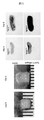
- FIG. 2 is a photograph of the three-dimensional structure on the 24th day under conditions 1 and 2.
- FIG. Both were fused smoothly, but under condition 1, they had no strength at all, and under condition 2, they were softer than rubber.
- the histopathological tissue was stained red for cartilage-specific proteoglycans (safranin 0 fast green staining) and immunostained for type 2 collagen.
- FIG. 3 is a photograph of the three-dimensional structure on the 24th day under condition 4.
- FIG. 3 is a photograph of the three-dimensional structure on the 24th day under condition 4.
- FIG. 3 When the culture period of the spheroids was extended, the induction of cartilage differentiation progressed too much, and it was observed that the spheroids did not stick to the needle of Tsurugiyama. In condition 4, fusion between spheroids was poor, a smooth curved surface was not formed, and a highly uneven structure was obtained. Even if this is transplanted into articular cartilage, it cannot be expected to function as a joint. Pathologically, the expression of safranin 0 is prominent, and the expression of type 2 collagen is also robust. The strength of the structure is very strong.
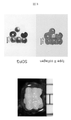
- FIG. 4 is a photograph of the three-dimensional structure on the 24th day under condition 3.
- FIG. 4 When the spheroids were printed on the 9th or 10th day after the start of spheroid formation, the spheroids were well fused together, and a structure having a firm strength was obtained.
- a histopathological section also confirmed the strong expression of proteoglycan and the expression of type 2 collagen, confirming that the conditions were effective for the regeneration of articular cartilage (Fig. 4).
Landscapes
- Health & Medical Sciences (AREA)
- Engineering & Computer Science (AREA)
- Life Sciences & Earth Sciences (AREA)
- Biomedical Technology (AREA)
- Genetics & Genomics (AREA)
- Chemical & Material Sciences (AREA)
- Organic Chemistry (AREA)
- Zoology (AREA)
- Biotechnology (AREA)
- Wood Science & Technology (AREA)
- Bioinformatics & Cheminformatics (AREA)
- Rheumatology (AREA)
- Microbiology (AREA)
- Cell Biology (AREA)
- Biochemistry (AREA)
- General Engineering & Computer Science (AREA)
- General Health & Medical Sciences (AREA)
- Materials Engineering (AREA)
- Manufacturing & Machinery (AREA)
- Micro-Organisms Or Cultivation Processes Thereof (AREA)
- Materials For Medical Uses (AREA)
Priority Applications (4)
| Application Number | Priority Date | Filing Date | Title |
|---|---|---|---|
| US18/573,536 US20240294877A1 (en) | 2021-06-29 | 2022-06-28 | METHOD FOR PRODUCING CARTILAGE CELL STRUCTURE DERIVED FROM iPS CELL |
| CN202280045456.2A CN117561328A (zh) | 2021-06-29 | 2022-06-28 | iPS细胞来源的软骨细胞结构体的制造方法 |
| EP22833339.9A EP4365281A4 (en) | 2021-06-29 | 2022-06-28 | METHOD FOR PRODUCING A CARTILAGE CELL STRUCTURE DERIVED FROM AN SPI CELL |
| JP2023532097A JP7716773B2 (ja) | 2021-06-29 | 2022-06-28 | iPS細胞由来軟骨細胞構造体の製造方法 |
Applications Claiming Priority (2)
| Application Number | Priority Date | Filing Date | Title |
|---|---|---|---|
| JP2021-107656 | 2021-06-29 | ||
| JP2021107656 | 2021-06-29 |
Publications (1)
| Publication Number | Publication Date |
|---|---|
| WO2023277195A1 true WO2023277195A1 (ja) | 2023-01-05 |
Family
ID=84692811
Family Applications (1)
| Application Number | Title | Priority Date | Filing Date |
|---|---|---|---|
| PCT/JP2022/026536 Ceased WO2023277195A1 (ja) | 2021-06-29 | 2022-06-28 | iPS細胞由来軟骨細胞構造体の製造方法 |
Country Status (5)
| Country | Link |
|---|---|
| US (1) | US20240294877A1 (https=) |
| EP (1) | EP4365281A4 (https=) |
| JP (1) | JP7716773B2 (https=) |
| CN (1) | CN117561328A (https=) |
| WO (1) | WO2023277195A1 (https=) |
Cited By (1)
| Publication number | Priority date | Publication date | Assignee | Title |
|---|---|---|---|---|
| WO2024213562A1 (en) * | 2023-04-14 | 2024-10-17 | Novadip Biosciences | Composition comprising neo-synthesized extracellular matrix derived from induced pluripotent stem cells (ipscs) |
Citations (3)
| Publication number | Priority date | Publication date | Assignee | Title |
|---|---|---|---|---|
| WO2007069666A1 (ja) | 2005-12-13 | 2007-06-21 | Kyoto University | 核初期化因子 |
| WO2008123614A1 (ja) | 2007-03-30 | 2008-10-16 | Kyushu University, National University Corporation | 細胞の立体構造体の製造方法 |
| JP2020202785A (ja) * | 2019-06-17 | 2020-12-24 | 株式会社サイフューズ | 細胞立体構造体及びその製造方法 |
-
2022
- 2022-06-28 EP EP22833339.9A patent/EP4365281A4/en active Pending
- 2022-06-28 JP JP2023532097A patent/JP7716773B2/ja active Active
- 2022-06-28 CN CN202280045456.2A patent/CN117561328A/zh active Pending
- 2022-06-28 WO PCT/JP2022/026536 patent/WO2023277195A1/ja not_active Ceased
- 2022-06-28 US US18/573,536 patent/US20240294877A1/en active Pending
Patent Citations (3)
| Publication number | Priority date | Publication date | Assignee | Title |
|---|---|---|---|---|
| WO2007069666A1 (ja) | 2005-12-13 | 2007-06-21 | Kyoto University | 核初期化因子 |
| WO2008123614A1 (ja) | 2007-03-30 | 2008-10-16 | Kyushu University, National University Corporation | 細胞の立体構造体の製造方法 |
| JP2020202785A (ja) * | 2019-06-17 | 2020-12-24 | 株式会社サイフューズ | 細胞立体構造体及びその製造方法 |
Non-Patent Citations (10)
| Title |
|---|
| "NCBI", Database accession no. NM _001113515 |
| DRIESSEN BRITTA J.H.; LOGIE COLIN; VONK LUCIENNE A.: "Cellular reprogramming for clinical cartilage repair", CELL BIOLOGY AND TOXICOLOGY., SPRINGER NETHERLANDS, NL, vol. 33, no. 4, 31 January 2017 (2017-01-31), NL , pages 329 - 349, XP036268693, ISSN: 0742-2091, DOI: 10.1007/s10565-017-9382-0 * |
| KATSUTSUGU UMEDA, ZHAO JIANGANG, SIMMONS PAUL, STANLEY EDOUARD, ELEFANTY ANDREW, NAKAYAMA NAOKI: "Human chondrogenic paraxial mesoderm, directed specification and prospective isolation from pluripotent stem cells", SCIENTIFIC REPORTS, vol. 2, pages 1 - 11, XP055278227, DOI: 10.1038/srep00455 * |
| MATSUDA S ET AL., NATURE, 2020 |
| MURATA DAIKI, FUJIMOTO RYOTA, NAKAYAMA KOICHI: "Osteochondral Regeneration Using Adipose Tissue-Derived Mesenchymal Stem Cells", INTERNATIONAL JOURNAL OF MOLECULAR SCIENCES, vol. 21, no. 10, pages 3589, XP093019664, DOI: 10.3390/ijms21103589 * |
| OKITA K ET AL., NATURE, 2007, pages 448 |
| PRETEMER Y ET AL., STEM CELL REPORTS, 2021 |
| See also references of EP4365281A4 |
| YAMANAKA S ET AL., CELL, vol. 126, 2006, pages 663 - 676 |
| YAMASHITA A ET AL., STEM CELL REPORTS, 2015 |
Cited By (2)
| Publication number | Priority date | Publication date | Assignee | Title |
|---|---|---|---|---|
| WO2024213562A1 (en) * | 2023-04-14 | 2024-10-17 | Novadip Biosciences | Composition comprising neo-synthesized extracellular matrix derived from induced pluripotent stem cells (ipscs) |
| BE1031503B1 (fr) * | 2023-04-14 | 2024-11-13 | Novadip Biosciences | Composition comprenant une matrice extracellulaire neo-synthesisée déivée de cellules stem pluripotentes induites (ipsc) |
Also Published As
| Publication number | Publication date |
|---|---|
| EP4365281A4 (en) | 2025-07-16 |
| EP4365281A1 (en) | 2024-05-08 |
| JPWO2023277195A1 (https=) | 2023-01-05 |
| JP7716773B2 (ja) | 2025-08-01 |
| US20240294877A1 (en) | 2024-09-05 |
| CN117561328A (zh) | 2024-02-13 |
Similar Documents
| Publication | Publication Date | Title |
|---|---|---|
| Ferretti et al. | Periosteum derived stem cells for regenerative medicine proposals: Boosting current knowledge | |
| T Brown et al. | Stem cell-based tissue engineering approaches for musculoskeletal regeneration | |
| Fukuta et al. | Derivation of mesenchymal stromal cells from pluripotent stem cells through a neural crest lineage using small molecule compounds with defined media | |
| Kadari et al. | Robust generation of cardiomyocytes from human iPS cells requires precise modulation of BMP and WNT signaling | |
| Illich et al. | Concise review: induced pluripotent stem cells and lineage reprogramming: prospects for bone regeneration | |
| Li et al. | Cells derived from murine induced pluripotent stem cells (iPSC) by treatment with members of TGF-beta family give rise to osteoblasts differentiation and form bone in vivo | |
| JP5700301B2 (ja) | 多能性幹細胞からの神経堤細胞群の分化誘導方法 | |
| Ma et al. | Pluripotent stem cells for Schwann cell engineering | |
| Fecek et al. | Chondrogenic derivatives of embryonic stem cells seeded into 3D polycaprolactone scaffolds generated cartilage tissue in vivo | |
| JP7211979B2 (ja) | ヒト誘導神経ボーダー幹細胞の生成と利用のための新規な方法 | |
| Srinivasan et al. | Comparative craniofacial bone regeneration capacities of mesenchymal stem cells derived from human neural crest stem cells and bone marrow | |
| Mahboudi et al. | Enhanced chondrogenesis differentiation of human induced pluripotent stem cells by MicroRNA-140 and transforming growth factor beta 3 (TGFβ3) | |
| JP7094567B2 (ja) | 神経堤細胞および交感神経細胞の製造方法 | |
| CN118475685A (zh) | 神经嵴细胞的培养方法及制造方法 | |
| Cho et al. | Dental-derived cells for regenerative medicine: stem cells, cell reprogramming, and transdifferentiation | |
| JPWO2019177118A1 (ja) | 多能性幹細胞から各種細胞への段階的製造方法 | |
| WO2023277195A1 (ja) | iPS細胞由来軟骨細胞構造体の製造方法 | |
| Hsiao et al. | Application of dental stem cells in three-dimensional tissue regeneration | |
| JP2010094062A (ja) | 間葉系幹細胞の多分化能維持用培地、培養方法、及び分化方法 | |
| Fu et al. | Potential replication of induced pluripotent stem cells for craniofacial reconstruction | |
| Mi et al. | Highly efficient multipotent differentiation of human periodontal ligament fibroblasts induced by combined BMP4 and hTERT gene transfer | |
| Mohammadi et al. | Stem Cell Research And Textbook 2:(Dental-derived stem cells, Induced pluripotent stem cells, hematopoietic stem cells, Embryonic, and Cancer Stem cells) | |
| WO2023133726A1 (en) | Human urine-derived induced presomitic mesoderm progenitor cells and uses thereof | |
| Ozdal Kurt et al. | Potential clinical use of differentiated cells from embryonic or mesencyhmal stem cells in orthopaedic problems | |
| JP6785516B2 (ja) | ヒト臍帯由来間葉系幹細胞から骨芽細胞の製造を目的としたアクチン重合阻害剤による分化誘導技術 |
Legal Events
| Date | Code | Title | Description |
|---|---|---|---|
| 121 | Ep: the epo has been informed by wipo that ep was designated in this application |
Ref document number: 22833339 Country of ref document: EP Kind code of ref document: A1 |
|
| WWE | Wipo information: entry into national phase |
Ref document number: 18573536 Country of ref document: US Ref document number: 2023532097 Country of ref document: JP |
|
| WWE | Wipo information: entry into national phase |
Ref document number: 202280045456.2 Country of ref document: CN |
|
| WWE | Wipo information: entry into national phase |
Ref document number: 2022833339 Country of ref document: EP |
|
| NENP | Non-entry into the national phase |
Ref country code: DE |
|
| ENP | Entry into the national phase |
Ref document number: 2022833339 Country of ref document: EP Effective date: 20240129 |