WO2023100910A1 - 糞便中エラスターゼ1を測定する方法 - Google Patents
糞便中エラスターゼ1を測定する方法 Download PDFInfo
- Publication number
- WO2023100910A1 WO2023100910A1 PCT/JP2022/044078 JP2022044078W WO2023100910A1 WO 2023100910 A1 WO2023100910 A1 WO 2023100910A1 JP 2022044078 W JP2022044078 W JP 2022044078W WO 2023100910 A1 WO2023100910 A1 WO 2023100910A1
- Authority
- WO
- WIPO (PCT)
- Prior art keywords
- α1at
- antitrypsin
- reagent
- complex
- sample
- Prior art date
Links
Images


Classifications
-
- G—PHYSICS
- G01—MEASURING; TESTING
- G01N—INVESTIGATING OR ANALYSING MATERIALS BY DETERMINING THEIR CHEMICAL OR PHYSICAL PROPERTIES
- G01N33/00—Investigating or analysing materials by specific methods not covered by groups G01N1/00 - G01N31/00
- G01N33/48—Biological material, e.g. blood, urine; Haemocytometers
- G01N33/50—Chemical analysis of biological material, e.g. blood, urine; Testing involving biospecific ligand binding methods; Immunological testing
- G01N33/53—Immunoassay; Biospecific binding assay; Materials therefor
- G01N33/543—Immunoassay; Biospecific binding assay; Materials therefor with an insoluble carrier for immobilising immunochemicals
Definitions
- the present invention relates to an immunoassay reagent and immunoassay method for fecal elastase 1, and a method for detecting pancreatic disease.
- pancreas Diseases of the pancreas are said to be difficult to diagnose even today, when various examination methods have been developed.
- the reason for this is that the pancreas is located in the deepest part of the abdominal cavity, so it is difficult to grasp the existence of the disease or the nature of the disease by means of classical diagnostic methods such as palpation, visual inspection, auscultation, and X-ray examination.
- the clinical symptoms of the disease are similar to those of other digestive system diseases, and there is no simple and reliable test method (Patent Document 1).
- pancreatic cancer has an extremely short 5-year survival rate, and early detection is said to be essential for improving the prognosis.
- the pancreatic enzyme elastase 1 (CELA1; chymotrypsin-like elastase family, member 1, hereinafter sometimes simply referred to as E1) is measured.
- E1 belongs to the serine protease family and is immunologically distinct from elastase, which is also present in leukocytes, platelets, and spleen.
- E1 is secreted from the pancreas into the duodenum along with other digestive enzymes, but is leaked into the blood due to pancreatic duct stenosis or pancreatitis.
- ⁇ 1-antitrypsin (hereinafter sometimes simply referred to as ⁇ 1AT), and measurement of blood concentration is clinically useful.
- ⁇ 1AT ⁇ 1-antitrypsin
- it is useful as an index for diagnosis or follow-up of pancreatic diseases, since it shows abnormally high values at a relatively early stage and at a high frequency, reflecting pancreatitis associated with pancreatic cancer (especially pancreatic head) (Patent Document 2).
- Patent Document 2 since it is an invasive test, it is difficult to say that it is actively implemented, and there are not many reports on E1 in blood.
- E1 When E1 is used for diagnostic purposes in human fecal samples, it has been reported that its concentration is 5 to 6 times higher than in pancreatic juice. At present, it cannot be said that the optimal concentration in feces is fully understood.
- a stool sample usually contains water, food residue, intestinal mucosal cells, and intestinal bacteria. It has a unique property of being diverse in shape, pH, and the like. For this reason, there are various influences and causes on measurement results, and in tests using stool samples, handling such as suppression of non-specific reactions requires labor that is different from that for normal blood samples.
- Kampanis et al. discloses the measurement of elastase 1 in human feces, but it is difficult to say that it is suitable for practical use when performing a large number of specimen tests because of the complicated handling (non-patent Reference 1).
- the wet extraction method is very labor intensive, as wet or loose fecal samples must be dried, weighed, and finally diluted with the extraction solution. can give rise to Furthermore, it is difficult to unify the application of the reference concentration among the extraction methods, which is one of the factors that make accurate measurement difficult.
- Fecal occult blood means that blood is contained in feces, but it may not be measured accurately due to the influence of bleeding in the intestine, anal fissure, menstrual blood, and the like. In general, the fecal occult blood test has a positive rate of about 5-10%. must consider.
- An object of the present invention is to analyze E1 present in human fecal samples in a simple manner so as to be able to handle a large amount of specimens without requiring special facilities or equipment, and to be able to analyze in a short time so as to be able to respond to emergency tests.
- Another object of the present invention is to provide an E1 immunoanalytical reagent and an immunoanalytical method that enable quantitative analysis over a wide range from low concentration regions to high concentration regions.
- the inventors have made intensive studies to solve the above problems. As a result, the present inventors have found that E1 in human fecal samples can be accurately measured in a state of forming a complex with E1 using ⁇ 1AT, leading to the completion of the present invention.
- the present invention is based on an antigen-antibody reaction using a monoclonal antibody specific to the E1- ⁇ 1AT complex in a state in which E1 in a human fecal sample is stabilized by forming a complex with its inhibitor ⁇ 1AT. It relates to an immunoassay method for analysis. Furthermore, the present invention relates to a method for detecting pancreatic disease by analyzing E1 by the immunoassay method.
- the present invention provides: [1] A method for measuring pancreatic elastase 1 present in a fecal sample, comprising: (a) adding ⁇ 1-antitrypsin to a fecal sample obtained from a subject; (b) incubating the stool sample to react pancreatic elastase 1 with ⁇ 1-antitrypsin to form a complex; (c) incubating a solution containing a carrier on which an immunological partner that recognizes the pancreatic elastase 1- ⁇ 1-antitrypsin complex is immobilized with the stool sample; (d) analyzing changes caused by the reaction of the pancreatic elastase 1- ⁇ 1-antitrypsin complex with an immunological partner; method including.
- [2] The method of [1], wherein the step (a) is performed in a pretreatment step for extracting a stool sample.
- [5] The method according to any one of [1] to [4], wherein the amount of ⁇ 1-antitrypsin contained in the reaction solution to be subjected to the reaction step in step (b) is 100 ng or more.
- a method for assisting detection of pancreatic diseases comprising analyzing pancreatic elastase 1 by the measuring method of any one of [1] to [5].
- the analysis performed in step (d) includes chemiluminescence immunoassay, electrochemiluminescence immunoassay, fluorescence immunoassay, radioimmunoassay, immunochromatography, western blotting, latex agglutination, immunoassay, The method according to any one of [1] to [6], which is any of turbidimetric methods.
- a reagent for immunoassay comprising a solid-phase carrier carrying an immunological partner that recognizes the pancreatic elastase 1- ⁇ 1-antitrypsin complex, and ⁇ 1-antitrypsin.
- the immunoassay reagent of [8] wherein 100 ng of ⁇ 1-antitrypsin is contained in the reaction solution to be subjected to the reaction step.
- a pretreatment extract containing ⁇ 1-antitrypsin which is used in the pretreatment step of a stool sample to be subjected to a reagent for measuring pancreatic elastase 1 or pancreatic elastase 1- ⁇ 1-antitrypsin.
- E1 present in human fecal samples which is the method of the present invention, it is possible to easily assist in the detection of pancreatic diseases.
- Accurate measurement of E1 in human stool samples enables its use as a marker for monitoring pancreatic exocrine function, and is expected to be useful for treatment policy determination.
- FIG. 10 is a diagram showing E1 concentration measurement results with respect to ⁇ 1AT solution addition concentration.
- FIG. 10 is a diagram showing the recovery rate for each ⁇ 1AT solution concentration with respect to the theoretical value for E1 sample preparation.
- FIG. 10 is a diagram comparing measured values when ⁇ 1AT is added using a plurality of measurement reagents.
- the present invention relates to an immunoassay method for measuring E1 present in a human fecal sample in a state of being complexed with ⁇ 1AT.
- the present invention also relates to immunoassay reagents for measuring the E1- ⁇ 1AT complex present in human fecal samples.
- the present invention includes: A method for detecting pancreatic disease by measuring pancreatic elastase 1 present in a fecal sample; A method to aid in the detection of pancreatic disease by measuring pancreatic elastase 1 present in fecal samples; A method of measuring pancreatic elastase 1 present in a fecal sample to detect pancreatic disease; A method for the in vitro detection of pancreatic disease that measures pancreatic elastase 1 present in fecal samples; use of an immunological partner that recognizes the pancreatic elastase 1- ⁇ 1-antitrypsin complex in the manufacture of a kit for the detection of pancreatic diseases; Methods of measuring pancreatic elastase 1 present in fecal samples are included to provide the information necessary for the detection of pancreatic disease.
- Human-derived fecal samples, intestinal washings, etc. can be used as samples for the measurement of the present invention.
- the specific form of the stool sample is not particularly limited as long as it is a sample derived from stool. For example, it can be used regardless of its shape, such as hardness (hard stool, normal stool, soft stool, diarrheal stool, watery stool, etc.) and water content.
- Colon cleansing fluid means that collected through the intestinal lumen, and orally ingested colon cleansing agents also include oral cleansing fluid collected through the intestinal lumen.
- the intestinal lavage fluid may be collected after being excreted from the subject, or collected in the rectum of the subject just before excretion.
- the term "stool sample” is used in the sense of "sample” used for measurement (eg, claim 1), it includes not only the stool sample but also intestinal washings and the like.
- Fecal samples can be used by adopting general methods such as extraction of E1 and removal of insoluble fractions as pretreatment.
- Physiological saline can be used for the extraction solution used in the extraction operation, but buffers such as Good's buffers and phosphates, and solutions containing proteins such as BSA (Bovine Serum Albumin) and surfactants can also be used. may be used.
- BSA Bovine Serum Albumin
- surfactants can also be used.
- a person skilled in the art can appropriately set and use various conditions of the extraction solution such as the concentration/pH of the buffer solution, the concentrations of the surfactant and BSA, and the like.
- the extract is added, the stool sample is sufficiently dispersed, and E1 is eluted into the extract.
- a homogenator or a vortex mixer may be used.
- the stool sample may be left to stand for about 30 minutes to 1 hour after being dispersed in the extract.
- Centrifugation or filter filtration can be used to remove the insoluble fraction.
- Conditions for centrifugation and filter membranes and the like used for filtration are not particularly limited as long as they can be used for pretreatment, and can be used.
- carriers constituting filters include polypropylene (PP), polyvinylidene fluoride (PVDF), glass fiber (GF), polyethersulfone (PES), nylon (NY), polytetrafluoroethylene (PTFE), regenerated Cellulose (RC), cellulose acetate (CA), methacrylate butadiene styrene (MBS) may be included.
- PP polypropylene
- PVDF polyvinylidene fluoride
- GF glass fiber
- PES polyethersulfone
- nylon NY
- PTFE polytetrafluoroethylene
- RC regenerated Cellulose
- CA cellulose acetate
- MFS methacrylate butadiene styrene
- MFS methacrylate but
- the above operations can also be performed using a general stool collection kit.
- a stool collection kit As an example of a stool collection kit, OC-Hemocatch (registered trademark) S (manufactured by Eiken Chemical Co., Ltd.), which is a stool collection kit for fecal occult blood, may be used.
- ⁇ 1AT which will be described later, may be added to the extraction solution used for these extraction operations.
- Methods for immunologically detecting proteins include, for example, enzyme immunoassay (ELISA method), chemiluminescence immunoassay, electrochemiluminescence immunoassay, fluorescence immunoassay, radioimmunoassay, immunochromatography Any method can be used as long as it is an immunoassay method using a labeled antibody such as , Western blotting method, latex agglutination method, immunoturbidimetric method, and the like.
- immunological partner used in the present invention means a partner that immunologically specifically binds to a substance to be measured, such as various proteins, polysaccharides, lipids, nucleic acids, haptens, complexes or fragments thereof, etc.
- an immunological substance ie, antigen or antibody
- the antibody to be used may be a monoclonal antibody, a polyclonal antibody, or an enzyme-treated fragment thereof.
- An antibody fragment is preferably a functional fragment containing the antigen-binding region of an antibody or its variable region, and examples thereof include F(ab') 2 , Fab', and Fab.
- F(ab') 2 and Fab' are produced by treating immunoglobulin with a protease (eg, pepsin or papain), and are present between two H chains in the hinge region. It is an antibody fragment produced by digestion before and after the disulfide bond that binds.
- a protease eg, pepsin or papain
- multiple types of immunological partners may be used in combination.
- the antibody to be used may be an antibody capable of recognizing the E1- ⁇ 1AT complex, which specifically detects E1. It may be an anti-E1 antibody that recognizes, or an anti-E1- ⁇ 1AT complex antibody that specifically recognizes the complex with E1- ⁇ 1AT. Any anti-E1 antibody can be appropriately selected and used as long as it does not recognize the E1- ⁇ 1AT complex.
- the number of antibodies used may be only one type of antibody that specifically recognizes E1, a first antibody that specifically recognizes E1, a second antibody that recognizes E1 that is different from the first antibody may be a combination of antibodies.
- only one type of antibody that specifically recognizes the E1- ⁇ 1AT complex may be used, or a first antibody that specifically recognizes the E1- ⁇ 1AT complex, and an E1- ⁇ 1AT that is different from the first antibody.
- It may be a combination of a second antibody that recognizes the complex.
- these may be used in combination (hereinafter, they may be collectively referred to as "anti-E1 antibody").
- anti-E1 antibody There is no particular limitation as long as the anti-E1 antibodies have different specific binding sites.
- An anti-E1 antibody can be prepared, for example, using a polypeptide containing part or all of the amino acid sequence of E1 or E1- ⁇ 1AT complex as an immunogen.
- the antigen polypeptide may be a synthetic polypeptide chemically synthesized according to a known method, or one produced by genetic recombination or the like.
- the antibody used in the present invention can be used as an immobilized antibody carried on an insoluble carrier such as a solid phase carrier, or as a labeled antibody labeled with a labeling substance.
- Immobilized antibody refers to an antibody that is supported by physical adsorption or chemical bonding on an insoluble carrier. These immobilized antibodies can be used to detect or quantify analytes contained in samples.
- insoluble carriers that can be used to support the antibody include latex, rubber, polyethylene, polypropylene, polystyrene, styrene-butadiene copolymer, polyvinyl chloride, polyvinyl acetate, polyacrylamide, polymethacrylate, styrene-methacrylate copolymer.
- polymer polyglycidyl methacrylate, acrolein-ethylene glycol dimethacrylate copolymer, polyvinylidene difluoride (PVDF), silicone and other polymeric materials; agarose; gelatin; Examples include inorganic materials. You may combine these 1 type(s) or 2 or more types.
- the method of the present invention can be carried out, for example, by supporting an anti-E1 antibody on latex particles.
- the latex particles that can be used in that case are not particularly limited as long as they are latex particles that can be used for ordinary immunoassay reagents. etc. can be mentioned.
- the average particle size of the latex particles can be appropriately selected depending on the detection concentration of the object to be measured or the measuring equipment. For example, particles having a particle size of 0.05 to 0.5 ⁇ m can be used. By using latex particles with different particle sizes, it is possible to accurately analyze from low values to high values, which is preferable.
- the average particle size of latex particles in the present invention means a value measured with an electron microscope.
- anti-E1 antibodies are immobilized on latex particles, for example, two types of latex particles with different particle sizes carrying two types of anti-E1 antibodies with different specificities for E1 or E1- ⁇ 1AT complexes, preferably (1) a first latex particle bearing a first anti-E1 antibody against an E1 or E1- ⁇ 1AT complex; and (2) an E1 or E1- ⁇ 1AT complex having a specificity different from said first anti-E1 antibody. may include at least second latex particles having a different particle size than the first latex particles, carrying a second anti-E1 antibody against.
- Enzyme immunoassay (ELISA), chemiluminescence immunoassay, electrochemiluminescence immunoassay, fluorescence immunoassay, radioimmunoassay, immunochromatography and other immunoassays using labeled antibodies, labeling substances is preferably used.
- the labeling substance is not particularly limited as long as it is a labeling substance that can be used in ordinary immunoassays, and examples thereof include enzymes, fluorescent substances, radioisotopes, insoluble particulate substances, and the like.
- the labeling enzymes include alkaline phosphatase, peroxidase, glucose oxidase, tyrosinase, acid phosphatase and the like.
- Fluorescent substances include fluorescein isothiocyanate (FITC), green fluorescent protein (GFP), luciferin, and the like.
- Radioisotopes include 125 I, 14 C, 32 P, and the like.
- the labeling substance when the labeling substance is an enzyme, the labeling substance can be measured by performing a luminescence, fluorescence, or color development reaction using a substrate for the enzyme.
- the substrate when the enzyme is alkaline phosphatase, the substrate is CDP-star® (2-chloro-5-(4-methoxyspiro ⁇ 1,2-dioxetane-3,2′-(5′-chloro )-tricyclo[3.3.1.13,7]decane ⁇ -4-yl)-1-phenylphosphate disodium), CSPD® (3-(4-methoxyspiro ⁇ 1,2-dioxetane -3,2-(5′-chloro)tricyclo[3.3.1.13,7]decan ⁇ -4-yl)phenyl phosphate disodium), AMPPD® (adamantylmethoxyphenylphosphoryldioxycetane) ), chemiluminescent substrates such as APS-5; fluorescent substrates such as 4-
- the immunoassay method of the present invention can be carried out using an immunoassay reagent composed of one or two or more liquids.
- the reagent for carrying out the present invention consists of one liquid, it can consist of, for example, a reaction reagent containing an insoluble carrier carrying at least an immunological partner for forming an immunological complex.
- a sample is reacted with a reagent and a signal is detected according to known methods.
- the ⁇ 1AT used in the present invention may be mixed with the sample finally, and may be contained in the pretreatment reagent used in the pretreatment step, or may be contained in the stabilizing reagent. , may be contained in the reagent containing the above-mentioned insoluble carrier. In the present invention, it was found that it is important to add ⁇ 1AT in a certain concentration range to the sample or reagent in order to accurately measure E1 present in the stool sample.
- the ⁇ 1AT used in the present invention may be added to the sample before the sample and the reagent react, or may be supplied in the state of being added to the reagent. It is sufficient if it is contained in the reaction solution in which the E1- ⁇ 1AT complex, which is the substance to be measured, undergoes an immune reaction. and a reagent for reacting with the E1- ⁇ 1AT complex, which is the substance to be measured, and the immobilized anti-E1 antibody, and the labeled antibody and the measurement sample, which are mixed and reacted, and the labeled antibody and the E1- ⁇ 1AT complex, which is the substance to be measured. It may be added to either or each of the reagents that react with the body.
- the ⁇ 1AT to be used may be one that forms a complex with human pancreatic elastase 1, and preferably human-derived ⁇ 1AT is used.
- Human-derived ⁇ 1AT may be purified ⁇ 1AT present in the blood, or may be recombinantly expressed using cultured cells or the like, and may be appropriately selected and used by those skilled in the art. can do.
- the amount of ⁇ 1AT added to form a complex with E1 is preferably an amount sufficient to form a complex with E1.
- the lower limit to be added is 20 ⁇ g or more, and the upper limit may be appropriately set according to the amount of E1 contained in the sample.
- the lower limit is preferably 41 ⁇ g or more, more preferably 49 ⁇ g or more.
- the upper limit is preferably 975 ⁇ g or less.
- the said lower limit and upper limit can be combined suitably.
- the lower limit of the amount of ⁇ 1AT contained in the reaction solution may be appropriately set at a concentration at which there is a sufficient amount to form a complex with E1 and the reactivity is confirmed compared to when ⁇ 1AT is not added, and is 100 ng or more. Just do it. For example, 514 ng or more is preferable, and 609 ng or more is more preferable.
- the upper limit of the amount of ⁇ 1AT contained in the reaction solution is preferably 12188 ng or less, more preferably 6094 ng or less, and even more preferably 5143 ng or less. A person skilled in the art can determine the optimum addition amount for each measurement reagent by considering the E1 concentration assumed to be contained in the stool sample. In addition, the said lower limit and upper limit can be combined suitably.
- the amount to be added may be determined by the weight ratio of E1 and ⁇ 1AT according to the measurable range of the reagent used for measurement. In that case, for example, by adding ⁇ 1AT in a weight that is 6 to 2400 times the weight of E1, it becomes possible to sufficiently form a complex between E1 and ⁇ 1AT, and E1 measurement in a fecal sample can be performed accurately. can do.
- the reagent of the present invention when the reagent of the present invention is composed of two or more liquids, it can be composed of, for example, a stabilizing reagent and a reactive reagent containing at least an insoluble carrier carrying an antibody or antigen for forming an immunological complex.
- the stabilizing reagent is a reagent that dilutes the sample to an appropriate concentration or performs pretreatment, and can be prepared according to a known method. The sample is reacted with a stabilizing reagent, followed by a reaction reagent, and the signal is detected according to known methods.
- ⁇ 1AT used in the present invention is preferably added to the stabilizing reagent and/or the reaction reagent before the reaction between the sample and the reaction reagent, but is preferably added to the stabilizing reagent and/or the reaction reagent. can be supplied as is.
- E1- ⁇ 1AT complex It is known that nearly 90% of the E1 present in the blood forms a complex with ⁇ 1AT, and the measurement of the E1- ⁇ 1AT complex is considered to be the measurement of E1, so it can be used for clinical testing.
- a serine protease inhibitor belonging to the serpin superfamily, to which ⁇ 1AT belongs is covalently linked to the serine residue in the active center of the protease molecule, and is strongly attracted to the serpin molecule following loop insertion, thereby opening the structure near the active center. It is said that it breaks down and dissociation of proteases due to hydrolysis does not occur.
- E1 and ⁇ 1AT are linked by a covalent bond and are not easily dissociated. It was thought that E1 and ⁇ 1AT were in a state of forming a complex even in a stool sample. there were.
- the original data or statistically processed data for calculating the determination threshold value includes the E1 concentration in a stool sample and various diseases
- a determination threshold value (cutoff value) calculated from statistical data or the like showing a correlation with the may be appropriately used.
- a person skilled in the art can appropriately set the cut-off value and use it in view of its relevance to pancreatic disease.
- an ROC curve Receiveiver Operating Characteristic Curve
- the measured E1 concentration can be used as a value for risk assessment. It can be used as an index for determining whether pancreatic inflammation, pancreatic cancer, or the like is improved by treatment such as medication. For example, if the E1 concentration remains high or increases, it suggests the need to reconsider the treatment strategy.
- the reagent of the present invention includes various additives that can be added to the latex reagent, such as buffers, aggregation promoters (e.g., water-soluble polymers such as polyethylene glycol), non-specific Inhibitors (eg, alkali metal salts, sugars, etc.), or proteins [eg, bovine serum albumin (BSA)], etc. may be further contained.
- aggregation promoters e.g., water-soluble polymers such as polyethylene glycol
- non-specific Inhibitors eg, alkali metal salts, sugars, etc.
- proteins eg, bovine serum albumin (BSA)], etc.
- the buffer solution a buffer solution having a buffering capacity at pH 6 to 8.5 is preferable.
- the pH 6-8.5 buffer is a conventionally known buffer, and examples thereof include Tris buffer, phosphate buffer, Good's buffer and the like.
- the Tris concentration in the Tris buffer is adjusted to the predetermined Tris concentration described below in a system in which the latex agglutination reaction is performed when the reagent is used.
- the Tris concentration in the Tris buffer is adjusted to the predetermined Tris concentration described below in a system in which the latex agglutination reaction is performed when the reagent is used.
- it is not particularly limited as long as it is a concentration that can achieve, it is preferably 0.1 to 0.5 mol / L.
- the Tris concentration in the system in which the latex agglutination reaction is carried out is not particularly limited as long as it is a concentration that can suppress the self-agglutination reaction of the latex particles. It can be selected as appropriate depending on the concentration of the substance.
- the Tris concentration in the system in which the latex agglutination reaction is performed is preferably 0.1 to 0.5 mol/L, more preferably 0.2 to 0.3 mol/L. If it is less than 0.1 mol/L, the latex particles may undergo a self-agglutination reaction, and if it exceeds 0.5 mol/L, the antigen-antibody reaction may be suppressed, resulting in poor detection sensitivity.
- the lower and upper limits of Tris concentration can be appropriately combined, for example, 0.1 to 0.3 mol/L.
- the pH of the buffer solution is preferably 6 to 8.5. If the pH is out of this range, the latex particles may self-aggregate, or problems may occur in terms of measurement accuracy.
- each latex particle carrying an antibody a pH 6-8.5 buffer solution, a pH 6-8.5 buffer solution, a pH 6-8.5 buffer solution
- the condition of each anti-E1 antibody-carrying latex particle and pH 6 to 8.5 buffer solution in the reagent is not particularly limited as long as it can be contacted with the test sample. That is, in this case, the form of the reagent of the present invention is not particularly limited, and for example, a one-liquid reagent containing both anti-E1 antibody-supporting latex particles and a buffer solution of pH 6 to 8.5.
- it can be a two-liquid system reagent composed of a first reagent containing each anti-E1 antibody-supported latex particle and a second reagent that is a buffer solution of pH 6 to 8.5.
- the antibody-carrying latex particles are brought into contact with the test sample, preferably under conditions of pH 6 to 8.5, to induce an antigen-antibody reaction and latex agglutination reaction caused thereby. and analyzing the degree of aggregation, E1 in the test sample can be analyzed.
- each anti-E1 antibody-supported latex particle and pH 6-8. 5 buffer and the test sample the antigen-antibody reaction does not proceed in the absence of a pH 6-8.5 buffer (that is, each anti-E1 antibody-carrying latex particle and the test sample).
- the contact order is not particularly limited as long as the sample is not contacted first.
- each anti-E1 antibody-carrying latex particle and a pH 6-8.5 buffer solution can be brought into contact in advance, and the mixture can be brought into contact with the test sample. It is also possible to contact the anti-E1 antibody-carrying latex particles with each of the anti-E1 antibody-carrying latex particles after contacting them with the buffer solution of .5 in advance.
- the conditions for the antigen-antibody reaction in the measurement method of the present invention can be the same as the conditions for carrying out ordinary immunological latex turbidimetric analysis methods.
- the pH of the reaction is preferably carried out at 6-8.5.
- the reaction temperature is preferably 0 to 50°C, more preferably 20 to 40°C.
- the reaction time can be determined appropriately, and for example, the measurement can be completed in 10 to 15 minutes with a general-purpose automatic analyzer.
- the above lower limit and upper limit of the reaction temperature can be appropriately combined, for example, 0 to 40°C.
- the degree of aggregation caused by the antigen-antibody reaction can be analyzed by a known analysis method, such as an optical analysis method.
- the optical analysis method include a method of irradiating a reaction solution with light and analyzing scattered light or transmitted light. More specifically, scattered light intensity, absorbance, or transmitted light intensity is measured. Analysis can be performed using measuring optics. A preferred measurement wavelength is 300-800 nm.
- an increase or decrease in scattered light intensity, absorbance, or transmitted light intensity is measured by selecting the size and/or concentration of the latex particles to be used and setting the reaction time according to known methods. It can be implemented by Moreover, it is also possible to use these methods together.
- pancreatic disease detection method serum or plasma is used as a test sample, and pancreatic disease (particularly acute pancreatitis) is detected (diagnosed) by analyzing E1 in a stool sample by the measuring method of the present invention. It can be performed.
- Iatro IRE1II (hereinafter referred to as IRE1, manufactured by LSI Rulece) was used as a reagent for measuring E1 in fecal samples, and it was confirmed whether a measured value of Iatro IRE1II could be obtained by adding ⁇ 1-antitrypsin to the stool extraction solution.
- ⁇ 1AT derived from human plasma was dissolved in Tris buffer to prepare ⁇ 1AT solutions with concentrations of 0, 16, 130, 325, 813, 3250, and 6500 ⁇ g/mL in terms of the extinction coefficient of ⁇ 1AT. bottom.
- stool extraction solution 150 ⁇ L of the human stool extraction solution (hereinafter referred to as stool extraction solution)
- 150 ⁇ L of the prepared ⁇ 1AT solution at each concentration was added to prepare an ⁇ 1AT-added stool extraction solution.
- the ⁇ 1AT-added stool extract solution was further diluted 80-fold to bring it into the measurement range of the IRE1 reagent.
- DD dimer diluent manufactured by LSI stipulatece
- H7180 Hitachi High-Tech
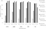
- Figure 1 shows the measurement results of the IRE1 reagent against the concentration of the ⁇ 1AT solution added to the fecal extract solution. It was confirmed that the addition of ⁇ 1AT to the stool extract solution increased the measured value of the IRE1 reagent, and the maximum value was obtained when the concentration of the ⁇ 1AT solution was 325 to 3250 ⁇ g/mL. At the maximum concentration of 6500 ⁇ g/mL among the experimental conditions, a 7.5% decrease in the measured value was observed compared to the measured value of 3250 ⁇ g/mL, but the recovery rate was sufficient for use in E1 measurement. confirmed.
- the amount of each ⁇ 1AT added to the sample subjected to the measurement step at this time was 49, 122, 488, and 975 ⁇ g, but since it was subjected to measurement after being diluted 80 times, it was substantially 609, 1523, 6094, 12188 ng. From this, it was confirmed that the measurement value of the IRE1 reagent can be obtained from the stool sample by adding ⁇ 1AT.
- Example 2 Examination of ⁇ 1AT solution concentration
- the optimum concentration of ⁇ 1AT to be added was examined in the measurement range of IRE1 reagent from 80 to 4000 ng/dL, and the ⁇ 1AT solution concentration to be used in future experiments was determined.
- Elastase 1 purified from human pancreatic juice was used in a phosphate buffer to prepare E1 samples having concentrations of 14, 57, 142, 285 and 571 ng/mL in terms of the absorption coefficient of E1.
- a phosphate buffer was also used for ⁇ 1AT to prepare ⁇ 1AT solutions having concentrations of 171, 875, 3429, 8571, 17143 and 34268 ng/mL in terms of the absorption coefficient of ⁇ 1AT.
- An E1- ⁇ 1AT mixed solution was prepared by adding 150 ⁇ L of the ⁇ 1AT solution to 150 ⁇ L of the E1 sample. H7180 was loaded with the IRE1 reagent, and the E1- ⁇ 1AT mixed solution was measured at a multiplicity of 3. The measured value of the mixed solution of 571 ng/mL E1 sample 1 and 34268 ng/mL ⁇ 1AT solution 1 was taken as the prepared theoretical value of E1 sample 1 (3778 ng/dL). The theoretical preparation value for each E1 sample was set from the dilution ratio from E1 sample 1.
- Figure 2 shows the recovery rate for each ⁇ 1AT solution concentration relative to the theoretical value for each E1 sample preparation.
- ⁇ 1AT solutions 1-4 with ⁇ 1AT concentrations of 3429-34286 ng/mL yielded greater than 90% recovery.
- the ⁇ 1AT concentration of 3429 to 17143 ng/mL is favorable as a recovery rate relative to the theoretical value of preparation.
- the amounts of ⁇ 1AT added when the recovery of E1 was 70% or more were 514, 1286, 2571 and 5143 ng, respectively. Therefore, in subsequent experiments, 8600 ng/mL was tentatively used as the concentration of the ⁇ 1AT solution, and 1290 ng was included in the sample subjected to measurement.
- Example 3 Comparative experiment of IRE1 reagent and fPELA reagent
- the reactivity of the existing fecal elastase 1 LTX reagent was compared with the evaluation system in which ⁇ 1AT was added using the IRE1 reagent.
- fPELA turbo hereinafter referred to as fPELA, manufactured by BUHLMANN
- E1 samples having concentrations of 14, 57, 142, 285 and 571 ng/mL in terms of the extinction coefficient of E1 were prepared.
- a phosphate buffer was also used for ⁇ 1AT to prepare an ⁇ 1AT solution having a concentration of 8600 ng/mL in terms of the extinction coefficient of ⁇ 1AT.
- 200 ⁇ L of each prepared E1 solution and 200 ⁇ L of the ⁇ 1AT solution were mixed to prepare an E1- ⁇ 1AT mixed solution.
- Each E1 sample, the E1- ⁇ 1AT mixed solution, the IRE1 reagent standard, and the IRE1 reagent control were measured at a multiplicity of 2 using the fPELA reagent mounted on the automatic analyzer cobas c501 (hereinafter c501, manufactured by Roche). .
- the E1- ⁇ 1AT mixed solution was measured at a multiplicity of 2 using the IRE1 reagent loaded on the H7180.
- FIG. 3 plots the measured values of each E1 sample and E1- ⁇ 1AT mixed solution, IRE1 reagent standard, and IRE1 reagent control fPELA reagent against the IRE1 value.
- Good concentration-dependent linearity was obtained between the measured value of the E1- ⁇ 1AT mixed solution with the IRE1 reagent and the value when the E1 sample was measured with the fPELA reagent.
- the fPELA reagent when the E1- ⁇ 1AT mixed solution was measured, the measured value was lower than when the E1 solution was measured as it was.
- the measured value of the E1- ⁇ 1AT mixed solution of the fPELA reagent was close to the measured value of the IRE1 reagent standard and control.
- the IRE1 reagent standard and control contained E1 as a complex with ⁇ 1AT, indicating that the fPELA reagent was affected by ⁇ 1AT.
- E1 a complex with ⁇ 1AT
- ⁇ 1AT in the blood may be mixed and affect the measurement value.
- the measured values were not affected even in specimens with fecal occult blood. Since the measured value of the IRE1 reagent that recognizes the E1- ⁇ 1AT complex correlates with the measured value of the fPELA reagent that recognizes and measures E1, the results obtained by adding ⁇ 1AT to the fecal sample and measuring , was confirmed to be reliable data.
- the measuring reagent and measuring method of the present invention it becomes possible to measure the concentration of E1 in a stool sample without being affected by measurement-interfering substances, and it can be used as an aid in diagnosing pancreatic diseases.
- the E1 concentration in fecal samples from healthy subjects can be accurately determined, it can be used not only for the diagnosis of pancreatic diseases, but also for the selection of appropriate treatment methods, monitoring of therapeutic effects, and prognosis prediction, thereby providing highly reliable measurements. By providing value, it is possible to provide extremely useful information for medical care.
Landscapes
- Health & Medical Sciences (AREA)
- Life Sciences & Earth Sciences (AREA)
- Immunology (AREA)
- Engineering & Computer Science (AREA)
- Molecular Biology (AREA)
- Biomedical Technology (AREA)
- Chemical & Material Sciences (AREA)
- Hematology (AREA)
- Urology & Nephrology (AREA)
- Biotechnology (AREA)
- Microbiology (AREA)
- Cell Biology (AREA)
- Food Science & Technology (AREA)
- Medicinal Chemistry (AREA)
- Physics & Mathematics (AREA)
- Analytical Chemistry (AREA)
- Biochemistry (AREA)
- General Health & Medical Sciences (AREA)
- General Physics & Mathematics (AREA)
- Pathology (AREA)
- Investigating Or Analysing Biological Materials (AREA)
Abstract
ヒト糞便試料中に存在するエラスターゼ1(E1)を特別な施設や器材を必要とすることなく大量の検体に対応できるよう簡便で、緊急検査に対応し得るよう短時間で分析可能な、かつ、低濃度領域から高濃度領域まで広範囲にわたって定量的に分析可能な、エラスターゼ1の免疫分析用試薬及び免疫分析方法を提供する。前記分析方法は、(a)被検者から得た糞便試料にα1-アンチトリプシン(α1AT)を添加する工程、(b)前記糞便試料をインキュベートし、E1とα1ATを反応させ複合体を形成させる工程、(c)前記EI-α1AT複合体を認識する免疫学的パートナーが固相された担体を含む溶液を、前記糞便試料と共にインキュベートする工程、(d)E1-α1AT複合体と免疫学的パートナーとの反応により生じた変化を分析する工程を含む。
Description
本発明は、糞便中エラスターゼ1の免疫分析用試薬及び免疫分析方法並びに膵疾患の検出方法に関する。
膵臓の疾患は各種検査法が発達した現在でも診断の困難な疾患であるといわれている。その理由は、膵臓が腹腔内の最深奥に位置するため、触診、視診、聴診等の古典的診断方法及びX線検査等では疾患の存在又は疾患の性質の把握は困難であること、また膵臓疾患の臨床症状が他の消化器系疾患と類似していること、及び簡便で確実な検査法が無いことなどである(特許文献1)。特に膵癌は5年生存率が極めて短く、予後の改善には早期発見が不可欠と言われている。
膵疾患の診断に使用するバイオマーカーとして、膵酵素であるエラスターゼ1(CELA1;Chymotrypsin-like elastase family,member 1、以下、単にE1と称することがある)が測定されている。E1は、セリンプロテアーゼ族に属し、白血球、血小板、及び脾臓などにも存在するエラスターゼとは免疫学的に区別される。E1は他の消化酵素と並行して、膵臓から十二指腸へと分泌されるが、膵管の狭窄や膵炎によって血中に漏出される。血中では、そのほとんどがα1-アンチトリプシン(以下、単にα1ATと称することがある)と結合しており、血中濃度の測定が臨床的に有用とされる。特に、膵臓癌(特に膵頭部)に伴う膵炎を反映して、比較的早期から高頻度に異常高値を示すことから、膵臓疾患の診断の指標又は経過観察に有用である(特許文献2)。しかし、侵襲性のある検査であることから積極的に実施されているとは言い難く、血中E1についての報告は多くはない。
E1分泌と、膵臓のリパーゼ、アミラーゼ及びトリプシン分泌との間には、線形相関があり、さらに、十二指腸内へのE1分泌は、糞便中のE1濃度と相関を示す(特許文献3)。膵外分泌機能障害は、E1の分泌低下に関係しており、その分泌低下の結果、便中の酵素濃度が低下すると考えられるため、E1は膵外分泌機能不全の診断、真性糖尿病、嚢胞性線維症及び慢性膵炎における膵外分泌機能の監視のためのマーカーとしての使用が報告されている。
ヒト糞便試料中におけるE1を診断用途として使用する場合、その濃度は、膵液中よりも5~6倍高いといった報告があるものの、その詳細な実態や膵疾患との関連については、血中濃度と糞便中濃度のいずれが最適であるかなど十分に理解されているとは言えないのが現状である。
糞便試料中のE1を正確に測定しその実態が把握されていない理由として夾雑物の影響と、糞便試料の取扱いが挙げられる。糞便試料は、通常、水分、食物残渣、腸粘膜細胞、腸内細菌などを含むが、体内に吸収されずに排泄される成分や固体の夾雑物が多いこと、また、排出状態によって、成分や形状、pHなどが多様であるという特有の性質を有する。このため、測定結果に与える影響も原因も様々であり、糞便試料を使用した検査において非特異的反応の抑制等の取扱いに通常の血液検体とは異なる労力を要する。糞便試料の取扱いについては、例えば、Kampanis et al.の乾式抽出法は、ヒト糞便中のエラスターゼ1の測定に関して開示しているが、実際の取扱いが煩雑で大量の検体検査を実施するに際して現実的な使用に向いているとは言い難い(非特許文献1)。また、湿式抽出法の場合、湿った糞便試料又は緩い糞便試料は、乾燥させた後に秤量して、最後に抽出溶液で希釈する必要があるなど、非常に重労働であるうえ、衛生上の問題も生じさせ得る。更に、抽出方法間の基準濃度の適用に関して統一が難しく正確な測定が難しい一因ともなる。
糞便を臨床検査の試料として使用した場合、便潜血による影響を考慮しなければならない。便潜血は糞便中に血液が含まれることを意味するが、腸内の出血や裂肛、経血等による影響を受けて正確に測定できないことがある。一般的に便潜血検査において陽性率は5~10%程度とされているが、便中の共存物質によって生じ得る影響に加えて、被検者によっては便潜血による血液中の物質による影響をも検討しなければならない。
上記のような状況から、ヒト糞便試料中におけるE1を正確に測定することができる方法及び試薬の開発が待たれていた。
Ann.Clin.Biochem 46;33-7,2009
本発明の課題は、ヒト糞便試料中に存在するE1を特別な施設や器材を必要とすることなく大量の検体に対応できるよう簡便で、緊急検査に対応し得るよう短時間で分析可能な、かつ、低濃度領域から高濃度領域まで広範囲にわたって定量的に分析可能な、E1の免疫分析用試薬及び免疫分析方法を提供することにある。
本発明者らは前記課題を解決するために鋭意検討を行った。その結果、α1ATを使用してE1と複合体を形成させた状態でヒト糞便試料中のE1を正確に測定可能であることを見出し、本発明を完成させるに至った。
本発明は、ヒト糞便試料中のE1をその阻害剤であるα1ATと複合体を形成させて安定化させた状態でE1-α1AT複合体に対して特異的なモノクローナル抗体を使用した抗原抗体反応により分析する免疫分析方法に関する。更に、本発明は、前記免疫分析方法によりE1を分析する膵疾患の検出方法に関する。
すなわち、本発明は以下を提供する:
[1]糞便試料中に存在する膵臓エラスターゼ1の測定方法であって、
(a)被検者から得た糞便試料にα1-アンチトリプシンを添加する工程、
(b)前記糞便試料をインキュベートし、膵臓エラスターゼ1とα1-アンチトリプシンを反応させ複合体を形成させる工程、
(c)前記膵臓エラスターゼ1-α1-アンチトリプシン複合体を認識する免疫学的パートナーが固相された担体を含む溶液を、前記糞便試料と共にインキュベートする工程、
(d)膵臓エラスターゼ1-α1-アンチトリプシン複合体と免疫学的パートナーとの反応により生じた変化を分析する工程、
を含む方法。
[2]前記工程(a)を、糞便試料を抽出する前処理工程において実施する、[1]の方法。
[3]前記工程(a)において糞便試料にα1-アンチトリプシンを添加する前に、糞便試料を希釈する工程を含む、[1]又は[2]の方法。
[4]前記工程(a)において糞便試料に添加するα1-アンチトリプシンが、20μg以上である、[1]~[3]のいずれかの方法。
[5]前記工程(b)において反応工程に供される反応液中に含まれるα1-アンチトリプシン量が100ng以上である、[1]~[4]のいずれかの方法。
[6][1]~[5]のいずれかの測定方法により膵臓エラスターゼ1を分析する、膵臓疾患の検出を補助する方法。
[7]前記工程(d)において実施する分析が、化学発光免疫測定法、電気化学発光免疫測定法、蛍光免疫測定法、放射免疫測定法、免疫クロマトグラフィー、ウェスタンブロッティング法、ラテックス凝集法、免疫比濁法のいずれかである、[1]~[6]のいずれかの方法。
[8]膵臓エラスターゼ1-α1-アンチトリプシン複合体を認識する免疫学的パートナーを担持した固相担体、及びα1-アンチトリプシンを含む、免疫学的測定用試薬。
[9]反応工程に供される反応液中に含まれるα1-アンチトリプシンが100ngである、[8]の免疫学的測定用試薬。
[10]膵臓エラスターゼ1又は膵臓エラスターゼ1-α1-アンチトリプシン測定用試薬に供する糞便試料の前処理工程に使用される、α1-アンチトリプシを含む前処理用抽出液。
[11]前記α1-アンチトリプシンが41~975ng含有する、[10]の前処理用抽出液。
本発明の方法である、ヒト糞便試料中に存在するE1を測定することで、簡便に膵疾患の検出の補助を行うことが可能となる。ヒト糞便試料中のE1を正確に測定することによって、膵外分泌機能の監視のためのマーカーとしての使用が可能となり、治療方針決定などの有用性が期待される。
本発明は、ヒト糞便試料中に存在するE1をα1ATと複合体を形成させた状態で測定する免疫分析方法に関する。また、本発明は、ヒト糞便試料中に存在するE1-α1AT複合体を測定する免疫学的分析試薬に関する。
以下において、ヒト糞便試料中に存在するE1を測定する方法の実施態様について詳細に説明するが、利用方法の態様はこれに限定されるものではない。例えば、本発明には、
糞便試料中に存在する膵臓エラスターゼ1を測定する、膵臓疾患の検出方法;
糞便試料中に存在する膵臓エラスターゼ1を測定する、膵臓疾患の検出を補助する方法;
膵臓疾患を検出するために、糞便試料中に存在する膵臓エラスターゼ1を測定する方法;
糞便試料中に存在する膵臓エラスターゼ1を測定する、膵臓疾患のin vitro検出方法;
膵臓エラスターゼ1-α1-アンチトリプシン複合体を認識する免疫学的パートナーの、膵臓疾患の検出用キットの製造における使用;
膵臓疾患の検出に必要な情報を提供するために、糞便試料中に存在する膵臓エラスターゼ1を測定する方法
が含まれる。
なお、本明細書における「測定」(広義)には、分析対象物の量を定量的または半定量的に決定する「測定」(狭義)に加えて、分析対象物の存在の有無を判定する「検出」の意味が含まれるものとする。
本発明の測定に用いる試料としては、ヒト由来糞便試料、腸管洗浄液等を使用することができる。糞便試料は、糞便由来の試料である限りその具体的な形態は特に制限されない。例えば、硬さ(硬便、普通便、軟便、下痢便、水様便など)等の形状や水分の含有量などは問わず使用することができる。腸管洗浄液とは腸管内腔を経て回収されたものを意味し、経口で摂取された腸管洗浄剤が、腸管内腔を経て回収された経口腸管洗浄液をも含む。腸管洗浄液は、対象から排泄されたものを回収してもよいし、対象の直腸に貯留している排泄寸前のものを回収してもよい。なお、本明細書において、測定に用いる「試料」の意味で用語「糞便試料」を用いる場合(例えば、請求項1)には、糞便試料だけでなく、腸管洗浄液等を含むものとする。
糞便試料は、前処理としてE1の抽出操作や不溶性画分の除去を行う一般的な手法を採用して使用することができる。抽出操作に用いる抽出液には、生理食塩水を使用することができるが、Good’s bufferやリン酸塩等の緩衝剤、BSA(Bovine Serum Albumin)等のたんぱく質や界面活性剤を含んだ溶液を使用してよい。このとき緩衝液の濃度・pH、界面活性剤やBSAの濃度等の抽出液の各種条件は、当業者であれば適宜設定して使用することができる。
抽出操作は、前記抽出液を添加して糞便試料を十分に分散させてE1を抽出液に溶出させる。この時、必要に応じてホモジネーターやボルテックスミキサーを用いても良い。よりE1を溶出させるため、糞便試料を抽出液に分散した後30分から1時間程度静置してもよい。
不溶性画分の除去には遠心分離やフィルターろ過を用いることができる。遠心分離条件やろ過に使用するフィルターメンブレン等は、前処理用に使用可能なものであれば特に制限されず、使用することができる。例えばフィルターを構成する担体としては、例えば、ポリプロピレン(PP)、ポリフッ化ビリニデン(PVDF)、ガラス繊維(GF)、ポリエーテルスルホン(PES)、ナイロン(NY)、ポリテトラフルオロエチレン(PTFE)、再生セルロース(RC)、酢酸セルロース(CA)、メタクリレートブタジエンスチレン(MBS)を含むことができる。また、これらの構成要素が複数組み合わさって構成されたハイブリッド型であってもよい。
以上の操作は一般的な採便キットを用いて行うこともできる。採便キットの例として、便潜血のための採便キットであるOC-ヘモキャッチ(登録商標)S(栄研化学社製)を使用してもよい。また前処理後の糞便試料は冷暗所または冷凍で保存することが好ましい。より好ましくは超低温冷凍庫(-85~-40℃)を用いた保存であり、測定の際には凍結した糞便試料を融解して使用することができる。
これらの抽出操作に使用する抽出液に、後述するα1ATを添加させるようにしてもよい。
本発明のE1を測定するための方法は、特に制限されないが、E1を測定できる免疫学的パートナーを使用することが可能である。免疫学的にタンパク質の検出を行う方法としては、例えば、酵素免疫測定法(ELISA法)、化学発光免疫測定法、電気化学発光免疫測定法、蛍光免疫測定法、放射免疫測定法、免疫クロマトグラフィー等の標識抗体を用いた免疫測定法、あるいは、ウェスタンブロッティング法、ラテックス凝集法、免疫比濁法等のそれ自体公知の通常用いられる方法であればいかなる方法でも用い得る。
本発明において用いる用語「免疫学的パートナー」とは、測定対象物質と免疫学的に特異的に結合するパートナー、例えば、各種タンパク質、多糖類、脂質、核酸、ハプテン及びそれらの複合体又は断片等と特異的に結合することのできる免疫学的物質(すなわち、抗原又は抗体)を意味する。免疫学的パートナーが抗体である場合、使用する抗体はモノクローナル抗体でもポリクローナル抗体でもよく、それらを酵素などで処理した断片でもよい。抗体の断片とは、抗体の抗原結合領域またはその可変領域を含む機能性の断片であることが好ましく、例えば、F(ab')2、Fab'、Fabなどが挙げられる。F(ab')2、Fab'とは、イムノグロブリンを、蛋白分解酵素(例えば、ペプシンまたはパパイン等)で処理することにより製造されるもので、ヒンジ領域中の2本のH鎖間に存在するジスルフィド結合の前後で消化されて生成される抗体断片である。また、免疫学的パートナーは複数種類を組み合わせて使用してもよい。
以下、例として、免疫学的パートナーとして抗体を使用する場合について記載する。本発明の方法において、E1-α1ATの複合体を測定対象物質として測定を実施するため、使用する抗体は、E1-α1AT複合体を認識することができる抗体であればよく、E1を特異的に認識する抗E1抗体であってもよいし、E1-α1ATとの複合体を特異的に認識する抗E1-α1AT複合体抗体であってもよい。抗E1抗体は、E1-α1AT複合体を認識しない抗体でなければ、適宜選択して使用することができる。使用する抗体の数は、E1を特異的に認識する抗体1種類のみであってもよいし、E1を特異的に認識する第1の抗体、第1の抗体とは異なるE1を認識する第2の抗体の組合せであってもよい。また、E1-α1AT複合体を特異的に認識する抗体1種類のみであってもよいし、E1-α1AT複合体を特異的に認識する第1の抗体、第1の抗体とは異なるE1-α1AT複合体を認識する第2の抗体の組合せであってもよい。また、これらを組み合わせて使用してもよい(以下、これらを総称して「抗E1抗体」と称することがある)。特異的に結合する部位が異なる抗E1抗体である限り、特に限定されるものではない。
抗E1抗体は、例えば、E1またはE1-α1AT複合体のアミノ酸配列の一部または全部を含むポリペプチドを免疫原として作製することができる。抗原ポリペプチドは、公知の方法に従って化学的に合成された合成ポリペプチドでも、遺伝子組み換え等により産生されたものでもよい。
本発明に用いる抗体は、固相担体などの不溶性担体上に担持された固定化抗体として使用したり、標識物質で標識した標識抗体として使用したりすることができる。
固定化抗体とは、不溶性担体に物理的吸着あるいは化学的結合等によって坦持された状態にある抗体を言う。これらの固定化抗体は、試料中に含まれる測定対象物質を検出または定量するために用いることができる。抗体を担持させるのに使用できる不溶性担体としては、例えば、ラテックス、ゴム、ポリエチレン、ポリプロピレン、ポリスチレン、スチレン-ブタジエン共重合体、ポリ塩化ビニル、ポリ酢酸ビニル、ポリアクリルアミド、ポリメタクリレート、スチレン-メタクリレート共重合体、ポリグリシジルメタクリレート、アクロレイン-エチレングリコールジメタクリレート共重合体、ポリビニリデンジフルオライド(PVDF)、シリコーンなどのポリマー材料;アガロース;ゼラチン;赤血球;シリカゲル、ガラス、不活性アルミナ、磁性体などの無機材料などが挙げられる。これらの1種又は2種以上を組み合わせてもよい。
本発明の方法においては、例えば、抗E1抗体をラテックス粒子に担持して実施することができる。その場合に使用することができるラテックス粒子は、通常の免疫分析用試薬に使用可能なラテックス粒子である限り、特に限定されるものではなく、例えば、ポリスチレン、又はスチレン-スチレンスルホン酸塩共重合体などを挙げることができる。ラテックス粒子の平均粒径は、測定対象物の検出濃度又は測定機器によって適宜選択することができ、例えば、0.05~0.5μmの粒径を有する粒子を使用することができる。異なる粒径のラテックス粒子を使用することにより、特に低値から高値までを正確に分析することができ好ましい。特に、高値側では、高濃度エラスターゼ1であっても凝集能の低下による誤認(過度な高濃度によって凝集能が低下し、見かけ上凝集が減少してしまい、実際の値より少ない測定結果となってしまう、いわゆるプロゾーン現象)を防ぐことができるため、好ましい。なお、本発明におけるラテックス粒子の平均粒径とは、電子顕微鏡により測定した値を意味する。
抗E1抗体をラテックス粒子に固相する場合、例えば、E1またはE1-α1AT複合体に対して異なる特異性を有する2種類の抗E1抗体を担持した異なる粒径の2種類のラテックス粒子、好ましくは、(1)E1またはE1-α1AT複合体に対する第1の抗E1抗体を担持した第1のラテックス粒子と、(2)前記第1抗E1抗体と異なる特異性を有するE1またはE1-α1AT複合体に対する第2の抗E1抗体を担持した、前記第1ラテックス粒子と異なる粒径の第2のラテックス粒子とを少なくとも含むようにしてよい。
酵素免疫測定法(ELISA法)、化学発光免疫測定法、電気化学発光免疫測定法、蛍光免疫測定法、放射免疫測定法、免疫クロマトグラフィー等の標識抗体を用いた免疫測定法の場合、標識物質を使用することが好ましい。標識物質は、通常の免疫学的測定法において用い得る標識物質であれば特に限定されず、例えば、酵素、蛍光物質、放射性同位元素、不溶性粒状物質などが挙げられる。該標識用の酵素としては、アルカリホスファターゼ、ペルオキシダーゼ、グルコースオキシダーゼ、チロシナーゼ、酸性ホスファターゼなどが挙げられる。蛍光物質としては、フルオレセインイソチオシアネート(FITC)、グリーン蛍光タンパク質(GFP)、ルシフェリンなどが挙げられる。放射性同位元素としては、125I、14C、32Pなどが挙げられる。
また、標識物質が酵素である場合、該酵素に対する基質を用いて発光、蛍光、又は発色反応を行うことにより、標識物質を測定できる。例えば、酵素がアルカリホスファターゼである場合、基質としては、CDP-star(登録商標)(2-クロロ-5-(4-メトキシスピロ{1,2-ジオキセタン-3,2´-(5´-クロロ)-トリシクロ[3.3.1.13,7]デカン}-4-イル)-1-フェニルホスフェート・二ナトリウム)、CSPD(登録商標)(3-(4-メトキシスピロ{1,2-ジオキセタン-3,2-(5’-クロロ)トリシクロ[3.3.1.13,7]デカン}-4-イル)フェニルリン酸2ナトリウム)、AMPPD(登録商標)(アダマンチルメトキシフェニルホスホリルジオキシセタン)、APS-5などの化学発光基質;4-メチルウンベリフェリルフォスフェート(4-methylumbelliferylphosphate)などの蛍光基質;p-ニトロフェニルホスフェート、BCIP(5-ブロモ-4-クロロ-3-インドリル-リン酸)、NBT(4-ニトロブルーテトラゾリウムクロリド)、INT(ヨードニトロテトラゾリウム)などの発色基質を用いることができる。
本発明の免疫学的測定方法は、1液もしくは2液以上から構成される免疫学的測定試薬を用いて実施することができる。
本発明を実施するための試薬が1液から構成される場合、例えば、少なくとも免疫学的複合体を形成するための免疫学的パートナーを担持させた不溶性担体を含む反応試薬からなることができる。公知の方法に従い、試料を試薬と反応させ、シグナルを検出する。
本発明を実施するための試薬が1液から構成される場合、例えば、少なくとも免疫学的複合体を形成するための免疫学的パートナーを担持させた不溶性担体を含む反応試薬からなることができる。公知の方法に従い、試料を試薬と反応させ、シグナルを検出する。
糞便試料中に存在するE1を直接正確に測定することは難しいため、本発明の測定では、E1とα1ATとで複合体を形成させた状態で測定する。
本発明で用いるα1ATは最終的に試料と混和される態様であればよく、前処理工程に使用される前処理試薬に含有されていても良いし、安定化試薬に含有されていても良いし、前述の不溶性担体を含む試薬に含有されていても良い。本発明では糞便試料中に存在するE1を正確に測定するためには、一定濃度範囲のα1ATを試料中又は試薬に添加することが重要であることを見出した。
本発明で用いるα1ATは最終的に試料と混和される態様であればよく、前処理工程に使用される前処理試薬に含有されていても良いし、安定化試薬に含有されていても良いし、前述の不溶性担体を含む試薬に含有されていても良い。本発明では糞便試料中に存在するE1を正確に測定するためには、一定濃度範囲のα1ATを試料中又は試薬に添加することが重要であることを見出した。
本発明で用いるα1ATは、試料と試薬とが反応する前に、試料に添加されても良いし、試薬に添加された状態で供給されてもよい。測定対象物質であるE1-α1AT複合体と免疫反応が行われる反応液中に含まれていればよく、例えば、測定試料をあらかじめ希釈する希釈液、不溶性担体に結合した固相化した抗E1抗体と測定試料を混合し測定対象物質であるE1-α1AT複合体と固相化した抗E1抗体と反応させる試薬、標識抗体と測定サンプルと混合反応し標識抗体と測定対象物質であるE1-α1AT複合体と反応させる試薬のいずれか、又はこれらそれぞれの試薬に添加してもよい。
使用するα1ATは、ヒト膵臓由来エラスターゼ1と複合体を形成するものであればよく、好ましくは、ヒト由来α1ATが使用される。ヒト由来α1ATは血液中に存在するα1ATを精製したものであってもよいし、培養細胞等を使用して組換え発現させたものであってもよく、当業者であれば適宜選択して使用することができる。
E1と複合体を形成させるために添加されるα1ATの量は、E1と複合体を形成できる十分量が添加されることが好ましい。添加される下限量は20μg以上あればよく、上限量は試料中に含まれるE1の量に応じて適宜設定してよい。例えば、下限は41μg以上が好ましく、49μg以上がより好ましい。上限は975μg以下が好ましい。なお、前記下限および上限は適宜組み合わせることができる。
反応液中に含まれるα1AT量の下限としては、E1と複合体を形成できる十分量が存在し、α1AT無添加時に比べて反応性が確認される濃度において、適宜設定してよく、100ng以上あればよい。例えば、514ng以上が好ましく、609ng以上がより好ましい。また、反応液中に含まれるα1AT量の上限は、12188ng以下が好ましく、6094ng以下がより好ましく、5143ng以下が更に好ましい。当業者であれば糞便試料中に含まれると想定されるE1濃度を考慮の上、測定試薬毎に最適な添加量を決定することができる。なお、前記下限および上限は適宜組み合わせることができる。
また、測定に用いる試薬の測定可能範囲に応じてE1とα1ATの重量比によって添加する量を決定してもよい。その場合、例えば、E1重量に対して6~2400倍の重量のα1ATを添加することでE1とα1ATとの複合体を十分に形成させることが可能となり、糞便試料中のE1測定を正確に実施することができる。
本発明の試薬が2液以上から構成される場合、例えば、安定化試薬と、少なくとも免疫学的複合体を形成するための抗体あるいは抗原を担持させた不溶性担体を含む反応試薬からなることができる。安定化試薬は、試料を適当な濃度に希釈したり、前処理を行ったりする試薬で、公知の方法に従って調製することができる。公知の方法に従い、試料を安定化試薬と反応させ、その後、反応試薬と反応させ、シグナルを検出する。本発明で用いるα1ATは、試料と反応試薬とが反応する前に、安定化試薬及び/又は反応試薬に添加されていると良いが、好ましくは、該安定化試薬及び/又は該反応試薬に添加された状態で供給することができる。
血中に存在するE1は、その9割近くがα1ATと複合体を形成していることが知られており、E1-α1AT複合体を測定することでE1の測定とみなすことで臨床検査に用いられている。α1ATが属するセルピンスーパーファミリーのセリンプロテアーゼ阻害剤は、プロテアーゼ分子の活性中心のセリン残基と共有結合でつながって、ループインサーションに伴って、セルピン分子に強く引き寄せられることにより活性中心近傍の構造が壊れ、加水分解によるプロテアーゼの解離が起こらなくなるとされる。このように、E1とα1ATとは共有結合によってつながって容易に解離しないと考えられる。糞便試料中においてもE1とα1ATとは複合体を形成している状態にあると考えられていたところ、糞便試料中にα1ATを添加することによってE1が測定可能となったことは意外な効果であった。
本発明のE1測定方法を膵臓疾患の検出の補助を行う方法として、判定用閾値(カットオフ値)を算出するためのオリジナルデータ又は統計処理データなどとしては、糞便試料中のE1濃度と各種疾患との相関を示す統計データ等から算出された判定用閾値(カットオフ値)を適宜使用して実施してもよい。当業者であれば膵疾患との関連性からカットオフ値を適宜設定して使用することができる。例えば、これらのカットオフ値を算出するための方法としては、例えば、E1濃度からROC曲線(Receiver Operating Characteristic Curve)を作成し解析を行って、診断の感度と特異度が有効な範囲からカットオフ値を設定することができる。
また、測定されたE1濃度をリスク評価のための値として使用することも可能である。膵臓の炎症や膵臓癌などが投薬等の治療によって改善しているかどうかを判定するための指標とすることが可能である。例えば、E1濃度が高値を持続する又は増加するような場合には治療方針を再検討する必要性を示唆する。
本発明の試薬は、抗E1抗体担持ラテックス粒子以外にも、ラテックス試薬に添加可能な種々の添加剤、例えば、緩衝液、凝集促進剤(例えば、ポリエチレングリコール等の水溶性高分子)、非特異的反応抑制剤(例えば、アルカリ金属塩又は糖類等)、又はタンパク質[例えば、ウシ血清アルブミン(BSA)]等を更に含有させてよい。
前記緩衝液としては、pH6~8.5に緩衝能を有する緩衝液が好ましい。pH6~8.5の緩衝液は、従来周知の緩衝液であり、例えば、トリス緩衝液、リン酸緩衝液、又はグッド緩衝液等が挙げられる。
本発明の試薬において、例えば、トリス緩衝液を用いる場合には、トリス緩衝液中のトリス濃度は、それを使用する際に、ラテックス凝集反応を実施する系において、以下に説明する所定のトリス濃度を達成することができる濃度である限り、特に限定されるものではないが、0.1~0.5mol/Lであることが好ましい。
ラテックス凝集反応を実施する系におけるトリス濃度は、ラテックス粒子の自己凝集反応を抑制することのできる濃度である限り、特に限定されるものではなく、共存する塩、タンパク質、及び/又は糖類等の添加物の濃度によって適宜選択することができる。ラテックス凝集反応を実施する系におけるトリス濃度は、0.1~0.5mol/Lであることが好ましく、0.2~0.3mol/Lであることがより好ましい。0.1mol/L未満であると、ラテックス粒子が自己凝集反応を起こすことがあり、0.5mol/Lを越えると、抗原抗体反応を抑制してしまい、検出感度が悪くなることがある。なお、トリス濃度の前記下限および上限は、例えば、0.1~0.3mol/Lのように、適宜組み合わせることができる。
また、緩衝液のpHは、6~8.5であることが好ましい。pHがこの範囲外であると、ラテックス粒子が自己凝集したり、測定精度の面で不都合が生じることがある。
本発明の試薬が、pH6~8.5の緩衝液を含有する場合には、その使用時におけるラテックス凝集反応の際に、抗体を担持したそれぞれのラテックス粒子、pH6~8.5の緩衝液、及び被検試料が接触することができる限り、試薬中における各抗E1抗体担持ラテックス粒子及びpH6~8.5の緩衝液の状態は特に限定されるものではない。すなわち、この場合、本発明の試薬の形態は、特に限定されるものではなく、例えば、各抗E1抗体担持ラテックス粒子とpH6~8.5の緩衝液との両方を含む1液系の試薬であることもできるし、あるいは、各抗E1抗体担持ラテックス粒子を含む第1試薬と、pH6~8.5の緩衝液である第2試薬とで構成される2液系の試薬であることもできる。
本発明の測定方法では、好ましくはpH6~8.5の条件下にて、抗体を担持したそれぞれのラテックス粒子と、被検試料とを接触させることにより、抗原抗体反応及びそれによって生じるラテックス凝集反応を行わせ、その凝集程度を分析することにより、被検試料中のE1を分析することができる。
本発明の測定方法では、好ましくはpH6~8.5の条件下にて、抗体を担持したそれぞれのラテックス粒子と、被検試料とを接触させることにより、抗原抗体反応及びそれによって生じるラテックス凝集反応を行わせ、その凝集程度を分析することにより、被検試料中のE1を分析することができる。
本発明の測定方法において、pH6~8.5の緩衝液を用いてpH6~8.5の条件下でラテックス凝集反応を実施する場合には、各抗E1抗体担持ラテックス粒子と、pH6~8.5の緩衝液と、被検試料とを接触させる際には、pH6~8.5の緩衝液不在下で抗原抗体反応が進行することがない(すなわち、各抗E1抗体担持ラテックス粒子と被検試料とを先に接触させない)限り、その接触順序は特に限定されるものではない。例えば、各抗E1抗体担持ラテックス粒子とpH6~8.5の緩衝液とを予め接触させておき、その混合物と被検試料とを接触させることもできるし、あるいは、被検試料とpH6~8.5の緩衝液とを予め接触させておき、その混合物と各抗E1抗体担持ラテックス粒子とを接触させることもできる。
本発明の測定方法における抗原抗体反応の条件は、通常の免疫学的ラテックス比濁分析方法の実施条件と同様であることができる。例えば、反応のpHは、6~8.5で実施することが好ましい。反応温度は0~50℃であることが好ましく、20~40℃がより好ましい。反応時間は、適宜決定することができ、例えば、汎用自動分析機では10~15分間で測定を完了することができる。なお、反応温度の前記下限および上限は、例えば、0~40℃のように、適宜組み合わせることができる。
抗原抗体反応により生じた凝集の程度は、公知の分析方法、例えば、光学的分析方法により分析することができる。前記光学的分析方法としては、例えば、反応液に光を照射して散乱光又は透過光を分析する方法を挙げることができ、より具体的には、散乱光強度、吸光度、又は透過光強度を測定する光学機器を用いて分析を行うことができる。好ましい測定波長は300~800nmである。前記光学機器を用いた分析では、公知の方法に従って、用いるラテックス粒子の大きさ及び/又は濃度の選択、並びに反応時間の設定により、散乱光強度、吸光度、又は透過光強度の増加又は減少を測定することにより実施することができる。また、これらの方法を併用することも可能である。
本発明による膵臓疾患の検出方法では、被検試料として血清又は血漿を用い、本発明の測定方法により糞便試料中のE1を分析することによって、膵臓疾患(特には急性膵炎)の検出(診断)を行うことができる。
これらのことから、本発明を使用してヒト糞便試料中に存在するE1を迅速かつ正確に測定することは、膵臓疾患の診断の補助のみならず、治療経過のモニタリングにおいて、より信頼性の高い測定値を提供することなり、極めて有用な情報を提供することが可能となる。
以下、実施例によって本発明を具体的に説明するが、これらは本発明の範囲を限定するものではない。
[実施例1:便抽出溶液へのα1AT添加実験]
糞便試料中のE1測定試薬としてイアトロIRE1II(以下、IRE1、LSIメディエンス社製)を使用し、便抽出溶液へα1アンチトリプシン添加することで、イアトロIRE1IIの測定値が得られるかを確認した。
糞便試料中のE1測定試薬としてイアトロIRE1II(以下、IRE1、LSIメディエンス社製)を使用し、便抽出溶液へα1アンチトリプシン添加することで、イアトロIRE1IIの測定値が得られるかを確認した。
ヒト血漿由来α1ATの精製品(シグマアルドリッチ社製)をTrisバッファーに溶解し、α1ATの吸光係数による換算で0、16、130、325、813、3250、6500μg/mLの濃度となるα1AT溶液を調製した。150μLのヒト便の抽出溶液(以下、便抽出溶液)に、調製した各濃度のα1AT溶液150μLを加えてα1AT添加便抽出溶液を調製した。IRE1試薬の測定範囲に入れるため、α1AT添加便抽出溶液をさらに80倍に希釈した。希釈には市販のD-Dダイマー希釈液(LSIメディエンス社製)を用いた。80倍に希釈したα1AT添加便抽出溶液を、7180型日立自動分析装置(以下、H7180、日立ハイテク社製)に搭載したIRE1試薬を用いて、多重度2で測定した。
図1に、便抽出溶液に添加したα1AT溶液の濃度に対するIRE1試薬の測定結果を示した。便抽出溶液にα1ATを添加することでIRE1試薬の測定値が増加することが確認でき、α1AT溶液の濃度が325~3250μg/mLの時に最大を示した。実験条件のうちで最大濃度である6500μg/mLでは、測定値が3250μg/mLに比べ7.5%の低下が見られたものの、E1測定に使用するためには十分な回収率であることが確認された。この時測定工程に供された試料中に添加された各α1AT量は、49、122、488、975μgであるが、80倍希釈されて測定に供されているため、実質609、1523、6094、12188ngとなる。このことから、α1AT添加を添加することで、便抽出検体でIRE1試薬の測定値が得られることを確認した。
[実施例2:α1AT溶液濃度の検討]
IRE1試薬の測定レンジである80~4000ng/dLにおける、添加するα1ATの至適濃度を検討し、今後の実験で用いるα1AT溶液濃度を決めた。
ヒト膵液より精製したエラスターゼ1を、リン酸バッファーを用いて、E1の吸光係数による換算で14、57、142、285、571ng/mLの濃度となるE1サンプルを調製した。α1ATも同様にリン酸バッファーを用いて、α1ATの吸光係数による換算で171、875、3429、8571、17143、34268ng/mLの濃度となるα1AT溶液を調製した。150μLのE1サンプルにα1AT溶液150μLを加えて、E1-α1AT混合溶液を調製した。
H7180にIRE1試薬を搭載し、E1-α1AT混合溶液を多重度3で測定した。571ng/mLのE1サンプル1と34268ng/mLのα1AT溶液1の混合溶液の測定値をE1サンプル1の調製理論値(3778ng/dL)とした。各E1サンプルの調製理論値は、E1サンプル1からの希釈倍率から設定した。
図2に、各E1サンプルの調製理論値に対する、α1AT溶液濃度ごとの回収率を示した。調製理論値が378~3778ng/dLであるE1サンプル1~4では、α1AT濃度が3429~34286ng/mLのα1AT溶液1~4で90%以上の回収率が得られた。調製理論値が94ng/dLであるE1サンプル5では、α1AT濃度が3429~17143ng/mLのα1AT溶液2~4で最大の87.6%を示し、34286ng/mLα1AT溶液では69.7%の回収率であり、E1測定としては十分な回収率であることが確認された。IRE1試薬の測定レンジである80~4000ng/dLとなるE1濃度を対象とした時、α1AT濃度3429~17143ng/mLが、調製理論値に対する回収率として良好である。具体的には、E1の回収率が70%以上となる場合のα1ATの添加量は、それぞれ514、1286、2571、5143ngであった。そのため、以降の実験では暫定的に8600ng/mLをα1AT溶液の濃度として使用し、1290ngが測定に供される試料中に含まれるようにした。
[実施例3:IRE1試薬とfPELA試薬の比較実験]
IRE1試薬を用いてα1ATを添加する評価系と既存の便中エラスターゼ1LTX試薬の反応性を比較した。
既存の便中E1測定試薬としてfPELA turbo(以下、fPELA、BUHLMANN社製)を使用した。
IRE1試薬を用いてα1ATを添加する評価系と既存の便中エラスターゼ1LTX試薬の反応性を比較した。
既存の便中E1測定試薬としてfPELA turbo(以下、fPELA、BUHLMANN社製)を使用した。
ヒト膵液より精製したE1と、リン酸バッファーを用いて、E1の吸光係数による換算で14、57、142、285、571ng/mLの濃度となるE1サンプルを調製した。α1ATも同様にリン酸バッファーを用いて、α1ATの吸光係数による換算で8600ng/mLの濃度となるα1AT溶液を調製した。調製した各E1溶液200μLとα1AT溶液200μLを混合して、E1-α1AT混合溶液を調製した。
自動分析装置cobas c501(以下、c501、Roche社製)に搭載したfPELA試薬を用いて、各E1サンプルとE1-α1AT混合溶液、IRE1試薬の標準品、IRE1試薬のコントロールを多重度2で測定した。
H7180に搭載したIRE1試薬を用いて、E1-α1AT混合溶液を多重度2で測定した。
自動分析装置cobas c501(以下、c501、Roche社製)に搭載したfPELA試薬を用いて、各E1サンプルとE1-α1AT混合溶液、IRE1試薬の標準品、IRE1試薬のコントロールを多重度2で測定した。
H7180に搭載したIRE1試薬を用いて、E1-α1AT混合溶液を多重度2で測定した。
図3に、IRE1の値に対する各E1サンプルとE1-α1AT混合溶液、IRE1試薬の標準品、IRE1試薬のコントロールのfPELA試薬の測定値をプロットした。IRE1試薬におけるE1-α1AT混合溶液の測定値と、fPELA試薬でE1サンプルを測定した時の値は濃度依存的に良好な線形が得られた。fPELA試薬では、E1-α1AT混合溶液を測定すると、E1溶液をそのまま測定した時よりも測定値が低下した。fPELA試薬のE1-α1AT混合溶液の測定値は、IRE1試薬の標準品やコントロールを測定した値に近しかった。IRE1試薬の標準品およびコントロールではE1はα1ATとの複合体として含まれており、fPELA試薬はα1ATの影響を受けることがわかった。糞便試料の場合には便潜血が見られる検体である場合があり、血中のα1ATが混入して測定値に影響を与える可能性があるが、E1-α1AT複合体を認識する試薬を使用すると、便潜血が見られる検体でも測定値が影響を受けないことが確認された。
E1-α1AT複合体を認識するIRE1試薬の測定値と、E1を認識して測定するfPELA試薬の測定値とが相関することから、糞便試料にα1ATを添加して測定して得られた結果が、信頼性のあるデータであることが確認された。
E1-α1AT複合体を認識するIRE1試薬の測定値と、E1を認識して測定するfPELA試薬の測定値とが相関することから、糞便試料にα1ATを添加して測定して得られた結果が、信頼性のあるデータであることが確認された。
本発明の測定試薬及び測定方法を使用することにより、測定妨害物質の影響を受けることなく糞便試料中のE1の濃度測定が可能となり、膵臓疾患の診断の補助の使用することができる。
また、健常人糞便試料中のE1濃度が正確に把握できることから、膵臓疾患の診断だけでなく、適切な治療法の選択や治療効果のモニタリング、予後予測として使用することによって、信頼性の高い測定値を提供することなり、診療に極めて有用な情報を提供できる。
また、健常人糞便試料中のE1濃度が正確に把握できることから、膵臓疾患の診断だけでなく、適切な治療法の選択や治療効果のモニタリング、予後予測として使用することによって、信頼性の高い測定値を提供することなり、診療に極めて有用な情報を提供できる。
Claims (11)
- 糞便試料中に存在する膵臓エラスターゼ1の測定方法であって、
(a)被検者から得た糞便試料にα1-アンチトリプシンを添加する工程、
(b)前記糞便試料をインキュベートし、膵臓エラスターゼ1とα1-アンチトリプシンを反応させ複合体を形成させる工程、
(c)前記膵臓エラスターゼ1-α1-アンチトリプシン複合体を認識する免疫学的パートナーが固相された担体を含む溶液を、前記糞便試料と共にインキュベートする工程、
(d)膵臓エラスターゼ1-α1-アンチトリプシン複合体と免疫学的パートナーとの反応により生じた変化を分析する工程、
を含む方法。 - 前記工程(a)を、糞便試料を抽出する前処理工程において実施する、請求項1に記載の方法。
- 前記工程(a)において糞便試料にα1-アンチトリプシンを添加する前に、糞便試料を希釈する工程を含む、請求項1又は2に記載の方法。
- 前記工程(a)において糞便試料に添加するα1-アンチトリプシンが、20μg以上である、請求項1~3のいずれか一項に記載の方法。
- 前記工程(b)において反応工程に供される反応液中に含まれるα1-アンチトリプシン量が100ng以上である、請求項1~4のいずれか一項に記載の方法。
- 請求項1~5のいずれか一項に記載の測定方法により膵臓エラスターゼ1を分析する、膵臓疾患の検出を補助する方法。
- 前記工程(d)において実施する分析が、化学発光免疫測定法、電気化学発光免疫測定法、蛍光免疫測定法、放射免疫測定法、免疫クロマトグラフィー、ウェスタンブロッティング法、ラテックス凝集法、免疫比濁法のいずれかである、請求項1~6のいずれか一項に記載の方法。
- 膵臓エラスターゼ1-α1-アンチトリプシン複合体を認識する免疫学的パートナーを担持した固相担体、及びα1-アンチトリプシンを含む、免疫学的測定用試薬。
- 反応工程に供される反応液中に含まれるα1-アンチトリプシンが100ngである、請求項8に記載の免疫学的測定用試薬。
- 膵臓エラスターゼ1又は膵臓エラスターゼ1-α1-アンチトリプシン測定用試薬に供する糞便試料の前処理工程に使用される、α1-アンチトリプシを含む前処理用抽出液。
- 前記α1-アンチトリプシンが41~975ng含有する、請求項10に記載の前処理用抽出液。
Applications Claiming Priority (2)
| Application Number | Priority Date | Filing Date | Title |
|---|---|---|---|
| JP2021194361 | 2021-11-30 | ||
| JP2021-194361 | 2021-11-30 |
Publications (1)
| Publication Number | Publication Date |
|---|---|
| WO2023100910A1 true WO2023100910A1 (ja) | 2023-06-08 |
Family
ID=86612196
Family Applications (1)
| Application Number | Title | Priority Date | Filing Date |
|---|---|---|---|
| PCT/JP2022/044078 WO2023100910A1 (ja) | 2021-11-30 | 2022-11-30 | 糞便中エラスターゼ1を測定する方法 |
Country Status (1)
| Country | Link |
|---|---|
| WO (1) | WO2023100910A1 (ja) |
Citations (7)
| Publication number | Priority date | Publication date | Assignee | Title |
|---|---|---|---|---|
| JPS6373152A (ja) * | 1986-09-17 | 1988-04-02 | Dainabotsuto Kk | エラスタ−ゼ1の免疫学的測定方法及び測定用試薬 |
| JPH03215747A (ja) * | 1990-01-19 | 1991-09-20 | Maruko Seiyaku Kk | エラスターゼ1の酵素免疫学的測定方法および測定用試薬 |
| JPH05508770A (ja) * | 1990-07-28 | 1993-12-09 | シェーボ・テヒ・メディツィーニシュ―ビオローギッシェ・フォールシュングスゲゼルシャフト・ミット・ベシュレンクテル・ハフツング | 膵臓エラスターゼ1特異性抗体、その獲得方法および該抗体を含有するテストキット |
| JP2002524737A (ja) * | 1998-09-08 | 2002-08-06 | プリファテス インスティトゥット バイオサルフ ゲーエムベーハー | 膵機能障害認識のための診断方法 |
| WO2002079782A1 (fr) * | 2001-03-30 | 2002-10-10 | Mitsubishi Kagaku Iatron, Inc. | Reactif et procede pour l'immunoanalyse de l'elastase 1, et procede de detection de pathologie pancreatique |
| JP2004524522A (ja) * | 2001-01-17 | 2004-08-12 | ヴィヴォテック バイオメディカル テクノロジーズ ゲーエムベーハー | 膵臓及び胃腸疾患の検出方法 |
| JP2017516088A (ja) * | 2014-05-21 | 2017-06-15 | ビュールマン ラボラトリーズ アクツィエンゲゼルシャフトBuhlmann Laboratories Ag | 蛋白の測定方法 |
-
2022
- 2022-11-30 WO PCT/JP2022/044078 patent/WO2023100910A1/ja unknown
Patent Citations (7)
| Publication number | Priority date | Publication date | Assignee | Title |
|---|---|---|---|---|
| JPS6373152A (ja) * | 1986-09-17 | 1988-04-02 | Dainabotsuto Kk | エラスタ−ゼ1の免疫学的測定方法及び測定用試薬 |
| JPH03215747A (ja) * | 1990-01-19 | 1991-09-20 | Maruko Seiyaku Kk | エラスターゼ1の酵素免疫学的測定方法および測定用試薬 |
| JPH05508770A (ja) * | 1990-07-28 | 1993-12-09 | シェーボ・テヒ・メディツィーニシュ―ビオローギッシェ・フォールシュングスゲゼルシャフト・ミット・ベシュレンクテル・ハフツング | 膵臓エラスターゼ1特異性抗体、その獲得方法および該抗体を含有するテストキット |
| JP2002524737A (ja) * | 1998-09-08 | 2002-08-06 | プリファテス インスティトゥット バイオサルフ ゲーエムベーハー | 膵機能障害認識のための診断方法 |
| JP2004524522A (ja) * | 2001-01-17 | 2004-08-12 | ヴィヴォテック バイオメディカル テクノロジーズ ゲーエムベーハー | 膵臓及び胃腸疾患の検出方法 |
| WO2002079782A1 (fr) * | 2001-03-30 | 2002-10-10 | Mitsubishi Kagaku Iatron, Inc. | Reactif et procede pour l'immunoanalyse de l'elastase 1, et procede de detection de pathologie pancreatique |
| JP2017516088A (ja) * | 2014-05-21 | 2017-06-15 | ビュールマン ラボラトリーズ アクツィエンゲゼルシャフトBuhlmann Laboratories Ag | 蛋白の測定方法 |
Similar Documents
| Publication | Publication Date | Title |
|---|---|---|
| JP4516124B2 (ja) | 肝線維症の診断方法 | |
| EP1887358B1 (en) | Method of immunologically analyzing plasmin degradation product of stabilized fibrin | |
| US20070178442A1 (en) | Method for diagnosing liver fibrosis | |
| US20120252040A1 (en) | Kit for diagnosing prostate cancer and diagnosis method | |
| US5552292A (en) | Method of screening for colorectal cancer | |
| JP4814788B2 (ja) | 間質性膀胱炎の検査方法 | |
| US5994085A (en) | Methods and devices for detecting non-complexed prostate specific antigen | |
| US11320435B2 (en) | Method of detecting proteins in human samples and uses of such methods | |
| EP3919906A1 (en) | Method for immunologically analyzing free aim in biological specimen, and method for detecting nash in subject | |
| JP6578119B2 (ja) | 前立腺特異抗原の測定方法及び測定キット | |
| WO2023100910A1 (ja) | 糞便中エラスターゼ1を測定する方法 | |
| US6649420B1 (en) | Methods and devices for detecting no-complexed prostate specific I antigen | |
| WO2017204295A1 (ja) | 消化器癌の判定方法 | |
| US20230055382A1 (en) | Detecting gut barrier dysfunction and/or cirrhosis | |
| JP2000193662A (ja) | 尿中シスタチンc測定試薬及び診断方法並びにキット | |
| WO2023163176A1 (ja) | セリンプロテアーゼの検出用または測定用試薬 | |
| WO2022211009A1 (ja) | 高感度な免疫学的測定試薬及び測定方法 | |
| JP7483168B2 (ja) | フェリチン測定試薬 | |
| WO2023068249A1 (ja) | I型コラーゲン架橋n-テロペプチドの測定試薬、その調製方法、及びそれを用いた免疫測定方法 | |
| Huang et al. | Development of a fluorescence immunochromatography method for quantitative measurement of matrix metalloproteinase-9 | |
| JPH0968524A (ja) | 消化管疾患の検出方法 | |
| US20190285633A1 (en) | Point of care assays | |
| JPH1164333A (ja) | 腎症の早期診断用キット | |
| OA19228A (en) | Point of care assays | |
| JP2003107088A (ja) | 播種性血管内凝固症候群及びその発症前段階を検出する方法 |
Legal Events
| Date | Code | Title | Description |
|---|---|---|---|
| 121 | Ep: the epo has been informed by wipo that ep was designated in this application |
Ref document number: 22901342 Country of ref document: EP Kind code of ref document: A1 |