WO2023074873A1 - 細胞純化方法 - Google Patents
細胞純化方法 Download PDFInfo
- Publication number
- WO2023074873A1 WO2023074873A1 PCT/JP2022/040488 JP2022040488W WO2023074873A1 WO 2023074873 A1 WO2023074873 A1 WO 2023074873A1 JP 2022040488 W JP2022040488 W JP 2022040488W WO 2023074873 A1 WO2023074873 A1 WO 2023074873A1
- Authority
- WO
- WIPO (PCT)
- Prior art keywords
- cells
- mrna
- cell
- switch
- gene
- Prior art date
- Legal status (The legal status is an assumption and is not a legal conclusion. Google has not performed a legal analysis and makes no representation as to the accuracy of the status listed.)
- Ceased
Links
Images


Classifications
-
- A—HUMAN NECESSITIES
- A61—MEDICAL OR VETERINARY SCIENCE; HYGIENE
- A61K—PREPARATIONS FOR MEDICAL, DENTAL OR TOILETRY PURPOSES
- A61K35/00—Medicinal preparations containing materials or reaction products thereof with undetermined constitution
- A61K35/12—Materials from mammals; Compositions comprising non-specified tissues or cells; Compositions comprising non-embryonic stem cells; Genetically modified cells
-
- A—HUMAN NECESSITIES
- A61—MEDICAL OR VETERINARY SCIENCE; HYGIENE
- A61P—SPECIFIC THERAPEUTIC ACTIVITY OF CHEMICAL COMPOUNDS OR MEDICINAL PREPARATIONS
- A61P35/00—Antineoplastic agents
-
- C—CHEMISTRY; METALLURGY
- C12—BIOCHEMISTRY; BEER; SPIRITS; WINE; VINEGAR; MICROBIOLOGY; ENZYMOLOGY; MUTATION OR GENETIC ENGINEERING
- C12N—MICROORGANISMS OR ENZYMES; COMPOSITIONS THEREOF; PROPAGATING, PRESERVING, OR MAINTAINING MICROORGANISMS; MUTATION OR GENETIC ENGINEERING; CULTURE MEDIA
- C12N15/00—Mutation or genetic engineering; DNA or RNA concerning genetic engineering, vectors, e.g. plasmids, or their isolation, preparation or purification; Use of hosts therefor
- C12N15/09—Recombinant DNA-technology
- C12N15/11—DNA or RNA fragments; Modified forms thereof; Non-coding nucleic acids having a biological activity
- C12N15/113—Non-coding nucleic acids modulating the expression of genes, e.g. antisense oligonucleotides; Antisense DNA or RNA; Triplex- forming oligonucleotides; Catalytic nucleic acids, e.g. ribozymes; Nucleic acids used in co-suppression or gene silencing
-
- C—CHEMISTRY; METALLURGY
- C12—BIOCHEMISTRY; BEER; SPIRITS; WINE; VINEGAR; MICROBIOLOGY; ENZYMOLOGY; MUTATION OR GENETIC ENGINEERING
- C12N—MICROORGANISMS OR ENZYMES; COMPOSITIONS THEREOF; PROPAGATING, PRESERVING, OR MAINTAINING MICROORGANISMS; MUTATION OR GENETIC ENGINEERING; CULTURE MEDIA
- C12N5/00—Undifferentiated human, animal or plant cells, e.g. cell lines; Tissues; Cultivation or maintenance thereof; Culture media therefor
- C12N5/10—Cells modified by introduction of foreign genetic material
-
- C—CHEMISTRY; METALLURGY
- C12—BIOCHEMISTRY; BEER; SPIRITS; WINE; VINEGAR; MICROBIOLOGY; ENZYMOLOGY; MUTATION OR GENETIC ENGINEERING
- C12Q—MEASURING OR TESTING PROCESSES INVOLVING ENZYMES, NUCLEIC ACIDS OR MICROORGANISMS; COMPOSITIONS OR TEST PAPERS THEREFOR; PROCESSES OF PREPARING SUCH COMPOSITIONS; CONDITION-RESPONSIVE CONTROL IN MICROBIOLOGICAL OR ENZYMOLOGICAL PROCESSES
- C12Q1/00—Measuring or testing processes involving enzymes, nucleic acids or microorganisms; Compositions therefor; Processes of preparing such compositions
- C12Q1/02—Measuring or testing processes involving enzymes, nucleic acids or microorganisms; Compositions therefor; Processes of preparing such compositions involving viable microorganisms
- C12Q1/04—Determining presence or kind of microorganism; Use of selective media for testing antibiotics or bacteriocides; Compositions containing a chemical indicator therefor
-
- C—CHEMISTRY; METALLURGY
- C12—BIOCHEMISTRY; BEER; SPIRITS; WINE; VINEGAR; MICROBIOLOGY; ENZYMOLOGY; MUTATION OR GENETIC ENGINEERING
- C12Q—MEASURING OR TESTING PROCESSES INVOLVING ENZYMES, NUCLEIC ACIDS OR MICROORGANISMS; COMPOSITIONS OR TEST PAPERS THEREFOR; PROCESSES OF PREPARING SUCH COMPOSITIONS; CONDITION-RESPONSIVE CONTROL IN MICROBIOLOGICAL OR ENZYMOLOGICAL PROCESSES
- C12Q1/00—Measuring or testing processes involving enzymes, nucleic acids or microorganisms; Compositions therefor; Processes of preparing such compositions
- C12Q1/68—Measuring or testing processes involving enzymes, nucleic acids or microorganisms; Compositions therefor; Processes of preparing such compositions involving nucleic acids
Definitions
- the present invention relates to a cell purification method using an mRNA switch and a kit used therefor.
- the tissues and organs of multicellular organisms are composed of many types of cells. There are about 411 types of cells that make up humans, including only mature cells. Techniques for selecting only target cells from a population of cell types with different properties are very important for regenerative medicine.
- a method using a flow cytometer is known as a cell sorting technology.
- cell sorting methods using a flow cytometer individual cells are identified and separated one by one. Therefore, it was difficult to perform mass processing for cell sorting.
- RNA switch technology that uses mRNA that turns ON translation (miRNA-ON switch) or turns OFF translation (miRNA-OFF switch) in response to miRNA endogenous to the cell.
- miRNA-ON switch mRNA that turns ON translation
- miRNA-OFF switch turns OFF translation
- the cell sorting technology using RNA switch technology has the problem that the sorting efficiency varies greatly depending on the cell type. This is thought to be due to differences in the amount of RNA switch introduced, translation efficiency, sensitivity to lethal gene products, and the like, depending on the cell type.
- the amount of the switch In order to control cell fate with a single miRNA-OFF switch encoding a lethal gene, the amount of the switch must be tightly tuned so that the expression level of the lethal gene is below the threshold for undesired target cell death. (FIGS. 8A and 8B).
- the miRNA-OFF switch alone cannot completely suppress the leak expression of the lethal gene when the target miRNA is detected. can't.
- a miRNA-OFF switch that can regulate a pro-apoptotic protein (Bim).
- Bim is leaky expressed in cells to be enriched, efficient cell purification has been an issue.
- the Bax-encoding cell sorting circuit sensed miR-21-5p and selectively killed HeLa cells, but leaked expression of Bax resulted in non-target 293 cells. was also found to have died. Moreover, previous studies using the miR-21-5p response circuit selectively killed HeLa cells, but not 293FT cells. This may be due to differences in sensitivity to apoptotic genes resulting from differences in intracellular apoptotic pathways between the two cell types (Figs. 9A, B, C).
- an mRNA switch that turns on translation in response to miRNAs activated in cells As a result of intensive studies, the present inventors found that an mRNA switch that turns on translation in response to miRNAs activated in cells, an mRNA switch that turns off translation in response to miRNAs, and an mRNA switch that is essential for cell survival.
- the inventors have found that introduction of mRNA encoding a gene into a cell population makes it possible to select cells with high accuracy, and have completed the present invention.
- the present invention includes the following aspects. [1] introducing a first mRNA, a second mRNA, a third mRNA, or a DNA encoding them into a cell population; and culturing the cell population in the presence of a drug, (1) the first mRNA is (1a) a nucleic acid sequence specifically recognized by miRNAs activated in the target cell or non-target cell; (1b) an OFF switch mRNA operably linked to a nucleic acid sequence encoding a lethal or anti-lethal gene; (2) the second mRNA is (2a) a nucleic acid sequence encoding an anti-lethal gene that inactivates the lethal gene of (1b) or a lethal gene that is inactivated by the anti-lethal gene of (1b); (2b) PolyA tail provided on the 3' side of (2a); (2c) a nucleic acid sequence that is specifically recognized by miRNA activated in the target cell or non-target cell provided on the 3' side of (2b); (2d) an
- the purification method according to [1], wherein the step of introducing is a step of co-introducing the first mRNA, the second mRNA and the third mRNA.
- the lethal gene is a gene that encodes an RNase, and the anti-lethal gene is a gene that encodes a protein that inactivates the RNase; or the lethal gene is a gene that encodes an apoptosis-inducing protein. and the anti-lethal gene is a gene encoding an apoptosis inhibitor protein.
- the RNase is Barnase, and the protein that inactivates the RNase is Barstar; or the apoptosis-inducing protein is Bax or Bim, and the apoptosis-inhibiting protein is Bcl-2 or Bcl-
- a method for producing a cell population enriched with the target cells comprising the step of purifying the target cells by the purification method according to any one of [1] to [6].
- a target cell purification kit comprising a first mRNA, a second mRNA and a third mRNA, or DNAs encoding them, (1) the first mRNA is (1a) a nucleic acid sequence specifically recognized by miRNAs activated in the target cell or non-target cell; (1b) an OFF switch mRNA operably linked to a nucleic acid sequence encoding a lethal or anti-lethal gene; (2) the second mRNA is (2a) a nucleic acid sequence encoding an anti-lethal gene that inactivates the lethal gene of (1b) or a lethal gene that is inactivated by the anti-lethal gene of (1b); (2b) PolyA tail provided on the 3' side of (2a); (2c) a nucleic acid sequence that is specifically recognized by miRNA activated in the target cell or
- the lethal gene is a gene that encodes an RNase
- the anti-lethal gene is a gene that encodes a protein that inactivates the RNase; or the lethal gene is a gene that encodes an apoptosis-inducing protein. and the anti-lethal gene is a gene encoding an apoptosis inhibitor protein.
- the RNase is Barnase
- the protein that inactivates the RNase is Barstar
- the apoptosis-inducing protein is Bax or Bim
- the apoptosis-suppressing protein is Bcl-2 or Bcl- 10.
- the activities of miRNAs in cells are different, it is possible to simultaneously select a large number of desired cell types. This allows the preparation of large amounts of cells required for transplantation into mammals, especially humans, in a short time and safely without the risk of contamination. Ultimately, this will lead to cost reductions in cell transplantation medicine.
- FIG. 1A schematically shows an miRNA-responsive OFF switch and an ON switch, regarding the design and response of miRNA-responsive ON switches.
- the OFF switch (left) is translated like normal mRNA (ON state) and inhibited in the presence of target miRNA (OFF state).
- the ON switch (right) consists of a normal mRNA and a translational repression sequence downstream of the PolyA tail bound by a sequence complementary to the target miRNA. In the absence of the target miRNA, the hidden PolyA tail suppresses translation (OFF state). When the target miRNA is present, the miRNA removes the translational repressing sequence and promotes translation (ON state).
- FIG. 1B shows fold-change in translational efficiency of ON switches in response to miR-21-5p.
- miR-21 indicates an ON switch having a sequence complementary to miR-21-5p
- “Shuffle” indicates an ON switch having a shuffled sequence complementary to miR-21-5p. All values are shown as the ratio of translation efficiency (EGFP/iRFP670) between 293FT and HeLa containing mRNAs with translational repression sequences (also called additional sequences, 495nt, 1250nt, or 30CAG repeats) downstream of the PolyA tail. There is Error bars represent mean ⁇ SD and data for each repeat are indicated by dots. ****P ⁇ 0.0005 (Welch's t-test). FIG.
- 1C is a ratiometric image of cells co-transfected with iRFP670 mRNA and a miR-21-5p-responsive ON switch with a sequence complementary to or shuffled with miR-21-5p.
- iRFP670 mRNA was used as an internal control.
- the EGFP intensity at each pixel within the cell was divided by the iRFP670 intensity.
- Red ratiometric pseudocolor indicates a high ratio of EGFP to iRFP670 (translational efficiency). Scale bar indicates 200 ⁇ m.
- 2A shows miRNA-responsive ON-switch responses to various mimics, inhibitors, and endogenous miRNAs, co-introducing miR-21-5p-responsive ON-switches and OFF-switches with miR-21-5p mimics/inhibitors.
- 1 is a ratiometric image of HeLa cells. The EGFP intensity at each pixel within the cell was calculated in the same manner as in Figure 1C. "miR-21-5p-ON” and “miR-21-5p-OFF” indicate miR-21-5p-responsive ON and OFF switches, respectively. "EGFP mRNA” indicates mRNA encoding EGFP that does not contain a sequence complementary to miR-21-5p. Scale bar indicates 200 ⁇ m. In FIG.
- FIG. 2B is a ratiometric image of HeLa cells co-introduced with various mimics and their ON switches. The EGFP intensity at each pixel within the cell was calculated in the same manner as in Figure 1C. Columns indicate each miRNA-responsive switch and rows indicate miRNA mimics. Scale bar indicates 200 ⁇ m.
- FIG. 2C is a ratiometric image of HeLa cells co-introduced with various mimics and their ON switches. The EGFP intensity at each pixel within the cell was calculated in the same manner as in Figure 1C. Columns indicate each miRNA-responsive switch and rows indicate miRNA mimics. Scale bar indicates 200 ⁇ m.
- FIG. 2D shows the relative intensity of various miRNA-responsive ON switches and their mimics (EGFP/iRFP670). Error bars indicate mean ⁇ SD. Each value is indicated by a dot. *p ⁇ 0.05, **p ⁇ 0.001.
- FIG. 2E is a ratiometric image of 293FT cells co-introduced with a miR-302-5p responsive switch. EGFP intensity at each pixel in the cell was calculated by the same method as in FIG. 1C. In 293FT cells, the translation efficiency of the miR-302-5p-responsive ON switch (EGFP/iRFP670) was suppressed. Scale bar indicates 200 ⁇ m.
- 2F is a ratiometric image of 201B7 cells (human iPS cells: iPSCs) co-introduced with a miR-302-5p responsive switch. EGFP intensity at each pixel in the cell was calculated by the same method as in FIG. 1C. Translation efficiency was enhanced in 201B7 cells with high miR-302-5p activity. In cells transfected with an OFF switch, translation was repressed in iPSCs but not in 293FT. Scale bar indicates 200 ⁇ m.
- FIG. 2G shows a representative flow cytometry dot plot. 293FT and iPSCs co-introduced with miR-302a-5p ON or OFF switches are shown in red and green, respectively.
- FIG. 2H shows the relative intensity of 293FT and iPSCs co-introduced with miR-302a-5p responsive switch (EGFP/iRFP670). Error bars represent mean ⁇ SD. Data for each repeat are indicated by dots. **P ⁇ 0.01.
- FIG. 3A Robustness of miRNA-responsive ON/OFF switches encoding Barnase and Barstar.
- A A phase diagram showing the relationship between cell death and the expression levels of the lethal RNase Barnase and its inhibitor Barstar using single-switch or dual-switch selection. A region shown with fine dots indicates a region where cells die (Cell death). White areas indicate areas where cells survive (Cell Survive).
- FIG. 3B is a schematic diagram of transfection for detecting intracellular Barnase activity through translation of EGFP to select specific cells.
- miR-21-5p-responsive OFF switch encoding Barstar and miR-21-5p-responsive ON switch encoding Barnase are decreased or increased in expression due to high activity of miR-21-5p. , resulting in higher Barnase activity.
- Barnase activated in HeLa cells suppresses the translation of EGFP from co-introduced EGFP-mRNA and kills HeLa cells.
- miR-21-5p-Bn-ON and miR-21-5p-Bs-ON represent miR-21-5p-responsive ON switches encoding Barnase and Barstar, respectively.
- -Bn-OFF and miR-21-5p-Bs-OFF indicate miR-21-5p-responsive OFF switches encoding Barnase and Barstar, respectively.
- EGFP mRNA is degraded due to high Barnase activity, resulting in no EGFP signal.
- FIG. 3D shows the dependence of barnase activity on the amount of transfected RNA. Values are the relative percentage of EGFP-positive cells co-transfected with RNA switch and EGFP mRNA normalized by the percentage of EGFP-positive cells transfected with EGFP mRNA alone.
- miR-21-5p-Bn-ON and miR-21-5p-Bs-ON represent miR-21-5p-responsive ON switches encoding Barnase and Barstar, respectively
- miR-21-5p-Bn-OFF and miR-21-5p-Bs-OFF represent miR-21-5p-responsive OFF switches encoding Barnase and Barstar, respectively.
- Open circle is HeLa cell transfected with miR-21-5p responsive ON switch encoding Barnase and miR-21-5p responsive OFF switch encoding Barstar
- Open triangle is miR-21-5p responsive encoding barnase HeLa cells transfected with an OFF switch and miR-21-5p-responsive ON switch encoding Barstar
- Closed circle and Closed triangle represent 293FT cells cotransfected with the same switch as HeLa cells, respectively.
- the percentage of EGFP-positive cells is largely unaffected by the total amount of RNA introduced. Error bars represent mean ⁇ SD.
- FIG. 4A shows a schematic diagram showing the combination of switches to efficiently purify miR-21-5p-active (HeLa) or -inactive (293FT) cells for purification of cells by miRNA-ON and -OFF switches.
- HeLa cells since miR-21-5p activity is high, the translation of the miR-21-5p-responsive ON switch encoding Barnase and the OFF switch encoding Barstar are increased and decreased, respectively, and barnase is activated. , resulting in accelerated cell death.
- 293FT cells the translation of Barnase and Barstar decreased and increased, respectively, and 293FT cells survived.
- FIG. 4B shows photomicrographs of each cell two days after transfection.
- miR-21-5p-Bn-ON, miR-21-5p-Bs-ON, miR-21-5p-Bn-OFF, miR-21-5p-Bs-OFF are the same as in FIG. 3D.
- miR-21-5p-Bs-ON and miR-21-5p-Bn-OFF (upper left)
- 293FT cells exhibited an abnormal shape and detached from the bottom of the well.
- miR-21-5p-Bs-OFF and miR-21-5p-Bn-ON bottom left
- Both cell types transfected with either a single miR-21-5p-Bn-OFF or miR-21-5p-Bn-ON switch exhibited abnormal morphology.
- Scale bar indicates 200 ⁇ m.
- FIG. 4C shows the measurement results of cell viability by WST1 assay.
- WST1 assay was performed 2 days after transfection. Absorbance was subtracted with the absorbance of blank wells and normalized with the absorbance of untransfected cells. Error bars represent the mean ⁇ SD, and each data point. *p ⁇ 0.05, ***p ⁇ 0.005.
- FIG. 4D is a photomicrograph of co-cultured HeLa and 293FT cells 3 days after transfection. To distinguish between cell lines, HeLa cells stably expressing high hmAG1-M9 and 293FT cells stably expressing iRFP670-M9 were used.
- FIG. 4E is a representative two-dimensional plot of flow cytometry, with squares indicating gates for each cell.
- FIG. 4F shows the percentage of each cell. The number of each cell in the box in FIG. 4E was counted and the percentage of each cell line was calculated. Error bars represent the mean ⁇ SD, and each data point. ****P ⁇ 0.001.
- 5A shows a schematic representation of double selection of iPSCs for purification of iPSCs from a mixture of HeLa cells and iPSCs.
- miR-302-Bn-OFF and miR-302-Bs-ON represent miR-302a-5p-responsive OFF and ON switches encoding barnase and Barstar, respectively.
- mRNAs encoding these switches and the blasticidin resistance gene (bsd) were co-introduced into a heterogeneous cell population containing the cells of interest.
- bsd blasticidin resistance gene
- FIG. 5B is a representative two-dimensional plot of flow cytometry.
- iPS cells expressing GFP and HeLa cells expressing iRFP670-M9 were co-cultured and treated with miR-302-5p-responsive OFF and ON switches encoding Barnase and Barstar, respectively. After passaging the co-cultured cells, all cells were analyzed by flow cytometry. Squares indicate gates for iPS cells and HeLa cells, respectively.
- FIG. 5C shows the ratio of 201B7 cells and HeLa cells after passaging after transfection. The number of each cell within the box in FIG. 5B was counted and the percentage of each cell line was calculated.
- FIG. 5D shows merged fluorescence images of cells treated with switch followed by passage. 201B7-GFP and HeLa-iRFP670-M9 cells were colored green and red, respectively. Scale bar indicates 200 ⁇ m.
- FIG. 6A shows a schematic representation of iPSC-cardiomyocyte double selection for purification of cardiomyocytes from differentiated heterologous cells. miR-208-Bn-OFF and miR-1-Bs-ON represent miR-208a-3p-responsive OFF and miR-1-3p-responsive ON switches encoding Barnase and Barstar, respectively.
- FIG. 6B shows a representative two-dimensional plot of flow cytometry. Trapezoids indicate gates for cardiomyocytes expressing EGFP produced from the MYH6 promoter.
- FIG. 6C shows microscopic merged images of heterogeneous cell populations treated with switch and G418. Hoechst-stained nuclei are shown in blue, and cardiomyocytes expressing GFP are shown in green. The composite image in the lower panel is the dashed square portion of the image in the upper panel. Red arrowheads indicate contractile cells. Areas indicated by red arrows are not occupied by cells due to the removal of non-target cells.
- FIG. 6D shows the percentage of heterogeneous cardiomyocytes derived from 201B7 counted within the gates of FIG. 6B.
- the sample names are the same as in FIG. 6B.
- FIG. 7 is a scatter plot of miR-21a-5p responsive ON switches to endogenous miRNAs. Cells co-introduced with iRFP670 mRNA and a miR-21-5p responsive switch with complementary or shuffled sequences to miR-21-5p are shown. iRFP670 mRNA was used as an internal control. HeLa and 293FT cells are indicated by red and green dots, respectively.
- FIG. 8A is a diagram schematically showing the distribution of gene expression in each cell type.
- the level of lethal gene expression by the OFF switch is lower on average than in non-target cells (death) due to endogenous miRNAs in target cells (survive), but to some extent due to variations in RNA uptake and translational activity in individual cells. (Green and Orange distributions).
- the amount of mRNA introduced into individual cells is variable and difficult to control precisely.
- translational activity depends on cell conditions such as cell size, cell cycle, and health condition, the distribution of the expression level in each cell increases. Therefore, the expression level in target cells and the expression level in non-target cells overlap (overlap).
- FIG. 8B is a diagram schematically showing the distribution of gene expression in each cell type when the amount of mRNA switch introduced into the cell is increased compared to FIG. 8A. Increasing the amount of mRNA switches shifts the distribution of target and non-target cells to higher expression levels. In this situation, although the purity of the target cells is increased, the expression level of the lethal gene in some of the target cells exceeds the lethal threshold, resulting in a low yield of the target cells.
- FIG. 8B is a diagram schematically showing the distribution of gene expression in each cell type when the amount of mRNA switch introduced into the cell is increased compared to FIG. 8A. Increasing the amount of mRNA switches shifts the distribution of target and non-target cells to higher expression levels. In this situation, although the purity of the target cells is increased, the expression level of the lethal gene in some of the target cells exceeds the lethal threshold, resulting in a low yield of the target cells.
- FIG. 9A shows the purification of HeLa cells with the miR-21a-5p responsive OFF-switch encoding the pro-apoptotic gene Bim
- FIG. 9B shows dose dependence of the selective efficiency of the miR-21-5p-responsive Bim OFF switch.
- Co-cultured HeLa and 293FT were treated with a miR-21-5p-responsive OFF-switch that encodes the apoptotic protein Bim. The number of each cell was counted in the same manner as in FIG. 4E, and the ratio of each cell line was calculated.
- FIG. 9C is a merged fluorescence microscope image of co-cultured cells. To distinguish between cell lines, HeLa cells stably expressing high hmAG1-M9 and 293FT cells stably expressing iRFP670-M9 were used. Co-cultured cells were transduced with a miR-21-5p-responsive OFF switch encoding Bim EL. HeLa cells were expected to survive because highly active miR-21-5p downregulates Bim EL expression. However, both cells were eliminated by the switch. Scale bar indicates 200 ⁇ m. FIG.
- FIG. 10A shows a representative two-dimensional plot of flow cytometry without passaging for the purification of iPSCs from a mixture of HeLa cells and iPSCs without passaging.
- iPS cells expressing GFP and HeLa cells expressing iRFP670-M9 were co-cultured and treated with miR-302-responsive OFF and ON switches encoding Barnase and Barstar, respectively. After passaging the co-cultured cells, all cells were analyzed by flow cytometry. Squares indicate gates for iPS cells and HeLa cells, respectively.
- FIG. 10B shows the ratio of 201B7 cells and HeLa cells after transfection without passaging. The number of each cell in the square in FIG. 5B was counted and the percentage of each cell line was calculated.
- FIG. 10C shows merged fluorescence images of cells treated with switch followed by passage. 201B7-GFP and HeLa-iRFP670-M9 cells were colored green and red, respectively. Scale bar indicates 200 ⁇ m.
- FIG. 11A is a graph depicting the results of purification of HeLa or 293FT cells from HeLa/293FT co-cultures.
- FIG. 11B is a graph depicting the results of purification of HeLa or 293FT cells from HeLa/293FT co-cultures.
- FIG. 12 is a graph showing the results of purification of iPS cells from HeLa/iPS co-culture.
- the present invention relates to a method for purifying target cells, and includes the following steps (I) and (II). (I) introducing the first mRNA, the second mRNA and the third mRNA into the cell population; (II) Step of culturing the cell population in the presence of an agent Optionally, the following step (III) may be included. (III) culturing the cell population that has undergone step (II) in the absence of the drug
- purification of target cells refers to selecting one type of target cells from a heterogeneous cell population that may contain two or more types of cells. In the above, it refers to selection so that the ratio of target cells in the cell population is large.
- purification of target cells means killing cells other than the one type of target cells and maintaining the one type of target cells in a viable state, particularly using a sorting instrument such as a flow cytometer. It means to implement without
- the cell population obtained as a result of performing the purification method is referred to as a purified cell population.
- the ratio of target cells is increased compared to before the purification method.
- the purified cell population may have a target cell ratio of 90% or more, preferably a target cell ratio of 95% or more, 96% or more, 97% or more, 98% or more, or 99% or more.
- the ratio of target cells can be measured, for example, by using a method known per se as the ratio of cells expressing the gene product to the total number of cells, using the expression of a target cell-specific gene product as an index.
- Step Step (I) of the method for purifying target cells is a step of introducing the first mRNA, the second mRNA and the third mRNA into the cell population.
- the term "cell” is not particularly limited and may be any cell. Moreover, the term “cell population” refers to a collection of two or more cells. There is no upper limit to the number of “cell populations”, but, for example, a population consisting of about 10 9 cells is referred to.
- a “target cell” is a cell to be purified in the method of the present invention. In this specification, a target cell may be referred to as a “target cell”, and a cell other than the target cell may be referred to as a "non-target cell”.
- Cells may be, for example, cells collected from multicellular organisms, or artificially manipulated cells (including cell lines). Cells derived from mammals (eg, humans, mice, monkeys, pigs, rats, etc.) are preferred, and cells derived from humans are most preferred. There are no particular restrictions on the degree of cell differentiation or the age of the animal from which the cells are collected, and the cells may be (A) stem cells, (B) progenitor cells, (C) terminally differentiated somatic cells, or (D) other cells. may
- stem cells include, but are not limited to, embryonic stem (ES) cells, embryonic stem (ntES) cells derived from cloned embryos obtained by nuclear transfer, spermatogonial stem cells (“GS cells”). , embryonic germ cells (“EG cells”), induced pluripotent stem (iPS) cells, and the like. Among these, ES cells and iPS cells are preferred, and iPS cells are particularly preferred.
- ES embryonic stem
- ntES embryonic stem
- GS cells spermatogonial stem cells
- EG cells embryonic germ cells
- iPS induced pluripotent stem
- progenitor cells include tissue stem cells (somatic stem cells) such as neural stem cells, hematopoietic stem cells, mesenchymal stem cells, and dental pulp stem cells.
- tissue stem cells such as neural stem cells, hematopoietic stem cells, mesenchymal stem cells, and dental pulp stem cells.
- Somatic cells include, for example, keratinizing epithelial cells (e.g., keratinizing epidermal cells), mucosal epithelial cells (e.g., tongue epithelial cells), exocrine gland epithelial cells (e.g., mammary gland cells), hormone secretion cells (e.g., adrenal medulla cells), cells for metabolism and storage (e.g., hepatocytes), luminal epithelial cells that make up the interface (e.g., type I alveolar cells), luminal epithelial cells of the inner chain duct ( vascular endothelial cells), ciliated cells with carrying capacity (e.g.
- keratinizing epithelial cells e.g., keratinizing epidermal cells
- mucosal epithelial cells e.g., tongue epithelial cells
- exocrine gland epithelial cells e.g., mammary gland cells
- hormone secretion cells
- airway epithelial cells extracellular matrix-secreting cells (e.g. fibroblasts), contractile cells (e.g. smooth muscle cells), blood and Immune system cells (e.g. T lymphocytes), sensory cells (e.g. rod cells), central and peripheral nervous system neurons and glial cells (e.g. astrocytes), pigment cells (e.g. retinal pigment epithelium) cells), and their progenitor cells (tissue progenitor cells).
- extracellular matrix-secreting cells e.g. fibroblasts
- contractile cells e.g. smooth muscle cells
- blood and Immune system cells e.g. T lymphocytes
- sensory cells e.g. rod cells
- central and peripheral nervous system neurons and glial cells e.g. astrocytes
- pigment cells e.g. retinal pigment epithelium cells
- progenitor cells tissue progenitor cells
- Other cells include, for example, cells that have undergone differentiation induction, including progenitor cells and somatic cells that have undergone differentiation induction from pluripotent stem cells.
- cells induced by so-called “direct reprogramming (also referred to as trans-differentiation)" in which somatic cells or progenitor cells are directly differentiated into desired cells without passing through an undifferentiated state, may be used.
- the cell population may be a cell population that contains cells that have undergone induction of differentiation of cardiomyocytes, skeletal muscle cells, blood cells, and nerve cells as target cells, and that may contain undifferentiated cells. . It may be a cell population that contains undifferentiated cells as target cells and may contain cells that have undergone differentiation. Another example is a cell population that contains normal cells as target cells and may contain cancer cells. Cells of a specific subtype may be included as target cells, and cell populations composed of a plurality of subtypes may also be used.
- the mRNAs to be introduced into the cell population are the following first mRNA, second mRNA and third mRNA.
- the first mRNA is an OFF switch mRNA;
- an mRNA switch in which translation is suppressed in the presence of a specific miRNA activity and translation is performed in the absence of miRNA activity is referred to as an OFF switch mRNA.
- the first mRNA is the OFF switch mRNA.
- OFF switch mRNA is sometimes abbreviated and referred to as OFF switch.
- OFF switch mRNA and ON switch and mRNA to be described later may be collectively referred to as switch, miRNA switch, or switch mRNA.
- the nucleic acid sequence of (1a) is present on the 5' side or 3' side of the nucleic acid sequence of (1b), and the (1a) and (1b) are operably linked.
- a more specific structure of the first mRNA may be a structure in which a 5'-UTR, a coding region, and a 3'-UTR are linked in the 5' to 3' direction of the mRNA molecule.
- the nucleic acid sequence of (1a) is a nucleic acid sequence specifically recognized by miRNAs activated in target cells or non-target cells.
- Activated miRNA in target cells or non-target cells refers to miRNA that exists in a state where mature miRNA interacts with multiple predetermined proteins to form an RNA-induced silencing complex (RISC). shall mean.
- RISC RNA-induced silencing complex
- “Mature miRNA” is a single-stranded RNA (20-25 bases) generated from pre-miRNA by cleavage by Dicer outside the nucleus, and "pre-miRNA” is partially cleaved by an intranuclear enzyme called Drosha. It arises from pri-mRNA, a single-stranded RNA transcribed from DNA.
- the miRNA in the present invention is appropriately selected from at least 10,000 types of miRNA.
- miRNA registered in database information e.g., http://www.mirbase.org/ or http://www.microrna.org/
- database information e.g., http://www.mirbase.org/ or http://www.microrna.org/
- literature information described in the database are appropriately selected from the miRNAs described in . That is, in the present invention, miRNAs activated in target cells or non-target cells are not limited to specific miRNAs.
- a specific miRNA is “activated” in a specific cell means that the activity level of the specific miRNA in a specific cell is compared with the activity level of the specific miRNA in other cells, for example, It means 1.1 times or more, preferably 1.5 times or more, more preferably 5 times or more, still more preferably 10 times or more.
- the level of activity of a particular miRNA in a cell can be determined from the literature, or decreased fluorescence or luminescence from an mRNA that contains the complementary sequence of the particular miRNA and encodes a reporter protein such as a fluorescent protein, a luminescent protein, etc. It can also be quantified and confirmed by a method such as measuring the amount.
- the nucleic acid sequence of (1a) is also referred to herein as the "miRNA target sequence".
- a miRNA target sequence is a nucleic acid sequence capable of specifically binding to miRNAs that are activated in cells of interest or non-targets.
- the miRNA target sequence is preferably, for example, a sequence that is fully complementary to the miRNA that is activated in the target cell.
- the miRNA target sequence may have a mismatch (mismatch) with a completely complementary sequence as long as it can be recognized by miRNAs activated in target cells or non-target cells.
- the mismatch from the sequence that is completely complementary to the miRNA may be a mismatch that can be normally recognized by the miRNA in the desired cell, and the original function in the cell in vivo is about 40 to 50% mismatch. It is said that it is acceptable to have Such mismatches include, but are not limited to, 1, 2, 3, 4, 5, 6, 7, 8, 9, or 10 bases or 1%, 5, or 1% of the total recognition sequence. %, 10%, 20%, 30%, or 40% discrepancies are exemplified. Also, in particular, like the miRNA target sequence on the mRNA that cells have, in particular, the 5' side of the target sequence, which corresponds to about 16 bases on the 3' side of the miRNA, other than the seed region. A region may contain multiple mismatches, and a portion of the seed region may contain no mismatches, 1-, 2-, or 3-base mismatches.
- Examples of combinations of typical cell types and typical miRNAs activated in the cells are shown below. However, the present invention is not limited to the specific miRNA targeting techniques described below.
- the cells exemplified below can be both target cells for purification and non-target cells.
- the nucleic acid sequence of (1b) is a nucleic acid sequence encoding a lethal gene or an anti-lethal gene.
- Lethal genes include genes encoding RNA degrading enzymes such as barnase derived from Bacillus amyloliquefaciens, HokB, Fst, GhoT (membrane disruption), HipA (inhibition of nucleic acid elongation by phosphorylation), RelE, YafO, VapC, MazF, MqsR, Genes encoding toxins such as PemKHicA (endonuclease), FicT (Adenylation), Doc (Phosphorylation), CcdB, ParE (Gyrase inhibitor), Tact (Inhibitor of translation), cbtA (Inhibitor of cytoskeletal protein), Bax, Bim, etc.
- RNA degrading enzymes such as barnase derived from Bacillus amyloliquefaciens, Hok
- Genes encoding apoptosis-inducing proteins include, but are not limited to.
- Anti-lethal genes are generally used in combination with lethal genes.
- proteins that inactivate RNases such as barstar from Bacillus amyloliquefaciens.
- toxins for proteins that inactivate toxins and for apoptosis-inducing proteins Bax ((BCL2 associated X, apoptosis regulator) and Bim (Bcl-2 interacting mediator, BCL2L11, BCL2 like 11) , Bcl-2 (B-cell/CLL lymphoma 2, also called BCL2 apoptosis regulator) and Bcl-xL (B-cell lymphoma-extra large, BCL2L1, BCL2 like 1), which are apoptosis inhibitor proteins.
- Bcl-2 B-cell/CLL lymphoma 2
- Bcl-xL B-cell lymphoma-extra large, BCL2L1, BCL2 like 1
- RNase and proteins that inactivate RNase include barnase and barstar.
- An example of the amino acid sequence of barnase is shown in SEQ ID NO: 42
- an example of the amino acid sequence of barstar is shown in SEQ ID NO: 43, but other types of barnase and barstar can also be suitably used.
- the barnase and barstar genes that can be encoded by the first mRNA or the second mRNA in this embodiment refer to DNAs encoding barnase and barstar proteins, respectively. Examples include DNAs having nucleotide sequences of the sequence represented by ID 75093094 (Barnase), or their transcript variants, splice variants and homologues.
- it may be a DNA having a degree of complementarity that allows it to hybridize to nucleic acids having these sequences under stringent conditions.
- Stringent conditions are melting of nucleic acids that bind complexes or probes, as taught by Berger and Kimmel (1987, Guide to Molecular Cloning Techniques in Enzymology, Vol. 152, Academic Press, San Diego CA). It can be determined based on temperature (Tm). For example, the conditions for washing after hybridization are usually about "1 ⁇ SSC, 0.1% SDS, 37° C.”.
- the complementary strand is preferably one that maintains a hybridized state with the target positive strand even after washing under such conditions.
- such a complementary strand includes a strand consisting of a base sequence that is completely complementary to the base sequence of the target positive strand, and at least 90%, preferably 95% or more, more preferably 95% or more of the strand.
- the Bax, Bim, Bcl-xL, and Bcl-2 genes that can be encoded by the first mRNA or the second mRNA in this embodiment contain DNAs encoding Bax, Bim, Bcl-xL, and Bcl-2 proteins, respectively.
- sequences represented by NCBI-defined Gene ID 581 (Bax), Gene ID 10018 (BimEL), Gene ID 598 (Bcl-xL), Gene ID 596 (Bcl-2), or transcriptional variants thereof DNAs having transcript variants, splice variants and homologous nucleotide sequences are included.
- it may be a DNA having a degree of complementarity that allows it to hybridize to nucleic acids having these sequences under stringent conditions. Stringent conditions are defined as above.
- the nucleic acid sequence (1a) is a nucleic acid sequence that is specifically recognized by miRNA activated in the target cell
- the nucleic acid sequence (1b) is preferably an anti-lethal gene.
- the nucleic acid sequence (1a) is a nucleic acid sequence that is specifically recognized by miRNAs activated in non-target cells
- the nucleic acid sequence (1b) is preferably a lethal gene.
- (1a) and (1b) are operably linked, and (1a) is linked to the 5′ side of (1b) in the first embodiment, and (1a) is ( There can be a second embodiment linked to the 3' side of 1b) and an embodiment comprising both. Each aspect will be described.
- the first mRNA has the nucleic acid sequence (1a) on the 5' side of the nucleic acid sequence (1b).
- the 5'-UTR may be a structure in which [Cap structure or Cap analog] and [nucleic acid sequence of (1a)] are linked in order from the 5' end.
- the Cap structure may be a 7-methylguanosine 5' phosphate.
- the Cap analog is a modified structure recognized by eIF4E, which is a translation initiation factor similar to the Cap structure, and is manufactured by Ambion's Anti-Reverse Cap Analog (ARCA ), m7G(5')ppp(5')G RNA Cap Structure Analog from New England Biolabs, CleanCap from TriLink, and the like.
- Cap analogs may be other modified structures recognized by translation initiation factors.
- the 3' side of the Cap structure or Cap analog and the 5' side of the nucleic acid sequence of (1a) may contain an arbitrary nucleic acid sequence of, for example, about 0 to 50 bases, preferably about 0 to 30 bases. good. At least one nucleic acid sequence of (1a) may be included, but 2 repeats, 3 repeats, 4 repeats, or more repeats of the nucleic acid sequence of (1a) are included in the 5'-UTR good too.
- the 3' end of the 5'-UTR, which is the 3' side of the nucleic acid sequence of (1a) may contain an arbitrary nucleic acid sequence of, for example, about 0 to 50 bases, preferably about 10 to 30 bases.
- nucleic acid sequences are preferably nucleic acid sequences that do not form secondary structures and do not specifically interact with the second and third mRNAs.
- AUG which serves as an initiation codon, does not exist within the 5'-UTR.
- frameshift can be avoided by adding one or two bases to the end of the sequence.
- a termination codon sequence may be added outside of the nucleic acid sequence (1a) counted in units of 3 bases from the aforementioned AUG.
- one or more bases of AUG can be converted into arbitrary bases for use as long as they do not affect the interaction with the protein.
- the coding region of the first mRNA according to the first aspect comprises the nucleic acid sequence of (1b).
- the nucleic acid sequence of (1b) is as described above.
- a nucleic acid sequence encoding a fluorescent protein may be included.
- the 3'-UTR of the first mRNA according to the first aspect contains PolyA tail.
- PolyA tail may have a total length of A of 50 mer or more, and may contain nucleic acid bases other than A in the middle.
- the 5' side of PolyA tail may also contain the nucleic acid sequence of (1a).
- the first mRNA has the nucleic acid sequence (1a) on the 3' side of the nucleic acid sequence (1b).
- the 5'-UTR consists of a nucleic acid sequence having a [Cap structure or Cap analogue] at the 5' end and a total base length of about 30 to 100 bases.
- the nucleic acid sequence that constitutes the 5'-UTR in this case can be determined in the same manner as the arbitrary nucleic acid sequence of the first aspect.
- the coding region of the first mRNA according to the second aspect may be the same as the coding region of the first mRNA according to the first aspect.
- the 3'-UTR of the first mRNA according to the second aspect may have a structure in which [nucleic acid sequence of (1a)] and [Poly A tail] are linked in order from the 5' end.
- the 3'-UTR of the first mRNA according to the second aspect may have a structure in which [nucleic acid sequence of (1a)] is inserted into Poly A tail.
- the first mRNA may have a structure combining the first aspect and the second aspect. In this case, both the 5'-UTR and the 3'-UTR are provided with the [nucleic acid sequence of (1a)].
- the second mRNA is an ON switch mRNA; (2a) a nucleic acid sequence encoding an anti-lethal gene that inactivates the lethal gene of (1b) or a lethal gene that is inactivated by the anti-lethal gene of (1b); (2b) PolyA tail provided on the 3' side of (2a); (2c) a nucleic acid sequence that is specifically recognized by miRNA activated in the target cell or non-target cell provided on the 3' side of (2b); (2d) a translation suppression sequence provided on the 3' side of (2c).
- ON switch mRNA an mRNA switch whose translation is performed in the presence of a specific miRNA activity and whose translation is repressed in the absence of the miRNA activity is referred to as an ON switch mRNA.
- the second mRNA is the ON switch mRNA.
- ON switch mRNA may be abbreviated as ON switch or switch mRNA.
- the nucleic acid sequences (2a), (2b), (2c), and (2d) are linked in this order from 5' to 3'.
- a more specific structure of the second mRNA may be a structure in which a 5'-UTR, a coding region, and a 3'-UTR are linked in the 5' to 3' direction of the mRNA molecule.
- the 5'-UTR of the second mRNA has a [Cap structure or Cap analog] at the 5' end and consists of a nucleic acid sequence with a total base length of about 30-100. This nucleic acid sequence does not have the nucleic acid sequence of (1a).
- the 5'-UTR of the second mRNA can be determined similarly to any nucleic acid sequence that may be included in the 5'-UTR according to the second aspect of the first mRNA.
- the coding region of the second mRNA contains the nucleic acid sequence of (2a).
- the nucleic acid sequence of (2a) is determined in relation to the nucleic acid sequence of (1b) of the first mRNA.
- the nucleic acid sequence of (2a) of the second mRNA is an anti-lethal gene that inactivates the lethal gene.
- nucleic acid sequence (1b) of the first mRNA is a nucleic acid sequence encoding a specific anti-lethal gene
- nucleic acid sequence (2a) of the second mRNA is inactivated by the anti-lethal gene. It is a nucleic acid sequence encoding a lethal gene.
- the 3'-UTR of the second mRNA contains the nucleic acid sequences of (2b), (2c), (2d) in this order from 5' to 3'. It is preferred that no arbitrary nucleic acid sequence is included between the nucleic acid sequences (2b) and (2c). Any nucleic acid sequence, even if included, can be 10 bases or less, preferably 2 bases or less. Any nucleic acid sequence within 500 bases may be included between the nucleic acid sequences of (2c) and (2d).
- the PolyA tail of (2b) may be the same as the polyA tail of the first mRNA.
- the nucleic acid sequence of (2c) is a nucleic acid sequence specifically recognized by miRNAs activated in target cells or non-target cells.
- the miRNAs that are active in target cells or non-target cells are identical to the miRNAs defined in the nucleic acid sequences of (1a).
- the nucleic acid sequence of (2c) may be the same as (1a) or partially different from (1a) as long as it is specifically recognized by the miRNA.
- the nucleic acid sequence of (1a) is identical to the nucleic acid sequence of (2c), preferably a complementary sequence to miRNAs that are activated in target or non-target cells.
- the nucleic acid sequence of (2d) is bound downstream of the nucleic acid sequence of (2c), and can suppress translation of the second mRNA in the absence of miRNA that specifically recognizes the nucleic acid sequence of (2c). It is a simple translation repressing sequence.
- This translation suppression sequence is sometimes referred to as an "additional sequence" in the present invention.
- the translation suppression sequence is not particularly limited and may be any nucleic acid sequence.
- the translation inhibitory sequence may be, for example, a nucleic acid sequence consisting of 5 or more bases, regardless of the type and sequence of the bases.
- nucleic acid sequence (2d) consists of 5 or more bases
- translation of the nucleic acid sequence (2a) is turned off in the absence of miRNA that specifically recognizes the nucleic acid sequence (2c).
- Hold (translation repressed state) translation of the nucleic acid sequence of (2a) can be turned ON in the presence of miRNA that specifically recognizes the nucleic acid sequence of (2c), and the fold-change is 2 or more has been confirmed.
- nucleic acid sequences consisting of 5 or more bases include nucleic acid sequences of 5 to 10 bases having the same nucleobase, and 10 to 20 bases in which two or more types of nucleic acids having different nucleobases are continuously linked.
- nucleic acid sequence consisting of 5 or more bases may optionally consist of 10 or more, 20 or more, 30 or more, 40 or more, 50 or more, or 100 or more, 300 or more, 500 or more, 1000 or more, 1500 or more bases. It may be an array.
- the third mRNA is an mRNA that contains a nucleic acid sequence encoding a drug-resistant gene and does not have the nucleic acid sequence of (1a). That is, the third mRNA is an mRNA that expresses a drug resistance protein independently of the activity of the introduced intracellular miRNA.
- a more specific structure of the third mRNA may be a structure in which the 5'-UTR, the coding region, and the 3'-UTR are linked in order from the 5' side of the mRNA molecule.
- the 5'-UTR of the third mRNA can be designed in the same configuration as the 5'-UTR of the second mRNA.
- the third mRNA coding region contains a nucleic acid sequence encoding a drug resistance gene.
- a drug-resistant gene can be determined in relation to the drug used for culturing the cell population in step (II).
- the drug is not particularly limited as long as it is a drug commonly used in culturing cells derived from mammals. Therefore, examples of drug-resistant genes include blasticidin resistance gene, G418 (geneticin) resistance gene, kanamycin resistance gene, ampicillin resistance gene, puromycin resistance gene, gentamicin resistance gene, tetracycline resistance gene, chloram Examples include, but are not limited to, phenicol resistance gene and the like.
- the 3'-UTR of the third mRNA preferably does not have a nucleic acid sequence specifically recognized by miRNA and has a PolyA tail.
- the polyA tail may be similar to the polyA tail of the first mRNA.
- the first mRNA, the second mRNA and the third mRNA contain modified bases such as 1-methylpseudouridine, pseudouridine, and 5-methylcytidine in place of normal uridine and cytidine to reduce cytotoxicity. may contain.
- modified bases such as 1-methylpseudouridine, pseudouridine, and 5-methylcytidine in place of normal uridine and cytidine to reduce cytotoxicity. may contain.
- the positions of the modified bases can be all or part of them independently, and when they are part, they can be random positions at any ratio.
- the first mRNA, second mRNA and third mRNA can be synthesized by a person skilled in the art by any method known in genetic engineering, once the molecular structures and nucleic acid sequences are determined according to the above. For example, it can be obtained as a synthetic mRNA molecule by an in vitro synthesis method using a template DNA containing a promoter sequence as a template. It is an advantage of the present invention that synthetic mRNA molecules as designed can be obtained in a convenient manner.
- the first mRNA, second mRNA and third mRNA can be prepared as separate RNA molecules, or can be prepared as one or two mRNA molecules. That is, any two or more of the first mRNA, the second mRNA and the third mRNA may exist on the same molecule. DNAs encoding them can also be prepared as separate DNA molecules, or can be prepared as one or two DNA molecules. In the latter case, each mRNA may be controlled by one promoter.
- the first mRNA, the second mRNA and the third mRNA are prepared as separate RNA molecules, at least the first mRNA and the third mRNA among the first mRNA, the second mRNA and the third mRNA
- the mRNA of 2 is co-introduced into the cell population. It is further preferred to co-introduce the first mRNA, the second mRNA and the third mRNA into the cell population. This is because the activity ratio of proteins expressed from two or more co-introduced mRNAs is constant within the cell population.
- RNA molecules can be introduced directly into cells.
- Advantages of the introduction of synthetic RNA molecules include that there is no integration into the genome, and that cells after introduction of mRNA switches can be easily used for medical applications.
- DNA constructs such as RNA expression vectors can be used to introduce the first mRNA, second mRNA and third mRNA into the cell population.
- an expression vector that encodes an mRNA molecule can be designed, and the expression vector can be directly introduced into cells using the same introduction method as described above.
- Expression vectors encoding the sequences of the first mRNA, the second mRNA and the third mRNA can be those well known and commonly used in the art, for example, virus vectors, artificial chromosome vectors, plasmid vectors, transposons The expression system used (sometimes called a transposon vector) and the like can be mentioned.
- viral vectors examples include retroviral vectors, lentiviral vectors, adenoviral vectors, adeno-associated viral vectors, Sendai viral vectors and the like.
- artificial chromosome vectors include human artificial chromosomes (HAC), yeast artificial chromosomes (YAC), bacterial artificial chromosomes (BAC, PAC) and the like.
- a plasmid vector mammalian plasmids in general can be used, and for example, an episomal vector can be used.
- transposon vectors include expression vectors using piggyBac transposons. For example, but not limited to, the vectors disclosed in US Pat. No. 10,378,070 by the present inventors, which is incorporated herein by reference. .
- the mRNA switch transcribed from the expression vector and produced within the cell can function like a directly introduced synthetic mRNA molecule.
- the introduction amount and introduction ratio of the first mRNA, second mRNA, and third mRNA into the cell population vary depending on the type of target cell and the structure of the mRNA, and are not limited to specific amounts.
- the amounts of the first mRNA and the second mRNA to be introduced are determined by a preliminary experiment or the like so that the expression level of the lethal gene in (1b) or (2a) is below the threshold for undesirable target cell death. can be done.
- the culturing step is a step of culturing the cell population introduced with the first mRNA, the second mRNA and the third mRNA in the presence of a drug.
- a drug corresponding to the drug resistance gene encoded by the third mRNA is used. Cultivation conditions vary depending on the cell population containing the cells of interest, and can be cultured for about 1 to 4 days using an appropriate medium in an appropriate temperature and atmosphere.
- Step (III) Culturing Step in the Absence of Drugs Step (III) is an optional step, and is a step of culturing the cell population that has undergone step (II) in the absence of drugs.
- Culture conditions vary depending on the cell population containing the cells of interest, and can be carried out by an appropriate method.
- the main culture step may be, for example, a subculture step.
- Subculture may be performed for one passage only, or may be performed for two or more passages.
- By subculturing dead cells and the like adhering to the culture vessel can be removed, and the target cells can be purified with higher accuracy.
- the cell population that has undergone step (III) can be improved in purity by about 1 to 4%, and the purity of the target cells can be up to 99% or higher. Purification is possible.
- target cells can be specifically survived and cells other than target cells can be specifically killed, and a large amount of target cells can be produced without using a device such as a cell sorter. can be purified all at once in a short period of time.
- the present invention can also be regarded as a method for producing a cell population enriched with the target cells, including the step of purifying the target cells by the purification method described above.
- Target cell purification kit The present invention, according to another embodiment, also relates to a target cell purification kit.
- a purification kit comprises a first mRNA, a second mRNA and a third mRNA, and optionally an agent.
- the first mRNA, second mRNA, third mRNA, and drug may be the mRNA and drug described in the purification method above.
- a medium suitable for culturing the target cells and instructions for handling the purification kit may be included. According to the target cell purification kit according to the present embodiment, target cells can be obtained safely and in large quantities with high purity.
- Barstar was amplified by fusion PCR of six synthetic oligo DNAs (YF472, YF473, YF474, YF475, YF476, YF477) and digested with NcoI and BglII. The digested fragment was cloned into the previously constructed pSM vector or pCM vector having the same sequence as the pSM vector except for the drug resistance gene, and the nucleotide sequence was determined. Barstar's ORF was then amplified using primers YF783 and YF804 and cloned into the same linear vector described above.
- Tables 2A, 2B, and 2C show the base sequences of the oligo-DNAs synthesized in Examples, the peptide sequences of Barstar and Barnase used, and the translation inhibitory sequences, and Tables 3A and 3A show the combinations of PCR primers and DNA fragments.
- 3B The template DNA sequences of Bcl-xL, Bcl2, BimEL, and Bax used in Examples are shown in SEQ ID NOS:61-64.
- Template DNA for in vitro transcription was amplified by PCR (TOYOBO) using synthetic oligo DNA (Eurofin or Fasmac, listed in Tables 1 and 2).
- An mRNA template for simple gene expression was amplified from a vector containing 5'-UTR, ORF and 3'UTR using primers with T7 promoter and polyA tail.
- a miRNA-responsive OFF-switch template was amplified from the same plasmid using a primer containing a sequence complementary to the miRNA in the 5'-UTR.
- GE healthcare SpeedBeads magnetic carboxylate modified particles
- QIAGEN MinElute PCR purification Kit
- ARCA Anti-Reverse Cap Analog
- RNA was introduced into cells using Lipofectamine MessengerMax (Thermo Fisher Scientific) according to the manufacturer's protocol.
- Cell clusters after differentiation into cardiomyocytes for at least 13 days were transferred to a conical tube, and harvested after the cell clusters naturally fell off. The collected cell aggregates were suspended in a 2 mg/mL collagenase I solution containing 10 ⁇ g/mL DNase I, and rotated at 37° C. for 2 hours or longer.
- the supernatant was aspirated to resuspend the cell mass pellet, which was cultured at 37°C for about 30 minutes with AccuMax (Nacali tesque). Cell suspensions were then pipetted to dissociate cell clumps into single cells and then diluted with fresh differentiation medium. The dissociated cell suspension was centrifuged at 120 xg for 5 minutes at room temperature and the supernatant was aspirated. Cells were resuspended in differentiation medium and counted with Countess II (Thermo Fisher Scientific).
- T302a-5p-responsive Barnase OFF switch and T302a-5p-responsive Barstar ON switch, and blasticidin-resistant mRNA (Bsd mRNA) were transfected using Lipofectamine Messenger MAX Transfection Reagent (ThermoFisher) according to the manufacturer's protocol. Four hours after transfection, the medium was changed to StemFit AK02N containing blasticidin (160 ⁇ g/mL). Before flow cytometry analysis, cells were washed with PBS and imaged with Cytell Cell Imaging System (GE Healthcare Life Science).
- Embryoid bodies after differentiating cardiomyocytes for more than 14 days were collected and treated with collagenase (2 mg/mL collagenase I (SIGMA) and 10 ⁇ g/mL DNase I (EMD MilliPore)) at 37°C for 2 hours. , and treated with AccuMax at 37°C for 30 minutes.
- Cells were dissociated by pipetting with differentiation-inducing medium and washed once with the same medium. Cells were reverse transfected with the RNAs listed in Table 4 and plated on fibronectin (Sigma) coated multiwell plates.
- HeLa or 293FT cells from HeLa/293FT co-culture Purification of HeLa or 293FT cells from HeLa/293FT co-cultures was performed using a combination of the apoptotic gene as the lethal gene and the anti-apoptotic gene as the anti-lethal gene. Apoptotic gene/anti-apoptotic gene combinations were tested for Bax/Bcl-xL, Bax/Bcl-2, Bim/Bcl-xL. To distinguish between cells, HeLa cells (HeLa-hmAG1) constitutively expressing hmAG1-M9 (a green fluorescent protein linked to a nuclear localization signal) and iRFP670-M9 (a red fluorescent protein linked to a nuclear localization signal) were used.
- miR-21-5p-responsive Bcl-xL ON switch and miR-21-5p-responsive Bax OFF switch, or miR-21-5p-responsive Bcl-xL OFF switch and miR-21-5p-responsive Bax ON switch were treated with Lipofectamine. Transfections were performed using MessengerMAX Transfection Reagent (ThermoFisher) according to the manufacturer's protocol.
- miR-21-5p-responsive Bcl-2 ON switch and miR-21-5p-responsive Bax OFF switch were performed using Lipofectamine MessengerMAX Transfection Reagent (ThermoFisher) according to the manufacturer's protocol.
- One day after transfection dead cells were washed away with medium, and cells adhering to the wells were observed under a fluorescence microscope (CQ1). After that, AccuMax (Funakoshi) was added to the cells and incubated at 37°C, 5% CO 2 for 10 minutes.
- iPS cells from HeLa/iPS co-culture Purification of iPS cells from HeLa/iPS co-culture was performed using a combination of apoptotic genes as lethal genes and anti-apoptotic genes as anti-lethal genes. Apoptotic gene/anti-apoptotic gene combinations were tested for Bax/Bcl-2, Bim/Bcl-xL, Bax/Bcl-xL. To identify the cells, HeLa cells that constitutively express iRFP670 (HeLa-iRFP670) and iPS cells that constitutively express EGFP (iPS-EGFP) were mixed and seeded.
- iRFP670 HeLa-iRFP670
- iPS-EGFP iPS-EGFP
- miR-302a-5p-responsive BclxL or Bcl-2 ON switch and miR-302a-5p-responsive Bax or Bim OFF switch were transfected using Lipofectamine Messenger MAX Transfection Reagent (ThermoFisher) according to the manufacturer's protocol.
- the amount introduced for the ON switch was 133.4 ng
- the amount introduced for the OFF switch was 66.6 ng.
- Two days after transfection of the OFF switch dead cells were washed away with medium, and cells adhering to the wells were observed under a fluorescence microscope (CQ1). After that, AccuMax (Funakoshi) was added to the cells and incubated at 37°C, 5% CO 2 for 10 minutes.
- miRNA-responsive ON-switch-mRNAs (miRNA-ON-switches) that upregulate gene expression in response to target miRNAs (Fig. 1A, right panel). It is known that mRNA in nature terminates with a polyA tail, and translation is promoted when the polyA tail is recognized by a polyA-binding protein.
- anti-miRNA a sequence complementary to the target miRNA
- Ex an extra sequence
- a complementary sequence of miR-21-5p (miR-21-5p is highly expressed in cancer cells) is introduced downstream of the polyA tail of the mRNA encoding EGFP, and a translational repression sequence is further downstream of it.
- miR-21-Ex495nt was introduced (switch-EGFP mRNA, Fig. 1B). Ex495nt means that the translational repression sequence consists of 495 bases.
- HeLa and 293FT cells were cotransfected with iRFP670 mRNA and switch-EGFP mRNA as an internal control. HeLa and 293FT cells differed in their expression levels of miR-21-5p, with high activity of miR-21-5p in HeLa and low activity in 293FT.
- EGFP expression from the switch mRNA was increased in HeLa cells, but not in 293FT cells, compared to the control mRNA with the miR-21-5p shuffle sequence (Fig. 1B, 1C and FIG. 7).
- mRNA miR-21-Ex1250nt
- it showed activity similar to that of miR-21-Ex 495nt, but the translational activity in HeLa cells was low. efficiency did not improve.
- mRNA with a CAG repeat at the 5'-UTR and a GUC repeat downstream of the complementary sequence of miR-21-5p we designed an mRNA with a CAG repeat at the 5'-UTR and a GUC repeat downstream of the complementary sequence of miR-21-5p, and expected translational repression by the interaction between the CAG and GUC sequences.
- the protein expression level of this mRNA was also increased in HeLa cells, although not as much as miR-21-495nt, but not in 293FT cells (Figs. 1B, 1C and 7). Based on the data obtained, a miRNA-ON switch with 495nt was used for the following studies. In addition, miRNA-ON switches using various other sequences were also tested as translation-repressing sequences.
- a miRNA switch and a miRNA inhibitor or mimic were co-introduced into HeLa cells, and one day after introduction, the EGFP/iRFP670 ratio was confirmed using a fluorescence microscope and a flow cytometer (Fig. 2).
- a miR-21-5p-responsive OFF switch in which a sequence completely complementary to miR-21-5p was inserted into the 5'-UTR of the mRNA, and suppressed translation in the presence of miR-21-5p. (Fig. 2A, middle row).
- miRNA-ON switches function to detect cells for stem cell therapy (i.e., human iPSCs). Examined. The activity of miR-302a-5p is high in iPSCs and low in differentiated cell types, suggesting that miR-302a-5p contains a sequence complementary to miR-302a-5p after the polyA teil of the EGFP-encoding mRNA. Built -ON switch. In addition, miR302a-5p-OFF switch containing a sequence perfectly complementary to miR-21-5p in the 5'-UTR was constructed as a control.
- miR-302a-5p-ON or -OFF switch When miR-302a-5p-ON or -OFF switch was introduced into human iPSCs (strain 201B7) and 293FT cells, miR-302a-5p-OFF switch suppressed EGFP expression in iPSCs but not in 293FT cells. didn't.
- the miR-302a-5p-ON switch activated EGFP expression only in iPSCs (FIGS. 2E and 2F).
- Flow cytometer two-dimensional plots observed iPSCs and 293FT cells as separate populations, confirming that miR-302a-5p-ON switch clearly distinguishes iPSCs (Fig. 2G).
- the miR-302a-5p-ON and -OFF switches exhibited opposite behavior towards the iPSC target miR-302a-5p. This indicates that the ON switch senses miR-302a-5p and selectively activates translation, distinguishing iPSCs from other miR-302a-5p-inactive cells by fluorescent protein expression. (Fig. 2H).
- the gene expression level of the introduced gene shows a wide distribution due to differences in the amount of introduced DNA/RNA and protein expression level in each cell. Therefore, if the lethal gene is controlled by a single OFF switch (e.g., the Bim-OFF switch), the group showing high expression levels of the lethal gene in the population of target cells to be purified is the non-target cell to be killed. Overlap population (Fig. 8A). Therefore, some non-target cells survive. This phenomenon causes contamination and affects the efficiency of cell purification. When the amount of transgene was adjusted to increase purity, leaky expression of the lethal gene resulted in a significant number of target cells in the region indicated by the fine dots (Fig. 3A left or green in the overlap region in Fig. 8B). distributions or dots) are eliminated. Therefore, it would be difficult to obtain a sufficient number of target cells using a single OFF switch that controls the level of a single pro-apoptotic gene.
- a single OFF switch e.g., the Bim-OFF switch
- the switch pair shown in Figure 4A was introduced into HeLa cells and 293FT cells, and the cells were observed under a microscope. As a result, cells with high Barnase activity detached from the bottom surface and exhibited an abnormal morphology (Fig. 4B). In contrast, cells with negligible or no Barnase activity exhibited normal morphology. As expected, either HeLa or 293FT cells were selectively killed when both ON and OFF switches were combined. In contrast to combinations of ON and OFF switches, introduction of either the Bn-OFF switch or the Bn-ON switch promoted cell death in both cell types, presumably due to leaky expression of Barnase. thought to be something.
- HeLa cells used a combination of human thistle green (hmAG) and NLS (M9) (hmAG1-M9)
- 293FT cells used a combination of iRFP670 and M9 (293FT-iRFP670-M9). Only one fluorescence (either HeLa or 293FT) was observed when cell purification was performed with the corresponding ON and OFF switch pairs (Fig. 4D).
- the miR-21-5p-Bim-OFF switch encoding the apoptotic gene Bim was introduced into 293FT cells. and HeLa cells, and verified whether HeLa cells can be selected. Due to the low activity of miR-21-5p in 293FT cells, Bim expression is expected to lead to 293FT-specific cell death. However, we found that 293FT cells were not efficiently killed and HeLa cells were not purified, regardless of the amount of miR-21-5p-Bim-OFF switch (Fig. 9).
- Bsd blasticidin resistance gene
- iPSCs a stable expression strain expressing EGFP (green: iPSCs) and a stable expression strain expressing iRFP670 (red: HeLa) were used, respectively.
- iPSCs green: iPSCs
- iRFP670 red: HeLa
- cardiomyocyte-specific miRNA-sensing ON/OFF switch human iPSCs were differentiated into cardiomyocytes and the cardiomyocytes were purified. For future applications, it will be important to purify cardiomyocytes differentiated from iPSCs without using a flow cytometer, as transplantation and cardiac therapy require a larger amount of cardiomyocytes.
- the Bim-OFF switch which responds to miR-1 and miR-208, purified iPSC-derived cardiomyocytes with an efficiency of approximately 90%. Using the Bim-OFF switch also required optimizing the amount of mRNA introduced in each experiment, trading purity and yield.
- FIG. 8A This is because the leak expression level of Bim in each cell varies, resulting in overlapping Bim expression levels in target iPSC-cardiomyocytes and non-target cell populations (FIGS. 8A and 8B).
- Bn-OFF and Bs-ON switches that sense miR-1-3p and miR-208 (Fig. 6A). Cardiomyocytes differentiated from iPSCs were identified using GFP downstream of the MYH6 promoter. After iPSCs were differentiated into cardiomyocytes for more than two weeks, miR-208-Bn-OFF switch and miR-1-Bs-ON switch were introduced into the cell population. In addition, mRNA encoding the G418 resistance gene (aph) was introduced into the cells.
- aph G418 resistance gene
- GFP-positive cardiomyocytes were measured with a flow cytometer or fluorescence microscope (Fig. 6B, C, D). As a result, the purity of GFP-positive cells was improved to 91.6% by introducing an ON/OFF switch. Furthermore, by removing non-transfected cells and double-selecting non-target cells with G418, we successfully increased cardiomyocyte purity to over 95%.
- Figures 11A, 11B depict the results of cell purification using miR-21-5p responsive ON and OFF switch combinations designed to purify HeLa or 293FT cells from HeLa/293FT co-cultures.
- the combination of the miR-21-5p-responsive Bcl-xL OFF switch and the miR-21-5p-responsive Bax ON switch can purify 293FT to the extent that it contains more than 95% of the 293FT (Fig. 11A, middle).
- the combination of the miR-21-5p-responsive Bcl-xL ON switch and the miR-21-5p-responsive Bax OFF switch could purify HeLa to include nearly 90% HeLa (Fig. 11A, right).
- Figure 12 shows the results of purification of iPS cells using a combination of miR-302a-5p responsive ON and OFF switches designed to allow only iPS cells to survive from HeLa/iPS co-culture.
- miR-302a-5p-responsive Bax OFF switch combined with miR-302a-5p-responsive Bcl-2 ON switch
- miR-302a-5p-responsive Bim OFF switch combined with miR-302a-5p-responsive Bcl-xL ON switch
- RNA transfection efficiency depends on various parameters such as cell type, cell number, and cell activity. 30 ng) can stably control Barnase activity.
- the lethal RNase Barnase can self-regulate RNA-based circuits.
- Co-introduced exogenous mRNA can be regulated by the activity of Barnase. Gene expression from co-introduced mRNA was almost completely suppressed in response to Barnase activity.
- this system was applied to purify two types of cells: human iPSCs and iPSC-derived cardiomyocytes.
- two types of cells could be efficiently selected from heterogeneous populations without using a cell sorter.
- cells were approximately 95% pure after selection (FIGS. 4F, 10B, 6D), whereas iPSC purification yielded over 99% purity in a single passage. (Fig. 5C). It is believed that 5% of the population remained because moribund or dead cells adhering to the wells were not removed during washing (Fig. 6C). Thus, in cell culture, it is necessary to improve the purification efficiency of cells.
- the cell purification system consisting of RNA only shown in this study has a low risk of inserting foreign RNA into the genome. Furthermore, large numbers of cells need to be processed simultaneously, as opposed to identifying cells with antibodies and analyzing them one by one with a flow cytometer, which is expensive and has the risk of contamination. No equipment required. Therefore, the cell sorting method according to the present invention is suitable for scale-up.
- the RNA system according to the present invention can provide general-purpose techniques for preparing cells for transplantation and regenerative medicine in the future.
Landscapes
- Health & Medical Sciences (AREA)
- Life Sciences & Earth Sciences (AREA)
- Chemical & Material Sciences (AREA)
- Engineering & Computer Science (AREA)
- Organic Chemistry (AREA)
- Zoology (AREA)
- Genetics & Genomics (AREA)
- Wood Science & Technology (AREA)
- Biotechnology (AREA)
- Bioinformatics & Cheminformatics (AREA)
- Biomedical Technology (AREA)
- General Health & Medical Sciences (AREA)
- General Engineering & Computer Science (AREA)
- Microbiology (AREA)
- Biochemistry (AREA)
- Molecular Biology (AREA)
- Proteomics, Peptides & Aminoacids (AREA)
- Biophysics (AREA)
- Physics & Mathematics (AREA)
- Immunology (AREA)
- Pharmacology & Pharmacy (AREA)
- Cell Biology (AREA)
- Analytical Chemistry (AREA)
- Veterinary Medicine (AREA)
- Public Health (AREA)
- Medicinal Chemistry (AREA)
- Animal Behavior & Ethology (AREA)
- Toxicology (AREA)
- Nuclear Medicine, Radiotherapy & Molecular Imaging (AREA)
- General Chemical & Material Sciences (AREA)
- Chemical Kinetics & Catalysis (AREA)
- Developmental Biology & Embryology (AREA)
- Virology (AREA)
- Epidemiology (AREA)
- Plant Pathology (AREA)
- Micro-Organisms Or Cultivation Processes Thereof (AREA)
Abstract
フローサイトメーター等の細胞選別機器を用いることなく実施することができ、細胞種の影響を最小限に抑えた、高精度な細胞純化方法を提供する。 致死遺伝子もしくはアンチ致死遺伝子をコードするmiRNA応答性OFFスイッチmRNAである第1のmRNA、前記致死遺伝子を不活性化するアンチ致死遺伝子もしくは前記アンチ致死遺伝子により不活性化される致死遺伝子をコードするmiRNA応答性ONスイッチmRNAである第2のmRNA、及び薬剤耐性遺伝子をコードする第3のmRNAを細胞集団に導入する工程と、前記細胞集団を薬剤の存在下で培養する工程とを含む、目的細胞の純化方法。
Description
本発明は、mRNAスイッチを用いた細胞純化方法及びこれに用いるキットに関する。
多細胞生物の組織や器官は、多種類の細胞で構成されている。ヒトを構成する細胞の種類は、成熟細胞だけでも約411種にも及ぶ。異なる性質を持った細胞種の集団の中から、目的細胞のみを選別する技術は、再生医療にとって非常に重要である。
細胞選別技術として、フローサイトメーターを用いた方法が知られている。フローサイトメーターを用いる細胞選別方法では、個々の細胞を1つ1つ確認して分離する。そのため、細胞選別の大量処理が困難であった。フローサイトメトリーに代わる細胞選別技術として、細胞内在性のmiRNAに応答して翻訳がONになるmRNA(miRNA-ONスイッチ)またはOFFになるmRNA(miRNA-OFFスイッチ)を用いる、RNAスイッチ技術を用いて致死遺伝子を発現させる方法が開発されている(特許文献1、非特許文献1)。
Cell Stem Cell . 2015 Jun 4;16(6):699-711.
しかし、RNAスイッチ技術を用いた細胞選別技術は、細胞種ごとに選別効率が大きく異なるという問題があった。これは、RNAスイッチの導入量、翻訳効率、致死遺伝子産物に対する感受性等が細胞種ごとに異なるためと考えられている。
致死遺伝子をコードする単一のmiRNA-OFFスイッチで細胞運命を制御するためには、致死遺伝子の発現レベルが望ましくない目的細胞死の閾値以下になるように、スイッチの量を厳密に調整する必要がある(図8A、図8B)。しかし、スイッチからの発現量は、スイッチと細胞内で発現しているmiRNAのバランスによって決まるため、miRNA-OFFスイッチだけでは、標的miRNAを感知した際の致死遺伝子のリーク発現を完全に抑制することはできない。例えば、我々は以前、プロアポトーシスタンパク質(Bim)を制御可能な、miRNA-OFFスイッチを設計したことがある。しかし、濃縮したい細胞にBimがリーク発現しているため、効率的な細胞精製が課題となっていた。別の例では、Baxをコードする細胞選別回路がmiR-21-5pを感知してHeLa細胞を選択的に死滅させたが、Baxの発現がリークしているため、非目的細胞である293細胞も一部死滅することがわかった。さらに、miR-21-5p応答回路を用いたこれまでの研究では、HeLa細胞は選択的に死滅したが、293FT細胞は死滅しなかった。これは、2つの細胞種の間で細胞内アポトーシス経路が異なることに起因する、アポトーシス遺伝子に対する感受性の違いによるものと考えられる(図9A、B、C)。
細胞種ごとの感受性の影響を最小限に抑え、より高精度かつ大量の細胞を選別する技術の開発が切望されている。
本発明者らは鋭意検討の結果、細胞内で活性化しているmiRNAに応答して翻訳がONになるmRNAスイッチ、miRNAに応答して翻訳がOFFになるmRNAスイッチ、並びに細胞の生存に必須の遺伝子をコードするmRNAを細胞集団に導入することにより、高い精度で細胞の選別が可能になることを見出し、本発明を完成するに至った。
すなわち、本発明は、以下の態様を含む。
[1] 第1のmRNA、第2のmRNA及び第3のmRNA、またはこれらをコードするDNAを細胞集団に導入する工程と、
前記細胞集団を薬剤の存在下で培養する工程とを含む、目的細胞の純化方法であって、
(1)前記第1のmRNAが、
(1a)前記目的細胞または非目的細胞で活性化しているmiRNAによって特異的に認識される核酸配列と、
(1b)致死遺伝子もしくはアンチ致死遺伝子をコードする核酸配列とを作動可能に連結したOFFスイッチmRNAであり、
(2)前記第2のmRNAが、
(2a)前記(1b)の致死遺伝子を不活性化するアンチ致死遺伝子もしくは前記(1b)のアンチ致死遺伝子により不活性化される致死遺伝子をコードする核酸配列と、
(2b)前記(2a)の3'側に設けられるPolyA tailと、
(2c)前記(2b)の3'側に設けられる前記目的細胞または非目的細胞で活性化しているmiRNAによって特異的に認識される核酸配列と、
(2d)前記(2c)の3'側に設けられる翻訳抑制配列とを含むONスイッチmRNAであり、
(3)前記第3のmRNAが、
前記薬剤に対する耐性遺伝子をコードする核酸配列を含み、前記目的細胞または非目的細胞で活性化しているmiRNAによって特異的に認識される核酸配列を持たないmRNAである、
方法。
[2] 前記導入する工程が、前記第1のmRNA、第2のmRNA及び第3のmRNAを、共導入する工程である、[1]に記載の純化方法。
[3] 前記致死遺伝子がRNA分解酵素をコードする遺伝子であり、前記アンチ致死遺伝子が前記RNA分解酵素を不活性化するタンパク質をコードする遺伝子である;または致死遺伝子がアポトーシス誘導タンパク質をコードする遺伝子であり、アンチ致死遺伝子がアポトーシス抑制タンパク質をコードする遺伝子である、[1]に記載の純化方法。
[4] 前記RNA分解酵素がBarnaseであり、前記RNA分解酵素を不活性化するタンパク質がBarstarである;または前記アポトーシス誘導タンパク質がBaxもしくはBimであり、前記アポトーシス抑制タンパク質がBcl-2もしくはBcl-xLである、[3]に記載の純化方法。
[5] 前記薬剤が、G418、ブラストサイジンである、[1]~[4]のいずれか1項に記載の純化方法。
[6] 前記培養する工程を経た細胞集団を、前記薬剤の非存在下で培養する工程をさらに含む、[1]~[5]のいずれか1項に記載の純化方法。
[7] [1]~[6]のいずれか1項に記載の純化方法により目的細胞を純化する工程を含む、前記目的細胞が富化された細胞集団を製造する方法。
[8] 第1のmRNA、第2のmRNA及び第3のmRNA、またはこれらをコードするDNAを含む、目的細胞の純化キットであって、
(1)前記第1のmRNAが、
(1a)前記目的細胞または非目的細胞で活性化しているmiRNAによって特異的に認識される核酸配列と、
(1b)致死遺伝子もしくはアンチ致死遺伝子をコードする核酸配列とを作動可能に連結したOFFスイッチmRNAであり、
(2)前記第2のmRNAが、
(2a)前記(1b)の致死遺伝子を不活性化するアンチ致死遺伝子もしくは前記(1b)のアンチ致死遺伝子により不活性化される致死遺伝子をコードする核酸配列と、
(2b)前記(2a)の3'側に設けられるPolyA tailと、
(2c)前記(2b)の3'側に設けられる前記目的細胞または非目的細胞で活性化しているmiRNAによって特異的に認識される核酸配列と、
(2d)前記(2c)の3'側に設けられる翻訳抑制配列とを含むONスイッチmRNAであり、
(3)前記第3のmRNAが、
前記薬剤に耐性の遺伝子をコードする核酸配列を含み、前記目的細胞または非目的細胞で活性化しているmiRNAによって特異的に認識される核酸配列を持たないmRNAである、キット。
[9] 前記致死遺伝子がRNA分解酵素をコードする遺伝子であり、前記アンチ致死遺伝子が前記RNA分解酵素を不活性化するタンパク質をコードする遺伝子である;または致死遺伝子がアポトーシス誘導タンパク質をコードする遺伝子であり、アンチ致死遺伝子がアポトーシス抑制タンパク質をコードする遺伝子である、[8]に記載のキット。
[10] 前記RNA分解酵素がBarnaseであり、前記RNA分解酵素を不活性化するタンパク質がBarstarである;または前記アポトーシス誘導タンパク質がBaxもしくはBimであり、前記アポトーシス抑制タンパク質がBcl-2もしくはBcl-xLである、請求項9に記載のキット。
[11] 前記薬剤が、G418、ブラストサイジンである、[8]~[10]のいずれかに記載のキット。
本発明によれば、細胞内のmiRNAの活性が異なっていれば、任意の目的の細胞種を大量に同時に選別することが可能になる。これによって、ヒトを中心とする哺乳動物への移植のために必要な大量な細胞の調製を、短時間で、コンタミネーションのリスクなく安全に行うことが可能になる。そして、最終的に細胞移植医療のコストダウンにつながる。
以下に、本発明の実施の形態を説明する。ただし、本発明は、以下に説明する実施の形態によって限定されるものではない。
[1.細胞純化方法]
本発明は、第1実施形態によれば、目的細胞の細胞純化方法に関し、以下の工程(I)及び(II)を含む。
(I)第1のmRNA、第2のmRNA及び第3のmRNAを細胞集団に導入する工程と、
(II)前記細胞集団を薬剤の存在下で培養する工程
任意選択的に、以下の工程(III)を含んでもよい。
(III)前記工程(II)を経た細胞集団を、前記薬剤の非存在下で培養する工程
本発明は、第1実施形態によれば、目的細胞の細胞純化方法に関し、以下の工程(I)及び(II)を含む。
(I)第1のmRNA、第2のmRNA及び第3のmRNAを細胞集団に導入する工程と、
(II)前記細胞集団を薬剤の存在下で培養する工程
任意選択的に、以下の工程(III)を含んでもよい。
(III)前記工程(II)を経た細胞集団を、前記薬剤の非存在下で培養する工程
本明細書において、目的細胞の純化とは、二種以上の細胞が含まれうる異種細胞集団から、目的とする一種の細胞を選別することをいい、純化の操作前と比較して、操作後における、細胞集団中の目的細胞の比率が大きくなるように選別することをいう。好ましくは、目的細胞の純化は、目的とする一種の細胞以外の細胞を死滅させ、目的とする一種の細胞を生存した状態に保持することをいい、特にはフローサイトメーター等の選別機器を用いることなく実施することをいう。
また、本明細書において、純化方法を実施した結果として得られる細胞集団を、純化された細胞集団という。純化された細胞集団においては、純化方法を行う前と比較して目的細胞の比率が増加している。例えば、純化された細胞集団においては、目的細胞の比率が90%以上であってよく、好ましくは目的細胞の割合が95%以上、96%以上、97%以上、98%以上、または99%以上である。目的細胞の比率は、例えば、目的細胞特異的な遺伝子産物の発現を指標として、総細胞数に占める前記遺伝子産物発現細胞の割合として、自体公知の方法を用いて計測することができる。
(I)導入工程
目的細胞の純化方法の工程(I)は、第1のmRNA、第2のmRNA及び第3のmRNAを細胞集団に導入する工程である。
目的細胞の純化方法の工程(I)は、第1のmRNA、第2のmRNA及び第3のmRNAを細胞集団に導入する工程である。
本明細書において、「細胞」とは、特に限定されるものではなく任意の細胞であってよい。また、「細胞集団」とは、2以上の細胞の集まりをいうものとする。「細胞集団」には数の上限はないが、例えば、109個程度の細胞からなる集団をいうものとする。「目的細胞」とは、本発明の方法において純化する対象となる細胞である。本明細書において、目的細胞を「標的細胞」と指称する場合があり、目的細胞以外の細胞を「非標的細胞」と指称する場合がある。
「細胞」は、例えば、多細胞生物種から採取した細胞であってもよく、さらに人為的な操作を加えた細胞(細胞株を含む)であってもよい。好ましくは、哺乳動物(例えば、ヒト、マウス、サル、ブタ、ラット等)に由来する細胞であり、最も好ましくはヒトに由来する細胞である。細胞の分化の程度や細胞を採取する動物の齢などに特に制限はなく、(A)幹細胞、(B)前駆細胞、(C)最終分化した体細胞、(D)そのほかの細胞のいずれであってもよい。
(A)幹細胞の例としては、以下のものに限定されないが、胚性幹(ES)細胞、核移植により得られるクローン胚由来の胚性幹(ntES)細胞、精子幹細胞(「GS細胞」)、胚性生殖細胞(「EG細胞」)、人工多能性幹(iPS)細胞などが挙げられる。このうち、ES細胞、およびiPS細胞が好ましく、特に好ましくはiPS細胞である。
(B)前駆細胞としては、たとえば神経幹細胞、造血幹細胞、間葉系幹細胞、歯髄幹細胞等の組織幹細胞(体性幹細胞)が挙げられる。
(C)体細胞としては、例えば、角質化する上皮細胞(例、角質化表皮細胞)、粘膜上皮細胞(例、舌表層の上皮細胞)、外分泌腺上皮細胞(例、乳腺細胞)、ホルモン分泌細胞(例、副腎髄質細胞)、代謝・貯蔵用の細胞(例、肝細胞)、境界面を構成する内腔上皮細胞(例、I型肺胞細胞)、内鎖管の内腔上皮細胞(例、血管内皮細胞)、運搬能をもつ繊毛のある細胞(例、気道上皮細胞)、細胞外マトリックス分泌用細胞(例、線維芽細胞)、収縮性細胞(例、平滑筋細胞)、血液と免疫系の細胞(例、Tリンパ球)、感覚に関する細胞(例、桿細胞)、中枢・抹消神経系の神経細胞とグリア細胞(例、星状グリア細胞)、色素細胞(例、網膜色素上皮細胞)、およびそれらの前駆細胞(組織前駆細胞)等が挙げられる。
(D)そのほかの細胞としては、例えば、分化誘導を経た細胞が挙げられ、多能性幹細胞から分化誘導した前駆細胞および体細胞も含まれる。また、体細胞または前駆細胞から未分化な状態を経ることなく直接所望の細胞に分化した、いわゆる「ダイレクトコンバージョン(direct reprogramming、trans-differentiationともいう)」により誘導された細胞であってもよい。
本発明の特に好ましい態様においては、細胞集団とは、目的細胞として心筋細胞、骨格筋細胞、血液細胞、神経細胞の分化誘導を経た細胞を含み、未分化細胞を含みうる細胞集団であってよい。目的細胞として未分化細胞を含み、分化が進行した細胞を含みうる細胞集団であってよい。他の例としては、目的細胞として正常細胞を含み、がん細胞を含みうる細胞集団であってもよい。目的細胞として特定のサブタイプからなる細胞を含み、複数のサブタイプから構成される細胞集団であってもよい。
細胞集団に導入するmRNAは、以下の第1のmRNA、第2のmRNA及び第3のmRNAである。
(1)第1のmRNAは、OFFスイッチmRNAであり、
(1a)前記目的細胞または非目的細胞で活性化しているmiRNAによって特異的に認識される核酸配列と、
(1b)致死遺伝子もしくはアンチ致死遺伝子をコードする核酸配列とを作動可能に連結してなる。
(1)第1のmRNAは、OFFスイッチmRNAであり、
(1a)前記目的細胞または非目的細胞で活性化しているmiRNAによって特異的に認識される核酸配列と、
(1b)致死遺伝子もしくはアンチ致死遺伝子をコードする核酸配列とを作動可能に連結してなる。
本明細書において、特定のmiRNA活性の存在下で翻訳が抑制され、miRNA活性の非存在下で翻訳が行われるmRNAスイッチをOFFスイッチmRNAと指称する。第1のmRNAは、OFFスイッチmRNAである。OFFスイッチmRNAを省略して、OFFスイッチと指称する場合がある。また、OFFスイッチmRNAと、後述するONスイッチとmRNAとの総称として、スイッチ、miRNAスイッチ、もしくはスイッチmRNAと指称する場合がある。
第1のmRNAにおいて、(1a)の核酸配列は(1b)の核酸配列の5’側または3'側に存在して、前記(1a)及び(1b)が作動可能に連結されている。第1のmRNAのより具体的な構造は、mRNA分子の5’から3’の向きに、5'-UTR、コーディング領域、3'-UTRが連結した構造であってよい。
(1a)の核酸配列は目的細胞または非目的細胞で活性化しているmiRNAによって特異的に認識される核酸配列である。目的細胞または非目的細胞で「活性化しているmiRNA」とは、成熟miRNAが、所定の複数の蛋白質と相互作用して、RNA-induced silencing complex(RISC)を形成した状態で存在しているmiRNAをいうものとする。「成熟miRNA」は、一本鎖RNA(20~25塩基)であり、核外でDicerによる切断によってpre-miRNAから生じ、「pre-miRNA」は、Droshaと呼ばれる核内酵素による部分切断によって、DNAから転写された一本鎖RNAであるpri-mRNAから生じる。本発明におけるmiRNAとは、少なくとも10,000種類以上のmiRNAから適宜選択される。詳細には、データベースの情報(例えば、http://www.mirbase.org/又はhttp://www.microrna.org/)に登録されたmiRNA、及び/または当該データベースに記載されている文献情報に記載されたmiRNAより適宜選択される。すなわち、本発明においては、目的細胞または非目的細胞で活性化しているmiRNAは特定のmiRNAに限定されるものではない。
目的細胞で活性化しているmiRNAによって特異的に認識される核酸配列から、特定の(1a)の核酸配列を選択する場合、非目的細胞においては活性化していないmiRNAから選択することが好ましい。同様に、非目的細胞で活性化しているmiRNAによって特異的に認識される核酸配列から、特定の(1a)の核酸配列を選択する場合、目的細胞においては活性化していないmiRNAから選択することが好ましい。ある特定の細胞において、特定のmiRNAが「活性化している」とは、ある特定の細胞における特定のmiRNAの活性レベルが、その他の細胞における当該特定のmiRNAの活性レベルと比較して、例えば、1.1倍以上、好ましくは1.5倍以上、さらに好ましくは5倍以上、さらにより好ましくは10倍以上であることをいう。細胞における特定のmiRNAの活性レベルは、文献により確認することもできるし、当該特定のmiRNAの相補配列を含み、蛍光タンパク質、発光タンパク質などのレポータータンパク質をコードするmRNAからの蛍光、あるいは発光の減少量を測定するといった方法により定量化し、確認することもできる。
(1a)の核酸配列は、本明細書において「miRNA標的配列」とも指称する。miRNA標的配列は、目的細胞または非目的細胞において活性化しているmiRNAに特異的に結合可能な核酸配列である。miRNA標的配列は、例えば、目的細胞において活性化しているmiRNAに完全に相補的な配列であることが好ましい。あるいは、当該miRNA標的配列は、目的細胞または非目的細胞において活性化しているmiRNAに認識され得る限り、完全に相補的な配列との不一致(ミスマッチ)を有していても良い。当該miRNAに完全に相補的な配列からの不一致は、所望の細胞において、通常にmiRNAが認識し得る不一致であれば良く、生体内における細胞内の本来の機能では、40~50%程度の不一致があっても良いとされている。このような不一致は、特に限定されないが、1塩基、2塩基、3塩基、4塩基、5塩基、6塩基、7塩基、8塩基、9塩基、若しくは10塩基又は全認識配列の1%、5%、10%、20%、30%、若しくは40%の不一致が例示される。また、特には、細胞が備えているmRNA上のmiRNA標的配列のように、特に、シード領域以外の部分に、すなわちmiRNAの3'側16塩基程度に対応する、標的配列内の5’側の領域に、多数の不一致を含んでもよく、シード領域の部分は、不一致を含まないか、1塩基、2塩基、若しくは3塩基の不一致を含んでもよい。
以下に、典型的な細胞種と、当該細胞において活性化している代表的なmiRNAとの組み合わせを例示する。しかし、本発明は以下に記載する特定のmiRNAを標的とする技術に限定されるものではない。下記に例示する細胞は、純化の目的細胞にもなりうるし、非目的細胞にもなりうる。
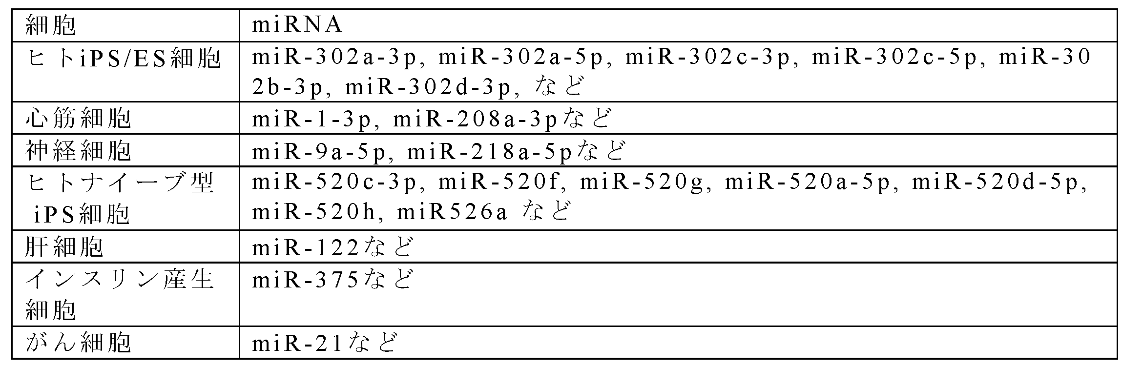
(1b)の核酸配列は、致死遺伝子もしくはアンチ致死遺伝子をコードする核酸配列である。致死遺伝子としては、Bacillus amyloliquefaciens由来のbarnaseなどのRNA分解酵素をコードする遺伝子、HokB, Fst, GhoT (膜破壊)、HipA(リン酸化による核酸伸長阻害), RelE,YafO,VapC,MazF,MqsR,PemKHicA(endonuclease)、FicT(Adenylation), Doc(Phosphorylation), CcdB, ParE(Gyrase inhibitor), Tact(Inhibitor of translation), cbtA (Inhibitor of cytoskeletal protein)などの毒素をコードする遺伝子、Bax、Bimなどのアポトーシス誘導タンパク質をコードする遺伝子が挙げられるが、これらには限定されない。アンチ致死遺伝子は、一般的には致死遺伝子との組み合わせで用いられる。RNA分解酵素に対しては、RNA分解酵素を不活性化するタンパク質、例えば、Bacillus amyloliquefaciens由来のbarstarが挙げられる。毒素に対しては、毒素を不活性化するタンパク質、アポトーシス誘導タンパク質であるBax((BCL2 associated X, apoptosis regulator)やBim(Bcl-2 interacting mediator、BCL2L11, BCL2 like 11ともいう)に対しては、アポトーシス抑制タンパク質である、Bcl-2(B-cell/CLL lymphoma 2、BCL2 apoptosis regulatorともいう)やBcl-xL(B-cell lymphoma-extra large、BCL2L1, BCL2 like 1ともいう)が挙げられる。
RNA分解酵素と、RNA分解酵素を不活性化するタンパク質の組み合わせとしては、barnaseとbarstarが挙げられる。barnaseのアミノ酸配列の一例は配列番号42、barstar のアミノ酸配列の一例は配列番号43で示すが、他種のbarnase、barstarも好適に用いることができる。本実施形態における第1のmRNAまたは第2のmRNAがコードしうるbarnase、barstar遺伝子は、それぞれ、barnase、barstarタンパク質をコードするDNAをいい、例えば、NCBIが定義するGene ID 75094541(Barstar)、Gene ID 75093094 (Barnase)で表される配列、またはそれらの転写派生体(transcript variant)、スプライシング変異体及びホモログのヌクレオチド配列を有するDNAが挙げられる。また、これらの配列を有する核酸に、ストリンジェントな条件でハイブリダイズすることができる程度の相補性を有するDNAであってもよい。ストリンジェントな条件とは、Berger and Kimmel(1987, Guide to Molecular Cloning Techniques Methods in Enzymology, Vol.152, Academic Press, San Diego CA)に教示されるように、複合体又はプローブを結合する核酸の融解温度(Tm)に基づいて決定することができる。例えばハイブリダイズ後の洗浄条件として、通常「1×SSC、0.1%SDS、37℃」程度の条件を挙げることができる。相補鎖はかかる条件で洗浄しても対象とする正鎖とハイブリダイズ状態を維持するものであることが好ましい。特に制限されないが、より厳しいハイブリダイズ条件として「0.5×SSC、0.1%SDS、42℃」程度の洗浄条件、さらに厳しくは「0.1×SSC、0.1%SDS、65℃」程度の洗浄条件で洗浄しても正鎖と相補鎖とがハイブリダイズ状態を維持する条件を挙げることができる。具体的には、このような相補鎖として、対象の正鎖の塩基配列と完全に相補的な関係にある塩基配列からなる鎖、及び該鎖と少なくとも90%、好ましくは95%以上、より好ましくは97%以上、いっそう好ましくは98%以上、特に好ましくは99%以上の同一性を有する塩基配列からなる鎖を例示することができる。
アポトーシス誘導タンパク質とアポトーシス抑制タンパク質の組み合わせとしては、BaxとBcl-2、BaxとBcl-xL、BimとBcl-2、BimとBcl-xLのいずれの組み合わせであっても、致死遺伝子とアンチ致死遺伝子として機能する。本実施形態における第1のmRNAまたは第2のmRNAがコードしうるBax、Bim、Bcl-xL、Bcl-2遺伝子は、それぞれ、Bax、Bim、Bcl-xL、Bcl-2タンパク質をコードするDNAをいい、例えば、NCBIが定義するGene ID 581 (Bax)、Gene ID 10018(BimEL)、Gene ID 598 (Bcl-xL)、Gene ID 596 (Bcl-2)で表される配列、またはそれらの転写派生体(transcript variant)、スプライシング変異体及びホモログのヌクレオチド配列を有するDNAが挙げられる。また、これらの配列を有する核酸に、ストリンジェントな条件でハイブリダイズすることができる程度の相補性を有するDNAであってもよい。ストリンジェントな条件は、上記のとおりに定義される。
(1a)の核酸配列を、目的細胞で活性化しているmiRNAによって特異的に認識される核酸配列とする場合、(1b)の核酸配列は、アンチ致死遺伝子とすることが好ましい。また、(1a)の核酸配列を、非目的細胞で活性化しているmiRNAによって特異的に認識される核酸配列とする場合、(1b)の核酸配列は、致死遺伝子とすることが好ましい。
第1のmRNAは、前記(1a)及び(1b)が作動可能に連結されていればよく、(1a)が(1b)の5'側に連結される第1態様と、(1a)が(1b)の3'側に連結される第2態様と、これらの両者を備える態様がありうる。各態様について説明する。
第1のmRNAは、第1態様によれば、(1a)の核酸配列が(1b)の核酸配列の5’側に存在する。この場合、5'-UTRは、5’末端から順に、[Cap構造もしくはCapアナログ]、[(1a)の核酸配列]が連結した構造であってよい。Cap構造は、7メチルグアノシン5’リン酸であってよい、CapアナログはCap構造と同様に翻訳開始因子であるeIF4Eによって認識される修飾構造であって、Ambion製のAnti-Reverse Cap Analog(ARCA)、New England Biolabs製の、m7G(5')ppp(5')G RNA Cap Structure Analog、TriLink製のCleanCapなどが挙げられるが、これらには限定されない。Capアナログは、翻訳開始因子により認識されるその他の修飾構造であってもよい。Cap構造もしくはCapアナログの3'側であって、(1a)の核酸配列の5’側には、例えば0~50塩基程度、好ましくは0~30塩基程度の任意核酸配列が含まれていてもよい。(1a)の核酸配列は、少なくとも1つ含まれていればよいが、2リピート、3リピート、4リピート、またはそれ以上の(1a)の核酸配列の繰り返しが5'-UTRに含まれていてもよい。(1a)の核酸配列の3'側である5'-UTRの3'末端側には、例えば0~50塩基程度、好ましくは10~30塩基程度の任意核酸配列が含まれていてもよい。これらの任意核酸配列は、二次構造を形成せず、第2のmRNA及び第3のmRNAと特異的に相互作用しない核酸配列であることが好ましい。また、5'-UTR内には、開始コドンとなるAUGが存在しないことが好ましい。例えば、(1a)の核酸配列内にAUGを含む場合、配列の最後に塩基を1つあるいは2つ付加することでフレームシフトを回避することができる。または、前述AUGから3塩基単位で数えた(1a)の核酸配列外に終止コドン配列を付加しても良い。または、AUGの塩基一つ以上をタンパク質との相互作用に影響しない限り任意の塩基に変換して使用することも可能である。
第1態様による第1のmRNAのコーディング領域は、(1b)の核酸配列を含む。(1b)の核酸配列は先に述べたとおりである。(1b)の核酸配列のほかに、蛍光タンパク質をコードする核酸配列を含んでもよい。
第1態様による第1のmRNAの3'-UTRは、PolyA tailを含む。PolyA tailは、Aの合計長が50 mer 以上であってよく、途中にA以外の核酸塩基が含まれていてもよい。任意選択的に、PolyA tailの5’側にも、(1a)の核酸配列が含まれていてもよい。
第1のmRNAは、第2態様によれば、(1a)の核酸配列が(1b)の核酸配列の3'側に存在する。この場合、5'-UTRは、5’末端に、[Cap構造もしくはCapアナログ]が設けられ、総塩基長が30~100程度の核酸配列からなる。この場合の5'-UTRを構成する核酸配列は、第1態様の任意核酸配列と同様に決定することができる。第2態様による第1のmRNAのコーディング領域は、第1態様による第1のmRNAのコーディング領域と同じであってよい。第2態様による第1のmRNAの3'-UTRは、5’末端から順に、[(1a)の核酸配列]、[Poly A tail]が連結した構造であってよい。あるいは、第2態様による第1のmRNAの3'-UTRは、Poly A tail中に[(1a)の核酸配列]が挿入された構造であってもよい。第1のmRNAは、第1態様と第2態様を組み合わせた構造であってもよい。この場合、5'-UTRと3'-UTRとの両方に[(1a)の核酸配列]を備える。
(2)第2のmRNAは、ONスイッチmRNAであり、
(2a)前記(1b)の致死遺伝子を不活性化するアンチ致死遺伝子もしくは前記(1b)のアンチ致死遺伝子により不活性化される致死遺伝子をコードする核酸配列と、
(2b)前記(2a)の3'側に設けられるPolyA tailと、
(2c)前記(2b)の3'側に設けられる前記目的細胞または非目的細胞で活性化しているmiRNAによって特異的に認識される核酸配列と、
(2d)前記(2c)の3'側に設けられる翻訳抑制配列とを含む。
(2a)前記(1b)の致死遺伝子を不活性化するアンチ致死遺伝子もしくは前記(1b)のアンチ致死遺伝子により不活性化される致死遺伝子をコードする核酸配列と、
(2b)前記(2a)の3'側に設けられるPolyA tailと、
(2c)前記(2b)の3'側に設けられる前記目的細胞または非目的細胞で活性化しているmiRNAによって特異的に認識される核酸配列と、
(2d)前記(2c)の3'側に設けられる翻訳抑制配列とを含む。
本明細書において、特定のmiRNA活性の存在下で翻訳が行われ、miRNA活性の非存在下で翻訳が抑制されるmRNAスイッチをONスイッチmRNAと指称する。第2のmRNAは、ONスイッチmRNAである。ONスイッチmRNAを、省略して、ONスイッチ、スイッチmRNAと指称する場合がある。
第2のmRNAにおいては、5'から3'の向きに、(2a)、(2b)、(2c)、(2d)の核酸配列がこの順に連結されている。第2のmRNAのより具体的な構造は、mRNA分子の5’から3’の向きに、5'-UTR、コーディング領域、3'-UTRが連結した構造であってよい。
第2のmRNAの5'-UTRは、5’末端に、[Cap構造もしくはCapアナログ]が設けられ、総塩基長が30~100程度の核酸配列からなる。この核酸配列は、(1a)の核酸配列を持たない。第2のmRNAの5'-UTRは、第1のmRNAの第2態様による5'-UTRに含まれていてもよい任意核酸配列と同様に決定することができる。
第2のmRNAのコーディング領域は、(2a)の核酸配列を含む。(2a)の核酸配列は、第1のmRNAの(1b)の核酸配列との関係で決定される。第1のmRNAの(1b)の核酸配列が、ある特定の致死遺伝子をコードする核酸配列である場合、第2のmRNAの(2a)の核酸配列は、当該致死遺伝子を不活性化するアンチ致死遺伝子をコードする核酸配列である。第1のmRNAの(1b)の核酸配列が、ある特定のアンチ致死遺伝子をコードする核酸配列である場合、第2のmRNAの(2a)の核酸配列は、当該アンチ致死遺伝子により不活性化される致死遺伝子をコードする核酸配列である。
第2のmRNAの3'-UTRは、5'から3'の向きに、(2b)、(2c)、(2d)の核酸配列をこの順に含む。(2b)、(2c)の核酸配列間には、任意核酸配列を含まないことが好ましい。
含むとしても、任意核酸配列は、10塩基以内、好ましくは2塩基以内とすることができる。(2c)、(2d)の核酸配列間に500塩基以内の任意核酸配列を含んでいてもよい。
(2b)のPolyA tailは、第1のmRNAのpolyA tail と同様のものとしてよい。
含むとしても、任意核酸配列は、10塩基以内、好ましくは2塩基以内とすることができる。(2c)、(2d)の核酸配列間に500塩基以内の任意核酸配列を含んでいてもよい。
(2b)のPolyA tailは、第1のmRNAのpolyA tail と同様のものとしてよい。
(2c)の核酸配列は、目的細胞または非目的細胞で活性化しているmiRNAによって特異的に認識される核酸配列である。目的細胞または非目的細胞で活性化しているmiRNAは、(1a)の核酸配列において定義されたmiRNAと同一である。しかし、(2c)の核酸配列は、当該miRNAに特異的に認識される限り、(1a)と同一であってもよく、(1a)と一部が異なっていてもよい。典型的には、(1a)の核酸配列は、(2c)の核酸配列と同一であり、目的細胞または非目的細胞で活性化しているmiRNAに相補的な配列であることが好ましい。
(2d)の核酸配列は、(2c)の核酸配列の下流に結合されて、(2c)の核酸配列を特異的に認識するmiRNAが存在しない場合に、当該第2のmRNAの翻訳を抑制可能な翻訳抑制配列である。この翻訳抑制配列は、本発明においては、「付加配列」と指称する場合もある。翻訳抑制配列は、特には限定されず、任意の核酸配列であってよい。翻訳抑制配列は、例えば、5以上の塩基からなる核酸配列であってよく、塩基の種類及びおよび塩基の配列は問わない。(2d)の核酸配列が5以上の塩基からなる核酸配列であれば、(2c)の核酸配列を特異的に認識するmiRNAの非存在下では(2a)の核酸配列の翻訳をOFFの状態に保持し(翻訳抑制状態)、(2c)の核酸配列を特異的に認識するmiRNAの存在下で(2a)の核酸配列の翻訳をONの状態とすることができ、fold-change 2以上であることが確認されている。
5以上の塩基からなる核酸配列の例としては、同一の核酸塩基を備える5~10塩基の核酸配列、核酸塩基が異なる2種の以上の核酸がそれぞれ連続して結合した10~20塩基からなる核酸配列、核酸塩基が異なる2種以上の核酸がそれぞれ連続して結合した30~50塩基からなる核酸配列、3~10塩基程度の特定の核酸配列の繰り返しを含む核酸配列、PolyA tailを特異的に認識する20以上の塩基からなる核酸配列、5'-UTRを特異的に認識する核酸配列であってもよい。また、5以上の塩基からなる核酸配列は、場合により、10以上、20以上、30以上、40以上、50以上、あるいは100以上、300以上、500以上、1000以上、1500以上の塩基からなる核酸配列であってもよい。
(3)第3のmRNAは、薬剤に耐性の遺伝子をコードする核酸配列を含み、(1a)の核酸配列を持たないmRNAである。すなわち、第3のmRNAは、導入される細胞内のmiRNA活性に依存せずに薬剤耐性蛋白質を発現するmRNAである。
第3のmRNAのより具体的な構造は、mRNA分子の5’側から順に、5'-UTR、コーディング領域、3'-UTRが連結した構造であってよい。第3のmRNAの5'-UTRは、第2のmRNAの5'-UTRと同じ構成にて設計することができる。
第3のmRNAのコーディング領域は、薬剤に耐性の遺伝子をコードする核酸配列を含む。
薬剤に耐性の遺伝子は、工程(II)において、細胞集団の培養に用いる薬剤との関係で決定することができる。薬剤は、哺乳動物に由来する細胞の培養において一般的に用いられある薬剤であれば特に限定はされない。したがって、薬剤に耐性の遺伝子の例としては、例えば、ブラストサイジン耐性遺伝子、G418(ジェネティシン)耐性遺伝子、カナマイシン耐性遺伝子、アンピシリン耐性遺伝子、ピューロマイシン耐性遺伝子、ゲンタマイシン耐性遺伝子、テトラサイクリン耐性遺伝子、クロラムフェニコール耐性遺伝子等が挙げられるが、これらには限定されない。
薬剤に耐性の遺伝子は、工程(II)において、細胞集団の培養に用いる薬剤との関係で決定することができる。薬剤は、哺乳動物に由来する細胞の培養において一般的に用いられある薬剤であれば特に限定はされない。したがって、薬剤に耐性の遺伝子の例としては、例えば、ブラストサイジン耐性遺伝子、G418(ジェネティシン)耐性遺伝子、カナマイシン耐性遺伝子、アンピシリン耐性遺伝子、ピューロマイシン耐性遺伝子、ゲンタマイシン耐性遺伝子、テトラサイクリン耐性遺伝子、クロラムフェニコール耐性遺伝子等が挙げられるが、これらには限定されない。
第3のmRNAの3'-UTRは、miRNAによって特異的に認識される核酸配列を持たず、PolyA tailを備えることが好ましい。PolyA tailは、第1のmRNAのpolyA tail と同様のものとしてよい。
第1のmRNA、第2のmRNA及び第3のmRNAは、通常のウリジン、シチジンに替えて、細胞毒性を低減させるため、1-メチルシュードウリジン、シュードウリジン、5-メチルシチジンなどの修飾塩基を含んでいてもよい。修飾塩基の位置は、ウリジン、シチジンいずれの場合も、独立に、全てあるいは一部とすることができ、一部である場合には、任意の割合でランダムな位置とすることができる。
第1のmRNA、第2のmRNA及び第3のmRNAは、上記に従って、分子構造、核酸配列が決定されれば、遺伝子工学的に既知の任意の方法により当業者が合成することができる。例えば、プロモーター配列を含むテンプレートDNAを鋳型として用いたin vitro合成法により、合成mRNA分子として得ることができる。設計どおりの合成mRNA分子を簡便な方法で得ることができることは本発明の1つの利点である。
第1のmRNA、第2のmRNA及び第3のmRNAは、別個のRNA分子として調製することもでき、1つまたは2つのmRNA分子として調製することもできる。すなわち、第1のmRNA、第2のmRNA及び第3のmRNAのいずれか2以上が同一分子上に存在してもよい。また、これらをコードするDNAも、別個のDNA分子として調製することもでき、1つまたは2つのDNA分子として調製することもできる。後者の場合には、mRNAごとに1つのプロモーターに制御されてもよい。
第1のmRNA、第2のmRNA及び第3のmRNAを別個のRNA分子として調製する場合には、第1のmRNA、第2のmRNA及び第3のmRNAのうち、少なくとも第1のmRNAと第2のmRNAとは細胞集団に共導入することが好ましい。第1のmRNA、第2のmRNA及び第3のmRNAを細胞集団に共導入することがさらに好ましい。共導入した2以上のmRNAから発現する蛋白質の活性比は、細胞集団内において一定となるためである。
細胞集団への第1のmRNA、第2のmRNA及び第3のmRNAの導入は、RNAを細胞に導入する方法として一般に用いられる任意の方法を用いることができる。RNA分子を直接、細胞に導入する方法としては、例えば、リポフェクション法、リポソーム法、エレクトロポレーション法、リン酸カルシウム共沈殿法、DEAEデキストラン法、マイクロインジェクション法、遺伝子銃法などの導入法を用いて、RNA分子を直接、細胞に導入することができる。合成RNA分子の導入による利点は、ゲノムへの組み込みがなく、mRNAスイッチを導入した後の細胞を医療応用などに使用しやすいことが挙げられる。
あるいは、細胞集団への第1のmRNA、第2のmRNA及び第3のmRNAの導入は、RNA発現ベクター等のDNA構築物を用いることもできる。この場合、mRNA分子をコードする発現ベクターを設計し、上記と同様の導入法にて、発現ベクターを直接、細胞に導入することができる。第1のmRNA、第2のmRNA及び第3のmRNAの配列をコードする発現ベクターは、当該分野において周知慣用のものを用いることができ、例えば、ウイルスベクター、人工染色体ベクター、プラスミドベクター、トランスポゾンを用いた発現システム(トランスポゾンベクターと呼ばれる場合がある)等が挙げられる。ウイルスベクターとしては、レトロウイルスベクター、レンチウイルスベクター、アデノウイルスベクター、アデノ随伴ウイルスベクター、センダイウイルスベクター等が例示される。人工染色体ベクターとしては、例えばヒト人工染色体(HAC)、酵母人工染色体(YAC)、細菌人工染色体(BAC、PAC)等が挙げられる。プラスミドベクターとしては、哺乳動物用プラスミド全般を使用することができ、例えば、エピソーマルベクターであってもよい。トランスポゾンベクターとしては、piggyBacトランスポゾンを用いた発現ベクター等が例示される。例えば、本発明者らによる米国特許であって、引用することにより本明細書の一部をなすものとする米国特許第10,378,070号に開示されたベクターを用いることができるが、それらには限定されない。mRNAスイッチをコードする発現ベクターを細胞に導入することで、発現ベクターから転写され、細胞内で生成したmRNAスイッチを、直接導入した合成mRNA分子と同様に機能させることができる。
細胞集団への第1のmRNA、第2のmRNA及び第3のmRNAの導入量及び導入比は、目的細胞の種類や、mRNAの構造によっても異なり、特定の量に限定されるものではない。例えば、(1b)または(2a)の致死遺伝子の発現レベルが望ましくない目的細胞死の閾値以下になるように、予備実験等により、第1のmRNA及び第2のmRNAの導入量を決定することができる。
(II)薬剤存在下での培養工程
導入工程に次いで、薬剤存在下での培養工程を実施することができる。培養工程は、第1のmRNA、第2のmRNA及び第3のmRNAを導入した前記細胞集団を薬剤の存在下で培養する工程である。薬剤は、第3のmRNAがコードする薬剤耐性遺伝子に対応する薬剤を用いる。培養条件は、目的とする細胞を含む細胞集団によって異なり、適切な培地を用いて、適切な温度、雰囲気にて1~4日程度培養することができる。
導入工程に次いで、薬剤存在下での培養工程を実施することができる。培養工程は、第1のmRNA、第2のmRNA及び第3のmRNAを導入した前記細胞集団を薬剤の存在下で培養する工程である。薬剤は、第3のmRNAがコードする薬剤耐性遺伝子に対応する薬剤を用いる。培養条件は、目的とする細胞を含む細胞集団によって異なり、適切な培地を用いて、適切な温度、雰囲気にて1~4日程度培養することができる。
(III)薬剤非存在下での培養工程
工程(III)は任意選択的な工程であって、前記工程(II)を経た細胞集団を、薬剤非存在下で培養する工程である。培養条件は、目的とする細胞を含む細胞集団によって異なり、適切な方法にて行うことができる。本培養工程は、一例として、継代培養工程であってよい。継代培養は、1継代のみ行ってもよく、2継代以上行ってもよい。継代培養を行うことで、培養容器に付着した死細胞等を除去することができ、より高い精度にて目的細胞を純化することができる。例えば、工程(II)を経た細胞集団と比較して、工程(III)を経た細胞集団は、純度を約1~4%向上することができ、最大で99%以上の純度にまで目的細胞を純化することが可能となる。
工程(III)は任意選択的な工程であって、前記工程(II)を経た細胞集団を、薬剤非存在下で培養する工程である。培養条件は、目的とする細胞を含む細胞集団によって異なり、適切な方法にて行うことができる。本培養工程は、一例として、継代培養工程であってよい。継代培養は、1継代のみ行ってもよく、2継代以上行ってもよい。継代培養を行うことで、培養容器に付着した死細胞等を除去することができ、より高い精度にて目的細胞を純化することができる。例えば、工程(II)を経た細胞集団と比較して、工程(III)を経た細胞集団は、純度を約1~4%向上することができ、最大で99%以上の純度にまで目的細胞を純化することが可能となる。
本発明に係る細胞の純化方法によれば、目的細胞を特異的に生存させ、目的細胞以外の細胞を特異的に死滅させることができ、セルソーターなどの機器を使用することなく、大量の目的細胞を一度に、短時間で純化することが可能になる。なお、本発明は、先に説明した純化方法により目的細胞を純化する工程を含む、前記目的細胞が富化された細胞集団を製造する方法と捉えることもできる。
[2.目的細胞の純化キット]
本発明はまた別の実施形態によれば、目的細胞の純化キットに関する。純化キットは、第1のmRNA、第2のmRNA及び第3のmRNAを含み、任意選択的に薬剤を含む。第1のmRNA、第2のmRNA、第3のmRNA、及び薬剤は、先の純化方法にて説明したmRNA及び薬剤であってよい。さらに任意選択的に、目的細胞の培養に適した培地や、純化キットの取り扱いのための説明書を含んでいてもよい。本実施形態による目的細胞の純化キットによれば、安全にかつ大量に、目的細胞を高純度で取得することができる。
本発明はまた別の実施形態によれば、目的細胞の純化キットに関する。純化キットは、第1のmRNA、第2のmRNA及び第3のmRNAを含み、任意選択的に薬剤を含む。第1のmRNA、第2のmRNA、第3のmRNA、及び薬剤は、先の純化方法にて説明したmRNA及び薬剤であってよい。さらに任意選択的に、目的細胞の培養に適した培地や、純化キットの取り扱いのための説明書を含んでいてもよい。本実施形態による目的細胞の純化キットによれば、安全にかつ大量に、目的細胞を高純度で取得することができる。
以下に、本発明の実施例を用いてより詳細に説明する。以下の実施例は、本発明を限定するものではない。
[実験方法]
[プラスミドの構築]
合成した5'-UTRおよび3'UTRオリゴDNAを用いて,In-Fusion HD Cloning Kit(TaKaRa)を用いて調製したUTRを含むpUC19ベクター(TaKaRa)を生成した。UTRを含むベクターが構築された後、インバースPCRにより5'-UTRと3'-UTRを含む直鎖状のベクターを調製し、In-Fusion HD Cloning KitによりORFのみをベクターに挿入して、5'-UTR、ORF、3'-UTRを含むpUC19ベクターを構築した。Barstarは、6つの合成オリゴDNA(YF472、YF473、YF474、YF475、YF476、YF477)の融合PCRによって増幅され、NcoIとBglIIで消化された。消化された断片を、先に構築したpSMベクター、または薬剤耐性遺伝子を除いてpSMベクターと同じ配列を持つpCMベクターにクローニングし、塩基配列を決定した。次に、BarstarのORFをプライマー、YF783およびYF804を用いて増幅し、上述の同じ直鎖状ベクターにクローニングした。Barnaseは、プライマーYF770およびYF807を用いてpFN19K HaloTag T7 SP6 Flexi Vector(Promega)から増幅した。Barnaseのリーク発現は大腸菌にとって致命的であるため、プライマーYF805およびYF806を用いて増幅したBarstarと、プライマーYF770およびYF807を用いて増幅したBarnase断片を融合させ、Barnase活性をキャンセルさせた。融合PCRの後、In-Fusion HD Cloning Kitを用いて、BarstarとBarnaseを含む断片を直線化したベクターにクローニングした。実施例で合成したオリゴDNAの塩基配列、及び使用したBarstarとBarnaseのペプチド配列、及び翻訳抑制配列を表2A、表2B、及び表2Cに、PCR用プライマーとDNA断片の組み合わせを表3A及び表3Bに示す。また、実施例で使用したBcl-xL、Bcl2、BimEL、Baxの鋳型DNA配列を、配列番号61~64に示す。
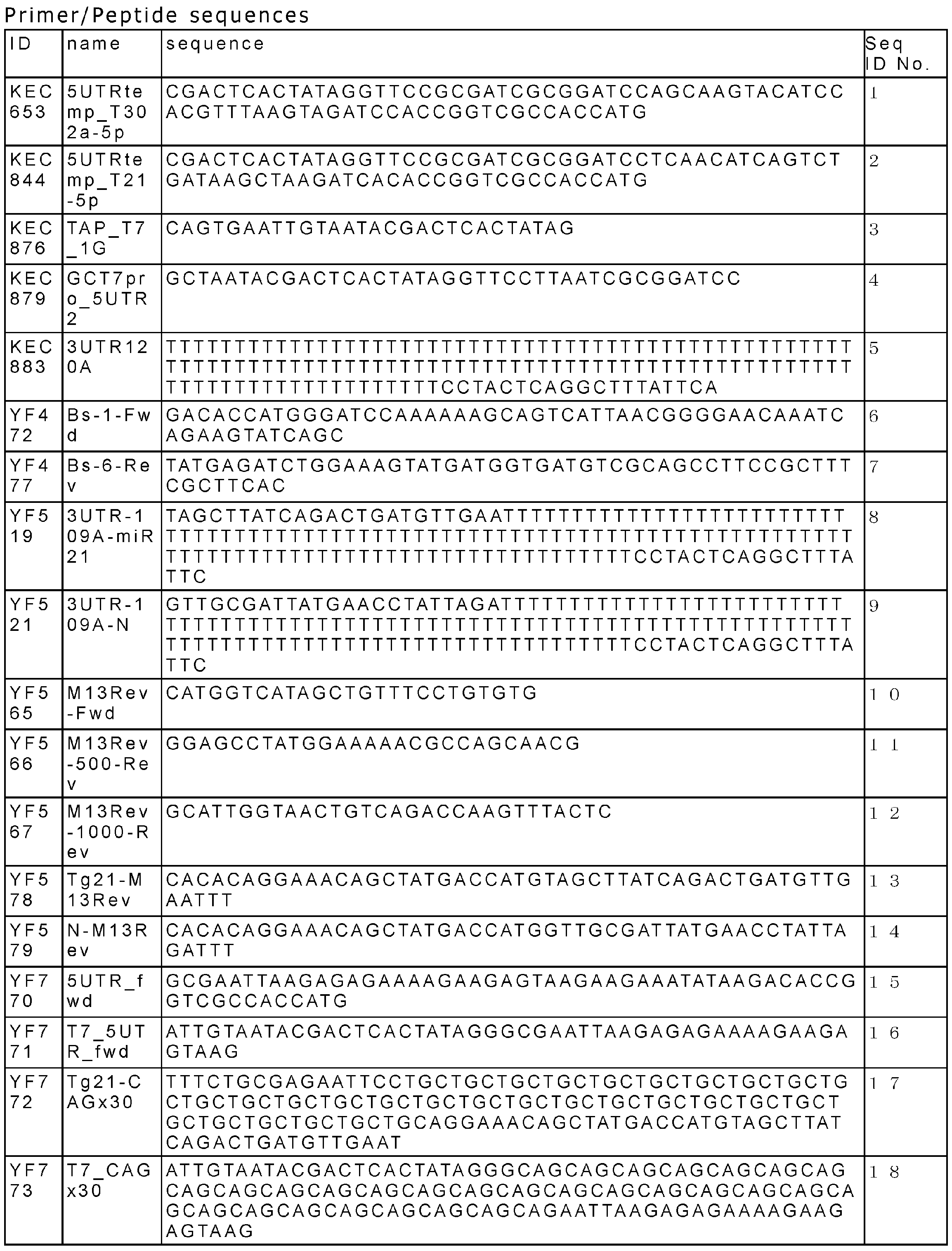
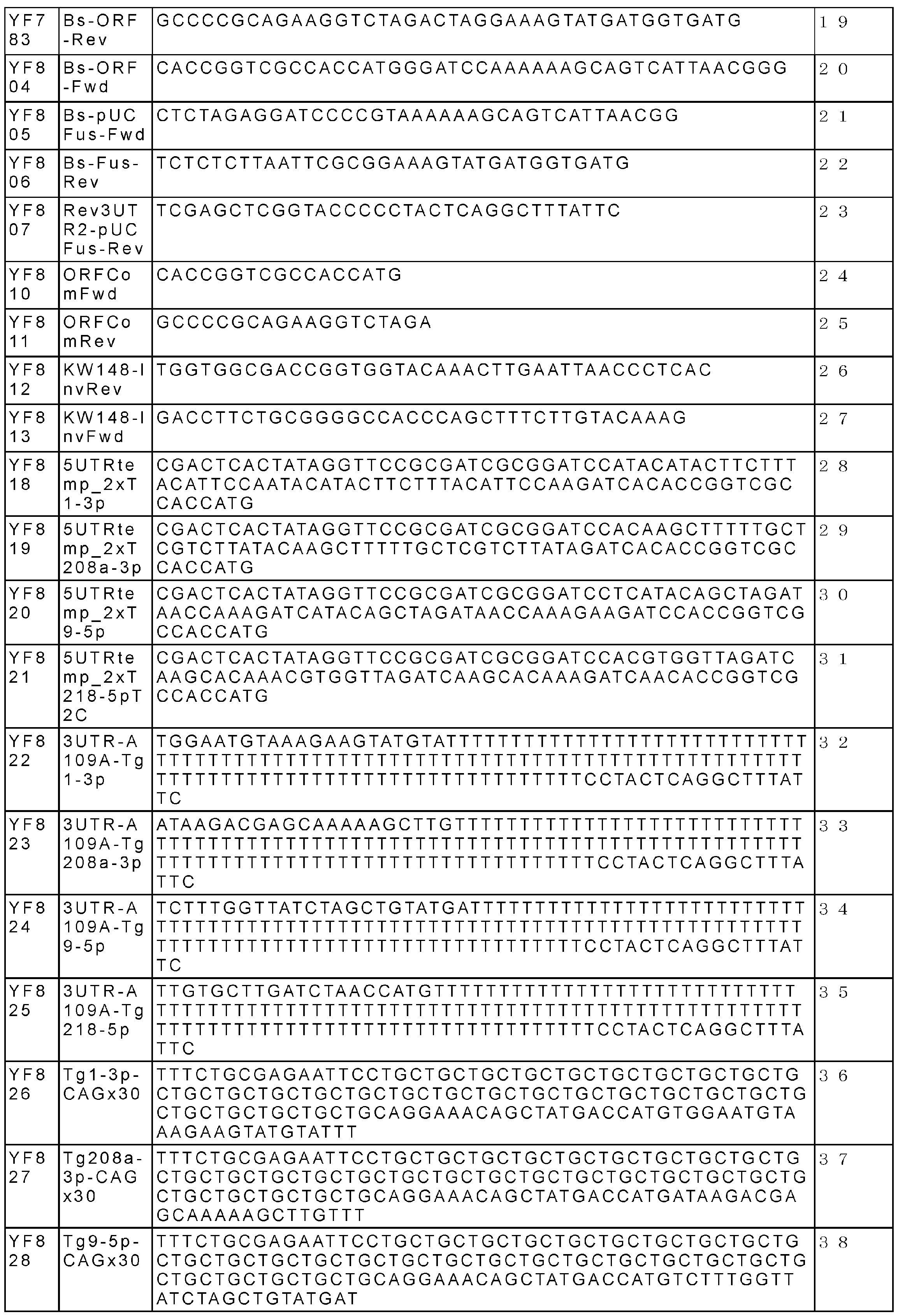
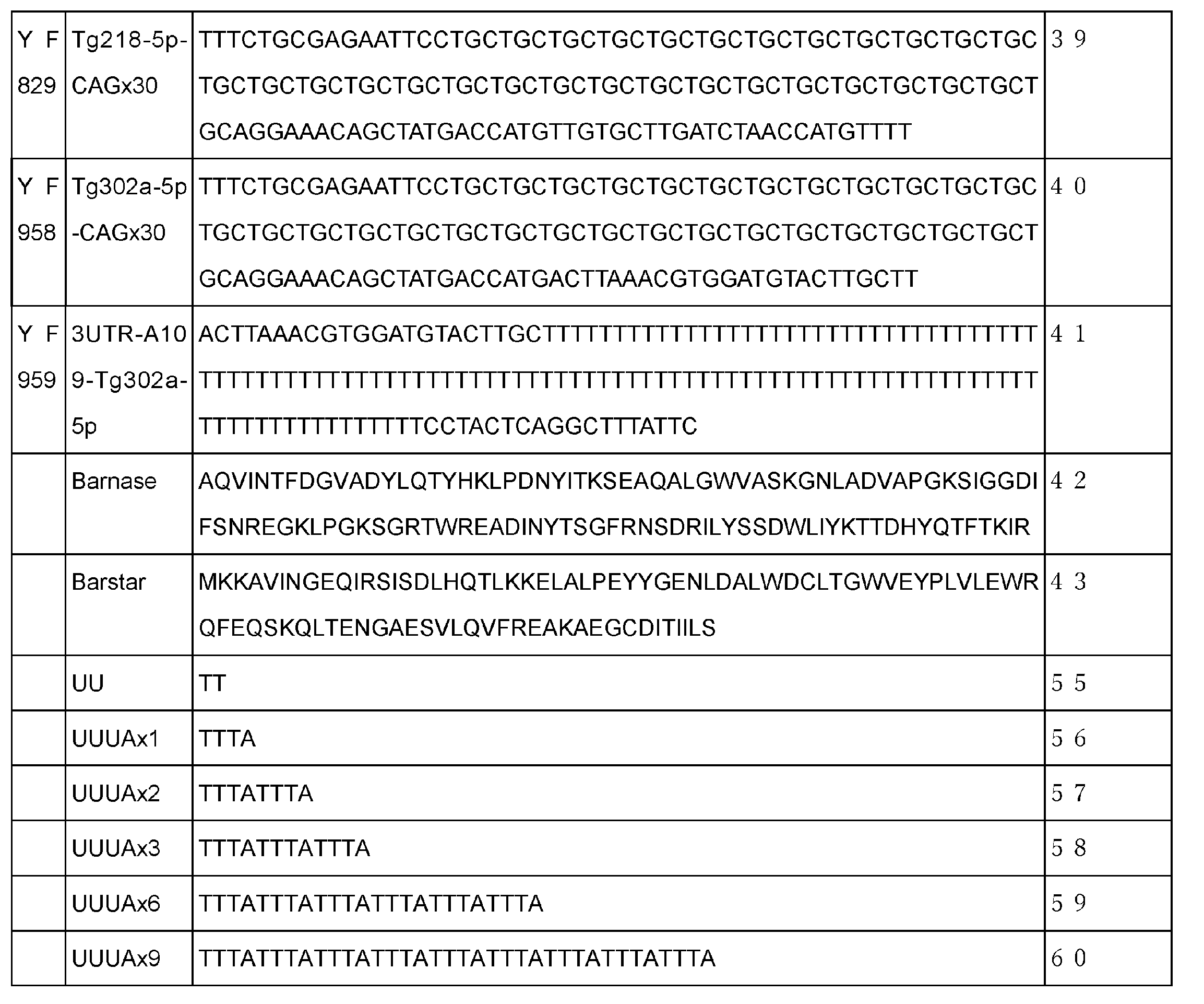
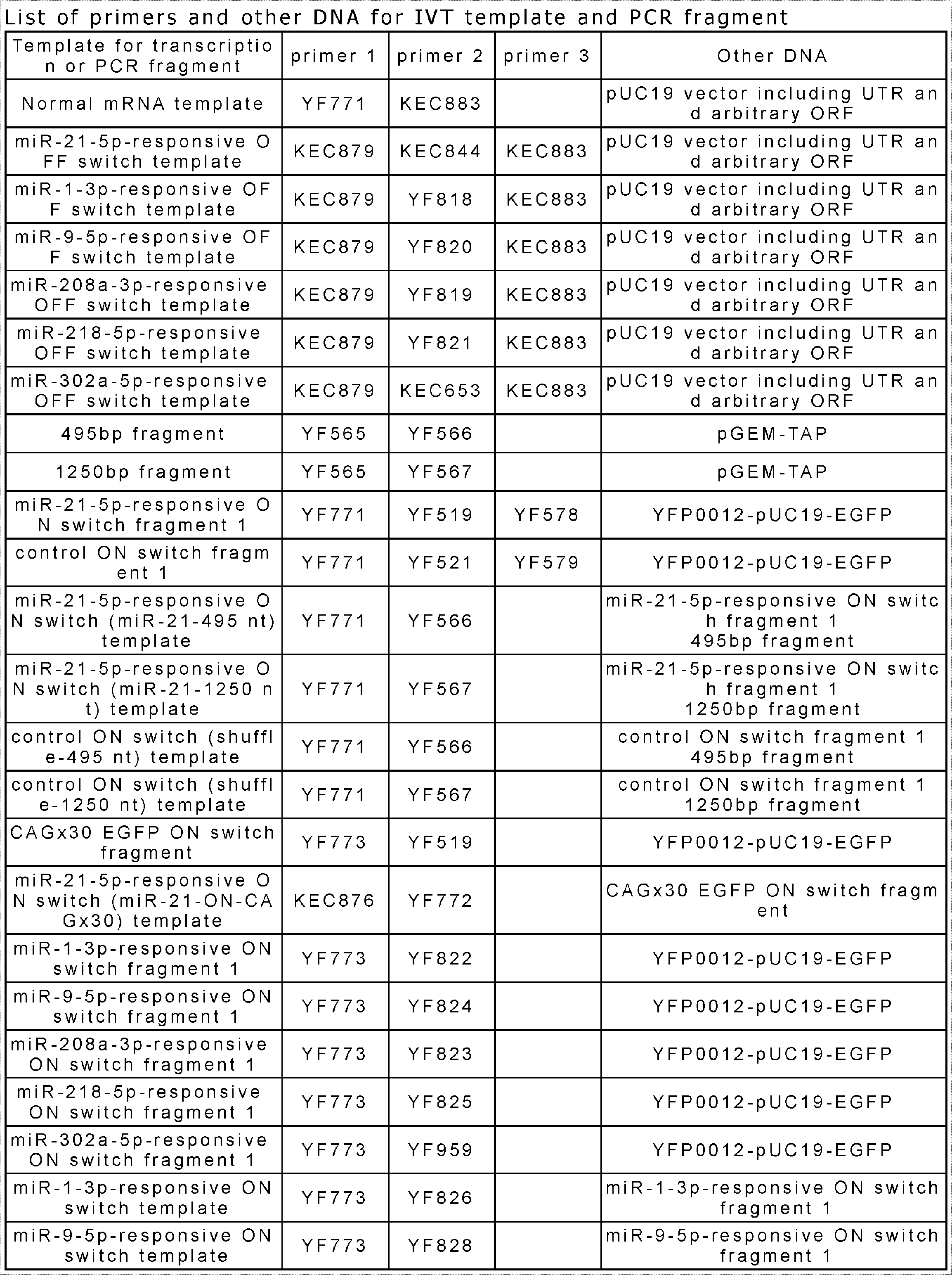
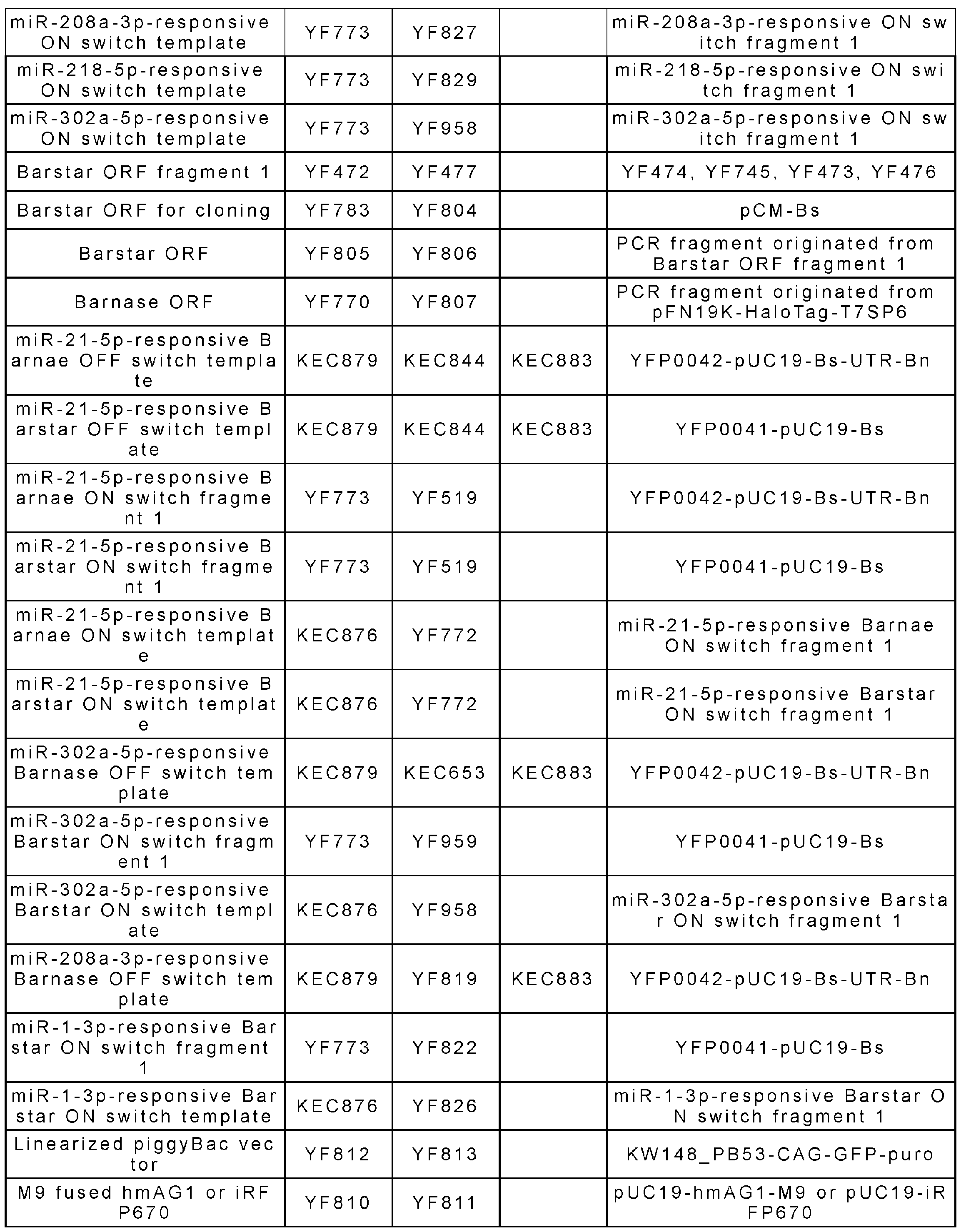
[mRNAの調製]
試験管内転写用のテンプレートDNAは、合成オリゴDNA(EurofinまたはFasmac、表1及び表2に記載)を用いてPCR(TOYOBO)により増幅した。単純に遺伝子を発現させるためのmRNAの鋳型は、5'-UTR、ORF、3'UTRを含むベクターから、T7プロモーターとpolyA tailを持つプライマーを用いて増幅した。同じプラスミドから、5'-UTRにmiRNAに相補的な配列を含むプライマーを用いて、miRNA応答性OFFスイッチの鋳型を増幅した。miRNA応答性ONスイッチでは、ORF、polyA tail、miRNAアンチセンス配列、翻訳抑制配列(付加配列)の順に融合させ、in vitro転写用のテンプレートを作成した。鋳型はSpeedBeads magnetic carboxylate modified particles (GE healthcare)またはMinElute PCR purification Kit (QIAGEN)を用いて精製した。MEGAScript T7 Transcription Kit (Thermo Fisher Scientific)を用いて、37℃で約4時間、RNAを転写した。免疫反応を回避し、翻訳を促進するために、ウリジン三リン酸(U)の代わりに1-メチルシュードウリジン-5'-三リン酸(Ψ)を用い、Anti-Reverse Cap Analog(ARCA)(TriLink社)を使用した。iPS/HeLa共培養実験からのiPSCの精製では、天然のrUTPとrCTPの代わりに、シュードウリジン-5′ -三リン酸と5-メチルシチジン-5′ -三リン酸(TriLink Bio Technologies)をそれぞれ使用した。転写されたRNAは、Monarch RNA Cleanup Columns(NEB社)、RNeasy MinElute Cleanup Kit(QIAGEN社)、SpeedBeads magnetic carboxylate modified particles(GE healthcare社)を用いて精製した。RNAはTURBO DNase(Thermo Fisher Scientific)およびAntarctic Phosphatase(NEB)またはrAPid Alkaline Phosphatase(Roche)で処理した。
試験管内転写用のテンプレートDNAは、合成オリゴDNA(EurofinまたはFasmac、表1及び表2に記載)を用いてPCR(TOYOBO)により増幅した。単純に遺伝子を発現させるためのmRNAの鋳型は、5'-UTR、ORF、3'UTRを含むベクターから、T7プロモーターとpolyA tailを持つプライマーを用いて増幅した。同じプラスミドから、5'-UTRにmiRNAに相補的な配列を含むプライマーを用いて、miRNA応答性OFFスイッチの鋳型を増幅した。miRNA応答性ONスイッチでは、ORF、polyA tail、miRNAアンチセンス配列、翻訳抑制配列(付加配列)の順に融合させ、in vitro転写用のテンプレートを作成した。鋳型はSpeedBeads magnetic carboxylate modified particles (GE healthcare)またはMinElute PCR purification Kit (QIAGEN)を用いて精製した。MEGAScript T7 Transcription Kit (Thermo Fisher Scientific)を用いて、37℃で約4時間、RNAを転写した。免疫反応を回避し、翻訳を促進するために、ウリジン三リン酸(U)の代わりに1-メチルシュードウリジン-5'-三リン酸(Ψ)を用い、Anti-Reverse Cap Analog(ARCA)(TriLink社)を使用した。iPS/HeLa共培養実験からのiPSCの精製では、天然のrUTPとrCTPの代わりに、シュードウリジン-5′ -三リン酸と5-メチルシチジン-5′ -三リン酸(TriLink Bio Technologies)をそれぞれ使用した。転写されたRNAは、Monarch RNA Cleanup Columns(NEB社)、RNeasy MinElute Cleanup Kit(QIAGEN社)、SpeedBeads magnetic carboxylate modified particles(GE healthcare社)を用いて精製した。RNAはTURBO DNase(Thermo Fisher Scientific)およびAntarctic Phosphatase(NEB)またはrAPid Alkaline Phosphatase(Roche)で処理した。
[mRNAのトランスフェクション]
トランスフェクションの前日に、HeLa細胞、293FT細胞、iPS細胞、および蛍光タンパク質を発現するそれらの改変細胞をマルチウェルプレートに播種した。Lipofectamine MessengerMax(Thermo Fisher Scientific)を用いて、製造元のプロトコルに従って転写RNAを細胞内に導入した。心筋細胞への分化を少なくとも13日以上行った後の細胞塊をコニカルチューブに移し、細胞塊が自然に落下した後に収穫した。回収した細胞塊は,10μg/mLのDNase Iを添加した2mg/mLのコラゲナーゼI溶液に懸濁し,37℃で2時間以上回転させた。培養後、上清を吸引して細胞塊のペレットを再懸濁し、AccuMax(Nacali tesque)で、37℃で約30分間培養した。その後、細胞懸濁液をピペッティングして細胞塊を単細胞に解離した後、新鮮な分化誘導培地で希釈した。解離した細胞懸濁液を120×gで5分間、室温で遠心分離し、上澄みを吸引した。細胞を分化誘導培地に再懸濁し、Countess II(Thermo Fisher Scientific)でカウントした。
トランスフェクションの前日に、HeLa細胞、293FT細胞、iPS細胞、および蛍光タンパク質を発現するそれらの改変細胞をマルチウェルプレートに播種した。Lipofectamine MessengerMax(Thermo Fisher Scientific)を用いて、製造元のプロトコルに従って転写RNAを細胞内に導入した。心筋細胞への分化を少なくとも13日以上行った後の細胞塊をコニカルチューブに移し、細胞塊が自然に落下した後に収穫した。回収した細胞塊は,10μg/mLのDNase Iを添加した2mg/mLのコラゲナーゼI溶液に懸濁し,37℃で2時間以上回転させた。培養後、上清を吸引して細胞塊のペレットを再懸濁し、AccuMax(Nacali tesque)で、37℃で約30分間培養した。その後、細胞懸濁液をピペッティングして細胞塊を単細胞に解離した後、新鮮な分化誘導培地で希釈した。解離した細胞懸濁液を120×gで5分間、室温で遠心分離し、上澄みを吸引した。細胞を分化誘導培地に再懸濁し、Countess II(Thermo Fisher Scientific)でカウントした。
[iPS/HeLa共培養からのiPSC精製]
iPS-EGFP細胞とHeLa-iRFP670細胞を、Y-27632(和光)を含むStemFit AK02Nを用いて、ラミニンコートした24ウェルプレートに播種した(比率:iPS:HeLa=3:2、Total:1.2×10⁵ cells/well)。トランスフェクションの前に、培地を、Y-27632を含まないStemFit AK02Nに変更した。T302a-5p応答性Barnase OFFスイッチとT302a-5p応答性Barstar ONスイッチ、およびブラストサイジン耐性mRNA(Bsd mRNA)を、Lipofectamine MessengerMAX Transfection Reagent(ThermoFisher)を用いて、製造者のプロトコルに従ってトランスフェクションした。トランスフェクションから4時間後、培養液を、ブラストサイジン(160μg/mL)を含むStemFit AK02Nに変更した。フローサイトメトリー解析の前に、細胞をPBSで洗浄し、Cytell Cell Imaging System(GEヘルスケアライフサイエンス)で撮影した。トランスフェクション後3日目に、細胞をPBSで洗浄し、200 μLのAccumax(フナコシ)で処理し、37℃、5% CO2で10分間インキュベートした。サンプルは,FL1(533/30 nm)およびFL4(675/25 nm)のフィルターを備えたAccuri C6(BD Bioscience)フローサイトメーターで分析した。iPS細胞とHeLa細胞を、それぞれFL1-A(EGFP)とFL4-A(iRFP670)陽性集団と定義した。継代については、細胞像を撮影した後、PBSで洗浄し、200 μLのAccumaxで処理し、37℃、5% CO2で10分間インキュベートした。これらの細胞を1.5 mLのチューブに集め、200gで5分間、24℃で遠心分離し、上清を吸引し、Y-27632を含むStemFitを加え、ラミニンコートした6ウェルプレートに播種した。播種から7日後、Accuri C6フローサイトメーターで上記のように分析した。
iPS-EGFP細胞とHeLa-iRFP670細胞を、Y-27632(和光)を含むStemFit AK02Nを用いて、ラミニンコートした24ウェルプレートに播種した(比率:iPS:HeLa=3:2、Total:1.2×10⁵ cells/well)。トランスフェクションの前に、培地を、Y-27632を含まないStemFit AK02Nに変更した。T302a-5p応答性Barnase OFFスイッチとT302a-5p応答性Barstar ONスイッチ、およびブラストサイジン耐性mRNA(Bsd mRNA)を、Lipofectamine MessengerMAX Transfection Reagent(ThermoFisher)を用いて、製造者のプロトコルに従ってトランスフェクションした。トランスフェクションから4時間後、培養液を、ブラストサイジン(160μg/mL)を含むStemFit AK02Nに変更した。フローサイトメトリー解析の前に、細胞をPBSで洗浄し、Cytell Cell Imaging System(GEヘルスケアライフサイエンス)で撮影した。トランスフェクション後3日目に、細胞をPBSで洗浄し、200 μLのAccumax(フナコシ)で処理し、37℃、5% CO2で10分間インキュベートした。サンプルは,FL1(533/30 nm)およびFL4(675/25 nm)のフィルターを備えたAccuri C6(BD Bioscience)フローサイトメーターで分析した。iPS細胞とHeLa細胞を、それぞれFL1-A(EGFP)とFL4-A(iRFP670)陽性集団と定義した。継代については、細胞像を撮影した後、PBSで洗浄し、200 μLのAccumaxで処理し、37℃、5% CO2で10分間インキュベートした。これらの細胞を1.5 mLのチューブに集め、200gで5分間、24℃で遠心分離し、上清を吸引し、Y-27632を含むStemFitを加え、ラミニンコートした6ウェルプレートに播種した。播種から7日後、Accuri C6フローサイトメーターで上記のように分析した。
[iPSC-心筋細胞の精製]
心筋細胞を14日以上分化させた後の胚様体を回収し、コラゲナーゼ(2mg/mL collagenase I(SIGMA)および10μg/mL DNase I(EMD MilliPore))を用いて37℃で2時間処理した後、AccuMaxを用いて37℃で30分処理した。分化誘導培地でピペッティングして細胞を解離させ、同培地で1回洗浄した。表4に記載のRNAを細胞にリバーストランスフェクションし、フィブロネクチン(Sigma)でコーティングしたマルチウェルプレートに播種した。トランスフェクション4時間後、400μg/mLのG418を添加した等量の培地を加え、トランスフェクションされていない細胞および心筋細胞以外の細胞を除去した。200μg/mLのG418を添加した培地に交換しながら、37℃、5%CO2で3日間培養した。Cytell Cell Imaging Systemを用いて画像を撮影する前に、10μg/mLのHoechst 33342(Thermo Fisher Scientific社)を用いて核を30分間染色した。
心筋細胞を14日以上分化させた後の胚様体を回収し、コラゲナーゼ(2mg/mL collagenase I(SIGMA)および10μg/mL DNase I(EMD MilliPore))を用いて37℃で2時間処理した後、AccuMaxを用いて37℃で30分処理した。分化誘導培地でピペッティングして細胞を解離させ、同培地で1回洗浄した。表4に記載のRNAを細胞にリバーストランスフェクションし、フィブロネクチン(Sigma)でコーティングしたマルチウェルプレートに播種した。トランスフェクション4時間後、400μg/mLのG418を添加した等量の培地を加え、トランスフェクションされていない細胞および心筋細胞以外の細胞を除去した。200μg/mLのG418を添加した培地に交換しながら、37℃、5%CO2で3日間培養した。Cytell Cell Imaging Systemを用いて画像を撮影する前に、10μg/mLのHoechst 33342(Thermo Fisher Scientific社)を用いて核を30分間染色した。

[HeLa/293FT 共培養からのHeLaまたは293FT細胞の精製]
致死遺伝子としてアポトーシス遺伝子、アンチ致死遺伝子としてアンチアポトーシス遺伝子の組み合わせを用いて、HeLa/293FT 共培養からのHeLaまたは293FT細胞の精製を行った。アポトーシス遺伝子/アンチアポトーシス遺伝子の組み合わせは、Bax/Bcl-xL、Bax/Bcl-2、Bim/Bcl-xLについて試験を行った。細胞を識別するため、hmAG1-M9 (緑色蛍光タンパク質に核移行シグナルを連結したもの)を恒常発現するHeLa細胞(HeLa-hmAG1)と、iRFP670-M9 (赤色蛍光タンパク質に核移行シグナルを連結したもの)を恒常発現する293FT細胞(293FT-iRFP670)を混合して播種を行った。播種の翌日、miR-21-5p応答Bcl-xL ONスイッチとmiR-21-5p応答Bax OFF スイッチ、またはmiR-21-5p応答Bcl-xL OFFスイッチとmiR-21-5p応答Bax ON スイッチをLipofectamine MessengerMAX Transfection Reagent(ThermoFisher)を用いて、製造者のプロトコルに従ってトランスフェクションした。同様にして、miR-21-5p応答Bcl-2 ONスイッチとmiR-21-5p応答Bax OFF スイッチ、またはmiR-21-5p応答Bcl-2 OFFスイッチとmiR-21-5p応答Bax ON スイッチを、Lipofectamine MessengerMAX Transfection Reagent(ThermoFisher)を用いて、製造者のプロトコルに従ってトランスフェクションした。トランスフェクション1日後、死んだ細胞を培地で洗い流したのち、ウェルに接着している細胞を蛍光顕微鏡(CQ1)で観察した。その後、細胞にAccuMax(フナコシ)を添加し、37℃、5% CO2で10分間インキュベートした。サンプルは、FL1(533/30 nm)およびFL4(675/25 nm)のフィルターを備えたAccuri C6(BD Bioscience)フローサイトメーターで分析した。HeLa-hmAG1細胞と293FT-iRFP670細胞は、それぞれFL1-AとFL4-A陽性集団としてカウントした。
致死遺伝子としてアポトーシス遺伝子、アンチ致死遺伝子としてアンチアポトーシス遺伝子の組み合わせを用いて、HeLa/293FT 共培養からのHeLaまたは293FT細胞の精製を行った。アポトーシス遺伝子/アンチアポトーシス遺伝子の組み合わせは、Bax/Bcl-xL、Bax/Bcl-2、Bim/Bcl-xLについて試験を行った。細胞を識別するため、hmAG1-M9 (緑色蛍光タンパク質に核移行シグナルを連結したもの)を恒常発現するHeLa細胞(HeLa-hmAG1)と、iRFP670-M9 (赤色蛍光タンパク質に核移行シグナルを連結したもの)を恒常発現する293FT細胞(293FT-iRFP670)を混合して播種を行った。播種の翌日、miR-21-5p応答Bcl-xL ONスイッチとmiR-21-5p応答Bax OFF スイッチ、またはmiR-21-5p応答Bcl-xL OFFスイッチとmiR-21-5p応答Bax ON スイッチをLipofectamine MessengerMAX Transfection Reagent(ThermoFisher)を用いて、製造者のプロトコルに従ってトランスフェクションした。同様にして、miR-21-5p応答Bcl-2 ONスイッチとmiR-21-5p応答Bax OFF スイッチ、またはmiR-21-5p応答Bcl-2 OFFスイッチとmiR-21-5p応答Bax ON スイッチを、Lipofectamine MessengerMAX Transfection Reagent(ThermoFisher)を用いて、製造者のプロトコルに従ってトランスフェクションした。トランスフェクション1日後、死んだ細胞を培地で洗い流したのち、ウェルに接着している細胞を蛍光顕微鏡(CQ1)で観察した。その後、細胞にAccuMax(フナコシ)を添加し、37℃、5% CO2で10分間インキュベートした。サンプルは、FL1(533/30 nm)およびFL4(675/25 nm)のフィルターを備えたAccuri C6(BD Bioscience)フローサイトメーターで分析した。HeLa-hmAG1細胞と293FT-iRFP670細胞は、それぞれFL1-AとFL4-A陽性集団としてカウントした。
[HeLa/iPS 共培養からのiPS細胞の精製]
致死遺伝子としてアポトーシス遺伝子、アンチ致死遺伝子としてアンチアポトーシス遺伝子の組み合わせを用いて、HeLa/iPS 共培養からのiPS細胞の精製を行った。アポトーシス遺伝子/アンチアポトーシス遺伝子の組み合わせは、Bax/Bcl-2、Bim/Bcl-xL、Bax/Bcl-xLについて試験を行った。細胞を識別するため、iRFP670を恒常発現するHeLa細胞(HeLa-iRFP670)と、EGFPを恒常発現するiPS細胞(iPS-EGFP)を混合して播種を行った。播種の翌日、miR-302a-5p応答BclxL or Bcl-2 ONスイッチとmiR-302a-5p応答Bax or Bim OFF スイッチをLipofectamine MessengerMAX Transfection Reagent(ThermoFisher)を用いて、製造者のプロトコルに従ってトランスフェクションした。 ONスイッチの導入量は133.4 ng、OFFスイッチの導入量は66.6ngとした。OFFスイッチのトランスフェクション2日後、死んだ細胞を培地で洗い流したのち、ウェルに接着している細胞を蛍光顕微鏡(CQ1)で観察した。その後、細胞にAccuMax(フナコシ)を添加し、37℃、5% CO2で10分間インキュベートした。サンプルは,FL1(533/30 nm)およびFL4(675/25 nm)のフィルターを備えたAccuri C6(BD Bioscience)フローサイトメーターで分析した。HeLa-iRFP細胞とiPS-EGFP細胞は、それぞれFL4-AとFL1-A陽性集団としてカウントした。
致死遺伝子としてアポトーシス遺伝子、アンチ致死遺伝子としてアンチアポトーシス遺伝子の組み合わせを用いて、HeLa/iPS 共培養からのiPS細胞の精製を行った。アポトーシス遺伝子/アンチアポトーシス遺伝子の組み合わせは、Bax/Bcl-2、Bim/Bcl-xL、Bax/Bcl-xLについて試験を行った。細胞を識別するため、iRFP670を恒常発現するHeLa細胞(HeLa-iRFP670)と、EGFPを恒常発現するiPS細胞(iPS-EGFP)を混合して播種を行った。播種の翌日、miR-302a-5p応答BclxL or Bcl-2 ONスイッチとmiR-302a-5p応答Bax or Bim OFF スイッチをLipofectamine MessengerMAX Transfection Reagent(ThermoFisher)を用いて、製造者のプロトコルに従ってトランスフェクションした。 ONスイッチの導入量は133.4 ng、OFFスイッチの導入量は66.6ngとした。OFFスイッチのトランスフェクション2日後、死んだ細胞を培地で洗い流したのち、ウェルに接着している細胞を蛍光顕微鏡(CQ1)で観察した。その後、細胞にAccuMax(フナコシ)を添加し、37℃、5% CO2で10分間インキュベートした。サンプルは,FL1(533/30 nm)およびFL4(675/25 nm)のフィルターを備えたAccuri C6(BD Bioscience)フローサイトメーターで分析した。HeLa-iRFP細胞とiPS-EGFP細胞は、それぞれFL4-AとFL1-A陽性集団としてカウントした。
[結果]
標的miRNAに応答して遺伝子発現をアップレギュレートするmiRNA応答性ON-スイッチ-mRNA(miRNA-ONスイッチ)を構築した(図1A、右パネル)。自然界のmRNAはpolyA tailで終結しており、polyA tailがpolyA結合タンパク質に認識されると翻訳が促進されることが知られている。本実験では、polyA tailの下流に標的miRNA(anti-miRNA)の相補的な配列を導入し、polyA tailの翻訳促進を阻害するための付加的な配列(an extra sequence (Ex))である翻訳抑制配列を加えた。miRNAは、配列が完全に一致した標的mRNAをスライサー活性によって切断することから、標的miRNAが存在すると、miRNA認識部位の部分でmRNAが切断され、polyA tailが末端に露出することで、mRNAが不活性型から活性型に変化するのではないかと予想した(図1A、右下パネル)。
標的miRNAに応答して遺伝子発現をアップレギュレートするmiRNA応答性ON-スイッチ-mRNA(miRNA-ONスイッチ)を構築した(図1A、右パネル)。自然界のmRNAはpolyA tailで終結しており、polyA tailがpolyA結合タンパク質に認識されると翻訳が促進されることが知られている。本実験では、polyA tailの下流に標的miRNA(anti-miRNA)の相補的な配列を導入し、polyA tailの翻訳促進を阻害するための付加的な配列(an extra sequence (Ex))である翻訳抑制配列を加えた。miRNAは、配列が完全に一致した標的mRNAをスライサー活性によって切断することから、標的miRNAが存在すると、miRNA認識部位の部分でmRNAが切断され、polyA tailが末端に露出することで、mRNAが不活性型から活性型に変化するのではないかと予想した(図1A、右下パネル)。
EGFPをコードするmRNAのpolyA tailの下流に、miR-21-5p(miR-21-5pはがん細胞で高発現している)の相補的な配列を導入し、さらにその下流に翻訳抑制配列(miR-21-Ex495nt)を導入した(スイッチ-EGFP mRNA、図1B)。Ex495ntは、翻訳抑制配列が495塩基から構成されることを意味する。HeLa細胞と293FT細胞に、内部コントロールとしてiRFP670 mRNAをスイッチ-EGFP mRNAと共導入した。HeLa細胞と293FT細胞は、miR-21-5pの発現レベルが互いに異なり、HeLaではmiR-21-5pの活性が高く、293FTでは活性が低い。そして、フローサイトメーターと蛍光顕微鏡を用いて、EGFPとiRFP670の比率を観察した。miR-21-5pのシャッフル配列を持つコントロールmRNAと比較して、スイッチmRNA(miR-21-Ex495nt)からのEGFP発現は、HeLa細胞では増加したが、293FT細胞では増加しなかった(図1B、1Cおよび図7)。また、polyA tailの後の翻訳抑制配列の塩基長を長くしたmRNA(miR-21-Ex1250nt)を設計したところ、miR-21-Ex 495ntと同様の活性を示したが、HeLa細胞での翻訳活性化の効率は向上しなかった。さらに、5'-UTRにCAGリピートを持ち、miR-21-5pの相補的配列の下流にGUCリピートを持つmRNAを設計し、CAG配列とGUC配列の相互作用による翻訳抑制を期待した。このmRNAのタンパク質発現量もまた、miR-21-495ntほどではないが、HeLa細胞では増加したが、293FT細胞では増加しなかった(図1B、1Cおよび図7)。得られたデータに基づき、495ntを有するmiRNA-ONスイッチを以下の研究に使用した。なお、翻訳抑制配列として、他にも各種配列を用いたmiRNA-ONスイッチについて試験した。その結果、2塩基以上の任意の配列について、miR-21-Ex 495ntを用いたmiRNA-ONスイッチと同様の活性を示すことが実証された。活性を確認した翻訳抑制配列のうち、代表的な翻訳抑制配列の鋳型DNAの配列を、配列表の配列番号44~60に示す。配列番号44は、tttttの5塩基からなる。
具体的な実験は、miRNAの標的配列をもつONスイッチmRNA を哺乳類細胞にトランスフェクション後、フローサイトメトリー解析を行って翻訳制御効果を確認した。用いた翻訳抑制配列は、表2Bの配列番号55から60とした。結果を表5に示す。表中、waterは、ONスイッチmRNAとともに水を加えた場合の翻訳抑制効果を、miR-inhibitorはONスイッチmRNAとともにmiR-inhibitorを加えた場合の翻訳抑制効果を表す。UU(配列番号55)のような短い翻訳抑制配列であっても、翻訳抑制効果を有し、miRNA-ONスイッチとして機能することが明らかとなった。

次に、設計したmiRNA-ONスイッチが、標的miRNAに反応して選択的に翻訳を活性化できるかどうかを調べた。miRNAスイッチとmiRNA阻害剤またはmimic(mirVana)をHeLa細胞に共導入し、導入1日後に蛍光顕微鏡とフローサイトメーターでEGFP/iRFP670比を確認した(図2)。コントロールとして、mRNAの5'-UTRにmiR-21-5pに対し完全相補的な配列を挿入したmiR-21-5p応答性OFFスイッチも導入し、miR-21-5pの存在下で翻訳を抑制した(図2A、中列)。miR-21-5pに応答するOFFスイッチmRNAからのEGFP/iRFPの発現は、コントロール(水)およびmiR-21-5pのmimicを導入した細胞では著しく低く、OFFスイッチからの翻訳抑制が確認された。一方、これらの条件では、miR-21-5p-ONスイッチでより高いEGFP/iRFPの発現が観察された(図2A、左列)。さらに、miR-21-5p阻害剤を添加すると、miR-21-5p-OFFスイッチを導入した細胞ではタンパク質の発現量が増加したが、miR-21-5p-ONスイッチを導入した細胞では減少した(図2A、2段目)。HeLaでは、内因性のmiR-21-5pに反応するONスイッチとOFFスイッチの間に相反する効果があることが確認された(図2B)。miR-21-5pのmimicを添加しても、コントロールサンプルと比較してEGFP/iRFPの発現は変化しなかったことから、内因性miR-21-5pのレベルはHeLa細胞のスイッチを制御するのに十分なレベルであることがわかった。
miRNA-ONスイッチの汎用性と特異性を調べるために、同じストラテジーで異なるmiRNAに応答する4種類のmiR-ONスイッチをデザインした。それぞれ、miR-21の代わりに、低発現miRNA(miR-1a-3p、miR-9a-5p、miR-208a-3p、miR-218-5p)に相補的な配列を持つ合成ONスイッチmRNAを調製し、HeLa細胞に導入した。その結果、対応するmiRNAの mimicを導入した場合にのみGFPシグナルが増加することが確認され、標的miRNAの存在下で選択的に翻訳が活性化されることが示唆された(図2C、D)。以上のことから、ONスイッチのmRNAは、内因性および合成を含む種々のmiRNAに応答し、miRNA特異的に翻訳を活性化すると結論づけられた。
次に、臨床細胞移植のためには、他の細胞種からiPSCを識別する必要があることから、miRNA-ONスイッチが幹細胞治療用の細胞(すなわち、ヒトiPSC)の検出に機能するかどうかを調べた。miR-302a-5pの活性は、iPSCでは高く、分化した細胞種では低いことから、EGFPをコードしたmRNAのpolyA teilの後ろにmiR-302a-5pに相補的な配列を含むmiR-302a-5p-ONスイッチを構築した。また、5'-UTRにmiR-21-5pに対し完全相補的な配列を含むmiR302a-5p-OFFスイッチをコントロールとして構築した。miR-302a-5p-ONまたは-OFFスイッチをヒトiPSC(201B7株)および293FT細胞に導入したところ、miR-302a-5p-OFFスイッチは、iPSCではEGFPの発現を抑制したが、293FT細胞では抑制しなかった。一方、miR-302a-5p-ONスイッチは、iPSCでのみEGFPの発現を活性化した(図2Eおよび図2F)。フローサイトメーターの2次元プロットでは、iPSCと293FT細胞が別々の集団として観察され、miR-302a-5p-ONスイッチがiPSCを明確に区別していることが確認された(図2G)。注目すべきは、miR-302a-5p-ONスイッチと-OFFスイッチが、iPSCの標的miR-302a-5pに対して逆の挙動を示したことである。これは、ONスイッチがmiR-302a-5pを感知して選択的に翻訳を活性化していることを示しており、蛍光タンパク質の発現によってiPSCと他のmiR-302a-5p不活性細胞とを区別することが可能となった(図2H)。
[miRNA-ONスイッチ/miRNA -OFFスイッチによるBarnase、Barstarの活性制御]
アポトーシス感受性の違いに関わらず目的の細胞を除去できる、よりロバストで効率的な細胞精製システムを構築するために、miRNAに反応するONとOFFのスイッチを持つ合成mRNAベースの回路を設計した。この回路には、Barnase(Bn)とその阻害タンパク質であるBarstar(Bs)がコードされている。Bnは、RNAを分解することで細胞死を誘導する致死性のRNaseである。Bsは、単量体(Bs:Bn=1:1)の相互作用により、BnのRNase活性を効率的に阻害する。
アポトーシス感受性の違いに関わらず目的の細胞を除去できる、よりロバストで効率的な細胞精製システムを構築するために、miRNAに反応するONとOFFのスイッチを持つ合成mRNAベースの回路を設計した。この回路には、Barnase(Bn)とその阻害タンパク質であるBarstar(Bs)がコードされている。Bnは、RNAを分解することで細胞死を誘導する致死性のRNaseである。Bsは、単量体(Bs:Bn=1:1)の相互作用により、BnのRNase活性を効率的に阻害する。
導入遺伝子の遺伝子発現レベルは、各細胞における導入DNA/RNAの量やタンパク質の発現レベルの違いにより、広い分布を示す。したがって、致死遺伝子が単一のOFFスイッチ(例えば、Bim-OFFスイッチ)で制御されている場合、精製すべき目的細胞の集団において致死遺伝子の高い発現レベルを示すグループは、死滅させるべき非目的細胞集団とオーバーラップする(図8A)。そのため、一部の非目的細胞が生き残ってしまう。この現象はコンタミネーションの原因となり、細胞の精製効率に影響を与える。純度を上げるために導入遺伝子量を調整すると、致死遺伝子のリーク発現により、細かいドットを付して示した領域にあるかなりの数の目的細胞(図3A左または図8Bのoverlap領域内の緑色の分布またはドット)が排除されてしまう。したがって、単一のアポトーシス促進遺伝子のレベルを制御する単一のOFFスイッチを用いて、十分な数の目的細胞を得ることは困難であると考えられる。
BarnaseとBarstarをコードするmRNAをmiR-21-5pに応答するONとOFFのスイッチ(miR-21-5p-Bn-OFF(ON)、miR-21-5p-Bs-ON(OFF))を準備した。miR-21-5p-Bs-OFFとmiR-21-5p-Bn-ONを導入した293FT細胞(miR-21-5p活性が低い)の場合、BarstarとBarnaseはそれぞれONとOFFになっているはずである。仮にBarnaseのリーク発現が見られたとしても、Barnaseよりもレベルの高いBarstarの発現がRNase活性を阻害するはずである。一方、HeLa細胞(miR-21-5p活性が高い)の場合、BarstarとBarnaseの発現は、それぞれOFFとONが望ましい。Barnaseの発現は、Barstarのリーク発現よりも高いレベルで、効率的な細胞殺傷を誘導するはずである(図3B)。生きている細胞でBarnaseの活性をモニターするために、EGFPをコードするmRNAとこれらのBarnase/Barstarスイッチを共導入した。細胞内でBarnaseの活性が高ければ、導入されたEGFP mRNAは急速に分解され、EGFPの発現が減少するはずである(図3B、図3C)。
システムのロバスト性を観察するために、様々な量のmiR-21-5p-ONおよび-OFFスイッチ(3-30ng)とEGFP mRNAをHeLa細胞および293FT細胞に共導入し、EGFP蛍光を定量して細胞内Bn活性を解析した。miR-21-5p-Bn-ONスイッチとmiR-21-5p-Bs-OFFスイッチ(HeLa-killing)を細胞に導入した場合、EGFP蛍光は293FT細胞でのみ観察された。一方、miR-21-5p-Bn-OFFおよびmiR-21-5p-Bs-ONスイッチ(293FT-killing)を導入した場合、EGFPの蛍光はHeLa細胞でのみ観察された(図3Cおよび図3D)。注目すべきは、導入されたRNAの量にかかわらず、蛍光強度が維持されていることが観察されたことで、デュアルON/OFFスイッチ選択の効果は、各細胞の全mRNAスイッチの量に対してロバストであり、Bnのリーク発現をキャンセルすることが確認された。
[RNA-ON/OFFスイッチを用いた特定の細胞タイプの精製]
次に、miRNA-ONとOFFのスイッチを組み合わせることで、望ましくない細胞種を殺して目的の細胞を精製できるかどうかをさらに検討した。HeLa細胞と293FT細胞を含む細胞集団を用いてモデル実験を行い、miR-21-5p応答性のONスイッチとOFFスイッチを用いてどちらかの細胞タイプが効率的に精製できるかどうかを確認した。これらの細胞にmiR-21-5p-Bn-ONとmiR-21-5p-Bs-OFFのペアを導入した場合、Barnase活性はHeLa細胞にのみ現れ、選択的な細胞死を誘導するはずである(図4A)。一方、miR-21-5p-Bs-ONとmiR-21-5p-Bn-OFFのスイッチのペアを導入した場合、293FT細胞にのみBn活性が現れて排除されるはずである(図4A)。
次に、miRNA-ONとOFFのスイッチを組み合わせることで、望ましくない細胞種を殺して目的の細胞を精製できるかどうかをさらに検討した。HeLa細胞と293FT細胞を含む細胞集団を用いてモデル実験を行い、miR-21-5p応答性のONスイッチとOFFスイッチを用いてどちらかの細胞タイプが効率的に精製できるかどうかを確認した。これらの細胞にmiR-21-5p-Bn-ONとmiR-21-5p-Bs-OFFのペアを導入した場合、Barnase活性はHeLa細胞にのみ現れ、選択的な細胞死を誘導するはずである(図4A)。一方、miR-21-5p-Bs-ONとmiR-21-5p-Bn-OFFのスイッチのペアを導入した場合、293FT細胞にのみBn活性が現れて排除されるはずである(図4A)。
図4Aに示したスイッチペアを、HeLa細胞と293FT細胞に導入し、顕微鏡で細胞を観察した。その結果、Barnase活性が高い細胞は底面から剥離して異常な形態を示した(図4B)。これに対し、Barnase活性が無視できる、あるいはBarnase活性がない細胞は正常な形態を示した。予想通り、ONスイッチとOFFスイッチの両方を組み合わせた場合、HeLa細胞と293FT細胞のいずれかが選択的に死滅した。 ONスイッチとOFFスイッチの組み合わせとは対照的に、Bn-OFFスイッチまたはBn-ONスイッチのいずれかを導入すると、どちらの細胞種でも細胞死が促進されたが、これはおそらくBarnaseのリーク発現によるものであると考えられた。また、WST-1の細胞生存率測定では、ONとOFFの両方のスイッチを使うことで、非目的細胞の選択的死滅と目的細胞の精製が確認されたが、どちらか一方のスイッチだけでは確認されなかった(図4Bおよび図4C)。重要なのは、ONとOFFのスイッチを切り替えても、目的細胞の生存率は大きく低下しなかったことであり(図4C)、我々の以前の報告と一致している。
次に、HeLa細胞と293FT細胞を混合し、1つのディッシュで共培養している間にどちらかの細胞タイプを精製することを目指した。細胞を識別するために、異なる蛍光タンパク質を発現する安定した細胞株を作製した。HeLa細胞ではヒトアザミグリーン(hmAG)とNLS(M9)を組み合わせたもの(hmAG1-M9)、293FT細胞ではiRFP670とM9を組み合わせたもの(293FT-iRFP670-M9)を用いた。対応するONスイッチとOFFスイッチのペアで細胞精製を行ったところ、期待した蛍光(HeLaまたは293FTのいずれか)が1つだけ観測された(図4D)。フローサイトメーターでHeLa細胞と293FT細胞の比率を測定したところ、目的の細胞が95%以上の収率で濃縮されていることがわかった(図4E、図4F)。Bnの効果により、アポトーシスに抵抗性のあることが知られている293FT細胞を排除して、HeLa細胞を効率的に精製できたことは注目に値する。
今回開発したONとOFFのスイッチを、先に開発した単一のOFFスイッチ(Bim-スイッチ)とさらに比較するために、アポトーシス遺伝子BimをコードするmiR-21-5p-Bim-OFFスイッチを293FT細胞とHeLa細胞に導入し、HeLa細胞が選択できるかどうかを検証した。293FT細胞ではmiR-21-5pの活性が低いため、Bimが発現して293FT特有の細胞死が起こることが予想される。しかし、miR-21-5p-Bim-OFFスイッチの量をどのように変化させても、293FT細胞は効率的に死滅せず、HeLa細胞は精製されないことがわかった(図9)。293T細胞はアポトーシスに対する感受性が低いと考えられるため、293FT細胞のBimの発現量がHeLa細胞のそれよりも高くても、HeLa細胞を精製することは困難である。これに対し、BarnaseとBarstarをコードするONとOFFのスイッチの組み合わせでは、目的の細胞を濃縮することに成功し(図4)、先に開発したBimスイッチよりも優れた細胞精製性能を示した。
[セルソーターを使わずに、ヒトiPSCとiPSC由来心筋細胞をロバストかつ効率的に精製する方法]
miRNAを感知するONとOFFのスイッチの汎用性を調べるために、次に混入した細胞(HeLaなど)を排除してヒトiPSCを精製することを目指した。臨床応用のためには、iPSCの質を高めることが重要である。従来、抗体を用いた方法(TRA1-60など)が多く用いられてきたが、セルソーターが必要で時間がかかるため、下流の臨床応用に支障をきたす可能性があった。ブラストサイジン耐性遺伝子(Bsd)をコードするmRNAをiPSC選択性ON/OFFスイッチに共導入し、培地中にブラストサイジンを添加することで、mRNAを導入した細胞を選択し、RNAを導入していない細胞を排除した(図5A)。Bsd-mRNAの発現は、混入したHeLa細胞のBn活性によって抑制されることを考えると、未導入細胞からの除去だけでなく、Bnとブラストサイジンのダブルセレクションを行うことで、望ましくない細胞を排除することができる。iPSCsと混入した細胞(例えば、HeLa細胞)を区別するために、EGFPを発現する安定発現株(green:iPSCs)とiRFP670を発現する安定発現株(red:HeLa)をそれぞれ使用した。これらの細胞を共培養し、miR-302-Bn-OFFとmiR-302-Bs-ONのスイッチでトランスフェクションした後、フローサイトメーターで細胞の割合を測定した。その結果、miR-302-ONおよびOFFスイッチを用いた場合、約95%の割合でiPSCsが濃縮されたのに対し、トランスフェクションしていないサンプルには、19%と高い割合でHeLa細胞が含まれていた(図10A、B、C)。さらに、iPSCsの生存を確認するために細胞を継代したり、残存するHeLa細胞を除去したりすることで、iPSCsの純度が99%以上に高まることがわかった(図5B、図5C)。したがって、スイッチで選択した直後の意図しない細胞集団(~5%)は、細胞洗浄が不十分だったためにウェルの底から放出されなかった死細胞であると考えられる。さらに、正常なiPSCsのコロニーと増殖が観察されたことから、iPSCsにおけるスイッチの細胞毒性効果はないことがわかった(図5D)。
miRNAを感知するONとOFFのスイッチの汎用性を調べるために、次に混入した細胞(HeLaなど)を排除してヒトiPSCを精製することを目指した。臨床応用のためには、iPSCの質を高めることが重要である。従来、抗体を用いた方法(TRA1-60など)が多く用いられてきたが、セルソーターが必要で時間がかかるため、下流の臨床応用に支障をきたす可能性があった。ブラストサイジン耐性遺伝子(Bsd)をコードするmRNAをiPSC選択性ON/OFFスイッチに共導入し、培地中にブラストサイジンを添加することで、mRNAを導入した細胞を選択し、RNAを導入していない細胞を排除した(図5A)。Bsd-mRNAの発現は、混入したHeLa細胞のBn活性によって抑制されることを考えると、未導入細胞からの除去だけでなく、Bnとブラストサイジンのダブルセレクションを行うことで、望ましくない細胞を排除することができる。iPSCsと混入した細胞(例えば、HeLa細胞)を区別するために、EGFPを発現する安定発現株(green:iPSCs)とiRFP670を発現する安定発現株(red:HeLa)をそれぞれ使用した。これらの細胞を共培養し、miR-302-Bn-OFFとmiR-302-Bs-ONのスイッチでトランスフェクションした後、フローサイトメーターで細胞の割合を測定した。その結果、miR-302-ONおよびOFFスイッチを用いた場合、約95%の割合でiPSCsが濃縮されたのに対し、トランスフェクションしていないサンプルには、19%と高い割合でHeLa細胞が含まれていた(図10A、B、C)。さらに、iPSCsの生存を確認するために細胞を継代したり、残存するHeLa細胞を除去したりすることで、iPSCsの純度が99%以上に高まることがわかった(図5B、図5C)。したがって、スイッチで選択した直後の意図しない細胞集団(~5%)は、細胞洗浄が不十分だったためにウェルの底から放出されなかった死細胞であると考えられる。さらに、正常なiPSCsのコロニーと増殖が観察されたことから、iPSCsにおけるスイッチの細胞毒性効果はないことがわかった(図5D)。
最後に、心筋細胞特異的なmiRNA感知のON/OFFスイッチを用いて、ヒトiPSCsから心筋細胞を分化させ、その心筋細胞を精製した。今後の応用として、移植や心臓治療にはより大量の心筋細胞が必要となるため、フローサイトメーターを使わずに、iPSCsから分化させた心筋細胞を精製することが重要であると考えられる。我々は以前の研究で,miR-1およびmiR-208に応答するBim-OFFスイッチが,iPSC由来の心筋細胞を精製することを示したが,その精製効率は約90%であった。また、Bim-OFFスイッチを使用することで、各実験において導入されたmRNAの量を最適化し、純度と歩留まりをトレードする必要があった。これは、各細胞におけるBimのリーク発現レベルは変動し、その結果、目的iPSC-心筋細胞と非目的細胞集団におけるBimの発現レベルがオーバーラップするためである(図8A、図8B)。これらの問題を解決するために、miR-1-3pおよびmiR-208を感知するBn-OFFおよびBs-ONスイッチを設計した(図6A)。iPSCsから分化した心筋細胞は、MYH6プロモーターの下流にあるGFPを用いて同定した。iPSCsから心筋細胞に2週間以上分化させた後、miR-208-Bn-OFFスイッチとmiR-1-Bs-ONスイッチを細胞集団に導入した。また、G418耐性遺伝子(aph)をコードするmRNAを細胞に導入した。mRNAが導入されていない細胞は、培地に添加したG418によって除去し、Bnによってaphの発現が抑制された不要な細胞(心筋細胞以外)を除去した。その後、フローサイトメーターや蛍光顕微鏡でGFP陽性の心筋細胞を測定した(図6B、C、D)。その結果、ONとOFFのスイッチを導入することで、GFP陽性細胞の純度が91.6%まで向上した。さらに、トランスフェクションされていない細胞を除去し、G418で非目的細胞をダブルセレクションすることで、心筋細胞の純度を95%超にまで高めることに成功した。スイッチによる選択後、心筋細胞以外の細胞がウェルに付着していることを確認したが、その形状は異常に丸みを帯びており、死んだ細胞とみなされた。このことから、実際の心筋細胞の精製効率は、iPSCの精製と同様に、細胞培養時に高める必要があると考えられた(図10B、図5C)。このように、miRNA応答性のON/OFFスイッチと薬剤耐性遺伝子をコードするmRNAを組み合わせることで、フローサイトメーターを使わずに、様々な細胞(HeLa, 293FT, iPSC, 心筋細胞)を効率的かつ堅牢に精製することができ、非目的細胞を選択的に死滅させることができることを確認した。
図11A、図11Bは、HeLa/293FT 共培養からのHeLaまたは293FT細胞の精製を意図して設計された、miR-21-5p応答ONスイッチとOFFスイッチの組み合わせを用いた細胞の精製結果を表す。miR-21-5p応答Bcl-xL OFFスイッチとmiR-21-5p応答Bax ON スイッチの組み合わせでは、95%を超える割合の293FTを含む程度にまで293FTを精製することができ(図11A、中央)、miR-21-5p応答Bcl-xL ONスイッチとmiR-21-5p応答Bax OFF スイッチの組み合わせでは、90%近いHeLaを含む程度にまでHeLaを精製することができた(図11A、右)。さらに、miR-21-5p応答Bcl-2 OFFスイッチとmiR-21-5p応答Bax ON スイッチの組み合わせ、miR-21-5p応答Bcl-2 ONスイッチとmiR-21-5p応答Bax OFF スイッチの組み合わせでも、概ね同様の結果が得られた(図11B)。
図12は、HeLa/iPS 共培養からiPS細胞のみを生存させることを意図して設計された、miR-302a-5p応答ONスイッチとOFFスイッチの組み合わせを用いたiPS細胞の精製結果を表す。miR-302a-5p応答Bax OFF スイッチとmiR-302a-5p応答Bcl-2 ONスイッチの組み合わせ、miR-302a-5p応答Bim OFF スイッチとmiR-302a-5p応答Bcl-xL ONスイッチの組み合わせ、miR-302a-5p応答Bax OFF スイッチとmiR-302a-5p応答Bcl-xL ONスイッチの組み合わせのいずれを用いた場合でも、80%以上のiPS細胞を含む程度にまで、iPS細胞を精製することができた。
[考察]
本研究では、mRNAのpolyA tailの下流(3'側)にmiRNA標的配列と翻訳抑制配列を導入することで、miRNA応答性のONスイッチを新たに設計することができた(図1A~C、図2A~H)。そして、標的miRNAを感知することで、このmRNAからの翻訳活性化を制御することができた。polyA tail以降の付加的な配列、すなわち翻訳抑制配列は、細胞内での翻訳活性のあるmRNAの形成を阻害する可能性があり、また、miRNA依存的なmRNAの切断によってこの配列が除去されると、PolyA結合タンパク質がより効果的にリクルートされ、翻訳準備の整った活性のあるmRNAが形成されるのではないかと推測している。その結果、私たちは、致死性のRNaseであるBarnaseと、その阻害遺伝子であるBarstarのペアをコードする合成mRNAベースのON/OFFスイッチからなる、ロバストで汎用性の高い細胞精製システムを構築した(図3A~D)。
本研究では、mRNAのpolyA tailの下流(3'側)にmiRNA標的配列と翻訳抑制配列を導入することで、miRNA応答性のONスイッチを新たに設計することができた(図1A~C、図2A~H)。そして、標的miRNAを感知することで、このmRNAからの翻訳活性化を制御することができた。polyA tail以降の付加的な配列、すなわち翻訳抑制配列は、細胞内での翻訳活性のあるmRNAの形成を阻害する可能性があり、また、miRNA依存的なmRNAの切断によってこの配列が除去されると、PolyA結合タンパク質がより効果的にリクルートされ、翻訳準備の整った活性のあるmRNAが形成されるのではないかと推測している。その結果、私たちは、致死性のRNaseであるBarnaseと、その阻害遺伝子であるBarstarのペアをコードする合成mRNAベースのON/OFFスイッチからなる、ロバストで汎用性の高い細胞精製システムを構築した(図3A~D)。
実際、miR-21-5p-Barstar-ONとBarnase-OFFの両スイッチを細胞に導入すると、Barstarによって致死的なBarnase遺伝子のリーク活性を最小限にすることができる。このため、望ましくない細胞死を厳密に抑制することに成功し、HeLa細胞の生存率を低下させることなく、HeLa細胞を精製することができた(図4Bおよび図4C)。これは、先に開発した、アポトーシス遺伝子(Bim)をコードする単一のmiR-21-OFFスイッチ(図9)が、293FT細胞を排除できず、HeLa細胞の精製効率が低下したのとは対照的である。また、293FT細胞は、HeLaと比較して、Bim依存性のアポトーシス経路に対する感受性が低く、細胞死に対してよりロバストであると考えられる。また、ONとOFFのスイッチを対にすることで、RNAの導入量を厳密にコントロールする必要がない。このように、mRNAのトランスフェクション効率は、細胞の種類、細胞数、細胞の活性度など様々なパラメータに依存するため、実用化が望まれている広範囲のトランスフェクション量(10倍差:3~30ng)でBarnase活性を安定的に制御することが可能である。
アポトーシス遺伝子を含む他の致死遺伝子と比較して、致死性RNaseであるBarnaseは、RNAベースの回路を自ら制御することができる。共導入された外因性のmRNAは、Barnaseの活性によって制御することができる。共導入したmRNAからの遺伝子発現は、Barnase活性に応じてほぼ完全に抑制された。
将来の細胞治療への応用を目指し、本システムをヒトiPSCとiPSC由来の心筋細胞という2種類の細胞の精製に適用した。その結果、セルソーターを使わずに、異種の集団から効率的に2種類の細胞を選択できることを確認した。注目すべきは、選択後の細胞の純度が約95%(図4F、図10B、図6D)であったのに対し、iPSC精製の場合、1回の継代で99%以上の純度が得られたことである(図5C)。これは、ウェルに付着した瀕死の細胞や死細胞が洗浄時に除去されなかったために、集団の5%が残存したものと考えられる(図6C)。このように、細胞培養の際には、細胞の精製効率を向上させる必要がある。また、同時に導入する薬剤耐性遺伝子のmRNAはBarnaseで分解されていたことから、第2の選択薬剤にはBarnase以外にも市販の様々な薬剤を使用することができる。したがって、この方法は、mRNAが導入されていない細胞だけでなく、Barnaseに耐性のある細胞も同時に除去することができ、幅広い種類の細胞に適用して、高純度に精製することができる。これらの結果は、miRNAに応答するONとOFFのスイッチが、移植に使用できる他の細胞種にも使用できることを意味している。重要なことは、miRNA-Bn/Bs-ONおよび-OFFスイッチによって精製された細胞は、正常な細胞形態と細胞生存率を維持しており(図4Aから図4F、図5Aから図5D)、合成mRNAを用いた細胞精製システムの安全性が確認されたことである。
本研究で示したRNAのみからなる細胞精製システムは、外来RNAをゲノムに挿入するリスクが低い。さらに、抗体で細胞を識別し、フローサイトメーターで1つずつ分析して細胞を分離するのとは対照的に、大量の細胞を同時に処理する必要があるため、コンタミネーションのリスクがある高価な装置は必要ない。そのため、本発明による細胞選別方法はスケールアップに適している。本発明によるRNAシステムは、将来的に移植や再生医療のための細胞を調整するための汎用的な技術を提供することができる。
Claims (11)
- 第1のmRNA、第2のmRNA及び第3のmRNA、またはこれらをコードするDNAを細胞集団に導入する工程と、
前記細胞集団を薬剤の存在下で培養する工程とを含む、目的細胞の純化方法であって、
(1)前記第1のmRNAが、
(1a)前記目的細胞または非目的細胞で活性化しているmiRNAによって特異的に認識される核酸配列と、
(1b)致死遺伝子もしくはアンチ致死遺伝子をコードする核酸配列とを作動可能に連結したOFFスイッチmRNAであり、
(2)前記第2のmRNAが、
(2a)前記(1b)の致死遺伝子を不活性化するアンチ致死遺伝子もしくは前記(1b)のアンチ致死遺伝子により不活性化される致死遺伝子をコードする核酸配列と、
(2b)前記(2a)の3'側に設けられるPolyA tailと、
(2c)前記(2b)の3'側に設けられる前記目的細胞または非目的細胞で活性化しているmiRNAによって特異的に認識される核酸配列と、
(2d)前記(2c)の3'側に設けられる翻訳抑制配列とを含むONスイッチmRNAであり、
(3)前記第3のmRNAが、
前記薬剤に対する耐性遺伝子をコードする核酸配列を含み、前記目的細胞または非目的細胞で活性化しているmiRNAによって特異的に認識される核酸配列を持たないmRNAである、
方法。 - 前記導入する工程が、前記第1のmRNA、第2のmRNA及び第3のmRNAを、共導入する工程である、請求項1に記載の純化方法。
- 前記致死遺伝子がRNA分解酵素をコードする遺伝子であり、前記アンチ致死遺伝子が前記RNA分解酵素を不活性化するタンパク質をコードする遺伝子である;または致死遺伝子がアポトーシス誘導タンパク質をコードする遺伝子であり、アンチ致死遺伝子がアポトーシス抑制タンパク質をコードする遺伝子である、請求項1に記載の純化方法。
- 前記RNA分解酵素がBarnaseであり、前記RNA分解酵素を不活性化するタンパク質がBarstarである;または前記アポトーシス誘導タンパク質がBaxもしくはBimであり、前記アポトーシス抑制タンパク質がBcl-2もしくはBcl-xLである、請求項3に記載の純化方法。
- 前記薬剤が、G418、ブラストサイジンである、請求項1に記載の純化方法。
- 前記培養する工程を経た細胞集団を、前記薬剤の非存在下で培養する工程をさらに含む、請求項1に記載の純化方法。
- 請求項1に記載の純化方法により目的細胞を純化する工程を含む、前記目的細胞が富化された細胞集団を製造する方法。
- 第1のmRNA、第2のmRNA及び第3のmRNA、またはこれらをコードするDNAを含む、目的細胞の純化キットであって、
(1)前記第1のmRNAが、
(1a)前記目的細胞または非目的細胞で活性化しているmiRNAによって特異的に認識される核酸配列と、
(1b)致死遺伝子もしくはアンチ致死遺伝子をコードする核酸配列とを作動可能に連結したOFFスイッチmRNAであり、
(2)前記第2のmRNAが、
(2a)前記(1b)の致死遺伝子を不活性化するアンチ致死遺伝子もしくは前記(1b)のアンチ致死遺伝子により不活性化される致死遺伝子をコードする核酸配列と、
(2b)前記(2a)の3'側に設けられるPolyA tailと、
(2c)前記(2b)の3'側に設けられる前記目的細胞または非目的細胞で活性化しているmiRNAによって特異的に認識される核酸配列と、
(2d)前記(2c)の3'側に設けられる翻訳抑制配列とを含むONスイッチmRNAであり、
(3)前記第3のmRNAが、
前記薬剤に耐性の遺伝子をコードする核酸配列を含み、前記目的細胞または非目的細胞で活性化しているmiRNAによって特異的に認識される核酸配列を持たないmRNAである、キット。 - 前記致死遺伝子がRNA分解酵素をコードする遺伝子であり、前記アンチ致死遺伝子が前記RNA分解酵素を不活性化するタンパク質をコードする遺伝子である;または致死遺伝子がアポトーシス誘導タンパク質をコードする遺伝子であり、アンチ致死遺伝子がアポトーシス抑制タンパク質をコードする遺伝子である、請求項8に記載のキット。
- 前記RNA分解酵素がBarnaseであり、前記RNA分解酵素を不活性化するタンパク質がBarstarである;または前記アポトーシス誘導タンパク質がBaxもしくはBimであり、前記アポトーシス抑制タンパク質がBcl-2もしくはBcl-xLである、請求項9に記載のキット。
- 前記薬剤が、G418、ブラストサイジンである、請求項8に記載のキット。
Priority Applications (1)
| Application Number | Priority Date | Filing Date | Title |
|---|---|---|---|
| JP2023556687A JPWO2023074873A1 (ja) | 2021-10-29 | 2022-10-28 |
Applications Claiming Priority (2)
| Application Number | Priority Date | Filing Date | Title |
|---|---|---|---|
| JP2021177971 | 2021-10-29 | ||
| JP2021-177971 | 2021-10-29 |
Publications (1)
| Publication Number | Publication Date |
|---|---|
| WO2023074873A1 true WO2023074873A1 (ja) | 2023-05-04 |
Family
ID=86159533
Family Applications (1)
| Application Number | Title | Priority Date | Filing Date |
|---|---|---|---|
| PCT/JP2022/040488 Ceased WO2023074873A1 (ja) | 2021-10-29 | 2022-10-28 | 細胞純化方法 |
Country Status (2)
| Country | Link |
|---|---|
| JP (1) | JPWO2023074873A1 (ja) |
| WO (1) | WO2023074873A1 (ja) |
Citations (4)
| Publication number | Priority date | Publication date | Assignee | Title |
|---|---|---|---|---|
| WO2015105172A1 (ja) * | 2014-01-10 | 2015-07-16 | 国立大学法人京都大学 | miRNAの発現を指標として所望の細胞種を判別する方法 |
| WO2015141827A1 (ja) * | 2014-03-20 | 2015-09-24 | 国立大学法人京都大学 | 心筋細胞の選別方法 |
| WO2016010119A1 (ja) * | 2014-07-16 | 2016-01-21 | 国立大学法人京都大学 | 分化細胞の抽出方法 |
| WO2018003779A1 (ja) * | 2016-06-27 | 2018-01-04 | 国立大学法人京都大学 | miRNAの発現に応答して蛋白質遺伝子を発現させる方法 |
-
2022
- 2022-10-28 WO PCT/JP2022/040488 patent/WO2023074873A1/ja not_active Ceased
- 2022-10-28 JP JP2023556687A patent/JPWO2023074873A1/ja active Pending
Patent Citations (4)
| Publication number | Priority date | Publication date | Assignee | Title |
|---|---|---|---|---|
| WO2015105172A1 (ja) * | 2014-01-10 | 2015-07-16 | 国立大学法人京都大学 | miRNAの発現を指標として所望の細胞種を判別する方法 |
| WO2015141827A1 (ja) * | 2014-03-20 | 2015-09-24 | 国立大学法人京都大学 | 心筋細胞の選別方法 |
| WO2016010119A1 (ja) * | 2014-07-16 | 2016-01-21 | 国立大学法人京都大学 | 分化細胞の抽出方法 |
| WO2018003779A1 (ja) * | 2016-06-27 | 2018-01-04 | 国立大学法人京都大学 | miRNAの発現に応答して蛋白質遺伝子を発現させる方法 |
Also Published As
| Publication number | Publication date |
|---|---|
| JPWO2023074873A1 (ja) | 2023-05-04 |
Similar Documents
| Publication | Publication Date | Title |
|---|---|---|
| CN111448318B (zh) | 修饰在真核细胞中用于沉默基因表达的非编码rna分子的特异性的方法 | |
| US10519443B2 (en) | Microrna inhibitor system and methods of use thereof | |
| EP1462525A1 (en) | siRNA EXPRESSION SYSTEM AND PROCESS FO RPRODUCING FUNCTIONAL GENE KNOCKDOWN CELL OR THE LIKE USING THE SAME | |
| WO2015113004A9 (en) | Oligonucleotides and methods for treatment of cardiomyopathy using rna interference | |
| EP2217709B1 (en) | Genetically modified stem cells and methods for identifying tissues differentiated therefrom | |
| JP7770447B2 (ja) | タウの播種または凝集の遺伝的修飾因子を同定するためのcrispr/casスクリーニングプラットフォーム | |
| CN107002031B (zh) | 选择分化细胞的方法 | |
| JP2023507181A (ja) | ポリヌクレオチドをエクソソームに送達するための核酸コンストラクト | |
| WO2021253078A1 (en) | Process to produce photoreceptor cells | |
| WO2015185691A9 (en) | Low-leakage cellular biosensor system | |
| JP6985293B2 (ja) | 分化細胞からの腎細胞の作製方法 | |
| CN107012158B (zh) | 一种端粒酶启动基因表达方法及其应用 | |
| Xie et al. | Long noncoding RNA lnc-NAP sponges mmu-miR-139-5p to modulate Nanog functions in mouse ESCs and embryos | |
| White et al. | A molecular toolbox for rapid generation of viral vectors to up-or down-regulate neuronal gene expression in vivo | |
| Wang et al. | Cell compatibility of an eposimal vector mediated by the characteristic motifs of matrix attachment regions | |
| WO2020241748A1 (ja) | 標的遺伝子を編集する蛋白質を細胞特異的に制御する方法 | |
| JPWO2018003339A1 (ja) | 細胞特異的にヌクレアーゼを制御する方法 | |
| CN119082248A (zh) | 应激颗粒诱导剂在制备脐带间充质干细胞衰老抑制剂中的应用 | |
| EP3344768B1 (en) | Micrornas for the treatment of heart diseases | |
| WO2026005077A1 (ja) | 可逆的不死化細胞から不死化解除細胞を製造する方法 | |
| Er | Establishment of a lentivirus vector-based neuron-specific CRISPR-Cas9 system to dissect neuroprotective signaling pathways in primary dopaminergic neurons | |
| Weiß | Post-transcriptional regulation of microRNA biogenesis and localization in mammalian neurons | |
| Stacey | Investigating the Mechanism of Establishment of a Prototype Episomal Gene Therapy Vector | |
| Siti | Construction and characterization of a cell model of Spinal Muscular Atrophy (SMA) | |
| Suh | Differential Effect of DLX2 in Neural Precursors Derived from the Anterior and Hippocampal Subventricular zone |
Legal Events
| Date | Code | Title | Description |
|---|---|---|---|
| 121 | Ep: the epo has been informed by wipo that ep was designated in this application |
Ref document number: 22887198 Country of ref document: EP Kind code of ref document: A1 |
|
| ENP | Entry into the national phase |
Ref document number: 2023556687 Country of ref document: JP Kind code of ref document: A |
|
| NENP | Non-entry into the national phase |
Ref country code: DE |
|
| 122 | Ep: pct application non-entry in european phase |
Ref document number: 22887198 Country of ref document: EP Kind code of ref document: A1 |