WO2022230924A1 - 筋強直性ジストロフィー1型治療薬 - Google Patents
筋強直性ジストロフィー1型治療薬 Download PDFInfo
- Publication number
- WO2022230924A1 WO2022230924A1 PCT/JP2022/019038 JP2022019038W WO2022230924A1 WO 2022230924 A1 WO2022230924 A1 WO 2022230924A1 JP 2022019038 W JP2022019038 W JP 2022019038W WO 2022230924 A1 WO2022230924 A1 WO 2022230924A1
- Authority
- WO
- WIPO (PCT)
- Prior art keywords
- ppr
- vector
- pharmaceutical composition
- asparagine
- protein
- Prior art date
- Legal status (The legal status is an assumption and is not a legal conclusion. Google has not performed a legal analysis and makes no representation as to the accuracy of the status listed.)
- Ceased
Links
Images









Classifications
-
- A—HUMAN NECESSITIES
- A61—MEDICAL OR VETERINARY SCIENCE; HYGIENE
- A61K—PREPARATIONS FOR MEDICAL, DENTAL OR TOILETRY PURPOSES
- A61K38/00—Medicinal preparations containing peptides
- A61K38/16—Peptides having more than 20 amino acids; Gastrins; Somatostatins; Melanotropins; Derivatives thereof
-
- A—HUMAN NECESSITIES
- A61—MEDICAL OR VETERINARY SCIENCE; HYGIENE
- A61K—PREPARATIONS FOR MEDICAL, DENTAL OR TOILETRY PURPOSES
- A61K48/00—Medicinal preparations containing genetic material which is inserted into cells of the living body to treat genetic diseases; Gene therapy
- A61K48/005—Medicinal preparations containing genetic material which is inserted into cells of the living body to treat genetic diseases; Gene therapy characterised by an aspect of the 'active' part of the composition delivered, i.e. the nucleic acid delivered
-
- A—HUMAN NECESSITIES
- A61—MEDICAL OR VETERINARY SCIENCE; HYGIENE
- A61K—PREPARATIONS FOR MEDICAL, DENTAL OR TOILETRY PURPOSES
- A61K38/00—Medicinal preparations containing peptides
- A61K38/16—Peptides having more than 20 amino acids; Gastrins; Somatostatins; Melanotropins; Derivatives thereof
- A61K38/168—Peptides having more than 20 amino acids; Gastrins; Somatostatins; Melanotropins; Derivatives thereof from plants
-
- A—HUMAN NECESSITIES
- A61—MEDICAL OR VETERINARY SCIENCE; HYGIENE
- A61K—PREPARATIONS FOR MEDICAL, DENTAL OR TOILETRY PURPOSES
- A61K48/00—Medicinal preparations containing genetic material which is inserted into cells of the living body to treat genetic diseases; Gene therapy
- A61K48/0008—Medicinal preparations containing genetic material which is inserted into cells of the living body to treat genetic diseases; Gene therapy characterised by an aspect of the 'non-active' part of the composition delivered, e.g. wherein such 'non-active' part is not delivered simultaneously with the 'active' part of the composition
- A61K48/0025—Medicinal preparations containing genetic material which is inserted into cells of the living body to treat genetic diseases; Gene therapy characterised by an aspect of the 'non-active' part of the composition delivered, e.g. wherein such 'non-active' part is not delivered simultaneously with the 'active' part of the composition wherein the non-active part clearly interacts with the delivered nucleic acid
- A61K48/0033—Medicinal preparations containing genetic material which is inserted into cells of the living body to treat genetic diseases; Gene therapy characterised by an aspect of the 'non-active' part of the composition delivered, e.g. wherein such 'non-active' part is not delivered simultaneously with the 'active' part of the composition wherein the non-active part clearly interacts with the delivered nucleic acid the non-active part being non-polymeric
-
- A—HUMAN NECESSITIES
- A61—MEDICAL OR VETERINARY SCIENCE; HYGIENE
- A61P—SPECIFIC THERAPEUTIC ACTIVITY OF CHEMICAL COMPOUNDS OR MEDICINAL PREPARATIONS
- A61P21/00—Drugs for disorders of the muscular or neuromuscular system
-
- C—CHEMISTRY; METALLURGY
- C07—ORGANIC CHEMISTRY
- C07K—PEPTIDES
- C07K14/00—Peptides having more than 20 amino acids; Gastrins; Somatostatins; Melanotropins; Derivatives thereof
- C07K14/415—Peptides having more than 20 amino acids; Gastrins; Somatostatins; Melanotropins; Derivatives thereof from plants
-
- C—CHEMISTRY; METALLURGY
- C07—ORGANIC CHEMISTRY
- C07K—PEPTIDES
- C07K14/00—Peptides having more than 20 amino acids; Gastrins; Somatostatins; Melanotropins; Derivatives thereof
- C07K14/435—Peptides having more than 20 amino acids; Gastrins; Somatostatins; Melanotropins; Derivatives thereof from animals; from humans
-
- C—CHEMISTRY; METALLURGY
- C12—BIOCHEMISTRY; BEER; SPIRITS; WINE; VINEGAR; MICROBIOLOGY; ENZYMOLOGY; MUTATION OR GENETIC ENGINEERING
- C12N—MICROORGANISMS OR ENZYMES; COMPOSITIONS THEREOF; PROPAGATING, PRESERVING, OR MAINTAINING MICROORGANISMS; MUTATION OR GENETIC ENGINEERING; CULTURE MEDIA
- C12N15/00—Mutation or genetic engineering; DNA or RNA concerning genetic engineering, vectors, e.g. plasmids, or their isolation, preparation or purification; Use of hosts therefor
- C12N15/09—Recombinant DNA-technology
- C12N15/63—Introduction of foreign genetic material using vectors; Vectors; Use of hosts therefor; Regulation of expression
- C12N15/79—Vectors or expression systems specially adapted for eukaryotic hosts
- C12N15/85—Vectors or expression systems specially adapted for eukaryotic hosts for animal cells
- C12N15/86—Viral vectors
-
- C—CHEMISTRY; METALLURGY
- C12—BIOCHEMISTRY; BEER; SPIRITS; WINE; VINEGAR; MICROBIOLOGY; ENZYMOLOGY; MUTATION OR GENETIC ENGINEERING
- C12N—MICROORGANISMS OR ENZYMES; COMPOSITIONS THEREOF; PROPAGATING, PRESERVING, OR MAINTAINING MICROORGANISMS; MUTATION OR GENETIC ENGINEERING; CULTURE MEDIA
- C12N9/00—Enzymes; Proenzymes; Compositions thereof; Processes for preparing, activating, inhibiting, separating or purifying enzymes
- C12N9/10—Transferases (2.)
- C12N9/12—Transferases (2.) transferring phosphorus containing groups, e.g. kinases (2.7)
-
- A—HUMAN NECESSITIES
- A61—MEDICAL OR VETERINARY SCIENCE; HYGIENE
- A61K—PREPARATIONS FOR MEDICAL, DENTAL OR TOILETRY PURPOSES
- A61K38/00—Medicinal preparations containing peptides
-
- C—CHEMISTRY; METALLURGY
- C07—ORGANIC CHEMISTRY
- C07K—PEPTIDES
- C07K2319/00—Fusion polypeptide
- C07K2319/01—Fusion polypeptide containing a localisation/targetting motif
- C07K2319/09—Fusion polypeptide containing a localisation/targetting motif containing a nuclear localisation signal
-
- C—CHEMISTRY; METALLURGY
- C07—ORGANIC CHEMISTRY
- C07K—PEPTIDES
- C07K2319/00—Fusion polypeptide
- C07K2319/80—Fusion polypeptide containing a DNA binding domain, e.g. Lacl or Tet-repressor
-
- C—CHEMISTRY; METALLURGY
- C12—BIOCHEMISTRY; BEER; SPIRITS; WINE; VINEGAR; MICROBIOLOGY; ENZYMOLOGY; MUTATION OR GENETIC ENGINEERING
- C12N—MICROORGANISMS OR ENZYMES; COMPOSITIONS THEREOF; PROPAGATING, PRESERVING, OR MAINTAINING MICROORGANISMS; MUTATION OR GENETIC ENGINEERING; CULTURE MEDIA
- C12N2750/00—MICROORGANISMS OR ENZYMES; COMPOSITIONS THEREOF; PROPAGATING, PRESERVING, OR MAINTAINING MICROORGANISMS; MUTATION OR GENETIC ENGINEERING; CULTURE MEDIA ssDNA viruses
- C12N2750/00011—Details
- C12N2750/14011—Parvoviridae
- C12N2750/14111—Dependovirus, e.g. adenoassociated viruses
- C12N2750/14141—Use of virus, viral particle or viral elements as a vector
- C12N2750/14143—Use of virus, viral particle or viral elements as a vector viral genome or elements thereof as genetic vector
Definitions
- the present invention relates to a therapeutic pharmaceutical composition or therapeutic method for myotonic dystrophy type 1.
- Myotonic dystrophy type 1 (Myotonic Dystrophy type I, hereinafter sometimes abbreviated as "DM1”) is the most common muscle disease in adults.
- DM1 is an autosomal dominant multiple organ disorder affecting skeletal and smooth muscle as well as the eye, heart, endocrine system, and central nervous system; There are a wide range of diseases, including resistance, hypogonadism, cardiac conduction system disorders, anterior baldness, and intellectual disability.
- DM1 is caused by abnormal expansion of the CTG repeat sequence in the 3'UTR (3' untranslated region) of the myotonin protein kinase (DMPK) gene.
- DMPK myotonin protein kinase
- a specific splicing regulatory factor binds to a transcript (mRNA) having an abnormally expanded CUG repeat sequence, resulting in a lack of the splicing regulatory factor necessary for normal splicing, thereby causing splicing abnormality.
- Non-Patent Documents 1 and 2 Treatment remains primarily symptomatic.
- An object of the present invention is to provide an effective pharmaceutical composition or method for the treatment of myotonic dystrophy type 1 (DM1).
- DM1 myotonic dystrophy type 1
- a pentatricopeptide repeat (PPR) protein is a protein containing repeated PPR motifs with a length of about 35 amino acids, and it is known that one PPR motif specifically or selectively binds to one base.
- PPR motif specifically or selectively binds to one base.
- amino acids that function when the PPR motif exhibits RNA-binding properties have been identified, and the relationship between the structure of the PPR motif and the target base has been clarified. (eg WO2013/058404).
- DM1 can be treated by using a modified PPR protein.
- the present invention is based on the above findings and may have the following features.
- a pharmaceutical composition for treating myotonic dystrophy type 1 comprises a nucleic acid encoding a protein that specifically binds to the CUG repeat sequence;
- the protein that specifically binds to the CUG repeat sequence contains at least 6 pentatricopeptide repeat (PPR) motifs consisting of a 30-38 amino acid long polypeptide represented by Formula 1,
- PPR pentatricopeptide repeat
- a 1 to A 12 each independently represent an amino acid
- X is absent or is a moiety consisting of 1-9 amino acids in length
- Helix B is a portion capable of forming an ⁇ -helical structure, consisting of 11-13 amino acids in length
- L is a moiety of Formula 3, 2-7 amino acids in length;
- each amino acid is numbered from the C-terminal side with "i" (-1), "ii" (-2), However, L iii to L vii may not exist.
- each said PPR motif binds to C, U or G so that said protein is configured to specifically bind to said CUG repeat sequence.
- [2] The pharmaceutical composition of [1], wherein the protein that specifically binds to the CUG repeat sequence contains 9 to 30 of the PPR motifs, pharmaceutical composition.
- [3] The pharmaceutical composition of [2], wherein the protein that specifically binds to the CUG repeat sequence contains 12 to 24 of the PPR motifs, pharmaceutical composition.
- [4] The pharmaceutical composition according to any one of [1] to [3], wherein the combination of three amino acids A 1 , A 4 and L ii in each PPR motif is;
- the target base of the PPR motif is A (adenine)
- the combination of three amino acids A 1 , A 4 and L ii is (A 1 , A 4 , L ii ) in the order (valine).
- the combination of three amino acids A 1 , A 4 and L ii is (A 1 , A 4 , L ii ) in the order of (glutamic acid , glycine, aspartate), (valine, threonine, aspartate), (lysine, threonine, aspartate), or (leucine, threonine, aspartate);
- the target base of the PPR motif is U (uracil)
- the combination of three amino acids A 1 , A 4 and L ii is (A 1 , A 4 , L ii ) in the order (valine , Asparagine
- [5] The pharmaceutical composition according to any one of [1] to [3], wherein the combination of two amino acids A 4 and L ii in each PPR motif is;
- the target base of the PPR motif is A (adenine)
- the combination of two amino acids A 4 and L ii is (A 4 , L ii ) in the order (threonine, asparagine), (serine, asparagine), or (glycine, asparagine);
- the target base of the PPR motif is G (guanine)
- the combination of two amino acids A 4 and L ii is (A 4 , L ii ) followed by (threonine, aspartic acid) or (glycine, aspartic acid)
- the target base of the PPR motif is U (uracil)
- the combination of two amino acids A 4 and L ii is (A 4 , L ii ) in the order of (asparagine, aspartic acid), (proline , aspart
- [6] The pharmaceutical composition according to any one of [1] to [5], A nucleic acid encoding a protein that specifically binds to the CUG repeat sequence is incorporated into an expression vector, pharmaceutical composition.
- [7] The pharmaceutical composition according to any one of [1] to [5], A nucleic acid encoding a protein that specifically binds to the CUG repeat sequence is incorporated into a viral vector, pharmaceutical composition.
- [8] The pharmaceutical composition of [7], wherein the viral vector is a viral vector having tropism for muscle tissue, pharmaceutical composition.
- [9] The pharmaceutical composition of [8], wherein said viral vector is an adeno-associated viral (AAV) vector, an adenoviral vector, a retroviral vector, a lentiviral vector, or a herpes simplex viral vector; pharmaceutical composition.
- AAV adeno-associated viral
- [10] The pharmaceutical composition according to [9], wherein the viral vector is an AAV vector, pharmaceutical composition.
- AAV vector is an AAV1 vector, AAV2 vector, AAV6 vector, AAV7 vector, AAV8 vector, AAV9 vector, AAV10 vector, AAV11 vector or AAV12 vector; pharmaceutical composition.
- [14] A protein that specifically binds to the CUG repeat sequence, produced from the cell of [13].
- [15] A viral expression vector comprising a nucleic acid encoding a protein that specifically binds to the CUG repeat sequence defined in [1].
- [16] A cell containing the viral expression vector of [15].
- [17] A viral vector produced from the cell of [16].
- the present invention can treat or alleviate symptoms of myotonic dystrophy type 1 (DM1).
- DM1 myotonic dystrophy type 1
- FIG. 1 shows that the PPR protein of the invention specifically bound to the CUG repeat sequence.
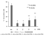
- FIG. 2 shows that the PPR protein of the present invention suppressed the formation of RNA-foci in DM1 model cells.
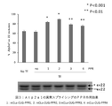
- Figure 3 shows that the PPR protein of the present invention ameliorated splicing defects in DM1 model cells.
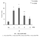
- Figure 4 shows that the PPR protein of the present invention improved muscle differentiation efficiency in DM1 model animals.
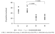
- FIG. 5 shows that the PPR protein of the present invention suppressed the formation of RNA-foci in DM1 model animals.
- FIG. 6 shows that the PPR protein of the present invention ameliorated splicing defects in DM1 model animals.
- Figure 7 shows that the PPR protein of the present invention improved muscle tonicity in DM1 model animals.
- FIG. 1 shows that the PPR protein of the invention specifically bound to the CUG repeat sequence.
- FIG. 2 shows that the PPR protein of the present invention suppressed the formation of RNA-foci in DM1
- FIG. 8 shows the relationship between the effect of improving aberrant splicing and the effect of improving muscle tonicity according to the present invention.
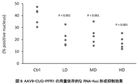
- FIG. 9 shows that AAV9-CUG-PPR1 dose-dependently suppressed the formation of RNA-foci in DM1 model animals.
- “Ctrl” is the PBS administration group
- "LD” is the AAV9-CUG-PPR1 low dose administration group
- "MD” is the AAV9-CUG-PPR1 medium dose administration group
- “HD” is the AAV9-CUG-PPR1 high dose.
- Administration groups are indicated.
- the vertical axis indicates the RNA-foci-positive cell rate.
- FIG. 10 shows that AAV9-CUG-PPR1 ameliorated abnormal splicing in DM1 model animals in a dose-dependent manner.
- the left figure shows the results of Clcn1, and the right figure shows the results of the abnormal splicing detection system of Atp2a1.
- “Ctrl” is the PBS administration group
- "LD” is the AAV9-CUG-PPR1 low dose administration group
- "MD” is the AAV9-CUG-PPR1 medium dose administration group
- “HD” is the AAV9-CUG-PPR1 high dose.
- Administration groups are indicated.
- the vertical axis indicates the content (%) of normal splicing isoforms.
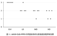
- FIG. 11 shows that AAV9-CUG-PPR1 dose-dependently improved muscle tonicity in DM1 model animals.
- the vertical axis indicates the myotonia score, and the frequency of myotonic discharge decreases in the order of scores 3, 2, 1, and 0. The frequency and severity of myotonic discharges are correlated.
- “Ctrl” is the PBS administration group
- "LD” is the AAV9-CUG-PPR1 low dose administration group
- MD is the AAV9-CUG-PPR1 medium dose administration group
- “HD” is the AAV9-CUG-PPR1 high dose.
- FIG. 12 shows the relationship between the administration dose of AAV9-CUG-PPR1 and the expression level of PPR mRNA.
- the internal standard uses GAPDH. Each individual point indicates the amount of PPR mRNA expression in the thigh muscle of each individual mouse.
- the vertical axis is displayed in Log display.
- “Ctrl” is the PBS administration group
- "LD” is the AAV9-CUG-PPR1 low dose administration group
- "MD” is the AAV9-CUG-PPR1 medium dose administration group
- “HD” is the AAV9-CUG-PPR1 high dose.
- FIG. 13 shows that AAV9-CUG-PPR1 suppressed RNA-foci formation in DM1 model animals in a time-dependent manner.
- “Ctrl” indicates the PBS-administered group.
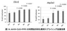
- FIG. 14 shows that AAV9-CUG-PPR1 ameliorated abnormal splicing in DM1 model animals in a time-dependent manner.
- the left figure shows the results of Clcn1, and the right figure shows the results of the abnormal splicing detection system of Atp2a1.
- “Ctrl” indicates the PBS-administered group.
- "2w”, “4w”, “8w”, and "16w” indicate test periods after administration of AAV9-CUG-PPR1, respectively.
- FIG. 15 shows that AAV9-CUG-PPR1 improved muscle tonicity in DM1 model animals in a time-dependent manner.
- the vertical axis indicates the myotonia score, and the frequency of myotonic discharge decreases in the order of scores 3, 2, 1, and 0. The frequency and severity of myotonic discharges are correlated.
- “Ctrl” indicates the PBS-administered group.
- "2w”, “4w”, “8w”, and "16w” indicate test periods after administration of AAV9-CUG-PPR1, respectively.
- FIG. 16 shows global normalization of splicing abnormalities after administration of AAV9-CUG-PPR1.
- FIG. 16a shows how splicing events (left) with changes in PSI (Percent spliced in) of 0.2 or more between WT and PBS-administered HSA-LR mice were observed in PPR-administered HSA-LR mice (right). It is the figure which showed whether it changed.
- FIG. 16b is a comparison of Tanner et al.
- FIG. 2 shows changes for DM1-associated splicing events identified in (2021). Black circles indicate signals from normal mice, blue squares indicate signals from PBS-administered drug mice, and red triangles indicate signals from PPR-administered drug mice.
- Figure 16c shows the results of Gene ontology analysis for the genes at each point in Figure a.
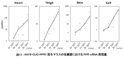
- FIG. 17 shows the expression level of PPR mRNA in each organ of AAV9-CUG-PPR1-administered mice.
- the vertical axis indicates the relative expression level in Log representation when the expression level in mice administered with 1 ⁇ 10 13 vg/kg is set to 1.
- GAPDH mRNA expression level is used as an internal standard.
- the horizontal axis indicates the AAV9-CUG-PPR1 dose (vg/kg body weight of mouse).
- FIG. 18 shows the persistence of mRNA expression in each organ of AAV9-CUG-PPR1-administered mice.
- the vertical axis indicates the relative expression level when the expression level of one mouse after 2 weeks of AAV administration (Day 14) is set to 1.
- GAPDH mRNA expression level is used as an internal standard.
- “Ctrl” indicates the PBS group, and "14", “28", "56", "112", and "183” indicate the number of days after administration.
- the values for each individual mouse are plotted with circles, and the mean values are connected by lines.
- PPR motif and PPR protein When the term "PPR motif" is used in the present disclosure, the E value obtained by PF01535 in Pfam and PS51375 in Prosite when analyzing the amino acid sequence with a protein domain search program on the Web is less than or equal to a predetermined value, unless otherwise specified.
- the position numbers of the amino acids that make up the PPR motif defined in the present disclosure are almost synonymous with PF01535, while the amino acid position of PS51375 minus 2 (e.g., No. 1 of the present invention ⁇ No. 3 of PS51375) Equivalent to.
- the second amino acid from the rear (C-terminal side) of the amino acid constituting the PPR motif or two N amino acids for the first amino acid of the next PPR motif It is the terminal side, that is, the -2nd amino acid. If the next PPR motif is not unambiguously identified, the two previous amino acids are designated as "ii" for the first amino acid of the next helix structure.
- the conserved amino acid sequence of the PPR motif is less conserved at the amino acid level, but the two ⁇ -helices are well conserved on the secondary structure.
- a typical PPR motif consists of 35 amino acids, but its length varies from 30 to 38 amino acids.
- the basic skeleton of the PPR motif is known to be represented by Formula 1.
- Helix A is a 12 amino acid long portion capable of forming an ⁇ -helical structure and is represented by Formula 2,
- a 1 to A 12 each independently represent an amino acid;
- X is absent or is a moiety consisting of 1-9 amino acids in length;
- Helix B is a portion capable of forming an ⁇ -helical structure, consisting of 11-13 amino acids in length;
- L is a moiety of Formula 3, 2-7 amino acids in length;
- each amino acid is numbered from the C-terminal side with “i” (-1), “ii” (-2), However, L iii to L vii may not exist.
- the term “PPR protein” in the present disclosure refers to a PPR protein having one or more, preferably two or more, of the above-described PPR motifs.
- the term “protein” refers to any substance consisting of a polypeptide (a chain in which a plurality of amino acids are peptide-bonded), and includes those consisting of relatively low-molecular-weight polypeptides, unless otherwise specified.
- amino acid may refer to ordinary amino acid molecules, and may also refer to amino acid residues constituting peptide chains. Which one is meant will be clear to a person skilled in the art from the context.
- the term “selective” or “specific” with respect to the binding activity of the PPR motif to RNA bases means that, unless otherwise specified, the binding activity to any one of the RNA bases is Higher than the binding activity to bases. This selectivity or specificity can be confirmed by designing experiments based on known methods by those skilled in the art, or can be determined by calculation by those skilled in the art.
- RNA base refers to a ribonucleotide base that constitutes RNA. Specifically, adenine (A), guanine (G), cytosine (C), or uracil ( U).
- A adenine
- G guanine
- C cytosine
- U uracil
- PPR proteins can be selective for bases in RNA, they do not bind to nucleic acid monomers.
- PPR proteins are abundant in plants, and about 500 proteins and about 5000 motifs can be found in Arabidopsis thaliana. PPR motifs and PPR proteins with diverse amino acid sequences are also present in many land plants such as rice, poplar, and selaginella. In the present invention, naturally occurring PPR motifs and PPR proteins may be used, or PPR motifs and PPR proteins designed based on the method disclosed in WO2013/058404, for example, may be used. Specifically, desired PPR motifs and PPR proteins can be designed based on the following information disclosed in WO2013/058404.
- the present invention can utilize the knowledge about the combination of three amino acids A 1 , A 4 and Lii and/or the combination of two amino acids A 4 and Lii disclosed in WO2013 / 058404 . can.
- the PPR motif binds strongly to A, but to G, U and C has a selective RNA base-binding ability that does not bind to (3-13) If the combination of three amino acids A 1 , A 4 and L ii is, in order, isoleucine, methionine, aspartic acid, the PPR motif binds strongly to U and then to C It has selective RNA base binding ability, binding to A and G but not to A and G.
- the above rule statistically determines the possibility that the PPR motif binds to the target base by the combination of amino acids (A 1 , A 4 , L ii ) or (A 4 , L ii ) of the PPR motif. It does not mean that a PPR motif (or PPR protein) that binds to the target base (or base sequence) with 100% probability cannot be produced according to the above rule.
- a person skilled in the art can produce several types to several tens of types of candidate PPR proteins based on the above-mentioned rules, so that a PPR motif (or PPR protein) can be obtained.
- the selection of a desired PPR protein through production and confirmation of candidate PPR proteins can be performed by those skilled in the art within the scope of ordinary trial and error, and does not impose an undue burden on those skilled in the art.
- PPR motif can recognize a specific base in RNA. Then, according to the present invention, a PPR motif selective for each of A, U, G and C can be selected or designed by appropriately selecting amino acids at specific positions. Furthermore, proteins containing suitable stretches of such PPR motifs can specifically or selectively recognize corresponding base sequences. Furthermore, the above findings allow the design of proteins with PPR motifs that can selectively bind to desired RNA bases and multiple PPR motifs that can sequence-specifically bind to desired RNAs. In designing, the sequence information of the natural PPR motif may be referred to for the portions other than the amino acids at the important positions in the PPR motif.
- the number of repeats of the PPR motif can be appropriately determined according to the target sequence, and can be, for example, 2 or more, and can be 2-30.
- the present invention relates to a PPR protein that specifically binds to mRNA containing an abnormally expanded CUG repeat sequence that causes DM1.
- the PPR protein according to the present invention can be produced by linking multiple sets of [a C-binding PPR motif, a U-binding PPR motif, and a G-binding PPR motif] based on the theory described above. can.
- the first PPR motif of the PPR protein according to the present invention does not necessarily have to be a PPR motif that binds to C, as long as the entire PPR protein specifically binds to the CUG repeat sequence, the first PPR motif binds to U.
- PPR motif that binds to G or a PPR motif that binds to G.
- the number of PPR motifs contained in the PPR protein need not be a multiple of 3, as long as the PPR protein of the present invention as a whole has a configuration that specifically binds to the CUG repeat sequence.
- Non-limiting examples of C-linked PPR motifs that may be used in the present invention include PPR motifs having the following sequences.
- sequence homology or sequence identity
- binding to C may be used.
- substitutions, additions, and/or deletions and having C-binding properties may be used.
- the amino acid substitution may be, for example, conservative amino acid substitution known in the art.
- Non-limiting examples of U-linked PPR motifs that may be used in the present invention include PPR motifs having the following sequences.
- 80 % or more (preferably , 85 % or more, 90% or more, 95% or more, 96% or more, 97% or more, 98% or more, 99% or more) sequence homology (or sequence identity) and a PPR motif that has a binding property to U may be used.
- substitutions, additions, and/or deletions and having binding properties for U may be used.
- the amino acid substitution may be, for example, conservative amino acid substitution known in the art.
- Non-limiting examples of G-linked PPR motifs that may be used in the present invention include PPR motifs having the following sequences.
- 80 % or more (preferably , 85 % or more, 90% or more, 95% or more, 96% or more, 97% or more, 98% or more, 99% or more) sequence homology (or sequence identity) and G-binding PPR motif may be used.
- substitutions, additions, and/or deletions and having binding properties for G may be used.
- the amino acid substitution may be, for example, conservative amino acid substitution known in the art.
- searches and analyzes for the identity of nucleotide sequences or amino acid sequences can be performed by algorithms or programs (eg, BLASTN, BLASTP, BLASTX, ClustalW) well known to those skilled in the art. Parameters when using programs can be appropriately set by those skilled in the art, and default parameters of each program may be used. Specific techniques of these analysis methods are also well known to those skilled in the art.
- amino acids with similar properties refer to amino acids with similar physical properties such as hydropathy, charge, pKa, solubility, and the like.
- the effect of the present invention is exerted by binding of the PPR protein of the present invention to mRNA containing an abnormally expanded CUG repeat sequence in the target cell.
- the means for delivering the PPR protein into the cells of the subject is not limited, and a nucleic acid encoding the PPR protein of the present invention may be delivered into the cells of the subject to express the PPR protein of the present invention within the cells of the subject. Alternatively, the PPR protein of the present invention itself may be delivered into the subject's cells.
- the means for delivering proteins and nucleic acids is not limited, and various means known in the art can be used.
- Non-limiting examples of means for delivery of nucleic acids or proteins into cells of a subject that can be used in the present invention include liposomes, lipid nanoparticles (LNPs), polymeric micelles, emulsions, polymeric microcapsules, antibodies -nucleic acid complexes, antibody-drug complexes, viruses, and the like.
- Non-limiting examples of viral vectors that may be used in the present invention include adeno-associated viral (AAV) vectors, adenoviral vectors, retroviral vectors, lentiviral vectors, or herpes simplex viral vectors.
- AAV adeno-associated viral
- adenoviral vectors adenoviral vectors
- retroviral vectors retroviral vectors
- lentiviral vectors lentiviral vectors
- herpes simplex viral vectors Alternatively, it can be appropriately selected by those skilled in the art depending on the tropism to tissues, the necessity of nucleic acid integration into the host genome, and the like.
- AAV vectors are particularly suitable for use in the present invention because they are capable of infecting both dividing and quiescent cells, and can transfer genetic material to a wide variety of cell types. Twelve serotypes of AAV (AAV1-AAV12) have been described to date, and all known serotypes are capable of infecting various types of tissue cells. Although the serotype of the AAV vector that may be used in the present invention is not limited, for example, AAV1 vector, AAV2 vector, AAV6 vector, which are known to have tropism for muscle tissue (skeletal muscle, cardiac muscle, and/or smooth muscle) A vector, AAV7 vector, AAV8 vector, AAV9 vector, AAV10 vector, AAV11 vector or AAV12 vector may be used.
- Treatment of DM1 using the present invention may be an embodiment of applying the present invention directly in vivo in a subject suffering from DM1 (in vivo), or treating cells or tissues ex vivo using the present invention. , may be introduced into the body of the subject (ex vivo).
- the method for producing an expression vector containing a nucleic acid encoding the PPR protein of the present invention is not limited, and a person skilled in the art can use a conventional method (e.g., use of a commercially available protein expression vector production kit, use of a commercially available virus expression vector production kit, production It can be manufactured by outsourcing to a contractor, etc.).
- the method for producing the PPR protein of the present invention is not limited, for example, a cell (e.g., The desired PPR protein can be obtained by selectively extracting and/or purifying the PPR protein from animal cells, plant cells, Escherichia coli, yeast, etc.) by a conventional method.
- CUG-PPR CUG repeat RNA sequence
- coli pellet add 1.5 mL of lysis buffer (20 mM Tris-HCl, pH 8.0, 150 mM NaCl, 0.5% NP-40, 1 mM MgCl 2 , 2 mg/ml lysozyme, 1 mM PMSF, 2 ⁇ l DNase) was added and frozen at -80°C for 20 minutes. Cells were freeze fractured with shaking at 25°C for 30 minutes. Subsequently, centrifugation was performed at 3700 rpm, 4° C. for 15 minutes, and the supernatant (E. coli lysate) containing soluble PPR protein was recovered and used in the following experiments.
- lysis buffer 20 mM Tris-HCl, pH 8.0, 150 mM NaCl, 0.5% NP-40, 1 mM MgCl 2 , 2 mg/ml lysozyme, 1 mM PMSF, 2 ⁇ l DNase
- PPR Protein-to-RNA Binding Test was performed using a method for testing the binding of PPR protein to biotinylated RNA on a streptavidin plate.
- a 30-base RNA containing a target CUG ⁇ 7 sequence (GACA CUGCUGCUGCUGCUGCUGCUG AUGCA (SEQ ID NO: 15)) and a 30-base RNA containing a non-target CAG ⁇ 6 (GACAUGC CAGCAGCAGCAGCAGCAG GACUG (SEQ ID NO: 16)
- An RNA probe modified with biotin at the 5' end was synthesized (requested by Greiner).
- biotinylated RNA probe 2.5 pmol was added to a streptavidin-coated plate (Cat No. 15502, Thermo Fisher) and allowed to react for 30 minutes at room temperature. Washed with probe wash buffer (20 mM Tris-HCl (pH 7.6), 150 mM NaCl, 5 mM MgCl 2 , 0.5% NP-40, 1 mM DTT, 0.1% BSA). For background measurement, wells containing lysis buffer without biotinylated RNA were also prepared (referred to as "-Probe").
- a blocking buffer (20 mM Tris-HCl (pH 7.6), 150 mM NaCl, 5 mM MgCl 2 , 0.5% NP-40, 1 mM DTT, 1% BSA) was added to block the plate surface at room temperature for 30 minutes. gone. 100 ⁇ L of E. coli lysate containing a luciferase-fused PPR protein with a luminescence level of 1.5 ⁇ 10 8 LU/ ⁇ L was added to the wells, and the binding reaction was allowed to proceed for 30 minutes at room temperature.
- washing buffer (20 mM Tris-HCl (pH 7.6), 150 mM NaCl, 5 mM MgCl 2 , 0.5% NP-40, 1 mM DTT) and diluted 2500-fold with the washing buffer.
- 40 ⁇ L of luciferase substrate Promega, E151A was added to the wells and allowed to react for 5 minutes, luminescence was measured with a plate reader (PerkinElmer, Cat No. 5103-35).
- Example 2 Effect of PPR on RNA aggregate formation in DM1 model cells
- PPR protein was allowed to act on DM1 model cells, and the number of RNA aggregates was measured by FISH (fluorescent in situ hybridization) method.
- DM1 model cells As a DM1 cell model, C2C12 cells into which a DMPK gene region containing 800 CTG repeats was introduced (C2C12-DMPK800R, Nucleic Acids Research, 2014, 42 (10), 6591-6602) were used. C2C12-DMPK800 was produced by the following procedure. That is, both a plasmid (pLC16) containing a sequence in which 800CTG was inserted into the DMPK gene 3' untranslated region and a plasmid expressing PhiC31 integrase were applied to mouse myoblast cell line C2C12 using Nucleofector (trade name, Lonza).
- Nucleofector trade name, Lonza
- stable expression strains were selected using selective medium supplemented with puromycin. Subsequently, the stable expression strain was transfected with a plasmid expressing Cre recombinase, and a selective medium supplemented with hygromycin was used to select clones that induce transcription of mRNA having 800 CUG repeats.
- sequences of the PPR proteins (PPR1 to PPR4) encoded in each expression plasmid vector are the same as the sequences of the PPR proteins listed in Table 1.
- DM1 model cells were cultured under conditions of 37° C. and 5% CO 2 using DMEM medium containing 10% FBS and penicillin/streptomycin. 200 ng of the plasmid DNA constructed above, 0.6 ⁇ L Fugene®-HD (Promega, E2311), and 200 ⁇ L Opti-MEM are mixed, added to the total well, and placed at 37° C., 5% CO 2 for 72 hours. After culturing, the cells were fixed with 3% paraformaldehyde for 15 minutes at room temperature. After fixation, the cells were washed twice with PBS and then permeabilized with PBS containing 0.5% Triton X-100 for 5 minutes.
- prehybridization treatment was performed for 10 minutes with 2 ⁇ SSC buffer containing 30% formamide. After that, hybridization was performed at 37° C. for 1 hour with 2 ⁇ SSC buffer containing 30% formamide, 2 ⁇ g/mL BSA, 66 ⁇ g/mL yeast tRNA, 2 mM vanadyl complex, and 1 ng/ ⁇ L Texas Red CAG probe. After post-hybridization treatment with 2 ⁇ SSC buffer containing 30% formamide at 42° C. for 30 minutes, the plate was washed once with 1 ⁇ SSC buffer and then twice with PBS. After fixing with Vectashield with DAPI (trade name, Vector Laboratories), the number of intranuclear RNA aggregates was counted under a fluorescence microscope (Keyence PZ-9000).
- Example 3 Effect of PPR protein on abnormal splicing in DM1 model cells
- DM1 model cell The same DM1 model cell (C2C12-DMPK800R) as in Example 2 was used.
- RT-PCR products were subjected to electrophoresis on a 2% agarose gel, stained with GelRed, and then normal and abnormal PCR products were quantified using an image analyzer (ChemiDoc Touch Imaging System, BioRad).
- Atp2a1 exon 22 RT primer Fw GCTCATGGTCCTCAAGATCTCAC (SEQ ID NO: 22)
- Rv GGGTCAGTGCCTCAGCTTTG (SEQ ID NO: 23)
- Results> The results are shown in FIG.
- the lower graph shows the results of electrophoresis of RT-PCR products
- the upper graph shows the proportion of normal type in RT-PCR products.
- the ratio of abnormal type was higher than that of normal type. It was shown that the action of the PPR protein significantly increased the ratio of the normal type in the DM1 model cells and improved the abnormal splicing.
- Example 4 Effect of PPR protein on abnormal muscle differentiation in DM1 model cells
- a PPR protein was allowed to act on DM1 model cells to verify whether or not the muscle differentiation efficiency of DM1 model cells was improved.
- DM1 model cell The same DM1 model cell (C2C12-DMPK800R) as in Example 2 was used.
- the results are shown in FIG.
- the fusion index was about 3 in the empty vector-administered group (indicated as "no" in the figure).
- the fusion index was about 5 to 15, indicating a significant improvement in muscle differentiation efficiency.
- Example 5 Effect of PPR in DM1 model mice
- AAV6 carrying the CUG-PPR gene was administered to DM1 model mice once to the tibialis anterior muscle, and the effect on muscle tonic symptoms in the tibialis anterior muscle, the effect on abnormal splicing of the Atp2a1 gene and the skeletal muscle chloride channel (Clcn1) gene, Also, the number of RNA aggregates formed was verified.
- HSA LR mice Animals used HSA LR mice (transferred from Dr. Charles Thornton (University of Rochester) and bred at the experimental animal facility of Osaka University) were used as DM1 model mice.
- HSA LR mice are transgenic animals in which a 220-fold extension of CTG repeats in the 3′ untranslated region of the hACTA gene (a human gene that is constitutively expressed in muscle cells) is integrated into the genome. Symptoms of DM1 appear due to the expression of mRNA with an extended sequence (Science, 2000, 289(5485), 1769-73).
- wild-type mice FVB/NJcl mice, purchased from CLEA Japan
- 14-week-old male and female mice were used.
- AAV vector AAVpro (trademark) Helper Free System (AAV6), Takara, 6651 component
- AAV6 Helper Free System
- Takara 6651 component
- the fused gene was inserted.
- the expression size of the constructed gene was confirmed by PCR, and the inserted sequence was confirmed by sequencing. Expression of the fusion gene is controlled by the CMV promoter and human growth hormone polyA signal.
- AAV vector constructed in the previous section was used as a pRC6 vector, and 20 ⁇ g of each of the phelper vector (both components of AAVpro (trademark) Helper Free System (AAV6), Takara, 6651) were added to Polyethylene, Linear, MW25000 (Polysciences, Inc., 23966- 1) was used to introduce the gene into 2.5 ⁇ 10 8 cells of AAVproTM HEK293T cell line (Takara, 632273). AAV-producing cells were obtained 3 days after transfection. AAV purification used AAVproTM Purification Kit Maxi (all serotype) (Takara, 6666). AAV production kit for real time PCR ver.
- AAVproTM Purification Kit Maxi all serotype
- AAV6-mCLo-CUG-PPR1, AAV6-mCLo-CUG-PPR2 and AAV6-mCLo-empty were used in the subsequent tests.
- the sequences of the PPR proteins (PPR1, PPR2) encoded in each AAV vector are the same as the sequences of the PPR proteins listed in Table 1.
- RNA Aggregates Fifty-six days after the final administration, the tibialis anterior muscle was collected from the mouse and sectioned. After washing twice with PBS, the cells were permeabilized with PBS containing 0.5% Triton X-100 for 5 minutes. Next, prehybridization treatment was performed for 10 minutes with 2 ⁇ SSC buffer containing 30% formamide. After that, hybridization was performed at 37° C. for 1 hour with 2 ⁇ SSC buffer containing 30% formamide, 2 ⁇ g/mL BSA, 66 ⁇ g/mL yeast tRNA, 2 mM vanadyl complex, and 1 ng/ ⁇ L Texas Red CAG probe.
- Tibialis anterior muscles were harvested from mice 56 days after the final administration. After extracting total RNA using TRI Reagent (trade name, MRC), cDNA was prepared using SuperScript III First Strand Synthesis System (Invitrogen). After treating the cDNA with RNaseH, RT-PCR was performed using the same Atp2a1 exon22 RT primer as in Example 2 and the following Clcn1 exon 7a RT primer.
- RT-PCR product was electrophoresed on a 2% agarose gel and stained with GelRed, normal PCR products and abnormal PCR products were quantified with an image analyzer (ChemiDoc Touch Imaging System, BioRad), and RT - The percentage of normal in the PCR products was calculated. t-test was used for statistical analysis.
- Clcn1 exon 7a RT primer Fw TGAAGGAATACCTCACACTCAAGG (SEQ ID NO: 24)
- Rv CACGGAACACAAAGGCACTG (SEQ ID NO: 25)
- RNA-foci analysis The results of RNA-foci analysis are shown in FIG.
- the PBS-administered group and the AAV6-mCLo-empty-administered group showed an RNA-foci-positive cell rate of about 30%.
- a significant decrease in the RNA-foci positive rate was observed in the AAV6-mCLo-CUG-PPR1 and AAV6-mCLo-CUG-PPR2 administration groups.
- Fig. 6 shows the evaluation results of splicing abnormalities.
- the PBS-administered DM1 group and the AAV6-mCLo-Empty-administered MyoD group exhibited a normal type rate of about 60%.
- the ratio of normal RT-PCR products was significantly increased, suggesting that PPR protein administration improved the abnormal splicing of the Atp2a1 gene.
- Clcn1 it was also found that administration of PPR improved the splicing abnormality of the Clcn1 gene.
- Fig. 7 shows the analysis results of muscle tonicity. Severity 2 myotonia was detected in 5 out of 5 animals of the PBS-administered group and the AAV6-mCLo-empty-administered group. In both the AAV6-mCLo-CUG-PPR1 and AAV6-mCLo-CUG-PPR2-administered DM1-administered groups, 2 out of 5 animals showed severity 1, and the remaining 3 animals showed severity 2. In addition, two improved cases belonged to the experimental group with a high effect of improving abnormal Clcn1 splicing (FIG. 8).
- Example 6 Dose dependence test of efficacy using DM1 model mice
- AAV9 carrying the CUG-PPR gene was added to DM1 model mice at 3 ⁇ 10 13 , 1 ⁇ 10 14 , and 3 ⁇ 10 14 vg/kg.
- a single dose was administered into the tail vein, and the effect on myotonic symptoms in thigh muscles, the effect on abnormal splicing of Atp2a1 gene and skeletal muscle chloride channel (Clcn1) gene, and the number of RNA aggregates formed were examined.
- HSA LR mice Animals used HSA LR mice (transferred from Dr. Charles Thornton (University of Rochester) and bred at the experimental animal facility of Osaka University) were used as DM1 model mice.
- HSA LR mice are transgenic animals in which a 220-fold extension of CTG repeats in the 3′ untranslated region of the hACTA gene (a human gene that is constitutively expressed in muscle cells) is integrated into the genome. Symptoms of DM1 appear due to the expression of mRNA with an extended sequence (Science, 2000, 289(5485), 1769-73).
- wild-type mice FVB/NJcl mice, purchased from CLEA Japan
- 14-week-old male and female mice were used for the experiment.
- PPR protein production of AAV vector
- a gene in which a PPR protein and a 3 ⁇ nuclear localization signal were fused in this order was inserted into the multiple cloning site of a pAAV-CMV vector (AAVproTM Helper Free System, Takara, 6651 component).
- the expression size of the constructed gene was confirmed by PCR, and the inserted sequence was confirmed by sequencing. Expression of the fusion gene is controlled by the CMV promoter and human growth hormone polyA signal.
- AAV vector (Preparation of AAV vector)
- the AAV vector constructed in the previous section was outsourced to SignaGen Laboratories (hereinafter abbreviated as SG).
- Packaging was carried out by co-transfecting the Rep/Cap plasmid and Helper plasmid owned by SG and the AAV vector sent from our company into the packaging cell line.
- a cell lysate containing the packaged AAV was purified by density gradient centrifugation and the titer was measured.
- the AAV obtained was 1.16 ⁇ 10 14 vg/ml and the volume was 5 ml. This was used in subsequent experiments as AAV9-CUG-PPR1.
- the sequence of the PPR protein (PPR1) encoded in each AAV vector is the same as the PPR protein sequence shown in Table 1.
- AAV9-CUG-PPR1 low dose administration group (hereinafter abbreviated as Low dose: LD), AAV9-CUG-PPR1 middle dose administration group (hereinafter abbreviated as Middle dose: MD),
- AAV9-CUG-PPR1 high dose administration group (hereinafter abbreviated as High dose: HD)
- PBS administration group (hereinafter abbreviated as PBS).
- Each group n 5 ( ⁇ 3, ⁇ 2 mice).
- the dose of AAV was 3 ⁇ 10 13 vg/kg for LD, 1 ⁇ 10 14 vg/kg for MD, and 3 ⁇ 10 14 vg/kg for HD, and was administered once into the tail vein.
- the same volume of PBS was administered once to the PBS-administered group.
- RNA Aggregates Twenty-eight days after the final administration, thigh muscles were collected from the mice and sections were prepared. After washing twice with PBS, the cells were permeabilized with PBS containing 0.5% Triton X-100 for 5 minutes. Next, prehybridization treatment was performed for 10 minutes with 2 ⁇ SSC buffer containing 30% formamide. After that, hybridization was performed at 37° C. for 1 hour with 2 ⁇ SSC buffer containing 30% formamide, 2 ⁇ g/mL BSA, 66 ⁇ g/mL yeast tRNA, 2 mM vanadyl complex, and 1 ng/ ⁇ L Texas Red CAG probe.
- RNA samples were collected from the mice. After extracting total RNA using TRI ReagentTM (MRC), cDNA was prepared using SuperScript III First Strand Synthesis System (Invitrogen). After treating the cDNA with RNaseH, RT-PCR was performed using the same Atp2a1 exon22 RT primer as in Example 2 and the following Clcn1 exon 7a RT primer. After the RT-PCR product was electrophoresed on a 2% agarose gel and stained with GelRed, normal PCR products and abnormal PCR products were quantified with an image analyzer (ChemiDoc Touch Imaging System, BioRad), and RT - The percentage of normal in the PCR products was calculated. t-test was used for statistical analysis.
- muscle tonicity (myotonia) Twenty-eight days after the final administration, the phenomenon of electrical tonicity in the mouse thigh muscle was analyzed by needle electromyography under anesthesia. Specifically, the occurrence frequency of the electrical muscle tonic phenomenon when the needle electrode was inserted into the thigh muscle of the mouse 20 times was evaluated according to the following four grades. Severity 0: No electrical muscle tonic phenomenon is observed. Severity 1: Electrical myotonia is present in less than 50% of cases. Severity 2: Electrical myotonia is observed in 50% or more and less than 90% of all cases. Severity 3: 90% or more of electrical myotonia is observed.
- RNA 300 ⁇ L of the aqueous phase was collected and subjected to a Maxwell RSC Instrument, a nucleic acid purifier, to obtain purified total-RNA. The concentration and purity of RNA were measured by NanoDrop 8000 absorbance. In order to confirm the degree of degradation of the obtained RNA, 20 to 250 ng/ ⁇ L of total-RNA was used to perform electrophoresis using LabChip GX Touch HT (PerkinElmer, CLS137031J). It was confirmed that decomposition had not progressed. For synthesis of cDNA, 20 to 100 ng/ ⁇ L of obtained RNA was subjected to reverse transcription reaction according to the product protocol of SuperScript III Reverse Transcriptase (Thermo Fisher Scientific (Life Technologies), 18080085).
- the amount of PPR mRNA and GAPDH mRNA was measured by mixing the prepared cDNA, Brilliant III Ultra-Fast SYBR Green QPCR Master Mix (Agilent, 600882) and the primers described below according to the manual, and then using a real-time PCR system Aria MX (Agilent). Amplified and quantitatively analyzed.
- PPR quantitative PCR primer Fw GATGAGGCTTTGGAACTGTTTG (SEQ ID NO: 26)
- Rv TCTCTGGCTCTGCCGGCCTTGC (SEQ ID NO: 27)
- GAPDH quantitative PCR primer Fw ATCATCCCTGCCTCTACTGG (SEQ ID NO: 28)
- Rv CTGCTTCACCACCTTCTTGA (SEQ ID NO: 29)
- RNA-FISH method was performed using the obtained thigh muscle organ section, and the effect of suppressing RNA-foci formation by AAV9-CUG-PPR1 was verified. A formation inhibitory effect was confirmed.
- the rate of RNA-foci-positive cells was 39% in the PBS administration group, 18% in the AAV9-CUG-PPR1 low dose group, 23% in the AAV9-CUG-PPR1 medium dose group, and 17% in the AAV9-CUG-PPR1 high dose group. formation inhibitory effect was observed.
- Fig. 10 shows the evaluation results of splicing abnormalities.
- Total RNA was collected from the collected organs, reverse transcription PCR was performed, and abnormal splicing of the Clcn1 gene and Atp2a1 gene was evaluated.
- Aberrant splicing of Clcn1 is a Cl channel that is known to directly cause myotonic discharge.
- Atp2a1 is also known as a Ca transporter related to myotonic discharge and a sensitive marker gene.
- the Clcn1 detection system showed 58% normal splicing in the PBS administration group, 72% in the AAV9-CUG-PPR1 low dose group, 78% in the AAV9-CUG-PPR1 medium dose group, and 78% in the AAV9
- the -CUG-PPR1 high dose group showed 82%, confirming a dose-dependent splicing improvement.
- the Atp2a1 detection system showed 25% normal splicing in the PBS-administered group, 56% in the AAV9-CUG-PPR1 low-dose group, 60% in the AAV9-CUG-PPR1 medium-dose group, and 60% in the AAV9-CUG-PPR1 high-dose group.
- the dose group showed 64%, showing a dose-dependent splicing improvement.
- the effect of reducing muscle tonic discharge by PPR was expressed as a score in 4 stages based on the number of myotonia discharges. That is, a score of 0 is 0 out of 20, a score of 1 is 1 to 9 out of 20, a score of 2 is 10 to 19 out of 20, and a score of 3 is 20 out of 20 myotonic discharges. means detection.
- the PBS administration group scored 3
- the AAV9-CUG-PPR1 low dose group showed a score of 3 in 1/5 cases
- the score of 2 in 4/5 cases. 4/5 cases showed a score of 2
- 4/5 cases showed a score of 2
- 1/5 cases showed a score of 1
- 3/5 cases showed a score of 2/5 cases showed an improvement to a score of 1 (Fig. 11).
- RNA was extracted from the thigh muscle of the analyzed organ, and quantitative PCR of PPR mRNA was performed. As a result of RT-qPCR, dose-dependent PPR mRNA expression was confirmed (Fig. 12).
- a relative value was used when the value of the individual with the lowest signal in the AAV9-CUG-PPR1 low-dose administration group was set to 1 using GAPDH as an internal standard. Taking the median value of each group, the PBS administration group was 0.2, the low dose administration group was 0.7, the medium dose administration group was 1.8, and the high dose administration group was 2.6. A significant PPR expression was confirmed (FIG. 12).
- AAV9-CUG-PPR1 dose-dependently suppresses myotonic discharge, suppresses the formation of RNA-foci, and improves aberrant splicing.
- Example 7 Time axis change in drug efficacy in DM1 model mice.
- a single dose of AAV9 carrying the CUG-PPR gene was administered to DM1 model mice into the tail vein, and at each time point, muscle tonic symptoms in the thigh muscles were tested.
- the effect, the effect on abnormal splicing of the Atp2a1 gene and the skeletal muscle chloride channel (Clcn1) gene, and the number of RNA aggregates formed were verified.
- HSA LR mice Animals used HSA LR mice (transferred from Dr. Charles Thornton (University of Rochester) and bred at the experimental animal facility of Osaka University) were used as DM1 model mice.
- HSA LR mice are transgenic animals in which a 220-fold extension of CTG repeats in the 3′ untranslated region of the hACTA gene (a human gene that is constitutively expressed in muscle cells) is integrated into the genome. Symptoms of DM1 appear due to the expression of mRNA with an extended sequence (Science, 2000, 289(5485), 1769-73).
- wild-type mice FVB/NJcl mice, purchased from CLEA Japan
- PPR protein production of AAV vector
- a gene fusing a PPR protein and a 3 ⁇ nuclear localization signal in this order was inserted into the multiple cloning site of a pAAV-CMV vector (AAVproTM Helper Free System, Takara, 6651 component).
- the expression size of the constructed gene was confirmed by PCR, and the inserted sequence was confirmed by sequencing. Expression of the fusion gene is controlled by the CMV promoter and human growth hormone polyA signal.
- AAV vector (Preparation of AAV vector) The production of the AAV vector constructed in the preceding section was entrusted to SignaGen Laboratories (hereinafter abbreviated as SG). Packaging was carried out by co-transfecting the Rep/Cap plasmid and Helper plasmid owned by SG and the AAV vector sent by our company into the packaging cell line. A cell lysate containing the packaged AAV was purified by density gradient centrifugation and the titer was measured. The AAV obtained was 1.16 ⁇ 10 14 vg/ml and the volume was 5 ml. This was used in subsequent experiments as AAV9-CUG-PPR1. The sequence of the PPR protein (PPR1) encoded in each AAV vector is the same as the PPR protein sequence shown in Table 1.
- RNA Aggregates 14, 28, 56, or 112 days after the final administration thigh muscles were collected from mice and sections were prepared. After washing twice with PBS, the cells were permeabilized with PBS containing 0.5% Triton X-100 for 5 minutes. Next, prehybridization treatment was performed for 10 minutes with 2 ⁇ SSC buffer containing 30% formamide. After that, hybridization was performed at 37° C. for 1 hour with 2 ⁇ SSC buffer containing 30% formamide, 2 ⁇ g/mL BSA, 66 ⁇ g/mL yeast tRNA, 2 mM vanadyl complex, and 1 ng/ ⁇ L Texas Red CAG probe.
- Thigh muscles were collected from mice 14, 28, 56, or 112 days after the final administration. After extracting total RNA using TRI ReagentTM (MRC), cDNA was prepared using SuperScript III First Strand Synthesis System (Invitrogen). After treating the cDNA with RNaseH, RT-PCR was performed using the same Atp2a1 exon22 RT primer as in Example 2 and the following Clcn1 exon 7a RT primer.
- MRC TRI ReagentTM
- cDNA was prepared using SuperScript III First Strand Synthesis System (Invitrogen). After treating the cDNA with RNaseH, RT-PCR was performed using the same Atp2a1 exon22 RT primer as in Example 2 and the following Clcn1 exon 7a RT primer.
- RT-PCR product was electrophoresed on a 2% agarose gel and stained with GelRed, normal PCR products and abnormal PCR products were quantified with an image analyzer (ChemiDoc Touch Imaging System, BioRad), and RT - The percentage of normal in the PCR products was calculated. t-test was used for statistical analysis.
- RNA-FISH method was performed using the obtained thigh muscle organ section, and the effect of suppressing RNA-foci formation by AAV9-CUG-PPR1 was verified. A formation inhibitory effect was confirmed.
- the value of the PBS-administered group used in the dose-dependence test of Example 6 was used as a control.
- the RNA-foci-positive cell rate in the control group was 39%, 36% in the group 14 days after administration of AAV9-CUG-PPR1, 23% in the group 28 days after administration, and 19% in the group 56 days after administration. In the group on the 112th day after administration, an 18% formation inhibitory effect was confirmed (Fig. 13).
- Fig. 14 shows the evaluation results of splicing abnormalities.
- Total RNA was collected from the collected organs, reverse transcription PCR was performed, and abnormal splicing of the Clcn1 gene and Atp2a1 gene was evaluated.
- Aberrant splicing of Clcn1 is a Cl channel that is known to directly cause myotonic discharge.
- Atp2a1 is also known as a Ca transporter related to myotonic discharge and a sensitive marker gene.
- the PBS-administered group shown in Example 6 showed 58% normal splicing.
- Splicing improvement was confirmed by 76% on the 28th day after administration of CUG-PPR1, 79% on the 56th day after administration of AAV9-CUG-PPR1, and 80% on the 112th day after administration of AAV9-CUG-PPR1.
- the PBS-administered group shown in Example 6 showed 25% normal splicing.
- Splicing was improved by 63% on day 28, 76% on day 56 after administration of AAV9-CUG-PPR1, and 75% on day 112 after administration of AAV9-CUG-PPR1 (Fig. 14).
- the effect of reducing muscle tonic discharge by PPR was expressed as a score in 4 stages based on the number of myotonia discharges. That is, a score of 0 is 0 out of 20, a score of 1 is 1 to 9 out of 20, a score of 2 is 10 to 19 out of 20, and a score of 3 is 20 out of 20 myotonic discharges. means detection.
- the PBS administration group shown in Example 6 scored 3, and the experimental group on the 14th day after AAV9-CUG-PPR1 administration scored 3 in 4/5 cases and 1/5 cases.
- Example 8 Comprehensive improvement of DM1-related splicing abnormalities by AAV9-CUG-PPR1 administration
- Analytical organs A single dose of AAV9-CUG-PPR1 was administered to HSALR mice (14 weeks old) at 3 ⁇ 10 14 vg/kg into the tail vein, and thigh muscle tissue pieces were excised 8 weeks after administration (Day 56). served as the PPR treatment group.
- a single dose of the same volume of PBS was administered and excised thigh muscle was placed as a PPR untreated group. Thigh muscle tissue strips from genetically matched wild-type FVB/NJcl mice were used as the wild-type group.
- RNA sequencing (Illumina Hiseq 2 ⁇ 150 bp sequencing) was performed as follows. The ribosomal RNA in the total RNA was removed from the sent RNA by Poly A selection method. RNA was reverse transcribed, cDNA was synthesized, and an adaptor-attached library was prepared. Sequencing was performed, and adapter trimming was performed using Trimmomatic (v0.33)59 on the obtained read information.
- the trimmed read sequences were aligned with STAR (v2.7.9a) using GRCm39 (obtained from Ensemble) as the reference genome. Identification of splicing events was detected with rMATS (v3.2.5) based on STAR-aligned files (.bam). To identify altered splicing events, the wild-type group and the PPR-untreated group (PBS-administered HSALR mice) are compared, and the presence or absence of variation is determined using "p-adjusted-value" and "InclLevelDifference" as indices. Only candidates defined as fluctuating were extracted.
- a cut-off threshold of FDR (False Discovery Rate)-adjusted P value ⁇ 0.05 was used for comparison between groups.
- Percent spliced in index (PSI) was defined as the percentage of spliced-in transcript abundance to the total number of reads for the gene of interest. Gene ontology analysis used Metascape (https://metascape.org/gp/index.html#/main/step1).
- Thigh muscle isolated from the wild-type mouse (14 weeks old) described in the previous section (hereinafter referred to as wild-type group)
- Thigh muscle isolated 8 weeks after administration of PBS to HSALR mice (hereinafter referred to as PPR untreated group )
- total RNA was extracted from the excised thigh muscle (hereinafter referred to as the PPR-treated group) 8 weeks after administration of AAV9-CUG-PPR1 (Day 56), and comprehensive analysis of RNA expressed by next-generation sequencing technology carried out.
- PSI percent spliced in index
- Example 9 PPR mRNA expression level analysis in each organ of normal mice to which AAV9-CUG-PPR1 was applied]
- RNA was subjected to reverse transcription reaction according to the product procedure of SuperScript III Reverse Transcriptase (Thermo Fisher Scientific (Life Technologies), 18080085).
- Quantitative PCR was performed using the real-time PCR system Aria MX (Agilent) according to the general-purpose manual of Brilliant III Ultra-Fast SYBR Green QPCR Master Mix (Agilent, 600882).
- PPR quantitative PCR primer Fw GATGAGGCTTTGGAACTGTTTG (SEQ ID NO: 26)
- Rv TCTCTGGCTCTGCCGGCCTTGC (SEQ ID NO: 27)
- GAPDH quantitative PCR primer Fw ATCATCCCTGCCTCTACTGG (SEQ ID NO: 28)
- Rv CTGCTTCACCACCTTCTTGA (SEQ ID NO: 29)
- Results of quantitative PCR are shown in FIG.
- a dose-dependent increase in PPR expression level was confirmed in each tissue, and the maximum expression level was shown in the maximum dose group of 3 ⁇ 10 14 vg/kg.
- AAV9-CUG-PPR1 can be delivered and expressed systemically, suggesting that it is effective against DM1, a multiorgan disease that affects the heart, in addition to skeletal muscle and smooth muscle. rice field.
- Example 10 PPR mRNA expression persistence analysis in each organ of normal mice to which AAV9-CUG-PPR1 was applied]
- RNA was subjected to reverse transcription reaction according to the product protocol of SuperScript III Reverse Transcriptase (Thermo Fisher Scientific (Life Technologies), 18080085). Quantitative PCR was performed using a real-time PCR system Aria MX (Agilent) according to the general-purpose manual of Brilliant III Ultra-Fast SYBR Green QPCR Master Mix (Agilent, 600882).
- PPR quantitative PCR primer Fw GATGAGGCTTTGGAACTGTTTG (SEQ ID NO: 26)
- Rv TCTCTGGCTCTGCCGGCCTTGC (SEQ ID NO: 27)
- GAPDH quantitative PCR primer Fw ATCATCCCTGCCTCTACTGG (SEQ ID NO: 28)
- Rv CTGCTTCACCACCTTCTTGA (SEQ ID NO: 29)
- Results of quantitative PCR are shown in FIG. As a result of quantitative PCR, sustained expression was confirmed even 183 days after administration, and a time-dependent increase in expression was also confirmed. Previous papers have shown that AAV9 expression can be maintained for a long period of time (6 months or more) when AAV9 is administered via the tail vein, and the results of this experiment are consistent with this finding.
- the present invention can produce desired effects over a long period of time with a small number of administrations to subjects.
Landscapes
- Health & Medical Sciences (AREA)
- Life Sciences & Earth Sciences (AREA)
- Chemical & Material Sciences (AREA)
- Engineering & Computer Science (AREA)
- Genetics & Genomics (AREA)
- General Health & Medical Sciences (AREA)
- Organic Chemistry (AREA)
- Bioinformatics & Cheminformatics (AREA)
- Medicinal Chemistry (AREA)
- Biotechnology (AREA)
- Molecular Biology (AREA)
- Pharmacology & Pharmacy (AREA)
- Veterinary Medicine (AREA)
- Public Health (AREA)
- Animal Behavior & Ethology (AREA)
- Zoology (AREA)
- Wood Science & Technology (AREA)
- Biochemistry (AREA)
- Epidemiology (AREA)
- Biomedical Technology (AREA)
- General Engineering & Computer Science (AREA)
- Proteomics, Peptides & Aminoacids (AREA)
- Gastroenterology & Hepatology (AREA)
- Biophysics (AREA)
- Immunology (AREA)
- Microbiology (AREA)
- Botany (AREA)
- Plant Pathology (AREA)
- Physics & Mathematics (AREA)
- Virology (AREA)
- General Chemical & Material Sciences (AREA)
- Physical Education & Sports Medicine (AREA)
- Chemical Kinetics & Catalysis (AREA)
- Orthopedic Medicine & Surgery (AREA)
- Nuclear Medicine, Radiotherapy & Molecular Imaging (AREA)
- Neurology (AREA)
- Toxicology (AREA)
- Peptides Or Proteins (AREA)
- Medicines That Contain Protein Lipid Enzymes And Other Medicines (AREA)
- Micro-Organisms Or Cultivation Processes Thereof (AREA)
Priority Applications (11)
| Application Number | Priority Date | Filing Date | Title |
|---|---|---|---|
| AU2022265246A AU2022265246A1 (en) | 2021-04-30 | 2022-04-27 | Therapeutic drug for myotonic dystrophy type 1 |
| CN202280030329.5A CN117597151A (zh) | 2021-04-30 | 2022-04-27 | 强直性肌肉营养不良1型治疗药 |
| EP22795837.8A EP4331619A4 (en) | 2021-04-30 | 2022-04-27 | Therapeutic drug for myotonic dystrophy type 1 |
| IL308029A IL308029A (en) | 2021-04-30 | 2022-04-27 | Therapeutic drug for myotonic dystrophy type 1 |
| US18/557,146 US20250281571A1 (en) | 2021-04-30 | 2022-04-27 | Therapeutic drug for myotonic dystrophy type 1 |
| KR1020237041073A KR20240004715A (ko) | 2021-04-30 | 2022-04-27 | 근강직성 디스트로피 1형 치료약 |
| JP2023517588A JP7461687B2 (ja) | 2021-04-30 | 2022-04-27 | 筋強直性ジストロフィー1型治療薬 |
| CA3216313A CA3216313A1 (en) | 2021-04-30 | 2022-04-27 | Myotonic dystrophy type 1 therapeutic drug |
| BR112023022129A BR112023022129A2 (pt) | 2021-04-30 | 2022-04-27 | Fármaco terapêutico para distrofia miotônica tipo 1 |
| ZA2023/09721A ZA202309721B (en) | 2021-04-30 | 2023-10-18 | Therapeutic drug for myotonic dystrophy type 1 |
| JP2024038955A JP7854207B2 (ja) | 2021-04-30 | 2024-03-13 | 筋強直性ジストロフィー1型治療薬 |
Applications Claiming Priority (2)
| Application Number | Priority Date | Filing Date | Title |
|---|---|---|---|
| JP2021-077262 | 2021-04-30 | ||
| JP2021077262 | 2021-04-30 |
Publications (1)
| Publication Number | Publication Date |
|---|---|
| WO2022230924A1 true WO2022230924A1 (ja) | 2022-11-03 |
Family
ID=83848514
Family Applications (1)
| Application Number | Title | Priority Date | Filing Date |
|---|---|---|---|
| PCT/JP2022/019038 Ceased WO2022230924A1 (ja) | 2021-04-30 | 2022-04-27 | 筋強直性ジストロフィー1型治療薬 |
Country Status (12)
| Country | Link |
|---|---|
| US (1) | US20250281571A1 (https=) |
| EP (1) | EP4331619A4 (https=) |
| JP (2) | JP7461687B2 (https=) |
| KR (1) | KR20240004715A (https=) |
| CN (1) | CN117597151A (https=) |
| AU (1) | AU2022265246A1 (https=) |
| BR (1) | BR112023022129A2 (https=) |
| CA (1) | CA3216313A1 (https=) |
| IL (1) | IL308029A (https=) |
| TW (1) | TW202304948A (https=) |
| WO (1) | WO2022230924A1 (https=) |
| ZA (1) | ZA202309721B (https=) |
Citations (5)
| Publication number | Priority date | Publication date | Assignee | Title |
|---|---|---|---|---|
| WO2013058404A1 (ja) | 2011-10-21 | 2013-04-25 | 国立大学法人九州大学 | Pprモチーフを利用したrna結合性蛋白質の設計方法及びその利用 |
| WO2018030488A1 (ja) * | 2016-08-10 | 2018-02-15 | 和光純薬工業株式会社 | Pprモチーフを利用したdna結合性タンパク質およびその利用 |
| JP2020196751A (ja) * | 2012-09-25 | 2020-12-10 | ジェンザイム・コーポレーション | 筋強直性ジストロフィーを治療するためのペプチド結合モルホリノアンチセンスオリゴヌクレオチド |
| WO2021201198A1 (ja) * | 2020-03-31 | 2021-10-07 | エディットフォース株式会社 | 標的rnaを編集する方法 |
| WO2021235293A1 (ja) * | 2020-05-20 | 2021-11-25 | 国立大学法人大阪大学 | Cugリピート配列の結合剤 |
Family Cites Families (1)
| Publication number | Priority date | Publication date | Assignee | Title |
|---|---|---|---|---|
| US12503495B2 (en) * | 2014-04-14 | 2025-12-23 | Association Institut De Myologie | Treatment of myotonic dystrophy |
-
2022
- 2022-04-27 CA CA3216313A patent/CA3216313A1/en active Pending
- 2022-04-27 AU AU2022265246A patent/AU2022265246A1/en active Pending
- 2022-04-27 KR KR1020237041073A patent/KR20240004715A/ko active Pending
- 2022-04-27 WO PCT/JP2022/019038 patent/WO2022230924A1/ja not_active Ceased
- 2022-04-27 IL IL308029A patent/IL308029A/en unknown
- 2022-04-27 US US18/557,146 patent/US20250281571A1/en active Pending
- 2022-04-27 EP EP22795837.8A patent/EP4331619A4/en active Pending
- 2022-04-27 BR BR112023022129A patent/BR112023022129A2/pt unknown
- 2022-04-27 JP JP2023517588A patent/JP7461687B2/ja active Active
- 2022-04-27 CN CN202280030329.5A patent/CN117597151A/zh active Pending
- 2022-04-29 TW TW111116452A patent/TW202304948A/zh unknown
-
2023
- 2023-10-18 ZA ZA2023/09721A patent/ZA202309721B/en unknown
-
2024
- 2024-03-13 JP JP2024038955A patent/JP7854207B2/ja active Active
Patent Citations (5)
| Publication number | Priority date | Publication date | Assignee | Title |
|---|---|---|---|---|
| WO2013058404A1 (ja) | 2011-10-21 | 2013-04-25 | 国立大学法人九州大学 | Pprモチーフを利用したrna結合性蛋白質の設計方法及びその利用 |
| JP2020196751A (ja) * | 2012-09-25 | 2020-12-10 | ジェンザイム・コーポレーション | 筋強直性ジストロフィーを治療するためのペプチド結合モルホリノアンチセンスオリゴヌクレオチド |
| WO2018030488A1 (ja) * | 2016-08-10 | 2018-02-15 | 和光純薬工業株式会社 | Pprモチーフを利用したdna結合性タンパク質およびその利用 |
| WO2021201198A1 (ja) * | 2020-03-31 | 2021-10-07 | エディットフォース株式会社 | 標的rnaを編集する方法 |
| WO2021235293A1 (ja) * | 2020-05-20 | 2021-11-25 | 国立大学法人大阪大学 | Cugリピート配列の結合剤 |
Non-Patent Citations (8)
| Title |
|---|
| ALTSCHUL ET AL., J. MOL. BIOL., vol. 215, 1990, pages 403 - 410 |
| MATTHEW K TANNER ET AL., NUCLEIC ACIDS RESEARCH, vol. 49, no. 4, 2021 |
| NAKAMORI ET AL., ANN CLIN TRANSL NEUROL. |
| NAKAMORI, MASAYUKI ET AL.: "Myotonic Dystrophy: Therapeutic Approaches to RNA Toxicity", BRAIN AND NERVE, vol. 63, no. 11, 2011, pages 1161 - 1168, XP009510941 * |
| NUCLEIC ACIDS RESEARCH, vol. 42, no. 10, 2014, pages 6591 - 6602 |
| SCIENCE, vol. 289, no. 5485, 2000, pages 1769 - 73 |
| See also references of EP4331619A4 |
| YUSUKE YAGI, SHIMPEI HAYASHI, KEIKO KOBAYASHI, TAKASHI HIRAYAMA, TAKAHIRO NAKAMURA: "Elucidation of the RNA Recognition Code for Pentatricopeptide Repeat Proteins Involved in Organelle RNA Editing in Plants", PLOS ONE, vol. 8, no. 3, pages e57286, XP055172927, DOI: 10.1371/journal.pone.0057286 * |
Also Published As
| Publication number | Publication date |
|---|---|
| JP2024069430A (ja) | 2024-05-21 |
| CN117597151A (zh) | 2024-02-23 |
| JPWO2022230924A1 (https=) | 2022-11-03 |
| BR112023022129A2 (pt) | 2024-01-09 |
| EP4331619A1 (en) | 2024-03-06 |
| JP7854207B2 (ja) | 2026-05-01 |
| CA3216313A1 (en) | 2022-11-03 |
| AU2022265246A1 (en) | 2023-11-02 |
| EP4331619A4 (en) | 2024-12-25 |
| IL308029A (en) | 2023-12-01 |
| TW202304948A (zh) | 2023-02-01 |
| US20250281571A1 (en) | 2025-09-11 |
| JP7461687B2 (ja) | 2024-04-04 |
| KR20240004715A (ko) | 2024-01-11 |
| ZA202309721B (en) | 2025-03-26 |
Similar Documents
| Publication | Publication Date | Title |
|---|---|---|
| KR102398039B1 (ko) | 선택적 유전자 치료 발현 시스템 | |
| EP2871239A9 (en) | Cell capable of producing adeno-associated virus vector | |
| CN105960413A (zh) | 人工dna-结合蛋白及其用途 | |
| JP7667595B2 (ja) | Aqp1 RNAを標的とするsgRNA及びそのベクターと使用 | |
| US20230174958A1 (en) | Crispr-inhibition for facioscapulohumeral muscular dystrophy | |
| CN113710693B (zh) | Dna结合结构域反式激活因子及其用途 | |
| KR20200141435A (ko) | 망막 질환 치료를 위한 조성물 및 방법 | |
| CN117580941A (zh) | 多重CRISPR/Cas9介导的靶基因激活系统 | |
| TW202221119A (zh) | Dna結合域轉活化子及其用途 | |
| JP2021101713A (ja) | 筋強直性ジストロフィーの処置 | |
| KR20230123925A (ko) | Neurod1 및 dlx2 벡터 | |
| JP7461687B2 (ja) | 筋強直性ジストロフィー1型治療薬 | |
| CN115996758A (zh) | 使sgcg在肌肉和心脏中充分表达的基因治疗表达系统 | |
| CN109337928B (zh) | 通过过表达腺相关病毒受体提高基因治疗效率的方法 | |
| WO2024081411A1 (en) | Engineered constructs for enhanced stability or localization of rna payloads | |
| US12503495B2 (en) | Treatment of myotonic dystrophy | |
| HK40101152A (zh) | 强直性肌肉营养不良1型治疗药 | |
| HK40101760A (en) | Therapeutic drug for myotonic dystrophy type 1 | |
| CN113227375A (zh) | 合成的微小rna模拟物 | |
| RU2839162C2 (ru) | Молекула олигомерной нуклеиновой кислоты и ее применение | |
| WO2024036343A2 (en) | Synergistic nucleic acid based therapeutics and methods of use for treating genetic disorders | |
| CN116761812A (zh) | Neurod1和dlx2载体 |
Legal Events
| Date | Code | Title | Description |
|---|---|---|---|
| 121 | Ep: the epo has been informed by wipo that ep was designated in this application |
Ref document number: 22795837 Country of ref document: EP Kind code of ref document: A1 |
|
| WWE | Wipo information: entry into national phase |
Ref document number: 2023517588 Country of ref document: JP |
|
| WWE | Wipo information: entry into national phase |
Ref document number: 2022265246 Country of ref document: AU Ref document number: AU2022265246 Country of ref document: AU |
|
| WWE | Wipo information: entry into national phase |
Ref document number: 804735 Country of ref document: NZ |
|
| WWE | Wipo information: entry into national phase |
Ref document number: 3216313 Country of ref document: CA |
|
| WWE | Wipo information: entry into national phase |
Ref document number: 202280030329.5 Country of ref document: CN |
|
| WWE | Wipo information: entry into national phase |
Ref document number: 308029 Country of ref document: IL |
|
| REG | Reference to national code |
Ref country code: BR Ref legal event code: B01A Ref document number: 112023022129 Country of ref document: BR |
|
| ENP | Entry into the national phase |
Ref document number: 2022265246 Country of ref document: AU Date of ref document: 20220427 Kind code of ref document: A |
|
| WWE | Wipo information: entry into national phase |
Ref document number: 2023130413 Country of ref document: RU |
|
| WWE | Wipo information: entry into national phase |
Ref document number: 202347079629 Country of ref document: IN |
|
| ENP | Entry into the national phase |
Ref document number: 20237041073 Country of ref document: KR Kind code of ref document: A |
|
| WWE | Wipo information: entry into national phase |
Ref document number: 2022795837 Country of ref document: EP |
|
| ENP | Entry into the national phase |
Ref document number: 2022795837 Country of ref document: EP Effective date: 20231130 |
|
| NENP | Non-entry into the national phase |
Ref country code: DE |
|
| WWE | Wipo information: entry into national phase |
Ref document number: 11202307878R Country of ref document: SG |
|
| ENP | Entry into the national phase |
Ref document number: 112023022129 Country of ref document: BR Kind code of ref document: A2 Effective date: 20231024 |
|
| WWP | Wipo information: published in national office |
Ref document number: 18557146 Country of ref document: US |