WO2022230924A1 - Therapeutic drug for myotonic dystrophy type 1 - Google Patents
Therapeutic drug for myotonic dystrophy type 1 Download PDFInfo
- Publication number
- WO2022230924A1 WO2022230924A1 PCT/JP2022/019038 JP2022019038W WO2022230924A1 WO 2022230924 A1 WO2022230924 A1 WO 2022230924A1 JP 2022019038 W JP2022019038 W JP 2022019038W WO 2022230924 A1 WO2022230924 A1 WO 2022230924A1
- Authority
- WO
- WIPO (PCT)
- Prior art keywords
- ppr
- vector
- pharmaceutical composition
- asparagine
- protein
- Prior art date
Links
- 201000009340 myotonic dystrophy type 1 Diseases 0.000 title claims abstract description 15
- 208000037140 Steinert myotonic dystrophy Diseases 0.000 title claims abstract description 12
- 229940126585 therapeutic drug Drugs 0.000 title 1
- 239000008194 pharmaceutical composition Substances 0.000 claims abstract description 51
- 238000000034 method Methods 0.000 claims abstract description 28
- 239000003814 drug Substances 0.000 claims abstract description 11
- 108090000623 proteins and genes Proteins 0.000 claims description 160
- 102000004169 proteins and genes Human genes 0.000 claims description 111
- 235000001014 amino acid Nutrition 0.000 claims description 109
- 235000018102 proteins Nutrition 0.000 claims description 105
- 229940024606 amino acid Drugs 0.000 claims description 102
- 150000001413 amino acids Chemical class 0.000 claims description 102
- 210000004027 cell Anatomy 0.000 claims description 68
- 210000003205 muscle Anatomy 0.000 claims description 67
- DCXYFEDJOCDNAF-UHFFFAOYSA-N Asparagine Natural products OC(=O)C(N)CC(N)=O DCXYFEDJOCDNAF-UHFFFAOYSA-N 0.000 claims description 66
- 229960001230 asparagine Drugs 0.000 claims description 63
- 235000009582 asparagine Nutrition 0.000 claims description 63
- DCXYFEDJOCDNAF-REOHCLBHSA-N L-asparagine Chemical compound OC(=O)[C@@H](N)CC(N)=O DCXYFEDJOCDNAF-REOHCLBHSA-N 0.000 claims description 60
- 239000013598 vector Substances 0.000 claims description 58
- CKLJMWTZIZZHCS-REOHCLBHSA-N L-aspartic acid Chemical compound OC(=O)[C@@H](N)CC(O)=O CKLJMWTZIZZHCS-REOHCLBHSA-N 0.000 claims description 52
- 235000003704 aspartic acid Nutrition 0.000 claims description 44
- 229960005261 aspartic acid Drugs 0.000 claims description 44
- OQFSQFPPLPISGP-UHFFFAOYSA-N beta-carboxyaspartic acid Natural products OC(=O)C(N)C(C(O)=O)C(O)=O OQFSQFPPLPISGP-UHFFFAOYSA-N 0.000 claims description 44
- AYFVYJQAPQTCCC-GBXIJSLDSA-N L-threonine Chemical compound C[C@@H](O)[C@H](N)C(O)=O AYFVYJQAPQTCCC-GBXIJSLDSA-N 0.000 claims description 33
- AYFVYJQAPQTCCC-UHFFFAOYSA-N Threonine Natural products CC(O)C(N)C(O)=O AYFVYJQAPQTCCC-UHFFFAOYSA-N 0.000 claims description 33
- 239000004473 Threonine Substances 0.000 claims description 33
- 229960002898 threonine Drugs 0.000 claims description 33
- 102000039446 nucleic acids Human genes 0.000 claims description 24
- 108020004707 nucleic acids Proteins 0.000 claims description 24
- 150000007523 nucleic acids Chemical class 0.000 claims description 24
- DHMQDGOQFOQNFH-UHFFFAOYSA-N Glycine Chemical compound NCC(O)=O DHMQDGOQFOQNFH-UHFFFAOYSA-N 0.000 claims description 22
- 239000013607 AAV vector Substances 0.000 claims description 21
- KZSNJWFQEVHDMF-BYPYZUCNSA-N L-valine Chemical compound CC(C)[C@H](N)C(O)=O KZSNJWFQEVHDMF-BYPYZUCNSA-N 0.000 claims description 20
- KZSNJWFQEVHDMF-UHFFFAOYSA-N Valine Natural products CC(C)C(N)C(O)=O KZSNJWFQEVHDMF-UHFFFAOYSA-N 0.000 claims description 20
- 229960004295 valine Drugs 0.000 claims description 20
- 239000004474 valine Substances 0.000 claims description 20
- 235000014393 valine Nutrition 0.000 claims description 20
- AGPKZVBTJJNPAG-WHFBIAKZSA-N L-isoleucine Chemical compound CC[C@H](C)[C@H](N)C(O)=O AGPKZVBTJJNPAG-WHFBIAKZSA-N 0.000 claims description 17
- 229960000310 isoleucine Drugs 0.000 claims description 17
- AGPKZVBTJJNPAG-UHFFFAOYSA-N isoleucine Natural products CCC(C)C(N)C(O)=O AGPKZVBTJJNPAG-UHFFFAOYSA-N 0.000 claims description 17
- 235000014705 isoleucine Nutrition 0.000 claims description 17
- 239000013603 viral vector Substances 0.000 claims description 16
- MTCFGRXMJLQNBG-REOHCLBHSA-N (2S)-2-Amino-3-hydroxypropansäure Chemical compound OC[C@H](N)C(O)=O MTCFGRXMJLQNBG-REOHCLBHSA-N 0.000 claims description 15
- MTCFGRXMJLQNBG-UHFFFAOYSA-N Serine Natural products OCC(N)C(O)=O MTCFGRXMJLQNBG-UHFFFAOYSA-N 0.000 claims description 15
- 229960001153 serine Drugs 0.000 claims description 15
- 239000013604 expression vector Substances 0.000 claims description 13
- 238000004519 manufacturing process Methods 0.000 claims description 13
- COLNVLDHVKWLRT-QMMMGPOBSA-N L-phenylalanine Chemical compound OC(=O)[C@@H](N)CC1=CC=CC=C1 COLNVLDHVKWLRT-QMMMGPOBSA-N 0.000 claims description 12
- COLNVLDHVKWLRT-UHFFFAOYSA-N phenylalanine Natural products OC(=O)C(N)CC1=CC=CC=C1 COLNVLDHVKWLRT-UHFFFAOYSA-N 0.000 claims description 12
- 239000004471 Glycine Substances 0.000 claims description 11
- ONIBWKKTOPOVIA-BYPYZUCNSA-N L-Proline Chemical compound OC(=O)[C@@H]1CCCN1 ONIBWKKTOPOVIA-BYPYZUCNSA-N 0.000 claims description 11
- ONIBWKKTOPOVIA-UHFFFAOYSA-N Proline Natural products OC(=O)C1CCCN1 ONIBWKKTOPOVIA-UHFFFAOYSA-N 0.000 claims description 11
- FFEARJCKVFRZRR-BYPYZUCNSA-N L-methionine Chemical compound CSCC[C@H](N)C(O)=O FFEARJCKVFRZRR-BYPYZUCNSA-N 0.000 claims description 10
- ISAKRJDGNUQOIC-UHFFFAOYSA-N Uracil Chemical compound O=C1C=CNC(=O)N1 ISAKRJDGNUQOIC-UHFFFAOYSA-N 0.000 claims description 10
- OPTASPLRGRRNAP-UHFFFAOYSA-N cytosine Chemical compound NC=1C=CNC(=O)N=1 OPTASPLRGRRNAP-UHFFFAOYSA-N 0.000 claims description 10
- UYTPUPDQBNUYGX-UHFFFAOYSA-N guanine Chemical compound O=C1NC(N)=NC2=C1N=CN2 UYTPUPDQBNUYGX-UHFFFAOYSA-N 0.000 claims description 10
- 229930182817 methionine Natural products 0.000 claims description 10
- ROHFNLRQFUQHCH-YFKPBYRVSA-N L-leucine Chemical compound CC(C)C[C@H](N)C(O)=O ROHFNLRQFUQHCH-YFKPBYRVSA-N 0.000 claims description 8
- ROHFNLRQFUQHCH-UHFFFAOYSA-N Leucine Natural products CC(C)CC(N)C(O)=O ROHFNLRQFUQHCH-UHFFFAOYSA-N 0.000 claims description 8
- 229940009098 aspartate Drugs 0.000 claims description 8
- 230000003612 virological effect Effects 0.000 claims description 7
- WHUUTDBJXJRKMK-UHFFFAOYSA-N Glutamic acid Natural products OC(=O)C(N)CCC(O)=O WHUUTDBJXJRKMK-UHFFFAOYSA-N 0.000 claims description 6
- WHUUTDBJXJRKMK-VKHMYHEASA-N L-glutamic acid Chemical compound OC(=O)[C@@H](N)CCC(O)=O WHUUTDBJXJRKMK-VKHMYHEASA-N 0.000 claims description 6
- KDXKERNSBIXSRK-YFKPBYRVSA-N L-lysine Chemical compound NCCCC[C@H](N)C(O)=O KDXKERNSBIXSRK-YFKPBYRVSA-N 0.000 claims description 6
- OUYCCCASQSFEME-QMMMGPOBSA-N L-tyrosine Chemical compound OC(=O)[C@@H](N)CC1=CC=C(O)C=C1 OUYCCCASQSFEME-QMMMGPOBSA-N 0.000 claims description 6
- KDXKERNSBIXSRK-UHFFFAOYSA-N Lysine Natural products NCCCCC(N)C(O)=O KDXKERNSBIXSRK-UHFFFAOYSA-N 0.000 claims description 6
- 239000004472 Lysine Substances 0.000 claims description 6
- 235000013922 glutamic acid Nutrition 0.000 claims description 6
- 239000004220 glutamic acid Substances 0.000 claims description 6
- 108090000765 processed proteins & peptides Proteins 0.000 claims description 6
- OUYCCCASQSFEME-UHFFFAOYSA-N tyrosine Natural products OC(=O)C(N)CC1=CC=C(O)C=C1 OUYCCCASQSFEME-UHFFFAOYSA-N 0.000 claims description 6
- 229930024421 Adenine Natural products 0.000 claims description 5
- GFFGJBXGBJISGV-UHFFFAOYSA-N Adenine Chemical compound NC1=NC=NC2=C1N=CN2 GFFGJBXGBJISGV-UHFFFAOYSA-N 0.000 claims description 5
- 229960000643 adenine Drugs 0.000 claims description 5
- 229940104302 cytosine Drugs 0.000 claims description 5
- 229920001184 polypeptide Polymers 0.000 claims description 5
- 102000004196 processed proteins & peptides Human genes 0.000 claims description 5
- 229940035893 uracil Drugs 0.000 claims description 5
- 210000004899 c-terminal region Anatomy 0.000 claims description 4
- 230000010415 tropism Effects 0.000 claims description 4
- 241001655883 Adeno-associated virus - 1 Species 0.000 claims description 3
- 241000702423 Adeno-associated virus - 2 Species 0.000 claims description 3
- 241001164823 Adeno-associated virus - 7 Species 0.000 claims description 3
- 241001164825 Adeno-associated virus - 8 Species 0.000 claims description 3
- 241000649045 Adeno-associated virus 10 Species 0.000 claims description 3
- 241000649046 Adeno-associated virus 11 Species 0.000 claims description 3
- 241000649047 Adeno-associated virus 12 Species 0.000 claims description 3
- 208000009889 Herpes Simplex Diseases 0.000 claims description 3
- 230000001177 retroviral effect Effects 0.000 claims description 3
- 241000972680 Adeno-associated virus - 6 Species 0.000 claims 1
- 229940079593 drug Drugs 0.000 abstract description 8
- 238000002560 therapeutic procedure Methods 0.000 abstract description 3
- 108091032973 (ribonucleotides)n+m Proteins 0.000 description 82
- 230000027455 binding Effects 0.000 description 58
- 238000009739 binding Methods 0.000 description 56
- 241000699670 Mus sp. Species 0.000 description 43
- 230000014509 gene expression Effects 0.000 description 35
- 210000000689 upper leg Anatomy 0.000 description 28
- 108020004999 messenger RNA Proteins 0.000 description 26
- ZHNUHDYFZUAESO-UHFFFAOYSA-N Formamide Chemical compound NC=O ZHNUHDYFZUAESO-UHFFFAOYSA-N 0.000 description 24
- 239000000047 product Substances 0.000 description 24
- 230000002159 abnormal effect Effects 0.000 description 22
- 101150086792 CLCN1 gene Proteins 0.000 description 21
- 210000000056 organ Anatomy 0.000 description 21
- 101150071060 ATP2A1 gene Proteins 0.000 description 20
- 238000004458 analytical method Methods 0.000 description 20
- 238000003753 real-time PCR Methods 0.000 description 20
- 241001465754 Metazoa Species 0.000 description 19
- 241000699666 Mus <mouse, genus> Species 0.000 description 19
- 230000015572 biosynthetic process Effects 0.000 description 19
- 230000000694 effects Effects 0.000 description 19
- 206010061533 Myotonia Diseases 0.000 description 17
- 239000000872 buffer Substances 0.000 description 17
- 239000002299 complementary DNA Substances 0.000 description 15
- 125000003275 alpha amino acid group Chemical group 0.000 description 13
- 230000003274 myotonic effect Effects 0.000 description 13
- 238000003757 reverse transcription PCR Methods 0.000 description 13
- 230000001256 tonic effect Effects 0.000 description 12
- 230000005856 abnormality Effects 0.000 description 11
- 230000004927 fusion Effects 0.000 description 11
- 238000005259 measurement Methods 0.000 description 11
- 239000002609 medium Substances 0.000 description 11
- 238000010172 mouse model Methods 0.000 description 11
- 238000006467 substitution reaction Methods 0.000 description 11
- 238000012360 testing method Methods 0.000 description 11
- 239000013613 expression plasmid Substances 0.000 description 10
- 239000000463 material Substances 0.000 description 10
- 238000002474 experimental method Methods 0.000 description 9
- 208000035390 photoparoxysmal response 2 Diseases 0.000 description 9
- 210000002027 skeletal muscle Anatomy 0.000 description 9
- 102100031181 Glyceraldehyde-3-phosphate dehydrogenase Human genes 0.000 description 8
- FAPWRFPIFSIZLT-UHFFFAOYSA-M Sodium chloride Chemical compound [Na+].[Cl-] FAPWRFPIFSIZLT-UHFFFAOYSA-M 0.000 description 8
- 238000001514 detection method Methods 0.000 description 8
- 230000004069 differentiation Effects 0.000 description 8
- 108020004445 glyceraldehyde-3-phosphate dehydrogenase Proteins 0.000 description 8
- 238000009396 hybridization Methods 0.000 description 8
- 239000013612 plasmid Substances 0.000 description 8
- 239000000523 sample Substances 0.000 description 8
- 208000024891 symptom Diseases 0.000 description 8
- 210000003462 vein Anatomy 0.000 description 8
- 241000588724 Escherichia coli Species 0.000 description 7
- 238000002360 preparation method Methods 0.000 description 7
- 238000012163 sequencing technique Methods 0.000 description 7
- 238000003786 synthesis reaction Methods 0.000 description 7
- 210000001519 tissue Anatomy 0.000 description 7
- HEDRZPFGACZZDS-UHFFFAOYSA-N Chloroform Chemical compound ClC(Cl)Cl HEDRZPFGACZZDS-UHFFFAOYSA-N 0.000 description 6
- 230000001594 aberrant effect Effects 0.000 description 6
- 239000008346 aqueous phase Substances 0.000 description 6
- 238000010367 cloning Methods 0.000 description 6
- 210000002216 heart Anatomy 0.000 description 6
- FWBHETKCLVMNFS-UHFFFAOYSA-N 4',6-Diamino-2-phenylindol Chemical compound C1=CC(C(=N)N)=CC=C1C1=CC2=CC=C(C(N)=N)C=C2N1 FWBHETKCLVMNFS-UHFFFAOYSA-N 0.000 description 5
- 108010077850 Nuclear Localization Signals Proteins 0.000 description 5
- 240000004808 Saccharomyces cerevisiae Species 0.000 description 5
- 108091036066 Three prime untranslated region Proteins 0.000 description 5
- 231100000673 dose–response relationship Toxicity 0.000 description 5
- 238000001962 electrophoresis Methods 0.000 description 5
- QKNYBSVHEMOAJP-UHFFFAOYSA-N 2-amino-2-(hydroxymethyl)propane-1,3-diol;hydron;chloride Chemical compound Cl.OCC(N)(CO)CO QKNYBSVHEMOAJP-UHFFFAOYSA-N 0.000 description 4
- 102100034343 Integrase Human genes 0.000 description 4
- 108010052185 Myotonin-Protein Kinase Proteins 0.000 description 4
- 229920004890 Triton X-100 Polymers 0.000 description 4
- 239000013504 Triton X-100 Substances 0.000 description 4
- 239000011543 agarose gel Substances 0.000 description 4
- 230000001668 ameliorated effect Effects 0.000 description 4
- 239000011324 bead Substances 0.000 description 4
- 238000005119 centrifugation Methods 0.000 description 4
- 238000005516 engineering process Methods 0.000 description 4
- 238000002509 fluorescent in situ hybridization Methods 0.000 description 4
- JGBUYEVOKHLFID-UHFFFAOYSA-N gelred Chemical compound [I-].[I-].C=1C(N)=CC=C(C2=CC=C(N)C=C2[N+]=2CCCCCC(=O)NCCCOCCOCCOCCCNC(=O)CCCCC[N+]=3C4=CC(N)=CC=C4C4=CC=C(N)C=C4C=3C=3C=CC=CC=3)C=1C=2C1=CC=CC=C1 JGBUYEVOKHLFID-UHFFFAOYSA-N 0.000 description 4
- 238000003384 imaging method Methods 0.000 description 4
- 230000002401 inhibitory effect Effects 0.000 description 4
- 238000004806 packaging method and process Methods 0.000 description 4
- 239000011780 sodium chloride Substances 0.000 description 4
- 239000000243 solution Substances 0.000 description 4
- 239000006228 supernatant Substances 0.000 description 4
- MPLHNVLQVRSVEE-UHFFFAOYSA-N texas red Chemical compound [O-]S(=O)(=O)C1=CC(S(Cl)(=O)=O)=CC=C1C(C1=CC=2CCCN3CCCC(C=23)=C1O1)=C2C1=C(CCC1)C3=[N+]1CCCC3=C2 MPLHNVLQVRSVEE-UHFFFAOYSA-N 0.000 description 4
- 230000036962 time dependent Effects 0.000 description 4
- 238000001890 transfection Methods 0.000 description 4
- 125000005287 vanadyl group Chemical group 0.000 description 4
- 206010002091 Anaesthesia Diseases 0.000 description 3
- 239000004475 Arginine Substances 0.000 description 3
- 108010062745 Chloride Channels Proteins 0.000 description 3
- 102000011045 Chloride Channels Human genes 0.000 description 3
- 241000725101 Clea Species 0.000 description 3
- 241000196324 Embryophyta Species 0.000 description 3
- 108010043121 Green Fluorescent Proteins Proteins 0.000 description 3
- 102000004144 Green Fluorescent Proteins Human genes 0.000 description 3
- 102000002265 Human Growth Hormone Human genes 0.000 description 3
- 108010000521 Human Growth Hormone Proteins 0.000 description 3
- 239000000854 Human Growth Hormone Substances 0.000 description 3
- XUJNEKJLAYXESH-REOHCLBHSA-N L-Cysteine Chemical compound SC[C@H](N)C(O)=O XUJNEKJLAYXESH-REOHCLBHSA-N 0.000 description 3
- ODKSFYDXXFIFQN-BYPYZUCNSA-P L-argininium(2+) Chemical compound NC(=[NH2+])NCCC[C@H]([NH3+])C(O)=O ODKSFYDXXFIFQN-BYPYZUCNSA-P 0.000 description 3
- ZDXPYRJPNDTMRX-VKHMYHEASA-N L-glutamine Chemical compound OC(=O)[C@@H](N)CCC(N)=O ZDXPYRJPNDTMRX-VKHMYHEASA-N 0.000 description 3
- QIVBCDIJIAJPQS-VIFPVBQESA-N L-tryptophane Chemical compound C1=CC=C2C(C[C@H](N)C(O)=O)=CNC2=C1 QIVBCDIJIAJPQS-VIFPVBQESA-N 0.000 description 3
- 108091028043 Nucleic acid sequence Proteins 0.000 description 3
- 108010092799 RNA-directed DNA polymerase Proteins 0.000 description 3
- 238000000692 Student's t-test Methods 0.000 description 3
- QIVBCDIJIAJPQS-UHFFFAOYSA-N Tryptophan Natural products C1=CC=C2C(CC(N)C(O)=O)=CNC2=C1 QIVBCDIJIAJPQS-UHFFFAOYSA-N 0.000 description 3
- 238000002835 absorbance Methods 0.000 description 3
- 238000007792 addition Methods 0.000 description 3
- 230000037005 anaesthesia Effects 0.000 description 3
- 238000010171 animal model Methods 0.000 description 3
- ODKSFYDXXFIFQN-UHFFFAOYSA-N arginine Natural products OC(=O)C(N)CCCNC(N)=N ODKSFYDXXFIFQN-UHFFFAOYSA-N 0.000 description 3
- 238000004422 calculation algorithm Methods 0.000 description 3
- 230000015556 catabolic process Effects 0.000 description 3
- 238000006243 chemical reaction Methods 0.000 description 3
- 239000003153 chemical reaction reagent Substances 0.000 description 3
- XUJNEKJLAYXESH-UHFFFAOYSA-N cysteine Natural products SCC(N)C(O)=O XUJNEKJLAYXESH-UHFFFAOYSA-N 0.000 description 3
- 235000018417 cysteine Nutrition 0.000 description 3
- 238000000354 decomposition reaction Methods 0.000 description 3
- 230000007423 decrease Effects 0.000 description 3
- 238000006731 degradation reaction Methods 0.000 description 3
- 238000012217 deletion Methods 0.000 description 3
- 230000037430 deletion Effects 0.000 description 3
- 238000002567 electromyography Methods 0.000 description 3
- 238000011156 evaluation Methods 0.000 description 3
- ZDXPYRJPNDTMRX-UHFFFAOYSA-N glutamine Natural products OC(=O)C(N)CCC(N)=O ZDXPYRJPNDTMRX-UHFFFAOYSA-N 0.000 description 3
- 235000004554 glutamine Nutrition 0.000 description 3
- 239000005090 green fluorescent protein Substances 0.000 description 3
- 239000000203 mixture Substances 0.000 description 3
- 210000000663 muscle cell Anatomy 0.000 description 3
- 239000012074 organic phase Substances 0.000 description 3
- 208000009842 photoparoxysmal response 3 Diseases 0.000 description 3
- 238000010839 reverse transcription Methods 0.000 description 3
- 210000002460 smooth muscle Anatomy 0.000 description 3
- 238000007619 statistical method Methods 0.000 description 3
- 238000012353 t test Methods 0.000 description 3
- 230000009261 transgenic effect Effects 0.000 description 3
- 239000011534 wash buffer Substances 0.000 description 3
- 238000005406 washing Methods 0.000 description 3
- YBJHBAHKTGYVGT-ZKWXMUAHSA-N (+)-Biotin Chemical group N1C(=O)N[C@@H]2[C@H](CCCCC(=O)O)SC[C@@H]21 YBJHBAHKTGYVGT-ZKWXMUAHSA-N 0.000 description 2
- 108010078791 Carrier Proteins Proteins 0.000 description 2
- 108091006146 Channels Proteins 0.000 description 2
- QNAYBMKLOCPYGJ-REOHCLBHSA-N L-alanine Chemical compound C[C@H](N)C(O)=O QNAYBMKLOCPYGJ-REOHCLBHSA-N 0.000 description 2
- HNDVDQJCIGZPNO-YFKPBYRVSA-N L-histidine Chemical compound OC(=O)[C@@H](N)CC1=CN=CN1 HNDVDQJCIGZPNO-YFKPBYRVSA-N 0.000 description 2
- 102100022437 Myotonin-protein kinase Human genes 0.000 description 2
- 240000007594 Oryza sativa Species 0.000 description 2
- 235000007164 Oryza sativa Nutrition 0.000 description 2
- 108010029485 Protein Isoforms Proteins 0.000 description 2
- 102000001708 Protein Isoforms Human genes 0.000 description 2
- 108020004518 RNA Probes Proteins 0.000 description 2
- 239000003391 RNA probe Substances 0.000 description 2
- 230000004570 RNA-binding Effects 0.000 description 2
- 238000011529 RT qPCR Methods 0.000 description 2
- 108010090804 Streptavidin Proteins 0.000 description 2
- 241000700605 Viruses Species 0.000 description 2
- 235000004279 alanine Nutrition 0.000 description 2
- 238000011888 autopsy Methods 0.000 description 2
- 239000013592 cell lysate Substances 0.000 description 2
- 238000007796 conventional method Methods 0.000 description 2
- 230000002596 correlated effect Effects 0.000 description 2
- 230000007547 defect Effects 0.000 description 2
- 238000000432 density-gradient centrifugation Methods 0.000 description 2
- 208000037265 diseases, disorders, signs and symptoms Diseases 0.000 description 2
- HNDVDQJCIGZPNO-UHFFFAOYSA-N histidine Natural products OC(=O)C(N)CC1=CN=CN1 HNDVDQJCIGZPNO-UHFFFAOYSA-N 0.000 description 2
- 230000002209 hydrophobic effect Effects 0.000 description 2
- 238000001727 in vivo Methods 0.000 description 2
- 238000004020 luminiscence type Methods 0.000 description 2
- 239000006166 lysate Substances 0.000 description 2
- 239000012139 lysis buffer Substances 0.000 description 2
- 239000003550 marker Substances 0.000 description 2
- 230000002688 persistence Effects 0.000 description 2
- YBYRMVIVWMBXKQ-UHFFFAOYSA-N phenylmethanesulfonyl fluoride Chemical compound FS(=O)(=O)CC1=CC=CC=C1 YBYRMVIVWMBXKQ-UHFFFAOYSA-N 0.000 description 2
- 238000000746 purification Methods 0.000 description 2
- RXWNCPJZOCPEPQ-NVWDDTSBSA-N puromycin Chemical compound C1=CC(OC)=CC=C1C[C@H](N)C(=O)N[C@H]1[C@@H](O)[C@H](N2C3=NC=NC(=C3N=C2)N(C)C)O[C@@H]1CO RXWNCPJZOCPEPQ-NVWDDTSBSA-N 0.000 description 2
- 238000011002 quantification Methods 0.000 description 2
- 238000003762 quantitative reverse transcription PCR Methods 0.000 description 2
- 230000001105 regulatory effect Effects 0.000 description 2
- 238000011160 research Methods 0.000 description 2
- 235000009566 rice Nutrition 0.000 description 2
- 239000006152 selective media Substances 0.000 description 2
- 230000010473 stable expression Effects 0.000 description 2
- UCSJYZPVAKXKNQ-HZYVHMACSA-N streptomycin Chemical compound CN[C@H]1[C@H](O)[C@@H](O)[C@H](CO)O[C@H]1O[C@@H]1[C@](C=O)(O)[C@H](C)O[C@H]1O[C@@H]1[C@@H](NC(N)=N)[C@H](O)[C@@H](NC(N)=N)[C@H](O)[C@H]1O UCSJYZPVAKXKNQ-HZYVHMACSA-N 0.000 description 2
- 239000000126 substance Substances 0.000 description 2
- 230000002459 sustained effect Effects 0.000 description 2
- HBZBAMXERPYTFS-SECBINFHSA-N (4S)-2-(6,7-dihydro-5H-pyrrolo[3,2-f][1,3]benzothiazol-2-yl)-4,5-dihydro-1,3-thiazole-4-carboxylic acid Chemical compound OC(=O)[C@H]1CSC(=N1)c1nc2cc3CCNc3cc2s1 HBZBAMXERPYTFS-SECBINFHSA-N 0.000 description 1
- 102000040650 (ribonucleotides)n+m Human genes 0.000 description 1
- 108020005345 3' Untranslated Regions Proteins 0.000 description 1
- 201000004384 Alopecia Diseases 0.000 description 1
- 241000219195 Arabidopsis thaliana Species 0.000 description 1
- 108010051219 Cre recombinase Proteins 0.000 description 1
- 108020004414 DNA Proteins 0.000 description 1
- 102000016911 Deoxyribonucleases Human genes 0.000 description 1
- 108010053770 Deoxyribonucleases Proteins 0.000 description 1
- 101100408967 Dictyostelium discoideum ppp4r2 gene Proteins 0.000 description 1
- 239000006144 Dulbecco’s modified Eagle's medium Substances 0.000 description 1
- YQYJSBFKSSDGFO-UHFFFAOYSA-N Epihygromycin Natural products OC1C(O)C(C(=O)C)OC1OC(C(=C1)O)=CC=C1C=C(C)C(=O)NC1C(O)C(O)C2OCOC2C1O YQYJSBFKSSDGFO-UHFFFAOYSA-N 0.000 description 1
- 108010093488 His-His-His-His-His-His Proteins 0.000 description 1
- 206010058359 Hypogonadism Diseases 0.000 description 1
- 108010061833 Integrases Proteins 0.000 description 1
- 201000006347 Intellectual Disability Diseases 0.000 description 1
- 108060001084 Luciferase Proteins 0.000 description 1
- 239000005089 Luciferase Substances 0.000 description 1
- 208000011948 Multi-organ disease Diseases 0.000 description 1
- 102000016943 Muramidase Human genes 0.000 description 1
- 108010014251 Muramidase Proteins 0.000 description 1
- 208000029578 Muscle disease Diseases 0.000 description 1
- 206010068871 Myotonic dystrophy Diseases 0.000 description 1
- 108010062010 N-Acetylmuramoyl-L-alanine Amidase Proteins 0.000 description 1
- 238000011887 Necropsy Methods 0.000 description 1
- 108091005461 Nucleic proteins Proteins 0.000 description 1
- 239000012124 Opti-MEM Substances 0.000 description 1
- 101150079577 PPR2 gene Proteins 0.000 description 1
- 229930040373 Paraformaldehyde Natural products 0.000 description 1
- 229930182555 Penicillin Natural products 0.000 description 1
- JGSARLDLIJGVTE-MBNYWOFBSA-N Penicillin G Chemical compound N([C@H]1[C@H]2SC([C@@H](N2C1=O)C(O)=O)(C)C)C(=O)CC1=CC=CC=C1 JGSARLDLIJGVTE-MBNYWOFBSA-N 0.000 description 1
- -1 Polyethylene Polymers 0.000 description 1
- 239000004698 Polyethylene Substances 0.000 description 1
- 241000219000 Populus Species 0.000 description 1
- 238000002123 RNA extraction Methods 0.000 description 1
- 238000003559 RNA-seq method Methods 0.000 description 1
- 102000007056 Recombinant Fusion Proteins Human genes 0.000 description 1
- 108010008281 Recombinant Fusion Proteins Proteins 0.000 description 1
- 108091081062 Repeated sequence (DNA) Proteins 0.000 description 1
- 108091028664 Ribonucleotide Proteins 0.000 description 1
- 101100260232 Saccharomyces cerevisiae (strain ATCC 204508 / S288c) DST1 gene Proteins 0.000 description 1
- 241000195974 Selaginella Species 0.000 description 1
- 239000012163 TRI reagent Substances 0.000 description 1
- 101001023030 Toxoplasma gondii Myosin-D Proteins 0.000 description 1
- 230000002378 acidificating effect Effects 0.000 description 1
- VREFGVBLTWBCJP-UHFFFAOYSA-N alprazolam Chemical compound C12=CC(Cl)=CC=C2N2C(C)=NN=C2CN=C1C1=CC=CC=C1 VREFGVBLTWBCJP-UHFFFAOYSA-N 0.000 description 1
- 125000000539 amino acid group Chemical group 0.000 description 1
- AVKUERGKIZMTKX-NJBDSQKTSA-N ampicillin Chemical compound C1([C@@H](N)C(=O)N[C@H]2[C@H]3SC([C@@H](N3C2=O)C(O)=O)(C)C)=CC=CC=C1 AVKUERGKIZMTKX-NJBDSQKTSA-N 0.000 description 1
- 229960000723 ampicillin Drugs 0.000 description 1
- 210000004102 animal cell Anatomy 0.000 description 1
- 229940125644 antibody drug Drugs 0.000 description 1
- 238000013459 approach Methods 0.000 description 1
- 229960002685 biotin Drugs 0.000 description 1
- 235000020958 biotin Nutrition 0.000 description 1
- 239000011616 biotin Substances 0.000 description 1
- 230000000903 blocking effect Effects 0.000 description 1
- 230000037396 body weight Effects 0.000 description 1
- 238000004364 calculation method Methods 0.000 description 1
- 230000011128 cardiac conduction Effects 0.000 description 1
- 210000003169 central nervous system Anatomy 0.000 description 1
- 150000001875 compounds Chemical class 0.000 description 1
- 208000028925 conduction system disease Diseases 0.000 description 1
- 238000012790 confirmation Methods 0.000 description 1
- 230000000875 corresponding effect Effects 0.000 description 1
- 238000012258 culturing Methods 0.000 description 1
- 230000003247 decreasing effect Effects 0.000 description 1
- 238000013461 design Methods 0.000 description 1
- 201000010099 disease Diseases 0.000 description 1
- 208000035475 disorder Diseases 0.000 description 1
- 239000000839 emulsion Substances 0.000 description 1
- 210000000750 endocrine system Anatomy 0.000 description 1
- 210000003414 extremity Anatomy 0.000 description 1
- 210000001508 eye Anatomy 0.000 description 1
- 108091008053 gene clusters Proteins 0.000 description 1
- 230000003676 hair loss Effects 0.000 description 1
- 238000002169 hydrotherapy Methods 0.000 description 1
- 230000010354 integration Effects 0.000 description 1
- BPHPUYQFMNQIOC-NXRLNHOXSA-N isopropyl beta-D-thiogalactopyranoside Chemical compound CC(C)S[C@@H]1O[C@H](CO)[C@H](O)[C@H](O)[C@H]1O BPHPUYQFMNQIOC-NXRLNHOXSA-N 0.000 description 1
- 210000002414 leg Anatomy 0.000 description 1
- 150000002632 lipids Chemical class 0.000 description 1
- 239000002502 liposome Substances 0.000 description 1
- 229960000274 lysozyme Drugs 0.000 description 1
- 235000010335 lysozyme Nutrition 0.000 description 1
- 239000004325 lysozyme Substances 0.000 description 1
- 238000013507 mapping Methods 0.000 description 1
- 239000000693 micelle Substances 0.000 description 1
- 239000003094 microcapsule Substances 0.000 description 1
- 238000002156 mixing Methods 0.000 description 1
- 239000000178 monomer Substances 0.000 description 1
- 230000004220 muscle function Effects 0.000 description 1
- 210000003098 myoblast Anatomy 0.000 description 1
- 210000004165 myocardium Anatomy 0.000 description 1
- 239000002105 nanoparticle Substances 0.000 description 1
- 230000007935 neutral effect Effects 0.000 description 1
- 238000007481 next generation sequencing Methods 0.000 description 1
- 238000010606 normalization Methods 0.000 description 1
- 238000012758 nuclear staining Methods 0.000 description 1
- 239000002773 nucleotide Substances 0.000 description 1
- 125000003729 nucleotide group Chemical group 0.000 description 1
- 238000012946 outsourcing Methods 0.000 description 1
- 229920002866 paraformaldehyde Polymers 0.000 description 1
- 239000008188 pellet Substances 0.000 description 1
- 229940049954 penicillin Drugs 0.000 description 1
- 230000000704 physical effect Effects 0.000 description 1
- 239000013600 plasmid vector Substances 0.000 description 1
- 229920000573 polyethylene Polymers 0.000 description 1
- 101150103640 ppr4 gene Proteins 0.000 description 1
- 108020001580 protein domains Proteins 0.000 description 1
- 229950010131 puromycin Drugs 0.000 description 1
- 230000002441 reversible effect Effects 0.000 description 1
- 239000002336 ribonucleotide Substances 0.000 description 1
- 125000002652 ribonucleotide group Chemical group 0.000 description 1
- 108020004418 ribosomal RNA Proteins 0.000 description 1
- 238000010187 selection method Methods 0.000 description 1
- 150000003384 small molecules Chemical class 0.000 description 1
- 238000010186 staining Methods 0.000 description 1
- 229960005322 streptomycin Drugs 0.000 description 1
- 229910052717 sulfur Inorganic materials 0.000 description 1
- 239000011593 sulfur Substances 0.000 description 1
- 230000002123 temporal effect Effects 0.000 description 1
- 229940124597 therapeutic agent Drugs 0.000 description 1
- 230000001225 therapeutic effect Effects 0.000 description 1
- 238000013518 transcription Methods 0.000 description 1
- 230000035897 transcription Effects 0.000 description 1
- 238000012546 transfer Methods 0.000 description 1
- 238000009966 trimming Methods 0.000 description 1
- 230000000007 visual effect Effects 0.000 description 1
Images


Classifications
-
- A—HUMAN NECESSITIES
- A61—MEDICAL OR VETERINARY SCIENCE; HYGIENE
- A61K—PREPARATIONS FOR MEDICAL, DENTAL OR TOILETRY PURPOSES
- A61K48/00—Medicinal preparations containing genetic material which is inserted into cells of the living body to treat genetic diseases; Gene therapy
- A61K48/005—Medicinal preparations containing genetic material which is inserted into cells of the living body to treat genetic diseases; Gene therapy characterised by an aspect of the 'active' part of the composition delivered, i.e. the nucleic acid delivered
-
- A—HUMAN NECESSITIES
- A61—MEDICAL OR VETERINARY SCIENCE; HYGIENE
- A61K—PREPARATIONS FOR MEDICAL, DENTAL OR TOILETRY PURPOSES
- A61K35/00—Medicinal preparations containing materials or reaction products thereof with undetermined constitution
- A61K35/66—Microorganisms or materials therefrom
- A61K35/76—Viruses; Subviral particles; Bacteriophages
- A61K35/761—Adenovirus
-
- A—HUMAN NECESSITIES
- A61—MEDICAL OR VETERINARY SCIENCE; HYGIENE
- A61K—PREPARATIONS FOR MEDICAL, DENTAL OR TOILETRY PURPOSES
- A61K35/00—Medicinal preparations containing materials or reaction products thereof with undetermined constitution
- A61K35/66—Microorganisms or materials therefrom
- A61K35/76—Viruses; Subviral particles; Bacteriophages
- A61K35/763—Herpes virus
-
- A—HUMAN NECESSITIES
- A61—MEDICAL OR VETERINARY SCIENCE; HYGIENE
- A61K—PREPARATIONS FOR MEDICAL, DENTAL OR TOILETRY PURPOSES
- A61K38/00—Medicinal preparations containing peptides
- A61K38/16—Peptides having more than 20 amino acids; Gastrins; Somatostatins; Melanotropins; Derivatives thereof
-
- A—HUMAN NECESSITIES
- A61—MEDICAL OR VETERINARY SCIENCE; HYGIENE
- A61K—PREPARATIONS FOR MEDICAL, DENTAL OR TOILETRY PURPOSES
- A61K38/00—Medicinal preparations containing peptides
- A61K38/16—Peptides having more than 20 amino acids; Gastrins; Somatostatins; Melanotropins; Derivatives thereof
- A61K38/168—Peptides having more than 20 amino acids; Gastrins; Somatostatins; Melanotropins; Derivatives thereof from plants
-
- A—HUMAN NECESSITIES
- A61—MEDICAL OR VETERINARY SCIENCE; HYGIENE
- A61P—SPECIFIC THERAPEUTIC ACTIVITY OF CHEMICAL COMPOUNDS OR MEDICINAL PREPARATIONS
- A61P21/00—Drugs for disorders of the muscular or neuromuscular system
-
- C—CHEMISTRY; METALLURGY
- C07—ORGANIC CHEMISTRY
- C07K—PEPTIDES
- C07K14/00—Peptides having more than 20 amino acids; Gastrins; Somatostatins; Melanotropins; Derivatives thereof
- C07K14/415—Peptides having more than 20 amino acids; Gastrins; Somatostatins; Melanotropins; Derivatives thereof from plants
-
- C—CHEMISTRY; METALLURGY
- C07—ORGANIC CHEMISTRY
- C07K—PEPTIDES
- C07K14/00—Peptides having more than 20 amino acids; Gastrins; Somatostatins; Melanotropins; Derivatives thereof
- C07K14/435—Peptides having more than 20 amino acids; Gastrins; Somatostatins; Melanotropins; Derivatives thereof from animals; from humans
-
- C—CHEMISTRY; METALLURGY
- C07—ORGANIC CHEMISTRY
- C07K—PEPTIDES
- C07K16/00—Immunoglobulins [IGs], e.g. monoclonal or polyclonal antibodies
- C07K16/18—Immunoglobulins [IGs], e.g. monoclonal or polyclonal antibodies against material from animals or humans
-
- C—CHEMISTRY; METALLURGY
- C12—BIOCHEMISTRY; BEER; SPIRITS; WINE; VINEGAR; MICROBIOLOGY; ENZYMOLOGY; MUTATION OR GENETIC ENGINEERING
- C12N—MICROORGANISMS OR ENZYMES; COMPOSITIONS THEREOF; PROPAGATING, PRESERVING, OR MAINTAINING MICROORGANISMS; MUTATION OR GENETIC ENGINEERING; CULTURE MEDIA
- C12N5/00—Undifferentiated human, animal or plant cells, e.g. cell lines; Tissues; Cultivation or maintenance thereof; Culture media therefor
- C12N5/10—Cells modified by introduction of foreign genetic material
-
- C—CHEMISTRY; METALLURGY
- C12—BIOCHEMISTRY; BEER; SPIRITS; WINE; VINEGAR; MICROBIOLOGY; ENZYMOLOGY; MUTATION OR GENETIC ENGINEERING
- C12N—MICROORGANISMS OR ENZYMES; COMPOSITIONS THEREOF; PROPAGATING, PRESERVING, OR MAINTAINING MICROORGANISMS; MUTATION OR GENETIC ENGINEERING; CULTURE MEDIA
- C12N9/00—Enzymes; Proenzymes; Compositions thereof; Processes for preparing, activating, inhibiting, separating or purifying enzymes
- C12N9/10—Transferases (2.)
- C12N9/12—Transferases (2.) transferring phosphorus containing groups, e.g. kinases (2.7)
-
- C—CHEMISTRY; METALLURGY
- C07—ORGANIC CHEMISTRY
- C07K—PEPTIDES
- C07K2319/00—Fusion polypeptide
- C07K2319/01—Fusion polypeptide containing a localisation/targetting motif
- C07K2319/09—Fusion polypeptide containing a localisation/targetting motif containing a nuclear localisation signal
-
- C—CHEMISTRY; METALLURGY
- C07—ORGANIC CHEMISTRY
- C07K—PEPTIDES
- C07K2319/00—Fusion polypeptide
- C07K2319/80—Fusion polypeptide containing a DNA binding domain, e.g. Lacl or Tet-repressor
-
- C—CHEMISTRY; METALLURGY
- C12—BIOCHEMISTRY; BEER; SPIRITS; WINE; VINEGAR; MICROBIOLOGY; ENZYMOLOGY; MUTATION OR GENETIC ENGINEERING
- C12N—MICROORGANISMS OR ENZYMES; COMPOSITIONS THEREOF; PROPAGATING, PRESERVING, OR MAINTAINING MICROORGANISMS; MUTATION OR GENETIC ENGINEERING; CULTURE MEDIA
- C12N15/00—Mutation or genetic engineering; DNA or RNA concerning genetic engineering, vectors, e.g. plasmids, or their isolation, preparation or purification; Use of hosts therefor
- C12N15/09—Recombinant DNA-technology
- C12N15/63—Introduction of foreign genetic material using vectors; Vectors; Use of hosts therefor; Regulation of expression
- C12N15/79—Vectors or expression systems specially adapted for eukaryotic hosts
- C12N15/85—Vectors or expression systems specially adapted for eukaryotic hosts for animal cells
- C12N15/86—Viral vectors
-
- C—CHEMISTRY; METALLURGY
- C12—BIOCHEMISTRY; BEER; SPIRITS; WINE; VINEGAR; MICROBIOLOGY; ENZYMOLOGY; MUTATION OR GENETIC ENGINEERING
- C12N—MICROORGANISMS OR ENZYMES; COMPOSITIONS THEREOF; PROPAGATING, PRESERVING, OR MAINTAINING MICROORGANISMS; MUTATION OR GENETIC ENGINEERING; CULTURE MEDIA
- C12N2750/00—MICROORGANISMS OR ENZYMES; COMPOSITIONS THEREOF; PROPAGATING, PRESERVING, OR MAINTAINING MICROORGANISMS; MUTATION OR GENETIC ENGINEERING; CULTURE MEDIA ssDNA viruses
- C12N2750/00011—Details
- C12N2750/14011—Parvoviridae
- C12N2750/14111—Dependovirus, e.g. adenoassociated viruses
- C12N2750/14141—Use of virus, viral particle or viral elements as a vector
- C12N2750/14143—Use of virus, viral particle or viral elements as a vector viral genome or elements thereof as genetic vector
Definitions
- the present invention relates to a therapeutic pharmaceutical composition or therapeutic method for myotonic dystrophy type 1.
- Myotonic dystrophy type 1 (Myotonic Dystrophy type I, hereinafter sometimes abbreviated as "DM1”) is the most common muscle disease in adults.
- DM1 is an autosomal dominant multiple organ disorder affecting skeletal and smooth muscle as well as the eye, heart, endocrine system, and central nervous system; There are a wide range of diseases, including resistance, hypogonadism, cardiac conduction system disorders, anterior baldness, and intellectual disability.
- DM1 is caused by abnormal expansion of the CTG repeat sequence in the 3'UTR (3' untranslated region) of the myotonin protein kinase (DMPK) gene.
- DMPK myotonin protein kinase
- a specific splicing regulatory factor binds to a transcript (mRNA) having an abnormally expanded CUG repeat sequence, resulting in a lack of the splicing regulatory factor necessary for normal splicing, thereby causing splicing abnormality.
- Non-Patent Documents 1 and 2 Treatment remains primarily symptomatic.
- An object of the present invention is to provide an effective pharmaceutical composition or method for the treatment of myotonic dystrophy type 1 (DM1).
- DM1 myotonic dystrophy type 1
- a pentatricopeptide repeat (PPR) protein is a protein containing repeated PPR motifs with a length of about 35 amino acids, and it is known that one PPR motif specifically or selectively binds to one base.
- PPR motif specifically or selectively binds to one base.
- amino acids that function when the PPR motif exhibits RNA-binding properties have been identified, and the relationship between the structure of the PPR motif and the target base has been clarified. (eg WO2013/058404).
- DM1 can be treated by using a modified PPR protein.
- the present invention is based on the above findings and may have the following features.
- a pharmaceutical composition for treating myotonic dystrophy type 1 comprises a nucleic acid encoding a protein that specifically binds to the CUG repeat sequence;
- the protein that specifically binds to the CUG repeat sequence contains at least 6 pentatricopeptide repeat (PPR) motifs consisting of a 30-38 amino acid long polypeptide represented by Formula 1,
- PPR pentatricopeptide repeat
- a 1 to A 12 each independently represent an amino acid
- X is absent or is a moiety consisting of 1-9 amino acids in length
- Helix B is a portion capable of forming an ⁇ -helical structure, consisting of 11-13 amino acids in length
- L is a moiety of Formula 3, 2-7 amino acids in length;
- each amino acid is numbered from the C-terminal side with "i" (-1), "ii" (-2), However, L iii to L vii may not exist.
- each said PPR motif binds to C, U or G so that said protein is configured to specifically bind to said CUG repeat sequence.
- [2] The pharmaceutical composition of [1], wherein the protein that specifically binds to the CUG repeat sequence contains 9 to 30 of the PPR motifs, pharmaceutical composition.
- [3] The pharmaceutical composition of [2], wherein the protein that specifically binds to the CUG repeat sequence contains 12 to 24 of the PPR motifs, pharmaceutical composition.
- [4] The pharmaceutical composition according to any one of [1] to [3], wherein the combination of three amino acids A 1 , A 4 and L ii in each PPR motif is;
- the target base of the PPR motif is A (adenine)
- the combination of three amino acids A 1 , A 4 and L ii is (A 1 , A 4 , L ii ) in the order (valine).
- the combination of three amino acids A 1 , A 4 and L ii is (A 1 , A 4 , L ii ) in the order of (glutamic acid , glycine, aspartate), (valine, threonine, aspartate), (lysine, threonine, aspartate), or (leucine, threonine, aspartate);
- the target base of the PPR motif is U (uracil)
- the combination of three amino acids A 1 , A 4 and L ii is (A 1 , A 4 , L ii ) in the order (valine , Asparagine
- [5] The pharmaceutical composition according to any one of [1] to [3], wherein the combination of two amino acids A 4 and L ii in each PPR motif is;
- the target base of the PPR motif is A (adenine)
- the combination of two amino acids A 4 and L ii is (A 4 , L ii ) in the order (threonine, asparagine), (serine, asparagine), or (glycine, asparagine);
- the target base of the PPR motif is G (guanine)
- the combination of two amino acids A 4 and L ii is (A 4 , L ii ) followed by (threonine, aspartic acid) or (glycine, aspartic acid)
- the target base of the PPR motif is U (uracil)
- the combination of two amino acids A 4 and L ii is (A 4 , L ii ) in the order of (asparagine, aspartic acid), (proline , aspart
- [6] The pharmaceutical composition according to any one of [1] to [5], A nucleic acid encoding a protein that specifically binds to the CUG repeat sequence is incorporated into an expression vector, pharmaceutical composition.
- [7] The pharmaceutical composition according to any one of [1] to [5], A nucleic acid encoding a protein that specifically binds to the CUG repeat sequence is incorporated into a viral vector, pharmaceutical composition.
- [8] The pharmaceutical composition of [7], wherein the viral vector is a viral vector having tropism for muscle tissue, pharmaceutical composition.
- [9] The pharmaceutical composition of [8], wherein said viral vector is an adeno-associated viral (AAV) vector, an adenoviral vector, a retroviral vector, a lentiviral vector, or a herpes simplex viral vector; pharmaceutical composition.
- AAV adeno-associated viral
- [10] The pharmaceutical composition according to [9], wherein the viral vector is an AAV vector, pharmaceutical composition.
- AAV vector is an AAV1 vector, AAV2 vector, AAV6 vector, AAV7 vector, AAV8 vector, AAV9 vector, AAV10 vector, AAV11 vector or AAV12 vector; pharmaceutical composition.
- [14] A protein that specifically binds to the CUG repeat sequence, produced from the cell of [13].
- [15] A viral expression vector comprising a nucleic acid encoding a protein that specifically binds to the CUG repeat sequence defined in [1].
- [16] A cell containing the viral expression vector of [15].
- [17] A viral vector produced from the cell of [16].
- the present invention can treat or alleviate symptoms of myotonic dystrophy type 1 (DM1).
- DM1 myotonic dystrophy type 1
- FIG. 1 shows that the PPR protein of the invention specifically bound to the CUG repeat sequence.
- FIG. 2 shows that the PPR protein of the present invention suppressed the formation of RNA-foci in DM1 model cells.
- Figure 3 shows that the PPR protein of the present invention ameliorated splicing defects in DM1 model cells.
- Figure 4 shows that the PPR protein of the present invention improved muscle differentiation efficiency in DM1 model animals.
- FIG. 5 shows that the PPR protein of the present invention suppressed the formation of RNA-foci in DM1 model animals.
- FIG. 6 shows that the PPR protein of the present invention ameliorated splicing defects in DM1 model animals.
- Figure 7 shows that the PPR protein of the present invention improved muscle tonicity in DM1 model animals.
- FIG. 1 shows that the PPR protein of the invention specifically bound to the CUG repeat sequence.
- FIG. 2 shows that the PPR protein of the present invention suppressed the formation of RNA-foci in DM1
- FIG. 8 shows the relationship between the effect of improving aberrant splicing and the effect of improving muscle tonicity according to the present invention.
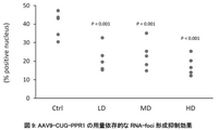
- FIG. 9 shows that AAV9-CUG-PPR1 dose-dependently suppressed the formation of RNA-foci in DM1 model animals.
- “Ctrl” is the PBS administration group
- "LD” is the AAV9-CUG-PPR1 low dose administration group
- "MD” is the AAV9-CUG-PPR1 medium dose administration group
- “HD” is the AAV9-CUG-PPR1 high dose.
- Administration groups are indicated.
- the vertical axis indicates the RNA-foci-positive cell rate.
- FIG. 10 shows that AAV9-CUG-PPR1 ameliorated abnormal splicing in DM1 model animals in a dose-dependent manner.
- the left figure shows the results of Clcn1, and the right figure shows the results of the abnormal splicing detection system of Atp2a1.
- “Ctrl” is the PBS administration group
- "LD” is the AAV9-CUG-PPR1 low dose administration group
- "MD” is the AAV9-CUG-PPR1 medium dose administration group
- “HD” is the AAV9-CUG-PPR1 high dose.
- Administration groups are indicated.
- the vertical axis indicates the content (%) of normal splicing isoforms.
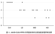
- FIG. 11 shows that AAV9-CUG-PPR1 dose-dependently improved muscle tonicity in DM1 model animals.
- the vertical axis indicates the myotonia score, and the frequency of myotonic discharge decreases in the order of scores 3, 2, 1, and 0. The frequency and severity of myotonic discharges are correlated.
- “Ctrl” is the PBS administration group
- "LD” is the AAV9-CUG-PPR1 low dose administration group
- MD is the AAV9-CUG-PPR1 medium dose administration group
- “HD” is the AAV9-CUG-PPR1 high dose.
- FIG. 12 shows the relationship between the administration dose of AAV9-CUG-PPR1 and the expression level of PPR mRNA.
- the internal standard uses GAPDH. Each individual point indicates the amount of PPR mRNA expression in the thigh muscle of each individual mouse.
- the vertical axis is displayed in Log display.
- “Ctrl” is the PBS administration group
- "LD” is the AAV9-CUG-PPR1 low dose administration group
- "MD” is the AAV9-CUG-PPR1 medium dose administration group
- “HD” is the AAV9-CUG-PPR1 high dose.
- FIG. 13 shows that AAV9-CUG-PPR1 suppressed RNA-foci formation in DM1 model animals in a time-dependent manner.
- “Ctrl” indicates the PBS-administered group.
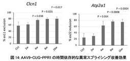
- FIG. 14 shows that AAV9-CUG-PPR1 ameliorated abnormal splicing in DM1 model animals in a time-dependent manner.
- the left figure shows the results of Clcn1, and the right figure shows the results of the abnormal splicing detection system of Atp2a1.
- “Ctrl” indicates the PBS-administered group.
- "2w”, “4w”, “8w”, and "16w” indicate test periods after administration of AAV9-CUG-PPR1, respectively.
- FIG. 15 shows that AAV9-CUG-PPR1 improved muscle tonicity in DM1 model animals in a time-dependent manner.
- the vertical axis indicates the myotonia score, and the frequency of myotonic discharge decreases in the order of scores 3, 2, 1, and 0. The frequency and severity of myotonic discharges are correlated.
- “Ctrl” indicates the PBS-administered group.
- "2w”, “4w”, “8w”, and "16w” indicate test periods after administration of AAV9-CUG-PPR1, respectively.
- FIG. 16 shows global normalization of splicing abnormalities after administration of AAV9-CUG-PPR1.
- FIG. 16a shows how splicing events (left) with changes in PSI (Percent spliced in) of 0.2 or more between WT and PBS-administered HSA-LR mice were observed in PPR-administered HSA-LR mice (right). It is the figure which showed whether it changed.
- FIG. 16b is a comparison of Tanner et al.
- FIG. 2 shows changes for DM1-associated splicing events identified in (2021). Black circles indicate signals from normal mice, blue squares indicate signals from PBS-administered drug mice, and red triangles indicate signals from PPR-administered drug mice.
- Figure 16c shows the results of Gene ontology analysis for the genes at each point in Figure a.
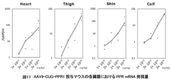
- FIG. 17 shows the expression level of PPR mRNA in each organ of AAV9-CUG-PPR1-administered mice.
- the vertical axis indicates the relative expression level in Log representation when the expression level in mice administered with 1 ⁇ 10 13 vg/kg is set to 1.
- GAPDH mRNA expression level is used as an internal standard.
- the horizontal axis indicates the AAV9-CUG-PPR1 dose (vg/kg body weight of mouse).
- FIG. 18 shows the persistence of mRNA expression in each organ of AAV9-CUG-PPR1-administered mice.
- the vertical axis indicates the relative expression level when the expression level of one mouse after 2 weeks of AAV administration (Day 14) is set to 1.
- GAPDH mRNA expression level is used as an internal standard.
- “Ctrl” indicates the PBS group, and "14", “28", "56", "112", and "183” indicate the number of days after administration.
- the values for each individual mouse are plotted with circles, and the mean values are connected by lines.
- PPR motif and PPR protein When the term "PPR motif" is used in the present disclosure, the E value obtained by PF01535 in Pfam and PS51375 in Prosite when analyzing the amino acid sequence with a protein domain search program on the Web is less than or equal to a predetermined value, unless otherwise specified.
- the position numbers of the amino acids that make up the PPR motif defined in the present disclosure are almost synonymous with PF01535, while the amino acid position of PS51375 minus 2 (e.g., No. 1 of the present invention ⁇ No. 3 of PS51375) Equivalent to.
- the second amino acid from the rear (C-terminal side) of the amino acid constituting the PPR motif or two N amino acids for the first amino acid of the next PPR motif It is the terminal side, that is, the -2nd amino acid. If the next PPR motif is not unambiguously identified, the two previous amino acids are designated as "ii" for the first amino acid of the next helix structure.
- the conserved amino acid sequence of the PPR motif is less conserved at the amino acid level, but the two ⁇ -helices are well conserved on the secondary structure.
- a typical PPR motif consists of 35 amino acids, but its length varies from 30 to 38 amino acids.
- the basic skeleton of the PPR motif is known to be represented by Formula 1.
- Helix A is a 12 amino acid long portion capable of forming an ⁇ -helical structure and is represented by Formula 2,
- a 1 to A 12 each independently represent an amino acid;
- X is absent or is a moiety consisting of 1-9 amino acids in length;
- Helix B is a portion capable of forming an ⁇ -helical structure, consisting of 11-13 amino acids in length;
- L is a moiety of Formula 3, 2-7 amino acids in length;
- each amino acid is numbered from the C-terminal side with “i” (-1), “ii” (-2), However, L iii to L vii may not exist.
- the term “PPR protein” in the present disclosure refers to a PPR protein having one or more, preferably two or more, of the above-described PPR motifs.
- the term “protein” refers to any substance consisting of a polypeptide (a chain in which a plurality of amino acids are peptide-bonded), and includes those consisting of relatively low-molecular-weight polypeptides, unless otherwise specified.
- amino acid may refer to ordinary amino acid molecules, and may also refer to amino acid residues constituting peptide chains. Which one is meant will be clear to a person skilled in the art from the context.
- the term “selective” or “specific” with respect to the binding activity of the PPR motif to RNA bases means that, unless otherwise specified, the binding activity to any one of the RNA bases is Higher than the binding activity to bases. This selectivity or specificity can be confirmed by designing experiments based on known methods by those skilled in the art, or can be determined by calculation by those skilled in the art.
- RNA base refers to a ribonucleotide base that constitutes RNA. Specifically, adenine (A), guanine (G), cytosine (C), or uracil ( U).
- A adenine
- G guanine
- C cytosine
- U uracil
- PPR proteins can be selective for bases in RNA, they do not bind to nucleic acid monomers.
- PPR proteins are abundant in plants, and about 500 proteins and about 5000 motifs can be found in Arabidopsis thaliana. PPR motifs and PPR proteins with diverse amino acid sequences are also present in many land plants such as rice, poplar, and selaginella. In the present invention, naturally occurring PPR motifs and PPR proteins may be used, or PPR motifs and PPR proteins designed based on the method disclosed in WO2013/058404, for example, may be used. Specifically, desired PPR motifs and PPR proteins can be designed based on the following information disclosed in WO2013/058404.
- the present invention can utilize the knowledge about the combination of three amino acids A 1 , A 4 and Lii and/or the combination of two amino acids A 4 and Lii disclosed in WO2013 / 058404 . can.
- the PPR motif binds strongly to A, but to G, U and C has a selective RNA base-binding ability that does not bind to (3-13) If the combination of three amino acids A 1 , A 4 and L ii is, in order, isoleucine, methionine, aspartic acid, the PPR motif binds strongly to U and then to C It has selective RNA base binding ability, binding to A and G but not to A and G.
- the above rule statistically determines the possibility that the PPR motif binds to the target base by the combination of amino acids (A 1 , A 4 , L ii ) or (A 4 , L ii ) of the PPR motif. It does not mean that a PPR motif (or PPR protein) that binds to the target base (or base sequence) with 100% probability cannot be produced according to the above rule.
- a person skilled in the art can produce several types to several tens of types of candidate PPR proteins based on the above-mentioned rules, so that a PPR motif (or PPR protein) can be obtained.
- the selection of a desired PPR protein through production and confirmation of candidate PPR proteins can be performed by those skilled in the art within the scope of ordinary trial and error, and does not impose an undue burden on those skilled in the art.
- PPR motif can recognize a specific base in RNA. Then, according to the present invention, a PPR motif selective for each of A, U, G and C can be selected or designed by appropriately selecting amino acids at specific positions. Furthermore, proteins containing suitable stretches of such PPR motifs can specifically or selectively recognize corresponding base sequences. Furthermore, the above findings allow the design of proteins with PPR motifs that can selectively bind to desired RNA bases and multiple PPR motifs that can sequence-specifically bind to desired RNAs. In designing, the sequence information of the natural PPR motif may be referred to for the portions other than the amino acids at the important positions in the PPR motif.
- the number of repeats of the PPR motif can be appropriately determined according to the target sequence, and can be, for example, 2 or more, and can be 2-30.
- the present invention relates to a PPR protein that specifically binds to mRNA containing an abnormally expanded CUG repeat sequence that causes DM1.
- the PPR protein according to the present invention can be produced by linking multiple sets of [a C-binding PPR motif, a U-binding PPR motif, and a G-binding PPR motif] based on the theory described above. can.
- the first PPR motif of the PPR protein according to the present invention does not necessarily have to be a PPR motif that binds to C, as long as the entire PPR protein specifically binds to the CUG repeat sequence, the first PPR motif binds to U.
- PPR motif that binds to G or a PPR motif that binds to G.
- the number of PPR motifs contained in the PPR protein need not be a multiple of 3, as long as the PPR protein of the present invention as a whole has a configuration that specifically binds to the CUG repeat sequence.
- Non-limiting examples of C-linked PPR motifs that may be used in the present invention include PPR motifs having the following sequences.
- sequence homology or sequence identity
- binding to C may be used.
- substitutions, additions, and/or deletions and having C-binding properties may be used.
- the amino acid substitution may be, for example, conservative amino acid substitution known in the art.
- Non-limiting examples of U-linked PPR motifs that may be used in the present invention include PPR motifs having the following sequences.
- 80 % or more (preferably , 85 % or more, 90% or more, 95% or more, 96% or more, 97% or more, 98% or more, 99% or more) sequence homology (or sequence identity) and a PPR motif that has a binding property to U may be used.
- substitutions, additions, and/or deletions and having binding properties for U may be used.
- the amino acid substitution may be, for example, conservative amino acid substitution known in the art.
- Non-limiting examples of G-linked PPR motifs that may be used in the present invention include PPR motifs having the following sequences.
- 80 % or more (preferably , 85 % or more, 90% or more, 95% or more, 96% or more, 97% or more, 98% or more, 99% or more) sequence homology (or sequence identity) and G-binding PPR motif may be used.
- substitutions, additions, and/or deletions and having binding properties for G may be used.
- the amino acid substitution may be, for example, conservative amino acid substitution known in the art.
- searches and analyzes for the identity of nucleotide sequences or amino acid sequences can be performed by algorithms or programs (eg, BLASTN, BLASTP, BLASTX, ClustalW) well known to those skilled in the art. Parameters when using programs can be appropriately set by those skilled in the art, and default parameters of each program may be used. Specific techniques of these analysis methods are also well known to those skilled in the art.
- amino acids with similar properties refer to amino acids with similar physical properties such as hydropathy, charge, pKa, solubility, and the like.
- the effect of the present invention is exerted by binding of the PPR protein of the present invention to mRNA containing an abnormally expanded CUG repeat sequence in the target cell.
- the means for delivering the PPR protein into the cells of the subject is not limited, and a nucleic acid encoding the PPR protein of the present invention may be delivered into the cells of the subject to express the PPR protein of the present invention within the cells of the subject. Alternatively, the PPR protein of the present invention itself may be delivered into the subject's cells.
- the means for delivering proteins and nucleic acids is not limited, and various means known in the art can be used.
- Non-limiting examples of means for delivery of nucleic acids or proteins into cells of a subject that can be used in the present invention include liposomes, lipid nanoparticles (LNPs), polymeric micelles, emulsions, polymeric microcapsules, antibodies -nucleic acid complexes, antibody-drug complexes, viruses, and the like.
- Non-limiting examples of viral vectors that may be used in the present invention include adeno-associated viral (AAV) vectors, adenoviral vectors, retroviral vectors, lentiviral vectors, or herpes simplex viral vectors.
- AAV adeno-associated viral
- adenoviral vectors adenoviral vectors
- retroviral vectors retroviral vectors
- lentiviral vectors lentiviral vectors
- herpes simplex viral vectors Alternatively, it can be appropriately selected by those skilled in the art depending on the tropism to tissues, the necessity of nucleic acid integration into the host genome, and the like.
- AAV vectors are particularly suitable for use in the present invention because they are capable of infecting both dividing and quiescent cells, and can transfer genetic material to a wide variety of cell types. Twelve serotypes of AAV (AAV1-AAV12) have been described to date, and all known serotypes are capable of infecting various types of tissue cells. Although the serotype of the AAV vector that may be used in the present invention is not limited, for example, AAV1 vector, AAV2 vector, AAV6 vector, which are known to have tropism for muscle tissue (skeletal muscle, cardiac muscle, and/or smooth muscle) A vector, AAV7 vector, AAV8 vector, AAV9 vector, AAV10 vector, AAV11 vector or AAV12 vector may be used.
- Treatment of DM1 using the present invention may be an embodiment of applying the present invention directly in vivo in a subject suffering from DM1 (in vivo), or treating cells or tissues ex vivo using the present invention. , may be introduced into the body of the subject (ex vivo).
- the method for producing an expression vector containing a nucleic acid encoding the PPR protein of the present invention is not limited, and a person skilled in the art can use a conventional method (e.g., use of a commercially available protein expression vector production kit, use of a commercially available virus expression vector production kit, production It can be manufactured by outsourcing to a contractor, etc.).
- the method for producing the PPR protein of the present invention is not limited, for example, a cell (e.g., The desired PPR protein can be obtained by selectively extracting and/or purifying the PPR protein from animal cells, plant cells, Escherichia coli, yeast, etc.) by a conventional method.
- CUG-PPR CUG repeat RNA sequence
- coli pellet add 1.5 mL of lysis buffer (20 mM Tris-HCl, pH 8.0, 150 mM NaCl, 0.5% NP-40, 1 mM MgCl 2 , 2 mg/ml lysozyme, 1 mM PMSF, 2 ⁇ l DNase) was added and frozen at -80°C for 20 minutes. Cells were freeze fractured with shaking at 25°C for 30 minutes. Subsequently, centrifugation was performed at 3700 rpm, 4° C. for 15 minutes, and the supernatant (E. coli lysate) containing soluble PPR protein was recovered and used in the following experiments.
- lysis buffer 20 mM Tris-HCl, pH 8.0, 150 mM NaCl, 0.5% NP-40, 1 mM MgCl 2 , 2 mg/ml lysozyme, 1 mM PMSF, 2 ⁇ l DNase
- PPR Protein-to-RNA Binding Test was performed using a method for testing the binding of PPR protein to biotinylated RNA on a streptavidin plate.
- a 30-base RNA containing a target CUG ⁇ 7 sequence (GACA CUGCUGCUGCUGCUGCUGCUG AUGCA (SEQ ID NO: 15)) and a 30-base RNA containing a non-target CAG ⁇ 6 (GACAUGC CAGCAGCAGCAGCAGCAG GACUG (SEQ ID NO: 16)
- An RNA probe modified with biotin at the 5' end was synthesized (requested by Greiner).
- biotinylated RNA probe 2.5 pmol was added to a streptavidin-coated plate (Cat No. 15502, Thermo Fisher) and allowed to react for 30 minutes at room temperature. Washed with probe wash buffer (20 mM Tris-HCl (pH 7.6), 150 mM NaCl, 5 mM MgCl 2 , 0.5% NP-40, 1 mM DTT, 0.1% BSA). For background measurement, wells containing lysis buffer without biotinylated RNA were also prepared (referred to as "-Probe").
- a blocking buffer (20 mM Tris-HCl (pH 7.6), 150 mM NaCl, 5 mM MgCl 2 , 0.5% NP-40, 1 mM DTT, 1% BSA) was added to block the plate surface at room temperature for 30 minutes. gone. 100 ⁇ L of E. coli lysate containing a luciferase-fused PPR protein with a luminescence level of 1.5 ⁇ 10 8 LU/ ⁇ L was added to the wells, and the binding reaction was allowed to proceed for 30 minutes at room temperature.
- washing buffer (20 mM Tris-HCl (pH 7.6), 150 mM NaCl, 5 mM MgCl 2 , 0.5% NP-40, 1 mM DTT) and diluted 2500-fold with the washing buffer.
- 40 ⁇ L of luciferase substrate Promega, E151A was added to the wells and allowed to react for 5 minutes, luminescence was measured with a plate reader (PerkinElmer, Cat No. 5103-35).
- Example 2 Effect of PPR on RNA aggregate formation in DM1 model cells
- PPR protein was allowed to act on DM1 model cells, and the number of RNA aggregates was measured by FISH (fluorescent in situ hybridization) method.
- DM1 model cells As a DM1 cell model, C2C12 cells into which a DMPK gene region containing 800 CTG repeats was introduced (C2C12-DMPK800R, Nucleic Acids Research, 2014, 42 (10), 6591-6602) were used. C2C12-DMPK800 was produced by the following procedure. That is, both a plasmid (pLC16) containing a sequence in which 800CTG was inserted into the DMPK gene 3' untranslated region and a plasmid expressing PhiC31 integrase were applied to mouse myoblast cell line C2C12 using Nucleofector (trade name, Lonza).
- Nucleofector trade name, Lonza
- stable expression strains were selected using selective medium supplemented with puromycin. Subsequently, the stable expression strain was transfected with a plasmid expressing Cre recombinase, and a selective medium supplemented with hygromycin was used to select clones that induce transcription of mRNA having 800 CUG repeats.
- sequences of the PPR proteins (PPR1 to PPR4) encoded in each expression plasmid vector are the same as the sequences of the PPR proteins listed in Table 1.
- DM1 model cells were cultured under conditions of 37° C. and 5% CO 2 using DMEM medium containing 10% FBS and penicillin/streptomycin. 200 ng of the plasmid DNA constructed above, 0.6 ⁇ L Fugene®-HD (Promega, E2311), and 200 ⁇ L Opti-MEM are mixed, added to the total well, and placed at 37° C., 5% CO 2 for 72 hours. After culturing, the cells were fixed with 3% paraformaldehyde for 15 minutes at room temperature. After fixation, the cells were washed twice with PBS and then permeabilized with PBS containing 0.5% Triton X-100 for 5 minutes.
- prehybridization treatment was performed for 10 minutes with 2 ⁇ SSC buffer containing 30% formamide. After that, hybridization was performed at 37° C. for 1 hour with 2 ⁇ SSC buffer containing 30% formamide, 2 ⁇ g/mL BSA, 66 ⁇ g/mL yeast tRNA, 2 mM vanadyl complex, and 1 ng/ ⁇ L Texas Red CAG probe. After post-hybridization treatment with 2 ⁇ SSC buffer containing 30% formamide at 42° C. for 30 minutes, the plate was washed once with 1 ⁇ SSC buffer and then twice with PBS. After fixing with Vectashield with DAPI (trade name, Vector Laboratories), the number of intranuclear RNA aggregates was counted under a fluorescence microscope (Keyence PZ-9000).
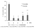
- Example 3 Effect of PPR protein on abnormal splicing in DM1 model cells
- DM1 model cell The same DM1 model cell (C2C12-DMPK800R) as in Example 2 was used.
- RT-PCR products were subjected to electrophoresis on a 2% agarose gel, stained with GelRed, and then normal and abnormal PCR products were quantified using an image analyzer (ChemiDoc Touch Imaging System, BioRad).
- Atp2a1 exon 22 RT primer Fw GCTCATGGTCCTCAAGATCTCAC (SEQ ID NO: 22)
- Rv GGGTCAGTGCCTCAGCTTTG (SEQ ID NO: 23)
- Results> The results are shown in FIG.
- the lower graph shows the results of electrophoresis of RT-PCR products
- the upper graph shows the proportion of normal type in RT-PCR products.
- the ratio of abnormal type was higher than that of normal type. It was shown that the action of the PPR protein significantly increased the ratio of the normal type in the DM1 model cells and improved the abnormal splicing.
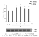
- Example 4 Effect of PPR protein on abnormal muscle differentiation in DM1 model cells
- a PPR protein was allowed to act on DM1 model cells to verify whether or not the muscle differentiation efficiency of DM1 model cells was improved.
- DM1 model cell The same DM1 model cell (C2C12-DMPK800R) as in Example 2 was used.
- the results are shown in FIG.
- the fusion index was about 3 in the empty vector-administered group (indicated as "no" in the figure).
- the fusion index was about 5 to 15, indicating a significant improvement in muscle differentiation efficiency.
- Example 5 Effect of PPR in DM1 model mice
- AAV6 carrying the CUG-PPR gene was administered to DM1 model mice once to the tibialis anterior muscle, and the effect on muscle tonic symptoms in the tibialis anterior muscle, the effect on abnormal splicing of the Atp2a1 gene and the skeletal muscle chloride channel (Clcn1) gene, Also, the number of RNA aggregates formed was verified.
- HSA LR mice Animals used HSA LR mice (transferred from Dr. Charles Thornton (University of Rochester) and bred at the experimental animal facility of Osaka University) were used as DM1 model mice.
- HSA LR mice are transgenic animals in which a 220-fold extension of CTG repeats in the 3′ untranslated region of the hACTA gene (a human gene that is constitutively expressed in muscle cells) is integrated into the genome. Symptoms of DM1 appear due to the expression of mRNA with an extended sequence (Science, 2000, 289(5485), 1769-73).
- wild-type mice FVB/NJcl mice, purchased from CLEA Japan
- 14-week-old male and female mice were used.
- AAV vector AAVpro (trademark) Helper Free System (AAV6), Takara, 6651 component
- AAV6 Helper Free System
- Takara 6651 component
- the fused gene was inserted.
- the expression size of the constructed gene was confirmed by PCR, and the inserted sequence was confirmed by sequencing. Expression of the fusion gene is controlled by the CMV promoter and human growth hormone polyA signal.
- AAV vector constructed in the previous section was used as a pRC6 vector, and 20 ⁇ g of each of the phelper vector (both components of AAVpro (trademark) Helper Free System (AAV6), Takara, 6651) were added to Polyethylene, Linear, MW25000 (Polysciences, Inc., 23966- 1) was used to introduce the gene into 2.5 ⁇ 10 8 cells of AAVproTM HEK293T cell line (Takara, 632273). AAV-producing cells were obtained 3 days after transfection. AAV purification used AAVproTM Purification Kit Maxi (all serotype) (Takara, 6666). AAV production kit for real time PCR ver.
- AAVproTM Purification Kit Maxi all serotype
- AAV6-mCLo-CUG-PPR1, AAV6-mCLo-CUG-PPR2 and AAV6-mCLo-empty were used in the subsequent tests.
- the sequences of the PPR proteins (PPR1, PPR2) encoded in each AAV vector are the same as the sequences of the PPR proteins listed in Table 1.
- RNA Aggregates Fifty-six days after the final administration, the tibialis anterior muscle was collected from the mouse and sectioned. After washing twice with PBS, the cells were permeabilized with PBS containing 0.5% Triton X-100 for 5 minutes. Next, prehybridization treatment was performed for 10 minutes with 2 ⁇ SSC buffer containing 30% formamide. After that, hybridization was performed at 37° C. for 1 hour with 2 ⁇ SSC buffer containing 30% formamide, 2 ⁇ g/mL BSA, 66 ⁇ g/mL yeast tRNA, 2 mM vanadyl complex, and 1 ng/ ⁇ L Texas Red CAG probe.
- Tibialis anterior muscles were harvested from mice 56 days after the final administration. After extracting total RNA using TRI Reagent (trade name, MRC), cDNA was prepared using SuperScript III First Strand Synthesis System (Invitrogen). After treating the cDNA with RNaseH, RT-PCR was performed using the same Atp2a1 exon22 RT primer as in Example 2 and the following Clcn1 exon 7a RT primer.
- RT-PCR product was electrophoresed on a 2% agarose gel and stained with GelRed, normal PCR products and abnormal PCR products were quantified with an image analyzer (ChemiDoc Touch Imaging System, BioRad), and RT - The percentage of normal in the PCR products was calculated. t-test was used for statistical analysis.
- Clcn1 exon 7a RT primer Fw TGAAGGAATACCTCACACTCAAGG (SEQ ID NO: 24)
- Rv CACGGAACACAAAGGCACTG (SEQ ID NO: 25)
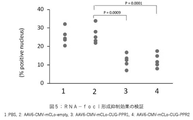
- RNA-foci analysis The results of RNA-foci analysis are shown in FIG.
- the PBS-administered group and the AAV6-mCLo-empty-administered group showed an RNA-foci-positive cell rate of about 30%.
- a significant decrease in the RNA-foci positive rate was observed in the AAV6-mCLo-CUG-PPR1 and AAV6-mCLo-CUG-PPR2 administration groups.
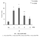
- Fig. 6 shows the evaluation results of splicing abnormalities.
- the PBS-administered DM1 group and the AAV6-mCLo-Empty-administered MyoD group exhibited a normal type rate of about 60%.
- the ratio of normal RT-PCR products was significantly increased, suggesting that PPR protein administration improved the abnormal splicing of the Atp2a1 gene.
- Clcn1 it was also found that administration of PPR improved the splicing abnormality of the Clcn1 gene.
- Fig. 7 shows the analysis results of muscle tonicity. Severity 2 myotonia was detected in 5 out of 5 animals of the PBS-administered group and the AAV6-mCLo-empty-administered group. In both the AAV6-mCLo-CUG-PPR1 and AAV6-mCLo-CUG-PPR2-administered DM1-administered groups, 2 out of 5 animals showed severity 1, and the remaining 3 animals showed severity 2. In addition, two improved cases belonged to the experimental group with a high effect of improving abnormal Clcn1 splicing (FIG. 8).
- Example 6 Dose dependence test of efficacy using DM1 model mice
- AAV9 carrying the CUG-PPR gene was added to DM1 model mice at 3 ⁇ 10 13 , 1 ⁇ 10 14 , and 3 ⁇ 10 14 vg/kg.
- a single dose was administered into the tail vein, and the effect on myotonic symptoms in thigh muscles, the effect on abnormal splicing of Atp2a1 gene and skeletal muscle chloride channel (Clcn1) gene, and the number of RNA aggregates formed were examined.
- HSA LR mice Animals used HSA LR mice (transferred from Dr. Charles Thornton (University of Rochester) and bred at the experimental animal facility of Osaka University) were used as DM1 model mice.
- HSA LR mice are transgenic animals in which a 220-fold extension of CTG repeats in the 3′ untranslated region of the hACTA gene (a human gene that is constitutively expressed in muscle cells) is integrated into the genome. Symptoms of DM1 appear due to the expression of mRNA with an extended sequence (Science, 2000, 289(5485), 1769-73).
- wild-type mice FVB/NJcl mice, purchased from CLEA Japan
- 14-week-old male and female mice were used for the experiment.
- PPR protein production of AAV vector
- a gene in which a PPR protein and a 3 ⁇ nuclear localization signal were fused in this order was inserted into the multiple cloning site of a pAAV-CMV vector (AAVproTM Helper Free System, Takara, 6651 component).
- the expression size of the constructed gene was confirmed by PCR, and the inserted sequence was confirmed by sequencing. Expression of the fusion gene is controlled by the CMV promoter and human growth hormone polyA signal.
- AAV vector (Preparation of AAV vector)
- the AAV vector constructed in the previous section was outsourced to SignaGen Laboratories (hereinafter abbreviated as SG).
- Packaging was carried out by co-transfecting the Rep/Cap plasmid and Helper plasmid owned by SG and the AAV vector sent from our company into the packaging cell line.
- a cell lysate containing the packaged AAV was purified by density gradient centrifugation and the titer was measured.
- the AAV obtained was 1.16 ⁇ 10 14 vg/ml and the volume was 5 ml. This was used in subsequent experiments as AAV9-CUG-PPR1.
- the sequence of the PPR protein (PPR1) encoded in each AAV vector is the same as the PPR protein sequence shown in Table 1.
- AAV9-CUG-PPR1 low dose administration group (hereinafter abbreviated as Low dose: LD), AAV9-CUG-PPR1 middle dose administration group (hereinafter abbreviated as Middle dose: MD),
- AAV9-CUG-PPR1 high dose administration group (hereinafter abbreviated as High dose: HD)
- PBS administration group (hereinafter abbreviated as PBS).
- Each group n 5 ( ⁇ 3, ⁇ 2 mice).
- the dose of AAV was 3 ⁇ 10 13 vg/kg for LD, 1 ⁇ 10 14 vg/kg for MD, and 3 ⁇ 10 14 vg/kg for HD, and was administered once into the tail vein.
- the same volume of PBS was administered once to the PBS-administered group.
- RNA Aggregates Twenty-eight days after the final administration, thigh muscles were collected from the mice and sections were prepared. After washing twice with PBS, the cells were permeabilized with PBS containing 0.5% Triton X-100 for 5 minutes. Next, prehybridization treatment was performed for 10 minutes with 2 ⁇ SSC buffer containing 30% formamide. After that, hybridization was performed at 37° C. for 1 hour with 2 ⁇ SSC buffer containing 30% formamide, 2 ⁇ g/mL BSA, 66 ⁇ g/mL yeast tRNA, 2 mM vanadyl complex, and 1 ng/ ⁇ L Texas Red CAG probe.
- RNA samples were collected from the mice. After extracting total RNA using TRI ReagentTM (MRC), cDNA was prepared using SuperScript III First Strand Synthesis System (Invitrogen). After treating the cDNA with RNaseH, RT-PCR was performed using the same Atp2a1 exon22 RT primer as in Example 2 and the following Clcn1 exon 7a RT primer. After the RT-PCR product was electrophoresed on a 2% agarose gel and stained with GelRed, normal PCR products and abnormal PCR products were quantified with an image analyzer (ChemiDoc Touch Imaging System, BioRad), and RT - The percentage of normal in the PCR products was calculated. t-test was used for statistical analysis.
- muscle tonicity (myotonia) Twenty-eight days after the final administration, the phenomenon of electrical tonicity in the mouse thigh muscle was analyzed by needle electromyography under anesthesia. Specifically, the occurrence frequency of the electrical muscle tonic phenomenon when the needle electrode was inserted into the thigh muscle of the mouse 20 times was evaluated according to the following four grades. Severity 0: No electrical muscle tonic phenomenon is observed. Severity 1: Electrical myotonia is present in less than 50% of cases. Severity 2: Electrical myotonia is observed in 50% or more and less than 90% of all cases. Severity 3: 90% or more of electrical myotonia is observed.
- RNA 300 ⁇ L of the aqueous phase was collected and subjected to a Maxwell RSC Instrument, a nucleic acid purifier, to obtain purified total-RNA. The concentration and purity of RNA were measured by NanoDrop 8000 absorbance. In order to confirm the degree of degradation of the obtained RNA, 20 to 250 ng/ ⁇ L of total-RNA was used to perform electrophoresis using LabChip GX Touch HT (PerkinElmer, CLS137031J). It was confirmed that decomposition had not progressed. For synthesis of cDNA, 20 to 100 ng/ ⁇ L of obtained RNA was subjected to reverse transcription reaction according to the product protocol of SuperScript III Reverse Transcriptase (Thermo Fisher Scientific (Life Technologies), 18080085).
- the amount of PPR mRNA and GAPDH mRNA was measured by mixing the prepared cDNA, Brilliant III Ultra-Fast SYBR Green QPCR Master Mix (Agilent, 600882) and the primers described below according to the manual, and then using a real-time PCR system Aria MX (Agilent). Amplified and quantitatively analyzed.
- PPR quantitative PCR primer Fw GATGAGGCTTTGGAACTGTTTG (SEQ ID NO: 26)
- Rv TCTCTGGCTCTGCCGGCCTTGC (SEQ ID NO: 27)
- GAPDH quantitative PCR primer Fw ATCATCCCTGCCTCTACTGG (SEQ ID NO: 28)
- Rv CTGCTTCACCACCTTCTTGA (SEQ ID NO: 29)
- RNA-FISH method was performed using the obtained thigh muscle organ section, and the effect of suppressing RNA-foci formation by AAV9-CUG-PPR1 was verified. A formation inhibitory effect was confirmed.
- the rate of RNA-foci-positive cells was 39% in the PBS administration group, 18% in the AAV9-CUG-PPR1 low dose group, 23% in the AAV9-CUG-PPR1 medium dose group, and 17% in the AAV9-CUG-PPR1 high dose group. formation inhibitory effect was observed.
- Fig. 10 shows the evaluation results of splicing abnormalities.
- Total RNA was collected from the collected organs, reverse transcription PCR was performed, and abnormal splicing of the Clcn1 gene and Atp2a1 gene was evaluated.
- Aberrant splicing of Clcn1 is a Cl channel that is known to directly cause myotonic discharge.
- Atp2a1 is also known as a Ca transporter related to myotonic discharge and a sensitive marker gene.
- the Clcn1 detection system showed 58% normal splicing in the PBS administration group, 72% in the AAV9-CUG-PPR1 low dose group, 78% in the AAV9-CUG-PPR1 medium dose group, and 78% in the AAV9
- the -CUG-PPR1 high dose group showed 82%, confirming a dose-dependent splicing improvement.
- the Atp2a1 detection system showed 25% normal splicing in the PBS-administered group, 56% in the AAV9-CUG-PPR1 low-dose group, 60% in the AAV9-CUG-PPR1 medium-dose group, and 60% in the AAV9-CUG-PPR1 high-dose group.
- the dose group showed 64%, showing a dose-dependent splicing improvement.
- the effect of reducing muscle tonic discharge by PPR was expressed as a score in 4 stages based on the number of myotonia discharges. That is, a score of 0 is 0 out of 20, a score of 1 is 1 to 9 out of 20, a score of 2 is 10 to 19 out of 20, and a score of 3 is 20 out of 20 myotonic discharges. means detection.
- the PBS administration group scored 3
- the AAV9-CUG-PPR1 low dose group showed a score of 3 in 1/5 cases
- the score of 2 in 4/5 cases. 4/5 cases showed a score of 2
- 4/5 cases showed a score of 2
- 1/5 cases showed a score of 1
- 3/5 cases showed a score of 2/5 cases showed an improvement to a score of 1 (Fig. 11).
- RNA was extracted from the thigh muscle of the analyzed organ, and quantitative PCR of PPR mRNA was performed. As a result of RT-qPCR, dose-dependent PPR mRNA expression was confirmed (Fig. 12).
- a relative value was used when the value of the individual with the lowest signal in the AAV9-CUG-PPR1 low-dose administration group was set to 1 using GAPDH as an internal standard. Taking the median value of each group, the PBS administration group was 0.2, the low dose administration group was 0.7, the medium dose administration group was 1.8, and the high dose administration group was 2.6. A significant PPR expression was confirmed (FIG. 12).
- AAV9-CUG-PPR1 dose-dependently suppresses myotonic discharge, suppresses the formation of RNA-foci, and improves aberrant splicing.
- Example 7 Time axis change in drug efficacy in DM1 model mice.
- a single dose of AAV9 carrying the CUG-PPR gene was administered to DM1 model mice into the tail vein, and at each time point, muscle tonic symptoms in the thigh muscles were tested.
- the effect, the effect on abnormal splicing of the Atp2a1 gene and the skeletal muscle chloride channel (Clcn1) gene, and the number of RNA aggregates formed were verified.
- HSA LR mice Animals used HSA LR mice (transferred from Dr. Charles Thornton (University of Rochester) and bred at the experimental animal facility of Osaka University) were used as DM1 model mice.
- HSA LR mice are transgenic animals in which a 220-fold extension of CTG repeats in the 3′ untranslated region of the hACTA gene (a human gene that is constitutively expressed in muscle cells) is integrated into the genome. Symptoms of DM1 appear due to the expression of mRNA with an extended sequence (Science, 2000, 289(5485), 1769-73).
- wild-type mice FVB/NJcl mice, purchased from CLEA Japan
- PPR protein production of AAV vector
- a gene fusing a PPR protein and a 3 ⁇ nuclear localization signal in this order was inserted into the multiple cloning site of a pAAV-CMV vector (AAVproTM Helper Free System, Takara, 6651 component).
- the expression size of the constructed gene was confirmed by PCR, and the inserted sequence was confirmed by sequencing. Expression of the fusion gene is controlled by the CMV promoter and human growth hormone polyA signal.
- AAV vector (Preparation of AAV vector) The production of the AAV vector constructed in the preceding section was entrusted to SignaGen Laboratories (hereinafter abbreviated as SG). Packaging was carried out by co-transfecting the Rep/Cap plasmid and Helper plasmid owned by SG and the AAV vector sent by our company into the packaging cell line. A cell lysate containing the packaged AAV was purified by density gradient centrifugation and the titer was measured. The AAV obtained was 1.16 ⁇ 10 14 vg/ml and the volume was 5 ml. This was used in subsequent experiments as AAV9-CUG-PPR1. The sequence of the PPR protein (PPR1) encoded in each AAV vector is the same as the PPR protein sequence shown in Table 1.
- RNA Aggregates 14, 28, 56, or 112 days after the final administration thigh muscles were collected from mice and sections were prepared. After washing twice with PBS, the cells were permeabilized with PBS containing 0.5% Triton X-100 for 5 minutes. Next, prehybridization treatment was performed for 10 minutes with 2 ⁇ SSC buffer containing 30% formamide. After that, hybridization was performed at 37° C. for 1 hour with 2 ⁇ SSC buffer containing 30% formamide, 2 ⁇ g/mL BSA, 66 ⁇ g/mL yeast tRNA, 2 mM vanadyl complex, and 1 ng/ ⁇ L Texas Red CAG probe.
- Thigh muscles were collected from mice 14, 28, 56, or 112 days after the final administration. After extracting total RNA using TRI ReagentTM (MRC), cDNA was prepared using SuperScript III First Strand Synthesis System (Invitrogen). After treating the cDNA with RNaseH, RT-PCR was performed using the same Atp2a1 exon22 RT primer as in Example 2 and the following Clcn1 exon 7a RT primer.
- MRC TRI ReagentTM
- cDNA was prepared using SuperScript III First Strand Synthesis System (Invitrogen). After treating the cDNA with RNaseH, RT-PCR was performed using the same Atp2a1 exon22 RT primer as in Example 2 and the following Clcn1 exon 7a RT primer.
- RT-PCR product was electrophoresed on a 2% agarose gel and stained with GelRed, normal PCR products and abnormal PCR products were quantified with an image analyzer (ChemiDoc Touch Imaging System, BioRad), and RT - The percentage of normal in the PCR products was calculated. t-test was used for statistical analysis.
- RNA-FISH method was performed using the obtained thigh muscle organ section, and the effect of suppressing RNA-foci formation by AAV9-CUG-PPR1 was verified. A formation inhibitory effect was confirmed.
- the value of the PBS-administered group used in the dose-dependence test of Example 6 was used as a control.
- the RNA-foci-positive cell rate in the control group was 39%, 36% in the group 14 days after administration of AAV9-CUG-PPR1, 23% in the group 28 days after administration, and 19% in the group 56 days after administration. In the group on the 112th day after administration, an 18% formation inhibitory effect was confirmed (Fig. 13).
- Fig. 14 shows the evaluation results of splicing abnormalities.
- Total RNA was collected from the collected organs, reverse transcription PCR was performed, and abnormal splicing of the Clcn1 gene and Atp2a1 gene was evaluated.
- Aberrant splicing of Clcn1 is a Cl channel that is known to directly cause myotonic discharge.
- Atp2a1 is also known as a Ca transporter related to myotonic discharge and a sensitive marker gene.
- the PBS-administered group shown in Example 6 showed 58% normal splicing.
- Splicing improvement was confirmed by 76% on the 28th day after administration of CUG-PPR1, 79% on the 56th day after administration of AAV9-CUG-PPR1, and 80% on the 112th day after administration of AAV9-CUG-PPR1.
- the PBS-administered group shown in Example 6 showed 25% normal splicing.
- Splicing was improved by 63% on day 28, 76% on day 56 after administration of AAV9-CUG-PPR1, and 75% on day 112 after administration of AAV9-CUG-PPR1 (Fig. 14).
- the effect of reducing muscle tonic discharge by PPR was expressed as a score in 4 stages based on the number of myotonia discharges. That is, a score of 0 is 0 out of 20, a score of 1 is 1 to 9 out of 20, a score of 2 is 10 to 19 out of 20, and a score of 3 is 20 out of 20 myotonic discharges. means detection.
- the PBS administration group shown in Example 6 scored 3, and the experimental group on the 14th day after AAV9-CUG-PPR1 administration scored 3 in 4/5 cases and 1/5 cases.
- Example 8 Comprehensive improvement of DM1-related splicing abnormalities by AAV9-CUG-PPR1 administration
- Analytical organs A single dose of AAV9-CUG-PPR1 was administered to HSALR mice (14 weeks old) at 3 ⁇ 10 14 vg/kg into the tail vein, and thigh muscle tissue pieces were excised 8 weeks after administration (Day 56). served as the PPR treatment group.
- a single dose of the same volume of PBS was administered and excised thigh muscle was placed as a PPR untreated group. Thigh muscle tissue strips from genetically matched wild-type FVB/NJcl mice were used as the wild-type group.
- RNA sequencing (Illumina Hiseq 2 ⁇ 150 bp sequencing) was performed as follows. The ribosomal RNA in the total RNA was removed from the sent RNA by Poly A selection method. RNA was reverse transcribed, cDNA was synthesized, and an adaptor-attached library was prepared. Sequencing was performed, and adapter trimming was performed using Trimmomatic (v0.33)59 on the obtained read information.
- the trimmed read sequences were aligned with STAR (v2.7.9a) using GRCm39 (obtained from Ensemble) as the reference genome. Identification of splicing events was detected with rMATS (v3.2.5) based on STAR-aligned files (.bam). To identify altered splicing events, the wild-type group and the PPR-untreated group (PBS-administered HSALR mice) are compared, and the presence or absence of variation is determined using "p-adjusted-value" and "InclLevelDifference" as indices. Only candidates defined as fluctuating were extracted.
- a cut-off threshold of FDR (False Discovery Rate)-adjusted P value ⁇ 0.05 was used for comparison between groups.
- Percent spliced in index (PSI) was defined as the percentage of spliced-in transcript abundance to the total number of reads for the gene of interest. Gene ontology analysis used Metascape (https://metascape.org/gp/index.html#/main/step1).
- Thigh muscle isolated from the wild-type mouse (14 weeks old) described in the previous section (hereinafter referred to as wild-type group)
- Thigh muscle isolated 8 weeks after administration of PBS to HSALR mice (hereinafter referred to as PPR untreated group )
- total RNA was extracted from the excised thigh muscle (hereinafter referred to as the PPR-treated group) 8 weeks after administration of AAV9-CUG-PPR1 (Day 56), and comprehensive analysis of RNA expressed by next-generation sequencing technology carried out.
- PSI percent spliced in index
- Example 9 PPR mRNA expression level analysis in each organ of normal mice to which AAV9-CUG-PPR1 was applied]
- RNA was subjected to reverse transcription reaction according to the product procedure of SuperScript III Reverse Transcriptase (Thermo Fisher Scientific (Life Technologies), 18080085).
- Quantitative PCR was performed using the real-time PCR system Aria MX (Agilent) according to the general-purpose manual of Brilliant III Ultra-Fast SYBR Green QPCR Master Mix (Agilent, 600882).
- PPR quantitative PCR primer Fw GATGAGGCTTTGGAACTGTTTG (SEQ ID NO: 26)
- Rv TCTCTGGCTCTGCCGGCCTTGC (SEQ ID NO: 27)
- GAPDH quantitative PCR primer Fw ATCATCCCTGCCTCTACTGG (SEQ ID NO: 28)
- Rv CTGCTTCACCACCTTCTTGA (SEQ ID NO: 29)
- Results of quantitative PCR are shown in FIG.
- a dose-dependent increase in PPR expression level was confirmed in each tissue, and the maximum expression level was shown in the maximum dose group of 3 ⁇ 10 14 vg/kg.
- AAV9-CUG-PPR1 can be delivered and expressed systemically, suggesting that it is effective against DM1, a multiorgan disease that affects the heart, in addition to skeletal muscle and smooth muscle. rice field.
- Example 10 PPR mRNA expression persistence analysis in each organ of normal mice to which AAV9-CUG-PPR1 was applied]
- RNA was subjected to reverse transcription reaction according to the product protocol of SuperScript III Reverse Transcriptase (Thermo Fisher Scientific (Life Technologies), 18080085). Quantitative PCR was performed using a real-time PCR system Aria MX (Agilent) according to the general-purpose manual of Brilliant III Ultra-Fast SYBR Green QPCR Master Mix (Agilent, 600882).
- PPR quantitative PCR primer Fw GATGAGGCTTTGGAACTGTTTG (SEQ ID NO: 26)
- Rv TCTCTGGCTCTGCCGGCCTTGC (SEQ ID NO: 27)
- GAPDH quantitative PCR primer Fw ATCATCCCTGCCTCTACTGG (SEQ ID NO: 28)
- Rv CTGCTTCACCACCTTCTTGA (SEQ ID NO: 29)
- Results of quantitative PCR are shown in FIG. As a result of quantitative PCR, sustained expression was confirmed even 183 days after administration, and a time-dependent increase in expression was also confirmed. Previous papers have shown that AAV9 expression can be maintained for a long period of time (6 months or more) when AAV9 is administered via the tail vein, and the results of this experiment are consistent with this finding.
- the present invention can produce desired effects over a long period of time with a small number of administrations to subjects.
Abstract
Description
前記医薬組成物はCUGリピート配列に特異的に結合するタンパク質をコードする核酸を含み、
前記CUGリピート配列に特異的に結合するタンパク質は、式1で表される30~38アミノ酸長のポリペプチドからなるペンタトリコペプチドリピート(PPR)モチーフを少なくとも6個含み、

Helix Aは、12アミノ酸長の、αヘリックス構造を形成可能な部分であって、式2で表され、

Xは、存在しないか、または、1~9アミノ酸長からなる部分であり;
Helix Bは、11~13アミノ酸長からなる、αヘリックス構造を形成可能な部分であり;
Lは、2~7アミノ酸長の、式3で表される部分であり;

ただし、Liii~Lviiは存在しない場合がある。)
前記の各PPRモチーフにおけるA1、A4、及びLiiの3つのアミノ酸の組み合わせ、又はA4、Liiの2つのアミノ酸の組み合わせが、前記の各PPRモチーフがC、UまたはGに結合するように選択され、それにより前記タンパク質が前記CUGリピート配列に特異的に結合するように構成されている、
医薬組成物。 [1]: A pharmaceutical composition for treating
the pharmaceutical composition comprises a nucleic acid encoding a protein that specifically binds to the CUG repeat sequence;
The protein that specifically binds to the CUG repeat sequence contains at least 6 pentatricopeptide repeat (PPR) motifs consisting of a 30-38 amino acid long polypeptide represented by Formula 1,
Helix A is a 12 amino acid long portion capable of forming an α-helical structure and is represented by Formula 2,
X is absent or is a moiety consisting of 1-9 amino acids in length;
Helix B is a portion capable of forming an α-helical structure, consisting of 11-13 amino acids in length;
L is a moiety of
However, L iii to L vii may not exist. )
three amino acid combinations of A 1 , A 4 , and Lii in each of said PPR motifs, or two amino acid combinations of A 4 , Lii , each said PPR motif binds to C, U or G so that said protein is configured to specifically bind to said CUG repeat sequence.
pharmaceutical composition.
医薬組成物。 [2]: The pharmaceutical composition of [1], wherein the protein that specifically binds to the CUG repeat sequence contains 9 to 30 of the PPR motifs,
pharmaceutical composition.
医薬組成物。 [3]: The pharmaceutical composition of [2], wherein the protein that specifically binds to the CUG repeat sequence contains 12 to 24 of the PPR motifs,
pharmaceutical composition.
PPRモチーフの標的とする塩基がA(アデニン)である場合には、A1、A4、およびLiiの3つのアミノ酸の組み合わせが、(A1、A4、Lii)の順に、(バリン、トレオニン、アスパラギン)、(フェニルアラニン、セリン、アスパラギン)、(フェニルアラニン、トレオニン、アスパラギン)、(イソロイシン、アスパラギン、アスパラギン酸)、または(トレオニン、トレオニン、アスパラギン)であり;
PPRモチーフの標的とする塩基がG(グアニン)である場合には、A1、A4、およびLiiの3つのアミノ酸の組み合わせが、(A1、A4、Lii)の順に、(グルタミン酸、グリシン、アスパラギン酸)、(バリン、トレオニン、アスパラギン酸)、(リジン、トレオニン、アスパラギン酸)、または(ロイシン、トレオニン、アスパラギン酸)であり;
PPRモチーフの標的とする塩基がU(ウラシル)である場合には、A1、A4、およびLiiの3つのアミノ酸の組み合わせが、(A1、A4、Lii)の順に、(バリン、アスパラギン、アスパラギン酸)、(イソロイシン、アスパラギン、アスパラギン)、(イソロイシン、アスパラギン、アスパラギン酸)、(イソロイシン、メチオニン、アスパラギン酸)、(フェニルアラニン、プロリン、アスパラギン酸)、または(チロシン、プロリン、アスパラギン酸)であり;または、
PPRモチーフの標的とする塩基がC(シトシン)である場合には、A1、A4、およびLiiの3つのアミノ酸の組み合わせが、(A1、A4、Lii)の順に、(バリン、アスパラギン、アスパラギン)、(イソロイシン、アスパラギン、アスパラギン)、(バリン、アスパラギン、セリン)、または(イソロイシン、メチオニン、アスパラギン酸)である、
医薬組成物。 [4]: The pharmaceutical composition according to any one of [1] to [3], wherein the combination of three amino acids A 1 , A 4 and L ii in each PPR motif is;
When the target base of the PPR motif is A (adenine), the combination of three amino acids A 1 , A 4 and L ii is (A 1 , A 4 , L ii ) in the order (valine). , threonine, asparagine), (phenylalanine, serine, asparagine), (phenylalanine, threonine, asparagine), (isoleucine, asparagine, aspartic acid), or (threonine, threonine, asparagine);
When the target base of the PPR motif is G (guanine), the combination of three amino acids A 1 , A 4 and L ii is (A 1 , A 4 , L ii ) in the order of (glutamic acid , glycine, aspartate), (valine, threonine, aspartate), (lysine, threonine, aspartate), or (leucine, threonine, aspartate);
When the target base of the PPR motif is U (uracil), the combination of three amino acids A 1 , A 4 and L ii is (A 1 , A 4 , L ii ) in the order (valine , Asparagine, Aspartic Acid), (Isoleucine, Asparagine, Asparagine), (Isoleucine, Asparagine, Aspartic Acid), (Isoleucine, Methionine, Aspartic Acid), (Phenylalanine, Proline, Aspartic Acid), or (Tyrosine, Proline, Aspartic Acid) ); or
When the target base of the PPR motif is C (cytosine), the combination of three amino acids A 1 , A 4 and L ii is (A 1 , A 4 , L ii ) followed by (valine). , asparagine, asparagine), (isoleucine, asparagine, asparagine), (valine, asparagine, serine), or (isoleucine, methionine, aspartic acid),
pharmaceutical composition.
PPRモチーフの標的とする塩基がA(アデニン)である場合には、A4およびLiiの2つのアミノ酸の組み合わせが、(A4、Lii)の順に、(トレオニン、アスパラギン)、
(セリン、アスパラギン)、または(グリシン、アスパラギン)であり;
PPRモチーフの標的とする塩基がG(グアニン)である場合には、A4およびLiiの2つのアミノ酸の組み合わせが、(A4、Lii)の順に、(トレオニン、アスパラギン酸)
または(グリシン、アスパラギン酸)であり;
PPRモチーフの標的とする塩基がU(ウラシル)である場合には、A4およびLiiの2つのアミノ酸の組み合わせが、(A4、Lii)の順に、(アスパラギン、アスパラギン酸)、(プロリン、アスパラギン酸)、(メチオニン、アスパラギン酸)、または(バリン、トレオニン)であり;
PPRモチーフの標的とする塩基がC(シトシン)である場合には、A4およびLiiの2つのアミノ酸の組み合わせが、(A4、Lii)の順に、(アスパラギン、アスパラギン)、
(アスパラギン、セリン)、または(ロイシン、アスパラギン酸)である、
医薬組成物。 [5]: The pharmaceutical composition according to any one of [1] to [3], wherein the combination of two amino acids A 4 and L ii in each PPR motif is;
When the target base of the PPR motif is A (adenine), the combination of two amino acids A 4 and L ii is (A 4 , L ii ) in the order (threonine, asparagine),
(serine, asparagine), or (glycine, asparagine);
When the target base of the PPR motif is G (guanine), the combination of two amino acids A 4 and L ii is (A 4 , L ii ) followed by (threonine, aspartic acid)
or (glycine, aspartic acid);
When the target base of the PPR motif is U (uracil), the combination of two amino acids A 4 and L ii is (A 4 , L ii ) in the order of (asparagine, aspartic acid), (proline , aspartic acid), (methionine, aspartic acid), or (valine, threonine);
When the target base of the PPR motif is C (cytosine), the combination of two amino acids A 4 and L ii is (A 4 , L ii ) in the order (asparagine, asparagine),
is (asparagine, serine), or (leucine, aspartic acid),
pharmaceutical composition.
前記CUGリピート配列に特異的に結合するタンパク質をコードする核酸が発現ベクターに組み込まれていることを特徴とする、
医薬組成物。 [6]: The pharmaceutical composition according to any one of [1] to [5],
A nucleic acid encoding a protein that specifically binds to the CUG repeat sequence is incorporated into an expression vector,
pharmaceutical composition.
前記CUGリピート配列に特異的に結合するタンパク質をコードする核酸がウイルスベクターに組み込まれていることを特徴とする、
医薬組成物。 [7]: The pharmaceutical composition according to any one of [1] to [5],
A nucleic acid encoding a protein that specifically binds to the CUG repeat sequence is incorporated into a viral vector,
pharmaceutical composition.
前記ウイルスベクターが、筋組織に対する指向性を有するウイルスベクターであることを特徴とする、
医薬組成物。 [8]: The pharmaceutical composition of [7],
wherein the viral vector is a viral vector having tropism for muscle tissue,
pharmaceutical composition.
前記ウイルスベクターが、アデノ随伴ウイルス(AAV)ベクター、アデノウイルスベクター、レトロウイルスベクター、レンチウイルスベクター、または、単純ヘルペスウイルスベクターであることを特徴とする、
医薬組成物。 [9]: The pharmaceutical composition of [8],
wherein said viral vector is an adeno-associated viral (AAV) vector, an adenoviral vector, a retroviral vector, a lentiviral vector, or a herpes simplex viral vector;
pharmaceutical composition.
前記ウイルスベクターが、AAVベクターであることを特徴とする、
医薬組成物。 [10]: The pharmaceutical composition according to [9],
wherein the viral vector is an AAV vector,
pharmaceutical composition.
前記AAVベクターが、AAV1ベクター、AAV2ベクター、AAV6ベクター、AAV7ベクター、AAV8ベクター、AAV9ベクター、AAV10ベクター、AAV11ベクターまたはAAV12ベクターであることを特徴とする、
医薬組成物。 [11]: The pharmaceutical composition according to [10],
wherein the AAV vector is an AAV1 vector, AAV2 vector, AAV6 vector, AAV7 vector, AAV8 vector, AAV9 vector, AAV10 vector, AAV11 vector or AAV12 vector;
pharmaceutical composition.
対象に治療上有効量の[1]に記載の医薬組成物を適用するステップを含む、
方法。 [19]: A method for treating
applying a therapeutically effective amount of the pharmaceutical composition of [1] to the subject;
Method.
本開示で「PPRモチーフ」というときは、特に記載した場合を除き、Web上のタンパク質ドメイン検索プログラムでアミノ酸配列を解析した際に、PfamにおいてPF01535、PrositeにおいてPS51375で得られるE値が所定値以下(望ましくはE-03)のアミノ酸配列をもつ30~38アミノ酸で構成されるポリペプチドをいう。本開示で定義するPPRモチーフを構成するアミノ酸の位置番号は、PF01535とほぼ同義である一方で、PS51375のアミノ酸の場所から2引いた数(例;本発明の1番→PS51375の3番)に相当する。ただし、“ii”(-2)番のアミノ酸というときは、PPRモチーフを構成するアミノ酸の後ろ(C末端側)から2番目のアミノ酸、又は次のPPRモチーフの1番アミノ酸に対して2つN末端側、すなわち-2番目のアミノ酸とする。次のPPRモチーフが明確に同定されない場合、次のヘリックス構造の1番目のアミノ酸に対して、2つ前のアミノ酸を“ii”とする。 [PPR motif and PPR protein]
When the term "PPR motif" is used in the present disclosure, the E value obtained by PF01535 in Pfam and PS51375 in Prosite when analyzing the amino acid sequence with a protein domain search program on the Web is less than or equal to a predetermined value, unless otherwise specified. A polypeptide consisting of 30 to 38 amino acids having an amino acid sequence (preferably E-03). The position numbers of the amino acids that make up the PPR motif defined in the present disclosure are almost synonymous with PF01535, while the amino acid position of PS51375 minus 2 (e.g., No. 1 of the present invention → No. 3 of PS51375) Equivalent to. However, when referring to the "ii" (-2) amino acid, the second amino acid from the rear (C-terminal side) of the amino acid constituting the PPR motif, or two N amino acids for the first amino acid of the next PPR motif It is the terminal side, that is, the -2nd amino acid. If the next PPR motif is not unambiguously identified, the two previous amino acids are designated as "ii" for the first amino acid of the next helix structure.

Helix Aは、12アミノ酸長の、αヘリックス構造を形成可能な部分であって、式2で表され、
Helix A is a 12 amino acid long portion capable of forming an α-helical structure and is represented by

Xは、存在しないか、または、1~9アミノ酸長からなる部分であり;
Helix Bは、11~13アミノ酸長からなる、αヘリックス構造を形成可能な部分であり;
Lは、2~7アミノ酸長の、式3で表される部分であり;
X is absent or is a moiety consisting of 1-9 amino acids in length;
Helix B is a portion capable of forming an α-helical structure, consisting of 11-13 amino acids in length;
L is a moiety of

ただし、Liii~Lviiは存在しない場合がある。
However, L iii to L vii may not exist.
PPRモチーフの、1、4、“ii”(-2)番の3つのアミノ酸の組み合わせ(A1、A4、Lii)、又は4、“ii”(-2)番の2つのアミノ酸の組み合わせ(A4、Lii)が、RNA塩基との選択的な結合のために重要であり、これらの組み合わせにより、結合するRNA塩基がいずれであるかを決定できる。 (I) information about the positions of amino acids important for selective binding, a combination of three amino acids (A 1 , A 4 , L ii ) numbered 1, 4 and “ii” (−2) of the PPR motif, or 4, the combination of two amino acids (A 4 , L ii ) of the “ii” (−2) number is important for selective binding to RNA bases, and these combinations allow the binding RNA bases to You can decide which one.
(3-1)A1、A4、及びLiiの3つのアミノ酸の組み合わせが、順に、バリン、アスパラギン及びアスパラギン酸の場合、そのPPRモチーフは、Uに強く結合し、次にCに、その次にA又はGに対して結合するという、選択的なRNA塩基結合能を有する。
(3-2)A1、A4、及びLiiの3つのアミノ酸の組み合わせが、順に、バリン、トレオニン、アスパラギンの場合、そのPPRモチーフは、Aに強く結合し、次にGに、その次にCに対して結合するが、Uには結合しないという、選択的なRNA塩基結合能を有する。
(3-3)A1、A4、及びLiiの3つのアミノ酸の組み合わせが、順に、バリン、アスパラギン、アスパラギンの場合、そのPPRモチーフは、Cに強く結合し、次にA又はUに対して結合するが、Gには結合しないという、選択的なRNA塩基結合能を有する。
(3-4)A1、A4、及びLiiの3つのアミノ酸の組み合わせが、順に、グルタミン酸、グリシン、アスパラギン酸の場合、そのPPRモチーフは、Gに強く結合するが、A、U及びCには結合しないという、選択的なRNA塩基結合能を有する。
(3-5)A1、A4、及びLiiの3つのアミノ酸の組み合わせが、順に、イソロイシン、アスパラギン、アスパラギンの場合、そのPPRモチーフは、Cに強く結合し、次にUに、その次にAに対して結合するが、Gには結合しないという、選択的なRNA塩基結合能を有する。
(3-6)A1、A4、及びLiiの3つのアミノ酸の組み合わせが、順に、バリン、トレオニン、アスパラギン酸の場合、そのPPRモチーフは、Gに強く結合し、次にUに対して結合するが、AとCには結合しないという、選択的なRNA塩基結合能を有する。
(3-7)A1、A4、及びLiiの3つのアミノ酸の組み合わせが、順に、リジン、トレオニン、アスパラギン酸、の場合、そのPPRモチーフは、Gに強く結合し、次にAに対して結合するが、U及びCには結合しないという、選択的なRNA塩基結合能を有する。
(3-8)A1、A4、及びLiiの3つのアミノ酸の組み合わせが、順に、フェニルアラニン、セリン、アスパラギンの場合、そのPPRモチーフは、Aに強く結合し、次にCに、その次にG及びUに対して結合するという、選択的なRNA塩基結合能を有する。
(3-9)A1、A4、及びLiiの3つのアミノ酸の組み合わせが、順に、バリン、アスパラギン、セリンの場合、そのPPRモチーフは、Cに強く結合し、次にUに対して結合するが、A及びGには結合しないという、選択的なRNA塩基結合能を有する。
(3-10)A1、A4、及びLiiの3つのアミノ酸の組み合わせが、順に、フェニルアラニン、トレオニン、アスパラギンの場合、そのPPRモチーフは、Aに強く結合するが、G、U及びCには結合しないという、選択的なRNA塩基結合能を有する。
(3-11)A1、A4、及びLiiの3つのアミノ酸の組み合わせが、順に、イソロイシン、アスパラギン、アスパラギン酸の場合、そのPPRモチーフは、Uに強く結合し、次にAに対して結合するが、G及びCには結合しないという、選択的なRNA塩基結合能を有する。
(3-12)A1、A4、及びLiiの3つのアミノ酸の組み合わせが、順に、トレオニン、トレオニン、アスパラギンの場合、そのPPRモチーフは、Aに強く結合するが、G、U及びCには結合しないという、選択的なRNA塩基結合能を有する。
(3-13)A1、A4、及びLiiの3つのアミノ酸の組み合わせが、順に、イソロイシン、メチオニン、アスパラギン酸の場合、そのPPRモチーフは、Uに強く結合し、次にCに対して結合するが、A及びGには結合しないという、選択的なRNA塩基結合能を有する。
(3-14)A1、A4、及びLiiの3つのアミノ酸の組み合わせが、順に、フェニルアラニン、プロリン、アスパラギン酸の場合PPR、そのモチーフは、Uに強く結合し、次にCに対して結合するが、A及びGには結合しないという、選択的なRNA塩基結合能を有する。
(3-15)A1、A4、及びLiiの3つのアミノ酸の組み合わせが、順に、チロシン、プロリン、アスパラギン酸の場合、そのPPRモチーフは、Uに強く結合するが、A、G及びCには結合しないという、選択的なRNA塩基結合能を有する。
(3-16)A1、A4、及びLiiの3つのアミノ酸の組み合わせが、順に、ロイシン、トレオニン、アスパラギン酸の場合、そのPPRモチーフは、Gに強く結合するが、A、U及びCには結合しないという、選択的なRNA塩基結合能を有する。 (II) Information on the correspondence between the combination of the three amino acids A 1 , A 4 and Lii and the RNA base (3-1) The combination of the three amino acids A 1 , A 4 and Lii is, in order, valine In the case of , asparagine and aspartic acid, the PPR motif has selective RNA base-binding ability, binding strongly to U, then to C, then to A or G.
(3-2) If the combination of three amino acids A 1 , A 4 and L ii is, in order, valine, threonine, asparagine, the PPR motif binds strongly to A, then to G, then to It has a selective RNA base binding ability that binds to C but not to U.
(3-3) When the three amino acid combinations of A 1 , A 4 and L ii are, in order, valine, asparagine, asparagine, the PPR motif strongly binds to C and then to A or U It has a selective RNA base-binding ability that binds to G but does not bind to G.
(3-4) When the three amino acid combinations of A 1 , A 4 and L ii are glutamic acid, glycine and aspartic acid in that order, the PPR motif binds strongly to G, but A, U and C It has a selective RNA base binding ability that does not bind to
(3-5) If the three amino acid combinations of A 1 , A 4 and L ii are, in order, isoleucine, asparagine, asparagine, then the PPR motif binds strongly to C, then to U, then to It has a selective RNA base binding ability that binds to A but not to G.
(3-6) If the combination of three amino acids A 1 , A 4 , and L ii is, in order, valine, threonine, and aspartic acid, the PPR motif strongly binds to G and then to U It has selective RNA base binding ability, binding to A and C but not to A and C.
(3-7) If the three amino acid combinations of A 1 , A 4 and L ii are, in order, lysine, threonine, and aspartic acid, the PPR motif strongly binds to G and then to A It has a selective RNA base-binding ability that binds to U and C but not to U and C.
(3-8) If the combination of three amino acids A 1 , A 4 and L ii is, in order, phenylalanine, serine, and asparagine, then the PPR motif binds strongly to A, then C, then It has a selective RNA base-binding ability that binds to G and U to .
(3-9) If the three amino acid combinations of A 1 , A 4 and L ii are, in order, valine, asparagine, serine, then the PPR motif binds strongly to C and then to U. However, it does not bind to A and G, so it has selective RNA base binding ability.
(3-10) If the combination of three amino acids A 1 , A 4 and L ii is, in order, phenylalanine, threonine, asparagine, the PPR motif binds strongly to A, but to G, U and C has a selective RNA base-binding ability that does not bind to
(3-11) If the combination of three amino acids A 1 , A 4 , and L ii is, in order, isoleucine, asparagine, and aspartic acid, the PPR motif binds strongly to U and then to A. It has selective RNA base-binding ability to bind to G and C but not to G and C.
(3-12) If the combination of three amino acids A 1 , A 4 and L ii is threonine, threonine, asparagine in that order, the PPR motif binds strongly to A, but to G, U and C has a selective RNA base-binding ability that does not bind to
(3-13) If the combination of three amino acids A 1 , A 4 and L ii is, in order, isoleucine, methionine, aspartic acid, the PPR motif binds strongly to U and then to C It has selective RNA base binding ability, binding to A and G but not to A and G.
(3-14) PPR when the combination of three amino acids A 1 , A 4 , and L ii is, in turn, phenylalanine, proline, and aspartic acid, and the motif binds strongly to U and then to C It has selective RNA base binding ability, binding to A and G but not to A and G.
(3-15) If the three amino acid combinations of A 1 , A 4 and L ii are, in order, tyrosine, proline, aspartic acid, then the PPR motif binds strongly to U, but A, G and C It has a selective RNA base binding ability that does not bind to
(3-16) If the combination of three amino acids A 1 , A 4 and L ii is, in order, leucine, threonine, aspartic acid, then the PPR motif binds strongly to G, but A, U and C It has a selective RNA base binding ability that does not bind to
(2-1)A4、Liiが、順に、アスパラギン、アスパラギン酸の場合、そのPPRモチーフは、Uに強く結合し、次にC、その次にA及びGに対して結合するという、選択的なRNA塩基結合能を有する。
(2-2)A4、Liiが、順に、アスパラギン、アスパラギンの場合、そのPPRモチーフは、Cに強く結合し、次にU、その次にA及びGに対して結合するという、選択的なRNA塩基結合能を有する。
(2-3)A4、Liiが、順に、トレオニン、アスパラギンの場合、そのPPRモチーフは、Aに強く結合し、次にG、U及びCに対して弱く結合するという、選択的なRNA塩基結合能を有する。
(2-4)A4、Liiが、順に、トレオニン、アスパラギン酸の場合、そのPPRモチーフは、Gに強く結合し、次にA、U及びCに対して弱く結合するという、選択的なRNA塩基結合能を有する。
(2-5)A4、Liiが、順に、セリン、アスパラギンの場合、そのPPRモチーフは、Aに強く結合し、次にG、U及びCに対して結合するという、選択的なRNA塩基結合能を有する。
(2-6)A4、Liiが、順に、グリシン、アスパラギン酸の場合、そのPPRモチーフは、Gに強く結合し、次にU、その次にAと結合するが、Cに結合しないという、選択的なRNA塩基結合能を有する。
(2-7)A4、Liiが、順に、アスパラギン、セリンの場合、そのPPRモチーフは、Cに強く結合し、次にU、その次にA及びGに対して結合するという、選択的なRNA塩基結合能を有する。
(2-8)A4、Liiが、順に、プロリン、アスパラギン酸の場合、そのPPRモチーフは、Uに強く結合し、次にG、C及びCに対して結合するが、Aに結合しないという、選択的なRNA塩基結合能を有する。
(2-9)A4、Liiが、順に、グリシン、アスパラギンの場合、そのPPRモチーフは、Aに強く結合し、次にGに対して結合するが、C及びUに結合しないという、選択的なRNA塩基結合能を有する。
(2-10)A4、Liiが、順に、メチオニン、アスパラギン酸の場合、そのPPRモチーフは、Uに強く結合し、次にA、G及びCに対して弱く結合するという、選択的なRNA塩基結合能を有する。
(2-11)A4、Liiが、順に、ロイシン、アスパラギン酸の場合、そのPPRモチーフは、Cに強く結合し、次にUに対して結合するが、A及びGに結合しないという、選択的なRNA塩基結合能を有する。
(2-12)A4、Liiが、順に、バリン、トレオニンの場合、そのPPRモチーフは、Uに強く結合し、次にAに対して結合するが、G及びCに結合しないという、選択的なRNA塩基結合能を有する。 (II) Information on the correspondence between the combination of two amino acids A 4 and L ii and the RNA base (2-1) When A 4 and L ii are asparagine and aspartic acid in that order, the PPR motif is U It has selective RNA base-binding ability, strongly binding to , then to C, then to A and G.
(2-2) When A 4 and L ii are asparagine and asparagine in that order, the PPR motif strongly binds to C, then to U, then to A and G. It has a strong RNA base-binding ability.
(2-3) When A 4 and L ii are threonine and asparagine in order, the PPR motif binds strongly to A and then weakly to G, U and C. It has base binding ability.
(2-4) When A 4 and L ii are threonine and aspartic acid, respectively, the PPR motif binds strongly to G and then weakly to A, U and C. It has RNA base binding ability.
(2-5) Selective RNA bases where A 4 , L ii are serine, asparagine in that order, the PPR motif binds strongly to A and then to G, U and C It has binding capacity.
(2-6) When A 4 and L ii are, in order, glycine and aspartic acid, the PPR motif strongly binds to G, then to U, then to A, but not to C. , has selective RNA base-binding ability.
(2-7) When A 4 , L ii are in turn asparagine, serine, the PPR motif binds strongly to C, then to U, then to A and G. It has a strong RNA base-binding ability.
(2-8) When A 4 , L ii are, in turn, proline, aspartic acid, the PPR motif binds strongly to U, then to G, C and C, but not to A It has a selective RNA base binding ability.
(2-9) When A 4 and L ii are glycine and asparagine in that order, the PPR motif binds strongly to A and then to G, but not to C and U. has a significant RNA base-binding ability.
(2-10) When A 4 , L ii are, in turn, methionine, aspartic acid, the PPR motif binds strongly to U and then weakly to A, G and C. It has RNA base binding ability.
(2-11) When A 4 and L ii are leucine and aspartic acid in that order, the PPR motif binds strongly to C and then to U, but not to A and G. It has selective RNA base binding ability.
(2-12) When A 4 , L ii are valine, threonine in that order, the PPR motif binds strongly to U and then to A, but not to G and C. has a significant RNA base-binding ability.
1つのPPRモチーフは、RNAの特定の塩基を認識しうる。そして、本発明に基づけば、特定の位置のアミノ酸を適切に選択することで、A、U、G、Cそれぞれに選択的なPPRモチーフを選択または設計することができる。さらに、そのようなPPRモチーフの適切な連続を含むタンパク質は、対応する塩基配列を特異的または選択的に認識しうる。さらに、上述の知見により、所望のRNA塩基に選択的に結合可能なPPRモチーフ、および所望のRNAに配列特異的に結合可能な、複数個のPPRモチーフを有するタンパク質を設計することができる。設計に際し、PPRモチーフ中の重要な位置のアミノ酸以外の部分は、天然型のPPRモチーフの配列情報を参考にしてもよい。また、全体として天然型のPPRモチーフを用い、前記の重要な位置のアミノ酸だけを置換することにより、設計してもよい。PPRモチーフの繰り返し数は、標的配列に応じ、適宜とすることができるが、例えば2個以上とすることができ、2~30個とすることができる。 [Use of PPR motifs and PPR proteins]
One PPR motif can recognize a specific base in RNA. Then, according to the present invention, a PPR motif selective for each of A, U, G and C can be selected or designed by appropriately selecting amino acids at specific positions. Furthermore, proteins containing suitable stretches of such PPR motifs can specifically or selectively recognize corresponding base sequences. Furthermore, the above findings allow the design of proteins with PPR motifs that can selectively bind to desired RNA bases and multiple PPR motifs that can sequence-specifically bind to desired RNAs. In designing, the sequence information of the natural PPR motif may be referred to for the portions other than the amino acids at the important positions in the PPR motif. It may also be designed by using the naturally occurring PPR motif as a whole and substituting only the amino acids at the above-mentioned important positions. The number of repeats of the PPR motif can be appropriately determined according to the target sequence, and can be, for example, 2 or more, and can be 2-30.
本発明は、DM1の原因となる異常伸長したCUGリピート配列を含むmRNAに特異的に結合するPPRタンパク質に関する。本発明に係るPPRタンパク質は、前述の理論に基づいて、[Cに結合するPPRモチーフ、Uに結合するPPRモチーフ、Gに結合するPPRモチーフ]のセットを、複数連結させることで作製することができる。なお、本発明に係るPPRタンパク質の最初のPPRモチーフが必ずしもCに結合するPPRモチーフである必要はなく、PPRタンパク質全体としてCUGリピート配列に特異的に結合する限り、最初のPPRモチーフはUに結合するPPRモチーフであってもGに結合するPPRモチーフであってもよい。また、当然ながら、本発明に係るPPRタンパク質が全体としてCUGリピート配列に特異的に結合する構成となっている限り、PPRタンパク質に含まれるPPRモチーフの数が3の倍数である必要もない。 [PPR protein that specifically binds to the CUG repeat sequence]
The present invention relates to a PPR protein that specifically binds to mRNA containing an abnormally expanded CUG repeat sequence that causes DM1. The PPR protein according to the present invention can be produced by linking multiple sets of [a C-binding PPR motif, a U-binding PPR motif, and a G-binding PPR motif] based on the theory described above. can. In addition, the first PPR motif of the PPR protein according to the present invention does not necessarily have to be a PPR motif that binds to C, as long as the entire PPR protein specifically binds to the CUG repeat sequence, the first PPR motif binds to U. It may be a PPR motif that binds to G or a PPR motif that binds to G. Moreover, as a matter of course, the number of PPR motifs contained in the PPR protein need not be a multiple of 3, as long as the PPR protein of the present invention as a whole has a configuration that specifically binds to the CUG repeat sequence.


・疎水性アミノ酸;アラニン、バリン、グリシン、イソロイシン、ロイシン、フェニルアラニン、プロリン、トリプトファン、チロシン;
・非疎水性アミノ酸;アルギニン、アスパラギン、アスパラギン酸、グルタミン酸、グルタミン、リジン、セリン、トレオニン、システイン、ヒスチジン;
・親水性アミノ酸;アルギニン、アスパラギン、アスパラギン酸、グルタミン酸、グルタミン、リジン、セリン、トレオニン;
・酸性アミノ酸:アスパラギン酸、グルタミン酸;
・塩基性アミノ酸:リジン、アルギニン、ヒスチジン;
・中性アミノ酸:アラニン、アスパラギン、システイン、グルタミン、グリシン、イソロイシン、ロイシン、メチオニン、フェニルアラニン、プロリン、セリン、トレオニン、トリプトファン、チロシン、バリン;
・含硫アミノ酸:メチオニン、システイン;
・含芳香環アミノ酸:チロシン、トリプトファン、フェニルアラニン。 Conservative substitution of amino acids may be, for example, substitution of amino acids with similar properties. Amino acids with similar properties refer to amino acids with similar physical properties such as hydropathy, charge, pKa, solubility, and the like.
Hydrophobic amino acids; alanine, valine, glycine, isoleucine, leucine, phenylalanine, proline, tryptophan, tyrosine;
- non-hydrophobic amino acids; arginine, asparagine, aspartic acid, glutamic acid, glutamine, lysine, serine, threonine, cysteine, histidine;
Hydrophilic amino acids; arginine, asparagine, aspartic acid, glutamic acid, glutamine, lysine, serine, threonine;
- Acidic amino acids: aspartic acid, glutamic acid;
- basic amino acids: lysine, arginine, histidine;
Neutral amino acids: alanine, asparagine, cysteine, glutamine, glycine, isoleucine, leucine, methionine, phenylalanine, proline, serine, threonine, tryptophan, tyrosine, valine;
- Sulfur-containing amino acids: methionine, cysteine;
- Aromatic ring-containing amino acids: tyrosine, tryptophan, phenylalanine.
本発明の効果は、本発明のPPRタンパク質が対象の細胞内において異常伸長したCUGリピート配列を含むmRNAに結合することにより発揮される。対象の細胞内へのPPRタンパク質の送達手段は限定されず、本発明のPPRタンパク質をコードする核酸を対象の細胞内へ送達し、対象の細胞内で本発明のPPRタンパク質を発現させてもよいし、本発明のPPRタンパク質自体を対象の細胞内へ送達してもよい。タンパク質や核酸の送達手段は限定されず、本分野において知られている様々な手段を用いることができる。本発明に用いることができる、対象の細胞内への核酸またはタンパク質の送達手段の非限定的な例としては、リポソーム、脂質ナノ粒子(LNP)、高分子ミセル、エマルジョン、高分子マイクロカプセル、抗体-核酸複合体、抗体-薬物複合体、ウイルス、等を挙げることができる。 [Treatment of DM1 according to the present invention]
The effect of the present invention is exerted by binding of the PPR protein of the present invention to mRNA containing an abnormally expanded CUG repeat sequence in the target cell. The means for delivering the PPR protein into the cells of the subject is not limited, and a nucleic acid encoding the PPR protein of the present invention may be delivered into the cells of the subject to express the PPR protein of the present invention within the cells of the subject. Alternatively, the PPR protein of the present invention itself may be delivered into the subject's cells. The means for delivering proteins and nucleic acids is not limited, and various means known in the art can be used. Non-limiting examples of means for delivery of nucleic acids or proteins into cells of a subject that can be used in the present invention include liposomes, lipid nanoparticles (LNPs), polymeric micelles, emulsions, polymeric microcapsules, antibodies -nucleic acid complexes, antibody-drug complexes, viruses, and the like.
(1)PPR発現プラスミドベクターの調製
大腸菌発現プラスミドベクターpET-22bのMultiple cloning siteへ、ルシフェラーゼ、PPRタンパク質、3xヘキサヒスチジンタグの順番で融合した遺伝子をクローニングした。融合遺伝子の発現はT7プロモーターに制御される。PCR法にて正しいサイズの遺伝子のクローニングを確認し、遺伝子配列をシーケンシングにて確認をした(配列番号11~14)。Luc-CUG-PPR1、Luc-CUG-PPR2、Luc-CUG-PPR3、Luc-CUG-PPR4とし、以降の試験に用いた。 <Experimental materials and methods>
(1) Preparation of PPR Expression Plasmid Vector A gene fused with luciferase, PPR protein and 3x hexahistidine tag was cloned in the order of multiple cloning site of E. coli expression plasmid vector pET-22b. Expression of the fusion gene is controlled by the T7 promoter. Cloning of the gene with the correct size was confirmed by PCR, and the gene sequence was confirmed by sequencing (SEQ ID NOS: 11 to 14). Luc-CUG-PPR1, Luc-CUG-PPR2, Luc-CUG-PPR3, and Luc-CUG-PPR4 were used in subsequent tests.


Details of the sequences of the PPR motifs contained in the PPR proteins in Table 1 above are shown below. Every PPR protein contains 18 PPR motifs.
上記で構築した発現プラスミドベクターを大腸菌Rosetta(DE3)株へ遺伝子導入した。この大腸菌を2mLの100μg/μLアンピシリン入りLB培地で37℃で12時間培養した。OD600が、0.5から0.8に到達した時に15℃のインキュベーターに培養液を移し、30分間静置させた。その後、100μL(終濃度0.1mM IPTG)を加え、15℃で16時間培養を行った。5,000×g、4℃、10分間遠心を行い、大腸菌ペレットを回収し、1.5mLの溶解バッファー(20mM Tris-HCl、pH8.0、150mM NaCl、0.5%NP-40、1mM MgCl2、2mg/mlリゾチーム、1mM PMSF、2μlのDNase)を添加し、-80℃で20分間凍結させた。25℃、30分間振盪しながら細胞の凍結破砕を実施した。続いて、3700rpm、4℃、15分間遠心操作を行い、可溶性のPPRタンパク質を含む上清(大腸菌ライセート)を回収し、以下の実験に用いた。 (2) Preparation of PPR protein The expression plasmid vector constructed above was introduced into Escherichia coli Rosetta (DE3) strain. This E. coli was cultured in 2 mL of LB medium containing 100 µg/µL ampicillin at 37°C for 12 hours. When the OD600 reached 0.5 to 0.8, the culture was transferred to an incubator at 15°C and allowed to stand for 30 minutes. After that, 100 μL (final concentration 0.1 mM IPTG) was added and cultured at 15° C. for 16 hours. Centrifuge at 5,000 × g, 4 ° C. for 10 minutes, collect the E. coli pellet, add 1.5 mL of lysis buffer (20 mM Tris-HCl, pH 8.0, 150 mM NaCl, 0.5% NP-40, 1 mM MgCl 2 , 2 mg/ml lysozyme, 1 mM PMSF, 2 μl DNase) was added and frozen at -80°C for 20 minutes. Cells were freeze fractured with shaking at 25°C for 30 minutes. Subsequently, centrifugation was performed at 3700 rpm, 4° C. for 15 minutes, and the supernatant (E. coli lysate) containing soluble PPR protein was recovered and used in the following experiments.
PPRタンパク質とRNAとの結合試験は、ストレプトアビジンプレート上でのPPRタンパク質とビオチン化RNAとの結合実験方法を用いて実施した。標的であるCUG×7配列を含む30塩基のRNA(GACACUGCUGCUGCUGCUGCUGCUGAUGCA(配列番号15))、並びに非標的であるCAG×6を含む30塩基のRNA(GACAUGCCAGCAGCAGCAGCAGCAGGACUG(配列番号16))に、5’末端にビオチンを修飾したRNAプローブをそれぞれ合成した(Greiner社に依頼)。2.5pmolビオチン化RNAプローブをストレプトアビジンコーティングプレート(Cat No.15502、Thermo fisher)に添加し、30分間、室温で反応させた。プローブ洗浄バッファー(20mM Tris-HCl(pH7.6)、150mM NaCl、5mM MgCl2、0.5% NP-40、1mM DTT、0.1% BSA)で洗浄した。バックグラウンド測定のために、ビオチン化RNAを加えずに溶解バッファーを加えたウェルも準備した(“-Probe”とする)。その後、ブロッキングバッファー(20mM Tris-HCl(pH7.6)、150mM NaCl、5mM MgCl2、0.5% NP-40、1mM DTT、1% BSA)を加え、30分間、室温でプレート表面のブロッキングを行った。1.5×10⁸LU/μL発光量を有するルシフェラーゼ融合PPRタンパク質が含まれる大腸菌ライセートを100μLをウェルに添加し、30分間、室温で結合反応を行った。200μLの洗浄バッファー(20mM Tris-HCl(pH7.6)、150mM NaCl、5mM MgCl2、0.5% NP-40、1mM DTT)で5回洗浄を行い、洗浄バッファーを用いて2500倍に希釈したルシフェラーゼ基質(Promega、E151A)40μLをウェルへ加えて5分間反応させた後、発光量をプレートリーダー(PerkinElmer、Cat No.5103-35)で測定した。 (3) PPR Protein-to-RNA Binding Test A PPR protein-to-RNA binding test was performed using a method for testing the binding of PPR protein to biotinylated RNA on a streptavidin plate. A 30-base RNA containing a target CUG×7 sequence (GACA CUGCUGCUGCUGCUGCUGCUG AUGCA (SEQ ID NO: 15)) and a 30-base RNA containing a non-target CAG×6 (GACAUGC CAGCAGCAGCAGCAGCAG GACUG (SEQ ID NO: 16)), An RNA probe modified with biotin at the 5' end was synthesized (requested by Greiner). 2.5 pmol biotinylated RNA probe was added to a streptavidin-coated plate (Cat No. 15502, Thermo Fisher) and allowed to react for 30 minutes at room temperature. Washed with probe wash buffer (20 mM Tris-HCl (pH 7.6), 150 mM NaCl, 5 mM MgCl 2 , 0.5% NP-40, 1 mM DTT, 0.1% BSA). For background measurement, wells containing lysis buffer without biotinylated RNA were also prepared (referred to as "-Probe"). After that, a blocking buffer (20 mM Tris-HCl (pH 7.6), 150 mM NaCl, 5 mM MgCl 2 , 0.5% NP-40, 1 mM DTT, 1% BSA) was added to block the plate surface at room temperature for 30 minutes. gone. 100 μL of E. coli lysate containing a luciferase-fused PPR protein with a luminescence level of 1.5×10 8 LU/μL was added to the wells, and the binding reaction was allowed to proceed for 30 minutes at room temperature. Washed five times with 200 μL of washing buffer (20 mM Tris-HCl (pH 7.6), 150 mM NaCl, 5 mM MgCl 2 , 0.5% NP-40, 1 mM DTT) and diluted 2500-fold with the washing buffer. After 40 μL of luciferase substrate (Promega, E151A) was added to the wells and allowed to react for 5 minutes, luminescence was measured with a plate reader (PerkinElmer, Cat No. 5103-35).
結果を図1に示した。Luc-CUG-PPR1、Luc-CUG-PPR2、Luc-CUG-PPR3、Luc-CUG-PPR4の全てについて、CUGプローブに対する結合性を有し、CAGプローブには結合性を有しなかった。すなわち、いずれのPPRタンパク質も、CUGリピート配列に対する高い結合特異性を有していた。 <Results>
The results are shown in FIG. All of Luc-CUG-PPR1, Luc-CUG-PPR2, Luc-CUG-PPR3 and Luc-CUG-PPR4 had binding properties to the CUG probe, but did not have binding properties to the CAG probe. That is, all PPR proteins had high binding specificity to the CUG repeat sequence.
DM1モデル細胞にPPRタンパク質を作用させ、FISH(fluorescent in situ hybridization)法にてRNA凝集体の数を測定した。 [Example 2: Effect of PPR on RNA aggregate formation in DM1 model cells]
PPR protein was allowed to act on DM1 model cells, and the number of RNA aggregates was measured by FISH (fluorescent in situ hybridization) method.
(1)DM1モデル細胞
DM1細胞モデルとして、C2C12細胞に800CTGリピートを含むDMPK遺伝子領域を導入した細胞(C2C12-DMPK800R、Nucleic Acids Research,2014,42(10),6591-6602)を使用した。C2C12-DMPK800は以下の手順で作製した。すなわち、DMPK遺伝子3’非翻訳領域に800CTGを挿入した配列を含むプラスミド(pLC16)とPhiC31インテグラーゼを発現するプラスミドを、共にマウス筋芽細胞株C2C12にNucleofector(商品名、Lonza社)を用いてトランスフェクションした後、ピューロマイシンを添加した選択培地を用いて安定発現株を選択した。続いて、安定発現株にCreリコンビナーゼを発現するプラスミドをトランスフェクションし、ハイグロマイシンを添加した選択培地を用いて、800のCUGリピートをもつmRNAの転写が誘導されるクローンを選択した。 <Experimental materials and methods>
(1) DM1 model cells As a DM1 cell model, C2C12 cells into which a DMPK gene region containing 800 CTG repeats was introduced (C2C12-DMPK800R, Nucleic Acids Research, 2014, 42 (10), 6591-6602) were used. C2C12-DMPK800 was produced by the following procedure. That is, both a plasmid (pLC16) containing a sequence in which 800CTG was inserted into the DMPK gene 3' untranslated region and a plasmid expressing PhiC31 integrase were applied to mouse myoblast cell line C2C12 using Nucleofector (trade name, Lonza). After transfection, stable expression strains were selected using selective medium supplemented with puromycin. Subsequently, the stable expression strain was transfected with a plasmid expressing Cre recombinase, and a selective medium supplemented with hygromycin was used to select clones that induce transcription of mRNA having 800 CUG repeats.
哺乳類発現プラスミドベクターへ、順に緑色蛍光タンパク質mCLover3遺伝子、PPRタンパク質遺伝子、3×核局在性シグナル、3×FLAGエピトープタグの融合遺伝子をクローニングした。融合遺伝子の発現はCMVプロモーターとSV40 poly-Aシグナルにより制御される。PCR法にて正しいサイズの遺伝子のクローニングを確認し、遺伝子の配列をシーケンシングにて確認をした。また、コントロールとしてPPRタンパク質遺伝子を含まない融合遺伝子、即ち、緑色蛍光タンパク質mCLover3遺伝子、3×核局在性シグナル、3×FLAGエピトープタグの融合遺伝子についても同手法にて調製を行なった。順に、mCLo-CUG-PPR1,mCLo-CUG-PPR2,mCLo-CUG-PPR3, mCLo-CUG-PPR4,mCLo-emptyとし、以降の試験に用いた。なお、各発現プラスミドベクターにおいてコードされるPPRタンパク質(PPR1~PPR4)の配列は、表1に記載の各PPRタンパク質の配列と同一である。 (2) Preparation of PPR protein expression plasmid vector for cell transfection Cloning of green fluorescent protein mCLover3 gene, PPR protein gene, 3x nuclear localization signal, and 3x FLAG epitope tag fusion gene into mammalian expression plasmid vector in this order. did. Expression of the fusion gene is controlled by the CMV promoter and SV40 poly-A signal. Cloning of the gene with the correct size was confirmed by PCR, and the sequence of the gene was confirmed by sequencing. As a control, a fusion gene containing no PPR protein gene, that is, a fusion gene of green fluorescent protein mCLover3 gene, 3×nuclear localization signal, and 3×FLAG epitope tag was also prepared by the same method. mCLo-CUG-PPR1, mCLo-CUG-PPR2, mCLo-CUG-PPR3, mCLo-CUG-PPR4, and mCLo-empty in order, and used for subsequent tests. The sequences of the PPR proteins (PPR1 to PPR4) encoded in each expression plasmid vector are the same as the sequences of the PPR proteins listed in Table 1.


DM1モデル細胞は、10%FBS、ペニシリン/ストレプトマイシン含有DMEM培地を用いて、37℃、5%CO2条件下で培養した。上記で構築した200ngのプラスミドDNA、0.6μL Fugene(登録商標)-HD(Promega、E2311)、200μL Opti-MEMを混ぜ合わせ、全量ウェルへ加え、37℃、5%CO2環境下で72時間培養したのちに3%パラホルムアルデヒドで15分間室温にて固定した。固定後、PBSで2回洗浄した後に、0.5%TritonX-100含有PBSで5分間透過処理した。次に30%ホルムアミド含有の2×SSCバッファーで10分間プレハイブリダイゼーション処理した。その後、30%ホルムアミド、2μg/mL BSA、66μg/mL yeast tRNA、2mMバナジル複合体(vanadyl complex)、1ng/μL Texas Red CAG probe含有の2×SSCバッファーで37℃、1時間ハイブリダイゼーション処理した。30%ホルムアミド含有の2×SSCバッファーで42℃、30分間ポストハイブリダイゼーション処理した後に、1×SSCバッファーで1回洗浄し、その後にPBSで2回洗浄した。Vectashield with DAPI(商品名、Vector Laboratories社)を用いて固定した後、蛍光顕微鏡(Keyence PZ-9000)で核内のRNA凝集体の数を計測した。 (3) Measurement of Number of Formed RNA Aggregates DM1 model cells were cultured under conditions of 37° C. and 5% CO 2 using DMEM medium containing 10% FBS and penicillin/streptomycin. 200 ng of the plasmid DNA constructed above, 0.6 μL Fugene®-HD (Promega, E2311), and 200 μL Opti-MEM are mixed, added to the total well, and placed at 37° C., 5% CO 2 for 72 hours. After culturing, the cells were fixed with 3% paraformaldehyde for 15 minutes at room temperature. After fixation, the cells were washed twice with PBS and then permeabilized with PBS containing 0.5% Triton X-100 for 5 minutes. Next, prehybridization treatment was performed for 10 minutes with 2×SSC buffer containing 30% formamide. After that, hybridization was performed at 37° C. for 1 hour with 2×SSC buffer containing 30% formamide, 2 μg/mL BSA, 66 μg/mL yeast tRNA, 2 mM vanadyl complex, and 1 ng/μL Texas Red CAG probe. After post-hybridization treatment with 2×SSC buffer containing 30% formamide at 42° C. for 30 minutes, the plate was washed once with 1×SSC buffer and then twice with PBS. After fixing with Vectashield with DAPI (trade name, Vector Laboratories), the number of intranuclear RNA aggregates was counted under a fluorescence microscope (Keyence PZ-9000).
結果を図2に示した。図2から明らかなように、未処理群のDM1モデル細胞では、RNA凝集体陽性細胞率が40%を超えていたが、PPRタンパク質を適用した4実験群では、RNA凝集体陽性細胞率は有意に低下した。 <Results>
The results are shown in FIG. As is clear from FIG. 2, in the DM1 model cells of the untreated group, the RNA aggregate-positive cell rate exceeded 40%, but in the 4 experimental groups to which the PPR protein was applied, the RNA aggregate-positive cell rate was significant. decreased to
DM1モデル細胞にPPRタンパク質を適用し、Atp2a1遺伝子のexon 22におけるスプライシング異常が改善するか否かを検証した。 [Example 3: Effect of PPR protein on abnormal splicing in DM1 model cells]
By applying the PPR protein to DM1 model cells, it was verified whether splicing abnormalities in exon 22 of the Atp2a1 gene were improved.
(1)DM1モデル細胞
実施例2と同じDM1モデル細胞(C2C12-DMPK800R)を使用した。 <Experimental materials and methods>
(1) DM1 model cell The same DM1 model cell (C2C12-DMPK800R) as in Example 2 was used.
実施例2と同じ発現プラスミドベクターを使用した。 (2) PPR protein The same expression plasmid vector as in Example 2 was used.
DM1モデル細胞に実施例2と同手法にてPPRタンパク質発現プラスミドベクターを遺伝子導入し、72時間後にRNeasy Mini Plus Kit(商品名、Qiagen)を用いてtotal RNAを抽出した。コントロールとして、PPRタンパク質発現プラスミドベクターを遺伝子導入していないDM1モデル細胞を用い、同様にtotal RNAを抽出した。続いて、SuperScript III First Strand Symthesis System(Invitrogen)を用いてcDNAを調製した。cDNAをRNaseH処理した後、以下のAtp2a1 exon 22 RTプライマーを用いてRT-PCRを行った。RT-PCR産物を2%アガロースゲルで電気泳動し、GelRedを用いて染色した後、イメージアナライザー(ChemiDoc Touchイメージングシステム、BioRad)で正常型のPCR産物と異常型のPCR産物をそれぞれ定量した。
Atp2a1 exon 22 RTプライマー
Fw: GCTCATGGTCCTCAAGATCTCAC (配列番号22)
Rv: GGGTCAGTGCCTCAGCTTTG (配列番号23) (3) Quantitation of Splicing Products A PPR protein expression plasmid vector was introduced into DM1 model cells by the same method as in Example 2, and after 72 hours, total RNA was extracted using RNeasy Mini Plus Kit (trade name, Qiagen). . As a control, DM1 model cells not transfected with the PPR protein expression plasmid vector were used, and total RNA was similarly extracted. cDNA was subsequently prepared using the SuperScript III First Strand Synthesis System (Invitrogen). After treating the cDNA with RNaseH, RT-PCR was performed using the following Atp2a1 exon 22 RT primers. The RT-PCR products were subjected to electrophoresis on a 2% agarose gel, stained with GelRed, and then normal and abnormal PCR products were quantified using an image analyzer (ChemiDoc Touch Imaging System, BioRad).
Atp2a1 exon 22 RT primer Fw: GCTCATGGTCCTCAAGATCTCAC (SEQ ID NO: 22)
Rv: GGGTCAGTGCCTCAGCTTTG (SEQ ID NO: 23)
結果を図3に示した。下段はRT-PCR産物の電気泳動の結果であり、上段はRT-PCR産物における正常型の割合を示すグラフである。PPRタンパク質未処理群では異常型の比率が正常型より多かった。PPRタンパク質を作用させることにより、DM1モデル細胞における正常型の比率が有意に増加し、スプライシング異常が改善することが示された。 <Results>
The results are shown in FIG. The lower graph shows the results of electrophoresis of RT-PCR products, and the upper graph shows the proportion of normal type in RT-PCR products. In the PPR protein untreated group, the ratio of abnormal type was higher than that of normal type. It was shown that the action of the PPR protein significantly increased the ratio of the normal type in the DM1 model cells and improved the abnormal splicing.
DM1モデル細胞にPPRタンパク質を作用させ、DM1モデル細胞の筋分化効率が改善するか否かを検証した。 [Example 4: Effect of PPR protein on abnormal muscle differentiation in DM1 model cells]
A PPR protein was allowed to act on DM1 model cells to verify whether or not the muscle differentiation efficiency of DM1 model cells was improved.
(1)DM1モデル細胞
実施例2と同じDM1モデル細胞(C2C12-DMPK800R)を使用した。 <Experimental materials and methods>
(1) DM1 model cell The same DM1 model cell (C2C12-DMPK800R) as in Example 2 was used.
実施例2と同じPPRタンパク質の発現プラスミドベクターを使用した。 (2) PPR protein The same PPR protein expression plasmid vector as in Example 2 was used.
DM1モデル細胞に実施例2と同手法にてPPRタンパク質発現プラスミドベクターを遺伝子導入した。翌日に分化培地に交換をし、トランスフェクション72時間後にDAPIのよる核染色、並びに抗MyHC抗体を用いた抗体染色を実施した。まず抗体染色され、さらに細胞内の核数が2以上の細胞を筋管細胞と定め、観測視野で確認された筋管細胞中の核の総数を計測した(以下、値Aとする。)。続いて、前文の筋管細胞だけでなく、未分化細胞も含んだ視野全体の核の総数を計測する(以下、値Bとする。)。Fusion indexは、値Aを値Bで除した百分率を表し、筋分化効率を意味する。 (3) Quantification of Muscle Differentiation Efficiency A PPR protein-expressing plasmid vector was introduced into DM1 model cells in the same manner as in Example 2. The differentiation medium was changed on the following day, and nuclear staining with DAPI and antibody staining with an anti-MyHC antibody were performed 72 hours after transfection. First, cells stained with antibodies and having 2 or more nuclei in the cells were defined as myotubes, and the total number of nuclei in the myotubes confirmed in the observation field was counted (hereinafter referred to as value A). Subsequently, the total number of nuclei in the entire visual field including not only myotube cells but also undifferentiated cells is counted (hereinafter referred to as value B). Fusion index represents the percentage obtained by dividing value A by value B, and means muscle differentiation efficiency.
結果を図4に示した。空ベクター投与群(図中で“no”と表示)ではFusion indexは3程度を示した。一方、PPR投与群ではいずれもFusion indexが5~15程度であり、筋分化効率の有意な改善が認められた。 <Results>
The results are shown in FIG. The fusion index was about 3 in the empty vector-administered group (indicated as "no" in the figure). On the other hand, in the PPR-administered group, the fusion index was about 5 to 15, indicating a significant improvement in muscle differentiation efficiency.
DM1モデルマウスにCUG-PPR遺伝子を搭載したAAV6を前脛骨筋へ単回投与し、前脛骨筋における筋強直症状に対する効果、Atp2a1遺伝子と骨格筋型クロライドチャネル(Clcn1)遺伝子のスプライシング異常に対する効果、並びにRNA凝集体形成数を検証した。 [Example 5: Effect of PPR in DM1 model mice]
AAV6 carrying the CUG-PPR gene was administered to DM1 model mice once to the tibialis anterior muscle, and the effect on muscle tonic symptoms in the tibialis anterior muscle, the effect on abnormal splicing of the Atp2a1 gene and the skeletal muscle chloride channel (Clcn1) gene, Also, the number of RNA aggregates formed was verified.
(1)使用動物
DM1モデルマウスとしてHSALRマウス(Dr. Charles Thornton(University of Rochester)より譲渡され、大阪大学の実験動物施設で繁殖させたもの)を使用した。HSALRマウスはhACTA遺伝子(筋細胞で恒常的に発現するヒト遺伝子)3’側非翻訳領域にCTG反復配列を220回に伸長したものをゲノム中に組み込んだトランスジェニック動物で、筋細胞でCUG配列が伸長したmRNAが発現することによって、DM1の症状が現れる(Science,2000,289(5485),1769-73)。コントロールとして、野生型マウス(FVB/NJclマウス、日本クレアより購入)を使用した。実験には、14週齢の雌雄マウスを用いた。 <Experimental materials and methods>
(1) Animals used HSA LR mice (transferred from Dr. Charles Thornton (University of Rochester) and bred at the experimental animal facility of Osaka University) were used as DM1 model mice. HSA LR mice are transgenic animals in which a 220-fold extension of CTG repeats in the 3′ untranslated region of the hACTA gene (a human gene that is constitutively expressed in muscle cells) is integrated into the genome. Symptoms of DM1 appear due to the expression of mRNA with an extended sequence (Science, 2000, 289(5485), 1769-73). As a control, wild-type mice (FVB/NJcl mice, purchased from CLEA Japan) were used. For the experiments, 14-week-old male and female mice were used.
(AAVベクターの製造)
AAVベクター(AAVpro(商標) Helper Free System (AAV6),Takara,6651構成品)のマルチクローニングサイトに、順に、緑色蛍光タンパク質mCLover3、PPRタンパク質、3×核局在シグナル、3×FLAGエピトープタグが順に融合した遺伝子を挿入した。PCRでの構築遺伝子の発現サイズの確認、およびシーケンシングによって挿入配列の確認を行なった。CMVプロモーター、およびHuman growth hormone polyAシグナルにより融合遺伝子の発現は制御される。 (2) PPR protein (production of AAV vector)
AAV vector (AAVpro (trademark) Helper Free System (AAV6), Takara, 6651 component) was added to the multicloning site in order with green fluorescent protein mCLover3, PPR protein, 3x nuclear localization signal, and 3x FLAG epitope tag. The fused gene was inserted. The expression size of the constructed gene was confirmed by PCR, and the inserted sequence was confirmed by sequencing. Expression of the fusion gene is controlled by the CMV promoter and human growth hormone polyA signal.
前項で構築したAAVベクターをpRC6ベクター、並びにphelperベクター(共にAAVpro(商標) Helper Free System (AAV6), Takara, 6651の構成品)のそれぞれ20μgをPolyethylenimine,Linear,MW25000(Polysciences, Inc., 23966-1)を用いて2.5×10⁸細胞数のAAVpro(商標)HEK293T cell line(Takara, 632273)に遺伝子導入した。トランスフェクション3日後にAAV産生細胞を取得した。AAV精製はAAVpro(商標)Purification Kit Maxi(all serotype)(Takara,6666)を用いた。AAVpro titration kit for real time PCR ver.2(Takara, 6233)を用いてタイター測定を実施し、5×10¹¹vg/mLにD-PBSで希釈し投与溶液とした。順に、AAV6-mCLo-CUG-PPR1、AAV6-mCLo-CUG-PPR2、AAV6-mCLo-emptyとし、以降の試験に用いた。なお、各AAVベクターにおいてコードされるPPRタンパク質(PPR1、PPR2)の配列は、表1に記載の各PPRタンパク質の配列と同一である。 (Preparation of AAV vector)
The AAV vector constructed in the previous section was used as a pRC6 vector, and 20 μg of each of the phelper vector (both components of AAVpro (trademark) Helper Free System (AAV6), Takara, 6651) were added to Polyethylene, Linear, MW25000 (Polysciences, Inc., 23966- 1) was used to introduce the gene into 2.5×10 8 cells of AAVpro™ HEK293T cell line (Takara, 632273). AAV-producing cells were obtained 3 days after transfection. AAV purification used AAVpro™ Purification Kit Maxi (all serotype) (Takara, 6666). AAV production kit for real time PCR ver. 2 (Takara, 6233), and diluted with D-PBS to 5×10 11 vg/mL as the dosing solution. AAV6-mCLo-CUG-PPR1, AAV6-mCLo-CUG-PPR2 and AAV6-mCLo-empty were used in the subsequent tests. The sequences of the PPR proteins (PPR1, PPR2) encoded in each AAV vector are the same as the sequences of the PPR proteins listed in Table 1.
AAV6-mCLo-CUG-PPR1投与DM1群、AAV6-mCLo-CUG-PPR2投与DM1群、AAV6-mCLo-empty投与DM1群、PBS投与DM1群の4群を設けた(各群n=5(♂3、♀2の左右の肢を使用)。AAVの投与量は1×10¹⁰vg/legとし、単回投与した。PBS投与DM1群には、同容量のPBSを単回投与した。 (3) Grouping, administration schedule There were four groups: AAV6-mCLo-CUG-PPR1 administration DM1 group, AAV6-mCLo-CUG-PPR2 administration DM1 group, AAV6-mCLo-empty administration DM1 group, and PBS administration DM1 group ( Each group n = 5 (left and right limbs of ♂3 and ♀2 were used.) AAV was administered at a dose of 1 x 10¹⁰ vg/leg in a single dose.In the PBS-administered DM1 group, the same volume of PBS was administered. A single dose was administered.
最終投与の56日後に、マウスから前脛骨筋を採取し切片を作製した。PBSで2回洗浄した後に、0.5%TritonX-100含有PBSで5分間透過処理した。次に30%ホルムアミド含有の2×SSCバッファーで10分間プレハイブリダイゼーション処理した。その後、30%ホルムアミド、2μg/mL BSA、66μg/mL yeast tRNA、2mMバナジル複合体(vanadyl complex)、1ng/μL Texas Red CAG probe含有の2×SSCバッファーで37℃、1時間ハイブリダイゼーション処理した。30%ホルムアミド含有の2×SSCバッファーで42℃、30分間ポストハイブリダイゼーション処理した後に、1×SSCバッファーで1回洗浄し、その後にPBSで2回洗浄した。Vectashield with DAPI(商品名、Vector Laboratories社)を用いて固定した後、蛍光顕微鏡(Keyence PZ-9000)で核内のRNA凝集体の数を計測した。 (4) Measurement of Number of Formed RNA Aggregates Fifty-six days after the final administration, the tibialis anterior muscle was collected from the mouse and sectioned. After washing twice with PBS, the cells were permeabilized with PBS containing 0.5% Triton X-100 for 5 minutes. Next, prehybridization treatment was performed for 10 minutes with 2×SSC buffer containing 30% formamide. After that, hybridization was performed at 37° C. for 1 hour with 2×SSC buffer containing 30% formamide, 2 μg/mL BSA, 66 μg/mL yeast tRNA, 2 mM vanadyl complex, and 1 ng/μL Texas Red CAG probe. After post-hybridization treatment with 2×SSC buffer containing 30% formamide at 42° C. for 30 minutes, the plate was washed once with 1×SSC buffer and then twice with PBS. After fixing with Vectashield with DAPI (trade name, Vector Laboratories), the number of intranuclear RNA aggregates was counted under a fluorescence microscope (Keyence PZ-9000).
最終投与の56日後に、マウスから前脛骨筋を採取した。TRI Reagent(商品名、MRC)を用いてtotal RNAを抽出した後、SuperScript III First Strand Symthesis System(Invitrogen)を用いてcDNAを調製した。cDNAをRNaseH処理した後、実施例2と同様のAtp2a1 exon22 RTプライマーと、以下のClcn1 exon 7a RTプライマーを用いてRT-PCRを行った。RT-PCR産物を2%アガロースゲルで電気泳動し、GelRedを用いて染色した後、イメージアナライザー(ChemiDoc Touchイメージングシステム、BioRad)で正常型のPCR産物と異常型のPCR産物をそれぞれ定量し、RT-PCR産物における正常型の割合を算出した。統計解析にはt-testを用いた。
Clcn1 exon 7a RTプライマー
Fw:TGAAGGAATACCTCACACTCAAGG (配列番号24)
Rv:CACGGAACACAAAGGCACTG (配列番号25) (5) Quantitation of spliced products Tibialis anterior muscles were harvested from mice 56 days after the final administration. After extracting total RNA using TRI Reagent (trade name, MRC), cDNA was prepared using SuperScript III First Strand Synthesis System (Invitrogen). After treating the cDNA with RNaseH, RT-PCR was performed using the same Atp2a1 exon22 RT primer as in Example 2 and the following Clcn1 exon 7a RT primer. After the RT-PCR product was electrophoresed on a 2% agarose gel and stained with GelRed, normal PCR products and abnormal PCR products were quantified with an image analyzer (ChemiDoc Touch Imaging System, BioRad), and RT - The percentage of normal in the PCR products was calculated. t-test was used for statistical analysis.
Clcn1 exon 7a RT primer Fw: TGAAGGAATACCTCACACTCAAGG (SEQ ID NO: 24)
Rv: CACGGAACACAAAGGCACTG (SEQ ID NO: 25)
最終投与の56日後に、麻酔下にてマウス前脛骨筋の電気的筋強直現象を針筋電図で解析した。具体的には、マウス前脛骨筋へ20回の針電極を刺入した時の電気的筋強直現象の発生頻度を次の4段階に分けて評価した。
重症度0:電気的筋強直現象が全くみられない。
重症度1:全体の50%未満に電気的筋強直現象がみられる。
重症度2:全体の50%以上、90%未満に電気的筋強直現象がみられる。
重症度3:電気的筋強直現象が90%以上みられる。 (6) Measurement of muscle tonicity (myotonia) Fifty-six days after the final administration, the phenomenon of electrical tonicity in the tibialis anterior muscle of the mouse was analyzed by needle electromyography under anesthesia. Specifically, the occurrence frequency of electrical muscle tonicity when the needle electrode was inserted into the tibialis anterior muscle of the
Severity 0: No electrical muscle tonic phenomenon is observed.
Severity 1: Electrical myotonia is present in less than 50% of cases.
Severity 2: Electrical myotonia is observed in 50% or more and less than 90% of all cases.
Severity 3: 90% or more of electrical myotonia is observed.
RNA-fociの解析結果を図5に示した。PBS投与群とAAV6-mCLo-empty投与群ではRNA-fociの陽性細胞率が30%程度を示した。一方、AAV6-mCLo-CUG-PPR1、並びにAAV6-mCLo-CUG-PPR2投与群では有意なRNA-foci陽性率の減少が認められた。 <Results>
The results of RNA-foci analysis are shown in FIG. The PBS-administered group and the AAV6-mCLo-empty-administered group showed an RNA-foci-positive cell rate of about 30%. On the other hand, a significant decrease in the RNA-foci positive rate was observed in the AAV6-mCLo-CUG-PPR1 and AAV6-mCLo-CUG-PPR2 administration groups.
DM1モデルマウスを用いた薬効の用量依存性を評価するために、DM1モデルマウスにCUG-PPR遺伝子を搭載したAAV9を3×1013,1×1014,3×1014vg/kg量をそれぞれ尾静脈内へ単回投与し、大腿筋における筋強直症状に対する効果、Atp2a1遺伝子と骨格筋型クロライドチャネル(Clcn1)遺伝子のスプライシング異常に対する効果、並びにRNA凝集体形成数を検証した。 [Example 6: Dose dependence test of efficacy using DM1 model mice]
In order to evaluate the dose dependence of drug efficacy using DM1 model mice, AAV9 carrying the CUG-PPR gene was added to DM1 model mice at 3×10 13 , 1×10 14 , and 3×10 14 vg/kg. A single dose was administered into the tail vein, and the effect on myotonic symptoms in thigh muscles, the effect on abnormal splicing of Atp2a1 gene and skeletal muscle chloride channel (Clcn1) gene, and the number of RNA aggregates formed were examined.
(1)使用動物
DM1モデルマウスとしてHSALRマウス(Dr. Charles Thornton(University of Rochester)より譲渡され、大阪大学の実験動物施設で繁殖させたもの)を使用した。HSALRマウスはhACTA遺伝子(筋細胞で恒常的に発現するヒト遺伝子)3’側非翻訳領域にCTG反復配列を220回に伸長したものをゲノム中に組み込んだトランスジェニック動物で、筋細胞でCUG配列が伸長したmRNAが発現することによって、DM1の症状が現れる(Science,2000,289(5485),1769-73)。コントロールとして、野生型マウス(FVB/NJclマウス、日本クレアより購入)を使用した。実験には14週齢の雌雄マウスを用いた。 <Experimental materials and methods>
(1) Animals used HSA LR mice (transferred from Dr. Charles Thornton (University of Rochester) and bred at the experimental animal facility of Osaka University) were used as DM1 model mice. HSA LR mice are transgenic animals in which a 220-fold extension of CTG repeats in the 3′ untranslated region of the hACTA gene (a human gene that is constitutively expressed in muscle cells) is integrated into the genome. Symptoms of DM1 appear due to the expression of mRNA with an extended sequence (Science, 2000, 289(5485), 1769-73). As a control, wild-type mice (FVB/NJcl mice, purchased from CLEA Japan) were used. 14-week-old male and female mice were used for the experiment.
(AAVベクターの製造)
pAAV-CMVベクター(AAVpro(商標) Helper Free System,Takara,6651構成品)のマルチクローニングサイトに、PPRタンパク質、3×核局在シグナルが順に融合した遺伝子を挿入した。PCRでの構築遺伝子の発現サイズの確認、およびシーケンシングによって挿入配列の確認を行なった。CMVプロモーター、およびHuman growth hormone polyAシグナルにより融合遺伝子の発現は制御される。 (2) PPR protein (production of AAV vector)
A gene in which a PPR protein and a 3× nuclear localization signal were fused in this order was inserted into the multiple cloning site of a pAAV-CMV vector (AAVpro™ Helper Free System, Takara, 6651 component). The expression size of the constructed gene was confirmed by PCR, and the inserted sequence was confirmed by sequencing. Expression of the fusion gene is controlled by the CMV promoter and human growth hormone polyA signal.
前項で構築したAAVベクターをSignaGen Laboratories社(以下、SG社と略す)で外部委託製造した。パッケージングは、SG社保有のRep/CapプラスミドとHelperプラスミド、並びに弊社から送付したAAVベクターをパッケージング細胞株へコトランスフェクションして実施した。パッケージングされたAAVを含む細胞破砕液を密度勾配遠心法にて精製しタイターを測定した。取得されたAAVは、1.16×1014vg/mlであり容量は5mlであった。これをAAV9-CUG-PPR1として以降の実験に用いた。なお、各AAVベクターにおいてコードされるPPRタンパク質(PPR1)の配列は、表1に記載のPPRタンパク質の配列と同一である。 (Preparation of AAV vector)
The AAV vector constructed in the previous section was outsourced to SignaGen Laboratories (hereinafter abbreviated as SG). Packaging was carried out by co-transfecting the Rep/Cap plasmid and Helper plasmid owned by SG and the AAV vector sent from our company into the packaging cell line. A cell lysate containing the packaged AAV was purified by density gradient centrifugation and the titer was measured. The AAV obtained was 1.16×10 14 vg/ml and the volume was 5 ml. This was used in subsequent experiments as AAV9-CUG-PPR1. The sequence of the PPR protein (PPR1) encoded in each AAV vector is the same as the PPR protein sequence shown in Table 1.
AAV9-CUG-PPR1低用量投与群(以下、Low dose:LDと略する)、AAV9-CUG-PPR1中用量投与群(以下、Middle dose:MDと略する)、AAV9-CUG-PPR1高用量投与群(以下、High dose:HDと略する)、PBS投与群(以下、PBSと略する)の4群を設けた。各群n=5(♂3、♀2のマウス)。AAVの投与量は、LDが3×10¹³vg/kg、MDが1×1014vg/kg、HDが3×1014vg/kgとし、尾静脈内に単回投与した。PBS投与群には、同容量のPBSを単回投与した。 (3) Grouping, administration schedule AAV9-CUG-PPR1 low dose administration group (hereinafter abbreviated as Low dose: LD), AAV9-CUG-PPR1 middle dose administration group (hereinafter abbreviated as Middle dose: MD), Four groups were provided: AAV9-CUG-PPR1 high dose administration group (hereinafter abbreviated as High dose: HD) and PBS administration group (hereinafter abbreviated as PBS). Each group n=5 (♂3, ♀2 mice). The dose of AAV was 3×10 13 vg/kg for LD, 1×10 14 vg/kg for MD, and 3×10 14 vg/kg for HD, and was administered once into the tail vein. The same volume of PBS was administered once to the PBS-administered group.
最終投与の28日後に、マウスから大腿筋を採取し切片を作製した。PBSで2回洗浄した後に、0.5%TritonX-100含有PBSで5分間透過処理した。次に30%ホルムアミド含有の2×SSCバッファーで10分間プレハイブリダイゼーション処理した。その後、30%ホルムアミド、2μg/mL BSA、66μg/mL yeast tRNA、2mMバナジル複合体(vanadyl complex)、1ng/μL Texas Red CAG probe含有の2×SSCバッファーで37℃、1時間ハイブリダイゼーション処理した。30%ホルムアミド含有の2×SSCバッファーで42℃、30分間ポストハイブリダイゼーション処理した後に、1×SSCバッファーで1回洗浄し、その後にPBSで2回洗浄した。Vectashield with DAPI(商品名、Vector Laboratories社)を用いて固定した後、蛍光顕微鏡(Keyence PZ-9000)で核内のRNA凝集体の数を計測した。 (4) Measurement of Number of Formed RNA Aggregates Twenty-eight days after the final administration, thigh muscles were collected from the mice and sections were prepared. After washing twice with PBS, the cells were permeabilized with PBS containing 0.5% Triton X-100 for 5 minutes. Next, prehybridization treatment was performed for 10 minutes with 2×SSC buffer containing 30% formamide. After that, hybridization was performed at 37° C. for 1 hour with 2×SSC buffer containing 30% formamide, 2 μg/mL BSA, 66 μg/mL yeast tRNA, 2 mM vanadyl complex, and 1 ng/μL Texas Red CAG probe. After post-hybridization treatment with 2×SSC buffer containing 30% formamide at 42° C. for 30 minutes, the plate was washed once with 1×SSC buffer and then twice with PBS. After fixing with Vectashield with DAPI (trade name, Vector Laboratories), the number of intranuclear RNA aggregates was counted under a fluorescence microscope (Keyence PZ-9000).
最終投与の28日後に、マウスから大腿筋を採取した。TRI Reagent(商標)(MRC)を用いてtotal RNAを抽出した後、SuperScript III First Strand Symthesis System(Invitrogen)を用いてcDNAを調製した。cDNAをRNaseH処理した後、実施例2と同様のAtp2a1 exon22 RTプライマーと、以下のClcn1 exon 7a RTプライマーを用いてRT-PCRを行った。RT-PCR産物を2%アガロースゲルで電気泳動し、GelRedを用いて染色した後、イメージアナライザー(ChemiDoc Touchイメージングシステム、BioRad)で正常型のPCR産物と異常型のPCR産物をそれぞれ定量し、RT-PCR産物における正常型の割合を算出した。統計解析にはt-testを用いた。 (5) Quantification of Splicing Products Twenty-eight days after the final administration, thigh muscles were collected from the mice. After extracting total RNA using TRI Reagent™ (MRC), cDNA was prepared using SuperScript III First Strand Synthesis System (Invitrogen). After treating the cDNA with RNaseH, RT-PCR was performed using the same Atp2a1 exon22 RT primer as in Example 2 and the following Clcn1 exon 7a RT primer. After the RT-PCR product was electrophoresed on a 2% agarose gel and stained with GelRed, normal PCR products and abnormal PCR products were quantified with an image analyzer (ChemiDoc Touch Imaging System, BioRad), and RT - The percentage of normal in the PCR products was calculated. t-test was used for statistical analysis.
Fw:TGAAGGAATACCTCACACTCAAGG (配列番号24)
Rv:CACGGAACACAAAGGCACTG (配列番号25) Clcn1 exon 7a RT primer Fw: TGAAGGAATACCTCACACTCAAGG (SEQ ID NO: 24)
Rv: CACGGAACACAAAGGCACTG (SEQ ID NO: 25)
最終投与の28日後に、麻酔下にてマウス大腿筋の電気的筋強直現象を針筋電図で解析した。具体的には、マウス大腿筋へ20回の針電極を刺入した時の電気的筋強直現象の発生頻度を次の4段階に分けて評価した。
重症度0:電気的筋強直現象が全くみられない。
重症度1:全体の50%未満に電気的筋強直現象がみられる。
重症度2:全体の50%以上、90%未満に電気的筋強直現象がみられる。
重症度3:電気的筋強直現象が90%以上みられる。 (6) Measurement of muscle tonicity (myotonia) Twenty-eight days after the final administration, the phenomenon of electrical tonicity in the mouse thigh muscle was analyzed by needle electromyography under anesthesia. Specifically, the occurrence frequency of the electrical muscle tonic phenomenon when the needle electrode was inserted into the thigh muscle of the
Severity 0: No electrical muscle tonic phenomenon is observed.
Severity 1: Electrical myotonia is present in less than 50% of cases.
Severity 2: Electrical myotonia is observed in 50% or more and less than 90% of all cases.
Severity 3: 90% or more of electrical myotonia is observed.
摘出した大腿筋臓器片をTRIzol Reagent 800μLに湿潤した。破砕用ビーズ(TOMY, SUB-30)を2粒添加し、ビーズ式細胞破砕装置 Micro Smash (トミー精工, MS-100R)を用いて臓器破砕した。4℃、15,000gで5分間遠心して取得された上澄みを600μL回収し、クロロホルム溶液160μLを添加して室温で5分間の転倒混和を行った。その後、4℃、15,000gで5分間遠心し、水相と有機相に分離した。水相を300μL回収し、核酸精製装置Maxwell RSC Instrumentに供し、Total-RNAの精製物を取得した。RNAの濃度と純度は、NanoDrop 8000で吸光度測定を実施した。取得されたRNAの分解度を確認するために、20~250ng/μLのtotal-RNAを使用してLabChip GX Touch HT (PerkinElmer, CLS137031J) による電気泳動を行い、28Sと18S rRNAのバンド強度比から分解が進んでいないことを確認した。cDNAの合成は、取得されたRNA 20~100ng/μLをSuperScript III Reverse Transcriptase (サーモフィッシャーサイエンティフィック(ライフテクノロジーズ), 18080085)の製品手順書に従い逆転写反応を行なった。PPR mRNA 及び GAPDH mRNA量は、作成したcDNA, Brilliant III Ultra-Fast SYBR Green QPCR Master Mix (Agilent, 600882)と下記に記したプライマーをマニュアルに従い混合後、リアルタイムPCRシステム Aria MX(アジレント)を用いて増幅し定量解析を行った。 (7) Measurement of PPR mRNA Expression Level in AAV-Administered Mouse Tissues The excised thigh muscle organ pieces were soaked in 800 μL of TRIzol Reagent. Two crushing beads (TOMY, SUB-30) were added, and the organ was crushed using a bead type cell crusher Micro Smash (Tomy Seiko, MS-100R). 600 μL of supernatant obtained by centrifugation at 4° C. and 15,000 g for 5 minutes was recovered, 160 μL of chloroform solution was added, and mixed by inversion at room temperature for 5 minutes. After that, it was centrifuged at 4° C. and 15,000 g for 5 minutes to separate the aqueous phase and the organic phase. 300 μL of the aqueous phase was collected and subjected to a Maxwell RSC Instrument, a nucleic acid purifier, to obtain purified total-RNA. The concentration and purity of RNA were measured by NanoDrop 8000 absorbance. In order to confirm the degree of degradation of the obtained RNA, 20 to 250 ng/μL of total-RNA was used to perform electrophoresis using LabChip GX Touch HT (PerkinElmer, CLS137031J). It was confirmed that decomposition had not progressed. For synthesis of cDNA, 20 to 100 ng/μL of obtained RNA was subjected to reverse transcription reaction according to the product protocol of SuperScript III Reverse Transcriptase (Thermo Fisher Scientific (Life Technologies), 18080085). The amount of PPR mRNA and GAPDH mRNA was measured by mixing the prepared cDNA, Brilliant III Ultra-Fast SYBR Green QPCR Master Mix (Agilent, 600882) and the primers described below according to the manual, and then using a real-time PCR system Aria MX (Agilent). Amplified and quantitatively analyzed.
Fw:GATGAGGCTTTGGAACTGTTTG (配列番号26)
Rv:TCTCTGGCTCTGCCGGCCTTGC (配列番号27) PPR quantitative PCR primer Fw: GATGAGGCTTTGGAACTGTTTG (SEQ ID NO: 26)
Rv: TCTCTGGCTCTGCCGGCCTTGC (SEQ ID NO: 27)
Fw:ATCATCCCTGCCTCTACTGG (配列番号28)
Rv:CTGCTTCACCACCTTCTTGA (配列番号29) GAPDH quantitative PCR primer Fw: ATCATCCCTGCCTCTACTGG (SEQ ID NO: 28)
Rv: CTGCTTCACCACCTTCTTGA (SEQ ID NO: 29)
RNA-fociの解析結果を図9に示した。取得した大腿筋臓器切片を用いてRNA-FISH法を実施し、AAV9-CUG-PPR1によるRNA-foci形成抑制効果を検証した結果、AAV9-CUG-PPR1投与群で用量依存的にRNA-fociの形成抑制効果が確認された。PBS投与群ではRNA-foci陽性細胞率が39%、AAV9-CUG-PPR1低用量群では18%、AAV9-CUG-PPR1中用量群では23%、AAV9-CUG-PPR1高用量投与群では17%の形成抑制効果が観察された。 <Results>
The results of RNA-foci analysis are shown in FIG. RNA-FISH method was performed using the obtained thigh muscle organ section, and the effect of suppressing RNA-foci formation by AAV9-CUG-PPR1 was verified. A formation inhibitory effect was confirmed. The rate of RNA-foci-positive cells was 39% in the PBS administration group, 18% in the AAV9-CUG-PPR1 low dose group, 23% in the AAV9-CUG-PPR1 medium dose group, and 17% in the AAV9-CUG-PPR1 high dose group. formation inhibitory effect was observed.
DM1モデルマウスを用いた薬効における時間軸変化を評価するために、DM1モデルマウスにCUG-PPR遺伝子を搭載したAAV9を尾静脈内へ単回投与し、各タイムポイントで大腿筋における筋強直症状に対する効果、Atp2a1遺伝子と骨格筋型クロライドチャネル(Clcn1)遺伝子のスプライシング異常に対する効果、並びにRNA凝集体形成数を検証した。 [Example 7: Time axis change in drug efficacy in DM1 model mice]
In order to evaluate temporal changes in drug efficacy using DM1 model mice, a single dose of AAV9 carrying the CUG-PPR gene was administered to DM1 model mice into the tail vein, and at each time point, muscle tonic symptoms in the thigh muscles were tested. The effect, the effect on abnormal splicing of the Atp2a1 gene and the skeletal muscle chloride channel (Clcn1) gene, and the number of RNA aggregates formed were verified.
(1)使用動物
DM1モデルマウスとしてHSALRマウス(Dr. Charles Thornton(University of Rochester)より譲渡され、大阪大学の実験動物施設で繁殖させたもの)を使用した。HSALRマウスはhACTA遺伝子(筋細胞で恒常的に発現するヒト遺伝子)3’側非翻訳領域にCTG反復配列を220回に伸長したものをゲノム中に組み込んだトランスジェニック動物で、筋細胞でCUG配列が伸長したmRNAが発現することによって、DM1の症状が現れる(Science,2000,289(5485),1769-73)。コントロールとして、野生型マウス(FVB/NJclマウス、日本クレアより購入)を使用した。実験には12週齢の雌雄マウスを用いた。 <Experimental materials and methods>
(1) Animals used HSA LR mice (transferred from Dr. Charles Thornton (University of Rochester) and bred at the experimental animal facility of Osaka University) were used as DM1 model mice. HSA LR mice are transgenic animals in which a 220-fold extension of CTG repeats in the 3′ untranslated region of the hACTA gene (a human gene that is constitutively expressed in muscle cells) is integrated into the genome. Symptoms of DM1 appear due to the expression of mRNA with an extended sequence (Science, 2000, 289(5485), 1769-73). As a control, wild-type mice (FVB/NJcl mice, purchased from CLEA Japan) were used. Twelve-week-old male and female mice were used for the experiment.
(AAVベクターの製造)
pAAV-CMVベクター(AAVpro(商標) Helper Free System,Takara,6651構成品)のマルチクローニングサイトに、順に、PPRタンパク質、3×核局在シグナルが順に融合した遺伝子を挿入した。PCRでの構築遺伝子の発現サイズの確認、およびシーケンシングによって挿入配列の確認を行なった。CMVプロモーター、およびHuman growth hormone polyAシグナルにより融合遺伝子の発現は制御される。 (2) PPR protein (production of AAV vector)
A gene fusing a PPR protein and a 3× nuclear localization signal in this order was inserted into the multiple cloning site of a pAAV-CMV vector (AAVpro™ Helper Free System, Takara, 6651 component). The expression size of the constructed gene was confirmed by PCR, and the inserted sequence was confirmed by sequencing. Expression of the fusion gene is controlled by the CMV promoter and human growth hormone polyA signal.
前項で構築したAAVベクターをSignaGen Laboratories社(以下、SG社と略す)に製造を委託した。パッケージングは、SG社保有のRep/Cap プラスミドとHelperプラスミド、並びに弊社から送付したAAVベクターをパッケージング細胞株へコトランスフェクションして実施した。パッケージングされたAAVを含む細胞破砕液を密度勾配遠心法にて精製しタイターを測定した。取得されたAAVは、1.16×10¹⁴vg/mlであり容量は5mlであった。これをAAV9-CUG-PPR1として以降の実験に用いた。なお、各AAVベクターにおいてコードされるPPRタンパク質(PPR1)の配列は、表1に記載のPPRタンパク質の配列と同一である。 (Preparation of AAV vector)
The production of the AAV vector constructed in the preceding section was entrusted to SignaGen Laboratories (hereinafter abbreviated as SG). Packaging was carried out by co-transfecting the Rep/Cap plasmid and Helper plasmid owned by SG and the AAV vector sent by our company into the packaging cell line. A cell lysate containing the packaged AAV was purified by density gradient centrifugation and the titer was measured. The AAV obtained was 1.16×10 14 vg/ml and the volume was 5 ml. This was used in subsequent experiments as AAV9-CUG-PPR1. The sequence of the PPR protein (PPR1) encoded in each AAV vector is the same as the PPR protein sequence shown in Table 1.
AAV9-CUG-PPR1投与後14日(2w)、投与後28日(4w)、投与後56日(8w)、投与後112日(16w)の4群を設置した。各群n=5(♂2、♀3)のマウスを使用した。AAVの投与量を3×10¹⁴vg/kgとし、尾静脈内に単回投与した。 (3) Grouping,
最終投与後14、28、56、あるいは112日後に、マウスから大腿筋を採取し切片を作製した。PBSで2回洗浄した後に、0.5%TritonX-100含有PBSで5分間透過処理した。次に30%ホルムアミド含有の2×SSCバッファーで10分間プレハイブリダイゼーション処理した。その後、30%ホルムアミド、2μg/mL BSA、66μg/mL yeast tRNA、2mMバナジル複合体(vanadyl complex)、1ng/μL Texas Red CAG probe含有の2×SSCバッファーで37℃、1時間ハイブリダイゼーション処理した。30%ホルムアミド含有の2×SSCバッファーで42℃、30分間ポストハイブリダイゼーション処理した後に、1×SSCバッファーで1回洗浄し、その後にPBSで2回洗浄した。Vectashield with DAPI(商品名、Vector Laboratories社)を用いて固定した後、蛍光顕微鏡(Keyence PZ-9000)で核内のRNA凝集体の数を計測した。 (4) Measurement of Number of Formed
最終投与後14、28、56、あるいは112日後に、マウスから大腿筋を採取した。TRI Reagent(商標)(MRC)を用いてtotal RNAを抽出した後、SuperScript III First Strand Symthesis System(Invitrogen)を用いてcDNAを調製した。cDNAをRNaseH処理した後、実施例2と同様のAtp2a1 exon22 RTプライマーと、以下のClcn1 exon 7a RTプライマーを用いてRT-PCRを行った。RT-PCR産物を2%アガロースゲルで電気泳動し、GelRedを用いて染色した後、イメージアナライザー(ChemiDoc Touchイメージングシステム、BioRad)で正常型のPCR産物と異常型のPCR産物をそれぞれ定量し、RT-PCR産物における正常型の割合を算出した。統計解析にはt-testを用いた。 (5) Quantitation of Splicing Products Thigh muscles were collected from
Fw:TGAAGGAATACCTCACACTCAAGG (配列番号24)
Rv:CACGGAACACAAAGGCACTG (配列番号25) Clcn1 exon 7a RT primer Fw: TGAAGGAATACCTCACACTCAAGG (SEQ ID NO: 24)
Rv: CACGGAACACAAAGGCACTG (SEQ ID NO: 25)
剖検直前に、麻酔下にてマウス大腿筋の電気的筋強直現象を針筋電図で解析した。具体的には、マウス大腿筋へ20回の針電極を刺入した時の電気的筋強直現象の発生頻度を次の4段階に分けて評価した。
重症度0:電気的筋強直現象が全くみられない。
重症度1:全体の50%未満に電気的筋強直現象がみられる。
重症度2:全体の50%以上、90%未満に電気的筋強直現象がみられる。
重症度3:電気的筋強直現象が90%以上みられる。 (6) Measurement of muscle tonicity (myotonia) Immediately before necropsy, the electrical tonic phenomenon of the mouse thigh muscle was analyzed by needle electromyography under anesthesia. Specifically, the occurrence frequency of the electrical muscle tonic phenomenon when the needle electrode was inserted into the thigh muscle of the
Severity 0: No electrical muscle tonic phenomenon is observed.
Severity 1: Electrical myotonia is present in less than 50% of cases.
Severity 2: Electrical myotonia is observed in 50% or more and less than 90% of all cases.
Severity 3: 90% or more of electrical myotonia is observed.
RNA-fociの解析結果を図13に示した。取得した大腿筋臓器切片を用いてRNA-FISH法を実施し、AAV9-CUG-PPR1によるRNA-foci形成抑制効果を検証した結果、AAV9-CUG-PPR1投与群で用量依存的にRNA-fociの形成抑制効果が確認された。実施例6の用量依存性試験で用いたPBS投与群の値をコントロールとして用いた。コントロール群のRNA-foci陽性細胞率は39%、AAV9-CUG-PPR1投与後14日目の群では36%、投与後28日目の群では23%、投与後56日目の群では19%、投与後112日目の群では18%の形成抑制効果が確認された(図13)。 <Results>
The results of RNA-foci analysis are shown in FIG. RNA-FISH method was performed using the obtained thigh muscle organ section, and the effect of suppressing RNA-foci formation by AAV9-CUG-PPR1 was verified. A formation inhibitory effect was confirmed. The value of the PBS-administered group used in the dose-dependence test of Example 6 was used as a control. The RNA-foci-positive cell rate in the control group was 39%, 36% in the
<実験材料および方法>
(1)解析臓器
HSALRマウス(14週齢)にAAV9-CUG-PPR1を3×10¹⁴vg/kgで尾静脈内に単回投与し、投与後8週(Day56)に摘出した大腿筋組織片をPPR治療群とした。同容量のPBSを単回投与し摘出した大腿筋をPPR未処理群として設置した。遺伝的背景を揃えた野生型FVB/NJclマウスの大腿筋組織片を野生型群として用いた。 [Example 8: Comprehensive improvement of DM1-related splicing abnormalities by AAV9-CUG-PPR1 administration]
<Experimental materials and methods>
(1) Analytical organs A single dose of AAV9-CUG-PPR1 was administered to HSALR mice (14 weeks old) at 3 × 10 14 vg/kg into the tail vein, and thigh muscle tissue pieces were excised 8 weeks after administration (Day 56). served as the PPR treatment group. A single dose of the same volume of PBS was administered and excised thigh muscle was placed as a PPR untreated group. Thigh muscle tissue strips from genetically matched wild-type FVB/NJcl mice were used as the wild-type group.
実施例6に記述のtotal RNA抽出法に従い、PPR治療群とPPR未処理群と野生型群の大腿筋からTotal-RNAを抽出し、GENEZWIZ社に必要量を送付してRNAシーケンス(Illumina Hiseq 2×150bp シークエンス)を実施した。送付したRNAに対してPoly Aセレクション法によって、トータルRNA中のribosomal RNAを除去した。RNAを逆転写しcDNAを合成しアダプター付きのライブラリーを調整した。シーケンシングを行い、取得されたリード情報に対してアダプタートリミングをTrimmomatic(v0.33)59を用いて実施した。トリミング処理後のリード配列について参照ゲノムにGRCm39(Ensembleより入手)を用いてアライメント処理をSTAR(v2.7.9a)で行った。スプライシングイベントの特定は、STAR-aligned file (.bam)を基に、 rMATS(v3.2.5) で検出した。変動したスプライシングイベントの特定は、野生型群とPPR未処理群(PBSを投与したHSALRマウス)の比較を行い、「p-adjusted-value」と「InclLevelDifference」を指標として変動の有無を判断し、変動していると定義した候補のみを抽出した。変動スプライシングイベントに対して、FDR(False Discovery Rate)-adjusted P value < 0.05の閾値でカットオフを行い、群間の比較試験に用いた。Percent spliced in index (PSI)は、対象とする遺伝子の全リード数に対するスプライスドインされた転写物量の割合と定義した。Gene ontology解析は、Metascape(https://metascape.org/gp/index.html#/main/step1)を使用した。 (2) Analysis of aberrant splicing events According to the total RNA extraction method described in Example 6, total-RNA was extracted from the thigh muscles of the PPR-treated group, the PPR-untreated group, and the wild-type group, and the required amount was sent to GENEZWIZ. RNA sequencing (
前項で記述した野生型マウス(14週齢)から摘出した大腿筋(以降、野生型群とする), HSALRマウスにPBSを投与し8週間後に摘出した大腿筋(以降、PPR未処理群とする),さらにAAV9-CUG-PPR1を投与し8週間後(Day56)に摘出した大腿筋(以降、PPR処理群)からトータルRNA抽出し、次世代シークエンシング技術により発現しているRNAの網羅的解析を実施した。 <Results>
Thigh muscle isolated from the wild-type mouse (14 weeks old) described in the previous section (hereinafter referred to as wild-type group), Thigh muscle isolated 8 weeks after administration of PBS to HSALR mice (hereinafter referred to as PPR untreated group ), total RNA was extracted from the excised thigh muscle (hereinafter referred to as the PPR-treated group) 8 weeks after administration of AAV9-CUG-PPR1 (Day 56), and comprehensive analysis of RNA expressed by next-generation sequencing technology carried out.
参考文献:Matthew K Tanner et al. Nucleic Acids Research, Volume 49, Issue 4(2021) In the literature below, splicing events reported to cause splicing abnormalities in HSA-LR other than Atp2a1 and Clcn1 were also improved (Fig. 16b).
Reference: Matthew K Tanner et al. Nucleic Acids Research, Volume 49, Issue 4 (2021)
(1)解析臓器
AAV9-CUG-PPR1(投与用量:1×10¹³vg/kg,3×1013vg/kg,または3×1014vg/kg)をiv投与した正常マウスから摘出した4臓器片(心臓、大腿筋、腓腹筋、前脛骨筋)を使用した。 <Experimental materials and methods>
(1)
週齢8週の雌の正常マウスを用いた。投与用量を1×10¹³vg/kg,3×1013vg/kg,または3×1014vg/kgとして尾静脈内投与を行い、容量は10μL/gとして設定した。観察期間は投与後28日とした。マウス各個体から心臓、大腿筋、腓腹筋、前脛骨筋を摘出しPPR mRNAの解析を行なった。 (2) Administration schedule and autopsy timing Eight-week-old normal female mice were used. The administration dose was 1×10 13 vg/kg, 3×10 13 vg/kg, or 3×10 14 vg/kg, which was administered via tail vein, and the volume was set at 10 μL/g. The observation period was 28 days after administration. The heart, thigh muscle, gastrocnemius muscle, and tibialis anterior muscle were extracted from each individual mouse and analyzed for PPR mRNA.
摘出臓器片からTotal-RNAを抽出しcDNAを合成し定量PCRを実施した。まず、前項で取得した臓器片の50mg(±5mg)片をTRIzol Reagent 800μLに湿潤した。破砕用ビーズ(TOMY, SUB-30)を2粒添加し、ビーズ式細胞破砕装置 Micro Smash(トミー精工, MS-100R)を用いて臓器破砕し、4℃、15,000gで5分間遠心した。取得された上澄みを600μL回収し、クロロホルム溶液を160μL添加して室温で5分間の転倒混和を行った。その後、4℃、15,000gで5分間遠心し、水相と有機相に分離した。水相を300μL回収し、核酸精製装置Maxwell RSC Instrumentに供し、Total-RNAの精製物を取得した。RNAの濃度と純度は、NanoDrop 8000で吸光度測定を実施した。取得されたRNAの分解度を確認するために、20~250ng/μLのtotal-RNAを使用してLabChip GX Touch HT (PerkinElmer, CLS137031J)による電気泳動を行い、28Sと18S rRNAのバンド強度比から分解が進んでいないことを確認した。 (3) Measurement of PPR mRNA Expression Level in AAV-Administered Mouse Tissue Total-RNA was extracted from the excised organ pieces, cDNA was synthesized, and quantitative PCR was performed. First, a 50 mg (±5 mg) piece of the organ piece obtained in the previous section was soaked in 800 μL of TRIzol Reagent. Two crushing beads (TOMY, SUB-30) were added, and the organs were crushed using a bead-type cell crusher Micro Smash (Tomy Seiko, MS-100R), followed by centrifugation at 4° C. and 15,000 g for 5 minutes. 600 μL of the obtained supernatant was collected, 160 μL of chloroform solution was added, and mixed by inversion for 5 minutes at room temperature. After that, it was centrifuged at 4° C. and 15,000 g for 5 minutes to separate the aqueous phase and the organic phase. 300 μL of the aqueous phase was collected and subjected to a Maxwell RSC Instrument, a nucleic acid purifier, to obtain purified total-RNA. The concentration and purity of RNA were measured by NanoDrop 8000 absorbance. In order to confirm the degree of degradation of the obtained RNA, 20 to 250 ng/μL of total-RNA was used to perform electrophoresis using LabChip GX Touch HT (PerkinElmer, CLS137031J). It was confirmed that decomposition had not progressed.
Fw:GATGAGGCTTTGGAACTGTTTG (配列番号26)
Rv:TCTCTGGCTCTGCCGGCCTTGC (配列番号27) PPR quantitative PCR primer Fw: GATGAGGCTTTGGAACTGTTTG (SEQ ID NO: 26)
Rv: TCTCTGGCTCTGCCGGCCTTGC (SEQ ID NO: 27)
Fw:ATCATCCCTGCCTCTACTGG (配列番号28)
Rv:CTGCTTCACCACCTTCTTGA (配列番号29) GAPDH quantitative PCR primer Fw: ATCATCCCTGCCTCTACTGG (SEQ ID NO: 28)
Rv: CTGCTTCACCACCTTCTTGA (SEQ ID NO: 29)
定量PCRの結果を図17に示す。定量PCRの結果、各組織において用量依存的なPPR発現量の上昇が確認され、最大投与3×10¹⁴vg/kg投与群で最大発現量を示した。 <Results>
Results of quantitative PCR are shown in FIG. As a result of quantitative PCR, a dose-dependent increase in PPR expression level was confirmed in each tissue, and the maximum expression level was shown in the maximum dose group of 3×10 14 vg/kg.
(1)解析臓器
AAV9-CUG-PPR1(投与用量:1×10¹⁴vg/kg)をiv投与した正常マウスから摘出した4臓器片(心臓、大腿筋、腓腹筋、前脛骨筋)を使用した。 <Experimental materials and methods>
(1) Analysis Organs Four organ pieces (heart, femoral muscle, gastrocnemius muscle, tibialis anterior muscle) excised from normal mice to which AAV9-CUG-PPR1 (dosage: 1×10 14 vg/kg) was administered iv were used.
週齢8週の雄の正常マウスを用いた。投与用量を1×10¹⁴vg/kgとして尾静脈内投与を行い、容量は10μL/gとして設定した。観察期間は投与後14日目、投与後28日目、投与後56日目、投与後112日目、投与後182日目とした。マウス各個体から心臓、大腿筋、腓腹筋、前脛骨筋を摘出しPPR mRNAの解析を行なった。 (2) Administration schedule and autopsy timing Eight-week-old normal male mice were used. The administration dose was 1×10 14 vg/kg and administration was performed via the tail vein, and the volume was set at 10 μL/g. The observation period was 14 days after administration, 28 days after administration, 56 days after administration, 112 days after administration, and 182 days after administration. The heart, thigh muscle, gastrocnemius muscle, and tibialis anterior muscle were extracted from each individual mouse and analyzed for PPR mRNA.
摘出臓器片からTotal-RNAを抽出しcDNAを合成し定量PCRを実施した。まず、前項で取得した臓器片の50mg(±5mg)片をTRIzol Reagent 800μLに湿潤し、破砕用ビーズ(TOMY, SUB-30)を2粒添加し、ビーズ式細胞破砕装置 Micro Smash (トミー精工, MS-100R)を用いて臓器破砕した。4℃、15,000gで5分間遠心し取得された上澄みを600μL回収し、クロロホルム溶液を160μL添加して室温で5分間の転倒混和を行った。その後、4℃、15,000gで5分間遠心し、水相と有機相に分離した。水相を300μL回収し、核酸精製装置Maxwell RSC Instrumentに供し、Total-RNAの精製物を取得した。RNAの濃度と純度は、NanoDrop 8000で吸光度測定を実施した。取得されたRNAの分解度を確認するために、20~250ng/μLのtotal-RNAを使用してLabChip GX Touch HT (PerkinElmer, CLS137031J) による電気泳動を行い、28Sと18S rRNAのバンド強度比から分解が進んでいないことを確認した。cDNAの合成は、取得されたRNA 20~100ng/μLをSuperScript III Reverse Transcriptase (サーモフィッシャーサイエンティフィック(ライフテクノロジーズ), 18080085)の製品手順書に従い逆転写反応を行なった。定量PCRは、リアルタイムPCRシステム Aria MX(アジレント)を用いてBrilliant III Ultra-Fast SYBR Green QPCR Master Mix (Agilent, 600882)の汎用マニュアルに従い実施した。 (3) Measurement of PPR mRNA Expression Level in AAV-Administered Mouse Tissue Total-RNA was extracted from the excised organ pieces, cDNA was synthesized, and quantitative PCR was performed. First, a 50 mg (±5 mg) piece of the organ piece obtained in the previous section was wetted in 800 μL of TRIzol Reagent, 2 crushing beads (TOMY, SUB-30) were added, and the cell crusher Micro Smash (Tomy Seiko, Inc.) was used. Organs were crushed using MS-100R). After centrifugation at 4° C. and 15,000 g for 5 minutes, 600 μL of the obtained supernatant was recovered, 160 μL of chloroform solution was added, and mixed by inversion at room temperature for 5 minutes. After that, it was centrifuged at 4° C. and 15,000 g for 5 minutes to separate the aqueous phase and the organic phase. 300 μL of the aqueous phase was collected and subjected to a Maxwell RSC Instrument, a nucleic acid purifier, to obtain purified total-RNA. The concentration and purity of RNA were measured by NanoDrop 8000 absorbance. In order to confirm the degree of degradation of the obtained RNA, 20 to 250 ng/μL of total-RNA was used to perform electrophoresis using LabChip GX Touch HT (PerkinElmer, CLS137031J). It was confirmed that decomposition had not progressed. For synthesis of cDNA, 20 to 100 ng/μL of obtained RNA was subjected to reverse transcription reaction according to the product protocol of SuperScript III Reverse Transcriptase (Thermo Fisher Scientific (Life Technologies), 18080085). Quantitative PCR was performed using a real-time PCR system Aria MX (Agilent) according to the general-purpose manual of Brilliant III Ultra-Fast SYBR Green QPCR Master Mix (Agilent, 600882).
Fw:GATGAGGCTTTGGAACTGTTTG (配列番号26)
Rv:TCTCTGGCTCTGCCGGCCTTGC (配列番号27) PPR quantitative PCR primer Fw: GATGAGGCTTTGGAACTGTTTG (SEQ ID NO: 26)
Rv: TCTCTGGCTCTGCCGGCCTTGC (SEQ ID NO: 27)
Fw:ATCATCCCTGCCTCTACTGG (配列番号28)
Rv:CTGCTTCACCACCTTCTTGA (配列番号29) GAPDH quantitative PCR primer Fw: ATCATCCCTGCCTCTACTGG (SEQ ID NO: 28)
Rv: CTGCTTCACCACCTTCTTGA (SEQ ID NO: 29)
定量PCRの結果を図18に示す。定量PCRの結果、投与後183日目においても発現持続が確認され、時間依存的な発現上昇も確認された。先行論文においては、AAV9を尾静脈内投与した場合に、長期間(6ヶ月以上)に渡って発現を持続できることが示されているが、本実験の結果も当該知見と一致する。 <Results>
Results of quantitative PCR are shown in FIG. As a result of quantitative PCR, sustained expression was confirmed even 183 days after administration, and a time-dependent increase in expression was also confirmed. Previous papers have shown that AAV9 expression can be maintained for a long period of time (6 months or more) when AAV9 is administered via the tail vein, and the results of this experiment are consistent with this finding.
Claims (19)
- 筋強直性ジストロフィー1型治療用の医薬組成物であって、
前記医薬組成物はCUGリピート配列に特異的に結合するタンパク質をコードする核酸を含み、
前記CUGリピート配列に特異的に結合するタンパク質は、式1で表される30~38アミノ酸長のポリペプチドからなるペンタトリコペプチドリピート(PPR)モチーフを少なくとも6個含み、

Helix Aは、12アミノ酸長の、αヘリックス構造を形成可能な部分であって、式2で表され、

Xは、存在しないか、または、1~9アミノ酸長からなる部分であり;
Helix Bは、11~13アミノ酸長からなる、αヘリックス構造を形成可能な部分であり;
Lは、2~7アミノ酸長の、式3で表される部分であり;

ただし、Liii~Lviiは存在しない場合がある。)
前記の各PPRモチーフにおけるA1、A4、及びLiiの3つのアミノ酸の組み合わせ、又はA4、Liiの2つのアミノ酸の組み合わせが、前記の各PPRモチーフがC、UまたはGに結合するように選択され、それにより前記タンパク質が前記CUGリピート配列に特異的に結合するように構成されている、
医薬組成物。 A pharmaceutical composition for the treatment of myotonic dystrophy type 1, comprising:
the pharmaceutical composition comprises a nucleic acid encoding a protein that specifically binds to the CUG repeat sequence;
The protein that specifically binds to the CUG repeat sequence contains at least 6 pentatricopeptide repeat (PPR) motifs consisting of a 30-38 amino acid long polypeptide represented by Formula 1,
Helix A is a 12 amino acid long portion capable of forming an α-helical structure and is represented by Formula 2,
X is absent or is a moiety consisting of 1-9 amino acids in length;
Helix B is a portion capable of forming an α-helical structure, consisting of 11-13 amino acids in length;
L is a moiety of Formula 3, 2-7 amino acids in length;
However, L iii to L vii may not exist. )
three amino acid combinations of A 1 , A 4 , and Lii in each of said PPR motifs, or two amino acid combinations of A 4 , Lii , each said PPR motif binds to C, U or G so that said protein is configured to specifically bind to said CUG repeat sequence.
pharmaceutical composition. - 請求項1に記載の医薬組成物であって、前記CUGリピート配列に特異的に結合するタンパク質は、前記PPRモチーフを9~30個含む、
医薬組成物。 The pharmaceutical composition according to claim 1, wherein the protein that specifically binds to the CUG repeat sequence contains 9 to 30 of the PPR motifs,
pharmaceutical composition. - 請求項2に記載の医薬組成物であって、前記CUGリピート配列に特異的に結合するタンパク質は、前記PPRモチーフを12~24個含む、
医薬組成物。 The pharmaceutical composition according to claim 2, wherein the protein that specifically binds to the CUG repeat sequence contains 12 to 24 of the PPR motifs,
pharmaceutical composition. - 請求項1~3のいずれか1項に記載の医薬組成物であって、前記の各PPRモチーフにおけるA1、A4、およびLiiの3つのアミノ酸の組み合わせが;
PPRモチーフの標的とする塩基がA(アデニン)である場合には、A1、A4、およびLiiの3つのアミノ酸の組み合わせが、(A1、A4、Lii)の順に、(バリン、トレオニン、アスパラギン)、(フェニルアラニン、セリン、アスパラギン)、(フェニルアラニン、トレオニン、アスパラギン)、(イソロイシン、アスパラギン、アスパラギン酸)、または(トレオニン、トレオニン、アスパラギン)であり;
PPRモチーフの標的とする塩基がG(グアニン)である場合には、A1、A4、およびLiiの3つのアミノ酸の組み合わせが、(A1、A4、Lii)の順に、(グルタミン酸、グリシン、アスパラギン酸)、(バリン、トレオニン、アスパラギン酸)、(リジン、トレオニン、アスパラギン酸)、または(ロイシン、トレオニン、アスパラギン酸)であり;
PPRモチーフの標的とする塩基がU(ウラシル)である場合には、A1、A4、およびLiiの3つのアミノ酸の組み合わせが、(A1、A4、Lii)の順に、(バリン、アスパラギン、アスパラギン酸)、(イソロイシン、アスパラギン、アスパラギン)、(イソロイシン、アスパラギン、アスパラギン酸)、(イソロイシン、メチオニン、アスパラギン酸)、(フェニルアラニン、プロリン、アスパラギン酸)、または(チロシン、プロリン、アスパラギン酸)であり;または、
PPRモチーフの標的とする塩基がC(シトシン)である場合には、A1、A4、およびLiiの3つのアミノ酸の組み合わせが、(A1、A4、Lii)の順に、(バリン、アスパラギン、アスパラギン)、(イソロイシン、アスパラギン、アスパラギン)、(バリン、アスパラギン、セリン)、または(イソロイシン、メチオニン、アスパラギン酸)である、
医薬組成物。 4. The pharmaceutical composition according to any one of claims 1 to 3, wherein the three amino acid combinations A 1 , A 4 and L ii in each of said PPR motifs are:
When the target base of the PPR motif is A (adenine), the combination of three amino acids A 1 , A 4 and L ii is (A 1 , A 4 , L ii ) in the order (valine). , threonine, asparagine), (phenylalanine, serine, asparagine), (phenylalanine, threonine, asparagine), (isoleucine, asparagine, aspartic acid), or (threonine, threonine, asparagine);
When the target base of the PPR motif is G (guanine), the combination of three amino acids A 1 , A 4 and L ii is (A 1 , A 4 , L ii ) in the order of (glutamic acid , glycine, aspartate), (valine, threonine, aspartate), (lysine, threonine, aspartate), or (leucine, threonine, aspartate);
When the target base of the PPR motif is U (uracil), the combination of three amino acids A 1 , A 4 and L ii is (A 1 , A 4 , L ii ) in the order (valine , Asparagine, Aspartic Acid), (Isoleucine, Asparagine, Asparagine), (Isoleucine, Asparagine, Aspartic Acid), (Isoleucine, Methionine, Aspartic Acid), (Phenylalanine, Proline, Aspartic Acid), or (Tyrosine, Proline, Aspartic Acid) ); or
When the target base of the PPR motif is C (cytosine), the combination of three amino acids A 1 , A 4 and L ii is (A 1 , A 4 , L ii ) followed by (valine). , asparagine, asparagine), (isoleucine, asparagine, asparagine), (valine, asparagine, serine), or (isoleucine, methionine, aspartic acid),
pharmaceutical composition. - 請求項1~3のいずれか1項に記載の医薬組成物であって、前記の各PPRモチーフにおけるA4およびLiiの2つのアミノ酸の組み合わせが;
PPRモチーフの標的とする塩基がA(アデニン)である場合には、A4およびLiiの2つのアミノ酸の組み合わせが、(A4、Lii)の順に、(トレオニン、アスパラギン)、
(セリン、アスパラギン)、または(グリシン、アスパラギン)であり;
PPRモチーフの標的とする塩基がG(グアニン)である場合には、A4およびLiiの2つのアミノ酸の組み合わせが、(A4、Lii)の順に、(トレオニン、アスパラギン酸)
または(グリシン、アスパラギン酸)であり;
PPRモチーフの標的とする塩基がU(ウラシル)である場合には、A4およびLiiの2つのアミノ酸の組み合わせが、(A4、Lii)の順に、(アスパラギン、アスパラギン酸)、(プロリン、アスパラギン酸)、(メチオニン、アスパラギン酸)、または(バリン、トレオニン)であり;
PPRモチーフの標的とする塩基がC(シトシン)である場合には、A4およびLiiの2つのアミノ酸の組み合わせが、(A4、Lii)の順に、(アスパラギン、アスパラギン)、
(アスパラギン、セリン)、または(ロイシン、アスパラギン酸)である、
医薬組成物。 The pharmaceutical composition according to any one of claims 1-3, wherein the combination of two amino acids A 4 and L ii in each PPR motif is:
When the target base of the PPR motif is A (adenine), the combination of two amino acids A 4 and L ii is (A 4 , L ii ) in the order (threonine, asparagine),
(serine, asparagine), or (glycine, asparagine);
When the target base of the PPR motif is G (guanine), the combination of two amino acids A 4 and L ii is (A 4 , L ii ) followed by (threonine, aspartic acid)
or (glycine, aspartic acid);
When the target base of the PPR motif is U (uracil), the combination of two amino acids A 4 and L ii is (A 4 , L ii ) in the order of (asparagine, aspartic acid), (proline , aspartic acid), (methionine, aspartic acid), or (valine, threonine);
When the target base of the PPR motif is C (cytosine), the combination of two amino acids A 4 and L ii is (A 4 , L ii ) in the order (asparagine, asparagine),
is (asparagine, serine), or (leucine, aspartic acid),
pharmaceutical composition. - 請求項1に記載の医薬組成物であって、
前記CUGリピート配列に特異的に結合するタンパク質をコードする核酸が発現ベクターに組み込まれていることを特徴とする、
医薬組成物。 A pharmaceutical composition according to claim 1,
A nucleic acid encoding a protein that specifically binds to the CUG repeat sequence is incorporated into an expression vector,
pharmaceutical composition. - 請求項1に記載の医薬組成物であって、
前記CUGリピート配列に特異的に結合するタンパク質をコードする核酸がウイルスベクターに組み込まれていることを特徴とする、
医薬組成物。 A pharmaceutical composition according to claim 1,
A nucleic acid encoding a protein that specifically binds to the CUG repeat sequence is incorporated into a viral vector,
pharmaceutical composition. - 請求項7に記載の医薬組成物であって、
前記ウイルスベクターが、筋組織に対する指向性を有するウイルスベクターであることを特徴とする、
医薬組成物。 A pharmaceutical composition according to claim 7,
wherein the viral vector is a viral vector having tropism for muscle tissue;
pharmaceutical composition. - 請求項8に記載の医薬組成物であって、
前記ウイルスベクターが、アデノ随伴ウイルス(AAV)ベクター、アデノウイルスベクター、レトロウイルスベクター、レンチウイルスベクター、または、単純ヘルペスウイルスベクターであることを特徴とする、
医薬組成物。 A pharmaceutical composition according to claim 8,
wherein said viral vector is an adeno-associated viral (AAV) vector, an adenoviral vector, a retroviral vector, a lentiviral vector, or a herpes simplex viral vector;
pharmaceutical composition. - 請求項9に記載の医薬組成物であって、
前記ウイルスベクターが、AAVベクターであることを特徴とする、
医薬組成物。 A pharmaceutical composition according to claim 9,
wherein the viral vector is an AAV vector,
pharmaceutical composition. - 請求項10に記載の医薬組成物であって、
前記AAVベクターが、AAV1ベクター、AAV2ベクター、AAV6ベクター、AAV7ベクター、AAV8ベクター、AAV9ベクター、AAV10ベクター、AAV11ベクター、または、AAV12ベクターであることを特徴とする、
医薬組成物。 A pharmaceutical composition according to claim 10,
wherein the AAV vector is an AAV1 vector, AAV2 vector, AAV6 vector, AAV7 vector, AAV8 vector, AAV9 vector, AAV10 vector, AAV11 vector, or AAV12 vector;
pharmaceutical composition. - 請求項1において定義されるCUGリピート配列に特異的に結合するタンパク質をコードする核酸を含む、発現ベクター。 An expression vector comprising a nucleic acid encoding a protein that specifically binds to the CUG repeat sequence defined in claim 1.
- 請求項12に記載の発現ベクターを含む、細胞。 A cell containing the expression vector according to claim 12.
- 請求項13に記載の細胞から産生される、CUGリピート配列に特異的に結合するタンパク質。 A protein that specifically binds to the CUG repeat sequence, produced from the cell according to claim 13.
- 請求項1において定義されるCUGリピート配列に特異的に結合するタンパク質をコードする核酸を含む、ウイルス発現ベクター。 A viral expression vector comprising a nucleic acid encoding a protein that specifically binds to the CUG repeat sequence defined in claim 1.
- 請求項15に記載のウイルス発現ベクターを含む、細胞。 A cell comprising the viral expression vector according to claim 15.
- 請求項16に記載の細胞から産生される、ウイルスベクター。 A viral vector produced from the cell according to claim 16.
- 筋強直性ジストロフィー1型治療用の医薬の製造における、請求項1において定義されるCUGリピート配列に特異的に結合するタンパク質をコードする核酸の使用。 Use of a nucleic acid encoding a protein that specifically binds to the CUG repeat sequence defined in claim 1 in the manufacture of a medicament for the treatment of myotonic dystrophy type 1.
- 筋強直性ジストロフィー1型の治療方法であって、
対象に治療上有効量の請求項1に記載の医薬組成物を適用するステップを含む、
方法。
A method of treating myotonic dystrophy type 1, comprising:
applying to the subject a therapeutically effective amount of the pharmaceutical composition of claim 1;
Method.
Priority Applications (8)
| Application Number | Priority Date | Filing Date | Title |
|---|---|---|---|
| CA3216313A CA3216313A1 (en) | 2021-04-30 | 2022-04-27 | Myotonic dystrophy type 1 therapeutic drug |
| BR112023022129A BR112023022129A2 (en) | 2021-04-30 | 2022-04-27 | THERAPEUTIC DRUG FOR MYOTONIC DYSTROPHY TYPE 1 |
| IL308029A IL308029A (en) | 2021-04-30 | 2022-04-27 | Therapeutic drug for myotonic dystrophy type 1 |
| KR1020237041073A KR20240004715A (en) | 2021-04-30 | 2022-04-27 | Muscular dystrophy type 1 treatment drug |
| JP2023517588A JP7461687B2 (en) | 2021-04-30 | 2022-04-27 | Drug for treating myotonic dystrophy type 1 |
| AU2022265246A AU2022265246A1 (en) | 2021-04-30 | 2022-04-27 | Therapeutic drug for myotonic dystrophy type 1 |
| CN202280030329.5A CN117597151A (en) | 2021-04-30 | 2022-04-27 | Tonic muscular dystrophy 1-type therapeutic agent |
| EP22795837.8A EP4331619A1 (en) | 2021-04-30 | 2022-04-27 | Therapeutic drug for myotonic dystrophy type 1 |
Applications Claiming Priority (2)
| Application Number | Priority Date | Filing Date | Title |
|---|---|---|---|
| JP2021-077262 | 2021-04-30 | ||
| JP2021077262 | 2021-04-30 |
Publications (1)
| Publication Number | Publication Date |
|---|---|
| WO2022230924A1 true WO2022230924A1 (en) | 2022-11-03 |
Family
ID=83848514
Family Applications (1)
| Application Number | Title | Priority Date | Filing Date |
|---|---|---|---|
| PCT/JP2022/019038 WO2022230924A1 (en) | 2021-04-30 | 2022-04-27 | Therapeutic drug for myotonic dystrophy type 1 |
Country Status (10)
| Country | Link |
|---|---|
| EP (1) | EP4331619A1 (en) |
| JP (1) | JP7461687B2 (en) |
| KR (1) | KR20240004715A (en) |
| CN (1) | CN117597151A (en) |
| AU (1) | AU2022265246A1 (en) |
| BR (1) | BR112023022129A2 (en) |
| CA (1) | CA3216313A1 (en) |
| IL (1) | IL308029A (en) |
| TW (1) | TW202304948A (en) |
| WO (1) | WO2022230924A1 (en) |
Citations (5)
| Publication number | Priority date | Publication date | Assignee | Title |
|---|---|---|---|---|
| WO2013058404A1 (en) | 2011-10-21 | 2013-04-25 | 国立大学法人九州大学 | Design method for rna-binding protein using ppr motif, and use thereof |
| WO2018030488A1 (en) * | 2016-08-10 | 2018-02-15 | 和光純薬工業株式会社 | Dna-binding protein using ppr motif and use of said dna-binding protein |
| JP2020196751A (en) * | 2012-09-25 | 2020-12-10 | ジェンザイム・コーポレーション | Peptide-linked morpholino antisense oligonucleotides for treatment of myotonic dystrophy |
| WO2021201198A1 (en) * | 2020-03-31 | 2021-10-07 | エディットフォース株式会社 | Method for editing target rna |
| WO2021235293A1 (en) * | 2020-05-20 | 2021-11-25 | 国立大学法人大阪大学 | Cug repeat sequence binding agent |
-
2022
- 2022-04-27 CN CN202280030329.5A patent/CN117597151A/en active Pending
- 2022-04-27 KR KR1020237041073A patent/KR20240004715A/en unknown
- 2022-04-27 BR BR112023022129A patent/BR112023022129A2/en unknown
- 2022-04-27 AU AU2022265246A patent/AU2022265246A1/en active Pending
- 2022-04-27 EP EP22795837.8A patent/EP4331619A1/en active Pending
- 2022-04-27 CA CA3216313A patent/CA3216313A1/en active Pending
- 2022-04-27 JP JP2023517588A patent/JP7461687B2/en active Active
- 2022-04-27 WO PCT/JP2022/019038 patent/WO2022230924A1/en active Application Filing
- 2022-04-27 IL IL308029A patent/IL308029A/en unknown
- 2022-04-29 TW TW111116452A patent/TW202304948A/en unknown
Patent Citations (5)
| Publication number | Priority date | Publication date | Assignee | Title |
|---|---|---|---|---|
| WO2013058404A1 (en) | 2011-10-21 | 2013-04-25 | 国立大学法人九州大学 | Design method for rna-binding protein using ppr motif, and use thereof |
| JP2020196751A (en) * | 2012-09-25 | 2020-12-10 | ジェンザイム・コーポレーション | Peptide-linked morpholino antisense oligonucleotides for treatment of myotonic dystrophy |
| WO2018030488A1 (en) * | 2016-08-10 | 2018-02-15 | 和光純薬工業株式会社 | Dna-binding protein using ppr motif and use of said dna-binding protein |
| WO2021201198A1 (en) * | 2020-03-31 | 2021-10-07 | エディットフォース株式会社 | Method for editing target rna |
| WO2021235293A1 (en) * | 2020-05-20 | 2021-11-25 | 国立大学法人大阪大学 | Cug repeat sequence binding agent |
Non-Patent Citations (7)
| Title |
|---|
| ALTSCHUL ET AL., J. MOL. BIOL., vol. 215, 1990, pages 403 - 410 |
| MATTHEW K TANNER ET AL., NUCLEIC ACIDS RESEARCH, vol. 49, no. 4, 2021 |
| NAKAMORI ET AL., ANN CLIN TRANSL NEUROL. |
| NAKAMORI, MASAYUKI ET AL.: "Myotonic Dystrophy: Therapeutic Approaches to RNA Toxicity", BRAIN AND NERVE, vol. 63, no. 11, 2011, pages 1161 - 1168, XP009510941 * |
| NUCLEIC ACIDS RESEARCH, vol. 42, no. 10, 2014, pages 6591 - 6602 |
| SCIENCE, vol. 289, no. 5485, 2000, pages 1769 - 73 |
| YUSUKE YAGI, SHIMPEI HAYASHI, KEIKO KOBAYASHI, TAKASHI HIRAYAMA, TAKAHIRO NAKAMURA: "Elucidation of the RNA Recognition Code for Pentatricopeptide Repeat Proteins Involved in Organelle RNA Editing in Plants", PLOS ONE, vol. 8, no. 3, pages e57286, XP055172927, DOI: 10.1371/journal.pone.0057286 * |
Also Published As
| Publication number | Publication date |
|---|---|
| JPWO2022230924A1 (en) | 2022-11-03 |
| IL308029A (en) | 2023-12-01 |
| EP4331619A1 (en) | 2024-03-06 |
| CN117597151A (en) | 2024-02-23 |
| TW202304948A (en) | 2023-02-01 |
| JP7461687B2 (en) | 2024-04-04 |
| BR112023022129A2 (en) | 2024-01-09 |
| KR20240004715A (en) | 2024-01-11 |
| CA3216313A1 (en) | 2022-11-03 |
| AU2022265246A1 (en) | 2023-11-02 |
Similar Documents
| Publication | Publication Date | Title |
|---|---|---|
| KR102398039B1 (en) | Selective gene therapy expression system | |
| US20220025378A1 (en) | Inhibitors of cacna1a/alpha1a subunit internal ribosomal entry site (ires) and methods of treating spinocerebellar ataxia type 6 | |
| EP2871239A9 (en) | Cell capable of producing adeno-associated virus vector | |
| KR20180118259A (en) | Systems and methods for the targeted production of a therapeutic protein within a target cell | |
| TW202033224A (en) | Method for treating muscular dystrophy by targeting utrophin gene | |
| JP2019502376A (en) | An effective gene therapy tool for dystrophin exon 53 skipping | |
| US20230174958A1 (en) | Crispr-inhibition for facioscapulohumeral muscular dystrophy | |
| US7604798B2 (en) | Methods and compositions for importing nucleic acids into cell nuclei | |
| KR20200141435A (en) | Compositions and methods for treatment of retinal diseases | |
| JP2022543722A (en) | Use of Ptbp1 inhibitors in the prevention and/or treatment of neurological diseases associated with functional neuronal death | |
| US20230383275A1 (en) | Sgrna targeting aqp1 rna, and vector and use thereof | |
| WO2022230924A1 (en) | Therapeutic drug for myotonic dystrophy type 1 | |
| CN109337928B (en) | Method for improving gene therapy efficiency by over-expressing adeno-associated virus receptor | |
| CN115996758A (en) | Gene therapy expression system for enabling SGCG to be fully expressed in muscle and heart | |
| JP2021101713A (en) | Treatment of myotonic dystrophy | |
| KR20210138030A (en) | Compositions and methods for treating oropharyngeal muscular dystrophy (OPMD) | |
| CN113227375A (en) | Synthetic microRNA mimetics | |
| KR20230123925A (en) | NEUROD1 and DLX2 vectors | |
| CN117580941A (en) | Multiple CRISPR/Cas9 mediated target gene activation system | |
| WO2024036343A2 (en) | Synergistic nucleic acid based therapeutics and methods of use for treating genetic disorders | |
| TW202221119A (en) | Dna-binding domain transactivators and uses thereof | |
| WO2024081411A1 (en) | Engineered constructs for enhanced stability or localization of rna payloads | |
| CN116761812A (en) | NEUROD1 and DLX2 vectors | |
| JP2023513188A (en) | MIRNA-485 inhibitors for increased gene expression |
Legal Events
| Date | Code | Title | Description |
|---|---|---|---|
| 121 | Ep: the epo has been informed by wipo that ep was designated in this application |
Ref document number: 22795837 Country of ref document: EP Kind code of ref document: A1 |
|
| WWE | Wipo information: entry into national phase |
Ref document number: 2023517588 Country of ref document: JP |
|
| WWE | Wipo information: entry into national phase |
Ref document number: 2022265246 Country of ref document: AU Ref document number: AU2022265246 Country of ref document: AU |
|
| WWE | Wipo information: entry into national phase |
Ref document number: 804735 Country of ref document: NZ |
|
| WWE | Wipo information: entry into national phase |
Ref document number: 3216313 Country of ref document: CA |
|
| WWE | Wipo information: entry into national phase |
Ref document number: 308029 Country of ref document: IL |
|
| REG | Reference to national code |
Ref country code: BR Ref legal event code: B01A Ref document number: 112023022129 Country of ref document: BR |
|
| ENP | Entry into the national phase |
Ref document number: 2022265246 Country of ref document: AU Date of ref document: 20220427 Kind code of ref document: A |
|
| WWE | Wipo information: entry into national phase |
Ref document number: 2023130413 Country of ref document: RU |
|
| ENP | Entry into the national phase |
Ref document number: 20237041073 Country of ref document: KR Kind code of ref document: A |
|
| WWE | Wipo information: entry into national phase |
Ref document number: 2022795837 Country of ref document: EP |
|
| ENP | Entry into the national phase |
Ref document number: 2022795837 Country of ref document: EP Effective date: 20231130 |
|
| NENP | Non-entry into the national phase |
Ref country code: DE |
|
| ENP | Entry into the national phase |
Ref document number: 112023022129 Country of ref document: BR Kind code of ref document: A2 Effective date: 20231024 |