WO2022154037A1 - がんの予後バイオマーカー - Google Patents
がんの予後バイオマーカー Download PDFInfo
- Publication number
- WO2022154037A1 WO2022154037A1 PCT/JP2022/000848 JP2022000848W WO2022154037A1 WO 2022154037 A1 WO2022154037 A1 WO 2022154037A1 JP 2022000848 W JP2022000848 W JP 2022000848W WO 2022154037 A1 WO2022154037 A1 WO 2022154037A1
- Authority
- WO
- WIPO (PCT)
- Prior art keywords
- lrh1
- cancer
- pser510
- antibody
- amino acid
- Prior art date
Links
- 206010028980 Neoplasm Diseases 0.000 title claims abstract description 203
- 201000011510 cancer Diseases 0.000 title claims abstract description 189
- 239000000092 prognostic biomarker Substances 0.000 title description 3
- 238000004393 prognosis Methods 0.000 claims abstract description 110
- 125000003275 alpha amino acid group Chemical group 0.000 claims abstract description 91
- 125000003607 serino group Chemical group [H]N([H])[C@]([H])(C(=O)[*])C(O[H])([H])[H] 0.000 claims abstract description 48
- 239000000090 biomarker Substances 0.000 claims abstract description 47
- 238000000034 method Methods 0.000 claims description 118
- 230000026731 phosphorylation Effects 0.000 claims description 55
- 238000006366 phosphorylation reaction Methods 0.000 claims description 55
- 201000007270 liver cancer Diseases 0.000 claims description 52
- 208000014018 liver neoplasm Diseases 0.000 claims description 52
- 206010061902 Pancreatic neoplasm Diseases 0.000 claims description 47
- 208000015486 malignant pancreatic neoplasm Diseases 0.000 claims description 47
- 201000002528 pancreatic cancer Diseases 0.000 claims description 47
- 208000008443 pancreatic carcinoma Diseases 0.000 claims description 47
- 239000000126 substance Substances 0.000 claims description 38
- 239000012634 fragment Substances 0.000 claims description 36
- 238000012360 testing method Methods 0.000 claims description 36
- 238000011282 treatment Methods 0.000 claims description 34
- 238000001514 detection method Methods 0.000 claims description 26
- 210000004185 liver Anatomy 0.000 claims description 26
- 206010058467 Lung neoplasm malignant Diseases 0.000 claims description 17
- 201000005202 lung cancer Diseases 0.000 claims description 17
- 208000020816 lung neoplasm Diseases 0.000 claims description 17
- 239000003814 drug Substances 0.000 claims description 14
- 229940124597 therapeutic agent Drugs 0.000 claims description 13
- 238000012216 screening Methods 0.000 claims description 12
- 206010009944 Colon cancer Diseases 0.000 claims description 10
- 208000029742 colonic neoplasm Diseases 0.000 claims description 9
- 238000010837 poor prognosis Methods 0.000 claims description 9
- 206010006187 Breast cancer Diseases 0.000 claims description 8
- 208000026310 Breast neoplasm Diseases 0.000 claims description 8
- 206010060862 Prostate cancer Diseases 0.000 claims description 8
- 208000000236 Prostatic Neoplasms Diseases 0.000 claims description 8
- 239000002246 antineoplastic agent Substances 0.000 claims description 7
- 208000005718 Stomach Neoplasms Diseases 0.000 claims description 6
- 206010017758 gastric cancer Diseases 0.000 claims description 6
- 201000011549 stomach cancer Diseases 0.000 claims description 6
- 208000000461 Esophageal Neoplasms Diseases 0.000 claims description 5
- 208000008839 Kidney Neoplasms Diseases 0.000 claims description 5
- 206010030155 Oesophageal carcinoma Diseases 0.000 claims description 5
- 206010033128 Ovarian cancer Diseases 0.000 claims description 5
- 206010061535 Ovarian neoplasm Diseases 0.000 claims description 5
- 206010038389 Renal cancer Diseases 0.000 claims description 5
- 201000004101 esophageal cancer Diseases 0.000 claims description 5
- 201000010982 kidney cancer Diseases 0.000 claims description 5
- FWMNVWWHGCHHJJ-SKKKGAJSSA-N 4-amino-1-[(2r)-6-amino-2-[[(2r)-2-[[(2r)-2-[[(2r)-2-amino-3-phenylpropanoyl]amino]-3-phenylpropanoyl]amino]-4-methylpentanoyl]amino]hexanoyl]piperidine-4-carboxylic acid Chemical compound C([C@H](C(=O)N[C@H](CC(C)C)C(=O)N[C@H](CCCCN)C(=O)N1CCC(N)(CC1)C(O)=O)NC(=O)[C@H](N)CC=1C=CC=CC=1)C1=CC=CC=C1 FWMNVWWHGCHHJJ-SKKKGAJSSA-N 0.000 claims description 4
- 102100022669 Nuclear receptor subfamily 5 group A member 2 Human genes 0.000 abstract description 21
- 101710105538 Nuclear receptor subfamily 5 group A member 2 Proteins 0.000 abstract description 15
- 210000001519 tissue Anatomy 0.000 description 114
- 210000004027 cell Anatomy 0.000 description 76
- 108090000765 processed proteins & peptides Proteins 0.000 description 54
- 239000000427 antigen Substances 0.000 description 52
- 108091007433 antigens Proteins 0.000 description 52
- 102000036639 antigens Human genes 0.000 description 52
- 230000004083 survival effect Effects 0.000 description 36
- 238000012744 immunostaining Methods 0.000 description 31
- 239000000523 sample Substances 0.000 description 27
- 239000000243 solution Substances 0.000 description 27
- 102100035360 Cerebellar degeneration-related antigen 1 Human genes 0.000 description 26
- 238000011532 immunohistochemical staining Methods 0.000 description 22
- 230000008595 infiltration Effects 0.000 description 22
- 238000001764 infiltration Methods 0.000 description 22
- 108090000623 proteins and genes Proteins 0.000 description 22
- 102000004169 proteins and genes Human genes 0.000 description 21
- 235000018102 proteins Nutrition 0.000 description 20
- 102000005962 receptors Human genes 0.000 description 18
- 108020003175 receptors Proteins 0.000 description 18
- 206010027476 Metastases Diseases 0.000 description 17
- 230000009401 metastasis Effects 0.000 description 17
- 238000001356 surgical procedure Methods 0.000 description 17
- 230000036210 malignancy Effects 0.000 description 16
- 125000000539 amino acid group Chemical group 0.000 description 15
- 108091023037 Aptamer Proteins 0.000 description 14
- 235000001014 amino acid Nutrition 0.000 description 13
- 230000030609 dephosphorylation Effects 0.000 description 13
- 238000006209 dephosphorylation reaction Methods 0.000 description 13
- 201000005243 lung squamous cell carcinoma Diseases 0.000 description 13
- 150000001413 amino acids Chemical class 0.000 description 12
- 230000027455 binding Effects 0.000 description 12
- 201000010099 disease Diseases 0.000 description 12
- 208000037265 diseases, disorders, signs and symptoms Diseases 0.000 description 12
- 102000039446 nucleic acids Human genes 0.000 description 12
- 108020004707 nucleic acids Proteins 0.000 description 12
- 150000007523 nucleic acids Chemical class 0.000 description 12
- 238000005259 measurement Methods 0.000 description 11
- YBJHBAHKTGYVGT-ZKWXMUAHSA-N (+)-Biotin Chemical compound N1C(=O)N[C@@H]2[C@H](CCCCC(=O)O)SC[C@@H]21 YBJHBAHKTGYVGT-ZKWXMUAHSA-N 0.000 description 10
- 241000700159 Rattus Species 0.000 description 10
- 238000006243 chemical reaction Methods 0.000 description 10
- 230000001900 immune effect Effects 0.000 description 10
- 208000010507 Adenocarcinoma of Lung Diseases 0.000 description 9
- LFQSCWFLJHTTHZ-UHFFFAOYSA-N Ethanol Chemical compound CCO LFQSCWFLJHTTHZ-UHFFFAOYSA-N 0.000 description 9
- 108060003951 Immunoglobulin Proteins 0.000 description 9
- 238000004458 analytical method Methods 0.000 description 9
- 230000000875 corresponding effect Effects 0.000 description 9
- 230000034994 death Effects 0.000 description 9
- 231100000517 death Toxicity 0.000 description 9
- 238000003745 diagnosis Methods 0.000 description 9
- 238000011156 evaluation Methods 0.000 description 9
- 102000018358 immunoglobulin Human genes 0.000 description 9
- 201000005249 lung adenocarcinoma Diseases 0.000 description 9
- 238000010186 staining Methods 0.000 description 9
- WSFSSNUMVMOOMR-UHFFFAOYSA-N Formaldehyde Chemical compound O=C WSFSSNUMVMOOMR-UHFFFAOYSA-N 0.000 description 8
- WZUVPPKBWHMQCE-UHFFFAOYSA-N Haematoxylin Chemical compound C12=CC(O)=C(O)C=C2CC2(O)C1C1=CC=C(O)C(O)=C1OC2 WZUVPPKBWHMQCE-UHFFFAOYSA-N 0.000 description 8
- 101001109685 Homo sapiens Nuclear receptor subfamily 5 group A member 2 Proteins 0.000 description 8
- 230000000694 effects Effects 0.000 description 8
- 238000004949 mass spectrometry Methods 0.000 description 8
- 239000002609 medium Substances 0.000 description 8
- 239000000203 mixture Substances 0.000 description 8
- 239000000872 buffer Substances 0.000 description 7
- 230000001419 dependent effect Effects 0.000 description 7
- 238000003364 immunohistochemistry Methods 0.000 description 7
- MHAJPDPJQMAIIY-UHFFFAOYSA-N Hydrogen peroxide Chemical compound OO MHAJPDPJQMAIIY-UHFFFAOYSA-N 0.000 description 6
- OKKJLVBELUTLKV-UHFFFAOYSA-N Methanol Chemical compound OC OKKJLVBELUTLKV-UHFFFAOYSA-N 0.000 description 6
- MTCFGRXMJLQNBG-UHFFFAOYSA-N Serine Natural products OCC(N)C(O)=O MTCFGRXMJLQNBG-UHFFFAOYSA-N 0.000 description 6
- 238000007490 hematoxylin and eosin (H&E) staining Methods 0.000 description 6
- 210000004940 nucleus Anatomy 0.000 description 6
- 239000012188 paraffin wax Substances 0.000 description 6
- 238000002360 preparation method Methods 0.000 description 6
- 230000008569 process Effects 0.000 description 6
- QKNYBSVHEMOAJP-UHFFFAOYSA-N 2-amino-2-(hydroxymethyl)propane-1,3-diol;hydron;chloride Chemical compound Cl.OCC(N)(CO)CO QKNYBSVHEMOAJP-UHFFFAOYSA-N 0.000 description 5
- 238000002965 ELISA Methods 0.000 description 5
- 108010021625 Immunoglobulin Fragments Proteins 0.000 description 5
- 102000008394 Immunoglobulin Fragments Human genes 0.000 description 5
- 108010079855 Peptide Aptamers Proteins 0.000 description 5
- 239000011616 biotin Substances 0.000 description 5
- 229960002685 biotin Drugs 0.000 description 5
- 235000020958 biotin Nutrition 0.000 description 5
- 230000013595 glycosylation Effects 0.000 description 5
- 238000006206 glycosylation reaction Methods 0.000 description 5
- 206010073071 hepatocellular carcinoma Diseases 0.000 description 5
- 231100000844 hepatocellular carcinoma Toxicity 0.000 description 5
- 238000002372 labelling Methods 0.000 description 5
- 230000003211 malignant effect Effects 0.000 description 5
- 239000000463 material Substances 0.000 description 5
- 230000004048 modification Effects 0.000 description 5
- 238000012986 modification Methods 0.000 description 5
- 108091008104 nucleic acid aptamers Proteins 0.000 description 5
- 102000004196 processed proteins & peptides Human genes 0.000 description 5
- 238000011002 quantification Methods 0.000 description 5
- 238000011158 quantitative evaluation Methods 0.000 description 5
- 238000001959 radiotherapy Methods 0.000 description 5
- 238000002271 resection Methods 0.000 description 5
- QAPSNMNOIOSXSQ-YNEHKIRRSA-N 1-[(2r,4s,5r)-4-[tert-butyl(dimethyl)silyl]oxy-5-(hydroxymethyl)oxolan-2-yl]-5-methylpyrimidine-2,4-dione Chemical compound O=C1NC(=O)C(C)=CN1[C@@H]1O[C@H](CO)[C@@H](O[Si](C)(C)C(C)(C)C)C1 QAPSNMNOIOSXSQ-YNEHKIRRSA-N 0.000 description 4
- 108090001008 Avidin Proteins 0.000 description 4
- 239000006144 Dulbecco’s modified Eagle's medium Substances 0.000 description 4
- 108010001336 Horseradish Peroxidase Proteins 0.000 description 4
- VEXZGXHMUGYJMC-UHFFFAOYSA-N Hydrochloric acid Chemical compound Cl VEXZGXHMUGYJMC-UHFFFAOYSA-N 0.000 description 4
- 241000124008 Mammalia Species 0.000 description 4
- 239000002202 Polyethylene glycol Substances 0.000 description 4
- 230000000890 antigenic effect Effects 0.000 description 4
- 238000001574 biopsy Methods 0.000 description 4
- OPTASPLRGRRNAP-UHFFFAOYSA-N cytosine Chemical compound NC=1C=CNC(=O)N=1 OPTASPLRGRRNAP-UHFFFAOYSA-N 0.000 description 4
- 238000002651 drug therapy Methods 0.000 description 4
- 210000004408 hybridoma Anatomy 0.000 description 4
- 238000003018 immunoassay Methods 0.000 description 4
- 239000003112 inhibitor Substances 0.000 description 4
- 210000004072 lung Anatomy 0.000 description 4
- 210000000056 organ Anatomy 0.000 description 4
- 210000000496 pancreas Anatomy 0.000 description 4
- 229920001223 polyethylene glycol Polymers 0.000 description 4
- 238000011160 research Methods 0.000 description 4
- 238000006467 substitution reaction Methods 0.000 description 4
- 102100023635 Alpha-fetoprotein Human genes 0.000 description 3
- 108020004414 DNA Proteins 0.000 description 3
- 102000004190 Enzymes Human genes 0.000 description 3
- 108090000790 Enzymes Proteins 0.000 description 3
- 206010064571 Gene mutation Diseases 0.000 description 3
- 241000282412 Homo Species 0.000 description 3
- 108010033276 Peptide Fragments Proteins 0.000 description 3
- 102000007079 Peptide Fragments Human genes 0.000 description 3
- 108091030071 RNAI Proteins 0.000 description 3
- 108020004511 Recombinant DNA Proteins 0.000 description 3
- 230000004520 agglutination Effects 0.000 description 3
- 108010026331 alpha-Fetoproteins Proteins 0.000 description 3
- 230000000903 blocking effect Effects 0.000 description 3
- 239000012228 culture supernatant Substances 0.000 description 3
- 238000012258 culturing Methods 0.000 description 3
- 238000007876 drug discovery Methods 0.000 description 3
- 210000001198 duodenum Anatomy 0.000 description 3
- 229940088598 enzyme Drugs 0.000 description 3
- MHMNJMPURVTYEJ-UHFFFAOYSA-N fluorescein-5-isothiocyanate Chemical compound O1C(=O)C2=CC(N=C=S)=CC=C2C21C1=CC=C(O)C=C1OC1=CC(O)=CC=C21 MHMNJMPURVTYEJ-UHFFFAOYSA-N 0.000 description 3
- 239000007850 fluorescent dye Substances 0.000 description 3
- 230000009368 gene silencing by RNA Effects 0.000 description 3
- 230000028993 immune response Effects 0.000 description 3
- 230000001965 increasing effect Effects 0.000 description 3
- 239000003446 ligand Substances 0.000 description 3
- 238000000491 multivariate analysis Methods 0.000 description 3
- 230000007935 neutral effect Effects 0.000 description 3
- 201000008129 pancreatic ductal adenocarcinoma Diseases 0.000 description 3
- 239000008188 pellet Substances 0.000 description 3
- 125000002467 phosphate group Chemical group [H]OP(=O)(O[H])O[*] 0.000 description 3
- 229920001184 polypeptide Polymers 0.000 description 3
- 230000002980 postoperative effect Effects 0.000 description 3
- 241000894007 species Species 0.000 description 3
- 239000006228 supernatant Substances 0.000 description 3
- 230000001225 therapeutic effect Effects 0.000 description 3
- 229910021642 ultra pure water Inorganic materials 0.000 description 3
- 239000012498 ultrapure water Substances 0.000 description 3
- 238000007473 univariate analysis Methods 0.000 description 3
- 102000002260 Alkaline Phosphatase Human genes 0.000 description 2
- 108020004774 Alkaline Phosphatase Proteins 0.000 description 2
- 241000271566 Aves Species 0.000 description 2
- 108091003079 Bovine Serum Albumin Proteins 0.000 description 2
- 238000012270 DNA recombination Methods 0.000 description 2
- IAZDPXIOMUYVGZ-UHFFFAOYSA-N Dimethylsulphoxide Chemical compound CS(C)=O IAZDPXIOMUYVGZ-UHFFFAOYSA-N 0.000 description 2
- 241000196324 Embryophyta Species 0.000 description 2
- WQZGKKKJIJFFOK-GASJEMHNSA-N Glucose Natural products OC[C@H]1OC(O)[C@H](O)[C@@H](O)[C@@H]1O WQZGKKKJIJFFOK-GASJEMHNSA-N 0.000 description 2
- FFEARJCKVFRZRR-BYPYZUCNSA-N L-methionine Chemical compound CSCC[C@H](N)C(O)=O FFEARJCKVFRZRR-BYPYZUCNSA-N 0.000 description 2
- 241001465754 Metazoa Species 0.000 description 2
- 241000699670 Mus sp. Species 0.000 description 2
- CTQNGGLPUBDAKN-UHFFFAOYSA-N O-Xylene Chemical compound CC1=CC=CC=C1C CTQNGGLPUBDAKN-UHFFFAOYSA-N 0.000 description 2
- 102000003992 Peroxidases Human genes 0.000 description 2
- 108091000080 Phosphotransferase Proteins 0.000 description 2
- 239000004698 Polyethylene Substances 0.000 description 2
- 229920001030 Polyethylene Glycol 4000 Polymers 0.000 description 2
- 241000288906 Primates Species 0.000 description 2
- 108010090804 Streptavidin Proteins 0.000 description 2
- 108091023040 Transcription factor Proteins 0.000 description 2
- 102000040945 Transcription factor Human genes 0.000 description 2
- 230000002159 abnormal effect Effects 0.000 description 2
- 230000005856 abnormality Effects 0.000 description 2
- 238000002835 absorbance Methods 0.000 description 2
- 230000004913 activation Effects 0.000 description 2
- 210000004102 animal cell Anatomy 0.000 description 2
- 239000007864 aqueous solution Substances 0.000 description 2
- 210000004899 c-terminal region Anatomy 0.000 description 2
- 238000000738 capillary electrophoresis-mass spectrometry Methods 0.000 description 2
- 238000005119 centrifugation Methods 0.000 description 2
- 230000008859 change Effects 0.000 description 2
- 150000001875 compounds Chemical class 0.000 description 2
- 229940104302 cytosine Drugs 0.000 description 2
- MTHSVFCYNBDYFN-UHFFFAOYSA-N diethylene glycol Chemical compound OCCOCCO MTHSVFCYNBDYFN-UHFFFAOYSA-N 0.000 description 2
- 238000010494 dissociation reaction Methods 0.000 description 2
- 230000005593 dissociations Effects 0.000 description 2
- 239000000839 emulsion Substances 0.000 description 2
- YQGOJNYOYNNSMM-UHFFFAOYSA-N eosin Chemical compound [Na+].OC(=O)C1=CC=CC=C1C1=C2C=C(Br)C(=O)C(Br)=C2OC2=C(Br)C(O)=C(Br)C=C21 YQGOJNYOYNNSMM-UHFFFAOYSA-N 0.000 description 2
- 230000005713 exacerbation Effects 0.000 description 2
- 102000034287 fluorescent proteins Human genes 0.000 description 2
- 108091006047 fluorescent proteins Proteins 0.000 description 2
- 239000007789 gas Substances 0.000 description 2
- 239000011521 glass Substances 0.000 description 2
- 239000008103 glucose Substances 0.000 description 2
- 102000055401 human NR5A2 Human genes 0.000 description 2
- 230000003053 immunization Effects 0.000 description 2
- 238000002649 immunization Methods 0.000 description 2
- 230000002163 immunogen Effects 0.000 description 2
- 230000009545 invasion Effects 0.000 description 2
- 239000004816 latex Substances 0.000 description 2
- 229920000126 latex Polymers 0.000 description 2
- 239000007788 liquid Substances 0.000 description 2
- 210000005228 liver tissue Anatomy 0.000 description 2
- 210000001165 lymph node Anatomy 0.000 description 2
- 230000000873 masking effect Effects 0.000 description 2
- 229930182817 methionine Natural products 0.000 description 2
- 238000010369 molecular cloning Methods 0.000 description 2
- 239000013642 negative control Substances 0.000 description 2
- 238000010827 pathological analysis Methods 0.000 description 2
- 108040007629 peroxidase activity proteins Proteins 0.000 description 2
- 102000020233 phosphotransferase Human genes 0.000 description 2
- 208000037920 primary disease Diseases 0.000 description 2
- 239000000047 product Substances 0.000 description 2
- 230000002285 radioactive effect Effects 0.000 description 2
- 230000004043 responsiveness Effects 0.000 description 2
- PYWVYCXTNDRMGF-UHFFFAOYSA-N rhodamine B Chemical compound [Cl-].C=12C=CC(=[N+](CC)CC)C=C2OC2=CC(N(CC)CC)=CC=C2C=1C1=CC=CC=C1C(O)=O PYWVYCXTNDRMGF-UHFFFAOYSA-N 0.000 description 2
- 238000012163 sequencing technique Methods 0.000 description 2
- 238000001179 sorption measurement Methods 0.000 description 2
- 239000013589 supplement Substances 0.000 description 2
- 238000002198 surface plasmon resonance spectroscopy Methods 0.000 description 2
- 238000003786 synthesis reaction Methods 0.000 description 2
- 238000004885 tandem mass spectrometry Methods 0.000 description 2
- 230000008685 targeting Effects 0.000 description 2
- MPLHNVLQVRSVEE-UHFFFAOYSA-N texas red Chemical compound [O-]S(=O)(=O)C1=CC(S(Cl)(=O)=O)=CC=C1C(C1=CC=2CCCN3CCCC(C=23)=C1O1)=C2C1=C(CCC1)C3=[N+]1CCCC3=C2 MPLHNVLQVRSVEE-UHFFFAOYSA-N 0.000 description 2
- 238000011144 upstream manufacturing Methods 0.000 description 2
- 230000002792 vascular Effects 0.000 description 2
- 238000012795 verification Methods 0.000 description 2
- 238000005406 washing Methods 0.000 description 2
- 239000008096 xylene Substances 0.000 description 2
- DQJCDTNMLBYVAY-ZXXIYAEKSA-N (2S,5R,10R,13R)-16-{[(2R,3S,4R,5R)-3-{[(2S,3R,4R,5S,6R)-3-acetamido-4,5-dihydroxy-6-(hydroxymethyl)oxan-2-yl]oxy}-5-(ethylamino)-6-hydroxy-2-(hydroxymethyl)oxan-4-yl]oxy}-5-(4-aminobutyl)-10-carbamoyl-2,13-dimethyl-4,7,12,15-tetraoxo-3,6,11,14-tetraazaheptadecan-1-oic acid Chemical compound NCCCC[C@H](C(=O)N[C@@H](C)C(O)=O)NC(=O)CC[C@H](C(N)=O)NC(=O)[C@@H](C)NC(=O)C(C)O[C@@H]1[C@@H](NCC)C(O)O[C@H](CO)[C@H]1O[C@H]1[C@H](NC(C)=O)[C@@H](O)[C@H](O)[C@@H](CO)O1 DQJCDTNMLBYVAY-ZXXIYAEKSA-N 0.000 description 1
- 108091032973 (ribonucleotides)n+m Proteins 0.000 description 1
- 102000040650 (ribonucleotides)n+m Human genes 0.000 description 1
- IXPNQXFRVYWDDI-UHFFFAOYSA-N 1-methyl-2,4-dioxo-1,3-diazinane-5-carboximidamide Chemical compound CN1CC(C(N)=N)C(=O)NC1=O IXPNQXFRVYWDDI-UHFFFAOYSA-N 0.000 description 1
- NFGXHKASABOEEW-UHFFFAOYSA-N 1-methylethyl 11-methoxy-3,7,11-trimethyl-2,4-dodecadienoate Chemical compound COC(C)(C)CCCC(C)CC=CC(C)=CC(=O)OC(C)C NFGXHKASABOEEW-UHFFFAOYSA-N 0.000 description 1
- UAIUNKRWKOVEES-UHFFFAOYSA-N 3,3',5,5'-tetramethylbenzidine Chemical compound CC1=C(N)C(C)=CC(C=2C=C(C)C(N)=C(C)C=2)=C1 UAIUNKRWKOVEES-UHFFFAOYSA-N 0.000 description 1
- HSTOKWSFWGCZMH-UHFFFAOYSA-N 3,3'-diaminobenzidine Chemical compound C1=C(N)C(N)=CC=C1C1=CC=C(N)C(N)=C1 HSTOKWSFWGCZMH-UHFFFAOYSA-N 0.000 description 1
- 206010004593 Bile duct cancer Diseases 0.000 description 1
- 206010005003 Bladder cancer Diseases 0.000 description 1
- 208000003174 Brain Neoplasms Diseases 0.000 description 1
- UXVMQQNJUSDDNG-UHFFFAOYSA-L Calcium chloride Chemical compound [Cl-].[Cl-].[Ca+2] UXVMQQNJUSDDNG-UHFFFAOYSA-L 0.000 description 1
- 241000282832 Camelidae Species 0.000 description 1
- 241000283707 Capra Species 0.000 description 1
- 241000700198 Cavia Species 0.000 description 1
- 206010057248 Cell death Diseases 0.000 description 1
- 206010008342 Cervix carcinoma Diseases 0.000 description 1
- 108010077544 Chromatin Proteins 0.000 description 1
- 208000001333 Colorectal Neoplasms Diseases 0.000 description 1
- 108010047041 Complementarity Determining Regions Proteins 0.000 description 1
- 108091035707 Consensus sequence Proteins 0.000 description 1
- 108091008102 DNA aptamers Proteins 0.000 description 1
- BWGNESOTFCXPMA-UHFFFAOYSA-N Dihydrogen disulfide Chemical compound SS BWGNESOTFCXPMA-UHFFFAOYSA-N 0.000 description 1
- KCXVZYZYPLLWCC-UHFFFAOYSA-N EDTA Chemical compound OC(=O)CN(CC(O)=O)CCN(CC(O)=O)CC(O)=O KCXVZYZYPLLWCC-UHFFFAOYSA-N 0.000 description 1
- 206010014733 Endometrial cancer Diseases 0.000 description 1
- 206010014759 Endometrial neoplasm Diseases 0.000 description 1
- 241000283086 Equidae Species 0.000 description 1
- 241000283074 Equus asinus Species 0.000 description 1
- 238000000729 Fisher's exact test Methods 0.000 description 1
- 241000287828 Gallus gallus Species 0.000 description 1
- 201000003741 Gastrointestinal carcinoma Diseases 0.000 description 1
- 208000032612 Glial tumor Diseases 0.000 description 1
- 206010018338 Glioma Diseases 0.000 description 1
- 239000004366 Glucose oxidase Substances 0.000 description 1
- 108010015776 Glucose oxidase Proteins 0.000 description 1
- 108010015899 Glycopeptides Proteins 0.000 description 1
- 102000002068 Glycopeptides Human genes 0.000 description 1
- 108090000288 Glycoproteins Proteins 0.000 description 1
- 102000003886 Glycoproteins Human genes 0.000 description 1
- 206010023825 Laryngeal cancer Diseases 0.000 description 1
- 208000007433 Lymphatic Metastasis Diseases 0.000 description 1
- 206010025323 Lymphomas Diseases 0.000 description 1
- PEEHTFAAVSWFBL-UHFFFAOYSA-N Maleimide Chemical compound O=C1NC(=O)C=C1 PEEHTFAAVSWFBL-UHFFFAOYSA-N 0.000 description 1
- 208000003445 Mouth Neoplasms Diseases 0.000 description 1
- 208000034578 Multiple myelomas Diseases 0.000 description 1
- 241000699666 Mus <mouse, genus> Species 0.000 description 1
- 206010029260 Neuroblastoma Diseases 0.000 description 1
- 108020005497 Nuclear hormone receptor Proteins 0.000 description 1
- 108010038807 Oligopeptides Proteins 0.000 description 1
- 102000015636 Oligopeptides Human genes 0.000 description 1
- 241000283973 Oryctolagus cuniculus Species 0.000 description 1
- 102000004316 Oxidoreductases Human genes 0.000 description 1
- 108090000854 Oxidoreductases Proteins 0.000 description 1
- 229910019142 PO4 Inorganic materials 0.000 description 1
- 208000008900 Pancreatic Ductal Carcinoma Diseases 0.000 description 1
- 108090000526 Papain Proteins 0.000 description 1
- 241001494479 Pecora Species 0.000 description 1
- 102000057297 Pepsin A Human genes 0.000 description 1
- 108090000284 Pepsin A Proteins 0.000 description 1
- 108010067902 Peptide Library Proteins 0.000 description 1
- 208000009565 Pharyngeal Neoplasms Diseases 0.000 description 1
- 206010034811 Pharyngeal cancer Diseases 0.000 description 1
- 206010035226 Plasma cell myeloma Diseases 0.000 description 1
- 229920002873 Polyethylenimine Polymers 0.000 description 1
- 208000012287 Prolapse Diseases 0.000 description 1
- 239000004365 Protease Substances 0.000 description 1
- 229940124158 Protease/peptidase inhibitor Drugs 0.000 description 1
- 108010029485 Protein Isoforms Proteins 0.000 description 1
- 102000001708 Protein Isoforms Human genes 0.000 description 1
- 102000016611 Proteoglycans Human genes 0.000 description 1
- 108010067787 Proteoglycans Proteins 0.000 description 1
- 108091008103 RNA aptamers Proteins 0.000 description 1
- 208000015634 Rectal Neoplasms Diseases 0.000 description 1
- 206010038111 Recurrent cancer Diseases 0.000 description 1
- 241000271567 Struthioniformes Species 0.000 description 1
- 208000024313 Testicular Neoplasms Diseases 0.000 description 1
- 206010057644 Testis cancer Diseases 0.000 description 1
- 208000024770 Thyroid neoplasm Diseases 0.000 description 1
- 208000007097 Urinary Bladder Neoplasms Diseases 0.000 description 1
- 208000006105 Uterine Cervical Neoplasms Diseases 0.000 description 1
- 230000002378 acidificating effect Effects 0.000 description 1
- DZBUGLKDJFMEHC-UHFFFAOYSA-O acridine;hydron Chemical compound C1=CC=CC2=CC3=CC=CC=C3[NH+]=C21 DZBUGLKDJFMEHC-UHFFFAOYSA-O 0.000 description 1
- 230000003213 activating effect Effects 0.000 description 1
- 238000007792 addition Methods 0.000 description 1
- 208000009956 adenocarcinoma Diseases 0.000 description 1
- 239000002671 adjuvant Substances 0.000 description 1
- 238000010171 animal model Methods 0.000 description 1
- 230000000692 anti-sense effect Effects 0.000 description 1
- 229940041181 antineoplastic drug Drugs 0.000 description 1
- 229940064452 artec Drugs 0.000 description 1
- 125000003118 aryl group Chemical group 0.000 description 1
- 230000002238 attenuated effect Effects 0.000 description 1
- 230000002146 bilateral effect Effects 0.000 description 1
- 210000000941 bile Anatomy 0.000 description 1
- 208000026900 bile duct neoplasm Diseases 0.000 description 1
- 201000009036 biliary tract cancer Diseases 0.000 description 1
- 208000020790 biliary tract neoplasm Diseases 0.000 description 1
- 239000012472 biological sample Substances 0.000 description 1
- 238000004159 blood analysis Methods 0.000 description 1
- 229940098773 bovine serum albumin Drugs 0.000 description 1
- 210000000481 breast Anatomy 0.000 description 1
- 238000004364 calculation method Methods 0.000 description 1
- 238000005251 capillar electrophoresis Methods 0.000 description 1
- 150000001720 carbohydrates Chemical class 0.000 description 1
- 235000014633 carbohydrates Nutrition 0.000 description 1
- 238000004113 cell culture Methods 0.000 description 1
- 230000007910 cell fusion Effects 0.000 description 1
- 230000001413 cellular effect Effects 0.000 description 1
- 201000010881 cervical cancer Diseases 0.000 description 1
- 239000003153 chemical reaction reagent Substances 0.000 description 1
- 239000003795 chemical substances by application Substances 0.000 description 1
- 238000002512 chemotherapy Methods 0.000 description 1
- 235000013330 chicken meat Nutrition 0.000 description 1
- 208000012191 childhood neoplasm Diseases 0.000 description 1
- 208000006990 cholangiocarcinoma Diseases 0.000 description 1
- 210000003483 chromatin Anatomy 0.000 description 1
- 239000007979 citrate buffer Substances 0.000 description 1
- 238000003776 cleavage reaction Methods 0.000 description 1
- 238000010367 cloning Methods 0.000 description 1
- 238000000366 colloid method Methods 0.000 description 1
- 238000007796 conventional method Methods 0.000 description 1
- 230000002596 correlated effect Effects 0.000 description 1
- 239000013078 crystal Substances 0.000 description 1
- 210000004748 cultured cell Anatomy 0.000 description 1
- 230000001186 cumulative effect Effects 0.000 description 1
- 208000035250 cutaneous malignant susceptibility to 1 melanoma Diseases 0.000 description 1
- 125000000151 cysteine group Chemical group N[C@@H](CS)C(=O)* 0.000 description 1
- 230000009089 cytolysis Effects 0.000 description 1
- 238000000151 deposition Methods 0.000 description 1
- 238000000502 dialysis Methods 0.000 description 1
- 238000010790 dilution Methods 0.000 description 1
- 239000012895 dilution Substances 0.000 description 1
- 239000000539 dimer Substances 0.000 description 1
- 238000009826 distribution Methods 0.000 description 1
- 229940079593 drug Drugs 0.000 description 1
- 238000005538 encapsulation Methods 0.000 description 1
- 239000006274 endogenous ligand Substances 0.000 description 1
- 238000001839 endoscopy Methods 0.000 description 1
- 210000003238 esophagus Anatomy 0.000 description 1
- 230000001747 exhibiting effect Effects 0.000 description 1
- 239000012091 fetal bovine serum Substances 0.000 description 1
- 238000001914 filtration Methods 0.000 description 1
- GNBHRKFJIUUOQI-UHFFFAOYSA-N fluorescein Chemical compound O1C(=O)C2=CC=CC=C2C21C1=CC=C(O)C=C1OC1=CC(O)=CC=C21 GNBHRKFJIUUOQI-UHFFFAOYSA-N 0.000 description 1
- 230000006870 function Effects 0.000 description 1
- 238000002290 gas chromatography-mass spectrometry Methods 0.000 description 1
- 230000002068 genetic effect Effects 0.000 description 1
- 229940116332 glucose oxidase Drugs 0.000 description 1
- 235000019420 glucose oxidase Nutrition 0.000 description 1
- PCHJSUWPFVWCPO-UHFFFAOYSA-N gold Chemical compound [Au] PCHJSUWPFVWCPO-UHFFFAOYSA-N 0.000 description 1
- 230000035931 haemagglutination Effects 0.000 description 1
- 201000010536 head and neck cancer Diseases 0.000 description 1
- 208000014829 head and neck neoplasm Diseases 0.000 description 1
- 230000001744 histochemical effect Effects 0.000 description 1
- 238000000265 homogenisation Methods 0.000 description 1
- 210000005260 human cell Anatomy 0.000 description 1
- 238000003384 imaging method Methods 0.000 description 1
- 230000036039 immunity Effects 0.000 description 1
- 229940072221 immunoglobulins Drugs 0.000 description 1
- 238000012308 immunohistochemistry method Methods 0.000 description 1
- 239000012535 impurity Substances 0.000 description 1
- 230000001939 inductive effect Effects 0.000 description 1
- 201000002313 intestinal cancer Diseases 0.000 description 1
- 102000027411 intracellular receptors Human genes 0.000 description 1
- 108091008582 intracellular receptors Proteins 0.000 description 1
- 230000001678 irradiating effect Effects 0.000 description 1
- 230000001788 irregular Effects 0.000 description 1
- 210000003734 kidney Anatomy 0.000 description 1
- 238000010030 laminating Methods 0.000 description 1
- 208000003849 large cell carcinoma Diseases 0.000 description 1
- 210000002429 large intestine Anatomy 0.000 description 1
- 206010023841 laryngeal neoplasm Diseases 0.000 description 1
- 230000003902 lesion Effects 0.000 description 1
- 208000032839 leukemia Diseases 0.000 description 1
- 208000012987 lip and oral cavity carcinoma Diseases 0.000 description 1
- 150000002632 lipids Chemical class 0.000 description 1
- 238000001325 log-rank test Methods 0.000 description 1
- 210000003141 lower extremity Anatomy 0.000 description 1
- 238000009607 mammography Methods 0.000 description 1
- 238000004519 manufacturing process Methods 0.000 description 1
- 238000000691 measurement method Methods 0.000 description 1
- 230000007246 mechanism Effects 0.000 description 1
- 201000001441 melanoma Diseases 0.000 description 1
- 239000012528 membrane Substances 0.000 description 1
- 239000011259 mixed solution Substances 0.000 description 1
- 208000008338 non-alcoholic fatty liver disease Diseases 0.000 description 1
- 102000006255 nuclear receptors Human genes 0.000 description 1
- 108020004017 nuclear receptors Proteins 0.000 description 1
- 238000012758 nuclear staining Methods 0.000 description 1
- 238000010899 nucleation Methods 0.000 description 1
- 239000002773 nucleotide Substances 0.000 description 1
- 125000003729 nucleotide group Chemical group 0.000 description 1
- 150000002894 organic compounds Chemical class 0.000 description 1
- 201000008968 osteosarcoma Diseases 0.000 description 1
- 210000001672 ovary Anatomy 0.000 description 1
- 229940055729 papain Drugs 0.000 description 1
- 235000019834 papain Nutrition 0.000 description 1
- 239000002245 particle Substances 0.000 description 1
- 230000001575 pathological effect Effects 0.000 description 1
- 229940111202 pepsin Drugs 0.000 description 1
- 239000000137 peptide hydrolase inhibitor Substances 0.000 description 1
- 238000002823 phage display Methods 0.000 description 1
- NBIIXXVUZAFLBC-UHFFFAOYSA-K phosphate Chemical group [O-]P([O-])([O-])=O NBIIXXVUZAFLBC-UHFFFAOYSA-K 0.000 description 1
- 239000010452 phosphate Substances 0.000 description 1
- 239000013641 positive control Substances 0.000 description 1
- 230000000644 propagated effect Effects 0.000 description 1
- 210000002307 prostate Anatomy 0.000 description 1
- 238000004445 quantitative analysis Methods 0.000 description 1
- 238000002601 radiography Methods 0.000 description 1
- 230000009257 reactivity Effects 0.000 description 1
- 206010038038 rectal cancer Diseases 0.000 description 1
- 201000001275 rectum cancer Diseases 0.000 description 1
- 230000009467 reduction Effects 0.000 description 1
- 230000000241 respiratory effect Effects 0.000 description 1
- 230000007017 scission Effects 0.000 description 1
- 238000007790 scraping Methods 0.000 description 1
- DUIOPKIIICUYRZ-UHFFFAOYSA-N semicarbazide Chemical compound NNC(N)=O DUIOPKIIICUYRZ-UHFFFAOYSA-N 0.000 description 1
- 230000035945 sensitivity Effects 0.000 description 1
- 238000002864 sequence alignment Methods 0.000 description 1
- 208000000649 small cell carcinoma Diseases 0.000 description 1
- 150000003384 small molecules Chemical class 0.000 description 1
- 235000010413 sodium alginate Nutrition 0.000 description 1
- 239000000661 sodium alginate Substances 0.000 description 1
- 229940005550 sodium alginate Drugs 0.000 description 1
- 230000009870 specific binding Effects 0.000 description 1
- 206010041823 squamous cell carcinoma Diseases 0.000 description 1
- 150000003431 steroids Chemical class 0.000 description 1
- 210000002784 stomach Anatomy 0.000 description 1
- 230000001629 suppression Effects 0.000 description 1
- 230000009897 systematic effect Effects 0.000 description 1
- 239000013076 target substance Substances 0.000 description 1
- 201000003120 testicular cancer Diseases 0.000 description 1
- ABZLKHKQJHEPAX-UHFFFAOYSA-N tetramethylrhodamine Chemical compound C=12C=CC(N(C)C)=CC2=[O+]C2=CC(N(C)C)=CC=C2C=1C1=CC=CC=C1C([O-])=O ABZLKHKQJHEPAX-UHFFFAOYSA-N 0.000 description 1
- 238000002560 therapeutic procedure Methods 0.000 description 1
- 201000002510 thyroid cancer Diseases 0.000 description 1
- 239000013638 trimer Substances 0.000 description 1
- 230000004614 tumor growth Effects 0.000 description 1
- 238000002604 ultrasonography Methods 0.000 description 1
- 201000005112 urinary bladder cancer Diseases 0.000 description 1
- XLYOFNOQVPJJNP-UHFFFAOYSA-N water Substances O XLYOFNOQVPJJNP-UHFFFAOYSA-N 0.000 description 1
- 238000001262 western blot Methods 0.000 description 1
Images


Classifications
-
- C—CHEMISTRY; METALLURGY
- C07—ORGANIC CHEMISTRY
- C07K—PEPTIDES
- C07K16/00—Immunoglobulins [IGs], e.g. monoclonal or polyclonal antibodies
- C07K16/18—Immunoglobulins [IGs], e.g. monoclonal or polyclonal antibodies against material from animals or humans
- C07K16/28—Immunoglobulins [IGs], e.g. monoclonal or polyclonal antibodies against material from animals or humans against receptors, cell surface antigens or cell surface determinants
-
- G—PHYSICS
- G01—MEASURING; TESTING
- G01N—INVESTIGATING OR ANALYSING MATERIALS BY DETERMINING THEIR CHEMICAL OR PHYSICAL PROPERTIES
- G01N33/00—Investigating or analysing materials by specific methods not covered by groups G01N1/00 - G01N31/00
- G01N33/15—Medicinal preparations ; Physical properties thereof, e.g. dissolubility
-
- G—PHYSICS
- G01—MEASURING; TESTING
- G01N—INVESTIGATING OR ANALYSING MATERIALS BY DETERMINING THEIR CHEMICAL OR PHYSICAL PROPERTIES
- G01N33/00—Investigating or analysing materials by specific methods not covered by groups G01N1/00 - G01N31/00
- G01N33/48—Biological material, e.g. blood, urine; Haemocytometers
- G01N33/50—Chemical analysis of biological material, e.g. blood, urine; Testing involving biospecific ligand binding methods; Immunological testing
- G01N33/53—Immunoassay; Biospecific binding assay; Materials therefor
- G01N33/574—Immunoassay; Biospecific binding assay; Materials therefor for cancer
Definitions
- the present invention relates to a biomarker for predicting the prognosis of a cancer patient, an antibody for predicting the prognosis of a cancer patient, a method for predicting the prognosis of a cancer patient, and an inhibitor or therapeutic agent for cancer. On how to screen.
- Non-Patent Documents 1 to 5 Non-Patent Documents 1 to 5
- All of these cancers have a high metastasis rate and recurrence rate, and many cases have a poor prognosis, so they are regarded as typical intractable cancers.
- pancreatic cancer is difficult to detect early and progresses quickly, so its 5-year survival rate is extremely low at about 10%.
- the number of cases of liver cancer caused by non-alcoholic fatty liver disease caused by lifestyle-related diseases is increasing, and it is estimated that the number of deaths in 2030 will reach about 1 million annually.
- Non-Patent Documents 1 and 2 the effects of existing molecular-targeted drugs on liver cancer are limited to an extension of overall survival of 1 to 3 months. Therefore, it is necessary to develop a new method that enables diagnosis and treatment of intractable cancer.
- An object of the present invention is to provide a biomarker for predicting the prognosis of a cancer patient and an antibody for detecting the biomarker.
- Liver receptor homolog-1 (LRH-1; LRH1) is also called nuclear receptor subfamily 5 Group A member 2 (Nuclear Receptor Subfamily 5 Group A Member 2; NR5A2) and is a nuclear receptor. It is a ligand-dependent transcription factor belonging to the body superfamily.
- LRH1 / NR5A2 expression abnormalities and gene mutations are known to be associated with exacerbations of various cancers such as pancreatic cancer, liver cancer, lung cancer, colon cancer, prostate cancer, and breast cancer (Benod). , C, et al., Proc Natl Acad Sci USA, 2011, 108: 16926-16931 .; Bianco, S, et al., Cancer Res, 2014, 74: 2015-2025.
- the present inventors conducted diligent research in search of a new biomarker for predicting the prognosis of cancer. As a result, it was found that phosphorylation of the serine residue at position 510 (Ser510) in LRH1 can be an effective biomarker for determining the malignancy of cancer and predicting the prognosis. In particular, phosphorylation of Ser510 was strongly detected in the advanced infiltration of highly malignant cancers. Furthermore, we have developed an antibody (anti-pSer510-LRH1 antibody) capable of specifically detecting LRH1 (pSer510-LRH1) in which Ser510 is phosphorylated, and have completed the present invention. The present invention is based on this finding and provides the following.
- a biomarker for predicting the prognosis of cancer patients which comprises liver receptor homologue 1 (pSer510-LRH1) in which the serine residue at position 510 is phosphorylated in the amino acid sequence shown in SEQ ID NO: 1.
- the cancer according to (1) wherein the cancer is liver cancer, pancreatic cancer, lung cancer, esophageal cancer, kidney cancer, ovarian cancer, stomach cancer, colon cancer, prostate cancer, or breast cancer.
- Biomarker (3) Anti-pSer510-LRH1 antibody or a fragment thereof for predicting the prognosis of cancer patients.
- the anti-pSer510-LRH1 antibody or fragment thereof according to.
- anti-pSer510-LRH1 antibody or a fragment thereof contains a heavy chain variable region consisting of the amino acid sequence shown in SEQ ID NO: 8 and a light chain variable region consisting of the amino acid sequence shown in SEQ ID NO: 9.
- Anti-pSer510-LRH1 antibody or fragment thereof (6) Prognosis of cancer patients containing the anti-pSer510-LRH1 antibody or fragment thereof according to any one of (3) to (5) for detecting the biomarker according to (1) or (2). Kit for predicting.
- liver receptor homologue 1 (pSer510-LRH1) in which the serine residue at position 510 in the amino acid sequence shown in SEQ ID NO: 1 is phosphorylated as a biomarker for predicting the prognosis of cancer patients. .. (8) A method for predicting the prognosis of a cancer patient. In a sample derived from a cancer patient, the serine residue at position 510 in the amino acid sequence shown by SEQ ID NO: 1 of liver receptor homologue 1 (LRH1). A method comprising a detection step of detecting phosphorylation of a cancer patient, wherein if the sample is positive for said phosphorylation, the prognosis of the cancer patient is poor.
- biomarker for predicting the prognosis of a cancer patient and an antibody for detecting the biomarker.
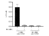
- FIG. 1 It is a figure which shows the antigen specificity of the anti-pSer510-LRH1 monoclonal antibody clone 55 (FMU-P2-C2).
- A The results of immunostaining of HEK293T cells introduced with LRH1S510E using anti-pSer510-LRH1 monoclonal antibody clone 55 (FMU-P2-C2) are shown.
- B The result of immunostaining after adsorption by the antigen peptide in which Ser510 is phosphorylated is shown.
- the scale bar indicates 100 ⁇ m.
- the scale bar indicates 100 ⁇ m. It is a figure which shows the result of the semi-quantification of pSer510-LRH1 staining in pancreatic cancer tissue and liver cancer tissue. The left side of the figure shows the results of semi-quantification of the stainability of anti-pSer510-LRH1 immunostaining in pancreatic cancer tissue by the Allred score. The right side of the figure shows the results of semi-quantification of the stainability of anti-pSer510-LRH1 immunostaining in liver cancer tissue by the Allred score.
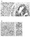
- FIG. 1 shows the result of the immunohistochemical staining in the lung cancer tissue.
- A The results of anti-pSer510-LRH1 immunostaining in lung adenocarcinoma tissue are shown.
- B The results of anti-pSer510-LRH1 immunostaining in lung squamous cell carcinoma tissue are shown. In each case, the left side shows a representative example of the low pSer510-LRH1 group, and the right side shows a representative example of the high pSer510-LRH1 group.
- C Another example of anti-pSer510-LRH1 immunostaining in lung adenocarcinoma tissue is shown.
- Figures 7C-a and 7C-b show enlarged views of the advanced infiltration and the inside of the tumor parenchyma, respectively.
- the scale bar indicates 100 ⁇ m. It is a figure which shows the influence of dephosphorylation treatment on immunostaining by anti-pSer510-LRH1 monoclonal antibody clone 55 (FMU-P2-C2). Results of immunostaining of sections of formalin-fixed paraffin-embedded cell blocks of typical human pancreatic cancer cell lines (AsPC1, HPAFII, and PANC1) under two conditions, with (+) / without (-) dephosphorylation. show. The scale bar indicates 100 ⁇ m. It is a figure which shows the result of immunohistochemical staining in pancreatic cancer tissue.
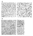
- FIG. 1 The results of anti-pSer510-LRH1 immunostaining in pancreatic cancer tissue are shown.
- the left side shows a representative example of the low pSer510-LRH1 group, and the right side shows a representative example of the high pSer510-LRH1 group.
- FIGB-a and 9B-b show enlarged views of the advanced infiltration and the inside of the tumor parenchyma, respectively.
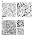
- the scale bar indicates 100 ⁇ m. It is a figure which shows the result of the immunohistochemical staining in the liver cancer tissue.
- FIG. 7B-a and 7B-b show enlarged views of the advanced infiltration and the inside of the tumor parenchyma, respectively.
- the scale bar indicates 100 ⁇ m. It is a figure which shows the result of the immunohistochemical staining in the lung squamous cell carcinoma tissue.
- (A) The results of anti-pSer510-LRH1 immunostaining in squamous cell lung carcinoma are shown.
- the left side shows a representative example of the low pSer510-LRH1 group, and the right side shows a representative example of the high pSer510-LRH1 group.
- (B) Another example of anti-pSer510-LRH1 immunostaining in squamous cell lung carcinoma is shown.
- 11B-a and 11B-b show enlarged views of the advanced infiltration and the inside of the tumor parenchyma, respectively.
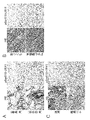
- the scale bar indicates 100 ⁇ m. It is a figure which shows the result that pSer510-LRH1 was not detected in the normal tissue (the non-cancer part normal tissue).
- A The results of HE (Hematoxylin-Eosin) staining and anti-pSer510-LRH1 immunostaining in normal lung tissue (normal tissue around lung cancer) are shown.
- B The results of HE staining and anti-pSer510-LRH1 immunostaining in normal liver tissue (normal tissue around liver cancer) are shown.
- C The results of HE staining and anti-pSer510-LRH1 immunostaining in normal tissues of the pancreas and duodenum (normal tissues surrounding pancreatic cancer) are shown.
- the scale bar indicates 100 ⁇ m.
- the pSer510-LRH1 shown in the last row of the table was shown to be an independent poor prognostic factor.
- the first aspect of the present invention is a biomarker for predicting the prognosis of a cancer patient.
- the biomarker of the present invention is a liver receptor homologue 1 in which the serine residue at position 510 (referred to as "Ser510" in the present specification) in the amino acid sequence shown in SEQ ID NO: 1 is phosphorylated (in the present specification, "Ser510”). It is written as "pSer510-LRH1" etc.).
- cancer is not limited, and examples thereof include adenocarcinoma, squamous cell carcinoma, small cell carcinoma, and large cell carcinoma.
- Specific types of cancer include, for example, malignant melanoma, oral cancer, laryngeal cancer, pharyngeal cancer, thyroid cancer, lung cancer, breast cancer, esophageal cancer, gastric cancer, colon cancer (colon cancer and colon cancer and (Including rectal cancer), small bowel cancer, bladder cancer, prostate cancer, testis cancer, uterine body cancer, cervical cancer, endometrial cancer, ovarian cancer, gastric cancer, renal cancer, liver
- pediatric tumors such as cancer, pancreatic cancer, biliary tract cancer (including bile sac cancer and bile duct cancer), brain tumor, head and neck cancer, mesenteric tumor, osteosarcoma, glioma, and neuroblastoma Examples include leukemia and lymphoma.
- the cancer is preferably liver cancer, pancreatic cancer,
- prognosis refers to reduction of tumor mass, suppression of tumor growth, or course of disease (eg, eg, after cancer treatment (eg, surgery, chemotherapy (pharmaceutical therapy), or radiation therapy)). Presence or absence of recurrence, presence or absence of metastasis, length of survival after treatment, life or death, etc.). "Prediction of prognosis” includes recurrence risk (eg, recurrence-free survival rate), metastasis risk, survival, and a certain period of time after surgery (eg, 1 year, 2 years, 3 years, 4 years, 5 years, 10 years, 15 years).
- prediction of prognosis includes prediction of recurrence risk (eg, recurrence-free survival) or prediction of metastasis risk.
- the recurrence-free survival rate is the proportion of patients who do not develop recurrent cancer such as cancer associated with the initial cancer
- the disease-specific survival rate is the proportion of patients who do not die associated with the initial cancer. Means. Prediction of prognosis can also be said to determine, evaluate, diagnose, or assist in prognosis.
- judgment means to judge the malignancy of cancer. In particular, it refers to determining the malignancy of cancer in a subject (cancer patient) suffering from cancer.
- the "cancer patient” is, for example, a mammal, preferably a primate, and more preferably a human.
- malignancy refers to the degree of infiltration of cancer into surrounding tissues, metastasis to other organs, and / or the degree of recurrence. More specifically, it means the ability of cancer cells to proliferate and / or migrate. By determining the malignancy of cancer, it is possible to predict the prognosis and select cases with poor prognosis with high infiltration, metastasis, and recurrence. In the present specification, the determination of malignancy also includes prediction of prognosis.
- the "biomarker for predicting the prognosis of a cancer patient can predict the prognosis of cancer or indicate the prognosis of cancer.
- a biomarker Specifically, it is a liver receptor homologue 1 (pSer510-LRH1) in which the serine residue (Ser510) at position 510 in the amino acid sequence shown in SEQ ID NO: 1 is phosphorylated.
- Liver receptor homolog-1 (LRH-1; LRH1) is also called nuclear receptor subfamily 5 Group A member 2 (Nuclear Receptor Subfamily 5 Group A Member 2; NR5A2) and is a nucleus. It is a ligand-dependent transcription factor belonging to the internal receptor superfamily. No endogenous ligand for LRH1 has been identified. It is known that abnormal expression of LRH1 and gene mutation are associated with exacerbation of various cancers such as pancreatic cancer, liver cancer, lung cancer, colon cancer, prostate cancer, and breast cancer. Specific examples of LRH1 include human LRH1 consisting of the amino acid sequence shown in SEQ ID NO: 1.
- LRH1 contains a serine residue (Ser510) at position 510 in the amino acid sequence shown in SEQ ID NO: 1, the isoform and the like are not particularly limited.
- LRH1 generally indicates a human-derived LRH1 protein, but 80% or more, 85% or more, 90% or more, 95% or more, 96% or more, 97% or more with respect to the amino acid sequence shown in SEQ ID NO: 1. , 98% or more, or 99% or more identity, or comprises a mutant LRH1 protein in which one or more amino acids have been added, deleted, or substituted with respect to the amino acid sequence shown in SEQ ID NO: 1. ..
- LRH1 orthologs of other species having the same activity as human LRH1 consisting of the amino acid sequence shown in SEQ ID NO: 1 are also included.
- the serine residue at position 510 in the amino acid sequence shown in SEQ ID NO: 1 is the total length of the amino acid sequence shown in SEQ ID NO: 1. Not only the serine residue at position 510, but also the corresponding serine residue in any LRH1 or any peptide fragment thereof (for example, the corresponding serine residue in a peptide fragment consisting of a partial sequence of the amino acid sequence shown in SEQ ID NO: 1). Etc.).
- the serine residue at position 510 in the amino acid sequence shown in SEQ ID NO: 1 is preferably a serine residue that is phosphorylated in the AKT / SGK phosphorylation consensus sequence.
- the phosphorylated Ser510 (phosphorylated Ser510) is referred to as “pSer510” or “pS510".
- the non-phosphorylated Ser510 is particularly distinguished from the phosphorylated Ser510, it is referred to as “non-pSer510” or “non-pS510”.
- liver receptor homolog 1 in which the serine residue at position 510 in the amino acid sequence shown in SEQ ID NO: 1 is phosphorylated (“pSer510-LRH1", “pS510-LRH1”, or “pS510-LRH1” in the present specification.
- Phosphorylated LRH1 (Ser510) ", or” pSer510-NR5A2 ",” pS510-NR5A2 “, or” phosphorylated NR5A2 (Ser510) ", etc.) is the serine at position 510 in the amino acid sequence shown in SEQ ID NO: 1.
- LRH1 consisting of the amino acid sequence shown in SEQ ID NO: 1 in which the residue is phosphorylated, but also any LRH1 in which the corresponding serine residue is phosphorylated (the serine residue is phosphorylated, SEQ ID NO: 1).
- LRH1 consisting of the amino acid sequence shown in SEQ ID NO: 1 in which the residue is phosphorylated, but also any LRH1 in which the corresponding serine residue is phosphorylated (the serine residue is phosphorylated, SEQ ID NO: 1).
- LRH1 consisting of the amino acid sequence shown in SEQ ID NO: 1 in which the residue is phosphorylated
- LRH1 includes LRH1 or any peptide fragment thereof consisting of an amino acid sequence other than.
- the presence or absence of phosphorylation of LRH1 other than the above Ser510 is not particularly limited.
- the term "subject” refers to a human individual who provides a sample and is subjected to an examination. In principle, it is an individual, but the present specification may sometimes include tissues and cells derived from humans. Further, the individual may be not only a healthy body but also a patient having some kind of disease (for example, malignant tumor) or an individual who may be affected by the disease (for example, malignant tumor).
- the term "healthy body” refers to a human individual who does not have a specific cancer, preferably a human individual who does not have any cancer, and more preferably a healthy individual who does not have any disease.
- a human individual in a state preferably a human individual who does not have any cancer, and more preferably a healthy individual who does not have any disease.
- a human individual in a state preferably a human individual who does not have any cancer, and more preferably a healthy individual who does not have any disease.
- a human individual in a state in the present specification, healthy human cells are also included in a healthy body in a broad sense. Therefore, if it is in a healthy state not only at the individual level but also at the cellular level, such as a normal part of a tissue collected from a cancer patient, it is referred to as a healthy body.
- the "invasion advanced part” means a part of the invading cancer that is in contact with the boundary with normal tissue.
- amino acid identity means the ratio (%) of the number of matching amino acid residues to the total number of amino acid residues of the two amino acid sequences to be compared. Specifically, the two amino acid sequences are aligned, and if necessary, a gap is inserted in one or both of them as appropriate. At this time, 1 gap is counted as 1 amino acid residue in the total number of amino acid residues.
- Amino acid sequence alignment can be performed using, for example, known programs such as Blast, FASTA, ClustalW (Karlin, S. et al., 1993, Proc. Natl. Acad. Sci. USA, 90: 5873- 5877; Altschul, S.F.et al., 1990, J. Mol. Biol., 215: 403-410; Pearson, WR et al., 1988, Proc. Natl. Acad. Sci. USA, 85 : 2444-2448). If the total number of amino acid residues differs between the two amino acid sequences to be compared, the longer one is taken as the total number of amino acid residues. It is calculated by dividing the number of the same amino acid residues when the degree of amino acid matching is highest in the two amino acid sequences to be compared by the total number of amino acid residues.
- substitution refers to a group of conservative amino acids having similar properties such as charge, side chain, polarity, and aromaticity among the 20 amino acids that make up a natural protein. Refers to replacement. For example, a group of uncharged polar amino acids (Gly, Asn, Gln, Ser, Thr, Cys, Tyr) having a low polar side chain, a group of branched amino acids (Leu, Val, Ile), and a group of neutral amino acids (Gly, Ile).
- the biomarker for predicting the prognosis of cancer patients of the present invention is phosphorylation of the serine residue at position 510 in the amino acid sequence shown in SEQ ID NO: 1, or the serine residue at position 510 in the amino acid sequence shown in SEQ ID NO: 1. It consists of phosphorylated liver receptor homologue 1 (pSer510-LRH1).
- the biomarker for predicting the prognosis of cancer patients of the present invention can be used for predicting the prognosis of cancer patients suffering from any cancer.
- the biomarkers for predicting prognosis of cancer patients of the present invention include liver cancer, pancreatic cancer, lung cancer, esophageal cancer, kidney cancer, ovarian cancer, gastric cancer, colon cancer, and prostate cancer. , Or used to predict the prognosis of breast cancer.
- the prognosis of cancer patients can be predicted with high accuracy. For example, the risk of recurrence and / or metastasis of a cancer patient can be determined. This makes it possible to select cases with a poor prognosis with high infiltration, metastasis, and / or recurrence from cancers.
- phosphorylation of the serine residue at position 510 in the amino acid sequence shown in SEQ ID NO: 1 or the serine residue at position 510 in the amino acid sequence shown in SEQ ID NO: 1 The use of phosphorylated liver receptor homologue 1 (pSer510-LRH1) as a biomarker for predicting the prognosis of cancer patients is provided.
- a second aspect of the present invention is an anti-pSer510-LRH1 antibody or fragment thereof for predicting the prognosis of a cancer patient.
- the anti-pSer510-LRH1 antibody of the present invention or a fragment thereof can predict the prognosis of cancer in a subject by detecting the phosphorylation of Ser510, which can be phosphorylated in highly malignant cancer.
- Anti-pSer510-LRH1 antibody “Anti-pSer510-LRH1 antibody” (hereinafter referred to as “anti-phosphorylated LRH1 (Ser510) antibody”, “anti-pSer510-NR5A2 antibody”, “anti-phosphorylated NR5A2 (Ser510) antibody”, etc.) is referred to as SEQ ID NO: 1.
- the anti-pSer510-LRH1 antibody of the present invention preferably has liver receptor homologue 1 in which the serine residue at position 510 in the amino acid sequence shown in SEQ ID NO: 1 is not phosphorylated (hereinafter, "Ser510 non-phosphorylated LRH1", "non”.
- non-pSer510-LRH1 -Compare with "pSer510-LRH1", “non-pS510-LRH1”, etc.) or its fragment containing the non-phosphorylated serine residue (hereinafter referred to as "non-pSer510-LRH1 or its fragment”, etc.) It is an antibody that preferentially binds to pSer510-LRH1 or a fragment thereof, and more preferably, it specifically binds to pSer510-LRH1 or a fragment thereof as compared with non-pSer510-LRH1 or a fragment thereof. It is an antibody.
- the species from which the anti-pSer510-LRH1 antibody of the present invention is derived is not particularly limited. Antibodies derived from birds and mammals are preferred. For example, chickens, ostriches, mice, rats, guinea pigs, rabbits, goats, donkeys, sheep, camels, horses, humans and the like.
- the anti-pSer510-LRH1 antibody of the present invention may be either a monoclonal antibody or a polyclonal antibody as long as it is an antibody that recognizes pSer510-LRH1 and exhibits immunoresponsiveness.
- a monoclonal antibody having a stable antibody titer is preferable.
- polyclonal antibody refers to a group of different immunoglobulins that can specifically bind to and recognize an antigen.
- the “monoclonal antibody” includes a framework region (hereinafter referred to as “FR”) and a complementarity determining region (hereinafter referred to as “CDR”).
- FR framework region
- CDR complementarity determining region
- the immunoglobulin is in any class (eg IgG, IgE, IgM, IgA, IgD, and IgY) or any subclass (eg IgG1, IgG2, IgG3). , IgG4, IgA1, and IgA2).
- the epitope of pSer510-LRH1 recognized by the anti-pSer510-LRH1 antibody of the present invention is an epitope peculiar to pSer510-LRH1 or a fragment thereof.
- Such epitopes are found in epitopes (eg, non-pSer510-LRH1 or fragments thereof) that are preferentially (preferably specifically) present in pSer510-LRH1 or fragments thereof compared to non-pSer510-LRH1 or fragments thereof.
- the position of the epitope is not particularly limited.
- the epitope recognized by the anti-pSer510-LRH1 antibody of the present invention arises as a result of a phosphate group-containing epitope of pSer510 or a phosphate group-free epitope of pSer510 (for example, a protein conformational change associated with phosphorylation). It may be any of an epitope having a structure that does not contain a phosphate group), but an epitope that contains a phosphate group of pSer510 is preferable.
- the anti-pSer510-LRH1 antibody that recognizes the above epitope include rat anti-pSer510-LRH1 monoclonal antibody clone 55 (FMU-P2-C2) of Examples described later.
- FMU-P2-C2 antibody the heavy chain variable region consists of the amino acid sequence shown in SEQ ID NO: 8, and the light chain variable region consists of the amino acid sequence shown in SEQ ID NO: 9.
- CDR1 is present in the heavy chain variable region of the FMU-P2-C2 antibody.
- CDR2 consists of the amino acid sequence shown in SEQ ID NO: 3
- CDR3 consists of the amino acid sequence shown in SEQ ID NO: 4.
- CDR1 consists of the amino acid sequence shown in SEQ ID NO: 5
- CDR2 consists of the amino acid sequence shown in SEQ ID NO: 6
- CDR3 consists of the amino acid sequence shown in SEQ ID NO: 7. ..
- the amino acid sequences of SEQ ID NOs: 2 to 9 are shown in Table 1 below.
- nucleic acid As a nucleic acid (nucleotide) encoding the amino acid sequence shown in SEQ ID NO: 8 corresponding to the heavy chain variable region of the FMU-P2-C2 antibody, for example, a nucleic acid consisting of the base sequence shown in SEQ ID NO: 10 can be mentioned. Further, as a nucleic acid encoding the amino acid sequence shown in SEQ ID NO: 9 corresponding to the light chain variable region of the FMU-P2-C2 antibody, for example, a nucleic acid consisting of the base sequence shown in SEQ ID NO: 11 can be mentioned.
- examples of the base sequence encoding CDR1, CDR2, and CDR3 of the heavy chain variable region in the FMU-P2-C2 antibody include nucleic acids consisting of the base sequences shown in SEQ ID NOs: 12, 13, and 14, respectively.
- examples of the base sequence encoding CDR1, CDR2, and CDR3 of the light chain variable region in the FMU-P2-C2 antibody include nucleic acids consisting of the base sequences shown in SEQ ID NOs: 15, 16 and 17, respectively.
- Recombinant antibody refers to a chimeric antibody or a humanized antibody.
- a “chimeric antibody” is an antibody produced by combining amino acid sequences of antibodies derived from different animals, in which the constant region (C region) of one antibody is replaced with the C region of another antibody.
- C region constant region
- an antibody in which the C region of a rat monoclonal antibody is replaced with the C region of a human antibody is applicable.
- the heavy chain variable region of a human antibody against an arbitrary antigen is replaced with the heavy chain variable region consisting of the amino acid sequence shown by SEQ ID NO: 8 in the above-mentioned FMU-P2-C2 antibody, and the human antibody Examples thereof include an antibody obtained by substituting the light chain variable region with the light chain variable region consisting of the amino acid sequence shown in SEQ ID NO: 9. This can reduce the immune response to the antibody in the human body.
- a "humanized antibody” is a mosaic antibody in which the CDR in a human antibody is replaced with the CDR in an antibody derived from a non-human mammal.
- variable region (V region) of the immunoglobulin molecule consists of four FRs (FR1, FR2, FR3 and FR4) and three CDRs (CDR1, CDR2 and CDR3) from the N-terminal side. It is configured by concatenating in the order of CDR3-FR4. Of these, FR is a relatively conserved region that constitutes the skeleton of the variable region, and CDR directly contributes to the antigen-binding specificity of the antibody.
- the humanized antibody is, for example, a set of CDR1, CDR2 and CDR3 in the light chain or heavy chain of a rat-derived anti-pSer510-LRH1 antibody and a set of CDR1, CDR2 in the light chain or heavy chain of a human antibody against any antigen.
- CDR3 By substituting with and CDR3, respectively, it can be constructed as a human antibody that inherits the antigen-binding specificity of the rat anti-pSer510-LRH1 antibody.
- CDR1 consisting of the amino acid sequence represented by SEQ ID NO: 2 derived from the heavy chain in the above-mentioned FMU-P2-C2 antibody
- CDR2 consisting of the amino acid sequence represented by SEQ ID NO: 3
- amino acid represented by SEQ ID NO: 4 CDR3 consisting of the sequence is replaced with the heavy chain CDR1, CDR2, and CDR3 of the human antibody, respectively
- CDR1, SEQ ID NO: 6 consisting of the amino acid sequence shown by SEQ ID NO: 5 derived from the light chain in the above-mentioned FMU-P2-C2 antibody.
- Examples thereof include an antibody obtained by substituting CDR2 consisting of the amino acid sequence shown and CDR3 consisting of the amino acid sequence shown in SEQ ID NO: 7 with the light chains CDR1, CDR2, and CDR3 of a human antibody, respectively. Since such a humanized antibody is derived from a human antibody other than CDR, the immune response to the antibody in the human body can be reduced more than that of a chimeric antibody.
- “Synthetic antibody” refers to an antibody synthesized chemically or by using a recombinant DNA method.
- an antibody newly synthesized using the recombinant DNA method can be mentioned.
- Specific examples thereof include scFv (single chain Fragment of variable region: single chain antibody), diabody, triabody, tetrabody and the like.
- scFv single chain Fragment of variable region: single chain antibody
- diabody diabody
- triabody triabody
- tetrabody tetrabody and the like.
- a set of variable regions light chain variable region V L and heavy chain variable region V H
- a functional antigen binding site are located on separate polypeptide chains, a light chain and a heavy chain. do.
- scFv is a synthetic antibody having a structure in which VL and V H are linked by a flexible linker of sufficient length and contained in one polypeptide chain in an immunoglobulin molecule and having a molecular weight of about 35 kDa or less.
- a set of variable regions can self-assemble with each other to form one functional antigen-binding site.
- the scFv can be obtained by incorporating the recombinant DNA encoding it into a vector using a known technique and expressing it.
- Diabody is a molecule with a structure based on the dimeric structure of scFv (Holliger et al., 1993, Proc. Natl. Acad. Sci. USA 90: 6444-6448).
- the two variable regions within the scFv cannot self-assemble, but by interacting the two scFvs to form a diabody, one scFv. V L can be assembled with V H of the other sc Fv to form two functional antigen binding sites. Furthermore, by adding a cysteine residue to the C-terminal of scFv, a disulfide bond between two scFvs becomes possible, and a stable diabody can be formed. Thus, the diabody is a divalent antibody fragment.
- Triabodies and tetrabodies are trivalent and tetravalent antibodies having a trimer and tetramer structure based on the scFv structure like the diabody, respectively.
- Diabodies, triabodies, and tetrabodies may be multispecific antibodies.
- the “multispecific antibody” refers to a multivalent antibody, that is, an antibody having a plurality of antigen binding sites in one molecule, in which each antigen binding site binds to a different epitope.
- a bispecific antibody (Bispecific antibody) in which each antigen binding site binds to a different epitope in the diabody can be mentioned.
- a diabody in which one antigen-binding site binds to pSer510 and the other antigen-binding site binds to an epitope on pSer510-LRH1 other than pSer510 Is applicable.
- the anti-pSer510-LRH1 antibody of the present invention can also be modified.
- modification includes functional modification required for antigen-specific binding activity such as glycosylation and labeling modification required for antibody detection.
- Glycosylation modification on the anti-pSer510-LRH1 antibody is performed to adjust the affinity of the anti-pSer510-LRH1 antibody for the target pSer510-LRH1.
- a modification that causes loss of glycosylation at the site by introducing a substitution into an amino acid residue constituting glycosylation to remove the glycosylation site, etc. can be mentioned.
- Labeling of anti-pSer510-LRH1 antibodies includes, for example, fluorescent dyes (FITC, Rhodamine, Texas Red, Cy3, Cy5), fluorescent proteins (eg PE, APC, GFP), enzymes (eg horseradish peroxidase, alkaline phosphatase, etc.) Glucose oxidase), labeling with radioactive isotopes (eg, 3 H, 14 C, 35 S) or biotin or (streptavidin) avidin.
- fluorescent dyes eg., Rhodamine, Texas Red, Cy3, Cy5
- fluorescent proteins eg PE, APC, GFP
- enzymes eg horseradish peroxidase, alkaline phosphatase, etc.
- Glucose oxidase eg, labeling with radioactive isotopes (eg, 3 H, 14 C, 35 S) or biotin or (streptavidin) avidin.
- the anti-pSer510-LRH1 antibody of the present invention preferably has a dissociation constant with the pSer510-LRH1 protein of 10 -7 M or less, preferably having a high affinity of, for example, 10 -8 M or less, and more preferably. It is 10 -9 M or less, particularly preferably 10 -10 M or less.
- the dissociation constant can be measured using a technique known in the art. For example, it may be measured by the Biacore system (GE Healthcare) using the speed evaluation kit software.
- fragment thereof refers to an antibody fragment consisting of a part of an anti-pSer510-LRH1 antibody and exhibiting an immune response to pSer510-LRH1 like the anti-pSer510-LRH1 antibody.
- Fab, F (ab') 2 , Fab', etc. are applicable.
- Fab is an antibody fragment produced by cleavage of an IgG molecule by papain on the N-terminal side of the disulfide bond of the hinge portion, and constitutes an H chain constant region (heavy chain constant region: hereinafter referred to as CH) 3 It is composed of C H 1 and V H adjacent to V H in one domain (C H 1, C H 2, C H 3), and a full-length L chain.
- F (ab') 2 is a dimer of Fab'generated by pepsin cleaving the IgG molecule on the C-terminal side of the disulfide bond at the hinge.
- Fab' has a slightly longer H chain than Fab because it includes a hinge portion, but has a structure substantially equivalent to that of Fab.
- Fab' can be obtained by reducing F (ab') 2 under mild conditions and breaking the disulfide link in the hinge region. Since all of these antibody fragments include an antigen-binding site, they have the ability to specifically bind to an antigen epitope.
- anti-pSer510-LRH1 antibody Preparation of anti-pSer510-LRH1 antibody
- the anti-pSer510-LRH1 antibody of the present invention can be obtained by a conventional method in the art. Further, if the amino acid sequence of the monoclonal antibody is clear, it can be prepared by using a chemical synthesis method or a DNA recombination technique based on the amino acid sequence. In addition, monoclonal antibodies can also be obtained from hybridomas that produce the antibody.
- the antigenic peptide that can be used as an immunogen for the anti-pSer510-LRH1 antibody of the present invention is any part of pSer510-LRH1 including pSer510 (hereinafter referred to as "pSer510-LRH1 antigen peptide").
- the antigenic peptide that can be used as an immunogen for the anti-pSer510-LRH1 antibody of the present invention is a peptide containing a serine residue corresponding to the serine residue at position 510 (Ser510) in the amino acid sequence shown in SEQ ID NO: 1.
- pSer510 in the amino acid sequence (RLPEIRAISMQAEE, SEQ ID NO: 18) corresponding to positions 502 to 515 (with the starting methionine as position 1) of the peptide in which the serine residue is phosphorylated, for example, the human LRH1 protein shown in SEQ ID NO: 1.
- Examples thereof include peptides in which serine at the position is phosphorylated and modified, and cytosine (C) is added to the N-terminal.
- the pSer510-LRH1 antigen peptide can be prepared, for example, using a chemical synthesis method or DNA recombination technique.
- a third aspect of the present invention is a kit for predicting the prognosis of a cancer patient.
- the kit for predicting the prognosis of cancer patients of the present invention contains the anti-pSer510-LRH1 antibody of the second aspect or a fragment thereof having immunoresponsiveness as an essential component, and is used for predicting the prognosis of cancer patients other than pSer510-LRH1.
- An antibody against a biomarker hereinafter referred to as "another prognostic biomarker”
- another prognostic antibody hereinafter referred to as “another prognostic antibody”
- an antibody against immune responsiveness is included as a selective component.
- the kit for predicting the prognosis of a cancer patient of this aspect includes the anti-pSer510-LRH1 antibody of the second aspect or a fragment thereof as an essential component.
- the anti-pSer510-LRH1 antibody contained in the kit for predicting the prognosis of a cancer patient of the present invention may be a single type or a plurality of types.
- the kit for predicting the prognosis of a cancer patient of this embodiment may further contain one or more other prognostic antibodies or fragments thereof having immune responsiveness as a selective component.
- the other prognosis-predicting antibody may be any antibody as long as it can improve the accuracy of cancer prognosis prediction when used in combination with the above-mentioned anti-pSer510-LRH1 antibody.
- Antibodies to prognostic biomarkers can be used.
- the kit for predicting prognosis of cancer patients of the present invention includes other reagents necessary for predicting cancer prognosis, such as buffers and secondary antibodies, detection and detection. It may include instructions used to determine the result.
- a fourth aspect of the present invention is a method for predicting the prognosis of a cancer patient.
- the method for predicting the prognosis of a cancer patient of the present invention can predict the prognosis of a cancer patient by detecting pSer510-LRH1 in a sample derived from the cancer patient.
- the method for predicting the prognosis of a cancer patient of the present invention includes a detection step as an essential step, and the prognosis of the cancer patient is indicated based on the positive / negative determination result of the biomarker.
- the “detection step” is to measure the amount of biomarker for prognosis prediction of a cancer patient in a sample derived from a subject suffering from cancer, and based on the measured value, the biomarker is positive. This is a step of determining whether or not there is a negative value (hereinafter, referred to as “positive / negative determination”).
- detection includes any of measurement, qualitative, quantitative, and semi-quantitative.
- the "sample” is taken from a test or a healthy body or a healthy body group and used for the prognosis prediction method of a cancer patient of this embodiment, and corresponds to, for example, a tissue or a cell. ..
- the "tissue” and “cell” referred to here correspond to a tissue and cell suffering from cancer of a subject (for example, a cell in an advanced infiltration part), and a corresponding tissue and cell in a healthy body.
- the sample used in the prognosis prediction method of this embodiment is a sample collected by biopsy from a subject suffering from cancer or excised by surgery.
- it is a portion of the cancer (eg, tissue or cell) that has been biopsied or surgically resected, more preferably an advanced infiltration.
- these tissues or cells may be those embedded in paraffin after formalin fixation (FFPE: Formalin-Fixed Paraffin Embedded).
- FFPE paraffin after formalin fixation
- tissues or cells may be obtained by biopsy or surgical removal by surgery.
- the amount of sample required in the prognosis prediction method of this embodiment is not particularly limited. If it is a tissue or cell, at least 10 ⁇ g, preferably at least 0.1 mg is desirable, but a biopsy material may also be used.
- the sample can be prepared and processed as needed so that the biomarker for predicting the prognosis of the cancer patient can be detected. For example, if the sample is a tissue or a cell, homogenization treatment, cytolysis treatment, removal of impurities by centrifugation or filtration, addition of a protease inhibitor, or the like can be mentioned. Details of these processes are described in detail in Green & Sambrook, Molecular Cloning, 2012, Fourth Ed., Cold Spring Harbor Laboratory Press, and can be referred to.
- the “measured value of the biomarker for prognosis prediction of cancer patients” is specifically referred to as “Ser510 phosphorylation” of Ser510 of liver receptor homologue 1 (LRH1). ) Or the measured value of pSer510-LRH1.
- the "measured value” is a measured value obtained by measuring a biomarker.
- the measured value may be an absolute value in which the amount of protein in the sample is expressed in units such as ng (nanogram) or ⁇ g (microgram), or it is expressed in terms of the absorbance with respect to the control value, the fluorescence intensity of the labeled molecule, and the like. It may be a relative value, or it may be a score (score) calculated by a certain formula from the spatial distribution (for example, staining pattern) of the biomarker in the sample.
- control value may be a measured value of any biomarker other than LRH1, a measured value of non-pS510-LRH1, or a measured value of the entire LRH1 which is a combination of pSer510-LRH1 and non-pS510-LRH1.
- the method for measuring Ser510 phosphorylation is a method for measuring the phosphorylation level of Ser510 or a method for measuring the amount of protein in pSer510-LRH1.
- the method for measuring Ser510 phosphorylation is not particularly limited.
- known quantification methods such as immunological detection method, aptamer analysis method, and mass spectrometry can be used.
- each quantitative method will be described.
- Immunological detection method is a method for quantifying a target molecule using an antibody or a binding fragment thereof that specifically binds to the target molecule.
- Immunological detection methods include, for example, enzyme immunoassay (including ELISA and EIA methods), fluorescence immunoassay, radioimmunosassay (RIA), luminescent immunoassay, and surface plasmon resonance (SPR).
- Crystal transducer microbalance (QCM) method Crystal transducer microbalance (QCM) method, immunoturbidimetric method, latex agglutination immunoassay, latex agglutination method, hemagglutination reaction, particle agglutination reaction method, gold colloid method, capillary electrophoresis method, western blot method or immunity Histochemical methods (immunosasay) and the like are known, but any detection method may be used in this method. Immunohistochemistry is preferred, but not limited.
- the method for quantifying the phosphorylation level of Ser510 or the method for quantifying the protein of pSer510-LRH1 is not limited. For example, it may be quantified as a score calculated using a certain calculation formula based on the observation of the staining pattern of the tissue section. In general, but not limited to, LRH1 functions in the nucleus, so the amount of staining in the nucleus may be quantified.
- the antibody used for immunological detection in this step may be either a monoclonal antibody or a polyclonal antibody, and the immunoglobulin constituting the antibody may be in any class or any subclass, and mammals and birds. It may be derived from any animal including, and may be an artificially produced antibody, for example, a recombinant antibody, a synthetic antibody, or an antibody fragment. The morphology of these antibodies has been described in the second aspect, and detailed description thereof will be omitted here.
- the antibody used for immunological detection in this step is the anti-pSer510-LRH1 antibody or fragment thereof according to the second aspect.
- Aptamer analysis method is a method for quantifying a biomarker protein for prognosis prediction of a cancer patient, which is a target molecule, using an aptamer that strongly and specifically binds to a target substance by a three-dimensional structure. Is. Aptamers can be roughly classified into nucleic acid aptamers and peptide aptamers according to the type of the molecule, but any aptamer may be used.
- Nucleic acid aptamer refers to an aptamer composed of nucleic acids.
- the nucleic acid constituting the nucleic acid aptamer may be DNA, RNA, or a combination thereof. If desired, chemically modified nucleic acids such as PNA, LNA / BNA, methylphosphonate-type DNA, phosphorothioate-type DNA, and 2'-O-methyl-type RNA can be included.
- anti-pSer510-LRH1 RNA aptamer, anti-pSer510-LRH1 DNA aptamer and the like can be mentioned.
- Nucleic acid aptamers can be prepared by using pSer510-LRH1 or a part thereof as a target molecule and using a method known in the art, for example, the SELEX (systematic evolution of ligands by exponential enrichment) method.
- the SELEX method is a known method, and the specific method may be, for example, according to Pan et al. (Proc. Natl. Acad. Sci. U.S.A., 1995, 92: 11509-11513).
- a peptide aptamer is an aptamer composed of amino acids, and is a peptide molecule of 1 to 6 kDa that recognizes the surface structure of a specific target molecule and specifically binds to it, like an antibody.
- anti-pSer510-LRH1 peptide aptamers and the like can be mentioned.
- the peptide aptamer may be prepared based on a production method known in the art. For example, Whaley, S. R., et al., Nature, 2000, 405, 665-668 can be referred to. Usually, it can be prepared by using a phage display method or a cell surface display method.
- the above antibody or aptamer may be labeled if necessary.
- a labeling substance known in the art may be used.
- fluorescent dyes fluorescein, FITC, rhodamine, Texas red, Cy3, Cy5
- fluorescent proteins eg PE, APC, GFP
- enzymes eg horseradish peroxidase, alkaline phosphatase, glucose. It can be labeled with oxidase), radioactive isotopes (eg, 3H, 14C, 35S) or biotin or (streptavidin) avidin.
- nucleic acid aptamers for example, radioisotopes (eg 32P, 3H, 14C), DIG, biotin, fluorescent dyes (eg FITC, Texas, cy3, cy5, cy7, FAM, HEX, VIC, JOE, Rox , TET, Bodipy493, NBD, TAMRA), or luminescent material (eg, acridinium estar).
- radioisotopes eg 32P, 3H, 14C
- DIG diatomadiotin
- biotin eg.g., fluorescent dyes (eg FITC, Texas, cy3, cy5, cy7, FAM, HEX, VIC, JOE, Rox , TET, Bodipy493, NBD, TAMRA), or luminescent material (eg, acridinium estar).
- fluorescent dyes eg FITC, Texas, cy3, cy5, cy7, FAM
- Mass Spectrometry includes high-speed liquid chromatograph mass spectrometry (LC-MS), high-speed liquid chromatograph tandem mass spectrometry (LC-MS / MS), and gas chromatograph mass spectrometry (GC). -MS), gas chromatograph tandem mass spectrometry (GC-MS / MS), capillary electrophoresis mass spectrometry (CE-MS) and ICP mass spectrometry (ICP-MS).
- LC-MS liquid chromatograph mass spectrometry
- LC-MS / MS high-speed liquid chromatograph tandem mass spectrometry
- GC gas chromatograph mass spectrometry
- CE-MS capillary electrophoresis mass spectrometry
- ICP-MS ICP mass spectrometry
- the immunological detection method, the aptamer analysis method, and the mass spectrometry method are all known techniques in the art, and may be performed according to these methods.
- the method for determining positive / negative based on the measured value is not limited.
- a method of determining a cutoff value for a measured value of Ser510 phosphorylation and determining positive / negative based on the cutoff value can be mentioned. That is, a predetermined value is defined as a cutoff value, and if the measured value is greater than or equal to that value, Ser510 phosphorylation can be determined to be positive, and conversely, if it is less than the cutoff value, it can be determined to be negative.
- the cutoff value is the boundary value for classifying the measured value into positive and negative.
- the cutoff value can usually be calculated based on the morbidity of the disease and the sensitivity and specificity calculated from the ROC curve (receiver operating characteristic curve).
- the method of setting the cutoff value is not particularly limited.
- the cutoff value is the average value of the measured value of the sample derived from a healthy body not suffering from cancer or the measured value of the sample derived from a healthy body group, and the measured value of the subject is larger than the cutoff value. When it is high, it can be determined to be positive.
- a positive value can also be determined when the measured value of the subject is higher than the cutoff value, with a cutoff value of 5 times or more or 6 times or more.
- the measured values obtained from the control group by percentile and use the percentile value used for the classification as the cutoff value. For example, if the 95th percentile of the measured value obtained from the control is set as the cutoff value, the value above that value is positive, and the value below that value is negative, if the measured value of the subject is 95th percentile or more, it is determined to be positive. be able to.
- the measured value of the healthy body as a control is different from the measured value of the subject, and it is not always necessary to measure each time. For example, if the amount of the sample used for the measurement, the measurement method of the biomarker for prognosis prediction of the cancer patient, and the measurement conditions are kept constant, the previously measured measurement value of the control healthy body can be reused.
- the prognosis of a cancer patient is predicted (the prognosis is shown) based on the detection result of Ser510 phosphorylation obtained in the above detection step. Specifically, when a sample derived from a cancer patient is positive for the phosphorylation, it is indicated that the prognosis of the cancer patient is poor or the prognosis of the cancer patient is likely to be poor. Conversely, if a sample derived from a cancer patient is negative for the phosphorylation, it indicates that the prognosis of the cancer patient is good or that the prognosis of the cancer patient is likely to be good.
- poor prognosis means poor clinical outcome (eg, after surgical resection), high risk of cancer recurrence or high recurrence rate, risk of cancer metastasis or metastasis. High rate, low recurrence-free survival rate, low disease (cancer) -specific survival rate, or low overall survival rate). If the prognosis is poor, the recurrence-free survival rate or disease-specific survival rate after 5 years may be 95% or less, 90% or less, 85% or less, 80% or less, 75% or less, or 70% or less. In the present invention, the survival rate means the cumulative survival rate.
- good prognosis means that the clinical outcome is good. If the prognosis is good, the recurrence-free survival rate or survival rate 5 years after the cancer resection surgery is 60% or more, 70% or more, 80% or more, 90% or more, 95% or more, or 100%. good.
- a biomarker for prognosis prediction of a cancer patient other than Ser510 phosphorylation is detected, and the cancer is based on the detection result and the detection result of Ser510 phosphorylation.
- the prognosis of the patient may be predicted.
- the biomarkers for predicting the prognosis of cancer patients other than Ser510 phosphorylation conform to the description of the third aspect.
- any one or more of cell atypia, structural atypia, infiltration, and metastasis is detected in a sample derived from a cancer patient, and the detection result and the detection result of Ser510 phosphorylation are detected.
- the prognosis of cancer patients may be predicted.
- “cell atypia” is a distance from the normal cell structure, specifically, an increase in the ratio of nuclear vesicles, irregularities in the size of cells and nuclei, irregular nuclear shape, increased amount of nuclear chromatin, and nucleoli. Refers to the increase or increase of, the increase of nucleoli, and / or the appearance of abnormal nucleoli.
- structural atypia is a distance from a normal tissue structure, that is, an irregularity of the tissue structure.
- the method for detecting cell atypia or structural atypia is not limited. For example, it can be visualized using hematoxylin and eosin staining.
- Invasion is the continuous progression of a malignant tumor while destroying the surrounding normal tissues and organs, and can be determined by obscuring the boundary with the surrounding tissues.
- Metalastasis is the discontinuous progression of a malignant tumor to an organ distant from the primary lesion.
- the prognosis of the subject who provided the sample can be accurately predicted by examining the biological sample removed by biopsy or surgery.
- the prolapse prediction method of this embodiment which has a high accuracy rate, determines the risk of recurrence and metastasis, and based on the results, determines the treatment policy (for example, type of anticancer drug, dose, dosing interval, etc.). Alternatively, the interval between tests for cancer recurrence and metastasis can be determined.
- the present invention indicates that a cancer patient has a poor prognosis, the patient is given drug therapy and / or radiation therapy to prevent recurrence of the cancer, improve the prognosis, or improve the survival rate.
- a method for assisting the prediction of the prognosis of cancer is also provided.
- a method for determining the malignancy of the cancer of the subject and a method for assisting the determination of the malignancy of the cancer of the subject are also provided.
- the method of the present invention includes other methods (for example, radiography; ultrasonography; endoscopy; mammography; pelvic examination; rectal examination; imaging examination such as CT examination and MRI examination; blood examination; cytology and histology examination, etc. Pathological examination; and / or genetic diagnosis) and other factors (eg, stage classification, tumor diameter, presence or absence of lymph node metastasis, histological grade, etc.) can be used in combination.
- other methods for example, radiography; ultrasonography; endoscopy; mammography; pelvic examination; rectal examination; imaging examination such as CT examination and MRI examination; blood examination; cytology and histology examination, etc.
- Pathological examination e.g, stage classification, tumor diameter, presence or absence of lymph node metastasis, histological grade, etc.
- a method for treating cancer and a method for improving the prognosis of a cancer patient including a step of predicting the prognosis of a cancer patient by the method of the present invention, are also provided.
- a fifth aspect of the present invention is a method for screening a cancer inhibitor or therapeutic agent. According to the screening method of this embodiment, an inhibitor or therapeutic agent capable of reducing Ser510 phosphorylation in liver receptor homologue 1 (LRH1) can be identified.
- LLRH1 liver receptor homologue 1
- the screening method of the present invention includes a test substance treatment step, a phosphorylation measurement step, and an inhibitor / therapeutic agent identification step.
- test substance treatment step refers to cells expressing the liver receptor homologue 1 (LRH1), which consists of the amino acid sequence shown in SEQ ID NO: 1 and in which the serine residue at position 510 in the amino acid sequence is phosphorylated, as the test substance. This is the process of treatment.
- LHL1 liver receptor homologue 1
- test substance subject to this screening method is not particularly limited.
- the test substance is any substance, specifically, a natural molecule (for example, amino acid, peptide, oligopeptide, polypeptide, protein, nucleic acid, lipid, carbohydrate (sugar, etc.), steroid, glycopeptide, glycoprotein, proteoglycan, etc. ), Synthetic analogs or derivatives of natural molecules (eg, peptide mimetics, nucleic acid molecules (aptamers, antisense nucleic acids, double-stranded RNA (RNAi), etc.)), and non-natural molecules such as low molecular weight compounds (eg, low molecular weight inorganic). Compounds and low molecular weight organic compounds), etc .; and mixtures thereof.
- a natural molecule for example, amino acid, peptide, oligopeptide, polypeptide, protein, nucleic acid, lipid, carbohydrate (sugar, etc.), steroid, glycopeptide, glycoprotein, proteog
- test substance a single test substance may be tested independently, or a mixture of several candidate test substances (for example, a library) may be tested.
- candidate test substances for example, a library
- the library containing a plurality of test substances include a synthetic compound library, a peptide library (combinatorial library, etc.) and the like.
- contact is by culturing the cells in a medium supplemented with the test substance, by immersing the cells in a solution containing the test substance, by laminating the test substance on the cells, or by depositing the cells on the test substance. It can be carried out by culturing in the presence of.
- the effect and effectiveness of the test substance can be examined under several conditions.
- Such conditions include time or duration of treatment with the test substance, amount (large or small), number of times, and the like.
- a plurality of doses can be set by preparing a dilution series of the test substance.
- the treatment period of the test substance can be appropriately set, and for example, the treatment can be performed over a period of one day to several weeks, months, or years.
- the cell used in the screening method of the present invention comprises the amino acid sequence shown in SEQ ID NO: 1 and expresses the liver receptor homologue 1 (LRH1) in which the serine residue at position 510 in the amino acid sequence is phosphorylated. .. "A cell consisting of the amino acid sequence shown in SEQ ID NO: 1 and expressing the liver receptor homologue 1 (LRH1) in which the serine residue at position 510 in the amino acid sequence is phosphorylated" is derived from the amino acid sequence shown in SEQ ID NO: 1. It is a cell that expresses the liver receptor homologue 1 (LRH1) and phosphorylates the serine residue at position 510 of the liver receptor homologue 1 (LRH1).
- Cells in which the serine residue at position 510 of liver receptor homolog 1 (LRH1) is phosphorylated are limited to cells in which the serine residue at position 510 of liver receptor homolog 1 (LRH1) can be phosphorylated. do not do.
- the cells are naturally occurring cells in which the serine residue at position 510 of the liver receptor homolog 1 (LRH1) is phosphorylated, or the phosphorylation of the serine residue at position 510 of the liver receptor homolog 1 (LRH1) is induced. It may be any of the cells to be subjected to. As long as it is a naturally occurring cell, it may be a cell derived from cancer in which the serine residue at position 510 of the liver receptor homologue 1 (LRH1) is phosphorylated.
- It may be a cell to which an activating agent is added.
- the cell type is not particularly limited as long as it is an animal cell capable of inducing phosphorylation of the serine residue at position 510 of the liver receptor homologue 1 (LRH1).
- Examples of the biological species from which the animal cells are derived include mammals (for example, primates such as humans, experimental animals such as rats and mice), and are any of primary cultured cells, subcultured cells, and frozen cells. You may. Further, it may be a normal cell or a cancer cell (for example, a cancer cell line derived from liver cancer or pancreatic cancer).
- the “phosphorylation measurement step” is a step of measuring Ser510 phosphorylation in cells treated with a test substance by the test substance treatment step.
- the method for measuring Ser510 phosphorylation is a method for measuring the phosphorylation level of Ser510 or a method for measuring the amount of protein in pSer510-LRH1.
- the method for measuring Ser510 phosphorylation is not particularly limited, and a known measuring method can be used.
- a) immunological detection method, (b) aptamer analysis method, or (c) mass spectrometry method described in the fourth aspect can be used.
- an immunological detection method eg, immunohistochemistry
- an immunological detection method eg, immunohistochemistry
- the phosphorylation measurement step is performed at an appropriate time following the test substance treatment step. For example, immediately after the test substance treatment step, 30 minutes, 1 hour, 3 hours, 5 hours, 10 hours, 15 hours, 20 hours, 24 hours (1 day), 2 to 10 days, Measurements are taken 10 to 20 days later, 20 to 30 days later, and 1 to 6 months later.
- the "inhibitor / therapeutic agent identification step” is a step of identifying the test substance as a cancer inhibitor or therapeutic agent when the phosphorylation of Ser510 is reduced.
- reduced phosphorylation of Ser510 means that phosphorylation of Ser510 is reduced as compared with a predetermined cutoff value or a value when no treatment is performed with the test substance.
- the phosphorylation of Ser510 is 100% or less, 90% or less, 80% or less, 70% or less, 60% or less, 50% or less, 20% or less, 10 compared to the value when not treated with the test substance.
- the test substance can be identified as a cancer suppressant or therapeutic agent when it is% or less, 5% or less, 1% or less, or 0.1% or less.
- this screening method can identify a cancer suppressant or therapeutic agent (or a candidate substance thereof) and further confirm its effectiveness.
- Example 1 Preparation of anti-pSer510-LRH1 monoclonal antibody> (Purpose)
- a monoclonal antibody (anti-pSer510-LRH1 monoclonal antibody) that can detect LRH1 (pSer510-LRH1) in which serine at position 510 (Ser510) is phosphorylated.
- the monoclonal antibody was prepared according to the following procedure based on the method described in Kishiro Y, et al., 1995, Cell Struct Funct, 20 (2): 151-6.
- antigen peptide As an antigen peptide, position 510 in the amino acid sequence (RLPEIRAISMQAEE, SEQ ID NO: 18) corresponding to positions 502 to 515 of the human LRH1 protein shown in SEQ ID NO: 1 (with starting methionine as position 1). A peptide in which serine was phosphorylated and modified and cytosine (C) was added to the N-terminal was used.
- Imject Maleimide Activated mcKLH (Thermo Fisher Scientific) 2 mg was dissolved in 200 ⁇ L of ultrapure water to prepare a 10 mg / mL KLH solution.
- the antigen peptide was dissolved in ultrapure water to prepare a 5 mg / mL antigen peptide solution.
- 200 ⁇ L of each KLH solution and antigen peptide solution were mixed and allowed to stand at room temperature for 2 hours.
- the mixture was transferred to a boiled dialysis membrane and dialyzed using PBS as an external solution.
- the obtained solution was used as an antigen solution.
- 400 ⁇ L of the antigen solution was mixed with 1 mL of Freund's complete adjuvant (Sigma-Aldrich) and emulsified to prepare an antigen emulsion.
- PEG polyethylene glycol
- the iliac lymph nodes were excised from the rats 14 days after immunization and placed in a sterile petri dish with 1 mL of DMEM. Lymph nodes were shredded with scissors and then filtered through a 100 ⁇ m cell strainer (BD Falcon). Approximately 107 mouse multiple myeloma cell lines SP2 were added to the petri dish, mixed well with a pipette, centrifuged at 1200 rpm / min for 5 minutes, and the supernatant was removed by suction. The PEG solution at 37 ° C. was slowly added dropwise over about 1 minute, left for 2 minutes, and then 9 mL of DMEM medium was slowly added dropwise over 5 minutes.
- the above-mentioned antigen-peptide solution and a peptide solution containing a non-phosphorylated product of serine at position 510 as a negative control were prepared at 3 ⁇ g / mL, 50 ⁇ L each was added to the wells of a 96-well ELISA plate, and the temperature was 4 ° C. overnight. It was left still. Then, the antigen peptide solution is removed from each well, washed once with 200 ⁇ L of 0.1% Tween20-added Tris-hydrochloric acid buffer (TBS-T), and then 200 ⁇ L of blocking solution (1% bovine serum albumin / TBS) is added. In addition, it was allowed to stand at 37 ° C. for 1 hour.
- TBS-T Tris-hydrochloric acid buffer
- the blocking solution was removed from each well, washed once with 200 ⁇ L of TBS-T, 50 ⁇ L of the culture supernatant was added, and the mixture was reacted at 37 ° C. for 1 hour. After removing the culture supernatant from each well, the cells were washed 3 times with 200 ⁇ L of TBS-T. Subsequently, 50 ⁇ L of ECL TM Rat IgG and HRP-linked whole antibody (Cytiva) diluted 2,000 times with a blocking solution were added to each well as a secondary antibody, and the reaction was carried out at 37 ° C. for 1 hour.
- the secondary antibody solution is removed from each well, washed three times with TBS-T, and then colored by the method recommended by the manufacturer using the TMB Substrate Set (BioLegend), and the absorbance (OD) at the wavelength of 490 nm is developed. 490 ) was measured.
- the positive clones were subcultured in a 12-well culture dish, and when the confluency reached approximately 50%, they were transferred to a 10 cm culture dish and further propagated.
- the ELISA results of clone 55 (FMU-P2-C2), which is the anti-pSer510-LRH1 monoclonal antibody analyzed below, are shown in Table 2 below. Shown in. In Table 2, the antigen peptide in which Ser510 is phosphorylated is shown as pS510-LRH1 peptide, and the antigen peptide in which Ser510 is not phosphorylated is shown as non-pS510-LRH1 peptide.
- Example 2 Verification of antigen specificity of anti-pSer510-LRH1 monoclonal antibody clone 55 (FMU-P2-C2)> (Purpose) The antigen specificity of the anti-pSer510-LRH1 monoclonal antibody clone 55 (FMU-P2-C2) obtained in Example 1 will be verified.
- the specimen was deparaffinized in xylene for 10 minutes, followed by dexylene in 100% ethanol. After treating with 0.3% hydrogen peroxide / methanol for 10 minutes to inactivate the endogenous peroxidase, the cells were washed with Tris-hydrochloric acid buffer for 5 minutes. Specimens were treated in 0.1% semicarbazide hydrochloric acid (Wako Fuji Film Industries, Ltd.) aqueous solution for 1 hour and washed with Tris-hydrochloric acid buffer (TBS) for 5 minutes. Antigen activation was performed by incubating in a hybrid oven at 70 ° C. for one day and night using an immunosaver (Nisshin EM) diluted 200-fold with ultrapure water.
- an immunosaver Neshin EM
- the specimen was washed with TBS for 5 minutes, and the anti-pSer510-LRH1 monoclonal antibody was used as the primary antibody and reacted at 4 ° C. for 24 hours.
- the biotin-labeled anti-rat IgG secondary antibody which was washed 3 times with TBS for 5 minutes and diluted 100-fold with Signal Booster Immunostain F solution (Beacle Inc.), was reacted at room temperature for 30 minutes.
- the mixture was washed 3 times with TBS for 5 minutes, and the avidin / horseradish peroxidase solution diluted 50-fold with the same buffer was reacted at room temperature for 30 minutes.
- Nuclear staining was performed by shaking in a solution of Tissue Tech (registered trademark) hematoxylin 3G (Sakura Finetech Japan Co., Ltd.) for 5 seconds, and then washing with running water for 10 minutes. Next, it was shaken for 5 seconds in a 0.5% hydrochloric acid / 70% ethanol solution to remove excess hematoxylin. Specimens were dehydrated using 100% ethanol and transparent treated with xylene, and preparations were prepared using an automatic preparation encapsulation machine (Shiraimatsu Kikai Co., Ltd.).
- Figure 1 shows the results of immunostaining with anti-pSer510-LRH1 monoclonal antibody clone 55 (FMU-P2-C2).
- FMU-P2-C2 anti-pSer510-LRH1 monoclonal antibody clone 55
- LRH1S510E cells positive signals were observed in the nucleus and nucleolus (Fig. 1A).
- no positive signal was detected after adsorption by the phosphorylated antigenic peptide of Ser510 (Fig. 1B).
- FIG. 2 shows the results of examining the concentration dependence of the reaction between the purified anti-pSer510-LRH1 monoclonal antibody and the antigen peptide.
- the pSer510-LRH1 monoclonal antibody reacted concentration-dependently with the antigen peptide (phosphorylated LRH1 peptide) in which Ser510 was phosphorylated (Fig. 2).
- no antibody reaction was detected when Ser510 used an antigen peptide that was not phosphorylated (non-phosphorylated LRH1 peptide) or when only the Tris-hydrochloric acid buffer used to dilute the peptide was used. ..
- Example 3 CDR sequencing of anti-pSer510-LRH1 monoclonal antibody clone 55 (FMU-P2-C2)> (Purpose) The CDR sequence of anti-pSer510-LRH1 monoclonal antibody clone 55 (FMU-P2-C2) is determined. (Method and result) The heavy and light chain variable regions and CDRs of the anti-pSer510-LRH1 monoclonal antibody clone 55 (FMU-P2-C2) were sequenced. The clone was sent to Bio-Peak and sequenced by degenerate primer PCR. CDR identification was performed according to the North / AHo antibody numbering system.
- Figure 4 shows the results of sequencing the heavy and light chain variable regions and CDRs of the anti-pSer510-LRH1 monoclonal antibody clone 55 (FMU-P2-C2).
- the heavy chain CDR1, CDR2, and CDR3 consist of the amino acid sequences set forth in SEQ ID NOs: 2, 3, and 4, respectively, and the light chain CDR1, CDR2, and CDR3 consist of the amino acid sequences set forth in SEQ ID NOs: 5, 6, and 7, respectively.
- the heavy chain variable region consists of the amino acid sequence shown in SEQ ID NO: 8
- the light chain variable region consists of the amino acid sequence shown in SEQ ID NO: 9.
- Example 4 Immunohistochemistry in pancreatic cancer tissue and liver cancer tissue> (Purpose)
- the anti-pSer510-LRH1 monoclonal antibody clone 55 (FMU-P2-C2) is used to detect the pSer510-LRH1 protein in pancreatic and liver cancer tissues and determine its malignancy.
- Method (1) Collection of tissue specimens 39 patients (pancreatic cancer) who underwent surgery for pancreatic cancer and liver cancer diagnosis between 2012 and 2017 at Fukushima Prefectural Medical University Hospital Hepatobiliary Pancreatic and Transplant Surgery Tissue specimens were collected from 19 cases of pancreatic cancer and 20 cases of liver cancer, and the expression of pSer510-LRH1 was evaluated. The target cases were limited to patients whose survival information was known for 3 to 5 years after diagnosis, and death cases not dependent on the underlying disease were excluded from the target cases. The collection of tissue materials was approved by the Fukushima Medical University Ethics Committee (approval number 2020-058) and was carried out in compliance with the ethical guidelines related to clinical research.
- a score of 0 was evaluated as pSer510-LRH1 negative, a score of 1 to 5 was evaluated as pSer510-LRH1 weak positive, and a score of 6 or more was evaluated as pSer510-LRH1 strong positive.
- pancreatic cancer pancreatic ductal adenocarcinoma or invasive pancreatic ductal carcinoma
- hepatocellular carcinoma hepatocellular carcinoma
- pancreatic cancer All 19 cases of pancreatic cancer were strongly positive for pSer510-LRH1, but it reflects the fact that pancreatic cancer is difficult to detect early and is quite advanced when diagnosed in most cases. It is considered that it is coded with the result of the above-mentioned liver cancer case.
- pSer510-LRH1 by detecting pSer510-LRH1 in cancer tissue, it is possible to determine the malignancy of cancer and predict recurrence and patient prognosis, and based on this, it is possible to perform postoperative treatment such as drug therapy and / or radiotherapy. It was shown that the need can be determined. It was also shown that pSer510-LRH1 could be a promising drug discovery target for cancer treatment.
- Example 5 Immunohistochemistry in lung cancer tissue> (Purpose)
- anti-pSer510-LRH1 monoclonal antibody clone 55 FMU-P2-C2 is used to detect the pSer510-LRH1 protein in lung cancer tissue and determine its malignancy.
- FIGS. 7A and 7C Examples of anti-pSer510-LRH1 immunostaining in lung adenocarcinoma tissue are shown in FIGS. 7A and 7C.
- the left side shows a representative example of the low pSer510-LRH1 group
- the right side shows a representative example of the high pSer510-LRH1 group.
- a stronger pSer510-LRH1 signal was observed in the advanced infiltration area (Fig. 7C-a) than in the tumor parenchyma (Fig.
- Example 6 Further verification of antigen specificity of anti-pSer510-LRH1 monoclonal antibody clone 55 (FMU-P2-C2)> (Purpose)
- FMU-P2-C2 Anti-pSer510-LRH1 monoclonal antibody clone 55 obtained in Example 1
- the effect of dephosphorylation treatment on a section of a pancreatic cancer cell line will be examined, and the antigen specificity will be verified.
- the anti-pSer510-LRH1 monoclonal antibody clone 55 (FMU-P2-C2) can specifically detect phosphorylated LRH1 from dephosphorylated LRH1 in pancreatic cancer cell lines.
- Example 7 Further immunohistochemistry in pancreatic cancer tissue and liver cancer tissue> (Purpose) Increase the number of tissue samples in Example 4, detect pSer510-LRH1 protein in pancreatic cancer and liver cancer tissues by anti-pSer510-LRH1 monoclonal antibody clone 55 (FMU-P2-C2), and additionally perform malignancy determination. ..
- the tissue sample used in this example includes all of the tissue samples used in Example 4.
- Tissue specimens 72 patients who underwent surgery for pancreatic cancer diagnosis between 2008 and 2018 at Fukushima Prefectural Medical University Hospital Hepatobiliary Pancreatic Transplant Surgery and Cooperating Hospital, Liver between 2006 and 2016 Tissue specimens were collected from 127 patients who underwent surgery for cancer diagnosis and evaluated for pSer510-LRH1 expression.
- the target cases were limited to patients whose survival information was known for at least 3 to 5 years from the diagnosis, and death cases not dependent on the primary disease were excluded from the target cases.
- the collection of tissue materials was approved by the Fukushima Medical University Ethics Committee (approval number 2020-058) and was carried out in compliance with the ethical guidelines related to clinical research.
- Immunohistochemical staining and histological evaluation were performed according to the method described in Example 4. However, in the semi-quantitative evaluation of liver cancer tissue, a score of 0 was evaluated as pSer510-LRH1 negative, a score of 1 to 6 was evaluated as pSer510-LRH1 weakly positive, and a score of 7 or more was evaluated as pSer510-LRH1 strongly positive. The pSer510-LRH1 negative and pSer510-LRH1 weak positives were classified into the low pSer510-LRH1 group, and the pSer510-LRH1 strong positives were classified into the high pSer510-LRH1 group. The semi-quantitative evaluation of pancreatic cancer tissue was performed according to the score evaluation described in Example 4.
- FIGS. 9A and 9B An example of anti-pSer510-LRH1 immunostaining in pancreatic cancer (pancreatic ductal adenocarcinoma or invasive pancreatic ductal cancer) tissue is shown in FIGS. 9A and 9B.
- FIGS. 10A and 10B An example of anti-pSer510-LRH1 immunostaining in liver cancer (hepatocellular carcinoma) tissue is shown in FIGS. 10A and 10B.
- the left side shows a representative example of the low pSer510-LRH1 group
- the right side shows a representative example of the high pSer510-LRH1 group.
- pSer510-LRH1 signal stronger than that inside the tumor parenchyma (Fig. 9B-b, Fig. 10B-b) is emitted at the advanced infiltration part (Fig. 9B-a, Fig. 10B-a). Admitted.
- pancreatic cancer of the high pSer510-LRH1 group 54 of 72 cases of pancreatic cancer were in the high pSer510-LRH1 group (Table 5).
- pancreatic cancer of the high pSer510-LRH1 group recurrence was observed in 33 of 35 deaths of less than 3 years and 12 of 19 survivors of 3 years or more.
- pancreatic cancer of the low pSer510-LRH1 group 5 of 9 patients who survived for 3 years or more had no recurrence.
- liver cancer of the high pSer510-LRH1 group 28 of 127 cases of liver cancer were in the high pSer510-LRH1 group (Table 6). Furthermore, in the liver cancer of the high pSer510-LRH1 group, recurrence was observed in 9 of 12 deaths of less than 5 years and 9 of 16 survivals of 5 years or more. On the other hand, in the liver cancer of the low pSer510-LRH1 group, 35 of 57 patients who survived for 5 years or more had no recurrence.
- Ser510 of LRH1 can be phosphorylated in highly malignant cancer tissues, and pSer510-LRH1 protein in cancer tissues is converted by anti-pSer510-LRH1 monoclonal antibody clone 55 (FMU-P2-C2). It was shown to be detectable.
- pSer510-LRH1 by detecting pSer510-LRH1 in cancer tissue, it is possible to determine the malignancy of cancer and predict recurrence and patient prognosis, and based on this, it is possible to perform postoperative treatment such as drug therapy and / or radiotherapy. It was shown that the need can be determined.
- the pSer510-LRH1 signal was significantly enhanced in the advanced infiltration areas of pancreatic cancer tissue and liver cancer tissue, by targeting pSer510-LRH1 as a therapeutic target, cancer infiltration (and metastasis) can be prevented. It was shown that it could be stopped.
- pSer510-LRH1 could be a promising drug discovery target for cancer treatment.
- Example 8 Further immunohistochemistry in lung cancer tissue> (Purpose)
- the pSer510-LRH1 protein in the lung squamous cell carcinoma tissue is additionally detected by the anti-pSer510-LRH1 monoclonal antibody clone 55 (FMU-P2-C2) with respect to the lung squamous cell carcinoma tissue specimen of Example 5.
- FIGS. 11A and 11B Additional examples of anti-pSer510-LRH1 immunostaining in lung squamous cell carcinoma tissue are shown in FIGS. 11A and 11B.
- FIG. 11A the left side shows a representative example of the low pSer510-LRH1 group, and the right side shows a representative example of the high pSer510-LRH1 group.
- a stronger pSer510-LRH1 signal was observed in the advanced infiltration area (Fig. 11B-a) than in the tumor parenchyma (Fig. 11B-b).
- This result is similar to the lung adenocarcinoma tissue examined in Example 5 (advanced infiltration and tumor parenchymal interior are shown in FIGS. 7C-a and 7C-b, respectively).
- Example 9 Immunohistochemistry in normal tissue (non-cancerous normal tissue)> (Purpose) Verify that the pSer510-LRH1 protein is not detected in normal tissue (non-cancerous normal tissue).
- Immunohistochemical staining with (FMU-P2-C2) is performed.
- a liver cancer tissue sample of the pSer510-LRH1 strongly positive group was stained to confirm a positive signal.
- FIG. 12A An example of HE staining and anti-pSer510-LRH1 immunostaining in normal lung tissue (normal tissue around lung cancer) is shown in FIG. 12A.
- FIG. 12B shows an example of HE staining and anti-pSer510-LRH1 immunostaining in normal liver tissue (normal tissue around liver cancer).
- FIG. 12C An example of HE staining and anti-pSer510-LRH1 immunostaining in normal tissues of the pancreas and duodenum (normal tissues surrounding pancreatic cancer) is shown in FIG. 12C. No pSer510-LRH1 signal was detected in any normal tissue (non-cancerous normal tissue) of the lung, liver, pancreas, or duodenum.
- Example 10 Prognosis evaluation of liver cancer patients based on pSer510-LRH1 signal> (Purpose) The prognosis of 127 patients who underwent resection surgery for liver cancer (hepatocellular carcinoma) will be evaluated based on immunohistochemical staining of pSer510-LRH1. Further clinical pathological analysis is performed.
- IS Intensity Score
- liver cancer patients in the low pSer510-LRH1 group have a better prognosis after liver cancer resection surgery
- liver cancer patients in the high pSer510-LRH1 group have a better prognosis after liver cancer resection surgery. It was shown that the prognosis tended to be poor.
Landscapes
- Health & Medical Sciences (AREA)
- Life Sciences & Earth Sciences (AREA)
- Chemical & Material Sciences (AREA)
- Engineering & Computer Science (AREA)
- Immunology (AREA)
- Molecular Biology (AREA)
- Medicinal Chemistry (AREA)
- Biomedical Technology (AREA)
- General Health & Medical Sciences (AREA)
- Biochemistry (AREA)
- Hematology (AREA)
- Urology & Nephrology (AREA)
- Physics & Mathematics (AREA)
- Analytical Chemistry (AREA)
- General Physics & Mathematics (AREA)
- Pathology (AREA)
- Food Science & Technology (AREA)
- Organic Chemistry (AREA)
- Microbiology (AREA)
- Cell Biology (AREA)
- Biotechnology (AREA)
- Biophysics (AREA)
- Genetics & Genomics (AREA)
- Proteomics, Peptides & Aminoacids (AREA)
- Bioinformatics & Cheminformatics (AREA)
- Pharmacology & Pharmacy (AREA)
- Hospice & Palliative Care (AREA)
- Oncology (AREA)
- Peptides Or Proteins (AREA)
Abstract
がん患者の予後を予測するためのバイオマーカー、及び当該バイオマーカーを検出するための抗体を提供することである。 配列番号1で示すアミノ酸配列において510位のセリン残基がリン酸化されている肝臓受容体ホモログ1(pSer510-LRH1)からなる、がん患者の予後を予測するためのバイオマーカーを提供する。
Description
本発明は、がん患者の予後を予測するためのバイオマーカー、がん患者の予後を予測するための抗体、がん患者の予後を予測するための方法、及びがんの抑制剤又は治療剤をスクリーニングする方法に関する。
わが国において、がんは死亡原因全体の中で最も多く、約3割を占めている。
部位別では、膵がん、肝がん、及び肺がんは、日本国内及び世界において死亡数の上位を占めている(非特許文献1~5)。これらのがんは、いずれも転移率及び再発率が高く、予後不良な症例が多いことから、代表的な難治性がんとされている。特に膵がんは、早期発見が困難な上に進行が早いため、その5年生存率は約10%と極めて低い。また、肝がんでは生活習慣病を背景とする非アルコール性脂肪性肝疾患を原因とする症例が増加しており、2030年における死亡数は年間約100万人に達すると試算されている。しかし、肝がんに対する既存の分子標的薬の効果は、全生存期間の1~3か月の延長にとどまり、その効果は限定的である。したがって、難治性がんの診断及び治療を可能とする新たな方法の開発が必要とされている(非特許文献1~2)。
部位別では、膵がん、肝がん、及び肺がんは、日本国内及び世界において死亡数の上位を占めている(非特許文献1~5)。これらのがんは、いずれも転移率及び再発率が高く、予後不良な症例が多いことから、代表的な難治性がんとされている。特に膵がんは、早期発見が困難な上に進行が早いため、その5年生存率は約10%と極めて低い。また、肝がんでは生活習慣病を背景とする非アルコール性脂肪性肝疾患を原因とする症例が増加しており、2030年における死亡数は年間約100万人に達すると試算されている。しかし、肝がんに対する既存の分子標的薬の効果は、全生存期間の1~3か月の延長にとどまり、その効果は限定的である。したがって、難治性がんの診断及び治療を可能とする新たな方法の開発が必要とされている(非特許文献1~2)。
一方、大腸がん、前立腺がん、及び乳がんは、比較的予後良好な悪性腫瘍である。しかしながら、これらのがんは患者の総数が多く、予後不良な症例が少なからず存在する。したがって、がん患者の中から、悪性度が高く、浸潤、転移、及び再発能の高い、予後不良ながんを選別する方法が必要とされている(非特許文献6~8)。
よって、がん患者の予後を予測するためのバイオマーカー、及びがん治療のための創薬標的の同定が嘱望されている。
Mizrahi, JD, et al., Lancet, 2020, 395:2008-2020.
Ryan, DP, et al., N Engl J Med, 2014, 371:1301-1314.
Forner, A, et al., Lancet, 2018, 391:2008-2020.
Villanueva, A, N Engl J Med, 2019, 380:1450-1462.
Hirsch, FR, et al., Lancet, 2017, 389:299-311.
Gazder, AF, Nat Rev Cancer, 2017, 37:725-737.
Attard, G, et al., Lancet, 2016, 387:70-82
Harbeck, N, and Grant, M, Lancet, 2017, 389:1134-1150.
本発明の目的は、がん患者の予後を予測するためのバイオマーカー、及び当該バイオマーカーを検出するための抗体を提供することである。
肝臓受容体ホモログ1(Liver receptor homolog-1;LRH-1;LRH1)は、核内受容体サブファミリー5グループAメンバー2(Nuclear Receptor Subfamily 5 Group A Member 2;NR5A2)とも呼ばれ、核内受容体スーパーファミリーに属するリガンド依存性転写因子である。LRH1/NR5A2の発現異常や遺伝子変異は、膵がん、肝がん、肺がん、大腸がん、前立腺がん、乳がん等、様々ながん腫の増悪と関連することが知られている(Benod, C, et al., Proc Natl Acad Sci USA, 2011, 108:16926-16931.;Bianco, S, et al., Cancer Res, 2014, 74:2015-2025.;Bayrer, JR, et al., Proc Natl Acad Sci USA, 2015, 112:2467-2472.;Xu, P, et al., Genes Dev, 2016, 30:1450-1462.;Bailey, P, et al., Nature, 2016, 531:47-52.;Coho, I, et al., Nature, 2018, 554:533-537.)。しかしながら、LRH1/NR5A2がその発現異常や遺伝子変異以外でがんと関連する機序は知られていない。
本発明者らは、がんの予後を予測するための新たなバイオマーカーを求めて鋭意研究を行った。その結果、LRH1における510位のセリン残基(Ser510)のリン酸化が、がんの悪性度を判定し、予後を予測するための有効なバイオマーカーとなり得ることを見出した。特に、Ser510のリン酸化は、悪性度の高いがんの浸潤先進部において強く検出された。さらに、Ser510がリン酸化されたLRH1(pSer510-LRH1)を特異的に検出することができる抗体(抗pSer510-LRH1抗体)を開発し、本発明を完成させるに至った。本発明は、当該知見に基づくものであって以下を提供する。
(1)配列番号1で示すアミノ酸配列において510位のセリン残基がリン酸化されている肝臓受容体ホモログ1(pSer510-LRH1)からなる、がん患者の予後を予測するためのバイオマーカー。
(2)前記がんが肝臓がん、膵臓がん、肺がん、食道がん、腎臓がん、卵巣がん、胃がん、大腸がん、前立腺がん、又は乳がんである、(1)に記載のバイオマーカー。
(3)がん患者の予後を予測するための抗pSer510-LRH1抗体又はその断片。
(4)前記抗pSer510-LRH1抗体又はその断片が、配列番号2で示すアミノ酸配列からなるCDR1、配列番号3で示すアミノ酸配列からなるCDR2、及び配列番号4で示すアミノ酸配列からなるCDR3を含む重鎖可変領域と、配列番号5で示すアミノ酸配列からなるCDR1、配列番号6で示すアミノ酸配列からなるCDR2、及び配列番号7で示すアミノ酸配列からなるCDR3を含む軽鎖可変領域を含む、(3)に記載の抗pSer510-LRH1抗体又はその断片。
(5)前記抗pSer510-LRH1抗体又はその断片が配列番号8で示すアミノ酸配列からなる重鎖可変領域、及び配列番号9で示すアミノ酸配列からなる軽鎖可変領域を含む、(4)に記載の抗pSer510-LRH1抗体又はその断片。
(6)(1)又は(2)に記載のバイオマーカーを検出するための、(3)~(5)のいずれかに記載の抗pSer510-LRH1抗体又はその断片を含む、がん患者の予後を予測するためのキット。
(7)配列番号1で示すアミノ酸配列において510位のセリン残基がリン酸化されている肝臓受容体ホモログ1(pSer510-LRH1)の、がん患者の予後を予測するためのバイオマーカーとしての使用。
(8)がん患者の予後を予測するための方法であって、がん患者に由来する試料において、肝臓受容体ホモログ1(LRH1)の配列番号1で示すアミノ酸配列における510位のセリン残基のリン酸化を検出する検出工程を含み、ここで、前記試料が前記リン酸化について陽性である場合、がん患者の予後が悪いことを示す、方法。
(9)前記リン酸化は(3)~(5)のいずれかに記載の抗pSer510-LRH1抗体又はその断片を用いて検出される、(8)に記載の方法。
(10)がんの抑制剤又は治療剤をスクリーニングする方法であって、配列番号1で示すアミノ酸配列からなり、該アミノ酸配列における510位のセリン残基がリン酸化される肝臓受容体ホモログ1(LRH1)を発現する細胞を被験物質で処置する工程、前記リン酸化を測定する工程、及び被験物質で処置しない場合と比較して前記リン酸化が減少する場合に被験物質をがんの抑制剤又は治療剤として同定する工程を含む方法。
本明細書は本願の優先権の基礎となる日本国特許出願番号2021-004066号の開示内容を包含する。
本発明によれば、がん患者の予後を予測するためのバイオマーカー、及び当該バイオマーカーを検出するための抗体を提供することができる。
1.がん患者の予後予測用バイオマーカー
1-1.概要
本発明の第1の態様は、がん患者の予後予測用バイオマーカーである。本発明のバイオマーカーは、配列番号1で示すアミノ酸配列において510位のセリン残基(本明細書において「Ser510」と表記する)がリン酸化されている肝臓受容体ホモログ1(本明細書において「pSer510-LRH1」等と表記する)である。悪性度の高いがんにおいてリン酸化され得るSer510のリン酸化をバイオマーカーとして使用することで、がんの悪性度の判定や予後の予測を行うことが可能となる。
1-1.概要
本発明の第1の態様は、がん患者の予後予測用バイオマーカーである。本発明のバイオマーカーは、配列番号1で示すアミノ酸配列において510位のセリン残基(本明細書において「Ser510」と表記する)がリン酸化されている肝臓受容体ホモログ1(本明細書において「pSer510-LRH1」等と表記する)である。悪性度の高いがんにおいてリン酸化され得るSer510のリン酸化をバイオマーカーとして使用することで、がんの悪性度の判定や予後の予測を行うことが可能となる。
1-2.定義
本明細書において、「がん」の種類は限定しないが、例えば、腺がん、扁平上皮がん、小細胞がん及び大細胞がん等が挙げられる。具体的ながんの種類としては、例えば、悪性黒色腫、口腔がん、喉頭がん、咽頭がん、甲状腺がん、肺がん、乳がん、食道がん、胃がん、大腸がん(結腸がん及び直腸がんを含む)、小腸がん、膀胱がん、前立腺がん、精巣がん、子宮体がん、子宮頚がん、子宮内膜がん、卵巣がん、胃がん、腎がん、肝がん、膵がん、胆道がん(胆嚢がん及び胆管がんを含む)、脳腫瘍、頭頸部がん、中皮腫、骨肉腫、神経膠腫、神経芽腫を始めとする小児腫瘍、白血病、リンパ腫等が挙げられる。がんは、好ましくは肝臓がん、膵臓がん、肺がん、食道がん、腎臓がん、卵巣がん、胃がん、大腸がん、前立腺がん、又は乳がんである。
本明細書において、「がん」の種類は限定しないが、例えば、腺がん、扁平上皮がん、小細胞がん及び大細胞がん等が挙げられる。具体的ながんの種類としては、例えば、悪性黒色腫、口腔がん、喉頭がん、咽頭がん、甲状腺がん、肺がん、乳がん、食道がん、胃がん、大腸がん(結腸がん及び直腸がんを含む)、小腸がん、膀胱がん、前立腺がん、精巣がん、子宮体がん、子宮頚がん、子宮内膜がん、卵巣がん、胃がん、腎がん、肝がん、膵がん、胆道がん(胆嚢がん及び胆管がんを含む)、脳腫瘍、頭頸部がん、中皮腫、骨肉腫、神経膠腫、神経芽腫を始めとする小児腫瘍、白血病、リンパ腫等が挙げられる。がんは、好ましくは肝臓がん、膵臓がん、肺がん、食道がん、腎臓がん、卵巣がん、胃がん、大腸がん、前立腺がん、又は乳がんである。
本明細書において、「予後」は、がん治療(例えば、手術、化学療法(薬物療法)、又は放射線治療等)後の、腫瘍量の低減、腫瘍増殖の抑制、又は疾患の経過(例えば、再発の有無、転移の有無、治療後の生存期間の長さ、生死等)をいう。「予後の予測」は、再発リスク(例えば無再発生存率)、転移リスク、生存期間、手術から一定期間後(例えば、1年、2年、3年、4年、5年、10年、15年若しくは20年後又はそれ以上の時点)の生存率、無再発生存率(Relapse-free survival、RFS)、又は疾患特異的生存率(Disease-free survival、DFS)の予測であってもよい。一実施形態において、予後の予測は、再発リスク(例えば無再発生存率)の予測又は転移リスクの予測を含む。なお、本明細書において無再発生存率は、初発がんと関連付けられるがん等の再発がん発症のない患者の割合であり、疾患特異的生存率は、初発がんと関連する死亡のない患者の割合を意味する。予後の予測は、予後の判定、評価、診断、又はこれらの補助ということもできる。
本明細書において「判定」とは、がんの悪性度を判定することをいう。特にがんに罹患している被検体(がん患者)において、がんの悪性度を判定することをいう。
本明細書において、「がん患者」は、例えば哺乳動物、好ましくは霊長類、より好ましくはヒトである。
本明細書において「悪性度」とは、がんの周囲組織への浸潤、他臓器への転移、及び/又は再発能の程度等をいう。より具体的には、がん細胞の増殖能及び/又は遊走能を意味する。がんの悪性度を判定することによって、予後を予測し、浸潤、転移、及び再発能の高い予後不良例を選別することが可能となる。本明細書において、悪性度の判定には予後の予測も含まれる。
本明細書において「がん患者の予後を予測するためのバイオマーカー(がん患者の予後予測用バイオマーカー)」とは、がんの予後を予測する、又はがんの予後を示すことができるバイオマーカーをいう。具体的には、配列番号1で示すアミノ酸配列において510位のセリン残基(Ser510)がリン酸化されている肝臓受容体ホモログ1(pSer510-LRH1)である。
「肝臓受容体ホモログ1(Liver receptor homolog-1;LRH-1;LRH1)」は、核内受容体サブファミリー5グループAメンバー2(Nuclear Receptor Subfamily 5 Group A Member 2;NR5A2)とも呼ばれ、核内受容体スーパーファミリーに属するリガンド依存性転写因子である。LRH1の内因性リガンドは同定されていない。LRH1の発現異常や遺伝子変異は、膵がん、肝がん、肺がん、大腸がん、前立腺がん、乳がん等、様々ながん腫の増悪と関連することが知られている。LRH1の具体例として、配列番号1で示すアミノ酸配列からなるヒトLRH1が挙げられる。本明細書においては、LRH1は、配列番号1で示すアミノ酸配列において510位のセリン残基(Ser510)を含む限り、アイソフォーム等は特に限定しない。本明細書においてLRH1は原則としてヒト由来のLRH1タンパク質を示すが、配列番号1で示すアミノ酸配列に対して80%以上、85%以上、90%以上、95%以上、96%以上、97%以上、98%以上、若しくは99%以上の同一性を有するか、又は配列番号1で示すアミノ酸配列に対して1又は複数個のアミノ酸が付加、欠失、若しくは置換された変異型LRH1タンパク質を含むものとする。また、配列番号1で示すアミノ酸配列からなるヒトLRH1と同等の活性を有する他生物種のLRH1オルソログも包含される。
本明細書において「配列番号1で示すアミノ酸配列において510位のセリン残基」(本明細書において「Ser510」又は「S510」等と表記する)とは、配列番号1で示すアミノ酸配列の全長における510位のセリン残基だけでなく、任意のLRH1若しくはその任意のペプチド断片においてそれに対応するセリン残基(例えば、配列番号1で示すアミノ酸配列の部分配列からなるペプチド断片においてそれに対応するセリン残基等)を包含する。配列番号1で示すアミノ酸配列において510位のセリン残基は、好ましくはAKT/SGKリン酸化コンセンサス配列中でリン酸化されるセリン残基である。また、リン酸化されているSer510(リン酸化Ser510)を「pSer510」又は「pS510」等と表記する。なお、リン酸化されていないSer510を特にリン酸化Ser510と区別する場合には、「non-pSer510」又は「non-pS510」等と表記する。
本明細書において「配列番号1で示すアミノ酸配列において510位のセリン残基がリン酸化されている肝臓受容体ホモログ1」(本明細書において「pSer510-LRH1」、「pS510-LRH1」、若しくは「リン酸化LRH1(Ser510)」、又は「pSer510-NR5A2」、「pS510-NR5A2」、若しくは「リン酸化NR5A2(Ser510)」等と表記する)とは、配列番号1で示すアミノ酸配列において510位のセリン残基がリン酸化された、配列番号1で示すアミノ酸配列からなるLRH1だけでなく、それに対応するセリン残基がリン酸化された任意のLRH1(当該セリン残基がリン酸化された、配列番号1以外のアミノ酸配列からなるLRH1)若しくはその任意のペプチド断片を包含する。なお、LRH1の上記Ser510以外のリン酸化の有無等については特に問わない。
本明細書において「被検体」とは、試料を提供し、検査に供されるヒト個体をいう。原則として個体であるが、本明細書では、時としてヒト由来の組織や細胞も包含し得る。また、個体は、健常体のみならず、何らかの疾患(例えば悪性腫瘍)を有する患者、又は疾患(例えば悪性腫瘍)の罹患可能性のある個体のいずれであってもよい。
本明細書において「健常体」とは、特定のがんに罹患していないヒト個体、好ましくはいかなるがんにも罹患していないヒト個体、より好ましくはいずれの疾患にも罹患していない健常状態にあるヒト個体をいう。ただし、本明細書では健常ヒト細胞も広義の健常体に含むものとする。したがって、個体レベルのみならず、例えば、がん患者から採取した組織のうちの正常部分のように、細胞レベルで健常状態にあれば健常体と称することとする。
本明細書において「浸潤先進部」とは、浸潤するがんの一部であって、正常組織との境界に接する部分を意味する。
本明細書において「複数個」とは、例えば、2~50個、2~40個、2~30個、2~20個、2~18個、2~16個、2~14個、2~12個、2~10個、2~8個、2~7個、2~6個、2~5個、2~4個又は2~3個をいう。また、本明細書において「アミノ酸同一性」とは、比較する2つのアミノ酸配列の全アミノ酸残基数における一致したアミノ酸残基数の割合(%)をいう。具体的には、2つのアミノ酸配列を整列(アラインメント)し、必要に応じ、一方又は双方に適宜ギャップを挿入する。このとき、1ギャップは、1アミノ酸残基として全アミノ酸残基数にカウントする。アミノ酸配列の整列化は、例えば、Blast、FASTA、ClustalW等の既知プログラムを用いて行うことができる(Karlin,S.et al., 1993, Proc. Natl. Acad. Sci. USA, 90: 5873-5877;Altschul,S.F.et al., 1990, J. Mol. Biol., 215: 403-410;Pearson,W.R.et al., 1988, Proc. Natl. Acad. Sci. USA, 85: 2444-2448)。比較する2つのアミノ酸配列間で全アミノ酸残基数が異なる場合には、長い方を全アミノ酸残基数とする。比較する2つのアミノ酸配列においてアミノ酸一致度が最も高くなるようにしたときの同一アミノ酸残基数を全アミノ酸残基数で除して算出される。
本明細書において「(アミノ酸の)置換」とは、天然のタンパク質を構成する20種類のアミノ酸間において、電荷、側鎖、極性、芳香族性等の性質の類似する保存的アミノ酸群内での置換をいう。例えば、低極性側鎖を有する無電荷極性アミノ酸群(Gly, Asn, Gln, Ser, Thr, Cys, Tyr)、分枝鎖アミノ酸群(Leu, Val, Ile)、中性アミノ酸群(Gly, Ile, Val, Leu, Ala, Met, Pro)、親水性側鎖を有する中性アミノ酸群(Asn, Gln, Thr, Ser, Tyr, Cys)、酸性アミノ酸群(Asp, Glu)、塩基性アミノ酸群(Arg, Lys, His)、芳香族アミノ酸群(Phe, Tyr, Trp)内での置換が挙げられる。これらの群内でのアミノ酸置換であれば、ペプチドの性質に変化を生じにくいことが知られているため好ましい。
1-3.構成
本発明のがん患者の予後予測用バイオマーカーは、配列番号1で示すアミノ酸配列において510位のセリン残基のリン酸化、又は配列番号1で示すアミノ酸配列において510位のセリン残基がリン酸化されている肝臓受容体ホモログ1(pSer510-LRH1)からなる。
本発明のがん患者の予後予測用バイオマーカーは、配列番号1で示すアミノ酸配列において510位のセリン残基のリン酸化、又は配列番号1で示すアミノ酸配列において510位のセリン残基がリン酸化されている肝臓受容体ホモログ1(pSer510-LRH1)からなる。
本発明のがん患者の予後予測用バイオマーカーは、任意のがんに罹患したがん患者の予後予測に使用することができる。
一実施形態において、本発明のがん患者の予後予測用バイオマーカーは、肝臓がん、膵臓がん、肺がん、食道がん、腎臓がん、卵巣がん、胃がん、大腸がん、前立腺がん、又は乳がんの予後予測に使用される。
1-4.効果
本発明のがん患者の予後予測用バイオマーカーを使用することで、がん患者の予後を高い精度で予測することができる。例えば、がん患者の再発リスク、及び/又は転移リスクを判定することができる。このことによって、がんの中から浸潤、転移、及び/又は再発能の高い予後不良例を選別することが可能となる。
本発明のがん患者の予後予測用バイオマーカーを使用することで、がん患者の予後を高い精度で予測することができる。例えば、がん患者の再発リスク、及び/又は転移リスクを判定することができる。このことによって、がんの中から浸潤、転移、及び/又は再発能の高い予後不良例を選別することが可能となる。
本発明のがん患者の予後予測用バイオマーカーによれば、配列番号1で示すアミノ酸配列において510位のセリン残基のリン酸化、又は配列番号1で示すアミノ酸配列において510位のセリン残基がリン酸化されている肝臓受容体ホモログ1(pSer510-LRH1)の、がん患者の予後予測用バイオマーカーとしての使用が提供される。
2.がん患者の予後を予測するための抗pSer510-LRH1抗体又はその断片
2-1.概要
本発明の第2の態様は、がん患者の予後を予測するための抗pSer510-LRH1抗体又はその断片である。本発明の抗pSer510-LRH1抗体又はその断片は、悪性度の高いがんにおいてリン酸化され得るSer510のリン酸化を検出することによって、被検体におけるがんの予後を予測することができる。
2-1.概要
本発明の第2の態様は、がん患者の予後を予測するための抗pSer510-LRH1抗体又はその断片である。本発明の抗pSer510-LRH1抗体又はその断片は、悪性度の高いがんにおいてリン酸化され得るSer510のリン酸化を検出することによって、被検体におけるがんの予後を予測することができる。
2-2.構成
(抗pSer510-LRH1抗体)
「抗pSer510-LRH1抗体」(以下、「抗リン酸化LRH1(Ser510)抗体」、「抗pSer510-NR5A2抗体」、「抗リン酸化NR5A2(Ser510)抗体」等と表記する)とは、配列番号1で示すアミノ酸配列において510位のセリン残基がリン酸化されている肝臓受容体ホモログ1(pSer510-LRH1)又は当該リン酸化セリン残基を含むその断片(以下、「pSer510-LRH1又はその断片」と表記する)に対して免疫応答性を示す抗体をいう。本発明の抗pSer510-LRH1抗体は、好ましくは配列番号1で示すアミノ酸配列において510位のセリン残基がリン酸化されていない肝臓受容体ホモログ1(以下、「Ser510非リン酸化LRH1」、「non-pSer510-LRH1」、「non-pS510-LRH1」等と表記する)又は当該非リン酸化セリン残基を含むその断片(以下、「non-pSer510-LRH1又はその断片」等と表記する)と比較してpSer510-LRH1又はその断片に対して優先的に結合する抗体であり、より好ましくは、non-pSer510-LRH1又はその断片と比較してpSer510-LRH1又はその断片に対して特異的に結合する抗体である。
本発明の抗pSer510-LRH1抗体の由来生物種は、特に限定しない。好ましくは鳥類及び哺乳動物由来の抗体である。例えば、ニワトリ、ダチョウ、マウス、ラット、モルモット、ウサギ、ヤギ、ロバ、ヒツジ、ラクダ、ウマ、又はヒト等が挙げられる。
本発明の抗pSer510-LRH1抗体は、pSer510-LRH1を認識し、免疫応答性を示す抗体である限り、モノクローナル抗体又はポリクローナル抗体のいずれであってもよい。好ましくは抗体価が安定しているモノクローナル抗体である。
本明細書において「ポリクローナル抗体」とは、抗原に特異的に結合し、かつそれを認識することのできる異なる複数種の免疫グロブリン群をいう。
また、本明細書において「モノクローナル抗体」とは、フレームワーク領域(Framework region:以下、「FR」と表記する)及び相補性決定領域(Complementarity determining region:以下、「CDR」と表記する)を含み、抗原に特異的に結合し、かつそれを認識することのできる単一種の免疫グロブリン、又は免疫グロブリンに含まれる少なくとも1組の軽鎖可変領域(VL領域)及び重鎖可変領域(VH領域)を包含する組換え抗体又は合成抗体をいう。
抗pSer510-LRH1抗体が免疫グロブリン分子で構成される場合、免疫グロブリンは任意のクラス(例えば、IgG、IgE、IgM、IgA、IgD、及びIgY)、又は任意のサブクラス(例えば、IgG1、IgG2、IgG3、IgG4、IgA1、及びIgA2)とすることができる。
本発明の抗pSer510-LRH1抗体が認識するpSer510-LRH1のエピトープは、pSer510-LRH1又はその断片に特有なエピトープである。そのようなエピトープは、non-pSer510-LRH1又はその断片と比較してpSer510-LRH1又はその断片に優先的に(好ましくは特異的に)存在するエピトープ(例えば、non-pSer510-LRH1又はその断片に含まれていないエピトープ)である限り、そのエピトープの位置は特に限定しない。本発明の抗pSer510-LRH1抗体が認識するエピトープは、pSer510のリン酸基を含むエピトープ、又はpSer510のリン酸基を含まないエピトープ(例えば、リン酸化に伴うタンパク質のコンホメーション変化の結果として生じるエピトープであってリン酸基を含まない構造からなるもの)のいずれであってもよいが、pSer510のリン酸基を含むエピトープであることが好ましい。
上記エピトープを認識する抗pSer510-LRH1抗体の具体例として、後述する実施例のラット抗pSer510-LRH1モノクローナル抗体クローン55(FMU-P2-C2)が挙げられる。このFMU-P2-C2抗体は、重鎖可変領域が配列番号8で示すアミノ酸配列からなり、また軽鎖可変領域が配列番号9で示すアミノ酸配列からなる。Kabatのルール(Kabat E.A., et al., 1991, Sequences of proteins of immunological interest, Vol.1, eds. 5, NIH publication)によれば、FMU-P2-C2抗体の重鎖可変領域において、CDR1は配列番号2で示すアミノ酸配列からなり、CDR2は配列番号3で示すアミノ酸配列からなり、CDR3は配列番号4で示すアミノ酸配列からなる。また、FMU-P2-C2抗体の軽鎖可変領域において、CDR1は配列番号5で示すアミノ酸配列からなり、CDR2は配列番号6で示すアミノ酸配列からなり、CDR3は配列番号7で示すアミノ酸配列からなる。配列番号2~9のアミノ酸配列を以下の表1に示す。

なお、FMU-P2-C2抗体の重鎖可変領域に相当する前記配列番号8で示すアミノ酸配列をコードする核酸(ヌクレオチド)として、例えば、配列番号10で示す塩基配列からなる核酸が挙げられる。また、FMU-P2-C2抗体の軽鎖可変領域に相当する前記配列番号9で示すアミノ酸配列をコードする核酸として、例えば、配列番号11で示す塩基配列からなる核酸が挙げられる。さらに、上記FMU-P2-C2抗体における重鎖可変領域のCDR1、CDR2、及びCDR3をコードする塩基配列として、例えば、それぞれ配列番号12、13、及び14で示す塩基配列からなる核酸が挙げられる。また、上記FMU-P2-C2抗体における軽鎖可変領域のCDR1、CDR2、及びCDR3をコードする塩基配列として、例えば、それぞれ配列番号15、16、及び17で示す塩基配列からなる核酸が挙げられる。
「組換え抗体」とは、キメラ抗体、又はヒト化抗体をいう。「キメラ抗体」とは、異なる動物由来の抗体のアミノ酸配列を組み合わせて作製される抗体で、ある抗体の定常領域(C領域)を他の抗体のC領域で置換した抗体である。例えば、ラットモノクローナル抗体のC領域をヒト抗体のC領域と置き換えた抗体が該当する。具体的な例を挙げると、任意の抗原に対するヒト抗体の重鎖可変領域を前述のFMU-P2-C2抗体における配列番号8で示すアミノ酸配列からなる重鎖可変領域と置換し、またヒト抗体の軽鎖可変領域を配列番号9で示すアミノ酸配列からなる軽鎖可変領域と置換してなる抗体が挙げられる。これによりヒト体内における当該抗体に対する免疫反応を軽減し得る。「ヒト化抗体」とは、ヒト抗体におけるCDRをヒト以外の哺乳動物由来の抗体におけるCDRと置換したモザイク抗体である。免疫グロブリン分子の可変領域(V領域)は、4つのFR(FR1、FR2、FR3及びFR4)と3つのCDR(CDR1、CDR2及びCDR3)がN末端側からFR1-CDR1-FR2-CDR2-FR3-CDR3-FR4の順序で連結されて構成されている。このうちFRは可変領域の骨格を構成する相対的に保存された領域であり、CDRが抗体の抗原結合特異性に直接寄与する。ヒト化抗体は、例えば、ラット由来の抗pSer510-LRH1抗体の軽鎖又は重鎖における一組のCDR1、CDR2及びCDR3を任意の抗原に対するヒト抗体の軽鎖又は重鎖における一組のCDR1、CDR2、及びCDR3とそれぞれ置換することによって、ラット抗pSer510-LRH1抗体の抗原結合特異性を受け継いだヒト抗体として構築することができる。具体的な例を挙げると、前述のFMU-P2-C2抗体における重鎖由来の配列番号2で示すアミノ酸配列からなるCDR1、配列番号3で示すアミノ酸配列からなるCDR2、及び配列番号4で示すアミノ酸配列からなるCDR3をヒト抗体の重鎖CDR1、CDR2、及びCDR3とそれぞれ置換し、また前述のFMU-P2-C2抗体における軽鎖由来の配列番号5で示すアミノ酸配列からなるCDR1、配列番号6で示すアミノ酸配列からなるCDR2、及び配列番号7で示すアミノ酸配列からなるCDR3をヒト抗体の軽鎖CDR1、CDR2、及びCDR3とそれぞれ置換してなる抗体が挙げられる。このようなヒト化抗体は、CDR以外はヒト抗体由来であることからヒト体内における当該抗体に対する免疫反応をキメラ抗体以上に軽減し得る。
「合成抗体」とは、化学的に又は組換えDNA法を用いることによって合成した抗体をいう。例えば、組換えDNA法を用いて新たに合成された抗体が挙げられる。具体的には、例えば、scFv(single chain Fragment of variable region:単鎖抗体)、ダイアボディ(diabody)、トリアボディ(triabody)又はテトラボディ(tetrabody)等が挙げられる。免疫グロブリン分子において、機能的な抗原結合部位を形成する一組の可変領域(軽鎖可変領域VL及び重鎖可変領域VH)は、軽鎖と重鎖という別々のポリペプチド鎖上に位置する。scFvは、免疫グロブリン分子において、VL及びVHを十分な長さの柔軟性リンカーによって連結し、1本のポリペプチド鎖に包含した構造を有する分子量約35 kDa以下の合成抗体である。scFv内において1組の可変領域は、互いに自己集合して1つの機能的な抗原結合部位を形成することができる。scFvは、それをコードする組換えDNAを、公知技術を用いてベクターに組み込み、発現させることで得ることができる。ダイアボディは、scFvの二量体構造を基礎とした構造を有する分子である(Holliger et al., 1993, Proc. Natl. Acad. Sci. USA 90:6444-6448)。例えば、上記リンカーの長さが約12アミノ酸残基よりも短い場合、scFv内の2つの可変領域は自己集合できないが、2つのscFvを相互作用させてダイアボディを形成させることにより、一方のscFvのVLが他方のscFvのVHと集合可能となり、2つの機能的な抗原結合部位を形成することができる。さらに、scFvのC末端にシステイン残基を付加させることにより、2本のscFvどうしのジスルフィド結合が可能となり、安定的なダイアボディを形成させることもできる。このようにダイアボディは二価の抗体断片である。トリアボディ、及びテトラボディは、ダイアボディと同様にscFv構造を基本とした、その三量体、及び四量体構造を有する、それぞれ、三価、及び四価の抗体である。ダイアボディ、トリアボディ、及びテトラボディは、多重特異性抗体であってもよい。「多重特異性抗体」とは、多価抗体、すなわち抗原結合部位を一分子内に複数有する抗体において、それぞれの抗原結合部位が異なるエピトープと結合する抗体をいう。例えば、ダイアボディにおいて、それぞれの抗原結合部位が異なるエピトープと結合する二重特異性抗体(Bispecific抗体)が挙げられる。具体的には、例えば、本発明の抗pSer510-LRH1抗体であれば、一方の抗原結合部位がpSer510と結合し、他方の抗原結合部位がpSer510以外のpSer510-LRH1上のエピトープと結合するダイアボディが該当する。
本発明の抗pSer510-LRH1抗体は、修飾することもできる。ここでいう「修飾」とは、グリコシル化のような抗原特異的結合活性に必要な機能上の修飾や抗体検出に必要な標識上の修飾を含む。
抗pSer510-LRH1抗体上のグリコシル化修飾は、標的であるpSer510-LRH1に対する抗pSer510-LRH1抗体の親和性を調整するために行われる。具体的には、例えば、抗pSer510-LRH1抗体のFRにおいて、グリコシル化を構成するアミノ酸残基に置換を導入してグリコシル化部位を除去することで、その部位のグリコシル化を喪失させる改変等が挙げられる。
抗pSer510-LRH1抗体の標識には、例えば、蛍光色素(FITC、ローダミン、テキサスレッド、Cy3、Cy5)、蛍光タンパク質(例えば、PE、APC、GFP)、酵素(例えば、西洋ワサビペルオキシダーゼ、アルカリフォスファターゼ、グルコースオキシダーゼ)、放射性同位元素(例えば、3H、14C、35S)又はビオチン若しくは(ストレプト)アビジンによる標識が挙げられる。
本発明の抗pSer510-LRH1抗体は、pSer510-LRH1タンパク質との解離定数が、10-7 M以下であることが好ましく、例えば10-8M以下の高い親和性を有することが好ましく、より好ましくは10-9M以下、特に好ましくは10-10 M以下である。上記解離定数は、当該分野で公知の技術を用いて測定することができる。例えば、Biacoreシステム(GE Healthcare社)により速度評価キットソフトウェアを用いて測定してもよい。
(その断片)
本明細書において「その断片」とは、抗pSer510-LRH1抗体の一部からなり、かつ抗pSer510-LRH1抗体と同様にpSer510-LRH1に対して免疫応答性を示す抗体断片をいう。例えば、Fab、F(ab')2、Fab'等が該当する。
本明細書において「その断片」とは、抗pSer510-LRH1抗体の一部からなり、かつ抗pSer510-LRH1抗体と同様にpSer510-LRH1に対して免疫応答性を示す抗体断片をいう。例えば、Fab、F(ab')2、Fab'等が該当する。
Fabは、IgG分子がパパインによってヒンジ部のジスルフィド結合よりもN末端側で切断されて生じる抗体断片であって、H鎖定常領域(重鎖定常領域:以下CHと表記する)を構成する3つのドメイン(CH1、CH2、CH3)のうちVHに隣接するCH1とVH、及び完全長のL鎖から構成される。
F(ab')2は、IgG分子がペプシンによってヒンジ部のジスルフィド結合よりもC末端側で切断されて生じるFab'の二量体である。Fab'は、Fabよりもヒンジ部を含む分だけH鎖が若干長いが実質的にはFabと同等の構造を有する。Fab'は、F(ab')2をマイルドな条件下で還元し、ヒンジ領域のジスルフィド連結を切断することによって得ることができる。これらの抗体断片は、いずれも抗原結合部位を包含していることから、抗原エピトープと特異的に結合する能力を有している。
2-3.抗pSer510-LRH1抗体の作製
本発明の抗pSer510-LRH1抗体は、当該分野の常法によって得ることができる。また、モノクローナル抗体のアミノ酸配列が明らかであれば、そのアミノ酸配列に基づいて、化学的合成法やDNA組換え技術を用いることによって調製することもできる。さらに、モノクローナル抗体は、その抗体を産生するハイブリドーマから得ることもできる。
本発明の抗pSer510-LRH1抗体は、当該分野の常法によって得ることができる。また、モノクローナル抗体のアミノ酸配列が明らかであれば、そのアミノ酸配列に基づいて、化学的合成法やDNA組換え技術を用いることによって調製することもできる。さらに、モノクローナル抗体は、その抗体を産生するハイブリドーマから得ることもできる。
本発明の抗pSer510-LRH1抗体の免疫原として使用可能な抗原ペプチドは、pSer510-LRH1の中の、pSer510を含む任意の一部(以下、「pSer510-LRH1抗原ペプチド」と表記する)である。例えば、本発明の抗pSer510-LRH1抗体の免疫原として使用可能な抗原ペプチドは、配列番号1で示すアミノ酸配列において510位のセリン残基(Ser510)に対応するセリン残基を含むペプチドで、当該セリン残基がリン酸化されているペプチド、例えば、配列番号1で示すヒトLRH1タンパク質の502~515位(開始メチオニンを1位とする)に相当するアミノ酸配列(RLPEIRAISMQAEE、配列番号18)において、510位のセリンをリン酸化修飾し、N末端にシトシン(C)を付加したペプチドが挙げられる。pSer510-LRH1抗原ペプチドは、例えば、化学的合成法又はDNA組換え技術を用いて調製することができる。
3.がん患者の予後予測用キット
3-1.概要
本発明の第3の態様は、がん患者の予後予測用キットである。本発明のがん患者の予後予測用キットは、第2態様の抗pSer510-LRH1抗体又は免疫応答性を有するその断片を必須の構成要素として含み、pSer510-LRH1以外のがん患者の予後予測用バイオマーカー(以下、「他の予後予測用バイオマーカー」と称する)に対する抗体(以下、「他の予後予測用抗体」と称する)又は免疫応答性を有するその断片を選択構成物として含む。
3-1.概要
本発明の第3の態様は、がん患者の予後予測用キットである。本発明のがん患者の予後予測用キットは、第2態様の抗pSer510-LRH1抗体又は免疫応答性を有するその断片を必須の構成要素として含み、pSer510-LRH1以外のがん患者の予後予測用バイオマーカー(以下、「他の予後予測用バイオマーカー」と称する)に対する抗体(以下、「他の予後予測用抗体」と称する)又は免疫応答性を有するその断片を選択構成物として含む。
3-2.構成
(必須構成物)
本態様のがん患者の予後予測用キットは、第2態様の抗pSer510-LRH1抗体又はその断片を必須の構成要素として含む。本発明のがん患者の予後予測用キットに含まれる抗pSer510-LRH1抗体は単一種であってもよいし、複数種であってもよい。
(必須構成物)
本態様のがん患者の予後予測用キットは、第2態様の抗pSer510-LRH1抗体又はその断片を必須の構成要素として含む。本発明のがん患者の予後予測用キットに含まれる抗pSer510-LRH1抗体は単一種であってもよいし、複数種であってもよい。
(選択構成物)
本態様のがん患者の予後予測用キットは、選択構成物として、1種類又は複数種類の他の予後予測用抗体又は免疫応答性を有するその断片をさらに含んでもよい。ここで、他の予後予測用抗体は、上記の抗pSer510-LRH1抗体と組み合わせて用いた場合にがんの予後予測の精度を向上し得るものであれば任意の抗体でよく、がんの任意の予後予測用バイオマーカーに対する抗体を使用することができる。
本態様のがん患者の予後予測用キットは、選択構成物として、1種類又は複数種類の他の予後予測用抗体又は免疫応答性を有するその断片をさらに含んでもよい。ここで、他の予後予測用抗体は、上記の抗pSer510-LRH1抗体と組み合わせて用いた場合にがんの予後予測の精度を向上し得るものであれば任意の抗体でよく、がんの任意の予後予測用バイオマーカーに対する抗体を使用することができる。
本発明のがん患者の予後予測用キットは、がん患者の予後予測用バイオマーカーに対する抗体の他に、がんの予後予測に必要な他の試薬、例えば、バッファーや二次抗体、検出及び結果の判定に用いる説明書を含んでいてもよい。
4.がん患者の予後予測方法
4-1.概要
本発明の第4の態様は、がん患者の予後予測方法である。本発明のがん患者の予後予測方法は、がん患者由来の試料中のpSer510-LRH1を検出することによって、がん患者の予後を予測することができる。
4-1.概要
本発明の第4の態様は、がん患者の予後予測方法である。本発明のがん患者の予後予測方法は、がん患者由来の試料中のpSer510-LRH1を検出することによって、がん患者の予後を予測することができる。
4-2.方法
本発明のがん患者の予後予測方法は、必須工程として検出工程を含み、バイオマーカーの陽性/陰性の決定結果に基づいてがん患者の予後が示される。
本発明のがん患者の予後予測方法は、必須工程として検出工程を含み、バイオマーカーの陽性/陰性の決定結果に基づいてがん患者の予後が示される。
(検出工程)
「検出工程」とは、がんに罹患している被検体に由来する試料において、がん患者の予後予測用バイオマーカーの量を測定し、その測定値に基づいて、当該バイオマーカーが陽性であるか又は陰性であるかを決定(以下、「陽性/陰性の決定」と表記する)する工程である。
「検出工程」とは、がんに罹患している被検体に由来する試料において、がん患者の予後予測用バイオマーカーの量を測定し、その測定値に基づいて、当該バイオマーカーが陽性であるか又は陰性であるかを決定(以下、「陽性/陰性の決定」と表記する)する工程である。
本明細書において、「検出」という用語には、測定、定性、定量、及び半定量のいずれもが包含される。
本明細書において「試料」とは、被検又は健常体若しくは健常体群から採取され、本態様のがん患者の予後予測方法に供されるものであって、例えば、組織又は細胞が該当する。ここでいう「組織」及び「細胞」は被検体のがんに罹患している組織及び細胞(例えば、浸潤先進部の細胞)、並びに健常体における対応する組織及び細胞が該当する。例えば、肝臓、膵臓、肺、食道、腎臓、卵巣、胃、大腸、前立腺、又は乳房に由来する組織又は細胞が挙げられる。
本態様の予後予測方法に供される試料は、がんに罹患している被検体から生検により採取された、又は手術により切除された検体である。好ましくは、生検により採取された、又は手術により切除されたがんの一部(例えば、組織又は細胞)、より好ましくは浸潤先進部である。なお、これらの組織又は細胞は、ホルマリン固定後パラフィンに包埋されたもの(FFPE:Formalin-Fixed Paraffin Embedded)でもよい。本発明の方法の対象となる患者が患うがんのステージは限定しない。
試料の採取は、生検又は手術による外科的摘出により組織又は細胞を入手すればよい。本態様の予後予測方法において必要となる試料の量は、特に限定するものではない。組織又は細胞であれば少なくとも10 μg、好ましくは少なくとも0.1 mgあれば望ましいが、生検材料でも構わない。試料は、がん患者の予後予測用バイオマーカーを検出可能なように、必要に応じて調製、処理することができる。例えば、試料が組織又は細胞であれば、ホモジナイズ処理や細胞溶解処理、遠心や濾過による夾雑物除去、プロテアーゼインヒビターの添加等が挙げられる。これらの処理の詳細についてはGreen & Sambrook, Molecular Cloning, 2012, Fourth Ed., Cold Spring Harbor Laboratory Pressに詳しく記載されており、参考にすればよい。
本明細書において「がん患者の予後予測用バイオマーカーの測定値」とは、具体的には、肝臓受容体ホモログ1(LRH1)のSer510のリン酸化(以下、「Ser510リン酸化」と表記する)の測定値、又はpSer510-LRH1の測定値である。
本明細書において「測定値」とは、バイオマーカーの測定によって得られた測定値である。測定値は、試料中のタンパク質量をng(ナノグラム)やμg(マイクログラム)等の単位で表した絶対値であってもよいし、又は対照値に対する吸光度や標識分子による蛍光強度等で表した相対値であってもよく、或いは、試料中におけるバイオマーカーの空間分布(例えば、染色パターン)から一定の計算式により算出されるスコア(点数)であってもよい。ここで対照値は、LRH1以外の任意のバイオマーカーの測定値、non-pS510-LRH1の測定値、又はpSer510-LRH1とnon-pS510-LRH1を合わせたLRH1全体の測定値であってもよい。
以下、本検出工程を構成する、(1)Ser510リン酸化の測定、及び(2)Ser510リン酸化の陽性/陰性の決定について説明する。
(1)Ser510リン酸化の測定
Ser510リン酸化を測定する方法は、Ser510のリン酸化レベルを測定する方法、又はpSer510-LRH1のタンパク質量を測定する方法である。Ser510リン酸化を測定する方法は、特に限定はしない。例えば、免疫学的検出法、アプタマー解析法、又は質量分析法等の公知の定量方法を利用できる。以下、各定量法について説明をする。
Ser510リン酸化を測定する方法は、Ser510のリン酸化レベルを測定する方法、又はpSer510-LRH1のタンパク質量を測定する方法である。Ser510リン酸化を測定する方法は、特に限定はしない。例えば、免疫学的検出法、アプタマー解析法、又は質量分析法等の公知の定量方法を利用できる。以下、各定量法について説明をする。
(a)免疫学的検出法
「免疫学的検出法」とは、標的分子と特異的に結合する抗体又はその結合断片を用いて、その標的分子を定量する方法である。免疫学的検出法には、例えば、酵素免疫測定法(ELISA法、EIA法を含む)、蛍光免疫測定法、放射免疫測定法(RIA)、発光免疫測定法、表面プラズモン共鳴法(SPR法)、水晶振動子マイクロバランス(QCM)法、免疫比濁法、ラテックス凝集免疫測定法、ラテックス比濁法、赤血球凝集反応、粒子凝集反応法、金コロイド法、キャピラリー電気泳動法、ウエスタンブロット法又は免疫組織化学法(免疫染色法)等が知られているが、本方法ではいずれの検出法を用いてもよい。限定はしないが、免疫組織化学法が好適である。
「免疫学的検出法」とは、標的分子と特異的に結合する抗体又はその結合断片を用いて、その標的分子を定量する方法である。免疫学的検出法には、例えば、酵素免疫測定法(ELISA法、EIA法を含む)、蛍光免疫測定法、放射免疫測定法(RIA)、発光免疫測定法、表面プラズモン共鳴法(SPR法)、水晶振動子マイクロバランス(QCM)法、免疫比濁法、ラテックス凝集免疫測定法、ラテックス比濁法、赤血球凝集反応、粒子凝集反応法、金コロイド法、キャピラリー電気泳動法、ウエスタンブロット法又は免疫組織化学法(免疫染色法)等が知られているが、本方法ではいずれの検出法を用いてもよい。限定はしないが、免疫組織化学法が好適である。
免疫組織化学法を用いる場合には、Ser510のリン酸化レベルの定量方法、又はpSer510-LRH1のタンパク質定量方法は、限定しない。例えば、組織切片の染色パターンの観察に基づいて、一定の計算式を用いて算出されるスコアとして定量してもよい。限定しないが、一般にLRH1は核内で機能するため核内での染色量を定量してもよい。
本工程の免疫学的検出に使用する抗体は、モノクローナル抗体又はポリクローナル抗体のいずれであってもよく、抗体を構成する免疫グロブリンは任意のクラス又は任意のサブクラスであってもよく、哺乳動物及び鳥を含めたいずれの動物由来でもよく、人為的に作製した抗体、例えば、組換え抗体、合成抗体、又は抗体断片であってもよい。これらの抗体の形態については、第2態様で説明されており、ここでの詳細な説明は省略する。
一実施形態において、本工程の免疫学的検出に使用する抗体は、第2態様に記載の抗pSer510-LRH1抗体又はその断片である。
一実施形態において、本工程の免疫学的検出に使用する抗体は、第2態様に記載の抗pSer510-LRH1抗体又はその断片である。
(b)アプタマー解析法
「アプタマー解析法」は、立体構造によって標的物質と強固、かつ特異的に結合するアプタマーを用いて、標的分子であるがん患者の予後予測用バイオマーカータンパク質を定量する方法である。アプタマーは、その分子の種類により、核酸アプタマーとペプチドアプタマーに大別することができるが、いずれのアプタマーであってもよい。
「アプタマー解析法」は、立体構造によって標的物質と強固、かつ特異的に結合するアプタマーを用いて、標的分子であるがん患者の予後予測用バイオマーカータンパク質を定量する方法である。アプタマーは、その分子の種類により、核酸アプタマーとペプチドアプタマーに大別することができるが、いずれのアプタマーであってもよい。
「核酸アプタマー」とは、核酸で構成されるアプタマーをいう。核酸アプタマーを構成する核酸は、DNA、RNA又はそれらの組合せのいずれであってもよい。必要に応じて、PNA、LNA/BNA、メチルホスホネート型DNA、ホスホロチオエート型DNA、2'-O-メチル型RNA等の化学修飾核酸を含むこともできる。本発明であれば、抗pSer510-LRH1 RNAアプタマー又は抗pSer510-LRH1 DNAアプタマー等が挙げられる。
核酸アプタマーは、pSer510-LRH1又はその一部を標的分子として、当該分野で公知の方法、例えば、SELEX(systematic evolution of ligands by exponential enrichment)法を用いて作製することができる。SELEX法は、公知の方法であり、具体的な方法は、例えば、Panら(Proc. Natl. Acad. Sci. U.S.A., 1995, 92: 11509-11513)に準じて行えばよい。
ペプチドアプタマーとは、アミノ酸で構成されるアプタマーで、抗体と同様に、特定の標的分子の表面構造を認識して、特異的に結合する1~6 kDaのペプチド分子である。本発明であれば、抗pSer510-LRH1ペプチドアプタマー等が挙げられる。ペプチドアプタマーは、当該分野で公知製造の方法に基づいて作製すればよい。例えば、Whaley, S. R., et al., Nature, 2000, 405, 665-668を参照することができる。通常は、ファージディスプレイ法や細胞表層ディスプレイ法を用いて作製することができる。
上記抗体又はアプタマーは、必要に応じて標識されていてもよい。標識は、当該分野で公知の標識物質を利用すればよい。抗体及びペプチドアプタマーの場合、例えば、蛍光色素(フルオレセイン、FITC、ローダミン、テキサスレッド、Cy3、Cy5)、蛍光タンパク質(例えば、PE、APC、GFP)、酵素(例えば、西洋ワサビペルオキシダーゼ、アルカリフォスファターゼ、グルコースオキシダーゼ)、放射性同位元素(例えば、3H、14C、35S)又はビオチン若しくは(ストレプト)アビジンにより標識することができる。また、核酸アプタマーの場合、例えば、放射性同位元素(例えば、32P、3H、14C)、DIG、ビオチン、蛍光色素(例えば、FITC、Texas、cy3、cy5、cy7、FAM、HEX、VIC、JOE、Rox、TET、Bodipy493、NBD、TAMRA)、又は発光物質(例えば、アクリジニウムエスター)が挙げられる。標識物質で標識された抗体やアプタマーは、標的タンパク質と結合したアプタマーを検出する際に有用なツールとなり得る。
(c)質量分析法
「質量分析法」には、高速液体クロマトグラフ質量分析法(LC-MS)、高速液体クロマトグラフタンデム質量分析法(LC-MS/MS)、ガスクロマトグラフ質量分析法(GC-MS)、ガスクロマトグラフタンデム質量分析法(GC-MS/MS)、キャピラリー電気泳動質量分析法(CE-MS)及びICP質量分析法(ICP-MS)が挙げられる。
「質量分析法」には、高速液体クロマトグラフ質量分析法(LC-MS)、高速液体クロマトグラフタンデム質量分析法(LC-MS/MS)、ガスクロマトグラフ質量分析法(GC-MS)、ガスクロマトグラフタンデム質量分析法(GC-MS/MS)、キャピラリー電気泳動質量分析法(CE-MS)及びICP質量分析法(ICP-MS)が挙げられる。
上記免疫学的検出法、アプタマー解析法、及び質量分析法は、いずれも当該分野に公知の技術であって、それらの方法に準じて行えばよい。例えば、Green, M.R. and Sambrook, J., 2012, Molecular Cloning: A Laboratory Manual Fourth Ed., Cold Spring Harbor Laboratory Press, Cold Spring Harbor, New York;Christopher J., et al., 2005, Chemical Review,105:1103-1169;Iijima Y. et al., 2008,.The Plant Journal, 54,949-962;Hirai M. et al.,2004, Proc Natl Acad Sci USA, 101(27) 10205-10210;Sato S, et al., 2004,,The Plant Journal, 40(1)151-163; Shimizu M. et al., 2005, Proteomics, 5,3919-3931に記載の方法に準じて行うことができる。また、各メーカーからペプチド定量キットが市販されており、それらを利用することもできる。
(2)Ser510リン酸化の陽性/陰性の決定
次に、(1)で得られた測定値に基づいて、試料中におけるSer510リン酸化の陽性/陰性を決定する。
測定値に基づいた陽性/陰性の決定方法は、限定はしない。例えば、Ser510リン酸化の測定値に対するカットオフ値を定め、そのカットオフ値に基づいて陽性/陰性を決定する方法が挙げられる。すなわち、所定の値をカットオフ値と定め、測定値がその値以上であれば、Ser510リン酸化は陽性であり、逆にカットオフ値未満であれば陰性と決定することができる。
次に、(1)で得られた測定値に基づいて、試料中におけるSer510リン酸化の陽性/陰性を決定する。
測定値に基づいた陽性/陰性の決定方法は、限定はしない。例えば、Ser510リン酸化の測定値に対するカットオフ値を定め、そのカットオフ値に基づいて陽性/陰性を決定する方法が挙げられる。すなわち、所定の値をカットオフ値と定め、測定値がその値以上であれば、Ser510リン酸化は陽性であり、逆にカットオフ値未満であれば陰性と決定することができる。
カットオフ値は、測定値を陽性、陰性に分類するための境界値をいう。カットオフ値は、通常、疾患の罹患率とROC曲線(receiver operating characteristic curve)より算出された感度及び特異度に基づき算出することができる。カットオフ値の設定法は特に限定しない。
例えば、がんに罹患していない健常体に由来する試料の測定値、若しくは健常体群に由来する試料の測定値の平均値をカットオフ値として、被検体の測定値がカットオフ値よりも高いときに、陽性と決定することができる。
或いは、がんに罹患していない健常体に由来する試料の測定値、又は健常体群に由来する試料の測定値の平均値の1.5倍以上、2.0倍以上、3.0倍以上、4倍以上、5倍以上、又は6倍以上をカットオフ値として、被検体の測定値がカットオフ値よりも高いときに、陽性と決定することもできる。
また、対照群から得られた測定値をパーセンタイルで分類し、その分類に用いたパーセンタイル値をカットオフ値とすることもできる。例えば、対照者から得られた測定値の95パーセンタイルをカットオフ値とし、その値以上を陽性、値未満を陰性とした場合、被検体の測定値が95パーセンタイル以上であれば、陽性と決定することができる。
なお、対照とする健常体の測定値は、被検体の測定値と異なり、必ずしもその都度測定する必要はない。例えば、測定に用いる試料の量、がん患者の予後予測用バイオマーカーの測定方法、測定条件を一定にしておけば、以前に測定した対照健常体の測定値を再利用することができる。
(がん患者の予後)
本態様の予後予測方法では、上記検出工程で得られたSer510リン酸化の検出結果に基づいて、がん患者の予後を予測する(予後が示される)。具体的には、がん患者に由来する試料が前記リン酸化について陽性である場合、がん患者の予後が悪い、又はがん患者の予後が悪い可能性が高いことが示される。反対に、がん患者に由来する試料が前記リン酸化について陰性である場合、がん患者の予後が良好である、又はがん患者の予後が良好である可能性が高いことが示される。
本態様の予後予測方法では、上記検出工程で得られたSer510リン酸化の検出結果に基づいて、がん患者の予後を予測する(予後が示される)。具体的には、がん患者に由来する試料が前記リン酸化について陽性である場合、がん患者の予後が悪い、又はがん患者の予後が悪い可能性が高いことが示される。反対に、がん患者に由来する試料が前記リン酸化について陰性である場合、がん患者の予後が良好である、又はがん患者の予後が良好である可能性が高いことが示される。
本明細書において、「予後が悪い」とは、(例えば外科的手術による切除後の)臨床転帰が不良である(例えば、がんの再発リスク又は再発率が高い、がんの転移リスク又は転移率が高い、無再発生存率が低い、疾患(がん)特異的生存率が低い、又は全生存率が低い)ことをいう。予後が悪い場合、5年後の無再発生存率又は疾患特異的生存率は95%以下、90%以下、85%以下、80%以下、75%以下、又は70%以下であってよい。本発明では生存率は、累積生存率を意味する。
本明細書において、「予後が良い」とは、臨床転帰が良好であることをいう。予後が良い場合、がんの切除手術後の5年後の無再発生存率又は生存率は60%以上、70%以上、80%以上、90%以上、95%以上、又は100%であってよい。
一実施形態において、がん患者に由来する試料において、Ser510リン酸化以外の他のがん患者の予後予測用バイオマーカーを検出し、その検出結果とSer510リン酸化の検出結果に基づいて、がん患者の予後を予測してもよい。ここでSer510リン酸化以外の他のがん患者の予後予測用バイオマーカーは、第3態様の記載に準じる。
また、本発明の予後予測方法では、がん患者に由来する試料において、細胞異型、構造異型、浸潤、及び転移のうちいずれか1つ以上を検出し、その検出結果とSer510リン酸化の検出結果とに基づいて、がん患者の予後を予測してもよい。ここで、「細胞異型」とは、正常の細胞構造からの隔たりであり、具体的には核胞体比の増大、細胞や核の大小不同、核形の不整、核クロマチンの増量、核小体の増大や増加、核分裂像の増加、及び/又は異常核分裂像の出現を指す。また、「構造異型」とは、正常の組織構造からの隔たり、即ち組織構造の不規則化である。本態様において細胞異型又は構造異型の検出方法は限定しない。例えば、ヘマトキシリン・エオジン染色を用いて可視化することができる。「浸潤」とは、悪性腫瘍が周囲の正常組織や臓器を破壊しながら連続性に進展することであり、周囲組織との境界が不明瞭になることで判定できる。「転移」とは、悪性腫瘍が原発巣から離れた臓器に非連続性に進展することである。
4-3.効果
本態様のがん患者の予後予測方法によれば、生検や手術により摘出した生体試料を調べることで、その検体を提供した被検体の予後を正確に予測することができる。正診率の高い本態様の予後予測方法によって、再発リスクや転移リスクを判定し、その結果に基づき、治療方針(例えば、抗がん剤の種類、投与量、投与間隔など)を決定し、又はがんの再発及び転移の検査の間隔を決定することができる。本発明により、がん患者の予後が悪いことが示された場合、がんの再発を防止し、予後を改善し、又は生存率を改善するために、患者に薬物療法及び/又は放射線療法を行っても良い。したがって、本発明の方法により予後が悪いことが示されたがん患者に薬物療法及び放射線療法の少なくとも一つを行うことを含む、がんの再発を防止し、予後を改善し、又は生存率を改善する方法もまた提供される。また、がん患者の予後が悪いと予測された場合、がんの再発を早期発見するために、検査頻度を上げることもできる。
本態様のがん患者の予後予測方法によれば、生検や手術により摘出した生体試料を調べることで、その検体を提供した被検体の予後を正確に予測することができる。正診率の高い本態様の予後予測方法によって、再発リスクや転移リスクを判定し、その結果に基づき、治療方針(例えば、抗がん剤の種類、投与量、投与間隔など)を決定し、又はがんの再発及び転移の検査の間隔を決定することができる。本発明により、がん患者の予後が悪いことが示された場合、がんの再発を防止し、予後を改善し、又は生存率を改善するために、患者に薬物療法及び/又は放射線療法を行っても良い。したがって、本発明の方法により予後が悪いことが示されたがん患者に薬物療法及び放射線療法の少なくとも一つを行うことを含む、がんの再発を防止し、予後を改善し、又は生存率を改善する方法もまた提供される。また、がん患者の予後が悪いと予測された場合、がんの再発を早期発見するために、検査頻度を上げることもできる。
本発明のがん患者の予後予測方法によれば、がんの予後予測を補助する方法も提供される。また、被検体のがんの悪性度を判定する方法や、被検体のがんの悪性度判定を補助する方法もまた提供される。
本発明の方法は、他の方法(例えば、レントゲン撮影;超音波検査;内視鏡検査;マンモグラフィー;内診;直腸診;CT検査やMRI検査等の画像検査;血液検査;細胞診や組織診等の病理検査;及び/又は遺伝子診断)や他の因子(例えば、ステージによる分類、腫瘍径、リンパ節転移の有無、組織学的グレード等)と組み合わせて用いることができる。他の方法と組み合わせることで、本発明の予後予測方法の精度を高めることができる。
さらに、本発明の方法でがん患者の予後を予測する工程を含む、がんを治療する方法や、がん患者の予後を改善する方法も提供される。
5.がんの抑制剤又は治療剤をスクリーニングする方法
5-1.概要
本発明の第5の態様は、がんの抑制剤又は治療剤をスクリーニングする方法である。本態様のスクリーニング方法によれば、肝臓受容体ホモログ1(LRH1)におけるSer510リン酸化を減少し得る抑制剤又は治療剤を同定することができる。
5-1.概要
本発明の第5の態様は、がんの抑制剤又は治療剤をスクリーニングする方法である。本態様のスクリーニング方法によれば、肝臓受容体ホモログ1(LRH1)におけるSer510リン酸化を減少し得る抑制剤又は治療剤を同定することができる。
5-2.方法
本発明のスクリーニング方法は、被験物質処置工程、リン酸化測定工程、及び抑制剤/治療剤の同定工程を含む。
本発明のスクリーニング方法は、被験物質処置工程、リン酸化測定工程、及び抑制剤/治療剤の同定工程を含む。
(被験物質処置工程)
「被験物質処置工程」とは、配列番号1で示すアミノ酸配列からなり、該アミノ酸配列における510位のセリン残基がリン酸化される肝臓受容体ホモログ1(LRH1)を発現する細胞を被験物質で処置する工程である。
「被験物質処置工程」とは、配列番号1で示すアミノ酸配列からなり、該アミノ酸配列における510位のセリン残基がリン酸化される肝臓受容体ホモログ1(LRH1)を発現する細胞を被験物質で処置する工程である。
本スクリーニング方法の対象となる被験物質の種類は特に限定されない。被験物質は、任意の物質、具体的には、天然分子(例えば、アミノ酸、ペプチド、オリゴペプチド、ポリペプチド、タンパク質、核酸、脂質、炭水化物(糖等)、ステロイド、グリコペプチド、糖タンパク質、プロテオグリカン等)、天然分子の合成アナログ又は誘導体(例えば、ペプチド擬態物、核酸分子(アプタマー、アンチセンス核酸、二本鎖RNA(RNAi)等))、及び低分子化合物等の非天然分子(例えば低分子無機化合物及び低分子有機化合物)等;並びにそれらの混合物を挙げることができる。
また、被験物質としては単一の被験物質を独立に試験しても、いくつかの候補となる被験物質の混合物(例えばライブラリ等)について試験をしてもよい。複数の被験物質を含むライブラリとしては、合成化合物ライブラリやペプチドライブラリ(コンビナトリアルライブラリ等)等が挙げられる。
細胞を被験物質と接触させる場合、その接触の条件は、その物質の種類により異なるが、当業者であれば容易に決定することができる。例えば、接触は、細胞を被験物質を添加した培地中で培養することにより、細胞を被験物質を含む溶液中に浸漬することにより、細胞上に被験物質を積層することにより、又は細胞を被験物質の存在下で培養することにより行うことができる。
また、被験物質の効果及び有効性は、いくつかの条件で検討することも可能である。そのような条件としては、被験物質で処置する時間又は期間、量(大小)、回数などが挙げられる。例えば、被験物質の希釈系列を調製するなどして複数の用量を設定することができる。被験物質の処置期間も適宜設定することができるが、例えば、1日から数週間、数ヶ月、数年の期間にわたって処置を行うことができる。
本発明のスクリーニング方法で使用する細胞は、配列番号1で示すアミノ酸配列からなり、該アミノ酸配列における510位のセリン残基がリン酸化される肝臓受容体ホモログ1(LRH1)を発現する細胞である。「配列番号1で示すアミノ酸配列からなり、該アミノ酸配列における510位のセリン残基がリン酸化される肝臓受容体ホモログ1(LRH1)を発現する細胞」とは、配列番号1で示すアミノ酸配列からなる肝臓受容体ホモログ1(LRH1)を発現し、かつ肝臓受容体ホモログ1(LRH1)の510位のセリン残基がリン酸化される細胞である。「肝臓受容体ホモログ1(LRH1)の510位のセリン残基がリン酸化される細胞」は、肝臓受容体ホモログ1(LRH1)の510位のセリン残基がリン酸化され得る細胞であれば限定しない。当該細胞は、肝臓受容体ホモログ1(LRH1)の510位のセリン残基がリン酸化される天然由来の細胞、又は肝臓受容体ホモログ1(LRH1)の510位のセリン残基のリン酸化が誘導される細胞のいずれであってもよい。天然由来の細胞であれば、例えば肝臓受容体ホモログ1(LRH1)の510位のセリン残基がリン酸化されるがんに由来する細胞であってもよい。リン酸化が誘導される細胞であれば、肝臓受容体ホモログ1(LRH1)の510位のセリン残基のリン酸化を誘導するキナーゼやその上流因子を発現する細胞;又は、当該キナーゼや上流因子を活性化する薬剤が添加される細胞であってもよい。細胞の種類は、肝臓受容体ホモログ1(LRH1)の510位のセリン残基のリン酸化が誘導され得る動物細胞であれば特に限定しない。動物細胞の由来となる生物種は、例えば、哺乳動物(例えばヒト等の霊長類、ラット及びマウス等の実験動物)が挙げられ、初代培養細胞、継代培養細胞、及び凍結細胞のいずれであってもよい。また、正常細胞又はがん細胞(例えば肝がん又は膵がんに由来するがん細胞株)であってもよい。
(リン酸化測定工程)
「リン酸化測定工程」とは、被験物質処置工程により被験物質で処置された細胞におけるSer510リン酸化を測定する工程である。
「リン酸化測定工程」とは、被験物質処置工程により被験物質で処置された細胞におけるSer510リン酸化を測定する工程である。
Ser510リン酸化を測定する方法は、Ser510のリン酸化レベルを測定する方法、又はpSer510-LRH1のタンパク質量を測定する方法である。Ser510リン酸化を測定する方法は、特に限定せず、公知の測定方法を使用することができる。例えば、第4態様で述べた(a)免疫学的検出法、(b)アプタマー解析法、又は(c)質量分析法を用いることができる。好ましくは免疫学的検出法(例えば免疫組織化学)を用いることができる。
リン酸化測定工程は、被験物質処置工程に続いて、適当な時期に行う。例えば、被験物質処置工程の直後、30分後、1時間後、3時間後、5時間後、10時間後、15時間後、20時間後、24時間(1日)後、2~10日後、10~20日後、20~30日後、1ヶ月~6ヶ月後に測定を行う。
(抑制剤/治療剤の同定工程)
「抑制剤/治療剤の同定工程」とは、Ser510のリン酸化が減少する場合に被験物質をがんの抑制剤又は治療剤として同定する工程である。
「抑制剤/治療剤の同定工程」とは、Ser510のリン酸化が減少する場合に被験物質をがんの抑制剤又は治療剤として同定する工程である。
本工程において「Ser510のリン酸化が減少する」とは、所定のカットオフ値や被験物質で処置しない場合の値と比較してSer510のリン酸化が減少することを意味する。例えば、Ser510のリン酸化が、被験物質で処置しない場合の値と比較して、100%以下、90%以下、80%以下、70%以下、60%以下、50%以下、20%以下、10%以下、5%以下、1%以下、又は0.1%以下である場合に、被験物質をがんの抑制剤又は治療剤として同定することができる。
以上のように、本スクリーニング方法により、がんの抑制剤又は治療剤(又はその候補物質)を同定し、さらにその有効性を確認することができる。
以下に実施例を挙げて本発明を具体的に説明するが、この実施例は単なる一例示に過ぎず、本発明は実施例に記載の範囲に限定されるものではない。
<実施例1:抗pSer510-LRH1モノクローナル抗体の作製>
(目的)
510位のセリン(Ser510)がリン酸化されているLRH1(pSer510-LRH1)を検出できるモノクローナル抗体(抗pSer510-LRH1モノクローナル抗体)を開発する。
(方法と結果)
モノクローナル抗体の作製は、Kishiro Y, et al., 1995, Cell Struct Funct, 20(2):151-6に記載の方法に基づき、以下の手順で行った。
(目的)
510位のセリン(Ser510)がリン酸化されているLRH1(pSer510-LRH1)を検出できるモノクローナル抗体(抗pSer510-LRH1モノクローナル抗体)を開発する。
(方法と結果)
モノクローナル抗体の作製は、Kishiro Y, et al., 1995, Cell Struct Funct, 20(2):151-6に記載の方法に基づき、以下の手順で行った。
(1)抗原ペプチドの調製
抗原ペプチドとして、配列番号1で示すヒトLRH1タンパク質の502~515位(開始メチオニンを1位とする)に相当するアミノ酸配列(RLPEIRAISMQAEE、配列番号18)において、510位のセリンをリン酸化修飾し、N末端にシトシン(C)を付加したペプチドを使用した。Imject Maleimide Activated mcKLH(Thermo Fisher Scientific)2 mgを200 μLの超純水に溶解して10 mg/mLのKLH溶液を調製した。抗原ペプチドを超純水で溶解して、5 mg/mLの抗原ペプチド溶液とした。各々200 μLのKLH溶液と抗原ペプチド溶液を混合し、室温で2時間静置した。混合液からKLH溶液由来のEDTAを除去するため、煮沸した透析膜に移して、PBSを外液として透析を行った。得られた溶液を抗原溶液とした。2 mLルアーロック式ガラス注射器を用いて、400 μLの抗原溶液と1 mLのフロイント完全アジュバント(Sigma-Aldrich)を混和して乳化し、抗原エマルジョンを調製した。
抗原ペプチドとして、配列番号1で示すヒトLRH1タンパク質の502~515位(開始メチオニンを1位とする)に相当するアミノ酸配列(RLPEIRAISMQAEE、配列番号18)において、510位のセリンをリン酸化修飾し、N末端にシトシン(C)を付加したペプチドを使用した。Imject Maleimide Activated mcKLH(Thermo Fisher Scientific)2 mgを200 μLの超純水に溶解して10 mg/mLのKLH溶液を調製した。抗原ペプチドを超純水で溶解して、5 mg/mLの抗原ペプチド溶液とした。各々200 μLのKLH溶液と抗原ペプチド溶液を混合し、室温で2時間静置した。混合液からKLH溶液由来のEDTAを除去するため、煮沸した透析膜に移して、PBSを外液として透析を行った。得られた溶液を抗原溶液とした。2 mLルアーロック式ガラス注射器を用いて、400 μLの抗原溶液と1 mLのフロイント完全アジュバント(Sigma-Aldrich)を混和して乳化し、抗原エマルジョンを調製した。
(2)免疫
麻酔した8週齢の雌ラット(Wistar系)の両後肢に100 μLの抗原エマルジョンを注射し、免疫した。
麻酔した8週齢の雌ラット(Wistar系)の両後肢に100 μLの抗原エマルジョンを注射し、免疫した。
(3)ポリエチレングリコール(PEG)溶液の調製
5 gのPEG4000(81240, Sigma-Aldrich)をオートクレーブにより滅菌した。8 mLのダルベッコ改変イーグル培地(高グルコース; DMEM; D5796, Sigma-Aldrich)に0.4 mLのジメチルスルホキシド(D2650, Sigma-Aldrich)を加え、50℃に加温した。これに滅菌後のPEG4000を加え、素早く混和してPEG溶液を調製した。
5 gのPEG4000(81240, Sigma-Aldrich)をオートクレーブにより滅菌した。8 mLのダルベッコ改変イーグル培地(高グルコース; DMEM; D5796, Sigma-Aldrich)に0.4 mLのジメチルスルホキシド(D2650, Sigma-Aldrich)を加え、50℃に加温した。これに滅菌後のPEG4000を加え、素早く混和してPEG溶液を調製した。
(4)細胞融合と細胞培養
免疫14日後のラットより腸骨リンパ節を摘出し、1 mLのDMEMと共に滅菌シャーレに置いた。リンパ節を鋏で細断した後、100 μmのセルストレーナー(BD Falcon)で濾過した。前記シャーレに約107個のマウス多発性骨髄腫細胞株SP2を加えて、ピペットでよく混和した後、1200 rpm/minで5分間遠心分離し、上清を吸引除去した。37℃のPEG溶液を約1分間かけて緩徐に滴下した後、2分間放置して、その後、5分間かけて9 mLのDMEM培地を緩徐に滴下した。900 rpm/minで5分間遠心分離し、上清を吸引除去した。ハイブリドーマ培地(78% GIT培地[和光富士フィルム工業株式会社], 2% HAT Supplement [Thermo Fisher Scientific], 10% BM Condimed H1 Hybridoma Cloning Supplement [Roche], 10%ウシ胎児血清)を40 mL加えて、100 μLずつ96穴培養皿4枚に播種した後、37℃のCO2インキュベーターで培養した。
免疫14日後のラットより腸骨リンパ節を摘出し、1 mLのDMEMと共に滅菌シャーレに置いた。リンパ節を鋏で細断した後、100 μmのセルストレーナー(BD Falcon)で濾過した。前記シャーレに約107個のマウス多発性骨髄腫細胞株SP2を加えて、ピペットでよく混和した後、1200 rpm/minで5分間遠心分離し、上清を吸引除去した。37℃のPEG溶液を約1分間かけて緩徐に滴下した後、2分間放置して、その後、5分間かけて9 mLのDMEM培地を緩徐に滴下した。900 rpm/minで5分間遠心分離し、上清を吸引除去した。ハイブリドーマ培地(78% GIT培地[和光富士フィルム工業株式会社], 2% HAT Supplement [Thermo Fisher Scientific], 10% BM Condimed H1 Hybridoma Cloning Supplement [Roche], 10%ウシ胎児血清)を40 mL加えて、100 μLずつ96穴培養皿4枚に播種した後、37℃のCO2インキュベーターで培養した。
(5)スクリーニング
培養7日後に100 μLのハイブリドーマ培地で培地交換を行った。交換2日後の培養上清50 μLを用いてELISA法により陽性のクローンをスクリーニングした。スクリーニングは以下の手順で行った。
培養7日後に100 μLのハイブリドーマ培地で培地交換を行った。交換2日後の培養上清50 μLを用いてELISA法により陽性のクローンをスクリーニングした。スクリーニングは以下の手順で行った。
まず、前述の抗原ペプチド溶液及び陰性対照として510位セリンの非リン酸化体を含むペプチド溶液を3 μg/mLに調製し、96穴ELISAプレートのウェルに50 μLずつ加えて、4℃で一晩静置した。その後、各ウェルから抗原ペプチド溶液を除去して、200 μLの0.1% Tween20加トリス塩酸緩衝液(TBS-T)で1回洗浄した後、ブロッキング液(1%ウシ血清アルブミン/TBS)200 μLを加えて37℃で1時間静置した。その後、各ウェルからブロッキング液を除去して、200 μLのTBS-Tで1回洗浄した後、50 μLの培養上清を加えて、37℃で1時間反応させた。各ウェルから培養上清を除去した後、200 μLのTBS-Tで3回洗浄した。続いて、各ウェルにブロッキング液で2,000倍希釈したECL(商標)Rat IgG, HRP-linked whole antibody(Cytiva)を二次抗体として50 μL加え、37℃で1時間反応をさせた。その後、各ウェルから二次抗体液を除去して、TBS-Tで3回洗浄した後、TMB Substrate Set(BioLegend)によりメーカーが推奨する方法で発色させて、その490 nmの波長における吸光度(OD490)を測定した。
陽性クローンは12穴培養皿で継代培養した後、コンフルエンシーが概ね50%に達した時点で10 cm培養皿に移して、さらに増殖させた。
スクリーニングの結果から陽性候補として選択された35個のクローンのうち、以下で解析の対象とした抗pSer510-LRH1モノクローナル抗体であるクローン55(FMU-P2-C2)のELISAの結果を以下の表2に示す。表2では、Ser510がリン酸化されている抗原ペプチドをpS510-LRH1 peptideとして、Ser510がリン酸化されていない抗原ペプチドをnon-pS510-LRH1 peptideとして示す。

<実施例2:抗pSer510-LRH1モノクローナル抗体クローン55(FMU-P2-C2)の抗原特異性の検証>
(目的)
実施例1で得られた抗pSer510-LRH1モノクローナル抗体クローン55(FMU-P2-C2)について、抗原特異性を検証する。
(目的)
実施例1で得られた抗pSer510-LRH1モノクローナル抗体クローン55(FMU-P2-C2)について、抗原特異性を検証する。
(方法と結果)
(1)セルブロックの免疫組織化学染色
配列番号1で示すヒト野生型LRH1タンパク質のアミノ酸配列において510位のセリンをグルタミン酸置換(S510E)した変異型LRH1(LRH1S510E;恒常的リン酸化体)を発現するベクターをHEK293T細胞にポリエチレンイミン“MAX”(24765-1, コスモバイオ)で一過性導入し、2日後に擦過により細胞を回収した。10%中性ホルマリンで4℃、16時間固定し、1 mLのPBSで1回洗浄した。1%アルギン酸ナトリウム溶液1 mLを加え細胞塊を1 mL用チップの先端を切った先太チップでゆるやかに懸濁後、2000 rpmで5分間遠心して細胞をペレット化した。その後、上清を除去し、1 Mの塩化カルシウム水溶液を1 mL加え、細胞のペレットをゲル化した。その後ゲル化したペレットのみをカセットに入れ、自動固定包埋装置(Tissue-Tek VIP(登録商標) 5 Jr, サクラファインテックジャパン)とパラフィン包埋ブロック作製装置(Tissue-Tek(登録商標) TEC(商標), サクラファインテックジャパン)を用いてホルマリン固定パラフィン包埋ブロックを作製した。ブロックは滑走型ミクロトームを用いて3 μmの厚さに薄切し、剥離防止コートスライドガラス(CRE-12, 松波硝子工業株式会社)に乗せ、室温にて乾燥させた。
標本はキシレン中で10分間脱パラフィンを行い、続いて100%エタノール中で脱キシレンを行った。0.3%過酸化水素/メタノールで10分間処理し、内因性ペルオキシダーゼを失活させた後、トリス塩酸緩衝液で5分間洗浄した。0.1%セミカルバジド塩酸(和光富士フィルム工業株式会社)水溶液中で1時間標本を処理し、トリス塩酸緩衝液(TBS)で5分間洗浄した。超純水で200倍に希釈したイムノセイバー(日新EM)を用いて70℃のハイブリオーブンで1昼夜インキュベートし、抗原賦活化を行った。標本を室温に戻したのちにTBSで5分間標本を洗浄し、抗pSer510-LRH1モノクローナル抗体を一次抗体として4℃で一昼夜反応させた。TBSで5分間3回洗浄し、Signal Booster Immunostain F 溶液(Beacle Inc.)にて100倍に希釈したビオチン標識抗ラットIgG二次抗体を室温で30分間反応させた。TBSで5分間3回洗浄し、同緩衝液で50倍に希釈したアビジン・西洋わさびペルオキシダーゼ溶液を室温で30分間反応させた。TBSで5分間3回洗浄後、発色溶液[0.13 mg/mL 3,3'-ジアミノベンジジン(同人化学研究所), 0.011%過酸化水素水溶液]中で適当な染色像が得られるまで反応させた。なおビオチン標識抗ラットIgG二次抗体、アビジン・西洋わさびペルオキシダーゼ溶液はVECTASTAIN(登録商標) Elite ABC-HRP Kit, Peroxidase(Rat IgG)キット(Vector laboratories)を用いた。核染色はティシュー・テック(登録商標)ヘマトキシリン3G(サクラファインテックジャパン株式会社)溶液中で5秒間振り洗いを行い、流水で10分間洗浄した。次に0.5%塩酸/70%エタノール溶液中で5秒間振り洗いを行い、余分なヘマトキシリンを除いた。100%エタノールを用いて標本の脱水、キシレンにより透徹処理を行い、プレパラート自動封入機(白井松器械株式会社)を用いてプレパラートを作製した。
抗pSer510-LRH1モノクローナル抗体クローン55(FMU-P2-C2)による免疫染色の結果を図1に示す。HEK293T:LRH1S510E細胞では、核及び核小体に陽性シグナルが認められた(図1A)。一方、Ser510がリン酸化されている抗原ペプチドによる吸着を行った後では、陽性シグナルは検出されなかった(図1B)。
(2)抗原ペプチドとの濃度依存性反応
精製抗pSer510-LRH1モノクローナル抗体と抗原ペプチドとの反応について、濃度依存性を検討した結果を図2に示す。
pSer510-LRH1モノクローナル抗体は、Ser510がリン酸化されている抗原ペプチド(リン酸化LRH1ペプチド)と濃度依存的に反応した(図2)。一方、Ser510がリン酸化されていない抗原ペプチド(非リン酸化LRH1ペプチド)を用いた場合や、ペプチドの希釈に使用したトリス塩酸緩衝液のみを用いた場合には、抗体反応は全く検出されなかった。
精製抗pSer510-LRH1モノクローナル抗体と抗原ペプチドとの反応について、濃度依存性を検討した結果を図2に示す。
pSer510-LRH1モノクローナル抗体は、Ser510がリン酸化されている抗原ペプチド(リン酸化LRH1ペプチド)と濃度依存的に反応した(図2)。一方、Ser510がリン酸化されていない抗原ペプチド(非リン酸化LRH1ペプチド)を用いた場合や、ペプチドの希釈に使用したトリス塩酸緩衝液のみを用いた場合には、抗体反応は全く検出されなかった。
(3)脱リン酸化処理の影響
Ser510がリン酸化されている抗原ペプチド(リン酸化LRH1ペプチド)について、脱リン酸化処理有り(+)/無し(-)の2条件でELISAを行った。その結果、精製抗pSer510-LRH1モノクローナル抗体とpSer510-LRH1 peptideとの反応性は、抗原ペプチドの脱リン酸化処理によって著しく減弱した(図3)。
一方、Ser510がリン酸化されていない抗原ペプチド(非リン酸化LRH1ペプチド)については、脱リン酸化処理の有無にかかわらず、抗体反応が検出されなかった。
以上の結果から、抗pSer510-LRH1モノクローナル抗体クローン55(FMU-P2-C2)が、リン酸化LRH1ペプチド(pSer510-LRH1)を特異的に検出できることが示された。
<実施例3:抗pSer510-LRH1モノクローナル抗体クローン55(FMU-P2-C2)のCDR配列決定>
(目的)
抗pSer510-LRH1モノクローナル抗体クローン55(FMU-P2-C2)のCDR配列を決定する。
(方法と結果)
抗pSer510-LRH1モノクローナル抗体クローン55(FMU-P2-C2)の重鎖及び軽鎖の各可変領域及び各CDRの配列を決定した。同クローンをBio-Peak社に送り、縮重プライマーPCR法により配列決定を行った。CDRの同定はNorth/AHoの抗体ナンバリングシステムに従った。 抗pSer510-LRH1モノクローナル抗体クローン55(FMU-P2-C2)の重鎖及び軽鎖の各可変領域及び各CDRの配列を決定した結果を図4に示す。重鎖のCDR1、CDR2、及びCDR3はそれぞれ配列番号2、3、及び4で示すアミノ酸配列からなり、軽鎖のCDR1、CDR2、及びCDR3はそれぞれ配列番号5、6、及び7で示すアミノ酸配列からなる。また、重鎖可変領域は配列番号8で示すアミノ酸配列からなり、軽鎖可変領域は配列番号9で示すアミノ酸配列からなる。
<実施例4:膵がん組織及び肝がん組織における免疫組織化学>
(目的)
抗pSer510-LRH1モノクローナル抗体クローン55(FMU-P2-C2)を用いて、膵がん及び肝がん組織におけるpSer510-LRH1タンパク質を検出し、その悪性度を判定する。
(目的)
抗pSer510-LRH1モノクローナル抗体クローン55(FMU-P2-C2)を用いて、膵がん及び肝がん組織におけるpSer510-LRH1タンパク質を検出し、その悪性度を判定する。
(方法)
(1)組織標本の収集
福島県立医科大学附属病院肝胆膵・移植外科にて、2012年から2017年までの間に膵がん及び肝がんの診断で手術を受けた患者39人(膵がん19例、肝がん20例)を対象として組織標本を収集し、pSer510-LRH1発現を評価した。対象症例は診断から3~5年間の生存情報が判明している患者に限り、原疾患に依らない死亡症例については対象から除外した。組織材料の収集にあたっては、福島県立医科大学倫理委員会の承認(承認番号2020-058)を受け、臨床研究に関わる倫理指針を遵守して実施した。
(2)免疫組織化学染色
採取された膵がん及び肝がん組織は全て10%ホルマリン固定後にパラフィン包埋し、ヘマトキシリン・エオシン(Hematoxylin eosin:HE)染色と免疫組織化学染色を行った。免疫組織化学染色は実施例2の(1)と同様に実施した。
(3)組織学的評価
転帰等の患者背景をマスキングした上で、2名の病理医及び1名の外科医がAllredスコアに従って染色性を半定量評価した。Allredスコアの算出は、文献(Allred DC et al., Mod Pathol., 1998, 11(2):155-168.)に記載の方法に従った。まず、陽性細胞の割合(%)に基づいて、Proportion Score (PS)=0(0%)、PS=1(1%未満)、PS=2(1%~10%)、PS=3(11%~33%)、PS=4(34%~66%)、及びPS=5(67%以上)に分類した。次に、染色強度に基づいて、Intensity Score (IS)=0(なし)、IS=1(弱)、IS=2(中程度)、及びIS=3(強)に分類した。PS及びISの和をAllredスコアとした。さらにAllredスコアに基づいて、スコア0をpSer510-LRH1陰性、スコア1~5をpSer510-LRH1弱陽性、スコア6以上をpSer510-LRH1強陽性として評価した。
(結果)
膵がん(膵管腺癌又は浸潤性膵管癌)組織及び肝がん(肝細胞癌)組織における抗pSer510-LRH1免疫染色の一例を図5A及び図5Bに示す。
免疫組織化学染色結果に対して半定量評価を行った結果、膵がん19例の全症例がpSer510-LRH1強陽性であった(図6、膵がん)。
また、肝がん20例のうち14例がpSer510-LRH1強陽性であった(図6、肝がん)。一方、肝がん20例中2例はpSer510-LRH1陰性、4例がpSer510-LRH1弱陽性であり(図6)、これら6例中4例は5年無再発生存症例であり、予後が良好であることが判明した。
以上の結果から、悪性度の高いがん組織においてLRH1のSer510がリン酸化され得ること、さらに、がん組織におけるpSer510-LRH1タンパク質が抗pSer510-LRH1モノクローナル抗体クローン55(FMU-P2-C2)によって検出できることが示された。なお、膵がんでは19例の全症例がpSer510-LRH1強陽性であったが、膵がんは早期発見が難しく、ほとんどの場合で診断されたときにはかなり進行しているという事実を反映するものと考えられ、上記の肝がん症例の結果と符号している。
また、がん組織におけるpSer510-LRH1検出によって、がんの悪性度を判定し、再発や患者予後を予測することが可能であり、それに基づいて薬物療法及び/又は放射線療法等の術後治療の必要性を判定できることが示された。また、pSer510-LRH1ががん治療のための有望な創薬標的となり得ることが示された。
<実施例5:肺がん組織における免疫組織化学>
(目的)
さらなる肺がん症例を対象として、抗pSer510-LRH1モノクローナル抗体クローン55(FMU-P2-C2)を用いて、肺がん組織におけるpSer510-LRH1タンパク質を検出し、その悪性度を判定する。
(目的)
さらなる肺がん症例を対象として、抗pSer510-LRH1モノクローナル抗体クローン55(FMU-P2-C2)を用いて、肺がん組織におけるpSer510-LRH1タンパク質を検出し、その悪性度を判定する。
(方法)
(1)組織標本の収集
福島県立医科大学附属病院呼吸器外科にて、2013年から2015年までの間に肺腺癌及び肺扁平上皮癌の診断で手術を受けた患者34人(肺腺癌18例、肺扁平上皮癌16例)を対象として組織標本を収集し、pSer510-LRH1発現を評価した。対象症例は診断から5年間以上の生存情報が判明している患者に限り、原疾患に依らない死亡症例については対象から除外した。組織材料の収集にあたっては、福島県立医科大学倫理委員会の承認(承認番号2020-058)を受け、臨床研究に関わる倫理指針を遵守して実施した。
(2)免疫組織化学染色
採取された肺がん組織は全て10%ホルマリン固定後にパラフィン包埋し、ヘマトキシリン・エオシン(Hematoxylin eosin:HE)染色と免疫組織化学染色を行った。免疫組織化学染色は実施例2の(1)と同様に実施した。
(3)組織学的評価
組織学的評価は、実施例4と同様に行った。pSer510-LRH1陰性及びpSer510-LRH1弱陽性を低pSer510-LRH1群、pSer510-LRH1強陽性を高pSer510-LRH1群に分類した。
(結果)
肺腺癌組織における抗pSer510-LRH1免疫染色の例を図7A及び図7Cに示す。また、肺扁平上皮癌組織における抗pSer510-LRH1免疫染色の例を図7Bに示す。図7A及び図7Bでは、いずれも左側が低pSer510-LRH1群の代表例、右側は高pSer510-LRH1群の代表例を示す。肺腺癌組織では浸潤先進部(図7C-a)にて腫瘍実質内部(図7C-b)に比して強いpSer510-LRH1シグナルを認めた(図7C)。
免疫組織化学染色結果に対して半定量評価を行った結果、肺腺癌18例のうち15例がpSer510-LRH1強陽性であった(表3)。また、肺扁平上皮癌16例のうち10例がpSer510-LRH1強陽性であった(表4)。
一方、肺腺癌18例中3例はpSer510-LRH1陰性で、いずれも5年無再発生存症例であった(表3)。また肺扁平上皮癌16例中6例はpSer510-LRH1陰性で、このうち4例が5年無再発生存症例であった。


以上の結果から、がん組織におけるpSer510-LRH1検出によって、がんの悪性度を判定し、再発や患者予後を予測することが可能であることが示された。また、pSer510-LRH1シグナルが浸潤先進部において顕著に増強していた結果から、pSer510-LRH1を治療標的とすることで、がんの浸潤(ひいては転移)を阻止できる可能性が示された。
<実施例6:抗pSer510-LRH1モノクローナル抗体クローン55(FMU-P2-C2)の抗原特異性のさらなる検証>
(目的)
実施例1で得られた抗pSer510-LRH1モノクローナル抗体クローン55(FMU-P2-C2)について、膵癌細胞株の切片に対する脱リン酸化処理の影響を検討し、抗原特異性を検証する。
(目的)
実施例1で得られた抗pSer510-LRH1モノクローナル抗体クローン55(FMU-P2-C2)について、膵癌細胞株の切片に対する脱リン酸化処理の影響を検討し、抗原特異性を検証する。
(方法と結果)
代表的なヒト膵癌細胞株であるAsPC1細胞株、HPAFII細胞株、及びPANC1細胞株のセルブロックをホルマリン固定パラフィン包埋した切片について、脱リン酸化処理有り(+)/無し(-)の2条件で抗pSer510-LRH1モノクローナル抗体クローン55(FMU-P2-C2)による免疫染色を行った。免疫組織化学染色は実施例2に記載の方法に準じて実施した。ただし、脱キシレン後の0.3%過酸化水素/メタノール処理は20分間行い、抗原賦活化は10 mMクエン酸緩衝液(pH 6.0)中で電子レンジ(RE-T1, シャープ)にて10分間マイクロ波を照射することにより行った。
代表的なヒト膵癌細胞株であるAsPC1細胞株、HPAFII細胞株、及びPANC1細胞株のセルブロックをホルマリン固定パラフィン包埋した切片について、脱リン酸化処理有り(+)/無し(-)の2条件で抗pSer510-LRH1モノクローナル抗体クローン55(FMU-P2-C2)による免疫染色を行った。免疫組織化学染色は実施例2に記載の方法に準じて実施した。ただし、脱キシレン後の0.3%過酸化水素/メタノール処理は20分間行い、抗原賦活化は10 mMクエン酸緩衝液(pH 6.0)中で電子レンジ(RE-T1, シャープ)にて10分間マイクロ波を照射することにより行った。
結果を図8に示す。脱リン酸化処理無しの切片では、いずれのヒト膵癌細胞株においても核に限局した陽性シグナルが認められた(図8、脱リン酸化(-))。一方、脱リン酸化処理後の切片では、いずれのヒト膵癌細胞株においても陽性シグナルは消失した(図8、脱リン酸化(+))。
以上の結果から、抗pSer510-LRH1モノクローナル抗体クローン55(FMU-P2-C2)が、膵癌細胞株においてリン酸化LRH1を脱リン酸化LRH1と区別して特異的に検出できることが示された。
<実施例7:膵がん組織及び肝がん組織におけるさらなる免疫組織化学>
(目的)
実施例4の組織標本数を増やし、抗pSer510-LRH1モノクローナル抗体クローン55(FMU-P2-C2)による膵がん及び肝がん組織におけるpSer510-LRH1タンパク質の検出、並びに悪性度判定を追加で行う。なお、本実施例で使用した組織標本は、実施例4で使用した組織標本の全部を包含する。
(目的)
実施例4の組織標本数を増やし、抗pSer510-LRH1モノクローナル抗体クローン55(FMU-P2-C2)による膵がん及び肝がん組織におけるpSer510-LRH1タンパク質の検出、並びに悪性度判定を追加で行う。なお、本実施例で使用した組織標本は、実施例4で使用した組織標本の全部を包含する。
(方法)
福島県立医科大学附属病院肝胆膵・移植外科及び協力病院にて、2008年から2018年までの間に膵がんの診断で手術を受けた患者72例、2006年から2016年までの間に肝がんの診断で手術を受けた患者127例を対象として組織標本を収集し、pSer510-LRH1発現を評価した。対象症例は診断から少なくとも3~5年間の生存情報が判明している患者に限り、原疾患に依らない死亡症例については対象から除外した。組織材料の収集にあたっては、福島県立医科大学倫理委員会の承認(承認番号2020-058)を受け、臨床研究に関わる倫理指針を遵守して実施した。
福島県立医科大学附属病院肝胆膵・移植外科及び協力病院にて、2008年から2018年までの間に膵がんの診断で手術を受けた患者72例、2006年から2016年までの間に肝がんの診断で手術を受けた患者127例を対象として組織標本を収集し、pSer510-LRH1発現を評価した。対象症例は診断から少なくとも3~5年間の生存情報が判明している患者に限り、原疾患に依らない死亡症例については対象から除外した。組織材料の収集にあたっては、福島県立医科大学倫理委員会の承認(承認番号2020-058)を受け、臨床研究に関わる倫理指針を遵守して実施した。
免疫組織化学染色及び組織学的評価は、実施例4に記載の方法に準じて行った。ただし、肝がん組織の半定量評価では、スコア0をpSer510-LRH1陰性、スコア1~6をpSer510-LRH1弱陽性、スコア7以上をpSer510-LRH1強陽性として評価した。pSer510-LRH1陰性及びpSer510-LRH1弱陽性を低pSer510-LRH1群、pSer510-LRH1強陽性を高pSer510-LRH1群に分類した。なお、膵がん組織半定量評価は、実施例4に記載のスコア評価に準じて行った。
(結果)
膵がん(膵管腺癌又は浸潤性膵管癌)組織における抗pSer510-LRH1免疫染色の一例を図9A及び図9Bに示す。また、肝がん(肝細胞癌)組織における抗pSer510-LRH1免疫染色の一例を図10A及び図10Bに示す。図9A及び図10Aでは、いずれも左側が低pSer510-LRH1群の代表例、右側は高pSer510-LRH1群の代表例を示す。膵がん組織及び肝がん組織では浸潤先進部(図9B-a、図10B-a)にて腫瘍実質内部(図9B-b、図10B-b)に比して強いpSer510-LRH1シグナルを認めた。
膵がん(膵管腺癌又は浸潤性膵管癌)組織における抗pSer510-LRH1免疫染色の一例を図9A及び図9Bに示す。また、肝がん(肝細胞癌)組織における抗pSer510-LRH1免疫染色の一例を図10A及び図10Bに示す。図9A及び図10Aでは、いずれも左側が低pSer510-LRH1群の代表例、右側は高pSer510-LRH1群の代表例を示す。膵がん組織及び肝がん組織では浸潤先進部(図9B-a、図10B-a)にて腫瘍実質内部(図9B-b、図10B-b)に比して強いpSer510-LRH1シグナルを認めた。
免疫組織化学染色結果に対して半定量評価を行った結果、膵がん72例のうち54例が高pSer510-LRH1群であった(表5)。また、高pSer510-LRH1群の膵がんでは、3年未満死亡例35例中33例、3年以上生存例19例中12例に再発が認められた。一方、低pSer510-LRH1群の膵がんでは、3年以上生存例9例中5例が無再発であった。
また、肝がん127例のうち28例が高pSer510-LRH1群であった(表6)。さらに、高pSer510-LRH1群の肝がんでは、5年未満死亡例12例中9例、5年以上生存例16例中9例に再発が認められた。一方、低pSer510-LRH1群の肝がんでは、5年以上生存例57例中35例が無再発であった。


以上の結果から、悪性度の高いがん組織においてLRH1のSer510がリン酸化され得ること、さらに、がん組織におけるpSer510-LRH1タンパク質が抗pSer510-LRH1モノクローナル抗体クローン55(FMU-P2-C2)によって検出できることが示された。
また、がん組織におけるpSer510-LRH1検出によって、がんの悪性度を判定し、再発や患者予後を予測することが可能であり、それに基づいて薬物療法及び/又は放射線療法等の術後治療の必要性を判定できることが示された。また、pSer510-LRH1シグナルが膵がん組織及び肝がん組織の浸潤先進部において顕著に増強していた結果から、pSer510-LRH1を治療標的とすることで、がんの浸潤(ひいては転移)を阻止できる可能性が示された。さらに、pSer510-LRH1ががん治療のための有望な創薬標的となり得ることが示された。
<実施例8:肺がん組織におけるさらなる免疫組織化学>
(目的)
実施例5の肺扁平上皮癌組織標本に対して、抗pSer510-LRH1モノクローナル抗体クローン55(FMU-P2-C2)による肺扁平上皮癌組織におけるpSer510-LRH1タンパク質の検出を追加で行う。
(目的)
実施例5の肺扁平上皮癌組織標本に対して、抗pSer510-LRH1モノクローナル抗体クローン55(FMU-P2-C2)による肺扁平上皮癌組織におけるpSer510-LRH1タンパク質の検出を追加で行う。
(方法と結果)
組織標本の収集及び免疫組織化学染色は実施例5に準じて行った。
肺扁平上皮癌組織における抗pSer510-LRH1免疫染色の追加例を図11A及び図11Bに示す。図11Aでは、左側が低pSer510-LRH1群の代表例、右側は高pSer510-LRH1群の代表例を示す。肺扁平上皮癌組織では浸潤先進部(図11B-a)にて腫瘍実質内部(図11B-b)に比して強いpSer510-LRH1シグナルを認めた。この結果は、実施例5で検討した肺腺癌組織(浸潤先進部及び腫瘍実質内部はそれぞれ図7C-a及び図7C-bに示される)と同様である。
組織標本の収集及び免疫組織化学染色は実施例5に準じて行った。
肺扁平上皮癌組織における抗pSer510-LRH1免疫染色の追加例を図11A及び図11Bに示す。図11Aでは、左側が低pSer510-LRH1群の代表例、右側は高pSer510-LRH1群の代表例を示す。肺扁平上皮癌組織では浸潤先進部(図11B-a)にて腫瘍実質内部(図11B-b)に比して強いpSer510-LRH1シグナルを認めた。この結果は、実施例5で検討した肺腺癌組織(浸潤先進部及び腫瘍実質内部はそれぞれ図7C-a及び図7C-bに示される)と同様である。
<実施例9:正常組織(非がん部正常組織)における免疫組織化学>
(目的)
正常組織(非がん部正常組織)においてpSer510-LRH1タンパク質が検出されないことを検証する。実施例5の肺がん組織標本、並びに実施例7の膵がん組織標本及び肝がん組織標本のうちpSer510-LRH1陰性例の非がん部正常組織を対象として、抗pSer510-LRH1モノクローナル抗体クローン55(FMU-P2-C2)による免疫組織染色を行う。本実施例では、陽性対照として、pSer510-LRH1強陽性群の肝がん組織標本を染色し、陽性シグナルを確認した。
(目的)
正常組織(非がん部正常組織)においてpSer510-LRH1タンパク質が検出されないことを検証する。実施例5の肺がん組織標本、並びに実施例7の膵がん組織標本及び肝がん組織標本のうちpSer510-LRH1陰性例の非がん部正常組織を対象として、抗pSer510-LRH1モノクローナル抗体クローン55(FMU-P2-C2)による免疫組織染色を行う。本実施例では、陽性対照として、pSer510-LRH1強陽性群の肝がん組織標本を染色し、陽性シグナルを確認した。
(方法と結果)
組織標本の収集及び免疫組織化学染色は実施例5及び実施例7に準じて行った。
肺の正常組織(肺がんの周囲正常組織)におけるHE染色と抗pSer510-LRH1免疫染色の一例を図12Aに示す。また、肝臓の正常組織(肝がんの周囲正常組織)におけるHE染色と抗pSer510-LRH1免疫染色の一例を図12Bに示す。さらに、膵臓及び十二指腸の正常組織(膵がんの周囲正常組織)におけるHE染色と抗pSer510-LRH1免疫染色の一例を図12Cに示す。肺、肝臓、膵臓、及び十二指腸のいずれの正常組織(非がん部正常組織)においても、pSer510-LRH1シグナルは全く検出されなかった。
組織標本の収集及び免疫組織化学染色は実施例5及び実施例7に準じて行った。
肺の正常組織(肺がんの周囲正常組織)におけるHE染色と抗pSer510-LRH1免疫染色の一例を図12Aに示す。また、肝臓の正常組織(肝がんの周囲正常組織)におけるHE染色と抗pSer510-LRH1免疫染色の一例を図12Bに示す。さらに、膵臓及び十二指腸の正常組織(膵がんの周囲正常組織)におけるHE染色と抗pSer510-LRH1免疫染色の一例を図12Cに示す。肺、肝臓、膵臓、及び十二指腸のいずれの正常組織(非がん部正常組織)においても、pSer510-LRH1シグナルは全く検出されなかった。
以上の結果から、pSer510-LRH1シグナルは各臓器の正常組織(非がん部正常組織)では検出されず、治療標的としてのpSer510-LRH1の妥当性が示された。
<実施例10:pSer510-LRH1シグナルに基づく肝がん患者の予後評価>
(目的)
肝がん(肝細胞癌)の切除手術を受けた患者127人の予後をpSer510-LRH1の免疫組織化学染色に基づいて評価する。さらに臨床病理学的解析を行う。
(目的)
肝がん(肝細胞癌)の切除手術を受けた患者127人の予後をpSer510-LRH1の免疫組織化学染色に基づいて評価する。さらに臨床病理学的解析を行う。
(方法と結果)
(1)組織標本の収集
実施例7に記載の肝がん患者127症例を対象とした。患者の臨床情報は、医療記録を再調査することによって遡及的に得た。
(1)組織標本の収集
実施例7に記載の肝がん患者127症例を対象とした。患者の臨床情報は、医療記録を再調査することによって遡及的に得た。
(2)組織学的評価及び予後評価
患者から切除した肝がん組織に対する免疫組織化学染色は、実施例4と同様に行った。
転帰等の患者背景をマスキングした上で、2名の病理医及び1名の外科医がImmunoreactive score (IRS;Remmele et al., 1986, Virchows Arch, 409: 127-147)を一部改変した方法を用いて染色性を半定量評価した。具体的には、陽性細胞の割合(%)に基づいて、Proportion Score (PS)=0(1%未満)、PS=1(1%~10%)、PS=2(11%~30%)、PS=3(31%~50%)、及びPS=4(51%以上)に分類した。次に、染色強度に基づいて、Intensity Score (IS)=0(なし)、IS=1(弱)、IS=2(中程度)、及びIS=3(強)に分類した。PS及びISの乗じた値をIRSスコアとした。さらにIRSスコアに基づいて、スコア0/1/2/3/4/6を低pSer510-LRH1群、スコア8/9/12を高Ser510-LRH1群として評価した。
低pSer510-LRH1群(96症例)及び高Ser510-LRH1群(31症例)の各々についてカプラン-マイヤー(Kaplan-Meier)法により術後経過年数に対する無再発生存率(Relapse-free survival rate)を計算した。さらに2群間の生存率をログランク検定により比較した。全ての統計分析は両側(two-sided)であり、Graphpad Prism及びSPSS Statisticsを用いて行った。全てのP値は両側であり、0.05未満のP値を統計学的に有意であるとした。
無再発生存率の結果を図13に示す。低pSer510-LRH1群の肝がん患者と比較して、高Ser510-LRH1群は有意に低い無再発生存率を示した(図13;P=0.034)。
以上の結果から、低pSer510-LRH1群の肝がん患者は、肝がん切除手術後の予後が良いのに対し、高pSer510-LRH1群の肝がん患者は、肝がん切除手術後の予後が悪い傾向があることが示された。
(3)臨床病理学的解析
127症例の肝細胞癌患者について、臨床病理学的解析を実施した。具体的には無再発生存期間(DFS)に関する単変量解析及び多変量解析をCox比例ハザードモデルを用いて行った。pSer510-LRH1は、上記(2)と同様にIRSスコアに基づき評価を行い、解析にはStatFlex Ver.7.0.11(アーテック、大阪)を用いた。
結果を図14に示す。単変量解析より高pSer510-LRH1はDFSと有意な相関を示した(P=0.037、ハザード比(HR)=1.745、95%信頼区間(CI) 1.034-2.947)。また多変量解析より、pSer510-LRH1は独立した予後不良因子であることが示された(図14、P=0.003、HR=2.264、95%CI=1.320-3.882)。
さらに、pSer510-LRH1とアルファ・フェトプロテイン(AFP)及び血管浸潤との間の相関を解析した。解析はGraphpad Prismを用いてフィッシャーの正確確率検定法によりP値を算出し、0.05未満の数値を統計学的に有意であるとした。結果を以下の表7に示す。肝がんでは高pSer510-LRH1発現はAFPが400ng/mL以上であること(P=0.004)、血管浸潤があること(P=0.019)と有意な相関を示すことが判明した。

本明細書で引用した全ての刊行物、特許及び特許出願はそのまま引用により本明細書に組み入れられるものとする。
Claims (10)
- 配列番号1で示すアミノ酸配列において510位のセリン残基がリン酸化されている肝臓受容体ホモログ1(pSer510-LRH1)からなる、がん患者の予後を予測するためのバイオマーカー。
- 前記がんが肝臓がん、膵臓がん、肺がん、食道がん、腎臓がん、卵巣がん、胃がん、大腸がん、前立腺がん、又は乳がんである、請求項1に記載のバイオマーカー。
- がん患者の予後を予測するための抗pSer510-LRH1抗体又はその断片。
- 前記抗pSer510-LRH1抗体又はその断片が、
配列番号2で示すアミノ酸配列からなるCDR1、
配列番号3で示すアミノ酸配列からなるCDR2、及び
配列番号4で示すアミノ酸配列からなるCDR3
を含む重鎖可変領域と
配列番号5で示すアミノ酸配列からなるCDR1、
配列番号6で示すアミノ酸配列からなるCDR2、及び
配列番号7で示すアミノ酸配列からなるCDR3
を含む軽鎖可変領域を含む、請求項3に記載の抗pSer510-LRH1抗体又はその断片。 - 前記抗pSer510-LRH1抗体又はその断片が
配列番号8で示すアミノ酸配列からなる重鎖可変領域、及び
配列番号9で示すアミノ酸配列からなる軽鎖可変領域
を含む、請求項4に記載の抗pSer510-LRH1抗体又はその断片。 - 請求項1又は2に記載のバイオマーカーを検出するための、請求項3~5のいずれか一項に記載の抗pSer510-LRH1抗体又はその断片を含む、がん患者の予後を予測するためのキット。
- 配列番号1で示すアミノ酸配列において510位のセリン残基がリン酸化されている肝臓受容体ホモログ1(pSer510-LRH1)の、がん患者の予後を予測するためのバイオマーカーとしての使用。
- がん患者の予後を予測するための方法であって、
がん患者に由来する試料において、肝臓受容体ホモログ1(LRH1)の配列番号1で示すアミノ酸配列における510位のセリン残基のリン酸化を検出する検出工程を含み、
ここで、前記試料が前記リン酸化について陽性である場合、がん患者の予後が悪いことを示す、方法。 - 前記リン酸化は請求項3~5のいずれか一項に記載の抗pSer510-LRH1抗体又はその断片を用いて検出される、請求項8に記載の方法。
- がんの抑制剤又は治療剤をスクリーニングする方法であって、
配列番号1で示すアミノ酸配列からなり、該アミノ酸配列における510位のセリン残基がリン酸化される肝臓受容体ホモログ1(LRH1)を発現する細胞を被験物質で処置する工程、
前記リン酸化を測定する工程、及び
被験物質で処置しない場合と比較して前記リン酸化が減少する場合に被験物質をがんの抑制剤又は治療剤として同定する工程
を含む方法。
Priority Applications (1)
| Application Number | Priority Date | Filing Date | Title |
|---|---|---|---|
| JP2022575623A JPWO2022154037A1 (ja) | 2021-01-14 | 2022-01-13 |
Applications Claiming Priority (2)
| Application Number | Priority Date | Filing Date | Title |
|---|---|---|---|
| JP2021004066 | 2021-01-14 | ||
| JP2021-004066 | 2021-01-14 |
Publications (1)
| Publication Number | Publication Date |
|---|---|
| WO2022154037A1 true WO2022154037A1 (ja) | 2022-07-21 |
Family
ID=82446336
Family Applications (1)
| Application Number | Title | Priority Date | Filing Date |
|---|---|---|---|
| PCT/JP2022/000848 WO2022154037A1 (ja) | 2021-01-14 | 2022-01-13 | がんの予後バイオマーカー |
Country Status (2)
| Country | Link |
|---|---|
| JP (1) | JPWO2022154037A1 (ja) |
| WO (1) | WO2022154037A1 (ja) |
Citations (3)
| Publication number | Priority date | Publication date | Assignee | Title |
|---|---|---|---|---|
| JP2012515334A (ja) * | 2009-01-14 | 2012-07-05 | ザ ユナイテッド ステイツ オブ アメリカ, アズ リプレゼンテッド バイ ザ セクレタリー, デパートメント オブ ヘルス アンド ヒューマン サービシーズ | 比に基づく生体マーカーおよびそれを使用する方法 |
| JP2013245960A (ja) * | 2012-05-23 | 2013-12-09 | National Institute Of Biomedical Innovation | 乳がん患者手術治療後の予後判定方法 |
| JP2016535270A (ja) * | 2013-08-13 | 2016-11-10 | エレクトロフォレティクス リミテッド | 膵臓癌に関連する物質及び方法 |
-
2022
- 2022-01-13 JP JP2022575623A patent/JPWO2022154037A1/ja active Pending
- 2022-01-13 WO PCT/JP2022/000848 patent/WO2022154037A1/ja active Application Filing
Patent Citations (3)
| Publication number | Priority date | Publication date | Assignee | Title |
|---|---|---|---|---|
| JP2012515334A (ja) * | 2009-01-14 | 2012-07-05 | ザ ユナイテッド ステイツ オブ アメリカ, アズ リプレゼンテッド バイ ザ セクレタリー, デパートメント オブ ヘルス アンド ヒューマン サービシーズ | 比に基づく生体マーカーおよびそれを使用する方法 |
| JP2013245960A (ja) * | 2012-05-23 | 2013-12-09 | National Institute Of Biomedical Innovation | 乳がん患者手術治療後の予後判定方法 |
| JP2016535270A (ja) * | 2013-08-13 | 2016-11-10 | エレクトロフォレティクス リミテッド | 膵臓癌に関連する物質及び方法 |
Non-Patent Citations (2)
| Title |
|---|
| NADOLNY CHRISTINA, DONG XIAOQUN: "Liver receptor homolog-1 (LRH-1): a potential therapeutic target for cancer", CANCER BIOLOGY & THERAPY, vol. 16, no. 7, 1 January 2015 (2015-01-01), US , pages 997 - 1004, XP055951990, ISSN: 1538-4047, DOI: 10.1080/15384047.2015.1045693 * |
| SUN WENZHOU, SHI QINGTAO, LI JIAXIN, LI JINMENG, YU LIBO: "LRH1 Promotes Tumor Cell Proliferation and Migration and Is Correlated With Poor Prognosis in Ovarian Cancer", FRONTIERS IN ONCOLOGY, vol. 10, 20 October 2020 (2020-10-20), pages 1 - 11, XP055951987, DOI: 10.3389/fonc.2020.583566 * |
Also Published As
| Publication number | Publication date |
|---|---|
| JPWO2022154037A1 (ja) | 2022-07-21 |
Similar Documents
| Publication | Publication Date | Title |
|---|---|---|
| JP5689133B2 (ja) | 結腸直腸癌におけるpodxlタンパク質 | |
| EP3149042B1 (en) | Pd-l1 antibodies and uses thereof | |
| CN107011439B (zh) | 抗叶酸受体α抗体及其应用 | |
| JP2023040115A (ja) | 葉酸受容体1の検出用の抗体及びアッセイ | |
| JP6180931B2 (ja) | 癌の診断および/または予後のための新規な抗体 | |
| JP2017524725A (ja) | 抗b7−h3抗体及びその診断用途 | |
| AU2009339802B2 (en) | RBM3 as a marker for malignant melanoma prognosis | |
| JP6129956B2 (ja) | 抗c−Met抗体 | |
| US9702879B2 (en) | Methods and products for in vitro diagnosis, in vitro prognosis and the development of drugs against invasive carcinomas | |
| JP2018516244A (ja) | 肺がんを処置するための方法 | |
| JP6977105B2 (ja) | Igf−1r抗体および癌の診断のためのその使用 | |
| JP2017526916A (ja) | 癌の診断 | |
| JP7300687B2 (ja) | 肝細胞癌診断用バイオマーカーセレブロンと、これに特異的な新規なモノクローナル抗体 | |
| WO2022154037A1 (ja) | がんの予後バイオマーカー | |
| JP2021042960A (ja) | 悪性中皮腫鑑別方法、及び悪性中皮腫鑑別用抗cldn15抗体 | |
| CN107709362B (zh) | Igf-1r抗体及其用于癌症诊断的用途 | |
| JP2022072531A (ja) | 女性ホルモン依存性がんの悪性度及び予後の判定のためのバイオマーカー | |
| WO2023190820A1 (ja) | 抗CK2α抗体又はその断片 | |
| KR20110091476A (ko) | 항―tmap/ckap2 항체를 포함하는 암의 예후 진단용 조성물 | |
| JP2023133869A (ja) | 卵巣がん患者の予後予測用バイオマーカー | |
| EP2293070A1 (en) | Means and methods for ovarian cancer prognosis | |
| CN117836627A (zh) | 从脱石蜡细胞开始的单细胞elisa用于检测感兴趣分子的用途 |
Legal Events
| Date | Code | Title | Description |
|---|---|---|---|
| 121 | Ep: the epo has been informed by wipo that ep was designated in this application |
Ref document number: 22739442 Country of ref document: EP Kind code of ref document: A1 |
|
| ENP | Entry into the national phase |
Ref document number: 2022575623 Country of ref document: JP Kind code of ref document: A |
|
| NENP | Non-entry into the national phase |
Ref country code: DE |
|
| 122 | Ep: pct application non-entry in european phase |
Ref document number: 22739442 Country of ref document: EP Kind code of ref document: A1 |