WO2021241621A1 - 肺上皮細胞又は肺がん細胞からのオルガノイドの製造方法 - Google Patents
肺上皮細胞又は肺がん細胞からのオルガノイドの製造方法 Download PDFInfo
- Publication number
- WO2021241621A1 WO2021241621A1 PCT/JP2021/019947 JP2021019947W WO2021241621A1 WO 2021241621 A1 WO2021241621 A1 WO 2021241621A1 JP 2021019947 W JP2021019947 W JP 2021019947W WO 2021241621 A1 WO2021241621 A1 WO 2021241621A1
- Authority
- WO
- WIPO (PCT)
- Prior art keywords
- cells
- cell
- lung
- combination
- inhibitor
- Prior art date
- Legal status (The legal status is an assumption and is not a legal conclusion. Google has not performed a legal analysis and makes no representation as to the accuracy of the status listed.)
- Ceased
Links
Images











Classifications
-
- A—HUMAN NECESSITIES
- A61—MEDICAL OR VETERINARY SCIENCE; HYGIENE
- A61P—SPECIFIC THERAPEUTIC ACTIVITY OF CHEMICAL COMPOUNDS OR MEDICINAL PREPARATIONS
- A61P11/00—Drugs for disorders of the respiratory system
-
- C—CHEMISTRY; METALLURGY
- C12—BIOCHEMISTRY; BEER; SPIRITS; WINE; VINEGAR; MICROBIOLOGY; ENZYMOLOGY; MUTATION OR GENETIC ENGINEERING
- C12N—MICROORGANISMS OR ENZYMES; COMPOSITIONS THEREOF; PROPAGATING, PRESERVING, OR MAINTAINING MICROORGANISMS; MUTATION OR GENETIC ENGINEERING; CULTURE MEDIA
- C12N5/00—Undifferentiated human, animal or plant cells, e.g. cell lines; Tissues; Cultivation or maintenance thereof; Culture media therefor
- C12N5/06—Animal cells or tissues; Human cells or tissues
- C12N5/0602—Vertebrate cells
- C12N5/0688—Cells from the lungs or the respiratory tract
-
- A—HUMAN NECESSITIES
- A61—MEDICAL OR VETERINARY SCIENCE; HYGIENE
- A61K—PREPARATIONS FOR MEDICAL, DENTAL OR TOILETRY PURPOSES
- A61K35/00—Medicinal preparations containing materials or reaction products thereof with undetermined constitution
- A61K35/12—Materials from mammals; Compositions comprising non-specified tissues or cells; Compositions comprising non-embryonic stem cells; Genetically modified cells
- A61K35/42—Respiratory system, e.g. lungs, bronchi or lung cells
-
- C—CHEMISTRY; METALLURGY
- C12—BIOCHEMISTRY; BEER; SPIRITS; WINE; VINEGAR; MICROBIOLOGY; ENZYMOLOGY; MUTATION OR GENETIC ENGINEERING
- C12N—MICROORGANISMS OR ENZYMES; COMPOSITIONS THEREOF; PROPAGATING, PRESERVING, OR MAINTAINING MICROORGANISMS; MUTATION OR GENETIC ENGINEERING; CULTURE MEDIA
- C12N5/00—Undifferentiated human, animal or plant cells, e.g. cell lines; Tissues; Cultivation or maintenance thereof; Culture media therefor
- C12N5/06—Animal cells or tissues; Human cells or tissues
- C12N5/0602—Vertebrate cells
- C12N5/0693—Tumour cells; Cancer cells
-
- G—PHYSICS
- G01—MEASURING; TESTING
- G01N—INVESTIGATING OR ANALYSING MATERIALS BY DETERMINING THEIR CHEMICAL OR PHYSICAL PROPERTIES
- G01N33/00—Investigating or analysing materials by specific methods not covered by groups G01N1/00 - G01N31/00
- G01N33/48—Biological material, e.g. blood, urine; Haemocytometers
- G01N33/50—Chemical analysis of biological material, e.g. blood, urine; Testing involving biospecific ligand binding methods; Immunological testing
- G01N33/5005—Chemical analysis of biological material, e.g. blood, urine; Testing involving biospecific ligand binding methods; Immunological testing involving human or animal cells
- G01N33/5008—Chemical analysis of biological material, e.g. blood, urine; Testing involving biospecific ligand binding methods; Immunological testing involving human or animal cells for testing or evaluating the effect of chemical or biological compounds, e.g. drugs, cosmetics
- G01N33/5011—Chemical analysis of biological material, e.g. blood, urine; Testing involving biospecific ligand binding methods; Immunological testing involving human or animal cells for testing or evaluating the effect of chemical or biological compounds, e.g. drugs, cosmetics for testing antineoplastic activity
-
- G—PHYSICS
- G01—MEASURING; TESTING
- G01N—INVESTIGATING OR ANALYSING MATERIALS BY DETERMINING THEIR CHEMICAL OR PHYSICAL PROPERTIES
- G01N33/00—Investigating or analysing materials by specific methods not covered by groups G01N1/00 - G01N31/00
- G01N33/48—Biological material, e.g. blood, urine; Haemocytometers
- G01N33/50—Chemical analysis of biological material, e.g. blood, urine; Testing involving biospecific ligand binding methods; Immunological testing
- G01N33/5005—Chemical analysis of biological material, e.g. blood, urine; Testing involving biospecific ligand binding methods; Immunological testing involving human or animal cells
- G01N33/5008—Chemical analysis of biological material, e.g. blood, urine; Testing involving biospecific ligand binding methods; Immunological testing involving human or animal cells for testing or evaluating the effect of chemical or biological compounds, e.g. drugs, cosmetics
- G01N33/502—Chemical analysis of biological material, e.g. blood, urine; Testing involving biospecific ligand binding methods; Immunological testing involving human or animal cells for testing or evaluating the effect of chemical or biological compounds, e.g. drugs, cosmetics for testing non-proliferative effects
- G01N33/5023—Chemical analysis of biological material, e.g. blood, urine; Testing involving biospecific ligand binding methods; Immunological testing involving human or animal cells for testing or evaluating the effect of chemical or biological compounds, e.g. drugs, cosmetics for testing non-proliferative effects on expression patterns
-
- G—PHYSICS
- G01—MEASURING; TESTING
- G01N—INVESTIGATING OR ANALYSING MATERIALS BY DETERMINING THEIR CHEMICAL OR PHYSICAL PROPERTIES
- G01N33/00—Investigating or analysing materials by specific methods not covered by groups G01N1/00 - G01N31/00
- G01N33/48—Biological material, e.g. blood, urine; Haemocytometers
- G01N33/50—Chemical analysis of biological material, e.g. blood, urine; Testing involving biospecific ligand binding methods; Immunological testing
- G01N33/5005—Chemical analysis of biological material, e.g. blood, urine; Testing involving biospecific ligand binding methods; Immunological testing involving human or animal cells
- G01N33/5008—Chemical analysis of biological material, e.g. blood, urine; Testing involving biospecific ligand binding methods; Immunological testing involving human or animal cells for testing or evaluating the effect of chemical or biological compounds, e.g. drugs, cosmetics
- G01N33/5082—Supracellular entities, e.g. tissue, organisms
-
- C—CHEMISTRY; METALLURGY
- C12—BIOCHEMISTRY; BEER; SPIRITS; WINE; VINEGAR; MICROBIOLOGY; ENZYMOLOGY; MUTATION OR GENETIC ENGINEERING
- C12N—MICROORGANISMS OR ENZYMES; COMPOSITIONS THEREOF; PROPAGATING, PRESERVING, OR MAINTAINING MICROORGANISMS; MUTATION OR GENETIC ENGINEERING; CULTURE MEDIA
- C12N2320/00—Applications; Uses
- C12N2320/10—Applications; Uses in screening processes
-
- C—CHEMISTRY; METALLURGY
- C12—BIOCHEMISTRY; BEER; SPIRITS; WINE; VINEGAR; MICROBIOLOGY; ENZYMOLOGY; MUTATION OR GENETIC ENGINEERING
- C12N—MICROORGANISMS OR ENZYMES; COMPOSITIONS THEREOF; PROPAGATING, PRESERVING, OR MAINTAINING MICROORGANISMS; MUTATION OR GENETIC ENGINEERING; CULTURE MEDIA
- C12N2501/00—Active agents used in cell culture processes, e.g. differentation
- C12N2501/10—Growth factors
-
- C—CHEMISTRY; METALLURGY
- C12—BIOCHEMISTRY; BEER; SPIRITS; WINE; VINEGAR; MICROBIOLOGY; ENZYMOLOGY; MUTATION OR GENETIC ENGINEERING
- C12N—MICROORGANISMS OR ENZYMES; COMPOSITIONS THEREOF; PROPAGATING, PRESERVING, OR MAINTAINING MICROORGANISMS; MUTATION OR GENETIC ENGINEERING; CULTURE MEDIA
- C12N2501/00—Active agents used in cell culture processes, e.g. differentation
- C12N2501/10—Growth factors
- C12N2501/11—Epidermal growth factor [EGF]
-
- C—CHEMISTRY; METALLURGY
- C12—BIOCHEMISTRY; BEER; SPIRITS; WINE; VINEGAR; MICROBIOLOGY; ENZYMOLOGY; MUTATION OR GENETIC ENGINEERING
- C12N—MICROORGANISMS OR ENZYMES; COMPOSITIONS THEREOF; PROPAGATING, PRESERVING, OR MAINTAINING MICROORGANISMS; MUTATION OR GENETIC ENGINEERING; CULTURE MEDIA
- C12N2501/00—Active agents used in cell culture processes, e.g. differentation
- C12N2501/10—Growth factors
- C12N2501/117—Keratinocyte growth factors (KGF-1, i.e. FGF-7; KGF-2, i.e. FGF-12)
-
- C—CHEMISTRY; METALLURGY
- C12—BIOCHEMISTRY; BEER; SPIRITS; WINE; VINEGAR; MICROBIOLOGY; ENZYMOLOGY; MUTATION OR GENETIC ENGINEERING
- C12N—MICROORGANISMS OR ENZYMES; COMPOSITIONS THEREOF; PROPAGATING, PRESERVING, OR MAINTAINING MICROORGANISMS; MUTATION OR GENETIC ENGINEERING; CULTURE MEDIA
- C12N2501/00—Active agents used in cell culture processes, e.g. differentation
- C12N2501/10—Growth factors
- C12N2501/119—Other fibroblast growth factors, e.g. FGF-4, FGF-8, FGF-10
-
- C—CHEMISTRY; METALLURGY
- C12—BIOCHEMISTRY; BEER; SPIRITS; WINE; VINEGAR; MICROBIOLOGY; ENZYMOLOGY; MUTATION OR GENETIC ENGINEERING
- C12N—MICROORGANISMS OR ENZYMES; COMPOSITIONS THEREOF; PROPAGATING, PRESERVING, OR MAINTAINING MICROORGANISMS; MUTATION OR GENETIC ENGINEERING; CULTURE MEDIA
- C12N2501/00—Active agents used in cell culture processes, e.g. differentation
- C12N2501/10—Growth factors
- C12N2501/12—Hepatocyte growth factor [HGF]
-
- C—CHEMISTRY; METALLURGY
- C12—BIOCHEMISTRY; BEER; SPIRITS; WINE; VINEGAR; MICROBIOLOGY; ENZYMOLOGY; MUTATION OR GENETIC ENGINEERING
- C12N—MICROORGANISMS OR ENZYMES; COMPOSITIONS THEREOF; PROPAGATING, PRESERVING, OR MAINTAINING MICROORGANISMS; MUTATION OR GENETIC ENGINEERING; CULTURE MEDIA
- C12N2501/00—Active agents used in cell culture processes, e.g. differentation
- C12N2501/10—Growth factors
- C12N2501/15—Transforming growth factor beta (TGF-β)
-
- C—CHEMISTRY; METALLURGY
- C12—BIOCHEMISTRY; BEER; SPIRITS; WINE; VINEGAR; MICROBIOLOGY; ENZYMOLOGY; MUTATION OR GENETIC ENGINEERING
- C12N—MICROORGANISMS OR ENZYMES; COMPOSITIONS THEREOF; PROPAGATING, PRESERVING, OR MAINTAINING MICROORGANISMS; MUTATION OR GENETIC ENGINEERING; CULTURE MEDIA
- C12N2501/00—Active agents used in cell culture processes, e.g. differentation
- C12N2501/10—Growth factors
- C12N2501/155—Bone morphogenic proteins [BMP]; Osteogenins; Osteogenic factor; Bone inducing factor
-
- C—CHEMISTRY; METALLURGY
- C12—BIOCHEMISTRY; BEER; SPIRITS; WINE; VINEGAR; MICROBIOLOGY; ENZYMOLOGY; MUTATION OR GENETIC ENGINEERING
- C12N—MICROORGANISMS OR ENZYMES; COMPOSITIONS THEREOF; PROPAGATING, PRESERVING, OR MAINTAINING MICROORGANISMS; MUTATION OR GENETIC ENGINEERING; CULTURE MEDIA
- C12N2501/00—Active agents used in cell culture processes, e.g. differentation
- C12N2501/40—Regulators of development
-
- C—CHEMISTRY; METALLURGY
- C12—BIOCHEMISTRY; BEER; SPIRITS; WINE; VINEGAR; MICROBIOLOGY; ENZYMOLOGY; MUTATION OR GENETIC ENGINEERING
- C12N—MICROORGANISMS OR ENZYMES; COMPOSITIONS THEREOF; PROPAGATING, PRESERVING, OR MAINTAINING MICROORGANISMS; MUTATION OR GENETIC ENGINEERING; CULTURE MEDIA
- C12N2501/00—Active agents used in cell culture processes, e.g. differentation
- C12N2501/40—Regulators of development
- C12N2501/415—Wnt; Frizzeled
-
- C—CHEMISTRY; METALLURGY
- C12—BIOCHEMISTRY; BEER; SPIRITS; WINE; VINEGAR; MICROBIOLOGY; ENZYMOLOGY; MUTATION OR GENETIC ENGINEERING
- C12N—MICROORGANISMS OR ENZYMES; COMPOSITIONS THEREOF; PROPAGATING, PRESERVING, OR MAINTAINING MICROORGANISMS; MUTATION OR GENETIC ENGINEERING; CULTURE MEDIA
- C12N2501/00—Active agents used in cell culture processes, e.g. differentation
- C12N2501/70—Enzymes
- C12N2501/72—Transferases [EC 2.]
- C12N2501/727—Kinases (EC 2.7.)
-
- C—CHEMISTRY; METALLURGY
- C12—BIOCHEMISTRY; BEER; SPIRITS; WINE; VINEGAR; MICROBIOLOGY; ENZYMOLOGY; MUTATION OR GENETIC ENGINEERING
- C12N—MICROORGANISMS OR ENZYMES; COMPOSITIONS THEREOF; PROPAGATING, PRESERVING, OR MAINTAINING MICROORGANISMS; MUTATION OR GENETIC ENGINEERING; CULTURE MEDIA
- C12N2510/00—Genetically modified cells
-
- C—CHEMISTRY; METALLURGY
- C12—BIOCHEMISTRY; BEER; SPIRITS; WINE; VINEGAR; MICROBIOLOGY; ENZYMOLOGY; MUTATION OR GENETIC ENGINEERING
- C12N—MICROORGANISMS OR ENZYMES; COMPOSITIONS THEREOF; PROPAGATING, PRESERVING, OR MAINTAINING MICROORGANISMS; MUTATION OR GENETIC ENGINEERING; CULTURE MEDIA
- C12N2513/00—3D culture
-
- C—CHEMISTRY; METALLURGY
- C12—BIOCHEMISTRY; BEER; SPIRITS; WINE; VINEGAR; MICROBIOLOGY; ENZYMOLOGY; MUTATION OR GENETIC ENGINEERING
- C12N—MICROORGANISMS OR ENZYMES; COMPOSITIONS THEREOF; PROPAGATING, PRESERVING, OR MAINTAINING MICROORGANISMS; MUTATION OR GENETIC ENGINEERING; CULTURE MEDIA
- C12N2533/00—Supports or coatings for cell culture, characterised by material
- C12N2533/50—Proteins
- C12N2533/54—Collagen; Gelatin
-
- C—CHEMISTRY; METALLURGY
- C12—BIOCHEMISTRY; BEER; SPIRITS; WINE; VINEGAR; MICROBIOLOGY; ENZYMOLOGY; MUTATION OR GENETIC ENGINEERING
- C12N—MICROORGANISMS OR ENZYMES; COMPOSITIONS THEREOF; PROPAGATING, PRESERVING, OR MAINTAINING MICROORGANISMS; MUTATION OR GENETIC ENGINEERING; CULTURE MEDIA
- C12N2533/00—Supports or coatings for cell culture, characterised by material
- C12N2533/90—Substrates of biological origin, e.g. extracellular matrix, decellularised tissue
Definitions
- the present invention relates to a method for producing an organoid derived from lung epithelial cells.
- the present invention also relates to a method for producing an organoid derived from lung cancer cells.
- the present invention further relates to an organoid derived from lung epithelial cells or lung cancer cells, a composition for regenerative medicine containing an organoid derived from lung epithelial cells, and a method for screening a substance for treating lung cancer.
- Tissue stem cells contribute to proper turnover of differentiated cells under homeostasis. Tissue stem cells play a central role in regeneration from damage by supplying differentiated cells when an organ is damaged. There are multiple tissue stem cells in the lung. Organoid culture methods for various tissue stem cells have been established and are used for stem cell research and applied research aimed at regenerative medicine. As an organoid culture method, a culture method using feeder cells is widely used (Patent Document 1). In recent years, studies have been conducted on organoid culture methods that do not use feeder cells (Patent Document 2 and Non-Patent Document 1).
- the main object of the present disclosure is to provide a novel method for producing organoids derived from lung epithelial cells or lung cancer cells.
- the present disclosure is a method for producing an organoid from lung epithelial cells or lung cancer cells, wherein the method comprises culturing a sample containing lung epithelial cells or lung cancer cells in a culture medium, wherein the culture medium is 0. With at least one selected from the group comprising an extracellular matrix of ⁇ 10% v / v and consisting of keratinocyte growth factor (KGF), fibroblast growth factor (FGF) 10 and hepatocellular growth factor (HGF).
- KGF keratinocyte growth factor
- FGF fibroblast growth factor
- HGF hepatocellular growth factor
- a method comprising a combination of a bone-forming protein (BMP) inhibitor and a TGF ⁇ inhibitor, and said culture medium is substantially free of feeder cells.
- BMP bone-forming protein
- the present disclosure further provides organoids derived from lung epithelial cells or lung cancer cells produced by the above method.
- the present disclosure provides organoids derived from lung epithelial cells, including the lumen and the cell layer facing said lumen.
- the present disclosure further provides a composition for regenerative medicine containing organoids derived from lung epithelial cells produced by the above method.
- the present disclosure further relates to a method for screening a substance for treating lung cancer, wherein the organoid derived from lung cancer cells produced by the above method is brought into contact with the test substance, and after contact with the test substance.
- methods comprising measuring organoids and comparing organoid measurements and control values after contact with said test substance.
- the present disclosure further comprises culturing a sample containing lung epithelial cells or lung cancer cells in a culture medium, which comprises producing organoids from lung epithelial cells or lung cancer cells, wherein the culture medium is 0-10.
- a combination comprising a Wnt signal activator, a bone morphogenetic protein (BMP) inhibitor, a TGF ⁇ inhibitor, and hepatocellular growth factor (HGF);
- BMP bone morphogenetic protein
- HGF hepatocellular growth factor
- KGF keratinocyte growth factor
- FGF fibroblast growth factor
- a combination comprising a Wnt signal activator, a BMP inhibitor, a TGF ⁇ inhibitor, and HGF
- said culture medium provides a method that is substantially free of feeder cells.
- [Item 1] A method for producing an organoid from lung epithelial cells or lung cancer cells, which comprises culturing a sample containing lung epithelial cells or lung cancer cells in a culture medium, wherein the culture medium is 0 to 10% v. At least one selected from the group comprising an extracellular matrix of / v and consisting of keratinocyte growth factor (KGF), fibroblast growth factor (FGF) 10 and hepatocellular growth factor (HGF), and bone-forming proteins ( A method comprising a combination of a BMP) inhibitor and a TGF ⁇ inhibitor, and said culture medium substantially free of feeder cells.
- KGF keratinocyte growth factor
- FGF fibroblast growth factor
- HGF hepatocellular growth factor
- Item 2 A method comprising a combination of a BMP) inhibitor and a TGF ⁇ inhibitor, and said culture medium substantially free of feeder cells.
- a Wnt signaling activator and a Rho-kinase (ROCK) inhibitor.
- ROCK Rho-kinase
- the combination is a combination of KGF, a BMP inhibitor, and a TGF ⁇ inhibitor; a combination of KGF, a Wnt signal activator, a BMP inhibitor, and a TGF ⁇ inhibitor; FGF10 and BMP inhibition.
- Combination of Agent and TGF ⁇ Inhibitor Combination of FGF10, Wnt Signal Activator, BMP Inhibitor, TGF ⁇ Inhibitor; Combination of HGF, BMP Inhibitor, TGF ⁇ Inhibitor; HGF, Combination of Wnt signal activator, BMP inhibitor, TGF ⁇ inhibitor; KGF, FGF10, HGF, BMP inhibitor, TGF ⁇ inhibitor combination; KGF, FGF10, HGF, Wnt A combination of a signal activator, a BMP inhibitor and a TGF ⁇ inhibitor; a combination of a ROCK inhibitor, KGF, FGF10, HGF, a BMP inhibitor and a TGF ⁇ inhibitor; or a ROCK inhibitor.
- the lung epithelial stem cells contained in the sample include at least one cell type selected from the group consisting of basal cells, club cells, bronchial alveolar epithelial stem cells and alveolar epithelial cells. The method described in any one of the items.
- the lung epithelial cells contained in the sample are essentially composed of type 2 alveolar epithelial cells (hereinafter, also referred to as "AT2 cells”); essentially from club cells; or basal cells and club cells.
- A2 cells type 2 alveolar epithelial cells
- each of the club cells-I, AT2 cells and club cells-II is based on the cell markers listed in Table A and at least one of their homologues.
- An organoid derived from lung epithelial cells which comprises a lumen and a cell layer facing the lumen, wherein the lung epithelial cells are alveolar cells, and the cell layer is essentially from AT2 cells. Becomes; essentially from a combination of AT2 cells with cells expressing AT2 cell markers and bronchial epithelial markers; or essentially from cells expressing bronchial epithelial markers; or said lung epithelial cells are airway cells.
- the lung epithelial cells are alveolar cells, the cell layer is essentially made of AT2 cells, the AT2 cells express either or both of HT2-280 and SFTPC, and the HT2-.
- Item 12 The organoid according to Item 12, wherein 280 is localized in or near the cell membrane facing the lumen in the AT2 cell.
- Item 14 The organoid according to Item 12, wherein the AT2 cell marker is one or both of HT2-280 and SFTPC, the bronchial epithelial marker is SOX2, and / or the basal cell marker is KRT5.
- a composition for regenerative medicine comprising the cell markers listed in Table A and lung epithelial cells essentially consisting of Club cells-I, AT2 cells or Club cells-II expressing at least one of their homologs.
- Stuff [Item 17] A method for screening a substance for treating lung cancer, wherein the organoid derived from lung cancer cells produced by the method according to any one of Items 1 to 10 is brought into contact with the test substance.
- the method comprising measuring an organoid after contact with the test substance, and comparing the measured and control values of the organoid after contact with the test substance.
- Item 18 For Club Cell-II Pulmonary epithelium essentially consisting of Club Cell-II selected from samples containing lung epithelial cells based on the cell markers listed in Table A and at least one of their homologs.
- a pharmaceutical composition comprising cells and a drug for treating lung injury or disease.
- FIG. 1 is a dispersal diagram of the lung epithelial stem cells based on the fluorescence intensity of green fluorescent protein (GFP) from lung epithelial stem cells derived from SFTPC-GFP mice.
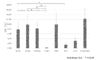
- FIG. 2 is a bar graph showing the formation ratio of organoids derived from lung epithelial stem cells in the GFP hi fraction, which was formed in a culture medium containing extracellular matrix as a scaffold.
- FIG. 3 is a bar graph showing the major axis of lung epithelial stem cell-derived organoids in the GFP hi fraction formed in a culture medium containing extracellular matrix as a scaffold.
- FIG. 1 is a dispersal diagram of the lung epithelial stem cells based on the fluorescence intensity of green fluorescent protein (GFP) from lung epithelial stem cells derived from SFTPC-GFP mice.
- FIG. 2 is a bar graph showing the formation ratio of organoids derived from lung epithelial stem cells in the GFP hi fraction, which was formed in a culture medium
- FIG. 4 is a bar graph showing the formation rate of organoids derived from lung epithelial stem cells in the GFP lo fraction formed in a culture medium containing extracellular matrix as a scaffold.
- FIG. 5 is a bar graph showing the major axis of lung epithelial stem cell-derived organoids in the GFP lo fraction formed in a culture medium containing extracellular matrix as a scaffold.
- FIG. 6 is a bar graph showing the formation ratio of organoids derived from lung epithelial stem cells formed in a culture medium containing extracellular matrix as a dispersion component.
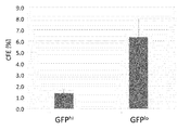
- FIG. 7 is a bar graph showing the formation rate of organoids derived from lung cancer cells.
- FIG. 8 is a scatter plot of the lung epithelial stem cells based on the GFP fluorescence intensity from the lung epithelial stem cells derived from SFTPC-GFP mice.
- FIG. 9 is a bar graph showing the expression level of each marker gene in each GFP fraction.
- FIG. 10 is a fluorescence image of organoids derived from lung epithelial stem cells in the GFP neg fraction.
- FIG. 11 is a fluorescence image of organoids derived from lung epithelial stem cells in the GFP lo fraction.
- FIG. 12 is a fluorescence image of organoids derived from lung epithelial stem cells in the GFP hi fraction.
- FIG. 13A is a bar graph showing the formation rate of organoids derived from lung epithelial stem cells in the GFP hi fraction formed in a culture medium containing extracellular matrix as a scaffold.
- FIG. 13B is a graph showing the major axis of lung epithelial stem cell-derived organoids in the GFP hi fraction formed in a culture medium containing extracellular matrix as a scaffold.
- FIG. 14A is a bar graph showing the formation rate of organoids derived from lung epithelial stem cells in the GFP lo fraction formed in a culture medium containing extracellular matrix as a scaffold.
- FIG. 14B is a graph showing the major axis of lung epithelial stem cell-derived organoids in the GFP lo fraction formed in a culture medium containing extracellular matrix as a scaffold.
- FIG. 15A is a bar graph showing the formation rate of organoids derived from lung epithelial stem cells in the GFP neg fraction formed in a culture medium containing extracellular matrix as a scaffold.
- FIG. 15B is a graph showing the major axis of lung epithelial stem cell-derived organoids in the GFP neg fraction formed in a culture medium containing extracellular matrix as a scaffold.
- FIG. 16A is a schematic diagram of the donor mouse and the recipient mouse used in Example 3.
- FIG. 16B is a scheme diagram of Example 3.
- FIG. 16C is a fluorescence image of a frozen section of the lung of a mouse administered with organoid-derived cells.
- FIG. 16D is an enlarged fluorescence image of the region surrounded by the white square in FIG. 16C.
- FIG. 17A is a microscopic image of an organoid formed from human alveolar cells.
- FIG. 17B is a microscopic image of organoids formed from human airway cells.
- FIG. 18A is a fluorescence image of an alveolar-type organoid formed from human alveolar cells.
- FIG. 18B is a fluorescence image of a bronchoalveolar-type organoid formed from human alveolar cells.
- FIG. 18C is a fluorescence image of a bronchial type organoid formed from human alveolar cells.
- FIG. 19A is a graph showing the area of cells forming organoids when cultured in a cell population.
- FIG. 19A is a graph showing the area of cells forming organoids when cultured in a cell population.
- FIG. 19B is a graph showing the perimeter of the cells forming the organoid when cultured in a cell population.
- FIG. 19C is a graph showing the major axis of organoid-forming cells when cultured in a cell population.
- FIG. 19D is a graph showing the minor axis of organoid-forming cells when cultured in a cell population.
- FIG. 19E is a graph showing the sphericity of organoid-forming cells when cultured in a cell population.
- FIG. 19F is a graph showing the gray value (average) of the cells forming the organoid when cultured in a cell population.
- FIG. 19G is a graph showing the gray value (mode) of the cells that formed organoids when cultured in a cell population.
- FIG. 19G is a graph showing the gray value (mode) of the cells that formed organoids when cultured in a cell population.
- FIG. 19H is a graph showing the center of gravity of organoid-forming cells when cultured in a cell population.
- FIG. 20A is a graph showing the area of cells forming organoids when cultured in a single cell.
- FIG. 20B is a graph showing the perimeter of the cells forming the organoid when cultured in a single cell.
- FIG. 20C is a graph showing the major axis of the cells that formed organoids when cultured in a single cell.
- FIG. 20D is a graph showing the minor axis of the cells that formed organoids when cultured in a single cell.
- FIG. 20E is a graph showing the sphericity of organoid-forming cells when cultured in a single cell.
- FIG. 20F is a graph showing the gray value (average) of the cells forming the organoid when cultured in one cell.
- FIG. 20G is a graph showing the gray value (mode) of the cells forming the organoid when cultured in one cell.
- FIG. 20H is a graph showing the center of gravity of organoid-forming cells when cultured in a single cell.
- FIG. 21A is a scatter plot showing the expression level of Scgb1a1 in each cluster.
- FIG. 21B is a scatter plot showing the expression level of Sftpc in each cluster.
- FIG. 21C is a scatter plot showing the cell type of each cluster.
- FIG. 21D is a graph showing the area of each cluster.
- FIG. 21A is a scatter plot showing the expression level of Scgb1a1 in each cluster.
- FIG. 21B is a scatter plot showing the expression level of Sftpc in each cluster.
- FIG. 21C is a scatter plot showing the cell type of each cluster
- FIG. 22 is a graph showing nine cell surface markers that are strongly expressed in cells corresponding to cluster 2.
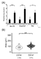
- FIG. 23A is a bar graph showing the expression level of the marker gene in CD14 +/- cells.
- FIG. 23B is a graph showing the area of CD14 +/- cells.
- FIG. 24A is a microscopic image showing CD14 +/- cells on day 9 of culture.
- FIG. 24B is a bar graph showing the rate of formation of organoids derived from CD14 +/- cells.
- FIG. 25 is a bar graph showing the expression level of a marker gene in organoids derived from CD14 +/- cells.
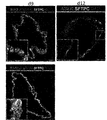
- FIG. 26A is a fluorescent image of immunostaining in organoids on day 9 of CD14 + cell culture.
- FIG. 26B is a fluorescent image of immunostaining in organoids on day 12 of CD14 + cell culture.
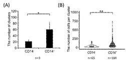
- FIG. 27A is a bar graph showing the number of clusters observed in the lungs administered with CD14 +/- cells.
- FIG. 27B is a graph showing the number of cells contained in the cluster observed in the lung to which CD14 +/- cells were administered.
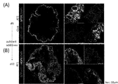
- CD14 + cells administered to a bleomycin-injured lung become type 1 alveolar epithelial cells (hereinafter also referred to as “AT1 cells”) or type 2 alveolar epithelial cells (hereinafter also referred to as “AT2 cells”). It is a fluorescent image showing that it has differentiated.
- FIG. 29 is a fluorescence image showing that CD14 + cells administered to a bleomycin-injured lung have differentiated into cultured cells.
- One aspect of the present disclosure provides a method of producing organoids from lung epithelial cells or lung cancer cells.
- the method comprises culturing a sample containing lung epithelial cells or lung cancer cells in a culture medium, wherein the culture medium comprises an extracellular matrix of 0-10% v / v, and the additives according to the present disclosure.
- the culture medium is substantially free of feeder cells.
- organoid as used herein means a three-dimensional living tissue-like cell aggregate formed in vitro.
- similar to living tissue or “similar to living tissue” means that the anatomy of the organoid and the anatomy of the living tissue are similar.
- Most of the cells constituting the organoid are, for example, cells having a differentiation ability and a proliferative ability.
- the cultured cells constituting the organoid can be differentiated into various cell types according to known methods. Cultured cells that make up organoids can be differentiated into multiple cell types, for example, by including or removing additives in the culture medium.
- the cultured cells constituting the organoid can be induced to differentiate, for example, by culturing in a culture medium containing no additives according to the present disclosure.
- Organoids may include some differentiated or non-differentiating cells.
- Organoids can be produced, for example, according to known cell culture methods.
- Organoids can be produced, for example, by maintaining predetermined cells in a culture medium containing the additives according to the present disclosure in a cell culture apparatus.
- the organoid is an organoid derived from lung epithelial stem cells or lung cancer cells.
- an organoid is an aggregate of cultured cells having a diameter of at least 50 ⁇ m when observed under a microscope. If the aggregate of cultured cells is oval, the diameter is the length of the long side. If the aggregate of cultured cells has a complex shape, the diameter is the diameter of the circumscribed circle of the aggregate.
- the efficiency of organoid formation (CFE: Colony Forming Efficiency) is the number of cell masses having a diameter of 50 ⁇ m or more in a culture after culturing a sample containing lung epithelial cells or lung cancer cells in a culture medium for 6 days. It is shown by the ratio [%] obtained by dividing by the number of cells of lung epithelial cells or lung cancer cells contained in the medium.
- stem cell as used herein means a cell having an ability to proliferate by itself (also referred to as “proliferative ability”) and an ability to differentiate into a specific cell type (also referred to as “differentiation ability”).
- Stem cells can, for example, differentiate into epithelial stem cells.
- Stem cells can be proliferated, for example, according to known methods, while maintaining their differentiation potential.
- Stem cells can also be proliferated while differentiating into specific cell types according to known methods.
- lung epithelial cell in the present specification means a cell constituting the epithelial tissue of the lung. Pulmonary epithelial cells have the ability to differentiate into, for example, Club cells, Cilated cells, neuroendocrine cells, Basal cells, cup cells, or alveolar epithelial cells, or said cells. It may be a lung epithelial stem cell.
- the alveolar epithelial cell may be, for example, one or both of type 1 alveolar epithelial cell (Type II alveolar epithelial cell: AT1 cell) and type 2 alveolar epithelial cell (AT2 cell).
- pulmonary epithelial stem cell means a cell that is present in lung tissue and has the ability to proliferate and differentiate into a particular lung epithelial cell.
- Lung tissue includes, for example, trachea, bronchi, bronchioles and alveoli.
- Lung epithelial stem cells have the ability to differentiate into at least one cell type selected from the group consisting of, for example, basal cells, club cells, and type 2 alveolar epithelial cells.
- the lung epithelial stem cells may be, for example, bronchoalveolar stem cells (BASCs).
- Lung epithelial stem cells can be prepared according to a known method (for example, the method described in Patent Document 2).
- Basal cells have the ability to differentiate into club cells and siliated cells during tracheal injury regeneration under homeostasis. Under homeostasis, club cells have the ability to differentiate into siliated cells during bronchial injury regeneration, but differentiate into AT2 and AT1 cells during severe alveolar injury. AT2 cells have the ability to differentiate into AT1 cells under homeostasis during alveolar injury.
- alveolar cell means a cell obtained from alveolar tissue.
- Alveolar cells can be prepared from alveolar tissue according to known methods. Alveolar cells are commercially available. Alveolar cells include, for example, alveolar epithelial cells. In one example, alveolar cells include one or both of AT1 cells and AT2 cells.
- airway cell means a cell obtained from airway tissue. Airway cells can be prepared from airway tissue according to known methods. Airway cells are commercially available.
- sample containing lung epithelial cells can be prepared, for example, from mammalian lung tissue.
- Samples containing lung epithelial cells can be prepared, for example, from mammalian lung tissue using a predetermined cell marker (eg, a specific protein on the cell membrane surface) as an index.
- the predetermined cell marker may be, for example, EPCAM.
- a sample containing a desired lung epithelial cell type may be prepared from a sample containing lung epithelial cells.
- the basal cells often use, for example, the nerve growth factor receptor (NGFR) as a cell marker (Proc. Natl. Acad. Sci. USA, vol.
- NGFR nerve growth factor receptor
- AT2 cells may use, for example, AT2-280 as a cell marker (Journal of Histochemistry & Cytochemistry, vol. 58 (10): 891-901, 2010).
- a given cell marker may be a specific RNA target present in a living cell.
- the RNA target may be, for example, EPCAM, Sftpc, KRT-5, or Scgb1a1.
- RNA target a sample containing lung epithelial cells is fractionated by FACS under specific conditions, and the cell type contained in the obtained partial sample of each fraction is RNA. It may include identifying based on a target (eg, RT-PCR).
- a sample containing lung epithelial cells for example, the lung tissue of a mammal is chopped, the lung tissue piece is treated with a protease (for example, collagenase, dispase, elastase or trypsin), and then a predetermined solution (for example, basal medium) is used. It can be prepared by transferring to and suspending. The cell suspension prepared as described above may be filtered and / or centrifuged to remove contaminants such as tissue debris.
- a sample containing lung epithelial cells can be prepared from adult, juvenile, neonatal or pediatric lung tissue of a mammal.
- a sample containing lung epithelial cells is prepared from mammalian lung tissue and then genetically modified to have the desired characteristics (but not including modifications that impart pluripotency). It may contain applied lung epithelial cells.
- the lung epithelial cells contained in the sample consist of a group consisting of basal cells, club cells, bronchial alveolar epithelial stem cells and alveolar epithelial cells (eg, AT1 cells and / or AT2 cells). Contains at least one cell type selected. Samples containing lung epithelial cells are commercially available. The sample containing lung epithelial cells may be, for example, a sample containing human alveolar cells or a sample containing human airway cells. In one embodiment, the lung epithelial cells contained in the sample are at least one, at least two, or at least three selected from the group consisting of basal cells, club cells, bronchial lung epithelial stem cells, and alveolar epithelial cells.
- the lung epithelial cells contained in the sample consist essentially of type 2 alveolar epithelial cells. In one embodiment, the lung epithelial cells contained in the sample are essentially Club cells. In one embodiment, the lung epithelial cells contained in the sample essentially consist of a combination of basal cells and club cells. "Being essential from” in the context of lung epithelial cells does not exclude “consisting only”. In one embodiment, the lung epithelial cells contained in the sample consist only of a specific cell type. The lung epithelial cells contained in the sample contain, for example, 80% or more, 85% or more, 90% or more, 95% or more, 97% or more, 98% or more, 99% or more of a specific cell type.
- the method according to this embodiment further comprises preparing a sample containing the lung epithelial cells or lung cancer cells from mammalian lung tissue.
- the preparation of the sample can be carried out according to the method disclosed herein or a known method.
- the term "lung cancer cell” is, for example, a tumor cell derived from a tumor of the lung.
- Lung cancer cells may be, for example, tumor cells circulating in the blood or non-circulating tumor cells.
- Lung cancer cells are, for example, commercially available or can be prepared from lung tissue containing tumor cells according to known methods.
- Lung cancer cells are, for example, lung adenocarcinoma cells.
- sample containing lung cancer cells in the present specification can be sorted from, for example, lung tissue of a mammal suffering from lung cancer by using a lung cancer cell marker as an index.
- the lung cancer cell marker may be, for example, any one of SOX2, CD24, CD44, CD133, CD166, and ESA, or a combination thereof.
- Lung cancer cells can be sorted using FACS, for example, using a lung cancer cell marker as an index.
- a sample containing lung cancer cells can be prepared, for example, by suspending commercially available cells obtained by culturing and growing lung cancer cells in a predetermined solution.
- the sample containing lung cancer cells can be prepared, for example, according to the method described for the sample containing lung epithelial cells.
- the sample containing lung cancer cells may contain, for example, normal lung epithelial cells.
- mammal in the present specification is, for example, human or non-human mammal.
- Non-human mammals include, for example, rodents such as mice, rats, guinea pigs and hamsters, non-human primates such as chimpanzees, even-toed ungibles such as cows, goats and sheep, odd-toed ungema such as horses, rabbits and dogs.
- the mammal is a rodent or non-human primate.
- the mammal is a human.
- an organoid derived from lung epithelial cell refers to a three-dimensional biological tissue analog derived from lung epithelial cell or lung cancer cell formed in vitro. means.
- an organoid derived from lung epithelial cells or an organoid derived from lung cancer cells is an organoid formed by culturing lung epithelial cells or lung cancer cells in a culture medium containing the additives according to the present disclosure.
- an organoid derived from lung epithelial cells comprises a lumen and a cell layer facing said lumen.
- the term "culture medium" in the present specification means a liquid containing components necessary for cell culture.
- the culture medium contains, for example, a basal medium and the additives according to the present disclosure.
- the culture medium can be prepared, for example, by combining a basal medium and the additives according to the present disclosure.
- the culture medium is, for example, a mixture of a predetermined amount of each component (solid) for preparing a basal medium and a predetermined amount of the additive (solid) according to the present disclosure, and an amount of pure water having a predetermined concentration.
- the culture medium may further contain either or both of the extracellular matrix and feeder cells that assist in the growth of stem cells.
- the culture medium can be prepared, for example, by adding one or both of the extracellular matrix and the feeder cells that assist the growth of stem cells to a mixture of the basal medium and the additives according to the present disclosure. can.
- the term "basic medium” may be a known cell culture medium, such as Dalbeco's Modified Eagle's Medium (DMEM), Minimal Essential Medium (MEM), Basic Medium Eagle (BME), or DMEM. / F12 may be used. In one embodiment, the basal medium is DMEM / F12.
- the "additive" according to the present disclosure means a combination of two or more substances that affect the efficiency of organoid formation derived from lung epithelial cells or lung cancer cells.
- the additive may be, for example, a combination of two or more substances that improve the efficiency of organoid formation derived from lung epithelial cells or lung cancer cells.
- Additives may include, for example, substances that maintain the differentiation potential of the cells that make up the formed organoids or induce differentiation into specific cell types.
- the additive is at least one selected from the group consisting of keratinocyte growth factor (KGF), hepatocyte growth factor (HGF) and fibroblast growth factor (FGF) 10, and bone morphogenetic protein (BMP).
- KGF keratinocyte growth factor
- HGF hepatocyte growth factor
- FGF fibroblast growth factor
- BMP bone morphogenetic protein
- Inhibitor (eg Noggin) and TGF ⁇ inhibitor may be a combination or a combination comprising.
- the combination preferably further comprises a Wnt signaling activator (eg CHIR99021).
- the combination may further comprise a Rock inhibitor (eg Y-27632).
- the additive may be a combination or a combination comprising a Wnt signal activator (eg CHIR99021), a BMP inhibitor (eg Noggin), a TGF ⁇ inhibitor (eg SB431542) and HGF. It is a combination of.
- the combination may further comprise a Rock inhibitor.
- the additive may be a combination containing one or both of KGF and FGF10, a BMP inhibitor (eg Noggin), a TGF ⁇ inhibitor (eg SB431542), and HGF. It is a combination consisting of.
- the additive is a combination or combination comprising KGF, FGF10, a BMP inhibitor (eg Noggin), a TGF ⁇ inhibitor (eg SB431542), and HGF.
- the combination preferably further comprises a Wnt signaling activator (eg CHIR99021).
- the combination may further comprise a Rock inhibitor.
- the additives are KGF, FGF10, Wnt signaling activator (eg CHIR99021), BMP inhibitor (eg Noggin), TGF ⁇ inhibitor (eg SB431542) and HGF. And a combination comprising or consisting of FGF10.
- Additives include, for example, KGF, FGF10, Wnt signal activator (eg CHIR99021), BMP inhibitor (eg Noggin), TGF ⁇ inhibitor (eg SB431542), HGF, FGF10, and Rock inhibitor. And a combination including or a combination consisting of.
- KGF keratinocyte growth factor
- HGF hepatocyte growth factor
- HGF hepatocyte growth factor
- HGF generally has a heterodimeric structure in which a heavy chain having a molecular weight of about 60,000 and a light chain having a molecular weight of about 35,000 are disulfide-bonded.
- HGF is commercially available, for example.
- HGF When HGF is present in the culture medium, it has a concentration of, for example, 5 to 150 ng / mL, 10 to 75 ng / mL, 20 to 40 ng / mL, preferably 30 ng / mL.
- HGF may be abbreviated as "H” herein.
- FGF 10 fibroblast growth factor 10
- FGF10 fibroblast growth factor 10
- FGF10 is commercially available, for example.
- FGF10 has a concentration of, for example, 10 to 250 ng / mL, 20 to 100 ng / mL, 30 to 70 ng / mL, preferably 50 ng / mL.
- FGF10 may be abbreviated as "F10" herein.
- Wnt signal activator means a substance that activates transcription via a T cell transcription factor (TCF / LEF) in the cell. Wnt signal activators are also referred to as Wnt agonists.
- the Wnt signaling activator is, for example, a substance that inhibits the action of GSK-3, which is a serine-threonine protein kinase that mediates the addition of phosphate molecules to amino acid residues of serine and threonine (“GSK-3 inhibitor””. It may also be a Wnt family protein, or a ⁇ -catenin degradation inhibitor, or a combination of two or more of these. Wnt signal activators are, for example, commercially available.
- the Wnt signal activator may be, for example, a GSK-3 inhibitor.
- the GSK-3 inhibitor may be, for example, SB216763 (Chemscene), CHIR-98014 (Abcam), TWS119 (Cayman Chemical), or CHIR99021 (SIGMA), or a combination of two or more thereof. good.
- the Wnt signaling activator may be, for example, a Wnt family protein (also referred to as a "Wnt ligand").
- the Wnt family protein may be, for example, any one or a combination of two or more of 19 kinds of Wnt gene-derived proteins known in the art.
- the Wnt family protein may be, for example, Wnt1, 2, 3a, 6, 7a, 7b, 8a, 9a, 10a, or 16, or may be a combination of two or more thereof.
- the Wnt family protein may be, for example, Wnt1.
- the Wnt signal activator may be, for example, a ⁇ -catenin degradation inhibitor.
- the ⁇ -catenin degradation inhibitor may be, for example, a substance that inhibits the phosphorylation of ⁇ -catenin (for example, UBE1-41).
- the Wnt signal activator is a GSK-3 inhibitor.
- the GSK-3 inhibitor is preferably CHIR99021.
- the Wnt signal activator is an amount that exhibits an inhibitory effect corresponding to that of CHIR99021, for example, 0.5 to 15 ⁇ M, 1 to 7 ⁇ M, 2 to 5 ⁇ M, preferably 3 ⁇ M. .. CHIR99021 may be abbreviated as "C" herein.
- BMP inhibitor means a substance that binds to a BMP molecule to form a complex.
- the BMP inhibitor may be, for example, a small molecule compound, a protein (eg, an antibody), DNA, RNA, a small interfering RNA, or an antisense oligonucleotide.
- BMP inhibitors are commercially available, for example.
- BMP inhibitors include, for example, Noggin (Bmp inhibitor), chordin-like protein containing chordin or its domain (R & D systems), follistatin or follistatin-related protein containing its domain (R & D systems), DAN or DAN cysteine-knot domain.
- the BMP inhibitor is Noggin.
- the BMP inhibitor When the BMP inhibitor is present in the culture medium, it has an inhibitory effect corresponding to that of Noggin, for example, 20 to 500 ng / ml, 40 to 250 ng / ml, 80 to 125 ng / ml, preferably 100 ng / ml. The amount shown. Noggin may be abbreviated as "N" herein.
- TGF ⁇ inhibitor means a substance that inhibits TGF ⁇ signal transduction.
- a TGF ⁇ inhibitor means, for example, a substance that inhibits intracellular signal transduction mediated by phosphorylation of Smad2 / 3 protein by serine / threonine kinase of type I receptor (ALK5) bound and activated by TGF ⁇ . ..
- the TGF- ⁇ inhibitor may be, for example, a small molecule compound, a protein (eg, an antibody), DNA, RNA, a small interfering RNA, or an antisense oligonucleotide.
- TGF- ⁇ inhibitors are A83-01 (Abcam), SB-431542 (Calbiochem), SB-505124 (R & D systems), SB-525334 (Chemscene LLC), LY364497 (Sigma-Aldrich), SD-208 (Sigma-Aldrich). It may be Aldrich) or SJN2511 (R & D systems), or it may be a combination of two or more thereof.
- the TGF ⁇ inhibitor is SB431542.
- the TGF ⁇ inhibitor is an amount that exhibits an inhibitory effect corresponding to that of SB431542 of, for example, 2 to 50 ⁇ M, 4 to 25 ⁇ M, 8 to 12 ⁇ M, preferably 10 ⁇ M.
- SB431542 may be abbreviated as "S" herein.
- EGF epidermal growth factor
- EGF generally has a molecular weight of about 6,000 daltons with 53 amino acid residues and three intramolecular disulfide bonds.
- EGF is, for example, commercially available (eg, Corning).
- EGF is, for example, 5 to 125 ng / ml, 10 to 60 ng / ml, 20 to 30 ng / ml, preferably 25 ng / ml.
- EGF may be abbreviated as "E” herein.
- Rho-kinase (ROCK) inhibitor refers to, for example, R- (+)-trans-4- (1-aminoethyl) -N- (4-pyridyl) cyclohexanecarboxamide dihydrochloride monohydrate.
- ROCK inhibitors are commercially available, for example.
- the ROCK inhibitor is Y-27632.
- the Rock inhibitor is an amount that exhibits an inhibitory effect corresponding to that of Y-27632, for example, 2 to 50 ⁇ M, 4 to 25 ⁇ M, 8 to 12 ⁇ M, preferably 10 ⁇ M.
- Y-27632 may be abbreviated as "Y" herein.
- the additive according to the present disclosure may be a combination of at least one selected from the group consisting of KGF, FGF10 and HGF, a BMP inhibitor and a TGF ⁇ inhibitor.
- the additive may further comprise one or both of the Wnt signaling activator and the ROCK inhibitor.
- the additive is, for example, a combination of KGF, a BMP inhibitor, and a TGF ⁇ inhibitor; a combination of KGF, a Wnt signal activator, a BMP inhibitor, and a TGF ⁇ inhibitor; Combination of FGF10, BMP inhibitor and TGF ⁇ inhibitor; combination of FGF10, Wnt signal activator, BMP inhibitor and TGF ⁇ inhibitor; HGF, BMP inhibitor and TGF ⁇ inhibitor Combinations; HGF, Wnt signal activator, BMP inhibitor, TGF ⁇ inhibitor combination; KGF, FGF10, HGF, BMP inhibitor, TGF ⁇ inhibitor combination; KGF, FGF10 and , HGF, Wnt signal activator, BMP inhibitor, TGF ⁇ inhibitor; ROCK inhibitor, KGF, FGF10, HGF, BMP inhibitor, TGF ⁇ inhibitor; or It may be a combination of a ROCK inhibitor, KGF, FGF10, HGF, a Wnt signal activator, a BMP inhibitor, and a TGF ⁇ inhibitor.
- the BMP inhibitor may be Noggin
- the TGF ⁇ inhibitor may be SB431542
- the Wnt signal activator may be CHIR99021
- / or the ROCK inhibitor may be Y. -27632 may be.
- the culture medium comprises an "extracellular matrix".
- the extracellular matrix includes, but is not limited to, water, polysaccharides, elastin, integrins, and glycoproteins. Glycoproteins include, for example, collagen, entactin (nydgen), fibronectin, and laminin.
- ECM can be prepared, for example, by culturing ECM-producing cells (eg, epithelial cells, endothelial cells, intramural embryonic cells, or fibroblasts) in vitro and then removing the ECM cells.
- ECM-producing cells include, for example, chondrocytes that produce predominantly collagen and proteoglycans, predominantly type IV collagen, laminin, interstitial procollagen, and fibroblasts that produce fibronectin, and predominantly collagen (types I, III, and). V-type), chondroitin sulfate proteoglycan, hyaluronic acid, fibronectin, and tenesin-C-producing colon myofibroblasts.
- ECM is commercially available.
- Commercially available extracellular matrix includes, for example, extracellular matrix protein (Invitrogen), basement membrane preparations from Engelbreth-Holm-Swarm (EHS) mouse sarcoma cells (eg, Cultrex® Basement Membrane Extract (Trevigen).
- the ECM may be a synthetic extracellular matrix (eg, ProNectin (Sigma Z378666)).
- the extracellular matrix may be one type or a mixture of two or more types.
- the ECM is a Matrix.
- the culture medium does not contain extracellular matrix.
- the extracellular matrix can exist as a "dispersion component" when present in the culture medium.
- the extracellular matrix as a dispersion component is, but is not limited to, a state in which the extracellular matrix component is dispersed or dissolved in the culture medium.
- the extracellular matrix as a dispersion component for example, the extracellular matrix is 10% v / v or less, 7% v / v or less, 5% v / v or less, for example, 2.5% v / v / / v / v / v / v / v / or less per volume of the culture medium. It exists in the quantity of v.
- the culture medium is 0-10% v / v, 0-7% v / v, 0-5% v / v, 0.1-10% v / v, 0.1-7%. v / v, 0.1-5% v / v, 1-10% v / v, 1-7% v / v, 1-5% v / v, 1-2.5% v / v, 2-
- the extracellular matrix is included in an amount of 10% v / v, 2-7% v / v, 2-5% v / v, or 2-2.5% v / v.
- the concentration of the extracellular matrix in the culture medium is determined from the viewpoint of preventing or reducing contamination of the produced organoid. A low concentration is preferred.
- the extracellular matrix can exist as a "scaffold" if it is present in the culture medium.
- the extracellular matrix as a scaffold is a form that provides, but is not limited to, a microenvironment in which cells at least partially mimic a naturally occurring cell niche.
- the extracellular matrix as a scaffold may be, for example, in the form of a gel.
- the extracellular matrix as a scaffold is formed, for example, at a concentration of more than 10% v / v, 20% v / v or more, 30% v / v or more, 50% v / v or more, or 70% v / v or more. It may be a gel.
- feeder cell means a cell type used to assist the proliferation of stem cells.
- Feeder cells are commonly used to co-culture with cells of interest and provide a suitable surface to assist the growth of the cells of interest.
- Feeder cells are administered with specific cell types (eg, primary adult mouse fibroblasts, mouse fibroblasts (MLg), human fetal fibroblasts: MRC-5) themselves, or antibiotics to the extent that they do not die in advance. It is prepared by doing or irradiating with gamma rays.
- the use of feeder cells is undesirable as it complicates the passage manipulation of the cells of interest.
- the culture medium is substantially free of feeder cells.
- substantially not included herein does not exclude “completely not included”.
- the culture medium that is substantially free of feeder cells is completely free of feeder cells. In one embodiment, the culture medium substantially free of feeder cells is less than 3%, less than 2%, less than 1%, or 0 relative to the number of cultured cells containing lung epithelial cells or lung cancer cells to be cultured. May contain less than 5.5% feeder cells. Preferably, the culture medium containing the lung epithelial cells or lung cancer cells to be cultured does not contain feeder cells.
- the culture may include, for example, adding a sample containing lung epithelial cells or lung cancer cells to the culture medium in a culture vessel and maintaining the culture medium at 37 ° C. under 5% CO 2. ..
- the temperature for culturing is not limited to 37 ° C., and a temperature known in the field of cell culture can be appropriately used.
- the CO 2 concentration is not limited to 5%, and a CO 2 concentration known in the cell culture field can be appropriately used.
- the culture medium can be replaced with fresh culture medium at arbitrary intervals, for example, every 1 to 14 days, every 2 to 12 days, or every 3 to 10 days.
- the culture medium may be replaced, for example, every 3 to 7 days, every 3 to 5 days, or every 3 days when the cells to be cultured are lung epithelial cells.
- the culture medium may be replaced, for example, every 3 to 14 days, every 5 to 10 days, or every 10 days when the cells to be cultured are lung cancer epithelial stem cells.
- the "organoid derived from lung epithelial cells” or the “organoid derived from lung cancer cells” formed by the culture may be isolated from the culture medium. Isolation may include, for example, fractionating cells based on their size to separate organoids from cells that do not form a cell mass. Isolation can be performed using, for example, CELL HANDLER (Yamaha).
- the method according to this embodiment is a club cell-I before culturing a sample containing lung epithelial cells prepared from mammalian lung tissue in a culture medium containing the additives according to the present disclosure.
- AT2 cells and club cells-II respectively, based on the cell markers listed in Table A and at least one of their homologs, to select club cells-I, AT2 cells or club cells-II from the sample.
- the method comprises selecting Club Cell-I from the sample based on the cell markers listed in Table A for Club Cell-I and at least one of their homologs. In one embodiment, the method comprises selecting AT2 cells from the sample based on the cell markers listed in Table A for AT2 cells and at least one of their homologs. In one embodiment, the method comprises selecting Club Cell-II from the sample based on the cell markers listed in Table A for Club Cell-II and at least one of their homologs.
- Club cell-I means a club cell that expresses at least one of the cell markers listed in Table A and their homologues.
- Club cells-I form organoids, for example, in a culture medium containing the additives according to the present disclosure.
- Club cells-I are, for example, Cbr2, Hp, Cyp2f2, Prdx6, Scgb1a1, Ldhb, Ces1d, Ckkar, Selenbp1, Gstm2, Scnn1b, Scgb1c1, Plpp3, Ephx1, Dcxr, Etl1 It expresses at least one, at least two, at least three, or at least four cell markers selected from the group consisting of those homologs.
- Club cell-II means a club cell that expresses at least one of the cell markers listed in Table A and their homologues.
- Club cells-II form organoids, for example, in a culture medium containing the additives according to the present disclosure.
- Club cells-II are more likely to form organoids than, for example, Club cells-I.
- Club cells-II include, for example, Tff2, Reg3g, Bpifb1, Muc5b, Sult1d1, Fxyd3, Scgb3a1, Lypd2, Chad, S100a6, Pglyrp1, Aldh3a1, Bpifa1, Gsto1, Lgas3, Gsto1, Lgals.
- Club cells-II are at least one, at least two, at least three, or at least selected from the group consisting of, for example, Fxyd3, Pigr, Cpd, Ly6a, Perp, Kcne3, Il13ra1, Slc15a2, and Cd14 and their homologs. It expresses 4 types of cell markers.
- AT2 cell or "type 2 alveolar epithelial cell” in an embodiment that selects a given cell based on a cell marker expresses at least one of the cell markers listed in Table A and their homologues. It means AT2 cells.
- the AT2 cells form organoids, for example, in a culture medium containing the additives according to the present disclosure.
- the AT2 cells include, for example, Lamp3, Lyz2, Napas, Slc34a2, Lyz1, Rnase4, Hc, Cd74, Scd1, Sftpc, Lgi3, Cldn18, Etv5, Cxcl15, S100g, Elovl1, Fas, H2-A, Fas, H2-A.
- at least one, at least two, at least three, or at least four cell markers selected from the group consisting of their homologs are expressed.
- selecting a predetermined cell means selecting a predetermined cell from a sample containing a plurality of types of cells.
- a predetermined cell marker for example, the cell marker shown in Table A
- sorting the cells expressing the marker as predetermined cells can be performed, for example, by fluorescence activated cell sorting (FACS).
- FACS can be performed, for example, with a commercially available FACS device using a fluorescent probe in which a fluorescent substance is conjugated to a probe (eg, an antibody) that binds to a predetermined cell marker.
- the test cell can be selected as the predetermined cell when the expression level of the predetermined cell marker in the test cell is higher than the expression level of the predetermined cell marker in the negative control.
- Fluorescent materials can be prepared, for example, according to known methods or are commercially available.
- Probes that bind to a given cellular marker, such as antibodies, can be prepared according to known methods or are commercially available. Conjugation of the fluorescent substance to the probe can be carried out, for example, according to a known method.
- the selection of predetermined lung epithelial cells can be performed based on the expression of at least one of the cell markers listed in Table A and their homologues.
- homolog means a gene or protein whose sequence and function are similar among organisms of the same species or different species.
- homologs include orthologs, paralogs and xenologs.
- a "homolog" of a particular gene or protein has, for example, at least 75%, 80%, 85% or 90% homology to, for example, the sequence of said particular gene or protein, preferably at least 95%. It has 96%, 97%, 98% or 99% homology.
- Homogeneity means the proportion of identical nucleotide or amino acid residues between these two sequences after the sequences of the two genes or proteins have been aligned using an alignment algorithm well known to those of skill in the art. Homology can be performed, for example, using known methods or publicly available computer programs (eg, BLAST or FASTA).
- Cbr2 refers to carbonyl reductase 2 and is involved in the synthesis of NADPH.
- Hp refers to haptoglobin and is a hemoglobin-binding protein.
- Cyp2f2 refers to cytochrome P450 (CYP) 2f2, a mouse-specific membrane-binding protein localized in the endoplasmic reticulum membrane. Cyp2f2 homologs in mammals other than mice are known, for example, the human homolog is CYO2F1.
- Prdx6 refers to peroxiredoxin-6, a protein belonging to the peroxiredoxin family of antioxidant enzymes. In humans, it is encoded by the PDRX6 gene.
- Sergb1a1 refers to secretoglobulin family 1A member 1 and has anti-inflammatory function.
- Ldhb lactate dehydrogenase B and is a monomer of the LDH enzyme encoded by the LDHB gene.
- Ces1d refers to carboxylesterase 1d.
- Cuckar refers to the cholecystokinin A receptor, which is subtype A of two different subtypes of G protein-coupled receptors that bind peptide hormones.
- the term “Selenium bp1” refers to a selenium-binding protein 1 and is a protein belonging to the selenium-binding protein family. Selenbp1 is encoded by the SELENBP1 gene in humans. As used herein, the term “Gstm2” refers to glutathione S-transferase Mu2, an enzyme encoded by the GSTM2 gene in humans. As used herein, the term “Scnn1b” refers to the sodium channel subunit beta 1 and is a protein encoded by the SCNN1B gene in humans.
- Scgb1c1 refers to secretoglobin 1C1 and is also known as the ligand binding protein RYD5. Scgb1c1 is encoded by the secretoglobin 1C1 gene.
- Plppp3 refers to phospholipid phosphatase 3, also known as phosphatidic acid phosphatase type 2B. Plpp3 is an enzyme encoded by the PPPA2B gene on chromosome 1 in humans.
- Ephx1 refers to epoxide hydrolase 1, which is an enzyme encoded by the EPHX1 gene in humans.
- the term “Dcxr” refers to dicarbonyl L-xylulose reductase, a multifunctional protein involved in various protein interaction processes in various physiological systems.
- the term “Alas1” is a protein also known as deltaaminolevulinic acid synthase 1, which is encoded by the ALAS1 gene in humans.
- the term “Rettnla” refers to a resistin-like molecule alpha, a protein belonging to the family of small, cysteine-rich, secreted proteins.
- the term “Sec14l3” refers to SEC14-like3, a phosphatidylinositol transfer protein that plays an important role in the biosynthesis of transport vesicles derived from the Golgi apparatus, also known as tocopherol-related protein 2.
- the term “Ptgr1” refers to prostaglandin reductase-1, a protein involved in eicosanoid catabolism and lipid peroxidation such as 4-hydroxynonenal.
- the term “Krtap17-1” refers to a keratin-related protein 17-1, a protein belonging to the keratin-related protein (KAP) family.
- Tff2 refers to trefoil factor 2, a protein encoded by the TFF2 gene in humans.
- Reg3g refers to Regenerating islet-developed protein 3 gamma, a protein encoded by the REG3G gene in humans.
- Bpifb1 refers to BPI Found Contining Family B member 1, a protein encoded by the BPIFB1 gene in humans.
- the term “Muc5b” refers to mucin 5 subtype B, a protein encoded by the MUC5B gene in humans.
- the term “Sult1d1” refers to sulfotransferase 1D member 1, which transfers a sulfate group to a hormone, a neurotransmitter. In the case of humans, it is known that they are pseudogeneized by mutation.
- the term “Fxyd3” refers to the FXYD domain-containing ion transport regulator 3 and is a protein encoded by the FXYD3 gene in humans.
- the term “Scgb3a1” refers to secretoglobin family 3A member 1 and is a protein encoded by the SCGB3A1 gene in humans.
- the term “Lypd2” refers to a Ly6 / PLAUR domain-containing protein 2, which is a protein encoded by the LYPD1 gene in humans.
- the term “Cad” refers to cadherin, a transmembrane glycoprotein involved in adhesion between animal cells.
- the term “S100a6” refers to the S100 calcium-binding protein A6, which is a protein encoded by the S100A6 gene in humans.
- the term “Pgyrp1” refers to peptidoglycan recognition protein 1, which is a protein encoded by the PGLYRP1 gene in humans.
- the term “Aldh3a1” refers to the aldehyde dehydrogenase 3 family member A1 and is a protein encoded by the ALDH3A1 gene in humans.
- Bpifa1 is also referred to as Palate, lung, and nasal epithelium clone protein (PLUNC), and is a protein coded in BPIFA1 (Bactericidal / permeability-increasing gene).
- Gsho1 refers to glutathione S-transferase omega-1, a protein encoded by the GSTO1 gene in humans.
- Lgals3 refers to galectin-3, a protein belonging to the lectin family encoded by the LGALS3 gene in humans.
- the term "Pigr” refers to a macromolecular immunoglobulin receptor, which is a transmembrane protein encoded by the PIGR gene in humans.
- the term “Scgb3a2” refers to secretoglobin family 3A member 2 and is a protein encoded by the SCGB3A2 gene in humans.
- the term “Ltf” refers to lactoferrin, which is an iron-binding glycoprotein mainly present in milk in mammals.
- QSox1 refers to sulfhydryl oxidase 1, a protein encoded by the QSOX1 gene in humans.
- Cyp2a5 refers to cytochrome P450 (CYP) 2A5, a protein expressed in the olfactory epithelium of the liver and nasal cavity in mice. Homologs or orthologs of Cyp2a5 in mammals other than mice are known, for example, the human ortholog is CYP2A6 / 13, and the rat ortholog is CYP2A3.
- Cpd refers to carboxypeptidase D, a protein encoded by the CPD gene in humans.
- Ly6a refers to the lymphocyte antigen 6 complex, locus A, and is also referred to as Sca-1, a glycosylphosphatidylinositol (GPI) anchored protein.
- Perp refers to a p53 apoptotic effector associated with PMP-22, a membrane protein encoded by the PERP gene in humans.
- Kcne3 refers to a potassium voltage gate channel, an Isk-related family, member 3, and is also referred to as a MinK-related peptide 2 (MiRP2).
- Kcne3 is a protein encoded by the KCNE3 gene in humans.
- Il13ra1 refers to the interleukin 13 receptor subunit alpha 1 and acts as a receptor for interleukin 13 by forming a heterodimer with the interleukin 4 receptor alpha.
- Slc15a2 refers to the solution carrier family 15, member 2, and functions as a proton-binding peptide transporter.
- CD14 refers to the CD14 protein and serves as a constitutive use of the innate immune system.
- the CD14 may be, for example, mCD14, which is a membrane-bound protein, or sCD14, which is a soluble protein. In the context of selecting Club cells-II cells, CD14 is, for example, mCD14.
- Lamp3 refers to lysosome-related membrane glycoprotein 3 and is a protein encoded by the LAMP3 gene in humans.
- the term “Lyz2” refers to lysozyme C-2.
- Napsa refers to Napsin A, which is an aspartic acid proteinase encoded by the NAPSA gene in humans.
- Slc34a2 refers to a sodium-dependent phosphate transport protein 2B (NaPi2b), which is a protein encoded by the SLC34A2 gene in humans.
- Lyz1 refers to lysozyme C-1.
- the term “Rnase4" refers to ribonuclease 4, a protein encoded by the RNASE4 gene in humans.
- the term “Hc” refers to hemolytic complement. It is a component of the complement system in innate immunity.
- the term “Cd74” refers to the HLA class II histocompatibility antigen gamma chain and is a protein encoded by the CD74 gene in humans.
- the term “Scd1” refers to stearoyl-CoA desaturase-1, an enzyme important for fatty acid metabolism.
- the term “Sftpc” refers to surfactant protein C (SP-C) and is one of the pulmonary surfactant proteins. Sftpc is encoded by the SFTPC gene in humans.
- the term “Lgi3” refers to leucine-rich glioma inactive 3 and is a secreted protein in vertebrates belonging to the LGI family.
- Cldn18 refers to claudin 18, a protein encoded by the CLDN18 gene in humans.
- Etv5 refers to Ets variant 5 and is also referred to as an ERM transcription factor.
- Etv5 is a transcription factor encoded by the ETV5 gene in humans.
- Cxcl15 refers to a chemokine (CXX motif) ligand 15 and is a cytokine belonging to the CXC chemokine family.
- S100g refers to S100 calcium-binding protein G, which is a protein encoded by the S100G gene in humans.
- Elovl1 refers to fatty acid elongation enzyme 1 and is an enzyme that plays an important role in the production of saturated and monounsaturated very long chain fatty acids.
- Fas refers to fatty acid synthase, which is an enzyme encoded by the FASN gene in humans.
- H2-Aa refers to histone H2A type 1-A, a protein encoded by the HIST1H2AA gene in humans.
- Fabp5 refers to a superficial fatty acid binding protein, which is a protein encoded by the FABP5 gene in humans.
- Rn4.5s refers to 4.5S RNA.
- the method according to the present embodiment further comprises culturing lung epithelial cells cultured in a culture medium containing the additive according to the present disclosure in a culture medium containing the additive.
- the lung epithelial cells cultured in a culture medium may form organoids or may be cell aggregates having a diameter of less than 50 ⁇ m.
- Base lung epithelial cells cultured in culture medium preferably form organoids.
- the culture medium containing no additives according to the present disclosure may be, for example, a basal medium.
- Culturing the cells in a culture medium containing the additives may include, for example, subculturing the cultured cells.
- Culturing the cells in a culture medium without the additives may include, for example, subculturing the cultured cells.
- the number of days for culturing the cells in the culture medium is not particularly limited.
- the culture of the cells in the culture medium may be, for example, 1 to 30 days, 1 to 25 days, or 1 to 20 days.
- Organoids derived from lung epithelial cells can, for example, mimic the basic physiological functions of lung epithelial tissue. Therefore, organoids derived from lung epithelial cells are useful in regenerative medicine. In addition, organoids derived from lung cancer cells are useful for screening substances for treating lung cancer (NATURE COMMUNICATIONS 2019 103991).
- Organoids derived from lung epithelial cells or lung cancer cells One aspect of the present disclosure provides organoids derived from lung epithelial cells or lung cancer cells.
- the organoid can be produced from a sample containing lung epithelial cells or lung cancer cells according to the method for producing an organoid according to the present disclosure.
- Organoids derived from lung epithelial cells or lung cancer cells produced include, for example, a lumen and a cell layer facing the lumen.
- the lung epithelial cell-derived organoid comprises a lumen and a cell layer facing the lumen.
- the organoid may be an organoid derived from lung epithelial cells or an organoid derived from airway cells.
- organoids derived from lung epithelial cells include a lumen and a cell layer facing the lumen, the cell layer essentially consisting of AT2 cells.
- the AT2 cells express, for example, one or both of HT2-280 and SFTPC, and the HT2-280 is localized in or near the cell membrane facing the lumen in the AT2 cells. ing.
- an organoid derived from lung epithelial cells comprises a lumen and a cell layer facing said lumen, wherein the cell layer comprises AT2 cells and cells expressing AT2 cell markers and bronchial epithelial markers. Essentially from the combination.
- an organoid derived from a lung epithelial cell comprises a lumen and a cell layer facing the lumen, the cell layer essentially consisting of cells expressing a bronchial epithelial marker.
- airway cell-derived organoids include a lumen and a cell layer facing the lumen, which is a combination of basal cells and cells expressing basal cell markers and bronchial epithelial markers. Becomes essential from.
- the AT2 cell marker is not particularly limited, and a known AT2 cell marker can be used.
- the AT2 cell marker may be, for example, one or both of HT2-280 and SFTPC.
- the bronchial epithelial marker is not particularly limited, and known bronchial epithelial markers can be used.
- the bronchial epithelial marker may be, for example, SOX2.
- the basal cell marker is not particularly limited, and a known basal cell marker can be used.
- the basal cell marker may be, for example, KRT5.
- HT2-280 in the present specification is a protein of 280 to 300 kDa expressed in type 2 alveolar epithelial cells.
- SFTPC means pulmonary surfactant protein C. SFTPC has a hydrophobic ⁇ -helix structure consisting of 26 amino acids and 9 hydrophilic amino acids linked to each other.
- SOX2 in the present specification means SRY-box connecting gene2.
- SOX2 is composed of an HMG (high mobility group) domain, which is a DNA binding site, and a transcription activation domain on the C-terminal side thereof. Since the HMG domain contains two nuclear localization signal sequences, SOX2 is predominantly localized in the nucleus.
- KRT5 herein is an intermediate filament protein and is encoded by the KRT5 gene in humans.
- composition for regenerative medicine comprises an organoid derived from lung epithelial cells according to the present disclosure and / or a cell of the organoid.
- the regenerative medicine composition consists essentially of Club cells-I, AT2 cells or Club cells-II expressing at least one of the cell markers listed in Table A and their homologues.
- the composition for regenerative medicine may be, for example, a pharmaceutical composition for repairing lung injury or lung disease.
- a regenerative medicine composition comprising lung epithelial cells essentially consisting of Club cells-I, AT2 cells or Club cells-II expressing at least one of the cell markers described above is according to the present disclosure.
- a regenerative medical composition comprising or consisting essentially of club cells-I produces an organoid without causing adverse effects such as inflammation in the lung to which the club cells-I have been delivered. Since it has the ability to form and differentiate into cells of the alveolar and bronchial system, it is useful as a composition for regenerative medicine for repairing lung injury or lung disease.
- a regenerative medical composition comprising or consisting essentially of Club Cell-II has the ability to efficiently form organoids in the lung to which Club Cell-II has been delivered. It is also useful as a regenerative medicine composition for repairing lung injury or disease because it has the ability to differentiate into cells of the alveoli and bronchial system.
- a regenerative medical composition comprising or consisting essentially of Club Cell-II lung epithelial cells is an agent for treating a lung that has injured Club Cell-II or a diseased lung.
- it is useful as a pharmaceutical composition for treating lung injury or lung disease.
- one aspect of the invention comprises lung epithelial cells prepared from mammalian lung tissue based on the cell markers listed in Table A for Club Cell-II and at least one of their homologues.
- pharmaceutical compositions comprising Club Cell-II selected from a sample, pulmonary epithelial cells essentially, and a drug for treating lung injury or disease.
- the composition for regenerative medicine may appropriately contain, for example, a pharmaceutically acceptable carrier.
- the composition for regenerative medicine can be appropriately produced according to a known method.
- the composition for regenerative medicine can be produced, for example, by mixing an organoid derived from lung epithelial cells and / or the cells of the organoid with a pharmaceutically acceptable carrier.
- “Pharmaceutically acceptable carrier” means any component other than organoids derived from lung epithelial cells according to the present disclosure, which is highly safe in mammals and has low allergic reactivity.
- Pharmaceutically acceptable carriers include, for example, aqueous or non-aqueous solvents, solutions (eg, saline, basal medium or cell suspension), antifreeze agents (eg, glycerol), water-soluble polymers suitable for pharmaceutical administration. Includes (eg, dextran), or buffer (eg, phosphate buffer).
- Organoids derived from lung epithelial cells can be prepared according to the production method of the present disclosure.
- Organoids derived from lung epithelial cells include, for example, a lumen and a cell layer facing the lumen.
- a "cell of an organoid derived from a lung epithelial cell” may be a form of a cell mass constituting an organoid derived from a lung epithelial cell and / or a form of an individual cell separated from an organoid derived from a lung epithelial cell. It's okay.
- Individual state cells isolated from lung epithelial cell-derived organoids are, for example, treated with a protease (eg, collagenase, dispase, elastase or trypsin) and then into a given solution (eg, basal medium). It can be prepared by suspending.
- a protease eg, collagenase, dispase, elastase or trypsin
- the regenerative medicine composition contains cells in the form of a cell mass of an organoid derived from lung epithelial cells
- the cell mass in the regenerative medicine composition is, for example, prior to administration to a mammal in need thereof. It may be processed to make cells in individual states.
- the regenerative medicine composition is administered to a mammal in need thereof, for example, by transplanting it to a predetermined site by a surgical technique or by injecting it into a predetermined site by injection.
- the mammal to which the composition for regenerative medicine is administered and the mammal that provided the lung epithelial cells of the organoid are preferably the same species and are the same individual from the viewpoint of reducing graft rejection. Is even more preferable.
- the mammal is, for example, a human.
- lung injury or "lung disease” is used, for example, as a pathogenic disease, lung cancer (eg, small cell lung cancer or non-small cell lung cancer (eg, adenocarcinoma, squamous cell cancer or large cell cancer)).
- lung cancer eg, small cell lung cancer or non-small cell lung cancer (eg, adenocarcinoma, squamous cell cancer or large cell cancer)).
- Interstitial lung disease pneumonia (eg organizing pneumonia), tuberculosis, cystic fibrosis, bronchitis, pulmonary fibrosis, sarcomatosis, type II hyperplasia, chronic obstructive lung disease, emphysema, asthma, lung Examples include edema, acute respiratory urgency syndrome, asthma, bronchial dilatation, Hantavirus lung syndrome, Middle East respiratory syndrome (MERS), severe acute respiratory syndrome (SARS) and dust lung.
- pneumonia eg organizing pneumonia
- tuberculosis cystic fibrosis
- cystic fibrosis cystic fibrosis
- bronchitis fibrosis
- pulmonary fibrosis pulmonary fibrosis
- sarcomatosis type II hyperplasia
- chronic obstructive lung disease emphysema
- asthma lung Examples include edema, acute respiratory urgency syndrome, asthma, bronchial dilatation, Hantavirus lung syndrome, Middle East respiratory syndrome (
- Pathogenic diseases include, for example, adenovirus, coronavirus (eg, COVID-19, SARS-CoV, SARS-CoV-2 or MERS-CoV, or variants thereof), human metapneumovirus, influenza virus, parainfluenza.
- Virus Respiratory Syndrome virus, Rhinovirus, Hunter virus, Enterovirus (eg, Enterovirus D68: EV-D68), Pertussis, Cramidophila pneumoniae, Zifteria, Cocciella brunetti, Influenza, Regionella pneumophylla, Moraxella. It may be a disease caused by Catalaris, tuberculosis, mycoplasma pneumoniae, staphylococcus aureus, pneumococcus pneumoniae, or purulent tunica albuginea.
- One aspect of the present disclosure provides a method of screening a substance for treating lung cancer.
- the screening method comprises contacting a lung cancer cell-derived organoid produced according to the present disclosure with a test substance, measuring the organoid after contact with the test substance, and measuring the organoid after contact with the test substance. Includes comparing measured and control values.
- the method comprises determining whether the test substance is a substance capable of treating lung cancer, based on the results of the comparison.
- lung cancer herein may be, but is not limited to, small cell lung cancer or non-small cell lung cancer (eg, adenocarcinoma, squamous cell cancer or large cell cancer).
- test substance may be, for example, a small molecule compound, a protein (eg, an antibody), DNA, RNA, a small interfering RNA, or an antisense oligonucleotide.
- the test substance may be, for example, a disease other than lung cancer or a drug for treating cancer.
- the test substance may be, for example, one kind or a mixture of two or more kinds.
- the test substance is preferably one kind of substance.
- Contacting the lung cancer cell-derived organoid according to the present disclosure with the test substance means placing the organoid and the test substance in a contactable condition.
- the contact between the organoid and the test substance may be, for example, adding the test substance to the solution containing the organoid.
- measuring an organoid may be, for example, measuring the size of an organoid or measuring the expression of a marker protein in a cell constituting the organoid.
- the size of the organoid can be measured, for example, using a known device (eg, microscope or FACS).
- Expression of marker proteins in cells that make up organoids can be measured using known devices (eg, microscopy or FACS) and known reagents (eg, fluorescently labeled antibodies).
- the size of the organoid may be, for example, the perimeter, diameter (eg, major and minor) or area of the organoid.
- the expression of the marker protein in the cells constituting the organoid may be, for example, the fluorescence intensity corresponding to the expression level of the marker protein per area of the organoid.
- the marker protein to be measured may be one kind or a combination of two or more kinds.
- the marker protein to be measured is preferably a combination of two or more.
- the term "measured value of an organoid" may be, for example, one measured value of one measurement, an average value of a plurality of measurements, or a measured value of a plurality of organoids. It may be an average value.
- the measured value of the organoid may be, for example, the value of the change before and after contacting the test substance (for example, a difference or a multiple).
- control value in the present specification may be, for example, a measured value when a therapeutic agent for lung cancer cells is used as a control substance.
- control value for example, when the value after contact with the test substance is used as the measured value of the organoid, the measured value of the control organoid before contacting with the test substance or without contact with the test substance may be used as the control value.
- the "determination" of whether the test substance is a substance that can treat lung cancer is, for example, when the measured value is the size of the organoid, or when the measured value of the size of the organoid contacted with the test substance is smaller than the control value. (Ie, when the degree of increase in organoid size decreases or decreases in size), the test substance may include determining that the test substance is a substance capable of treating lung cancer.
- the culture medium contains an extracellular matrix of 0-10% v / v and is selected from the group consisting of keratinocyte growth factor (KGF), fibroblast growth factor (FGF) 10 and hepatocyte growth factor (HGF). Includes at least one combination of a bone-forming protein (BMP) inhibitor and a TGF ⁇ inhibitor.
- KGF keratinocyte growth factor
- FGF fibroblast growth factor
- HGF hepatocyte growth factor
- BMP bone-forming protein
- the culture medium may be, for example, in the form of a liquid in which each component is blended so as to have a desired concentration.
- the culture medium may be in the form of a solid in which each component is blended to a desired concentration by mixing, for example, a predetermined amount of pure water.
- Preparation Example 1 (Preparation of suspension containing lung epithelial stem cells) SFTPC-GFP mice (J Immunol 2008; 180: 881-) that have a gene encoding green fluorescent protein (GFP) under the control of the human lung surfactant protein C (SFTPC) promoter and express GFP with the expression of Sftpc. 888 and Rapid Communications: L349-L356) were prepared. Immunostaining for Sftpc and Scgb1a1 (also known as CC10 or CCSP) was performed on tissue sections of the peripheral lung region of SFTPC-GFP mice.
- GFP green fluorescent protein
- SFTPC human lung surfactant protein C
- Scgb1a1 is known as a cell marker for club cells.
- the immunostaining showed that AT2 cells expressing Sftpc and GFP were present in the alveolar region, and bronchial alveolar epithelial stem cells (BASCs) expressing Sftpc, GFP and Scgb1a1 in the periphery of the bronchus, and Sftpc and GFP. It was shown that there are club cells (also known as variant club cells) that weakly express and express Scgb1a1.
- the expression pattern of GFP in the peripheral lung region was similar to the expression pattern reported in STEM CELLS 2012; 30: 1948-1960.
- AT2 cells which are lung epithelial stem cells
- GFP whose expression is under the control of the SFTPC promoter as an index. It suggests that BASCs and club cells can be identified.
- SFTPC-GFP mice were euthanized with carbon dioxide and blood was removed. The sternum was removed to expose the lungs and trachea. A surflo was inserted through the trachea, through which 1.5 mL of protease solution was injected into the lungs and trachea. Lungs were excised from SFTPC-GFP mice and transferred to a petri dish infused with 2.5 mL of protease solution. The excised lung was cut into small pieces in the petri dish, and a lung tissue suspension was obtained by a pipetting operation. The lung tissue suspension was centrifuged at 4 ° C. (400 g ⁇ 5 minutes) to separate the precipitate into a supernatant, and the supernatant was discarded. PBS containing 3% FBS was added to the precipitate and suspended to obtain a suspension containing lung epithelial stem cells.
- EpCAM epithelial cell adhesion molecule is a type I transmembrane glycoprotein known as an intercellular cell adhesion molecule specific for epithelial cells.
- FACS Aria II FACS Aria II (BD) is used to obtain fluorescence intensity derived from anti-EpCAM antibody. Based on, lung epithelial stem cells were identified. Further, based on the fluorescence intensity derived from GFP, the identified lung epithelial stem cells were divided into three fractions (P17, P18, P19) (FIG. 1).
- GFP hi fraction A suspension of lung epithelial stem cells of the GFP hi fraction was cultured in the same manner as in Test Example 1. However, in Test Example 2, unlike Test Example 1, a combination of the following additives was added to the culture medium, and a suspension containing one lung epithelial stem cell was prepared in 4 wells and 5,000 cells per well. It was cultured so as to be. +: Used as an additive-: Not used as an additive
- CFE %
- FIGS. 2 and 3 show that the combination of 4 kinds of additives "KCNS” had a CFE of about 0.18% and a major axis of about 136 ⁇ m, respectively.
- the CFE and major axis when the combination of additives "HCNS” in which the additive K was replaced with H in KCNS was used were equivalent to the CFE and major axis when KCNS was used, respectively.
- This result shows that the additives "K” and "H” are substitutable for efficient organoid formation in the combination KCNS of the four additives.
- FIGS. 2 and 3 show that the combination of 6 kinds of additives "KF10HCNS” had a CFE of about 0.87% and a major axis of about 113 ⁇ m, respectively.
- the CFE of "KF10HCNS” was significantly greater than the combination of the four additives “KCNS” and “F10CNS”(Tukey's test, *: P ⁇ 0.05).
- the major axis when “KF10HCNS” was used was equivalent to "KCNS” which showed the largest major axis among the combinations of the four kinds of additives. This result shows that the combination of 6 additives "KF10HCNS” is preferable for efficient organoid formation.
- FIGS. 4 and 5 show that the combination of 4 kinds of additives "KCNS” had a CFE of about 8.0% and a major axis of about 127 ⁇ m, respectively.
- the CFE and major axis of the additive combination "HCNS” or “F10CNS” in which the additive “K” in “KCNS” was replaced with “H” or “F10” was not significantly different from the CFE of "KCNS”.
- the CFE of the additive combination "ECNS” in which the additive "K” was replaced with "E” in “KCNS” was significantly smaller than the CFE of "KCNS"(Tukey's test, *: P ⁇ 0.05). ..
- Test Example 2 for efficient organoid formation, at least one selected from the group consisting of additives KGF (K), HGF (H) and FGF10 (F10), and a BMP inhibitor such as Noggin (N), A combination consisting of a TGF ⁇ inhibitor such as SB431542 (S) is preferred, and for more efficient organoid formation, a combination consisting of CHIR99021 (C), "N", “S” and “H” is more preferred. A combination of "K”, “N”, “S”, “H” and “F10” or a combination of "K", "C", “N", “S”, “H” and “F10”. Suggested that was even more preferred. For efficient formation of the organoids of AT2, it is preferable to include “C” as shown in Test Example 3.
- Test Example 3 shows that the addition of CHIR99021 (Wnt signaling activator) did not change the efficiency of organoid formation.
- Test Example 3 examines the effect of CHIR99021 (C) on transdifferentiation of lung epithelial stem cells.
- a Canonical Wnt inhibitor, XAV939 (X) was used.
- Suspensions containing lung epithelial stem cells of the GFP hi fraction and the GFP lo fraction were cultured in the same manner as in Test Example 2 to form organoids.
- a combination of four types of additives, "KCNS" or "KXNS” was added to the basal medium MTEC / B27 to prepare a culture medium. After culturing, paraffin sections of the formed organoids were prepared. The organoids in the paraffin section were subjected to tissue immunostaining using an antibody against Sftpc and an antibody against Sox2, which is a bronchial epithelial cell marker.
- the expression of Sox2 in organoids was weakly expressed in some organoids in the combination KCNS of the additive containing the Wnt signal activator CHIR99021 (C).
- Sox2 expression was strongly observed in some organoids.
- the expression of Sftpc tended to be stronger in the additive combination "KCNS” than in the additive combination "KXNS”.
- the GFP hi fraction mainly contains AT2 and partly contains BASCs.
- Test Example 3 suggests that the formation of organoids of lung epithelial stem cells in the presence of a Wnt signal activator (eg, CHIR99021) can induce the formation of organoids of AT2 from cell types other than AT2 cells.
- a Wnt signal activator eg, CHIR99021
- Example 1 Organoid formation from pulmonary epithelial stem cells Pulmonary epithelial stem cells of the GFP hi fraction were seeded on a 96-well ultra-low adsorption plate (cell repellent 96-well plate) so as to have 2,500 cells per well. .. Seven kinds of additives Y-27632 (Y), HGF (H), Fgf10 (F10), KGF (K), CHIR99021 (C), CHIR99021 (N) and SB431542 (S) in the basal medium MTEC / B27 cooled on ice. ) was added to the culture medium to which 2.5% Matrix was added, and the cells were cultured for 6 days in the same manner as in Test Example 2, and CFE was calculated (FIG. 6). Similarly, the lung epithelial stem cells of the GFP lo fraction were seeded so as to be 200 cells per well, cultured for 6 days, and then CFE was calculated (FIG. 6).
- FIG. 6 shows that by culturing lung epithelial stem cells in a culture medium containing a low concentration of Matrix of 2.5%, organoids of lung epithelial stem cells are formed, and the efficiency of organoid formation is GFP hi. It shows that the fraction was about 1.4% and the GFP lo fraction was about 6.4%. The results show that the efficiency of organoid formation of lung epithelial stem cells in culture medium containing gel with 50% or 75% Matrigel is the same as 0.86% in the GFP hi fraction and 12% in the GFP lo fraction. Indicates that it was a degree.
- Example 1 is dispersed even in a culture medium containing an extracellular matrix gel (eg, 50% or 75% Matrixel) as a scaffold in the presence of the additives according to the present disclosure. It is shown that organoids of lung epithelial stem cells can be formed even by culturing in a culture medium containing an extracellular matrix (for example, 2.5% Matrigel) as a component.
- an extracellular matrix gel eg, 50% or 75% Matrixel
- organoids of lung epithelial stem cells can be formed even by culturing in a culture medium containing an extracellular matrix (for example, 2.5% Matrigel) as a component.
- Sftpc-CreERT2; KRASLSLG12D; Rosa26-mTmG mouse was prepared by crossing three kinds of mice with Cg-Krastm4Tyj / CjDswJ (hereinafter referred to as KrasLSLG12D) (Jackson Lab, Stock No. 019104).
- KrasLSLG12D Cg-Krastm4Tyj / CjDswJ
- Cre recombinase is activated and recombination occurs in about 5-10% of cells without administration of tamoxifen (Barkauskas CE. et al., J Clin Invest. 2013; 123 (7): 3025-36.). Numerous nodules were identified in the lungs of Sftpc-CreERT2; KRASLSLG12D; Rosa26-mTmG mice, suggesting the presence of cancerous cells in the lungs of the mice.
- a suspension containing lung epithelial cells was prepared from Sftpc-CreERT2; KRASLSLG12D; Rosa26-mTmG mice in the same manner as in Preparation Example 1. Similar to Preparation Example 1, from the prepared suspension containing lung epithelial cells, Kras-activated lung adenocarcinoma cells based on fluorescence from anti-EpCAM antibody labeled with a fluorescent substance and fluorescence from GFP. A suspension containing the above was obtained.
- Example 2 Organoid formation from lung adenocarcinoma cells
- the lung adenocarcinoma cells obtained above are placed on a 96-well ultra-low adsorption plate (cell repellent 96well plate) so as to have 2,500 cells per well. Sown. Seven kinds of additives Y-27632 (Y), HGF (H), Fgf10 (F10), KGF (K), CHIR99021 (C), CHIR99021 (N) and SB431542 (S) in the basal medium MTEC / B27 cooled on ice. ) was added to the culture medium to which 2.5% Matrix was added, and the cells were cultured for 6 days in the same manner as in Test Example 2, and CFE was calculated (FIG. 7). Similarly, the lung adenocarcinoma cells obtained above were cultured in a culture medium containing the basal medium MTEC / B27 and 2.5% Matrigel for 6 days, and then CFE was calculated (FIG. 7).
- organoids were formed by culturing lung adenocarcinoma cells in the presence of the additive (“+” in FIG. 7), and the CFE was about 1.7%. In the absence of the additive (“ ⁇ ” in FIG. 7), no organoid formation in lung adenocarcinoma cells was observed.
- Example 2 shows that, in the presence of the additive according to the present disclosure, organoids of lung adenocarcinoma cells can be formed even by culturing in a culture medium without a scaffold.
- Preparation Example 3 In the same manner as in Preparation Example 1, a suspension containing lung epithelial stem cells was obtained from the lungs of SFTPC-GFP mice and identified using FACS Maria II (BD) based on the fluorescence intensity derived from GFP. Lung epithelial stem cells were divided into three fractions (GFP hi , GFP lo , and GFP neg ) (FIG. 8). The GFP hi fraction corresponds to the P19 fraction of Test Example 2. The GFP lo fraction corresponds to the P18 fraction of Test Example 2. The GFP neg fraction corresponds to the P17 fraction of Test Example 2.
- the expression pattern of the marker gene was examined by amplifying each marker gene by qPCR, and the cell type contained in each fraction was estimated from the expression pattern (FIG. 9).
- the GFP neg fraction mainly contained basal cells which are Krt5-positive cells and club cells which are positive for Scgb1a1 and Scgb3a2.
- the GFP neg fraction also contained ciliated cells and AT1 cells, similar to the P17 fraction of Test Example 1.
- the GFP lo fraction was shown to predominantly contain Club cells positive for Scgb1a1 and Scgb3a2.
- the GFP hi fraction was shown to contain predominantly Sftpc and Abca3 positive AT2 cells, suggesting that it contained some BASCs.
- the expression of the marker protein in the organoids formed before and after the promotion of differentiation was examined by immunostaining using the following reagents.
- Fig. 10 By culturing the cells of the GFP neg fraction, at least two kinds of organoids were formed (Fig. 10).
- one type of organoid Prior to differentiation (d9), one type of organoid was composed of a cell population containing Krt5-positive (basal cell marker) basal cells and SCGB1A1-positive (Club cell marker) club cells.
- the cells constituting the organoid formed under the differentiation conditions of Test Example 6 did not differentiate into ciliated cells.
- the other organoid is composed of a cell population containing SCGB1A1-positive (Club cell marker) club cells before differentiation (d9), and ACTUB-positive (Ciliated cell marker) after differentiation (d12). It was composed of a cell population containing.
- a type of organoid is composed of a cell population including SCGB1A1-positive (club cell marker) club cells, SOX2-positive (bronchial epithelial marker) cells and SFTPC-positive (AT2 cell marker) cells before differentiation (d9). After differentiation (d12), it was composed of a cell population containing cells of the alveolar lineage (lineage) that became AGER positive (AT1 cell marker).
- the other organoid is composed of a cell population that is SOX2-positive (bronchial epithelial marker) and SFTPC-positive (AT2 cell marker) before differentiation (d9), and is AGER-positive (AT1 cell) after differentiation (d12). It was composed of a cell population containing cells of the alveolar lineage that became a marker).
- the organoid is composed of a cell population containing SFTPC-positive (AT2 cell marker) cells before differentiation (d9), and is a cell population containing AGER-positive (AT1 cell marker) AT2 cells after differentiation (d12). It was configured.
- GFP hi fraction Lung epithelial stem cells of the GFP hi fraction were cultured in a culture medium supplemented with the following combination of additives in the same manner as in Test Example 2 to form organoids. In the above culture, the cells were seeded and cultured so as to have 5,000 cells per well. CFE [%] and major axis [ ⁇ m] were measured by the same method as in Test Example 2 (FIG. 13). +: Used as an additive-: Not used as an additive
- the largest CFE and major axis were obtained as in the result of Test Example 2 (FIGS. 14A and B).
- the CFE of "KF10HCNS” is the CFE of the combination of three kinds of additives "CNS", or the combination of four kinds of additives in which EGF (E) is further combined with the combination of the above three kinds of additives "ECNS”. It was significantly larger than CFE (Fig. 14A).
- the major axis in "KF10HCNS” was significantly larger than the major axis in "ECNS” (FIG. 14B).
- the cells of the GFP neg fraction showed a significant difference even when using ECNS than when using CNS (Fig. 13A, FIG. 14A and FIG. 15B). This result is consistent with the use of EGF when culturing basal cells without Feeder cells (Proc Natl Acad Sci USA. 2009 Aug 4; 106 (31): 12771-5).
- Test Example 7 for efficient organoid formation, at least one selected from the group consisting of additives KGF (K), HGF (H) and FGF10 (F10), and a BMP inhibitor such as Noggin (N), A combination consisting of a TGF ⁇ inhibitor such as SB431542 (S) is preferred, and for more efficient organoid formation, a combination consisting of CHIR99021 (C), "N", “S” and “H” is more preferred. A combination of "K”, “N”, “S”, “H” and “F10” or a combination of "K", "C", “N", “S", “H” and “F10". Suggested that was even more preferred.
- mice Genetically modified mice rShh-Cre; Rosa26-CAG-LSL-H2B-mCherry; SFTPC-GFP mice were prepared (FIG. 16A, donor mice).
- the recombinant mouse is mouse B6.
- a cell population (Club cell) of GFP lo was obtained by the same method as in Preparation Example 1.
- the nuclei of the acquired cells are stained with mCherry. When the cells become AT2 cells, GFP is strongly expressed.
- Organoids were formed from the cells obtained from the recombinant mice according to the following method, and the organoid-derived cells were administered to lung-injured mice (FIG. 16B).
- a cell suspension containing the cells obtained from the recombinant mice in a medium (MTEC / B27 + YHF10KCNS + 2.5% Matrigel 2 mL) was seeded on a 6-well plate so as to have 1 ⁇ 10 4 cells per well. Once every 3 days, 1 mL of the medium containing no Matrigel was added to the wells, and the cells were cultured for a total of 9 days.
- the culture solution containing the organoid was centrifuged (400 G, 3 minutes, 4 ° C.), and the organoid was recovered.
- a protease solution (accutase, Collagenase type I (450 U / mL, Worthington), DNase (0.1 mg / mL, SIGMA)) preheated to 37 ° C, and rotate at 37 ° C for 20 minutes. was gently stirred using.
- the solution was pipeted to obtain a cell suspension, which was centrifuged (400 G, 5 minutes, 4 ° C.) to recover the cell mass.
- nude mice 9 days before administration of the organoid-derived cells, nude mice were anesthetized with isoflurane (Pfizer), and a 3 mg / Kg volume of bleomycin solution (Nippon Kayaku) was nasally administered. Prepared.
- the lung-injured mouse On the day of administration of the organoid-derived cells, the lung-injured mouse was anesthetized with isoflurane (Pfizer), and the cell suspension prepared above was administered to the mouse from the trachea. Two weeks after cell administration, the lung-injured mice were dissected to prepare frozen sections of the lung. Fluorescence derived from mCherry and GFP in the frozen sections was photographed with a fluorescence microscope to examine the adhesion and survival of the administered cells to lung tissue (FIGS. 16C and D).
- Pfizer isoflurane
- Example 4 Organoid formation from human primary cells The following human alveolar cells (Human Pulmonary Alveolar Epithelial Cells: HPAEpiC) and human airway cells (Human Small Airway Epithelial Cells: HPSAEpi) were purchased.
- human alveolar cells Human Pulmonary Alveolar Epithelial Cells: HPAEpiC
- human airway cells Human Small Airway Epithelial Cells: HPSAEpi
- the purchased frozen human primary cells were thawed at 37 ° C., 100 ⁇ l of the thaw solution was suspended in 2 mL of culture medium (MTEC / B27 + “YHF10KCNS” + 5% Matrigel), and the cell suspension was repellent with 6 holes. It was sown in plate).
- HPAEpiC were seeded such that 1.44 ⁇ 10 5 cells per well.
- HPSAEpiC was seeded to 5.6 ⁇ 10 4 cells per well.
- On the third day of culture 1 ml of the culture medium was added. On day 6 cultures were passaged cultured cells such that the 2 ⁇ 10 5 cells per well. After that, the cultured cells were subcultured every 12 days.
- the cultured cells were observed on the 12th day of the culture of the subcultured cells on the 6th day of the culture of the human primary cells (Fig. 17). Organoids were observed in wells in which human alveolar cells and human airway cells were cultured (FIGS. 17A and 17B), respectively.
- the expression of the marker protein in the formed organoids was examined by immunostaining using the reagents shown in Table 4 of Test Example 6 (FIG. 18). Immunostaining was performed using paraffin sections of organoids. The results showed that three types of organoids were formed from human alveolar cells (FIGS. 18A-C) and one type of organoids were formed from human airway cells (FIG. 18D).
- FIG. 18A shows that organoids formed from human alveolar cells are alveolar types expressing human-specific AT2 cell markers (HT2-280) and AT2 cell markers common to mice (SFTPC).
- FIG. 18B shows that the organoid formed from human alveolar cells is a bronchoalveolar type that expresses the two marker proteins HT2-280 and SFTPC described above and also expresses the bronchial epithelial marker SOX2.
- FIG. 18C shows that organoids formed from human alveolar cells are a bronchial type that expresses only the bronchial epithelial marker SOX2.
- three types of organoids were formed from human alveolar cells. One reason for this result is considered to be that the purchased human alveolar cells were contaminated with peripheral airway cells.
- FIG. 18D shows that organoids formed from human airway cells were formed from basal cells expressing the basal cell marker (KRT5) and the bronchial epithelial marker (SOX2).
- Example 4 shows that organoids can be formed by culturing human lung primary cells in a culture medium containing the combination of additives disclosed herein.
- Example 5 Imaging of organoid formation in cell population culture Lung epithelial cells were prepared from recombinant mouse Scgb1a1-CreER; Rosa26-mTmG. When tamoxifen is administered to the recombinant mouse, Scgb1a1 is integrated into the Rosa26 locus by Cre recombinase. As a result, the expression of Scgb1a1 can be detected by fluorescence derived from cell membrane-localized GFP (mGFP).
- mGFP cell membrane-localized GFP
- Tamoxifen was administered to the recombinant mice 5 times. Three weeks after administration of tamoxifen, the lung from which the trachea was resected was removed. A cell suspension was prepared from the removed lung in the same manner as in Preparation Example 1. Antibodies to the cell suspension: anti-EPCAM-PE-Cy7 (# 25-5791-80, eBioscience), anti-CD24-APC (# 25-0242-80, Invitrogen), anti-CD45-biotin (# 13-). 0451, eBioscience) and anti-CD31-biotin (# 13-0311, eBioscience) were added and reacted on ice for 20 minutes.
- the reaction was suspended in 500 ⁇ L of DPBS ( ⁇ ) / 3% FBS, then centrifuged (400 g, 3 minutes, 4 ° C.) and the precipitated cells were reconstituted in 100 ⁇ L of DPBS ( ⁇ ) / 3% FBS. Suspended. 0.25 ⁇ L of streptavidin APC-Cy7 per 1 ⁇ 10 6 cells was added to the cell suspension and reacted on ice for 10 minutes. The cells were suspended in 500 ⁇ L DPBS ( ⁇ ) / 3% FBS and then centrifuged (400 g, 3 minutes, 4 ° C.) to resuspend the precipitated cells in 500 ⁇ L DPBS ( ⁇ ) / 3% FBS. bottom.
- the obtained cells are centrifuged (400 g, 5 minutes, 4 ° C.), the precipitated cells are suspended in a culture medium (MTEC / B27 + 5% FBS + "YHF10KNS"), and then the culture medium (MTEC / B27 + 5% FBS + "YHF10KNS"). + 2.5% Matrigel) was added to prepare a cell suspension containing 300 cells per 250 ⁇ L.
- the cell suspension was seeded on an ultra-low adsorption 96-well plate (CELLSTAR, # 655970, Greener) and the 96-well plate was placed on a Celldiscoverer 7 (Zeiss), an incubator-integrated microscope.
- the cultured cells While culturing the cells for 10 days, the cultured cells were photographed in a bright field at predetermined intervals. The first day (day 0) was taken every 4 hours, and the 1st to 10th days were taken at intervals of once a day. The magnification of the objective lens was 10 times. 17 photographs were taken with a Z width of 45 ⁇ m. Therefore, the captured image file contains 17 image data in the Z-axis direction at each timing.
- the captured image file was converted into a Tiff file using the image analysis software ZEN3.0 (blue edition), and then analyzed using the image analysis software ImageJ (ver. 1.52n).
- ImageJ image analysis software
- An image file was created by cutting out a region containing cells in which the process of organoid formation could be observed from one cell in the image data on the 0th day.
- the image data in the Z-axis direction of the clipped image file was integrated using the ImageJ plug-in, StackReg (http://bigwww.epfl.ch/thevenaz/stackreg/).
- the morphology of three-dimensionally constructed cells in the integrated image data was measured.
- the morphological measurement includes items of area, perimeter, major axis, minor axis, circularity, gray value, and center of gravity. I went about.
- FIG. 19A shows that the cells that formed organoids had a significantly larger area than the cells that did not form organoids.
- 19B-19D show that the cells that formed organoids had significantly larger perimeters, major and minor axes than the cells that did not form organoids, respectively.
- 19E-19H show that there was no significant difference in sphericity, gray value (mean, mode) and center of gravity between the cells that formed organoids and the cells that did not form organoids. show.
- Example 5 among the lung epithelial cells obtained from mice, relatively large cells, specifically cells having a large area, perimeter, major axis, and / or minor axis, form organoids. Indicates a high tendency.
- Example 6 Imaging of organoid formation in a single cell culture As in Example 5, when cells are cultured in a group and the culture progress of individual cells in the group is observed, it is over time. In some cases, cells could not be traced to the process of organoid formation in cells, such as forming a cell mass together with other cells. In Example 6, by seeding and culturing one cell in one well, the morphological characteristics of the cells that easily form organoids were investigated more accurately.
- a cell suspension of lung epithelial cells was prepared from mice, and then the cell suspension was subjected to FACS to collect Club cells.
- the Club cells were suspended in a culture medium (MTEC / B27 + 5% FBS + "YHF10KNS") to prepare a cell suspension containing 2,500 to 5,000 cells per 1 ml of the culture medium. 2 ml of the cell suspension was seeded on a non-adherent Elplasia 6-well plate (Corning 4444) using Biosurfine AWP-MRH (6% aq) (Oriental Synthetic) and the plate was allowed to stand for 30 minutes.
- Example 5 When the major axis of the cell mass was 50 ⁇ m or more, it was judged that organoids were formed. Organoid formation process from one cell was photographed at the same time interval as in Example 5. For the acquired image data, ImageJ (ver. 1.52n) was used to measure cell morphology items in the same manner as in Example 5.
- 1,053 cells obtained from three mice were cultured in individual wells, and the process of forming organoids from one cell was observed. Of the 1,053 cells, 307 cells formed organoids and 746 cells did not form organoids. The morphology of the cells that formed organoids and the cells that did not form organoids was measured (FIG. 20). The cells that formed organoids were significantly larger in area, perimeter, major axis and minor axis than cells that did not form organoids (FIGS. 20A-20D). There was no significant difference in sphericity, gray value (mean, mode) and center of gravity between the cells that formed organoids and the cells that did not form organoids (FIGS. 20E-20H).
- Example 6 similar to Example 5, among the lung epithelial cells obtained from mice, relatively large cells, specifically cells having a large area, perimeter, major axis, and / or minor axis. However, it shows that there is a high tendency to form organoids.
- Example 7 Combination of cell morphology measurement and RNA sequencing in one cell
- morphometry of a single cell The data and single-molecule RNA sequencing data are combined and analyzed.
- club cells were collected from lung epithelial cells obtained from mice, and the club cells were suspended in a culture medium (MTEC / B27 + 5% FBS + “Y”) and 2,500 per 1 ml of the culture medium. A cell suspension containing ⁇ 5,000 cells was prepared.
- RNA sequencing was performed for each of the cells described above, and single-cell RNA sequencing data was obtained.
- ImageJ (ver. 1.52n)
- the item of cell morphology was measured from the image data for each cell in the same manner as in Example 5.
- Image analysis data on the measured cell morphology and single-cell RNA sequencing data were integrated using analysis software Seurat (ver. 3).
- FIG. 21 shows the cluster shown in the upper right as “cluster 0", the cluster shown in the lower right is referred to as “cluster 1”, and the cluster shown in the lower left is referred to as "cluster 2".
- FIG. 21A shows the expression level of the club cell marker Scgb1a1 in each cluster, and shows that clusters 0 and 2 strongly expressed Scgb1a1. This result suggests that the cells corresponding to clusters 0 and 2 are club cells (FIG. 21C).
- FIG. 21B shows the expression level of Sftpc, which is an AT2 cell marker, in each cluster, and shows that cluster 1 strongly expressed Sftpc. This result indicates that the cell corresponding to cluster 1 is an AT2 cell (FIG. 21C).
- FIG. 21D shows the area of cells corresponding to each cluster.
- FIG. 21D shows that the club cells corresponding to cluster 2 (right side of FIG. 21D) have a larger cell area than the club cells corresponding to cluster 0 ((left side of FIG. 21D)).
- the relatively large Club cells corresponding to the cluster 2 are the cells having a high tendency to form organoids.
- the transcription level of the gene corresponding to the cell surface protein strongly expressed in the cells corresponding to cluster 2 was investigated as compared with the cells corresponding to other clusters (Fig. 22). As a result, it was revealed that Fxyd3, Pigr, Cpd, Ly6a, Perp, Kcne3, Il13ra1, Slc15a2, and Cd14 are relatively strongly expressed in cluster 2.
- Examples 5 to 7 show that by selecting lung epithelial cells expressing the marker found in Example 7, lung epithelial cells having a high tendency to form organoids can be selected.
- Example 8 Organoid formation of CD14 + club cells Using CD14 among the markers found in Example 7, CD14 + cells expressing CD14 (cells corresponding to cluster 2) were collected from lung epithelial cells. Then, it was investigated whether CD14 + cells had a high tendency to form organoids.
- anti-CD14-APC # 17-1401-81, invitrogen
- anti-MHC class2-eFluoro450 # 48-5321, eBioscience
- anti-CD24-PEcy7 # 25-0242-80, invitrogen
- anti-CD45 -Prepare cell suspensions for FACS in the same manner as in Example 5, except that biotin (# 13-0451, eBioscience) and anti-CD31-biotin (# 13-0311, eBioscience) were used. bottom.
- CD14 + cell sorting FACS removes 7-AAD-stained dead cells, anti-CD31-stained endothelial cells, and anti-CD45 antibody-stained blood cell lineages from the prepared cell suspension. Further, AT2 cells (cells corresponding to cluster 1) stained with the anti-MHC class2 antibody were removed. From the remaining cell population, GFP-derived fluorescent Scgb1a1-positive cells (mainly club cells) were collected, and tissue stem cells / progenitor cells stained with anti-CD24 antibody were collected (CD24 lo ). This selects cells for cluster 0 and cluster 2. Then, the CD14 + cells stained with the anti-CD14 antibody and the unstained CD14- cells were separated.
- CD14 + cells and CD14 by FACS - separated with cells was performed as follows. First, a cell suspension using an isotype antibody was subjected to FACS, and a gate was set so that the obtained cell distribution was contained. Next, a cell suspension using an anti-CD14 antibody was subjected to FACS, and among the obtained cell distributions, cells exceeding the set gate range were sorted as CD14 + cells, and the cells in the gate were used as CD14. - was collected as a cell minute.
- FIG. 23A shows that in CD14 + cells, the expression levels of the marker genes Muc5b, Scgb3a1 and Tff2, which are strongly expressed in the cells corresponding to cluster 2, were high. This result suggests that CD14 + cells are club cells corresponding to cluster 2.
- CD14 + cells were relatively large cells having a high tendency to form organoids (Fig. 23B).
- the CD14 +/- cells obtained as described above were centrifuged (400 G, 5 minutes, 4 ° C.), resuspended in 250 ⁇ L of basal medium, 5 ⁇ L of PI was added, and a black glass bottom 96-well plate was added. (Sensoplate, Greener # 655892) was dispensed, and the 96-well plate was allowed to stand for 30 minutes. Then, a bright field (BF) image and a GFP / PI fluorescence image were acquired for each well of the well. Dead cells stained with PI were excluded from subsequent analysis.
- BF bright field
- FIG. 23B shows that CD14 + cells have a significantly larger cell area than CD14-cells.
- FIG. 25 showed that both CD14 + cells and CD14 - cells had no significant difference in the expression of Alveolar lineage marker genes, specifically Sftpc, Ager and Hopx.
- the marker gene of the Bronchiolar lineage specifically Sox2 (bronchial epithelial cell marker)
- CD14 - cells were significantly smaller than CD14 + cells.
- Scgb1a1 Club cell marker
- Foxj1 Ciliary cell marker
- FIG. 26A shows that the organoids on day 9 of culture contain cells that co-express both the club cell marker SCGB1A1 and the AT2 cell marker SFTPC.
- FIG. 26B shows that the organoids on day 9 of culture co-express both the AT1 markers AGER and HOPX.
- CD14 +/- cells were recovered from the recombinant mouse Scgb1a1-CreER; Rosa26-mTmG in the same manner as in Example 8, and the CD14- cells and CD14 + cells were used as culture media (MTEC / B27 + 5% FBS + "YHF10KNS", respectively. ) To prepare a CD14 +/- cell suspension containing 7,000 cells per 75 ⁇ L of culture medium.
- nasal bleomycin was nasalized to 3 mg / Kg in nude mice BALB / cSlc-nu / nu (10 weeks old, 25 g or more) lacking thymus and T cell function.
- a mouse model of lung injury was prepared. The cell suspension was administered to the lungs of the lung injury model mouse through the trachea.
- the lung injury model mouse was dissected to examine the survival and proliferation of the administered cells in the lung (FIG. 27).
- the left lung of the mouse was used for the investigation.
- a frozen block was prepared using the left lung, and the block was made into a tissue section having a thickness of 8 ⁇ m using a microtome. The spacing between tissue sections was about 100 ⁇ m.
- GFP-derived fluorescence was observed in 40 tissue sections per mouse.
- the viability of the administered cells in the lung was evaluated based on the number of clusters identified in the lung (FIG. 27A) and the number of cells contained in one cluster (FIG. 27B). In Example 9, a cluster of cells having a distance of 100 ⁇ m or less between cells emitting fluorescence derived from GFP was defined as one cluster.
- FIG. 27A shows that CD14 + cells and CD14 - cells were able to survive in bleomycin-injured lungs, respectively.
- Figure 27A CD14 - compared to a cell indicates that a significant number of CD14 + cells were alive in the lungs.
- Figure 27B CD14 - indicates that the cluster from CD14 + cells compared to cluster derived cells consist significantly more cells. Fibrosis, presumably due to inflammation, was observed in the lungs of mice treated with CD14 + cells.
- CD14 - in the lungs of mice that received cells CD14 + cells compared to fibrosis observed in the lungs of mice treated with mild fibrosis was observed.
- the potency of the administered cells in the lung was determined by examining the expression of the cell marker in the GFP-derived fluorescent cells in the lung.
- the right lung of the mouse was used for the investigation.
- a paraffin block was prepared from the frozen right lung, and a tissue section having a thickness of 6 ⁇ m was prepared from the block.
- the tissue sections were immunostained.
- AT2 cells expressing GFP and expressing SFTPC which is an AT2 cell marker
- AT1 cells expressing GFP and PDPN which is an AT1 cell marker
- the administered CD14 + cells differentiated into AT2 cells and AT1 cells, which are cells of the alveolar lineage (FIG. 28).
- the immunostaining expresses Club cells expressing GFP and expressing SCGB1A1 which is a club cell marker, and AcTub which expresses GFP and is a ciliary cell marker in the lung to which CD14 + cells are administered. It was shown that Cilated cells were present.
- the administered CD14 + cells (GFP-positive cells) co-expressed SCGB1A1 which is a club cell marker and AcTUB which is a ciliated cell marker. It shows that it has differentiated (Fig. 29).
- Example 9 has the ability to form organoids in the delivered lung to which the cells corresponding to cluster 0 (eg, CD14 - cells) do not cause adverse effects such as inflammation, and differentiate into cells of the alveolar and bronchial lineage. It is suggested that it is useful as a composition for regenerative medicine for repairing lung injury or lung disease because it may have the ability to do so.
- the cells corresponding to cluster 2 for example, CD14 + cells
- the cells corresponding to cluster 2 have the ability to efficiently form organoids and differentiate into cells of the alveolar and bronchial lineages, and thus the lungs. It suggests that it is useful as a regenerative medicine composition for repairing injury or lung disease.
- cells corresponding to cluster 2 are useful as carriers for transporting agents (eg, organic compounds, genes or proteins) to repair damaged lungs or diseased lungs. Suggest that there is.
Landscapes
- Health & Medical Sciences (AREA)
- Engineering & Computer Science (AREA)
- Life Sciences & Earth Sciences (AREA)
- Biomedical Technology (AREA)
- Chemical & Material Sciences (AREA)
- Biotechnology (AREA)
- Bioinformatics & Cheminformatics (AREA)
- Immunology (AREA)
- Cell Biology (AREA)
- General Health & Medical Sciences (AREA)
- Zoology (AREA)
- Organic Chemistry (AREA)
- Wood Science & Technology (AREA)
- Microbiology (AREA)
- Biochemistry (AREA)
- Urology & Nephrology (AREA)
- Genetics & Genomics (AREA)
- Molecular Biology (AREA)
- Hematology (AREA)
- Medicinal Chemistry (AREA)
- Pulmonology (AREA)
- Physics & Mathematics (AREA)
- Analytical Chemistry (AREA)
- Toxicology (AREA)
- Tropical Medicine & Parasitology (AREA)
- Pathology (AREA)
- General Physics & Mathematics (AREA)
- General Engineering & Computer Science (AREA)
- Food Science & Technology (AREA)
- Animal Behavior & Ethology (AREA)
- Public Health (AREA)
- Veterinary Medicine (AREA)
- Pharmacology & Pharmacy (AREA)
- Nuclear Medicine, Radiotherapy & Molecular Imaging (AREA)
- Oncology (AREA)
- Chemical Kinetics & Catalysis (AREA)
- General Chemical & Material Sciences (AREA)
- Physiology (AREA)
- Developmental Biology & Embryology (AREA)
- Virology (AREA)
Priority Applications (7)
| Application Number | Priority Date | Filing Date | Title |
|---|---|---|---|
| CN202180038053.0A CN115667496A (zh) | 2020-05-27 | 2021-05-26 | 来自肺上皮细胞或肺癌细胞的类器官的制造方法 |
| US17/927,816 US20230203450A1 (en) | 2020-05-27 | 2021-05-26 | Method of producing organoid derived from lung epithelial cell or lung cancer cell |
| KR1020227045158A KR20230016653A (ko) | 2020-05-27 | 2021-05-26 | 폐 상피 세포 또는 폐암 세포로부터의 오르가노이드의 제조 방법 |
| EP21812959.1A EP4159277A4 (en) | 2020-05-27 | 2021-05-26 | METHOD FOR PRODUCING ORGANOID FROM PULMONARY EPITHELIAL CELLS OR PULMONARY CANCER CELLS |
| JP2022526605A JPWO2021241621A1 (https=) | 2020-05-27 | 2021-05-26 | |
| AU2021281829A AU2021281829A1 (en) | 2020-05-27 | 2021-05-26 | Method of producing organoid derived from lung epithelial cell or lung cancer cell |
| CA3185066A CA3185066A1 (en) | 2020-05-27 | 2021-05-26 | METHOD FOR PRODUCTION OF ORGANOID FROM LUNG EPITHELIAL CELLS OR LUNG CANCER CELLS |
Applications Claiming Priority (2)
| Application Number | Priority Date | Filing Date | Title |
|---|---|---|---|
| JP2020-092303 | 2020-05-27 | ||
| JP2020092303 | 2020-05-27 |
Publications (1)
| Publication Number | Publication Date |
|---|---|
| WO2021241621A1 true WO2021241621A1 (ja) | 2021-12-02 |
Family
ID=78744588
Family Applications (1)
| Application Number | Title | Priority Date | Filing Date |
|---|---|---|---|
| PCT/JP2021/019947 Ceased WO2021241621A1 (ja) | 2020-05-27 | 2021-05-26 | 肺上皮細胞又は肺がん細胞からのオルガノイドの製造方法 |
Country Status (9)
| Country | Link |
|---|---|
| US (1) | US20230203450A1 (https=) |
| EP (1) | EP4159277A4 (https=) |
| JP (1) | JPWO2021241621A1 (https=) |
| KR (1) | KR20230016653A (https=) |
| CN (1) | CN115667496A (https=) |
| AU (1) | AU2021281829A1 (https=) |
| CA (1) | CA3185066A1 (https=) |
| TW (1) | TW202200786A (https=) |
| WO (1) | WO2021241621A1 (https=) |
Cited By (6)
| Publication number | Priority date | Publication date | Assignee | Title |
|---|---|---|---|---|
| CN114292816A (zh) * | 2022-03-10 | 2022-04-08 | 北京大橡科技有限公司 | 肺癌类器官培养液及其培养试剂组合和培养方法 |
| CN115094022A (zh) * | 2022-05-31 | 2022-09-23 | 创芯国际生物科技(广州)有限公司 | 肺癌成纤维细胞与肺癌类器官共培养模型的构建方法 |
| WO2024019492A1 (ko) * | 2022-07-20 | 2024-01-25 | 가톨릭대학교 산학협력단 | 폐 오가노이드를 이용한 covid-19 및 호흡기 질환 바이러스 감염 모델 |
| WO2024024895A1 (ja) * | 2022-07-27 | 2024-02-01 | 慶應義塾 | 肺腺癌の治療剤 |
| WO2024075838A1 (ja) * | 2022-10-07 | 2024-04-11 | 国立研究開発法人理化学研究所 | 肺線維症モデル、及び、肺線維症の予防又は治療剤のスクリーニング方法 |
| WO2025065097A1 (en) * | 2023-09-29 | 2025-04-03 | Stemcell Technologies Canada Inc. | Compositions and methods for improved generation of organoids |
Families Citing this family (2)
| Publication number | Priority date | Publication date | Assignee | Title |
|---|---|---|---|---|
| IT202300008076A1 (it) * | 2023-04-26 | 2024-10-26 | Irccs Ospedale Policlinico San Martino | Terreno di coltura per la propagazione di colture primarie di cellule epiteliali e relativo metodo di utilizzo |
| CN117467701B (zh) * | 2023-09-27 | 2025-07-15 | 中国医学科学院医药生物技术研究所 | 一种过表达mr-1转基因肺腺癌小鼠模型的构建方法 |
Citations (4)
| Publication number | Priority date | Publication date | Assignee | Title |
|---|---|---|---|---|
| JP2016513469A (ja) | 2013-03-15 | 2016-05-16 | ザ ジャクソン ラボラトリー | 非胚性幹細胞の単離とその使用 |
| JP2018503360A (ja) | 2014-11-27 | 2018-02-08 | コーニンクレッカ ネザーランド アカデミー ヴァン ウェテンシャッペン | 培養培地 |
| JP2018110575A (ja) * | 2016-03-16 | 2018-07-19 | 公立大学法人横浜市立大学 | 腫瘍組織再現法 |
| JP2020511968A (ja) * | 2017-03-24 | 2020-04-23 | ザ・トラスティーズ・オブ・コロンビア・ユニバーシティ・イン・ザ・シティ・オブ・ニューヨーク | 分岐構造を有する肺芽オルガノイドの生成および肺疾患のモデリングのためのその使用 |
Family Cites Families (1)
| Publication number | Priority date | Publication date | Assignee | Title |
|---|---|---|---|---|
| CN106967672B (zh) * | 2017-03-24 | 2021-01-26 | 四川大学华西医院 | 一种肺及肺癌组织培养方法以及用其构建肺癌小鼠动物模型方法 |
-
2021
- 2021-05-26 US US17/927,816 patent/US20230203450A1/en active Pending
- 2021-05-26 CA CA3185066A patent/CA3185066A1/en active Pending
- 2021-05-26 CN CN202180038053.0A patent/CN115667496A/zh active Pending
- 2021-05-26 JP JP2022526605A patent/JPWO2021241621A1/ja active Pending
- 2021-05-26 TW TW110119034A patent/TW202200786A/zh unknown
- 2021-05-26 AU AU2021281829A patent/AU2021281829A1/en active Pending
- 2021-05-26 WO PCT/JP2021/019947 patent/WO2021241621A1/ja not_active Ceased
- 2021-05-26 KR KR1020227045158A patent/KR20230016653A/ko active Pending
- 2021-05-26 EP EP21812959.1A patent/EP4159277A4/en active Pending
Patent Citations (4)
| Publication number | Priority date | Publication date | Assignee | Title |
|---|---|---|---|---|
| JP2016513469A (ja) | 2013-03-15 | 2016-05-16 | ザ ジャクソン ラボラトリー | 非胚性幹細胞の単離とその使用 |
| JP2018503360A (ja) | 2014-11-27 | 2018-02-08 | コーニンクレッカ ネザーランド アカデミー ヴァン ウェテンシャッペン | 培養培地 |
| JP2018110575A (ja) * | 2016-03-16 | 2018-07-19 | 公立大学法人横浜市立大学 | 腫瘍組織再現法 |
| JP2020511968A (ja) * | 2017-03-24 | 2020-04-23 | ザ・トラスティーズ・オブ・コロンビア・ユニバーシティ・イン・ザ・シティ・オブ・ニューヨーク | 分岐構造を有する肺芽オルガノイドの生成および肺疾患のモデリングのためのその使用 |
Non-Patent Citations (11)
| Title |
|---|
| BARKAUSKAS CE, J CLIN INVEST, vol. 123, no. 7, 2013, pages 3025 - 36 |
| BIOCHEMICAL AND BIOPHYSICAL RESEARCH COMMUNICATIONS, vol. 515, 6 August 2019 (2019-08-06), pages 579 - 585 |
| J IMMUNOL, vol. 180, 2008, pages 881 - 888 |
| JOURNAL OF HISTOCHEMISTRY & CYTOCHEMISTRY, vol. 58, no. 10, 2010, pages 891 - 901 |
| NATURE COMMUNICATIONS, 2019, pages 103991 |
| NG-BLICHFELDT JOHN-POUL, SCHRIK ANNEKE, KORTEKAAS ROSA K., NOORDHOEK JACOBIEN A., HEIJINK IRENE H., HIEMSTRA PIETER S., STOLK JAN,: "Retinoic acid signaling balances adult distal lung epithelial progenitor cell growth and differentiation", EBIOMEDICINE, vol. 36, 17 September 2018 (2018-09-17), pages 461 - 474, XP055880302, Retrieved from the Internet <URL:http://dx.doi.org/10.1016/j.ebiom.2018.09.002> * |
| PROC NATL ACAD SCI USA., vol. 106, no. 31, 4 August 2009 (2009-08-04), pages 12771 - 5 |
| PROC. NATL. ACAD. SCI. USA, vol. 106, no. 31, pages 12771 - 12775 |
| RAPID COMMUNICATIONS, pages L349 - L356 |
| See also references of EP4159277A4 |
| STEM CELLS, vol. 30, 2012, pages 1948 - 1960 |
Cited By (6)
| Publication number | Priority date | Publication date | Assignee | Title |
|---|---|---|---|---|
| CN114292816A (zh) * | 2022-03-10 | 2022-04-08 | 北京大橡科技有限公司 | 肺癌类器官培养液及其培养试剂组合和培养方法 |
| CN115094022A (zh) * | 2022-05-31 | 2022-09-23 | 创芯国际生物科技(广州)有限公司 | 肺癌成纤维细胞与肺癌类器官共培养模型的构建方法 |
| WO2024019492A1 (ko) * | 2022-07-20 | 2024-01-25 | 가톨릭대학교 산학협력단 | 폐 오가노이드를 이용한 covid-19 및 호흡기 질환 바이러스 감염 모델 |
| WO2024024895A1 (ja) * | 2022-07-27 | 2024-02-01 | 慶應義塾 | 肺腺癌の治療剤 |
| WO2024075838A1 (ja) * | 2022-10-07 | 2024-04-11 | 国立研究開発法人理化学研究所 | 肺線維症モデル、及び、肺線維症の予防又は治療剤のスクリーニング方法 |
| WO2025065097A1 (en) * | 2023-09-29 | 2025-04-03 | Stemcell Technologies Canada Inc. | Compositions and methods for improved generation of organoids |
Also Published As
| Publication number | Publication date |
|---|---|
| JPWO2021241621A1 (https=) | 2021-12-02 |
| EP4159277A1 (en) | 2023-04-05 |
| US20230203450A1 (en) | 2023-06-29 |
| CN115667496A (zh) | 2023-01-31 |
| EP4159277A4 (en) | 2024-07-03 |
| KR20230016653A (ko) | 2023-02-02 |
| CA3185066A1 (en) | 2021-12-02 |
| TW202200786A (zh) | 2022-01-01 |
| AU2021281829A1 (en) | 2023-02-02 |
Similar Documents
| Publication | Publication Date | Title |
|---|---|---|
| WO2021241621A1 (ja) | 肺上皮細胞又は肺がん細胞からのオルガノイドの製造方法 | |
| US10526581B2 (en) | Modulation of cardiac stem-progenitor cell differentiation, assays and uses thereof | |
| CN108884441B (zh) | 集落形成培养基及其用途 | |
| JP6238445B2 (ja) | 大腸上皮幹細胞の単離・培養技術と、これを用いた大腸上皮移植技術 | |
| JP5863639B2 (ja) | 椎間板の状態に関する指標を得る方法、椎間板障害の治療または予防方法、および髄核細胞集団のポテンシャルまたは品質の評価方法 | |
| US20120282228A1 (en) | Method of producing progenitor cells from differentiated cells | |
| US20180171290A1 (en) | Method of producing progenitor cells from differentiated cells | |
| WO2021054449A9 (ja) | Lbm、cpc、opc、それらの調製方法及び品質管理方法、キット、移植材料並びに疾患モデル | |
| JP2022527998A (ja) | 高機能な製造されたabcb5+間葉系幹細胞 | |
| EP2614145A2 (en) | Tissue-specific differentiation matrices and uses thereof | |
| US9422522B2 (en) | Method of producing adipocytes from fibroblast cells | |
| JP7421049B2 (ja) | 糸球体ポドサイトの誘導方法、及び該誘導方法を用いた多能性幹細胞からのポドサイトの製造方法 | |
| Moon et al. | A system for treating ischemic disease using human embryonic stem cell-derived endothelial cells without direct incorporation | |
| CN112538458A (zh) | 用于重编程细胞的方法 | |
| CN109152799B (zh) | 胰腺干细胞及其用途 | |
| JP2013518588A (ja) | 間充織幹細胞の分離及び培養方法 | |
| US20240325456A1 (en) | Multipotent lung progenitor cells for lung regeneration | |
| US9982232B2 (en) | Methods of generating cells with multilineage potential | |
| JP7645553B2 (ja) | 硝子軟骨組織のin vitroでの産生のための方法 | |
| HK40086579A (zh) | 来自肺上皮细胞或肺癌细胞的类器官的制造方法 | |
| Wong | Bone Marrow Stem Cell-mediated Airway Epithelial Regeneration | |
| TW201741451A (zh) | 一種用於生物工程化組織的組合物與方法 |
Legal Events
| Date | Code | Title | Description |
|---|---|---|---|
| 121 | Ep: the epo has been informed by wipo that ep was designated in this application |
Ref document number: 21812959 Country of ref document: EP Kind code of ref document: A1 |
|
| ENP | Entry into the national phase |
Ref document number: 3185066 Country of ref document: CA Ref document number: 2022526605 Country of ref document: JP Kind code of ref document: A |
|
| ENP | Entry into the national phase |
Ref document number: 20227045158 Country of ref document: KR Kind code of ref document: A |
|
| NENP | Non-entry into the national phase |
Ref country code: DE |
|
| ENP | Entry into the national phase |
Ref document number: 2021812959 Country of ref document: EP Effective date: 20230102 |
|
| ENP | Entry into the national phase |
Ref document number: 2021281829 Country of ref document: AU Date of ref document: 20210526 Kind code of ref document: A |