WO2021085033A1 - 多能性幹細胞の選別方法、分化誘導結果の予測方法及び細胞製品の製造方法 - Google Patents
多能性幹細胞の選別方法、分化誘導結果の予測方法及び細胞製品の製造方法 Download PDFInfo
- Publication number
- WO2021085033A1 WO2021085033A1 PCT/JP2020/037590 JP2020037590W WO2021085033A1 WO 2021085033 A1 WO2021085033 A1 WO 2021085033A1 JP 2020037590 W JP2020037590 W JP 2020037590W WO 2021085033 A1 WO2021085033 A1 WO 2021085033A1
- Authority
- WO
- WIPO (PCT)
- Prior art keywords
- phase difference
- pluripotent stem
- stem cells
- density
- cells
- Prior art date
- Legal status (The legal status is an assumption and is not a legal conclusion. Google has not performed a legal analysis and makes no representation as to the accuracy of the status listed.)
- Ceased
Links
Images





Classifications
-
- G—PHYSICS
- G01—MEASURING; TESTING
- G01N—INVESTIGATING OR ANALYSING MATERIALS BY DETERMINING THEIR CHEMICAL OR PHYSICAL PROPERTIES
- G01N15/00—Investigating characteristics of particles; Investigating permeability, pore-volume or surface-area of porous materials
- G01N15/10—Investigating individual particles
- G01N15/14—Optical investigation techniques, e.g. flow cytometry
- G01N15/1434—Optical arrangements
-
- C—CHEMISTRY; METALLURGY
- C12—BIOCHEMISTRY; BEER; SPIRITS; WINE; VINEGAR; MICROBIOLOGY; ENZYMOLOGY; MUTATION OR GENETIC ENGINEERING
- C12N—MICROORGANISMS OR ENZYMES; COMPOSITIONS THEREOF; PROPAGATING, PRESERVING, OR MAINTAINING MICROORGANISMS; MUTATION OR GENETIC ENGINEERING; CULTURE MEDIA
- C12N5/00—Undifferentiated human, animal or plant cells, e.g. cell lines; Tissues; Cultivation or maintenance thereof; Culture media therefor
- C12N5/06—Animal cells or tissues; Human cells or tissues
- C12N5/0602—Vertebrate cells
- C12N5/0603—Embryonic cells ; Embryoid bodies
- C12N5/0606—Pluripotent embryonic cells, e.g. embryonic stem cells [ES]
-
- C—CHEMISTRY; METALLURGY
- C12—BIOCHEMISTRY; BEER; SPIRITS; WINE; VINEGAR; MICROBIOLOGY; ENZYMOLOGY; MUTATION OR GENETIC ENGINEERING
- C12N—MICROORGANISMS OR ENZYMES; COMPOSITIONS THEREOF; PROPAGATING, PRESERVING, OR MAINTAINING MICROORGANISMS; MUTATION OR GENETIC ENGINEERING; CULTURE MEDIA
- C12N5/00—Undifferentiated human, animal or plant cells, e.g. cell lines; Tissues; Cultivation or maintenance thereof; Culture media therefor
- C12N5/06—Animal cells or tissues; Human cells or tissues
- C12N5/0602—Vertebrate cells
- C12N5/0696—Artificially induced pluripotent stem cells, e.g. iPS
-
- G—PHYSICS
- G01—MEASURING; TESTING
- G01N—INVESTIGATING OR ANALYSING MATERIALS BY DETERMINING THEIR CHEMICAL OR PHYSICAL PROPERTIES
- G01N15/00—Investigating characteristics of particles; Investigating permeability, pore-volume or surface-area of porous materials
- G01N15/10—Investigating individual particles
- G01N15/14—Optical investigation techniques, e.g. flow cytometry
- G01N15/1429—Signal processing
-
- G—PHYSICS
- G01—MEASURING; TESTING
- G01N—INVESTIGATING OR ANALYSING MATERIALS BY DETERMINING THEIR CHEMICAL OR PHYSICAL PROPERTIES
- G01N15/00—Investigating characteristics of particles; Investigating permeability, pore-volume or surface-area of porous materials
- G01N15/10—Investigating individual particles
- G01N15/14—Optical investigation techniques, e.g. flow cytometry
- G01N15/1429—Signal processing
- G01N15/1433—Signal processing using image recognition
-
- G—PHYSICS
- G01—MEASURING; TESTING
- G01N—INVESTIGATING OR ANALYSING MATERIALS BY DETERMINING THEIR CHEMICAL OR PHYSICAL PROPERTIES
- G01N15/00—Investigating characteristics of particles; Investigating permeability, pore-volume or surface-area of porous materials
- G01N15/10—Investigating individual particles
- G01N15/14—Optical investigation techniques, e.g. flow cytometry
- G01N15/1456—Optical investigation techniques, e.g. flow cytometry without spatial resolution of the texture or inner structure of the particle, e.g. processing of pulse signals
- G01N15/1459—Optical investigation techniques, e.g. flow cytometry without spatial resolution of the texture or inner structure of the particle, e.g. processing of pulse signals the analysis being performed on a sample stream
-
- G—PHYSICS
- G06—COMPUTING OR CALCULATING; COUNTING
- G06V—IMAGE OR VIDEO RECOGNITION OR UNDERSTANDING
- G06V20/00—Scenes; Scene-specific elements
- G06V20/60—Type of objects
- G06V20/69—Microscopic objects, e.g. biological cells or cellular parts
- G06V20/695—Preprocessing, e.g. image segmentation
-
- C—CHEMISTRY; METALLURGY
- C12—BIOCHEMISTRY; BEER; SPIRITS; WINE; VINEGAR; MICROBIOLOGY; ENZYMOLOGY; MUTATION OR GENETIC ENGINEERING
- C12N—MICROORGANISMS OR ENZYMES; COMPOSITIONS THEREOF; PROPAGATING, PRESERVING, OR MAINTAINING MICROORGANISMS; MUTATION OR GENETIC ENGINEERING; CULTURE MEDIA
- C12N2501/00—Active agents used in cell culture processes, e.g. differentation
- C12N2501/10—Growth factors
- C12N2501/115—Basic fibroblast growth factor (bFGF, FGF-2)
-
- C—CHEMISTRY; METALLURGY
- C12—BIOCHEMISTRY; BEER; SPIRITS; WINE; VINEGAR; MICROBIOLOGY; ENZYMOLOGY; MUTATION OR GENETIC ENGINEERING
- C12N—MICROORGANISMS OR ENZYMES; COMPOSITIONS THEREOF; PROPAGATING, PRESERVING, OR MAINTAINING MICROORGANISMS; MUTATION OR GENETIC ENGINEERING; CULTURE MEDIA
- C12N2501/00—Active agents used in cell culture processes, e.g. differentation
- C12N2501/10—Growth factors
- C12N2501/155—Bone morphogenic proteins [BMP]; Osteogenins; Osteogenic factor; Bone inducing factor
-
- C—CHEMISTRY; METALLURGY
- C12—BIOCHEMISTRY; BEER; SPIRITS; WINE; VINEGAR; MICROBIOLOGY; ENZYMOLOGY; MUTATION OR GENETIC ENGINEERING
- C12N—MICROORGANISMS OR ENZYMES; COMPOSITIONS THEREOF; PROPAGATING, PRESERVING, OR MAINTAINING MICROORGANISMS; MUTATION OR GENETIC ENGINEERING; CULTURE MEDIA
- C12N2506/00—Differentiation of animal cells from one lineage to another; Differentiation of pluripotent cells
- C12N2506/02—Differentiation of animal cells from one lineage to another; Differentiation of pluripotent cells from embryonic cells
-
- C—CHEMISTRY; METALLURGY
- C12—BIOCHEMISTRY; BEER; SPIRITS; WINE; VINEGAR; MICROBIOLOGY; ENZYMOLOGY; MUTATION OR GENETIC ENGINEERING
- C12N—MICROORGANISMS OR ENZYMES; COMPOSITIONS THEREOF; PROPAGATING, PRESERVING, OR MAINTAINING MICROORGANISMS; MUTATION OR GENETIC ENGINEERING; CULTURE MEDIA
- C12N2506/00—Differentiation of animal cells from one lineage to another; Differentiation of pluripotent cells
- C12N2506/45—Differentiation of animal cells from one lineage to another; Differentiation of pluripotent cells from artificially induced pluripotent stem cells
-
- G—PHYSICS
- G01—MEASURING; TESTING
- G01N—INVESTIGATING OR ANALYSING MATERIALS BY DETERMINING THEIR CHEMICAL OR PHYSICAL PROPERTIES
- G01N15/00—Investigating characteristics of particles; Investigating permeability, pore-volume or surface-area of porous materials
- G01N15/10—Investigating individual particles
- G01N15/14—Optical investigation techniques, e.g. flow cytometry
- G01N15/149—Optical investigation techniques, e.g. flow cytometry specially adapted for sorting particles, e.g. by their size or optical properties
-
- G—PHYSICS
- G01—MEASURING; TESTING
- G01N—INVESTIGATING OR ANALYSING MATERIALS BY DETERMINING THEIR CHEMICAL OR PHYSICAL PROPERTIES
- G01N15/00—Investigating characteristics of particles; Investigating permeability, pore-volume or surface-area of porous materials
- G01N15/10—Investigating individual particles
- G01N2015/1006—Investigating individual particles for cytology
-
- G—PHYSICS
- G01—MEASURING; TESTING
- G01N—INVESTIGATING OR ANALYSING MATERIALS BY DETERMINING THEIR CHEMICAL OR PHYSICAL PROPERTIES
- G01N15/00—Investigating characteristics of particles; Investigating permeability, pore-volume or surface-area of porous materials
- G01N15/10—Investigating individual particles
- G01N15/14—Optical investigation techniques, e.g. flow cytometry
- G01N15/1434—Optical arrangements
- G01N2015/1452—Adjustment of focus; Alignment
-
- G—PHYSICS
- G01—MEASURING; TESTING
- G01N—INVESTIGATING OR ANALYSING MATERIALS BY DETERMINING THEIR CHEMICAL OR PHYSICAL PROPERTIES
- G01N15/00—Investigating characteristics of particles; Investigating permeability, pore-volume or surface-area of porous materials
- G01N15/10—Investigating individual particles
- G01N15/14—Optical investigation techniques, e.g. flow cytometry
- G01N15/1434—Optical arrangements
- G01N2015/1454—Optical arrangements using phase shift or interference, e.g. for improving contrast
-
- G—PHYSICS
- G03—PHOTOGRAPHY; CINEMATOGRAPHY; ANALOGOUS TECHNIQUES USING WAVES OTHER THAN OPTICAL WAVES; ELECTROGRAPHY; HOLOGRAPHY
- G03H—HOLOGRAPHIC PROCESSES OR APPARATUS
- G03H1/00—Holographic processes or apparatus using light, infrared or ultraviolet waves for obtaining holograms or for obtaining an image from them; Details peculiar thereto
- G03H1/04—Processes or apparatus for producing holograms
- G03H1/08—Synthesising holograms, i.e. holograms synthesized from objects or objects from holograms
- G03H1/0866—Digital holographic imaging, i.e. synthesizing holobjects from holograms
-
- G—PHYSICS
- G03—PHOTOGRAPHY; CINEMATOGRAPHY; ANALOGOUS TECHNIQUES USING WAVES OTHER THAN OPTICAL WAVES; ELECTROGRAPHY; HOLOGRAPHY
- G03H—HOLOGRAPHIC PROCESSES OR APPARATUS
- G03H1/00—Holographic processes or apparatus using light, infrared or ultraviolet waves for obtaining holograms or for obtaining an image from them; Details peculiar thereto
- G03H1/04—Processes or apparatus for producing holograms
- G03H1/08—Synthesising holograms, i.e. holograms synthesized from objects or objects from holograms
- G03H1/0866—Digital holographic imaging, i.e. synthesizing holobjects from holograms
- G03H2001/0875—Solving phase ambiguity, e.g. phase unwrapping
Definitions
- the disclosed technology relates to a method for selecting pluripotent stem cells, a method for predicting differentiation induction results, and a method for producing cells.
- the following techniques are known as techniques for determining the degree of differentiation, which indicates the degree of differentiation of pluripotent stem cells.
- cells are observed using a phase contrast microscope to obtain an optical path length N inside the cell nucleus region and an optical path length C outside the cell nucleus region, and the optical path length N and the optical path length are obtained. It is described that the degree of cell differentiation is determined based on the ratio C / N of C.
- Japanese Patent Application Laid-Open No. 2018-000008 describes a step of calculating the phase distribution of iPS cells from an image signal of a biological specimen imaged by a microscope that converts a phase distribution into an image intensity distribution, and a specific phase amount from the phase distribution. Using the step of extracting the region having the above phase amount and the region having the phase amount equal to or more than the specific phase amount, evaluation information as an index for evaluating the state of the iPS cell is created and the evaluation information is presented. The process to be performed and the evaluation support method including the process are described.
- the cell analysis method comprises carrying out a cell state determination step of determining whether the cell to be analyzed is in an undifferentiated state or an undifferentiated deviation state.
- pluripotent stem cells such as ES cells (embryonic stem cells) and iPS cells have the ability to differentiate into various types of cells, they can be applied to drug discovery and regenerative medicine.
- a cell product used in regenerative medicine is obtained by culturing iPS cells, proliferating them, and performing a differentiation-inducing treatment for differentiating them into target cells.
- pluripotent stem cells such as iPS cells, and the medium used for cell culture and the like.
- Drugs used for differentiation induction treatment are expensive. Therefore, if it is possible to select pluripotent stem cells that are expected to have high yields or predict the results of differentiation induction at a relatively early stage in the manufacturing process of cell products, pluripotent stem cells that cannot be expected to have high yields will be treated. For example, since measures such as suspending the treatment or changing the manufacturing conditions can be taken, it is possible to increase the productivity of the cell product and suppress the manufacturing cost.
- the disclosed technology was made in view of the above points, and is a method for selecting pluripotent stem cells, a method for predicting differentiation induction results, and a method for producing cell products, which can contribute to the improvement of cell product productivity.
- the purpose is to provide.
- a phase difference image of the aggregate is generated from a hologram obtained by imaging the aggregate of the pluripotent stem cell, and the phase difference of each of a plurality of pixels constituting the phase difference image. It includes deriving the phase difference amount density obtained by dividing the total phase difference amount, which is the integrated value of the amounts, by the volume of the aggregate, and selecting pluripotent stem cells based on the phase difference amount density.
- the selection of pluripotent stem cells means selecting clones containing aggregates having a retardation density within a predetermined range, and selecting production batches containing aggregates having a retardation density within a predetermined range. Select a plate containing aggregates with a retardation density within a predetermined range, select aggregates with a retardation density within a predetermined range, or select aggregates with a retardation density within a predetermined range. It may be to select pluripotent stem cells contained in.
- pluripotent stem cells selected as described above may be targeted for differentiation induction treatment for differentiating into specific cells.
- the ratio of the number of specific cells obtained by the differentiation-inducing treatment to the retardation density obtained for the aggregates before the differentiation-inducing treatment and the number of pluripotent stem cells at the time before the differentiation-inducing treatment may be set for the phase difference amount density based on the relationship with the obtained rate indicating.
- the method for predicting the differentiation induction result according to the disclosed technique is to generate a phase difference image of the aggregate from a hologram obtained by imaging the aggregate of pluripotent stem cells, and to generate a phase difference image of each of a plurality of pixels constituting the phase difference image.
- the total phase difference amount which is the integrated value of, is divided by the volume of the aggregate to derive the phase difference amount density, and based on the phase difference amount density, differentiation for differentiating pluripotent stem cells into specific cells.
- the predicted value may be a rate indicating the ratio of the number of specific cells obtained by the differentiation-inducing treatment to the number of pluripotent stem cells at the time point before the differentiation-inducing treatment.
- the predicted value may be derived using a function showing the relationship between the phase difference amount density and the gain rate.
- the method for producing a cell product according to the disclosed technique includes a culture step of culturing pluripotent stem cells, a sorting step of selecting pluripotent stem cells cultured in the culture step, and a pluripotent stem cell selected in the sorting step.
- it includes a differentiation-inducing step of performing a differentiation-inducing process for differentiating into a specific cell.
- a phase difference image of the aggregate is generated from a hologram obtained by imaging the aggregate of pluripotent stem cells, and the phase difference amount is a value obtained by integrating the phase difference amounts of each of a plurality of pixels constituting the phase difference image. It includes deriving the holographic density obtained by dividing the sum by the volume of the aggregate and selecting pluripotent stem cells based on the holographic density.
- the specific cell may be a cardiomyocyte.
- each method according to the disclosed technique it is preferable to derive the retardation density based on the retardation image generated from the hologram imaged for the agglomerates before the differentiation induction treatment.
- a method for selecting pluripotent stem cells, a method for predicting differentiation induction results, and a method for producing cell products, which can contribute to the improvement of cell product productivity, are provided.
- FIG. 1 is a process flow chart showing an example of a method for manufacturing a cell product according to an embodiment of the disclosed technology.
- the method for producing a cell product according to the present embodiment is to obtain specific cells that deviate from the undifferentiated state such as cardiomyocytes by inducing differentiation of pluripotent stem cells such as iPS cells.
- the method for producing a cell product according to the present embodiment includes an expansion culture step S1, a three-dimensional culture step S2, a selection step S3, and a differentiation induction step S4.
- expansion culture is performed to proliferate pluripotent stem cells.
- Expansion culture is performed, for example, by two-dimensional culture in which a plurality of pluripotent stem cells are cultured in a state of being adhered onto a substrate.
- the base material for example, a commercially available multi-well plate for cell culture can be used.
- medium exchange and subculture are performed a plurality of times until a desired number of pluripotent stem cells are obtained. It is also possible to carry out expansion culture by three-dimensional culture in which pluripotent stem cells are cultured in a state of being suspended in a medium.
- pluripotent stem cells are cultured so as to form spherical aggregates (hereinafter referred to as spheres).
- the pluripotent stem cells adhered to the substrate are detached from the substrate using an enzyme agent such as trypsin.
- the exfoliated pluripotent stem cells are, for example, stirred and cultured in a container containing a medium.
- Pluripotent stem cells proliferate in a container to form spherical spheres.
- a plate with a non-cell-adhesive surface treatment may be used, or a material for preventing sphere settling may be added to the medium.
- the sedimentation prevention material include heteropolysaccharide polymers such as Gellan Gum.
- pluripotent stem cells are sorted. That is, in this step, pluripotent stem cells to be targeted for the differentiation induction treatment performed in the differentiation induction step S4 are specified. Details of the method for selecting pluripotent stem cells will be described later.
- the differentiation induction treatment for differentiating the pluripotent stem cells targeted for the differentiation induction treatment in the selection step S3 into specific cells is performed.
- the differentiation induction treatment is performed at a timing when a predetermined period (for example, 2 days) has elapsed since the start of the 3D culture step S2.
- a predetermined period for example, 2 days
- a physiologically active substance that induces differentiation into mesoderm is administered into the medium.
- the physiologically active substance include bFGF, Activin, BMP4 and the like.
- a Wnt signal inhibitor that induces differentiation into cardiomyocytes is administered into the medium.
- Specific examples of the Wnt signal inhibitor include XAV939 and the like.
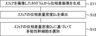
- FIG. 2 is a flow chart showing a flow of processing performed in the sorting step S3.
- step S11 a phase difference image of the sphere is generated from the hologram obtained by imaging the sphere formed in the three-dimensional culture step S2. Holography is performed, for example, on the sphere two days after the start of the three-dimensional culture step S2. Details of hologram photography and phase difference images will be described later.
- step S12 based on the phase difference image generated in step S11, to derive the amount of phase difference density D P of the sphere.
- Retardation amount density D P is calculated by dividing the amount of phase difference total sum P A is a value obtained by integrating the phase difference of each of the plurality of pixels constituting a phase difference image in a volume of spheres. It will be described later in detail retardation amount sum P A and the phase difference amount density D P.
- step S13 it performs a selection of pluripotent stem cells based on the phase difference amount density D P derived at step S12. Specifically, the phase difference amount density D P, who is within a predetermined range, pluripotent stem cells contained in the spheres is subjected to differentiation induction treatment. On the other hand, the phase difference amount density D P is not within the predetermined range, pluripotent stem cells contained in the spheres is excluded from the differentiation induction treatment.
- FIG. 3 is a diagram showing an example of the configuration of the imaging system 1 used for sorting pluripotent stem cells in the sorting step S3.
- the imaging system 1 includes a hologram optical system 10 for acquiring a hologram of a sphere using a known digital holography technique.
- Digital holography technology uses an image sensor to capture an image generated by the interference between the object light transmitted or reflected from the object and the reference light that is coherent to the object light, and the image obtained by the imaging is based on light propagation. It is a technology that restores the wave front of light waves from an object by performing numerical calculations. According to the digital holography technique, the phase distribution of an object can be quantified, and three-dimensional information of the object can be acquired without mechanically moving the focal position.
- the hologram optical system 10 includes a laser light source 11, beam splitters 12, 18, collimating lenses 13, 21, 22, 24, an objective lens 15, an imaging lens 17, and a CMOS (Complementary Metal Oxide Semiconductor) camera 19. There is.
- the sphere as the sample 14 set on the sample stage is arranged between the collimating lens 13 and the objective lens 15.
- the laser light source 11 for example, a HeNe laser having a wavelength of 632.8 nm can be used.
- the laser light emitted from the laser light source 11 is split into two laser lights by the beam splitter 12.
- One of the two laser beams is an object light and the other is a reference light.
- the object light is incident on the optical fiber 23 by the collimating lens 22, and is guided to the front of the collimating lens 13 by the optical fiber 23.
- the object light is collimated by the collimated lens 13 and then irradiated to the sphere, which is the sample 14 set on the sample stage.
- the diameter of the laser beam applied to the sphere is, for example, 2.2 mm.
- the image of the object light transmitted through the sphere is magnified by the objective lens 15.
- the object light transmitted through the objective lens 15 is converted into parallel light again by the imaging lens 17, and then imaged on the imaging surface of the CMOS camera 19 via the beam splitter 18.
- the reference light is incident on the optical fiber 20 by the collimating lens 24, and is guided to the front side of the collimating lens 21 by the optical fiber 20.
- the reference light emitted from the optical fiber 20 is converted into parallel light by the collimating lens 21 and incident on the imaging surface of the CMOS camera 19 via the beam splitter 18.
- the diameter of the laser beam emitted from the collimating lens 21 is, for example, 22 mm.
- the hologram generated by the interference between the object light and the reference light is recorded by the CMOS camera 19.
- CMOS camera 19 for example, a monochrome image sensor having a resolution of 2448 ⁇ 2048 and a sensor size of 3.45 ⁇ m ⁇ 3.45 ⁇ m can be used.
- the beam splitter 18 is tilted so that the optical axes of the object light and the reference light incident on the imaging surface of the CMOS camera 19 are different from each other, and the reference light is tilted by about 3 ° with respect to the object light. You may take a picture.
- the imaging system 1 it is possible to acquire a phase difference image of the sphere without destroying the sphere and without damaging the cells constituting the sphere.
- the configuration of the imaging system 1 described above is merely an example, and is not limited to the configuration described above.
- any imaging system capable of acquiring holograms using digital hologram technology can be used.
- FIG. 4A the hologram of the sphere illustrated in FIG. 4A acquired by the imaging system 1 is trimmed to a size of, for example, 2048 ⁇ 2048, and then two-dimensional Fourier transform is performed.
- FIG. 4B is an example of a Fourier transform image of the sphere obtained by this processing.
- FIG. 4B shows images based on direct light, object light, and conjugated light.
- the position of the object light is specified by specifying the amount of deviation of the object light with respect to the direct light in the Fourier transform image. Extract the amplitude component.
- the angular spectral method is applied to restore an image showing the phase of the sphere at an arbitrary spatial position.
- the wavefront u captured by the imaging surface of the CMOS camera 19 (x, y; 0) ; determining the angular spectrum U (0 f x, f y ) of the Fourier transform image.
- the wave surface at an arbitrary position z in the axial direction (z direction) is reproduced.
- the transfer function H (f x, f y; z) is the frequency response function (Fourier transform of the impulse response function (Green function)).
- wavefront U (f x, f y; z) at position z of the imaging system 1 in the optical axis direction (z direction) by performing an inverse Fourier transform on,
- the solution u (x, y; z) at position z is derived.
- FIG. 4C is an example of a phase difference image of the sphere obtained by each of the above processes before unwrapping.
- phase of the sphere before unwrapping shown in FIG. 4C is convoluted to a value of 0 to 2 ⁇ . Therefore, for example, by applying a phase connection (unwrapping) method such as Unweighted Least Squares (unweighted least squares method) or Flynn's Algorithm (Flynn's algorithm), and joining the parts of 2 ⁇ or more, as shown in FIG. 4D.
- a phase connection (unwrapping) method such as Unweighted Least Squares (unweighted least squares method) or Flynn's Algorithm (Flynn's algorithm)
- a final phase difference image of the sphere as illustrated can be obtained.
- Many unwrapping methods have been proposed, and an appropriate method that does not cause phase inconsistency may be appropriately selected.
- FIG. 5 is a diagram showing the concept of a phase difference image I P.
- phase difference image I retardation amount P in P is represented by the following equation (4).
- phase in the present specification is the phase of the electric field amplitude when light is regarded as an electromagnetic wave, and is used in a more general sense.
- the phase difference amount P k of each pixel k of the phase difference image I P can be represented by the following formula (5).
- n k is the index of refraction of the spheres at the site corresponding to each pixel k of the phase difference image I P
- d k is the thickness of the spheres at the site corresponding to each pixel k of the phase difference image I P
- ⁇ is the wavelength of the object light in the hologram optical system 10.
- the phase difference image of the sphere is an image showing the optical path length distribution of the object light transmitted through the sphere. Since the optical path length in the sphere corresponds to the product of the refractive index of the sphere and the thickness of the sphere, the phase difference image of the sphere shows the refractive index and thickness of the sphere as shown in Eq. (5). Contains information on the (shape).
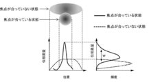
- focusing on the sphere means obtaining a phase difference image sliced near the center of the spherical sphere.
- the graph on the left of FIG. 6 is a graph showing an example of the relationship between the position in the plane direction and the amount of phase difference in the phase difference image of the sphere, and the solid line corresponds to the state where the sphere is in focus, and the dotted line corresponds to the sphere. Corresponds to the out-of-focus condition.
- the sphere is in focus, a steep peak appears at a specific position in the phase difference image.
- the peak is lower and gentler than when it is in focus.
- the graph on the right of FIG. 6 is an example of a histogram of the amount of phase difference in the phase difference image of the sphere, and the solid line corresponds to the state where the sphere is in focus and the dotted line corresponds to the state where the sphere is not in focus. To do. When the sphere is in focus, the curve width w (variation in the amount of phase difference) is relatively large, and when the sphere is out of focus, the width w of the curve (variation in the amount of phase difference) is. It becomes relatively small.
- the phase difference images of the spheres are acquired for each focal position (slice position) different from each other, and for each of the acquired phase difference images, the curve width w (variation of the phase difference amount) in the histogram of the amount of phase difference is obtained.
- Focusing can be realized by extracting a histogram image having the maximum width w out of the obtained width w as a histogram image focused on the sphere.
- FIG. 7 is a graph showing an example of the relationship between the focal position (slice position) and the variation in the amount of phase difference in the phase difference image of the sphere.
- FIG. 7 illustrates phase-difference images of spheres corresponding to focal positions of ⁇ 400 ⁇ m, ⁇ 200 ⁇ m, 0 ⁇ m, +200 ⁇ m, and +400 ⁇ m, along with graphs.
- the focal position where the variation in the amount of phase difference is maximum is set to 0 ⁇ m.
- the phase difference image corresponding to the focal position 0 ⁇ m at which the variation in the amount of phase difference is maximum is extracted as the focused phase difference image.
- the contour of the sphere is the clearest in the phase difference image corresponding to the focal position 0 ⁇ m where the variation in the amount of phase difference is maximum.
- Retardation amount sum P A described above is represented by the following equation (6).
- s is the area of each pixel k of the retardation image
- v k is the volume of the sphere at the portion corresponding to each pixel k of the retardation image.
- the phase difference amount sum P A the amount of phase difference P k for each pixel of the phase contrast image of the spheres corresponds to that obtained by integrating all the pixels k.
- the pixel value of the phase difference image corresponds to the phase difference amount P k.
- Retardation amount density D P is represented by the following equation (7).
- V is the volume of the sphere.
- the phase difference amount density D P corresponding to the phase difference amount sum P A in divided by the volume V of the spheres. Due to its homeostasis, living cells are considered to maintain a constant value of internal refractive index, which is different from the refractive index of the medium. On the other hand, it is considered that dead cells lose homeostasis and the internal refractive index becomes substantially the same as the refractive index of the medium. Accordingly, the phase difference amount density D P, it is possible to use as an index indicating the state of the cell.
- the 2 [pi / lambda it is possible to treat as a constant, upon derivation of the phase difference amount density D P, it may be omitted multiplication 2 [pi / lambda.
- D P (2 ⁇ / ⁇ ) ⁇ N ave
- the retardation density is a standardization of the difference in refractive index of the volume-averaged sphere by the length of the wavelength.
- the volume V of the sphere can be obtained by calculating the equivalent diameter of the sphere from the cross-sectional image of the phase image of the sphere. It is also possible to make it an ellipsoidal sphere more accurately.
- the volume average refractive index difference N ave can be treated as the phase difference amount density D P equivalent indicators.
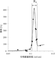
- Figure 8 is a graph showing the amount of phase difference density D P of iPS cell spheres, an example of the relationship between the yield ratio Y of myocardial cells obtained by the differentiation induction treatment for that iPS cells.
- the amount of phase difference density D P derived from the phase difference image generated from the acquired hologram for spheres of time before the differentiation induction treatment is carried out (after 2 days from the start of the three-dimensional culture process S2 in the present embodiment) It was done.
- the profit rate Y is expressed by the following equation (8).
- N i in equation (8) the number of pluripotent stem cells in production lots at the time prior to the differentiation induction treatment (first day started three-dimensional culture process S2 in the present embodiment)
- N c is It is the number of cardiomyocytes in the production lot obtained at the time point after the differentiation induction treatment (12 days after the start of the differentiation induction step S4 in the present embodiment). That is, yield ratio Y is the ratio of the number N c of the produced cardiomyocytes to the number N i to put pluripotent stem cells. Since the cells in the period from the measurement time of the N i to the measurement point of the N c are grown, yield ratio Y is sometimes exceeds 100%. Further, the number N c of cardiomyocytes can be obtained by measuring the number of cTnT-positive cells.
- pluripotent stem cells retardation amount density D P is included in the spheres is within the predetermined range R A is obtained ratio Y of cardiomyocytes obtained by subsequent differentiation induction treatment is significantly higher .. That is, FIG. 8, the pluripotent stem cells high yield ratio Y is obtained, show that it is possible screened by phase difference amount density D P of the sphere.
- Sorting method according to the embodiment of the disclosed technique, as exemplified in Figure 8, the amount of phase difference density D P obtained for spheres prior to the differentiation induction treatment, pluripotent stem cells contained in the sphere based on the relationship between yield ratio Y of myocardial cells obtained by the differentiation induction treatment for, the phase difference amount density D P, comprising determining the predetermined range R a for selection.
- the phase difference amount density D P when intending to obtain rate of cardiomyocytes to 100% or more, the phase difference amount density D P is 0.102 [rad / ⁇ m] or 0.114 [rad / ⁇ m] or less of Pluripotent stem cells contained in a range of spheres are targeted for differentiation induction treatment.
- the phase difference amount density D P since it varies with the wavelength of the object light ⁇ in the hologram optical system 10, it is preferable that the wavelength ⁇ is constant.
- the desired yield It is possible to identify pluripotent stem cells with a promising rate. Therefore, for pluripotent stem cells for which the desired yield cannot be expected, measures such as interrupting the treatment or changing the production conditions can be taken, so that the productivity of the cell product can be increased and the production cost can be suppressed. It will be possible.
- phase difference amount density D P it is preferred to derive the phase difference amount density D P based on the phase difference image generated from the image hologram for the previous spheres performing differentiation induction treatment.
- pluripotent stem cells can be selected before the differentiation-inducing treatment, so that the effect of increasing the productivity of cell products can be promoted.
- the measurement of the phase difference amount density D P is carried out within five days stirring culture after 2 hours later, more preferably be carried out within 3 days after 12 hours, to be performed within 2 days after 1 day Most preferred.
- phase difference amount density D P In the case of performing static culture instead of stirring culture in order to form spheres, to perform the measurement of the phase difference amount density D P within 5 days after 2 hours from the standing culture initiation for forming spheres Is preferable, and it is more preferable to carry out within 3 days after 12 hours, and most preferably within 2 days after 1 day. Even after differentiation induction treatment, if time to change due to differentiation of the phase difference amount density D P appears, the relationship between the amount of phase difference density D P and Tokuritsu Y is maintained, the it may derive the phase difference amount density D P based on the phase difference image generated from the image hologram for spheres of the period.
- Selection of pluripotent stem cells can be performed on a sphere basis or on a production lot basis.
- the production lot unit obtains the amount of phase difference density D P for some or all of the plurality of spheres in the production lots, the lot in the average value of the phase difference amount density D P If is within a predetermined range, all pluripotent stem cells contained in the production lot may be subject to the differentiation induction treatment.
- the relationship between the amount of phase difference density D P and Tokuritsu Y can be mathematically expressed using known function fitting techniques. For example, by using a linear combination model of the Gaussian function and sigmoid function, the phase difference amount density D P and a function representing the relationship between Tokuritsu Y below (9) shown in FIG. 8 expression can be derived is there.
- (9) shows that predictable from the phase difference amount density D P of spheres obtained in the intermediate stage in the manufacturing process of cell products, the yield ratio Y of myocardial cells obtained by the differentiation induction treatment.
- Prediction method of inducing differentiation results according to an exemplary embodiment of the disclosed technique is to use the relationship between the yield ratio Y of the phase difference amount density D P and cardiomyocytes, the phase difference of the spheres charge density D P Based on the above, it includes deriving a predicted value regarding the number of cardiomyocytes obtained by performing differentiation induction treatment on a plurality of pluripotent stem cells contained in the sphere.
- the amount of phase difference density D P of spheres by substituting the function representing the relationship between the the the phase difference amount density D P and Tokuritsu Y exemplified by equation (9), in the sphere
- the function representing the relationship between the amount of phase difference density D P and Tokuritsu Y is specified, based on the phase difference amount density D P of the sphere, the differentiation induction treatment performed on the spheres It is possible to derive a predicted value of the cardiomyocyte yield Y obtained in this case, that is, to predict the differentiation induction result.
- a function representing the relationship between the amount of phase difference density D P and Tokuritsu Y as the predicted value for the number of cardiomyocytes, it is also possible to derive the predicted value other than yield ratio.
- the differentiation-inducing result can be predicted at the stage before the differentiation-inducing treatment, so that the effect of increasing the productivity of the cell product can be promoted.
- the measurement of the phase difference amount density D P is carried out within five days stirring culture after 2 hours later, more preferably be carried out within 3 days after 12 hours, to be performed within 2 days after 1 day Most preferred.
- phase difference amount density D P In the case of performing static culture instead of stirring culture in order to form spheres, to perform the measurement of the phase difference amount density D P within 5 days after 2 hours from the standing culture initiation for forming spheres Is preferable, and it is more preferable to carry out within 3 days after 12 hours, and most preferably within 2 days after 1 day. Even after differentiation induction treatment, if time to change due to differentiation of the phase difference amount density D P appears, the relationship between the amount of phase difference density D P and Tokuritsu Y is maintained, the it may derive the phase difference amount density D P based on the phase difference image generated from the image hologram for spheres of the period.
- Examples of cells other than myocardial cells include endometrial cells, such as digestive system cells (hepatic cells, bile duct cells, pancreatic endocrine cells, aden cells, duct cells, resorbing cells, cup cells, panate cells, intestinal endocrine cells). Etc.), cells of tissues such as lung and thyroid, and examples of mesenchymal cells include hematopoietic stem cells, erythrocytes, platelets, macrophages, granulocytes, helper T cells, and killer T cells.
- endometrial cells such as digestive system cells (hepatic cells, bile duct cells, pancreatic endocrine cells, aden cells, duct cells, resorbing cells, cup cells, panate cells, intestinal endocrine cells).
- Etc. examples of tissues such as lung and thyroid
- mesenchymal cells include hematopoietic stem cells, erythrocytes, platelets, macrophages, granulocytes, help
- vasculature cells vascular endothelial cells, etc.
- osteoblasts bone cells, cartilage cells, tendon cells, fat cells, skeletal muscle cells, smooth muscle cells, etc.
- nervous system cells sensory organ cells (crystal body, retina, inner ear, etc.), skin epidermal cells, hair follicles and the like.
- IPS cell clones A to O were prepared from peripheral blood mononuclear cells containing different donor sources according to the method described in Japanese Patent No. 5894217.
- mTeSR1 Stem Cell Technologies
- Matrigel Matrigel
- the cells were collected and subcultured by treating with 0.5 mM EDTA solution (Invitrogen) for 7 minutes, and when the confluency reached about 80% in the T225 flask, the cells were separated by TrypLE Select (Thermo Fisher).
- Single cells were exfoliated and collected, and 3.0 in mTeSR1 containing 1 ⁇ M H1152 (Fujifilm Wako Pure Chemical Industries, Ltd.), 25 ⁇ g / ml Gentamicin (Thermo Fisher), and 100 ng / ml bFGF (Fujifilm Wako Pure Chemical Industries, Ltd.).
- the cells were adjusted to x10 6 cells / ml.
- 15 ml of the cell suspension was added to a 30 ml single-use bioreactor (Able), and the cells were stirred and cultured at a rotation speed of 40 rpm. After 2 to 4 hours from the start of the stirring culture, the mixture was scalpel-up in the same medium so that the final liquid volume was 30 ml, and the stirring culture was continued as it was.
- the final concentration was 1 ⁇ M H1152, 25 ⁇ g / ml Gentamicin, 100 ng / ml bFGF, 24 ng / ml Activin, 5% fetal bovine serum (GE Healthcare), ⁇ 0.5 mTeSR1, and ⁇ 0.5 DMEM Low-glucose ( The medium was replaced with a medium consisting of Thermo Fisher), and stirring culture was continued.
- the configuration of the imaging system 1 used is as follows.
- the laser light source 11 a HeNe laser having a wavelength of 632.8 nm and an output of 5 mW was used.
- the diameter of the laser beam emitted to the sphere was set to 2.2 mm.
- As the imaging lens 17, a lens having f 200 mm and an aperture diameter of 36 mm was used, and an image was formed on the imaging surface of a CMOS camera at an image magnification of 10 times.
- the CMOS camera 19 used a monochrome image sensor having a resolution of 2448 ⁇ 2048 and a sensor size of 3.45 ⁇ m ⁇ 3.45 ⁇ m.
- the beam splitter 18 is tilted so that the optical axes of the object light and the reference light incident on the imaging surface of the CMOS camera 19 are different from each other, and the reference light is tilted by about 3 ° with respect to the object light. I took a picture.
- phase difference amount density D P was derived phase difference amount density D P using the phase difference images (6) and (7).
- a part of the culture medium was sampled to measure the number of cells, and a medium consisting of final concentrations of 25 ⁇ g / ml Gentamaicin, 100 ng / ml bFGF, 24 ng / ml Activin, 40 ng / ml BMP4, 10% fetal bovine serum, and DMEM low-glucose.
- the content was adjusted to 1.0 ⁇ 10 6 cells / ml, and differentiation culture was started.
- the stirring culture was continued during the differentiation culture. From 3 to 7 days after the start of stirring culture, the medium was changed daily with the same medium, and stirring culture was continued.
- the ratio of the total number of cardiomyocytes to the number of cells at the start of stirring culture was derived as a rate Y.
- the phase difference amount density D P and Tokuritsu Y was obtained for each clone to plot on a graph, creating a graph showing the relationship between the amount of phase difference density D P and Tokuritsu Y shown in FIG. 8 did.
- FIG. 9 shows the relationship between the predicted value (vertical axis) and the measured value (horizontal axis) of the cardiomyocyte yield calculated by using equation (9) for each of the 15 clones.
- the predicted value and the measured value of the profit rate shown by each plot of FIG. 9 are the average values within the lot.
- the dotted line in FIG. 9 is an ideal line when the predicted value and the measured value match, and the solid line in FIG. 9 is an approximate straight line derived based on each plot.
- the coefficient of determination R 2 in this approximate straight line is 0.9426, and the correlation coefficient R is 0.9709. That is, it can be said that the accuracy of the predicted value of the profit rate derived by using the function represented by the equation (9) is extremely high.
Landscapes
- Health & Medical Sciences (AREA)
- Life Sciences & Earth Sciences (AREA)
- Engineering & Computer Science (AREA)
- Chemical & Material Sciences (AREA)
- Biomedical Technology (AREA)
- General Health & Medical Sciences (AREA)
- Biochemistry (AREA)
- Physics & Mathematics (AREA)
- General Physics & Mathematics (AREA)
- Bioinformatics & Cheminformatics (AREA)
- Genetics & Genomics (AREA)
- Organic Chemistry (AREA)
- Biotechnology (AREA)
- Zoology (AREA)
- Wood Science & Technology (AREA)
- Analytical Chemistry (AREA)
- Immunology (AREA)
- Pathology (AREA)
- Dispersion Chemistry (AREA)
- Developmental Biology & Embryology (AREA)
- Reproductive Health (AREA)
- Gynecology & Obstetrics (AREA)
- Cell Biology (AREA)
- Microbiology (AREA)
- General Engineering & Computer Science (AREA)
- Signal Processing (AREA)
- Multimedia (AREA)
- Transplantation (AREA)
- Molecular Biology (AREA)
- Theoretical Computer Science (AREA)
- Measuring Or Testing Involving Enzymes Or Micro-Organisms (AREA)
Priority Applications (3)
| Application Number | Priority Date | Filing Date | Title |
|---|---|---|---|
| EP20883205.5A EP4043864A4 (en) | 2019-10-28 | 2020-10-02 | METHOD FOR SORTING PLURIPOTENT STEM CELLS, METHOD FOR PREDICTING RESULTS OF INDUCTION OF DIFFERENTIATION, AND METHOD FOR MANUFACTURING CELLULAR PRODUCT |
| JP2021554228A JPWO2021085033A1 (https=) | 2019-10-28 | 2020-10-02 | |
| US17/729,267 US20220253016A1 (en) | 2019-10-28 | 2022-04-26 | Sorting method for pluripotent stem cell, prediction method for differentiation induction result, and production method for cell product |
Applications Claiming Priority (2)
| Application Number | Priority Date | Filing Date | Title |
|---|---|---|---|
| JP2019195670 | 2019-10-28 | ||
| JP2019-195670 | 2019-10-28 |
Related Child Applications (1)
| Application Number | Title | Priority Date | Filing Date |
|---|---|---|---|
| US17/729,267 Continuation US20220253016A1 (en) | 2019-10-28 | 2022-04-26 | Sorting method for pluripotent stem cell, prediction method for differentiation induction result, and production method for cell product |
Publications (1)
| Publication Number | Publication Date |
|---|---|
| WO2021085033A1 true WO2021085033A1 (ja) | 2021-05-06 |
Family
ID=75716239
Family Applications (1)
| Application Number | Title | Priority Date | Filing Date |
|---|---|---|---|
| PCT/JP2020/037590 Ceased WO2021085033A1 (ja) | 2019-10-28 | 2020-10-02 | 多能性幹細胞の選別方法、分化誘導結果の予測方法及び細胞製品の製造方法 |
Country Status (4)
| Country | Link |
|---|---|
| US (1) | US20220253016A1 (https=) |
| EP (1) | EP4043864A4 (https=) |
| JP (1) | JPWO2021085033A1 (https=) |
| WO (1) | WO2021085033A1 (https=) |
Cited By (1)
| Publication number | Priority date | Publication date | Assignee | Title |
|---|---|---|---|---|
| WO2022270179A1 (ja) * | 2021-06-24 | 2022-12-29 | 富士フイルム株式会社 | 情報処理装置、情報処理方法、及びプログラム |
Families Citing this family (2)
| Publication number | Priority date | Publication date | Assignee | Title |
|---|---|---|---|---|
| US20230137843A1 (en) * | 2021-10-29 | 2023-05-04 | Spheryx, Inc. | Holographic video microscopy cell viability assay |
| CN119963497B (zh) * | 2025-01-07 | 2025-07-22 | 华辰未来(北京)生物医学技术有限公司 | 基于人体干细胞的检测方法 |
Citations (6)
| Publication number | Priority date | Publication date | Assignee | Title |
|---|---|---|---|---|
| JP2015146747A (ja) | 2014-02-05 | 2015-08-20 | 浜松ホトニクス株式会社 | 細胞判定方法 |
| JP5984217B2 (ja) | 2010-06-15 | 2016-09-06 | セルラー ダイナミクス インターナショナル, インコーポレイテッド | 少量の末梢血からの人工多能性幹細胞の作製 |
| JP2018000048A (ja) | 2016-06-29 | 2018-01-11 | オリンパス株式会社 | 多能性幹細胞の無染色評価支援方法、プログラム、演算装置 |
| WO2018158901A1 (ja) | 2017-03-02 | 2018-09-07 | 株式会社島津製作所 | 細胞解析方法及び細胞解析装置 |
| WO2019176427A1 (ja) * | 2018-03-12 | 2019-09-19 | 富士フイルム株式会社 | 判定方法 |
| JP2019195670A (ja) | 2013-03-15 | 2019-11-14 | ウインドギャップ メディカル インコーポレイテッド | 携帯型薬剤混合及び送達システム並びに方法 |
Family Cites Families (1)
| Publication number | Priority date | Publication date | Assignee | Title |
|---|---|---|---|---|
| US20200172969A1 (en) * | 2017-06-19 | 2020-06-04 | Foundation For Biomedical Research And Innovation At Kobe | Method for predicting differentiation ability of pluripotent stem cell, and reagent for same |
-
2020
- 2020-10-02 JP JP2021554228A patent/JPWO2021085033A1/ja not_active Abandoned
- 2020-10-02 WO PCT/JP2020/037590 patent/WO2021085033A1/ja not_active Ceased
- 2020-10-02 EP EP20883205.5A patent/EP4043864A4/en not_active Withdrawn
-
2022
- 2022-04-26 US US17/729,267 patent/US20220253016A1/en not_active Abandoned
Patent Citations (6)
| Publication number | Priority date | Publication date | Assignee | Title |
|---|---|---|---|---|
| JP5984217B2 (ja) | 2010-06-15 | 2016-09-06 | セルラー ダイナミクス インターナショナル, インコーポレイテッド | 少量の末梢血からの人工多能性幹細胞の作製 |
| JP2019195670A (ja) | 2013-03-15 | 2019-11-14 | ウインドギャップ メディカル インコーポレイテッド | 携帯型薬剤混合及び送達システム並びに方法 |
| JP2015146747A (ja) | 2014-02-05 | 2015-08-20 | 浜松ホトニクス株式会社 | 細胞判定方法 |
| JP2018000048A (ja) | 2016-06-29 | 2018-01-11 | オリンパス株式会社 | 多能性幹細胞の無染色評価支援方法、プログラム、演算装置 |
| WO2018158901A1 (ja) | 2017-03-02 | 2018-09-07 | 株式会社島津製作所 | 細胞解析方法及び細胞解析装置 |
| WO2019176427A1 (ja) * | 2018-03-12 | 2019-09-19 | 富士フイルム株式会社 | 判定方法 |
Non-Patent Citations (1)
| Title |
|---|
| See also references of EP4043864A4 |
Cited By (2)
| Publication number | Priority date | Publication date | Assignee | Title |
|---|---|---|---|---|
| WO2022270179A1 (ja) * | 2021-06-24 | 2022-12-29 | 富士フイルム株式会社 | 情報処理装置、情報処理方法、及びプログラム |
| JPWO2022270179A1 (https=) * | 2021-06-24 | 2022-12-29 |
Also Published As
| Publication number | Publication date |
|---|---|
| EP4043864A4 (en) | 2022-11-30 |
| EP4043864A1 (en) | 2022-08-17 |
| US20220253016A1 (en) | 2022-08-11 |
| JPWO2021085033A1 (https=) | 2021-05-06 |
Similar Documents
| Publication | Publication Date | Title |
|---|---|---|
| US20220253016A1 (en) | Sorting method for pluripotent stem cell, prediction method for differentiation induction result, and production method for cell product | |
| KR101950339B1 (ko) | 살아있는 세포의 간섭법을 통한 신속한, 대용량 병렬 단일-세포 약물 반응 측정방법 | |
| Sahu et al. | Culturing primary neurons from rat hippocampus and cortex | |
| Llonch et al. | Organoid technology for retinal repair | |
| Rappaz et al. | Digital holographic microscopy: a quantitative label-free microscopy technique for phenotypic screening | |
| Pavillon et al. | Early cell death detection with digital holographic microscopy | |
| Cogger et al. | Three-dimensional structured illumination microscopy of liver sinusoidal endothelial cell fenestrations | |
| Groux et al. | Dynamic full-field optical coherence tomography allows live imaging of retinal pigment epithelium stress model | |
| EP3766984B1 (en) | Determination method` | |
| Kandel et al. | Multiscale assay of unlabeled neurite dynamics using phase imaging with computational specificity | |
| Mathieu et al. | Time-lapse lens-free imaging of cell migration in diverse physical microenvironments | |
| WO2020003194A1 (en) | Virtual staining systems and methods for observing one or more unstained cells | |
| Phillips | Monitoring neuron and astrocyte interactions with a 3D cell culture system | |
| US20220244243A1 (en) | Sorting method for pluripotent stem cell, prediction method for differentiation induction result, and production method for cell product | |
| JP7046211B2 (ja) | 判定方法 | |
| Seo et al. | Cellular imaging using phase holographic microscopy: for the study of pathophysiology of red blood cells and human umbilical vein endothelial cells | |
| Dou et al. | Raman imaging diagnosis of the early stage differentiation of mouse embryonic stem cell (mESC) | |
| Petecchia et al. | A biophysical approach to quantify skeletal stem cells trans-differentiation as a model for the study of osteoporosis | |
| Zicha et al. | Testing anti-cancer drugs with holographic incoherent-light-source quantitative phase imaging | |
| WO2023233827A1 (ja) | 位相画像取得方法及び定量データ取得方法 | |
| Singla et al. | Quantitative Detection of Morphological Alterations in Red Blood Cell During Storage Using PS-FF-OCT System | |
| GB2531646B (en) | Method for evaluating suppressive action on vascular hyper-permeability | |
| Gilerovich et al. | Detection of glomeruli in the human cerebellum using an immunocytochemical reaction for synaptophysin and confocal laser microscopy | |
| BLINCKO | Characterisation of Embryonic Dermal Precursor Cells | |
| Strbkova et al. | Biocompatibility of Thin Films Studied by Q-Phase |
Legal Events
| Date | Code | Title | Description |
|---|---|---|---|
| 121 | Ep: the epo has been informed by wipo that ep was designated in this application |
Ref document number: 20883205 Country of ref document: EP Kind code of ref document: A1 |
|
| ENP | Entry into the national phase |
Ref document number: 2021554228 Country of ref document: JP Kind code of ref document: A |
|
| NENP | Non-entry into the national phase |
Ref country code: DE |
|
| ENP | Entry into the national phase |
Ref document number: 2020883205 Country of ref document: EP Effective date: 20220426 |