WO2019131879A1 - Agent d'accélération de la production de prostaglandine d2 synthase de type lipocaline - Google Patents
Agent d'accélération de la production de prostaglandine d2 synthase de type lipocaline Download PDFInfo
- Publication number
- WO2019131879A1 WO2019131879A1 PCT/JP2018/048141 JP2018048141W WO2019131879A1 WO 2019131879 A1 WO2019131879 A1 WO 2019131879A1 JP 2018048141 W JP2018048141 W JP 2018048141W WO 2019131879 A1 WO2019131879 A1 WO 2019131879A1
- Authority
- WO
- WIPO (PCT)
- Prior art keywords
- lipocalin
- pgds
- type prostaglandin
- action
- brain
- Prior art date
Links
- 238000004519 manufacturing process Methods 0.000 title claims abstract description 59
- 102000048176 Prostaglandin-D synthases Human genes 0.000 title claims abstract description 21
- 108030003866 Prostaglandin-D synthases Proteins 0.000 title claims abstract description 21
- 239000000284 extract Substances 0.000 claims abstract description 79
- 239000000126 substance Substances 0.000 claims abstract description 78
- 230000014509 gene expression Effects 0.000 claims abstract description 49
- 210000003668 pericyte Anatomy 0.000 claims abstract description 49
- 210000001519 tissue Anatomy 0.000 claims abstract description 46
- 239000003795 chemical substances by application Substances 0.000 claims abstract description 30
- 239000003814 drug Substances 0.000 claims abstract description 29
- 241000700618 Vaccinia virus Species 0.000 claims abstract description 22
- 206010008118 cerebral infarction Diseases 0.000 claims abstract description 21
- 230000000694 effects Effects 0.000 claims abstract description 20
- 208000024827 Alzheimer disease Diseases 0.000 claims abstract description 18
- 206010012289 Dementia Diseases 0.000 claims abstract description 18
- 208000028867 ischemia Diseases 0.000 claims abstract description 16
- 210000004556 brain Anatomy 0.000 claims description 83
- BHMBVRSPMRCCGG-UHFFFAOYSA-N prostaglandine D2 Natural products CCCCCC(O)C=CC1C(CC=CCCCC(O)=O)C(O)CC1=O BHMBVRSPMRCCGG-UHFFFAOYSA-N 0.000 claims description 58
- 238000000034 method Methods 0.000 claims description 57
- 230000001737 promoting effect Effects 0.000 claims description 49
- 102000003960 Ligases Human genes 0.000 claims description 32
- 108090000364 Ligases Proteins 0.000 claims description 32
- 230000009471 action Effects 0.000 claims description 28
- BHMBVRSPMRCCGG-OUTUXVNYSA-N prostaglandin D2 Chemical compound CCCCC[C@H](O)\C=C\[C@@H]1[C@@H](C\C=C/CCCC(O)=O)[C@@H](O)CC1=O BHMBVRSPMRCCGG-OUTUXVNYSA-N 0.000 claims description 28
- 238000002360 preparation method Methods 0.000 claims description 27
- 208000026106 cerebrovascular disease Diseases 0.000 claims description 22
- 241000283973 Oryctolagus cuniculus Species 0.000 claims description 21
- 238000011156 evaluation Methods 0.000 claims description 19
- 102000004190 Enzymes Human genes 0.000 claims description 17
- 108090000790 Enzymes Proteins 0.000 claims description 17
- 230000003449 preventive effect Effects 0.000 claims description 16
- 102000019298 Lipocalin Human genes 0.000 claims description 14
- 108050006654 Lipocalin Proteins 0.000 claims description 14
- 238000012216 screening Methods 0.000 claims description 14
- 230000001681 protective effect Effects 0.000 claims description 13
- 239000003223 protective agent Substances 0.000 claims description 11
- 102000013455 Amyloid beta-Peptides Human genes 0.000 claims description 10
- 108010090849 Amyloid beta-Peptides Proteins 0.000 claims description 10
- 229940124597 therapeutic agent Drugs 0.000 claims description 10
- 210000004263 induced pluripotent stem cell Anatomy 0.000 claims description 9
- 238000002347 injection Methods 0.000 claims description 7
- 239000007924 injection Substances 0.000 claims description 7
- 230000006933 amyloid-beta aggregation Effects 0.000 claims description 6
- 230000002708 enhancing effect Effects 0.000 claims description 3
- 238000009472 formulation Methods 0.000 claims description 3
- 230000002401 inhibitory effect Effects 0.000 claims description 3
- 239000000203 mixture Substances 0.000 claims description 3
- 206010061218 Inflammation Diseases 0.000 claims description 2
- 230000004054 inflammatory process Effects 0.000 claims description 2
- 230000029142 excretion Effects 0.000 claims 1
- 239000005723 virus inoculator Substances 0.000 claims 1
- 102100033279 Prostaglandin-H2 D-isomerase Human genes 0.000 abstract description 142
- 238000011282 treatment Methods 0.000 abstract description 17
- 229940079593 drug Drugs 0.000 abstract description 12
- 230000002265 prevention Effects 0.000 abstract description 12
- 208000037265 diseases, disorders, signs and symptoms Diseases 0.000 abstract description 9
- 230000002490 cerebral effect Effects 0.000 abstract description 3
- 210000002894 multi-fate stem cell Anatomy 0.000 abstract description 2
- 201000008247 brain infarction Diseases 0.000 abstract 1
- 208000035475 disorder Diseases 0.000 abstract 1
- 230000002757 inflammatory effect Effects 0.000 abstract 1
- 208000019116 sleep disease Diseases 0.000 abstract 1
- 208000020685 sleep-wake disease Diseases 0.000 abstract 1
- 208000019553 vascular disease Diseases 0.000 abstract 1
- 101710145576 Prostaglandin-H2 D-isomerase Proteins 0.000 description 146
- 238000012360 testing method Methods 0.000 description 94
- 210000004027 cell Anatomy 0.000 description 24
- 108090000623 proteins and genes Proteins 0.000 description 21
- 238000009826 distribution Methods 0.000 description 20
- 241000699670 Mus sp. Species 0.000 description 18
- 210000003556 vascular endothelial cell Anatomy 0.000 description 15
- 102000004169 proteins and genes Human genes 0.000 description 14
- 210000003491 skin Anatomy 0.000 description 13
- 206010046865 Vaccinia virus infection Diseases 0.000 description 12
- 239000003463 adsorbent Substances 0.000 description 12
- 230000001965 increasing effect Effects 0.000 description 12
- 239000000243 solution Substances 0.000 description 12
- 208000007089 vaccinia Diseases 0.000 description 12
- 239000006144 Dulbecco’s modified Eagle's medium Substances 0.000 description 11
- 241000699666 Mus <mouse, genus> Species 0.000 description 11
- 230000006870 function Effects 0.000 description 11
- OKTJSMMVPCPJKN-UHFFFAOYSA-N Carbon Chemical compound [C] OKTJSMMVPCPJKN-UHFFFAOYSA-N 0.000 description 10
- 238000011532 immunohistochemical staining Methods 0.000 description 10
- HEMHJVSKTPXQMS-UHFFFAOYSA-M Sodium hydroxide Chemical compound [OH-].[Na+] HEMHJVSKTPXQMS-UHFFFAOYSA-M 0.000 description 9
- 230000000302 ischemic effect Effects 0.000 description 9
- 239000006228 supernatant Substances 0.000 description 9
- WQZGKKKJIJFFOK-GASJEMHNSA-N Glucose Natural products OC[C@H]1OC(O)[C@H](O)[C@@H](O)[C@@H]1O WQZGKKKJIJFFOK-GASJEMHNSA-N 0.000 description 8
- 241001465754 Metazoa Species 0.000 description 8
- 239000002033 PVDF binder Substances 0.000 description 8
- 102100024616 Platelet endothelial cell adhesion molecule Human genes 0.000 description 8
- 230000003431 anti-prostaglandin Effects 0.000 description 8
- 239000002771 cell marker Substances 0.000 description 8
- 239000012228 culture supernatant Substances 0.000 description 8
- 201000010099 disease Diseases 0.000 description 8
- 239000008103 glucose Substances 0.000 description 8
- 239000007788 liquid Substances 0.000 description 8
- 210000004379 membrane Anatomy 0.000 description 8
- 239000012528 membrane Substances 0.000 description 8
- 229920002981 polyvinylidene fluoride Polymers 0.000 description 8
- 238000003757 reverse transcription PCR Methods 0.000 description 8
- XLYOFNOQVPJJNP-UHFFFAOYSA-N water Substances O XLYOFNOQVPJJNP-UHFFFAOYSA-N 0.000 description 8
- 108091032973 (ribonucleotides)n+m Proteins 0.000 description 7
- 108010088225 Nestin Proteins 0.000 description 7
- 102000008730 Nestin Human genes 0.000 description 7
- FAPWRFPIFSIZLT-UHFFFAOYSA-M Sodium chloride Chemical compound [Na+].[Cl-] FAPWRFPIFSIZLT-UHFFFAOYSA-M 0.000 description 7
- 238000005119 centrifugation Methods 0.000 description 7
- 238000006243 chemical reaction Methods 0.000 description 7
- 238000000605 extraction Methods 0.000 description 7
- 239000003550 marker Substances 0.000 description 7
- 210000005055 nestin Anatomy 0.000 description 7
- 229910052760 oxygen Inorganic materials 0.000 description 7
- 239000000047 product Substances 0.000 description 7
- YIBNHAJFJUQSRA-YNNPMVKQSA-N prostaglandin H2 Chemical compound C1[C@@H]2OO[C@H]1[C@H](/C=C/[C@@H](O)CCCCC)[C@H]2C\C=C/CCCC(O)=O YIBNHAJFJUQSRA-YNNPMVKQSA-N 0.000 description 7
- 239000002904 solvent Substances 0.000 description 7
- 238000010186 staining Methods 0.000 description 7
- 108091003079 Bovine Serum Albumin Proteins 0.000 description 6
- VEXZGXHMUGYJMC-UHFFFAOYSA-N Hydrochloric acid Chemical compound Cl VEXZGXHMUGYJMC-UHFFFAOYSA-N 0.000 description 6
- 239000012091 fetal bovine serum Substances 0.000 description 6
- 239000000706 filtrate Substances 0.000 description 6
- RWSXRVCMGQZWBV-WDSKDSINSA-N glutathione Chemical compound OC(=O)[C@@H](N)CCC(=O)N[C@@H](CS)C(=O)NCC(O)=O RWSXRVCMGQZWBV-WDSKDSINSA-N 0.000 description 6
- 210000002569 neuron Anatomy 0.000 description 6
- 230000000144 pharmacologic effect Effects 0.000 description 6
- 239000002504 physiological saline solution Substances 0.000 description 6
- 150000003180 prostaglandins Chemical class 0.000 description 6
- 238000001262 western blot Methods 0.000 description 6
- VHRUMKCAEVRUBK-WKELIDJCSA-N (z)-7-[(1s,5e)-5-[(z)-oct-2-enylidene]-4-oxocyclopent-2-en-1-yl]hept-5-enoic acid Chemical compound CCCCC\C=C/C=C1\[C@@H](C\C=C/CCCC(O)=O)C=CC1=O VHRUMKCAEVRUBK-WKELIDJCSA-N 0.000 description 5
- 102000007469 Actins Human genes 0.000 description 5
- 108010085238 Actins Proteins 0.000 description 5
- ISWSIDIOOBJBQZ-UHFFFAOYSA-N Phenol Chemical compound OC1=CC=CC=C1 ISWSIDIOOBJBQZ-UHFFFAOYSA-N 0.000 description 5
- 230000033228 biological regulation Effects 0.000 description 5
- 210000001175 cerebrospinal fluid Anatomy 0.000 description 5
- 231100000673 dose–response relationship Toxicity 0.000 description 5
- 238000001914 filtration Methods 0.000 description 5
- 238000000386 microscopy Methods 0.000 description 5
- 235000002639 sodium chloride Nutrition 0.000 description 5
- 239000000758 substrate Substances 0.000 description 5
- VSRXYLYXIXYEST-KZTWKYQFSA-N 13,14-dihydro-15-ketoprostaglandin D2 Chemical compound CCCCCC(=O)CC[C@@H]1[C@@H](C\C=C/CCCC(O)=O)[C@@H](O)CC1=O VSRXYLYXIXYEST-KZTWKYQFSA-N 0.000 description 4
- IGAZHQIYONOHQN-UHFFFAOYSA-N Alexa Fluor 555 Chemical compound C=12C=CC(=N)C(S(O)(=O)=O)=C2OC2=C(S(O)(=O)=O)C(N)=CC=C2C=1C1=CC=C(C(O)=O)C=C1C(O)=O IGAZHQIYONOHQN-UHFFFAOYSA-N 0.000 description 4
- 208000016192 Demyelinating disease Diseases 0.000 description 4
- 108010001336 Horseradish Peroxidase Proteins 0.000 description 4
- 239000004480 active ingredient Substances 0.000 description 4
- 238000004458 analytical method Methods 0.000 description 4
- QVGXLLKOCUKJST-UHFFFAOYSA-N atomic oxygen Chemical compound [O] QVGXLLKOCUKJST-UHFFFAOYSA-N 0.000 description 4
- 230000000903 blocking effect Effects 0.000 description 4
- 210000003710 cerebral cortex Anatomy 0.000 description 4
- 230000003930 cognitive ability Effects 0.000 description 4
- 238000001514 detection method Methods 0.000 description 4
- XEYBRNLFEZDVAW-ARSRFYASSA-N dinoprostone Chemical compound CCCCC[C@H](O)\C=C\[C@H]1[C@H](O)CC(=O)[C@@H]1C\C=C/CCCC(O)=O XEYBRNLFEZDVAW-ARSRFYASSA-N 0.000 description 4
- 229960002986 dinoprostone Drugs 0.000 description 4
- 238000001962 electrophoresis Methods 0.000 description 4
- 230000002255 enzymatic effect Effects 0.000 description 4
- 238000004128 high performance liquid chromatography Methods 0.000 description 4
- 230000002209 hydrophobic effect Effects 0.000 description 4
- 230000006872 improvement Effects 0.000 description 4
- 239000002609 medium Substances 0.000 description 4
- 239000001301 oxygen Substances 0.000 description 4
- 229940094443 oxytocics prostaglandins Drugs 0.000 description 4
- BASFCYQUMIYNBI-UHFFFAOYSA-N platinum Chemical compound [Pt] BASFCYQUMIYNBI-UHFFFAOYSA-N 0.000 description 4
- XEYBRNLFEZDVAW-UHFFFAOYSA-N prostaglandin E2 Natural products CCCCCC(O)C=CC1C(O)CC(=O)C1CC=CCCCC(O)=O XEYBRNLFEZDVAW-UHFFFAOYSA-N 0.000 description 4
- UQOQENZZLBSFKO-POPPZSFYSA-N prostaglandin J2 Chemical compound CCCCC[C@H](O)\C=C\[C@@H]1[C@@H](C\C=C/CCCC(O)=O)C=CC1=O UQOQENZZLBSFKO-POPPZSFYSA-N 0.000 description 4
- 230000004043 responsiveness Effects 0.000 description 4
- 230000003248 secreting effect Effects 0.000 description 4
- 238000000926 separation method Methods 0.000 description 4
- 239000011780 sodium chloride Substances 0.000 description 4
- 239000007787 solid Substances 0.000 description 4
- FWBHETKCLVMNFS-UHFFFAOYSA-N 4',6-Diamino-2-phenylindol Chemical compound C1=CC(C(=N)N)=CC=C1C1=CC2=CC=C(C(N)=N)C=C2N1 FWBHETKCLVMNFS-UHFFFAOYSA-N 0.000 description 3
- 206010008089 Cerebral artery occlusion Diseases 0.000 description 3
- 206010012305 Demyelination Diseases 0.000 description 3
- 201000004624 Dermatitis Diseases 0.000 description 3
- 238000002965 ELISA Methods 0.000 description 3
- LFQSCWFLJHTTHZ-UHFFFAOYSA-N Ethanol Chemical compound CCO LFQSCWFLJHTTHZ-UHFFFAOYSA-N 0.000 description 3
- 102100031181 Glyceraldehyde-3-phosphate dehydrogenase Human genes 0.000 description 3
- OKKJLVBELUTLKV-UHFFFAOYSA-N Methanol Chemical compound OC OKKJLVBELUTLKV-UHFFFAOYSA-N 0.000 description 3
- 208000013738 Sleep Initiation and Maintenance disease Diseases 0.000 description 3
- 230000003542 behavioural effect Effects 0.000 description 3
- 230000015572 biosynthetic process Effects 0.000 description 3
- 230000017531 blood circulation Effects 0.000 description 3
- 210000004204 blood vessel Anatomy 0.000 description 3
- 210000003169 central nervous system Anatomy 0.000 description 3
- 230000003920 cognitive function Effects 0.000 description 3
- 230000003247 decreasing effect Effects 0.000 description 3
- 238000010195 expression analysis Methods 0.000 description 3
- 229960003180 glutathione Drugs 0.000 description 3
- 108020004445 glyceraldehyde-3-phosphate dehydrogenase Proteins 0.000 description 3
- 230000002055 immunohistochemical effect Effects 0.000 description 3
- 230000006698 induction Effects 0.000 description 3
- 206010022437 insomnia Diseases 0.000 description 3
- 230000003834 intracellular effect Effects 0.000 description 3
- 238000004949 mass spectrometry Methods 0.000 description 3
- 239000002207 metabolite Substances 0.000 description 3
- 201000007309 middle cerebral artery infarction Diseases 0.000 description 3
- 239000003068 molecular probe Substances 0.000 description 3
- 238000010172 mouse model Methods 0.000 description 3
- 210000005036 nerve Anatomy 0.000 description 3
- 210000001178 neural stem cell Anatomy 0.000 description 3
- 238000012758 nuclear staining Methods 0.000 description 3
- 235000020030 perry Nutrition 0.000 description 3
- 238000003753 real-time PCR Methods 0.000 description 3
- 230000002829 reductive effect Effects 0.000 description 3
- 230000004044 response Effects 0.000 description 3
- 150000003384 small molecules Chemical class 0.000 description 3
- 210000000130 stem cell Anatomy 0.000 description 3
- 230000001225 therapeutic effect Effects 0.000 description 3
- LDXJRKWFNNFDSA-UHFFFAOYSA-N 2-(2,4,6,7-tetrahydrotriazolo[4,5-c]pyridin-5-yl)-1-[4-[2-[[3-(trifluoromethoxy)phenyl]methylamino]pyrimidin-5-yl]piperazin-1-yl]ethanone Chemical compound C1CN(CC2=NNN=C21)CC(=O)N3CCN(CC3)C4=CN=C(N=C4)NCC5=CC(=CC=C5)OC(F)(F)F LDXJRKWFNNFDSA-UHFFFAOYSA-N 0.000 description 2
- QKNYBSVHEMOAJP-UHFFFAOYSA-N 2-amino-2-(hydroxymethyl)propane-1,3-diol;hydron;chloride Chemical compound Cl.OCC(N)(CO)CO QKNYBSVHEMOAJP-UHFFFAOYSA-N 0.000 description 2
- KJDSORYAHBAGPP-UHFFFAOYSA-N 4-(3,4-diaminophenyl)benzene-1,2-diamine;hydron;tetrachloride Chemical compound Cl.Cl.Cl.Cl.C1=C(N)C(N)=CC=C1C1=CC=C(N)C(N)=C1 KJDSORYAHBAGPP-UHFFFAOYSA-N 0.000 description 2
- CSCPPACGZOOCGX-UHFFFAOYSA-N Acetone Chemical compound CC(C)=O CSCPPACGZOOCGX-UHFFFAOYSA-N 0.000 description 2
- 239000012103 Alexa Fluor 488 Substances 0.000 description 2
- 239000005995 Aluminium silicate Substances 0.000 description 2
- 208000037259 Amyloid Plaque Diseases 0.000 description 2
- IJGRMHOSHXDMSA-UHFFFAOYSA-N Atomic nitrogen Chemical compound N#N IJGRMHOSHXDMSA-UHFFFAOYSA-N 0.000 description 2
- 208000008035 Back Pain Diseases 0.000 description 2
- BPYKTIZUTYGOLE-IFADSCNNSA-N Bilirubin Chemical compound N1C(=O)C(C)=C(C=C)\C1=C\C1=C(C)C(CCC(O)=O)=C(CC2=C(C(C)=C(\C=C/3C(=C(C=C)C(=O)N\3)C)N2)CCC(O)=O)N1 BPYKTIZUTYGOLE-IFADSCNNSA-N 0.000 description 2
- 241000255789 Bombyx mori Species 0.000 description 2
- 206010048962 Brain oedema Diseases 0.000 description 2
- 108010078791 Carrier Proteins Proteins 0.000 description 2
- SXRSQZLOMIGNAQ-UHFFFAOYSA-N Glutaraldehyde Chemical compound O=CCCCC=O SXRSQZLOMIGNAQ-UHFFFAOYSA-N 0.000 description 2
- PEDCQBHIVMGVHV-UHFFFAOYSA-N Glycerine Chemical compound OCC(O)CO PEDCQBHIVMGVHV-UHFFFAOYSA-N 0.000 description 2
- ZRALSGWEFCBTJO-UHFFFAOYSA-N Guanidine Chemical compound NC(N)=N ZRALSGWEFCBTJO-UHFFFAOYSA-N 0.000 description 2
- 102100029100 Hematopoietic prostaglandin D synthase Human genes 0.000 description 2
- 101710082112 Hematopoietic prostaglandin D synthase Proteins 0.000 description 2
- KFZMGEQAYNKOFK-UHFFFAOYSA-N Isopropanol Chemical compound CC(C)O KFZMGEQAYNKOFK-UHFFFAOYSA-N 0.000 description 2
- TWRXJAOTZQYOKJ-UHFFFAOYSA-L Magnesium chloride Chemical compound [Mg+2].[Cl-].[Cl-] TWRXJAOTZQYOKJ-UHFFFAOYSA-L 0.000 description 2
- 238000012347 Morris Water Maze Methods 0.000 description 2
- 206010029350 Neurotoxicity Diseases 0.000 description 2
- 101150020237 PTGDS gene Proteins 0.000 description 2
- 229930040373 Paraformaldehyde Natural products 0.000 description 2
- 102100026547 Platelet-derived growth factor receptor beta Human genes 0.000 description 2
- 101710164680 Platelet-derived growth factor receptor beta Proteins 0.000 description 2
- WCUXLLCKKVVCTQ-UHFFFAOYSA-M Potassium chloride Chemical compound [Cl-].[K+] WCUXLLCKKVVCTQ-UHFFFAOYSA-M 0.000 description 2
- 102000009389 Prostaglandin D receptors Human genes 0.000 description 2
- 108050000258 Prostaglandin D receptors Proteins 0.000 description 2
- 102100024212 Prostaglandin D2 receptor Human genes 0.000 description 2
- 239000012083 RIPA buffer Substances 0.000 description 2
- 238000011530 RNeasy Mini Kit Methods 0.000 description 2
- 206010044221 Toxic encephalopathy Diseases 0.000 description 2
- 239000002253 acid Substances 0.000 description 2
- 230000002378 acidificating effect Effects 0.000 description 2
- 239000008186 active pharmaceutical agent Substances 0.000 description 2
- OIRDTQYFTABQOQ-KQYNXXCUSA-N adenosine Chemical compound C1=NC=2C(N)=NC=NC=2N1[C@@H]1O[C@H](CO)[C@@H](O)[C@H]1O OIRDTQYFTABQOQ-KQYNXXCUSA-N 0.000 description 2
- 238000000246 agarose gel electrophoresis Methods 0.000 description 2
- 235000012211 aluminium silicate Nutrition 0.000 description 2
- 230000000202 analgesic effect Effects 0.000 description 2
- 238000010171 animal model Methods 0.000 description 2
- 210000000576 arachnoid Anatomy 0.000 description 2
- 210000001130 astrocyte Anatomy 0.000 description 2
- 210000002469 basement membrane Anatomy 0.000 description 2
- 229960002685 biotin Drugs 0.000 description 2
- 239000011616 biotin Substances 0.000 description 2
- 208000006752 brain edema Diseases 0.000 description 2
- 239000002299 complementary DNA Substances 0.000 description 2
- 239000000287 crude extract Substances 0.000 description 2
- 210000000805 cytoplasm Anatomy 0.000 description 2
- 238000011161 development Methods 0.000 description 2
- 230000018109 developmental process Effects 0.000 description 2
- 238000010828 elution Methods 0.000 description 2
- 239000012156 elution solvent Substances 0.000 description 2
- ZMMJGEGLRURXTF-UHFFFAOYSA-N ethidium bromide Chemical compound [Br-].C12=CC(N)=CC=C2C2=CC=C(N)C=C2[N+](CC)=C1C1=CC=CC=C1 ZMMJGEGLRURXTF-UHFFFAOYSA-N 0.000 description 2
- 229960005542 ethidium bromide Drugs 0.000 description 2
- 239000000834 fixative Substances 0.000 description 2
- 239000012634 fragment Substances 0.000 description 2
- 238000010438 heat treatment Methods 0.000 description 2
- 238000012151 immunohistochemical method Methods 0.000 description 2
- NLYAJNPCOHFWQQ-UHFFFAOYSA-N kaolin Chemical compound O.O.O=[Al]O[Si](=O)O[Si](=O)O[Al]=O NLYAJNPCOHFWQQ-UHFFFAOYSA-N 0.000 description 2
- 150000002632 lipids Chemical class 0.000 description 2
- 238000004895 liquid chromatography mass spectrometry Methods 0.000 description 2
- 238000012423 maintenance Methods 0.000 description 2
- 210000003657 middle cerebral artery Anatomy 0.000 description 2
- 230000007135 neurotoxicity Effects 0.000 description 2
- 231100000228 neurotoxicity Toxicity 0.000 description 2
- 210000004248 oligodendroglia Anatomy 0.000 description 2
- 229920002866 paraformaldehyde Polymers 0.000 description 2
- 210000000578 peripheral nerve Anatomy 0.000 description 2
- 239000008363 phosphate buffer Substances 0.000 description 2
- 229910052697 platinum Inorganic materials 0.000 description 2
- 239000002516 radical scavenger Substances 0.000 description 2
- 108020003175 receptors Proteins 0.000 description 2
- 102000005962 receptors Human genes 0.000 description 2
- 230000009467 reduction Effects 0.000 description 2
- 230000009711 regulatory function Effects 0.000 description 2
- 210000004116 schwann cell Anatomy 0.000 description 2
- 229940125724 sleeping drug Drugs 0.000 description 2
- 238000002415 sodium dodecyl sulfate polyacrylamide gel electrophoresis Methods 0.000 description 2
- 230000001954 sterilising effect Effects 0.000 description 2
- 238000004659 sterilization and disinfection Methods 0.000 description 2
- 238000003756 stirring Methods 0.000 description 2
- 230000035882 stress Effects 0.000 description 2
- 208000011580 syndromic disease Diseases 0.000 description 2
- 238000013518 transcription Methods 0.000 description 2
- 230000035897 transcription Effects 0.000 description 2
- 230000002792 vascular Effects 0.000 description 2
- AWSUSADYFDCYML-KUPUPYBPSA-N (z)-7-[(1s,5r)-5-[(e)-oct-1-enyl]-4-oxocyclopent-2-en-1-yl]hept-5-enoic acid Chemical compound CCCCCC\C=C\[C@@H]1[C@@H](C\C=C/CCCC(O)=O)C=CC1=O AWSUSADYFDCYML-KUPUPYBPSA-N 0.000 description 1
- OHVLMTFVQDZYHP-UHFFFAOYSA-N 1-(2,4,6,7-tetrahydrotriazolo[4,5-c]pyridin-5-yl)-2-[4-[2-[[3-(trifluoromethoxy)phenyl]methylamino]pyrimidin-5-yl]piperazin-1-yl]ethanone Chemical compound N1N=NC=2CN(CCC=21)C(CN1CCN(CC1)C=1C=NC(=NC=1)NCC1=CC(=CC=C1)OC(F)(F)F)=O OHVLMTFVQDZYHP-UHFFFAOYSA-N 0.000 description 1
- HMUNWXXNJPVALC-UHFFFAOYSA-N 1-[4-[2-(2,3-dihydro-1H-inden-2-ylamino)pyrimidin-5-yl]piperazin-1-yl]-2-(2,4,6,7-tetrahydrotriazolo[4,5-c]pyridin-5-yl)ethanone Chemical compound C1C(CC2=CC=CC=C12)NC1=NC=C(C=N1)N1CCN(CC1)C(CN1CC2=C(CC1)NN=N2)=O HMUNWXXNJPVALC-UHFFFAOYSA-N 0.000 description 1
- VHRUMKCAEVRUBK-GODQJPCRSA-N 15-deoxy-Delta(12,14)-prostaglandin J2 Chemical compound CCCCC\C=C\C=C1/[C@@H](C\C=C/CCCC(O)=O)C=CC1=O VHRUMKCAEVRUBK-GODQJPCRSA-N 0.000 description 1
- WZFUQSJFWNHZHM-UHFFFAOYSA-N 2-[4-[2-(2,3-dihydro-1H-inden-2-ylamino)pyrimidin-5-yl]piperazin-1-yl]-1-(2,4,6,7-tetrahydrotriazolo[4,5-c]pyridin-5-yl)ethanone Chemical compound C1C(CC2=CC=CC=C12)NC1=NC=C(C=N1)N1CCN(CC1)CC(=O)N1CC2=C(CC1)NN=N2 WZFUQSJFWNHZHM-UHFFFAOYSA-N 0.000 description 1
- JQMFQLVAJGZSQS-UHFFFAOYSA-N 2-[4-[2-(2,3-dihydro-1H-inden-2-ylamino)pyrimidin-5-yl]piperazin-1-yl]-N-(2-oxo-3H-1,3-benzoxazol-6-yl)acetamide Chemical compound C1C(CC2=CC=CC=C12)NC1=NC=C(C=N1)N1CCN(CC1)CC(=O)NC1=CC2=C(NC(O2)=O)C=C1 JQMFQLVAJGZSQS-UHFFFAOYSA-N 0.000 description 1
- YLZOPXRUQYQQID-UHFFFAOYSA-N 3-(2,4,6,7-tetrahydrotriazolo[4,5-c]pyridin-5-yl)-1-[4-[2-[[3-(trifluoromethoxy)phenyl]methylamino]pyrimidin-5-yl]piperazin-1-yl]propan-1-one Chemical compound N1N=NC=2CN(CCC=21)CCC(=O)N1CCN(CC1)C=1C=NC(=NC=1)NCC1=CC(=CC=C1)OC(F)(F)F YLZOPXRUQYQQID-UHFFFAOYSA-N 0.000 description 1
- FWMNVWWHGCHHJJ-SKKKGAJSSA-N 4-amino-1-[(2r)-6-amino-2-[[(2r)-2-[[(2r)-2-[[(2r)-2-amino-3-phenylpropanoyl]amino]-3-phenylpropanoyl]amino]-4-methylpentanoyl]amino]hexanoyl]piperidine-4-carboxylic acid Chemical compound C([C@H](C(=O)N[C@H](CC(C)C)C(=O)N[C@H](CCCCN)C(=O)N1CCC(N)(CC1)C(O)=O)NC(=O)[C@H](N)CC=1C=CC=CC=1)C1=CC=CC=C1 FWMNVWWHGCHHJJ-SKKKGAJSSA-N 0.000 description 1
- CONKBQPVFMXDOV-QHCPKHFHSA-N 6-[(5S)-5-[[4-[2-(2,3-dihydro-1H-inden-2-ylamino)pyrimidin-5-yl]piperazin-1-yl]methyl]-2-oxo-1,3-oxazolidin-3-yl]-3H-1,3-benzoxazol-2-one Chemical compound C1C(CC2=CC=CC=C12)NC1=NC=C(C=N1)N1CCN(CC1)C[C@H]1CN(C(O1)=O)C1=CC2=C(NC(O2)=O)C=C1 CONKBQPVFMXDOV-QHCPKHFHSA-N 0.000 description 1
- 239000012099 Alexa Fluor family Substances 0.000 description 1
- 206010002091 Anaesthesia Diseases 0.000 description 1
- 241000283690 Bos taurus Species 0.000 description 1
- 201000006474 Brain Ischemia Diseases 0.000 description 1
- 102000004219 Brain-derived neurotrophic factor Human genes 0.000 description 1
- 108090000715 Brain-derived neurotrophic factor Proteins 0.000 description 1
- 239000002126 C01EB10 - Adenosine Substances 0.000 description 1
- 101150043085 COCH gene Proteins 0.000 description 1
- 101000925430 Caenorhabditis elegans Sphingomyelin phosphodiesterase 1 Proteins 0.000 description 1
- 241000283707 Capra Species 0.000 description 1
- 235000014653 Carica parviflora Nutrition 0.000 description 1
- 102000014914 Carrier Proteins Human genes 0.000 description 1
- 241000282693 Cercopithecidae Species 0.000 description 1
- 206010008120 Cerebral ischaemia Diseases 0.000 description 1
- 241000243321 Cnidaria Species 0.000 description 1
- 238000000018 DNA microarray Methods 0.000 description 1
- 101100532034 Drosophila melanogaster RTase gene Proteins 0.000 description 1
- 238000008157 ELISA kit Methods 0.000 description 1
- 241000283086 Equidae Species 0.000 description 1
- 108700039887 Essential Genes Proteins 0.000 description 1
- 102000018233 Fibroblast Growth Factor Human genes 0.000 description 1
- 108050007372 Fibroblast Growth Factor Proteins 0.000 description 1
- 101150092680 GBP6 gene Proteins 0.000 description 1
- 108010024636 Glutathione Proteins 0.000 description 1
- 101001135402 Homo sapiens Prostaglandin-H2 D-isomerase Proteins 0.000 description 1
- 206010021143 Hypoxia Diseases 0.000 description 1
- DGAQECJNVWCQMB-PUAWFVPOSA-M Ilexoside XXIX Chemical compound C[C@@H]1CC[C@@]2(CC[C@@]3(C(=CC[C@H]4[C@]3(CC[C@@H]5[C@@]4(CC[C@@H](C5(C)C)OS(=O)(=O)[O-])C)C)[C@@H]2[C@]1(C)O)C)C(=O)O[C@H]6[C@@H]([C@H]([C@@H]([C@H](O6)CO)O)O)O.[Na+] DGAQECJNVWCQMB-PUAWFVPOSA-M 0.000 description 1
- 206010061216 Infarction Diseases 0.000 description 1
- 208000028226 Krabbe disease Diseases 0.000 description 1
- 241000283986 Lepus Species 0.000 description 1
- 241000025416 Lepus brachyurus Species 0.000 description 1
- 208000008930 Low Back Pain Diseases 0.000 description 1
- KDXKERNSBIXSRK-UHFFFAOYSA-N Lysine Natural products NCCCCC(N)C(O)=O KDXKERNSBIXSRK-UHFFFAOYSA-N 0.000 description 1
- 239000004472 Lysine Substances 0.000 description 1
- 239000012580 N-2 Supplement Substances 0.000 description 1
- CHJJGSNFBQVOTG-UHFFFAOYSA-N N-methyl-guanidine Natural products CNC(N)=N CHJJGSNFBQVOTG-UHFFFAOYSA-N 0.000 description 1
- 108010025020 Nerve Growth Factor Proteins 0.000 description 1
- 102000007072 Nerve Growth Factors Human genes 0.000 description 1
- 239000000020 Nitrocellulose Substances 0.000 description 1
- 241000283955 Ochotonidae Species 0.000 description 1
- 241000283977 Oryctolagus Species 0.000 description 1
- 208000002193 Pain Diseases 0.000 description 1
- 208000000114 Pain Threshold Diseases 0.000 description 1
- 241001494479 Pecora Species 0.000 description 1
- 206010036376 Postherpetic Neuralgia Diseases 0.000 description 1
- 206010036790 Productive cough Diseases 0.000 description 1
- 108010076504 Protein Sorting Signals Proteins 0.000 description 1
- 208000003251 Pruritus Diseases 0.000 description 1
- 101150034985 Ptgdr2 gene Proteins 0.000 description 1
- 241000700159 Rattus Species 0.000 description 1
- 206010039085 Rhinitis allergic Diseases 0.000 description 1
- VYPSYNLAJGMNEJ-UHFFFAOYSA-N Silicium dioxide Chemical compound O=[Si]=O VYPSYNLAJGMNEJ-UHFFFAOYSA-N 0.000 description 1
- DBMJMQXJHONAFJ-UHFFFAOYSA-M Sodium laurylsulphate Chemical compound [Na+].CCCCCCCCCCCCOS([O-])(=O)=O DBMJMQXJHONAFJ-UHFFFAOYSA-M 0.000 description 1
- 208000006011 Stroke Diseases 0.000 description 1
- 206010058009 Subacute myelo-opticoneuropathy Diseases 0.000 description 1
- 208000032851 Subarachnoid Hemorrhage Diseases 0.000 description 1
- 210000004241 Th2 cell Anatomy 0.000 description 1
- XSQUKJJJFZCRTK-UHFFFAOYSA-N Urea Chemical compound NC(N)=O XSQUKJJJFZCRTK-UHFFFAOYSA-N 0.000 description 1
- 208000024780 Urticaria Diseases 0.000 description 1
- 238000002835 absorbance Methods 0.000 description 1
- 239000013543 active substance Substances 0.000 description 1
- 229960005305 adenosine Drugs 0.000 description 1
- 201000010105 allergic rhinitis Diseases 0.000 description 1
- 230000037005 anaesthesia Effects 0.000 description 1
- 230000002547 anomalous effect Effects 0.000 description 1
- 239000000427 antigen Substances 0.000 description 1
- 108091007433 antigens Proteins 0.000 description 1
- 102000036639 antigens Human genes 0.000 description 1
- 206010003246 arthritis Diseases 0.000 description 1
- 208000010668 atopic eczema Diseases 0.000 description 1
- 230000003376 axonal effect Effects 0.000 description 1
- 239000002585 base Substances 0.000 description 1
- 230000001851 biosynthetic effect Effects 0.000 description 1
- 210000004369 blood Anatomy 0.000 description 1
- 239000008280 blood Substances 0.000 description 1
- 230000008499 blood brain barrier function Effects 0.000 description 1
- 210000001218 blood-brain barrier Anatomy 0.000 description 1
- 230000037396 body weight Effects 0.000 description 1
- 208000029028 brain injury Diseases 0.000 description 1
- 239000000872 buffer Substances 0.000 description 1
- 239000007853 buffer solution Substances 0.000 description 1
- 239000004202 carbamide Substances 0.000 description 1
- 230000005779 cell damage Effects 0.000 description 1
- 230000030833 cell death Effects 0.000 description 1
- 208000037887 cell injury Diseases 0.000 description 1
- 210000000170 cell membrane Anatomy 0.000 description 1
- 239000001913 cellulose Substances 0.000 description 1
- 229920002678 cellulose Polymers 0.000 description 1
- 230000008859 change Effects 0.000 description 1
- 239000007795 chemical reaction product Substances 0.000 description 1
- 210000002987 choroid plexus Anatomy 0.000 description 1
- 230000037326 chronic stress Effects 0.000 description 1
- 230000019771 cognition Effects 0.000 description 1
- 230000001149 cognitive effect Effects 0.000 description 1
- 230000003931 cognitive performance Effects 0.000 description 1
- 238000012733 comparative method Methods 0.000 description 1
- 230000001268 conjugating effect Effects 0.000 description 1
- 238000007796 conventional method Methods 0.000 description 1
- 230000002596 correlated effect Effects 0.000 description 1
- 238000012258 culturing Methods 0.000 description 1
- 230000003013 cytotoxicity Effects 0.000 description 1
- 231100000135 cytotoxicity Toxicity 0.000 description 1
- 230000006378 damage Effects 0.000 description 1
- 230000007423 decrease Effects 0.000 description 1
- 230000007812 deficiency Effects 0.000 description 1
- 230000002950 deficient Effects 0.000 description 1
- 229960003964 deoxycholic acid Drugs 0.000 description 1
- 230000008021 deposition Effects 0.000 description 1
- 230000003544 deproteinization Effects 0.000 description 1
- 238000010586 diagram Methods 0.000 description 1
- 238000010790 dilution Methods 0.000 description 1
- 239000012895 dilution Substances 0.000 description 1
- SWSQBOPZIKWTGO-UHFFFAOYSA-N dimethylaminoamidine Natural products CN(C)C(N)=N SWSQBOPZIKWTGO-UHFFFAOYSA-N 0.000 description 1
- 230000006806 disease prevention Effects 0.000 description 1
- 239000012153 distilled water Substances 0.000 description 1
- 229940088679 drug related substance Drugs 0.000 description 1
- 238000001035 drying Methods 0.000 description 1
- 230000000517 effect on sleep Effects 0.000 description 1
- 238000001493 electron microscopy Methods 0.000 description 1
- 238000001704 evaporation Methods 0.000 description 1
- 230000008020 evaporation Effects 0.000 description 1
- 238000002474 experimental method Methods 0.000 description 1
- 229940126864 fibroblast growth factor Drugs 0.000 description 1
- 239000007850 fluorescent dye Substances 0.000 description 1
- -1 for example Substances 0.000 description 1
- 238000004108 freeze drying Methods 0.000 description 1
- 238000007710 freezing Methods 0.000 description 1
- 230000008014 freezing Effects 0.000 description 1
- 210000001035 gastrointestinal tract Anatomy 0.000 description 1
- 239000000499 gel Substances 0.000 description 1
- 239000011521 glass Substances 0.000 description 1
- 235000011187 glycerol Nutrition 0.000 description 1
- 230000003710 glymphatic flow Effects 0.000 description 1
- 230000003394 haemopoietic effect Effects 0.000 description 1
- 230000009931 harmful effect Effects 0.000 description 1
- 230000036541 health Effects 0.000 description 1
- 210000003630 histaminocyte Anatomy 0.000 description 1
- 230000001744 histochemical effect Effects 0.000 description 1
- 239000003326 hypnotic agent Substances 0.000 description 1
- 230000007954 hypoxia Effects 0.000 description 1
- 238000003364 immunohistochemistry Methods 0.000 description 1
- 238000001727 in vivo Methods 0.000 description 1
- 230000001939 inductive effect Effects 0.000 description 1
- 230000007574 infarction Effects 0.000 description 1
- 238000011081 inoculation Methods 0.000 description 1
- 238000007689 inspection Methods 0.000 description 1
- 230000037041 intracellular level Effects 0.000 description 1
- 238000007918 intramuscular administration Methods 0.000 description 1
- 238000001990 intravenous administration Methods 0.000 description 1
- 208000037906 ischaemic injury Diseases 0.000 description 1
- 208000023589 ischemic disease Diseases 0.000 description 1
- 238000002955 isolation Methods 0.000 description 1
- 238000006317 isomerization reaction Methods 0.000 description 1
- 239000000644 isotonic solution Substances 0.000 description 1
- 210000003140 lateral ventricle Anatomy 0.000 description 1
- 238000004811 liquid chromatography Methods 0.000 description 1
- 244000144972 livestock Species 0.000 description 1
- 230000002934 lysing effect Effects 0.000 description 1
- 229910001629 magnesium chloride Inorganic materials 0.000 description 1
- 238000005259 measurement Methods 0.000 description 1
- 230000007246 mechanism Effects 0.000 description 1
- 230000007721 medicinal effect Effects 0.000 description 1
- 238000002493 microarray Methods 0.000 description 1
- 230000003278 mimic effect Effects 0.000 description 1
- 239000011259 mixed solution Substances 0.000 description 1
- 230000023105 myelination Effects 0.000 description 1
- 230000001338 necrotic effect Effects 0.000 description 1
- 210000000653 nervous system Anatomy 0.000 description 1
- 208000004296 neuralgia Diseases 0.000 description 1
- 210000004498 neuroglial cell Anatomy 0.000 description 1
- 239000003900 neurotrophic factor Substances 0.000 description 1
- 229920001220 nitrocellulos Polymers 0.000 description 1
- 229910052757 nitrogen Inorganic materials 0.000 description 1
- 108091027963 non-coding RNA Proteins 0.000 description 1
- 102000042567 non-coding RNA Human genes 0.000 description 1
- 238000006384 oligomerization reaction Methods 0.000 description 1
- 210000000056 organ Anatomy 0.000 description 1
- 239000003960 organic solvent Substances 0.000 description 1
- 229910052762 osmium Inorganic materials 0.000 description 1
- SYQBFIAQOQZEGI-UHFFFAOYSA-N osmium atom Chemical compound [Os] SYQBFIAQOQZEGI-UHFFFAOYSA-N 0.000 description 1
- 201000008482 osteoarthritis Diseases 0.000 description 1
- 230000036407 pain Effects 0.000 description 1
- 230000037040 pain threshold Effects 0.000 description 1
- 230000008447 perception Effects 0.000 description 1
- 230000003617 peroxidasic effect Effects 0.000 description 1
- 239000000825 pharmaceutical preparation Substances 0.000 description 1
- 229940127557 pharmaceutical product Drugs 0.000 description 1
- KSSNXJHPEFVKHY-UHFFFAOYSA-N phenol;hydrate Chemical compound O.OC1=CC=CC=C1 KSSNXJHPEFVKHY-UHFFFAOYSA-N 0.000 description 1
- 210000003446 pia mater Anatomy 0.000 description 1
- 239000001103 potassium chloride Substances 0.000 description 1
- 235000011164 potassium chloride Nutrition 0.000 description 1
- 238000001556 precipitation Methods 0.000 description 1
- 239000003755 preservative agent Substances 0.000 description 1
- 238000011321 prophylaxis Methods 0.000 description 1
- 150000003163 prostaglandin D2 derivatives Chemical class 0.000 description 1
- 150000003176 prostaglandin J2 derivatives Chemical class 0.000 description 1
- 230000009993 protective function Effects 0.000 description 1
- NGVDGCNFYWLIFO-UHFFFAOYSA-N pyridoxal 5'-phosphate Chemical compound CC1=NC=C(COP(O)(O)=O)C(C=O)=C1O NGVDGCNFYWLIFO-UHFFFAOYSA-N 0.000 description 1
- 230000009257 reactivity Effects 0.000 description 1
- 238000004064 recycling Methods 0.000 description 1
- 230000001105 regulatory effect Effects 0.000 description 1
- 230000008439 repair process Effects 0.000 description 1
- 230000001850 reproductive effect Effects 0.000 description 1
- 230000008672 reprogramming Effects 0.000 description 1
- 238000011160 research Methods 0.000 description 1
- 238000010839 reverse transcription Methods 0.000 description 1
- 238000005185 salting out Methods 0.000 description 1
- 150000003839 salts Chemical class 0.000 description 1
- 230000002000 scavenging effect Effects 0.000 description 1
- 210000000582 semen Anatomy 0.000 description 1
- 210000002966 serum Anatomy 0.000 description 1
- 230000001568 sexual effect Effects 0.000 description 1
- 208000017520 skin disease Diseases 0.000 description 1
- 210000002460 smooth muscle Anatomy 0.000 description 1
- 239000011734 sodium Substances 0.000 description 1
- 229910052708 sodium Inorganic materials 0.000 description 1
- FHHPUSMSKHSNKW-SMOYURAASA-M sodium deoxycholate Chemical compound [Na+].C([C@H]1CC2)[C@H](O)CC[C@]1(C)[C@@H]1[C@@H]2[C@@H]2CC[C@H]([C@@H](CCC([O-])=O)C)[C@@]2(C)[C@@H](O)C1 FHHPUSMSKHSNKW-SMOYURAASA-M 0.000 description 1
- 238000001179 sorption measurement Methods 0.000 description 1
- 210000003802 sputum Anatomy 0.000 description 1
- 208000024794 sputum Diseases 0.000 description 1
- 239000003381 stabilizer Substances 0.000 description 1
- 238000007447 staining method Methods 0.000 description 1
- 238000007920 subcutaneous administration Methods 0.000 description 1
- 230000001629 suppression Effects 0.000 description 1
- 239000004094 surface-active agent Substances 0.000 description 1
- 239000000725 suspension Substances 0.000 description 1
- 238000010257 thawing Methods 0.000 description 1
- 108010087967 type I signal peptidase Proteins 0.000 description 1
- 238000004704 ultra performance liquid chromatography Methods 0.000 description 1
- 238000000108 ultra-filtration Methods 0.000 description 1
- 238000002604 ultrasonography Methods 0.000 description 1
- VBEQCZHXXJYVRD-GACYYNSASA-N uroanthelone Chemical compound C([C@@H](C(=O)N[C@H](C(=O)N[C@@H](CS)C(=O)N[C@@H](CC(N)=O)C(=O)N[C@@H](CS)C(=O)N[C@H](C(=O)N[C@@H]([C@@H](C)CC)C(=O)NCC(=O)N[C@@H](CC=1C=CC(O)=CC=1)C(=O)N[C@@H](CO)C(=O)NCC(=O)N[C@@H](CC(O)=O)C(=O)N[C@@H](CCCNC(N)=N)C(=O)N[C@@H](CS)C(=O)N[C@@H](CCC(N)=O)C(=O)N[C@@H]([C@@H](C)O)C(=O)N[C@@H](CCCNC(N)=N)C(=O)N[C@@H](CC(O)=O)C(=O)N[C@@H](CC(C)C)C(=O)N[C@@H](CCCNC(N)=N)C(=O)N[C@@H](CC=1C2=CC=CC=C2NC=1)C(=O)N[C@@H](CC=1C2=CC=CC=C2NC=1)C(=O)N[C@@H](CCC(O)=O)C(=O)N[C@@H](CC(C)C)C(=O)N[C@@H](CCCNC(N)=N)C(O)=O)C(C)C)[C@@H](C)O)NC(=O)[C@H](CO)NC(=O)[C@H](CC(O)=O)NC(=O)[C@H](CC(C)C)NC(=O)[C@H](CO)NC(=O)[C@H](CCC(O)=O)NC(=O)[C@@H](NC(=O)[C@H](CC=1NC=NC=1)NC(=O)[C@H](CCSC)NC(=O)[C@H](CS)NC(=O)[C@@H](NC(=O)CNC(=O)CNC(=O)[C@H](CC(N)=O)NC(=O)[C@H](CC(C)C)NC(=O)[C@H](CS)NC(=O)[C@H](CC=1C=CC(O)=CC=1)NC(=O)CNC(=O)[C@H](CC(O)=O)NC(=O)[C@H](CC=1C=CC(O)=CC=1)NC(=O)[C@H](CO)NC(=O)[C@H](CO)NC(=O)[C@H]1N(CCC1)C(=O)[C@H](CS)NC(=O)CNC(=O)[C@H]1N(CCC1)C(=O)[C@H](CC=1C=CC(O)=CC=1)NC(=O)[C@H](CO)NC(=O)[C@@H](N)CC(N)=O)C(C)C)[C@@H](C)CC)C1=CC=C(O)C=C1 VBEQCZHXXJYVRD-GACYYNSASA-N 0.000 description 1
- 210000001215 vagina Anatomy 0.000 description 1
- 238000005406 washing Methods 0.000 description 1
- 239000003643 water by type Substances 0.000 description 1
Images


Classifications
-
- G—PHYSICS
- G01—MEASURING; TESTING
- G01N—INVESTIGATING OR ANALYSING MATERIALS BY DETERMINING THEIR CHEMICAL OR PHYSICAL PROPERTIES
- G01N33/00—Investigating or analysing materials by specific methods not covered by groups G01N1/00 - G01N31/00
- G01N33/48—Biological material, e.g. blood, urine; Haemocytometers
- G01N33/50—Chemical analysis of biological material, e.g. blood, urine; Testing involving biospecific ligand binding methods; Immunological testing
- G01N33/5005—Chemical analysis of biological material, e.g. blood, urine; Testing involving biospecific ligand binding methods; Immunological testing involving human or animal cells
- G01N33/5008—Chemical analysis of biological material, e.g. blood, urine; Testing involving biospecific ligand binding methods; Immunological testing involving human or animal cells for testing or evaluating the effect of chemical or biological compounds, e.g. drugs, cosmetics
- G01N33/5044—Chemical analysis of biological material, e.g. blood, urine; Testing involving biospecific ligand binding methods; Immunological testing involving human or animal cells for testing or evaluating the effect of chemical or biological compounds, e.g. drugs, cosmetics involving specific cell types
- G01N33/5073—Stem cells
-
- A—HUMAN NECESSITIES
- A61—MEDICAL OR VETERINARY SCIENCE; HYGIENE
- A61K—PREPARATIONS FOR MEDICAL, DENTAL OR TOILETRY PURPOSES
- A61K35/00—Medicinal preparations containing materials or reaction products thereof with undetermined constitution
- A61K35/12—Materials from mammals; Compositions comprising non-specified tissues or cells; Compositions comprising non-embryonic stem cells; Genetically modified cells
- A61K35/36—Skin; Hair; Nails; Sebaceous glands; Cerumen; Epidermis; Epithelial cells; Keratinocytes; Langerhans cells; Ectodermal cells
-
- A—HUMAN NECESSITIES
- A61—MEDICAL OR VETERINARY SCIENCE; HYGIENE
- A61K—PREPARATIONS FOR MEDICAL, DENTAL OR TOILETRY PURPOSES
- A61K35/00—Medicinal preparations containing materials or reaction products thereof with undetermined constitution
- A61K35/12—Materials from mammals; Compositions comprising non-specified tissues or cells; Compositions comprising non-embryonic stem cells; Genetically modified cells
- A61K35/44—Vessels; Vascular smooth muscle cells; Endothelial cells; Endothelial progenitor cells
-
- A—HUMAN NECESSITIES
- A61—MEDICAL OR VETERINARY SCIENCE; HYGIENE
- A61K—PREPARATIONS FOR MEDICAL, DENTAL OR TOILETRY PURPOSES
- A61K35/00—Medicinal preparations containing materials or reaction products thereof with undetermined constitution
- A61K35/66—Microorganisms or materials therefrom
- A61K35/76—Viruses; Subviral particles; Bacteriophages
-
- A—HUMAN NECESSITIES
- A61—MEDICAL OR VETERINARY SCIENCE; HYGIENE
- A61P—SPECIFIC THERAPEUTIC ACTIVITY OF CHEMICAL COMPOUNDS OR MEDICINAL PREPARATIONS
- A61P25/00—Drugs for disorders of the nervous system
-
- A—HUMAN NECESSITIES
- A61—MEDICAL OR VETERINARY SCIENCE; HYGIENE
- A61P—SPECIFIC THERAPEUTIC ACTIVITY OF CHEMICAL COMPOUNDS OR MEDICINAL PREPARATIONS
- A61P25/00—Drugs for disorders of the nervous system
- A61P25/20—Hypnotics; Sedatives
-
- A—HUMAN NECESSITIES
- A61—MEDICAL OR VETERINARY SCIENCE; HYGIENE
- A61P—SPECIFIC THERAPEUTIC ACTIVITY OF CHEMICAL COMPOUNDS OR MEDICINAL PREPARATIONS
- A61P25/00—Drugs for disorders of the nervous system
- A61P25/28—Drugs for disorders of the nervous system for treating neurodegenerative disorders of the central nervous system, e.g. nootropic agents, cognition enhancers, drugs for treating Alzheimer's disease or other forms of dementia
-
- A—HUMAN NECESSITIES
- A61—MEDICAL OR VETERINARY SCIENCE; HYGIENE
- A61P—SPECIFIC THERAPEUTIC ACTIVITY OF CHEMICAL COMPOUNDS OR MEDICINAL PREPARATIONS
- A61P9/00—Drugs for disorders of the cardiovascular system
- A61P9/10—Drugs for disorders of the cardiovascular system for treating ischaemic or atherosclerotic diseases, e.g. antianginal drugs, coronary vasodilators, drugs for myocardial infarction, retinopathy, cerebrovascula insufficiency, renal arteriosclerosis
-
- C—CHEMISTRY; METALLURGY
- C12—BIOCHEMISTRY; BEER; SPIRITS; WINE; VINEGAR; MICROBIOLOGY; ENZYMOLOGY; MUTATION OR GENETIC ENGINEERING
- C12Q—MEASURING OR TESTING PROCESSES INVOLVING ENZYMES, NUCLEIC ACIDS OR MICROORGANISMS; COMPOSITIONS OR TEST PAPERS THEREFOR; PROCESSES OF PREPARING SUCH COMPOSITIONS; CONDITION-RESPONSIVE CONTROL IN MICROBIOLOGICAL OR ENZYMOLOGICAL PROCESSES
- C12Q1/00—Measuring or testing processes involving enzymes, nucleic acids or microorganisms; Compositions therefor; Processes of preparing such compositions
- C12Q1/68—Measuring or testing processes involving enzymes, nucleic acids or microorganisms; Compositions therefor; Processes of preparing such compositions involving nucleic acids
- C12Q1/6844—Nucleic acid amplification reactions
-
- G—PHYSICS
- G01—MEASURING; TESTING
- G01N—INVESTIGATING OR ANALYSING MATERIALS BY DETERMINING THEIR CHEMICAL OR PHYSICAL PROPERTIES
- G01N33/00—Investigating or analysing materials by specific methods not covered by groups G01N1/00 - G01N31/00
- G01N33/15—Medicinal preparations ; Physical properties thereof, e.g. dissolubility
-
- G—PHYSICS
- G01—MEASURING; TESTING
- G01N—INVESTIGATING OR ANALYSING MATERIALS BY DETERMINING THEIR CHEMICAL OR PHYSICAL PROPERTIES
- G01N33/00—Investigating or analysing materials by specific methods not covered by groups G01N1/00 - G01N31/00
- G01N33/48—Biological material, e.g. blood, urine; Haemocytometers
- G01N33/50—Chemical analysis of biological material, e.g. blood, urine; Testing involving biospecific ligand binding methods; Immunological testing
-
- A—HUMAN NECESSITIES
- A61—MEDICAL OR VETERINARY SCIENCE; HYGIENE
- A61K—PREPARATIONS FOR MEDICAL, DENTAL OR TOILETRY PURPOSES
- A61K45/00—Medicinal preparations containing active ingredients not provided for in groups A61K31/00 - A61K41/00
- A61K45/06—Mixtures of active ingredients without chemical characterisation, e.g. antiphlogistics and cardiaca
-
- A—HUMAN NECESSITIES
- A61—MEDICAL OR VETERINARY SCIENCE; HYGIENE
- A61K—PREPARATIONS FOR MEDICAL, DENTAL OR TOILETRY PURPOSES
- A61K9/00—Medicinal preparations characterised by special physical form
- A61K9/0012—Galenical forms characterised by the site of application
- A61K9/0053—Mouth and digestive tract, i.e. intraoral and peroral administration
-
- C—CHEMISTRY; METALLURGY
- C12—BIOCHEMISTRY; BEER; SPIRITS; WINE; VINEGAR; MICROBIOLOGY; ENZYMOLOGY; MUTATION OR GENETIC ENGINEERING
- C12Q—MEASURING OR TESTING PROCESSES INVOLVING ENZYMES, NUCLEIC ACIDS OR MICROORGANISMS; COMPOSITIONS OR TEST PAPERS THEREFOR; PROCESSES OF PREPARING SUCH COMPOSITIONS; CONDITION-RESPONSIVE CONTROL IN MICROBIOLOGICAL OR ENZYMOLOGICAL PROCESSES
- C12Q2600/00—Oligonucleotides characterized by their use
- C12Q2600/136—Screening for pharmacological compounds
-
- G—PHYSICS
- G01—MEASURING; TESTING
- G01N—INVESTIGATING OR ANALYSING MATERIALS BY DETERMINING THEIR CHEMICAL OR PHYSICAL PROPERTIES
- G01N2333/00—Assays involving biological materials from specific organisms or of a specific nature
- G01N2333/005—Assays involving biological materials from specific organisms or of a specific nature from viruses
- G01N2333/01—DNA viruses
- G01N2333/065—Poxviridae, e.g. avipoxvirus
- G01N2333/07—Vaccinia virus; Variola virus
-
- G—PHYSICS
- G01—MEASURING; TESTING
- G01N—INVESTIGATING OR ANALYSING MATERIALS BY DETERMINING THEIR CHEMICAL OR PHYSICAL PROPERTIES
- G01N2333/00—Assays involving biological materials from specific organisms or of a specific nature
- G01N2333/90—Enzymes; Proenzymes
- G01N2333/99—Isomerases (5.)
-
- G—PHYSICS
- G01—MEASURING; TESTING
- G01N—INVESTIGATING OR ANALYSING MATERIALS BY DETERMINING THEIR CHEMICAL OR PHYSICAL PROPERTIES
- G01N2500/00—Screening for compounds of potential therapeutic value
-
- G—PHYSICS
- G01—MEASURING; TESTING
- G01N—INVESTIGATING OR ANALYSING MATERIALS BY DETERMINING THEIR CHEMICAL OR PHYSICAL PROPERTIES
- G01N2800/00—Detection or diagnosis of diseases
- G01N2800/28—Neurological disorders
- G01N2800/2814—Dementia; Cognitive disorders
-
- G—PHYSICS
- G01—MEASURING; TESTING
- G01N—INVESTIGATING OR ANALYSING MATERIALS BY DETERMINING THEIR CHEMICAL OR PHYSICAL PROPERTIES
- G01N2800/00—Detection or diagnosis of diseases
- G01N2800/28—Neurological disorders
- G01N2800/2864—Sleep disorders
-
- G—PHYSICS
- G01—MEASURING; TESTING
- G01N—INVESTIGATING OR ANALYSING MATERIALS BY DETERMINING THEIR CHEMICAL OR PHYSICAL PROPERTIES
- G01N2800/00—Detection or diagnosis of diseases
- G01N2800/28—Neurological disorders
- G01N2800/2871—Cerebrovascular disorders, e.g. stroke, cerebral infarct, cerebral haemorrhage, transient ischemic event
Definitions
- the present invention is sometimes referred to as lipocalin-type prostaglandin D2 synthetase (hereinafter sometimes referred to as "L-PGDS”. Also, one referred to as “lipocalin-type prostaglandin D synthetase” may be included in the present invention).
- L-PGDS lipocalin-type prostaglandin D synthetase
- lipocalin-type prostaglandin D synthetase may be included in the present invention.
- the present invention relates to a production promoter of
- Prostaglandin D2 synthetase includes lipocalin type (L-) PGDS distributed in the central nervous system, male reproductive organs, heart, etc. and hematopoietic type (H-) PGDS distributed in mast cells and Th2 cells
- L-PGDS has an activity of catalyzing the isomerization reaction from prostaglandin H2 (PGH2), which is a common intermediate of prostaglandin biosynthesis, to prostaglandin D2 (PGD2).
- PH2 prostaglandin H2
- PGD2 prostaglandin D2
- L-PGDS belongs to the lipocalin family which structurally functions as a carrier of a fat-soluble substance. Therefore, L-PGDS is a multifunctional protein having the duality of a PGD2 biosynthetic enzyme and a fat-soluble carrier.
- L-PGDS distributed in the central nervous system is also detected in cerebrospinal fluid and has a large lipophilic pocket compared to other lipocalins, thus the role of various lipid soluble molecules in the brain as transport proteins and scavengers Are considered to be responsible for As an example of the function of L-PGDS as a lipocalin, intracerebral L-PGDS is elevated after subarachnoid hemorrhage, and neurotoxicity is avoided by conjugating bilirubin which causes neurotoxicity (Inui T Et al., J. Cereb. Blood Flow Metab.
- L-PGDS amyloid ⁇ protein
- a ⁇ amyloid ⁇ protein
- senile plaque amyloid ⁇ protein
- animal models Tightly bound to various regions and inhibiting A ⁇ aggregation formation and cytotoxicity in cerebrospinal fluid (Kanekiyo T. et al., Proc. Natl. Acad. Sci. USA 104, 6412-6417, 2007) Etc.
- mice with demyelinating disease Clabe's disease due to galactosylceramidase deficiency, elevated L-PGDS gene expression was observed in axonal regions that are resistant to demyelination, and L-PGDS deficiency was further increased. It has been reported that demyelination is aggravated by loss of oligodendrocytes upon introduction (Taniike M. et al., J. Neurosci. 22, 4885-4896, 2002).
- L-PGDS deficient mice demyelination of peripheral nerves is reported to be induced in L-PGDS deficient mice, and it has been found that Gpr44, a PGD2 receptor of Schwann cells, is necessary for neuromyelination ( Trimarco A. et al., Nature Neurosci. 17, 1682-1992, 2014). From these findings, it is considered that L-PGDS and its product PGD2 are necessary for neuroaxonal myelination in oligodendrocytes (central nerves) and Schwann cells (peripheral nerves) and maintenance thereof.
- PGD2 in the brain synthesized by L-PGDS has long been known to have a sleep regulatory action (Ueno R. et al., Biochem. Biophys. Res. Commun. 109, 576-582, 1982). .
- the mechanism has been investigated by Japanese researchers in detail, and it is believed that PGD2 stimulates the sleep center by secreting adenosine via the fundus arachnoid DP1 receptor.
- L-PGDS has high affinity to the enzyme product PGD2, it is presumed to ensure the stability of PGD2 in cerebrospinal fluid and transport PGD2 to nearby receptors (Urade Y And Hayaishi O., Biochim. Biophys. Acta 1482, 259-271, 2000), which are considered to be largely involved in sleep regulation via PGD2.
- L-PGDS acts as an enzyme protein that synthesizes PGD2 and plays a role as binding, transport and scavenger of various hydrophobic small molecules in the brain, and it is responsible for brain environment regulation, brain protection function and sleep. It is considered to be a protein having various functions such as regulatory function. Therefore, promoting the production of L-PGDS will make these functions work effectively, and is considered to be useful for the prevention / treatment of diseases associated with L-PGDS. However, no substance has been reported so far that promotes the production of L-PGDS in vivo.
- the vaccinia virus-inoculated inflamed tissue extract (the present extract) has an excellent L-PGDS production promoting action, but the present extract or a preparation containing the same is very diverse.
- the action and effect are known.
- ischemic diseases such as cerebral infarction (Japanese Patent Laid-Open No. 2000-16942) and a production promoting action of neurotrophic factors such as BDNF (International Publication WO 2011/162317) Gazette) is known, but it has not been known that this extract has an action to promote the production of L-PGDS.
- the present invention provides a substance that promotes the production of L-PGDS having a brain protective action, a sleep action and the like, and a screening system of a drug having a brain protective action and a sleep effect and the like using the L-PGDS production promoting action as an index. It is In addition, it contains the substance as an active ingredient, and is effective and safe in the prevention / treatment or recurrence prevention of diseases involving L-PGDS such as cerebrovascular disorders such as cerebral infarction, dementia such as Alzheimer's disease, and insomnia. It provides highly medicinal medicine. Furthermore, the L-PGDS production promoter according to the present invention has an action of suppressing or alleviating cerebral ischemic injury, nerve cell injury and the like at the time of cerebrovascular disease and the like.
- treatment used in the present invention includes the meaning of “reduction”, “improvement”, “progression suppression” and the like.
- a drug having a sleep action is sometimes referred to as “sleeping drug” in the present invention, those referred to as “sleeping drug”, “sleep improving drug”, “sleep inducing drug”, and “hypnotic drug” are also referred to in the present invention. It can be included.
- the present inventors have examined from a mouse cerebral infarction model (middle cerebral artery permanent occlusion model), and it is believed that neurons, astrocytes, oligodendrolo within the infarcted area where mature neurons are dying by blocking blood flow.
- stem cells that can differentiate into various other cells including nervous system cells such as sites, and named them as ischemia-induced multipotent stem cells (iSCs).
- the neural stem cell marker nestin which is strongly expressed in iSCs, is histochemically distributed around the blood vessels from the pedicle to the cerebral cortex, and iSCs are PDGFR ⁇ (platelet-derived growth factor receptor ⁇ ) and NG2 (neuron-glial).
- the iSC is considered to be a pericyte distributed around blood vessels, for example, because it expresses a marker for pericyte (pericyte) such as antigen 2).
- pericyte a marker for pericyte
- the pericyte together with neurons, astrocytes, and vascular endothelial cells, constitute the neurovascular unit (NVU), which is a functional basic unit of the brain, and controls the formation and maintenance of the blood-brain barrier and nerve function. It is thought that it plays an important role such as involvement in the brain drainage system (glymphatic system).
- iSC is generated by reprogramming of pericytes during brain injury such as ischemia, and has been shown to differentiate into nerve cells by the microenvironment formed by vascular endothelial cells and the like. From these facts, iSC is considered to be a stem cell playing a major role in nerve repair after blood flow reconstruction after stroke.
- the present inventors examined the effect of the vaccinia virus-inoculated rabbit inflamed skin extract (this extract) on iSC as a part of the research using iSC. Then, after the present extract is added to iSC and cultured, as a result of performing comprehensive gene expression analysis with a DNA chip of about 28,000 genes, the present extract selectively selects the gene PTGDS encoding L-PGDS. Were found to have an action to promote expression. Moreover, also at the protein level, it was confirmed that this extract promotes the production of L-PGDS. Furthermore, as a result of examination by an immunohistochemical method, it was shown that L-PGDS expressed in ischemic brain is co-distributed with a pericyte marker. From these facts, L-PGDS was considered to be derived from pericyte or iSC dedifferentiated from pericyte.
- L-PGDS acts as an enzyme protein that synthesizes PGD2 and is mainly expressed in the brain, plays a role as binding, transport and scavenger of various hydrophobic small molecules, and regulates the brain environment and brain. It is considered to be a protein having various functions such as protective function and sleep regulatory function. From these facts, L-PGDS production promoter is useful as a preventive, therapeutic or recurrence preventive for diseases involving L-PGDS such as cerebrovascular disorder such as cerebral infarction, dementia such as Alzheimer's disease, and insomnia. it is conceivable that.
- This extract was confirmed to have an excellent L-PGDS production promoting action, and increased the amount of L-PGDS in the brain of Alzheimer's disease model mice and decreased the amount of A ⁇ , thus reducing cognitive function. Improved. Therefore, the present extract or a substance having a L-PGDS production promoting effect contained in the present extract, or a preparation containing the present extract is very useful as an L-PGDS production promoter.
- the present inventors have found that using the L-PGDS production promoting action in iSC as an index, they are useful as a screening method for a drug useful for the treatment of a disease involving L-PGDS.
- the screening method is considered to particularly contribute to the development of a drug having a brain protective action and a sleep action.
- the present invention is an action / effect of the present extract or a preparation containing the present extract by testing the present extract or a preparation containing the present extract, using the L-PGDS production promoting action in iSC as an index.
- the present invention provides a method for securing the efficacy of the present extract or a preparation containing the present extract.
- the present inventors have completed the present invention based on the above findings.
- the present invention includes, for example, the following aspects.
- the screening method according to (15) which uses as an indicator the expression promoting action of lipocalin-type prostaglandin D2 synthetase in the pericyte or ischemia-induced pluripotent stem cells dedifferentiated from the pericyte.
- the substance having a brain protective effect is a preventive / therapeutic agent for cerebral infarction or a preventive agent for relapse or a preventive / therapeutic agent for dementia.
- (22) A method for determining or assessing a vaccinia virus-infected inflamed tissue extract or a preparation containing the same, using the expression promoting action of lipocalin-type prostaglandin D 2 synthetic enzyme as an index.
- (23) The determination or evaluation method according to (22), which uses as an index the expression promoting action of lipocalin-type prostaglandin D2 synthetase in the pericyte or ischemia-induced pluripotent stem cells dedifferentiated from the pericyte.
- (24) The determination or evaluation method according to the above (22) or (23), wherein the inflamed tissue is inflamed skin tissue of a rabbit.
- a method for promoting the production of lipocalin-type prostaglandin D2 synthetase comprising administering an effective amount of vaccinia virus-inoculated inflamed tissue extract to a patient in need of the treatment.
- a vaccinia virus-inoculated inflamed tissue extract for use in promoting the production of lipocalin-type prostaglandin D2 synthetase (3) A vaccinia virus-inoculated inflamed tissue extract for use in promoting the production of lipocalin-type prostaglandin D2 synthetase. (34) The vaccinia virus-inoculated inflamed tissue extract according to (33) above, wherein the lipocalin-type prostaglandin D 2 synthetase is produced in the pericyte or in ischemia-induced pluripotent stem cells dedifferentiated from the pericyte object. (35) The vaccinia virus-inoculated inflamed tissue extract according to (33) or (34) above, which is for promoting the production of lipocalin-type prostaglandin D2 synthetase for brain protection or sleep induction / improvement.
- the brain protective agent is a preventive / therapeutic agent for cerebral infarction or a preventive agent for recurrence.
- the brain protective agent is an agent for the prophylaxis or treatment of dementia.
- the dementia is Alzheimer's disease.
- the L-PGDS production promoter of the present invention acts as a transporter that excretes various hydrophobic molecules in the brain by enhancing the function of L-PGDS expressed by pericyte or iSC as a lipocalin, and it is It is strongly expected to exert an effect of protecting the brain from harmful effects caused by blood damage and dementia.
- the L-PGDS production promoter of the present invention is expected to exert sleep regulation action etc. by secreting PGD2 synthesized in cells into cerebrospinal fluid and transporting it to PGD2 receptor in the brain. .
- the present extract promotes the production of L-PGDS in the ischemic brain of mice, and further, when the present extract is administered to an Alzheimer's disease model mouse, L -It was confirmed that as the amount of PGDS increases, the amount of A ⁇ decreases and cognitive function improves.
- the present extract promotes the production of L-PGDS and has an excellent pharmacological action.
- the preparation containing the present extract has been used for many years as a highly safe drug with few problems such as side effects. Therefore, the present invention is extremely useful.
- the present extract is a multicomponent substance consisting of a very large number of components, its action should be judged or evaluated based on the content of one or more components, etc., to ensure medicinal effects etc. Is extremely difficult.
- the method for determining or evaluating a present extract or a preparation containing the present extract which uses the expression promoting action of L-PGDS of the present invention as an index, the present extract or a preparation containing the present extract can be simply selected.
- the effects of the present invention can be determined or evaluated, which can be used to secure the efficacy of the present extract or a preparation containing the same.
- the present invention is highly useful. Note that "determination or evaluation" in the present application includes all concepts of examining an object by a test, an inspection or the like to determine effects, actions, suitability or the like.
- FIG. 1 is a graph showing the results of examining the expression level of L-PGDS gene (PTGDS) of cultured iSC to which a test substance was added by real-time RT-PCR.
- FIG. 2 is an electrophoretogram showing the results of examining the expression amount of L-PGDS gene (PTGDS) of cultured iSC to which a test substance was added by a classical RT-PCR method.
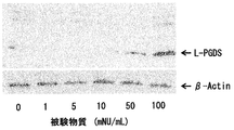
- FIG. 3 is an electrophoretogram showing the result of examining the expression level of L-PGDS protein of cultured iSC to which a test substance was added by western blotting.
- FIG. 1 is a graph showing the results of examining the expression level of L-PGDS gene (PTGDS) of cultured iSC to which a test substance was added by real-time RT-PCR.
- FIG. 2 is an electrophoretogram showing the results of examining the expression amount of L-PGDS gene (PTGDS) of cultured iSC to which a test substance was added by a
- FIG. 4 is a PVDF membrane which shows the result of having investigated the expression level of L-PGDS protein of culture
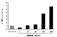
- FIG. 5 is a graph showing the results of examining the expression level of L-PGDS protein of cultured iSC to which a test substance was added by ELISA.
- FIG. 6 is a graph showing the results of liquid chromatography-mass spectrometry examining the amount of prostaglandins in the cell extract of cultured iSC to which a test substance has been added.
- FIG. 7 is a graph showing the results of examining the amount of reaction product by liquid chromatography-mass spectrometry after addition of a substrate for L-PGDS to the culture supernatant of culture iSC to which a test substance has been added.
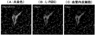
- FIG. 8 is an image showing the results of comparison of the distribution of L-PGDS, pericytes and vascular endothelial cells in the brain of mice subjected to ischemic load by immunohistochemical staining.
- FIG. 9 is an image showing the results of observation of distribution of L-PGDS in the brain of a mouse subjected to ischemia load by immunoelectron microscopy.
- FIG. 10 is an image showing the results of comparison of the distribution of L-PGDS and neural stem cell marker nestin in cultured iSC extracted from human cerebral infarct by immunohistochemical staining.
- FIG. 11 is an image showing the results of examination of amyloid ⁇ deposited in the brain of an Alzheimer type dementia model mouse administered with a test substance by immunohistochemical staining.
- FIG. 12 is an image showing the results of examination of L-PGDS in the brain of an Alzheimer type dementia model mouse to which a test substance has been administered by immunohistochemical staining.
- FIG. 13 is an image showing the results of comparison of the distribution of L-PGDS and vascular endothelial cells in the brains of Alzheimer's disease model mice treated with the test substance by immunohistochemical staining.
- FIG. 11 is an image showing the results of examination of amyloid ⁇ deposited in the brain of an Alzheimer type dementia model mouse administered with a test substance by immunohistochemical staining.
- FIG. 12 is an image showing the results of
- FIG. 14 is an electrophoretogram showing the results of examining the expression levels of amyloid ⁇ and L-PGDS protein in the brains of Alzheimer's disease model mice treated with a test substance by Western blotting.
- FIG. 15 is an electrophoresis diagram showing the results of examining the expression amount of L-PGDS gene (PTGDS) of cultured pericytes to which a test substance was added by a classical RT-PCR method.
- PTGDS L-PGDS gene
- This extract is an extract containing non-protein active substance extracted and separated from inflamed tissue of an animal inoculated with vaccinia virus.
- the extract is liquid in the extracted state, but can be made solid by drying.
- the present formulation is very useful as a pharmaceutical.
- a specific product that the applicant manufactures and sells in Japan as the present preparation is “vaccinia virus-inoculated rabbit skin inflammation-containing extract-containing preparation” (trade name: Neurotropin / NEUROTROPIN®) (hereinafter referred to as “neurotropin”)
- Neurotropin includes an injection and a tablet, both of which are medicinal drugs (ethical drugs).
- Indications for injections of neurotropin include: “Back pain, neck-and-shoulder arm syndrome, symptomatic neuralgia, pruritus associated with skin disease (eczema, dermatitis, urticaria), allergic rhinitis, cold feeling of semen (SMON) Anomalous perception and pain.
- Indications for neurotropin tablets are "postherpetic neuralgia, lumbago, cervico-shoulder-arm syndrome, shoulder arthritis, osteoarthritis”. This formulation was created by the applicant and developed as a pharmaceutical product, has been evaluated for its superiority in efficacy and safety, has been sold for many years, and has established a solid position in the Japanese pharmaceutical market It is a thing.
- the vaccinia virus-inoculated inflamed tissue extract is inoculated with vaccinia virus to crush the inflamed inflamed tissue, an extraction solvent is added to remove tissue fragments, and then deproteinized and adsorbed onto the adsorbent And then eluting the active ingredient. That is, for example, the following steps.
- (A) Collect skin tissues such as rabbits and mice inoculated with vaccinia virus and develop them, crush the sputum tissue, and add extraction solvents such as water, phenol water, physiological saline or phenol-supplemented water Then, the extract (filtrate or supernatant) is obtained by filtration or centrifugation.
- the extract is adjusted to an acidic pH, heated, and deproteinized.
- the deproteinized solution is then adjusted to alkaline and heated, and then filtered or centrifuged.
- C The obtained filtrate or supernatant is acidified and adsorbed onto an adsorbent such as activated carbon or kaolin.
- D An extraction solvent such as water is added to the adsorbent, the pH is adjusted to alkaline, and the adsorbed component is eluted to obtain an extract from inflamed tissue inoculated with vaccinia virus. Thereafter, if desired, the eluate can be evaporated to dryness by evaporation or lyophilization, as appropriate, under reduced pressure.
- vaccinia virus such as rabbits, cattle, horses, sheep, goats, monkeys, rats and mice can be used as animals for inoculating vaccinia virus and obtaining inflamed tissues, and rabbits can be used as inflamed tissues.
- Inflamed skin tissue is preferred.
- the rabbit may be any rabbit which belongs to the order of the rabbit. Examples include rabbits, chi rabbits (domestic rabbits domesticated), hares (Japanese hares), pikas, snow rabbits and the like. Of these, silkworms are preferred for use.
- rabbit in Japan, there is a so-called rabbit (fowl rabbit) which has been bred from the past and frequently used as livestock or experimental animals, and this is also another name of the silk rabbit.
- the vaccinia virus may be of any strain. Examples include Lister, Dairen, Ikeda, EM-63, New York City Board of Health, and the like.
- Step (A) Infected skin tissue obtained by intravaginal inoculation of vaccinia virus in rabbit skin is collected. The collected skin tissue is washed and disinfected with a phenol solution or the like. The inflamed skin tissue is broken and 1 to 5 times its volume of extraction solvent is added.
- the term "crushing” means crushing into a minced form using a mincing machine or the like.
- the extraction solvent distilled water, physiological saline, weakly acidic to weakly basic buffer solution, etc.
- Step (B) The crude extract obtained in step (A) is deproteinized.
- Deproteinization can be carried out by a known method which is usually carried out, and heat treatment, treatment with a protein denaturing agent (eg, acid, base, organic solvent such as urea, guanidine, acetone, etc.), isoelectric precipitation, salting out Etc. can be applied.
- a protein denaturing agent eg, acid, base, organic solvent such as urea, guanidine, acetone, etc.
- Step (C) The filtrate or supernatant obtained in step (B) is adjusted to acidity, preferably pH 3.5 to 5.5, and the adsorption operation to the adsorbent is carried out.
- adsorbents include activated carbon and kaolin.
- the adsorbent is added to the extract and stirred, or the extract is passed through an adsorbent-packed column to make the adsorbent effective. It can be adsorbed.
- an adsorbent is added to the extract solution, the solution is removed by filtration, centrifugation or the like to obtain an adsorbent on which the active component is adsorbed.
- Step (D) In order to elute (desorb) the active ingredient from the adsorbent obtained in step (C), a solvent for elution is added to the adsorbent to adjust the pH to preferably 9 to 12, preferably at room temperature. Alternatively, elution is performed by heating or stirring as appropriate, and the adsorbent is removed by a conventional method such as filtration or centrifugation.
- a basic solvent such as water adjusted to a basic pH, methanol, ethanol, isopropanol etc. or a suitable mixed solution thereof can be used, preferably water adjusted to a pH 9 to 12 Can be used.
- the amount of elution solvent can be set appropriately.
- the pH is appropriately adjusted to around neutrality, etc., and finally the vaccinia virus-inoculated rabbit inflammation skin extract (this extract) is obtained.
- the present extract is liquid at the time it is made, it can be made to have a desired concentration by concentration and dilution as appropriate.
- a preparation is produced from the present extract, it is preferable to carry out a heat sterilization treatment.
- sodium chloride and the like can be added to prepare an isotonic solution with physiological saline.
- oral administration can be performed in a liquid or gel state, a solid preparation for oral use such as tablets can also be produced by subjecting the extract to appropriate procedures such as concentration to dryness. Specific methods for producing such solid preparations for oral use from the present extract are described in the specifications of Japanese Patent No. 3818657 and No. 488379. Injections, oral preparations and the like thus obtained are examples of this preparation.
- the method of administration to a patient is not particularly limited, and can be appropriately selected according to the therapeutic purpose.
- the dosage can be appropriately set according to the type of vaccinia virus-inoculated inflamed tissue extract.
- the dosages approved for the marketed preparations are basically indicated as a medical drug to administer 16 NU daily for internal use and 3.6-7.2 NU daily for injection, the type of disease, It can be increased or decreased depending on the severity, individual difference of patients, administration method, administration period etc. (NU: Neurotropin unit.
- Neurotropin unit is SART stress mouse which is a chronic stress animal whose pain threshold is lower than that of normal animal) The test is conducted according to the modified Randall-Selitto method, and the ED50 value of analgesic efficacy is defined, wherein 1NU is an activity showing 1 mg of the analgesic active ingredient of the neurotropin preparation when the ED50 value is 100 mg / kg).
- Example 1 Production of the present extract Vaccinia virus was intradermally inoculated into the skin of a healthy mature rabbit, and the skin that developed was excised and collected. The collected skin is washed and disinfected with a phenol solution, excess phenol solution is removed, crushed, the phenol solution is added and mixed, left for 3 to 7 days, and then stirred for another 3 to 4 days It was warmed to 35-40 ° C. Thereafter, the extract obtained by solid-liquid separation was adjusted to pH 4.5-5.2 with hydrochloric acid, heat-treated at 90-100 ° C. for 30 minutes, and filtered to remove protein. Furthermore, the filtrate was adjusted to pH 9.0 to 9.5 with sodium hydroxide, heat-treated at 90 to 100 ° C. for 15 minutes, and then solid-liquid separated.
- the resulting deproteinized solution was adjusted to pH 4.0-4.3 with hydrochloric acid, activated carbon of 2% of the deproteinized solution weight was added, and stirred for 2 hours, followed by solid-liquid separation. Water was added to the collected activated carbon, adjusted to pH 9.5-10 with sodium hydroxide, stirred at 60 ° C. for 90 to 100 minutes, and centrifuged to obtain a supernatant. Water was again added to the activated carbon precipitated by centrifugation, adjusted to pH 10.5-11 with sodium hydroxide, stirred at 60 ° C. for 90 to 100 minutes, and centrifuged to obtain a supernatant. Both supernatants were combined and neutralized with hydrochloric acid to obtain the present extract.
- Example 2 Pharmacological Test Next, methods and results of a pharmacological test relating to the L-PGDS production promoting action using the extract obtained in Example 1 as a test substance will be shown.
- introduction of cerebral infarction to CB-17 mice and isolation culture of iSC from the cerebral infarction are described in Nakagomi, T. et al. Eur. J. Neurosci., 29, It carried out according to the method as described in 1842_1852, 2009.
- Test Example 1 Comprehensive gene expression analysis in a test substance treated iSC An ischemic load due to middle cerebral artery occlusion was applied to CB-17 mice (3 mice), and 3 strains of iSC were isolated from the infarct on the 3rd day.
- iSC Dulbecco's modified Eagle's medium F12 supplemented with 2% fetal bovine serum (FBS), 20 ng / mL fibroblast growth factor (FBS), 20 ng / mL epidermal growth factor (EGF) and 1% N2 supplement
- Test substances 50, 1000 mNU / mL or physiological saline (control) to 5% FBS DMEM / F12 F / E / N), 5 ⁇ 10 4 cells / 3 cm ⁇ dish) (Registered trademark, the same applies hereinafter)
- Test Example 2-1 Evaluation for L-PGDS gene expression (real-time RT-PCR method) Similar to Test Example 1, a test substance (50, 500 mNU / mL) or physiological saline (control) was added to cultured iSC (FBS-free DMEM / F12 F / ⁇ / ⁇ , 5 ⁇ 10 4 cells / 3 cm ⁇ dish) After culturing for 7 days, RNA was recovered from Isogen II (registered trademark) according to the manufacturer's manual (Nippon Gene Co., Ltd.).
- RNA purity is assayed using absorbance at 260 nm and 280 nm as an index
- reverse transcription reaction with SuperScript (registered trademark, similarly as below) IV RTase (Invitrogen) is performed in the presence of random primers, and single-stranded to total RNA cDNA was obtained.
- transcripts were quantified using the housekeeping gene ⁇ -Actin as a standard by the quantitative PCR method (Prism (registered trademark) 7900HT, Applied Biosystems) using PTGDS-specific primers (threshold recycling) Comparative method).
- sequences of the used primers for PTGDS and ⁇ -Actin are as follows [PTGDS: 5'-gactctgaaggacgagctgaag-3 '(SEQ ID NO: 1), 5'-tcttgaatgcacttatccggttgg-3' (SEQ ID NO: 2), -Actin: 5'-tacagcttccaccaccaca gc-3 '(SEQ ID NO: 3), 5'-aaggaaggctggaaaagagc-3' (SEQ ID NO: 4)].
- the result is shown by the expression amount ratio of PTGDS to-Actin when control is 1.
- An example of the results is shown in Table 2 and FIG.
- test substance promotes the expression of L-PGDS gene (PTGDS) in a dose-dependent manner in iSC, similarly to the results shown in Test Example 1.
- Test Example 2-2 Evaluation for L-PGDS gene expression (classical RT-PCR method) A medium containing FGF (DMEM / F12 F /-/-) or a medium containing no FGF in the presence of each test substance at a dose of 0 (control; saline), 1, 5, 50, 100 mNU / mL The iSCs were cultured for 4 days in DMEM / F12-/-/-). The total RNA recovered by the RNeasy Mini Kit (QIAGEN) was subjected to classical RT-PCR using the SuperScript III One-Step RT-PCR System with Platinum (Invitrogen) for the L-PGDS gene (PTGDS). The expression level was semi-quantified (35 cycles).
- the PCR products were separated by 2% agarose gel electrophoresis and the bands of PTGDS and GAPDH were detected visually by staining with ethidium bromide.
- the sequences of the used primers for PTGDS and GAPDH are as follows [PTGDS: 5'-cctccaactcaagctggttc-3 '(SEQ ID NO: 5), 5'- atagttggcctccaccactg-3' (SEQ ID NO: 6), GAPDH: 5 '-atcactgccacccagaagac-3' (SEQ ID NO: 7), 5'- cacattggggggtaggaacac-3 '(SEQ ID NO: 8)].
- An example of the result is shown in FIG.
- the L-PGDS gene (PTGDS) expression promoting action in iSC was examined at a smaller dose of the test substance. As shown in FIG. 2, the test substance exhibited the L-PGDS gene regardless of the presence or absence of FGF in the medium. The expression level of was increased in a dose-dependent manner.
- the homogenate is separated by SDS-PAGE (BIO-RAD AnykD (trademark)), polyvinylidene fluoride (PVDF) membrane (Immun-Blot PVDF membrane, BIO-RAD) After transcription to B, blocking with Bloking One (Nacalai Tesque, Inc.), L-PGDS (anti-Prostaglandin D Synthase (Lipocalin) antibody [EP12357], Abcam, 1: 2000 by Western blotting using a specific antibody. And ⁇ -Actin (Monoclonal anti- ⁇ -Actin [A1978], Sigma, 1: 100000) were detected. A highly sensitive chemiluminescence method (Chemi-Lumi One L, nacalai tesque) was used for detection. An example of the result is shown in FIG.
- test substance promotes the expression of L-PGDS protein in iSC cells.
- Test Example 3-2 Evaluation of L-PGDS Protein Expression 500 ⁇ L of culture supernatant of iSC cultured (DMEM / F12 F / ⁇ / ⁇ , 5 ⁇ 10 4 cells / 3 cm ⁇ dish) for 4 days with the test substance (0, 1, 10, 50, 100 mNU / mL) was used as a PVDF membrane (Immun) -Spot on Blot PVDF membrane, BIO-RAD, and block with Blocking One (Nacalai Tesque), and then anti-L-PGDS antibody (anti-Prostaglandin D Synthase (Lipocalin) antibody [EP12357], Abcam, 1: 2000 L-PGDS was detected by the dot blotting method using. An example of the result is shown in FIG.
- L-PGDS is considered to be secreted extracellularly since it has a typical secretory signal sequence and signal peptidase recognition sequence at its N-terminus. Therefore, when the L-PGDS protein in the culture supernatant of iSC to which the test substance was added was measured, it is apparent from FIG. 4 that the amount of L-PGDS protein in the culture supernatant (extracellular) was also added to the test substance. It was confirmed to increase depending on the dose.
- Test Example 3-3 Evaluation of L-PGDS Protein Expression The supernatant of iSC culture (DMEM / F12 F / ⁇ / ⁇ , 5 ⁇ 10 4 cells / 3 cm ⁇ dish) treated with the test substance (0 [control], 1, 10, 50, 100, 1000 mNU / mL) for 4 days It was collected.
- L-PGDS is an enzyme (EC 5.399.2) that biosynthesizes PGD2 using PGH2 as a substrate.
- iSC for the purpose of confirming that L-PGDS promoted by the test substance further produces PGD2 by its enzyme activity, A: intracellular and B: extracellular of PGD 2 and its metabolite, in iSC
- prostaglandins such as PGE2 produced using PGH2 as a substrate were comprehensively analyzed.
- A Evaluation of iSC intracellular enzyme activity Cultured for 3 days by adding a test substance (0, 10, 50 mNU / mL) (FBS without DMEM / F12 F /-/-, 5 ⁇ 10 4 cells / 3 cm ⁇ dish) Cell extracts were prepared from the treated iSCs by RIPA buffer treatment (4 ° C., 20 min). Determine the concentration of prostaglandins (PGD2, PGJ2, 15-deoxy- ⁇ 12,14-PGJ2, 13,14-dihydro-15-keto-PGD2 and PGE2) in the intracellular extract by HPLC mass spectrometry did.
- the intracellular level of PGD2 and its nonenzymatic metabolite PGJ2 increased in a dose-dependent manner by the addition of the test substance. Furthermore, the addition of the test substance at 50 mNU / mL also confirmed the production of 15-deoxy- ⁇ 12,14-PGJ2, which is a non-enzymatic metabolite of PGJ2. In addition, 13,14-dihydro-15-keto-PGD2, which is a non-enzymatic metabolite of 15-deoxy- ⁇ 12,14-PGJ2, was below the detection limit. On the other hand, for PGE2 produced from the substrate PGH2 common to PGD2, no dose-dependent production response could be observed.
- the concentration of prostaglandins (PGD2, PGJ2, 15-deoxy-.DELTA.12,14-PGJ2, 13,14-dihydro-15-keto-PGD2 and PGE2) in the reaction solution was determined by HPLC mass spectrometry. Each was measured. An example of a result is shown in Table 5 and FIG.
- the test substance increased the concentration of PGD2 and its non-enzymatic metabolite PGJ2, 15-deoxy- ⁇ 12, 14-PGJ2 in the reaction solution at a concentration of 100 mNU / mL or more This confirms that L-PGDS activity is present in the iSC culture supernatant. Also, for 13,14-dihydro-15-keto-PGD2, which is a non-enzymatic metabolite of 15-deoxy- ⁇ 12,14-PGJ2, it was dose-dependent by the addition of the test substance in the concentration range up to 100 mNU / mL Although production was observed, it was below the detection limit at 1000 mNU / mL.
- L-PGDS is known to be a secretory enzyme. And B. From the results in, it was confirmed that iSC released L-PGDS having enzyme activity out of cells, and addition of the test substance promoted its production and release.
- Test example 5-1 Examination of L-PGDS producing cells in mouse brain (immunohistochemical staining method) L-PGDS (panel B, green; anti-prostaglandin D synthase (lipocalin) antibody [EP12357] were prepared from brain sections of CB-17 mice (3 days after ischemia) subjected to ischemic load due to middle cerebral artery occlusion.
- Abcam 1: 1000
- pericyte marker ⁇ -SMA panel C, red
- anti-actin smooth muscle
- clone ASM-1 smooth muscle
- Millipore 1: 1000
- vascular endothelial cell marker CD31 panel D, red
- Immunohistochemical examination by confocal laser microscopy was performed using a specific antibody against anti-mouse CD31 (PECAM-1) monoclonal antibody, 550274, BD Pharmingen (1: 1000).
- L-PGDS panel B
- pericyte marker panel C
- L-PGDS panel B
- vascular endothelial cell marker panel D
- the images were detected and superimposed to compare the distribution of L-PGDS and pericyte markers or L-PGDS and vascular endothelial cell markers (panel A1, panel A2).
- An example of the result is shown in FIG.
- the green fluorescence of panel B shows the distribution of L-PGDS
- the red fluorescence of panel C shows the distribution of perisite markers, but in panel A1 in which both are superimposed, L-PGDS (green fluorescence) Partially coincided with the distribution of perisite marker (red fluorescence) (200 times).
- L-PGDS green fluorescence
- L-PGDS is a protein mainly distributed in the central nervous system, is present in the arachnoid membrane, the pia mater and the choroid plexus of the lateral ventricle, and is thought to be secreted in cerebrospinal fluid (Urade Y. et. al., J Lipid Mediat. Cell Signal. 14, 71-82, 1996), but the cells produced in the brain have not been sufficiently elucidated.
- the inventors examined the distribution of L-PGDS in the brain by immunohistochemical method, and the expression in the normal brain of CB-17 mice was weak, but in the above Test Example 4-1 When permanent ligation of the middle cerebral artery was introduced into this mouse, remarkable L-PGDS expression was observed in cerebral infarction (FIG.
- Test Example 5-2 Examination of L-PGDS-producing cells in mouse brain (immune electron microscopy) Brain sections of CB-17 mice (3 days after ischemia) subjected to ischemic load due to middle cerebral artery occlusion were prepared, and L-PGDS (anti-prostaglandin D synthase (lipocalin) antibody [EP12357], Abcam, 1: The expression site of L-PGDS was observed by immunoelectron microscopy using a specific antibody against 1000).
- a 2 ⁇ m thick brain section is prepared in a vibratome, and after reaction using avidin-biotin horseradish peroxidase (HRP) complex kit (Vector Laboratories) and 3,3'-diaminobenzenedehydrochloride (DAB), osmium treatment is performed After Epon-embedding, ultra-thin sections were prepared for electron microscopic observation. An example of the result is shown in FIG.
- HRP horseradish peroxidase
- DAB 3,3'-diaminobenzenedehydrochloride
- Test Example 6 Examination of L-PGDS-producing cells in human brain (immunohistochemical staining method) According to the method described in Tatebayashi K. et al., Stem Cells and Develop. 26, 787-797, 2017, L-PGDS (panel B, green color) in cultured iSC extracted from human cerebral infarction (necrotic tissue) Specific antibody against the anti-prostaglandin D synthase (lipocalin) antibody [EP12357], Abcam, 1: 1000) and the neural stem cell marker nestin (panel C, red; anti-nestin, clone 10C2, Millipore, 1: 1000) We performed immunohistochemical examination using.
- the cells were reacted with Alexa Fluor (registered trademark, hereinafter the same) 488-conjugated antibody or Alexa Fluor 555-conjugated antibody (1: 500; Molecular Probes, Eugene) and then reacted with 4 ', 6-diamidino-2-phenylindole (DAPI; 1).
- Alexa Fluor registered trademark, hereinafter the same
- DAPI 6-diamidino-2-phenylindole
- the nuclear staining was performed by Kirkegaard & Perry Laboratories.
- the fluorescence was observed with a fluorescent microscope (BX60; Olympus, Japan), the images of L-PGDS (panel B) and nestin (panel C) were superimposed, and the distributions of L-PGDS and nestin were compared (panel A).
- the green fluorescence of panel B shows the distribution of L-PGDS
- the red fluorescence of panel C shows the distribution of nesitin
- L-PGDS green fluorescence
- nestin green fluorescence
- red fluorescence 200 times
- the improvement effect of the cognitive ability by the test substance administration group was observed, and this action was statistically significant among the groups.
- brains were collected by decapitation under anesthesia and the left hemisphere was rapidly frozen in a liquid nitrogen bath.
- the remaining right hemisphere contains (A) glutaraldehyde-containing fixative (0.05% glutaraldehyde, 4% paraformaldehyde, 0.1 M phosphate buffer) or (B) PLP fixative (0.01 M metaperiodine). (At 4 ° C.) with sodium acid, 0.075 M lysine, 2% paraformaldehyde).
- Coronal plane sections (20 ⁇ m in thickness) including the cerebral cortex were prepared from fixed tissue samples by a cryostat (Leica CM1850).
- Amyloid ⁇ was subjected to nuclear staining with 4 ', 6-diamidino-2-phenylindole (DAPI; 1: 1000; Kirkegaard & Perry Laboratories) after reaction with Alexa Fluor 488-conjugated antibody (1: 500; Molecular Probes, Eugene) The fluorescence was observed with a fluorescent microscope (BX60; Olympus, Japan). An example of the result is shown in FIG.
- L-PGDS anti-prostaglandin D synthetase (lipocalin) antibody [EP12357], Abcam, 1: 1000
- vascular endothelial cell marker CD31 vascular endothelial cell marker CD31 (coral) in the coronal section of the test substance administration group prepared in (1) above.
- Immunohistochemical examination was conducted using a specific antibody against anti-mouse CD31 (PECAM-1) monoclonal antibody, 550274, BD Pharmingen (1: 1000).
- L-PGDS green fluorescence
- CD31 red fluorescence
- the amount of L-PGDS in the brain is increased compared to that in the control group, so the test substance enhances the production of L-PGDS in the brain. It was recognized that From the series of results of Test Example 7 above, it is considered that the test substance may inhibit the deposition of amyloid ⁇ in the brain by increasing the amount of L-PGDS expressed in the brain, and thus may improve the cognitive function.
- Test Example 8 Examination of L-PGDS expression promoting factor in human-derived pericyte (classical RT-PCR method) As described above, since iSCs that show responsiveness to a test substance are considered to be derived from brain pericytes, different oxygen and / or different oxygen concentrations are obtained in the commercially available human pericyte strain Human brain vascular pericytes (ScienCell). Under the condition of glucose concentration, the L-PGDS expression ability of the test substance was examined.
- the PCR products were separated by 2% agarose gel electrophoresis and the PTGDS and ⁇ -Actin bands were detected visually by staining with ethidium bromide.
- the primers for PTGDS and ⁇ -Actin used were the same as those used in Test Example 2-1 above. An example of the result is shown in FIG.
- L-PGDS is mainly expressed in the brain, plays a role as binding, transport and scavenging of various hydrophobic small molecules, and is a protein having various functions such as brain environment regulation / brain protection function and sleep regulation function.
- the L-PGDS production promoter of the present invention prevents or treats cerebrovascular disorders such as cerebral infarction, dementias such as Alzheimer's disease, diseases involving L-PGDS such as insomnia, etc. It is useful as a drug for preventing recurrence.
- the present extract and a preparation containing the present extract is an excellent L-PGDS production promoter and is highly useful as a highly safe drug with few problems such as side effects.
- L-PGDS production promoting activity in pericyte or iSC dedifferentiated from pericyte is used as an indicator, a substance useful for prevention / treatment or recurrence prevention of a disease in which L-PGDS is involved, especially brain protection action or sleep action
- the method of the present invention for screening substances is a very useful method that contributes to the development of new therapeutic agents.
Landscapes
- Health & Medical Sciences (AREA)
- Life Sciences & Earth Sciences (AREA)
- Engineering & Computer Science (AREA)
- Chemical & Material Sciences (AREA)
- Biomedical Technology (AREA)
- General Health & Medical Sciences (AREA)
- Medicinal Chemistry (AREA)
- Pharmacology & Pharmacy (AREA)
- Bioinformatics & Cheminformatics (AREA)
- Veterinary Medicine (AREA)
- Public Health (AREA)
- Animal Behavior & Ethology (AREA)
- Immunology (AREA)
- Cell Biology (AREA)
- Organic Chemistry (AREA)
- Chemical Kinetics & Catalysis (AREA)
- Neurosurgery (AREA)
- Neurology (AREA)
- General Chemical & Material Sciences (AREA)
- Nuclear Medicine, Radiotherapy & Molecular Imaging (AREA)
- Biotechnology (AREA)
- Molecular Biology (AREA)
- Urology & Nephrology (AREA)
- Epidemiology (AREA)
- Zoology (AREA)
- Developmental Biology & Embryology (AREA)
- Virology (AREA)
- Hematology (AREA)
- Microbiology (AREA)
- Biochemistry (AREA)
- Analytical Chemistry (AREA)
- Physics & Mathematics (AREA)
- Food Science & Technology (AREA)
- Pathology (AREA)
- General Physics & Mathematics (AREA)
- Dermatology (AREA)
- Anesthesiology (AREA)
- Psychiatry (AREA)
- Hospice & Palliative Care (AREA)
- Vascular Medicine (AREA)
Abstract
Priority Applications (9)
| Application Number | Priority Date | Filing Date | Title |
|---|---|---|---|
| CN202310272473.6A CN116270743A (zh) | 2017-12-28 | 2018-12-27 | 脂质运载蛋白型前列腺素d2合成酶产生促进剂 |
| EP18895831.8A EP3733205A4 (fr) | 2017-12-28 | 2018-12-27 | Agent d'accélération de la production de prostaglandine d2 synthase de type lipocaline |
| CN201880084260.8A CN111511403A (zh) | 2017-12-28 | 2018-12-27 | 脂质运载蛋白型前列腺素d2合成酶产生促进剂 |
| CA3087018A CA3087018A1 (fr) | 2017-12-28 | 2018-12-27 | Agent d'acceleration de la production de prostaglandine d2 synthase de type lipocaline |
| JP2019530517A JP6607655B1 (ja) | 2017-12-28 | 2018-12-27 | リポカリン型プロスタグランジンd2合成酵素産生促進剤 |
| AU2018394182A AU2018394182A1 (en) | 2017-12-28 | 2018-12-27 | Lipocalin-type prostaglandin D2 synthase production accelerating agent |
| US16/958,962 US20200338135A1 (en) | 2017-12-28 | 2018-12-27 | Lipocalin-type prostaglandin d2 synthase production promoting agent |
| KR1020207015608A KR20200103647A (ko) | 2017-12-28 | 2018-12-27 | 리포칼린형 프로스타글란딘 d2 합성 효소 산생 촉진제 |
| US18/355,925 US20240000853A1 (en) | 2017-12-28 | 2023-07-20 | Lipocalin-type prostaglandin d2 synthase production promoting agent |
Applications Claiming Priority (2)
| Application Number | Priority Date | Filing Date | Title |
|---|---|---|---|
| JP2017-253170 | 2017-12-28 | ||
| JP2017253170 | 2017-12-28 |
Related Child Applications (2)
| Application Number | Title | Priority Date | Filing Date |
|---|---|---|---|
| US16/958,962 A-371-Of-International US20200338135A1 (en) | 2017-12-28 | 2018-12-27 | Lipocalin-type prostaglandin d2 synthase production promoting agent |
| US18/355,925 Division US20240000853A1 (en) | 2017-12-28 | 2023-07-20 | Lipocalin-type prostaglandin d2 synthase production promoting agent |
Publications (1)
| Publication Number | Publication Date |
|---|---|
| WO2019131879A1 true WO2019131879A1 (fr) | 2019-07-04 |
Family
ID=67067534
Family Applications (1)
| Application Number | Title | Priority Date | Filing Date |
|---|---|---|---|
| PCT/JP2018/048141 WO2019131879A1 (fr) | 2017-12-28 | 2018-12-27 | Agent d'accélération de la production de prostaglandine d2 synthase de type lipocaline |
Country Status (9)
| Country | Link |
|---|---|
| US (2) | US20200338135A1 (fr) |
| EP (1) | EP3733205A4 (fr) |
| JP (2) | JP6607655B1 (fr) |
| KR (1) | KR20200103647A (fr) |
| CN (2) | CN111511403A (fr) |
| AU (1) | AU2018394182A1 (fr) |
| CA (1) | CA3087018A1 (fr) |
| TW (1) | TWI814760B (fr) |
| WO (1) | WO2019131879A1 (fr) |
Cited By (1)
| Publication number | Priority date | Publication date | Assignee | Title |
|---|---|---|---|---|
| JP2022521125A (ja) * | 2019-01-30 | 2022-04-06 | ジュン リウ | 脳内の炎症の抑制又は軽減剤 |
Families Citing this family (1)
| Publication number | Priority date | Publication date | Assignee | Title |
|---|---|---|---|---|
| WO2023184470A1 (fr) * | 2022-04-01 | 2023-10-05 | 星相生物技术有限公司 | Utilisation d'extraits de peau de lapin enflammée par le virus de la vaccine dans le traitement de la maladie d'alzheimer |
Citations (9)
| Publication number | Priority date | Publication date | Assignee | Title |
|---|---|---|---|---|
| JPS6479657A (en) * | 1987-09-22 | 1989-03-24 | Nippon Zoki Pharmaceutical Co | Vital decision of medicine |
| JP2000016942A (ja) | 1998-04-27 | 2000-01-18 | Nippon Zoki Pharmaceut Co Ltd | 虚血性疾患治療剤 |
| JP2001103869A (ja) * | 1999-10-06 | 2001-04-17 | Japan Science & Technology Corp | リポカリン型プロスタグランジンd合成酵素トランスジェニック動物とこれを用いた試験方法 |
| JP3818657B2 (ja) | 2004-12-01 | 2006-09-06 | 日本臓器製薬株式会社 | 乾燥物及びその製造方法 |
| JP2007284351A (ja) * | 2004-07-27 | 2007-11-01 | Osaka Bioscience Institute | アミロイド蛋白質の凝集を抑制する物質とその作用 |
| WO2011162317A1 (fr) | 2010-06-25 | 2011-12-29 | 日本臓器製薬株式会社 | Méthode de détermination ou d'évaluation d'une substance d'intérêt |
| JP4883798B2 (ja) | 2005-06-17 | 2012-02-22 | 日本臓器製薬株式会社 | 乾燥物及びその製造法 |
| JP2012508192A (ja) * | 2008-11-11 | 2012-04-05 | バンワールド ファーマシューティカル(ルガオ) カンパニー リミテッド | ワクシニアウイルスによって炎症を起こしたウサギ皮膚由来の抽出物の、急性脳血管疾患の治療用医薬の製造における使用 |
| JP2014076961A (ja) * | 2012-10-10 | 2014-05-01 | Nippon Zoki Pharmaceut Co Ltd | 抽出物及び製剤 |
Family Cites Families (6)
| Publication number | Priority date | Publication date | Assignee | Title |
|---|---|---|---|---|
| AU2729400A (en) * | 1999-01-19 | 2000-08-07 | James Daly | Application of a viral complement inhibitory protein in the treatment and diagnosis of alzheimer's disease |
| JP3752219B2 (ja) * | 2000-07-21 | 2006-03-08 | マルハ株式会社 | 痴呆性疾患の鑑別方法 |
| US20100251394A1 (en) * | 2007-09-20 | 2010-09-30 | The Johns Hopkins University | Treatment and prevention of ischemic brain injury |
| US9835621B2 (en) * | 2011-07-14 | 2017-12-05 | University Of Kentucky Research Foundation | Process for detection of alzheimer's disease from a serum sample |
| CN105163746A (zh) * | 2013-04-30 | 2015-12-16 | 日本脏器制药株式会社 | 提取物和含有该提取物的制剂 |
| CN110381966A (zh) * | 2017-03-06 | 2019-10-25 | 刘军 | Aβ-诱导的损伤的抑制或减轻药剂 |
-
2018
- 2018-12-27 JP JP2019530517A patent/JP6607655B1/ja active Active
- 2018-12-27 AU AU2018394182A patent/AU2018394182A1/en active Pending
- 2018-12-27 US US16/958,962 patent/US20200338135A1/en not_active Abandoned
- 2018-12-27 WO PCT/JP2018/048141 patent/WO2019131879A1/fr unknown
- 2018-12-27 KR KR1020207015608A patent/KR20200103647A/ko unknown
- 2018-12-27 EP EP18895831.8A patent/EP3733205A4/fr active Pending
- 2018-12-27 CN CN201880084260.8A patent/CN111511403A/zh active Pending
- 2018-12-27 CN CN202310272473.6A patent/CN116270743A/zh active Pending
- 2018-12-27 CA CA3087018A patent/CA3087018A1/fr active Pending
- 2018-12-28 TW TW107147748A patent/TWI814760B/zh active
-
2019
- 2019-10-18 JP JP2019190666A patent/JP7411175B2/ja active Active
-
2023
- 2023-07-20 US US18/355,925 patent/US20240000853A1/en active Pending
Patent Citations (9)
| Publication number | Priority date | Publication date | Assignee | Title |
|---|---|---|---|---|
| JPS6479657A (en) * | 1987-09-22 | 1989-03-24 | Nippon Zoki Pharmaceutical Co | Vital decision of medicine |
| JP2000016942A (ja) | 1998-04-27 | 2000-01-18 | Nippon Zoki Pharmaceut Co Ltd | 虚血性疾患治療剤 |
| JP2001103869A (ja) * | 1999-10-06 | 2001-04-17 | Japan Science & Technology Corp | リポカリン型プロスタグランジンd合成酵素トランスジェニック動物とこれを用いた試験方法 |
| JP2007284351A (ja) * | 2004-07-27 | 2007-11-01 | Osaka Bioscience Institute | アミロイド蛋白質の凝集を抑制する物質とその作用 |
| JP3818657B2 (ja) | 2004-12-01 | 2006-09-06 | 日本臓器製薬株式会社 | 乾燥物及びその製造方法 |
| JP4883798B2 (ja) | 2005-06-17 | 2012-02-22 | 日本臓器製薬株式会社 | 乾燥物及びその製造法 |
| JP2012508192A (ja) * | 2008-11-11 | 2012-04-05 | バンワールド ファーマシューティカル(ルガオ) カンパニー リミテッド | ワクシニアウイルスによって炎症を起こしたウサギ皮膚由来の抽出物の、急性脳血管疾患の治療用医薬の製造における使用 |
| WO2011162317A1 (fr) | 2010-06-25 | 2011-12-29 | 日本臓器製薬株式会社 | Méthode de détermination ou d'évaluation d'une substance d'intérêt |
| JP2014076961A (ja) * | 2012-10-10 | 2014-05-01 | Nippon Zoki Pharmaceut Co Ltd | 抽出物及び製剤 |
Non-Patent Citations (16)
| Title |
|---|
| ABDO H. ET AL., J. PHYSIOL., vol. 590, 2012, pages 2739 - 2750 |
| GARCIA-FERNANDEZ LF ET AL.: "Dexamethasone induces lipocalin-type prostaglandin D synthase gene expression in mouse neuronal cells", JOURNAL OF NEUROCHEMISTRY, vol. 75, no. 2, 2000, pages 460 - 470, XP55622513, ISSN: 0022-3042 * |
| INUI T. ET AL., J. CEREB. BLOOD FLOW METAB., vol. 34, 2014, pages 1558 - 1567 |
| KANEKIYO T ET AL.: "Lipocalin-type prostaglandin D synthase/beta-trace is a major amyloid beta-chaperone in human cerebrospinal fluid", PROCEEDINGS OF THE NATIONAL ACADEMY OF SCIENCES OF THE UNITED STATES OF AMERICA, vol. 104, no. 15, 10 April 2007 (2007-04-10), pages 6412 - 6417, XP55622522, ISSN: 0027-8424 * |
| KANEKIYO T. ET AL., PROC. NATL. ACAD. SCI. USA, vol. 104, 2007, pages 6412 - 6417 |
| KIMURA, HIROSHI ET AL.: "A pilot study for clinical application of Neurotropin to senile patients with dementia", JAPANESE PHARMACOLOGY & THERAPEUTICS, vol. 15, no. 10, October 1987 (1987-10-01), pages 4235 - 4251, XP009521902, ISSN: 0386-3603 * |
| NAKAGOMI, T. ET AL., EUR. J. NEUROSCI., vol. 29, 2009, pages 1842 - 1852 |
| SALEEM S ET AL.: "Lipocalin-prostaglandin D synthase is a critical beneficial factor in transient and permanent focal cerebral ischemia", NEUROSCIENCE, vol. 160, no. 1, 2009, pages 248 - 254, XP026036090, ISSN: 0306-4522, DOI: 10.1016/j.neuroscience.2009.02.039 * |
| SALEEM S. ET AL., NEUROSCIENCE, vol. 160, 2009, pages 248 - 254 |
| TANIGICHI H. ET AL., J. NEUROSCI., vol. 27, 2007, pages 4303 - 4312 |
| TANIIKE M. ET AL., J. NEUROSCI., vol. 22, 2002, pages 4885 - 4896 |
| TATEBAYASHI K. ET AL., STEM CELLS AND DEVELOP., vol. 26, 2017, pages 787 - 797 |
| TRIMARCO A. ET AL., NATURE NEUROSCI., vol. 17, 2014, pages 1682 - 1992 |
| UENO R. ET AL., BIOCHEM. BIOPHYS. RES. COMMUN., vol. 109, 1982, pages 576 - 582 |
| URADE Y. ET AL., J LIPID MEDIAT. CELL SIGNAL., vol. 14, 1996, pages 71 - 82 |
| URADE Y.HAYAISHI O., BIOCHIM. BIOPHYS. ACTA, vol. 1482, 2000, pages 259 - 271 |
Cited By (1)
| Publication number | Priority date | Publication date | Assignee | Title |
|---|---|---|---|---|
| JP2022521125A (ja) * | 2019-01-30 | 2022-04-06 | ジュン リウ | 脳内の炎症の抑制又は軽減剤 |
Also Published As
| Publication number | Publication date |
|---|---|
| CN116270743A (zh) | 2023-06-23 |
| CA3087018A1 (fr) | 2019-07-04 |
| JP7411175B2 (ja) | 2024-01-11 |
| TW201932130A (zh) | 2019-08-16 |
| CN111511403A (zh) | 2020-08-07 |
| EP3733205A1 (fr) | 2020-11-04 |
| TWI814760B (zh) | 2023-09-11 |
| JP6607655B1 (ja) | 2019-11-20 |
| JP2020007376A (ja) | 2020-01-16 |
| AU2018394182A1 (en) | 2020-07-16 |
| US20240000853A1 (en) | 2024-01-04 |
| KR20200103647A (ko) | 2020-09-02 |
| US20200338135A1 (en) | 2020-10-29 |
| JPWO2019131879A1 (ja) | 2020-01-16 |
| EP3733205A4 (fr) | 2021-12-08 |
Similar Documents
| Publication | Publication Date | Title |
|---|---|---|
| Zeng et al. | Lentivirus-mediated downregulation of α-synuclein reduces neuroinflammation and promotes functional recovery in rats with spinal cord injury | |
| Han et al. | Dimethyl fumarate attenuates experimental autoimmune neuritis through the nuclear factor erythroid-derived 2-related factor 2/hemoxygenase-1 pathway by altering the balance of M1/M2 macrophages | |
| US20240000853A1 (en) | Lipocalin-type prostaglandin d2 synthase production promoting agent | |
| US11419890B2 (en) | Compositions prepared from poultry and methods of their use | |
| Mannelli et al. | The neuropathy-protective agent acetyl-l-carnitine activates protein kinase C-γ and MAPKs in a rat model of neuropathic pain | |
| Zhang et al. | Carbon monoxide-releasing molecule-3 protects against cortical pyroptosis induced by hemorrhagic shock and resuscitation via mitochondrial regulation | |
| EP2587265B1 (fr) | Méthode de détermination ou d'évaluation d'une substance d'essai | |
| Fu et al. | Pelargonidin ameliorates MCAO-induced cerebral ischemia/reperfusion injury in rats by the action on the Nrf2/HO-1 pathway | |
| Xiao et al. | Reduction of lactoferrin aggravates neuronal ferroptosis after intracerebral hemorrhagic stroke in hyperglycemic mice | |
| Yuan et al. | Neuroprotective effects of nitidine against traumatic CNS injury via inhibiting microglia activation | |
| Chen et al. | Dynamic mobilization of PGC-1α mediates mitochondrial biogenesis for the protection of RGC-5 cells by resveratrol during serum deprivation | |
| Hashimoto et al. | Long-term activation of c-Fos and c-Jun in optic nerve head astrocytes in experimental ocular hypertension in monkeys and after exposure to elevated pressure in vitro | |
| Li et al. | Inhibition of miRNA-21 promotes retinal ganglion cell survival and visual function by modulating Müller cell gliosis after optic nerve crush | |
| Zaidi et al. | Histone deacetylases regulation by δ-opioids in human optic nerve head astrocytes | |
| Zhang et al. | Recombinant osteopontin provides protection for cerebral infarction by inhibiting the NLRP3 inflammasome in microglia | |
| Huang et al. | Targeting integrated stress response regulates microglial M1/M2 polarization and attenuates neuroinflammation following surgical brain injury in rat | |
| US20230193276A1 (en) | Composition of drug targets and method of using thereof | |
| Temizyürek et al. | Blood-brain barrier targeted delivery of lacosamide-conjugated gold nanoparticles: Improving outcomes in absence seizures | |
| WO2020154941A1 (fr) | Agent d'inhibition ou d'atténuation d'une inflammation dans le cerveau | |
| CN109528738A (zh) | 甘草酸在制备促进髓鞘再生抑制神经炎症药物的应用 | |
| Gao et al. | Spatial–temporal expression of NDRG2 in brain tissues in a rat model of intracerebral hemorrhage: A pilot study | |
| JP2022541720A (ja) | 細胞外小胞に含まれるenamptの産生および使用 | |
| Cheng et al. | Novel insights into the protective effects of leonurine against acute kidney injury: Inhibition of ER stress-associated ferroptosis via regulating ATF4/CHOP/ACSL4 pathway | |
| Liang et al. | Dental pulp mesenchymal stem cell-derived exosomes inhibit neuroinflammation and microglial pyroptosis in subarachnoid hemorrhage via the miRNA-197-3p/Foxo3 axis | |
| Gillmann et al. | Neuroprotection and neuroenhancement |
Legal Events
| Date | Code | Title | Description |
|---|---|---|---|
| ENP | Entry into the national phase |
Ref document number: 2019530517 Country of ref document: JP Kind code of ref document: A |
|
| 121 | Ep: the epo has been informed by wipo that ep was designated in this application |
Ref document number: 18895831 Country of ref document: EP Kind code of ref document: A1 |
|
| ENP | Entry into the national phase |
Ref document number: 3087018 Country of ref document: CA |
|
| NENP | Non-entry into the national phase |
Ref country code: DE |
|
| ENP | Entry into the national phase |
Ref document number: 2018394182 Country of ref document: AU Date of ref document: 20181227 Kind code of ref document: A |
|
| ENP | Entry into the national phase |
Ref document number: 2018895831 Country of ref document: EP Effective date: 20200728 |