WO2017104675A1 - Dnaメチル化解析のための試料調製方法 - Google Patents
Dnaメチル化解析のための試料調製方法 Download PDFInfo
- Publication number
- WO2017104675A1 WO2017104675A1 PCT/JP2016/087100 JP2016087100W WO2017104675A1 WO 2017104675 A1 WO2017104675 A1 WO 2017104675A1 JP 2016087100 W JP2016087100 W JP 2016087100W WO 2017104675 A1 WO2017104675 A1 WO 2017104675A1
- Authority
- WO
- WIPO (PCT)
- Prior art keywords
- dna
- sample
- pcr
- buffer
- reaction
- Prior art date
- Legal status (The legal status is an assumption and is not a legal conclusion. Google has not performed a legal analysis and makes no representation as to the accuracy of the status listed.)
- Ceased
Links
Images





Classifications
-
- C—CHEMISTRY; METALLURGY
- C12—BIOCHEMISTRY; BEER; SPIRITS; WINE; VINEGAR; MICROBIOLOGY; ENZYMOLOGY; MUTATION OR GENETIC ENGINEERING
- C12Q—MEASURING OR TESTING PROCESSES INVOLVING ENZYMES, NUCLEIC ACIDS OR MICROORGANISMS; COMPOSITIONS OR TEST PAPERS THEREFOR; PROCESSES OF PREPARING SUCH COMPOSITIONS; CONDITION-RESPONSIVE CONTROL IN MICROBIOLOGICAL OR ENZYMOLOGICAL PROCESSES
- C12Q1/00—Measuring or testing processes involving enzymes, nucleic acids or microorganisms; Compositions therefor; Processes of preparing such compositions
- C12Q1/25—Measuring or testing processes involving enzymes, nucleic acids or microorganisms; Compositions therefor; Processes of preparing such compositions involving enzymes not classifiable in groups C12Q1/26 - C12Q1/66
-
- C—CHEMISTRY; METALLURGY
- C12—BIOCHEMISTRY; BEER; SPIRITS; WINE; VINEGAR; MICROBIOLOGY; ENZYMOLOGY; MUTATION OR GENETIC ENGINEERING
- C12Q—MEASURING OR TESTING PROCESSES INVOLVING ENZYMES, NUCLEIC ACIDS OR MICROORGANISMS; COMPOSITIONS OR TEST PAPERS THEREFOR; PROCESSES OF PREPARING SUCH COMPOSITIONS; CONDITION-RESPONSIVE CONTROL IN MICROBIOLOGICAL OR ENZYMOLOGICAL PROCESSES
- C12Q1/00—Measuring or testing processes involving enzymes, nucleic acids or microorganisms; Compositions therefor; Processes of preparing such compositions
- C12Q1/34—Measuring or testing processes involving enzymes, nucleic acids or microorganisms; Compositions therefor; Processes of preparing such compositions involving hydrolase
- C12Q1/42—Measuring or testing processes involving enzymes, nucleic acids or microorganisms; Compositions therefor; Processes of preparing such compositions involving hydrolase involving phosphatase
-
- C—CHEMISTRY; METALLURGY
- C12—BIOCHEMISTRY; BEER; SPIRITS; WINE; VINEGAR; MICROBIOLOGY; ENZYMOLOGY; MUTATION OR GENETIC ENGINEERING
- C12Q—MEASURING OR TESTING PROCESSES INVOLVING ENZYMES, NUCLEIC ACIDS OR MICROORGANISMS; COMPOSITIONS OR TEST PAPERS THEREFOR; PROCESSES OF PREPARING SUCH COMPOSITIONS; CONDITION-RESPONSIVE CONTROL IN MICROBIOLOGICAL OR ENZYMOLOGICAL PROCESSES
- C12Q1/00—Measuring or testing processes involving enzymes, nucleic acids or microorganisms; Compositions therefor; Processes of preparing such compositions
- C12Q1/48—Measuring or testing processes involving enzymes, nucleic acids or microorganisms; Compositions therefor; Processes of preparing such compositions involving transferase
-
- C—CHEMISTRY; METALLURGY
- C12—BIOCHEMISTRY; BEER; SPIRITS; WINE; VINEGAR; MICROBIOLOGY; ENZYMOLOGY; MUTATION OR GENETIC ENGINEERING
- C12Q—MEASURING OR TESTING PROCESSES INVOLVING ENZYMES, NUCLEIC ACIDS OR MICROORGANISMS; COMPOSITIONS OR TEST PAPERS THEREFOR; PROCESSES OF PREPARING SUCH COMPOSITIONS; CONDITION-RESPONSIVE CONTROL IN MICROBIOLOGICAL OR ENZYMOLOGICAL PROCESSES
- C12Q1/00—Measuring or testing processes involving enzymes, nucleic acids or microorganisms; Compositions therefor; Processes of preparing such compositions
- C12Q1/68—Measuring or testing processes involving enzymes, nucleic acids or microorganisms; Compositions therefor; Processes of preparing such compositions involving nucleic acids
-
- C—CHEMISTRY; METALLURGY
- C12—BIOCHEMISTRY; BEER; SPIRITS; WINE; VINEGAR; MICROBIOLOGY; ENZYMOLOGY; MUTATION OR GENETIC ENGINEERING
- C12N—MICROORGANISMS OR ENZYMES; COMPOSITIONS THEREOF; PROPAGATING, PRESERVING, OR MAINTAINING MICROORGANISMS; MUTATION OR GENETIC ENGINEERING; CULTURE MEDIA
- C12N15/00—Mutation or genetic engineering; DNA or RNA concerning genetic engineering, vectors, e.g. plasmids, or their isolation, preparation or purification; Use of hosts therefor
- C12N15/09—Recombinant DNA-technology
Definitions
- the present invention relates to an improved DNA methylation analysis method. Specifically, the present invention relates to a method for increasing the yield of a DNA sample capable of PCR amplification after bisulfite treatment in DNA methylation analysis using bisulfite.
- DNA methylation is one of epigenetic mechanisms and is deeply involved in embryogenesis, cell differentiation, phenotypic differences, various diseases, and the like.
- various methods for DNA methylation analysis have been developed and used.
- DNA methylation analysis has also been used to select iPS cells that are suitable for the purpose.
- a method using bisulfite treatment is frequently used as a DNA methylation analysis method for a specific genomic region.
- Bisulfite converts unmethylated cytosine in DNA to uracil, but methylated cytosine is not converted to other bases. Utilizing this fact, the DNA methylation state can be known by performing sequence analysis after PCR amplification of the bisulfite-treated DNA.
- the RRBS method is a method capable of examining DNA methylation at a single base level, can reduce the sequence amount, and can obtain the same coverage as the whole genome analysis method.
- attempts have been made to perform methylation analysis of single-cell DNA using the BBRS method (see Non-Patent Documents 1 and 2).
- the bisulfite treatment is performed under severe conditions, so the DNA sample is fragmented and a sufficient amount for PCR amplification in the next step is obtained. There is a problem that DNA is not supplied.
- the problem to be solved by the present invention was to develop an effective and simple method for increasing the yield of a DNA sample capable of PCR amplification after bisulfite treatment.
- the present inventors have intensively studied to solve the above-mentioned problems, and can perform PCR amplification after bisulfite treatment by performing rescue including a step of treating a DNA sample after bisulfite treatment with single-stranded DNA ligase.
- the inventors have found that the yield of a simple DNA sample can be increased, and have completed the present invention.
- the present invention relates to the following.
- (1) A method for DNA methylation analysis using bisulfite treatment, wherein the DNA sample after bisulfite treatment is treated with a single-stranded DNA ligase, and thereby a DNA sample capable of PCR amplification after bisulfite treatment
- (2) The method according to (1), wherein the DNA methylation analysis using bisulfite treatment is based on the RRBS (Reduced Representation Bisulfite Sequencing) method.
- the DNA sample after the bisulfite treatment is subjected to the following steps: (A) The method according to (1) or (2), which is subjected to a treatment with DNA 5′-, 3′-phosphatase or DNA 3′-phosphatase, and (b) a treatment with DNA 5′-kinase. (4) The method according to any one of (1) to (3), wherein the DNA sample after the bisulfite treatment is immobilized on a carrier. (5) The method according to any one of (1) to (4), wherein step (b) is performed in the presence of ATP. (6) A kit for carrying out the method according to any one of (1) to (5), comprising a single-stranded DNA ligase. (7) The kit according to (6), further comprising DNA 5′-, 3′-phosphatase or DNA 3′-phosphatase and DNA 5 ′ kinase.
- the yield of a PCR-amplifiable DNA sample after bisulfite treatment can be increased by a simple method using a single-stranded DNA ligase, and an amount sufficient for the subsequent PCR and sequencing steps.
- a DNA sample can be supplied. Therefore, if this invention is used, methylation analysis of a trace amount DNA sample, such as DNA derived from a single cell, will be attained.
- FIG. 1 is a graph showing the effect of treatment with single-stranded DNA ligase.
- the horizontal axis represents the sample name, and the vertical axis represents the DNA amount (ng / ⁇ l).
- Sample I was not treated with bisulfite treatment and treatment with single-stranded DNA ligase
- Sample IV was treated with bisulfite treatment but was not treated with single-stranded DNA ligase
- Sample V was bisulfite
- the amount of PCR amplification product of the sample subjected to the fight treatment and the treatment with single-stranded DNA ligase is shown.
- FIG. 2 is a photograph showing the results of electrophoresis using a 2.0% E-gel of the PCR amplification product described in FIG.
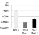
- FIG. 3 is a graph showing DNA yield results when a human iPS cell line is used as a sample.
- BS (+) Res (-) is bisulfite treatment only
- BS (+) Res (+) is subjected to bisulfite treatment. This is the case.
- FIG. 4 is a graph showing the results of DNA yield when a human ES cell line 12 single cell is used as a sample.
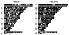
- FIG. 5 shows the result of analyzing the sequence obtained from the DNA obtained from the human ES cell line by the next generation sequence.
- the left figure shows the case where the method of the present invention is not applied, and the right figure shows the case where the method of the present invention is applied.
- FIG. 6 shows a Lorentz curve when DNA obtained from a human ES cell line was sequenced and analyzed by the next generation sequence. The left figure shows the case where the method of the present invention is not applied, and the right figure shows the case where the method of the present invention is applied.
- FIG. 5 shows the result of analyzing the sequence obtained from the DNA obtained from the human ES cell line by the next generation sequence.
- the left figure shows the case where the method of the present invention is not applied, and the right figure shows the case where the method of the present invention is applied.
- FIG. 7 is a graph showing the yield results when human genomic DNA is used as a sample.
- BS (+) Res (-) is bisulfite treatment only
- BS (+) Res (+) is subjected to bisulfite treatment.
- BS (+) Res (ATP) is the case where ATP is added during the kinase treatment.
- the present invention is a method for DNA methylation analysis using bisulfite treatment, wherein a DNA sample after bisulfite treatment is treated with a single-stranded DNA ligase, thereby
- the present invention relates to a method characterized by increasing the yield of a PCR-amplifiable DNA sample.
- DNA methylation analysis using bisulfite treatment is widely used.
- Bisulfite converts unmethylated cytosine in DNA to uracil, but methylated cytosine is not converted to other bases. Utilizing this fact, the DNA methylation state can be known by performing sequence analysis after PCR amplification of the bisulfite-treated DNA.
- the bisulfite itself is a highly reactive substance, and the processing conditions are also high. Therefore, when the bisulfite treatment is performed, the DNA sample is fragmented and a sufficient amount of DNA is supplied for the next PCR amplification. There is a problem that it is not.
- the present invention performs rescue by subjecting the DNA sample after the bisulfite treatment to a single-stranded DNA ligase treatment, and a sufficient amount for PCR amplification as the next step. It supplies DNA.
- rescue has not been performed in DNA methylation analysis using conventional bisulfite treatment.
- DNA methylation analysis methods using bisulfite treatment examples include the RRBS method and the whole genome bisulfite sequencing (WGBS) method.
- the present invention can be carried out in any DNA methylation analysis method using bisulfite, but is preferably carried out in the RRBS method. In the following, the invention will be described in relation to the RRBS method.
- the RRBS method includes the following steps (see H. Guo et al. Nature Protocols, 10 (5), 645-659 (2015), etc.): Digestion of DNA samples with restriction enzymes End repair and dA tailing Adapter ligation Bisulfite treatment PCR amplification Sequencing
- a DNA sample is rescued by ligating a fragmented DNA sample using a single-stranded DNA ligase between bisulfite treatment and PCR amplification.
- the single-stranded DNA ligase used here may be any enzyme as long as it has an activity of ligating single-stranded DNA, and is also commercially available.
- Examples of the single-stranded DNA ligase used in the present invention include, but are not limited to, Epicentre's CircLigase TM ssDNA Ligase, Wako Pure Chemical Industries, Ltd., Single Strand DNA Ligase, thermostable, recombinant, Solution, and the like.
- Preferred reaction conditions for single-stranded DNA ligase vary depending on the type of enzyme, but they are known to those skilled in the art or can be easily determined by those skilled in the art.
- single-stranded DNA ligase catalyzes a reaction of binding a hydroxyl group at the 3 'end of a polynucleotide and a phosphate group at the 5' end. Therefore, the 3 ′ end of the fragmented DNA sample after the bisulfite treatment has a hydroxyl group and a phosphate group is added to the 5 ′ end, so that the efficiency of the ligation reaction by single-stranded DNA ligase and the ligated DNA This leads to an increase in yield.
- treatment with DNA 5'-, 3'-phosphatase or DNA 3'-phosphatase and treatment with DNA 5'-kinase are preferably performed before the ligation reaction with single-stranded DNA ligase.
- the DNA 5′-, 3′-phosphatase used in the present invention is any one as long as it has an activity of hydrolyzing the phosphate groups at the 5 ′ end and 3 ′ end of DNA to generate hydroxyl groups.
- ThermoFisher SCIENTIFIC product number EF0654 may be used, but the present invention is not limited thereto.
- Preferred reaction conditions for DNA 5'-, 3'-phosphatase vary depending on the type of enzyme, but these are known to those skilled in the art or can be easily determined by those skilled in the art.
- the DNA 3'-phosphatase used in the present invention may be any DNA as long as it has an activity of hydrolyzing a phosphate group at the 3 'end of DNA to generate a hydroxyl group.
- Preferred reaction conditions for DNA 3'-phosphatase vary depending on the type of enzyme, but these are known to those skilled in the art or can be easily determined by those skilled in the art.
- Polynucleotide kinase 3'-phosphatase (PNKP) may also be used.
- PNKP is an enzyme having 3 'phosphatase activity and 5'-kinase activity.
- PNKPs are commercially available from QIAGEN.
- the 5′-kinase used in the present invention may be any as long as it has an activity of adding a phosphate group to the 5 ′ end of DNA, such as T4 polynucleotide kinase manufactured by Promega.
- Preferred reaction conditions for DNA5'-kinase vary depending on the type of enzyme, but these are known to those skilled in the art or can be easily determined by those skilled in the art.
- the yield of the DNA sample can be further increased by conducting the kinase reaction in the presence of ATP.
- the amount of ATP is not particularly limited and can be appropriately determined by those skilled in the art. Examples of the amount of ATP include, but are not limited to, a final concentration of about 1 mM to several mM in the kinase reaction system.
- DNA 5′-, 3′-phosphatase When DNA 5′-, 3′-phosphatase is used, the DNA 5′-, 3′-phosphatase is allowed to act before allowing DNA 5′-kinase to act, and the 5 ′ end of the fragmented DNA sample generated by bisulfite treatment is used. And the phosphate group at both 3 'ends is hydrolyzed to give a hydroxyl group at each end. Next, DNA 5'-kinase is allowed to act to add a phosphate group to the 5 'end of the fragmented DNA sample. Thereafter, a single-stranded DNA ligase is allowed to act, and the fragmented DNA samples are ligated and rescued.
- DNA 3 ′ phosphatase When DNA 3 ′ phosphatase is used instead of DNA 5′-, 3′-phosphatase, (i) treatment with DNA 3 ′ phosphatase and treatment with DNA 5′-kinase may be performed simultaneously.
- the treatment (i) may be performed using a NEB T4 polynucleotide kinase.
- DNA 3′-phosphatase has negligible activity to hydrolyze the phosphate group at the 5 ′ end of DNA, or such It is necessary to have no activity.
- the DNA sample In order to efficiently perform the ligation reaction with single-stranded DNA ligase and increase the yield of the rescued DNA sample, it is preferable to bind the DNA sample to an immobilization carrier. By collecting the carrier on which the DNA sample is immobilized, the DNA sample is concentrated and purified, the rescue efficiency is increased, and more DNA samples can be recovered.
- the DNA sample may be immobilized either before or after the bisulfite treatment, but preferably after the bisulfite treatment. The reason is that the immobilization support may interfere with the reaction during the reaction during the bisulfite treatment, and the purification after the bisulfite treatment uses a column, and thus the immobilization support cannot pass through the column. .
- a substance or functional group capable of binding to the carrier can be bound or introduced into the adapter.
- An adapter to which such a substance or functional group has been bound or introduced in advance may be linked to the DNA sample, or such substance or functional group may be bound or introduced after the adapter has been linked to the DNA sample.
- the adapter design is known to those skilled in the art, and can be performed according to the conditions such as the base sequence of the primers used in PCR amplification and the sequencer. You may use the adapter contained as a component of the commercial kit for methylation analysis.
- Substances to be bound to the adapter for immobilizing the DNA sample and functional groups to be introduced to the adapter are known to those skilled in the art, and their binding method, introduction method, and immobilization method to the carrier are also known to those skilled in the art. is there.
- biotin can be added to the end of the adapter, avidin bound to the carrier, and immobilization can be performed using a biotin-avidin bond.
- magnetic beads bound with avidin are suitable as the carrier.
- the present invention provides a method for increasing the yield of a DNA sample capable of PCR amplification after bisulfite treatment in DNA methylation analysis using bisulfite, wherein the DNA sample after bisulfite treatment is single-stranded.
- the present invention relates to a kit for carrying out a method characterized by treatment with DNA ligase. Therefore, the kit of the present invention contains a single-stranded DNA ligase.
- the kit of the present invention may further contain DNA 5'-, 3'-phosphatase or DNA 3'-phosphatase, and DNA 5'-kinase.
- Single-stranded DNA ligase, DNA5'-, 3'-phosphatase, DNA3'-phosphatase, and DNA5'-kinase are as described above.
- the rescue reaction of the present invention was performed after bisulfite treatment, and the increase in the yield of the recovered DNA sample was examined.
- the rescue reaction included four steps 5-1), 5-2), 5-3) and 5-4).
- the beads were washed twice with 1 ⁇ BWT buffer (5 mM Tris-HCl (pH 7.5), 0.5 mM EDTA, 1 M NaCl, 0.02% Tween-20), and then EBT buffer (10 mM Tris-HCl (pH 8)). 5), 0.02% Tween-20) and washed once.
- 1 ⁇ BWT buffer 5 mM Tris-HCl (pH 7.5), 0.5 mM EDTA, 1 M NaCl, 0.02% Tween-20
- EBT buffer 10 mM Tris-HCl (pH 8)
- phosphatase reaction A phosphatase mixture was prepared using 1x buffer and nuclease-free water. The beads of 5-1) were captured and the supernatant was removed, and then suspended in a phosphatase mixture and transferred to a PCR tube. 1 ⁇ L of FastAP Thermosensitive Alkaline Phosphatase (ThermoFisher SCIENTIFIC product number EF0654) was added, reacted at 37 ° C. for 10 minutes, and enzyme-inactivated at 75 ° C. for 5 minutes. After the reaction, it was transferred to a LoBind tube to capture the beads, and washed twice with EBT buffer.
- FastAP Thermosensitive Alkaline Phosphatase Thermosensitive Alkaline Phosphatase
- kinase reaction A kinase mixture was prepared using 1x buffer and nuclease-free water. The beads of 5-2) were captured and the supernatant was removed, and then suspended in a kinase mixture and transferred to a PCR tube. 1 ⁇ L of T4 polynucleotide kinase (TaKaRa product number 2021A) was added, reacted at 37 ° C. for 30 minutes, and enzyme-inactivated at 70 ° C. for 5 minutes. After the reaction, it was transferred to a LoBind tube to capture the beads, and washed twice with EBT buffer.
- T4 polynucleotide kinase T4 polynucleotide kinase
- the PCR reaction was performed as follows. 95 ° C. for 2 minutes ⁇ 95 ° C. for 20 seconds for 22 cycles, 60 ° C. for 30 seconds, 72 ° C. for 1 minute ⁇ 72 ° C. for 5 minutes ⁇ 4 ° C.
- Dynabeads® M-280 streptavidin beads were captured, the supernatant was collected, and the PCR product was purified twice with 1: 1 AMPure beads (see Guo et al., Supra).
- Second round PCR enrichment A PCR mixture was created as follows. 1 ⁇ Phusion HF PCR master mix with HF buffer (see Guo et al., Supra), 500 nM primer (sequence above), and sample. Thereafter, the PCR reaction was performed as follows. 98 ° C. for 2 minutes ⁇ 98 ° C. for 10 seconds for 22 cycles, 60 ° C. for 30 seconds, 72 ° C. for 1 minute ⁇ 72 ° C. for 5 minutes ⁇ 4 ° C. After completion of the PCR reaction, the PCR product was purified once with 1: 1 AMPure beads, eluted with 25 ⁇ L of nuclease-free water and collected.
- the PCR product measurement results are shown in Table 2 and FIG.
- the results of electrophoresis with 2.0% E-gel are shown in FIG. 2 (the center lane is sample I, the second lane from the right is sample IV, and the rightmost lane is sample V). According to the comparison between Sample IV and Sample V, a yield increase of about 2.3 times was recognized by the rescue reaction of the present invention.
- 201B7 cell line which is a human iPS cell line, was used as a sample. After culturing the 201B7 cell line on feeder cells, the cells were collected, and the cell suspension stained with anti-SSEA-4 antibodies (BD Pharmingen) and PI (BD Pharmingen) was used in single cell mode using FACS Aria II (BD Biosciences). Then, anti-SSEA-4 antibody positive and PI negative cells were sorted in a 96-well PCR plate by 300 cells.
- the experimental procedure included the following seven steps: 1) digestion with MspI, 2) end repair / dA tailing reaction, 3) adapter ligation, 4) bisulfite treatment, 5) rescue reaction, 6) first round. PCR enrichment, and 7) Second round PCR enrichment. In this additional data, 8) Quantitative PCR was carried out using human-specific gene primers, using the gel sample, the purified sample as a sequence, and the purified sample as a template.
- a Y-shaped adapter containing a biotinylated barcode on both sides of the following sequence was used as an adapter.
- a Y-shaped adapter was prepared by annealing a universal adapter with a biotinylated 5 ′ end and an index adapter with a biotinylated 3 ′ end.
- the sequence of the universal adapter is as follows.
- the final elution used 25 ⁇ L of elution buffer preheated at 50 ° C.
- the amount was measured and divided into two equal parts (newly sample II (BS (+) / rescue ( ⁇ ) in Table 3, BS (+) Res ( ⁇ ) in FIG. 3)), sample III (BS (+) in Table 3) / Rescue (+), named BS (+) Res (+)) in FIG.
- the rescue reaction included four steps 5-1), 5-2), 5-3) and 5-4).
- the beads were washed twice with 1 ⁇ BWT buffer (5 mM Tris-HCl (pH 7.5), 0.5 mM EDTA, 1 M NaCl, 0.02% Tween-20), and then EBT buffer (10 mM Tris-HCl (pH 8)). 5), 0.02% Tween-20) and washed once.
- 1 ⁇ BWT buffer 5 mM Tris-HCl (pH 7.5), 0.5 mM EDTA, 1 M NaCl, 0.02% Tween-20
- EBT buffer 10 mM Tris-HCl (pH 8)
- phosphatase reaction A phosphatase mixture was prepared using 1x buffer and nuclease-free water. The beads of 5-1) were captured and the supernatant was removed, and then suspended in a phosphatase mixture and transferred to a PCR tube. 1 ⁇ L of FastAP Thermosensitive Alkaline Phosphatase (ThermoFisher SCIENTIFIC product number EF0654) was added, reacted at 37 ° C. for 10 minutes, and enzyme-inactivated at 75 ° C. for 5 minutes. After the reaction, it was transferred to a LoBind tube to capture the beads, and washed twice with EBT buffer.
- FastAP Thermosensitive Alkaline Phosphatase Thermosensitive Alkaline Phosphatase
- kinase reaction A kinase mixture was prepared using 1x buffer and nuclease-free water. The beads of 5-2) were captured and the supernatant was removed, and then suspended in a kinase mixture and transferred to a PCR tube. 1 ⁇ L of T4 polynucleotide kinase (TaKaRa product number 2021A) was added, reacted at 37 ° C. for 30 minutes, and enzyme-inactivated at 70 ° C. for 5 minutes. After the reaction, it was transferred to a LoBind tube to capture the beads, and washed twice with EBT buffer.
- T4 polynucleotide kinase T4 polynucleotide kinase
- Second round PCR enrichment A PCR mixture was created as follows. 1 ⁇ Phusion HF PCR master mix with HF buffer (see Guo et al. Literature), 500 nM primer (see Guo et al. Literature), and sample. Thereafter, the PCR reaction was performed as follows. Set at 98 ° C. for 2 minutes ⁇ 98 ° C. for 10 seconds, 60 ° C. for 30 seconds, 72 ° C. for 1 minute 22 cycles ⁇ 72 ° C. for 5 minutes ⁇ 4 ° C. After completion of the PCR reaction, the PCR product was purified once with 1: 1 AMPure beads, eluted with 25 uL of nuclease-free water and collected.
- Quantitative PCR was performed with human-specific gene primers using the gel sample (250-700 bp) and purified sample as a template.
- the primer sequences used are as follows. 5'-TAGCAATAATCCCCATCCTCCATATAT-3 '(SEQ ID NO: 6) 5'-ACTTGTCCAATGATGGTAAAAGG-3 '(SEQ ID NO: 7)
- Adapter ligation 12 types of adapters with biotinylated barcodes were added to each well.
- 30 Weiss units of T4 DNA ligase, 1 ⁇ Tango buffer, 1 mM ATP, 12 types of adapters, nuclease-free water were added, reacted at 16 ° C. for 30 minutes, and subsequently reacted at 4 ° C. overnight, then at 65 ° C.
- the enzyme was inactivated for 20 minutes. The exact amount of sample was measured and adjusted with nuclease-free water to 25 ⁇ L.
- a Y-shaped adapter containing a biotinylated barcode on both sides of the following sequence was used as an adapter.
- a Y-shaped adapter was prepared by annealing a universal adapter with a biotinylated 5 ′ end and an index adapter with a biotinylated 3 ′ end.
- the sequence of the universal adapter is as follows.
- sample I (again, BS (+) / rescue ( ⁇ ) in Table 4, FIG. 4 BS (+) Res ( ⁇ )), sample II (named BS (+) / rescue (+) in Table 4, BS (+) Res (+)) in FIG. 4).
- the rescue reaction included four steps 5-1), 5-2), 5-3) and 5-4).
- the beads were washed twice with 1 ⁇ BWT buffer (5 mM Tris-HCl (pH 7.5), 0.5 mM EDTA, 1 M NaCl, 0.02% Tween-20), and then EBT buffer (10 mM Tris-HCl (pH 8)). 5), 0.02% Tween-20) and washed once.
- 1 ⁇ BWT buffer 5 mM Tris-HCl (pH 7.5), 0.5 mM EDTA, 1 M NaCl, 0.02% Tween-20
- EBT buffer 10 mM Tris-HCl (pH 8)
- phosphatase reaction A phosphatase mixture was prepared using 1x buffer and nuclease-free water. The beads of 5-1) were captured and the supernatant was removed, and then suspended in a phosphatase mixture and transferred to a PCR tube. 1 ⁇ L of FastAP Thermosensitive Alkaline Phosphatase (ThermoFisher SCIENTIFIC product number EF0654) was added, reacted at 37 ° C. for 10 minutes, and enzyme-inactivated at 75 ° C. for 5 minutes. After the reaction, it was transferred to a LoBind tube to capture the beads, and washed twice with EBT buffer.
- FastAP Thermosensitive Alkaline Phosphatase Thermosensitive Alkaline Phosphatase
- kinase reaction A kinase mixture was prepared using 1x buffer and nuclease-free water. The beads of 5-2) were captured and the supernatant was removed, and then suspended in a kinase mixture and transferred to a PCR tube. 1 ⁇ L of T4 polynucleotide kinase (TaKaRa product number 2021A) was added, reacted at 37 ° C. for 30 minutes, and enzyme-inactivated at 70 ° C. for 5 minutes. After the reaction, it was transferred to a LoBind tube to capture the beads, and washed twice with EBT buffer.
- T4 polynucleotide kinase T4 polynucleotide kinase
- Second round PCR enrichment A PCR mixture was created as follows. 1 ⁇ Phusion HF PCR master mix with HF buffer (see Guo et al. Literature), 500 nM primer (see Guo et al. Literature), and sample. Thereafter, the PCR reaction was performed as follows. Set at 98 ° C. for 2 minutes ⁇ 98 ° C. for 10 seconds, 60 ° C. for 30 seconds, 72 ° C. for 1 minute 26 cycles ⁇ 72 ° C. for 5 minutes ⁇ 4 ° C. After completion of the PCR reaction, the PCR product was purified once with 1: 1 AMPure beads, eluted with 25 uL of nuclease-free water and collected.
- Quantitative PCR was performed with human-specific gene primers using the gel sample (250-700 bp) and purified sample as a template.
- the primer sequences used are as follows. 5'-TAGCAATAATCCCCATCCTCCATATAT-3 '(SEQ ID NO: 6) 5'-ACTTGTCCAATGATGGTAAAAGG-3 '(SEQ ID NO: 7)
- the obtained gene sequence was analyzed using a next-generation sequencer (Illumina HiSeq 2500) for samples obtained using the human ES cell line (WA09 strain) 12 single cell.
- HiSeq 2500 was run at an installed concentration of 6 pM. After obtaining the array, data processing was performed according to the following flow and illustrated (FIG. 5). After mapping the sequence data to the human reference genome sequence (masked by GRCh38 release 85, N) downloaded from the Ensembl database using the Bismark command of the Bismark software (see the method of Guo et al.), Further reference using the bimark_methylation_extractor command The position information of the CpG site on the genome sequence was acquired and illustrated.
- the commands and options at the time of execution of bismark and bismark_methylation_extractor are as follows. In order to execute the Bismark software, the Bowtie 2 software and the SAMtools software need to be installed.
- the program for drawing the Lorentz curve and calculating the Gini coefficient was implemented using the R language (Fig. 6).
- a Y-shaped adapter containing a biotinylated barcode on both sides of the following sequence was used as an adapter.
- a Y-shaped adapter was prepared by annealing a universal adapter with a biotinylated 5 ′ end and an index adapter with a biotinylated 3 ′ end.
- the sequence of the universal adapter is as follows.
- samples II, III, IV 18 ⁇ L of samples (Samples II, III, IV) were subjected to bisulfite treatment. Nuclease-free water was added to the sample to adjust to 25 ⁇ L. 125 ⁇ L of CT conversion buffer was added and mixed well. Bisulfite treatment was performed at 98 ° C. for 10 minutes, at 64 ° C. for 2 hours, and then at 4 ° C. The treated DNA was centrifuged after adding 1 ⁇ l of tRNA (10 ng / ⁇ l) to the binding buffer using a methylcode bisulfite conversion kit (ThermoFisher SCIENTIFIC product number MECOV-50). The final elution used 25 ⁇ L of elution buffer preheated at 50 ° C. The amount was measured and divided into three equal parts (newly sample II (BS (+) Res (-) in FIG. 7), sample III (BS (+) Res (+) in FIG. 7), sample IV (BS in FIG. 7) (+) Res (ATP))).
- the rescue reaction included four steps 5-1), 5-2), 5-3) and 5-4).
- the beads were washed twice with 1 ⁇ BWT buffer (5 mM Tris-HCl (pH 7.5), 0.5 mM EDTA, 1 M NaCl, 0.02% Tween-20), and then EBT buffer (10 mM Tris-HCl (pH 8)). 5), 0.02% Tween-20) and washed once.
- 1 ⁇ BWT buffer 5 mM Tris-HCl (pH 7.5), 0.5 mM EDTA, 1 M NaCl, 0.02% Tween-20
- EBT buffer 10 mM Tris-HCl (pH 8)
- phosphatase reaction A phosphatase mixture was prepared using 1x buffer and nuclease-free water. The beads of 5-1) were captured and the supernatant was removed, and then suspended in a phosphatase mixture and transferred to a PCR tube. 1 ⁇ L of FastAP Thermosensitive Alkaline Phosphatase (ThermoFisher SCIENTIFIC product number EF0654) was added, reacted at 37 ° C. for 10 minutes, and enzyme-inactivated at 75 ° C. for 5 minutes. After the reaction, it was transferred to a LoBind tube to capture the beads, and washed twice with EBT buffer.
- FastAP Thermosensitive Alkaline Phosphatase Thermosensitive Alkaline Phosphatase
- a kinase mixture was prepared using 1x buffer and nuclease-free water. A final concentration of 1 mM ATP was added to the kinase mixture. The beads of 5-2) were captured and the supernatant was removed, and then suspended in a kinase mixture and transferred to a PCR tube. 1 ⁇ L of T4 polynucleotide kinase (TaKaRa product number 2021A) was added, reacted at 37 ° C. for 30 minutes, and enzyme-inactivated at 70 ° C. for 5 minutes. After the reaction, it was transferred to a LoBind tube to capture the beads, and washed twice with EBT buffer.
- T4 polynucleotide kinase T4 polynucleotide kinase
- Second round PCR enrichment A PCR mixture was created as follows. 1 ⁇ Phusion HF PCR master mix with HF buffer (see Guo et al. Literature), 500 nM primer (see Guo et al. Literature), and sample. Thereafter, the PCR reaction was performed as follows. Set at 98 ° C. for 2 minutes ⁇ 98 ° C. for 10 seconds, 60 ° C. for 30 seconds, 72 ° C. for 1 minute 22 cycles ⁇ 72 ° C. for 5 minutes ⁇ 4 ° C. After completion of the PCR reaction, the PCR product was purified once with 1: 1 AMPure beads, eluted with 25 uL of nuclease-free water and collected.
- Quantitative PCR was performed using a human-specific gene primer (Alu3 primer sequence) using the sample after gel cutting (250-700 bp) and purification as a template.
- the primer sequences used are as follows. 5'-TGGAAGACCAGTAGGATGATTG-3 '(SEQ ID NO: 20) 5'-GTCTTACCATGTATCTCTGTGC-3 '(SEQ ID NO: 21)
- the present invention methylation analysis of a small amount of DNA sample such as DNA derived from a single cell becomes possible. Therefore, the present invention has high utility value in fields such as medicine, pharmacy, biology, genetics, and agriculture.
- SEQ ID NO: 1 is the base sequence of the adapter used in the examples.
- the 5 'end is biotinylated and the cytosine is 5-methyl-dC.
- SEQ ID NO: 2 is the base sequence of the adapter used in the examples.
- the 3 'end is biotinylated and the cytosine is 5-methyl-dC.
- SEQ ID NO: 3 is the base sequence of the PCR amplification primer used in the examples.
- SEQ ID NO: 4 is the base sequence of the universal adapter used in the examples.
- the 5 ′ end is biotinylated, cytosine is 5-methyl-dC, and the 3 ′ end CT is phosphorothioated.
- SEQ ID NO: 5 is the base sequence of the index adapter used in the examples.
- the 5 'end is phosphorylated, cytosine is 5-methyl-dC, and the 3' end is biotinylated.
- SEQ ID NO: 6 is the base sequence of the primer for quantitative PCR used in the examples.
- SEQ ID NO: 7 is the base sequence of the primer for quantitative PCR used in the examples.
- SEQ ID NO: 8 is the base sequence of the index adapter used in the examples.
- the 5 'end is phosphorylated, cytosine is 5-methyl-dC, and the 3' end is biotinylated.
- SEQ ID NO: 9 is the base sequence of the index adapter used in the examples.
- the 5 'end is phosphorylated, cytosine is 5-methyl-dC, and the 3' end is biotinylated.
- SEQ ID NO: 10 is the base sequence of the index adapter used in the examples.
- the 5 'end is phosphorylated, cytosine is 5-methyl-dC, and the 3' end is biotinylated.
- SEQ ID NO: 11 is the base sequence of the index adapter used in the examples.
- the 5 'end is phosphorylated, cytosine is 5-methyl-dC, and the 3' end is biotinylated.
- SEQ ID NO: 12 is the base sequence of the index adapter used in the examples.
- the 5 'end is phosphorylated, cytosine is 5-methyl-dC, and the 3' end is biotinylated.
- SEQ ID NO: 13 is the base sequence of the index adapter used in the examples.
- the 5 'end is phosphorylated, cytosine is 5-methyl-dC, and the 3' end is biotinylated.
- SEQ ID NO: 14 is the base sequence of the index adapter used in the examples. The 5 'end is phosphorylated, cytosine is 5-methyl-dC, and the 3' end is biotinylated.
- SEQ ID NO: 15 is the base sequence of the index adapter used in the examples. The 5 'end is phosphorylated, cytosine is 5-methyl-dC, and the 3' end is biotinylated.
- SEQ ID NO: 16 is the base sequence of the index adapter used in the examples.
- the 5 'end is phosphorylated, cytosine is 5-methyl-dC, and the 3' end is biotinylated.
- SEQ ID NO: 17 is the base sequence of the index adapter used in the examples. The 5 'end is phosphorylated, cytosine is 5-methyl-dC, and the 3' end is biotinylated.
- SEQ ID NO: 18 is the base sequence of the index adapter used in the examples. The 5 'end is phosphorylated, cytosine is 5-methyl-dC, and the 3' end is biotinylated.
- SEQ ID NO: 19 is the base sequence of the index adapter used in the examples.
- SEQ ID NO: 20 is the base sequence of the primer for quantitative PCR used in the examples.
- SEQ ID NO: 21 is the base sequence of the primer for quantitative PCR used in the examples.
Landscapes
- Chemical & Material Sciences (AREA)
- Organic Chemistry (AREA)
- Life Sciences & Earth Sciences (AREA)
- Zoology (AREA)
- Wood Science & Technology (AREA)
- Proteomics, Peptides & Aminoacids (AREA)
- Health & Medical Sciences (AREA)
- Engineering & Computer Science (AREA)
- Microbiology (AREA)
- Immunology (AREA)
- Physics & Mathematics (AREA)
- Molecular Biology (AREA)
- Biotechnology (AREA)
- Biophysics (AREA)
- Analytical Chemistry (AREA)
- Biochemistry (AREA)
- Bioinformatics & Cheminformatics (AREA)
- General Engineering & Computer Science (AREA)
- General Health & Medical Sciences (AREA)
- Genetics & Genomics (AREA)
- Measuring Or Testing Involving Enzymes Or Micro-Organisms (AREA)
Abstract
本発明は、バイサルファイト処理を用いるDNAメチル化解析のための方法であって、バイサルファイト処理後のDNA試料を1本鎖DNAリガーゼで処理することにより、バイサルファイト処理後のPCR増幅可能なDNA試料の収量を増加させることを特徴とする方法を提供する。
Description
本発明は、DNAメチル化解析法の改良に関する。詳細には、本発明は、バイサルファイトを用いるDNAメチル化解析において、バイサルファイト処理後のPCR増幅可能なDNA試料の収量を増加させる方法に関する。
DNAのメチル化は、エピジェネティック機構の1つであり、胚発生、細胞分化、表現型の差異、様々な疾病などと深く関わっている。現在、DNAのメチル化解析のための様々な方法が開発され、使用されている。最近では、目的にかなったiPS細胞の選別にもDNAのメチル化解析が利用されている。
DNAのメチル化解析方法のなかで、特定ゲノム領域のDNAのメチル化解析法としてはバイサルファイト処理を用いる方法が多用されている。バイサルファイトは、DNA中の非メチル化シトシンをウラシルに変換するが、メチル化シトシンは他の塩基に変換されない。このことを利用して、バイサルファイト処理されたDNAをPCR増幅後、シークエンス解析を行うことによって、DNAのメチル化の状態を知ることができる。
バイサルファイトを用いるDNAのメチル化解析方法の1つとして、RRBS(Reduced Representation Bisulfite Sequencing)法がある。RRBS法は、DNAのメチル化を1塩基レベルで調べることができる方法であり、シークエンス量を減らすことができ、しかも全ゲノム解析法と同様のカバー率を得ることができる方法である。近年、BBRS法を用いてシングルセルDNAのメチル化解析を行う試みがなされている(非特許文献1、2参照)。シングルセル由来のDNAなどの微量試料のDNAメチル化解析を行う際に、バイサルファイト処理が過酷な条件で行われるためにDNA試料が断片化し、次工程であるPCR増幅のための十分な量のDNAが供給されないという問題がある。
H. Guo et al. Genome Research, 23, 2126-2135 (2013)
H. Guo et al. Nature Protocols, 10(5), 645-659 (2015)
本発明が解決すべき課題は、バイサルファイト処理後のPCR増幅可能なDNA試料の収量を増加させるための有効かつ簡単な方法を開発することであった。
本発明者らは、上記課題を解決せんと鋭意研究を重ね、バイサルファイト処理後のDNA試料を1本鎖DNAリガーゼで処理する工程を含むレスキューを行うことにより、バイサルファイト処理後のPCR増幅可能なDNA試料の収量を増加させることができることを見出し、本発明を完成するに至った。
すなわち、本発明は下記のものに関する。
(1)バイサルファイト処理を用いるDNAメチル化解析のための方法であって、バイサルファイト処理後のDNA試料を1本鎖DNAリガーゼで処理することにより、バイサルファイト処理後のPCR増幅可能なDNA試料の収量を増加させることを特徴とする方法。
(2)バイサルファイト処理を用いるDNAメチル化解析がRRBS(Reduced Representation Bisulfite Sequencing)法によるものである(1)記載の方法。
(3)1本鎖DNAリガーゼでの処理の前に、バイサルファイト処理後のDNA試料を下記工程:
(a)DNA5’-,3’-ホスファターゼまたはDNA3’-ホスファターゼでの処理、および
(b)DNA5’-キナーゼでの処理
に付すことを特徴とする(1)または(2)記載の方法。
(4)バイサルファイト処理後のDNA試料が担体に固定化されている、(1)~(3)のいずれかに記載の方法。
(5)工程(b)がATPの存在下において行われる、(1)~(4)のいずれかに記載の方法。
(6)(1)~(5)のいずれかに記載の方法を実施するためのキットであって、1本鎖DNAリガーゼを含むキット。
(7)さらにDNA5’-,3’-ホスファターゼまたはDNA3’-ホスファターゼおよびDNA5’キナーゼを含む(6)記載のキット。
(1)バイサルファイト処理を用いるDNAメチル化解析のための方法であって、バイサルファイト処理後のDNA試料を1本鎖DNAリガーゼで処理することにより、バイサルファイト処理後のPCR増幅可能なDNA試料の収量を増加させることを特徴とする方法。
(2)バイサルファイト処理を用いるDNAメチル化解析がRRBS(Reduced Representation Bisulfite Sequencing)法によるものである(1)記載の方法。
(3)1本鎖DNAリガーゼでの処理の前に、バイサルファイト処理後のDNA試料を下記工程:
(a)DNA5’-,3’-ホスファターゼまたはDNA3’-ホスファターゼでの処理、および
(b)DNA5’-キナーゼでの処理
に付すことを特徴とする(1)または(2)記載の方法。
(4)バイサルファイト処理後のDNA試料が担体に固定化されている、(1)~(3)のいずれかに記載の方法。
(5)工程(b)がATPの存在下において行われる、(1)~(4)のいずれかに記載の方法。
(6)(1)~(5)のいずれかに記載の方法を実施するためのキットであって、1本鎖DNAリガーゼを含むキット。
(7)さらにDNA5’-,3’-ホスファターゼまたはDNA3’-ホスファターゼおよびDNA5’キナーゼを含む(6)記載のキット。
本発明によれば、1本鎖DNAリガーゼを用いる簡単な方法により、バイサルファイト処理後のPCR増幅可能なDNA試料の収量を増加させることができ、その後のPCR工程およびシークエンス工程に十分な量のDNA試料を供給することができる。したがって、本発明を用いれば、シングルセル由来のDNAなどの微量なDNA試料のメチル化解析が可能となる。
本発明は、1の態様において、バイサルファイト処理を用いるDNAメチル化解析のための方法であって、バイサルファイト処理後のDNA試料を1本鎖DNAリガーゼで処理することにより、バイサルファイト処理後のPCR増幅可能なDNA試料の収量を増加させることを特徴とする方法に関する。
バイサルファイト処理を用いるDNAのメチル化解析は広く用いられている。バイサルファイトは、DNA中の非メチル化シトシンをウラシルに変換するが、メチル化シトシンは他の塩基に変換されない。このことを利用して、バイサルファイト処理されたDNAをPCR増幅後、シークエンス解析を行うことによって、DNAのメチル化の状態を知ることができる。しかし、バイサルファイト自体反応性に富む物質であり、処理条件も高温であるために、バイサルファイト処理を行うとDNA試料が断片化し、次工程であるPCR増幅のために十分な量のDNAが供給されないという問題がある。
本発明は、かかるDNA試料の損失を抑制するために、バイサルファイト処理後のDNA試料を1本鎖DNAリガーゼ処理に付すことによりレスキューを行い、次工程であるPCR増幅のために十分な量のDNAを供給するものである。このようなレスキューは、従来のバイサルファイト処理を用いるDNAのメチル化解析においては行われていなかった。
現在よく用いられているバイサルファイト処理を用いるDNAのメチル化解析法の例としては、RRBS法および全ゲノムバイサルファイトシークエンシング(WGBS)法などが挙げられる。本発明は、バイサルファイトを用いるいずれのDNAのメチル化解析法においても実施可能であるが、RRBS法において実施することが好ましい。以下において、RRBS法に関連して本発明を説明する。
一般に、RRBS法は以下の工程を含む(H. Guo et al. Nature Protocols, 10(5), 645-659 (2015)等を参照のこと):
制限酵素によるDNA試料の消化
エンドリペアおよびdAテイリング
アダプターライゲーション
バイサルファイト処理
PCR増幅
シークエンシング
制限酵素によるDNA試料の消化
エンドリペアおよびdAテイリング
アダプターライゲーション
バイサルファイト処理
PCR増幅
シークエンシング
本発明においては、バイサルファイト処理とPCR増幅との間に、1本鎖DNAリガーゼを用いて断片化したDNA試料を連結することにより、DNA試料のレスキューを行う。ここで用いられる1本鎖DNAリガーゼは、1本鎖DNAを連結する活性を有するものであればいずれの酵素であってもよく、市販もされている。本発明に使用する1本鎖DNAリガーゼの例としてはEpicentre社のCircLigaseTMssDNA Ligase、和光純薬工業株式会社のSingle Strand DNA Ligase, thermostable, recombinant, Solutionなどが挙げられるが、これらに限定されない。1本鎖DNAリガーゼの好ましい反応条件は酵素の種類により異なるが、それらは当業者に公知であるか、あるいは当業者が容易に定めうるものである。
一般に、1本鎖DNAリガーゼは、ポリヌクレオチドの3’末端の水酸基と、5’末端のリン酸基を結合する反応を触媒する。したがって、バイサルファイト処理後の断片化したDNA試料の3’末端を水酸基とし、5’末端にリン酸基を付加することが、1本鎖DNAリガーゼによる連結反応の効率化および連結されたDNAの収量増加につながる。
そのためには、1本鎖DNAリガーゼでの連結反応の前に、DNA5’-,3’-ホスファターゼまたはDNA3’-ホスファターゼでの処理、およびDNA5’-キナーゼでの処理を行うことが好ましい。
本発明に使用するDNA5’-,3’-ホスファターゼは、DNAの5’末端および3’末端のリン酸基を加水分解して水酸基を生じさせる活性を有するものであればいずれのものであってもよく、例えば、ThermoFisher SCIENTIFIC社の製品番号EF0654などが挙げられるが、これらに限定されない。DNA5’-,3’-ホスファターゼの好ましい反応条件は酵素の種類により異なるが、それらは当業者に公知であるか、あるいは当業者が容易に定めうるものである。
本発明に使用するDNA3’-ホスファターゼは、DNAの3’末端のリン酸基を加水分解して水酸基を生じさせる活性を有するものであればいずれのものであってもよい。DNA3’-ホスファターゼの好ましい反応条件は酵素の種類により異なるが、それらは当業者に公知であるか、あるいは当業者が容易に定めうるものである。また、ポリヌクレオチドキナーゼ 3’-ホスファターゼ(PNKP)を用いてもよい。PNKPは3’ホスファターゼ活性と5’-キナーゼ活性を有する酵素である。いくつかのPNKPがQIAGEN社などから市販されている。
本発明に使用する5’-キナーゼは、DNAの5’末端にリン酸基を付加する活性を有するものであればいずれのものであってもよく、例えば、Promega社のT4ポリヌクレオチドキナーゼなどが挙げられるが、これらに限定されない。DNA5’-キナーゼの好ましい反応条件は酵素の種類により異なるが、それらは当業者に公知であるか、あるいは当業者が容易に定めうるものである。また、ATPの存在下においてキナーゼ反応を行うことにより、DNA試料の収量をさらに増加させることができる。ATPの量は特に限定されず、当業者が適宜定めうるものである。ATPの量の例として、キナーゼ反応系中、終濃度として約1mM~数mMとすることが挙げられるが、これに限定されない。
DNA5’-,3’-ホスファターゼを用いる場合には、DNA5’-キナーゼを作用させる前に、DNA5’-,3’-ホスファターゼを作用させ、バイサルファイト処理により生じた断片化DNA試料の5’末端および3’末端の両方のリン酸基を加水分解して、各末端に水酸基を生じさせる。次いで、DNA5’-キナーゼを作用させ、断片化DNA試料の5’末端にリン酸基を付加する。その後、1本鎖DNAリガーゼを作用させて、断片化したDNA試料を連結してレスキューする。
DNA5’-,3’-ホスファターゼのかわりにDNA3’ホスファターゼを用いる場合は、(i)DNA3’ホスファターゼでの処理とDNA5’-キナーゼでの処理を同時に行ってもよく、(ii)先にDNA5’-キナーゼで処理し、その後DNA3’ホスファターゼで処理してもよい。例えばNEB社のT4ポリヌクレオチドキナーゼを用いて(i)の処理を行ってもよい。ただし、(i)、(ii)のような処理を行う場合は、DNA3’-ホスファターゼは、DNAの5’末端のリン酸基を加水分解する活性が無視できる程度であるか、あるいはそのような活性を有していないことが必要である。
1本鎖DNAリガーゼによる連結反応を効率よく行って、レスキューされるDNA試料の収量を増加させるために、DNA試料を固定化担体に結合させることが好ましい。DNA試料を固定化した担体を集めることにより、DNA試料が濃縮、精製されてレスキュー効率が上昇し、より多くのDNA試料を回収できる。DNA試料の固定化は、バイサルファイト処理の前、後いずれに行ってもよいが、バイサルファイト処理の後で行うほうが好ましい。その理由として、バイサルファイト処理中の反応時に固定化担体が反応を妨げる可能性があること、およびバイサルファイト処理後の精製はカラムを使用するため、固定化担体がカラムを通過できないことが挙げられる。
DNA試料の固定化のために、担体との結合を可能にする物質または官能基をアダプターに結合または導入することができる。かかる物質または官能基を予め結合または導入したアダプターをDNA試料に連結してもよく、アダプターをDNA試料に連結した後かかる物質または官能基を結合または導入してもよい。バイサルファイト処理前に固定化を行う場合は、担体に固定化されたDNA試料をバイサルファイト処理する。バイサルファイト処理後に固定化を行う場合は、アダプターを付したDNA試料をバイサルファイト処理し、その後、担体に固定化する。
アダプターの設計は当業者に公知であり、PCR増幅に用いるプライマーの塩基配列やシークエンサーなどの条件に応じて行うことができる。メチル化解析用の市販キットの構成成分として含まれているアダプターを使用してもよい。
DNA試料を固定化するためにアダプターに結合させる物質、およびアダプターに導入する官能基は当業者に公知であり、それらの結合方法、導入方法、および担体への固定化方法も当業者に公知である。例えば、アダプターの末端にビオチンを付加し、担体にアビジンを結合させておいて、ビオチン-アビジン結合を利用して固定化を行うことができる。上例の場合、担体としてはアビジンを結合した磁性ビーズが適当である。担体との結合を可能にする物質や官能基、固定化担体ならびに固定化方法は上記のものに限定されないことはいうまでもない。
本発明は、さらなる態様において、バイサルファイトを用いるDNAメチル化解析において、バイサルファイト処理後のPCR増幅可能なDNA試料の収量を増加させる方法であって、バイサルファイト処理後のDNA試料を1本鎖DNAリガーゼで処理することを特徴とする方法を実施するためのキットに関するものである。したがって、本発明のキットは、1本鎖DNAリガーゼを含む。
本発明のキットは、さらにDNA5’-,3’-ホスファターゼまたはDNA3’-ホスファターゼ、およびDNA5’-キナーゼを含んでいてもよい。1本鎖DNAリガーゼ、DNA5’-,3’-ホスファターゼ、DNA3’-ホスファターゼ、およびDNA5’-キナーゼについては上で説明したとおりである。
以下に実施例を示して本発明をさらに詳細かつ具体的に説明するが、実施例は本発明を限定するものと解してはならない。
RRBS法によりヒトゲノムDNAのメチル化を解析するにあたり、バイサルファイト処理後に本発明のレスキュー反応を行って、回収されるDNA試料の収量の増加を調べた。
1.実験手順
試料として、Promega 製品番号 G3041-1のヒトゲノムDNA3ngを用いた。実験手順は以下の7つの工程を含んでいた:1)MspIでの消化、2)エンドリペア/dAテイリング反応、3)アダプターライゲーション、4)バイサルファイト処理、5)レスキュー反応、6)第1ラウンドPCRエンリッチメント、および7)第2ラウンドPCRエンリッチメント。
試料として、Promega 製品番号 G3041-1のヒトゲノムDNA3ngを用いた。実験手順は以下の7つの工程を含んでいた:1)MspIでの消化、2)エンドリペア/dAテイリング反応、3)アダプターライゲーション、4)バイサルファイト処理、5)レスキュー反応、6)第1ラウンドPCRエンリッチメント、および7)第2ラウンドPCRエンリッチメント。
1)MspIでの消化
ヒトゲノムDNA 3ngを、9ユニットのMspI、1xTangoバッファー(Guo H et al, Nature Protocols, vol.10 (5), 645-659参照)、ヌクレアーゼフリーの水の混合物中で37℃にて3時間反応させ、次いで、80℃で20分間酵素を失活させた。
ヒトゲノムDNA 3ngを、9ユニットのMspI、1xTangoバッファー(Guo H et al, Nature Protocols, vol.10 (5), 645-659参照)、ヌクレアーゼフリーの水の混合物中で37℃にて3時間反応させ、次いで、80℃で20分間酵素を失活させた。
2)エンドリペア/dA-テイリング 5ユニットのクレノウフラグメントexo-(上記Guoらの文献参照)、1xTangoバッファー、40μM dATP、4μM dCTP、4μM dGTPを加え、37℃で40分反応させ、75℃で15分間酵素失活させた。
3)アダプターライゲーション
アダプターライゲーションにおいて、30WeissユニットのT4 DNAリガーゼ(上記Guoらの文献参照)、1xTangoバッファー、1mM ATP、アダプター、ヌクレアーゼフリーの水を加え、16℃で30分反応させ、引き続き4℃で一晩反応した後に、65℃で20分間酵素失活を行った。試料の正確な量を計測し、30μLになるようヌクレアーゼフリーの水で調整後、6μL(試料I)、12μL(試料II、III)、12μL(試料IV、V)に分注し、試料Iは-80℃で保存した。
アダプターライゲーションにおいて、30WeissユニットのT4 DNAリガーゼ(上記Guoらの文献参照)、1xTangoバッファー、1mM ATP、アダプター、ヌクレアーゼフリーの水を加え、16℃で30分反応させ、引き続き4℃で一晩反応した後に、65℃で20分間酵素失活を行った。試料の正確な量を計測し、30μLになるようヌクレアーゼフリーの水で調整後、6μL(試料I)、12μL(試料II、III)、12μL(試料IV、V)に分注し、試料Iは-80℃で保存した。
アダプターとして、以下の配列の片側ビオチン化バーコード入りアダプターのセットを設計し、使用した。
5'-AAGCAGTGGTATCAACGCAGAGTGATCTTT-3' (配列番号:1)
(5’-ビオチン化、Cは5-メチル-dC)
5'-AAGATCACTCTGCGTTGATACCACTGCTT-3' (配列番号:2)
(5’-リン酸化、3’-ビオチン化、Cは5-メチル-dC)
5'-AAGCAGTGGTATCAACGCAGAGTGATCTTT-3' (配列番号:1)
(5’-ビオチン化、Cは5-メチル-dC)
5'-AAGATCACTCTGCGTTGATACCACTGCTT-3' (配列番号:2)
(5’-リン酸化、3’-ビオチン化、Cは5-メチル-dC)
4)バイサルファイト処理
12μLの試料(試料IV、V)をバイサルファイト処理に付した。試料にフクレアーゼ不含水を加え、25μLに調整した。CTコンバージョンバッファーを125μL加え、よく混合した。バイサルファイト処理を98℃で10分、64℃で2時間、その後4℃で行った。処理済みDNAをMethylcode bisulfite conversion kit(ThermoFisher SCIENTIFIC製品番号MECOV-50)を用い、バインディングバッファーに1μlのtRNA(10ng/μl)を加え遠心した。最後の溶出は50℃でプレヒートした25μLのエリューションバッファーを使用した。量を測り取り、2等分した(改めて試料IV、Vと命名)。
12μLの試料(試料IV、V)をバイサルファイト処理に付した。試料にフクレアーゼ不含水を加え、25μLに調整した。CTコンバージョンバッファーを125μL加え、よく混合した。バイサルファイト処理を98℃で10分、64℃で2時間、その後4℃で行った。処理済みDNAをMethylcode bisulfite conversion kit(ThermoFisher SCIENTIFIC製品番号MECOV-50)を用い、バインディングバッファーに1μlのtRNA(10ng/μl)を加え遠心した。最後の溶出は50℃でプレヒートした25μLのエリューションバッファーを使用した。量を測り取り、2等分した(改めて試料IV、Vと命名)。
5)レスキュー反応
レスキュー反応は4つの工程5-1)、5-2)、5-3)、5-4)を含んでいた。
レスキュー反応は4つの工程5-1)、5-2)、5-3)、5-4)を含んでいた。
5-1)ビーズへの固定化
Dynabeads(登録商標) M-280 ストレプトアビジンビーズ(ThermoFisher SCIENTIFIC製品番号11205D)10μLをDNA LoBindチューブ(Eppendorf製品番号0030 108.051)に入れ、ビーズを捕捉し、2xBWTバッファー(10mM Tris-HCl(pH7.5)、1mM EDTA、2M NaCl、0.01% Tween-20)で2回洗浄後、10μLの2xBWTバッファーに懸濁した。試料V(約10μL)を加えて混合し、室温で15分インキュベートした。ビーズを捕捉した後、1xBWTバッファー(5mM Tris-HCl(pH7.5)、0.5mM EDTA、1M NaCl、0.02% Tween-20)で2回洗浄後、EBTバッファー(10mM Tris-HCl(pH8.5)、0.02% Tween-20)で1回洗浄した。
Dynabeads(登録商標) M-280 ストレプトアビジンビーズ(ThermoFisher SCIENTIFIC製品番号11205D)10μLをDNA LoBindチューブ(Eppendorf製品番号0030 108.051)に入れ、ビーズを捕捉し、2xBWTバッファー(10mM Tris-HCl(pH7.5)、1mM EDTA、2M NaCl、0.01% Tween-20)で2回洗浄後、10μLの2xBWTバッファーに懸濁した。試料V(約10μL)を加えて混合し、室温で15分インキュベートした。ビーズを捕捉した後、1xBWTバッファー(5mM Tris-HCl(pH7.5)、0.5mM EDTA、1M NaCl、0.02% Tween-20)で2回洗浄後、EBTバッファー(10mM Tris-HCl(pH8.5)、0.02% Tween-20)で1回洗浄した。
5-2)ホスファターゼ反応
1xバッファー、ヌクレアーゼフリーの水を用いてホスファターゼミクスチャーを作成した。5-1)のビーズを捕捉し、上清を取り除いた後、ホスファターゼミクスチャーに懸濁し、PCRチューブ移した。FastAP Thermosensitive Alkaline Phosphatase(ThermoFisher SCIENTIFIC製品番号EF0654)1μLを加え、37℃で10分反応させ、75℃で5分間酵素失活させた。反応後、LoBindチューブに移し、ビーズを捕捉し、EBTバッファーで2回洗浄した。
1xバッファー、ヌクレアーゼフリーの水を用いてホスファターゼミクスチャーを作成した。5-1)のビーズを捕捉し、上清を取り除いた後、ホスファターゼミクスチャーに懸濁し、PCRチューブ移した。FastAP Thermosensitive Alkaline Phosphatase(ThermoFisher SCIENTIFIC製品番号EF0654)1μLを加え、37℃で10分反応させ、75℃で5分間酵素失活させた。反応後、LoBindチューブに移し、ビーズを捕捉し、EBTバッファーで2回洗浄した。
5-3)キナーゼ反応
1xバッファー、ヌクレアーゼフリーの水を用いてキナーゼミクスチャーを作成した。5-2)のビーズを捕捉し、上清を取り除いた後、キナーゼミクスチャーに懸濁し、PCRチューブへ移した。T4ポリヌクレオチドキナーゼ(TaKaRa製品番号2021A)1μLを加え、37℃で30分反応させ、70℃で5分間酵素失活させた。反応後、LoBindチューブに移し、ビーズを捕捉し、EBTバッファーで2回洗浄した。
1xバッファー、ヌクレアーゼフリーの水を用いてキナーゼミクスチャーを作成した。5-2)のビーズを捕捉し、上清を取り除いた後、キナーゼミクスチャーに懸濁し、PCRチューブへ移した。T4ポリヌクレオチドキナーゼ(TaKaRa製品番号2021A)1μLを加え、37℃で30分反応させ、70℃で5分間酵素失活させた。反応後、LoBindチューブに移し、ビーズを捕捉し、EBTバッファーで2回洗浄した。
5-4)1本鎖DNAリガーゼ反応
1xバッファー、ヌクレアーゼフリーの水を用いて1本鎖DNAリガーゼミクスチャーを作成した。5-3)のビーズを捕捉し、上清を取り除いた後、1本鎖DNAミクスチャーに懸濁し、PCRチューブへ移した。CircLigase II 1本鎖DNAリガーゼ(epicentre製品番号CL9025K)1μLを加え、60℃で2時間反応させ、80℃で10分間酵素失活させた。反応後、LoBindチューブに移し、ビーズを捕捉し、EBTバッファーで2回洗浄した。最後はMethylcode bisulfite conversion kitのエリューションバッファー10μLに懸濁した。この試料を以下の核酸増幅に付した。
1xバッファー、ヌクレアーゼフリーの水を用いて1本鎖DNAリガーゼミクスチャーを作成した。5-3)のビーズを捕捉し、上清を取り除いた後、1本鎖DNAミクスチャーに懸濁し、PCRチューブへ移した。CircLigase II 1本鎖DNAリガーゼ(epicentre製品番号CL9025K)1μLを加え、60℃で2時間反応させ、80℃で10分間酵素失活させた。反応後、LoBindチューブに移し、ビーズを捕捉し、EBTバッファーで2回洗浄した。最後はMethylcode bisulfite conversion kitのエリューションバッファー10μLに懸濁した。この試料を以下の核酸増幅に付した。
6)第1ラウンドPCRエンリッチメント
レスキュー反応をしていない試料IおよびIVをDynabeads(登録商標) M-280 ストレプトアビジンビーズ10μLに固定化した。試料Iは、ヌクレアーゼフリーの水で10μLに調整してから混合した。
下記のようにPCRミクスチャーを作成した。1xPCR Pfu Cxバッファー(Guoらの上記文献参照)、1ユニットのPfu Turbo Cx hotstart DNAポリメラーゼ(Guoらの上記文献参照)、各200μM dNTPミクスチャー、300nM プライマー(5'-AAGCAGTGGTATCAACGCAGAGT-3')(配列番号:3)、ヌクレアーゼ不含の水、およびビーズ付着試料。
その後PCR反応を次のように行った。95℃で2分→95℃で20秒を22サイクル、60℃で30秒、72℃で1分→72℃で5分→4℃。PCR反応終了後、Dynabeads(登録商標) M-280 ストレプトアビジンビーズを捕捉し、上清を回収し、1:1 AMPureビーズ(Guoらの上記文献参照)でPCR産物を2回精製した。
レスキュー反応をしていない試料IおよびIVをDynabeads(登録商標) M-280 ストレプトアビジンビーズ10μLに固定化した。試料Iは、ヌクレアーゼフリーの水で10μLに調整してから混合した。
下記のようにPCRミクスチャーを作成した。1xPCR Pfu Cxバッファー(Guoらの上記文献参照)、1ユニットのPfu Turbo Cx hotstart DNAポリメラーゼ(Guoらの上記文献参照)、各200μM dNTPミクスチャー、300nM プライマー(5'-AAGCAGTGGTATCAACGCAGAGT-3')(配列番号:3)、ヌクレアーゼ不含の水、およびビーズ付着試料。
その後PCR反応を次のように行った。95℃で2分→95℃で20秒を22サイクル、60℃で30秒、72℃で1分→72℃で5分→4℃。PCR反応終了後、Dynabeads(登録商標) M-280 ストレプトアビジンビーズを捕捉し、上清を回収し、1:1 AMPureビーズ(Guoらの上記文献参照)でPCR産物を2回精製した。
7)第2ラウンドPCRエンリッチメント
下記のようにPCRミクスチャーを作成した。1x Phusion HF PCR master mix with HF buffer(Guoらの上記文献参照)、500nM プライマー(上記配列)、および試料。
その後PCR反応を次のように行った。98℃で2min→98℃で10秒を22サイクル、60℃で30秒、72℃で1分→72℃で5分→4℃。PCR反応終了後、1:1 AMPureビーズでPCR産物を1回精製し、25μLのヌクレアーゼフリーの水で溶出し、回収した。
収率を確認するために、回収した試料1μL、Qubit(ThermoFisher SCIENTIFIC Qubit(登録商標) 2.0 Fluorometer)1μL、Nano drop(ThermoFisher SCIENTIFIC NanoDrop 2000)15μLを2.0% E-gel(ThermoFisher SCIENTIFIC製品番号G521802)で泳動しチェックした。
下記のようにPCRミクスチャーを作成した。1x Phusion HF PCR master mix with HF buffer(Guoらの上記文献参照)、500nM プライマー(上記配列)、および試料。
その後PCR反応を次のように行った。98℃で2min→98℃で10秒を22サイクル、60℃で30秒、72℃で1分→72℃で5分→4℃。PCR反応終了後、1:1 AMPureビーズでPCR産物を1回精製し、25μLのヌクレアーゼフリーの水で溶出し、回収した。
収率を確認するために、回収した試料1μL、Qubit(ThermoFisher SCIENTIFIC Qubit(登録商標) 2.0 Fluorometer)1μL、Nano drop(ThermoFisher SCIENTIFIC NanoDrop 2000)15μLを2.0% E-gel(ThermoFisher SCIENTIFIC製品番号G521802)で泳動しチェックした。
2.実験結果
ヒトゲノムDNAを試料として用いた実験結果を以下に示す。
表1に試料とその処理の関係を示す。

BS: バイサルファイト処理の有無(上記プロトコル4-1)
Purify 1: Bisulfite conversion kit付属のカラムを使用し精製の有無(上記プロトコル4-1)
rescue: 1本鎖DNAリガーゼを用いるレスキュー
Purify 2: M280ストレプトアビジンビーズを使用し精製の有無(上記プロトコル5)
ヒトゲノムDNAを試料として用いた実験結果を以下に示す。
表1に試料とその処理の関係を示す。
Purify 1: Bisulfite conversion kit付属のカラムを使用し精製の有無(上記プロトコル4-1)
rescue: 1本鎖DNAリガーゼを用いるレスキュー
Purify 2: M280ストレプトアビジンビーズを使用し精製の有無(上記プロトコル5)
PCR産物測定結果(上記プロトコル7)を表2および図1に示す。2.0% E-gelでの電気泳動の結果を図2に示す(中央のレーンが試料I、右から2つめのレーンが試料IV、一番右のレーンが試料V)。

試料IVと試料Vの比較により、本発明のレスキュー反応によって2.3倍程度の収量増加が認められた。
試料IVと試料Vの比較により、本発明のレスキュー反応によって2.3倍程度の収量増加が認められた。
メチル化解析のマルチプレックス化に対応させるため、上記プロトコル3に記載のアダプターとは異なるアダプターを設計し、同様にレスキュー反応を行ったところ、同様の収量増加が認められた。
以下の実施例は、RRBS法によりヒトゲノムDNAのメチル化を解析するにあたり、バイサルファイト処理後に本発明のレスキュー反応を行って、回収されるDNA試料の収量の増加を調べた際の実験手順および結果を示す。
1.実験手順
試料として、ヒトiPS細胞株である201B7細胞株を用いた。201B7細胞株をフィーダー細胞上で培養後細胞を回収し、抗SSEA-4抗体(BD Pharmingen)及びPI(BD Pharmingen)で染色した細胞懸濁液をFACS AriaII(BD Biosciences)を使用しシングルセルモードで抗SSEA-4抗体陽性、PI陰性の細胞を96ウェルPCRプレートに300細胞ずつソーティングした。実験手順は以下の7つの工程を含んでいた:1)MspIでの消化、2)エンドリペア/dAテイリング反応、3)アダプターライゲーション、4)バイサルファイト処理、5)レスキュー反応、6)第1ラウンドPCRエンリッチメント、および7)第2ラウンドPCRエンリッチメント。今回の追加データでは8)ゲル切り出し、精製後のサンプルをシークエンス及び精製後のサンプルをテンプレートとし、ヒト特異的遺伝子プライマーにより定量的PCRを行った。
試料として、ヒトiPS細胞株である201B7細胞株を用いた。201B7細胞株をフィーダー細胞上で培養後細胞を回収し、抗SSEA-4抗体(BD Pharmingen)及びPI(BD Pharmingen)で染色した細胞懸濁液をFACS AriaII(BD Biosciences)を使用しシングルセルモードで抗SSEA-4抗体陽性、PI陰性の細胞を96ウェルPCRプレートに300細胞ずつソーティングした。実験手順は以下の7つの工程を含んでいた:1)MspIでの消化、2)エンドリペア/dAテイリング反応、3)アダプターライゲーション、4)バイサルファイト処理、5)レスキュー反応、6)第1ラウンドPCRエンリッチメント、および7)第2ラウンドPCRエンリッチメント。今回の追加データでは8)ゲル切り出し、精製後のサンプルをシークエンス及び精製後のサンプルをテンプレートとし、ヒト特異的遺伝子プライマーにより定量的PCRを行った。
1)MspIでの消化
ヒトゲノムDNA 3ngを、9ユニットのMspI、1xTangoバッファー(Guo H et al, Nature Protocols, vol.10 (5), 645-659参照)、ヌクレアーゼフリーの水の混合物中で37℃にて3時間反応させ、次いで、80℃で20分間酵素を失活させた。
ヒトゲノムDNA 3ngを、9ユニットのMspI、1xTangoバッファー(Guo H et al, Nature Protocols, vol.10 (5), 645-659参照)、ヌクレアーゼフリーの水の混合物中で37℃にて3時間反応させ、次いで、80℃で20分間酵素を失活させた。
2)エンドリペア/dA-テイリング
5ユニットのクレノウフラグメントexo-(上記Guoらの文献参照)、1xTangoバッファー、40μM dATP、4μM dCTP、4μM dGTPを加え、37℃で40分反応させ、75℃で15分間酵素失活させた。
5ユニットのクレノウフラグメントexo-(上記Guoらの文献参照)、1xTangoバッファー、40μM dATP、4μM dCTP、4μM dGTPを加え、37℃で40分反応させ、75℃で15分間酵素失活させた。
3)アダプターライゲーション
アダプターライゲーションにおいて、30WeissユニットのT4 DNAリガーゼ(上記Guoらの文献参照)、1xTangoバッファー、1mM ATP、アダプター、ヌクレアーゼフリーの水を加え、16℃で30分反応させ、引き続き4℃で一晩反応した後に、65℃で20分間酵素失活を行った。試料の正確な量を計測し、30uLになるようヌクレアーゼフリーの水で調整後、10uL(試料I(表1でcontrol、図3でBS(-)と表記))、20uL(試料II、III)に分注し、試料Iは-80℃で保存した。
アダプターライゲーションにおいて、30WeissユニットのT4 DNAリガーゼ(上記Guoらの文献参照)、1xTangoバッファー、1mM ATP、アダプター、ヌクレアーゼフリーの水を加え、16℃で30分反応させ、引き続き4℃で一晩反応した後に、65℃で20分間酵素失活を行った。試料の正確な量を計測し、30uLになるようヌクレアーゼフリーの水で調整後、10uL(試料I(表1でcontrol、図3でBS(-)と表記))、20uL(試料II、III)に分注し、試料Iは-80℃で保存した。
アダプターとして、以下の配列の両側ビオチン化バーコード入りY字アダプターを使用した。Y字アダプターは5’末端がビオチン化されたユニバーサルアダプターと3’末端がビオチン化されたインデックスアダプターをアニーリングさせ作成した。
ユニバーサルアダプターの配列は下記の通りである。
5’-AATGATACGGCGACCACCGAGATCTACACTCTTTCCCTACACGACGCTCTTCCGATCT-3’(配列番号:4)(5‘-ビオチン化、Cは5-メチル-dC、3’末端側CT間をホスホロチオネート化)
インデックスアダプターの配列は下記の通りである
5’-GATCGGAAGAGCACACGTCTGAACTCCAGTCACATCACGATCTCGTATGCCGTCTTCTGCTTG-3’(配列番号:5)(5’-リン酸化、Cは5-メチル-dC、3’-ビオチン化)
ユニバーサルアダプターの配列は下記の通りである。
5’-AATGATACGGCGACCACCGAGATCTACACTCTTTCCCTACACGACGCTCTTCCGATCT-3’(配列番号:4)(5‘-ビオチン化、Cは5-メチル-dC、3’末端側CT間をホスホロチオネート化)
インデックスアダプターの配列は下記の通りである
5’-GATCGGAAGAGCACACGTCTGAACTCCAGTCACATCACGATCTCGTATGCCGTCTTCTGCTTG-3’(配列番号:5)(5’-リン酸化、Cは5-メチル-dC、3’-ビオチン化)
4)バイサルファイト処理
20uLの試料(試料II、III)をバイサルファイト処理に付した。試料にヌクレアーゼ不含水を加え、25uLに調整した。CTコンバージョンバッファーを125μL加え、よく混合した。バイサルファイト処理を98℃で10分、64℃で2時間、その後4℃で行った。処理済みDNAをMethylcode bisulfite conversion kit(ThermoFisher SCIENTIFIC製品番号MECOV-50)を用い、バインディングバッファーに1μlのtRNA(10ng/μl)を加え遠心した。最後の溶出は50℃でプレヒートした25μLのエリューションバッファーを使用した。量を測り取り、2等分した(改めて試料II(表3でBS(+)/rescue(-)、図3でBS(+) Res(-))、試料III(表3でBS(+)/rescue(+)、図3でBS(+) Res(+))と命名)。
20uLの試料(試料II、III)をバイサルファイト処理に付した。試料にヌクレアーゼ不含水を加え、25uLに調整した。CTコンバージョンバッファーを125μL加え、よく混合した。バイサルファイト処理を98℃で10分、64℃で2時間、その後4℃で行った。処理済みDNAをMethylcode bisulfite conversion kit(ThermoFisher SCIENTIFIC製品番号MECOV-50)を用い、バインディングバッファーに1μlのtRNA(10ng/μl)を加え遠心した。最後の溶出は50℃でプレヒートした25μLのエリューションバッファーを使用した。量を測り取り、2等分した(改めて試料II(表3でBS(+)/rescue(-)、図3でBS(+) Res(-))、試料III(表3でBS(+)/rescue(+)、図3でBS(+) Res(+))と命名)。
5)レスキュー反応
レスキュー反応は4つの工程5-1)、5-2)、5-3)、5-4)を含んでいた。
レスキュー反応は4つの工程5-1)、5-2)、5-3)、5-4)を含んでいた。
5-1)ビーズへの固定化
Dynabeads(登録商標) M-280 ストレプトアビジンビーズ(ThermoFisher SCIENTIFIC製品番号11205D)10μLをDNA LoBindチューブ(Eppendorf製品番号0030 108.051)に入れ、ビーズを捕捉し、2xBWTバッファー(10mM Tris-HCl(pH7.5)、1mM EDTA、2M NaCl、0.01% Tween-20)で2回洗浄後、10μLの2xBWTバッファーに懸濁した。試料V(約10μL)を加えて混合し、室温で15分インキュベートした。ビーズを捕捉した後、1xBWTバッファー(5mM Tris-HCl(pH7.5)、0.5mM EDTA、1M NaCl、0.02% Tween-20)で2回洗浄後、EBTバッファー(10mM Tris-HCl(pH8.5)、0.02% Tween-20)で1回洗浄した。
Dynabeads(登録商標) M-280 ストレプトアビジンビーズ(ThermoFisher SCIENTIFIC製品番号11205D)10μLをDNA LoBindチューブ(Eppendorf製品番号0030 108.051)に入れ、ビーズを捕捉し、2xBWTバッファー(10mM Tris-HCl(pH7.5)、1mM EDTA、2M NaCl、0.01% Tween-20)で2回洗浄後、10μLの2xBWTバッファーに懸濁した。試料V(約10μL)を加えて混合し、室温で15分インキュベートした。ビーズを捕捉した後、1xBWTバッファー(5mM Tris-HCl(pH7.5)、0.5mM EDTA、1M NaCl、0.02% Tween-20)で2回洗浄後、EBTバッファー(10mM Tris-HCl(pH8.5)、0.02% Tween-20)で1回洗浄した。
5-2)ホスファターゼ反応
1xバッファー、ヌクレアーゼフリーの水を用いてホスファターゼミクスチャーを作成した。5-1)のビーズを捕捉し、上清を取り除いた後、ホスファターゼミクスチャーに懸濁し、PCRチューブ移した。FastAP Thermosensitive Alkaline Phosphatase(ThermoFisher SCIENTIFIC製品番号EF0654)1μLを加え、37℃で10分反応させ、75℃で5分間酵素失活させた。反応後、LoBindチューブに移し、ビーズを捕捉し、EBTバッファーで2回洗浄した。
1xバッファー、ヌクレアーゼフリーの水を用いてホスファターゼミクスチャーを作成した。5-1)のビーズを捕捉し、上清を取り除いた後、ホスファターゼミクスチャーに懸濁し、PCRチューブ移した。FastAP Thermosensitive Alkaline Phosphatase(ThermoFisher SCIENTIFIC製品番号EF0654)1μLを加え、37℃で10分反応させ、75℃で5分間酵素失活させた。反応後、LoBindチューブに移し、ビーズを捕捉し、EBTバッファーで2回洗浄した。
5-3)キナーゼ反応
1xバッファー、ヌクレアーゼフリーの水を用いてキナーゼミクスチャーを作成した。5-2)のビーズを捕捉し、上清を取り除いた後、キナーゼミクスチャーに懸濁し、PCRチューブへ移した。T4ポリヌクレオチドキナーゼ(TaKaRa製品番号2021A)1μLを加え、37℃で30分反応させ、70℃で5分間酵素失活させた。反応後、LoBindチューブに移し、ビーズを捕捉し、EBTバッファーで2回洗浄した。
1xバッファー、ヌクレアーゼフリーの水を用いてキナーゼミクスチャーを作成した。5-2)のビーズを捕捉し、上清を取り除いた後、キナーゼミクスチャーに懸濁し、PCRチューブへ移した。T4ポリヌクレオチドキナーゼ(TaKaRa製品番号2021A)1μLを加え、37℃で30分反応させ、70℃で5分間酵素失活させた。反応後、LoBindチューブに移し、ビーズを捕捉し、EBTバッファーで2回洗浄した。
5-4)1本鎖DNAリガーゼ反応
1xバッファー、1.3nM ユニバーサルアダプター、ヌクレアーゼフリーの水を用いて1本鎖DNAリガーゼミクスチャーを作成した。5-3)のビーズを捕捉し、上清を取り除いた後、1本鎖DNAミクスチャーに懸濁し、PCRチューブへ移した。CircLigase II 1本鎖DNAリガーゼ(epicentre製品番号CL9025K)1μLを加え、60℃で2時間反応させ、80℃で10分間酵素失活させた。反応後、LoBindチューブに移し、ビーズを捕捉し、EBTバッファーで2回洗浄した。最後はMethylcode bisulfite conversion kitのエリューションバッファー10μLに懸濁した。この試料を以下の核酸増幅に付した。
1xバッファー、1.3nM ユニバーサルアダプター、ヌクレアーゼフリーの水を用いて1本鎖DNAリガーゼミクスチャーを作成した。5-3)のビーズを捕捉し、上清を取り除いた後、1本鎖DNAミクスチャーに懸濁し、PCRチューブへ移した。CircLigase II 1本鎖DNAリガーゼ(epicentre製品番号CL9025K)1μLを加え、60℃で2時間反応させ、80℃で10分間酵素失活させた。反応後、LoBindチューブに移し、ビーズを捕捉し、EBTバッファーで2回洗浄した。最後はMethylcode bisulfite conversion kitのエリューションバッファー10μLに懸濁した。この試料を以下の核酸増幅に付した。
6)第1ラウンドPCRエンリッチメント
レスキュー反応をしていない試料IおよびIIをDynabeads(登録商標) M-280 ストレプトアビジンビーズ10μLに固定化した。試料Iは、ヌクレアーゼフリーの水で10μLに調整してから混合した。
下記のようにPCRミクスチャーを作成した。1xPCR Pfu Cxバッファー、1ユニットのPfu Turbo Cx hotstart DNAポリメラーゼ、各200uM dNTPミクスチャー、300nM プライマー(Guoらの文献参照)、ヌクレアーゼ不含の水、およびビーズ付着試料。
その後PCR反応を次のように行った。95℃で2分→95℃で20秒、60℃で30秒、72℃で1分のセットを22サイクル→72℃で5分→4℃。PCR反応終了後、Dynabeads(登録商標) M-280 ストレプトアビジンビーズを捕捉し、上清を回収し、1:1 AMPureビーズ(Guoらの上記文献参照)でPCR産物を2回精製した。
レスキュー反応をしていない試料IおよびIIをDynabeads(登録商標) M-280 ストレプトアビジンビーズ10μLに固定化した。試料Iは、ヌクレアーゼフリーの水で10μLに調整してから混合した。
下記のようにPCRミクスチャーを作成した。1xPCR Pfu Cxバッファー、1ユニットのPfu Turbo Cx hotstart DNAポリメラーゼ、各200uM dNTPミクスチャー、300nM プライマー(Guoらの文献参照)、ヌクレアーゼ不含の水、およびビーズ付着試料。
その後PCR反応を次のように行った。95℃で2分→95℃で20秒、60℃で30秒、72℃で1分のセットを22サイクル→72℃で5分→4℃。PCR反応終了後、Dynabeads(登録商標) M-280 ストレプトアビジンビーズを捕捉し、上清を回収し、1:1 AMPureビーズ(Guoらの上記文献参照)でPCR産物を2回精製した。
7)第2ラウンドPCRエンリッチメント
下記のようにPCRミクスチャーを作成した。1x Phusion HF PCR master mix with HF buffer (Guoらの文献参照)、500nM プライマー(Guoらの文献参照)、および試料。
その後PCR反応を次のように行った。98℃で2分→98℃で10秒、60℃で30秒、72℃で1分のセットを22サイクル→72℃で5分→4℃。PCR反応終了後、1:1 AMPureビーズでPCR産物を1回精製し、25uLのヌクレアーゼフリーの水で溶出し、回収した。
収率を確認するために、回収した試料1μL、Qubit(ThermoFisher SCIENTIFIC Qubit(登録商標) 2.0 Fluorometer)1μL、Nano drop(ThermoFisher SCIENTIFIC NanoDrop 2000)15μLを2.0% E-gel(ThermoFisher SCIENTIFIC製品番号G521802)で泳動しチェックした。
下記のようにPCRミクスチャーを作成した。1x Phusion HF PCR master mix with HF buffer (Guoらの文献参照)、500nM プライマー(Guoらの文献参照)、および試料。
その後PCR反応を次のように行った。98℃で2分→98℃で10秒、60℃で30秒、72℃で1分のセットを22サイクル→72℃で5分→4℃。PCR反応終了後、1:1 AMPureビーズでPCR産物を1回精製し、25uLのヌクレアーゼフリーの水で溶出し、回収した。
収率を確認するために、回収した試料1μL、Qubit(ThermoFisher SCIENTIFIC Qubit(登録商標) 2.0 Fluorometer)1μL、Nano drop(ThermoFisher SCIENTIFIC NanoDrop 2000)15μLを2.0% E-gel(ThermoFisher SCIENTIFIC製品番号G521802)で泳動しチェックした。
8)ゲル切り出し(250-700bp)、精製後のサンプルをテンプレートとし、ヒト特異的遺伝子プライマーにより定量的PCRを行った。
使用したプライマー配列は下記の通りである。
5’-TAGCAATAATCCCCATCCTCCATATAT-3’(配列番号:6)
5’-ACTTGTCCAATGATGGTAAAAGG-3’(配列番号:7)
使用したプライマー配列は下記の通りである。
5’-TAGCAATAATCCCCATCCTCCATATAT-3’(配列番号:6)
5’-ACTTGTCCAATGATGGTAAAAGG-3’(配列番号:7)
2.実験結果
1)下記の表3、および図3はヒトiPS細胞株(201B7株)を使用し、得られた結果である。今回はヒト配列を特異的に増幅していることを確認するためヒトミトコンドリア遺伝子プライマーを用いて確認を行った(配列番号:3、配列番号:4)。

レスキュー有りではレスキュー無しサンプルと比較し4.1倍の収量増加が見られた。
1)下記の表3、および図3はヒトiPS細胞株(201B7株)を使用し、得られた結果である。今回はヒト配列を特異的に増幅していることを確認するためヒトミトコンドリア遺伝子プライマーを用いて確認を行った(配列番号:3、配列番号:4)。
レスキュー有りではレスキュー無しサンプルと比較し4.1倍の収量増加が見られた。
1.実験手順
試料として、ヒトES細胞株であるWA09細胞株を用いた。WA09細胞株をフィーダー細胞上で培養後細胞を回収し、抗SSEA-4抗体(BD Pharmingen)及びPI(BD Pharmingen)で染色した細胞懸濁液をFACS AriaII(BD Biosciences)を使用しシングルセルモードで抗SSEA-4抗体陽性、PI陰性の細胞を96ウェルPCRプレートに1細胞ずつソーティングした。実験手順は以下の7つの工程を含んでいた:1)MspIでの消化、2)エンドリペア/dAテイリング反応、3)アダプターライゲーション、4)バイサルファイト処理、5)レスキュー反応、6)第1ラウンドPCRエンリッチメント、および7)第2ラウンドPCRエンリッチメント。今回の追加データでは8)ゲル切り出し、精製後のサンプルをシークエンス及び精製後のサンプルをテンプレートとし、ヒト特異的遺伝子プライマーにより定量的PCRを行った。
試料として、ヒトES細胞株であるWA09細胞株を用いた。WA09細胞株をフィーダー細胞上で培養後細胞を回収し、抗SSEA-4抗体(BD Pharmingen)及びPI(BD Pharmingen)で染色した細胞懸濁液をFACS AriaII(BD Biosciences)を使用しシングルセルモードで抗SSEA-4抗体陽性、PI陰性の細胞を96ウェルPCRプレートに1細胞ずつソーティングした。実験手順は以下の7つの工程を含んでいた:1)MspIでの消化、2)エンドリペア/dAテイリング反応、3)アダプターライゲーション、4)バイサルファイト処理、5)レスキュー反応、6)第1ラウンドPCRエンリッチメント、および7)第2ラウンドPCRエンリッチメント。今回の追加データでは8)ゲル切り出し、精製後のサンプルをシークエンス及び精製後のサンプルをテンプレートとし、ヒト特異的遺伝子プライマーにより定量的PCRを行った。
1)MspIでの消化
ヒトゲノムDNA 3ngを、9ユニットのMspI、1xTangoバッファー(Guo H et al, Nature Protocols, vol.10 (5), 645-659参照)、ヌクレアーゼフリーの水の混合物中で37℃にて3時間反応させ、次いで、80℃で20分間酵素を失活させた。
ヒトゲノムDNA 3ngを、9ユニットのMspI、1xTangoバッファー(Guo H et al, Nature Protocols, vol.10 (5), 645-659参照)、ヌクレアーゼフリーの水の混合物中で37℃にて3時間反応させ、次いで、80℃で20分間酵素を失活させた。
2)エンドリペア/dA-テイリング
5ユニットのクレノウフラグメントexo-(上記Guoらの文献参照)、1xTangoバッファー、40μM dATP、4μM dCTP、4μM dGTPを加え、37℃で40分反応させ、75℃で15分間酵素失活させた。
5ユニットのクレノウフラグメントexo-(上記Guoらの文献参照)、1xTangoバッファー、40μM dATP、4μM dCTP、4μM dGTPを加え、37℃で40分反応させ、75℃で15分間酵素失活させた。
3)アダプターライゲーション
各ウェルに12種類の片側ビオチン化バーコード入りアダプターをそれぞれ付加した。アダプターライゲーションにおいて、30WeissユニットのT4 DNAリガーゼ、1xTangoバッファー、1mM ATP、12種類のアダプター、ヌクレアーゼフリーの水を加え、16℃で30分反応させ、引き続き4℃で一晩反応した後に、65℃で20分間酵素失活を行った。試料の正確な量を計測し、25μLになるようヌクレアーゼフリーの水で調整した。
各ウェルに12種類の片側ビオチン化バーコード入りアダプターをそれぞれ付加した。アダプターライゲーションにおいて、30WeissユニットのT4 DNAリガーゼ、1xTangoバッファー、1mM ATP、12種類のアダプター、ヌクレアーゼフリーの水を加え、16℃で30分反応させ、引き続き4℃で一晩反応した後に、65℃で20分間酵素失活を行った。試料の正確な量を計測し、25μLになるようヌクレアーゼフリーの水で調整した。
アダプターとして、以下の配列の両側ビオチン化バーコード入りY字アダプターを使用した。Y字アダプターは5’末端がビオチン化されたユニバーサルアダプターと3’末端がビオチン化されたインデックスアダプターをアニーリングさせ作成した。
ユニバーサルアダプターの配列は下記の通りである。
5’-AATGATACGGCGACCACCGAGATCTACACTCTTTCCCTACACGACGCTCTTCCGATCT-3’ (配列番号:4)(5‘-ビオチン化、Cは5-メチル-dC、3’末端側CT間をホスホロチオネート化)
インデックスアダプターの配列は下記の12通りである(5’-リン酸化、Cは5-メチル-dC、3’-ビオチン化)
5’-GATCGGAAGAGCACACGTCTGAACTCCAGTCACATCACGATCTCGTATGCCGTCTTCTGCTTG-3’(配列番号:8)
5’-GATCGGAAGAGCACACGTCTGAACTCCAGTCACCGATGTATCTCGTATGCCGTCTTCTGCTTG-3’(配列番号:9)
5’-GATCGGAAGAGCACACGTCTGAACTCCAGTCACTTAGGCATCTCGTATGCCGTCTTCTGCTTG-3’(配列番号:10)
5’-GATCGGAAGAGCACACGTCTGAACTCCAGTCACTGACCAATCTCGTATGCCGTCTTCTGCTTG-3’(配列番号:11)
5’-GATCGGAAGAGCACACGTCTGAACTCCAGTCACACAGTGATCTCGTATGCCGTCTTCTGCTTG-3’(配列番号:12)
5’-GATCGGAAGAGCACACGTCTGAACTCCAGTCACGCCAATATCTCGTATGCCGTCTTCTGCTTG-3’(配列番号:13)
5’-GATCGGAAGAGCACACGTCTGAACTCCAGTCACCAGATCATCTCGTATGCCGTCTTCTGCTTG-3’(配列番号:14)
5’-GATCGGAAGAGCACACGTCTGAACTCCAGTCACACTTGAATCTCGTATGCCGTCTTCTGCTTG-3’(配列番号:15)
5’-GATCGGAAGAGCACACGTCTGAACTCCAGTCACGATCAGATCTCGTATGCCGTCTTCTGCTTG-3’(配列番号:16)
5’-GATCGGAAGAGCACACGTCTGAACTCCAGTCACTAGCTTATCTCGTATGCCGTCTTCTGCTTG-3’(配列番号:17)
5’-GATCGGAAGAGCACACGTCTGAACTCCAGTCACGGCTACATCTCGTATGCCGTCTTCTGCTTG-3’(配列番号:18)
5’-GATCGGAAGAGCACACGTCTGAACTCCAGTCACCTTGTAATCTCGTATGCCGTCTTCTGCTTG-3’(配列番号:19)
ユニバーサルアダプターの配列は下記の通りである。
5’-AATGATACGGCGACCACCGAGATCTACACTCTTTCCCTACACGACGCTCTTCCGATCT-3’ (配列番号:4)(5‘-ビオチン化、Cは5-メチル-dC、3’末端側CT間をホスホロチオネート化)
インデックスアダプターの配列は下記の12通りである(5’-リン酸化、Cは5-メチル-dC、3’-ビオチン化)
5’-GATCGGAAGAGCACACGTCTGAACTCCAGTCACATCACGATCTCGTATGCCGTCTTCTGCTTG-3’(配列番号:8)
5’-GATCGGAAGAGCACACGTCTGAACTCCAGTCACCGATGTATCTCGTATGCCGTCTTCTGCTTG-3’(配列番号:9)
5’-GATCGGAAGAGCACACGTCTGAACTCCAGTCACTTAGGCATCTCGTATGCCGTCTTCTGCTTG-3’(配列番号:10)
5’-GATCGGAAGAGCACACGTCTGAACTCCAGTCACTGACCAATCTCGTATGCCGTCTTCTGCTTG-3’(配列番号:11)
5’-GATCGGAAGAGCACACGTCTGAACTCCAGTCACACAGTGATCTCGTATGCCGTCTTCTGCTTG-3’(配列番号:12)
5’-GATCGGAAGAGCACACGTCTGAACTCCAGTCACGCCAATATCTCGTATGCCGTCTTCTGCTTG-3’(配列番号:13)
5’-GATCGGAAGAGCACACGTCTGAACTCCAGTCACCAGATCATCTCGTATGCCGTCTTCTGCTTG-3’(配列番号:14)
5’-GATCGGAAGAGCACACGTCTGAACTCCAGTCACACTTGAATCTCGTATGCCGTCTTCTGCTTG-3’(配列番号:15)
5’-GATCGGAAGAGCACACGTCTGAACTCCAGTCACGATCAGATCTCGTATGCCGTCTTCTGCTTG-3’(配列番号:16)
5’-GATCGGAAGAGCACACGTCTGAACTCCAGTCACTAGCTTATCTCGTATGCCGTCTTCTGCTTG-3’(配列番号:17)
5’-GATCGGAAGAGCACACGTCTGAACTCCAGTCACGGCTACATCTCGTATGCCGTCTTCTGCTTG-3’(配列番号:18)
5’-GATCGGAAGAGCACACGTCTGAACTCCAGTCACCTTGTAATCTCGTATGCCGTCTTCTGCTTG-3’(配列番号:19)
4)バイサルファイト処理
1ウェルあたり25μLの試料をバイサルファイト処理に付した。CTコンバージョンバッファーを125μL加え、よく混合した。バイサルファイト処理を98℃で10分、64℃で2時間、その後4℃で行った。処理済みDNAをMethylcode bisulfite conversion kit(ThermoFisher SCIENTIFIC製品番号MECOV-50)を用い、バインディングバッファーに1μlのtRNA(10ng/μl)を加え遠心した。最後の溶出は50℃でプレヒートした25μLのエリューションバッファーを使用した。12ウェル分を1本のチューブにまとめ量を測り取り、300uLにTE Bufferで量を合わせたのちに2等分した(改めて試料I(表4でBS(+)/rescue(-)、図4でBS(+) Res(-))、試料II(表4でBS(+)/rescue(+)、図4でBS(+) Res(+))と命名)。
1ウェルあたり25μLの試料をバイサルファイト処理に付した。CTコンバージョンバッファーを125μL加え、よく混合した。バイサルファイト処理を98℃で10分、64℃で2時間、その後4℃で行った。処理済みDNAをMethylcode bisulfite conversion kit(ThermoFisher SCIENTIFIC製品番号MECOV-50)を用い、バインディングバッファーに1μlのtRNA(10ng/μl)を加え遠心した。最後の溶出は50℃でプレヒートした25μLのエリューションバッファーを使用した。12ウェル分を1本のチューブにまとめ量を測り取り、300uLにTE Bufferで量を合わせたのちに2等分した(改めて試料I(表4でBS(+)/rescue(-)、図4でBS(+) Res(-))、試料II(表4でBS(+)/rescue(+)、図4でBS(+) Res(+))と命名)。
5)レスキュー反応
レスキュー反応は4つの工程5-1)、5-2)、5-3)、5-4)を含んでいた。
レスキュー反応は4つの工程5-1)、5-2)、5-3)、5-4)を含んでいた。
5-1)ビーズへの固定化
Dynabeads(登録商標) M-280 ストレプトアビジンビーズ(ThermoFisher SCIENTIFIC製品番号11205D)10μLをDNA LoBindチューブ(Eppendorf製品番号0030 108.051)に入れ、ビーズを捕捉し、2xBWTバッファー(10mM Tris-HCl(pH7.5)、1mM EDTA、2M NaCl、0.01% Tween-20)で2回洗浄後、10μLの2xBWTバッファーに懸濁した。試料V(約10μL)を加えて混合し、室温で15分インキュベートした。ビーズを捕捉した後、1xBWTバッファー(5mM Tris-HCl(pH7.5)、0.5mM EDTA、1M NaCl、0.02% Tween-20)で2回洗浄後、EBTバッファー(10mM Tris-HCl(pH8.5)、0.02% Tween-20)で1回洗浄した。
Dynabeads(登録商標) M-280 ストレプトアビジンビーズ(ThermoFisher SCIENTIFIC製品番号11205D)10μLをDNA LoBindチューブ(Eppendorf製品番号0030 108.051)に入れ、ビーズを捕捉し、2xBWTバッファー(10mM Tris-HCl(pH7.5)、1mM EDTA、2M NaCl、0.01% Tween-20)で2回洗浄後、10μLの2xBWTバッファーに懸濁した。試料V(約10μL)を加えて混合し、室温で15分インキュベートした。ビーズを捕捉した後、1xBWTバッファー(5mM Tris-HCl(pH7.5)、0.5mM EDTA、1M NaCl、0.02% Tween-20)で2回洗浄後、EBTバッファー(10mM Tris-HCl(pH8.5)、0.02% Tween-20)で1回洗浄した。
5-2)ホスファターゼ反応
1xバッファー、ヌクレアーゼフリーの水を用いてホスファターゼミクスチャーを作成した。5-1)のビーズを捕捉し、上清を取り除いた後、ホスファターゼミクスチャーに懸濁し、PCRチューブ移した。FastAP Thermosensitive Alkaline Phosphatase(ThermoFisher SCIENTIFIC製品番号EF0654)1μLを加え、37℃で10分反応させ、75℃で5分間酵素失活させた。反応後、LoBindチューブに移し、ビーズを捕捉し、EBTバッファーで2回洗浄した。
1xバッファー、ヌクレアーゼフリーの水を用いてホスファターゼミクスチャーを作成した。5-1)のビーズを捕捉し、上清を取り除いた後、ホスファターゼミクスチャーに懸濁し、PCRチューブ移した。FastAP Thermosensitive Alkaline Phosphatase(ThermoFisher SCIENTIFIC製品番号EF0654)1μLを加え、37℃で10分反応させ、75℃で5分間酵素失活させた。反応後、LoBindチューブに移し、ビーズを捕捉し、EBTバッファーで2回洗浄した。
5-3)キナーゼ反応
1xバッファー、ヌクレアーゼフリーの水を用いてキナーゼミクスチャーを作成した。5-2)のビーズを捕捉し、上清を取り除いた後、キナーゼミクスチャーに懸濁し、PCRチューブへ移した。T4ポリヌクレオチドキナーゼ(TaKaRa製品番号2021A)1μLを加え、37℃で30分反応させ、70℃で5分間酵素失活させた。反応後、LoBindチューブに移し、ビーズを捕捉し、EBTバッファーで2回洗浄した。
1xバッファー、ヌクレアーゼフリーの水を用いてキナーゼミクスチャーを作成した。5-2)のビーズを捕捉し、上清を取り除いた後、キナーゼミクスチャーに懸濁し、PCRチューブへ移した。T4ポリヌクレオチドキナーゼ(TaKaRa製品番号2021A)1μLを加え、37℃で30分反応させ、70℃で5分間酵素失活させた。反応後、LoBindチューブに移し、ビーズを捕捉し、EBTバッファーで2回洗浄した。
5-4)1本鎖DNAリガーゼ反応
1xバッファー、1.3nM ユニバーサルアダプター、ヌクレアーゼフリーの水を用いて1本鎖DNAリガーゼミクスチャーを作成した。5-3)のビーズを捕捉し、上清を取り除いた後、1本鎖DNAミクスチャーに懸濁し、PCRチューブへ移した。CircLigase II 1本鎖DNAリガーゼ(epicentre製品番号CL9025K)1μLを加え、60℃で2時間反応させ、80℃で10分間酵素失活させた。反応後、LoBindチューブに移し、ビーズを捕捉し、EBTバッファーで2回洗浄した。最後はMethylcode bisulfite conversion kitのエリューションバッファー10μLに懸濁した。この試料を以下の核酸増幅に付した。
1xバッファー、1.3nM ユニバーサルアダプター、ヌクレアーゼフリーの水を用いて1本鎖DNAリガーゼミクスチャーを作成した。5-3)のビーズを捕捉し、上清を取り除いた後、1本鎖DNAミクスチャーに懸濁し、PCRチューブへ移した。CircLigase II 1本鎖DNAリガーゼ(epicentre製品番号CL9025K)1μLを加え、60℃で2時間反応させ、80℃で10分間酵素失活させた。反応後、LoBindチューブに移し、ビーズを捕捉し、EBTバッファーで2回洗浄した。最後はMethylcode bisulfite conversion kitのエリューションバッファー10μLに懸濁した。この試料を以下の核酸増幅に付した。
6)第1ラウンドPCRエンリッチメント
レスキュー反応をしていない試料IをDynabeads(登録商標) M-280 ストレプトアビジンビーズ10μLに固定化した。試料Iは、ヌクレアーゼフリーの水で10μLに調整してから混合した。
下記のようにPCRミクスチャーを作成した。1xPCR Pfu Cxバッファー、1ユニットのPfu Turbo Cx hotstart DNAポリメラーゼ、各200uM dNTPミクスチャー、300nM プライマー(Guoらの文献参照)、ヌクレアーゼ不含の水、およびビーズ付着試料。
その後PCR反応を次のように行った。95℃で2分→95℃で20秒、60℃で30秒、72℃で1分のセットを22サイクル→72℃で5分→4℃。PCR反応終了後、Dynabeads(登録商標) M-280 ストレプトアビジンビーズを捕捉し、上清を回収し、1:1 AMPureビーズ(Guoらの上記文献参照)でPCR産物を2回精製した。
レスキュー反応をしていない試料IをDynabeads(登録商標) M-280 ストレプトアビジンビーズ10μLに固定化した。試料Iは、ヌクレアーゼフリーの水で10μLに調整してから混合した。
下記のようにPCRミクスチャーを作成した。1xPCR Pfu Cxバッファー、1ユニットのPfu Turbo Cx hotstart DNAポリメラーゼ、各200uM dNTPミクスチャー、300nM プライマー(Guoらの文献参照)、ヌクレアーゼ不含の水、およびビーズ付着試料。
その後PCR反応を次のように行った。95℃で2分→95℃で20秒、60℃で30秒、72℃で1分のセットを22サイクル→72℃で5分→4℃。PCR反応終了後、Dynabeads(登録商標) M-280 ストレプトアビジンビーズを捕捉し、上清を回収し、1:1 AMPureビーズ(Guoらの上記文献参照)でPCR産物を2回精製した。
7)第2ラウンドPCRエンリッチメント
下記のようにPCRミクスチャーを作成した。1x Phusion HF PCR master mix with HF buffer (Guoらの文献参照)、500nM プライマー(Guoらの文献参照)、および試料。
その後PCR反応を次のように行った。98℃で2分→98℃で10秒、60℃で30秒、72℃で1分のセットを26サイクル→72℃で5分→4℃。PCR反応終了後、1:1 AMPureビーズでPCR産物を1回精製し、25uLのヌクレアーゼフリーの水で溶出し、回収した。
収率を確認するために、回収した試料1μL、Qubit(ThermoFisher SCIENTIFIC Qubit(登録商標) 2.0 Fluorometer)1μL、Nano drop(ThermoFisher SCIENTIFIC NanoDrop 2000)15μLを2.0% E-gel(ThermoFisher SCIENTIFIC製品番号G521802)で泳動しチェックした。
下記のようにPCRミクスチャーを作成した。1x Phusion HF PCR master mix with HF buffer (Guoらの文献参照)、500nM プライマー(Guoらの文献参照)、および試料。
その後PCR反応を次のように行った。98℃で2分→98℃で10秒、60℃で30秒、72℃で1分のセットを26サイクル→72℃で5分→4℃。PCR反応終了後、1:1 AMPureビーズでPCR産物を1回精製し、25uLのヌクレアーゼフリーの水で溶出し、回収した。
収率を確認するために、回収した試料1μL、Qubit(ThermoFisher SCIENTIFIC Qubit(登録商標) 2.0 Fluorometer)1μL、Nano drop(ThermoFisher SCIENTIFIC NanoDrop 2000)15μLを2.0% E-gel(ThermoFisher SCIENTIFIC製品番号G521802)で泳動しチェックした。
8)ゲル切り出し(250-700bp)、精製後のサンプルをテンプレートとし、ヒト特異的遺伝子プライマーにより定量的PCRを行った。
使用したプライマー配列は下記の通りである。
5’-TAGCAATAATCCCCATCCTCCATATAT-3’(配列番号:6)
5’-ACTTGTCCAATGATGGTAAAAGG-3’(配列番号:7)
使用したプライマー配列は下記の通りである。
5’-TAGCAATAATCCCCATCCTCCATATAT-3’(配列番号:6)
5’-ACTTGTCCAATGATGGTAAAAGG-3’(配列番号:7)
2.実験結果
ヒトES細胞株12シングルセルでの収量結果を図4および表4に示す。

レスキュー有りではレスキュー無しサンプルと比較し4.8倍の収量増加が見られた。
ヒトES細胞株12シングルセルでの収量結果を図4および表4に示す。
レスキュー有りではレスキュー無しサンプルと比較し4.8倍の収量増加が見られた。
上記のごとく、にヒトES細胞株(WA09株)12シングルセルを使用し得られたサンプルについて、次世代シークエンサー(イルミナ社HiSeq2500)を使用し、取得した遺伝子配列を解析した。
1.実験手順
HiSeq2500には6pMのインストール濃度でランを行った。
配列を取得後、下記の流れでデータ処理を行い、図示を行った(図5)。
Bismarkソフトウェアのbismarkコマンドを用いてシークエンスデータをEnsemblデータベースからダウンロードしたヒトの参照ゲノム配列(GRCh38 release 85,Nによるマスク済み)にマッピングした後(Guoらの方法参照)、さらにbismark_methylation_extractorコマンドを用いて参照ゲノム配列上におけるCpGサイトの位置情報を取得し、図示を行った。bismarkとbismark_methylation_extractorの実行時のコマンドとオプションは下記の通りである。Bismarkソフトウェアの実行にはBowtie2ソフトウェアとSAMtoolsソフトウェアがインストールされている必要がある。
1.実験手順
HiSeq2500には6pMのインストール濃度でランを行った。
配列を取得後、下記の流れでデータ処理を行い、図示を行った(図5)。
Bismarkソフトウェアのbismarkコマンドを用いてシークエンスデータをEnsemblデータベースからダウンロードしたヒトの参照ゲノム配列(GRCh38 release 85,Nによるマスク済み)にマッピングした後(Guoらの方法参照)、さらにbismark_methylation_extractorコマンドを用いて参照ゲノム配列上におけるCpGサイトの位置情報を取得し、図示を行った。bismarkとbismark_methylation_extractorの実行時のコマンドとオプションは下記の通りである。Bismarkソフトウェアの実行にはBowtie2ソフトウェアとSAMtoolsソフトウェアがインストールされている必要がある。
$ bismark --path_to_bowtie <Bowtie2へのパス> --samtools_path <SAMtoolsへのパス> --quiet --non_directional -o <出力ディレクトリ> --temp_dir <一時出力ディレクトリ> <参照ゲノム配列のインデックス> <入力FASTQファイル>
$ bismark_methylation_extractor -s --bedGraph --counts --buffer_size 10G --samtools_path <SAMtoolsへのパス> -o <出力ディレクトリ> <入力BAMファイル>
$ bismark_methylation_extractor -s --bedGraph --counts --buffer_size 10G --samtools_path <SAMtoolsへのパス> -o <出力ディレクトリ> <入力BAMファイル>
ローレンツ曲線の描画とジニ係数計算のためのプログラムはR言語を用いて実装した(図6)。
2.実験結果
図5において、色の濃い部分(原図では赤色)は各染色体において取得できたCpGサイトの部位を示している。
レスキューを行うことにより、取得できるCpGサイトの数が約1.6倍に増加した(レスキュー無:15,181 CpGs、レスキュー有:24,413 CpGs)。また、レスキュー有のサンプルでは各染色体間でより偏りなく情報が得られることが確認された。ジニ係数が小さい値ほどデータの偏りがないことを示す(レスキュー無:0.22、レスキュー有:0.16)。
図5において、色の濃い部分(原図では赤色)は各染色体において取得できたCpGサイトの部位を示している。
レスキューを行うことにより、取得できるCpGサイトの数が約1.6倍に増加した(レスキュー無:15,181 CpGs、レスキュー有:24,413 CpGs)。また、レスキュー有のサンプルでは各染色体間でより偏りなく情報が得られることが確認された。ジニ係数が小さい値ほどデータの偏りがないことを示す(レスキュー無:0.22、レスキュー有:0.16)。
1.実験手順
試料として、Promega製品番号 G3041-1のヒトゲノムDNA 600 pgを用いた。実験手順は以下の7つの工程を含んでいた:1)MspIでの消化、2)エンドリペア/dAテイリング反応、3)アダプターライゲーション、4)バイサルファイト処理、5)レスキュー反応、6)第1ラウンドPCRエンリッチメント、および7)第2ラウンドPCRエンリッチメント。8)ゲル切り出し、精製後のサンプルをシークエンス及び精製後のサンプルをテンプレートとし、ヒト特異的遺伝子プライマーにより定量的PCRを行った。
試料として、Promega製品番号 G3041-1のヒトゲノムDNA 600 pgを用いた。実験手順は以下の7つの工程を含んでいた:1)MspIでの消化、2)エンドリペア/dAテイリング反応、3)アダプターライゲーション、4)バイサルファイト処理、5)レスキュー反応、6)第1ラウンドPCRエンリッチメント、および7)第2ラウンドPCRエンリッチメント。8)ゲル切り出し、精製後のサンプルをシークエンス及び精製後のサンプルをテンプレートとし、ヒト特異的遺伝子プライマーにより定量的PCRを行った。
1)MspIでの消化
ヒトゲノムDNA 3ngを、9ユニットのMspI、1xTangoバッファー(Guo H et al, Nature Protocols, vol.10 (5), 645-659参照)、ヌクレアーゼフリーの水の混合物中で37℃にて3時間反応させ、次いで、80℃で20分間酵素を失活させた。
ヒトゲノムDNA 3ngを、9ユニットのMspI、1xTangoバッファー(Guo H et al, Nature Protocols, vol.10 (5), 645-659参照)、ヌクレアーゼフリーの水の混合物中で37℃にて3時間反応させ、次いで、80℃で20分間酵素を失活させた。
2)エンドリペア/dA-テイリング
5ユニットのクレノウフラグメントexo-(上記Guoらの文献参照)、1xTangoバッファー、40μM dATP、4μM dCTP、4μM dGTPを加え、37℃で40分反応させ、75℃で15分間酵素失活させた。
5ユニットのクレノウフラグメントexo-(上記Guoらの文献参照)、1xTangoバッファー、40μM dATP、4μM dCTP、4μM dGTPを加え、37℃で40分反応させ、75℃で15分間酵素失活させた。
3)アダプターライゲーション
アダプターライゲーションにおいて、30WeissユニットのT4 DNAリガーゼ(上記Guoらの文献参照)、1xTangoバッファー、1mM ATP、アダプター、ヌクレアーゼフリーの水を加え、16℃で30分反応させ、引き続き4℃で一晩反応した後に、65℃で20分間酵素失活を行った。試料の正確な量を計測し、24μLになるようヌクレアーゼフリーの水で調整後、6μL(試料I(図7でBS(-)と表記))、18μL(試料II(図7でBS(+)Res (-)と表記)、試料III (図7でBS(+)Res(+)と表記)、試料IV(図7でBS(+)Res(ATP)と表記))に分注し、試料Iは-80℃で保存した。
アダプターライゲーションにおいて、30WeissユニットのT4 DNAリガーゼ(上記Guoらの文献参照)、1xTangoバッファー、1mM ATP、アダプター、ヌクレアーゼフリーの水を加え、16℃で30分反応させ、引き続き4℃で一晩反応した後に、65℃で20分間酵素失活を行った。試料の正確な量を計測し、24μLになるようヌクレアーゼフリーの水で調整後、6μL(試料I(図7でBS(-)と表記))、18μL(試料II(図7でBS(+)Res (-)と表記)、試料III (図7でBS(+)Res(+)と表記)、試料IV(図7でBS(+)Res(ATP)と表記))に分注し、試料Iは-80℃で保存した。
アダプターとして、以下の配列の両側ビオチン化バーコード入りY字アダプターを使用した。Y字アダプターは5’末端がビオチン化されたユニバーサルアダプターと3’末端がビオチン化されたインデックスアダプターをアニーリングさせ作成した。
ユニバーサルアダプターの配列は下記の通りである。
5’-AATGATACGGCGACCACCGAGATCTACACTCTTTCCCTACACGACGCTCTTCCGATCT-3’ (配列番号:4)(5‘-ビオチン化、Cは5-メチル-dC、3’末端側CT間をホスホロチオエート化)
インデックスアダプターの配列は下記の通りである(5’-リン酸化、Cは5-メチル-dC、3’-ビオチン化)
5’-GATCGGAAGAGCACACGTCTGAACTCCAGTCACATCACGATCTCGTATGCCGTCTTCTGCTTG-3’(配列番号:5)
ユニバーサルアダプターの配列は下記の通りである。
5’-AATGATACGGCGACCACCGAGATCTACACTCTTTCCCTACACGACGCTCTTCCGATCT-3’ (配列番号:4)(5‘-ビオチン化、Cは5-メチル-dC、3’末端側CT間をホスホロチオエート化)
インデックスアダプターの配列は下記の通りである(5’-リン酸化、Cは5-メチル-dC、3’-ビオチン化)
5’-GATCGGAAGAGCACACGTCTGAACTCCAGTCACATCACGATCTCGTATGCCGTCTTCTGCTTG-3’(配列番号:5)
18μLの試料(試料II、III、IV)をバイサルファイト処理に付した。試料にヌクレアーゼ不含水を加え、25μLに調整した。CTコンバージョンバッファーを125μL加え、よく混合した。バイサルファイト処理を98℃で10分、64℃で2時間、その後4℃で行った。処理済みDNAをMethylcode bisulfite conversion kit(ThermoFisher SCIENTIFIC製品番号MECOV-50)を用い、バインディングバッファーに1μlのtRNA(10ng/μl)を加え遠心した。最後の溶出は50℃でプレヒートした25μLのエリューションバッファーを使用した。量を測り取り、3等分した(改めて試料II(図7でBS(+)Res (-))、試料III(図7でBS(+)Res(+))、試料IV(図7でBS(+) Res(ATP))と命名)。
5)レスキュー反応
レスキュー反応は4つの工程5-1)、5-2)、5-3)、5-4)を含んでいた。
レスキュー反応は4つの工程5-1)、5-2)、5-3)、5-4)を含んでいた。
5-1)ビーズへの固定化
Dynabeads(登録商標) M-280 ストレプトアビジンビーズ(ThermoFisher SCIENTIFIC製品番号11205D)10μLをDNA LoBindチューブ(Eppendorf製品番号0030 108.051)に入れ、ビーズを捕捉し、2xBWTバッファー(10mM Tris-HCl(pH7.5)、1mM EDTA、2M NaCl、0.01% Tween-20)で2回洗浄後、10μLの2xBWTバッファーに懸濁した。試料V(約10μL)を加えて混合し、室温で15分インキュベートした。ビーズを捕捉した後、1xBWTバッファー(5mM Tris-HCl(pH7.5)、0.5mM EDTA、1M NaCl、0.02% Tween-20)で2回洗浄後、EBTバッファー(10mM Tris-HCl(pH8.5)、0.02% Tween-20)で1回洗浄した。
Dynabeads(登録商標) M-280 ストレプトアビジンビーズ(ThermoFisher SCIENTIFIC製品番号11205D)10μLをDNA LoBindチューブ(Eppendorf製品番号0030 108.051)に入れ、ビーズを捕捉し、2xBWTバッファー(10mM Tris-HCl(pH7.5)、1mM EDTA、2M NaCl、0.01% Tween-20)で2回洗浄後、10μLの2xBWTバッファーに懸濁した。試料V(約10μL)を加えて混合し、室温で15分インキュベートした。ビーズを捕捉した後、1xBWTバッファー(5mM Tris-HCl(pH7.5)、0.5mM EDTA、1M NaCl、0.02% Tween-20)で2回洗浄後、EBTバッファー(10mM Tris-HCl(pH8.5)、0.02% Tween-20)で1回洗浄した。
5-2)ホスファターゼ反応
1xバッファー、ヌクレアーゼフリーの水を用いてホスファターゼミクスチャーを作成した。5-1)のビーズを捕捉し、上清を取り除いた後、ホスファターゼミクスチャーに懸濁し、PCRチューブ移した。FastAP Thermosensitive Alkaline Phosphatase(ThermoFisher SCIENTIFIC製品番号EF0654)1μLを加え、37℃で10分反応させ、75℃で5分間酵素失活させた。反応後、LoBindチューブに移し、ビーズを捕捉し、EBTバッファーで2回洗浄した。
1xバッファー、ヌクレアーゼフリーの水を用いてホスファターゼミクスチャーを作成した。5-1)のビーズを捕捉し、上清を取り除いた後、ホスファターゼミクスチャーに懸濁し、PCRチューブ移した。FastAP Thermosensitive Alkaline Phosphatase(ThermoFisher SCIENTIFIC製品番号EF0654)1μLを加え、37℃で10分反応させ、75℃で5分間酵素失活させた。反応後、LoBindチューブに移し、ビーズを捕捉し、EBTバッファーで2回洗浄した。
5-3)キナーゼ反応
1xバッファー、ヌクレアーゼフリーの水を用いてキナーゼミクスチャーを作成した。キナーゼミクスチャーには終濃度1mMのATPを添加した。5-2)のビーズを捕捉し、上清を取り除いた後、キナーゼミクスチャーに懸濁し、PCRチューブへ移した。T4ポリヌクレオチドキナーゼ(TaKaRa製品番号2021A)1μLを加え、37℃で30分反応させ、70℃で5分間酵素失活させた。反応後、LoBindチューブに移し、ビーズを捕捉し、EBTバッファーで2回洗浄した。
1xバッファー、ヌクレアーゼフリーの水を用いてキナーゼミクスチャーを作成した。キナーゼミクスチャーには終濃度1mMのATPを添加した。5-2)のビーズを捕捉し、上清を取り除いた後、キナーゼミクスチャーに懸濁し、PCRチューブへ移した。T4ポリヌクレオチドキナーゼ(TaKaRa製品番号2021A)1μLを加え、37℃で30分反応させ、70℃で5分間酵素失活させた。反応後、LoBindチューブに移し、ビーズを捕捉し、EBTバッファーで2回洗浄した。
5-4)1本鎖DNAリガーゼ反応
1xバッファー、1.3nM ユニバーサルアダプター、ヌクレアーゼフリーの水を用いて1本鎖DNAリガーゼミクスチャーを作成した。5-3)のビーズを捕捉し、上清を取り除いた後、1本鎖DNAミクスチャーに懸濁し、PCRチューブへ移した。CircLigase II 1本鎖DNAリガーゼ(epicentre製品番号CL9025K)1μLを加え、60℃で2時間反応させ、80℃で10分間酵素失活させた。反応後、LoBindチューブに移し、ビーズを捕捉し、EBTバッファーで2回洗浄した。最後はMethylcode bisulfite conversion kitのエリューションバッファー10μLに懸濁した。この試料を以下の核酸増幅に付した。
1xバッファー、1.3nM ユニバーサルアダプター、ヌクレアーゼフリーの水を用いて1本鎖DNAリガーゼミクスチャーを作成した。5-3)のビーズを捕捉し、上清を取り除いた後、1本鎖DNAミクスチャーに懸濁し、PCRチューブへ移した。CircLigase II 1本鎖DNAリガーゼ(epicentre製品番号CL9025K)1μLを加え、60℃で2時間反応させ、80℃で10分間酵素失活させた。反応後、LoBindチューブに移し、ビーズを捕捉し、EBTバッファーで2回洗浄した。最後はMethylcode bisulfite conversion kitのエリューションバッファー10μLに懸濁した。この試料を以下の核酸増幅に付した。
6)第1ラウンドPCRエンリッチメント
レスキュー反応をしていない試料IおよびIIをDynabeads(登録商標) M-280 ストレプトアビジンビーズ10μLに固定化した。試料Iは、ヌクレアーゼフリーの水で10μLに調整してから混合した。
下記のようにPCRミクスチャーを作成した。1xPCR Pfu Cxバッファー、1ユニットのPfu Turbo Cx hotstart DNAポリメラーゼ、各200uM dNTPミクスチャー、300nM プライマー(Guoらの文献参照)、ヌクレアーゼ不含の水、およびビーズ付着試料。
その後PCR反応を次のように行った。95℃で2分→95℃で20秒、60℃で30秒、72℃で1分のセットを22サイクル→72℃で5分→4℃。PCR反応終了後、Dynabeads(登録商標) M-280 ストレプトアビジンビーズを捕捉し、上清を回収し、1:1 AMPureビーズ(Guoらの上記文献参照)でPCR産物を2回精製した。
レスキュー反応をしていない試料IおよびIIをDynabeads(登録商標) M-280 ストレプトアビジンビーズ10μLに固定化した。試料Iは、ヌクレアーゼフリーの水で10μLに調整してから混合した。
下記のようにPCRミクスチャーを作成した。1xPCR Pfu Cxバッファー、1ユニットのPfu Turbo Cx hotstart DNAポリメラーゼ、各200uM dNTPミクスチャー、300nM プライマー(Guoらの文献参照)、ヌクレアーゼ不含の水、およびビーズ付着試料。
その後PCR反応を次のように行った。95℃で2分→95℃で20秒、60℃で30秒、72℃で1分のセットを22サイクル→72℃で5分→4℃。PCR反応終了後、Dynabeads(登録商標) M-280 ストレプトアビジンビーズを捕捉し、上清を回収し、1:1 AMPureビーズ(Guoらの上記文献参照)でPCR産物を2回精製した。
7)第2ラウンドPCRエンリッチメント
下記のようにPCRミクスチャーを作成した。1x Phusion HF PCR master mix with HF buffer (Guoらの文献参照)、500nM プライマー(Guoらの文献参照)、および試料。
その後PCR反応を次のように行った。98℃で2分→98℃で10秒、60℃で30秒、72℃で1分のセットを22サイクル→72℃で5分→4℃。PCR反応終了後、1:1 AMPureビーズでPCR産物を1回精製し、25uLのヌクレアーゼフリーの水で溶出し、回収した。
収率を確認するために、回収した試料1μL、Qubit(ThermoFisher SCIENTIFIC Qubit(登録商標) 2.0 Fluorometer)1μL、Nano drop(ThermoFisher SCIENTIFIC NanoDrop 2000)15μLを2.0% E-gel(ThermoFisher SCIENTIFIC製品番号G521802)で泳動しチェックした。
下記のようにPCRミクスチャーを作成した。1x Phusion HF PCR master mix with HF buffer (Guoらの文献参照)、500nM プライマー(Guoらの文献参照)、および試料。
その後PCR反応を次のように行った。98℃で2分→98℃で10秒、60℃で30秒、72℃で1分のセットを22サイクル→72℃で5分→4℃。PCR反応終了後、1:1 AMPureビーズでPCR産物を1回精製し、25uLのヌクレアーゼフリーの水で溶出し、回収した。
収率を確認するために、回収した試料1μL、Qubit(ThermoFisher SCIENTIFIC Qubit(登録商標) 2.0 Fluorometer)1μL、Nano drop(ThermoFisher SCIENTIFIC NanoDrop 2000)15μLを2.0% E-gel(ThermoFisher SCIENTIFIC製品番号G521802)で泳動しチェックした。
8)ゲル切り出し(250-700bp)、精製後のサンプルをテンプレートとし、ヒト特異的遺伝子プライマー(Alu3プライマー配列)により定量的PCRを行った。
使用したプライマー配列は下記の通りである。
5’-TGGAAGACCAGTAGGATGATTG-3’ (配列番号:20)
5’-GTCTTACCATGTATCTCTGTGC-3’ (配列番号:21)
使用したプライマー配列は下記の通りである。
5’-TGGAAGACCAGTAGGATGATTG-3’ (配列番号:20)
5’-GTCTTACCATGTATCTCTGTGC-3’ (配列番号:21)
2.実験結果
図7からわかるように、ATP存在下でキナーゼ反応を行うことにより、DNA収量がさらに約2.5倍に増加した。
図7からわかるように、ATP存在下でキナーゼ反応を行うことにより、DNA収量がさらに約2.5倍に増加した。
本発明のレスキュー方法を用いることにより、バイサルファイト処理後のPCR増幅可能なDNA試料の収量を増加させることができる。したがって、シングルセル由来の試料など微量試料のDNAメチル化解析が確実かつ容易となる。
本発明によれば、シングルセル由来のDNAなどの微量なDNA試料のメチル化解析が可能となる。したがって、本発明は医学、薬学、生物学、遺伝学、農学等の分野において利用価値が高い。
配列番号:1は、実施例で用いたアダプターの塩基配列である。5’末端はビオチン化され、シトシンは5-メチル-dCである。
配列番号:2は、実施例で用いたアダプターの塩基配列である。3’末端はビオチン化され、シトシンは5-メチル-dCである。
配列番号:3は、実施例で用いたPCR増幅用プライマーの塩基配列である。
配列番号:4は、実施例で用いたユニバーサルアダプターの塩基配列である。5’末端はビオチン化され、シトシンは5-メチル-dCであり、3’末端側CT間はホスホロチオエート化されている。
配列番号:5は、実施例で用いたインデックスアダプターの塩基配列である。5’末端はリン酸化され、シトシンは5-メチル-dCであり、3’末端はビオチン化されている。
配列番号:6は、実施例で用いた定量的PCR用プライマーの塩基配列である。
配列番号:7は、実施例で用いた定量的PCR用プライマーの塩基配列である。
配列番号:8は、実施例で用いたインデックスアダプターの塩基配列である。5’末端はリン酸化され、シトシンは5-メチル-dCであり、3’末端はビオチン化されている。
配列番号:9は、実施例で用いたインデックスアダプターの塩基配列である。5’末端はリン酸化され、シトシンは5-メチル-dCであり、3’末端はビオチン化されている。
配列番号:10は、実施例で用いたインデックスアダプターの塩基配列である。5’末端はリン酸化され、シトシンは5-メチル-dCであり、3’末端はビオチン化されている。
配列番号:11は、実施例で用いたインデックスアダプターの塩基配列である。5’末端はリン酸化され、シトシンは5-メチル-dCであり、3’末端はビオチン化されている。
配列番号:12は、実施例で用いたインデックスアダプターの塩基配列である。5’末端はリン酸化され、シトシンは5-メチル-dCであり、3’末端はビオチン化されている。
配列番号:13は、実施例で用いたインデックスアダプターの塩基配列である。5’末端はリン酸化され、シトシンは5-メチル-dCであり、3’末端はビオチン化されている。
配列番号:14は、実施例で用いたインデックスアダプターの塩基配列である。5’末端はリン酸化され、シトシンは5-メチル-dCであり、3’末端はビオチン化されている。
配列番号:15は、実施例で用いたインデックスアダプターの塩基配列である。5’末端はリン酸化され、シトシンは5-メチル-dCであり、3’末端はビオチン化されている。
配列番号:16は、実施例で用いたインデックスアダプターの塩基配列である。5’末端はリン酸化され、シトシンは5-メチル-dCであり、3’末端はビオチン化されている。
配列番号:17は、実施例で用いたインデックスアダプターの塩基配列である。5’末端はリン酸化され、シトシンは5-メチル-dCであり、3’末端はビオチン化されている。
配列番号:18は、実施例で用いたインデックスアダプターの塩基配列である。5’末端はリン酸化され、シトシンは5-メチル-dCであり、3’末端はビオチン化されている。
配列番号:19は、実施例で用いたインデックスアダプターの塩基配列である。5’末端はリン酸化され、シトシンは5-メチル-dCであり、3’末端はビオチン化されている。
配列番号:20は、実施例で用いた定量的PCR用プライマーの塩基配列である。
配列番号:21は、実施例で用いた定量的PCR用プライマーの塩基配列である。
配列番号:2は、実施例で用いたアダプターの塩基配列である。3’末端はビオチン化され、シトシンは5-メチル-dCである。
配列番号:3は、実施例で用いたPCR増幅用プライマーの塩基配列である。
配列番号:4は、実施例で用いたユニバーサルアダプターの塩基配列である。5’末端はビオチン化され、シトシンは5-メチル-dCであり、3’末端側CT間はホスホロチオエート化されている。
配列番号:5は、実施例で用いたインデックスアダプターの塩基配列である。5’末端はリン酸化され、シトシンは5-メチル-dCであり、3’末端はビオチン化されている。
配列番号:6は、実施例で用いた定量的PCR用プライマーの塩基配列である。
配列番号:7は、実施例で用いた定量的PCR用プライマーの塩基配列である。
配列番号:8は、実施例で用いたインデックスアダプターの塩基配列である。5’末端はリン酸化され、シトシンは5-メチル-dCであり、3’末端はビオチン化されている。
配列番号:9は、実施例で用いたインデックスアダプターの塩基配列である。5’末端はリン酸化され、シトシンは5-メチル-dCであり、3’末端はビオチン化されている。
配列番号:10は、実施例で用いたインデックスアダプターの塩基配列である。5’末端はリン酸化され、シトシンは5-メチル-dCであり、3’末端はビオチン化されている。
配列番号:11は、実施例で用いたインデックスアダプターの塩基配列である。5’末端はリン酸化され、シトシンは5-メチル-dCであり、3’末端はビオチン化されている。
配列番号:12は、実施例で用いたインデックスアダプターの塩基配列である。5’末端はリン酸化され、シトシンは5-メチル-dCであり、3’末端はビオチン化されている。
配列番号:13は、実施例で用いたインデックスアダプターの塩基配列である。5’末端はリン酸化され、シトシンは5-メチル-dCであり、3’末端はビオチン化されている。
配列番号:14は、実施例で用いたインデックスアダプターの塩基配列である。5’末端はリン酸化され、シトシンは5-メチル-dCであり、3’末端はビオチン化されている。
配列番号:15は、実施例で用いたインデックスアダプターの塩基配列である。5’末端はリン酸化され、シトシンは5-メチル-dCであり、3’末端はビオチン化されている。
配列番号:16は、実施例で用いたインデックスアダプターの塩基配列である。5’末端はリン酸化され、シトシンは5-メチル-dCであり、3’末端はビオチン化されている。
配列番号:17は、実施例で用いたインデックスアダプターの塩基配列である。5’末端はリン酸化され、シトシンは5-メチル-dCであり、3’末端はビオチン化されている。
配列番号:18は、実施例で用いたインデックスアダプターの塩基配列である。5’末端はリン酸化され、シトシンは5-メチル-dCであり、3’末端はビオチン化されている。
配列番号:19は、実施例で用いたインデックスアダプターの塩基配列である。5’末端はリン酸化され、シトシンは5-メチル-dCであり、3’末端はビオチン化されている。
配列番号:20は、実施例で用いた定量的PCR用プライマーの塩基配列である。
配列番号:21は、実施例で用いた定量的PCR用プライマーの塩基配列である。
Claims (7)
- バイサルファイト処理を用いるDNAメチル化解析のための方法であって、バイサルファイト処理後のDNA試料を1本鎖DNAリガーゼで処理することにより、バイサルファイト処理後のPCR増幅可能なDNA試料の収量を増加させることを特徴とする方法。
- バイサルファイト処理を用いるDNAメチル化解析がRRBS(Reduced Representation Bisulfite Sequencing)法によるものである請求項1記載の方法。
- 1本鎖DNAリガーゼでの処理の前に、バイサルファイト処理後のDNA試料を下記工程:
(a)DNA5’-,3’-ホスファターゼまたはDNA3’-ホスファターゼでの処理、および
(b)DNA5’-キナーゼでの処理
に付すことを特徴とする請求項1または2記載の方法。 - バイサルファイト処理後のDNA試料が担体に固定化されている、請求項1~3のいずれか1項記載の方法。
- 工程(b)がATPの存在下において行われる、請求項1~4のいずれか1項記載の方法。
- 請求項1~5のいずれか1項記載の方法を実施するためのキットであって、1本鎖DNAリガーゼを含むキット。
- さらにDNA5’-,3’-ホスファターゼまたはDNA3’-ホスファターゼ、およびDNA5’-キナーゼを含む請求項6記載のキット。
Priority Applications (1)
| Application Number | Priority Date | Filing Date | Title |
|---|---|---|---|
| JP2017556076A JP6976567B2 (ja) | 2015-12-14 | 2016-12-13 | Dnaメチル化解析のための試料調製方法 |
Applications Claiming Priority (2)
| Application Number | Priority Date | Filing Date | Title |
|---|---|---|---|
| JP2015242932 | 2015-12-14 | ||
| JP2015-242932 | 2015-12-14 |
Publications (1)
| Publication Number | Publication Date |
|---|---|
| WO2017104675A1 true WO2017104675A1 (ja) | 2017-06-22 |
Family
ID=59056716
Family Applications (1)
| Application Number | Title | Priority Date | Filing Date |
|---|---|---|---|
| PCT/JP2016/087100 Ceased WO2017104675A1 (ja) | 2015-12-14 | 2016-12-13 | Dnaメチル化解析のための試料調製方法 |
Country Status (2)
| Country | Link |
|---|---|
| JP (1) | JP6976567B2 (ja) |
| WO (1) | WO2017104675A1 (ja) |
Citations (1)
| Publication number | Priority date | Publication date | Assignee | Title |
|---|---|---|---|---|
| JP2007525963A (ja) * | 2003-06-20 | 2007-09-13 | イルミナ インコーポレイテッド | 全ゲノム増幅および遺伝型決定のための方法および組成物 |
Family Cites Families (2)
| Publication number | Priority date | Publication date | Assignee | Title |
|---|---|---|---|---|
| WO2009131223A1 (ja) * | 2008-04-25 | 2009-10-29 | 地方独立行政法人東京都健康長寿医療センター | Dnaメチル化分析方法 |
| EP2682478A1 (en) * | 2012-07-02 | 2014-01-08 | Institut Curie | Methods and devices for detecting macroions in a liquid medium |
-
2016
- 2016-12-13 WO PCT/JP2016/087100 patent/WO2017104675A1/ja not_active Ceased
- 2016-12-13 JP JP2017556076A patent/JP6976567B2/ja active Active
Patent Citations (1)
| Publication number | Priority date | Publication date | Assignee | Title |
|---|---|---|---|---|
| JP2007525963A (ja) * | 2003-06-20 | 2007-09-13 | イルミナ インコーポレイテッド | 全ゲノム増幅および遺伝型決定のための方法および組成物 |
Non-Patent Citations (1)
| Title |
|---|
| GUO H. ET AL.: "Single- cell methylome landscapes of mouse embryonic stem cells and early embryos analyzed using reduced representation bisulfite sequencing", GENOME RES., vol. 23, no. 12, 2013, pages 2126 - 2135, XP055393268 * |
Also Published As
| Publication number | Publication date |
|---|---|
| JP6976567B2 (ja) | 2021-12-08 |
| JPWO2017104675A1 (ja) | 2018-10-11 |
Similar Documents
| Publication | Publication Date | Title |
|---|---|---|
| JP6766236B2 (ja) | 核酸プローブ及びゲノム断片検出方法 | |
| US20230407375A1 (en) | Products and processes for multiplex nucleic acid identification | |
| US12139746B2 (en) | Method of nucleic acid enrichment using site-specific nucleases followed by capture | |
| JP3715657B2 (ja) | 差引きハイブリダイゼーションおよび差異分析の方法 | |
| JP6722179B2 (ja) | 多重キャプチャー反応のためのユニバーサルブロッキングオリゴシステム及び改良されたハイブリダイゼーションキャプチャー方法 | |
| CN113106145B (zh) | 用于制备核酸文库的组合物和方法 | |
| CN109593757B (zh) | 一种探针及其适用于高通量测序的对目标区域进行富集的方法 | |
| KR20160138579A (ko) | 게놈 및 치료학적 적용을 위한 핵산 분자의 클론 복제 및 증폭을 위한 시스템 및 방법 | |
| JP2017525369A (ja) | アダプター連結アンプリコンの調製 | |
| CN104271770A (zh) | 靶dna富集和测序 | |
| WO2013074632A1 (en) | Mismatch nucleotide purification and identification | |
| WO2017215517A1 (zh) | 测序文库构建中5'和3'接头连接副产物的去除方法 | |
| CN110592200B (zh) | 一种改善扩增特异性和均一性的多重pcr方法 | |
| CN114787385A (zh) | 用于检测核酸修饰的方法和系统 | |
| CN102559856B (zh) | 去除测序文库中的载体片段的方法 | |
| CN117915922A (zh) | 与假尿苷和5-羟甲基胞嘧啶的修饰和检测相关的组合物和方法 | |
| JP6976567B2 (ja) | Dnaメチル化解析のための試料調製方法 | |
| TW201802244A (zh) | 建構環狀模板和檢測dna分子的方法 | |
| CN113957125B (zh) | 适用于重亚硫酸盐测序的Cot DNA、其制备方法及其应用 | |
| WO2020232621A1 (zh) | 微量dna甲基化高通量测序方法 | |
| CN117940581A (zh) | 核酸的富集和检测 | |
| CN115927539A (zh) | 靶标核酸富集方法及其试剂盒和应用 | |
| HK40016380A (en) | Compositions and methods for preparing nucleic acid libraries | |
| JP2018126078A (ja) | Rnaの分析方法 | |
| HK1169453B (en) | Method for removal of vector fragments in sequencing library |
Legal Events
| Date | Code | Title | Description |
|---|---|---|---|
| 121 | Ep: the epo has been informed by wipo that ep was designated in this application |
Ref document number: 16875652 Country of ref document: EP Kind code of ref document: A1 |
|
| ENP | Entry into the national phase |
Ref document number: 2017556076 Country of ref document: JP Kind code of ref document: A |
|
| NENP | Non-entry into the national phase |
Ref country code: DE |
|
| 122 | Ep: pct application non-entry in european phase |
Ref document number: 16875652 Country of ref document: EP Kind code of ref document: A1 |