WO2016204271A1 - 免疫機能発達促進剤及び成長促進剤 - Google Patents
免疫機能発達促進剤及び成長促進剤 Download PDFInfo
- Publication number
- WO2016204271A1 WO2016204271A1 PCT/JP2016/068098 JP2016068098W WO2016204271A1 WO 2016204271 A1 WO2016204271 A1 WO 2016204271A1 JP 2016068098 W JP2016068098 W JP 2016068098W WO 2016204271 A1 WO2016204271 A1 WO 2016204271A1
- Authority
- WO
- WIPO (PCT)
- Prior art keywords
- ccl25
- day
- group
- promoter
- immune function
- Prior art date
- Legal status (The legal status is an assumption and is not a legal conclusion. Google has not performed a legal analysis and makes no representation as to the accuracy of the status listed.)
- Ceased
Links
Images

Classifications
-
- A—HUMAN NECESSITIES
- A23—FOODS OR FOODSTUFFS; TREATMENT THEREOF, NOT COVERED BY OTHER CLASSES
- A23L—FOODS, FOODSTUFFS OR NON-ALCOHOLIC BEVERAGES, NOT OTHERWISE PROVIDED FOR; PREPARATION OR TREATMENT THEREOF
- A23L33/00—Modifying nutritive qualities of foods; Dietetic products; Preparation or treatment thereof
- A23L33/10—Modifying nutritive qualities of foods; Dietetic products; Preparation or treatment thereof using additives
- A23L33/17—Amino acids, peptides or proteins
- A23L33/19—Dairy proteins
-
- A—HUMAN NECESSITIES
- A23—FOODS OR FOODSTUFFS; TREATMENT THEREOF, NOT COVERED BY OTHER CLASSES
- A23L—FOODS, FOODSTUFFS OR NON-ALCOHOLIC BEVERAGES, NOT OTHERWISE PROVIDED FOR; PREPARATION OR TREATMENT THEREOF
- A23L33/00—Modifying nutritive qualities of foods; Dietetic products; Preparation or treatment thereof
- A23L33/40—Complete food formulations for specific consumer groups or specific purposes, e.g. infant formula
-
- A—HUMAN NECESSITIES
- A61—MEDICAL OR VETERINARY SCIENCE; HYGIENE
- A61K—PREPARATIONS FOR MEDICAL, DENTAL OR TOILETRY PURPOSES
- A61K9/00—Medicinal preparations characterised by special physical form
- A61K9/0012—Galenical forms characterised by the site of application
- A61K9/0053—Mouth and digestive tract, i.e. intraoral and peroral administration
-
- A—HUMAN NECESSITIES
- A61—MEDICAL OR VETERINARY SCIENCE; HYGIENE
- A61P—SPECIFIC THERAPEUTIC ACTIVITY OF CHEMICAL COMPOUNDS OR MEDICINAL PREPARATIONS
- A61P37/00—Drugs for immunological or allergic disorders
- A61P37/02—Immunomodulators
- A61P37/04—Immunostimulants
-
- C—CHEMISTRY; METALLURGY
- C07—ORGANIC CHEMISTRY
- C07K—PEPTIDES
- C07K14/00—Peptides having more than 20 amino acids; Gastrins; Somatostatins; Melanotropins; Derivatives thereof
- C07K14/435—Peptides having more than 20 amino acids; Gastrins; Somatostatins; Melanotropins; Derivatives thereof from animals; from humans
- C07K14/52—Cytokines; Lymphokines; Interferons
-
- A—HUMAN NECESSITIES
- A23—FOODS OR FOODSTUFFS; TREATMENT THEREOF, NOT COVERED BY OTHER CLASSES
- A23C—DAIRY PRODUCTS, e.g. MILK, BUTTER OR CHEESE; MILK OR CHEESE SUBSTITUTES; PREPARATION THEREOF
- A23C23/00—Other dairy products
-
- A—HUMAN NECESSITIES
- A61—MEDICAL OR VETERINARY SCIENCE; HYGIENE
- A61K—PREPARATIONS FOR MEDICAL, DENTAL OR TOILETRY PURPOSES
- A61K38/00—Medicinal preparations containing peptides
- A61K38/16—Peptides having more than 20 amino acids; Gastrins; Somatostatins; Melanotropins; Derivatives thereof
- A61K38/17—Peptides having more than 20 amino acids; Gastrins; Somatostatins; Melanotropins; Derivatives thereof from animals; from humans
- A61K38/19—Cytokines; Lymphokines; Interferons
- A61K38/195—Chemokines, e.g. RANTES
-
- A—HUMAN NECESSITIES
- A61—MEDICAL OR VETERINARY SCIENCE; HYGIENE
- A61K—PREPARATIONS FOR MEDICAL, DENTAL OR TOILETRY PURPOSES
- A61K9/00—Medicinal preparations characterised by special physical form
- A61K9/0087—Galenical forms not covered by A61K9/02 - A61K9/7023
- A61K9/0095—Drinks; Beverages; Syrups; Compositions for reconstitution thereof, e.g. powders or tablets to be dispersed in a glass of water; Veterinary drenches
Definitions
- the present invention relates to an immune function development promoter and a growth promoter.
- Chemokine (CC motif) ligand 25 (CCL25) is one type of chemokine that has been found in mouse and human thymus and is involved in intestinal immunity. So far, as a function of CCL25 in the maternal body, the possibility that CCL25 is involved in the transfer of IgA to colostrum in mammary gland tissue has been reported (Non-patent Document 1). However, the action of CCL25 in acquiring immunity of newborns by ingesting colostrum has not been clarified.
- the present invention aims to identify new functions of CCL25.
- the inventors have surprisingly found that CCL25, which was previously thought to be involved in the transfer of IgA to milk by being expressed in the mother's body, is contained in the milk itself and ingests the milk.
- the present inventors have found that it has a function of promoting the development of immune function and a function of promoting growth by directly acting on a subject, and has completed the present invention. That is, the present invention provides the following [1] to [6].
- [2] The immune function development promoter of [1] for infants.
- a growth promoter containing CCL25 [1] An immune function development promoter containing CCL25.
- the present invention also provides the following [7] to [24].
- CCL25 for use in promoting the development of immune function in a subject.
- the ingestion of the immune function development promoter of the present invention can promote the development of the target immune function.
- the growth of the target can be promoted.
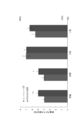
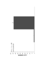
- the body weight increase of a newborn mouse group fed with artificial milk added with CCL25 (CCL25 added group) and a newborn mouse group fed with artificial milk not added with CCL25 (control group) over time It is the graph which compared. * Indicates that there was a significant difference compared to the control group (P> 0.05). It is the graph which compared the organ weight per body weight in a CCL25 addition group and a control group. * And ** indicate that there was a significant difference compared to the control group (*: P> 0.05, **: P> 0.01). It is the graph which compared the number of IgA producing cells in the intestinal tract of a CCL25 addition group and a control group.
- the immune function development promoting agent and growth promoting agent in this embodiment are intended for mammals including humans and non-human animals, and preferably for humans.
- the immune function development promoter and the growth promoter are preferably for infants, more preferably for newborns.
- Infant means a child growing on breast milk or artificial milk.
- an infant can mean a child less than one year after birth and a newborn can mean a child less than 28 days after birth.
- non-human animals it means individuals with periods corresponding to human infants and newborns, respectively.
- the immune function development promoter and growth promoter containing CCL25 in the present embodiment are not particularly limited as long as the composition contains CCL25 in an active state, but milk itself obtained from the mother is included. Absent.
- CCL25 those produced by a known method may be used, or commercially available ones may be used.
- the form of giving the immune function development promoter and growth promoter to the subject in the present embodiment is not particularly limited, and can be for oral intake, enteral intake, intravenous administration, etc. It is preferable to do.
- the form in which the immune function development promoter and growth promoter in the present embodiment are for oral intake of the subject is not particularly limited, and can be used by adding to a medium suitable for oral intake by the subject. It is preferable to use it by adding it to milk.
- Artificial milk does not use milk obtained from the mother as it is, but means artificially manufactured and processed milk, such as infant formulas, premature infant formulas, pharmaceutical formulas and liquid formulas for pharmaceutical use. Can be mentioned.
- Immune function development promoter means that when the composition is ingested or administered, the development of the subject's immune function is promoted compared to a subject who has not been ingested or who has not been administered. It means what is recognized.
- As an index for judging whether or not the gene is promoted for example, the expression level of genes and / or proteins involved in the immune system, the weight of an organ that is an immune system organ such as the intestinal tract (large and small intestine), spleen, and thymus Per unit weight, IgA producing cells, lymphocytes (T cells such as killer T cells and helper T cells, B cells and natural killer cells), the number of cells involved in the immune system such as double negative cells and double positive cells, small intestine The number and size of the Peyer plates can be used.
- the expression level of genes and / or proteins whose expression level increases when the immune function is working compared to a subject who does not take the composition or is not administered, intestinal tract, spleen, thymus
- the weight per body weight is significantly high, the number of IgA producing cells, the number of lymphocytes, the number of Peyer's patches are significantly increased, or the size of Peyer's patches is significantly large, etc. It can be determined that the development of immune function is promoted by administration.
- genes and / or proteins whose expression level increases when the immune function is working include surface markers of various immune cells, various cytokines, and immunoglobulins containing IgA.
- various immune cells include surface markers of various immune cells, various cytokines, and immunoglobulins containing IgA.
- IgA immunoglobulins containing IgA.
- lymphocytes killer T cells and helpers
- T cells such as T cells, B cells and natural killer cells), double negative cells, double positive cells, macrophages and leukocytes.
- Growth-promoting agent means that when the composition is ingested or administered, it is recognized that the growth of the subject is promoted compared to a subject who is not ingested or who is not administered To do.
- the expression level of a gene and / or protein involved in growth, the body weight, muscle mass, organ weight, etc. of the subject can be used. That is, compared to a subject who does not take the composition or a subject who is not administered, the expression amount of a gene and / or protein whose expression level increases when growth is promoted, the subject's body weight, muscle mass, When the organ weight or the like is significantly high, it can be determined that growth is promoted by ingestion or administration of the composition.
- genes and / or proteins whose expression level increases when growth is promoted include growth hormone, thyroid hormone, and glucocorticoid.
- the day when mating was confirmed was defined as the 0th day of pregnancy, and one female was isolated the day before childbirth (19th day of pregnancy).
- the day of birth is defined as day 0, day 0 after birth (parental cesarean section), day 1 after birth, day 2 after birth, day 5 after birth, day 10 after birth, day 21 after birth (after weaning) 6 groups were prepared, and the intestinal tract (small intestine and large intestine) was removed from the newborns at each period.
- RNAlater (TaKaRa), allowed to permeate at 4 ° C. overnight, and stored at ⁇ 80 ° C.
- the small intestine was divided into three equal parts, and each was (1) immersed in RNAlater (TaKaRa) as a sample for real-time PCR, allowed to permeate at 4 ° C overnight, and stored at -80 ° C.
- RNA was prepared according to the protocol using RNeasy Mini Kit (QIAGEN).
- Super Script TM First-Strand Synthesis System (Invitrogen) was used.
- Fast Start Essential DNA Green Master 2 x conc. (Roche) 10 ⁇ l, Sense primer (10 ⁇ M) 1 ⁇ l, Anti-sense primer (10 ⁇ M) 1 ⁇ l, cDNA 1 ⁇ l, Fast Start Essential DNA Green Master H 2 O (Roche) 7 ⁇ l were added in a total volume of 20 ⁇ l.
- real-time PCR was performed using Light Cycler (trademark) Nano (Roche).
- Primers used for real-time PCR were GAPDH (PCR product size 136 bp): 5′-GGGTGTGAACCCATGAGAAGT-3 ′ (SEQ ID NO: 1; forward), 5′-GACTGTGGTCCATGAGTTCCT-3 ′ (SEQ ID NO: 2; Reverse), CCL25 (PCR product size 131 bp): 5′-CCATCAGCAGCAGTAAGAGGG-3 ′ (SEQ ID NO: 3; forward), 5′-CTGTAGGGGACGGGTTTTAT-3 ′ (SEQ ID NO: 4; reverse), CCR9 (PCR product size 110 bp) : 5′-GCCTGAGCAGGGAGATTAT-3 ′ (SEQ ID NO: 5; forward), 5′-GAGCAGACAGAGTG-3 ′ (SEQ ID NO: 6; reverse).
- CCL25 and CCR9 and GAPDH as an internal standard were used.
- the reaction conditions of each gene were as follows: initial denaturation at 95 ° C. for 3 minutes, CCL25: heat denaturation (95 ° C., 60 seconds), annealing (55 ° C., 50 seconds), extension reaction (72 ° C., 60 seconds) 40 cycles in total, CCR9: heat denaturation (95 ° C, 60 seconds), annealing (58 ° C, 50 seconds), elongation reaction (72 ° C, 60 seconds), 45 cycles in total, GAPDH: heat denaturation (95 ° C, 60 seconds) , Annealing (55 ° C., 50 seconds) and extension reaction (72 ° C., 60 seconds) were carried out under a total of 45 cycles.
- the intestinal tissue stored at ⁇ 80 ° C. was equilibrated at ⁇ 20 ° C. for about 20 minutes before preparation of the slice (to prevent crushing during the preparation of the slice), and then the intestinal tissue block was made to a thickness of 8 ⁇ m with a cryostat. It was sliced and attached to a silane-coated non-fluorescent glass slide (FRC-11; Matsunami). After the sections were dried on the slide, they were immersed in cold acetone at ⁇ 20 ° C. for 10 minutes and air-dried. The sections were washed three times for 5 minutes each with a washing buffer (1 ⁇ PBS), and then immunostained.
- FRC-11 silane-coated non-fluorescent glass slide
- primary and secondary antibodies were used for CCL25 and IgA, respectively.
- Each section was blocked with 1% BSA-containing PBS blocking solution for 1 hour at room temperature.
- the blocking solution was removed, and the primary antibody diluted with the blocking solution was added to each section and incubated overnight at 4 ° C.
- the primary antibody solution was removed, and the sections were washed 3 times with 1 ⁇ PBS for 5 minutes each.
- a secondary antibody diluted with a 1% BSA-containing PBS blocking solution was added to each section and incubated at room temperature for 2 hours. Then, the secondary antibody was removed, and the section was washed 3 times with 1 ⁇ PBS for 5 minutes each.
- Casein (40.0 g) was added to the alkaline aqueous solution and dissolved. This Casein mixture was sterilized for 30 minutes in a bath with boiling water. Next, casein salt micelles were prepared.
- C Calcium and magnesium were added to Casein mixture. CaCl 2 ⁇ 2H 2 O (1.7 g), GlyCaPO 4 (8 g) and MgCl 2 ⁇ 6H 2 O (1.9 g) are dissolved in 50 ml of distilled water (autoclave at 121 ° C. for 10 minutes) and homogenized with a polytron-like mixer. did. This was slowly added to the Casein mixture (keep mixing with a homogenizer).
- the small intestine of an artificially bred mouse was removed, the small intestine (from the duodenum to the jejunum) was pinned, and immersion fixation was performed in a 10% formalin solution for 3 hours or more. After the fixing treatment, the plate was washed several times with distilled water, and stained with Mayer's Hematoxylin Solution for 7 minutes. Then, after washing once with distilled water, it was washed with running water for 1 hour.
- Example 1 Observation of CCL25 and CCR9 expression in the neonatal intestinal tract and changes in IgA-producing cells over time
- the expression of CCL25 mRNA in intestinal tissue was analyzed by real-time PCR.
- the expression level in the 21-day-old group at the weaning time is 1, the expression level of CCL25 mRNA in the small intestine is 0.12 ⁇ 0.07 in the 0-day group and 0 in the 1-day group. 0.09 ⁇ 0.06, 0.39 ⁇ 0.04 in the 2nd day group, 0.62 ⁇ 0.003 in the 5th day group, and 0.70 ⁇ 0.05 in the 10th day group. It was.
- the expression level of CCL25 mRNA begins to increase from the second day after birth, and the amount increases with time. From the fifth day group to the 21st day group, the expression level is higher than that of the 0th day group. Increased significantly. Moreover, the expression level in the large intestine is 0.41 ⁇ 0.04 in the 0th day group, 0.33 ⁇ 0.01 in the 1st day group, and 0.27 ⁇ 0.02 in the 2nd day group. The value was 0.08 ⁇ 0.006 in the 5th day group and 0.09 ⁇ 0.006 in the 10th day group, and was highest in the 21st day group.
- CCR9 the only receptor of CCL25
- the 21st day group is set to 1, 0.15 ⁇ 0.04 in the 0th day group, 0.25 ⁇ 0.006 in the 1st day group, and the second day group 0.02 ⁇ 0.01, 0.50 ⁇ 0.01 in the 5th day group, and 0.7 ⁇ 0.01 in the 10th day group. That is, the expression level of CCR9 mRNA began to increase significantly from the fifth day after birth compared with the first day after birth, and the amount increased with time. And the amount of CCR9 mRNA of the 21st day group showed the highest value.
- the expression level in the large intestine is 0.006 ⁇ 0.0005 in the 0th day group, 0.036 ⁇ 0.008 in the 1st day group, 0.005 ⁇ 0.001 in the 2nd day group, after birth It is 0.005 ⁇ 0.0003 in the 5th day group and 0.004 ⁇ 0.0004 in the 10th day group, and the expression level of CCR9 mRNA is the 1st day group, the 2nd day group, and the 5th day after birth.
- the eye group and the 10th day group showed low values, and it was found that the 21st day group was the peak.
- the mRNA expression levels of CCL25 and CCR9 in the large intestine and small intestine on the 10th day after birth were compared.
- the expression level of the small intestine was 1
- the expression level of CCL25 mRNA in the large intestine was 0.07 ⁇ 0.009.
- the CCR9 mRNA expression level was 0.06 ⁇ 0.003 when the expression level of the small intestine was 1, and the CCR9 mRNA expression level in the large intestine was 0.06 ⁇ 0.003.
- the expression levels of CCL25 mRNA and CCR9 mRNA in the large intestine were significantly lower than those in the small intestine. From this result, it was confirmed that the expression of CCL25 in the neonatal intestinal tract was mainly expressed in the small intestine as well as after weaning in which the intestinal tract function was established.
- Example 3 Effect of CCL25 on immune organ weight and Peyer's patch
- newborn mice were divided into a CCL25-added group and a control group, and the animals were dissected after being artificially incubated for 7 days until the age of 10 days. Comparison was made between the addition group and the control group.
- the spleen (SP) in the control group was 3.95% ⁇ 0.05
- the thymus (Thy) was 4.00% ⁇ 0.00.
- the large intestine (LI) was 7.02% ⁇ 0.08 and the small intestine (SI) was 5.46% ⁇ 0.10
- the spleen weight (SP) of the CCL25 addition group was 4.99. % ⁇ 0.058, thymus weight (Thy) 4.95% ⁇ 0.059
- large intestine weight (LI) 6.99% ⁇ 0.04 and small intestine weight (SI) 6.44% ⁇ 0.06 Met In the CCL25 addition group, the weight of the small intestine tended to be higher than that of the control group, and the spleen and thymus weights were significantly higher (FIG. 2). In contrast, the weight of the large intestine per body weight was almost the same in the control group and the CCL25 added group.
- the small intestine stained with hematoxin was observed with a microscope, the number of Peyer's patches in the small intestine was counted, and the number of Peyer's patches per animal was calculated. Further, the major axis and the minor axis of each Peyer plate were measured, and the average was taken as the size of one Peyer plate, and the size of the Peyer plate per animal was calculated. As a result of comparing the number and size of Peyer's plates for the control group and the CCL25 addition group, there was no difference in the size of each Peyer's plate, but the number of Peyer's plates was the control group in the CCL25 addition group. There were more trends.
- Example 4 Effect of CCL25 on CCL25 mRNA and CCR9 mRNA Expression in Intestinal Tissue
- the expression level of the small intestine CCL25 mRNA in the control group was 1
- the expression level in the CCL25 addition group was 0.79 ⁇ 0.11.
- the control group showed a higher expression level.
- the expression level in the CCL25 addition group was 1.29 ⁇ 0.16. Although there was no significant difference, there was a tendency for the expression level to be higher in the CCL25 addition group.
- Example 5 Effect of CCL25 on the number of IgA-producing cells in the intestinal villi of the small intestine Immunohistochemical staining of IgA-producing cells in small intestine tissues was performed using IgA-RITC antibody. Seven villi of one intestinal tract (small intestine) were selected, and the number of IgA-producing cells that showed positive among them was counted and compared between the control group and the CCL25 addition group. The number of IgA-producing cells that could be confirmed in the 7 villi was hardly seen at 1.33 ⁇ 0.2 in the control group, whereas it was significantly larger at 43.75 ⁇ 1.24 in the CCL25 addition group. (FIG. 3). From this result, it became clear that CCL25 in milk is essential for attracting IgA until the 10th day after delivery.
- Example 6 Effect of CCL25 on the number of cells in the thymus Experimental animals used were 10-20 week old ddY mice (Japan SLC) and newborns of ddY mice. In accordance with the method described in the literature of Yajima et al. (Biosci. Biotechnol. Biochem., 2007, 71 (10), 2420-2427 and Experimental animals, 2006, 55 (4), 391-397) After breastfeeding until 2 days after birth, artificial breast feeding was performed using each artificial milk for 8 days from 2 days to 10 days of age.
- Newborn mice were (1) a group in which 0.5 ⁇ g / mL of CCL25 (Recombinant Mouse CCL25 (TECK, Bio-Legend)) was added to artificial milk (CCL25 added group) and (2) a group in which no CCL25 antigen was added (control) Divided into two experimental groups.
- CCL25 Recombinant Mouse CCL25 (TECK, Bio-Legend)
- the artificial milk for mice was prepared by correcting the content of selenium from 16% to 18%.
- the method for producing artificial milk is shown below.
- (A) NaOH (1 g) and KOH (6 g) were dissolved in 800 ml of distilled water (alkaline aqueous solution). Three amino acids, serine (1.15 g), cysteine (0.9 g) and tryptophan (1.08 g) were added to the alkaline aqueous solution (always stirred with a stirrer, 60-70 ° C.).
- Casein (160.0 g) was added to the alkaline aqueous solution and dissolved. This Casein mixture was sterilized for 30 minutes in a bath with boiling water.
- C CaCl 2 ⁇ 2H 2 O (6.8 g), GlyCaPO 4 (32 g) and MgCl 2 ⁇ 6H 2 O (7.6 g) were dissolved in 200 ml of distilled water and homogenized with a polytron-like mixer. This was slowly added to the Casein mixture (keep mixing with a homogenizer).
- CaCO 3 .2H 2 O (10 g) and Ca-citrate (4.8 g) were dissolved in 100 ml of distilled water, autoclaved, and then slowly added to Casein mixture.
- the artificial milk was aseptically dispensed into 50 ml sterile polypropylene bottles and stored at ⁇ 80 ° C.
- the frozen artificial milk was sterilized by gamma irradiation (30 KGy) and stored at ⁇ 80 ° C.
- the newborn mouse that was breastfed from the 0th day to the 2nd day was separated from the parent on the day of birth as the 0th day after birth, and artificially administered by oral administration using a sucker once every 3 hours from the 2nd day of birth. Lactated milk.
- the amount of mammals and body weight were measured over time, and after 8 days of artificial nursing until the 10th day, they were euthanized by excessive inhalation of diethyl ether, dissected, and the thymus was removed.
- the weight of the extracted thymus was measured and taken out into a petri dish containing a phosphate-buffered salted salt solution (PBBS) containing 0.1% bovine serum albumin (BSA).
- PBBS phosphate-buffered salted salt solution
- BSA bovine serum albumin
- the remaining thymocytes were added with ammonium chloride solution for removing red blood cells (Tris-buffered ammonium chloride: ACTB), pipetted, and allowed to stand at room temperature for 5 minutes for hemolysis. Then, cold PBBS was added, centrifugation was performed at 800 rpm for 5 minutes, and the supernatant was removed by decantation. For washing, cold PBBS was added, centrifugation at 800 rpm for 5 minutes and supernatant removal were performed twice, and then transferred to an Eppendorf tube. The obtained solution was appropriately diluted, the total number of cells and the number of lymphocytes were counted, and compared in the control group and the CCL25 addition group. All statistical processing on experimental results was performed using t-test.
- the total number of cells in the thymus tended to be higher in the CCL25 added group than in the control group. Moreover, the number of lymphocytes per total cell number tended to be higher in the CCL25 addition group than in the control group (FIG. 4).
Landscapes
- Health & Medical Sciences (AREA)
- Life Sciences & Earth Sciences (AREA)
- Chemical & Material Sciences (AREA)
- Immunology (AREA)
- Medicinal Chemistry (AREA)
- General Health & Medical Sciences (AREA)
- Organic Chemistry (AREA)
- Engineering & Computer Science (AREA)
- Veterinary Medicine (AREA)
- Public Health (AREA)
- Pharmacology & Pharmacy (AREA)
- Animal Behavior & Ethology (AREA)
- Proteomics, Peptides & Aminoacids (AREA)
- Bioinformatics & Cheminformatics (AREA)
- Epidemiology (AREA)
- Zoology (AREA)
- Gastroenterology & Hepatology (AREA)
- Nutrition Science (AREA)
- Polymers & Plastics (AREA)
- Food Science & Technology (AREA)
- General Chemical & Material Sciences (AREA)
- Nuclear Medicine, Radiotherapy & Molecular Imaging (AREA)
- Chemical Kinetics & Catalysis (AREA)
- Biophysics (AREA)
- Toxicology (AREA)
- Biochemistry (AREA)
- Genetics & Genomics (AREA)
- Molecular Biology (AREA)
- Mycology (AREA)
- Physiology (AREA)
- Pediatric Medicine (AREA)
- Coloring Foods And Improving Nutritive Qualities (AREA)
- Medicines That Contain Protein Lipid Enzymes And Other Medicines (AREA)
- Medicines Containing Material From Animals Or Micro-Organisms (AREA)
Abstract
本発明は、CCL25を含有する免疫機能発達促進剤及び成長促進剤を開示する。本発明の免疫機能発達促進剤を摂取することで、対象の免疫機能の発達を促進することができる。また、本発明の成長促進剤を摂取することで、対象の成長を促進することができる。
Description
本発明は、免疫機能発達促進剤及び成長促進剤に関する。
新生児の初期免疫機能獲得には母乳摂取が重要な役割を果たしている。特に、出産直後に分泌される初乳には、IgA抗体や免疫機能に関与する物質が多く含まれており、新生児の感染防御に重要な役割を果たしている。その免疫機能関与物質の一つにケモカインと呼ばれるタンパク質があり、CXCL8(IL-8)、CCL5(RANTES)及びCCL28が母乳に含まれるケモカインとして知られている。しかしながら、例えば、初乳の製造・分泌時期には乳腺組織内でCCL28の発現がほとんど見られないことが報告されている。このように、初乳の摂取による新生児の免疫獲得において、ケモカインがどのように関与しているかについては依然として不明である。
上記のケモカインの他、マウス、ヒトの胸腺で発見され、腸管免疫に関与するケモカインの一種に、Chemokine(C-C motif)ligand25(CCL25)がある。これまでに、母体におけるCCL25の働きとして、乳腺組織内での初乳へのIgAの移行にCCL25が関わっている可能性が報告されている(非特許文献1)。しかしながら、CCL25についても初乳の摂取による新生児の免疫獲得における働きは明らかにされていない。
竹中正樹、茶山和敏、"妊娠および必乳中のマウス乳腺内におけるCCL25ケモカインの発現"、2010年3月5日、第11回静岡ライフサイエンスシンポジウム要旨集、p.19
本発明は、CCL25の新たな機能を同定することを目的とする。
本発明者らは、驚くべきことに、これまで母体で発現することによって乳汁へのIgAの移行に関与していると考えられていたCCL25が、乳汁自体に含まれており、乳汁を摂取する対象へ直接働きかけることで免疫機能の発達を促進する機能及び成長を促進する機能を有することを見出し、本発明を完成するに至った。すなわち、本発明は、以下の[1]~[6]を提供する。
[1]CCL25を含有する免疫機能発達促進剤。
[2]乳児用である[1]の免疫機能発達促進剤。
[3]経口摂取用である[1]又は[2]の免疫機能発達促進剤。
[4]CCL25を含有する成長促進剤。
[5]乳児用である[4]の成長促進剤。
[6]経口摂取用である[4]又は[5]の成長促進剤。
さらに、本発明は、以下の[7]~[24]も提供する。
[7]免疫機能発達促進剤の製造のためのCCL25の使用。
[8]免疫機能発達促進剤が乳児用である[7]の使用。
[9]免疫機能発達促進剤が経口摂取用である[7]又は[8]の使用。
[10]成長促進剤の製造のためのCCL25の使用。
[11]成長促進剤が乳児用である[10]の使用。
[12]成長促進剤が経口摂取用である[10]又は[11]の使用。
[13]対象の免疫機能発達促進に使用するためのCCL25。
[14]対象が乳児である[13]のCCL25。
[15]対象がCCL25を経口摂取する[13]又は[14]のCCL25。
[16]対象の成長促進に使用するためのCCL25。
[17]対象が乳児である[16]のCCL25。
[18]対象がCCL25を経口摂取する[16]又は[17]のCCL25。
[19]必要とする対象にCCL25を投与する、対象の免疫機能発達を促進する方法。
[20]対象が乳児である[19]の方法。
[21]CCL25を経口投与する[19]又は[20]の方法。
[22]必要とする対象にCCL25を投与する、対象の成長を促進する方法。
[23]対象が乳児である[22]の方法。
[24]CCL25を経口投与する[22]又は[23]の方法。
[1]CCL25を含有する免疫機能発達促進剤。
[2]乳児用である[1]の免疫機能発達促進剤。
[3]経口摂取用である[1]又は[2]の免疫機能発達促進剤。
[4]CCL25を含有する成長促進剤。
[5]乳児用である[4]の成長促進剤。
[6]経口摂取用である[4]又は[5]の成長促進剤。
さらに、本発明は、以下の[7]~[24]も提供する。
[7]免疫機能発達促進剤の製造のためのCCL25の使用。
[8]免疫機能発達促進剤が乳児用である[7]の使用。
[9]免疫機能発達促進剤が経口摂取用である[7]又は[8]の使用。
[10]成長促進剤の製造のためのCCL25の使用。
[11]成長促進剤が乳児用である[10]の使用。
[12]成長促進剤が経口摂取用である[10]又は[11]の使用。
[13]対象の免疫機能発達促進に使用するためのCCL25。
[14]対象が乳児である[13]のCCL25。
[15]対象がCCL25を経口摂取する[13]又は[14]のCCL25。
[16]対象の成長促進に使用するためのCCL25。
[17]対象が乳児である[16]のCCL25。
[18]対象がCCL25を経口摂取する[16]又は[17]のCCL25。
[19]必要とする対象にCCL25を投与する、対象の免疫機能発達を促進する方法。
[20]対象が乳児である[19]の方法。
[21]CCL25を経口投与する[19]又は[20]の方法。
[22]必要とする対象にCCL25を投与する、対象の成長を促進する方法。
[23]対象が乳児である[22]の方法。
[24]CCL25を経口投与する[22]又は[23]の方法。
本発明の免疫機能発達促進剤を摂取することで、対象の免疫機能の発達を促進することができる。また、本発明の成長促進剤を摂取することで、対象の成長を促進することができる。
本実施形態における免疫機能発達促進剤及び成長促進剤は、ヒト及び非ヒト動物を含む哺乳類を対象としており、ヒトを対象とすることが好ましい。
本実施形態において、免疫機能発達促進剤及び成長促進剤は、好ましくは乳児用であり、より好ましくは新生児用である。乳児とは、母乳又は人工乳で生育されている子供を意味する。出生からの経過期間に特に拘るものではないが、ヒトの場合、例えば乳児は出生後1年未満の子供を意味することができ、新生児は出生後28日未満の子供を意味することができる。非ヒト動物においては、それぞれヒトの乳児及び新生児に対応する期間の個体を意味する。
本実施形態におけるCCL25を含有する免疫機能発達促進剤及び成長促進剤は、CCL25が活性を有する状態で含まれている組成物であれば特に限定されないが、母体から得られた乳汁そのものは含まれない。
CCL25は、公知の方法により製造したものを用いてもよく、市販のものを用いてもよい。
本実施形態における免疫機能発達促進剤及び成長促進剤を対象に与える形式は、特に限定されず、経口摂取用、経腸摂取用、経静脈投与用等とすることができるが、経口摂取用とすることが好ましい。
本実施形態における免疫機能発達促進剤及び成長促進剤を対象の経口摂取用とする形態は、特に限定されず、対象が経口摂取するのに適当な媒体に添加して用いることができるが、人工乳に添加して用いることが好ましい。
人工乳は、母体から得られた乳汁をそのまま用いたものではなく、人工的に製造、加工された乳汁を意味し、例えば乳児用、未熟児用、医薬用の調製粉乳及び液体調製乳等が挙げられる。
免疫機能発達促進剤とは、その組成物を摂取した場合又は投与された場合に、対象の免疫機能の発達が、摂取していない対象又は投与されていない対象と比較して促進していると認められるものを意味する。促進しているか否かについて判断するための指標としては、例えば免疫系に関与する遺伝子及び/又はタンパク質の発現量、腸管(大腸及び小腸)、脾臓、胸腺等の免疫系器官である臓器の体重あたりの重量、IgA産生細胞、リンパ球(キラーT細胞及びヘルパーT細胞等のT細胞、B細胞及びナチュラルキラー細胞)、ダブルネガティブ細胞及びダブルポジティブ細胞等の免疫系に関与する細胞の数、小腸内のパイエル板の個数及び大きさ等を用いることができる。すなわち、その組成物を摂取していない対象又は投与されていない対象と比較して、免疫機能が働いている際に発現量が増加する遺伝子及び/又はタンパク質の発現量、腸管、脾臓、胸腺の体重あたりの重量が有意に高い場合、IgA産生細胞数、リンパ球数、パイエル板の個数が有意に多い場合、及びパイエル板の大きさが有意に大きい場合等には、その組成物の摂取又は投与により、免疫機能の発達が促進されていると判断することができる。
免疫機能が働いている際に発現量が増加する遺伝子及び/又はタンパク質としては、例えば各種免疫細胞の表面マーカー類、各種サイトカイン類及びIgAを含む免疫グロブリン等が挙げられる。免疫系器官としては、腸管、脾臓、胸腺の他にも、リンパ節、骨髄等が挙げられ、免疫系に関与する細胞としては、IgA産生細胞の他にも、リンパ球(キラーT細胞及びヘルパーT細胞等のT細胞、B細胞及びナチュラルキラー細胞)、ダブルネガティブ細胞、ダブルポジティブ細胞、マクロファージ及び白血球等が挙げられる。
成長促進剤とは、その組成物を摂取した場合又は投与された場合に、対象の成長が、摂取していない対象又は投与されていない対象と比較して促進していると認められるものを意味する。促進しているか否かについて判断するための指標としては、成長に関与する遺伝子及び/又はタンパク質の発現量、対象の体重、筋量、臓器重量等を用いることができる。すなわち、その組成物を摂取していない対象又は投与されていない対象と比較して、成長が促進される際に発現量が増加する遺伝子及び/又はタンパク質の発現量、対象の体重、筋量、臓器重量等が有意に高い場合には、その組成物の摂取又は投与により、成長が促進されている判断することができる。
成長が促進される際に発現量が増加する遺伝子及び/又はタンパク質としては、例えば成長ホルモン、甲状腺ホルモン及び糖質コルチコイド等が挙げられる。
本発明を以下の実施例に基づいてさらに詳細に説明するが、本発明はこれらの実施例に限定されるものではない。
(新生仔マウス実験群)
交尾が確認できた日を妊娠0日目とし、出産する前日(妊娠19日目)にメスを1匹に隔離した。そして、出産した日を0日目とし、生後0日目(親を帝王切開)、生後1日目、生後2日目、生後5日目、生後10日目、生後21日目(離乳後)の6群を作成し、各時期の新生仔から腸管(小腸及び大腸)を摘出した。
交尾が確認できた日を妊娠0日目とし、出産する前日(妊娠19日目)にメスを1匹に隔離した。そして、出産した日を0日目とし、生後0日目(親を帝王切開)、生後1日目、生後2日目、生後5日目、生後10日目、生後21日目(離乳後)の6群を作成し、各時期の新生仔から腸管(小腸及び大腸)を摘出した。
(腸管試料の作製)
マウス新生仔を、毒物の使用基準に従ってジエチルエーテル過剰吸入によって安楽死させ、腸管(小腸及び大腸)を摘出した。大腸はRNAlater(TaKaRa社)に漬けて一晩4℃で浸透させた後、-80℃で保存した。小腸は3等分にし、それぞれを、(1)リアルタイムPCRに用いるサンプルとして、RNAlater(TaKaRa社)に漬けて一晩4℃で浸透させた後、-80℃で保存し、(2)免疫染色に用いるサンプルとして、プラスチック製のクリオモルド(角型1号;Sakura Finetek社)に封入剤であるティシューテックO.C.T.コンパウンド(Sakura Finetek社)と共に入れ、ドライアイス上で速やかに凍結させ、凍結切片用のブロックを作製し、-80℃で保存し、(3)ヘマトキシリン・エオジン(Hematoxylin and Eosin Stain;HE Stain)組織染色に用いるサンプルとして、4%パラホルムアルデヒド固定液に24~36時間浸漬させ、固定処理を行った。組織染色用サンプルは固定処理後に1×PBSで3~4回洗浄し、組織標本の作製のために70%エタノール溶液中で保存した。
マウス新生仔を、毒物の使用基準に従ってジエチルエーテル過剰吸入によって安楽死させ、腸管(小腸及び大腸)を摘出した。大腸はRNAlater(TaKaRa社)に漬けて一晩4℃で浸透させた後、-80℃で保存した。小腸は3等分にし、それぞれを、(1)リアルタイムPCRに用いるサンプルとして、RNAlater(TaKaRa社)に漬けて一晩4℃で浸透させた後、-80℃で保存し、(2)免疫染色に用いるサンプルとして、プラスチック製のクリオモルド(角型1号;Sakura Finetek社)に封入剤であるティシューテックO.C.T.コンパウンド(Sakura Finetek社)と共に入れ、ドライアイス上で速やかに凍結させ、凍結切片用のブロックを作製し、-80℃で保存し、(3)ヘマトキシリン・エオジン(Hematoxylin and Eosin Stain;HE Stain)組織染色に用いるサンプルとして、4%パラホルムアルデヒド固定液に24~36時間浸漬させ、固定処理を行った。組織染色用サンプルは固定処理後に1×PBSで3~4回洗浄し、組織標本の作製のために70%エタノール溶液中で保存した。
(リアルタイムPCR法)
トータルRNAは、RNeasy Mini Kit(QIAGEN社)を用いて、プロトコルに従って調製した。cDNAの合成にはSuper Script(商標)First-Strand Synthesis System(Invitrogen社)を使用した。リアルタイムPCR用チューブにFast Start Essential DNA Green Master 2xconc.(Roche社)10μl、Sense primer(10μM)1μl、Anti-sense primer(10μM)1μl、cDNA1μl、Fast Start Essential DNA Green Master H2O(Roche社)7μlを加えて全反応量を20μlとした。そして、Light Cycler(商標)Nano(Roche社)を使用してリアルタイムPCRを行った。リアルタイムPCRに用いたプライマー(Sigma-Aldrich社)は、GAPDH(PCR産物のサイズ 136bp):5’-GGGTGTGAACCATGAGAAGT-3’(配列番号1;フォワード)、5’-GACTGTGGTCATGAGTCCT-3’(配列番号2;リバース)、CCL25(PCR産物のサイズ 131bp):5’-CCATCAGCAGCAGTAAGAGG-3’(配列番号3;フォワード)、5’-CTGTAGGGCGACGGTTTTAT-3’(配列番号4;リバース)、CCR9(PCR産物のサイズ 110bp):5’-GCCTGAGCAGGGAGATTAT-3’(配列番号5;フォワード)、5’-GAGCAGACAGAGTG-3’(配列番号6;リバース)である。解析する遺伝子としては、CCL25、CCR9及び内部標準としてGAPDHを用いた。それぞれ遺伝子の反応条件は、初期変性を95℃で3分間行った後、CCL25:熱変性(95℃、60秒)、アニーリング(55℃、50秒)、伸長反応(72℃、60秒)を全40サイクル、CCR9:熱変性(95℃、60秒)、アニーリング(58℃、50秒)、伸長反応(72℃、60秒)を全45サイクル、GAPDH:熱変性(95℃、60秒)、アニーリング(55℃、50秒)、伸長反応(72℃、60秒)を全45サイクル、の各条件で増幅を行った。
トータルRNAは、RNeasy Mini Kit(QIAGEN社)を用いて、プロトコルに従って調製した。cDNAの合成にはSuper Script(商標)First-Strand Synthesis System(Invitrogen社)を使用した。リアルタイムPCR用チューブにFast Start Essential DNA Green Master 2xconc.(Roche社)10μl、Sense primer(10μM)1μl、Anti-sense primer(10μM)1μl、cDNA1μl、Fast Start Essential DNA Green Master H2O(Roche社)7μlを加えて全反応量を20μlとした。そして、Light Cycler(商標)Nano(Roche社)を使用してリアルタイムPCRを行った。リアルタイムPCRに用いたプライマー(Sigma-Aldrich社)は、GAPDH(PCR産物のサイズ 136bp):5’-GGGTGTGAACCATGAGAAGT-3’(配列番号1;フォワード)、5’-GACTGTGGTCATGAGTCCT-3’(配列番号2;リバース)、CCL25(PCR産物のサイズ 131bp):5’-CCATCAGCAGCAGTAAGAGG-3’(配列番号3;フォワード)、5’-CTGTAGGGCGACGGTTTTAT-3’(配列番号4;リバース)、CCR9(PCR産物のサイズ 110bp):5’-GCCTGAGCAGGGAGATTAT-3’(配列番号5;フォワード)、5’-GAGCAGACAGAGTG-3’(配列番号6;リバース)である。解析する遺伝子としては、CCL25、CCR9及び内部標準としてGAPDHを用いた。それぞれ遺伝子の反応条件は、初期変性を95℃で3分間行った後、CCL25:熱変性(95℃、60秒)、アニーリング(55℃、50秒)、伸長反応(72℃、60秒)を全40サイクル、CCR9:熱変性(95℃、60秒)、アニーリング(58℃、50秒)、伸長反応(72℃、60秒)を全45サイクル、GAPDH:熱変性(95℃、60秒)、アニーリング(55℃、50秒)、伸長反応(72℃、60秒)を全45サイクル、の各条件で増幅を行った。
(免疫組織化学的染色)
-80℃で保存した腸管組織を、切片作製の前に、-20℃で約20分間平衡化(切片作製時の粗砕を防止)した後、腸管組織ブロックをクリオスタットで8μmの厚さに薄切し、シランコートした無蛍光スライドガラス(FRC-11;Matsunami社)に貼り付けた。切片がスライド上で乾いた後、冷アセトンで-20℃で10分間浸漬し、風乾した。洗浄バッファー(1XPBS)で切片を各5分間、3回洗浄した後、免疫染色を行った。免疫染色には、CCL25及びIgAそれぞれについて一次抗体及び二次抗体を用いた。1%BSA含有PBSブロッキング液で、各切片を室温で1時間ブロッキング処理した。ブロッキング液を除去し、ブロッキング液で希釈した一次抗体を各切片に添加し、4℃で一晩インキュベートした。一次抗体液を除去し、1XPBSで切片を各5分間、3回洗浄した。1%BSA含有PBSブロッキング液で希釈した二次抗体を各切片に添加し、室温で2時間インキュベートした後、二次抗体を除去し、1XPBSで切片を各5分間、3回洗浄した。その後、水溶性封入剤(Thermo Scientific社)にてスライドグラス上で封入し、実体蛍光顕微鏡(MZ10F;Leica社)にて観察を行った。免疫組織化学的染色に使用した抗体は、CCL25:Goat anti-mouse CCL25/TECK antibody(一次抗体;R&D Systems社)、Fluorescein(FITC)-conjugated AffiniPure Rabbit Anti-Goat++ IgG (H+L)(二次抗体;Jackson Immuno Research社)、IgA:Goat anti-mouse IgA affinity Purified(一次抗体;Bethyl Laboratories社)、Rhodamine(TRITC)-conjugated AffiniPure Donkey Anti-Goat++ IgG (H+L)(二次抗体;Jackson Immuno Research社)であり、それぞれ1:100の希釈倍率で用いた。
-80℃で保存した腸管組織を、切片作製の前に、-20℃で約20分間平衡化(切片作製時の粗砕を防止)した後、腸管組織ブロックをクリオスタットで8μmの厚さに薄切し、シランコートした無蛍光スライドガラス(FRC-11;Matsunami社)に貼り付けた。切片がスライド上で乾いた後、冷アセトンで-20℃で10分間浸漬し、風乾した。洗浄バッファー(1XPBS)で切片を各5分間、3回洗浄した後、免疫染色を行った。免疫染色には、CCL25及びIgAそれぞれについて一次抗体及び二次抗体を用いた。1%BSA含有PBSブロッキング液で、各切片を室温で1時間ブロッキング処理した。ブロッキング液を除去し、ブロッキング液で希釈した一次抗体を各切片に添加し、4℃で一晩インキュベートした。一次抗体液を除去し、1XPBSで切片を各5分間、3回洗浄した。1%BSA含有PBSブロッキング液で希釈した二次抗体を各切片に添加し、室温で2時間インキュベートした後、二次抗体を除去し、1XPBSで切片を各5分間、3回洗浄した。その後、水溶性封入剤(Thermo Scientific社)にてスライドグラス上で封入し、実体蛍光顕微鏡(MZ10F;Leica社)にて観察を行った。免疫組織化学的染色に使用した抗体は、CCL25:Goat anti-mouse CCL25/TECK antibody(一次抗体;R&D Systems社)、Fluorescein(FITC)-conjugated AffiniPure Rabbit Anti-Goat++ IgG (H+L)(二次抗体;Jackson Immuno Research社)、IgA:Goat anti-mouse IgA affinity Purified(一次抗体;Bethyl Laboratories社)、Rhodamine(TRITC)-conjugated AffiniPure Donkey Anti-Goat++ IgG (H+L)(二次抗体;Jackson Immuno Research社)であり、それぞれ1:100の希釈倍率で用いた。
(統計学的解析)
実験結果に関するすべての統計処理は、t-検定を用いて行い、統計学的有意水準は特に明示のない場合については5%(P>0.05)とした。
実験結果に関するすべての統計処理は、t-検定を用いて行い、統計学的有意水準は特に明示のない場合については5%(P>0.05)とした。
(人工乳汁の作製)
マウス用人工乳汁を用いた人工哺乳法に関する文献(Biosci. Biotechnol. Biochem.,2007, 71(10), 2420-2427及びExperimentalanimals, 2006, 55(4), 391-397)を参考に、脂肪の含有量を16%から18%に修正して、マウス用人工乳汁を作製した。以下に人工乳汁の作成方法を示す。初めに、Casein mixtureを作製した。
(a)セリン(0.2875g)、システイン(0.225g)及びトリプトファン(0.27g)の3種類のアミノ酸を、NaOH(0.25g)及びKOH(1.5g)を含む200mlのアルカリ水溶液に加えた(常にスターラーで撹拌、60~70℃)。
(b)カゼイン(40.0g)をアルカリ水溶液に加え、溶解した。このCasein mixtureを、沸騰しているお湯の入ったバス中で30分間滅菌した。次に、カゼイン塩のミセルを作製した。
(c)Casein mixtureにカルシウム及びマグネシウムを加えた。CaCl2・2H2O(1.7g)、GlyCaPO4(8g)及びMgCl2・6H2O(1.9g)を50mlの蒸留水(121℃で10分間オートクレーブ)に溶かし、ポリトロン様ミキサーでホモジナイズした。これをCasein mixtureにゆっくりと加えた(ホモジナイザーで混ぜ続ける)。
(d)CaCO3・2H2O(2.5g)及びCa-citrate(1.2g)を25mlの蒸留水に溶かし、オートクレーブした後、Casein mixtureにゆっくりと加えた。
(e)Na2HPO4(0.8g)及びKH2PO4(0.08g)を12.5mlの蒸留水に溶かし、オートクレーブで滅菌した後Casein mixtureに加えた。
(f)ラクトース水溶液(ラクトース18.91gを55mlの蒸留水に溶かしたもの)をオートクレーブで滅菌した後、ゆっくりとCasein mixtureに加えた。
(g)FeSO4・7H2O(0.48g)及びCitrate-H2O(0.01g)を5mlの蒸留水に溶かした。そのうちの2.5ml、ZnSO4・7H2O(0.3g)、CuSO4・5H2O(0.075g)及びMnSO4・5H2O(0.0125g)を6.25mlの蒸留水に溶かした。そのうちの1.25ml、NaF(0.00775g)及びKI(0.0125g)を6.25mlの蒸留水に溶かした。そのうちの1.25mlをCasein mixtureに加えた。この操作は、Millipore Filtration(0.45μm)で滅菌した後に、常に撹拌しながら行った。
(h)その後、Whey Protein Isolate(40g)及びWhey Protein Hydrolysate(50g)を250mlの滅菌蒸留水に溶かした。この溶液を、撹拌しながら40℃以下に冷ましたCasein mixtureに加えた。
(i)カルニチン(0.08g)、ピコリン酸(0.04g)、エタノールアミン(0.068g)及びタウリン(0.3g)を5mlの蒸留水に溶かした。そのうちの2.5mlを Casein mixtureに加えた。
(j)クエン酸二水素コリン(1.47g)を18.375mlの蒸留水に溶かした中和溶液(5N NaOHでpH7.0に中和)に、水溶性ビタミンmixture(シアノコバラミン(0.0119mg)、ビオチン(0.18mg)、葉酸(0.78mg)、チアミン塩酸塩(8.81mg)、ピリドキシン塩酸塩(1.25mg)、リン酸リボフラビンナトリウム(12.15mg)、パントテン酸カルシウム(26.43mg)、p-アミノ安息香酸(44.04mg)、ニコチン酸(49.88mg)、アスコルビン酸ナトリウム(674mg)、及び4-イノシトール(487.25mg))を溶かした。そのうちの17.5mlをCasein mixtureに加えた。
(k)脂溶性ビタミン(ビタミンK3(19.825mg)、ビタミンE:α-トコフェロール(23.45mg)及びビタミンAとDを含む混合物)並びに6種類の食用油(パーム油(53.85g)、ココナッツ油(44.9g)、コーン油(17.955g)、中鎖脂肪酸油(26.9325g)、大豆油(35.91g)及びコレステロール(0.4g))を沸騰しているお湯の入ったバス中で30分間滅菌した。この油性溶液を40~50℃まで冷ました後、撹拌しながらCasein mixtureに加えた。
(l)この最終混合物を、高圧条件下(180kg/cm2)で3回ホモジナイズした。
(m)ホモジナイズの後、人工乳汁は無菌的に50ml滅菌ポリプロピレンボトルに分注し、-20℃で凍結させた。
(n)冷凍した人工乳汁はガンマ線照射処理(30kGy)による滅菌をし、哺乳実験まで-20℃で保存した。
マウス用人工乳汁を用いた人工哺乳法に関する文献(Biosci. Biotechnol. Biochem.,2007, 71(10), 2420-2427及びExperimentalanimals, 2006, 55(4), 391-397)を参考に、脂肪の含有量を16%から18%に修正して、マウス用人工乳汁を作製した。以下に人工乳汁の作成方法を示す。初めに、Casein mixtureを作製した。
(a)セリン(0.2875g)、システイン(0.225g)及びトリプトファン(0.27g)の3種類のアミノ酸を、NaOH(0.25g)及びKOH(1.5g)を含む200mlのアルカリ水溶液に加えた(常にスターラーで撹拌、60~70℃)。
(b)カゼイン(40.0g)をアルカリ水溶液に加え、溶解した。このCasein mixtureを、沸騰しているお湯の入ったバス中で30分間滅菌した。次に、カゼイン塩のミセルを作製した。
(c)Casein mixtureにカルシウム及びマグネシウムを加えた。CaCl2・2H2O(1.7g)、GlyCaPO4(8g)及びMgCl2・6H2O(1.9g)を50mlの蒸留水(121℃で10分間オートクレーブ)に溶かし、ポリトロン様ミキサーでホモジナイズした。これをCasein mixtureにゆっくりと加えた(ホモジナイザーで混ぜ続ける)。
(d)CaCO3・2H2O(2.5g)及びCa-citrate(1.2g)を25mlの蒸留水に溶かし、オートクレーブした後、Casein mixtureにゆっくりと加えた。
(e)Na2HPO4(0.8g)及びKH2PO4(0.08g)を12.5mlの蒸留水に溶かし、オートクレーブで滅菌した後Casein mixtureに加えた。
(f)ラクトース水溶液(ラクトース18.91gを55mlの蒸留水に溶かしたもの)をオートクレーブで滅菌した後、ゆっくりとCasein mixtureに加えた。
(g)FeSO4・7H2O(0.48g)及びCitrate-H2O(0.01g)を5mlの蒸留水に溶かした。そのうちの2.5ml、ZnSO4・7H2O(0.3g)、CuSO4・5H2O(0.075g)及びMnSO4・5H2O(0.0125g)を6.25mlの蒸留水に溶かした。そのうちの1.25ml、NaF(0.00775g)及びKI(0.0125g)を6.25mlの蒸留水に溶かした。そのうちの1.25mlをCasein mixtureに加えた。この操作は、Millipore Filtration(0.45μm)で滅菌した後に、常に撹拌しながら行った。
(h)その後、Whey Protein Isolate(40g)及びWhey Protein Hydrolysate(50g)を250mlの滅菌蒸留水に溶かした。この溶液を、撹拌しながら40℃以下に冷ましたCasein mixtureに加えた。
(i)カルニチン(0.08g)、ピコリン酸(0.04g)、エタノールアミン(0.068g)及びタウリン(0.3g)を5mlの蒸留水に溶かした。そのうちの2.5mlを Casein mixtureに加えた。
(j)クエン酸二水素コリン(1.47g)を18.375mlの蒸留水に溶かした中和溶液(5N NaOHでpH7.0に中和)に、水溶性ビタミンmixture(シアノコバラミン(0.0119mg)、ビオチン(0.18mg)、葉酸(0.78mg)、チアミン塩酸塩(8.81mg)、ピリドキシン塩酸塩(1.25mg)、リン酸リボフラビンナトリウム(12.15mg)、パントテン酸カルシウム(26.43mg)、p-アミノ安息香酸(44.04mg)、ニコチン酸(49.88mg)、アスコルビン酸ナトリウム(674mg)、及び4-イノシトール(487.25mg))を溶かした。そのうちの17.5mlをCasein mixtureに加えた。
(k)脂溶性ビタミン(ビタミンK3(19.825mg)、ビタミンE:α-トコフェロール(23.45mg)及びビタミンAとDを含む混合物)並びに6種類の食用油(パーム油(53.85g)、ココナッツ油(44.9g)、コーン油(17.955g)、中鎖脂肪酸油(26.9325g)、大豆油(35.91g)及びコレステロール(0.4g))を沸騰しているお湯の入ったバス中で30分間滅菌した。この油性溶液を40~50℃まで冷ました後、撹拌しながらCasein mixtureに加えた。
(l)この最終混合物を、高圧条件下(180kg/cm2)で3回ホモジナイズした。
(m)ホモジナイズの後、人工乳汁は無菌的に50ml滅菌ポリプロピレンボトルに分注し、-20℃で凍結させた。
(n)冷凍した人工乳汁はガンマ線照射処理(30kGy)による滅菌をし、哺乳実験まで-20℃で保存した。
(人工哺育実験に用いたマウス)
10~12週齢のddY系マウス(JapanSLC社)及びddY系マウスの新生仔を用いた。
10~12週齢のddY系マウス(JapanSLC社)及びddY系マウスの新生仔を用いた。
(人工哺育実験)
予備実験として、帝王切開して3時間以内に人工乳汁で人工哺育を開始した。32匹の帝王切開による新生仔に人工哺育を行ったところ、3日目には約43%(14匹)の新生仔マウスが死亡した。そして、9日目までに全数の新生仔マウスが死亡した。このことから、本実施例においては、生後2日齢まで母乳哺育させた新生仔に、生後3日齢から、3時間に1回、人工乳汁を与えることとした。
予備実験として、帝王切開して3時間以内に人工乳汁で人工哺育を開始した。32匹の帝王切開による新生仔に人工哺育を行ったところ、3日目には約43%(14匹)の新生仔マウスが死亡した。そして、9日目までに全数の新生仔マウスが死亡した。このことから、本実施例においては、生後2日齢まで母乳哺育させた新生仔に、生後3日齢から、3時間に1回、人工乳汁を与えることとした。
(ヘマトキシン染色)
人工哺育したマウスの小腸を摘出し、小腸(十二指腸から空腸まで)をピンでとめて、10%ホルマリン液中で3時間以上浸漬固定処理を行った。固定処理後に蒸留水で数回洗浄し、Mayer’s Hematoxylin Solutionで7分間染色した。その後、蒸留水で一回洗浄した後、一時間流水洗した。
人工哺育したマウスの小腸を摘出し、小腸(十二指腸から空腸まで)をピンでとめて、10%ホルマリン液中で3時間以上浸漬固定処理を行った。固定処理後に蒸留水で数回洗浄し、Mayer’s Hematoxylin Solutionで7分間染色した。その後、蒸留水で一回洗浄した後、一時間流水洗した。
(実施例1 新生仔腸管内のCCL25及びCCR9の発現とIgA産生細胞の経時的変化の観察)
リアルタイムPCRにより、腸管組織のCCL25mRNAの発現について解析した。その結果、離乳時期である生後21日目群の発現量を1とした時、小腸でのCCL25mRNA発現量は、生後0日目群で0.12±0.07、生後1日目群で0.09±0.06、生後2日目群で0.39±0.04、生後5日目群で0.62±0.003及び生後10日目群で0.70±0.05であった。すなわち、CCL25mRNAの発現量は、生後2日目から増加し始めて、その量は経時的に増加し、生後5日目群から生後21日目群まで、その発現量は0日目群に比べて、有意に増加した。また、大腸での発現量は、生後0日目群で0.41±0.04、生後1日目群で0.33±0.01、生後2日目群で0.27±0.02、生後5日目群で0.08±0.006及び生後10日目群で0.09±0.006であり、生後21日目群が最も高かった。
リアルタイムPCRにより、腸管組織のCCL25mRNAの発現について解析した。その結果、離乳時期である生後21日目群の発現量を1とした時、小腸でのCCL25mRNA発現量は、生後0日目群で0.12±0.07、生後1日目群で0.09±0.06、生後2日目群で0.39±0.04、生後5日目群で0.62±0.003及び生後10日目群で0.70±0.05であった。すなわち、CCL25mRNAの発現量は、生後2日目から増加し始めて、その量は経時的に増加し、生後5日目群から生後21日目群まで、その発現量は0日目群に比べて、有意に増加した。また、大腸での発現量は、生後0日目群で0.41±0.04、生後1日目群で0.33±0.01、生後2日目群で0.27±0.02、生後5日目群で0.08±0.006及び生後10日目群で0.09±0.006であり、生後21日目群が最も高かった。
CCL25の唯一のレセプターであるCCR9について、リアルタイムPCRにより、腸管組織におけるmRNAの発現について解析した。その結果、小腸では、生後21日目群を1とした時、生後0日目群で0.15±0.04、生後1日目群で0.25±0.006、生後2日目群で0.02±0.01、生後5日目群で0.50±0.01及び生後10日目群で0.7±0.01であった。すなわち、CCR9mRNAの発現量は、生後0日目に比較して、生後5日目から有意に増加し始め、その量は経時的に増加していた。そして、生後21日目群のCCR9mRNA量が最も高い値を示した。大腸での発現量は、生後0日目群で0.006±0.0005、生後1日目群で0.036±0.008、生後2日目群で0.005±0.001、生後5日目群で0.005±0.0003及び生後10日目群で0.004±0.0004であり、CCR9mRNAの発現量は、生後1日目群、生後2日目群、生後5日目群及び生後10日目群では低い値を示しており、生後21日目群がピークになることが分かった。
次に、生後10日目の大腸及び小腸におけるCCL25及びCCR9のmRNA発現量を比較した。その結果、小腸の発現量を1とした時、大腸のCCL25mRNA発現量は0.07±0.009であった。また、CCR9mRNA発現量は、小腸の発現量を1とした時、大腸のCCR9mRNA発現量は0.06±0.003であった。大腸のCCL25mRNA及びCCR9mRNAの発現量は小腸に対して有意に低い値を示した。この結果から、新生仔腸管におけるCCL25の発現も、腸管機能が確立されている離乳後と同様に、主に小腸で発現していることが確認された。
次に、CCL25抗体を用いた免疫組織化学的染色法により、小腸組織のCCL25の経時的変化を確認した。CCL25-FITC抗体を用いて小腸組織においてCCL25の発現に免疫組織化学染色法を行った。蛍光実体顕微鏡で蛍光染色の陽性部位を観察した結果、生後0日目群、生後2日目群及び生後5日目群において小腸の絨毛上皮にCCL25の発現はほとんど確認できなかったが、生後1日目群、生後10日目群及び生後21日目群では、絨毛上皮にCCL25の発現が確認できた。
次に、免疫組織化学的染色法によるIgA抗体を用いて、小腸組織のIgA産生細胞の経時的変化を確認した。IgA-RITC抗体を用いて小腸組織においてIgA産生細胞の発現に免疫組織化学染色法を行った。蛍光実体顕微鏡で蛍光染色後に観察を行った結果、生後0日目群、生後2日目群及び生後5日目群では小腸の絨毛上皮にIgA産生細胞の発現はほとんど確認できなかったが、生後10日目群では、ほとんどの、生後21日目群ではすべての絨毛上皮にIgA産生細胞の存在が確認できた。
(実施例2 CCL25が体重増加に与える影響)
新生仔マウスを、Yajima et al.の文献(Biosci. Biotechnol. Biochem., 2007, 71(10),2420-2427及びExperimental animals, 2006, 55(4),391-397)に記載の方法に準じて、生後2日齢まで母乳哺育させた後に、3日齢から各人工乳汁を用いて人工哺育を開始した。新生仔マウスを(1)人工乳汁にCCL25(Recombinant Mouse CCL25(TECK)、Bio-Legend社)を0.5μg/ml添加した群(CCL25添加群)及び(2)人工乳汁CCL25抗原の無添加群(コントロール群)の2群に分けた。
新生仔マウスを、Yajima et al.の文献(Biosci. Biotechnol. Biochem., 2007, 71(10),2420-2427及びExperimental animals, 2006, 55(4),391-397)に記載の方法に準じて、生後2日齢まで母乳哺育させた後に、3日齢から各人工乳汁を用いて人工哺育を開始した。新生仔マウスを(1)人工乳汁にCCL25(Recombinant Mouse CCL25(TECK)、Bio-Legend社)を0.5μg/ml添加した群(CCL25添加群)及び(2)人工乳汁CCL25抗原の無添加群(コントロール群)の2群に分けた。
人工哺育した新生仔マウスの体重変化を調べた結果、CCL25添加群の新生仔マウスの体重は、生後4日齢まではコントロール群とほぼ同等の体重だったが、5日齢以降は、コントロール群に比べて有意に増加した(図1)。
(実施例3 CCL25が免疫器官の重量及びパイエル板に与える影響)
実施例2と同様に新生仔マウスをCCL25添加群及びコントロール群に分け、10日齢まで7日間人工保育した後に解剖し、腸管(小腸及び大腸)、脾臓及び胸腺を摘出し、各重量をCCL25添加群及びコントロール群で比較した。免疫系器官である脾臓、胸腺、及び腸管の体重当たりの重量で比較した結果、コントロール群の脾臓(SP)が3.95%±0.05、胸腺(Thy)が4.00%±0.06、大腸(LI)が7.02%±0.08及び小腸(SI)が5.46%±0.10であったのに対して、CCL25添加群の脾臓重量(SP)は4.99%±0.058、胸腺重量(Thy)は4.95%±0.059、大腸重量(LI)は6.99%±0.04及び小腸重量(SI)は6.44%±0.06であった。CCL25添加群では、コントロール群に対して、小腸の重量が高い傾向が見られ、さらに、脾臓及び胸腺の重さが有意に高かった(図2)。これに対して、体重あたりの大腸の重量はコントロール群とCCL25添加群でほぼ同じ値を示した。
実施例2と同様に新生仔マウスをCCL25添加群及びコントロール群に分け、10日齢まで7日間人工保育した後に解剖し、腸管(小腸及び大腸)、脾臓及び胸腺を摘出し、各重量をCCL25添加群及びコントロール群で比較した。免疫系器官である脾臓、胸腺、及び腸管の体重当たりの重量で比較した結果、コントロール群の脾臓(SP)が3.95%±0.05、胸腺(Thy)が4.00%±0.06、大腸(LI)が7.02%±0.08及び小腸(SI)が5.46%±0.10であったのに対して、CCL25添加群の脾臓重量(SP)は4.99%±0.058、胸腺重量(Thy)は4.95%±0.059、大腸重量(LI)は6.99%±0.04及び小腸重量(SI)は6.44%±0.06であった。CCL25添加群では、コントロール群に対して、小腸の重量が高い傾向が見られ、さらに、脾臓及び胸腺の重さが有意に高かった(図2)。これに対して、体重あたりの大腸の重量はコントロール群とCCL25添加群でほぼ同じ値を示した。
また、ヘマトキシン染色した小腸を顕微鏡で観察して、小腸内のパイエル板の数を数え、一匹当たりのパイエル板の数を算出した。さらに、一つ一つのパイエル板の長径及び短径を測定して、その平均を一つのパイエル板の大きさとし、一匹当たりのパイエル板の大きさを算出した。コントロール群及びCCL25添加群について、パイエル板の個数及び大きさを比較した結果、個々のパイエル板の大きさには差がみられなかったが、パイエル板の個数はCCL25添加群の方がコントロール群よりも多い傾向がみられた。
(実施例4 CCL25が腸管組織のCCL25mRNA及びCCR9mRNAの発現に与える影響)
腸管組織のCCL25mRNAの発現について調べた結果、コントロール群の小腸CCL25mRNA発現量を1とした時、CCL25添加群での発現量は0.79±0.11であった。有意差はなかったが、コントロール群で発現量がより高い傾向が見られた。また、腸管組織のCCR9mRNAの発現量について調べた結果、コントロール群の小腸CCR9mRNA発現量を1とした時、CCL25添加群での発現量は1.29±0.16であった。有意差はなかったが、CCL25添加群で発現量がより高い傾向が見られた。
腸管組織のCCL25mRNAの発現について調べた結果、コントロール群の小腸CCL25mRNA発現量を1とした時、CCL25添加群での発現量は0.79±0.11であった。有意差はなかったが、コントロール群で発現量がより高い傾向が見られた。また、腸管組織のCCR9mRNAの発現量について調べた結果、コントロール群の小腸CCR9mRNA発現量を1とした時、CCL25添加群での発現量は1.29±0.16であった。有意差はなかったが、CCL25添加群で発現量がより高い傾向が見られた。
(実施例5 CCL25が小腸の腸絨毛におけるIgA産生細胞数に与える影響)
IgA-RITC抗体を用いて小腸組織のIgA産生細胞の免疫組織化学染色を行った。一つの腸管(小腸)の絨毛を7本選び、その中で陽性を示したIgA産生細胞の数を数え、コントロール群及びCCL25添加群で比較した。7本の絨毛内に確認できたIgA産生細胞数は、コントロール群では1.33±0.2とほとんど見られなかったのに対し、CCL25添加群では43.75±1.24と有意に大きかった(図3)。この結果から、乳汁中のCCL25が出産後10日目までのIgAの誘引に必須であることが明らかとなった。
IgA-RITC抗体を用いて小腸組織のIgA産生細胞の免疫組織化学染色を行った。一つの腸管(小腸)の絨毛を7本選び、その中で陽性を示したIgA産生細胞の数を数え、コントロール群及びCCL25添加群で比較した。7本の絨毛内に確認できたIgA産生細胞数は、コントロール群では1.33±0.2とほとんど見られなかったのに対し、CCL25添加群では43.75±1.24と有意に大きかった(図3)。この結果から、乳汁中のCCL25が出産後10日目までのIgAの誘引に必須であることが明らかとなった。
(実施例6 CCL25が胸腺中の細胞数に与える影響)
実験動物は、10~20週齢のddY系マウス(JapanSLC社)及びddY系マウスの新生仔を用いた。
新生仔マウスを、Yajima et al.の文献(Biosci. Biotechnol. Biochem., 2007, 71(10), 2420-2427及びExperimental animals, 2006, 55(4), 391-397)に記載の方法に準じて、生後2日齢まで母乳哺育させた後に、2日齢から10日齢までの8日間、各人工乳汁を用いて人工哺育を行った。新生仔マウスを(1)人工乳汁にCCL25(Recombinant Mouse CCL25(TECK)、 Bio-Legend社)を0.5μg/mL添加した群(CCL25添加群)及び(2)CCL25抗原の無添加群(コントロール群)の2つの実験群に分けた。
実験動物は、10~20週齢のddY系マウス(JapanSLC社)及びddY系マウスの新生仔を用いた。
新生仔マウスを、Yajima et al.の文献(Biosci. Biotechnol. Biochem., 2007, 71(10), 2420-2427及びExperimental animals, 2006, 55(4), 391-397)に記載の方法に準じて、生後2日齢まで母乳哺育させた後に、2日齢から10日齢までの8日間、各人工乳汁を用いて人工哺育を行った。新生仔マウスを(1)人工乳汁にCCL25(Recombinant Mouse CCL25(TECK)、 Bio-Legend社)を0.5μg/mL添加した群(CCL25添加群)及び(2)CCL25抗原の無添加群(コントロール群)の2つの実験群に分けた。
マウス用人工乳汁を用いた人工哺乳法に関する文献(Biosci. Biotechnol. Biochem., 2007, 71(10), 2420-2427及びExperimental animals, 2006, 55(4), 391-397)を参考に、脂肪の含有量を16%から18%に修正してマウス用人工乳汁を作製した。以下に人工乳汁の作成方法を示す。
(a)NaOH(1g)、KOH(6g)を800mlの蒸留水に溶かした(アルカリ水溶液)。セリン(1.15g)、システイン(0.9g)及びトリプトファン(1.08g)の3種類のアミノ酸をアルカリ水溶液に加えた(常にスターラーで撹拌、60~70℃)。
(b)カゼイン(160.0g)をアルカリ水溶液に加え、溶解した。このCasein mixtureを、沸騰しているお湯の入ったバス中で30分間滅菌した。
(c)CaCl2・2H2O(6.8g)、GlyCaPO4(32g)、MgCl2・6H2O(7.6g)を200mlの蒸留水に溶かし、ポリトロン様ミキサーでホモジナイズした。これを、Casein mixtureにゆっくりと加えた(ホモジナイザーで混ぜ続ける)。
(d)CaCO3・2H2O(10g)、Ca-citrate(4.8g)を100mlの蒸留水に溶かし、オートクレーブした後、Casein mixtureにゆっくりと加えた。
(e)Na2HPO4(3.2g)、KH2PO4(0.32g)を50mlの蒸留水に溶かし、オートクレーブでした後Casein mixtureに加えた。
(f)ラクトース水溶液(ラクトース75.64gを220mlの蒸留水に溶かしたもの)をオートクレーブで滅菌した後、ゆっくりとCasein mixtureに加えた。
(g)FeSO4・7H2O(1.92g)、citrate-H2O(0.04g)を20mlの蒸留水に溶かした。そのうちの10ml、ZnSO4・7H2O(1.2g)、CuSO4・5H2O(0.3g)、MnSO4・5H2O(0.05g)を25mlの蒸留水に溶かした。そのうちの5ml、NaF(0.031g)、KI(0.05g)を25mlの蒸留水に溶かした。そのうちの5mlをCasein mixtureに加えた。この操作は、Millipore filtration(0.45μm)で滅菌した後に、常に撹拌しながら行った。
(h)ガンマ線照射による滅菌(30KGy)を行った、Whey protein isolate(160g)及びWhey protein hydrolysate(200g)を1000mlの滅菌蒸留水に溶かし、この溶液を撹拌しながら40℃以下に冷ましたCasein mixtureに加えた。
(i)カルニチン(0.32g)、ピコリン酸(0.16g)、エタノールアミン(0.272g)及びタウリン(1.2g)を20mlの蒸留水に溶かした。そのうちの10mlをCasein mixtureに加えた。
(j)クエン酸二水素コリン(5.88g)を73.5mlの蒸留水に溶かした(65℃のwater bath中で)中和溶液(1N NaOHでpH7.0に中和)に、水溶性ビタミンmixture(シアノコバラミン(0.0476mg)、ビオチン(0.72mg)、葉酸(3.16mg)、チアミン塩酸塩(35.24mg)、ピリドキシン塩酸塩(50.0mg)、リン酸リボフラビンナトリウム(56.6mg)、パントテン酸カルシウム(105.72mg)、p-アミノ安息香酸(176.16mg)、ニコチン酸(199.52mg)、アスコルビン酸ナトリウム(2.696g)、及び4-イノシトール(1.989g))を溶かした。そのうちの70mlをCasein mixtureに加えた。
(k)脂溶性ビタミン(ビタミンK3(79.3mg)、ビタミンE:α-トコフェロール(93.8mg)、ビタミンAとDを含む混合物)と6種類の食用油(パーム油(191.5g)、ココナッツ油(159.6g)、コーン油(63.84g)、中鎖脂肪酸油(95.76g)、大豆油(127.68g)、コレステロール(1.6g))を沸騰しているお湯の入ったバス中で30分間滅菌した。この油性溶液を40~50℃まで冷ました後、撹拌しながらCasein Mixtureに加えた。
(l)ホモジナイズの後、人工ミルクは無菌的に50ml滅菌ポリプロピレンボトルに分注し、-80℃で保存した。冷凍した人工ミルクはガンマ線照射(30KGy)による滅菌をし、-80℃で保存した。
(a)NaOH(1g)、KOH(6g)を800mlの蒸留水に溶かした(アルカリ水溶液)。セリン(1.15g)、システイン(0.9g)及びトリプトファン(1.08g)の3種類のアミノ酸をアルカリ水溶液に加えた(常にスターラーで撹拌、60~70℃)。
(b)カゼイン(160.0g)をアルカリ水溶液に加え、溶解した。このCasein mixtureを、沸騰しているお湯の入ったバス中で30分間滅菌した。
(c)CaCl2・2H2O(6.8g)、GlyCaPO4(32g)、MgCl2・6H2O(7.6g)を200mlの蒸留水に溶かし、ポリトロン様ミキサーでホモジナイズした。これを、Casein mixtureにゆっくりと加えた(ホモジナイザーで混ぜ続ける)。
(d)CaCO3・2H2O(10g)、Ca-citrate(4.8g)を100mlの蒸留水に溶かし、オートクレーブした後、Casein mixtureにゆっくりと加えた。
(e)Na2HPO4(3.2g)、KH2PO4(0.32g)を50mlの蒸留水に溶かし、オートクレーブでした後Casein mixtureに加えた。
(f)ラクトース水溶液(ラクトース75.64gを220mlの蒸留水に溶かしたもの)をオートクレーブで滅菌した後、ゆっくりとCasein mixtureに加えた。
(g)FeSO4・7H2O(1.92g)、citrate-H2O(0.04g)を20mlの蒸留水に溶かした。そのうちの10ml、ZnSO4・7H2O(1.2g)、CuSO4・5H2O(0.3g)、MnSO4・5H2O(0.05g)を25mlの蒸留水に溶かした。そのうちの5ml、NaF(0.031g)、KI(0.05g)を25mlの蒸留水に溶かした。そのうちの5mlをCasein mixtureに加えた。この操作は、Millipore filtration(0.45μm)で滅菌した後に、常に撹拌しながら行った。
(h)ガンマ線照射による滅菌(30KGy)を行った、Whey protein isolate(160g)及びWhey protein hydrolysate(200g)を1000mlの滅菌蒸留水に溶かし、この溶液を撹拌しながら40℃以下に冷ましたCasein mixtureに加えた。
(i)カルニチン(0.32g)、ピコリン酸(0.16g)、エタノールアミン(0.272g)及びタウリン(1.2g)を20mlの蒸留水に溶かした。そのうちの10mlをCasein mixtureに加えた。
(j)クエン酸二水素コリン(5.88g)を73.5mlの蒸留水に溶かした(65℃のwater bath中で)中和溶液(1N NaOHでpH7.0に中和)に、水溶性ビタミンmixture(シアノコバラミン(0.0476mg)、ビオチン(0.72mg)、葉酸(3.16mg)、チアミン塩酸塩(35.24mg)、ピリドキシン塩酸塩(50.0mg)、リン酸リボフラビンナトリウム(56.6mg)、パントテン酸カルシウム(105.72mg)、p-アミノ安息香酸(176.16mg)、ニコチン酸(199.52mg)、アスコルビン酸ナトリウム(2.696g)、及び4-イノシトール(1.989g))を溶かした。そのうちの70mlをCasein mixtureに加えた。
(k)脂溶性ビタミン(ビタミンK3(79.3mg)、ビタミンE:α-トコフェロール(93.8mg)、ビタミンAとDを含む混合物)と6種類の食用油(パーム油(191.5g)、ココナッツ油(159.6g)、コーン油(63.84g)、中鎖脂肪酸油(95.76g)、大豆油(127.68g)、コレステロール(1.6g))を沸騰しているお湯の入ったバス中で30分間滅菌した。この油性溶液を40~50℃まで冷ました後、撹拌しながらCasein Mixtureに加えた。
(l)ホモジナイズの後、人工ミルクは無菌的に50ml滅菌ポリプロピレンボトルに分注し、-80℃で保存した。冷凍した人工ミルクはガンマ線照射(30KGy)による滅菌をし、-80℃で保存した。
生まれた日を生後0日目として、生後0日目から2日目まで母乳哺育したマウス新生仔を親から離し、生後2日目から3時間に1回、哺乳器を用いて経口投与によって人工乳汁を授乳した。人工哺育期間中、哺乳量と体重を継時的に測定し、10日目まで8日間人工哺育した後に、ジエチルエーテル過剰吸入によって安楽死させ、解剖して胸腺を摘出した。
摘出した胸腺の重量を測定し、0.1%ウシ血清アルブミン(BSA)入りphosphate-buffered balanced salt solution(PBBS)の入ったシャーレへ取り出した。摘出した胸腺は、シャーレ内にて、ハサミで組織をある程度細切した後、スライドグラスのフロスト部分ですりつぶし、ピペットを用いて浮遊液を42Kのメッシュで濾して、コニカルチューブに移した。800rpmで5分間の遠心分離を行い、上清をデカンテーションによって除去した。残った胸腺細胞に、赤血球除去用塩化アンモニウム溶液(Tris-buffered ammonium chloride:ACTB)を加えてピペッティングし、室温で5分放置して溶血処理を行った。その後、冷PBBSを加え、800rpmで5分間の遠心分離を行い、上清をデカンテーションにより除去した。洗浄のため、冷PBBSを加えて、800rpmで5分間の遠心分離及び上清除去の操作を2回行った後、エッペンチューブへ移した。得られた溶液を適宜希釈し、総細胞数及びリンパ球数を数え、コントロール群及びCCL25添加群で比較した。実験結果に関するすべての統計処理は、t-検定を用いて行った。
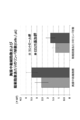
胸腺中の総細胞数は、コントロール群よりもCCL25添加群で多い傾向にあった。また、総細胞数あたりもリンパ球数も、コントロール群よりもCCL25添加群で多い傾向にあった(図4)。
以上の結果より、CCL25の添加による、免疫機能発達促進効果及び成長促進効果が確認された。
Claims (6)
- CCL25を含有する免疫機能発達促進剤。
- 乳児用である請求項1に記載の免疫機能発達促進剤。
- 経口摂取用である請求項1又は2に記載の免疫機能発達促進剤。
- CCL25を含有する成長促進剤。
- 乳児用である請求項4に記載の成長促進剤。
- 経口摂取用である請求項4又は5に記載の成長促進剤。
Priority Applications (2)
| Application Number | Priority Date | Filing Date | Title |
|---|---|---|---|
| JP2017524858A JP6799327B2 (ja) | 2015-06-19 | 2016-06-17 | 免疫機能発達促進剤及び成長促進剤 |
| US15/736,961 US10590180B2 (en) | 2015-06-19 | 2016-06-17 | Immune function development promoter and growth promoter |
Applications Claiming Priority (2)
| Application Number | Priority Date | Filing Date | Title |
|---|---|---|---|
| JP2015-123682 | 2015-06-19 | ||
| JP2015123682 | 2015-06-19 |
Publications (1)
| Publication Number | Publication Date |
|---|---|
| WO2016204271A1 true WO2016204271A1 (ja) | 2016-12-22 |
Family
ID=57545914
Family Applications (1)
| Application Number | Title | Priority Date | Filing Date |
|---|---|---|---|
| PCT/JP2016/068098 Ceased WO2016204271A1 (ja) | 2015-06-19 | 2016-06-17 | 免疫機能発達促進剤及び成長促進剤 |
Country Status (3)
| Country | Link |
|---|---|
| US (1) | US10590180B2 (ja) |
| JP (1) | JP6799327B2 (ja) |
| WO (1) | WO2016204271A1 (ja) |
Citations (1)
| Publication number | Priority date | Publication date | Assignee | Title |
|---|---|---|---|---|
| JP2005508647A (ja) * | 2001-11-12 | 2005-04-07 | マーズ インコーポレイテッド | 食品 |
Family Cites Families (4)
| Publication number | Priority date | Publication date | Assignee | Title |
|---|---|---|---|---|
| EP1496934A4 (en) | 2002-04-19 | 2006-08-02 | Endocyte Inc | IMPROVED IMMUNOTHERAPY BY ADJUVANT |
| US20060233762A1 (en) * | 2005-04-15 | 2006-10-19 | Mcmahon Robert J | Method for treating or preventing systemic inflammation in formula-fed infants |
| PL2999485T3 (pl) * | 2013-05-21 | 2018-07-31 | Tyg Oncology Ltd. | Kompozycja immunogenna peptydów gastrynowych |
| WO2015079952A1 (ja) * | 2013-11-29 | 2015-06-04 | テルモ株式会社 | アジュバント組成物およびこれを含むワクチン組成物、並びにこれらの製造方法 |
-
2016
- 2016-06-17 US US15/736,961 patent/US10590180B2/en active Active
- 2016-06-17 WO PCT/JP2016/068098 patent/WO2016204271A1/ja not_active Ceased
- 2016-06-17 JP JP2017524858A patent/JP6799327B2/ja active Active
Patent Citations (1)
| Publication number | Priority date | Publication date | Assignee | Title |
|---|---|---|---|---|
| JP2005508647A (ja) * | 2001-11-12 | 2005-04-07 | マーズ インコーポレイテッド | 食品 |
Non-Patent Citations (7)
| Title |
|---|
| KATHURIA, N. ET AL.: "Generation of antigen- specific immunity following systemic immunization with DNA vaccine encoding CCL25 chemokine immunoadjuvant", HUMAN VACCINES & IMMUNOTHERAPEUTICS, vol. 8, no. 11, 2012, pages 1607 - 1619, XP055339057 * |
| KAZUTOSHI SAYAMA ET AL.: "Mouse Bonyuchu CCL25 wa Shinseiji no Seicho Oyobi Men'eki Kino no Hattatsu o Sokushin suru", THE JAPANESE SOCIETY OF NUTRITION AND FOOD SCIENCE DAI 70 KAI TAIKAI IPPAN KOEN TOPICS-SHU, 25 April 2016 (2016-04-25), pages 37 - 40 * |
| KAZUTOSHI SAYAMA: "Elucidation of physiological functionality of CCL25 in milk for the enhancement of gut immunity in neonate", 25 July 2014 (2014-07-25), Retrieved from the Internet <URL:https://kaken.nii.ac.jp/ja/grant/KAKENHI- PROJECT-25560048> [retrieved on 20160905] * |
| KUNIO HIESHIMA: "CCL25, CCL28 wa IgA Kotai Sansei Saibo no Chokan eno Homing ni Kan'yo suru", CLINICAL IMMUNOLOGY, vol. 43, no. 5, 2005, pages 584 - 588 * |
| LEVAST, B. ET AL.: "Ultra-early weaning in piglets results in low serum IgA concentration and IL 17 mRNA expression", VETERINARY IMMUNOLOGY AND IMMUNOPATHOLOGY, vol. 137, 2010, pages 261 - 268, XP027258939 * |
| MASAKI TAKENAKA ET AL.: "Ninshin Oyobi Hitsunyuchu no Mouse Nyusen Soshikinai ni Okeru CCL25 no Hatsugen", PROCEEDINGS OF THE ANNUAL MEETING OF JAPAN SOCIETY FOR BIOSCIENCE, BIOTECHNOLOGY, AND AGROCHEMISTRY, 2010, pages 227 * |
| MEURENS, F. ET AL.: "Expression of mucosal chemokines TECK/CCL25 and MEC/CCL28 during fetal development of the ovine mucosal immune system", IMMUNOLOGY, vol. 120, 2007, pages 544 - 555, XP055339058 * |
Also Published As
| Publication number | Publication date |
|---|---|
| JPWO2016204271A1 (ja) | 2018-04-05 |
| JP6799327B2 (ja) | 2020-12-16 |
| US20180369335A1 (en) | 2018-12-27 |
| US10590180B2 (en) | 2020-03-17 |
Similar Documents
| Publication | Publication Date | Title |
|---|---|---|
| JP5188631B2 (ja) | インスリンを補った乳児用調製乳 | |
| CA2260007C (en) | Method for treating gastrointestinal damage | |
| CN113993526B (zh) | 包含源自牛奶的外泌体作为有效成分的用于治疗代谢性疾病的药物组合物 | |
| US20080081076A1 (en) | Nanofraction immune modulators, preparations and compositions including the same, and associated methods | |
| CA2267421A1 (en) | Oral administration of chicken yolk antibodies to treat disease | |
| PL211753B1 (pl) | Zastosowanie koncentratu antygenowo nie-specyficznych immunoglobulin pochodzących z krwi zwierząt i kompozycja farmaceutyczna zawierająca immunoglobuliny | |
| JPH05502881A (ja) | 免疫刺激剤としてのアルギニンの用途 | |
| US9066526B2 (en) | Allergy treatment using acid treated aqueous whey protein extract | |
| CN101179953B (zh) | 含哺乳动物初乳或乳汁的乳清级分的生长促进用食品组合物 | |
| CN1411378A (zh) | 含有抗病原菌特异抗体(IgY)的鸡蛋及含有该IgY的酸乳和冰淇淋的生产方法 | |
| CA2724833C (en) | Use of seleneium yeast composition for establishing and/or maintaining pregnancy | |
| JP6799327B2 (ja) | 免疫機能発達促進剤及び成長促進剤 | |
| CN116490081A (zh) | 用于改善胃肠耐受性的天然乳清蛋白组合物 | |
| RU2671634C2 (ru) | Стимулятор повышения колострального иммунитета и неспецифической резистентности - "Синэстрол 2%" и способ повышения клострального иммунитета и неспецифической резистентности | |
| EP3076981A1 (de) | Pharmazeutische zusammensetzung zur prophylaxe und/oder behandlung von erkrankungen mit einer gestörten lps- und/oder apoptose-regulation | |
| CN109731010B (zh) | 一种含猪脾多肽及核糖口服液 | |
| CN110141566B (zh) | Sglt2抑制剂在调控炎症中的应用 | |
| CN111647559A (zh) | 葡甘聚糖在提高nk细胞杀伤活性、制备nk细胞培养基方面的应用 | |
| JP4150631B2 (ja) | 魚類冷水病ワクチン | |
| Turkey | Investigation of Whey Protein on testicular albino rats changes based on: Histological, Physiological and Immunohistochemical study | |
| JP2001048804A (ja) | TNF−α産生抑制剤 | |
| JP2001294534A (ja) | 泌乳牛における乳腺の免疫賦活化剤および賦活化方法 | |
| Williams | Presence of soluble CD14 in bovine and equine milk | |
| PANDEY | EFFECT OF MICRONUTRIENTS SUPPLEMENTATION ON IMMUNOMODULATORY COMPONENTS OF COLOSTRUM IN PERIPARTURIENT CROSSBRED COWS | |
| CN101658678A (zh) | 筛选具有自然杀伤细胞增殖作用的物质和增殖自然杀伤细胞的方法 |
Legal Events
| Date | Code | Title | Description |
|---|---|---|---|
| 121 | Ep: the epo has been informed by wipo that ep was designated in this application |
Ref document number: 16811745 Country of ref document: EP Kind code of ref document: A1 |
|
| ENP | Entry into the national phase |
Ref document number: 2017524858 Country of ref document: JP Kind code of ref document: A |
|
| NENP | Non-entry into the national phase |
Ref country code: DE |
|
| 122 | Ep: pct application non-entry in european phase |
Ref document number: 16811745 Country of ref document: EP Kind code of ref document: A1 |