WO2016148089A1 - 刺激応答性磁性ナノ粒子を用いた検出対象を検出する方法 - Google Patents
刺激応答性磁性ナノ粒子を用いた検出対象を検出する方法 Download PDFInfo
- Publication number
- WO2016148089A1 WO2016148089A1 PCT/JP2016/057867 JP2016057867W WO2016148089A1 WO 2016148089 A1 WO2016148089 A1 WO 2016148089A1 JP 2016057867 W JP2016057867 W JP 2016057867W WO 2016148089 A1 WO2016148089 A1 WO 2016148089A1
- Authority
- WO
- WIPO (PCT)
- Prior art keywords
- detection target
- nucleic acid
- conjugate
- stimulus
- acid amplification
- Prior art date
- Legal status (The legal status is an assumption and is not a legal conclusion. Google has not performed a legal analysis and makes no representation as to the accuracy of the status listed.)
- Ceased
Links
Images

Classifications
-
- C—CHEMISTRY; METALLURGY
- C12—BIOCHEMISTRY; BEER; SPIRITS; WINE; VINEGAR; MICROBIOLOGY; ENZYMOLOGY; MUTATION OR GENETIC ENGINEERING
- C12N—MICROORGANISMS OR ENZYMES; COMPOSITIONS THEREOF; PROPAGATING, PRESERVING, OR MAINTAINING MICROORGANISMS; MUTATION OR GENETIC ENGINEERING; CULTURE MEDIA
- C12N15/00—Mutation or genetic engineering; DNA or RNA concerning genetic engineering, vectors, e.g. plasmids, or their isolation, preparation or purification; Use of hosts therefor
- C12N15/09—Recombinant DNA-technology
-
- C—CHEMISTRY; METALLURGY
- C12—BIOCHEMISTRY; BEER; SPIRITS; WINE; VINEGAR; MICROBIOLOGY; ENZYMOLOGY; MUTATION OR GENETIC ENGINEERING
- C12Q—MEASURING OR TESTING PROCESSES INVOLVING ENZYMES, NUCLEIC ACIDS OR MICROORGANISMS; COMPOSITIONS OR TEST PAPERS THEREFOR; PROCESSES OF PREPARING SUCH COMPOSITIONS; CONDITION-RESPONSIVE CONTROL IN MICROBIOLOGICAL OR ENZYMOLOGICAL PROCESSES
- C12Q1/00—Measuring or testing processes involving enzymes, nucleic acids or microorganisms; Compositions therefor; Processes of preparing such compositions
- C12Q1/68—Measuring or testing processes involving enzymes, nucleic acids or microorganisms; Compositions therefor; Processes of preparing such compositions involving nucleic acids
Definitions
- the present invention relates to a method for detecting a detection target using stimulus-responsive magnetic nanoparticles.
- Cryptosporidium is a protozoan that parasitizes the digestive tract of various animals widely distributed throughout the world.
- the protozoan oocysts are often caused by diarrhea due to water supply due to their strong chlorine tolerance. Therefore, Cryptosporidium has become the most prominent protozoa in clinical and public health.
- Cryptosporidium is usually detected by visual observation under a microscope.
- this method requires a lot of labor for detection because of complicated operation, low speed, and low detection rate. Therefore, in recent years, genetic techniques have also been used for the study of Cryptosporidium, and it has become relatively easy to identify and type species.
- Magnetic fine particles used in the immunomagnetic bead method include magnetic fine particles (for example, Dynabeads (registered trademark)) whose particle size is designed to be micro size in order to give magnetic beads magnetic separation ability. Yes.
- Cryptosporidium nucleic acid extraction is performed by physical treatment such as freezing and thawing or chemical treatment using a surfactant or an enzyme (Non-Patent Documents 3 and 4).
- Reagents used at this time include those requiring careful handling such as strongly alkaline sodium hydroxide solution and highly corrosive phenol solution.
- Non-Patent Documents 1 and 2 when the target microorganism is separated from the environment / biological sample using the immunomagnetic bead method using the magnetic fine particles described in Non-Patent Documents 1 and 2, the following work is required. That is, before extracting nucleic acids from microorganisms, it is necessary to dissociate microorganisms and magnetic fine particles by adding a predetermined dissociation solution, and to fix the magnetic fine particles with a magnet, and then collect the microorganism concentrate, which is complicated. It was.
- Non-Patent Documents 3 and 4 for example, after destroying a cell wall or a cell membrane by treatment with a physical or surfactant, an organic solvent such as water-saturated phenol or chloroform is used. Many steps were required, such as using them to remove, denature proteins, and separate nucleic acids. Therefore, in order to process these reagents contained in the extracted nucleic acid sample, a further purification step is required, and the work is complicated.
- the present inventors have conducted intensive research to find a method capable of easily detecting a detection target regardless of the type of detection target and without using a carefully handled reagent. That is, according to the present invention, when separating the detection target using the magnetic fine particles, it is not necessary to dissociate the collected magnetic fine particles from the detection target, and the nucleic acid can be extracted from the detection target as it is and obtained. It is an object of the present invention to provide a novel detection target detection method that can directly apply a nucleic acid sample to a reverse transcription reaction and / or a nucleic acid amplification test without requiring complicated steps such as subsequent nucleic acid purification. To do.
- the present inventors separated a detection target using stimulus-responsive magnetic nanoparticles to which an affinity substance for the detection target is bound, added an anionic surfactant to the separated detection target, and subjected to heat treatment And a method for extracting nucleic acid from the detection target was found.
- the extracted nucleic acid can be directly subjected to the reverse transcription reaction and / or the nucleic acid amplification test without passing through the step of separating the magnetic fine particles and the detection target, and the detection target can be easily detected.
- the present invention has the following configuration.
- a method for detecting a detection target in a test sample comprising the following steps (1) to (5).
- a step of generating a conjugate of an adsorbent having an affinity substance for a stimulus-responsive magnetic nanoparticle and a detection target and the detection target in an aqueous solution containing a test sample (2) Stimulation-responsive magnetic nanoparticle
- a step of aggregating the conjugate (3) a step of collecting the aggregated conjugate with a magnet (4) adding an anionic surfactant to the recovered conjugate, heat-treating, 1.
- Step of extracting nucleic acid from detection target (5) Step of performing nucleic acid amplification using the obtained nucleic acid as a sample 2.
- the anionic surfactant is at least one anion selected from the group consisting of sodium dodecyl sulfate (SDS), lithium dodecyl sulfate (LDS), sodium dodecylbenzenesulfonate (SDBS), sodium cholate, and N-lauroyl sarcosine.
- SDS sodium dodecyl sulfate
- LDS lithium dodecyl sulfate
- SDBS sodium dodecylbenzenesulfonate
- sodium cholate sodium cholate
- N-lauroyl sarcosine sodium cholate
- nucleic acid is used as a concept including DNA, RNA, and derivatives thereof.
- final concentration indicates the concentration of an anionic surfactant in the reaction tube at the time of nucleic acid amplification test.
- the present invention it is possible to easily separate a detection target and extract a nucleic acid regardless of the type of a test sample and without using a carefully handled reagent. Furthermore, the obtained nucleic acid sample can be directly subjected to a reverse transcription reaction and / or a nucleic acid amplification test without requiring complicated steps such as subsequent nucleic acid purification.
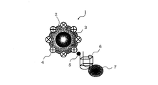
- FIG. 1 is a schematic configuration diagram of an adsorbent in a method according to an embodiment of the present invention.
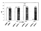
- FIG. 2 is a graph showing the effect of TM-AC concentration on the PCR after the SET method.
- FIG. 3 is a graph showing the effect of TM-AC concentration on PCR when the SET method is performed on TM-AC from which oocysts have been collected.
- the present invention is a method for detecting a detection target in a test sample, and includes a detection target including the following steps (1) to (5).
- a step of generating a conjugate of an adsorbent having an affinity substance for a stimulus-responsive magnetic nanoparticle and a detection target and the detection target in an aqueous solution containing a test sample (2) Stimulation-responsive magnetic nanoparticle
- a step of aggregating the conjugate (3) a step of collecting the aggregated conjugate with a magnet (4) adding an anionic surfactant to the recovered conjugate, heat-treating, Step of extracting nucleic acid from detection target (5) Step of performing nucleic acid amplification using the obtained nucleic acid as a sample.
- bonded_body of the adsorbent which has an affinity substance with respect to a stimulus-responsive magnetic nanoparticle and a detection target, and the said detection target in the aqueous solution containing a test sample The detection method of the detection target of this invention
- the nucleic acid is extracted from the detection target in the test sample, and the detection target is detected by amplifying the nucleic acid.
- in order to extract the nucleic acid from the detection target first, there is a step of collecting the detection target in the test sample. This step is a step of generating a conjugate of the adsorbent and the detection target in an aqueous solution as the first stage of recovery of the detection target.
- Test sample The test sample is not particularly limited as long as it contains a detection target described later.
- water samples existing in the environment such as raw water, river water, lake water, well water, ground water, sewage, waste water, pool water, park water, or ranch soil, farmland soil, lake soil, river soil, etc.
- environmental samples such as soil samples present in the environment, biological samples such as stool such as humans, livestock and pets, food samples such as beverage foods, raw vegetables and fruits.
- the detection target is not particularly limited as long as it can be detected by extracting and amplifying a nucleic acid.
- organisms such as animals (including humans), plants, microorganisms (including protozoa and protozoa) can be targeted.
- protozoa that formed oocysts such as Cryptosporidium are applicable as detection targets of the present invention.
- the adsorbent can be used to recover the detection target.
- the adsorbent has stimulus-responsive magnetic nanoparticles and an affinity substance for the detection target.
- stimulus-responsive magnetic nanoparticles are used for recovery of a detection target in an aqueous solution containing a test sample.
- the nucleic acid can be extracted from the detection target while the binding between the stimulus-responsive magnetic nanoparticle and the detection target is not dissociated.
- the detection target can be easily detected.
- the stimulus-responsive magnetic nanoparticles used in the present invention are obtained using a stimulus-responsive polymer and a magnetic substance (magnetic fine particles).
- the stimulus-responsive polymer is a magnetic nanoparticle that undergoes a structural change in response to an external stimulus and can adjust aggregation and dispersion.
- the type of stimulation is not particularly limited, and examples thereof include various physical or chemical signals such as heat, light, acid, base, pH, or electricity.
- thermoresponsive polymer a heat-responsive polymer that can be aggregated and dispersed by temperature change
- thermoresponsive polymer examples include a polymer having a lower critical solution temperature (hereinafter also referred to as LCST) and a polymer having an upper critical solution temperature (hereinafter also referred to as “UCST”).
- LCST lower critical solution temperature
- UCST upper critical solution temperature
- thermoresponsive magnetic nanoparticles having a thermoresponsive polymer Thermo-Max (manufactured by JNC Corporation) can be suitably used.
- Examples of the polymer having a lower critical solution temperature that can be used in the present invention include Nn-propylacrylamide, N-isopropylacrylamide, N-ethylacrylamide, N, N-dimethylacrylamide, N-acryloylpyrrolidine, N- Acryloylpiperidine, N-acryloylmorpholine, Nn-propylmethacrylamide, N-isopropylmethacrylamide, N-ethylmethacrylamide, N, N-dimethylmethacrylamide, N-methacryloylpyrrolidine, N-methacryloylpiperidine, N-methacryloylmorpholine Polymers composed of N-substituted (meth) acrylamide derivatives such as: hydroxypropyl cellulose, polyvinyl alcohol partially acetylated product, polyvinyl methyl ether, (polyoxyethylene Polyoxypropylene) block copolymer, polyoxyethylene alkylamine derivatives such as poly
- a copolymer of N-isopropylacrylamide and Nt-butylacrylamide can be preferably used.
- Examples of the polymer having an upper critical solution temperature that can be used in the present invention include, for example, at least one monomer selected from the group consisting of acryloylglycinamide, acryloylnipecotamide, acryloylasparaginamide, acryloylglutamineamide, and the like.
- the polymer which becomes is mentioned.
- the copolymer which consists of these at least 2 types of monomers may be sufficient.
- These polymers include other copolymers such as acrylamide, acetylacrylamide, biotinol acrylate, N-biotinyl-N'-methacryloyl trimethylene amide, acroyl sarcosine amide, methacryl sarcosine amide, acroyl methyl uracil, etc.
- Possible monomers may be copolymerized in a range having an upper critical solution temperature.
- a pH-responsive polymer that can be aggregated and dispersed by pH change can be used as the stimulus-responsive polymer.
- examples of such a pH-responsive polymer include a polymer containing a group such as carboxyl, phosphoric acid, sulfonyl, and amino as a functional group.
- acrylic acid methacrylic acid, maleic acid, vinyl sulfonic acid, vinyl benzene sulfonic acid, phosphoryl ethyl (meth) acrylate, aminoethyl methacrylate, aminopropyl (meth) acrylamide, dimethylaminopropyl (meth)
- examples include polymers containing acrylamide or a salt thereof as a copolymerization component.
- Magnetic fine particles which are magnetic substances, are composed of, for example, polyhydric alcohol and magnetite.
- the polyhydric alcohol is not particularly limited as long as it is an alcohol structure having at least two hydroxyl groups in a structural unit and capable of binding to iron ions.
- Examples include dextran, polyvinyl alcohol, mannitol, sorbitol and cyclodextrin.
- Japanese Patent Application Laid-Open No. 2005-82538 discloses a method for producing a particulate magnetic material using dextran.
- the compound which has an epoxy like a glycidyl methacrylate polymer and forms a polyhydric alcohol structure after ring-opening can also be used.
- the fine particle magnetic material (magnetic fine particles) prepared using such a polyhydric alcohol preferably has an average particle size of 0.9 nm or more and less than 1000 nm so as to have good dispersibility.
- the average particle size is preferably 2.9 nm or more and less than 200 nm, in particular, in order to increase the detection sensitivity of the target detection target.
- the average particle size of the magnetic fine particles is within the above range, the dispersibility in the liquid becomes good.
- the magnetic fine particles do not settle in the reaction solution and are appropriately dispersed, and the reverse transcription reaction and the nucleic acid amplification test can be performed efficiently.
- the affinity substance used in the present invention is not particularly limited as long as it binds to the detection target.
- antibodies, aptamers, substances having a cationic functional group for example, substances having a primary amino group, secondary amino group, tertiary amino group, quaternary ammonium group or guanidino group
- substances having an anionic functional group for example, substances having a primary amino group, secondary amino group, tertiary amino group, quaternary ammonium group or guanidino group
- proteins that interact with the target substance eg, enzymes, receptors
- chelating agents e.g, enzymes, receptors, chelating agents, and the like.
- the binding method between the stimulus-responsive magnetic nanoparticles and the affinity substance is not particularly limited.
- substances having affinity for example, biotin and streptavidin or avidin
- a stimulus-responsive magnetic nanoparticle is bound to an affinity substance.
- biotin is bonded to the stimulus-responsive polymer by adding biotin or the like to a polymerizable functional group such as methacryl or acryl as described in International Publication No. 2001/009141.
- a monomer is used and copolymerized with other monomers to immobilize streptavidin.
- labeling of the affinity substance with biotin or the like is performed according to a conventional method. When the affinity substance labeled with biotin and the streptavidin-immobilized stimulus-responsive polymer are mixed, the stimulus-responsive polymer and the affinity substance are bound via the binding between biotin and streptavidin.
- the stimulus-responsive magnetic nanoparticle 1 includes a particulate magnetic substance 2, and a stimulus-responsive polymer 3 is bonded to the surface of the magnetic substance 2.
- Streptavidin 4 is bound to the stimulus-responsive polymer 3, and an affinity substance 6 for the detection target 7 is bound via biotin 5.
- a material in which a stimulus-responsive polymer and a particulate magnetic substance are combined is referred to as a stimulus-responsive magnetic nanoparticle, and when the stimulus is heat, it is referred to as a thermoresponsive magnetic nanoparticle. Yes.
- the fine magnetic substance can be bonded to the stimulus-responsive polymer via a reactive functional group, or a polymerizable unsaturated bond is introduced into the active hydrogen or polyhydric alcohol on the polyhydric alcohol in the magnetic substance.
- the graft polymerization may be carried out by a method known in the art such as graft polymerization (for example, ADV. Polym. Sci., Vol. 4, p111, 1965 and J. Polymer Sci., Part-A, 3, p1031). 1965).
- the adsorbent / detection target combination described above is obtained by combining the adsorbent and the detection target in an aqueous solution by combining the adsorbent and the detection target in an arbitrary method. This produces a conjugate.
- the binding between the affinity substance and the detection target means specific binding or adsorption, and is not necessarily a covalent binding, and may be binding utilizing ionic or biochemical affinity (affinity). .
- Examples include binding between an antigen and an antibody, binding between an enzyme and a substrate, binding between a chelating agent and a metal ion, and specific binding such as avidin-biotin.
- the mixing operation is not particularly limited as long as the adsorbent and the detection target can be brought into contact with each other in an appropriate buffer. For example, it is sufficient that the test sample including the detection target and the tube provided with the adsorbent are lightly agitated or shaken, and examples thereof include a mixing operation using a commercially available vortex mixer.
- step (2) A step of aggregating the conjugate under conditions where the stimulus-responsive magnetic nanoparticles are aggregated. This step is a second stage of recovery of the detection target, and the adsorbent obtained in step (1) and the detection target In the conjugate, this is a step of aggregating the conjugate under conditions where the stimulus-responsive polymer in the conjugate is aggregated.
- the container containing the mixture may be transferred to a thermostatic bath at a temperature at which the thermoresponsive polymer aggregates.
- thermoresponsive polymers polymers having an upper critical solution temperature and polymers having a lower critical solution temperature.
- thermoresponsive polymer when a polymer having a lower critical solution temperature with an LCST of 37 ° C. is used, the thermoresponsive polymer can be agglomerated by moving the container containing the mixture to a constant temperature bath at 37 ° C. or higher.
- thermoresponsive polymer when a polymer having an upper critical solution temperature with a UCST of 5 ° C. is used, the thermoresponsive polymer can be agglomerated by transferring the container containing the mixture to a thermostatic bath of less than 5 ° C.
- the lower critical solution temperature and the upper critical solution temperature are determined as follows, for example. First, a sample is put into a cell of an absorptiometer and the sample is heated at a rate of 1 ° C./min. During this time, the change in transmittance at 550 nm is recorded. Here, when the transmittance when the polymer is transparently dissolved is 100% and when the transmittance when the polymer is completely aggregated is 0%, the temperature at which the transmittance is 50% is obtained as LCST. In the case of UCST, the sample is cooled at a rate of 1 ° C./min, and thereafter obtained by the same method as LCST.
- an acid solution or an alkali solution may be added to a container containing the mixture.
- an acid solution or an alkaline solution is added to a container containing a dispersed mixture outside the pH range where the pH-responsive polymer undergoes a structural change, and the pH-responsive polymer undergoes a structural change in the container. What is necessary is just to change to the pH range to raise.
- an acid solution may be added to a container containing a mixture dispersed at a pH above 5 so that the pH is 5 or less.
- an alkaline solution may be added to a container containing a mixture dispersed at a pH lower than 10 so that the pH becomes 10 or higher.
- the pH at which the pH-responsive polymer undergoes a structural change is not particularly limited, but is preferably pH 4 to 10, and more preferably pH 5 to 9.
- a photoresponsive polymer when a photoresponsive polymer is used, light having a wavelength capable of aggregating the polymer may be irradiated to a container containing the mixture.
- the preferred light for aggregation varies depending on the type and structure of the photoresponsive functional group contained in the photoresponsive polymer, but in general, ultraviolet light or visible light having a wavelength of 190 to 800 nm can be suitably used.
- the strength is preferably 0.1 to 1000 mW / cm 2 .
- the photoresponsive polymer is preferably one that is less likely to cause dispersion when irradiated with light used for turbidity measurement, in other words, that it can aggregate in that it can improve measurement accuracy.
- the measurement accuracy can be improved by shortening the irradiation time.
- Step of recovering aggregated conjugate with magnet This step is a step of recovering the detection target by separating the aggregate aggregated in step (2) with a magnet as the final stage of detection target recovery. By applying a magnetic force to the conjugate containing magnetic particles, the aggregated conjugate is separated from the mixed solution and recovered. As a result, the conjugate is separated from the contaminants containing the non-aggregated magnetic substance, and the detection target can be recovered.
- the adsorbent when the detection target and the adsorbent are combined in an appropriate tube, the adsorbent is held near the side wall of the tube by bringing the magnet close to the side wall of the tube from the outside, and becomes a supernatant portion from the inside of the tube. By discharging the liquid, the adsorbent to which the detection target is bound can be separated.
- the magnetic force of a magnet or the like used to separate the adsorbent to which the detection target is coupled differs depending on the magnitude of the magnetic force of the magnetic substance used.
- a magnetic force capable of collecting the target magnetic substance can be appropriately used.
- the material of the magnet for example, a material made of the above-described magnetic material can be used.
- a neodymium magnet manufactured by 26 Co., Ltd.
- the magnetic force of the neodymium magnet is preferably 3800 gauss or more.
- Step of adding an anionic surfactant to the recovered conjugate, heat-treating, and extracting the nucleic acid from the detection target This step is performed in the conjugate recovered in the above steps (1) to (3). In this step, nucleic acid is extracted from the detection target.
- the detection target and the anionic surfactant are brought into contact with each other by adding an anionic surfactant to the conjugate. After bringing the detection target into contact with the anionic surfactant, heat treatment is performed to extract the nucleic acid from the detection target.
- an anionic surfactant as an extraction reagent, it can be applied to any kind of cells such as bacteria and animal cells.
- anionic surfactant examples include sodium dodecyl sulfate (SDS), lithium dodecyl sulfate (LDS), sodium dodecylbenzenesulfonate (SDBS), sodium cholate, N-lauroyl sarcosine, and the like. From this viewpoint, sodium dodecyl sulfate (SDS) is preferred. In the present invention, various anionic surfactants may be used alone or in combination of two or more.
- nucleic acid loss may usually occur during the nucleic acid purification process, and the recovery rate of the nucleic acid after purification is at most about 80%.
- anionic surfactant is used as an extraction reagent.
- the method of the present invention can be applied to detection targets derived from all living organisms regardless of whether they are animals, plants, or microorganisms.
- the detection target in the conjugate and an anionic surfactant are brought into contact as necessary. It is also possible to provide a pretreatment step for decomposing these cell walls before. For example, they can be supplied as a stock or concentrated solution of a liquid (solution) dissolved and sealed together with a reagent such as a buffer solution required for each step.
- cell wall degrading enzymes for example, cellulase, zymolyase, lysozyme, etc.
- nucleic acid extraction can be performed by the above extraction method without causing cell wall degrading enzymes to act on these organisms.
- the step of contacting the anionic surfactant with the detection target in the conjugate and heat-treating can be repeated multiple times until the cell membrane of the detection target is sufficiently destroyed, particularly for samples for nucleic acid amplification tests.
- the time required for the process varies depending on the type and amount of the detection target.
- the heat treatment is usually performed at 40 to 100 ° C, preferably 80 to 100 ° C.
- the nucleic acid can be sufficiently extracted from the detection target by performing a heat treatment usually for 5 to 120 minutes, preferably 5 to 60 minutes.
- the contact time is about 5 to 30 minutes at 90 ° C., cell membranes derived from animals can be sufficiently destroyed.
- the concentration of the anionic surfactant only needs to be able to extract the quantity and quality necessary for subjecting the obtained nucleic acid to various analyzes such as nucleic acid amplification described below after extracting the nucleic acid from the detection target.
- concentration of the anionic surfactant only needs to be able to extract the quantity and quality necessary for subjecting the obtained nucleic acid to various analyzes such as nucleic acid amplification described below after extracting the nucleic acid from the detection target.
- the higher the treatment concentration of the anionic surfactant during nucleic acid extraction the higher the extraction power.
- the final concentration of the anionic surfactant is preferably 0.001 to 0.1% by mass, more preferably 0.001 to Dilute to 0.01% by weight.
- the concentration of the anionic surfactant during the nucleic acid extraction treatment is preferably 0.1 to 10% by mass, More preferably, contact with the detection target at a concentration of 0.1 to 1% by mass is preferable for reliable nucleic acid extraction, and further improves the efficiency of nucleic acid extraction and shortens the time required for extraction. , Improve detection sensitivity and achieve rapid detection.
- the concentration of the anionic surfactant during the nucleic acid extraction treatment is adjusted by diluting the anionic surfactant with, for example, water, 0.1 to 10% by mass, more preferably 0.1 to 1% by mass. It is possible to extract the nucleic acid from the detection target by introducing the conjugate into the solution and performing contact treatment and heat treatment at 90 ° C. for 15 minutes, for example.
- step (5) In the reverse transcription reaction or nucleic acid amplification test in step (5) described later, if the concentration of the stimulus-responsive magnetic nanoparticles in the reaction solution is high, the reverse transcription reaction or nucleic acid amplification can be inhibited.
- an anionic surfactant is added to the detection target and the stimulus-responsive magnetic nanoparticles, and the stimulus response is performed by, for example, heating at 90 ° C. for 15 minutes. Inhibition of reverse transcription reaction and nucleic acid amplification by the magnetic nanoparticles can be reduced.
- the reagent form of the anionic surfactant is not particularly limited. As long as the concentration of the anionic surfactant is within the above-described concentration range, it may be in a solution state such as an aqueous solution, or may be in a lyophilized state or a dry state. Moreover, when a reagent form is a freeze-dried state or a dry state, you may combine with the solution for melt
- nucleic acids from detection targets derived from animals include blood free DNA from blood components such as serum and plasma, DNA such as viral DNA, RNA such as blood free messenger RNA and viral RNA. Is mentioned.
- Cryptosporidium to be detected refers to a microorganism belonging to the genus Cryptosporidium, and specifically, Cryptosporidium parvum species (hereinafter abbreviated as C. parvum), Cryptosporidium. muris species (hereinafter abbreviated as C. muris), Cryptospodium baileyi species (hereinafter abbreviated as C. baileyi), Cryptosporidium serpentis species (hereinafter abbreviated as C. serpentis), Crytosporidium species, hereinafter. C. meleagridis), Cryptospodium felis species (hereinafter referred to as C. meleagridis) Abbreviated as C.Feris.) And the like, the method of the present invention is used to detect these species to species discriminative.
- C. parvum Cryptosporidium parvum
- C. muris Cryptosporidium. muris species
- C. baileyi Cryptospodium baileyi species
- C. serpentis Cryptosporidium serpentis species
- nucleic acid extraction from oocysts only requires treatment with an anionic surfactant, and there is no need to perform complicated steps such as protease treatment and purification treatment as in the prior art.
- the nucleic acid extraction treatment conditions with such an anionic surfactant are preferably, for example, 5 to 30 minutes at 60 to 100 ° C., and particularly preferably 10 to 20 minutes at 90 ° C.
- the nucleic acid extraction method described above is called a surfactant extraction treatment (hereinafter referred to as SET) method, and is the method described in the 48th Annual Meeting of the Japan Society on Water Environment, p.421 (2014).
- SET surfactant extraction treatment
- the concentration of the anionic surfactant used for the nucleic acid extraction treatment is 0.1 to 10% by mass, preferably 0.1 to 1% by mass (the anionic surfactant during nucleic acid amplification). In the case of diluting the sample treated in step 10), it is preferable to adjust. By being in the above range, inhibition of nucleic acid amplification by an anionic surfactant can be suppressed.
- Step of performing nucleic acid amplification using the obtained nucleic acid as a sample This step is a step of performing nucleic acid amplification using the nucleic acid obtained in the above step as a sample.
- concentration of stimuli-responsive magnetic nanoparticles in the adsorbent is high, reverse transcription reaction and nucleic acid amplification can be inhibited.
- the inhibition of reverse transcription reaction and nucleic acid amplification by the stimulus-responsive magnetic nanoparticles is reduced by heat-treating the stimulus-responsive magnetic nanoparticles together with the detection target in step (4) with an anionic surfactant. can do.
- the stimulus-responsive magnetic nanoparticles having excellent dispersibility are used as the magnetic fine particles, even if the magnetic fine particles remain in the reaction solution used in the reverse transcription reaction or nucleic acid amplification test described below. Since it is appropriately dispersed in the liquid, a reverse transcription reaction and a nucleic acid amplification test can be performed efficiently.
- the obtained nucleic acid is RNA
- reverse transcription reaction is performed to obtain DNA, and then nucleic acid amplification is performed.
- any conventionally known method can be employed using the RNA obtained in the above steps (1) to (4) as a template.
- any method can be used as a method for nucleic acid amplification, such as polymerase chain reaction (PCR method), LAMP method, strand displacement amplification, reverse transcriptase strand displacement amplification, reverse transcriptase polymerase chain reaction, Examples include reverse transcription LAMP method, nucleic acid sequence-based amplification, transcription-mediated amplification and rolling circle amplification method, SmartAmp method, reverse transcription SmartAmp method and the like.
- PCR method polymerase chain reaction
- LAMP method strand displacement amplification
- reverse transcriptase strand displacement amplification reverse transcriptase polymerase chain reaction
- reverse transcriptase polymerase chain reaction examples include reverse transcription LAMP method, nucleic acid sequence-based amplification, transcription-mediated amplification and rolling circle amplification method, SmartAmp method, reverse transcription SmartAmp method and the like.
- DNA amplification is generally performed by PCR.
- any conventionally known PCR method can be employed using the DNA obtained in the above step or the DNA obtained by reverse transcription reaction of RNA as a template.
- the nucleic acid amplification enzyme at the time of PCR amplification is any of Taq polymerase, Bst polymerase, or Aac polymerase, it can be suitably used.
- the anionic surfactant remains in the sample and becomes an inhibitor during reverse transcription reaction or nucleic acid amplification. Accordingly, when the nucleic acid extract is subjected to reverse transcription reaction or nucleic acid amplification, the final concentration of the anionic surfactant is preferably 0.0001 to 0.01% by mass, more preferably 0.0001 to 0.00. Dilute to 001% by weight. By being in the above range, inhibition of nucleic acid amplification by an anionic surfactant can be suppressed.
- the stimulus-responsive magnetic nanoparticles remaining in the sample are preferably 0.1 to 10% by mass, more preferably 0.5 to 1% by mass. By being in the above range, inhibition of nucleic acid amplification by the stimulus-responsive magnetic nanoparticles can be reduced.
- This step is preferably added after step (5).
- This step is a step of calibrating the DNA chain fragment (PCR amplification product) amplified in step (4) and determining the presence or absence of the detection target in the test sample.
- the analysis method in this step is not particularly limited as long as the PCR amplification product can be detected or quantified.
- electrophoresis method real-time PCR method and the like can be mentioned.
- the electrophoresis method the amount and size of the PCR amplification product can be evaluated.
- PCR amplification products can be quickly quantified.
- the change in fluorescence intensity is generally a noise level and is equal to zero until the number of amplification cycles is 1 to 10. Therefore, these are regarded as sample blanks with zero amplification products, and their standard deviation SD is calculated.
- the fluorescence value multiplied by 10 is defined as a threshold value, and the number of PCR cycles that first exceeds the threshold value is referred to as a cycle threshold value (Ct value). Therefore, the larger the initial DNA template amount in the PCR reaction solution, the smaller the Ct value, and the smaller the template DNA amount, the larger the Ct value. Even if the amount of template DNA is the same, the Ct value of the PCR reaction in the same region becomes larger as the ratio of cleavage in the PCR target sequence region in the template increases.
- a primer common to a plurality of types of detection targets when used, a plurality of types of detection targets in a test sample can be detected. Further, when a primer specific to a specific detection target is used, the specific detection target in the test sample can be detected.
- PCR conditions are not particularly limited as long as specific amplification occurs in accordance with the principle of PCR, and can be set as appropriate.
- Test Example 1 In Test Example 1, the effect of stimulation-responsive magnetic nanoparticles heat-treated with an SDS solution on real-time PCR was evaluated.
- thermoresponsive magnetic nanoparticles to which avidin is bonded (Therma-Max LA Avidin, 4 g / L, JNC Corporation) and C.I. 20 ⁇ L of biotinylated monoclonal antibody specific for parvum oocysts (Crypt-a-Glo biotin reagent kit (20-fold concentrated), Waterborne) was combined and bound to magnetic nanoparticles for recovery of oocysts (Therma-Max-Anti-Cryptosporidium) : TM-AC).
- PCR is a primer set specific to Cryptosporidium 18S rDNA gene forward: 5′-GGAAGGGTTGTATTATTATATAAAGAAACC-3 ′ (SEQ ID NO: 2), reverse: 5′-CTCCCTCCTCGAGAATCGAA-3 (SEQ ID NO: 3) (each 5 ⁇ M) 0.8 ⁇ L and TaqMan Probe: TCTGACCZATCAGCTT (SEQ ID NO: 4) (1 ⁇ M) 2.0 ⁇ L, PCR reagent Premix EX Taq (Takara Bio Inc.) 10 ⁇ L, real-time PCR (LightCycler Nano, Roche Diagnostics Inc.) was used.
- the added amount of the positive control (5 ⁇ 10 6 copies / mL) was 2 ⁇ L, and the PCR reaction solution volume was 20 ⁇ L.
- PCR conditions were “Hold; 1 cycle (95 ° C., 30 s), 2 Step PCR; 45 cycles (60 ° C., 30 s, 95 ° C., 10 s)”.
- the Ct value (Threshold Cycle) was analyzed with LightCycler Nano software.
- Reference Example 1-2 Reference Example 1-2 was tested in the same manner as Reference Example 1-1 except that the SET method for TM-AC was not performed.
- Reference Example 2-1 and Reference Example 2-2 Reference Example 2-1 was tested in the same manner as Reference Example 1-1 except that the final concentration of TM-AC was adjusted to 0.5%.
- Reference Example 2-2 was tested in the same manner as Reference Example 1-1 except that the SET method for TM-AC was not performed and the final concentration of TM-AC was adjusted to 0.5%.
- Reference Example 3-1 and Reference Example 3-2 Reference Example 3-1 was tested in the same manner as Reference Example 1-1 except that the final concentration of TM-AC was adjusted to 1.0%.
- Reference Example 3-2 was tested in the same manner as Reference Example 1-1 except that the SET method for TM-AC was not performed and the final concentration of TM-AC was adjusted to 1.0%.
- Control Example 1 Control Example 2
- Control 1 was tested in the same manner as Reference Example 1-1 except that the final TM-AC concentration was 0%.
- Control 2 was tested in the same manner as Reference Example 1-1 except that the SET method for TM-AC was not performed and the final concentration of TM-AC was 0%.
- PCR inhibition by TM-AC can be reduced by PCR using TM-AC after SET. It was also found that PCR can be performed without inhibition even when 0.2 ⁇ L of TM-AC (TM-AC final concentration 1%) is directly added to the PCR tube (20 ⁇ L of reaction solution) after SET.
- Example 2 In Test Example 2, Cryptosporidium oocysts were collected using TM-AC, the SET method was performed on TM-AC together with the collected oocysts, DNA was extracted from the oocysts, and real-time PCR was performed together with TM-AC. The detection sensitivity was evaluated. [Examples 1 to 3] (1) Recovery of Cryptosporidium oocysts Add 100 ⁇ L of the above TM-AC to oocyst-containing water (10 5 pieces / 750 mL), and incubate at room temperature for 15 minutes while slowly rotating (18 rpm) to bind TM-AC and oocysts. The body was made. Immediately after heating at 42 ° C. for 1 minute, a microtube was set on a magnetic board and placed for 1 minute or longer to magnetically separate TM-AC and oocyst conjugate. After magnetic separation, the supernatant was removed.
- TM-AC was dispersed by adding 100 ⁇ L of TE buffer (concentration 0.1%) containing TE buffer (pH 8.0, 0.01 M Tris-TCl, 1 mM EDTA-Na 2 ) (approximately the same concentration as the initial TM-AC concentration). To prepare). Thereafter, heat treatment was performed in a water bath (90 ° C., 15 minutes) to perform a SET method (Surfant extraction treatment method (surfactant extraction treatment method)), and DNA was extracted from oocysts.
- TE buffer concentration 0.1%) containing TE buffer (pH 8.0, 0.01 M Tris-TCl, 1 mM EDTA-Na 2 ) (approximately the same concentration as the initial TM-AC concentration).
- quantified oocysts (Iowa Isolate, Waterborne) recovered from the stool of mice using a density gradient centrifugation method were used by orally administering Cryptospodium parvum oocysts to mice. It was.
- PCR was performed using 0.8 ⁇ L primer set specific for Cryptosporidium 18S rDNA gene (5 ⁇ M each) and 2.0 ⁇ L TaqMan probe (1 ⁇ M), 10 ⁇ L PCR reagent Premix EX Taq (Takara Bio Inc.), real-time PCR (LightCycler Nano, Roche) ⁇ Diagnostics Co., Ltd.)
- the added amount of the positive control (5 ⁇ 10 6 copies / mL) was 2 ⁇ L, and the PCR reaction solution volume was 20 ⁇ L.
- PCR conditions were “Hold; 1 cycle (95 ° C., 30 s), 2 Step PCR; 45 cycles (60 ° C., 30 s, 95 ° C., 10 s)”.
- the Ct value was analyzed with LightCycler Nano software.
- DNA was extracted from the TM-AC from which oocysts were collected by the SET method, and then the DNA detection sensitivity was evaluated. Details of the samples used for PCR are shown in Table 1, and the results of real-time PCR are shown in FIG.
- the sample (200 oocysts) having a dilution ratio of 1/10 in Example 1 contained 1% TM-AC that inhibits PCR, but the inhibition was suppressed by SET and DNA was detected. Further, DNA could also be detected from the 20 oocyst samples of Example 2 (dilution ratio 1/100) and the 2 oocyst samples of Example 3 (dilution ratio 1/1000).
- TM-AC can efficiently recover oocysts, and that DNA can be extracted from the mixture of TM-AC and oocysts by the SET method.
- PCR can be performed without a DNA purification step by directly introducing 0.2 ⁇ L of TM-AC (1% TM-AC final concentration) after SET into a PCR tube (20 ⁇ L of reaction solution).
- the present invention is not limited to the above-described embodiment, but includes modifications and improvements as long as the object of the present invention can be achieved.
- target microorganisms particularly Cryptosporidium
- the obtained nucleic acid sample can be subjected to complicated nucleic acid purification and the like. It is useful in clinical examinations and public health examinations, for example, because it does not adversely affect the reverse transcription reaction and / or nucleic acid amplification test as it is without requiring a step.
Landscapes
- Life Sciences & Earth Sciences (AREA)
- Chemical & Material Sciences (AREA)
- Organic Chemistry (AREA)
- Health & Medical Sciences (AREA)
- Engineering & Computer Science (AREA)
- Genetics & Genomics (AREA)
- Zoology (AREA)
- Wood Science & Technology (AREA)
- General Engineering & Computer Science (AREA)
- Bioinformatics & Cheminformatics (AREA)
- Biotechnology (AREA)
- Microbiology (AREA)
- Molecular Biology (AREA)
- Physics & Mathematics (AREA)
- Biochemistry (AREA)
- Proteomics, Peptides & Aminoacids (AREA)
- Biophysics (AREA)
- General Health & Medical Sciences (AREA)
- Biomedical Technology (AREA)
- Immunology (AREA)
- Analytical Chemistry (AREA)
- Plant Pathology (AREA)
- Measuring Or Testing Involving Enzymes Or Micro-Organisms (AREA)
Abstract
本発明は、検出対象の種別を問わず、また慎重な取り扱いを有する試薬を使わずに簡便に検出対象を検出する方法を提供することを課題とする。本発明は、被検試料中の検出対象を検出する方法であって、以下の工程(1)~(5)を含む検出対象の検出方法に関する。(1)刺激応答性磁性ナノ粒子及び検出対象に対する親和性物質を有する吸着材と前記検出対象との結合体を、被検試料を含む水溶液中で生成させる工程(2)刺激応答性磁性ナノ粒子が凝集する条件下におき、前記結合体を凝集させる工程(3)凝集した結合体を磁石で回収する工程(4)回収した結合体に陰イオン性界面活性剤を添加し、加熱処理し、検出対象から核酸を抽出する工程(5)得られた核酸を試料として核酸増幅を行う工程
Description
本発明は、刺激応答性磁性ナノ粒子を用いた検出対象を検出する方法に関する。
クリプトスポリジウムは世界中に広く分布する、種々の動物の消化管に寄生する原虫である。また、本原虫のオーシストは強い塩素耐性を示すためにしばしば水道を介した集団下痢症を引き起こしている。このことから、クリプトスポリジウムは臨床上あるいは公衆衛生学的に現在最も注目される原虫となっている。
クリプトスポリジウムの検出は、通常、顕微鏡下での目視により行われているが、かかる方法は、操作が煩雑で迅速性が低くかつ検出率も低いため、検出に多大な労力を必要とする。従って、近年、クリプトスポリジウムの研究にも遺伝子学的手法が用いられるようになり、種の同定や型別を比較的容易に行うことができるようになってきている。
従来、環境・生体試料からのオーシストを分離する方法として免疫磁気ビーズ法が用いられている(非特許文献1、2)。免疫磁気ビーズ法に用いられる磁性微粒子は、磁気ビーズに磁気分離能を持たせるために、粒径をマイクロサイズに設計した磁性微粒子(例えば、ダイナビーズ(Dynabeads)(登録商標))が用いられている。
また、例えばクリプトスポリジウムの核酸抽出には凍結融解などによる物理的処理や、界面活性剤や酵素などによる化学的処理により抽出が行われている(非特許文献3、4)。この際使用される試薬には、強アルカリ性の水酸化ナトリウム溶液や腐食性の高いフェノール液等の慎重な取り扱いを要するものが含まれている。
EPA Method 1623(U.S.Environmental Protection Agency)
水道における指標菌及びクリプトスポリジウム等の検査方法について(平成24年3月2日付け健水発0302号 厚生労働省健康局水道課長通知)
クリプトスポリジウム症を中心とした原虫性下痢症の診断マニュアル,平成15年度,国立感染症研究所)
Applied and Environmental Microbiology,Vol.61,No.11(1995),3849-3855.
しかしながら、非特許文献1、2に記載の磁性微粒子を用いた免疫磁気ビーズ法を利用して環境・生体試料から標的の微生物を分離する場合は以下の作業が必要となる。すなわち、微生物から核酸を抽出する前に、所定の解離液を加えることによって微生物と磁性微粒子とを解離し、磁石で磁性微粒子を固定してから微生物濃縮液を回収する必要があり、作業が煩雑となっていた。
また、非特許文献3、4に記載の核酸を抽出する方法としては、例えば細胞壁や細胞膜を物理的にまたは界面活性剤等で処理して破壊した後、水飽和フェノールやクロロホルム等の有機溶媒を用いてこれらを取り除き、タンパク質を変性させ、核酸を分離する等の多くの工程を必要としていた。従って、抽出した核酸試料中に含まれるこれらの試薬を処理すべく、更なる精製工程が必要となり、作業が煩雑となっていた。
そこで本発明者らは、検出対象の種別を問わず、また慎重な取り扱いを有する試薬を使わずに簡便に検出対象の検出ができる方法を見出すべく鋭意研究を行った。すなわち本発明は、磁性微粒子を用いて検出対象の分離を行う際に、回収した磁性微粒子と検出対象を解離する必要がなく、そのまま検出対象から核酸を抽出することができ、且つ、得られた核酸試料を、その後の核酸精製等の煩雑な工程を必要とすることなく、そのまま逆転写反応及び/又は核酸増幅試験に課すことのできる、新規な検出対象の検出方法を提供することを課題とする。
本発明者らは、検出対象に対する親和性物質が結合している刺激応答性磁性ナノ微粒子を用いて検出対象を分離し、その分離された検出対象に陰イオン性界面活性剤を添加し加熱処理を行い、検出対象から核酸を抽出する方法を見出した。この方法によれば、磁性微粒子と検出対象を分離する工程を経なくても、抽出された核酸をそのまま逆転写反応及び/又は核酸増幅試験に課すことができ、簡便に検出対象の検出ができる。
具体的には、本発明は以下の構成を有する。
具体的には、本発明は以下の構成を有する。
1.被検試料中の検出対象を検出する方法であって、以下の工程(1)~(5)を含む検出対象の検出方法。
(1)刺激応答性磁性ナノ粒子及び検出対象に対する親和性物質を有する吸着材と前記検出対象との結合体を、被検試料を含む水溶液中で生成させる工程
(2)刺激応答性磁性ナノ粒子が凝集する条件下におき、前記結合体を凝集させる工程
(3)凝集した結合体を磁石で回収する工程
(4)回収した結合体に陰イオン性界面活性剤を添加し、加熱処理し、検出対象から核酸を抽出する工程
(5)得られた核酸を試料として核酸増幅を行う工程
2.前記核酸増幅を行う工程における、前記陰イオン性界面活性剤の終濃度が、0.0001~0.01質量%である、前記1に記載の検出対象の検出方法。
3.前記陰イオン性界面活性剤が、ドデシル硫酸ナトリウム(SDS)、ドデシル硫酸リチウム(LDS)、ドデシルベンゼンスルホン酸ナトリウム(SDBS)、コール酸ナトリウム、N-ラウロイルサルコシンからなる群より選ばれる少なくとも1の陰イオン性界面活性剤である、前記1または2に記載の検出対象の検出方法。
4.前記核酸増幅を行う工程における、前記刺激応答性磁性ナノ粒子の濃度が、0.1~10質量%である、前記1~3のいずれか1に記載の検出対象の検出方法。
5.前記核酸増幅を行う工程で使用する核酸増幅酵素が、Taqポリメラーゼ、Bstポリメラーゼ、又はAacポリメラーゼのいずれかであることを特徴とする、前記1~4のいずれか1に記載の検出対象の検出方法。
(1)刺激応答性磁性ナノ粒子及び検出対象に対する親和性物質を有する吸着材と前記検出対象との結合体を、被検試料を含む水溶液中で生成させる工程
(2)刺激応答性磁性ナノ粒子が凝集する条件下におき、前記結合体を凝集させる工程
(3)凝集した結合体を磁石で回収する工程
(4)回収した結合体に陰イオン性界面活性剤を添加し、加熱処理し、検出対象から核酸を抽出する工程
(5)得られた核酸を試料として核酸増幅を行う工程
2.前記核酸増幅を行う工程における、前記陰イオン性界面活性剤の終濃度が、0.0001~0.01質量%である、前記1に記載の検出対象の検出方法。
3.前記陰イオン性界面活性剤が、ドデシル硫酸ナトリウム(SDS)、ドデシル硫酸リチウム(LDS)、ドデシルベンゼンスルホン酸ナトリウム(SDBS)、コール酸ナトリウム、N-ラウロイルサルコシンからなる群より選ばれる少なくとも1の陰イオン性界面活性剤である、前記1または2に記載の検出対象の検出方法。
4.前記核酸増幅を行う工程における、前記刺激応答性磁性ナノ粒子の濃度が、0.1~10質量%である、前記1~3のいずれか1に記載の検出対象の検出方法。
5.前記核酸増幅を行う工程で使用する核酸増幅酵素が、Taqポリメラーゼ、Bstポリメラーゼ、又はAacポリメラーゼのいずれかであることを特徴とする、前記1~4のいずれか1に記載の検出対象の検出方法。
なお、本明細書において、「核酸」とは、DNA、RNA及びこれらの誘導体を含む概念として用いられているものである。
また、本明細書において、「終濃度」とは、核酸増幅試験時の反応チューブ内の陰イオン性界面活性剤の濃度を示す。
また、本明細書において、「終濃度」とは、核酸増幅試験時の反応チューブ内の陰イオン性界面活性剤の濃度を示す。
本発明によれば、被検試料の種別を問わず、また慎重な取り扱いを有する試薬を使わずに簡便に検出対象の分離と核酸抽出を行うことができる。さらに、得られた核酸試料を、その後の核酸精製等の煩雑な工程を必要とすることなく、そのまま逆転写反応及び/又は核酸増幅試験に課すことができる。
以下、本発明の一実施形態について説明する。
本発明は、被検試料中の検出対象を検出する方法であって、以下の工程(1)~(5)を含む検出対象の検出方法である。
(1)刺激応答性磁性ナノ粒子及び検出対象に対する親和性物質を有する吸着材と前記検出対象との結合体を、被検試料を含む水溶液中で生成させる工程
(2)刺激応答性磁性ナノ粒子が凝集する条件下におき、前記結合体を凝集させる工程
(3)凝集した結合体を磁石で回収する工程
(4)回収した結合体に陰イオン性界面活性剤を添加し、加熱処理し、検出対象から核酸を抽出する工程
(5)得られた核酸を試料として核酸増幅を行う工程。
本発明は、被検試料中の検出対象を検出する方法であって、以下の工程(1)~(5)を含む検出対象の検出方法である。
(1)刺激応答性磁性ナノ粒子及び検出対象に対する親和性物質を有する吸着材と前記検出対象との結合体を、被検試料を含む水溶液中で生成させる工程
(2)刺激応答性磁性ナノ粒子が凝集する条件下におき、前記結合体を凝集させる工程
(3)凝集した結合体を磁石で回収する工程
(4)回収した結合体に陰イオン性界面活性剤を添加し、加熱処理し、検出対象から核酸を抽出する工程
(5)得られた核酸を試料として核酸増幅を行う工程。
以下、各工程について説明する。
(1)刺激応答性磁性ナノ粒子及び検出対象に対する親和性物質を有する吸着材と前記検出対象との結合体を、被検試料を含む水溶液中で生成させる工程
本発明の検出対象の検出方法は、被検試料中の検出対象から核酸を抽出し、当該核酸を増幅することにより、検出対象の検出を行うものである。本発明においては、検出対象から核酸を抽出するために、まず被検試料中の検出対象を回収する工程を有する。本工程は検出対象回収の第1段階として、吸着材と検出対象との結合体を水溶液中で生成させる工程である。
(1)刺激応答性磁性ナノ粒子及び検出対象に対する親和性物質を有する吸着材と前記検出対象との結合体を、被検試料を含む水溶液中で生成させる工程
本発明の検出対象の検出方法は、被検試料中の検出対象から核酸を抽出し、当該核酸を増幅することにより、検出対象の検出を行うものである。本発明においては、検出対象から核酸を抽出するために、まず被検試料中の検出対象を回収する工程を有する。本工程は検出対象回収の第1段階として、吸着材と検出対象との結合体を水溶液中で生成させる工程である。
(被検試料)
被検試料としては、後述する検出対象を含有するものであれば特に限定はされない。例えば、水道原水、河川水、湖沼水、井戸水、地下水、下水、廃水、プールの水、公園の水等の環境中に存在する水試料、または、牧場土、農地土、湖沼土、河川土等の環境中に存在する土壌試料等の環境試料、ヒト、家畜、ペット等の糞便等の生体試料、飲料食品、生野菜、果物等の食品試料等が挙げられる。
被検試料としては、後述する検出対象を含有するものであれば特に限定はされない。例えば、水道原水、河川水、湖沼水、井戸水、地下水、下水、廃水、プールの水、公園の水等の環境中に存在する水試料、または、牧場土、農地土、湖沼土、河川土等の環境中に存在する土壌試料等の環境試料、ヒト、家畜、ペット等の糞便等の生体試料、飲料食品、生野菜、果物等の食品試料等が挙げられる。
(検出対象)
検出対象としては、核酸を抽出、増幅することにより検出が可能であるものであれば特に限定されない。例えば、動物(ヒトを含む)、植物、微生物(原虫、原生動物等も含む)等の生物を対象とすることが可能である。また、例えば、クリプトスポリジウムのようなオーシストを形成した原虫等であっても、本発明の検出対象として適用可能である。
検出対象としては、核酸を抽出、増幅することにより検出が可能であるものであれば特に限定されない。例えば、動物(ヒトを含む)、植物、微生物(原虫、原生動物等も含む)等の生物を対象とすることが可能である。また、例えば、クリプトスポリジウムのようなオーシストを形成した原虫等であっても、本発明の検出対象として適用可能である。
(吸着材)
本発明において吸着材は、検出対象を回収するために用いることができる。吸着材は、刺激応答性磁性ナノ粒子及び検出対象に対する親和性物質を有する。
本発明において吸着材は、検出対象を回収するために用いることができる。吸着材は、刺激応答性磁性ナノ粒子及び検出対象に対する親和性物質を有する。
(刺激応答性磁性ナノ粒子)
本発明においては、被検試料を含む水溶液中の検出対象の回収に刺激応答性磁性ナノ粒子を使用する。刺激応答性磁性ナノ粒子を使用することによって、刺激応答性磁性ナノ粒子と検出対象との結合を解離せずとも、両者が結合したまま検出対象から核酸を抽出し、その後の核酸増幅試験等を実施することができ、簡易に検出対象の検出を行うことができる。
本発明においては、被検試料を含む水溶液中の検出対象の回収に刺激応答性磁性ナノ粒子を使用する。刺激応答性磁性ナノ粒子を使用することによって、刺激応答性磁性ナノ粒子と検出対象との結合を解離せずとも、両者が結合したまま検出対象から核酸を抽出し、その後の核酸増幅試験等を実施することができ、簡易に検出対象の検出を行うことができる。
本発明で用いられる刺激応答性磁性ナノ粒子は、刺激応答性ポリマーと磁性物質(磁性微粒子)とを用いて得られる。この刺激応答性ポリマーは、外的な刺激に応答して構造変化を起こし、凝集及び分散を調整できる磁性ナノ粒子である。刺激の種類は、特に限定されないが、熱、光、酸、塩基、pH若しくは電気等の様々な物理的、または化学的信号が挙げられる。
特に、刺激応答性ポリマーとしては、温度変化によって凝集及び分散可能な熱応答性ポリマーが使用できる。なお、熱応答性ポリマーとしては、下限臨界溶液温度(以下、LCSTとも称する)を有するポリマーや、上限臨界溶液温度(以下「UCST」とも称する)を有するポリマーが挙げられる。熱応答性ポリマーを有する熱応答性磁性ナノ粒子としては、Therma-Max(JNC株式会社製)が好適に使用できる。
本発明で用いることのできる下限臨界溶液温度を有するポリマーとしては、例えば、N-n-プロピルアクリルアミド、N-イソプロピルアクリルアミド、N-エチルアクリルアミド、N、N-ジメチルアクリルアミド、N-アクリロイルピロリジン、N-アクリロイルピペリジン、N-アクリロイルモルホリン、N-n-プロピルメタクリルアミド、N-イソプロピルメタクリルアミド、N-エチルメタクリルアミド、N、N-ジメチルメタクリルアミド、N-メタクリロイルピロリジン、N-メタクリロイルピペリジン、N-メタクリロイルモルホリン等のN置換(メタ)アクリルアミド誘導体からなるポリマー;ヒドロキシプロピルセルロース、ポリビニルアルコール部分酢化物、ポリビニルメチルエーテル、(ポリオキシエチレン-ポリオキシプロピレン)ブロックコポリマー、ポリオキシエチレンラウリルアミン等のポリオキシエチレンアルキルアミン誘導体;ポリオキシエチレンソルビタンラウレート等のポリオキシエチレンソルビタンエステル誘導体;(ポリオキシエチレンノニルフェニルエーテル)アクリレート、(ポリオキシエチレンオクチルフェニルエーテル)メタクリレート等の(ポリオキシエチレンアルキルフェニルエーテル)(メタ)アクリレート類;及び(ポリオキシエチレンラウリルエーテル)アクリレート、(ポリオキシエチレンオレイルエーテル)メタクリレート等の(ポリオキシエチレンアルキルエーテル)(メタ)アクリレート類等のポリオキシエチレン(メタ)アクリル酸エステル誘導体等が挙げられる。更に、例えば、これらのポリマー及びこれらの少なくとも2種のモノマーからなるコポリマーが挙げられる。また、例えば、N-イソプロピルアクリルアミドとN-t-ブチルアクリルアミドのコポリマーが挙げられる。
(メタ)アクリルアミド誘導体を含むポリマーを使用する場合、このポリマーにその他の共重合可能なモノマーを、下限臨界溶液温度を有する範囲で共重合してもよい。本発明では、なかでも、N-n-プロピルアクリルアミド、N-イソプロピルアクリルアミド、N-エチルアクリルアミド、N、N-ジメチルアクリルアミド、N-アクリロイルピロリジン、N-アクリロイルピペリジン、N-アクリロイルモルホリン、N-n-プロピルメタクリルアミド、N-イソプロピルメタクリルアミド、N-エチルメタクリルアミド、N、N-ジメチルメタクリルアミド、N-メタクリロイルピロリジン、N-メタクリロイルピペリジン、N-メタクリロイルモルホリンからなる群から選ばれる少なくとも1種のモノマーからなるポリマー又はN-イソプロピルアクリルアミドとN-t-ブチルアクリルアミドのコポリマーが好ましく利用できる。
本発明で用いることのできる上限臨界溶液温度を有するポリマーとしては、例えば、アクロイルグリシンアミド、アクロイルニペコタミド、アクリロイルアスパラギンアミド及びアクリロイルグルタミンアミド等からなる群から選ばれる少なくとも1種のモノマーからなるポリマーが挙げられる。また、これらの少なくとも2種のモノマーからなるコポリマーであってもよい。これらのポリマーには、アクリルアミド、アセチルアクリルアミド、ビオチノールアクリレート、N-ビオチニル-N’-メタクロイルトリメチレンアミド、アクロイルザルコシンアミド、メタクリルザルコシンアミド、アクロイルメチルウラシル等、その他の共重合可能なモノマーを、上限臨界溶液温度を有する範囲で共重合してもよい。
また、本発明では刺激応答性ポリマーとして、pH変化によって凝集及び分散可能なpH応答性ポリマーが利用できる。
このようなpH応答性ポリマーとしては、例えば、カルボキシル、リン酸、スルホニル、アミノ等の基を官能基として含有するポリマーが挙げられる。より具体的には、例えば、アクリル酸、メタクリル酸、マレイン酸、ビニルスルホン酸、ビニルベンゼンスルホン酸、ホスホリルエチル(メタ)アクリレート、アミノエチルメタクリレート、アミノプロピル(メタ)アクリルアミド、ジメチルアミノプロピル(メタ)アクリルアミドまたはこれらの塩を共重合成分として含むポリマーが挙げられる。
このようなpH応答性ポリマーとしては、例えば、カルボキシル、リン酸、スルホニル、アミノ等の基を官能基として含有するポリマーが挙げられる。より具体的には、例えば、アクリル酸、メタクリル酸、マレイン酸、ビニルスルホン酸、ビニルベンゼンスルホン酸、ホスホリルエチル(メタ)アクリレート、アミノエチルメタクリレート、アミノプロピル(メタ)アクリルアミド、ジメチルアミノプロピル(メタ)アクリルアミドまたはこれらの塩を共重合成分として含むポリマーが挙げられる。
磁性物質である磁性微粒子は、例えば、多価アルコールとマグネタイトとで構成される。
多価アルコールは、構成単位に水酸基を少なくとも2個有し、且つ鉄イオンと結合可能なアルコール構造体であれば特に限定されない。例えば、デキストラン、ポリビニルアルコール、マンニトール、ソルビトール及びシクロデキストリンが挙げられる。例えば日本国特開2005-82538号公報には、デキストランを用いた微粒子状の磁性物質の製造方法が開示されている。また、グリシジルメタクリレート重合体のようにエポキシを有し、開環後多価アルコール構造体を形成する化合物も使用できる。
このような多価アルコールを用いて調製された微粒子状の磁性物質(磁性微粒子)は、良好な分散性を有するように、その平均粒径が0.9nm以上1000nm未満であることが好ましい。平均粒径は、特に目的とする検出対象の検出感度を高めるためには、2.9nm以上200nm未満であることが好ましい。磁性微粒子の平均粒径が前記範囲であることによって、液体中での分散性が良好となる。その結果、核酸増幅試験の際に反応液中で磁性微粒子が沈降せず適度に分散し、効率よく逆転写反応や核酸増幅試験を行うことができる。
(親和性物質)
本発明で用いられる親和性物質としては、検出対象に対して結合するものであれば特に限定されるものではない。例えば、抗体、アプタマー、カチオン性官能基を有する物質(例えば、1級アミノ基、2級アミノ基、3級アミノ基、4級アンモニウム基またはグアニジノ基を有する物質)、アニオン性官能基を有する物質、標的物質と相互作用する蛋白質(例えば、酵素、レセプター)、キレート剤などが挙げられる。
本発明で用いられる親和性物質としては、検出対象に対して結合するものであれば特に限定されるものではない。例えば、抗体、アプタマー、カチオン性官能基を有する物質(例えば、1級アミノ基、2級アミノ基、3級アミノ基、4級アンモニウム基またはグアニジノ基を有する物質)、アニオン性官能基を有する物質、標的物質と相互作用する蛋白質(例えば、酵素、レセプター)、キレート剤などが挙げられる。
(刺激応答性磁性ナノ粒子と親和性物質との結合)
刺激応答性磁性ナノ粒子と親和性物質との結合方法は、特に限定されない。例えば、刺激応答性磁性ナノ粒子側(例えば刺激応答性ポリマー部分)及び親和性物質側の双方に、互いに親和性の物質(例えば、ビオチン及びストレプトアビジンまたはアビジン)を結合させ、これら物質を介して刺激応答性磁性ナノ粒子と親和性物質を結合させる。
刺激応答性磁性ナノ粒子と親和性物質との結合方法は、特に限定されない。例えば、刺激応答性磁性ナノ粒子側(例えば刺激応答性ポリマー部分)及び親和性物質側の双方に、互いに親和性の物質(例えば、ビオチン及びストレプトアビジンまたはアビジン)を結合させ、これら物質を介して刺激応答性磁性ナノ粒子と親和性物質を結合させる。
具体的には、刺激応答性ポリマーへのビオチンの結合は、国際公開第2001/009141号に記載されているように、ビオチン等をメタクリルやアクリル等の重合性官能基と結合させて付加重合性モノマーとし、他のモノマーと共重合することにより行い、そこにストレプトアビジンを固定化する。また、親和性物質へのビオチン等の標識は常法に従って行う。ビオチンで標識された親和性物質とストレプトアビジン固定化刺激応答性ポリマーとを混合すると、ビオチンとストレプトアビジンとの結合を介して、刺激応答性ポリマー及び親和性物質が結合する。
具体的には、図1に示されるように、刺激応答性磁性ナノ粒子1は微粒子状の磁性物質2を含み、この磁性物質2の表面に刺激応答性ポリマー3が結合されている。刺激応答性ポリマー3にはストレプトアビジン4が結合しており、ビオチン5を介して検出対象7に対する親和性物質6が結合する。
なお、本明細書では、刺激応答性ポリマーと微粒子状の磁性物質とが結合した材料を刺激応答性磁性ナノ粒子と記し、その刺激が熱の場合には、熱応答性磁性ナノ粒子と記している。
微粒子状の磁性物質と刺激応答性ポリマーとの結合は、反応性官能基を介して結合する方法や、磁性物質中の多価アルコール上の活性水素又は多価アルコールに重合性不飽和結合を導入してグラフト重合する方法等の当技術分野で周知の方法で行ってよい(例えば、ADV.Polym.Sci.,Vol.4,p111,1965やJ.Polymer Sci.,Part-A,3,p1031,1965参照)。
(水溶液中における吸着材と前記検出対象の結合体の生成)
上述した吸着材と検出対象との結合体は、水溶液中で吸着材と検出対象を任意の方法で混合させることによって、吸着材中の親和性物質と検出対象が結合し、吸着材と検出対象の結合体が生成する。親和性物質と検出対象との結合は、特異的な結合ないし吸着を意味し、必ずしも共有結合である必要はなく、イオン的、生化学的親和性(アフィニティー)を利用した結合であってもよい。
上述した吸着材と検出対象との結合体は、水溶液中で吸着材と検出対象を任意の方法で混合させることによって、吸着材中の親和性物質と検出対象が結合し、吸着材と検出対象の結合体が生成する。親和性物質と検出対象との結合は、特異的な結合ないし吸着を意味し、必ずしも共有結合である必要はなく、イオン的、生化学的親和性(アフィニティー)を利用した結合であってもよい。
例えば、抗原と抗体の結合、酵素と基質の結合、キレート剤と金属イオンの結合、アビジン-ビオチン等の特異的な結合が例示できる。混合操作は、適当なバッファー中で、吸着材と検出対象が接触し得るならば特に制限はない。例えば、検出対象を含む被検試料及び吸着材が供されたチューブを軽く転倒撹拌または振とうさせる程度で十分であり、例えば市販のボルテックスミキサー等を用いて混合する操作等が挙げられる。
(2)刺激応答性磁性ナノ粒子が凝集する条件下におき、前記結合体を凝集させる工程
本工程は検出対象回収の第2段階として、工程(1)において得られた吸着材と検出対象の結合体において、結合体中の刺激応答性ポリマーが凝集する条件下におき、結合体を凝集させる工程である。
本工程は検出対象回収の第2段階として、工程(1)において得られた吸着材と検出対象の結合体において、結合体中の刺激応答性ポリマーが凝集する条件下におき、結合体を凝集させる工程である。
刺激応答性ポリマーが凝集する条件とするには、例えば熱応答性ポリマーを用いた場合、混合物の入った容器を熱応答性ポリマーの凝集する温度の恒温槽に移せばよい。熱応答性ポリマーには、上限臨界溶液温度を有するポリマーと、下限臨界溶液温度を有するポリマーの2種類がある。
例えば、LCSTが37℃である下限臨界溶液温度を有するポリマーを用いた場合には、混合物の入った容器を37℃以上の恒温槽に移すことで、熱応答性ポリマーを凝集させることができる。また、UCSTが5℃である上限臨界溶液温度を有するポリマーを用いた場合には、混合物の入った容器を5℃未満の恒温槽に移すことで、熱応答性ポリマーを凝集させることができる。
ここで、下限臨界溶液温度及び上限臨界溶液温度は、例えば以下のように決定する。まず、試料を吸光光度計のセルに入れ、1℃/分の速度で試料を昇温する。この間、550nmにおける透過率変化を記録する。ここで、ポリマーが透明に溶解しているときの透過率を100%、完全に凝集したときの透過率を0%としたとき、透過率が50%になるときの温度をLCSTとして求める。UCSTの場合は、1℃/分の速度で試料を冷却し、以下、LCST同様の方法で求める。
また、pH応答性ポリマーを用いた場合、混合物の入った容器に酸溶液又はアルカリ溶液を加えればよい。具体的には、pH応答性ポリマーが構造変化を起こすpH範囲の外にある分散している混合物の入った容器に、酸溶液又はアルカリ溶液を加え、容器内をpH応答性ポリマーが構造変化を起こすpH範囲に変更すればよい。
例えば、pH5以下で凝集、pH5超で分散するpH応答性ポリマーを用いた場合、pH5超で分散している混合物の入った容器に、pHが5以下になるように酸溶液を加えればよい。また、pH10以上で凝集、pH10未満で分散するpH応答性ポリマーを用いた場合、pH10未満で分散している混合物の入った容器に、pHが10以上になるようにアルカリ溶液を加えればよい。pH応答性ポリマーが構造変化を起こすpHは、特に限定されないが、pH4~10が好ましく、pH5~9であることがさらに好ましい。
また、光応答性ポリマーを用いた場合、混合物の入った容器にポリマーを凝集できる波長の光を照射すればよい。凝集させるための好ましい光は、光応答性ポリマーに含まれる光応答性官能基の種類及び構造により異なるが、一般に波長190~800nmの紫外光又は可視光が好適に使用できる。このとき、強度は0.1~1000mW/cm2が好ましい。
なお、光応答性ポリマーは、測定精度を向上できる点で、濁度の測定に用いられる光が照射された際に分散を生じにくいもの、換言すれば凝集するものであることが好ましい。光応答性ポリマーとして、濁度の測定に用いられる光が照射された際に分散を生じるものを用いる場合、照射時間を短縮することで測定精度を向上できる。
(3)凝集した結合体を磁石で回収する工程
本工程は検出対象回収の最終段階として、工程(2)で凝集した結合体を磁石で分離し、検出対象を回収する工程である。磁性粒子を含有する結合体に磁力を付加することで、凝集した結合体を混合液中から分離し、回収する。これにより、結合体が非凝集状態の磁性物質を含む夾雑物から分離され、検出対象の回収が可能となる。
本工程は検出対象回収の最終段階として、工程(2)で凝集した結合体を磁石で分離し、検出対象を回収する工程である。磁性粒子を含有する結合体に磁力を付加することで、凝集した結合体を混合液中から分離し、回収する。これにより、結合体が非凝集状態の磁性物質を含む夾雑物から分離され、検出対象の回収が可能となる。
例えば、検出対象と吸着材との結合を適当なチューブ内で行った場合、チューブの側壁に磁石を外側から近づけることによって吸着材をチューブの側壁近傍に保持しつつ、チューブ内から上澄み部分となる液体を排出することによって、検出対象が結合した吸着材を分離することができる。
検出対象が結合した吸着材の分離に用いる磁石等の磁力は、用いる磁性物質の有する磁力の大きさによって異なる。磁力は、目的の磁性物質を磁集可能な程度の磁力を適宜使用できる。磁石の素材としては、例えば、上述した磁性物質の素材で構成されたものを使用することができる。例えば、ネオジム磁石(株式会社二六製作所製)等が利用できる。ネオジム磁石の磁力は、3800ガウス以上が好ましい。
(4)回収した結合体に陰イオン性界面活性剤を添加し、加熱処理し、検出対象から核酸を抽出する工程
本工程は、上記工程(1)~(3)において回収した結合体中の検出対象から、核酸を抽出する工程である。本発明において、回収した結合体中の検出対象から核酸を抽出するために、結合物に陰イオン性界面活性剤を添加することにより、検出対象と陰イオン性界面活性剤を接触させる。検出対象と陰イオン性界面活性剤を接触させた後に、加熱処理を行い、検出対象から核酸を抽出する。抽出試薬として陰イオン性界面活性剤を用いることにより、細菌類や動物細胞などの任意の種類の細胞に適用することができる。
本工程は、上記工程(1)~(3)において回収した結合体中の検出対象から、核酸を抽出する工程である。本発明において、回収した結合体中の検出対象から核酸を抽出するために、結合物に陰イオン性界面活性剤を添加することにより、検出対象と陰イオン性界面活性剤を接触させる。検出対象と陰イオン性界面活性剤を接触させた後に、加熱処理を行い、検出対象から核酸を抽出する。抽出試薬として陰イオン性界面活性剤を用いることにより、細菌類や動物細胞などの任意の種類の細胞に適用することができる。
陰イオン性界面活性剤としては、例えば、ドデシル硫酸ナトリウム(SDS)、ドデシル硫酸リチウム(LDS)、ドデシルベンゼンスルホン酸ナトリウム(SDBS)、コール酸ナトリウム、N-ラウロイルサルコシン等が挙げられ、核酸抽出力の観点から、好ましくはドデシル硫酸ナトリウム(SDS)である。本発明においては、種々の陰イオン性界面活性剤を1種単独で用いてもよく2種以上を組合せて用いることもできる。
また、抽出試薬として陰イオン性界面活性剤を用いることにより、陰イオン性界面活性剤以外の抽出試薬が必要とならないため、簡便かつ低コストで検出対象から核酸を抽出することができる。さらに、通常、核酸精製の過程においては核酸の損失が生じることがあり、精製後の核酸の回収率は高くても80%程度であるが、抽出試薬として陰イオン性界面活性剤のみを用いて抽出反応を行うと、核酸精製による核酸の損失を最小限に抑えることができるため、ほぼ100%の核酸抽出効率を保持することができ、検出効率の向上を達成することが可能となる。
一般に、細胞膜はリン脂質とタンパク質から構成されているので、陰イオン性界面活性剤がこの細胞膜のリン脂質の脂質二重層の中に入り込み細胞膜を壊れやすくすると考えられる。したがって、動物、植物、微生物の別を問わず、すべての生物由来の検出対象に、本発明の方法を適用することができる。
なお、植物細胞や酵母、カビの仲間、グラム陽性菌など細胞壁を有する生物から核酸を抽出する場合には、必要に応じて、結合体中の検出対象と陰イオン性界面活性剤とを接触させる前に、これらの細胞壁を分解する前処理工程を設けることも可能である。例えば、それらは工程ごとに必要とされる緩衝液等の試薬と共に溶解し密封した液状物(溶液)の原液又は濃縮液として供給されることも可能である。
細胞壁は生物種によってその組成が異なるので、それぞれの組成に応じた細胞壁分解酵素(例えば、セルラーゼ、ザイモリアーゼ、リゾチウム等)を検出対象に接触させ、細胞壁を溶解させる。但し、これらの生物に対して細胞壁分解酵素を作用させなくとも、上記抽出方法によって核酸抽出を行うことができる。
結合体中の検出対象への陰イオン性界面活性剤の接触、加熱処理する工程は、検出対象の細胞膜を十分に破壊するまで複数回繰り返して実施できるものであり、特に核酸増幅試験用の試料の調製等の少量の試料の調製においては、検出対象の種類や量により当該工程に課す時間は異なる。
加熱処理は、通常40~100℃、好ましくは80~100℃で処理をする。このとき、ウォーターバスを用いた場合では、通常5~120分間、好ましくは5~60分間加熱処理をすることによって、検出対象から十分に核酸を抽出することができる。例えば、90℃で5~30分間程度の接触時間があれば十分に、動物由来等の細胞膜を破壊することができる。
陰イオン性界面活性剤の濃度は、検出対象から核酸を抽出した後に、得られた核酸を、後述する核酸増幅等の各種分析に供するのに必要な量と質を抽出できればよい。核酸抽出時の陰イオン性界面活性剤の処理濃度は、高いほど抽出力が高くなる。
後述する工程(5)において該核酸抽出液を核酸増幅に供する際には、陰イオン性界面活性剤の終濃度を、好ましくは0.001~0.1質量%、より好ましくは0.001~0.01質量%とするように希釈する。また、核酸増幅試験時には、前記核酸抽出処理された液を10~100倍に希釈するので、核酸抽出処理時の陰イオン性界面活性剤の濃度は、好適には0.1~10質量%、より好適には、0.1~1質量%の濃度で検出対象と接触させることが、核酸抽出を確実に行う上で好ましく、更なる核酸抽出効率の向上と抽出に要する時間の短縮化を図り、検出感度の向上と迅速な検出を達成するものである。
かかる陰イオン性界面活性剤の核酸抽出処理時の濃度調整は、陰イオン性界面活性剤を例えば水で希釈して、上記0.1~10質量%、更に好適には0.1~1質量%の溶液に調製すればよく、当該溶液に上記結合体を投入して、例えば90℃、15分間の接触処理、加熱処理を行って、検出対象から核酸を抽出することが可能である。
後述する工程(5)における逆転写反応や核酸増幅試験において、反応液中の刺激応答性磁性ナノ粒子の濃度が高いと、逆転写反応や核酸増幅を阻害しうる。しかしながら、上述のように、核酸の抽出工程において、検出対象及び刺激応答性磁性ナノ粒子に対し、陰イオン性界面活性剤を添加し、例えば90℃、15分間の加熱処理することにより、刺激応答性磁性ナノ粒子による逆転写反応及び核酸増幅の阻害を軽減することができる。
刺激応答性磁性ナノ粒子を検出対象とともに陰イオン性界面活性剤に接触させ、加熱処理をすることによって刺激応答性磁性ナノ粒子による逆転写反応、核酸増幅試の阻害を軽減することができる詳細は定かではないが、刺激応答性磁性ナノ粒子の表面に存在する核酸増幅の阻害物質が、上記処理により破壊されることによるものと推測される。
陰イオン性界面活性剤の試薬形態は特に限定されない。陰イオン性界面活性剤の使用時の濃度が前述した濃度範囲内となるのであれば、水溶液等の溶液状態のものでも、または凍結乾燥状態又は乾燥状態のものでもよい。
また、試薬形態が凍結乾燥状態又は乾燥状態である場合には、必要に応じてそれを溶解するための溶液と組み合わせてもよい。
また、試薬形態が凍結乾燥状態又は乾燥状態である場合には、必要に応じてそれを溶解するための溶液と組み合わせてもよい。
得られる核酸のうち、例えば、動物由来の検出対象からの核酸としては、血清、血漿等の血液成分からの血中遊離DNA、ウイルスDNA等のDNA、血中遊離メッセンジャーRNA、ウイルスRNA等のRNAが挙げられる。
本発明の好適な適用例として、検出対象が微生物の1つであるクリプトスポリジウムを用いて以下、本工程を説明する。
ここで、検出の対象となるクリプトスポリジウムとは、クリプトスポリジウム(Cryptosporidium)属に属する微生物を指し、クリプトスポリジウムには具体的には、Cryptosporidium parvum種(以下、C.parvumと略する。)、Cryptosporidium muris種(以下、C.murisと略する。)、Cryptosporidium baileyi種(以下、C.baileyiと略する。)、Cryptosporidium serpentis種(以下、C.serpentisと略する。)、Cryptosporidium meleagridis種(以下、C.meleagridisと略する。)、Cryptosporidium felis種(以下C.ferisと略する。)等が含まれ、本発明の方法は、これらの種を種識別的に検出するために用いられる。
オーシストから核酸の抽出は、上述したように陰イオン性界面活性剤で処理すれば足り、従来のように、プロテアーゼ処理や精製処理等の煩雑な工程を実施する必要がない。かかる陰イオン性界面活性剤での核酸抽出処理条件は、好ましくは、例えば、60~100℃にて5~30分間であり、特に好ましくは90℃にて10~20分間である。
上述の核酸抽出方法は、界面活性剤抽出処理(Surfactant extraction treatment,以下SET)法といい、第48回日本水環境学会年会講演集,p.421(2014)に記載の方法である。
核酸抽出処理に使用する陰イオン性界面活性剤の濃度は、上記したように、0.1~10質量%、好適には、0.1~1質量%(核酸増幅時に陰イオン性界面活性剤で処理された試料を10倍希釈する場合)に調整されることが好ましい。上記範囲であることによって、陰イオン性界面活性剤による核酸増幅阻害を抑えることができる。
(5)得られた核酸を試料として核酸増幅を行う工程
本工程は、上記工程で得られた核酸を試料として核酸増幅を行う工程である。上述したように、吸着材中の刺激応答性磁性ナノ粒子はその濃度が高いと、逆転写反応や核酸増幅を阻害しうる。しかしながら本発明では、工程(4)において検出対象とともに刺激応答性磁性ナノ粒子を陰イオン性界面活性剤で加熱処理することにより、刺激応答性磁性ナノ粒子による逆転写反応及び核酸増幅の阻害を軽減することができる。
本工程は、上記工程で得られた核酸を試料として核酸増幅を行う工程である。上述したように、吸着材中の刺激応答性磁性ナノ粒子はその濃度が高いと、逆転写反応や核酸増幅を阻害しうる。しかしながら本発明では、工程(4)において検出対象とともに刺激応答性磁性ナノ粒子を陰イオン性界面活性剤で加熱処理することにより、刺激応答性磁性ナノ粒子による逆転写反応及び核酸増幅の阻害を軽減することができる。
また、本発明においては磁性微粒子として分散性に優れた刺激応答性磁性ナノ粒子を使用するため、以下に記載する逆転写反応や核酸増幅試験に使用する反応液に磁性微粒子が残存していても、液中で適度に分散するため、効率よく逆転写反応や核酸増幅試験を行うことができる。
得られた核酸がRNAである場合には、逆転写反応を行いDNAとしてから核酸増幅を行う。逆転写反応は、上記工程(1)~(4)で得られたRNAを鋳型として、従来公知の任意の方法を採用することができる。
また、核酸増幅の方法としては、任意の方法を使用することができ、例えば、ポリメラーゼ連鎖反応(PCR法)、LAMP法、鎖置換増幅、逆転写酵素鎖置換増幅、逆転写酵素ポリメラーゼ連鎖反応、逆転写LAMP法、核酸配列に基づく増幅、転写媒介性増幅およびローリングサークル型増幅法、SmartAmp法、逆転写SmartAmp法等が挙げられる。
通常、DNAの増幅は、PCR法により行うことが一般的である。PCRの方法としては、上記工程で得られたDNA、またはRNAの逆転写反応により得られるDNAを鋳型として、従来の公知の任意のPCR方法を採用することができる。本発明においては、PCR増幅時の核酸増幅酵素が、Taqポリメラーゼ、Bstポリメラーゼ、又はAacポリメラーゼのいずれかである場合には、特に、好適に利用可能である。
上述したように、陰イオン性界面活性剤は試料中に残存することにより、逆転写反応や核酸増幅の際の阻害物質となる。したがって核酸抽出液を、逆転写反応や核酸増幅を行う際には、陰イオン性界面活性剤の終濃度を、好ましくは0.0001~0.01質量%、より好ましくは0.0001~0.001質量%となるように希釈する。上記範囲であることによって、陰イオン性界面活性剤による核酸増幅阻害を抑えることができる。
また、逆転写反応や核酸増幅を好適に行うため、試料中に残存する刺激応答性磁性ナノ粒子は、好ましくは0.1~10質量%、より好ましくは0.5~1質量%である。上記範囲であることによって、刺激応答性磁性ナノ粒子による核酸増幅阻害を軽減することができる。
(増幅したDNA断片を検量して、被検試料中の検出対象の存在の有無を判定する工程)
本工程は、工程(5)の後にさらに加えられることが好ましい。本工程は、工程(4)で増幅したDNA鎖断片(PCR増幅産物)を検量して、被検試料中の検出対象の存在の有無を判定する工程である。
本工程は、工程(5)の後にさらに加えられることが好ましい。本工程は、工程(4)で増幅したDNA鎖断片(PCR増幅産物)を検量して、被検試料中の検出対象の存在の有無を判定する工程である。
本工程における解析法は、PCR増幅産物の検出または定量が可能なものであれば特に制限されない。例えば、電気泳動法、リアルタイムPCR法等が挙げられる。電気泳動法によれば、PCR増幅産物の量、およびその大きさを評価することができる。
また、リアルタイムPCR法によれば、迅速にPCR増幅産物の定量を行うことができる。リアルタイムPCR法を採用する場合、一般に増幅サイクル数1~10までは蛍光強度の変化はノイズレベルでありゼロに等しいので、それらを増幅産物ゼロのサンプルブランクと見なし、それらの標準偏差SDを算出しその10を乗じた蛍光値をスレッショード値とし、そのスレッショード値を最初に上回るPCRサイクル数をサイクルスレッショード値(Ct値)という。したがって、PCR反応溶液に初期のDNA鋳型量が多い程、Ct値は小さな値となり、鋳型DNA量が少ない程、Ct値は大きな値となる。また、鋳型DNA量が同じでも、その鋳型内のPCRのターゲット配列領域に切断が生じている割合が多くなる程、同領域のPCR反応のCt値は大きな値となる。
また、複数種の検出対象に共通するプライマーを用いると、被検試料中の複数種の検出対象を検出することができる。また、特定の検出対象に特異的なプライマーを用いると、被検試料中の特定の検出対象を検出することができる。
PCRの条件は、PCRの原理に則った特異的な増幅が起る限り特に制限されず、適宜設定することができる。
本発明の方法によって生菌を検出する場合、PCR増幅産物の解析は、同定されている微生物の標準試料を用いて作成された微生物量および増幅産物との関連を示す標準曲線を用いると、生菌の有無または定量の精度を高めることができる。標準曲線は予め作成しておいたものを用いることができるが、被検試料と同時に標準試料について本発明の各ステップを行って作成した標準曲線を用いることが好ましい。また、予め微生物量とRNA量との相関を調べておけば、その微生物から単離されたDNAを標準試料として用いることもできる。
本発明を以下の実施例等により説明するが、これらによって制限されるものではない。
但し、「%」は「質量%」を示す。
但し、「%」は「質量%」を示す。
<試験例1>
試験例1では、SDS溶液で加熱処理された刺激応答性磁性ナノ粒子が、リアルタイムPCRに及ぼす影響について評価した。
試験例1では、SDS溶液で加熱処理された刺激応答性磁性ナノ粒子が、リアルタイムPCRに及ぼす影響について評価した。
[参考例1-1]
(1)オーシスト回収用磁性ナノ粒子(TM-AC)の作製
アビジンが結合している熱応答性磁性ナノ粒子(Therma-Max LA Avidin,4g/L,JNC株式会社)1mLとC.parvumオーシストに特異的なビオチン化モノクローナル抗体(Crypt-a-Glo biotin reagentキット(20倍濃縮),Waterborne社)20μLを混合・結合させて、オーシスト回収用磁性ナノ粒子(Therma-Max-Anti-Cryptosporidium:TM-AC)を作製した。
(1)オーシスト回収用磁性ナノ粒子(TM-AC)の作製
アビジンが結合している熱応答性磁性ナノ粒子(Therma-Max LA Avidin,4g/L,JNC株式会社)1mLとC.parvumオーシストに特異的なビオチン化モノクローナル抗体(Crypt-a-Glo biotin reagentキット(20倍濃縮),Waterborne社)20μLを混合・結合させて、オーシスト回収用磁性ナノ粒子(Therma-Max-Anti-Cryptosporidium:TM-AC)を作製した。
(2)TM-ACに対するSET法(界面活性剤抽出処理法)の実施
上記(1)で作製したTM-ACとSDS(終濃度0.1%)をマイクロチューブに投入し、ウォーターバスで加熱処理(90℃,15分)することにより、TM-ACに対してSET法(Surfactant extraction treatment法(界面活性剤抽出処理法))を実施した。
上記(1)で作製したTM-ACとSDS(終濃度0.1%)をマイクロチューブに投入し、ウォーターバスで加熱処理(90℃,15分)することにより、TM-ACに対してSET法(Surfactant extraction treatment法(界面活性剤抽出処理法))を実施した。
(3)リアルタイムPCRの実施
TM-ACの終濃度を0.1%に調製し、DNA試料とともに、リアルタイムPCRを行った。
<DNA試料>
クリプトスポリジウムのDNA試料として、人工合成DNA(C.parvum 18S rDNA gene,192bp)(ttta tggaagggtt gtatttatta gataaagaac caatataatt ggtgactcat aataacttta cggatcacat taaatgtgac atatcattca agtttctgac ctatcagctt tagacggtag ggtattggcc taccgtggca atgacgggta acggggaatt agggttcgat tccggagagg gagcctga:配列番号1)が挿入されたDNAプラスミド溶液(ユーロフィンジェノミクス社)を用いた。
TM-ACの終濃度を0.1%に調製し、DNA試料とともに、リアルタイムPCRを行った。
<DNA試料>
クリプトスポリジウムのDNA試料として、人工合成DNA(C.parvum 18S rDNA gene,192bp)(ttta tggaagggtt gtatttatta gataaagaac caatataatt ggtgactcat aataacttta cggatcacat taaatgtgac atatcattca agtttctgac ctatcagctt tagacggtag ggtattggcc taccgtggca atgacgggta acggggaatt agggttcgat tccggagagg gagcctga:配列番号1)が挿入されたDNAプラスミド溶液(ユーロフィンジェノミクス社)を用いた。
<PCR条件>
PCRはクリプトスポリジウム18S rDNA遺伝子に特異的なプライマーセット forward:5’-GGAAGGGTTGTATTTATTAGATAAAGAACC-3’(配列番号2)、reverse:5’-CTCCCTCTCCGGAATCGAA-3(配列番号3)(各5μM)0.8μLとTaqManプローブ:TCTGACCZATCAGCTT(配列番号4)(1μM)2.0μL、PCR試薬Premix EX Taq(タカラバイオ株式会社)10μL、リアルタイムPCR(LightCycler Nano,ロシュ・ダイアグノスティックス株式会社)を用いて行った。陽生対照(5×10⁶copies/mL)の添加量は2μL、PCRの反応液量は20μLで行った。
PCR条件は「Hold;1cycle(95℃、30s),2Step PCR;45cycles(60℃、30s,95℃、10s)」で行った。Ct値(Threshold Cycle)はLightCycler Nano ソフトウェアで解析した。
PCRはクリプトスポリジウム18S rDNA遺伝子に特異的なプライマーセット forward:5’-GGAAGGGTTGTATTTATTAGATAAAGAACC-3’(配列番号2)、reverse:5’-CTCCCTCTCCGGAATCGAA-3(配列番号3)(各5μM)0.8μLとTaqManプローブ:TCTGACCZATCAGCTT(配列番号4)(1μM)2.0μL、PCR試薬Premix EX Taq(タカラバイオ株式会社)10μL、リアルタイムPCR(LightCycler Nano,ロシュ・ダイアグノスティックス株式会社)を用いて行った。陽生対照(5×10⁶copies/mL)の添加量は2μL、PCRの反応液量は20μLで行った。
PCR条件は「Hold;1cycle(95℃、30s),2Step PCR;45cycles(60℃、30s,95℃、10s)」で行った。Ct値(Threshold Cycle)はLightCycler Nano ソフトウェアで解析した。
[参考例1-2]
参考例1-2は、TM-ACに対するSET法を行わなかったこと以外は、参考例1-1と同様に試験を行った。
[参考例2-1、参考例2-2]
参考例2-1は、TM-ACの終濃度を0.5%に調製したこと以外は、参考例1-1と同様に試験を行った。参考例2-2は、TM-ACに対するSET法を行わず、TM-ACの終濃度を0.5%に調製したこと以外は、参考例1-1と同様に試験を行った。
[参考例3-1、参考例3-2]
参考例3-1は、TM-ACの終濃度を1.0%に調製したこと以外は、参考例1-1と同様に試験を行った。参考例3-2は、TM-ACに対するSET法を行わず、TM-ACの終濃度を1.0%に調製したこと以外は、参考例1-1と同様に試験を行った。
[対照例1、対照例2]
対照例1は、TM-ACの終濃度を0%としたこと以外は、参考例1-1と同様に試験を行った。対照例2は、TM-ACに対するSET法を行わず、TM-ACの終濃度を0%としたこと以外は、参考例1-1と同様に試験を行った。
参考例1-2は、TM-ACに対するSET法を行わなかったこと以外は、参考例1-1と同様に試験を行った。
[参考例2-1、参考例2-2]
参考例2-1は、TM-ACの終濃度を0.5%に調製したこと以外は、参考例1-1と同様に試験を行った。参考例2-2は、TM-ACに対するSET法を行わず、TM-ACの終濃度を0.5%に調製したこと以外は、参考例1-1と同様に試験を行った。
[参考例3-1、参考例3-2]
参考例3-1は、TM-ACの終濃度を1.0%に調製したこと以外は、参考例1-1と同様に試験を行った。参考例3-2は、TM-ACに対するSET法を行わず、TM-ACの終濃度を1.0%に調製したこと以外は、参考例1-1と同様に試験を行った。
[対照例1、対照例2]
対照例1は、TM-ACの終濃度を0%としたこと以外は、参考例1-1と同様に試験を行った。対照例2は、TM-ACに対するSET法を行わず、TM-ACの終濃度を0%としたこと以外は、参考例1-1と同様に試験を行った。
参考例1-1、2-1、3-1としてSET後のTM-ACがリアルタイムPCRに及ぼす影響の評価結果(黒四角:SETあり)、及び参考例1-2、2-2、3-2として、SETなしのTM-ACの結果(白四角:SETなし)を、図1に示した。縦軸のCt値はPCR増幅産物がある一定量に達したときのサイクル数を表している。PCR増幅産物の量が多いほどCt値は小さくなり、増幅されない場合は検出されない。
参考例1-1のSET後TM-AC添加0.1%からわかるように、SETによるPCR阻害はみられなかった。また、参考例2-1のSET後0.5%TM-ACのCt値が21.8であったことから、0.5%TM-ACによるPCR阻害がSETによって抑制されたことがわかった。さらに、参考例3-1と参考例3-2を比べると、1%TM-ACのPCR阻害もSETにより抑制され、Ct値は0.5%TM-ACと同じ値であった。
この結果より、SET後TM-ACを用いたPCRでは、TM-ACによるPCR阻害を軽減できることが示された。また、PCRチューブ(反応液20μL)にSET後TM-ACを0.2μL(TM-AC終濃度1%)直接投入しても、阻害されることなくPCRを行えることがわかった。
<試験例2>
試験例2では、TM-ACを用いてクリプトスポリジウムオーシストを回収し、回収したオーシストとともにTM-ACに対しSET法を行い、オーシストからDNAを抽出後、TM-ACとともにリアルタイムPCR法を行いオーシストの検出感度を評価した。
[実施例1~3]
(1)クリプトスポリジウムオーシストの回収
オーシスト含有水(105個/750mL)に上記のTM-AC 100μLを添加し、ゆっくり回転(18rpm)させながら室温で15分間インキュベーションしてTM-ACとオーシストの結合体を作製した。42℃で1分間加温した直後、磁石盤にマイクロチューブをセットし1分間以上置き、TM-ACとオーシスト結合体を磁気分離した。磁気分離後、上清を除去した。
試験例2では、TM-ACを用いてクリプトスポリジウムオーシストを回収し、回収したオーシストとともにTM-ACに対しSET法を行い、オーシストからDNAを抽出後、TM-ACとともにリアルタイムPCR法を行いオーシストの検出感度を評価した。
[実施例1~3]
(1)クリプトスポリジウムオーシストの回収
オーシスト含有水(105個/750mL)に上記のTM-AC 100μLを添加し、ゆっくり回転(18rpm)させながら室温で15分間インキュベーションしてTM-ACとオーシストの結合体を作製した。42℃で1分間加温した直後、磁石盤にマイクロチューブをセットし1分間以上置き、TM-ACとオーシスト結合体を磁気分離した。磁気分離後、上清を除去した。
(2)回収したTM-ACとオーシスト結合体に対するSET法(界面活性剤抽出処理法)の実施
回収したTM-ACとオーシスト結合体が入っているマイクロチューブに氷浴で冷えているSDS(終濃度0.1%)含有TEバッファー(pH8.0、0.01M Tris-TCl、1mM EDTA-Na2)100μL投入してTM-ACを分散させた(最初のTM-AC濃度とほぼ同じ濃度に調製する)。その後、ウォーターバスで加熱処理(90℃,15分)することにより、SET法(Surfactant extraction treatment法(界面活性剤抽出処理法))を実施し、オーシストからDNAを抽出した。
回収したTM-ACとオーシスト結合体が入っているマイクロチューブに氷浴で冷えているSDS(終濃度0.1%)含有TEバッファー(pH8.0、0.01M Tris-TCl、1mM EDTA-Na2)100μL投入してTM-ACを分散させた(最初のTM-AC濃度とほぼ同じ濃度に調製する)。その後、ウォーターバスで加熱処理(90℃,15分)することにより、SET法(Surfactant extraction treatment法(界面活性剤抽出処理法))を実施し、オーシストからDNAを抽出した。
(3)リアルタイムPCRの実施
上記SET法で得られたDNA抽出液(SDS0.1%、TM-AC100%(製品濃度とほぼ同じ)、オーシスト1000個/μL)を10~1000倍希釈することによってPCR反応液を表1に示す濃度となるよう調整し、リアルタイムPCRを行った。
<DNA試料>
クリプトスポリジウムのDNA試料としては、上記オーシストから抽出したDNAを使用した。陽性対照としては、オーシストの陽生対照として、Cryptosporidium parvumオーシストをマウスに経口投与して感染させ、マウスの糞便から密度勾配遠心法を用いて回収された定量済みオーシスト(Iowa Isolate,Waterborne社)を用いた。
上記SET法で得られたDNA抽出液(SDS0.1%、TM-AC100%(製品濃度とほぼ同じ)、オーシスト1000個/μL)を10~1000倍希釈することによってPCR反応液を表1に示す濃度となるよう調整し、リアルタイムPCRを行った。
<DNA試料>
クリプトスポリジウムのDNA試料としては、上記オーシストから抽出したDNAを使用した。陽性対照としては、オーシストの陽生対照として、Cryptosporidium parvumオーシストをマウスに経口投与して感染させ、マウスの糞便から密度勾配遠心法を用いて回収された定量済みオーシスト(Iowa Isolate,Waterborne社)を用いた。
<PCR条件>
PCRはクリプトスポリジウム18S rDNA遺伝子に特異的なプライマーセット(各5μM)0.8μLとTaqManプローブ(1μM)2.0μL、PCR試薬Premix EX Taq(タカラバイオ株式会社)10μL、リアルタイムPCR(LightCycler Nano,ロシュ・ダイアグノスティックス株式会社)を用いて行った。陽生対照(5×10⁶copies/mL)の添加量は2μL、PCRの反応液量は20μLで行った。
PCR条件は「Hold;1cycle(95℃、30s),2Step PCR;45cycles(60℃、30s,95℃、10s)」で行った。Ct値(Threshold Cycle)はLightCycler Nano ソフトウェアで解析した。
PCRはクリプトスポリジウム18S rDNA遺伝子に特異的なプライマーセット(各5μM)0.8μLとTaqManプローブ(1μM)2.0μL、PCR試薬Premix EX Taq(タカラバイオ株式会社)10μL、リアルタイムPCR(LightCycler Nano,ロシュ・ダイアグノスティックス株式会社)を用いて行った。陽生対照(5×10⁶copies/mL)の添加量は2μL、PCRの反応液量は20μLで行った。
PCR条件は「Hold;1cycle(95℃、30s),2Step PCR;45cycles(60℃、30s,95℃、10s)」で行った。Ct値(Threshold Cycle)はLightCycler Nano ソフトウェアで解析した。
オーシストを回収したTM-ACからSET法でDNAを抽出後、DNAの検出感度を評価した。PCRに用いた試料の詳細を表1、リアルタイムPCRの結果を図2に示した。
実施例1の希釈率1/10の試料(オーシスト200個)は、PCRを阻害する1%TM-ACを含んでいるが、SETにより阻害が抑制されてDNAが検出された。また、実施例2のオーシスト20個の試料(希釈率1/100)、実施例3のオーシスト2個の試料(希釈率1/1000)からもDNAを検出することができた。オーシストの減少に伴いCt値は増加し、各希釈率のCt値とオーシスト数には相関がみられた。
この結果より、TM-ACでオーシストが効率よく回収され、さらにTM-ACとオーシストの混合物からSET法でDNAを抽出できることが示された。また、PCRチューブ(反応液20μL)にSET後TM-AC 0.2μL(TM-AC終濃度1%)を直接投入し、DNA精製工程なしでPCRを行えることがわかった。
実施例1の希釈率1/10の試料(オーシスト200個)は、PCRを阻害する1%TM-ACを含んでいるが、SETにより阻害が抑制されてDNAが検出された。また、実施例2のオーシスト20個の試料(希釈率1/100)、実施例3のオーシスト2個の試料(希釈率1/1000)からもDNAを検出することができた。オーシストの減少に伴いCt値は増加し、各希釈率のCt値とオーシスト数には相関がみられた。
この結果より、TM-ACでオーシストが効率よく回収され、さらにTM-ACとオーシストの混合物からSET法でDNAを抽出できることが示された。また、PCRチューブ(反応液20μL)にSET後TM-AC 0.2μL(TM-AC終濃度1%)を直接投入し、DNA精製工程なしでPCRを行えることがわかった。

本発明は前記実施形態に限定されるものではなく、本発明の目的を達成できる範囲での変形、改良等は本発明に含まれるものである。
本発明を特定の態様を用いて詳細に説明したが、本発明の意図と範囲を離れることなく様々な変更および変形が可能であることは、当業者にとって明らかである。なお本出願は、2015年3月13日付で出願された日本特許出願(特願2015-051076)に基づいており、その全体が引用により援用される。
本発明の検出方法によれば、環境・生体試料から標的の微生物、特にクリプトスポリジウムの検出を迅速かつ簡易に行うことができ、且つ、得られた核酸試料を、その後の核酸精製等の煩雑な工程を必要とすることなく、そのまま逆転写反応及び/又は核酸増幅試験に課しても悪影響を与えないため、例えば臨床検査や公衆衛生検査において有用である。
1 刺激応答性磁性ナノ粒子
2 磁性物質
3 刺激応答性ポリマー
4 ストレプトアビジン
5 ビオチン
6 親和性物質
7 検出対象
2 磁性物質
3 刺激応答性ポリマー
4 ストレプトアビジン
5 ビオチン
6 親和性物質
7 検出対象
Claims (5)
- 被検試料中の検出対象を検出する方法であって、以下の工程(1)~(5)を含む検出対象の検出方法。
(1)刺激応答性磁性ナノ粒子及び検出対象に対する親和性物質を有する吸着材と前記検出対象との結合体を、被検試料を含む水溶液中で生成させる工程
(2)刺激応答性磁性ナノ粒子が凝集する条件下におき、前記結合体を凝集させる工程
(3)凝集した結合体を磁石で回収する工程
(4)回収した結合体に陰イオン性界面活性剤を添加し、加熱処理し、検出対象から核酸を抽出する工程
(5)得られた核酸を試料として核酸増幅を行う工程 - 前記核酸増幅を行う工程における、前記陰イオン性界面活性剤の終濃度が、0.0001~0.01質量%である、請求項1に記載の検出対象の検出方法。
- 前記陰イオン性界面活性剤が、ドデシル硫酸ナトリウム(SDS)、ドデシル硫酸リチウム(LDS)、ドデシルベンゼンスルホン酸ナトリウム(SDBS)、コール酸ナトリウム、N-ラウロイルサルコシンからなる群より選ばれる少なくとも1の陰イオン性界面活性剤である、請求項1または2に記載の検出対象の検出方法。
- 前記核酸増幅を行う工程における、前記刺激応答性磁性ナノ粒子の濃度が、0.1~10質量%である、請求項1~3のいずれか1項に記載の検出対象の検出方法。
- 前記核酸増幅を行う工程で使用する核酸増幅酵素が、Taqポリメラーゼ、Bstポリメラーゼ、又はAacポリメラーゼのいずれかであることを特徴とする、請求項1~4のいずれか1項に記載の検出対象の検出方法。
Applications Claiming Priority (2)
| Application Number | Priority Date | Filing Date | Title |
|---|---|---|---|
| JP2015-051076 | 2015-03-13 | ||
| JP2015051076A JP6762082B2 (ja) | 2015-03-13 | 2015-03-13 | 刺激応答性磁性ナノ粒子を用いた検出対象を検出する方法 |
Publications (1)
| Publication Number | Publication Date |
|---|---|
| WO2016148089A1 true WO2016148089A1 (ja) | 2016-09-22 |
Family
ID=56919742
Family Applications (1)
| Application Number | Title | Priority Date | Filing Date |
|---|---|---|---|
| PCT/JP2016/057867 Ceased WO2016148089A1 (ja) | 2015-03-13 | 2016-03-11 | 刺激応答性磁性ナノ粒子を用いた検出対象を検出する方法 |
Country Status (2)
| Country | Link |
|---|---|
| JP (1) | JP6762082B2 (ja) |
| WO (1) | WO2016148089A1 (ja) |
Families Citing this family (3)
| Publication number | Priority date | Publication date | Assignee | Title |
|---|---|---|---|---|
| JP6518985B2 (ja) * | 2016-09-29 | 2019-05-29 | 住友ゴム工業株式会社 | がん細胞捕捉方法 |
| JP6779483B2 (ja) | 2016-09-29 | 2020-11-04 | 住友ゴム工業株式会社 | 医療用検査装置及び細胞検査方法 |
| JP6886667B2 (ja) * | 2019-03-27 | 2021-06-16 | 住友ゴム工業株式会社 | がん細胞捕捉方法 |
Citations (2)
| Publication number | Priority date | Publication date | Assignee | Title |
|---|---|---|---|---|
| JP2006345832A (ja) * | 2005-06-20 | 2006-12-28 | Daikin Ind Ltd | 微生物検出方法 |
| JP2007097551A (ja) * | 2005-10-07 | 2007-04-19 | Daikin Ind Ltd | 微生物検出方法 |
Family Cites Families (1)
| Publication number | Priority date | Publication date | Assignee | Title |
|---|---|---|---|---|
| JP4554375B2 (ja) * | 2005-01-11 | 2010-09-29 | 国立大学法人神戸大学 | 原虫類オーシストの測定方法及び検出用試薬 |
-
2015
- 2015-03-13 JP JP2015051076A patent/JP6762082B2/ja active Active
-
2016
- 2016-03-11 WO PCT/JP2016/057867 patent/WO2016148089A1/ja not_active Ceased
Patent Citations (2)
| Publication number | Priority date | Publication date | Assignee | Title |
|---|---|---|---|---|
| JP2006345832A (ja) * | 2005-06-20 | 2006-12-28 | Daikin Ind Ltd | 微生物検出方法 |
| JP2007097551A (ja) * | 2005-10-07 | 2007-04-19 | Daikin Ind Ltd | 微生物検出方法 |
Non-Patent Citations (3)
| Title |
|---|
| SEKIKAWA T. ET AL.: "A simple method for extracting DNA from Cryptosporidium oocysts using the anionic surfactant LSS", NEW BIOTECHNOLOGY, vol. 29, no. 1, 2011, pages 139 - 143, XP028109847, ISSN: 1871-6784 * |
| TAKAHIRO SEKIKAWA ET AL.: "Netsu Otosei Jisei Nano Ryushi o Mochiita Cryptosporidium Idenshi Kensaho no Kaihatsu", DAI 49 KAI JAPAN SOCIETY ON WATER ENVIRONMENT NENKAI KOENSHU, 16 March 2015 (2015-03-16), pages 407 * |
| TAKAHIRO SEKIKAWA: "Evaluation of Cryptosporidium parvum DNA extraction with surfactants", JOURNAL OF WATER AND WASTE, vol. 49, no. 11, 2007, pages 957 - 961, ISSN: 0513-5907 * |
Also Published As
| Publication number | Publication date |
|---|---|
| JP6762082B2 (ja) | 2020-09-30 |
| JP2016168019A (ja) | 2016-09-23 |
Similar Documents
| Publication | Publication Date | Title |
|---|---|---|
| JP4490112B2 (ja) | 標的細菌性細胞を感染させるためにファージを使用する低濃度の標的細菌を検出するための方法 | |
| Elaıssari et al. | Hydrophilic magnetic latex for nucleic acid extraction, purification and concentration | |
| US8093026B2 (en) | Method for transporting or storing a protein, a virus, a prokaryotic cell, or an eukaryotic cell | |
| CN101200716B (zh) | 使用聚多卡醇和衍生物的核酸分离 | |
| JP4887530B2 (ja) | 磁性微粒子、およびその製造方法 | |
| JP2012510296A (ja) | ユニバーサルな生物学的試料処理 | |
| CN104789569B (zh) | 一种用于检测石斑鱼虹彩病毒感染的dna核酸适配体及其筛选方法和应用 | |
| WO2016024263A1 (en) | Methods for isolating microbial dna from a blood sample | |
| Sun et al. | Efficient purification and concentration of viruses from a large body of high turbidity seawater | |
| Veyret et al. | Magnetic colloids for the generic capture of viruses | |
| EP3347485B1 (en) | Methods for isolating microbial cells from a blood sample | |
| WO2009084595A1 (ja) | 検出対象の検出方法及び定量方法 | |
| Voulgaris et al. | Evaluation of options for harvest of a recombinant E. coli fermentation producing a domain antibody using ultra scale‐down techniques and pilot‐scale verification | |
| WO2016148089A1 (ja) | 刺激応答性磁性ナノ粒子を用いた検出対象を検出する方法 | |
| JP5026251B2 (ja) | 検出対象の検出方法及び定量方法 | |
| Arkhis et al. | Capture of enveloped viruses using polymer tentacles containing magnetic latex particles | |
| JP5540467B2 (ja) | 上限臨界溶液温度を有する温度応答性ポリマーで表面修飾された磁性微粒子による電荷物質の回収方法及びキット | |
| JP5543694B2 (ja) | 生体関連物質の分離回収方法 | |
| CN118600064B (zh) | 一种高毒力耐药肺炎克雷伯菌的检测方法 | |
| CN105466896A (zh) | 核酸适配体功能化磁纳米颗粒分离富集-激光诱导荧光检测金黄色葡萄球菌 | |
| JP6762083B2 (ja) | 刺激応答性磁性ナノ粒子を用いた遺伝子検査方法 | |
| CN103436635B (zh) | 用于检测李痘病毒的引物、试剂盒及检测方法 | |
| US20230366878A1 (en) | Methods of isolating and detecting viruses from liquid with possibility of containing viruses | |
| EP2546652A1 (en) | A testing kit for detecting aquatic animal virus and method thereof | |
| JP7220144B2 (ja) | マイコプラズマの集菌方法 |
Legal Events
| Date | Code | Title | Description |
|---|---|---|---|
| 121 | Ep: the epo has been informed by wipo that ep was designated in this application |
Ref document number: 16764919 Country of ref document: EP Kind code of ref document: A1 |
|
| NENP | Non-entry into the national phase |
Ref country code: DE |
|
| 122 | Ep: pct application non-entry in european phase |
Ref document number: 16764919 Country of ref document: EP Kind code of ref document: A1 |