WO2016143866A1 - 胎盤を構成する細胞への分化能を有する哺乳動物の未分化前駆細胞及びその製造方法 - Google Patents
胎盤を構成する細胞への分化能を有する哺乳動物の未分化前駆細胞及びその製造方法 Download PDFInfo
- Publication number
- WO2016143866A1 WO2016143866A1 PCT/JP2016/057625 JP2016057625W WO2016143866A1 WO 2016143866 A1 WO2016143866 A1 WO 2016143866A1 JP 2016057625 W JP2016057625 W JP 2016057625W WO 2016143866 A1 WO2016143866 A1 WO 2016143866A1
- Authority
- WO
- WIPO (PCT)
- Prior art keywords
- cells
- cell
- placenta
- undifferentiated
- mammalian
- Prior art date
- Legal status (The legal status is an assumption and is not a legal conclusion. Google has not performed a legal analysis and makes no representation as to the accuracy of the status listed.)
- Ceased
Links
Images






Definitions
- the present invention relates to a mammalian undifferentiated progenitor cell having the ability to differentiate into cells constituting the placenta and a method for producing the same.
- Placental tissue is mainly composed of trophoblast tissue (trophoblast). It is considered that trophoblast stem cells (TS cells) are very useful for elucidating the differentiation control mechanism in trophoblast cells. However, such TS cells have not been established.
- trophoblast stem cells TS cells
- Non-patent Document 1 The present inventors have isolated human TS cells and reported this (Non-patent Document 1). However, although human TS cells could be isolated (primary culture), when they were subcultured, they differentiated into cells constituting the placenta, and the undifferentiated state could not be maintained. Therefore, such TS cells could not be established. Since the established TS cells are very useful for tests for elucidation of the differentiation control mechanism, their development is desired.
- Non-Patent Document 2 describes progenitor cells (TBPC) that differentiate into trophoblast cells. Among them, the presence of cells expressing Oct4 in placental tissue has been confirmed, and TBPC obtained through culturing a cell suspension obtained from placental tissue in a medium containing FGF2 and SB431542 is Oct4 And expresses ZO-1. However, it is described that the TBPC is differentiated after 8-10 passages. In this respect, TBPC cannot be a cell line capable of maintaining an undifferentiated state.
- Non-Patent Document 2 TBPC is non-epithelial because no adhesion between adjacent cells is observed as long as it is an epithelial cell. It is suggested that it is derived from cells. In literatures describing placenta-derived undifferentiated cells (including those expressed as stem cells or progenitor cells), the cells are similarly characterized by expressing Oct4 (for example, Patent Document 1, 2).
- Non-Patent Document 3 includes a description that ELF5 is expressed in human placental trophoblast cells and is more highly expressed in the early stages of pregnancy, which contributes to obtaining an early placental stage TS cell line.
- this document describes that it is promising to obtain CT cells that are highly proliferative and ELF5 and CDX2 are double-positive for obtaining TS cell lines.
- the non-patent document 2 TBPC cells do not express ELF5.
- the problem to be solved by the present invention is to provide a mammalian undifferentiated progenitor cell capable of differentiating into cells constituting the placenta, which can be subcultured while maintaining the undifferentiated state. That is.
- the present inventors conducted subculture while maintaining an undifferentiated state by subjecting a cell suspension obtained from a placental tissue of a mammal to a predetermined treatment. It has been found that undifferentiated progenitor cells capable of producing can be obtained. And when the present inventors analyzed the gene expression of such undifferentiated progenitor cells, they found that the gene ZNF750 and ELF5 were both positive, and the previously reported feature of the marker gene It was also clarified that there is a feature that does not match the expression pattern. The present invention is based on such novel findings.
- Item 1 A mammalian undifferentiated progenitor cell capable of differentiating into cells constituting the placenta, wherein both ZNF750 and ELF5 are positive.
- Item 2. The cell according to Item 1, wherein CDX2 is negative.
- Item 3. The cell according to Item 1 or 2, wherein Oct4 and / or Nanog is negative.
- Item 4. The cell according to any one of Items 1 to 3, wherein CD49f is positive.
- Item 5. The cell according to any one of Items 1 to 4, which is a primate cell.
- Item 6. The cell according to any one of Items 1 to 5, wherein the cell constituting the placenta is a syncytial trophoblast cell or an extravillous trophoblast cell.
- Item 7. The cell according to any one of Items 1 to 6, wherein an undifferentiated state is maintained after subculture.
- Item 8 A method for producing mammalian undifferentiated progenitor cells having the ability to differentiate into cells constituting the placenta, comprising the following steps: (1) a step of recovering CD49f antibody-positive cells from a cell suspension obtained from mammalian placenta tissue; (2) A step of culturing the cell suspension obtained in the above step (1) in a medium containing a growth factor and a ROCK inhibitor.
- Item 9 The method according to Item 8, wherein the growth factor is EGF and the ROCK inhibitor is trans-N- (4-pyridyl) -4- (1-aminoethyl) -cyclohexanecarboxamide or a salt thereof.
- the medium further comprises 4- [4- (1,3-benzodioxol-5-yl) -5- (2-pyridyl) -1H-imidazol-2-yl] benzamide or a salt thereof.
- Item 11 A mammalian undifferentiated progenitor cell obtained by the method according to any one of Items 8 to 10 and capable of differentiating into cells constituting the placenta.
- the undifferentiated progenitor cells according to the present invention are characterized in that they can be subcultured while maintaining an undifferentiated state, and also show differentiation potential into cells constituting the placenta under predetermined conditions. Therefore, it is very useful for the test for elucidating the differentiation control mechanism in the cells constituting the placenta.
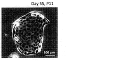
- the microscope picture of the CS cell obtained by [2] of the Example is shown.
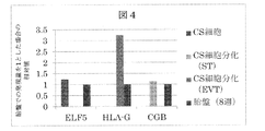
- the graph of the gene expression measured by [3] of the Example is shown.
- the micrograph of the cell which induced differentiation by [4] of the Example is shown.
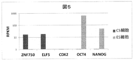
- the graph of the gene expression measured by [5] of the Example is shown.
- the graph of the gene expression of CS cell measured by [6] of an Example and ES cell is shown.
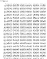
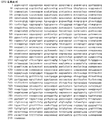
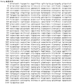
- the base sequence of ZNF750 is shown. Shows the base sequence of ELF5. Shows the base sequence of CDX2. Shows the base sequence of Oct4.
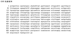
- the base sequence of Nanog is shown. Shows the base sequence of CD45. Shows the base sequence of CD49f. Shows the base sequence of CD49f.
- Undifferentiated progenitor cells The present invention provides mammalian undifferentiated progenitor cells having the ability to differentiate into cells constituting the placenta, both of which are positive for ZNF750 and ELF5.
- examples of “cells constituting the placenta” include syncytial trophoblast (ST) cells, extravillous trophoblast (EVT) cells, and the like.
- ST syncytial trophoblast
- EVT extravillous trophoblast
- the undifferentiated progenitor cells of the present invention have the ability to differentiate into these cells constituting the placenta.
- CT cells mean cellular trophoblast cells that are progenitor cells in placental tissue.
- CT cells are a concept that includes TS cells (trophoblast stem cells).
- a CS cell refers to a cell derived from a CT cell that maintains an undifferentiated state even after subculture under certain conditions.
- a certain gene is positive means that the gene is expressed. More specifically, for example, in the present invention, a gene can be positive when RNA of a certain gene is present at 1 copy or more per cell, and negative when it is less than 1 copy per cell. What is necessary is just to measure the expression level of a gene by a conventional method. Examples of the method for measuring the expression level include a method using a next-generation sequencer, a PCR method (Q-PCR method, etc.), etc. Among them, a method using a next-generation sequencer is preferable.
- RPKM per kilobase of exon per million mapped sequence reads
- RPKM 10 9 ⁇ X t / (l t N)
- X t represents the number of short leads mapped to the transcript t.
- l t indicates the length (bp) of the transcript t.
- N indicates the total number of mapped short leads].
- Mammals include, for example, primates, rodents, meats, and the like, and preferably primates.
- primates include humans, chimpanzees, rhesus monkeys, marmosets, and the like, preferably humans.
- the undifferentiated progenitor cells of the present invention are derived from cells of these mammals, preferably from cells that constitute the placenta.
- the undifferentiated progenitor cells of the present invention are characterized by positive expression of ZNF750 and ELF5 genes.
- ZNF750 include those indicated by GenBank accession number BC109037 (SEQ ID NO: 1).
- ELF5 what is shown by GenBank accession number AK075197 (sequence number 2) is mentioned. These base sequences are shown in FIGS.
- undifferentiated progenitor cells of the present invention those that are negative for CDX2 gene expression are preferred.
- examples of CDX2 include those indicated by GenBank accession number BC014461 (SEQ ID NO: 3). The base sequence is shown in FIG.
- the TS cells conventionally isolated by the present inventors were CDX2-positive. Therefore, among the present invention, it is clear that undifferentiated progenitor cells that are negative for CDX2 gene expression are also different from conventional ones from this point of view, and it is also known that such cells maintain undifferentiated ability. Is not something you can expect from.
- Oct4 is known as an undifferentiated marker, and includes one represented by GenBank accession number Z11898 (SEQ ID NO: 4).
- Nanog is also known as an undifferentiated marker, and includes one represented by GenBank accession number AB093576 (SEQ ID NO: 5).
- CD49f is preferably positive.
- the undifferentiated progenitor cells of the present invention may be negative for CD45. Examples of CD45 include those indicated by GenBank accession number AY567999 (SEQ ID NO: 6). Examples of CD49f include those indicated by GenBank accession number AB208842 (SEQ ID NO: 7). These base sequences are shown in FIGS.
- the undifferentiated progenitor cells of the present invention have a feature that they can maintain an undifferentiated state even after being subcultured.
- the undifferentiated progenitor cells of the present invention remain in an undifferentiated state even after being subcultured at least once (eg, 1-10 times, 2-5 times, or at least 8 times, at least 10 times, etc.). What is maintained is also included.
- the undifferentiated progenitor cells of the present invention include those that have not actually undergone subculture as long as they have the characteristic that an undifferentiated state is maintained after subculture.
- the undifferentiated progenitor cells of the present invention include, for example, those obtained by the production method described later.
- the present invention provides a method for producing mammalian undifferentiated progenitor cells having the ability to differentiate into cells constituting the placenta, comprising the following steps: (1) a step of recovering CD49f antibody-positive cells from a cell suspension obtained from mammalian placenta tissue; (2) A step of culturing the cell suspension obtained in the above step (1) in a medium containing a growth factor and a ROCK inhibitor.
- the method for obtaining a cell suspension from mammalian placental tissue is not particularly limited, and mechanical and / or enzymatic treatment known per se can be used as appropriate.
- the placental tissue can be minced and optionally washed with physiological saline or the like, and immersed in a cell dispersion.
- a proteolytic enzyme solution As the cell dispersion, what is called a proteolytic enzyme solution or a cell adhesion remover may be used. Specific examples of the cell dispersion include those marketed under the product names Accumax, TrypLE and the like. These cell dispersions can be used alone or in combination of two or more. You may perform the process of removing erythrocytes with respect to the said cell dispersion liquid. Examples of the method for removing red blood cells include treatment with silicate colloidal particles coated with polyvinylpyrrolidone (such as those commercially available as Percoll (registered trademark)).
- CD49f antibody positive cells are recovered from the cell suspension.
- a method for recovering CD49f antibody-positive cells a method known per se can be used.
- a step of removing CD45 antibody-positive cells may be performed after or before the step of collecting CD49f antibody-positive cells.
- CD45 antibody-positive cells can be removed according to a method known per se.
- the cell suspension that has undergone the process of collecting CD49f antibody-positive cells is cultured in a medium containing a growth factor and a ROCK inhibitor.
- the characteristic that the undifferentiated state is maintained after the subculture is the characteristic of the cell of this invention.
- the medium used in this step is not particularly limited, and examples thereof include product names DMEM, MEM, RPMI 1640, and the like. Serum, growth factors, pyruvate, amino acids, antibiotics, and the like can be appropriately added to the medium.
- the culture temperature is not particularly limited, but can be appropriately set within a range of, for example, 32 to 40 ° C, preferably 36 to 38 ° C.
- the culture time is not particularly limited, but can be appropriately set within a range of, for example, 12 to 240 hours, preferably 48 to 96 hours.
- the growth factor is not particularly limited, and examples thereof include epidermal growth factor (EGF), insulin, transforming growth factor (TGF) and the like.
- the amount of the growth factor added to the medium is not particularly limited, but can be appropriately set within a range of, for example, 0.1 to 1000 ng / ml, preferably 10 to 100 ng / ml.
- the ROCK (Rho-binding kinase) inhibitor is not particularly limited, and examples thereof include trans-N- (4-pyridyl) -4- (1-aminoethyl) -cyclohexanecarboxamide, 1- (5-isoquinolinyl). (Sulfonyl) homopiperazine (1- (5-Isoquinolinylsulfonyl) homopiperazine) or a salt thereof.
- CT cells were isolated from human placenta by the following reagents and techniques: [Reagents] ⁇ Accumax (Innovative cell technology, product number: AM105) ⁇ TrypLE (Life technology, product number: 12605036) ⁇ DMEM (Life technology, product number: 11995-065) Serum (Life technology, product number: 16141-079) ⁇ PBS (Wako, product number: 166-23555) ⁇ Anti-CD49f-PE antibody (MACS, product number: 130-100-096) -PE Selection Kit (EasySep, product number: ST-18551) -Magnet for cell separation (EasySep, product number: ST-18000) ⁇ Cell dispersion: 1: 1 mixture of Accumax and TrypLE ⁇ Neutralization solution: DMEM containing 10% bovine serum PBS-EDTA: PBS containing 1 mM EDTA and 2% bovine serum
- CD49f-positive cells obtained by the above technique are referred to as CT cells in the following examples.
- [Preparation of growth medium] 1. Mix the following to prepare a basal medium (for 1 L) DMEM 800 ml Serum 200 ml NEAA 10 ml Sodium pyruvate 10 ml 2-Mercaptoethanol 1.8 ml 2. Immediately before use, add EGF (final concentration: 100 ng / ml), Y27632 (final concentration: 5 ⁇ M), SB431542 (final concentration: 5 ⁇ M), CHIR99021 (final concentration: 3 ⁇ M) to the basal medium and use it as the growth medium To do.
- EGF final concentration: 100 ng / ml
- Y27632 final concentration: 5 ⁇ M
- SB431542 final concentration: 5 ⁇ M
- CHIR99021 final concentration: 3 ⁇ M
- Method 1 Add growth medium to isolated CT cells to 5 x 10 5 cells / ml 2. Add 2 ml of cell suspension to a 35 mm dish and incubate in an incubator (37 ° C, 5% CO 2 ) 3. Change the medium every 2 days and incubate until the cells are 70-80% confluent 4. Wash the plate with PBS, add 1 ml of TrypLE, and leave it at 37 ° C for 10 minutes. 5. Add 5 ml of growth medium 6. Centrifuge at 400 g for 3 minutes and remove the supernatant 7. Add 6 ml of growth medium (3-fold dilution) and disperse the cells by pipetting. 8. Add 2 ml of cell suspension to a 35 mm dish and continue culturing.
- FIG. 1 shows a micrograph (microscope (KEYENCE, product number: BZ-X710)) of cells obtained by subculturing the CS cells obtained above for 11 days and 55 days. It can be seen that the morphology is completely different from ST cells, which are multinucleated differentiated cells described later in [4].
- Method 1 Extract and purify RNA from cells using RNeasy mini kit 2. Prepare the annealing reaction solution as follows Oligo dT (50 ⁇ M) 0.5 ⁇ l dNTPs (10 mM) 0.5 ⁇ l Purified RNA (1 ⁇ g / ml) 1 ⁇ l H 2 O 3 ⁇ l 3. Use a thermal cycler to perform an annealing reaction at 65 ° C for 5 minutes. 4. Add the following to the above solution 5 ⁇ PrimeScript TM Buffer 2 ⁇ l PrimeScript TM Reverse Transcriptase 1 ⁇ l RNase Inhibitor 0.5 ⁇ l H 2 O 1.5 ⁇ l 5. Perform reverse transcription using a thermal cycler at [42 °C for 60 minutes ⁇ 95 °C for 5 minutes]. 6.
- Reverse transcription reaction solution (10-fold dilution) 2 ⁇ l SYBR (R) Premix Ex Taq II 10 ⁇ l Forward primer (10 ⁇ M) 1 ⁇ l Reverse primer (10 ⁇ M) 1 ⁇ l H 2 O 6 ⁇ l 7. Perform the following reaction using real-time PCR system (1) [95 ° C for 30 seconds] (2) 40 cycles of [95 ° C 5 seconds ⁇ 62 ° C 30 seconds].
- the results are shown in FIG.
- the CS cell line obtained in [2] above showed a marker expression profile similar to that of CT cells, including that differentiation markers such as HLA-G and CGB were not expressed. This indicates that the CS cell line can be established while maintaining the characteristics and functions of CT cells.
- the CS cell line is maintained in an undifferentiated state even after repeated subculture, and adhesion between adjacent cells observed when it is derived from the epithelial system. was also observed.
- EVT differentiation add Y27632 (final concentration: 5 ⁇ M) to the basal medium immediately before use and use it as the EVT differentiation medium.
- Y27632 final concentration: 5 ⁇ M
- Forskolin final concentration: 1 ⁇ M
- Method 1 Aspirate the medium from CT cells cultured in growth medium 2. Wash the plate with PBS 3. Add 2 ml of differentiation medium and incubate for 5 days.
- FIG. 3 shows a micrograph (microscope (KEYENCE, product number: BZ-X710)) of the cells induced to differentiate.
- the CS cell obtained in [2] has both the ability to differentiate into ST and the ability to differentiate into EVT.
- CS cells do not express differentiation markers such as HLA-G and CGB, whereas differentiation (ST) expresses differentiation marker CGB and differentiation (EVT) HLA-G, a differentiation marker, is expressed. Therefore, it can be seen from the point of gene expression that the CS cells obtained in [2] have differentiation ability.
- Method 1 Extract and purify RNA from cells using RNeasy mini kit 2. Create next-generation sequencer library using TruSeq RNA Sample Prep Kit 3. Perform sequence analysis using next-generation sequencer 4. Using next-generation sequencer analysis software Strand NGS, calculate the gene expression level (RPKM value: Reads per million mapped reads).
- CS cells are positive for both ZNF750 and ELF5. It was also clear (different from each other) that the cells were different from ES cells. In addition, the cells of the present invention have high ELF5 expression, but almost no CDX2 expression, and surprisingly little expression of Oct4 has been described in many literatures. It has become clear that there are unique features different from the cells reported so far.
Landscapes
- Micro-Organisms Or Cultivation Processes Thereof (AREA)
Abstract
本発明が解決すべき課題は、胎盤を構成する細胞への分化能を有する哺乳動物の未分化前駆細胞であって、未分化の状態を維持したまま継代培養することができるものを提供することである。本発明は胎盤を構成する細胞への分化能を有する哺乳動物の未分化前駆細胞であって、ZNF750及びELF5が共に陽性である、細胞を提供する。
Description
本発明は、胎盤を構成する細胞への分化能を有する哺乳動物の未分化前駆細胞及びその製造方法に関する。
胎盤組織は、栄養膜組織(トロホブラスト)が主要な構成成分である。栄養膜細胞における分化制御機構の解明には栄養膜幹細胞(TS細胞)が非常に有益であると考えられる。しかし、そのようなTS細胞は樹立されていない。
本発明者らはヒトTS細胞を分離し、これを報告している(非特許文献1)。しかし、ヒトTS細胞を分離(初代培養)することはできたものの、これを継代培養すると胎盤を構成する細胞に分化してしまい、未分化の状態を維持することができなかった。従って、係るTS細胞を株化することができていなかった。分化制御機構の解明等のための試験には、株化したTS細胞が非常に有用であるため、その開発が所望されている。
また、本発明者ら以外でも胎盤組織中の未分化な細胞の探索と分離は試みられている。非特許文献2では栄養膜細胞へ分化する前駆細胞(TBPC)が記載されている。この中で、胎盤組織中にOct4を発現する細胞の存在が確認されているとともに、胎盤組織から得た細胞懸濁液をFGF2およびSB431542を含む培地で培養することを経て得られたTBPCはOct4およびZO-1を発現する。しかしながら当該TBPCは8-10回の継代後は分化してしまうと記載されており、この点ではTBPCは未分化状態を維持できる細胞株ということはできない。トリプシン処理によって継代可能な細胞株が得られることも記載されているが、この処理で得られる細胞は前述のOct4及びZO-1は発現していないこともまた記載されていることから、この処理で得られる細胞が当該TBPCと同様の細胞と表現できるかについては議論のある結果と言える。特にZO-1は上皮系マーカーであるため、胎盤組織中の前駆細胞である細胞性栄養膜細胞(CT細胞)に由来する細胞株の場合は、CT細胞が上皮系細胞であることからもZO-1が発現していると推察されるため、TBPCは上皮系の細胞ではないことが予想される。加えて細胞形態の観点から、例えば非特許文献2の図2に示される通り、上皮系の細胞であれば観察される隣接細胞間の接着が見られないことからも、TBPCが非上皮系の細胞に由来するものであることが示唆される。胎盤由来の未分化状態の細胞(幹細胞又は前駆細胞と表現されるものを含む)を記載する文献では、同様に当該細胞がOct4を発現していることをもって特徴づけている(たとえば特許文献1、2)。
非特許文献3では、ELF5がヒト胎盤栄養膜細胞で発現し、妊娠初期であるほど高発現であることに注目するとより初期胎盤段階のTS細胞株を得るのに資するとの記載を含む。但し、この文献はTS細胞株取得に向けては、高増殖性で、ELF5およびCDX2が二重陽性のCT細胞を得ることが有望であると記載している。ちなみに、非特許文献2のTBPC細胞は、ELF5は発現していないことが記載されている。
このようにさまざまなTS細胞株取得の試みはなされているが、候補となる細胞のマーカー発現、その分化を観察した結果等を記載するに留まり、TS細胞株はいまだ樹立されていない。
有馬ほか(2005)日本産科婦人科學會雜誌 57,755
Genbacev et al (2011) Stem Cells 29, 1427-1436.
Hembergr et al (2010) Hum Mol Genet 19, 2456-2467.
本発明が解決すべき課題は、胎盤を構成する細胞への分化能を有する哺乳動物の未分化前駆細胞であって、未分化の状態を維持したまま継代培養することができるものを提供することである。
かかる状況の下、本発明者らは鋭意研究した結果、哺乳動物の胎盤組織から得た細胞懸濁液に対し所定の処理をすることにより、未分化の状態を維持したまま継代培養することのできる未分化前駆細胞を得ることができることを見出した。そして、本発明者らは、かかる未分化前駆細胞の遺伝子発現を解析したところ遺伝子ZNF750及びELF5が共に陽性であるという、従来の報告されていない特徴を見出したほか、従来報告されているマーカー遺伝子の発現パターンとは一致しない特徴があることも明らかにした。本発明はかかる新規の知見に基づくものである。
従って、本発明は、以下の項を提供する:
項1.胎盤を構成する細胞への分化能を有する哺乳動物の未分化前駆細胞であって、ZNF750及びELF5が共に陽性である、細胞。
項1.胎盤を構成する細胞への分化能を有する哺乳動物の未分化前駆細胞であって、ZNF750及びELF5が共に陽性である、細胞。
項2.CDX2が陰性である、項1に記載の細胞。
項3.Oct4及び/又はNanogが陰性である、項1又は2に記載の細胞。
項4.CD49fが陽性である、項1~3のいずれか1項に記載の細胞。
項5.霊長類の細胞である、項1~4のいずれか1項に記載の細胞。
項6.胎盤を構成する細胞が、合胞体栄養膜細胞又は絨毛外栄養膜細胞である、項1~5のいずれか1項に記載の細胞。
項7.継代培養後も未分化の状態が維持されている、項1~6のいずれか1項に記載の細胞。
項8.以下の工程を含む、胎盤を構成する細胞への分化能を有する哺乳動物の未分化前駆細胞の製造方法:
(1)哺乳動物の胎盤組織から得た細胞懸濁液から、CD49f抗体陽性細胞を回収する工程;
(2)上記工程(1)により得られた細胞懸濁液を、成長因子およびROCK阻害剤を含む培地で培養する工程。
(1)哺乳動物の胎盤組織から得た細胞懸濁液から、CD49f抗体陽性細胞を回収する工程;
(2)上記工程(1)により得られた細胞懸濁液を、成長因子およびROCK阻害剤を含む培地で培養する工程。
項9.成長因子がEGFであり、かつROCK阻害剤がtrans-N-(4-ピリジル)-4-(1-アミノエチル)-シクロヘキサンカルボキサミド又はその塩である、項8に記載の方法。
項10.上記工程(2)において培地が、さらに4-[4-(1,3-ベンゾジオキソール-5-イル)-5-(2-ピリジル)-1H-イミダゾール-2-イル]ベンズアミド又はその塩および6-[[2-[[4-(2,4-ジクロロフェニル)-5-(4-メチル-1H-イミダゾール-2-イル)-2-ピリミジニル]アミノ]エチル]アミノ]ニコチノニトリルを含む、項8又は9に記載の方法。
項11.項8~10のいずれか1項に記載の方法により得られる、胎盤を構成する細胞への分化能を有する哺乳動物の未分化前駆細胞。
本発明にかかる未分化前駆細胞は、未分化の状態を維持したまま継代培養することができ、かつ所定の条件では胎盤を構成する細胞への分化能も示すという特徴を有する。従って、胎盤を構成する細胞における分化制御機構を解明するための試験に非常に有用である。
未分化前駆細胞
本発明は、胎盤を構成する細胞への分化能を有する哺乳動物の未分化前駆細胞であって、ZNF750及びELF5が共に陽性である、細胞を提供する。
本発明は、胎盤を構成する細胞への分化能を有する哺乳動物の未分化前駆細胞であって、ZNF750及びELF5が共に陽性である、細胞を提供する。
本発明において、「胎盤を構成する細胞」としては、例えば、合胞体栄養膜(ST)細胞、絨毛外栄養膜(EVT)細胞等が挙げられる。本発明の未分化前駆細胞は、胎盤を構成するこれらの細胞への分化能を有する。
また、本明細書において、CT細胞とは、胎盤組織中の前駆細胞である細胞性栄養膜細胞を意味する。CT細胞は、TS細胞(栄養膜幹細胞)を包含する概念である。また、本明細書において、CS細胞とは、CT細胞から誘導される細胞であって一定条件下での継代培養後も未分化状態が維持されるような細胞を示す。
本願明細書において、ある遺伝子が陽性であるとは、当該遺伝子が発現していることを意味する。より具体的には、例えば、本発明において、ある遺伝子のRNAが細胞当たり1コピー以上存在する場合に当該遺伝子を陽性とし、細胞当たり1コピー未満の場合、陰性とすることができる。遺伝子の発現レベルは、常套的方法により測定すればよい。発現レベルの測定方法としては、次世代シークエンサを用いた方法、PCR法(Q-PCR法等)などが挙げられるが、なかでも次世代シークエンサを用いた方法が好適である。例えば、ある遺伝子についてRPKM値(per kilobase of exon per million mapped sequence reads)が1以上の場合に陽性とし、RPKM値が1未満の場合に陰性とすることができる。RPKM値は次世代シークエンサを用いて得ることができる値であり、転写物tのRPKMは下記の式で求めることができる:
RPKM=109×Xt/(ltN)
[式中、Xtは転写物tにマッピングされたショートリードの本数を示す。ltは転写物tの長さ(bp)を示す。Nはマッピングされたショートリードの総数を示す]。
RPKM=109×Xt/(ltN)
[式中、Xtは転写物tにマッピングされたショートリードの本数を示す。ltは転写物tの長さ(bp)を示す。Nはマッピングされたショートリードの総数を示す]。
哺乳動物としては、例えば、霊長類、げっ歯類、食肉類、等が挙げられ、好ましくは霊長類である。また、霊長類としては、例えば、ヒト、チンパンジー、アカゲザル、マーモセット等が挙げられ、好ましくはヒトである。本発明の未分化前駆細胞はこれらの哺乳動物の細胞に由来し、好ましくは、胎盤を構成する細胞に由来する。
本発明の未分化前駆細胞は、ZNF750及びELF5の遺伝子発現が陽性であることを特徴とする。ここで、ZNF750としては、GenBankアクセッション番号BC109037で示されるもの(配列番号1)が挙げられる。また、ELF5としては、GenBankアクセッション番号AK075197で示されるもの(配列番号2)が挙げられる。これらの塩基配列を図6及び7に示す。
また、本発明の未分化前駆細胞のうち、CDX2の遺伝子発現が陰性であるものが好ましい。ここで、CDX2としては、GenBankアクセッション番号BC014461で示されるもの(配列番号3)が挙げられる。その塩基配列を図8に示す。また本発明者らが従来分離したTS細胞は、CDX2陽性であった。従って、本発明のうち、CDX2遺伝子発現が陰性である未分化前駆細胞は、この点からも従来のものと異なることは明らかであり、またかかる細胞が未分化能を維持することも従来の知見から予想し得ることではない。
さらに、本発明の未分化前駆細胞においては、Oct4及びNanogの少なくとも一方が陰性であることが好ましく、これらの両方が陰性であることがより好ましい。Oct4は未分化マーカーとして知られているものであり、GenBankアクセッション番号Z11898で示されるもの(配列番号4)が挙げられる。Nanogも未分化マーカーとして知られており、GenBankアクセッション番号AB093576で示されるもの(配列番号5)が挙げられる。これらの塩基配列を図9及び10に示す。尚、さらに、本発明の未分化前駆細胞においては、CD49fが陽性であることが好ましい。また、本発明の未分化前駆細胞は、CD45が陰性であってもよい。CD45としては、GenBankアクセッション番号AY567999で示されるもの(配列番号6)が挙げられる。CD49fとしては、GenBankアクセッション番号AB208842で示されるもの(配列番号7)が挙げられる。これらの塩基配列を図11及び12に示す。
本発明の未分化前駆細胞は、継代培養をした後も未分化状態を維持することができるという特徴を有する。本発明の未分化前駆細胞には、少なくとも1回以上(例えば、1~10回、2~5回等、または少なくとも8回以上、少なくとも10回以上等)の継代培養後も未分化状態が維持されているものも包含される。また、本発明の未分化前駆細胞には、継代培養後に未分化状態が維持されるという特徴を有しているものであれば、実際に継代培養を経ていないものも包含される。従って、実際には継代培養を経ていない細胞であって、当該細胞を少なくとも1回以上(例えば、1~10回、2~5回等、または少なくとも8回以上、少なくとも10回以上等)継代培養した場合に、未分化状態が維持されるものも本発明の未分化前駆細胞に含まれ得る。また、本発明の未分化前駆細胞は、例えば、後述する製造方法により得られたものが包含される。
未分化前駆細胞の製造方法
本発明は、以下の工程を含む、胎盤を構成する細胞への分化能を有する哺乳動物の未分化前駆細胞の製造方法を提供する:
(1)哺乳動物の胎盤組織から得た細胞懸濁液から、CD49f抗体陽性細胞を回収する工程;
(2)上記工程(1)により得られた細胞懸濁液を、成長因子およびROCK阻害剤を含む培地で培養する工程。
本発明は、以下の工程を含む、胎盤を構成する細胞への分化能を有する哺乳動物の未分化前駆細胞の製造方法を提供する:
(1)哺乳動物の胎盤組織から得た細胞懸濁液から、CD49f抗体陽性細胞を回収する工程;
(2)上記工程(1)により得られた細胞懸濁液を、成長因子およびROCK阻害剤を含む培地で培養する工程。
哺乳動物の胎盤組織から細胞懸濁液を得る方法は特に限定されず、自体公知の機械的及び/又は酵素的処理を適宜使用することができる。例えば、胎盤組織を細かく刻み、任意選択で生理食塩水等で洗浄したものを、細胞分散液に漬けること等により行うことができる。
細胞分散液としては、蛋白分解酵素液、細胞接着剥離液と呼ばれるものを用いてもよい。また、細胞分散液の具体的な例としては、製品名Accumax、TrypLE等で市販されているものが挙げられる。これらの細胞分散液は1種単独で、又は2種以上を混合して用いることができる。上記細胞分散液に対し、赤血球を除去する工程を施してもよい。赤血球を除去する方法としては、ポリビニルピロリドンでコートされたケイ酸コロイド粒子(Percoll(登録商標)として市販されているもの等)での処理等が挙げられる。
次に、上記細胞懸濁液からCD49f抗体陽性細胞を回収する。CD49f抗体陽性細胞の回収方法としては、自体公知の方法に準じて行うことができる。
本発明においては、CD49f抗体陽性細胞の回収工程の後又は前に、CD45抗体陽性細胞を除去する工程を行ってもよい。としては、CD45抗体陽性細胞の除去も、自体公知の方法に準じて行うことができる。
本発明においては、CD49f抗体陽性細胞の回収工程を経た細胞懸濁液を、成長因子およびROCK阻害剤を含む培地で培養する。当該培養工程により、本発明の細胞の特徴である、経代培養後も未分化状態が維持されるという性質を獲得する。
本工程に用いる培地としては、特に限定されないが、例えば、製品名DMEM、MEM、RPMI 1640等を挙げることができる。当該培地には、血清、成長因子、ピルビン酸塩、アミノ酸、抗生物質等を適宜添加することができる。
培養温度は特に限定されないが、例えば、32~40℃、好ましくは36~38℃の範囲で適宜設定できる。培養時間は特に限定されないが、例えば、12~240時間、好ましくは48~96時間の範囲で適宜設定できる。
成長因子としては、特に限定されないが、例えば、上皮成長因子(EGF)、インスリン、トランスフォーミング成長因子(TGF)等が挙げられる。培地への成長因子の添加量は特に限定されないが、例えば、0.1~1000ng/ml、好ましくは10~100ng/mlの範囲で適宜設定できる。
またROCK(Rho結合キナーゼ)阻害剤としては、特に限定されないが、例えば、trans-N-(4-ピリジル)-4-(1-アミノエチル)-シクロヘキサンカルボキサミド、1-(5-イソキノリニルスルホニル)ホモピペラジン(1-(5-Isoquinolinylsulfonyl)homopiperazine)またはこれらの塩等が挙げられる。
trans-N-(4-ピリジル)-4-(1-アミノエチル)-シクロヘキサンカルボキサミド又はその塩の市販品としては、和光純薬より商品名Y27632として販売されている(R)-(+)- trans-N-(4-ピリジル)-4-(1-アミノエチル)-シクロヘキサンカルボキサミド((R)-(+)- trans-N-(4-pyridyl)-4-(1-aminoethyl)-cyclohexanecarboxamide)の二塩酸塩((R)-(+)- trans-N-(4-pyridyl)-4-(1-aminoethyl)-cyclohexanecarboxamide・2HCl・H2O)等が挙げられる。培地へのROCK阻害剤の添加量は特に限定されないが、例えば、0.1~50μM、好ましくは1~10μMの範囲で適宜設定できる。
また、上記培養工程において、培地に、さらに4-[4-(1,3-ベンゾジオキソール-5-イル)-5-(2-ピリジル)-1H-イミダゾール-2-イル]ベンズアミド又はその塩、および/又は6-[[2-[[4-(2,4-ジクロロフェニル)-5-(4-メチル-1H-イミダゾール-2-イル)-2-ピリミジニル]アミノ]エチル]アミノ]ニコチノニトリルを添加してもよい。
4-[4-(1,3-ベンゾジオキソール-5-イル)-5-(2-ピリジル)-1H-イミダゾール-2-イル]ベンズアミド又はその塩の市販品としては、商品名SB431542として販売されているもの等があげられる。また、6-[[2-[[4-(2,4-ジクロロフェニル)-5-(4-メチル-1H-イミダゾール-2-イル)-2-ピリミジニル]アミノ]エチル]アミノ]ニコチノニトリルの市販品としては、商品名CHIR99021として販売されているもの等が挙げられる。
4-[4-(1,3-ベンゾジオキソール-5-イル)-5-(2-ピリジル)-1H-イミダゾール-2-イル]ベンズアミド又はその塩の市販品としては、商品名SB431542として販売されているもの等があげられる。また、6-[[2-[[4-(2,4-ジクロロフェニル)-5-(4-メチル-1H-イミダゾール-2-イル)-2-ピリミジニル]アミノ]エチル]アミノ]ニコチノニトリルの市販品としては、商品名CHIR99021として販売されているもの等が挙げられる。
実施例
[1]ヒト胎盤からのCT細胞の単離
まず下記試薬及び手法によりヒト胎盤からCT細胞を単離した:
[試薬類]
・Accumax(Innovative cell technology, 製品番号:AM105)
・TrypLE(Life technology, 製品番号:12605036)
・DMEM(Life technology, 製品番号:11995-065)
・血清(Life technology, 製品番号:16141-079)
・PBS(Wako, 製品番号:166-23555)
・抗CD49f-PE抗体(MACS, 製品番号:130-100-096)
・PE Selection Kit(EasySep, 製品番号:ST-18551)
・細胞分離用磁石(EasySep, 製品番号:ST-18000)
・細胞分散液:AccumaxとTrypLEを1:1で混合
・中和液: 10%ウシ血清を含むDMEM
・PBS-EDTA:1 mM EDTAおよび2%ウシ血清を含むPBS
[手法]
1.ヒト胎盤(5~15週)を細かく刻み、生理食塩水で2-3回洗う
2. 細胞分散液20 mlを添加する
3. 37℃で20分間振とうする
4. 中和液5 mlを加える
5. 400 gで3分遠心し、上清を除去する
6. 中和液5 mlを加え、ピペットで分散させる
7. 70μmフィルターを通し、分散した細胞を回収する
8. 3-7の操作を2回(計3回)繰り返す
9. 回収した細胞を1本のチューブにまとめ、400 gで3分遠心し、上清を除去する
10. PBS-EDTAを1 x 10⁸細胞/mlとなるように加える
11. 抗CD49f-PE抗体を1 x 10⁸細胞につき100μl加える
12. PE selection cocktailを1 x 10⁸細胞につき100μl加える
13. Magnetic particleを1 x 10⁸細胞につき50μl加える
14. PBS-EDTAを加えて液量を5 mlとする
15. 細胞分離用磁石を用い、細胞を回収する
16. 14-15の操作を2回(計3回)繰り返す
17. CD49f陽性細胞(純度90-95%)が106-107細胞得られる。
上記手法により得られたCD49f陽性細胞を以下の実施例においてCT細胞と示す。
[2]CS細胞株の樹立・培養方法
上記[1]で得られたCT細胞を用い、下記の試薬及び手法によってCS細胞株を樹立、培養した:
[試薬類]
・DMEM(Life technology, 製品番号:11995-065)
・血清(Life technology, 製品番号:16141-079)
・NEAA(Life technology, 製品番号:11140-050)
・Sodium pyruvate(Life technology, 製品番号:11360-070)
・2-Mercaptoethanol(Life technology, 製品番号:21985-023)
・PBS(Wako, 製品番号:166-23555)
・TrypLE(Life technology, 製品番号:12605036)
・EGF(Wako, 製品番号:053-07871)
・Y27632(Wako, 製品番号:253-00513)
・CHIR99021(Wako, 製品番号:039-20831)
・SB435142 (Wako, 製品番号:194-15521)
・35 mmディッシュ(Nunc, 製品番号:153066)。
[増殖培地の作製]
1. 以下を混合し、基礎培地を作製する(1 L用)
DMEM 800 ml
血清 200 ml
NEAA 10 ml
Sodium pyruvate 10 ml
2-Mercaptoethanol 1.8 ml
2. 使用直前に、EGF(終濃度:100 ng/ml)、Y27632(終濃度:5μM)、SB431542(終濃度:5μM)、CHIR99021(終濃度:3μM)を基礎培地に加え、増殖培地として使用する。
1. 以下を混合し、基礎培地を作製する(1 L用)
DMEM 800 ml
血清 200 ml
NEAA 10 ml
Sodium pyruvate 10 ml
2-Mercaptoethanol 1.8 ml
2. 使用直前に、EGF(終濃度:100 ng/ml)、Y27632(終濃度:5μM)、SB431542(終濃度:5μM)、CHIR99021(終濃度:3μM)を基礎培地に加え、増殖培地として使用する。
[手法]
1. 単離したCT細胞に、増殖培地を5 x 10⁵細胞/mlとなるように加える
2. 35 mmディッシュに細胞懸濁液2 mlを加え、インキュベーター(37℃, 5 % CO2)で培養する
3. 2日毎に培地交換を行い、細胞が70~80% コンフルエントになるまで培養する
4. プレートをPBSで洗い、1 mlのTrypLEを加えて、37℃で10分静置する
5. 増殖培地5 mlを加える
6. 400 gで3分遠心し、上清を除去する
7. 増殖培地6 mlを加え(3倍希釈)、ピペッティングにより細胞を分散させる
8. 35 mmディッシュに細胞懸濁液を2 mlずつ加え、培養を続ける。
1. 単離したCT細胞に、増殖培地を5 x 10⁵細胞/mlとなるように加える
2. 35 mmディッシュに細胞懸濁液2 mlを加え、インキュベーター(37℃, 5 % CO2)で培養する
3. 2日毎に培地交換を行い、細胞が70~80% コンフルエントになるまで培養する
4. プレートをPBSで洗い、1 mlのTrypLEを加えて、37℃で10分静置する
5. 増殖培地5 mlを加える
6. 400 gで3分遠心し、上清を除去する
7. 増殖培地6 mlを加え(3倍希釈)、ピペッティングにより細胞を分散させる
8. 35 mmディッシュに細胞懸濁液を2 mlずつ加え、培養を続ける。
上記で得られたCS細胞を11代、55日間継代培養した細胞の顕微鏡写真(顕微鏡(KEYENCE, 製品番号:BZ-X710))を図1に示す。[4]で後述する多核化した分化細胞であるST細胞とは形態が全く異なることが分かる。
[3]遺伝子発現のリアルタイムPCR測定
前記[1]で単離したCT細胞、上記[2]に記載の方法で得、継代培養したCS細胞及び前記[1]記載の方法においてCT単離に使用したヒト胎盤(8週)について、ELF5、HLA-G及びCGBの発現量を下記に記載の試薬、機器を用い、下記方法に従いリアルタイムPCRにより測定した。
[試薬・機器類]
・PrimeScriptTMII 1st strand cDNA Synthesis Kit(Takara, 製品番号:6210A)
・サーマルサイクラー TProfessional(BM機器, 製品番号:070-000)
・SYBR(R) Premix Ex TaqTM II(Takara, 製品番号:RR820S)
・StepOnePlusTMリアルタイムPCRシステム(Life technology, 製品番号:StepOnePlus-01)
・プライマー(下表)

[方法]
1. RNeasy mini kitを用いて、細胞よりRNAを抽出・精製する
2. アニーリング反応液を以下のように調製する
Oligo dT (50μM) 0.5μl
dNTPs (10 mM) 0.5μl
精製RNA (1μg/ml) 1μl
H2O 3μl
3. サーマルサイクラーを用い、65℃で5分アニーリング反応を行う
4. 上記液に以下を加える
5 × PrimeScriptTM Buffer 2μl
PrimeScriptTM Reverse Transcriptase 1μl
RNase Inhibitor 0.5μl
H2O 1.5μl
5. サーマルサイクラーを用い[42℃ 60分 → 95℃ 5分]で逆転写反応を行う
6. リアルタイムPCR用の反応液を以下のように調製する
逆転写反応液(10倍希釈) 2μl
SYBR(R) Premix Ex Taq II 10μl
Forwardプライマー(10μM) 1μl
Reverseプライマー(10μM) 1μl
H2O 6μl
7. リアルタイムPCRシステムを用いて以下の反応を行う
(1)[95℃ 30秒]
(2)[95℃ 5秒 → 62℃ 30秒]を40サイクル。
結果を図2に示す。図2から明らかなように、上記[2]で得られたCS細胞株は、HLA-G及びCGBといった分化マーカーが発現していないことを含め、CT細胞と同様のマーカー発現プロファイルを示した。このことはCS細胞株がCT細胞の特徴および機能を維持したまま株化できていることを示している。また、図1の写真からも明らかなように、CS細胞株は継代培養を繰り返した後も未分化の状態が維持されており、上皮系由来である場合に観察される隣接細胞間の接着も観察された。
[4]CT細胞株の分化誘導方法
[2]で得られたCS細胞に対し、以下の試薬及び方法により、分化誘導を行った:
[試薬類]
・DMEM(Life technology, 製品番号:11995-065)
・血清(Life technology, 製品番号:16141-079)
・NEAA(Life technology, 製品番号:11140-050)
・Sodium pyruvate(Life technology, 製品番号:11360-070)
・2-Mercaptoethanol(Life technology, 製品番号:21985-023)
・Y27632(Wako, 製品番号:253-00513)
・Forskolin(Wako, 製品番号:067-02191)
・35 mmディッシュ(Nunc, 製品番号:153066)。
[分化培地の作製]
1. 以下を混合し、基礎培地を作製する(1 L用)。
1. 以下を混合し、基礎培地を作製する(1 L用)。
DMEM 800 ml
血清 200 ml
NEAA 10 ml
Sodium pyruvate 10 ml
2-Mercaptoethanol 1.8 ml
2. EVT分化誘導には、使用直前にY27632(終濃度:5 μM)を基礎培地に加え、EVT分化培地として使用する
3. ST分化誘導には、使用直前にY27632(終濃度:5μM)およびForskolin(終濃度:1μM)を基礎培地に加え、ST分化培地として使用する。
血清 200 ml
NEAA 10 ml
Sodium pyruvate 10 ml
2-Mercaptoethanol 1.8 ml
2. EVT分化誘導には、使用直前にY27632(終濃度:5 μM)を基礎培地に加え、EVT分化培地として使用する
3. ST分化誘導には、使用直前にY27632(終濃度:5μM)およびForskolin(終濃度:1μM)を基礎培地に加え、ST分化培地として使用する。
[手法]
1. 増殖培地で培養しているCT細胞より、培地を吸引除去する
2. プレートをPBSで洗う
3. 分化培地2 mlを加え、5日間培養する。
1. 増殖培地で培養しているCT細胞より、培地を吸引除去する
2. プレートをPBSで洗う
3. 分化培地2 mlを加え、5日間培養する。
分化誘導した細胞の顕微鏡写真(顕微鏡(KEYENCE, 製品番号:BZ-X710))を図3に示す。図3から明らかなように、[2]で得られたCS細胞は、STへの分化能も、EVTへの分化能も有している。
[5]CS細胞の未分化維持と分化能
前記[3]でリアルタイムPCR測定したCS細胞、前記[2]で得られたヒトCS細胞を[4]でY27632及びForskolinの存在下で培養したもの(分化(ST))、前記[2]で得られたヒトCS細胞を[4]でY27632の存在下で培養したもの(分化(EVT))、及び前記[1]記載の方法においてCT単離に使用したヒト胎盤(8週)について、ELF5、HLA-G及びCGBの発現量をリアルタイムPCRにより測定した。測定方法は、前記[3]に記載の方法に準じて行った。結果を図4に示す。
前記[3]でリアルタイムPCR測定したCS細胞、前記[2]で得られたヒトCS細胞を[4]でY27632及びForskolinの存在下で培養したもの(分化(ST))、前記[2]で得られたヒトCS細胞を[4]でY27632の存在下で培養したもの(分化(EVT))、及び前記[1]記載の方法においてCT単離に使用したヒト胎盤(8週)について、ELF5、HLA-G及びCGBの発現量をリアルタイムPCRにより測定した。測定方法は、前記[3]に記載の方法に準じて行った。結果を図4に示す。
図4から明らかなように、CS細胞はHLA-G及びCGBといった分化マーカーが発現していないのに対し、分化(ST)は、分化マーカであるCGBが発現しており、分化(EVT)は、分化マーカであるHLA-Gが発現している。従って、遺伝子発現の点からも、[2]で得られたCS細胞が分化能を有していることが分かる。
[6]CS細胞とES細胞の発現比較
前記[2]で得られたCS細胞及びES細胞(H1株)について、下記試薬、機器及び方法で遺伝子の発現解析を行いRPKM値を算出した:
遺伝子の発現解析方法
[試薬・機器類]
・RNeasy mini kit(Qiagen, 製品番号:74104)
・TruSeq RNA Sample Prep Kit(Illumina, 製品番号:RS-122-2001)
・次世代シークエンサー(IlluminaのHiSeq2500を使用)
・次世代シークエンサー解析ソフト Strand NGS(Digital Biology)。
[6]CS細胞とES細胞の発現比較
前記[2]で得られたCS細胞及びES細胞(H1株)について、下記試薬、機器及び方法で遺伝子の発現解析を行いRPKM値を算出した:
遺伝子の発現解析方法
[試薬・機器類]
・RNeasy mini kit(Qiagen, 製品番号:74104)
・TruSeq RNA Sample Prep Kit(Illumina, 製品番号:RS-122-2001)
・次世代シークエンサー(IlluminaのHiSeq2500を使用)
・次世代シークエンサー解析ソフト Strand NGS(Digital Biology)。
[手法]
1. RNeasy mini kitを用いて、細胞よりRNAを抽出・精製する
2. TruSeq RNA Sample Prep Kitを用い次世代シークエンサー用ライブラリーを作製する
3. 次世代シークエンサーを用い、シークエンス解析を行う
4. 次世代シークエンサー解析ソフト Strand NGSを用い、遺伝子の発現量(RPKM値:Reads per million mapped reads)を計算する。
1. RNeasy mini kitを用いて、細胞よりRNAを抽出・精製する
2. TruSeq RNA Sample Prep Kitを用い次世代シークエンサー用ライブラリーを作製する
3. 次世代シークエンサーを用い、シークエンス解析を行う
4. 次世代シークエンサー解析ソフト Strand NGSを用い、遺伝子の発現量(RPKM値:Reads per million mapped reads)を計算する。
結果を図5に示す。図5から明らかなように、CS細胞は、ZNF750及びELF5が共に陽性である。ES細胞とも異なる細胞であることも明らか(互いに区別できる)となった。また、本発明の細胞はELF5が高発現である一方でCDX2はほとんど発現していないこと、また多くの文献で記載されてきたOct4の発現については驚くべきことにほとんど発現していないこと、という従来報告されてきた細胞とは異なる独自の特徴があることが明らかになった。
Claims (11)
- 胎盤を構成する細胞への分化能を有する哺乳動物の未分化前駆細胞であって、ZNF750及びELF5が共に陽性である、細胞。
- CDX2が陰性である、請求項1に記載の細胞。
- Oct4及び/又はNanogが陰性である、請求項1又は2に記載の細胞。
- CD49fが陽性である、請求項1~3のいずれか1項に記載の細胞。
- 霊長類の細胞である、請求項1~4のいずれか1項に記載の細胞。
- 胎盤を構成する細胞が、合胞体栄養膜細胞又は絨毛外栄養膜細胞である、請求項1~5のいずれか1項に記載の細胞。
- 継代培養後も未分化の状態が維持されている、請求項1~6のいずれか1項に記載の細胞。
- 以下の工程を含む、胎盤を構成する細胞への分化能を有する哺乳動物の未分化前駆細胞の製造方法:
(1)哺乳動物の胎盤組織から得た細胞懸濁液から、CD49f抗体陽性細胞を回収する工程;
(2)上記工程(1)により得られた細胞懸濁液を、成長因子およびROCK阻害剤を含む培地で培養する工程。 - 成長因子がEGFであり、かつROCK阻害剤がtrans-N-(4-ピリジル)-4-(1-アミノエチル)-シクロヘキサンカルボキサミド又はその塩である、請求項8に記載の方法。
- 上記工程(2)において培地が、さらに4-[4-(1,3-ベンゾジオキソール-5-イル)-5-(2-ピリジル)-1H-イミダゾール-2-イル]ベンズアミド又はその塩および6-[[2-[[4-(2,4-ジクロロフェニル)-5-(4-メチル-1H-イミダゾール-2-イル)-2-ピリミジニル]アミノ]エチル]アミノ]ニコチノニトリルを含む、請求項8又は9に記載の方法。
- 請求項8~10のいずれか1項に記載の方法により得られる、胎盤を構成する細胞への分化能を有する哺乳動物の未分化前駆細胞。
Priority Applications (1)
| Application Number | Priority Date | Filing Date | Title |
|---|---|---|---|
| JP2017505404A JP6400832B2 (ja) | 2015-03-10 | 2016-03-10 | 胎盤を構成する細胞への分化能を有する哺乳動物の未分化前駆細胞及びその製造方法 |
Applications Claiming Priority (2)
| Application Number | Priority Date | Filing Date | Title |
|---|---|---|---|
| JP2015-047236 | 2015-03-10 | ||
| JP2015047236 | 2015-03-10 |
Publications (1)
| Publication Number | Publication Date |
|---|---|
| WO2016143866A1 true WO2016143866A1 (ja) | 2016-09-15 |
Family
ID=56879561
Family Applications (1)
| Application Number | Title | Priority Date | Filing Date |
|---|---|---|---|
| PCT/JP2016/057625 Ceased WO2016143866A1 (ja) | 2015-03-10 | 2016-03-10 | 胎盤を構成する細胞への分化能を有する哺乳動物の未分化前駆細胞及びその製造方法 |
Country Status (2)
| Country | Link |
|---|---|
| JP (1) | JP6400832B2 (ja) |
| WO (1) | WO2016143866A1 (ja) |
Cited By (5)
| Publication number | Priority date | Publication date | Assignee | Title |
|---|---|---|---|---|
| WO2020250438A1 (ja) * | 2019-06-14 | 2020-12-17 | 国立大学法人東北大学 | 胎盤を構成する細胞への分化能を有する細胞、およびその製造方法 |
| US11926845B2 (en) | 2020-03-11 | 2024-03-12 | Hitachi, Ltd. | Cell culture method and automatic cell culture apparatus |
| WO2025023316A1 (ja) * | 2023-07-26 | 2025-01-30 | 国立大学法人東北大学 | 着床率向上剤及び着床率向上剤の製造方法 |
| US12492372B2 (en) | 2020-11-24 | 2025-12-09 | Monash University | Induced stem cells |
| US12668775B2 (en) | 2019-11-13 | 2026-06-30 | Monash University | Methods for reprogramming cells |
Families Citing this family (1)
| Publication number | Priority date | Publication date | Assignee | Title |
|---|---|---|---|---|
| JP7710755B2 (ja) | 2021-08-18 | 2025-07-22 | 国立大学法人東北大学 | 子宮内膜モデル、子宮内膜モデルの作製方法、及び子宮内膜着床モデル |
Citations (2)
| Publication number | Priority date | Publication date | Assignee | Title |
|---|---|---|---|---|
| JP2005204590A (ja) * | 2004-01-23 | 2005-08-04 | Kumamoto Univ | ヒト成体由来の膵臓内分泌細胞に分化可能な細胞に特有な表面抗原とこの表面抗原による細胞分離法 |
| WO2012083023A1 (en) * | 2010-12-17 | 2012-06-21 | Anthrogenesis Corporation | Treatment of spinal cord injury and traumatic brain injury using amnion derived adherent cells |
Family Cites Families (1)
| Publication number | Priority date | Publication date | Assignee | Title |
|---|---|---|---|---|
| CN103114075B (zh) * | 2013-01-18 | 2014-11-12 | 内蒙古大学 | 一种牛滋养层干细胞建系方法 |
-
2016
- 2016-03-10 JP JP2017505404A patent/JP6400832B2/ja active Active
- 2016-03-10 WO PCT/JP2016/057625 patent/WO2016143866A1/ja not_active Ceased
Patent Citations (2)
| Publication number | Priority date | Publication date | Assignee | Title |
|---|---|---|---|---|
| JP2005204590A (ja) * | 2004-01-23 | 2005-08-04 | Kumamoto Univ | ヒト成体由来の膵臓内分泌細胞に分化可能な細胞に特有な表面抗原とこの表面抗原による細胞分離法 |
| WO2012083023A1 (en) * | 2010-12-17 | 2012-06-21 | Anthrogenesis Corporation | Treatment of spinal cord injury and traumatic brain injury using amnion derived adherent cells |
Non-Patent Citations (10)
| Title |
|---|
| CHEN YING ET AL.: "Roles of CDX2 and EOMES in human induced trophoblast progenitor cells", BIOCHEM. BIOPHYS. RES. COMMUN., vol. 431, no. 2, 2013, pages 197 - 202, XP028970948 * |
| COHEN IDAN ET AL.: "ZNF750 Is Expressed in Differentiated Keratinocytes and Regulates Epidermal Late Differentiation Genes", PLOS ONE, vol. 7, no. 8., 2012, pages e42628, XP055309428 * |
| FUKUSHIMA KOTARO ET AL.: "TNFα-Induced Apoptosis and Integrin Switching in Human Extravillous Trophoblast Cell Line", BIOLOGY OF REPRODUCTION, vol. 68, 2003, pages 1771 - 1778, XP055309425 * |
| GENBACEV OLGA ET AL.: "Establishment of Human Trophoblast Progenitor Cell Lines from the Chorion", STEM CELLS, vol. 29, 2011, pages 1427 - 1436, XP055135246 * |
| MOMO NAKANISHI ET AL.: "Jumo to Taiban o Meguru Shinchiken Eiyomaku Kansaibo", CLINICAL GYNECOLOGY AND OBSTETRICS, vol. 65, no. 3, 2011, pages 260 - 265 * |
| ODA MAYUMI ET AL.: "Establishment of trophoblast stem cell lines from somatic cell nuclear-transferred embryos", PROS. NATL. ACAD. SCI. USA, vol. 106, no. 38, 2009, pages 16293 - 16297, XP055309427 * |
| OHINATA YASUHIDE ET AL.: "Establishment of Trophoblast Stem Cells under Defined Culture Conditions in Mice", PLOS ONE, vol. 9, no. 9, 2014, pages e107308 * |
| PARK TEA SOON ET AL.: "Placental Perivascular Cells for Human Muscle Regeneration", STEM CELLS AND DEVELOPMENT, vol. 20, no. 3, 2011, pages 451 - 463, XP055309424 * |
| WU TONG ET AL.: "Reprogramming of Trophoblast Stem Cells into Pluripotent Stem Cells by Oct4", STEM CELLS, vol. 29, 2011, pages 755 - 763, XP055052764 * |
| YUSUKE HOSOI ET AL.: "Mouse Haibanho karano Eiyomaku Kansaibokabu Oyobi Haitaigai Naihaiyo Eiyomaku Kansaibokabu Oyobi Haitaigai Naihaiyo Saibokabu no Juritsu to Idenshi Hatsugen Kaiseki", TOHOKU JOURNAL OF ANIMAL SCIENCE AND TECHNOLOGY, vol. 60, no. 1, 2010, pages 6 - 12 * |
Cited By (6)
| Publication number | Priority date | Publication date | Assignee | Title |
|---|---|---|---|---|
| WO2020250438A1 (ja) * | 2019-06-14 | 2020-12-17 | 国立大学法人東北大学 | 胎盤を構成する細胞への分化能を有する細胞、およびその製造方法 |
| JPWO2020250438A1 (ja) * | 2019-06-14 | 2020-12-17 | ||
| US12668775B2 (en) | 2019-11-13 | 2026-06-30 | Monash University | Methods for reprogramming cells |
| US11926845B2 (en) | 2020-03-11 | 2024-03-12 | Hitachi, Ltd. | Cell culture method and automatic cell culture apparatus |
| US12492372B2 (en) | 2020-11-24 | 2025-12-09 | Monash University | Induced stem cells |
| WO2025023316A1 (ja) * | 2023-07-26 | 2025-01-30 | 国立大学法人東北大学 | 着床率向上剤及び着床率向上剤の製造方法 |
Also Published As
| Publication number | Publication date |
|---|---|
| JP6400832B2 (ja) | 2018-10-03 |
| JPWO2016143866A1 (ja) | 2017-08-03 |
Similar Documents
| Publication | Publication Date | Title |
|---|---|---|
| JP6400832B2 (ja) | 胎盤を構成する細胞への分化能を有する哺乳動物の未分化前駆細胞及びその製造方法 | |
| JP6678107B2 (ja) | 膵前駆細胞の増殖方法 | |
| JP5777127B1 (ja) | 原始腸内胚葉細胞及びその作製方法 | |
| KR20190018709A (ko) | 혈액을 유도 만능 줄기세포로 리프로그래밍하기 위한 신규하고 효율적인 방법 | |
| JPWO2018164228A1 (ja) | Ror1陽性の間葉系幹細胞を含有する、線維症を伴う疾患の予防又は処置のための医薬組成物、及びその調製方法、並びにror1陽性の間葉系幹細胞を用いる線維症を伴う疾患の予防又は処置方法 | |
| WO2019093340A1 (ja) | ナイーブ型多能性幹細胞からの原始内胚葉誘導方法 | |
| JPWO2017175876A1 (ja) | 幹細胞を再樹立する方法 | |
| JP6090937B2 (ja) | 新規ドーパミン産生神経前駆細胞マーカー | |
| JPWO2017038784A1 (ja) | Ror1陽性の間葉系幹細胞及びその調製方法、ror1陽性の間葉系幹細胞を含む医薬組成物及びその調製方法、並びにror1陽性の間葉系幹細胞を用いる疾患の予防又は治療方法 | |
| AU2016305705A1 (en) | Induced extended pluripotent stem cells, methods of making and using | |
| JP6373253B2 (ja) | 新規心筋細胞マーカー | |
| JPWO2014200030A1 (ja) | 人工多能性幹細胞の選別方法および血球への分化誘導方法 | |
| JP2011519280A (ja) | Dnaメチル化における改変を伴う神経再生細胞連邦政府の援助に関する申告適用なし | |
| JP2015505240A (ja) | 多能性細胞を選別するための方法 | |
| JP6025067B2 (ja) | 新規心筋細胞マーカー | |
| Zhi et al. | Elucidation of the pluripotent potential of bovine embryonic lineages facilitates the establishment of formative stem cell lines | |
| JP7173496B2 (ja) | 心筋細胞への分化指向性を有する多能性幹細胞を選抜するための方法 | |
| JP7209619B2 (ja) | 非ヒト霊長類人工多能性幹細胞由来肝細胞およびその使用 | |
| CA3140384A1 (en) | Method for purifying neural crest cells or corneal epithelial cells | |
| CN104884612A (zh) | Mait样细胞及其制作方法 | |
| US20220259557A1 (en) | Cells capable of differentiating into placenta-constituting cells, and method for producing same | |
| WO2020095423A1 (ja) | 多能性幹細胞から樹状分岐した集合管を伴う腎臓構造を作製する方法 | |
| Wilkinson et al. | Expanded potential stem cell media as a tool to study human developmental hematopoiesis in vitro | |
| Liu et al. | Nodal mutant e X traembryonic EN doderm (XEN) stem cells upregulate markers for the anterior visceral endoderm and impact the timing of cardiac differentiation in mouse embryoid bodies | |
| JP7273141B2 (ja) | 特定細胞に分化する能力を有する多能性幹細胞の製造方法およびその応用 |
Legal Events
| Date | Code | Title | Description |
|---|---|---|---|
| 121 | Ep: the epo has been informed by wipo that ep was designated in this application |
Ref document number: 16761832 Country of ref document: EP Kind code of ref document: A1 |
|
| ENP | Entry into the national phase |
Ref document number: 2017505404 Country of ref document: JP Kind code of ref document: A |
|
| NENP | Non-entry into the national phase |
Ref country code: DE |
|
| 122 | Ep: pct application non-entry in european phase |
Ref document number: 16761832 Country of ref document: EP Kind code of ref document: A1 |