WO2016006398A1 - 分析装置および分析方法 - Google Patents
分析装置および分析方法 Download PDFInfo
- Publication number
- WO2016006398A1 WO2016006398A1 PCT/JP2015/067386 JP2015067386W WO2016006398A1 WO 2016006398 A1 WO2016006398 A1 WO 2016006398A1 JP 2015067386 W JP2015067386 W JP 2015067386W WO 2016006398 A1 WO2016006398 A1 WO 2016006398A1
- Authority
- WO
- WIPO (PCT)
- Prior art keywords
- complex
- flow path
- immunoassay
- container
- mass spectrometry
- Prior art date
- Legal status (The legal status is an assumption and is not a legal conclusion. Google has not performed a legal analysis and makes no representation as to the accuracy of the status listed.)
- Ceased
Links
Images



Classifications
-
- G—PHYSICS
- G01—MEASURING; TESTING
- G01N—INVESTIGATING OR ANALYSING MATERIALS BY DETERMINING THEIR CHEMICAL OR PHYSICAL PROPERTIES
- G01N35/00—Automatic analysis not limited to methods or materials provided for in any single one of groups G01N1/00 - G01N33/00; Handling materials therefor
- G01N35/10—Devices for transferring samples or any liquids to, in, or from, the analysis apparatus, e.g. suction devices, injection devices
-
- G—PHYSICS
- G01—MEASURING; TESTING
- G01N—INVESTIGATING OR ANALYSING MATERIALS BY DETERMINING THEIR CHEMICAL OR PHYSICAL PROPERTIES
- G01N33/00—Investigating or analysing materials by specific methods not covered by groups G01N1/00 - G01N31/00
- G01N33/48—Biological material, e.g. blood, urine; Haemocytometers
- G01N33/50—Chemical analysis of biological material, e.g. blood, urine; Testing involving biospecific ligand binding methods; Immunological testing
- G01N33/53—Immunoassay; Biospecific binding assay; Materials therefor
- G01N33/543—Immunoassay; Biospecific binding assay; Materials therefor with an insoluble carrier for immobilising immunochemicals
- G01N33/54313—Immunoassay; Biospecific binding assay; Materials therefor with an insoluble carrier for immobilising immunochemicals the carrier being characterised by its particulate form
- G01N33/54326—Magnetic particles
-
- G—PHYSICS
- G01—MEASURING; TESTING
- G01N—INVESTIGATING OR ANALYSING MATERIALS BY DETERMINING THEIR CHEMICAL OR PHYSICAL PROPERTIES
- G01N15/00—Investigating characteristics of particles; Investigating permeability, pore-volume or surface-area of porous materials
- G01N15/10—Investigating individual particles
- G01N15/1031—Investigating individual particles by measuring electrical or magnetic effects
- G01N15/12—Investigating individual particles by measuring electrical or magnetic effects by observing changes in resistance or impedance across apertures when traversed by individual particles, e.g. by using the Coulter principle
-
- G—PHYSICS
- G01—MEASURING; TESTING
- G01N—INVESTIGATING OR ANALYSING MATERIALS BY DETERMINING THEIR CHEMICAL OR PHYSICAL PROPERTIES
- G01N15/00—Investigating characteristics of particles; Investigating permeability, pore-volume or surface-area of porous materials
- G01N15/10—Investigating individual particles
- G01N15/1031—Investigating individual particles by measuring electrical or magnetic effects
- G01N15/12—Investigating individual particles by measuring electrical or magnetic effects by observing changes in resistance or impedance across apertures when traversed by individual particles, e.g. by using the Coulter principle
- G01N15/131—Details
-
- G—PHYSICS
- G01—MEASURING; TESTING
- G01N—INVESTIGATING OR ANALYSING MATERIALS BY DETERMINING THEIR CHEMICAL OR PHYSICAL PROPERTIES
- G01N27/00—Investigating or analysing materials by the use of electric, electrochemical, or magnetic means
- G01N27/62—Investigating or analysing materials by the use of electric, electrochemical, or magnetic means by investigating the ionisation of gases, e.g. aerosols; by investigating electric discharges, e.g. emission of cathode
-
- G—PHYSICS
- G01—MEASURING; TESTING
- G01N—INVESTIGATING OR ANALYSING MATERIALS BY DETERMINING THEIR CHEMICAL OR PHYSICAL PROPERTIES
- G01N30/00—Investigating or analysing materials by separation into components using adsorption, absorption or similar phenomena or using ion-exchange, e.g. chromatography or field flow fractionation
- G01N30/02—Column chromatography
- G01N30/62—Detectors specially adapted therefor
- G01N30/72—Mass spectrometers
- G01N30/7233—Mass spectrometers interfaced to liquid or supercritical fluid chromatograph
-
- G—PHYSICS
- G01—MEASURING; TESTING
- G01N—INVESTIGATING OR ANALYSING MATERIALS BY DETERMINING THEIR CHEMICAL OR PHYSICAL PROPERTIES
- G01N33/00—Investigating or analysing materials by specific methods not covered by groups G01N1/00 - G01N31/00
- G01N33/48—Biological material, e.g. blood, urine; Haemocytometers
- G01N33/50—Chemical analysis of biological material, e.g. blood, urine; Testing involving biospecific ligand binding methods; Immunological testing
- G01N33/53—Immunoassay; Biospecific binding assay; Materials therefor
-
- G—PHYSICS
- G01—MEASURING; TESTING
- G01N—INVESTIGATING OR ANALYSING MATERIALS BY DETERMINING THEIR CHEMICAL OR PHYSICAL PROPERTIES
- G01N33/00—Investigating or analysing materials by specific methods not covered by groups G01N1/00 - G01N31/00
- G01N33/48—Biological material, e.g. blood, urine; Haemocytometers
- G01N33/50—Chemical analysis of biological material, e.g. blood, urine; Testing involving biospecific ligand binding methods; Immunological testing
- G01N33/82—Chemical analysis of biological material, e.g. blood, urine; Testing involving biospecific ligand binding methods; Immunological testing involving vitamins or their receptors
-
- G—PHYSICS
- G01—MEASURING; TESTING
- G01N—INVESTIGATING OR ANALYSING MATERIALS BY DETERMINING THEIR CHEMICAL OR PHYSICAL PROPERTIES
- G01N35/00—Automatic analysis not limited to methods or materials provided for in any single one of groups G01N1/00 - G01N33/00; Handling materials therefor
- G01N35/02—Automatic analysis not limited to methods or materials provided for in any single one of groups G01N1/00 - G01N33/00; Handling materials therefor using a plurality of sample containers moved by a conveyor system past one or more treatment or analysis stations
- G01N35/025—Automatic analysis not limited to methods or materials provided for in any single one of groups G01N1/00 - G01N33/00; Handling materials therefor using a plurality of sample containers moved by a conveyor system past one or more treatment or analysis stations having a carousel or turntable for reaction cells or cuvettes
-
- G—PHYSICS
- G01—MEASURING; TESTING
- G01N—INVESTIGATING OR ANALYSING MATERIALS BY DETERMINING THEIR CHEMICAL OR PHYSICAL PROPERTIES
- G01N35/00—Automatic analysis not limited to methods or materials provided for in any single one of groups G01N1/00 - G01N33/00; Handling materials therefor
- G01N35/08—Automatic analysis not limited to methods or materials provided for in any single one of groups G01N1/00 - G01N33/00; Handling materials therefor using a stream of discrete samples flowing along a tube system, e.g. flow injection analysis
-
- G—PHYSICS
- G01—MEASURING; TESTING
- G01N—INVESTIGATING OR ANALYSING MATERIALS BY DETERMINING THEIR CHEMICAL OR PHYSICAL PROPERTIES
- G01N35/00—Automatic analysis not limited to methods or materials provided for in any single one of groups G01N1/00 - G01N33/00; Handling materials therefor
- G01N35/10—Devices for transferring samples or any liquids to, in, or from, the analysis apparatus, e.g. suction devices, injection devices
- G01N35/1095—Devices for transferring samples or any liquids to, in, or from, the analysis apparatus, e.g. suction devices, injection devices for supplying the samples to flow-through analysers
-
- G—PHYSICS
- G01—MEASURING; TESTING
- G01N—INVESTIGATING OR ANALYSING MATERIALS BY DETERMINING THEIR CHEMICAL OR PHYSICAL PROPERTIES
- G01N15/00—Investigating characteristics of particles; Investigating permeability, pore-volume or surface-area of porous materials
- G01N15/01—Investigating characteristics of particles; Investigating permeability, pore-volume or surface-area of porous materials specially adapted for biological cells, e.g. blood cells
Definitions
- the present invention relates to an analysis apparatus and an analysis method.
- the present invention includes a “rotating microparticle (magnetic bead) trapping part arranged on a capillary channel and a high-performance liquid chromatograph-tandem mass spectrometer, in which an antibody is bound in advance in the microparticle trapping part. It is described as “an automated apparatus and method for supplementing a complex in which fine particles and a measurement substance are bound, washing, and eluting the measurement substance from the fine particles”. Similarly, Japanese Unexamined Patent Publication No. 1-187459 is a background art in this technical field.
- a fine particle in which an antigen-antibody reaction is caused to occur on a reaction solid phase and an antibody is bound in advance and a complex in which a measurement substance is bound by an antigen-antibody reaction are released from the reaction solid phase It is described as “an immunoassay method in which a target component is quantified by measuring by a method”.
- Patent Document 1 supplements a complex in which a microparticle to which an antibody is bound in advance and a measurement substance are bound, and after washing and elution of the measurement substance from the microparticle, measurement is performed with a high-performance liquid chromatograph-tandem mass spectrometer. (Hereinafter, this technology is referred to as mass spectrometry).
- mass spectrometry this technology is referred to as mass spectrometry.
- an antigen-antibody reaction is caused on a reaction solid phase, a microparticle to which an antibody is bound in advance, and a complex in which a measurement substance is bound by an antigen-antibody reaction is released from the reaction solid phase, A technique for measuring by spectroscopy is described (hereinafter, this technique is referred to as an immunoassay).
- the information obtained by the measurement is obtained by measuring the complex in which the microparticles and the measurement substance are bound by the spectroscopic method in the immunoassay, so that the total amount information of the measurement substance bound to the complex can be obtained.
- the immunoassay is a measurement technique that is simple and sensitive, and is suitable for quantitative measurement of trace components, but has a problem of cross-reactivity.
- Cross-reactivity is a phenomenon in which not only a measurement substance that should be recognized by an antibody but also a molecule (similar compound) having a similar structure such as a metabolite of the measurement substance.
- the measurement substance is eluted from the complex of the microparticles and the measurement substance, and measured with a mass spectrometer, so the mass number information of the measurement substance and the peak intensity of the molecule of that mass number are obtained. .
- mass spectrometry since molecules having similar structures can be separated with high accuracy, the measurement substance can be detected even when a cross-reaction occurs between the microparticles to which the antibody is bound in advance and the measurement substance. However, the mass spectrometry method needs to ionize the measurement substance.
- ionization inhibition occurs due to impurities in the biological sample. Since this ion suppression greatly affects the accuracy of quantification, an internal standard substance whose ionization efficiency is equivalent to the measurement substance and whose concentration is known in advance is required. In many cases, the stable isotope labeling substance of the measurement substance is used as the internal standard substance, but the stable isotope labeling substance is extremely expensive.
- the analyzer of the present invention includes a sample transport unit that transports a sample container, A reagent transport unit for transporting the reagent container; A reaction solution transport section for transporting a reaction vessel for performing the reaction; A sample probe for sucking and discharging a sample from the sample container to the reaction container; A reagent probe for aspirating and discharging the reagent from the reagent container to the reaction container; and A reaction solution probe that sucks and discharges the complex reacted in the reaction vessel to the separation unit; A separation part for quantitatively separating the complex for immunoassay and mass spectrometry; A mass spectrometer for measuring a complex separated for mass spectrometry using a mass spectrometer; An immunoassay unit for measuring the complex separated for immunoassay by an optical method; A control unit that performs data processing is provided.
- the present invention provides an analyzer that eliminates the cross-reactivity of immunoassays and does not use expensive internal standards in mass spectrometry.
- Vitamin D is a fat-soluble vitamin discovered as an anti-rickety factor and plays an important role in maintaining homeostasis of human blood calcium concentration.
- Vitamin D 3 is produced in humans.
- Vitamin D 2 is produced by plants and biosynthesis and is taken into the body as food and supplements.
- the vitamin D 3 metabolite will be described.
- Vitamin D 3 biosynthesized from 7-dehydrocholesterol in the skin is metabolized to 25-hydroxyvitamin D 3 (25-OH D 3 ) in the liver, and circulates in the body by combining with vitamin D binding protein (DBP).
- DBP vitamin D binding protein
- 25-OH D 3 is metabolized to 1 ⁇ , 25-dihydroxyvitamin D 3 [1 ⁇ , 25- (OH) 2 D 3 ] in the kidney.
- 25-OH D 3 is metabolized mainly to 24R, 25-dihydroxyvitamin D [24,25- (OH) 2 D 3 ].
- Vitamin D 2 is also metabolized by the same route as vitamin D 3 .
- the main vitamin D metabolites in the blood are vitamin D 3 , 25-OH D 3 , 1 ⁇ , 25- (OH) 2 D 3 , 24, 25- (OH) 2 D 3 , vitamin D 2 , There will be at least 8 types of 25-OH D 2 , 1 ⁇ , 25- (OH) 2 D 2 and 24,25- (OH) 2 D 2 .
- the total amount of 25-OH D 3 and 25-OH D 2 that are stably present at high concentrations in blood is used as an indicator that reflects blood vitamin D concentration.
- the conventional vitamin D measurement method using immunoassay measures the total amount of 25-OH D 3 and 25-OH D 2 circulating in the blood, and uses it as an index reflecting the blood vitamin D concentration. Yes.
- the processing steps of the immunoassay are as follows. Since 25-OH D 3 and 25-OH D 2 which are measurement substances in a biological sample are bonded to DBP, they are liberated by adding an acid, an alkali or a reducing agent. Next, an antibody that specifically recognizes the measurement substance is bound to the magnetic beads by an avidin-biotin bond or the like, and the treated biological sample is reacted.
- the magnetic beads to which the antibody is bound and the complex to which the measurement substance is bound are collected by a magnet, washed, and then subjected to a sandwich assay with an enzyme-labeled anti-immunoglobulin antibody. Next, an enzyme substrate (a luminescence or coloring reagent) is added, and the product of the enzyme reaction is detected with a detector.
- an enzyme substrate a luminescence or coloring reagent
- similar compounds may be captured in addition to the measurement substance, and the quantitative result may be higher than the true value.
- biological sample is a biological sample derived from biological sources of serum, plasma, whole blood, urine, saliva, bile, tears and cellular tissues.
- the conventional vitamin D measurement method using mass spectrometry quantifies the respective concentrations of 25-OH D 3 and 25-OH D 2 circulating in the blood.
- the quantitative determination is performed in advance by adding a stable isotope substance of a known concentration.
- the processing steps of mass spectrometry are as follows.
- Stable isotope labeling of a biological sample is an internal standard substance 25-OH D 3 of the stable isotope-labeled substance (e.g. 25-OH D 3 -d6) and 25-OH D 2 (e.g., 25-OH D 2 -d6) Add known amount of substance.
- 25-OH D 3 and 25-OH D 2 which are measurement substances in a biological sample are bonded to DBP, they are liberated by adding an acid, an alkali or a reducing agent.
- an antibody that specifically recognizes the measurement substance is bound to the magnetic beads by an avidin-biotin bond or the like, and the treated biological sample is reacted.
- the magnetic beads with the antibody and the complex with the measurement substance are collected by a magnet and washed. Then, acid, alkali or organic solvent is added, and the measurement substance and internal standard substance released from the antibody are added to the mass spectrometer. Test.
- 25-OH D 3 quantification calculates the ratio of 25-OH D 3 and 25-OH D 3 -d6, advance a calibration curve prepared by measuring the sample for calibration curve containing 25-OH D 3 of known concentration Is used to calculate the concentration of 25-OH D 3 .
- the concentration of 25-OH D 2 is calculated in the same manner. [Explanation when combined] In this example, as shown in FIG.
- a first step 101 for forming and purifying a complex of a microparticle and a measurement substance using an antigen-antibody reaction, and a complex purified in the first step A second step 102 for quantitatively separating; a third step 103 for obtaining information on the total amount of the separated complex by immunoassay; and a measurement substance and a similar compound are eluted from the separated complex, and the measurement substance
- the fourth step 104 for acquiring information on the ratio of the compound and the similar compound by mass spectrometry, the total amount of the complex acquired in the third step, and the abundance of the measurement substance and the similar compound acquired in the fourth step It comprises a fifth step 105 for accurately quantifying the measurement substance using the ratio.
- the definition of “antigen-antibody reaction” here is a method of separating a measurement target component and impurities by using a binding reaction between an antigen (measurement target component) and an antibody.
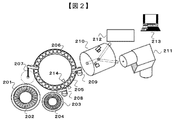
- the apparatus configuration of the present embodiment will be described with reference to FIGS.
- a sample transport unit 201 that transports the sample container 202
- a reagent transport unit 203 that transports the reagent container 204
- a reaction solution transport unit 205 that transports a reaction container 206 that performs various reactions
- a sample from the sample container 202 to the reaction container 206 is described with reference to FIGS.
- the sample transport unit 201, the reagent transport unit 203, and the reaction container transport unit 205 will be described as an example of a disk system that transports on an endless track as a transport mechanism, but is not limited thereto, a transport mechanism that transports an XY axis, Alternatively, a belt conveyor type transport mechanism may be used. Note that a stirring unit for stirring during various reactions in the reaction vessel and a cleaning unit for cleaning various probes are installed in the apparatus, although not particularly shown.
- the sample container 202 stores a biological sample, serum or plasma as a specimen in this embodiment, a calibrator sample, and a QC (Quality Control) sample.
- a biological sample serum or plasma as a specimen in this embodiment
- a calibrator sample a sample that samples a sample.
- a QC (Quality Control) sample 1 mol / L sodium hydroxide as a reagent for releasing vitamin D metabolite as a measurement substance from DBP, magnetic beads with antibody binding, and 10 mmol / L phosphate buffered physiological saline as a washing solution. Water (pH 7.4) is stored.
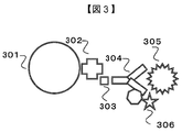
- the antibody-bound magnetic beads are constituted by an avidin-biotin bond using an anti-vitamin D antibody 304 and avidin 302 and biotin 303 in advance on the magnetic beads 301.
- reagent to be released from DBP in addition to using sodium hydroxide as in this example, formic acid, trifluoroacetic acid as acid, dithiothreitol as a reducing agent, mercaptoethanol, urea as denaturing agent, Tween 20, Triton X-100, Sodium dodecyl sulfate can also be used.
- surface modification of the magnetic beads in addition to using magnetic beads surface-modified with avidin that performs an avidin-biotin reaction as in this Example, a hydroxysuccinimide group, a 1,1-carbonyldiimidazole group, proteinA, proteinG In addition, magnetic beads surface-modified with an anti-mouse antibody can also be used.
- the washing solution in addition to using phosphate buffered saline as in this embodiment, Tris-HCl buffer, HEPES buffer, and water can also be used.
- the measurement flow of this example will be described.
- the first step 101 purification is performed by forming a complex of a magnetic bead and a measurement substance using an antigen-antibody reaction.
- the sample transport unit 201 and the reaction container transport unit 205 rotate to a predetermined position below the sample probe 207. Then, 100 ⁇ L of serum (or calibrator sample or QC sample) is aspirated and discharged from the sample container to the reaction container 206 using the sample probe 207.
- the reagent transport unit 203 and the reaction container transport unit 205 rotate to a predetermined position below the reagent probe 208. Then, using the reagent probe 208, 20 ⁇ L of 1 mol / L sodium hydroxide is sucked and discharged from the reagent container 204 to the reaction container 206 in which serum is stored. Next, antibody-bound magnetic beads (corresponding to 1 mg) suspended in slurry form in 50 ⁇ L of 10 mmol / L phosphate buffered saline (pH 7.4) from the reagent container 204 using the reagent probe 208. Is sucked and discharged into a reaction vessel 206 in which sodium hydroxide is added to serum.
- the reaction container 206 the measurement substance in the biological sample and the antibody-bound magnetic beads are reacted at room temperature for 10 minutes to form a complex in which the measurement substance and the antibody-bound magnetic beads are bound.
- the reaction container transport unit 205 rotates to a predetermined position under the magnet 214 and collects the complex in the reaction container 206, the reaction solution is sucked from the reaction container 206 using the reagent probe 208, and the waste liquid Discharge as The reagent transport unit 203 and the reaction container transport unit 205 rotate to a predetermined position below the reagent probe 208.
- the reaction solution transport unit 205 rotates to a predetermined position under the reaction solution probe 209. Then, using the reaction solution probe 209, the entire amount of the complex suspended in a slurry form in 10 mmol / L phosphate buffered saline (pH 7.4) in the reaction vessel 206 is sucked and discharged to the separation unit 210. .
- the second step 102 will be described.
- the complex purified in the first step is quantitatively separated.
- the separation unit 210 will be described with reference to FIG.
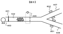
- the separation unit 210 includes an injector 401 that introduces the complex discharged from the reaction vessel probe 209 into the separation unit 210, a flow path 402, and a pump 403 that is positioned upstream of the injector 401 and that sends a solvent to the flow path 402.
- An active flow splitter 404 positioned downstream of the flow path 402 and positioned at a branch of the flow path 402, and a particle distribution measuring device capable of measuring in real time the amount of the complex on the flow path 402 between the injector 401 and the active flow splitter 404 (For example, a Coulter counter) 405, a mass spectrometry flow path 406 that is located after the active flow splitter 404 and sends the complex to the mass analyzer 211, and an immunoassay that is located after the active flow splitter 404 Flow path 407 for immunoassay for feeding the complex to unit 212 Et al constructed.
- a Coulter counter a mass spectrometry flow path 406 that is located after the active flow splitter 404 and sends the complex to the mass analyzer 211
- an immunoassay that is located after the active flow splitter 404
- an electrolyte solution 10 mmol / L phosphate buffered saline (pH 7.4) is fed into the flow path 402 in this example.
- the amount of the complex is detected by measuring the change in electrical resistance when the complex passes through the pores in the counter 405.
- the separation unit 210 includes a measured value of the particle distribution measuring device 405, a distance from the coulter counter 405 to the active flow splitter 404, an inner diameter of the flow path 402, a flow rate of the electrolyte solution flowing in the flow path 402, and a switching time of the active flow splitter 404. Is provided with a mechanism for calculating the amount of the complex fed to the mass spectrometry channel 406 and the immunoassay channel 407.
- the distance from the particle distribution measuring device 405 to the active flow splitter 404 is 30 cm
- the inner diameter of the flow path 402 is 0.2 mm
- the speed of the electrolyte solution flowing through the flow path 402 is 0.1 mL / min
- the active flow splitter 404 is active.
- the flow splitter 404 is opened in the direction of the immunoassay flow path 407 for 6 seconds, and is set so as to be switched to the mass spectrometry flow path 406 after feeding the complex. It was made to isolate
- the amount of the complex reaching the mass spectrometry channel 406 and the immunoassay channel 407 is adjusted by changing the switching time of the active flow splitter 404.
- the third step 103 will be described.
- the immune analyzer 212 will be described with reference to FIG.
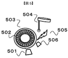
- the immunoassay unit 212 includes a magnet 501, an immunoassay reagent container 502 that holds the complex and immunoassay reagent sent from the immunoassay flow path 407, and an immunoassay reagent container 502. Aspirate the immunoassay reagent from the immunoassay reagent transport section 503 to be transported and the immunoassay reagent container 502 in which the immunoassay reagent is stored into the immunoassay reagent container 502 in which the complex is stored.
- an immunoassay probe 504 to be discharged, a light source 505 for irradiating light to the complex in the reaction tank 505, and a photometer 506.
- an antiglobulin antibody that is a secondary antibody that selectively binds to the primary antibody is used. This secondary antibody is fluorescently labeled and can be quantified by spectroscopy.
- the complex sent from the immunoassay flow path 407 is stored in an empty immunoassay reagent container 502.
- the immunoassay reagent transport section 503 rotates to a predetermined position below the immunoassay probe 504.
- the complex is stored with the labeled secondary antibody dissolved in 50 ⁇ L of 10 mmol / L phosphate buffered saline (pH 7.4) from the sample container.
- the reagent transport unit for immunoassay 503 rotates to a predetermined position below the magnet 501, collects the complex-secondary antibody, and cleans the complex-secondary antibody.
- the immunoassay reagent transport unit 503 rotates to a predetermined position below the light source 505, is irradiated with light from the light source 505, and is measured by the photometer 506.
- data on the total amount of the complex separated in the second step 102 is calculated by immunoassay.
- the immunodiagnostic anti-25-OH D 3 antibody used in this example is 100% 25-OH D 3 , 68% 25-OH D 2 , 100% 24,25- (OH) 2 D 3 , There is a description in the package insert when vitamin D 2 cross reacts 0.3%.
- the fourth step 104 will be described. In the fourth step 104, the measurement substance and the similar compound are eluted from the separated complex, and information on the ratio between the measurement substance and the similar compound is acquired by mass spectrometry.
- the mass spectrometer 211 will be described with reference to FIG.
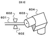
- the mass spectrometric unit 211 includes a flow path 601 connected to the mass spectrometry flow path 406, a magnet 602 that is arranged outside the pipe of the flow path 601 and collects the complex, and a high pressure is applied to the measurement substance.
- An ion source 603 for applying high voltage to ionize and a mass spectrometer 604 are included.
- the complex sent to the channel 601 is collected by a magnet 602 arranged outside the pipe of the channel 601 and the solvent in the channel 601 is changed to 0.1% glycine hydrochloride as an elution solvent.
- the measurement substance and the similar compound bound to the antibody on the complex are released and introduced into the ion source 603.
- the mass number is measured by the mass spectrometer 604.
- an acidic aqueous solution such as formic acid, a basic aqueous solution such as sodium hydroxide, and an organic solvent such as methanol can also be used.
- the ionization method using the ion source 603 is an atmospheric pressure chemical ionization (APCI) method in this embodiment.
- APCI atmospheric pressure chemical ionization
- an electrospray ionization method (ESI; Electrospray Ionization) can also be used.
- the mass spectrometer 604 uses a triple quadrupole mass spectrometer to analyze the component to be measured in the SRM (Selected Reaction Monitoring) mode.
- SRM Select Reaction Monitoring
- a quadrupole mass spectrometer, an ion trap mass spectrometer, or the like can be used. Peak intensities based on transitions of 25-OH D 2 , 25-OH D 3 , 24,25- (OH) 2 D 3 and vitamin D 2 which are measurement substances released from the complex separated in the second step 102 Alternatively, peak area data is calculated.
- SRM reaction monitoring
- the measurement substance and similar compounds released from the antibody may be subjected to liquid / liquid extraction or solid phase extraction purification or derivatization treatment.
- solid phase extraction means that the solute contained in the solution or suspension (mobile phase) flows through the solid (stationary phase) while adsorbing according to the affinity of each, or as it is. This is a method for separating the component to be measured and the impurities by using the flow.
- liquid / liquid extraction is based on the difference between the solute contained in the solution or suspension (mobile phase) and the solubility in other solvents (mainly water and non-polar organic solvent). This is a method for separating impurities.
- Derivatization is performed by adding a Cookson-type reagent such as 4-phenyl-1,2,4-triazoline-3,5-dione (PTAD) derivatization reagent to the measurement substance and internal standard substance released from the antibody.
- a method for improving ionization efficiency is also used.
- the definition of “derivatization” refers to a modification that does not significantly change the structure and properties, such as introduction of a functional group, oxidation, reduction, and atom replacement into a measurement substance. Derivatization is used to improve ionization efficiency and to suppress ion suppression due to contaminants by changing the mass number of the measurement substance to a high mass range when the sensitivity of MS is insufficient.
- PTAD 1,2,4-triazoline-3,5-dione
- TAD 1,2,4-triazoline-3,5-dione
- ATAD 4-amino-1,2,4-triazoline-3,5-dione
- PFBTAD 4-pentafluorobenzyl-1,2,4-triazoline-3,5-dione
- MOTAD 5-dione
- DEQTAD 2- (6,7-dimethoxy-4-methyl-3-oxo-3,4-dihydroquinosalyl) ethyl-3,5-dione
- the fifth step 105 will be described with reference to FIG.
- accurate quantification of the measurement substance is performed using the data on the total amount of the complex obtained in the third step and the data on the abundance ratio between the measurement substance and the similar compound obtained in the fourth process.
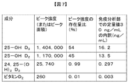
- the quantitative value obtained by the immunoassay in the third step 103 was 30 ng / mL.
- the peak area values obtained by mass spectrometry in the fourth step 104 are 1,404,000 for 25-OH D 3 and 1,170,000, 24,25- (OH) for 25-OH D 2.
- 2 D 3 is 25,740, vitamin D 2 was 260.
- an equal amount of the complex is set to be separated into the mass spectrometry channel 406 and the immunoassay channel 407, and is introduced into the mass analysis unit 211 and the immunoassay unit 212.
- the amount of complex is the same.
- 25-OH D 2 , 25-OH D 3 , 24, 25- (OH) When accurate quantitative values of 2 D 3 and vitamin D 2 are calculated, 25-OH D 3 is 16.2 ng / mL, 25-OH D 2 is 13.5 ng / mL, 24,25- (OH) 2 D 3 is 0.297 ng / mL, vitamin D 2 is 0.003 ng / mL.
- the quantitative value 30 ng / mL obtained by immunoassay in the third step 103 represents the total amount of 25-OH D 2 , 25-OH D 3 , 24,25- (OH) 2 D 3 and vitamin D 2.
- the quantitative value of the measurement substance is calculated using the total amount data of the complex obtained in the third step and the abundance ratio data of the measurement substance and similar compounds obtained in the fourth step. By doing so, an accurate quantitative value can be obtained.
- this invention is not limited to the above-mentioned Example, Various modifications are included.
- the above-described embodiments have been described in detail for easy understanding of the present invention, and are not necessarily limited to those having all the configurations described.
- a part of the configuration of one embodiment can be replaced with the configuration of another embodiment, and the configuration of another embodiment can be added to the configuration of one embodiment.
- each of the above-described configurations, functions, processing units, processing means, and the like may be realized by hardware by designing a part or all of them with, for example, an integrated circuit.
- Each of the above-described configurations, functions, and the like may be realized by software by interpreting and executing a program that realizes each function by the processor.
- Information such as programs, tables, and files for realizing each function can be stored in a memory, a storage device such as a hard disk or an SSD (Solid State Drive), or a storage medium such as an IC card, an SD card, or a DVD.
- control lines and information lines indicate what is considered necessary for the explanation, and not all the control lines and information lines on the product are necessarily shown. Actually, it may be considered that almost all the components are connected to each other.
- the present invention relates to the quantitative value of the immunoassay obtained in the third step 103, the measurement substance obtained by mass spectrometry in the fourth step 104, and the similar compound.
- the quantitative value based on the peak intensity ratio, it is possible to provide an analyzer that eliminates the cross-reactivity of the immunoassay and does not use an expensive internal standard substance in mass spectrometry.
- FIG. 8 is a diagram illustrating a second step according to an embodiment of the analyzer described in Example 2 of the present invention.
- the configuration of the analyzer according to the present embodiment will be described with respect to differences from the first embodiment.
- the present embodiment is different in that an electromagnet is used for switching between the mass spectrometry channel 406 and the immunoassay channel 407 in the separation unit 210 of the second step 102.
- This embodiment is different from the first embodiment only in the second step 102, and the other steps are the same.
- the configuration of the analyzer according to this embodiment will be described.
- the separation unit 210 in the present embodiment is located in the injector 401 that introduces the complex discharged from the reaction vessel probe 209 to the separation unit 210, the flow path 402, and the upstream of the injector 401, and sends the solvent to the flow path 402.
- the flow channel for mass spectrometry 406 for sending the complex to the mass analyzer 211 and the flow channel for immunoassay 407 for feeding the complex to the immune analyzer 212 which is the other channel.
- the electromagnet 801 and the electromagnet 802 control the amount of the complex flowing through the mass spectrometry channel 406 and the immunoassay channel 407 by controlling the magnetic force by turning on and off the current flowing through the electromagnet 801 and the electromagnet 802. It is a mechanism.
- the separation unit 210 includes a measurement value of the particle distribution measurement device 405, a distance from the particle distribution measurement device 405 to the electromagnet 801 and the electromagnet 802, an inner diameter of the flow channel 402, a flow rate of the electrolyte solution flowing in the flow channel 402, and the electromagnet 801 and the electromagnet. Based on the ON / OFF time of the current 802, a mechanism is provided for calculating the amount of the complex fed to the mass spectrometry channel 406 and the immunoassay channel 407.
- FIG. 9 is a diagram illustrating a second step according to an embodiment of the analyzer described in Example 3 of the present invention.
- the configuration of the analyzer according to the present embodiment will be described with respect to differences from the first embodiment.
- particle distribution measuring devices used for measuring the amount of the complex are provided in the flow path for the mass spectrometry 406 and the flow path for the immunoassay 207, respectively. Is different.
- This embodiment is different from the first embodiment only in the second step 102, and the other steps are the same.
- the configuration of the analyzer according to this embodiment will be described.
- the separation unit 210 in the present embodiment is located in the injector 401 that introduces the complex discharged from the reaction vessel probe 209 to the separation unit 210, the flow path 402, and the upstream of the injector 401, and sends the solvent to the flow path 402.
- the separation unit 210 measures the amount of the complex with the particle distribution measuring device 901 and the particle distribution measuring device 902 provided on the mass spectrometry channel 406 and the immunoassay channel 407, and the mass spectrometry unit 211 and the immunity A mechanism for calculating the amount of the complex introduced into the analysis unit 212 is provided.
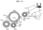
- FIG. 10 is a schematic diagram showing a schematic configuration according to an embodiment of the analyzer described in Example 4 of the present invention.
- the configuration of the analyzer according to the present embodiment will be described with respect to differences from the first embodiment.
- This embodiment is different in that the separation unit 210 in the second step 102 separates the complex by sucking and discharging the complex from the reaction vessel 206 to the separation vessel 1002 using a dedicated separation probe 1003 offline.
- This embodiment is different from the first embodiment only in the second step 102, and the other steps are the same.
- the configuration of the analyzer according to this embodiment will be described.
- the separation unit 210 sucks a certain amount of the complex from the separation container 1002 that receives the complex discharged from the reaction container probe 209, the separation container transport unit 1001 that transports the separation container 1002, and the separation container 1002.
- the separation probe 1003 is introduced into the mass analyzer 211 and the immune analyzer 212.
- the measurement flow of this example will be described.
- the reaction container transport unit 205 and the separation container transport unit 1001 rotate to a predetermined position below the reaction container probe 209. Then, using the reagent probe 209, the entire amount of the complex suspended in a slurry form from the reaction container 206 is sucked and discharged into the separation container 1002.
- the separation container transport unit 1001 rotates to a predetermined position below the separation probe 1003, and a certain amount of the complex is introduced from the separation container 1002 into the mass analysis unit 211 and the immune analysis unit 212 using the separation probe 1003. Since the separation probe 1003 can aspirate and discharge a certain amount of the complex, the amount of the complex introduced into the mass analyzer 211 and the immune analyzer 212 can be calculated.
- Embodiment 5 of the present invention is different in that a plurality of measurement substances are simultaneously measured using magnetic beads to which a plurality of types of antibodies are bound.
- a magnetic bead bound with an anti-triiodothyronine antibody (anti-FT3 antibody) and a magnetic bead bound with an anti-thyroxine antibody (anti-FT4 antibody) are mixed and measured.
- This example is different from Example 1 in that magnetic beads to which a plurality of types of antibodies are bound are used, and the other steps are the same.
- a complex is formed in which magnetic beads preliminarily bound with an antibody and triiodothyronine, thyroxine and similar compounds are bound.
- the separation unit 210 separates the complex into the immune analysis unit 211 and the mass analysis unit 212, and the complex is measured, so that the total amount of triiodothyronine, thyroxine and similar compounds is quantified in the immune analysis unit 211.
- the mass spectrometric section 212 measures the peak intensity ratio of triiodothyronine, thyroxine, and similar compounds. Triiodothyronine and thyroxine are quantified from the total amount of triiodothyronine, thyroxine and similar compounds and the peak intensity ratio.
Landscapes
- Health & Medical Sciences (AREA)
- Life Sciences & Earth Sciences (AREA)
- Chemical & Material Sciences (AREA)
- Immunology (AREA)
- Pathology (AREA)
- General Physics & Mathematics (AREA)
- General Health & Medical Sciences (AREA)
- Biochemistry (AREA)
- Analytical Chemistry (AREA)
- Physics & Mathematics (AREA)
- Engineering & Computer Science (AREA)
- Hematology (AREA)
- Molecular Biology (AREA)
- Biomedical Technology (AREA)
- Urology & Nephrology (AREA)
- Biotechnology (AREA)
- Microbiology (AREA)
- Cell Biology (AREA)
- Food Science & Technology (AREA)
- Medicinal Chemistry (AREA)
- Dispersion Chemistry (AREA)
- Chemical Kinetics & Catalysis (AREA)
- Electrochemistry (AREA)
- Other Investigation Or Analysis Of Materials By Electrical Means (AREA)
- Automatic Analysis And Handling Materials Therefor (AREA)
Abstract
Description
試薬容器を搬送する試薬搬送部と、
反応を行う反応容器を搬送する反応溶液搬送部と、
試料容器から試料を反応容器に吸引・吐出する試料プローブと、
試薬容器から試薬を反応容器に吸引・吐出する試薬プローブと、
反応容器内で反応した複合体を分離部に吸引・吐出する反応溶液プローブと、
複合体を免疫分析法用と質量分析法用に定量分離する分離部と、
質量分析法用に分離した複合体を質量分析計により測定する質量分析部と、
免疫分析法用に分離した複合体を光学的手法により測定する免疫分析部と、
データ処理を行う制御部を備えている。
〔ビタミンDの説明〕
ビタミンDは抗くる病因子として発見された脂溶性ビタミンであり、ヒト血中カルシウム濃度の恒常性維持に重要な役割を果たしている。ビタミンD3はヒトで生成される。ビタミンD2は植物や生合成で生成され、食物やサプリメントとして体内に摂取される。ビタミンD3代謝物について説明する。皮膚で7-デヒドロコレステロールから生合成されたビタミンD3は肝臓で25-ヒドロキシビタミンD3(25-OH D3)に代謝され、ビタミンD結合タンパク質(DBP)と結合して体内を循環する。血中カルシウム濃度が正常値以下となると25-OH D3は腎臓で1α,25-ジヒドロキシビタミンD3[1α,25-(OH)2 D3]に代謝される。血中カルシウム濃度が回復すると1α位への水酸化は抑制され、25-OH D3は主として24R,25-ジヒドロキシビタミンD[24,25-(OH)2 D3]に代謝される。ビタミンD2もビタミンD3と同じ経路で代謝が行われる。つまり、血中の主なビタミンD代謝物としては、ビタミンD3,25-OH D3,1α,25-(OH)2 D3,24,25-(OH)2 D3,ビタミンD2,25-OH D2,1α,25-(OH)2 D2および24,25-(OH)2 D2の少なくとも8種類存在することになる。臨床の現場では、血中に高濃度で安定に存在する25-OH D3および25-OH D2の総量を、血中ビタミンD濃度を反映する指標として用いている。
〔従来の免疫分析法の説明〕
従来の免疫分析法を用いたビタミンD測定方法は、血中を循環している25-OH D3および25-OH D2の総量を測定し、血中ビタミンD濃度を反映する指標として用いている。免疫分析法の処理工程は以下のとおりである。生体試料中の測定物質である25-OH D3および25-OH D2はDBPと結合しているため、酸、アルカリあるいは還元剤を添加して遊離させる。次に、磁性ビーズに測定物質を特異的に認識する抗体をあらかじめアビジン-ビオチン結合等により結合させ、処理した生体試料を反応させる。抗体が結合した磁性ビーズと測定物質が結合した複合体を磁石により集磁し、洗浄した後に、酵素標識した抗免疫グロブリン抗体によりサンドイッチアッセイを行う。次に、酵素の基質(発光あるいは発色試薬)を添加し、酵素反応の生成物を検出器で検出する。しかしながら、免疫分析法に用いる抗体の種類によっては測定物質のほかに類似化合物も捕捉されるため、定量結果が真値より高くなる場合がある。ここでいう「生体試料」の定義は、血清、血漿、全血、尿、唾液、胆汁、涙および細胞組織の生物学的な供給源由来の生体試料である。
〔従来の質量分析法の説明〕
一方、従来の質量分析法を用いたビタミンD測定方法は、血中を循環している25-OH D3および25-OH D2のそれぞれの濃度を定量している。定量はあらかじめ既知濃度の安定同位体物質を添加して相対定量を行っている。質量分析法の処理工程は以下のとおりである。生体試料に内部標準物質である25-OH D3の安定同位体標識物質(例えば25-OH D3-d6)および25-OH D2(例えば25-OH D2-d6)の安定同位体標識物質を既知量添加する。生体試料中の測定物質である25-OH D3および25-OH D2はDBPと結合しているため、酸、アルカリあるいは還元剤を添加して遊離させる。次に、磁性ビーズに測定物質を特異的に認識する抗体をあらかじめアビジン-ビオチン結合等により結合させ、処理した生体試料を反応させる。抗体が結合した磁性ビーズと測定物質が結合した複合体を磁石により集磁し、洗浄した後に、酸、アルカリあるいは有機溶媒を添加し、抗体から遊離した測定物質および内部標準物質を質量分析計に供試する。25-OH D3の定量は25-OH D3と25-OH D3-d6の比率を算出し、あらかじめ既知濃度の25-OH D3を含む検量線用の試料を測定し作成した検量線を用いて、25-OH D3の濃度を算出する。25-OH D2も同様に濃度を算出する。
〔組合せた場合の説明〕
本実施例は、図1に示すように、抗原-抗体反応を用いて微小粒子と測定物質との複合体を形成させ精製する第1の工程101と、第1の工程で精製した複合体を定量的に分離する第2の工程102と、分離した複合体の総量の情報を免疫分析法により取得する第3の工程103と、分離した複合体から測定物質および類似化合物を溶出させ、測定物質と類似化合物との比率の情報を質量分析法により取得する第4の工程104と、第3の工程で取得した複合体の総量と第4の工程で取得した測定物質と類似化合物との存在量比を用いて測定物質の正確な定量を行う第5の工程105から構成される。
〔分析装置についての説明〕
本実施例の装置構成について図2および3を用いて説明する。試料容器202を搬送する試料搬送部201と、試薬容器204を搬送する試薬搬送部203と、各種反応を行う反応容器206を搬送する反応溶液搬送部205と、試料容器202から試料を反応容器206に吸引・吐出する試料プローブ207と、試薬容器204から各種試薬を反応容器206に吸引・吐出する試薬プローブ208と、反応容器206内で反応した複合体を分離部210に吸引・吐出する反応溶液プローブ209と、複合体を免疫分析法用と質量分析法用に定量分離する分離部210と、質量分析法用に分離した複合体を質量分析計により測定する質量分析部211と、免疫分析法用に分離した複合体を光学的手法により測定する免疫分析部212と、本装置を構成している各々の部位の自動一括制御およびデータ処理を行う制御部213から構成される。試料搬送部201、試薬搬送部203および反応容器搬送部205は搬送機構として、無限軌道上を移送するディスク方式を例にして説明するが、それに限られず、X-Y軸を移送する搬送機構、またはベルトコンベア方式の搬送機構であってもよい。なお、反応容器内での各種反応時に攪拌を行う攪拌部、各種プローブを洗浄する洗浄部は特に図示しないが装置内に設置されている。
性ビーズは、磁性ビーズ301に抗ビタミンD抗体304を、あらかじめアビジン302およびビオチン303を用いたアビジン-ビオチン結合により構成される。本実施例では10 μgのImmunDiagnostik社の抗25-OH D3抗体を、50 μLの10 mmol/Lリン酸緩衝生理食塩水(pH 7.4)にスラリー状に懸濁した直径300 nmの磁性ビーズ(1 mg相当)にアビジン-ビオチン反応で結合した抗体結合済み磁性ビーズを用いた。DBPから遊離させる試薬は、本実施例のように水酸化ナトリウムを用いるほかに、酸としてギ酸、トリフルオロ酢酸、還元剤としてジチオトレイトール、メルカプトエタノール、変性剤として尿素、Tween20、TritonX-100、ドデシル硫酸ナトリウムを用いることもできる。磁性ビーズの表面修飾は、本実施例のようにアビジン-ビオチン反応を行うアビジンで表面修飾されている磁性ビーズを用いるほかに、ハイドロキシサクシミド基、1,1-カルボニルジイミダゾール基、proteinA、proteinGおよび抗マウス抗体で表面修飾された磁性ビーズを用いることもできる。洗浄溶液は、本実施例のようにリン酸緩衝生理食塩水を用いるほかに、トリス塩酸緩衝液、HEPES緩衝液、水を用いることもできる。
〔第1の工程についての説明〕
本実施例の測定の流れについて説明する。第1の工程101について説明する。第1の工程101は、抗原-抗体反応を用いて磁性ビーズと測定物質との複合体を形成させることにより精製を行う。試料搬送部201および反応容器搬送部205が試料プローブ207下の所定位置まで回転する。そして、試料プローブ207を用いて、試料容器から100 μLの血清(またはキャリブレーター試料、またはQC試料)を反応容器206に吸引・吐出する。次に、試薬搬送部203および反応容器搬送部205が試薬プローブ208下の所定位置まで回転する。そして、試薬プローブ208を用いて、試薬容器204から20 μLの1 mol/Lの水酸化ナトリウムを血清が格納された反応容器206に吸引・吐出する。次に、試薬プローブ208を用いて、試薬容器204から50 μLの10 mmol/Lリン酸緩衝生理食塩水(pH 7.4)にスラリー状に懸濁した抗体結合済み磁性ビーズ(1 mg相当)を血清に水酸化ナトリウムが添加された反応容器206に吸引・吐出する。そして、反応容器206内にて、生体試料中の測定物質と抗体結合済み磁性ビーズを10分間常温で反応させ、測定物質と抗体結合済み磁性ビーズが結合した複合体を形成する。次に、反応容器搬送部205が磁石214下の所定位置まで回転し、反応容器206内の複合体を集磁した後、試薬プローブ208を用いて、反応容器206から反応溶液を吸引し、廃液として吐出する。試薬搬送部203および反応容器搬送部205が試薬プローブ208下の所定位置まで回転する。そして、試薬プローブ208を用いて、試薬容器204から200 μLの10 mmol/Lリン酸緩衝生理食塩水(pH 7.4)を吸引・吐出し、複合体の洗浄を行い、非特異反応を低減する。この集磁-洗浄動作は3回行う。次に、反応溶液搬送部205が反応溶液プローブ209下の所定位置まで回転する。そして、反応溶液プローブ209を用いて、反応容器206内の10 mmol/Lリン酸緩衝生理食塩水(pH 7.4)にスラリー状に懸濁した複合体を分離部210に全量吸引・吐出する。
〔第2の工程についての説明〕
第2の工程102について説明する。第2の工程102は、第1の工程で精製した複合体を定量的に分離する。分離部210について図4を用いて説明する。分離部210は、反応容器プローブ209から吐出された複合体を分離部210へ導入するインジェクター401と、流路402と、インジェクター401の前段に位置し流路402に溶媒を送液するポンプ403と、流路402の後段に位置し流路402の分岐に位置するアクティブフロースプリッター404と、インジェクター401とアクティブフロースプリッター404の間の流路402上の複合体量をリアルタイムで測定できる粒子分布測定装置(例えば、コールターカウンター等)405と、アクティブフロースプリッター404の後段に位置し質量分析部211に複合体を送液する質量分析法用流路406と、アクティブフロースプリッター404の後段に位置し免疫分析部212に複合体を送液する免疫分析法用流路407から構成される。複合体量の測定に用いる粒子分布測定装置405は、流路402内に電解質溶液、本実施例では10 mmol/Lリン酸緩衝生理食塩水(pH 7.4)が送液されており、コールターカウンター405内の細孔を複合体が通過した際の電気抵抗の変化を測定することで複合体量を検出する。分離部210は、粒子分布測定装置405の測定値、コールターカウンター405からアクティブフロースプリッター404までの距離、流路402の内径、流路402内を流れる電解質溶液の流速およびアクティブフロースプリッター404の切換時間に基づいて、質量分析法用流路406と免疫分析法用流路407へ送液された複合体量を算出する機構を備える。
〔第3の工程についての説明〕
第3の工程103について説明する。第3の工程103は、分離した複合体の総量の情報を免疫分析法により取得する。免疫分析部212について図5を用いて説明する。免疫分析部212は、磁石501と、免疫分析法用流路407から送液された複合体および免疫分析法用試薬を保持する免疫分析法用試薬容器502と、免疫分析法用試薬容器502を搬送する免疫分析法用試薬搬送部503と、免疫分析法用試薬が保管された免疫分析法用試薬容器502から複合体が保管された免疫分析法用試薬容器502へ免疫分析法用試薬を吸引・吐出する免疫分析法用プローブ504と、反応槽505中の複合体に光を照射する光源505と、測光器506から構成される。免疫分析法用試薬には、1次抗体に選択的に結合する2次抗体である抗グロブリン抗体が用いられる。この2次抗体は蛍光標識済みであり、分光法で定量することができる。
〔第4の工程についての説明〕
第4の工程104について説明する。第4の工程104は、分離した複合体から測定物質および類似化合物を溶出させ、測定物質と類似化合物の比率の情報を質量分析法により取得する。質量分析部211について図6を用いて説明する。質量分析部211は、質量分析法用流路406と連結している流路601と、流路601の管の外側に配置し、複合体を集磁する磁石602と、測定物質に高圧力と高電圧を印加しイオン化するイオン源603と、質量分析計604から構成される。流路601に送液された複合体は、流路601の管の外側に配置された磁石602で集磁し、流路601内の溶媒を溶出溶媒である0.1%グリシン塩酸塩に変更することで、複合体上の抗体と結合している測定物質および類似化合物を遊離させ、イオン源603に導入する。イオン源603でイオン化した後に質量分析計604で質量数が測定される。溶出溶媒は本実施例のようにグリシン塩酸塩を用いるほかに、ギ酸等の酸性水溶液、水酸化ナトリウム等の塩基性水溶液およびメタノール等の有機溶媒を用いることもできる。イオン源603でのイオン化方法は、本実施例では、本実施例では大気圧化学イオン化法(APCI;Atomospheric Pressure Chemical Ionization)を用いた。他のイオン化法としては、エレクトロスプレーイオン化法(ESI;Electrospray Ionization)を用いることもできる。質量分析計604は、本実施例では、三連四重極型質量分析計を用いて、SRM(Selected Reaction Monitoring)モードにより測定対象成分の解析を行った。質量分析計604は、四重極型質量分析計、イオントラップ型質量分析計等を用いることもできる。第2の工程102で分離した複合体から遊離した測定物質である25-OH D2、25-OH D3、24,25-(OH)2 D3およびビタミンD2のトランジッションに基づいたピーク強度またはピーク面積のデータが算出される。本実施例では、三連四重極型質量分析計を用いてSelected Reaction Monitoring(SRM)モードによりMS/MS分析され、各測定物質のトランジッションは25-OH D3がQ1/Q3=401.3/383.3、25-OH D2がQ1/Q3=413.3/395.3、24,25-(OH)2 D3が417.3/399.3、ビタミンD2が397.3/379.3に設定し、ピーク強度のデータを得た。各ピーク強度またはピーク面積は、複合体に結合していた25-OH D2、25-OH D3、24,25-(OH)2 D3およびビタミンD2の存在量比を示す。
〔第5の工程についての説明〕
第5の工程105について図7を用いて説明する。第5の工程105は、第3の工程で取得した複合体の総量のデータと第4の工程で取得した測定物質と類似化合物との存在量比のデータを用いて測定物質の正確な定量を行う。第3の工程103で免疫分析法により取得した定量値は30 ng/mLであった。一方、第4の工程104で質量分析法により取得したピーク面積値は、25-OH D3が1,404,000、25-OH D2が1,170,000、24,25-(OH)2 D3が25,740、ビタミンD2が260であった。ピーク強度の存在量比は、25-OH D3が54%、25-OH D2が45%、24,25-(OH)2 D3が0.99%、ビタミンD2が0.01%となる。第2の工程102では等量の複合体が質量分析法用流路406と免疫分析法用流路407へ分離するように設定しており、質量分析部211と免疫分析部212に導入された複合体量は同量である。つまり、第3の工程103で取得した定量値と、第4の工程104で取得したピーク強度の存在比を用いて、25-OH D2、25-OH D3、24,25-(OH)2 D3およびビタミンD2の正確な定量値を算出すると、25-OH D3が16.2 ng/mL、25-OH D2が13.5 ng/mL、24,25-(OH)2 D3が0.297 ng/mL、ビタミンD2が0.003 ng/mLとなる。第3の工程103で免疫分析法により取得した定量値30 ng/mLは25-OH D2、25-OH D3、24,25-(OH)2 D3およびビタミンD2の総量を示すが、第5の工程105において第3の工程で取得した複合体の総量のデータと第4の工程で取得した測定物質と類似化合物との存在量比のデータを用いて測定物質の定量値を算出することで、正確な定量値を得ることができる。
102 第2の工程
103 第3の工程
104 第4の工程
105 第5の工程
201 試料搬送部
202 試料容器
203 試薬搬送部
204 試薬容器
205 反応溶液搬送部
206 反応容器
207 試料プローブ
208 試薬プローブ
209 反応容器プローブ
210 分離部
211 質量分析部
212 免疫分析部
213 制御部
214 磁石
301 磁性ビーズ
302 アビジン
303 ビオチン
304 抗ビタミンD抗体
305 測定物質
306 類似化合物
401 インジェクター
402 流路
403 ポンプ
404 アクティブフロースプリッター
405 粒子分布測定装置
406 質量分析法用流路
407 免疫分析法用流路
501 磁石
502 免疫分析法用試薬容器
503 免疫分析法用試薬容器搬送部
504 免疫分析法用プローブ
505 光源
506 測光部
601 流路
602 磁石
603 イオン源
604 質量分析計
801 電磁石(免疫分析法用流路)
802 電磁石(質量分析法用流路)
1001 分離容器搬送部
1002 分離容器
1003 分離プローブ
Claims (11)
- 試料容器を搬送する試料搬送部と、
試薬容器を搬送する試薬搬送部と、
反応を行う反応容器を搬送する反応溶液搬送部と、
試料容器から試料を反応容器に吸引・吐出する試料プローブと、
試薬容器から試薬を反応容器に吸引・吐出する試薬プローブと、
反応容器内で反応した複合体を分離部に吸引・吐出する反応溶液プローブと、
複合体を免疫分析法用と質量分析法用に定量分離する分離部と、
質量分析法用に分離した複合体を質量分析計により測定する質量分析部と、
免疫分析法用に分離した複合体を光学的手法により測定する免疫分析部と、
データ処理を行う制御部を備えたことを特徴とする分析装置。 - 請求項1に記載の分析装置であって、
前記分離部が、反応容器プローブから吐出された複合体を分離部へ導入するインジェクターと、分岐部を有する流路と、流路に溶媒を送液するポンプと、流路の分岐部に位置するアクティブフロースプリッターと、インジェクターとアクティブフロースプリッターの間の流路上の複合体量をリアルタイムで測定できる粒子分布測定装置と、アクティブフロースプリッターの後段に位置し質量分析部に複合体を送液する質量分析法用流路と、アクティブフロースプリッターの後段に位置し免疫分析部に複合体を送液する免疫分析法用流路から構成されることを特徴とする分析装置。 - 請求項1に記載の分析装置であって、
前記分離部が、反応容器プローブから吐出された複合体を分離部へ導入するインジェクターと、流路と、流路に溶媒を送液するポンプと、インジェクターの後段および流路の後段に設けられた2又に分岐した箇所の前段に設けられた粒子分布測定装置と、流路の後段に設けられた2又に分岐した片側の流路であり質量分析部に複合体を送液する質量分析法用流路と、もう片側の流路であり免疫分析部に複合体を送液する免疫分析法用流路と、質量分析法用流路の管の外側に配置された磁石と、免疫分析法用流路の管の外側に配置された磁石から構成されることを特徴とする分析装置。 - 請求項1に記載の分析装置であって、
前記分離部が、反応容器プローブから吐出された複合体を分離部へ導入するインジェクターと、流路と、流路に溶媒を送液するポンプと、流路の分岐に位置するアクティブフロースプリッターと、アクティブフロースプリッターの後段に位置し質量分析部に複合体を送液する質量分析法用流路と、アクティブフロースプリッターの後段に位置し免疫分析部に複合体を送液する免疫分析法用流路と、質量分析法用流路に設けられたコールターカウンターと、免疫分析法用流路に設けられた粒子分布測定装置から構成されることを特徴とする分析装置。 - 請求項1に記載の分析装置であって、
前記分離部が、反応容器プローブから吐出された複合体を受ける分離容器と、分離容器を搬送する分離容器搬送部と、分離容器から複合体を一定量吸引し、質量分析部および免疫分析部に導入する分離プローブから構成されることを特徴とする分析装置。 - 請求項1から5に記載の分析装置のいずれかであって、
制御部は、質量分析部にて求めたデータおよび免疫分析部にて求めたデータから、測定物質の定量を行うことを特徴とする分析装置。 - 請求項6に記載の分析装置であって、
質量分析部にて求めたデータは、複合体を構成する測定物質および類似化合物の総量であり、免疫分析部にて求めたデータは、複合体を構成する測定物質および類似化合物の比率であることを特徴とする分析装置。 - 微小粒子と測定物質との複合体を形成する第1の工程と、複合体を既知量分離する第2の工程と、分離した複合体を免疫分析法により測定する第3の工程と、分離した複合体を質量分析法により測定する第4の工程と、第3の工程で取得したデータと第4の工程で取得したデータから測定物質の定量を行う第5の工程を備え、
前記第2の工程において、複合体量に基づき、流路を切り換えながら分離を行うことを特徴とする分析方法。 - 請求項8に記載の分析方法であって、
第3の工程で得られるデータが複合体を構成する測定物質および類似化合物の総量であり、第4の工程で得られるデータが複合体を構成する測定物質および類似化合物の比率であり、第5の工程で複合体を構成する測定物質および類似化合物の総量および比率のデータから測定物質の定量値を算出することを
特徴とする分析方法。 - 請求項8に記載の分析方法であって、
微小粒子が磁性ビーズと複数種類の抗体から構成され、複数の測定物質を同時に測定することを特徴とする分析方法。 - 請求項8に記載の分析方法であって、
測定物質が25-ヒドロキシビタミンD2および25-ヒドロキシビタミンD3であり、類似化合物がビタミンD2、1α,25-ジヒドロキシビタミンD2、24R,25-ジヒドロキシビタミンD2、ビタミンD3、1α,25-ジヒドロキシビタミンD3、24R,25-ジヒドロキシビタミンD3のいずれかであることを特徴とする分析方法。
Priority Applications (5)
| Application Number | Priority Date | Filing Date | Title |
|---|---|---|---|
| GB1621150.0A GB2550631B (en) | 2014-07-07 | 2015-06-17 | Analysis device and analysis method |
| CN201580037164.4A CN106574934B (zh) | 2014-07-07 | 2015-06-17 | 分析装置以及分析方法 |
| DE112015002589.8T DE112015002589T5 (de) | 2014-07-07 | 2015-06-17 | Analysenvorrichtung und Analysenverfahren |
| US15/320,406 US10585089B2 (en) | 2014-07-07 | 2015-06-17 | Analysis device and analysis method |
| JP2016532513A JP6307607B2 (ja) | 2014-07-07 | 2015-06-17 | 分析装置および分析方法 |
Applications Claiming Priority (2)
| Application Number | Priority Date | Filing Date | Title |
|---|---|---|---|
| JP2014-139281 | 2014-07-07 | ||
| JP2014139281 | 2014-07-07 |
Publications (1)
| Publication Number | Publication Date |
|---|---|
| WO2016006398A1 true WO2016006398A1 (ja) | 2016-01-14 |
Family
ID=55064039
Family Applications (1)
| Application Number | Title | Priority Date | Filing Date |
|---|---|---|---|
| PCT/JP2015/067386 Ceased WO2016006398A1 (ja) | 2014-07-07 | 2015-06-17 | 分析装置および分析方法 |
Country Status (6)
| Country | Link |
|---|---|
| US (1) | US10585089B2 (ja) |
| JP (1) | JP6307607B2 (ja) |
| CN (1) | CN106574934B (ja) |
| DE (1) | DE112015002589T5 (ja) |
| GB (1) | GB2550631B (ja) |
| WO (1) | WO2016006398A1 (ja) |
Cited By (4)
| Publication number | Priority date | Publication date | Assignee | Title |
|---|---|---|---|---|
| JP2018081023A (ja) * | 2016-11-17 | 2018-05-24 | 学校法人東京理科大学 | ビタミンdの定量方法、質量分析装置およびビタミンd定量用試薬キット |
| WO2018217778A1 (en) * | 2017-05-22 | 2018-11-29 | Beckman Coulter, Inc. | Integrated sample processing system with multiple detection capability |
| JP2019528438A (ja) * | 2016-07-29 | 2019-10-10 | ダイアザイム ラボラトリーズ, インコーポレイテッド | ビタミンdをアッセイするための方法および組成物 |
| JP2022517150A (ja) * | 2019-01-15 | 2022-03-04 | パーキンエルマー ヘルス サイエンシーズ カナダ, インコーポレイテッド | 流体分析 |
Families Citing this family (9)
| Publication number | Priority date | Publication date | Assignee | Title |
|---|---|---|---|---|
| WO2018143208A1 (ja) * | 2017-02-03 | 2018-08-09 | 株式会社島津製作所 | 前処理システム |
| EP3729077B1 (en) * | 2017-12-19 | 2024-03-27 | Beckman Coulter, Inc. | Integrated sample processing system with variable workflows |
| US11125738B2 (en) | 2018-11-06 | 2021-09-21 | Thermo Finnigan Llc | Blood sample analysis systems and methods |
| WO2020225971A1 (ja) | 2019-05-08 | 2020-11-12 | 株式会社日立ハイテク | 自動分析装置の前処理方法 |
| CN111366646B (zh) * | 2020-02-27 | 2022-09-23 | 苏州新波生物技术有限公司 | 一种基于免疫纯化结合质谱检测的25-羟维生素d分析方法 |
| CN115485207B (zh) * | 2020-07-01 | 2023-08-15 | 株式会社日立高新技术 | 搬送容器及搬送方法 |
| CN111812337A (zh) * | 2020-08-24 | 2020-10-23 | 桂林优利特医疗电子有限公司 | 使用单值校准物实现特定蛋白全线性范围标定系统及方法 |
| EP4272006A4 (en) * | 2020-12-30 | 2025-02-26 | Beckman Coulter, Inc. | Clinical laboratory automation system with single calibrator |
| CN113063871B (zh) * | 2021-03-24 | 2022-12-27 | 杭州佰辰医疗器械有限公司 | 一种代谢小分子检测方法及检测系统 |
Citations (6)
| Publication number | Priority date | Publication date | Assignee | Title |
|---|---|---|---|---|
| JPS61161439A (ja) * | 1985-01-09 | 1986-07-22 | Sumitomo Electric Ind Ltd | 粒子センサ− |
| JPH07318559A (ja) * | 1994-05-25 | 1995-12-08 | Mitsubishi Chem Corp | 免疫化学的測定装置 |
| JPH10288601A (ja) * | 1997-04-15 | 1998-10-27 | Rikagaku Kenkyusho | 微粒子分析装置およびその方法 |
| JP2006121935A (ja) * | 2004-10-27 | 2006-05-18 | Konica Minolta Medical & Graphic Inc | 前処理手段および廃液貯留部を有する生体物質検査用マイクロリアクタ |
| WO2010092958A1 (ja) * | 2009-02-10 | 2010-08-19 | 株式会社日立ハイテクノロジーズ | 質量分析技術を用いた免疫分析方法および免疫分析システム |
| JP2014506332A (ja) * | 2011-01-07 | 2014-03-13 | ザ ジェネラル ホスピタル コーポレイション | ビタミンd欠乏症に関連するアッセイおよび治療方法 |
Family Cites Families (5)
| Publication number | Priority date | Publication date | Assignee | Title |
|---|---|---|---|---|
| JPH01187459A (ja) | 1988-01-21 | 1989-07-26 | Hitachi Ltd | 免疫分析方法 |
| EP2134735A1 (en) | 2007-03-05 | 2009-12-23 | Anderson Forschung Group LLC | Magnetic bead trap and mass spectrometer interface |
| JP4948230B2 (ja) | 2007-03-29 | 2012-06-06 | シスメックス株式会社 | 試料分析装置および試料分析方法 |
| US9052308B2 (en) | 2011-01-07 | 2015-06-09 | The General Hospital Corporation | Assays and methods of treatment relating to vitamin D insufficiency |
| CN202257125U (zh) * | 2011-10-18 | 2012-05-30 | 长沙高新技术产业开发区爱威科技实业有限公司 | 样本有形成分分析仪自动检测控制装置 |
-
2015
- 2015-06-17 US US15/320,406 patent/US10585089B2/en active Active
- 2015-06-17 GB GB1621150.0A patent/GB2550631B/en active Active
- 2015-06-17 WO PCT/JP2015/067386 patent/WO2016006398A1/ja not_active Ceased
- 2015-06-17 JP JP2016532513A patent/JP6307607B2/ja active Active
- 2015-06-17 CN CN201580037164.4A patent/CN106574934B/zh active Active
- 2015-06-17 DE DE112015002589.8T patent/DE112015002589T5/de active Pending
Patent Citations (6)
| Publication number | Priority date | Publication date | Assignee | Title |
|---|---|---|---|---|
| JPS61161439A (ja) * | 1985-01-09 | 1986-07-22 | Sumitomo Electric Ind Ltd | 粒子センサ− |
| JPH07318559A (ja) * | 1994-05-25 | 1995-12-08 | Mitsubishi Chem Corp | 免疫化学的測定装置 |
| JPH10288601A (ja) * | 1997-04-15 | 1998-10-27 | Rikagaku Kenkyusho | 微粒子分析装置およびその方法 |
| JP2006121935A (ja) * | 2004-10-27 | 2006-05-18 | Konica Minolta Medical & Graphic Inc | 前処理手段および廃液貯留部を有する生体物質検査用マイクロリアクタ |
| WO2010092958A1 (ja) * | 2009-02-10 | 2010-08-19 | 株式会社日立ハイテクノロジーズ | 質量分析技術を用いた免疫分析方法および免疫分析システム |
| JP2014506332A (ja) * | 2011-01-07 | 2014-03-13 | ザ ジェネラル ホスピタル コーポレイション | ビタミンd欠乏症に関連するアッセイおよび治療方法 |
Cited By (16)
| Publication number | Priority date | Publication date | Assignee | Title |
|---|---|---|---|---|
| JP7177767B2 (ja) | 2016-07-29 | 2022-11-24 | ダイアザイム ラボラトリーズ, インコーポレイテッド | ビタミンdをアッセイするための方法および組成物 |
| JP2019528438A (ja) * | 2016-07-29 | 2019-10-10 | ダイアザイム ラボラトリーズ, インコーポレイテッド | ビタミンdをアッセイするための方法および組成物 |
| US11435367B2 (en) | 2016-07-29 | 2022-09-06 | Diazyme Laboratories, Inc. | Methods and kits for assaying a vitamin D moiety |
| JP2018081023A (ja) * | 2016-11-17 | 2018-05-24 | 学校法人東京理科大学 | ビタミンdの定量方法、質量分析装置およびビタミンd定量用試薬キット |
| JP2020521958A (ja) * | 2017-05-22 | 2020-07-27 | ベックマン コールター, インコーポレイテッド | 複数の検出能力を有する統合試料処理システム |
| US11402386B2 (en) | 2017-05-22 | 2022-08-02 | Beckman Coulter, Inc. | Integrated sample processing system with multiple detection capability |
| CN110959185A (zh) * | 2017-05-22 | 2020-04-03 | 拜克门寇尔特公司 | 具有多种检测能力的集成样品处理系统 |
| WO2018217778A1 (en) * | 2017-05-22 | 2018-11-29 | Beckman Coulter, Inc. | Integrated sample processing system with multiple detection capability |
| JP2023060337A (ja) * | 2017-05-22 | 2023-04-27 | ベックマン コールター, インコーポレイテッド | 複数の検出能力を有する統合試料処理システム |
| CN110959185B (zh) * | 2017-05-22 | 2023-06-30 | 拜克门寇尔特公司 | 具有多种检测能力的集成样品处理系统 |
| US11988670B2 (en) | 2017-05-22 | 2024-05-21 | Beckman Coulter, Inc. | Integrated sample processing system with multiple detection capability |
| JP2024133264A (ja) * | 2017-05-22 | 2024-10-01 | ベックマン コールター, インコーポレイテッド | 複数の検出能力を有する統合試料処理システム |
| JP7594618B2 (ja) | 2017-05-22 | 2024-12-04 | ベックマン コールター, インコーポレイテッド | 複数の検出能力を有する統合試料処理システム |
| US12687548B2 (en) | 2017-05-22 | 2026-07-21 | Beckman Coulter, Inc. | Integrated sample processing system with multiple detection capability |
| JP2022517150A (ja) * | 2019-01-15 | 2022-03-04 | パーキンエルマー ヘルス サイエンシーズ カナダ, インコーポレイテッド | 流体分析 |
| JP7223167B2 (ja) | 2019-01-15 | 2023-02-15 | パーキンエルマー ヘルス サイエンシーズ カナダ, インコーポレイテッド | 流体分析 |
Also Published As
| Publication number | Publication date |
|---|---|
| US10585089B2 (en) | 2020-03-10 |
| CN106574934B (zh) | 2019-01-04 |
| JP6307607B2 (ja) | 2018-04-04 |
| JPWO2016006398A1 (ja) | 2017-04-27 |
| CN106574934A (zh) | 2017-04-19 |
| GB2550631B (en) | 2020-07-29 |
| DE112015002589T5 (de) | 2017-06-01 |
| GB2550631A (en) | 2017-11-29 |
| US20170160273A1 (en) | 2017-06-08 |
| GB201621150D0 (en) | 2017-01-25 |
Similar Documents
| Publication | Publication Date | Title |
|---|---|---|
| JP6307607B2 (ja) | 分析装置および分析方法 | |
| JP7853268B2 (ja) | 質量分析によるインスリンの定量 | |
| JP7594618B2 (ja) | 複数の検出能力を有する統合試料処理システム | |
| CN111670363B (zh) | 具有可变工作流的集成样品处理系统 | |
| JP6585743B2 (ja) | 複合試料中のステロイド化合物の質量分析 | |
| US11852636B2 (en) | Mass spectrometric determination of non-derivatized, non-metabolized vitamin D | |
| JP2018511812A (ja) | 質量分析によるインスリンレベルの定量の方法 | |
| JP2026012672A (ja) | インスリン及びc-ペプチドの定量の方法 | |
| US8865418B2 (en) | Immunoanalytical method and system using mass spectrometry technology | |
| US9885732B2 (en) | Analysis device and analysis method | |
| WO2011108177A1 (ja) | 分析装置 | |
| Keyfi et al. | Evaluation of 25-OH vitamin D by high performance liquid chromatography: validation and comparison with electrochemiluminescence | |
| Stigter et al. | Coupling surface-plasmon resonance and mass spectrometry to quantify and to identify ligands | |
| CN116670513A (zh) | 利用单个校准品的临床实验室自动化系统 | |
| AU2024247888A1 (en) | Methods for quantitation of adiponectin by mass spectrometry |
Legal Events
| Date | Code | Title | Description |
|---|---|---|---|
| 121 | Ep: the epo has been informed by wipo that ep was designated in this application |
Ref document number: 15819443 Country of ref document: EP Kind code of ref document: A1 |
|
| ENP | Entry into the national phase |
Ref document number: 2016532513 Country of ref document: JP Kind code of ref document: A |
|
| ENP | Entry into the national phase |
Ref document number: 201621150 Country of ref document: GB Kind code of ref document: A Free format text: PCT FILING DATE = 20150617 |
|
| WWE | Wipo information: entry into national phase |
Ref document number: 15320406 Country of ref document: US |
|
| WWE | Wipo information: entry into national phase |
Ref document number: 112015002589 Country of ref document: DE |
|
| 122 | Ep: pct application non-entry in european phase |
Ref document number: 15819443 Country of ref document: EP Kind code of ref document: A1 |