WO2015025508A1 - 細胞画像評価装置および方法並びにプログラム - Google Patents
細胞画像評価装置および方法並びにプログラム Download PDFInfo
- Publication number
- WO2015025508A1 WO2015025508A1 PCT/JP2014/004206 JP2014004206W WO2015025508A1 WO 2015025508 A1 WO2015025508 A1 WO 2015025508A1 JP 2014004206 W JP2014004206 W JP 2014004206W WO 2015025508 A1 WO2015025508 A1 WO 2015025508A1
- Authority
- WO
- WIPO (PCT)
- Prior art keywords
- cell
- evaluation
- image
- cell image
- maturity
- Prior art date
- Legal status (The legal status is an assumption and is not a legal conclusion. Google has not performed a legal analysis and makes no representation as to the accuracy of the status listed.)
- Ceased
Links
Images







Classifications
-
- G—PHYSICS
- G06—COMPUTING OR CALCULATING; COUNTING
- G06T—IMAGE DATA PROCESSING OR GENERATION, IN GENERAL
- G06T7/00—Image analysis
- G06T7/0002—Inspection of images, e.g. flaw detection
- G06T7/0012—Biomedical image inspection
-
- C—CHEMISTRY; METALLURGY
- C12—BIOCHEMISTRY; BEER; SPIRITS; WINE; VINEGAR; MICROBIOLOGY; ENZYMOLOGY; MUTATION OR GENETIC ENGINEERING
- C12M—APPARATUS FOR ENZYMOLOGY OR MICROBIOLOGY; APPARATUS FOR CULTURING MICROORGANISMS FOR PRODUCING BIOMASS, FOR GROWING CELLS OR FOR OBTAINING FERMENTATION OR METABOLIC PRODUCTS, i.e. BIOREACTORS OR FERMENTERS
- C12M41/00—Means for regulation, monitoring, measurement or control, e.g. flow regulation
- C12M41/30—Means for regulation, monitoring, measurement or control, e.g. flow regulation of concentration
- C12M41/36—Means for regulation, monitoring, measurement or control, e.g. flow regulation of concentration of biomass, e.g. colony counters or by turbidity measurements
-
- C—CHEMISTRY; METALLURGY
- C12—BIOCHEMISTRY; BEER; SPIRITS; WINE; VINEGAR; MICROBIOLOGY; ENZYMOLOGY; MUTATION OR GENETIC ENGINEERING
- C12M—APPARATUS FOR ENZYMOLOGY OR MICROBIOLOGY; APPARATUS FOR CULTURING MICROORGANISMS FOR PRODUCING BIOMASS, FOR GROWING CELLS OR FOR OBTAINING FERMENTATION OR METABOLIC PRODUCTS, i.e. BIOREACTORS OR FERMENTERS
- C12M41/00—Means for regulation, monitoring, measurement or control, e.g. flow regulation
- C12M41/46—Means for regulation, monitoring, measurement or control, e.g. flow regulation of cellular or enzymatic activity or functionality, e.g. cell viability
-
- G—PHYSICS
- G02—OPTICS
- G02B—OPTICAL ELEMENTS, SYSTEMS OR APPARATUS
- G02B21/00—Microscopes
- G02B21/36—Microscopes arranged for photographic purposes or projection purposes or digital imaging or video purposes including associated control and data processing arrangements
- G02B21/365—Control or image processing arrangements for digital or video microscopes
-
- G—PHYSICS
- G06—COMPUTING OR CALCULATING; COUNTING
- G06F—ELECTRIC DIGITAL DATA PROCESSING
- G06F18/00—Pattern recognition
- G06F18/20—Analysing
- G06F18/285—Selection of pattern recognition techniques, e.g. of classifiers in a multi-classifier system
-
- G—PHYSICS
- G06—COMPUTING OR CALCULATING; COUNTING
- G06V—IMAGE OR VIDEO RECOGNITION OR UNDERSTANDING
- G06V10/00—Arrangements for image or video recognition or understanding
- G06V10/70—Arrangements for image or video recognition or understanding using pattern recognition or machine learning
- G06V10/87—Arrangements for image or video recognition or understanding using pattern recognition or machine learning using selection of the recognition techniques, e.g. of a classifier in a multiple classifier system
-
- G—PHYSICS
- G06—COMPUTING OR CALCULATING; COUNTING
- G06V—IMAGE OR VIDEO RECOGNITION OR UNDERSTANDING
- G06V20/00—Scenes; Scene-specific elements
- G06V20/60—Type of objects
- G06V20/69—Microscopic objects, e.g. biological cells or cellular parts
- G06V20/695—Preprocessing, e.g. image segmentation
-
- G—PHYSICS
- G01—MEASURING; TESTING
- G01N—INVESTIGATING OR ANALYSING MATERIALS BY DETERMINING THEIR CHEMICAL OR PHYSICAL PROPERTIES
- G01N21/00—Investigating or analysing materials by the use of optical means, i.e. using sub-millimetre waves, infrared, visible or ultraviolet light
- G01N21/17—Systems in which incident light is modified in accordance with the properties of the material investigated
- G01N21/41—Refractivity; Phase-affecting properties, e.g. optical path length
- G01N2021/4173—Phase distribution
-
- G—PHYSICS
- G02—OPTICS
- G02B—OPTICAL ELEMENTS, SYSTEMS OR APPARATUS
- G02B21/00—Microscopes
- G02B21/06—Means for illuminating specimens
- G02B21/08—Condensers
- G02B21/14—Condensers affording illumination for phase-contrast observation
-
- G—PHYSICS
- G06—COMPUTING OR CALCULATING; COUNTING
- G06T—IMAGE DATA PROCESSING OR GENERATION, IN GENERAL
- G06T2207/00—Indexing scheme for image analysis or image enhancement
- G06T2207/10—Image acquisition modality
- G06T2207/10056—Microscopic image
-
- G—PHYSICS
- G06—COMPUTING OR CALCULATING; COUNTING
- G06T—IMAGE DATA PROCESSING OR GENERATION, IN GENERAL
- G06T2207/00—Indexing scheme for image analysis or image enhancement
- G06T2207/30—Subject of image; Context of image processing
- G06T2207/30004—Biomedical image processing
- G06T2207/30024—Cell structures in vitro; Tissue sections in vitro
Definitions
- the present invention relates to a cell image evaluation apparatus, method, and program for evaluating a cell image obtained by imaging a cell.
- Pluripotent stem cells such as ES cells and iPS cells have the ability to differentiate into cells of various tissues and are attracting attention as being applicable in regenerative medicine, drug development, disease elucidation, etc. Yes.
- Stem cells are seeded on a scaffold (culture bed) in a culture vessel set in a cell culture device, and are grown on the scaffold as a nutrient (culture medium), and the proliferated stem cells are repeatedly densely coupled. Grows as a stem cell colony.
- stem cells that have once differentiated into one tissue cannot change or grow into another tissue on the way, so the stem cells can be expanded to a sufficient number while maintaining an undifferentiated state.
- differentiation into cells of the target tissue is important in terms of productivity.
- stem cell colony only the highly undifferentiated region of the stem cell colony may be cut out and transplanted to another culture container, and passaged. In this passage, it is necessary to extract only undifferentiated stem cells. is there. That is, when culturing stem cells, it is necessary to appropriately evaluate the differentiation / undifferentiation of stem cells.
- Patent Document 1 and Patent Document 2 propose that imaging of stem cells over time and evaluating undifferentiation / differentiation by capturing changes over time in the observed image.
- Patent Document 3 it is proposed to evaluate the undifferentiation / differentiation of stem cells using several tens of kinds of feature amounts such as the number of stem cells and the number of nucleoli.
- stem cells have characteristics such as the distribution state of stem cells and the morphology of stem cell colonies as they grow from the start of seeding. Since it changes, evaluation of undifferentiation / differentiation using the same scale may result in a wrong evaluation result.
- a stem cell colony whose outer shape is closer to a circle can maintain an undifferentiated state.
- binding with surrounding stem cell colonies begins. Since it is in an undifferentiated state but is not circular, evaluation based on the circularity of the outer shape of the stem cell is meaningless.
- a higher degree of cell filling can maintain an undifferentiated state, but immediately after seeding of stem cells, it has not matured until a stem cell colony is formed. Although it is in a differentiated state, the degree of filling of stem cells is low, and evaluation based on the degree of filling of stem cells is meaningless.
- stem cells differ depending on the culture conditions such as the medium, scaffold, and seeding method used when culturing stem cells, it is wrong to evaluate undifferentiation / differentiation using the same scale. May result in evaluation.
- the present invention provides a cell image evaluation apparatus, method, and program capable of appropriately evaluating the state of a cell colony in each growth stage from the start of seeding of cells to the progress of growth to some extent. With the goal.
- the cell image evaluation apparatus of the present invention includes an image acquisition unit that acquires a cell image obtained by photographing a cell, a cell evaluation unit that evaluates a cell image, a maturity degree information acquisition unit that acquires information related to the maturity level of a cell, The cell evaluation unit determines a cell image evaluation method based on information related to maturity.
- the cell evaluation unit can evaluate cell images using a plurality of types of evaluation criteria.
- the cell evaluation unit can evaluate the cell image by assigning different weights to the plurality of evaluation criteria.
- an evaluation standard based on the uniformity of cell distribution and an evaluation standard based on the shape of a cell colony can be included.
- the cell evaluation unit increases the weighting on the outer shape of the colony of the stem cell when the maturity is equal to or higher than the preset degree than when the maturity is less than the preset degree. Can be evaluated.
- the cell evaluation unit can evaluate the cell image using the uniformity of the luminance of the cell image as an evaluation criterion when the maturity is equal to or higher than a preset level.
- the cell evaluation unit can evaluate the cell image using the uniformity of the thickness of the cells distributed in the cell image as an evaluation criterion when the maturity is not less than a preset level.
- the cell evaluation unit evaluates the cell image by using the degree of approximation between the multiple circle combination pattern obtained by combining multiple circles and the cell colony as an evaluation criterion. It can be carried out.
- the cell evaluation unit can change the evaluation method of the cell image based on the cell culture conditions.
- the cell evaluation unit can perform a process of separating an image of a different cell from the cell image when the culture condition uses a cell different from the cell.
- the cell evaluation unit performs a process of extracting a colony on the cell image, and the culture condition is single cell seeding of the cell. In this case, it is possible to prevent the cell image from being subjected to the process of extracting colonies.
- the cell image evaluation method of the present invention acquires a cell image obtained by photographing a cell, acquires information related to the maturity level of the cell when evaluating the acquired cell image, and information related to the acquired maturity level Based on the above, a method for evaluating a cell image is determined.
- the cell image evaluation program of the present invention includes a computer, an image acquisition unit that acquires a cell image obtained by photographing a cell, a cell evaluation unit that evaluates a cell image, and a maturity information that acquires information related to the maturity of a cell
- the cell image evaluation apparatus, method, and program of the present invention when acquiring a cell image obtained by photographing a cell and evaluating the cell image, information related to the maturity level of the cell is acquired and related to the maturity level. Since the evaluation method of the cell image is determined based on the information to be performed, the state of the cell colony can be appropriately evaluated at each growth stage from the start of cell seeding until the growth proceeds to some extent.
- the block diagram which shows schematic structure of the cell culture observation system using 1st Embodiment of the cell image evaluation apparatus of this invention.
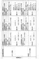
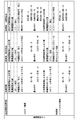
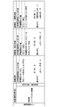
- Table showing an example of evaluation method for undifferentiation / differentiation according to stem cell maturity and culture conditions Table showing an example of evaluation method for undifferentiation / differentiation according to stem cell maturity and culture conditions
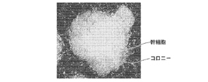
- Table showing an example of evaluation method for undifferentiation / differentiation according to stem cell maturity and culture conditions Diagram showing an example of an observation image of stem cells at the early seeding stage
- the figure which shows an example of the observation image of the stem cell in the stage where several days passed after seeding The figure which shows an example of the observation image of the stem cell in the stage where one week passed after seeding
- the figure which shows an example of the observation image of the stem cell in the stage where one week passed after seeding The figure which shows an example of the observation image when a stem cell colony extends in the plane direction and grows Flowchart for explaining the operation of the cell culture observation system shown in FIG.
- FIG. 1 is a block diagram showing a schematic configuration of a cell culture observation system.
- the cell culture observation system of this embodiment includes a cell culture device 1, a photographing device 2, a cell image evaluation device 3, a display 4, and an input device 5, as shown in FIG.
- the cell culture device 1 is a device for culturing cells.
- Examples of cells to be cultured include pluripotent stem cells such as iPS cells and ES cells, cells such as nerves, skin, myocardium, and liver induced by differentiation from stem cells, and cancer cells.
- pluripotent stem cells such as iPS cells and ES cells
- cells such as nerves, skin, myocardium, and liver induced by differentiation from stem cells, and cancer cells.
- a plurality of culture containers in which cells to be cultured are seeded in a medium are accommodated.
- the cell culture device 1 includes a stage 10, a transport unit 11, and a control unit 12.
- the stage 10 is provided with a culture container to be photographed by the photographing apparatus 2.
- the transport unit 11 selects a culture container to be imaged from among a plurality of culture containers accommodated at a predetermined position in the cell culture apparatus 1 and transports the selected culture container to the stage 10.
- the control unit 12 controls the entire cell culture apparatus 1, and in addition to the operations of the stage 10 and the transfer unit 11 described above, environmental conditions such as temperature, humidity, and CO 2 concentration in the cell culture apparatus 1 are controlled. It is something to control.
- the temperature, the configuration for adjusting the humidity and CO 2 concentration can be a known configuration.
- the imaging device 2 captures a cell image of a cell in a culture vessel installed on the stage 10.
- the imaging device 2 includes an optical system 20 that images a cell and outputs a cell image, and a control unit 21 that controls the optical system 20.
- the optical system 20 includes a phase contrast microscope and a differential interference microscope. And according to the maturity level of the stem cell culture
- microscopes include an image sensor such as a CMOS (Complementary Metal-Oxide Semiconductor) sensor or a CCD (charge-coupled device) sensor, and a cell image obtained by imaging cells is output from the image sensor. Note that switching of imaging by these microscopes will be described in detail later.
- CMOS Complementary Metal-Oxide Semiconductor
- CCD charge-coupled device
- the control unit 21 controls the entire imaging apparatus 2.
- the control unit 21 captures a cell image with the phase contrast microscope and a cell image with a differential interference microscope. Switching, optical magnification of the optical system 20, exposure time and resolution of the image sensor, switching of the exposure light quantity of the illumination light source provided in the optical system 20 and the wavelength of the illumination light, and the like.
- the control part 21 changes an imaging method according to the maturity degree of a stem cell based on the control signal output from the control part 35 in the cell image evaluation apparatus 3, The detail is mentioned later.
- the installation position of the culture vessel is changed between the imaging position of the phase contrast microscope and the imaging position of the differential interference microscope. What is necessary is just to switch.
- the cell image evaluation apparatus 3 is obtained by installing an embodiment of the cell image evaluation program of the present invention on a computer.
- the cell image evaluation apparatus 3 includes a central processing unit, a semiconductor memory, a hard disk, and the like, and an embodiment of the cell image evaluation program of the present invention is installed on the hard disk. And this program is performed by the control part 35 which has a central processing unit, The cell image acquisition part 30 (equivalent to an image acquisition part) as shown in FIG. 1, the cell evaluation part 31, the maturity information acquisition part 33 and the display control unit 34 operate.
- the cell image acquisition unit 30 acquires and stores a cell image captured by the imaging device 2.
- the cell image acquisition unit 30 outputs the acquired cell image to the cell evaluation unit 31 and the display control unit 34.
- the cell evaluation unit 31 evaluates the state of the cell based on the cell image acquired by the cell image acquisition unit 30. In this embodiment, the cell evaluation unit 31 evaluates undifferentiation / differentiation of the stem cell. .
- the cell evaluation unit 31 includes a feature amount acquisition unit 32 that acquires various feature amounts from a cell image, and evaluates undifferentiation / differentiation of stem cells based on the feature amount acquired by the feature amount acquisition unit 32 It is.
- the cell evaluation unit 31 changes the method for evaluating the undifferentiation / differentiation of stem cells according to the maturity of the stem cells and the culture conditions of the stem cells.
- the change in the evaluation method means a change in evaluation criteria used when evaluating undifferentiation / differentiation, and a change in weighting for each evaluation criterion when evaluating undifferentiation / differentiation using a plurality of evaluation criteria. And so on.
- the cell evaluation unit 31 of the present embodiment is divided into three stages: a stage at the initial stage of seeding of stem cells, a stage after several days after seeding of the stem cells, and a stage after one week after seeding of the stem cells.
- the evaluation method for undifferentiation / differentiation is changed depending on the situation.
- the information related to the maturity level of the cell may be any information as long as it is information indicating the stage of the maturity level of the cell.
- the culture period measured by a timer or the like can be acquired as information related to the maturity level.
- the image information of the cell colony area in the cell image is analyzed, not limited to the culture period, and the size of the cell colony, the number of cells in the cell colony, or the number of cells per unit area smaller than the cell colony is measured. As the number of cells measured increases, the degree of maturity is advanced, and information relating to the degree of maturity may be acquired. As the size of the cell colony, the area, perimeter, maximum diameter, etc. of the cell colony can be obtained.
- the brightness of the image of the cell colony region, and the texture such as uniformity and roughness may be acquired as information related to maturity.
- the degree of maturity advances and the density of the stem cells increases, and the stem cells are further stacked to gradually increase the luminance of the image. Therefore, it can be said that the higher the brightness is, the more mature the progress is.
- the feature quantity of the shape of the stem cell colony may be acquired.
- the shape of the stem cell colony gradually approaches a circular shape, and then the differentiation of the peripheral portion proceeds and the complexity of the edge increases. Therefore, the feature amount of the change in the shape of the stem cell colony can be acquired as the feature amount related to the maturity.
- the feature quantity of the thickness of the stem cell colony may be acquired. As the maturity of stem cells progresses, the stem cell colonies gradually become thicker. Therefore, such a feature quantity of the thickness of the stem cell colony can be acquired as a feature quantity related to maturity.
- the thickness of the stem cell colony may be measured by a separately provided measuring device, or may be set and input by the user using the input device 5.
- the user may set and input the cell passage number using the input device 5.
- the maturity level information acquisition unit 33 acquires information related to the maturity level of the cells as described above, and acquires the stage of maturity level of the cells from this information.
- the maturity is divided into three stages, but it is not limited to three stages, and may be divided into two stages or four or more stages.
- various intervals can be set for the intervals of each stage according to the culture conditions and the like.
- the cell evaluation unit 31 uses, as culture conditions, whether or not the culture is performed using heterogeneous cells, and whether the seeding method is colony seeding in which seeding is performed in units of colonies or seeding in units of stem cells. The condition of whether or not single cell sowing is performed is acquired.
- stem cells When culturing stem cells, different types of heterogeneous cells from the stem cells to be cultured may be used. And the way of growth of stem cells differs between the case of culture using heterogeneous cells and the culture without heterogeneous cells. In addition, when culturing stem cells, medium replacement and drug addition may be performed in the middle. In this case, whether the culture is performed using heterogeneous cells or without heterogeneous cells, or even during the culture. Depending on the state of the stem cell, the type of medium to be exchanged and the type of drug vary, and the manner of growth of the stem cell changes depending on such culture conditions, so it is preferable to change the evaluation criteria for undifferentiation / differentiation.
- the cell evaluation unit 31 acquires conditions for determining whether or not the culture is performed using heterogeneous cells and conditions for determining whether or not to perform medium replacement and drug addition. It changes the evaluation method of differentiation / differentiation. In addition, conditions for the type of medium exchanged and the type of drug added may be acquired, and the evaluation method for undifferentiation / differentiation may be changed according to these conditions.
- the cell evaluation part 31 of this embodiment acquires the conditions of a seeding method, and changes the evaluation method of undifferentiation and differentiation according to the acquired conditions of the seeding method.
- the culture conditions as described above may be set and input by the user using the input device 5, and the cell evaluation unit 31 may acquire the input culture conditions.
- the culture conditions are not limited to the above-described culture conditions, and other culture conditions that affect the progress of stem cell growth, such as information on the type of medium and scaffold, for example, may be used.
- the feature amount acquisition unit 32 acquires feature amounts according to the evaluation criteria in each evaluation method corresponding to the maturity of the stem cells and the culture conditions.
- a cell image is taken using a phase contrast microscope in the photographing apparatus 2, and the feature amount acquisition unit 32 performs a heterogeneous cell and a stem cell colony on the cell image. And image processing for extracting stem cell colonies is performed. Since different cells and stem cell colonies have different sizes in order units, it is possible to extract only stem cell colonies by separating the different cells and stem cell colonies by performing edge detection and pattern matching.
- the cell evaluation unit 31 evaluates undifferentiation / differentiation using the extracted stem cell colony shape and the uniformity of individual stem cells in the stem cell colony as evaluation criteria.
- the feature amount acquisition unit 32 extracts the outer peripheral shape and the internal defect as information on the shape of the stem cell colony.
- a stem cell colony generally has a circular shape when undifferentiated, and when differentiation progresses, stem cells are released and the circular shape collapses. Therefore, the undifferentiation / differentiation of the stem cell colony can be evaluated by evaluating the circularity of the outer peripheral shape of the stem cell colony.
- the internal defect of a stem cell colony is a hole etc. which are formed in the inside of a stem cell colony by differentiation.
- the feature amount acquisition unit 32 acquires the distribution state of the individual stem cells in the stem cell colony, and acquires information on how uniformly the individual stem cells are distributed. If stem cells are uniformly distributed, there is a high possibility that they are undifferentiated, and if stem cells are unevenly distributed, such as being concentrated in part, it is likely that they are differentiated. I can say that.
- the distribution state of individual stem cells in the stem cell colony may be obtained by detecting, for example, a pattern of stem cell nucleolus, or a halo generated due to diffracted light passing between stem cells. It may be obtained by detecting the pattern. Diffraction occurs when the illumination light passes between the stem cells. When the distance between the stem cells (slit gap) is an integral multiple of the wavelength of the illumination light, the phase of the diffracted light ( ⁇ 1st order diffracted light) and the direct light (0th order diffracted light) are synchronized, resulting in high brightness artifacts. appear. This high brightness artifact is halo.
- the cell evaluation part 31 calculates the evaluation value regarding the circularity of the outer peripheral shape of the stem cell colony, the evaluation value regarding the presence or absence and size of the internal defect, and the evaluation value regarding the uniformity of the individual stem cells in the stem cell colony, Each of these evaluation values is weighted and added to calculate a final evaluation value for evaluating undifferentiation / differentiation. If the evaluation value is equal to or greater than a preset threshold value, It is evaluated that it is differentiated, and if it is less than the threshold value, it is evaluated that it is differentiated.
- the weighting for the evaluation value of the uniformity of the stem cells is made larger than the weighting for the evaluation value of the shape of the stem cell colony (peripheral shape and internal defect).
- the maturity of the stem cell colony is not advanced, so that the outer periphery shape of the stem cell colony is not circular or the stem cells in the stem cell colony There may be gaps unrelated to differentiation between them. Therefore, it is possible to evaluate undifferentiation / differentiation with higher accuracy when the uniformity of the distribution of stem cells is more heavily evaluated than the outer shape of the stem cell colony.
- a cell image is taken using a phase contrast microscope, as in the initial seeding stage, and the feature amount acquisition unit 32 performs a cell image.
- An image process for extracting a stem cell colony is performed.
- the cell evaluation unit 31 evaluates undifferentiation / differentiation based on the extracted stem cell colony shape and the uniformity of individual stem cells in the stem cell colony as evaluation criteria. Different from stage.
- the cell evaluation unit 31 sets the weight for the evaluation value of the uniformity of the stem cells to be larger than the weight for the evaluation value of the shape of the stem cell colony.
- the weight for the evaluation value is set larger than the weight for the evaluation value of the shape of the stem cell colony in the initial seeding stage described above.
- the maturity of the stem cell colony has progressed to some extent, the outer shape of the stem cell colony becomes almost circular, and the degree of filling of the stem cells in the stem cell colony is high, as shown in FIG. This is because it is considered that evaluation of undifferentiation / differentiation can be performed with high accuracy by increasing the weighting on the evaluation value of the shape of the stem cell colony.
- a cell image is captured using a differential interference microscope instead of the phase contrast microscope in the imaging apparatus 2, and the feature amount acquisition unit 32 The cell image is subjected to image processing for separating the heterogeneous cells and the stem cell colonies and extracting the stem cell colonies.
- the growth of stem cell colonies develops and may be laminated as shown in FIG.
- the diffracted light component and the refracted light component from the stacked stem cells are superimposed, the diffracted light from one stem cell cannot be separated, and the light intensity of the entire image increases. Therefore, the micro features of individual stem cells in the stem cell colony cannot be measured with a phase contrast microscope. Therefore, at this stage, as described above, a cell image is taken with the differential interference microscope.
- the cell evaluation unit 31 evaluates undifferentiation / differentiation using the shape of the stem cell colony, the uniformity of the brightness of the stem cell colony, and the uniformity of the thickness of the stem cell colony as evaluation criteria.
- the feature amount acquisition unit 32 extracts the outer peripheral shape and internal defects of the stem cell colony, as in the previous stage. In addition, the feature amount acquisition unit 32 acquires the distribution of the luminance signal in the stem cell colony and acquires the uniformity thereof. In addition, the feature amount acquisition unit 32 acquires the uniformity of the thickness of the stem cell colony.
- the thickness of the stem cell colony can be measured using an interferometer such as an OCT (Optical Coherence Tomography) (not shown).
- the uniformity of the luminance signal and thickness for example, the standard deviation of the luminance signal and thickness may be acquired, or the difference between the maximum value and the minimum value may be acquired. If the luminance signal and the thickness are uniformly distributed, it can be said that there is a high possibility of being undifferentiated. Also, if the part with relatively high or low brightness compared to the surrounding area or the part with relatively large or small thickness is concentrated and distributed unevenly, it will be differentiated. It can be said that there is a possibility.
- the cell evaluation part 31 is related with the uniformity of the evaluation value regarding the circularity of the outer periphery shape of a stem cell colony, the evaluation value regarding the multiple circle approximation degree of an outer periphery shape, the evaluation value regarding the presence or absence of an internal defect, and a magnitude
- the evaluation value and the evaluation value regarding the uniformity of the thickness are calculated, and the final evaluation value for evaluating undifferentiation / differentiation is calculated by weighting and adding each of these evaluation values.
- the multiple circle approximation degree refers to the approximation degree between the outer peripheral shape of the stem cell colony and the multiple circle combination pattern obtained by combining multiple circles.
- weighting for each evaluation value relatively reduces the evaluation value related to the circularity, and relatively increases the evaluation value related to the multi-circle approximation, the internal defect, the luminance uniformity, and the thickness uniformity.
- a cell image is taken using a phase contrast microscope as in the case of colony seeding, but no colonies are formed at the initial seeding stage.
- the image processing for extracting colonies is not performed as in the case of colony seeding.
- the feature amount acquisition unit 32 extracts the outer peripheral shape and internal defects of the stem cell colony, but as described above, the colony is not clearly formed at this stage. . Therefore, the feature quantity acquisition unit 32 specifies an area where a stem cell colony is estimated to be formed in the future from the distribution state of the stem cells, and extracts the outer peripheral shape and internal defects of the specified area.
- the feature amount acquisition unit 32 acquires the distribution state of the heterogeneous cells and the stem cells, and acquires the uniformity of the distribution of these cells. At this time, since it is difficult to distinguish between a heterogeneous cell and a stem cell, uniformity is obtained for both cells.
- the cell evaluation unit 31 calculates an evaluation value related to the circularity of the outer peripheral shape of the stem cell colony, an evaluation value related to the presence / absence or size of the internal defect, and an evaluation value related to the uniformity of the stem cells and the heterogeneous cells.
- a final evaluation value for evaluating undifferentiation / differentiation is calculated by weighting and adding each value. At this time, the weighting for the evaluation value of the uniformity of the stem cells is made larger than the weighting for the evaluation value of the shape (peripheral shape and internal defect) of the stem cell colony.
- the evaluation method corresponding to each maturity in the case where the culture condition is culture without heterogeneous cells and colony seeding is shown in the upper part of FIG. 3. In this case, all maturity levels are shown.
- the evaluation method corresponding to is the same as the evaluation method in the case of culturing using heterogeneous cells and colony seeding.
- the evaluation method corresponding to each maturity in the case where the culture conditions are culture without heterogeneous cells and single cell seeding is shown in the lower part of FIG.
- the evaluation method when the maturity of the stem cells is in the initial seeding and the evaluation method when several days have elapsed after the seeding are as follows. It is the same, and it differs only about the evaluation method in the stage which passed for one week after sowing.
- a cell image is taken using a phase contrast microscope instead of a differential interference microscope at the stage where one week has elapsed after seeding.
- image processing is performed on the cell image to separate the heterogeneous cells and the stem cell colonies and extract the stem cell colonies.
- the cell image is taken using the phase contrast microscope instead of the differential interference microscope.
- the stem cell colony grows as described above, and the stem cells are stacked.
- the growth of stem cell colonies is faster than in the case of single cell seeding, and there is a high possibility that stacking will occur.
- Cell images are taken with a microscope.
- the cell evaluation unit 31 weights the evaluation value related to the circularity of the outer peripheral shape of the stem cell colony, the evaluation value related to the multiple circular approximation of the outer peripheral shape, and the evaluation value related to the presence / absence and size of the internal defect, respectively.
- the final undifferentiated / differentiated evaluation value is calculated, and the evaluation value related to luminance uniformity and the evaluation value related to thickness uniformity are not considered.
- the weighting for each evaluation value as in the case of culturing using heterogeneous cells, the evaluation value relating to the circularity is relatively reduced, and the evaluation values relating to the multi-circle approximation and the internal defect are relatively increased.
- the culture conditions correspond to each maturity in the case of culturing without heterogeneous cells and single cell seeding, and further when medium exchange and drug addition are performed several days after seeding of stem cells.
- the evaluation method is shown in FIG.
- the evaluation method at the initial stage of seeding and the evaluation method at the stage after one week after seeding are cases of culturing without heterogeneous cells and single-cell seeding. This is the same as the evaluation method when no drug is added. Then, when several days have elapsed after sowing, and when the medium is exchanged and the drug is added, undifferentiation / differentiation is evaluated using the same evaluation criteria as in the initial seeding stage. This is for evaluating the effect of the addition of the drug by using the same evaluation standard as the evaluation method before the addition of the drug.
- the display control unit 34 displays the cell image acquired by the cell image acquisition unit 30 on the display 4 or displays the undifferentiation / differentiation evaluation result in the cell evaluation unit 31 on the display 4. Is. Further, the display control unit 34 may display not only the evaluation result but also the feature amount and the evaluation value used for the evaluation on the display 4.
- the control unit 35 controls the entire cell image evaluation device 3, but the imaging device is configured such that the cell image imaging method is changed according to the maturity level information acquired by the maturity level information acquisition unit 33.
- the control signal is also output to the second control unit 21.
- the input device 5 is provided with a mouse, a keyboard, etc., and receives an operation input by the user. For example, the input device 5 accepts setting and changing of evaluation criteria in undifferentiated / differentiated evaluation, and accepts setting and changing of weighting used when calculating an evaluation value.
- the culture to be photographed is selected from the plurality of accommodated culture containers by the transport unit 11, and the selected culture container is placed on the stage 10 (S10).
- the cell image of the observation region including the stem cells in the culture vessel is photographed by the phase contrast microscope or the differential interference microscope of the photographing apparatus 2 (S12). Specifically, a 10 cm ⁇ 10 cm rectangular observation region as shown in FIG. 11 is photographed with 40 shots ⁇ 40 shots using a phase contrast microscope to obtain one cell image.
- the cell image photographed by the photographing device 2 is output to the cell image evaluation device 3, and is obtained by the cell image acquisition unit 30 of the cell image evaluation device 3 (S14).
- the culture period at the time when the cell image is taken is acquired as information related to the maturity level (S16), and the culture condition is input by the user using the input device 5. (S18).
- the cell image acquired by the cell image acquisition unit 30, the information related to the maturity acquired by the maturity information acquisition unit 33, and the culture conditions input by the user are output to the cell evaluation unit 31, and are characterized.
- the quantity acquisition unit 32 acquires feature quantities according to the maturity and culture conditions (S20).
- the cell evaluation unit 31 uses the evaluation method according to the maturity level of the stem cell and the culture condition to determine whether the stem cells in the cell image are undifferentiated / differentiated. Evaluation is performed (S22).
- the evaluation result of undifferentiation / differentiation in the cell evaluation unit 31 is output to the display control unit 34, and the display control unit 34 displays the input evaluation result of undifferentiation / differentiation and a cell image on the display 4. (S24).
- a cell image obtained by photographing a stem cell is acquired, and information related to the maturity level of the stem cell is acquired based on the cell image when evaluating undifferentiation / differentiation of the stem cell.
- the evaluation method for undifferentiation / differentiation was changed based on information related to the maturity level, undifferentiated / differentiated stem cells at each growth stage from the start of seeding of stem cells to the progress of growth to some extent. Can be appropriately evaluated.
- the undifferentiation / differentiation of stem cells can be appropriately evaluated even when the culture conditions are different.
- the evaluation criteria used when evaluating undifferentiation / differentiation are not limited to the uniformity of the stem cells and the shape of the stem cell colonies as described above.
- the degree of filling of the stem cells and the generation of halo in the stem cell colonies The state and the clarity of the stem cell colony boundary may be used.
- a cell image is taken with a phase-contrast microscope at the initial seeding stage and after several days after sowing, and at one stage after seeding, the cell image is taken with a differential interference microscope.
- the photographing method in the photographing apparatus 2 is changed according to the maturity level of the stem cell.
- a cell image is taken using a differential interference microscope, and in the case of culture without heterogeneous cells and single cell seeding
- a cell image is taken using a phase contrast microscope, that is, the photographing method in the photographing apparatus 2 is changed according to the culture conditions.
- the imaging method is changed by changing the type of the optical system 20 according to the maturity level or the culture conditions, but the imaging method is changed by changing the imaging condition of the optical system 20 or the imaging condition of the imaging device. You may do it.
- the change from the phase contrast microscope to the differential interference microscope can be said to be a change in the illumination method.
- the optical magnification in the optical system 20 is 1 after the initial seeding. It is desirable to make it higher than the optical magnification at the stage where a week has passed. Thereby, it is possible to photograph individual stem cells with high accuracy.
- the whole shape of the stem cell colony can be photographed by lowering the optical magnification relatively at the stage after one week after the initial seeding, the multiple circle approximation degree of the stem cell colony, internal defects, etc. It can be evaluated with high accuracy.
- the resolution of the imaging device of the optical system 20 be relatively high in the initial sowing stage and the stage after several days after sowing. In this stage, it is desirable to make the resolution relatively low.
- a plurality of image sensors having different resolutions may be switched, and when an image signal is read from one image sensor, for example, it is down-sampled by performing binning or the like. May be.
- the distribution of individual stem cells is evaluated more heavily than the outline of the stem cell colony, so the edges and halos of individual stem cells are detected with high accuracy. Therefore, it is desirable to set the exposure time of the image sensor of the optical system 20 to be longer than the exposure time after a week has elapsed since the initial seeding. Thereby, it is possible to photograph individual stem cells with high accuracy.
- FIG. 14 is a cell image obtained by photographing the stem cell colony that is the subject of photographing of the cell image of FIG. 13 with a short exposure time.
- the exposure time may be changed by changing the number of exposures. For example, in a seeding initial stage and a stage after several days after seeding, a plurality of cell images are added with a plurality of exposure times, and in a stage one week after seeding, the number of exposures is set to one. A cell image may be acquired. It can be said that the change of the number of exposures substantially corresponds to the change of the exposure time.
- the wavelength of the illumination light of the optical system 20 is 1 after the initial seeding. It is desirable to make it shorter than the wavelength of the illumination light after a week has passed. Thereby, the spatial resolution can be increased. On the other hand, at the stage where one week has passed after the initial seeding, the whole shape of the stem cell colony is photographed, and so high spatial resolution is not necessary. Further, when the wavelength of the illumination light is short, the scattering becomes strong, so that scattering occurs at the boundary between the stem cell and the non-stem cell part, and the boundary of the whole shape of the stem cell colony is blurred.
- FIG. 15 is a phase difference image photographed with relatively short wavelength illumination light
- FIG. 16 is a phase difference image photographed with relatively long wavelength illumination light.
- the arrows in FIGS. 15 and 16 indicate the fine structure in the stem cell, but the cell image in FIG. 15 taken with the short wavelength illumination light is finer than the cell image in FIG. 16 taken at the long wavelength. You can see that the structure is clear.
- the ellipses in FIG. 15 and FIG. 16 show the boundary part of the cell, but the cell image in FIG. 16 taken with the long wavelength illumination light is more bordered than the cell image in FIG. 15 taken at the short wavelength. You can see that the blur has been reduced.
- the method for evaluating undifferentiated / differentiated stem cells has been described.
- the degree of differentiation of differentiation-induced cells or cancer cells can be evaluated.
- An evaluation method such as a malignancy level may be determined based on information related to the maturity level. Since the change in morphological characteristics of cells induced to differentiate varies depending on the type of the cells, an evaluation method corresponding to the change in morphological characteristics may be set in advance. For example, when evaluating myocardial colonies, evaluate the distribution state of individual cardiomyocytes at the beginning of the culture, and evaluate the pulsation cycle at the stage when the myocardial cells begin to pulsate due to growth. It may be.
- the cell evaluation unit 31 may acquire the information on the cell type as described above, and may determine a cell colony evaluation method based on the information on the cell type and the maturity.
Landscapes
- Engineering & Computer Science (AREA)
- Chemical & Material Sciences (AREA)
- Health & Medical Sciences (AREA)
- Life Sciences & Earth Sciences (AREA)
- Physics & Mathematics (AREA)
- Theoretical Computer Science (AREA)
- Computer Vision & Pattern Recognition (AREA)
- Bioinformatics & Cheminformatics (AREA)
- General Health & Medical Sciences (AREA)
- General Physics & Mathematics (AREA)
- Wood Science & Technology (AREA)
- Organic Chemistry (AREA)
- Multimedia (AREA)
- Zoology (AREA)
- Analytical Chemistry (AREA)
- General Engineering & Computer Science (AREA)
- Biomedical Technology (AREA)
- Biochemistry (AREA)
- Genetics & Genomics (AREA)
- Medical Informatics (AREA)
- Sustainable Development (AREA)
- Microbiology (AREA)
- Biotechnology (AREA)
- Evolutionary Computation (AREA)
- Data Mining & Analysis (AREA)
- Artificial Intelligence (AREA)
- Quality & Reliability (AREA)
- Radiology & Medical Imaging (AREA)
- Nuclear Medicine, Radiotherapy & Molecular Imaging (AREA)
- Optics & Photonics (AREA)
- Cell Biology (AREA)
- Evolutionary Biology (AREA)
- Software Systems (AREA)
- Computing Systems (AREA)
- Bioinformatics & Computational Biology (AREA)
- Databases & Information Systems (AREA)
- Molecular Biology (AREA)
- Apparatus Associated With Microorganisms And Enzymes (AREA)
- Measuring Or Testing Involving Enzymes Or Micro-Organisms (AREA)
- Investigating Or Analysing Materials By Optical Means (AREA)
Abstract
【課題】細胞コロニーの成長にともなう形態変化に応じて細胞コロニーの状態を適切に評価することができる細胞画像評価装置および方法並びにプログラムを提供する。 【解決手段】細胞を撮影した細胞画像を取得する細胞画像取得部(30)と、細胞画像を評価する細胞評価部(31)と、細胞の成熟度に関連する情報を取得する成熟度情報取得部(33)とを備え、細胞評価部(31)が、成熟度に関連する情報に基づいて、細胞画像の評価方法を決定する。
Description
本発明は、細胞を撮像した細胞画像を評価する細胞画像評価装置および方法並びにプログラムに関するものである。
ES細胞やiPS細胞などの多能性幹細胞は、種々の組織の細胞に分化する能力を備えたものであり、再生医療、薬の開発、病気の解明などにおいて応用が可能なものとして注目されている。
幹細胞は、細胞培養装置内に設定された培養容器内の足場材(培養床)に播種され、足場材上において培地(培養液)を養分として増殖し、増殖した幹細胞同士が密集結合を繰り返しながら幹細胞コロニーとして成長する。
この幹細胞の成長過程においては、一旦ある組織に分化を開始した幹細胞は、途中で別の組織に変化・成長させることができないので、未分化状態を保持させたまま十分な数まで幹細胞を増殖させ、その後の過程で目的の組織の細胞に分化させることは、生産性の観点で重要である。
また、幹細胞コロニーの未分化性の高い領域のみを切り出して別の培養容器に移植し、継代を行うことがあるが、この継代の際には、未分化の幹細胞のみを抽出する必要がある。すなわち、幹細胞の培養に際しては、幹細胞の分化・未分化を適切に評価する必要がある。
そこで、たとえば特許文献1および特許文献2では、幹細胞を経時的に撮像し、その観察画像の経時的な変化を捉えることで未分化・分化の評価を行うことが提案されている。
また、特許文献3では、幹細胞の数や核小体の数など数十種の特徴量を用いて幹細胞の未分化・分化の評価を行うことが提案されている。
また、上述したような幹細胞の未分化・分化の評価に限らず、たとえば幹細胞を心筋や皮膚などの目的の組織に分化誘導した後の細胞や、がん細胞などを顕微鏡などで撮像し、その画像の特徴を捉えることで細胞の培養状態を評価する方法も提案されている。
しかしながら、特許文献1から特許文献3に記載のように幹細胞の未分化・分化の評価を行う際、幹細胞は、播種開始からその成長にともなって幹細胞の分布状態や幹細胞コロニーの形態などの特徴が変化するため、同一の尺度を用いて未分化・分化の評価を行ったのでは間違えた評価結果となる場合がある。
たとえば、一般的に、幹細胞コロニーの外形が円形に近い方が未分化状態を維持できていると評価することができるが、幹細胞コロニーの成長が進むと、周辺の幹細胞コロニーとの結合が始まって未分化状態ではあるが円形ではなくなるので、幹細胞の外形の円形度に基づく評価は意味がないものとなる。
また、一般的に、細胞の充填度が高い方が未分化状態を維持できていると評価することができるが、幹細胞の播種直後においては、幹細胞コロニーを形成するまで成熟していないため、未分化状態ではあるが幹細胞の充填度は低くなり、幹細胞の充填度に基づく評価は意味がないものとなる。
また、幹細胞を培養する際に用いられる培地や足場、播種方法などの培養条件によって幹細胞の成長の仕方や特徴が異なるため、同一の尺度を用いて未分化・分化の評価を行ったのでは間違えた評価結果となる場合がある。
本発明は、上記の問題に鑑み、細胞の播種開始からある程度成長が進むまでの各成長段階において、細胞コロニーの状態を適切に評価することができる細胞画像評価装置および方法並びにプログラムを提供することを目的とする。
本発明の細胞画像評価装置は、細胞を撮影した細胞画像を取得する画像取得部と、細胞画像を評価する細胞評価部と、細胞の成熟度に関連する情報を取得する成熟度情報取得部とを備え、細胞評価部が、成熟度に関連する情報に基づいて、細胞画像の評価方法を決定するものであることを特徴とする。
また、上記本発明の細胞画像評価装置においては、細胞評価部は、複数種類の評価基準を用いて細胞画像の評価を行うことできる。
また、細胞評価部は、複数の評価基準に対してそれぞれ異なる重み付けをして細胞画像の評価を行うことができる。
また、複数種類の評価基準として、細胞の分布の均一性に基づく評価基準と細胞のコロニーの形状に基づく評価基準とを含めることができる。
また、細胞評価部は、成熟度が予め設定された程度未満である場合よりも成熟度が予め設定された程度以上である場合の方を、幹細胞のコロニーの外形に対する重み付けを大きくして細胞画像の評価を行うことできる。
また、細胞評価部は、成熟度が予め設定された程度以上である場合に、細胞画像の輝度の均一性を評価基準として用いて細胞画像の評価を行うことができる。
また、細胞評価部は、成熟度が予め設定された程度以上である場合に、細胞画像内に分布する細胞の厚さの均一性を評価基準として用いて細胞画像の評価を行うことができる。
また、細胞評価部は、成熟度が予め設定された程度以上である場合に、複数の円を結合した複数円結合パターンと細胞のコロニーとの近似度を評価基準として用いて細胞画像の評価を行うことができる。
また、細胞評価部は、細胞の培養条件に基づいて、細胞画像の評価方法を変更することができる。
また、細胞評価部は、細胞とは異なる細胞を用いた培養条件である場合に、細胞画像に対して異なる細胞の画像を分離する処理を施すことができる。
また、細胞評価部は、成熟度が播種初期である場合において、培養条件が細胞のコロニー播種である場合に、細胞画像に対してコロニーを抽出する処理を施し、培養条件が細胞のシングルセル播種である場合に、細胞画像に対してコロニーを抽出する処理を施さないようにすることができる。
本発明の細胞画像評価方法は、細胞を撮影した細胞画像を取得し、該取得した細胞画像を評価する際、細胞の成熟度に関連する情報を取得し、その取得した成熟度に関連する情報に基づいて、細胞画像の評価方法を決定することを特徴とする。
本発明の細胞画像評価プログラムは、コンピュータを、細胞を撮影した細胞画像を取得する画像取得部と、細胞画像を評価する細胞評価部と、細胞の成熟度に関連する情報を取得する成熟度情報取得部として機能させる細胞画像評価プログラムであって、細胞評価部が、成熟度に関連する情報に基づいて、細胞画像の評価方法を決定するものであることを特徴とする。
本発明の細胞画像評価装置および方法並びにプログラムによれば、細胞を撮影した細胞画像を取得し、その細胞画像を評価する際、細胞の成熟度に関連する情報を取得し、その成熟度に関連する情報に基づいて、細胞画像の評価方法を決定するようにしたので、細胞の播種開始からある程度成長が進むまでの各成長段階において、細胞コロニーの状態を適切に評価することができる。
以下、本発明の細胞画像評価装置および方法並びにプログラムの一実施形態について、図面を参照しながら詳細に説明する。本発明は、細胞を撮像した細胞画像の評価方法に特徴を有するものであるが、まず、本発明の細胞画像評価装置の一実施形態を備えた細胞培養観察システムの全体構成について説明する。図1は、細胞培養観察システムの概略構成を示すブロック図である。
本実施形態の細胞培養観察システムは、図1に示すように、細胞培養装置1と、撮影装置2と、細胞画像評価装置3と、ディスプレイ4と、入力装置5とを備えている。
細胞培養装置1は、細胞の培養を行うための装置である。培養対象の細胞としては、たとえばiPS細胞やES細胞といった多能性幹細胞や、幹細胞から分化誘導された神経、皮膚、心筋、肝臓などの細胞や、がん細胞などがある。細胞培養装置1内には、培養対象の細胞を培地に播種した培養容器が複数収容されている。そして、細胞培養装置1は、ステージ10と搬送部11と制御部12とを備えている。
ステージ10は、撮影装置2による撮影対象の培養容器が設置されるものである。また、搬送部11は、細胞培養装置1内の所定位置に収容されている複数の培養容器の中から撮影対象の培養容器を選択し、その選択した培養容器をステージ10まで搬送するものでる。また、制御部12は、細胞培養装置1全体を制御するものであり、上述したステージ10や搬送部11の動作以外に、細胞培養装置1内の温度、湿度およびCO2濃度などの環境条件を制御するものである。なお、温度、湿度およびCO2濃度を調整するための構成については、公知な構成を用いることができる。
撮影装置2は、ステージ10に設置された培養容器内における細胞の細胞画像を撮像するものである。撮影装置2は、細胞を撮像して細胞画像を出力する光学系20と、光学系20を制御する制御部21とを備えている。
光学系20は、位相差顕微鏡と微分干渉顕微鏡とを備えたものである。そして、培養容器内における幹細胞の培養の成熟度に応じて、位相差顕微鏡による細胞画像の撮像と微分顕干渉微鏡による細胞画像の撮像とが切り替えられる。
これらの顕微鏡は、CMOS(Complementary Metal-Oxide Semiconductor)センサやCCD(charge-coupled device)センサなどの撮像素子を備えており、この撮像素子から細胞を撮像した細胞画像が出力される。なお、これらの顕微鏡による撮像の切り替えについては、後で詳述する。
制御部21は、撮影装置2全体を制御するものであるが、特に、本実施形態においては、制御部21は、上述した位相差顕微鏡による細胞画像の撮像と微分干渉顕微鏡による細胞画像の撮像との切り替え、光学系20の光学倍率、撮像素子の露光時間や解像度、光学系20に設けられた照明光源の露光光量や照明光の波長の切り替えなどを制御するものである。そして、制御部21は、細胞画像評価装置3における制御部35から出力された制御信号に基づいて、幹細胞の成熟度に応じて撮像方法を変更するものであるが、その詳細は後で述べる。なお、位相差顕微鏡による細胞画像の撮像と微分顕干渉微鏡による細胞画像の撮像との切り替えについては、たとえば、培養容器の設置位置を位相差顕微鏡の撮像位置と微分干渉顕微鏡の撮像位置とに切り替えるようにすればよい。
細胞画像評価装置3は、コンピュータに対して本発明の細胞画像評価プログラムの一実施形態がインストールされたものである。
細胞画像評価装置3は、中央処理装置、半導体メモリおよびハードディスクなどを備えており、ハードディスクに本発明の細胞画像評価プログラムの一実施形態がインストールされている。そして、このプログラムが中央処理装置を有する制御部35によって実行されることによって、図1に示すような細胞画像取得部30(画像取得部に相当する)、細胞評価部31、成熟度情報取得部33および表示制御部34が動作する。
細胞画像取得部30は、撮影装置2において撮像された細胞画像を取得して記憶するものである。また、細胞画像取得部30は、取得した細胞画像を細胞評価部31と表示制御部34に出力するものである。
細胞評価部31は、細胞画像取得部30によって取得された細胞画像に基づいて、細胞の状態などを評価するものであり、本実施形態においては、幹細胞の未分化・分化を評価するものである。
細胞評価部31は、細胞画像から種々の特徴量を取得する特徴量取得部32を備え、この特徴量取得部32によって取得された特徴量に基づいて、幹細胞の未分化・分化を評価するものである。また、細胞評価部31は、幹細胞の成熟度や幹細胞の培養条件に応じて、幹細胞の未分化・分化の評価方法を変更するものである。ここで、評価方法の変更とは、未分化・分化を評価する際に用いる評価基準の変更や、複数の評価基準を用いて未分化・分化の評価を行う場合における各評価基準に対する重み付けの変更などのことをいう。
本実施形態の細胞評価部31は、幹細胞の播種初期の段階と、幹細胞の播種後、数日経過した段階と、幹細胞の播種後、1週間経過した段階との3段階に区分された成熟度に応じて、未分化・分化の評価方法を変更するものである。
細胞の成熟度に関連する情報は、細胞の成熟度の段階を示す情報であれば如何なる情報でもよく、たとえばタイマなどによって計測された培養期間を成熟度に関連する情報として取得することができる。また、培養期間に限らず、たとえば細胞画像内の細胞コロニー領域の画像情報を解析し、細胞コロニーの大きさや、細胞コロニー内の細胞数または細胞コロニーよりも小さい単位面積当たりの細胞数を計測し、その計測した細胞数が多いほど成熟度が進んでいるものとして成熟度に関連する情報として取得するようにしてもよい。細胞コロニーの大きさとしては、細胞コロニーの面積、周囲長、最大径などを取得することができる。
また、たとえば細胞コロニー領域の画像の輝度や、均一性や粗さなどのテクスチャを成熟度に関連する情報として取得するようにしてもよい。たとえば撮像対象の細胞が幹細胞である場合には、その成熟度が進行すると幹細胞の密集度が高くなり、さらに幹細胞が積層されて、画像の輝度が次第に高くなる。したがって、輝度が高いほど成熟度が進行しているといえる。
また、成熟度が進行して上述したように幹細胞が増殖して積層された状態となった場合、画像の均一性が高くなり、また凹凸の少ない滑らかな画像となる。したがって、画像の均一性が高い、または画像が滑らかであるほど成熟度が進行しているといえる。画像の均一性や滑らかさの特徴量の取得方法については、既に公知な手法を用いることができる。
また、成熟度に関連する情報として、幹細胞コロニーの形状の特徴量を取得するようにしてもよい。幹細胞の成熟度が進行すると幹細胞コロニーの形状は次第に円形に近づいた後、周辺部分の分化が進行してエッジの複雑度が大きくなる。したがって、このような幹細胞コロニーの形状の変化の特徴量を成熟度に関連する特徴量として取得することができる。
また、成熟度に関連する情報として、幹細胞コロニーの厚さの特徴量を取得するようにしてもよい。幹細胞の成熟度が進行すると幹細胞コロニーは次第に厚くなっていく。したがって、このような幹細胞コロニーの厚さの特徴量を成熟度に関連する特徴量として取得することができる。幹細胞コロニーの厚さについては、別途設けられた計測装置によって計測するようにしてもよいし、ユーザが入力装置5を用いて設定入力するようにしてもよい。
また、上述した成熟度に関連する情報として、細胞の継代数をユーザが入力装置5を用いて設定入力するようにしてもよい。
成熟度情報取得部33は、上述したような細胞の成熟度に関連する情報を取得し、この情報から細胞の成熟度の段階を取得するものである。
また、本実施形態においては、上述したように成熟度を3段階に区分するようにしたが、3段階に限らず、2段階や4段階以上に区分してもよい。また、各段階の間隔についても、培養条件などに応じて種々の間隔を設定することができる。
また、本実施形態の細胞評価部31は、培養条件として、異種細胞を用いた培養であるか否かの条件と、播種方法がコロニー単位で播種を行うコロニー播種であるかまたは幹細胞単位で播種を行うシングルセル播種であるかの条件とを取得するものである。
幹細胞の培養を行う場合、培養対象の幹細胞とは異なる種類の異種細胞を用いる場合がある。そして、異種細胞を用いた培養の場合と、異種細胞なしの培養とでは、幹細胞の成長の仕方が異なる。また、幹細胞の培養を行う際には、途中で培地交換および薬剤添加を行う場合があり、この場合、異種細胞を用いた培養であるか異種細胞なしの培養であるか、さらには培養途中の幹細胞の状態によって、交換する培地の種類や薬剤の種類が異なり、このような培養条件によっても幹細胞の成長の仕方が変化するため、未分化・分化の評価基準を変更した方が好ましい。
そこで、本実施形態の細胞評価部31は、異種細胞を用いた培養であるか否かの条件と、培地交換および薬剤添加を行うか否かの条件を取得し、これらの条件に応じて未分化・分化の評価方法を変更するものである。なお、さらに途中交換される培地の種類や添加される薬剤の種類の条件も取得し、これらの条件に応じて未分化・分化の評価方法を変更するようにしてもよい。
また、幹細胞の培養を行う場合、幹細胞の播種方法として、コロニー単位で播種を行うコロニー播種と、幹細胞単位で播種を行うシングルセル播種とがある。そして、コロニー播種の場合とシングルセル播種の場合とでは、未分化・分化を評価する際に用いられる特徴量を抽出するための画像処理が異なっていたり、また、幹細胞の成長の仕方も異なるので未分化・分化の評価基準を変更した方が好ましい。したがって、本実施形態の細胞評価部31は、播種方法の条件を取得し、その取得した播種方法の条件に応じて未分化・分化の評価方法を変更するものである。
なお、上述したような培養条件については、ユーザが入力装置5を用いて設定入力し、細胞評価部31が、その入力された培養条件を取得するようにすればよい。また、培養条件は、上述したような培養条件に限らず、たとえば培地や足場の種類の情報など、幹細胞の成長の進行に影響を及ぼすようなその他の培養条件を用いるようにしてもよい。
また、特徴量取得部32は、幹細胞の成熟度や培養条件に対応するそれぞれの評価方法における評価基準に応じた特徴量を取得するものである。
以下、幹細胞の成熟度の各段階および培養条件に対応するそれぞれの評価方法について、図2から図4に示す表を参照しながら詳細に説明する。
まず、培養条件が、異種細胞を用いた培養であり、播種方法がコロニー播種である場合における各成熟度に対応する評価方法を、図2を参照しながら説明する。
まず、幹細胞の成熟度が播種初期である場合には、撮影装置2における位相差顕微鏡を用いて細胞画像が撮影され、特徴量取得部32において、細胞画像に対して、異種細胞と幹細胞コロニーとを分離して幹細胞コロニーを抽出する画像処理が施される。異種細胞と幹細胞コロニーとはオーダー単位で大きさが異なるものであるため、エッジ検出やパターンマッチングを行うことによって異種細胞と幹細胞コロニーとを分離して幹細胞コロニーのみを抽出することができる。
そして、この段階では、細胞評価部31は、抽出された幹細胞コロニーの形状と、幹細胞コロニー内の個々の幹細胞の均一性を評価基準として未分化・分化の評価を行う。
より具体的には、特徴量取得部32において、幹細胞コロニーの形状の情報として、外周形状と内部欠陥とが抽出される。幹細胞コロニーは、一般的には、未分化である場合には円形に近く、分化が進むと幹細胞が遊離して円形が崩れた状態となる。したがって、幹細胞コロニーの外周形状の円形度を評価することによって幹細胞コロニーの未分化・分化を評価することができる。また、幹細胞コロニーの内部欠陥とは、分化によって幹細胞コロニーの内部に形成される穴などである。
また、特徴量取得部32において、幹細胞コロニー内の個々の幹細胞の分布状態が取得され、個々の幹細胞がどの程度均一に分布しているかの情報が取得される。幹細胞が均一に分布している場合には未分化である可能性が高く、幹細胞が一部に集中して分布したりして不均一である場合には、分化している可能性が高いといえる。
なお、幹細胞コロニー内の個々の幹細胞の分布状態については、たとえば幹細胞の核小体のパターンを検出して取得するようにしてもよいし、幹細胞間を通過した回折光に起因して発生するハロのパターンを検出することによって取得するようにしてもよい。照明光が幹細胞間を通過する際、回折が起こる。そして、幹細胞間の距離(スリットギャップ)が照明光の波長の整数倍となった場合、回折光(±1次回折光)と直接光(0次回折光)の位相が同調し、高輝度のアーチファクトが発生する。この高輝度のアーチファクトがハロである。
そして、細胞評価部31は、幹細胞コロニーの外周形状の円形度に関する評価値と、内部欠陥の有無や大きさに関する評価値と、幹細胞コロニー内の個々の幹細胞の均一性に関する評価値を算出し、これらの評価値にそれぞれ重み付けをして加算することによって、未分化・分化を評価するための最終的な評価値を算出し、その評価値が予め設定された閾値以上である場合には、未分化であると評価し、閾値未満である場合には分化していると評価する。
そして、このとき幹細胞の均一性の評価値に対する重み付けの方を、幹細胞コロニーの形状(外周形状および内部欠陥)の評価値に対する重み付けよりも大きくする。
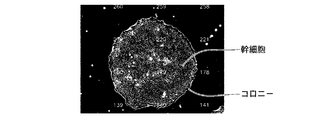
これは、幹細胞の成熟度が播種初期である場合には、図5に示すように、幹細胞コロニーの成熟度が進んでいないため、幹細胞コロニーの外周形状が円形ではなかったり、幹細胞コロニー内の幹細胞間に分化とは無関係な隙間が存在したりする場合がある。したがって、幹細胞コロニーの外形よりも幹細胞の分布の均一性をより重く評価した方が精度よく未分化・分化を評価することができるからである。
次に、幹細胞の成熟度が播種後、数日経過した段である場合には、播種初期段階と同様に、位相差顕微鏡を用いて細胞画像が撮影され、特徴量取得部32において、細胞画像に対して幹細胞コロニーを抽出する画像処理が施される。
そして、この段階においても、細胞評価部31は、抽出された幹細胞コロニーの形状と、幹細胞コロニー内の個々の幹細胞の均一性を評価基準として未分化・分化の評価を行うが、重み付けが播種初期段階とは異なる。
具体的には、この段階において、細胞評価部31は、幹細胞の均一性の評価値に対する重み付けの方を、幹細胞コロニーの形状の評価値に対する重み付けよりも大きくするが、このとき幹細胞コロニーの形状の評価値に対する重み付けは、上述した播種初期段階における幹細胞コロニーの形状の評価値に対する重み付けよりも大きくする。
これは、播種後から数日経過した段階では、図6に示すように、幹細胞コロニーの成熟度がある程度進み、幹細胞コロニーの外周形状が円形に近くなり、幹細胞コロニー内の幹細胞の充填度も高くなっていると考えられるため、幹細胞コロニーの形状の評価値に対する重み付けをより大きくした方が、未分化・分化の評価を高精度に行うことができると考えられるからである。
次に、幹細胞の成熟度が播種後、1週間経過した段階である場合には、撮影装置2における位相差顕微鏡ではなく、微分干渉顕微鏡を用いて細胞画像が撮影され、特徴量取得部32において、細胞画像に対して、異種細胞と幹細胞コロニーとを分離して幹細胞コロニーを抽出する画像処理が施される。
この段階においては、幹細胞コロニーの成長が発達し、図7に示すように、積層化する場合がある。このように積層化した場合、積層化された幹細胞からの回折光成分と屈折光成分との重ね合せを生じ、1つの幹細胞からの回折光を分離できず、かつ像全体の光強度が増してしまうため、幹細胞コロニー内の個々の幹細胞のミクロな特徴が位相差顕微鏡では計測できなくなる。したがって、この段階では、上述したように微分干渉顕微鏡によって細胞画像を撮影する。
そして、この段階では、細胞評価部31は、幹細胞コロニーの形状と、幹細胞コロニーの輝度の均一性と、幹細胞コロニーの厚みの均一性を評価基準として未分化・分化の評価を行う。
より具体的には、特徴量取得部32において、先の段階と同様に、幹細胞コロニーの外周形状と内部欠陥とが抽出される。また、特徴量取得部32において、幹細胞コロニー内の輝度信号の分布が取得され、その均一性が取得される。また、特徴量取得部32において、幹細胞コロニーの厚さの均一性が取得される。なお、幹細胞コロニーの厚さについては、たとえば、図示省略したOCT(Optical Coherence Tomography:光干渉断層計)などの干渉計を用いて計測することができる。
輝度信号や厚さの均一性としては、たとえば輝度信号や厚さの標準偏差を取得するようにしてもよいし、最大値と最小値との差を取得するようにしてもよい。輝度信号や厚さが均一に分布している場合には未分化である可能性が高いといえる。また、周囲と比較して相対的に高輝度または低輝度な部分や、相対的に厚さが大きいまたは小さい部分が一部に集中して分布して不均一である場合には、分化している可能性があるといえる。
そして、細胞評価部31は、幹細胞コロニーの外周形状の円形度に関する評価値と、外周形状の複数円近似度に関する評価値と、内部欠陥の有無や大きさに関する評価値と、輝度の均一性に関する評価値と、厚みの均一性に関する評価値を算出し、これらの評価値に対してそれぞれ重み付けをして加算することによって、未分化・分化を評価するための最終的な評価値を算出する。なお、複数円近似度とは、幹細胞コロニーの外周形状と、複数の円を結合した複数円結合パターンとの近似度のことをいう。
このとき各評価値に対する重み付けは、円形度に関する評価値を相対的に小さくし、複数円近似度、内部欠陥、輝度の均一性および厚みの均一性に関する評価値を相対的に大きくする。
これは、幹細胞の成熟度が播種後、1週間経過した段階では、図8に示すように、幹細胞コロニー同士が結合し、幹細胞コロニーの外周形状は円形を維持していない場合が多い。したがって、円形度の評価値に対する重み付けを小さくし、その他の複数円近似度に関する評価値などに対応する重み付けを大きくした方が精度よく未分化・分化を評価することができるからである。
以上が、培養条件が、異種細胞を用いた培養であってコロニー播種の場合における各成熟度に対応する評価方法の説明である。
次に、培養条件が、異種細胞を用いた培養であってシングルセル播種の場合における各成熟度に対応する評価方法について、図2を参照しながら説明する。
この場合、幹細胞の成熟度が播種初期段階の評価方法のみが、コロニー播種の場合と異なり、播種後、数日経過した段階の評価方法と、播種後、1週間経過した段階における評価方法は、コロニー播種の場合と同様である。
異種細胞を用いた培養であってシングルセル播種の場合、コロニー播種の場合と同様に、位相差顕微鏡を用いて細胞画像が撮影されるが、播種初期段階においては、コロニーが形成されていないので、コロニー播種の場合のようにコロニーを抽出する画像処理を行わない。
そして、コロニー播種の場合と同様に、特徴量取得部32において、幹細胞コロニーの外周形状と内部欠陥とが抽出されるが、上述したように、この段階においては、コロニーは明確に形成されていない。したがって、特徴量取得部32は、今後、幹細胞コロニーが形成されると推定される領域を幹細胞の分布状態から特定し、その特定した領域の外周形状と内部欠陥とを抽出する。
また、特徴量取得部32において、異種細胞および幹細胞の分布状態が取得され、これらの細胞の分布の均一性が取得される。なお、このとき異種細胞と幹細胞との区別をつけるのは困難であるため、両方の細胞についての均一性が取得される。
そして、細胞評価部31は、幹細胞コロニーの外周形状の円形度に関する評価値と内部欠陥の有無や大きさに関する評価値と、幹細胞および異種細胞の均一性に関する評価値とを算出し、これらの評価値にそれぞれ重み付けをして加算することによって、未分化・分化を評価するための最終的な評価値を算出する。そして、このとき幹細胞の均一性の評価値に対する重み付けの方を、幹細胞コロニーの形状(外周形状および内部欠陥)の評価値に対する重み付けよりも大きくする。
次に、培養条件が、異種細胞なしでの培養であってコロニー播種の場合における各成熟度に対応する評価方法については、図3の上段に示しているが、この場合は、全ての成熟度に対応する評価方法について、異種細胞を用いた培養であってコロニー播種の場合における評価方法と同様である。
次に、培養条件が、異種細胞なしでの培養であってシングルセル播種の場合における各成熟度に対応する評価方法を図3の下段に示す。この場合、幹細胞の成熟度が播種初期である場合の評価方法と、播種後、数日経過した場合の評価方法については、異種細胞を用いた培養であってシングルセル播種の場合における評価方法と同様であり、播種後、1週間経過した段階における評価方法についてのみ異なる。
具体的には、異種細胞なしでの培養であってシングルセル播種の場合において、播種後、1週間経過した段階では、微分干渉顕微鏡ではなく、位相差顕微鏡を用いて細胞画像が撮影され、特徴量取得部32において、細胞画像に対して、異種細胞と幹細胞コロニーとを分離して幹細胞コロニーを抽出する画像処理が施される。
このように、微分干渉顕微鏡でなく、位相差顕微鏡を用いて細胞画像を撮影するのは、異種細胞を用いた培養においては、幹細胞コロニーの成長によって、上述したような幹細胞の積層化が発生する場合があるが、異種細胞なしでの培養であってシングルセル播種の場合には、幹細胞が積層化するまでに時間がかかり、1週間程度の期間では、図9に示すように、幹細胞コロニーが平面方向に延びて成長するからである。なお、同じ異種細胞なしの培養であってもコロニー播種の場合には、シングルセル播種の場合よりも幹細胞コロニーの成長が早く、積層化が発生する可能性が高いので、上述したように微分干渉顕微鏡によって細胞画像を撮影するようにしている。
そして、細胞評価部31は、幹細胞コロニーの外周形状の円形度に関する評価値と、外周形状の複数円近似度に関する評価値と、内部欠陥の有無や大きさに関する評価値とにそれぞれ重み付けをして加算し、最終的な未分化・分化の評価値を算出し、輝度の均一性に関する評価値と、厚みの均一性に関する評価値とについては考慮しない。各評価値に対する重み付けとしては、異種細胞を用いた培養の場合と同様に、円形度に関する評価値を相対的に小さくし、複数円近似度および内部欠陥に関する評価値を相対的に大きくする。
次に、培養条件が、異種細胞なしでの培養であってシングルセル播種の場合であり、さらに幹細胞の播種後、数日経過前において培地交換および薬剤添加を行う場合における各成熟度に対応する評価方法を図4に示す。
上述した培養条件の場合、播種初期段階の評価方法と、播種後、1週間経過した段階の評価方法とについては、異種細胞なしでの培養であってシングルセル播種の場合であり、培地交換および薬剤添加なしの場合の評価方法と同様である。そして、播種後、数日経過した場合であって、培地交換および薬剤添加した場合には、播種初期段階と同様の評価基準を用いて未分化・分化の評価を行う。これは薬剤添加前の評価方法と同じ評価基準を用いることによって、薬剤添加による効果を評価するためである。
以上が、本実施形態の細胞評価部31における未分化・分化の評価方法についての説明である。
図1に戻り、表示制御部34は、細胞画像取得部30によって取得された細胞画像をディスプレイ4に表示させたり、細胞評価部31における未分化・分化の評価結果をディスプレイ4に表示させたりするものである。また、表示制御部34が、評価結果だけでなく、評価に用いられた特徴量や評価値をディスプレイ4に表示させるようにしてもよい。
制御部35は、細胞画像評価装置3全体を制御するものであるが、成熟度情報取得部33によって取得された成熟度の情報に応じて、細胞画像の撮影方法が変更されるように撮影装置2の制御部21に対して制御信号を出力するものでもある。
入力装置5は、マウスやキーボードなどを備えたものであり、ユーザによる操作入力を受け付けるものである。たとえば、入力装置5は、未分化・分化の評価における評価基準の設定や変更を受け付けたり、評価値を算出する際に用いられる重み付けの設定や変更を受け付けたりするものである。
次に、上述した幹細胞培養観察システムの作用について、図10に示すフローチャートを参照しながら説明する。
まず、細胞培養装置1において、搬送部11によって、収容されている複数の培養容器の中から撮影対象の培養が選択され、その選択された培養容器がステージ10に設置される(S10)。
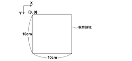
そして、撮影装置2の位相差顕微鏡または微分干渉顕微鏡によって、培養容器内の幹細胞を含む観察領域の細胞画像の撮影が行われる(S12)。具体的には、図11に示すような10cm×10cmの矩形の観察領域を位相差顕微鏡によって40ショット×40ショットで撮影して1枚の細胞画像を取得する。
そして、撮影装置2によって撮影された細胞画像は細胞画像評価装置3に出力され、細胞画像評価装置3の細胞画像取得部30によって取得される(S14)。
また、このとき成熟度情報取得部33において、たとえば細胞画像が撮影された時点における培養期間が成熟度に関連する情報として取得され(S16)、ユーザによって入力装置5を用いて培養条件が入力される(S18)。
そして、細胞画像取得部30によって取得された細胞画像と、成熟度情報取得部33によって取得された成熟度に関連する情報とユーザによって入力された培養条件とが細胞評価部31に出力され、特徴量取得部32は、入力された細胞画像と成熟度情報と培養条件に基づいて、成熟度および培養条件に応じた特徴量を取得する(S20)。
そして、細胞評価部31は、特徴量取得部32によって取得された特徴量に基づいて、幹細胞の成熟度および培養条件に応じた評価方法を用いて、細胞画像内の幹細胞の未分化・分化の評価を行う(S22)。
細胞評価部31における未分化・分化の評価結果は表示制御部34に出力され、表示制御部34は、入力された未分化・分化の評価結果と細胞画像とをディスプレイ4に表示させる。(S24)。
上記実施形態の細胞培養観察システムによれば、幹細胞を撮影した細胞画像を取得し、その細胞画像に基づいて、幹細胞の未分化・分化を評価する際、幹細胞の成熟度に関連する情報を取得し、その成熟度に関連する情報に基づいて、未分化・分化の評価方法を変更するようにしたので、幹細胞の播種開始からある程度成長が進むまでの各成長段階において、幹細胞の未分化・分化を適切に評価することができる。
また、幹細胞の培養条件に応じて未分化・分化の評価方法を変更するようにしたので、培養条件が異なる場合においても、幹細胞の未分化・分化を適切に評価することができる。
なお、未分化・分化の評価を行う際に用いられる評価基準としては、上述したような幹細胞の均一性や幹細胞コロニーの形状などに限らず、その他、幹細胞の充填度や幹細胞コロニーにおけるハロの発生状態や幹細胞コロニーの境界の明瞭度などを用いるようにしてもよい。
また、上記実施形態の説明においては、播種初期段階および播種後、数日経過後においては、位相差顕微鏡によって細胞画像を撮影し、播種後、1週間経過した段階においては、微分干渉顕微鏡によって細胞画像を撮影するようにし、すなわち幹細胞の成熟度に応じて撮影装置2における撮影方法を変更するようにしている。また、播種後、1週間経過した段階でも、異種細胞を用いた培養の場合には、微分干渉顕微鏡を用いて細胞画像を撮影し、異種細胞なしの培養であってシングルセル播種の場合には、位相差顕微鏡を用いて細胞画像を撮影するようにし、すなわち培養条件に応じて撮影装置2における撮影方法を変更するようにしている。
このように成熟度や培養条件に応じて光学系20の種類を変更して撮影方法を変更するだけでなく、光学系20の撮像条件や撮像素子の撮像条件を変更して撮影方法を変更するようにしてもよい。なお、位相差顕微鏡から微分干渉顕微鏡への変更は、照明方法の変更ともいえる。
以下、幹細胞の成熟度や培養条件に応じて撮像条件を変更する場合について、図12に示す表を参照しながら説明する。
まず、播種初期段階および播種初期後、数日経過した段階においては、幹細胞コロニーの外形よりも個々の幹細胞の分布状態をより重く評価するため、光学系20における光学倍率は、播種初期後、1週間経過した段階における光学倍率よりも高くすることが望ましい。これにより個々の幹細胞を高精度に撮影することができる。一方、播種初期後、1週間経過した段階においては、相対的に光学倍率を低くすることによって、幹細胞コロニーの全体形状を撮影することができるので、幹細胞コロニーの複数円近似度や内部欠陥などを高精度に評価することができる。
また、上記と同様の理由により、播種初期段階および播種初期後、数日経過した段階においては、光学系20の撮像素子の解像度を相対的に高くすることが望ましく、播種初期後、1週間経過した段階においては、解像度を相対的に低くすることが望ましい。なお、解像度の変更については、たとえば互いに解像度の異なる複数の撮像素子を切り替えるようにしてもよいし、1つの撮像素子から画像信号を読み出す際に、たとえばビニングなどを行うことによってダウンサンプリングするようにしてもよい。
また、播種初期段階および播種初期後、数日経過した段階においては、幹細胞コロニーの外形よりも個々の幹細胞の分布状態をより重く評価するため、個々の幹細胞のエッジやハロなどを高精度に検出するために、光学系20の撮像素子の露光時間は、播種初期後、1週間経過した段階における露光時間よりも長く設定することが望ましい。これにより個々の幹細胞を高精度に撮影することができる。
一方、播種初期後、1週間経過した段階においては、上述したよう幹細胞コロニーの積層化が生じる。そして、積層化された幹細胞からの回折光成分と屈折光成分との重ね合せを生じて像全体の光強度が増してしまうため、図13に示すように細胞画像内において白抜けが生じ、幹細胞コロニーの状態を正確に観察することができない。したがって、播種初期後、1週間経過した段階においては、撮像素子の露光時間を相対的に短く設定することが望ましい。図14は、図13の細胞画像の撮影対象である幹細胞コロニーを露光時間を短くして撮影した細胞画像である。
なお、露光時間の変更については、露光回数を変更することによって変更するようにしてもよい。たとえば、播種初期段階および播種初期後、数日経過した段階においては、露光回数を複数回として複数の細胞画像を加算し、播種初期後、1週間経過した段階においては、露光回数を1回として細胞画像を取得するようにしてもよい。露光回数の変更は、実質的には露光時間の変更に相当するものといえる。
また、上記と同様の理由により、播種初期段階および播種初期後、数日経過した段階においては、光学系20の光源の露光光量を相対的に大きくすることが望ましく、播種初期後、1週間経過した段階においては、露光光量を相対的に小さくすることが望ましい。
さらに、播種初期段階および播種初期後、数日経過した段階においては、個々の幹細胞のエッジなどを高分解能で撮影する必要があるため、光学系20の照明光の波長は、播種初期後、1週間経過した段階における照明光の波長よりも短くすることが望ましい。これにより空間分解能を高くすることができる。一方、播種初期後、1週間経過した段階においては、幹細胞コロニーの全体形状を撮影するのでそれほど高い空間分解能は必要ない。また、照明光の波長が短い場合、散乱が強くなるため、幹細胞と幹細胞ではない部分との境界で散乱が発生し、幹細胞コロニーの全体形状の境界がボケてしまう。
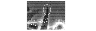
そこで、播種初期後、1週間経過した段階においては、相対的に長い波長の照明光を用いることが望ましい。図15は、相対的に短い波長の照明光によって撮影した位相差像であり、図16は、相対的に長い波長の照明光によって撮影した位相差像である。図15および図16の矢印は、幹細胞内の微細構造を示しているが、短波長の照明光で撮影した図15の細胞画像の方が、長波長で撮影した図16の細胞画像よりも微細構造が明確なのが分かる。図15および図16の楕円は、細胞の境界部分を示しているが、長波長の照明光で撮影した図16の細胞画像の方が、短波長で撮影した図15の細胞画像よりも境界のボケが軽減されているのは分かる。
なお、上記実施形態の説明では、幹細胞の未分化・分化の評価方法について説明したが、幹細胞の未分化・分化に限らず、たとえば分化誘導された細胞の分化度を評価したり、がん細胞の悪性度などの評価方法を成熟度に関連する情報に基づいて決定するようにしてもよい。分化誘導された細胞については、その細胞の種類によって形態的な特徴の変化が異なるので、その形態的な特徴の変化に応じた評価方法を予め設定するようにすればよい。たとえば、心筋コロニーを評価する場合には、培養初期は個々の心筋細胞の分布状態などを評価し、成長が進んで心筋細胞が拍動を始めた段階では、その拍動周期などを評価するようにしてもよい。
また、血管を含む組織を培養する場合には、培養初期は個々の細胞の分布状態などを評価し、成長が進んで血管がある程度形成された段階では、その血管の長さや状態を評価するようにしてもよい。
また、細胞評価部31は、上述したような細胞の種類の情報を取得し、その細胞の種類と成熟度に関連する情報とに基づいて細胞コロニーの評価方法を決定するようにしてもよい。
Claims (13)
- 細胞を撮像した細胞画像を取得する画像取得部と、
前記細胞画像を評価する細胞評価部と、
前記細胞の成熟度に関連する情報を取得する成熟度情報取得部とを備え、
前記細胞評価部が、前記成熟度に関連する情報に基づいて、前記細胞画像の評価方法を決定するものであることを特徴とする細胞画像評価装置。 - 前記細胞評価部が、複数種類の評価基準を用いて前記細胞画像の評価を行うものである請求項1記載の細胞画像評価装置。
- 前記細胞評価部が、前記複数の評価基準に対してそれぞれ異なる重み付けをして前記細胞画像の評価を行うものである請求項2記載の細胞画像評価装置。
- 前記複数種類の評価基準として、前記細胞の分布の均一性に基づく評価基準と前記細胞のコロニーの形状に基づく評価基準とを含むものである請求項2または3記載の細胞画像評価装置。
- 前記細胞評価部が、前記成熟度が予め設定された程度未満である場合よりも前記成熟度が予め設定された程度以上である場合の方を、前記細胞のコロニーの外形に対する重み付けを大きくして前記細胞画像の評価を行うものである請求項2から4いずれか1項記載の細胞画像評価装置。
- 前記細胞評価部が、前記成熟度が予め設定された程度以上である場合に、前記細胞画像の輝度の均一性を評価基準として用いて前記細胞画像の評価を行うものである請求項1から5いずれか1項記載の細胞画像評価装置。
- 前記細胞評価部が、前記成熟度が予め設定された程度以上である場合に、前記細胞画像内に分布する前記細胞の厚さの均一性を評価基準として用いて前記細胞画像の評価を行うものである請求項1から6いずれか1項記載の細胞画像評価装置。
- 前記細胞評価部が、前記成熟度が予め設定された程度以上である場合に、複数の円を結合した複数円結合パターンと前記細胞のコロニーとの近似度を評価基準として用いて前記細胞画像の評価を行うものである請求項1から7いずれか1項記載の細胞画像評価装置。
- 前記細胞評価部が、前記細胞の培養条件に基づいて、前記細胞画像の評価方法を変更するものである請求項1から8いずれか1項記載の細胞画像評価装置。
- 前記細胞評価部が、前記細胞とは異なる細胞を用いた前記培養条件である場合に、前記細胞画像に対して前記異なる細胞の画像を分離する処理を施すものである請求項1から9いずれか1項記載の細胞画像評価装置。
- 前記細胞評価部が、前記成熟度が播種初期である場合において、前記培養条件が前記細胞のコロニー播種である場合に、前記細胞画像に対して前記コロニーを抽出する処理を施し、前記培養条件が前記細胞のシングルセル播種である場合に、前記細胞画像に対して前記コロニーを抽出する処理を施さないものである請求項1から10いずれか1項記載の細胞画像評価装置。
- 細胞を撮影した細胞画像を取得し、該取得した細胞画像を評価する際、
前記細胞の成熟度に関連する情報を取得し、
該取得した成熟度に関連する情報に基づいて、前記細胞画像の評価方法を決定することを特徴とする細胞画像評価方法。 - コンピュータを、細胞を撮影した細胞画像を取得する画像取得部と、前記細胞画像を評価する細胞評価部と、前記細胞の成熟度に関連する情報を取得する成熟度情報取得部として機能させる細胞画像評価プログラムであって、
前記細胞評価部が、前記成熟度に関連する情報に基づいて、前記細胞画像の評価方法を決定するものであることを特徴とする細胞画像評価プログラム。
Priority Applications (1)
| Application Number | Priority Date | Filing Date | Title |
|---|---|---|---|
| US15/046,592 US10504223B2 (en) | 2013-08-22 | 2016-02-18 | Cell image evaluation device, method, and program |
Applications Claiming Priority (4)
| Application Number | Priority Date | Filing Date | Title |
|---|---|---|---|
| JP2013-172380 | 2013-08-22 | ||
| JP2013172380 | 2013-08-22 | ||
| JP2014-043255 | 2014-03-05 | ||
| JP2014043255A JP6066492B2 (ja) | 2013-08-22 | 2014-03-05 | 細胞画像評価装置および方法並びにプログラム |
Related Child Applications (1)
| Application Number | Title | Priority Date | Filing Date |
|---|---|---|---|
| US15/046,592 Continuation US10504223B2 (en) | 2013-08-22 | 2016-02-18 | Cell image evaluation device, method, and program |
Publications (1)
| Publication Number | Publication Date |
|---|---|
| WO2015025508A1 true WO2015025508A1 (ja) | 2015-02-26 |
Family
ID=52483303
Family Applications (1)
| Application Number | Title | Priority Date | Filing Date |
|---|---|---|---|
| PCT/JP2014/004206 Ceased WO2015025508A1 (ja) | 2013-08-22 | 2014-08-18 | 細胞画像評価装置および方法並びにプログラム |
Country Status (3)
| Country | Link |
|---|---|
| US (1) | US10504223B2 (ja) |
| JP (1) | JP6066492B2 (ja) |
| WO (1) | WO2015025508A1 (ja) |
Cited By (7)
| Publication number | Priority date | Publication date | Assignee | Title |
|---|---|---|---|---|
| WO2018105432A1 (ja) * | 2016-12-06 | 2018-06-14 | 富士フイルム株式会社 | 細胞画像評価装置および細胞画像評価制御プログラム |
| WO2018229920A1 (ja) * | 2017-06-15 | 2018-12-20 | オリンパス株式会社 | 定量評価装置 |
| WO2019069446A1 (ja) * | 2017-10-06 | 2019-04-11 | 株式会社ニコン | 画像処理装置、画像処理方法及び画像処理プログラム |
| WO2020075591A1 (ja) * | 2018-10-12 | 2020-04-16 | 株式会社ニコン | 継代時期算出装置、継代時期算出方法、及びプログラム |
| JPWO2020075635A1 (ja) * | 2018-10-12 | 2021-10-14 | 株式会社ニコン | 画像処理装置、画像処理方法、プログラム、及び評価方法 |
| JPWO2022014372A1 (ja) * | 2020-07-13 | 2022-01-20 | ||
| WO2023190139A1 (ja) * | 2022-03-30 | 2023-10-05 | 富士フイルム株式会社 | 構成方法、構成装置、及び構成プログラム |
Families Citing this family (5)
| Publication number | Priority date | Publication date | Assignee | Title |
|---|---|---|---|---|
| JP6595156B2 (ja) * | 2014-03-04 | 2019-10-23 | 富士フイルム株式会社 | 細胞画像取得装置および方法並びにプログラム |
| JP6143365B2 (ja) * | 2014-03-05 | 2017-06-07 | 富士フイルム株式会社 | 細胞画像評価装置および方法並びにプログラム |
| JP6301199B2 (ja) * | 2014-05-30 | 2018-03-28 | 富士フイルム株式会社 | 細胞評価装置および方法並びにプログラム |
| WO2020261455A1 (ja) * | 2019-06-26 | 2020-12-30 | 株式会社島津製作所 | 細胞機能の評価方法及び細胞解析装置 |
| CN120322547A (zh) * | 2023-10-26 | 2025-07-15 | 京东方科技集团股份有限公司 | 细胞培养方法、筛选方法以及间充质干细胞 |
Citations (3)
| Publication number | Priority date | Publication date | Assignee | Title |
|---|---|---|---|---|
| WO2011013319A1 (ja) * | 2009-07-31 | 2011-02-03 | 株式会社ニコン | 細胞塊の成熟判定手法、この手法を用いた画像処理プログラム及び画像処理装置、並びに細胞塊の製造方法 |
| JP2011229409A (ja) * | 2010-04-23 | 2011-11-17 | Nagoya Univ | 細胞評価装置、インキュベータ、細胞評価方法、細胞評価プログラムおよび細胞の培養方法 |
| WO2012115153A1 (ja) * | 2011-02-25 | 2012-08-30 | 株式会社ニコン | 細胞評価方法、細胞培養方法、細胞評価装置、インキュベータ、細胞評価プログラム、コロニー分類プログラム、幹細胞の培養方法、幹細胞評価装置および幹細胞評価プログラム |
Family Cites Families (11)
| Publication number | Priority date | Publication date | Assignee | Title |
|---|---|---|---|---|
| US8298780B2 (en) * | 2003-09-22 | 2012-10-30 | X-Body, Inc. | Methods of detection of changes in cells |
| US7907769B2 (en) * | 2004-05-13 | 2011-03-15 | The Charles Stark Draper Laboratory, Inc. | Image-based methods for measuring global nuclear patterns as epigenetic markers of cell differentiation |
| JP4852890B2 (ja) | 2005-05-31 | 2012-01-11 | 株式会社ニコン | 細胞の自動良否判定システム |
| JP2007072707A (ja) * | 2005-09-06 | 2007-03-22 | Hitachi Electronics Service Co Ltd | システムやサービスの成熟度評価方法及びアセスメントシート及びサービス成熟度評価システム |
| JP5816415B2 (ja) | 2010-04-23 | 2015-11-18 | 国立大学法人名古屋大学 | 細胞評価装置、インキュベータ、プログラム、および、培養方法 |
| JP2012095627A (ja) | 2010-11-05 | 2012-05-24 | Nikon Corp | 細胞培養装置およびプログラム |
| EP2707693B1 (en) * | 2011-05-12 | 2016-10-12 | Florin V. Chirila | Peripheral diagnostic methods for screening alzheimer's disease using beta amyloid and intercellular communication |
| WO2015041177A1 (ja) * | 2013-09-18 | 2015-03-26 | 株式会社ニコン | 画像解析装置、画像解析方法、画像解析プログラム、細胞の製造方法、細胞の培養方法、および細胞の製造装置 |
| JP5876453B2 (ja) * | 2013-09-26 | 2016-03-02 | 株式会社Screenホールディングス | 培養品質評価方法 |
| JP6301199B2 (ja) * | 2014-05-30 | 2018-03-28 | 富士フイルム株式会社 | 細胞評価装置および方法並びにプログラム |
| US9723989B2 (en) * | 2015-01-19 | 2017-08-08 | Purdue Research Foundation | Ex vivo motility contrast imaging |
-
2014
- 2014-03-05 JP JP2014043255A patent/JP6066492B2/ja active Active
- 2014-08-18 WO PCT/JP2014/004206 patent/WO2015025508A1/ja not_active Ceased
-
2016
- 2016-02-18 US US15/046,592 patent/US10504223B2/en active Active
Patent Citations (3)
| Publication number | Priority date | Publication date | Assignee | Title |
|---|---|---|---|---|
| WO2011013319A1 (ja) * | 2009-07-31 | 2011-02-03 | 株式会社ニコン | 細胞塊の成熟判定手法、この手法を用いた画像処理プログラム及び画像処理装置、並びに細胞塊の製造方法 |
| JP2011229409A (ja) * | 2010-04-23 | 2011-11-17 | Nagoya Univ | 細胞評価装置、インキュベータ、細胞評価方法、細胞評価プログラムおよび細胞の培養方法 |
| WO2012115153A1 (ja) * | 2011-02-25 | 2012-08-30 | 株式会社ニコン | 細胞評価方法、細胞培養方法、細胞評価装置、インキュベータ、細胞評価プログラム、コロニー分類プログラム、幹細胞の培養方法、幹細胞評価装置および幹細胞評価プログラム |
Non-Patent Citations (1)
| Title |
|---|
| RYOTA SOKEJIMA ET AL.: "Analysis aiding for epigenetic mechanism by image processing", ITE TECHNICAL REPORT, vol. 34, 2010, pages 97 - 100 * |
Cited By (14)
| Publication number | Priority date | Publication date | Assignee | Title |
|---|---|---|---|---|
| WO2018105432A1 (ja) * | 2016-12-06 | 2018-06-14 | 富士フイルム株式会社 | 細胞画像評価装置および細胞画像評価制御プログラム |
| US11037292B2 (en) | 2016-12-06 | 2021-06-15 | Fujifilm Corporation | Cell image evaluation device and cell image evaluation control program |
| JPWO2018229920A1 (ja) * | 2017-06-15 | 2020-04-16 | オリンパス株式会社 | 定量評価装置 |
| WO2018229920A1 (ja) * | 2017-06-15 | 2018-12-20 | オリンパス株式会社 | 定量評価装置 |
| WO2019069446A1 (ja) * | 2017-10-06 | 2019-04-11 | 株式会社ニコン | 画像処理装置、画像処理方法及び画像処理プログラム |
| JPWO2019069446A1 (ja) * | 2017-10-06 | 2020-11-05 | 株式会社ニコン | 画像処理装置、画像処理方法及び画像処理プログラム |
| WO2020075591A1 (ja) * | 2018-10-12 | 2020-04-16 | 株式会社ニコン | 継代時期算出装置、継代時期算出方法、及びプログラム |
| JPWO2020075591A1 (ja) * | 2018-10-12 | 2021-09-24 | 株式会社ニコン | 継代時期算出装置、システム、継代時期算出方法、及びプログラム |
| JPWO2020075635A1 (ja) * | 2018-10-12 | 2021-10-14 | 株式会社ニコン | 画像処理装置、画像処理方法、プログラム、及び評価方法 |
| JP7351311B2 (ja) | 2018-10-12 | 2023-09-27 | 株式会社ニコン | 画像処理装置、画像処理方法、プログラム、及び評価方法 |
| JP7417950B2 (ja) | 2018-10-12 | 2024-01-19 | 株式会社ニコン | 継代時期算出装置、継代時期算出方法、及びプログラム |
| JPWO2022014372A1 (ja) * | 2020-07-13 | 2022-01-20 | ||
| WO2022014372A1 (ja) * | 2020-07-13 | 2022-01-20 | 株式会社ニコン | 培養支援装置、培養支援方法、及びプログラム |
| WO2023190139A1 (ja) * | 2022-03-30 | 2023-10-05 | 富士フイルム株式会社 | 構成方法、構成装置、及び構成プログラム |
Also Published As
| Publication number | Publication date |
|---|---|
| JP2015061516A (ja) | 2015-04-02 |
| US10504223B2 (en) | 2019-12-10 |
| JP6066492B2 (ja) | 2017-01-25 |
| US20160163049A1 (en) | 2016-06-09 |
Similar Documents
| Publication | Publication Date | Title |
|---|---|---|
| JP6066492B2 (ja) | 細胞画像評価装置および方法並びにプログラム | |
| JP6071007B2 (ja) | 観察画像撮影評価装置および方法並びにプログラム | |
| JP6143365B2 (ja) | 細胞画像評価装置および方法並びにプログラム | |
| JP6595156B2 (ja) | 細胞画像取得装置および方法並びにプログラム | |
| JP6328494B2 (ja) | 細胞判定装置および方法並びにプログラム | |
| JP6097951B2 (ja) | 幹細胞分化判定装置および方法並びにプログラム | |
| JP6291388B2 (ja) | 細胞培養評価システムおよび方法 | |
| US20160369223A1 (en) | Cell imaging control device, method, and program | |
| WO2015182396A1 (ja) | 細胞評価装置および方法並びにプログラム | |
| JP6097952B2 (ja) | 観察画像判定装置および方法並びにプログラム | |
| WO2015025507A1 (ja) | 幹細胞分化判定装置および方法並びにプログラム | |
| JP2016208854A (ja) | 撮像装置および方法並びに撮像制御プログラム | |
| JP2018157830A (ja) | 細胞画像取得装置および方法並びにプログラム | |
| JP2017156443A (ja) | 細胞観察装置および方法 | |
| JP7100431B2 (ja) | 受精卵判定装置、受精卵判定システム、及び受精卵判定プログラム | |
| JP2016186446A (ja) | 細胞分化情報取得装置および方法並びにプログラム | |
| JP6534295B2 (ja) | 撮像装置および方法並びに撮像制御プログラム |
Legal Events
| Date | Code | Title | Description |
|---|---|---|---|
| 121 | Ep: the epo has been informed by wipo that ep was designated in this application |
Ref document number: 14838116 Country of ref document: EP Kind code of ref document: A1 |
|
| NENP | Non-entry into the national phase |
Ref country code: DE |
|
| 122 | Ep: pct application non-entry in european phase |
Ref document number: 14838116 Country of ref document: EP Kind code of ref document: A1 |