WO2014200237A1 - 연골손상 재생 치료용 조성물 - Google Patents
연골손상 재생 치료용 조성물 Download PDFInfo
- Publication number
- WO2014200237A1 WO2014200237A1 PCT/KR2014/005063 KR2014005063W WO2014200237A1 WO 2014200237 A1 WO2014200237 A1 WO 2014200237A1 KR 2014005063 W KR2014005063 W KR 2014005063W WO 2014200237 A1 WO2014200237 A1 WO 2014200237A1
- Authority
- WO
- WIPO (PCT)
- Prior art keywords
- csf
- group
- cartilage
- composition
- mfx
- Prior art date
- Legal status (The legal status is an assumption and is not a legal conclusion. Google has not performed a legal analysis and makes no representation as to the accuracy of the status listed.)
- Ceased
Links
Images


Classifications
-
- A—HUMAN NECESSITIES
- A61—MEDICAL OR VETERINARY SCIENCE; HYGIENE
- A61K—PREPARATIONS FOR MEDICAL, DENTAL OR TOILETRY PURPOSES
- A61K48/00—Medicinal preparations containing genetic material which is inserted into cells of the living body to treat genetic diseases; Gene therapy
-
- A—HUMAN NECESSITIES
- A61—MEDICAL OR VETERINARY SCIENCE; HYGIENE
- A61K—PREPARATIONS FOR MEDICAL, DENTAL OR TOILETRY PURPOSES
- A61K38/00—Medicinal preparations containing peptides
- A61K38/16—Peptides having more than 20 amino acids; Gastrins; Somatostatins; Melanotropins; Derivatives thereof
- A61K38/17—Peptides having more than 20 amino acids; Gastrins; Somatostatins; Melanotropins; Derivatives thereof from animals; from humans
- A61K38/19—Cytokines; Lymphokines; Interferons
- A61K38/193—Colony stimulating factors [CSF]
-
- A—HUMAN NECESSITIES
- A61—MEDICAL OR VETERINARY SCIENCE; HYGIENE
- A61K—PREPARATIONS FOR MEDICAL, DENTAL OR TOILETRY PURPOSES
- A61K38/00—Medicinal preparations containing peptides
- A61K38/16—Peptides having more than 20 amino acids; Gastrins; Somatostatins; Melanotropins; Derivatives thereof
- A61K38/17—Peptides having more than 20 amino acids; Gastrins; Somatostatins; Melanotropins; Derivatives thereof from animals; from humans
- A61K38/19—Cytokines; Lymphokines; Interferons
-
- A—HUMAN NECESSITIES
- A61—MEDICAL OR VETERINARY SCIENCE; HYGIENE
- A61P—SPECIFIC THERAPEUTIC ACTIVITY OF CHEMICAL COMPOUNDS OR MEDICINAL PREPARATIONS
- A61P19/00—Drugs for skeletal disorders
Definitions
- the present invention relates to a composition for treating cartilage damage regeneration.
- Articular or hyaline cartilage consists of avascular, aneural tissue with a very flat surface and viscoelastic properties.
- the function of articular cartilage allows for a smooth interaction between the two bones in the joint.
- damage causes impairment in joint movement fluidity.
- One of the main reasons why cartilage damage is not regenerated is the lack of replenishment of sufficient packed cells at the site of injury due to specific avascularity. Therefore, much effort is focused on delivering cells to the site of injury.
- Microfracture technique is the best known clinical treatment of cartilage damage. MFx assists in the regeneration of damaged cartilage tissue by inducing the release of blood clots containing various growth factors / cytokines and various types of cells from the bone marrow. MFx technology is currently considered clinically as the first line of treatment for cartilage damage because it causes minimal invasion in patients and there is no conventional treatment. However, tissues regenerated by MFx have limitations, mainly consisting of fibrocartilages, which are unstructured and have poor physical properties. In addition, the scientific mechanisms for the treatment of MFx are not fully understood, and there are few clinical studies to claim or refute the merits of therapeutic efficacy over other surgical procedures.
- Korean Patent Application No. 10-2008-0092917 discloses a scar formation inhibitor of damaged tissue containing granular leukocyte macrophage-colony stimulating factor (GM-CSF) as an active ingredient, cartilage of GM-CSF It differs from the present invention because no medical use is disclosed or recognized, such as damage regeneration.
- GM-CSF granular leukocyte macrophage-colony stimulating factor
- the present inventors have treated granulocyte macrophage-colony stimulating factor (GM-CSF) with cytoprotective and immuno-enhancing activity as well as mitogenic response. The effect was expected to be improved.
- GM-CSF granulocyte macrophage-colony stimulating factor
- cytokines and growth factors such as TGF- ⁇ were used in MFx, which could induce the differentiation of stem cells into chondrocytes and the production of cartilage matrix molecules.
- the present invention is the first to utilize a cytokine capable of exhibiting cell defense and immunomodulatory activity.
- the present inventors confirmed the effect of intravenous (IV) or intra-articular (IA) injection of GM-CSF on cartilage regeneration after microfracture in cartilage damaged rabbit model and completed the present invention.
- IV intravenous
- IA intra-articular
- the present invention is to provide a pharmaceutical composition for treating cartilage damage regeneration comprising granulocyte macrophage-colony stimulating factor (GM-CSF) as an active ingredient.
- GM-CSF granulocyte macrophage-colony stimulating factor
- the present invention provides a pharmaceutical composition for treating cartilage damage regeneration comprising granulocyte macrophage-colony stimulating factor (GM-CSF) as an active ingredient.
- GM-CSF granulocyte macrophage-colony stimulating factor
- the present invention relates to a pharmaceutical composition for treating cartilage damage regeneration comprising granulocyte macrophage-colony stimulating factor (GM-CSF) as an active ingredient, after microfracture in cartilage damaged rabbit model, The effect of intravenous (IV) or intra-articular (IA) injection of GM-CSF on regeneration was identified. Through this, the GM-CSF can be applied as a therapeutic agent for regeneration of damaged cartilage.
- GM-CSF granulocyte macrophage-colony stimulating factor
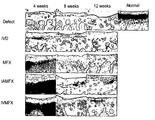
- Figure 1 shows the results of sulfated GAGs of tissue regenerated at the injury site at 4, 8, 12 weeks after surgery.
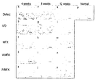
- Figure 2 shows histological observations of tissue regenerated at the site of injury 4, 8 and 12 weeks after surgery (H & E staining).
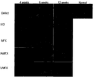
- Figure 3 shows the collagen structure of regenerated tissue at the site of injury 4, 8, 12 weeks after surgery (picro sirius-red staining).
- FIG. 4 is a quantitative graph showing International Cartilage Repair Society (ICRS) scores for histological results at 4, 8, 12 weeks after surgery.
- ICRS histology scores are systematically assessed with the following seven parameters. (1) cell morphology, (2) matrix-staining, (3) structural integrity, (4) surface regularity, (5) cartilage thickness, (6) regenerated subchondral bone, and (7) Integral with adjacent cartilage. Data are expressed as mean ⁇ SD (standard deviation). (* P ⁇ 0.05, ** p ⁇ 0.01 and *** p ⁇ 0.001 for each comparison group; ### p ⁇ 0.001 vs damage group)
- Figure 5 is a graph showing the results of the chemical analysis of the content of sulfated GAGs of regenerated tissue at the injury site at 4, 8, 12 weeks after surgery (chemical analysis-GAG content). GAG content in freshly regenerated tissue in the IVD, MFX, IAMFX and IVMFX groups compared to the injured group. Data are expressed as mean ⁇ SD (standard deviation). (* P ⁇ 0.05, ** p ⁇ 0.01 and *** p ⁇ 0.001 for each comparison group; ### p ⁇ 0.001 vs damage group)
- the present invention provides a pharmaceutical composition for treating cartilage damage regeneration comprising granulocyte macrophage-colony stimulating factor (GM-CSF) as an active ingredient.
- GM-CSF granulocyte macrophage-colony stimulating factor
- the GM-CSF is at least one selected from the group consisting of a GM-CSF full-length gene, a GM-CSF gene subdomain, a protein expressed from the genes, and a cell transformed to express the genes. It is done.
- Macrophage-colony-colony stimulating factor (GM-CSF) gene and protein information is registered in the National Center for Biotechnology Information (NCBI) (GeneID: 1437, NM_000758.3).
- composition in the group consisting of Microfracture technique (MFx), abrasion arthroplasty, drilling (drilling), bone marrow transplantation (BMT) and stem cell transplantation (stem cell transplantation) It is preferred, but not limited to, to be administered at any one of the selected procedures.
- the administration is intravenous (IV), intra-articular (IA), intra-defect, intramuscular (IM), subcutaneous or intraperitoneal; Preferably, but is not limited to.
- composition is characterized in that the treatment is administered to the implant during the bone marrow transplantation (BMT) or stem cell transplantation (stem cell transplantation) procedure.
- BMT bone marrow transplantation
- stem cell transplantation stem cell transplantation
- the pharmaceutical composition may include a pharmaceutically acceptable carrier in addition to the GM-CSF.
- pharmaceutically acceptable carriers are commonly used in pharmaceutical preparations, such as lactose and deck. Straw, Sucrose, Sorbitol, Mannitol, Starch, Acacia Rubber, Calcium Phosphate, Alginate, Gelatin, Calcium Silicate, Microcrystalline Cellulose, Polyvinylpyrrolidone, Cellulose, Water, Syrup, Methyl Cellulose, Methylhydroxybenzoate , But may include, but is not limited to, propylhydroxybenzoate, talc, magnesium stearate, mineral oil, and the like.
- the pharmaceutical composition may further include a lubricant, a humectant, a sweetener, a flavoring agent, an emulsifier, a suspending agent, a preservative, and the like as an additive.
- the pharmaceutical composition is determined in accordance with the degree of symptoms of cartilage damage, the method of administration is determined, the pharmaceutical composition of the present invention can be administered to various mammals such as rats, mice, livestock, humans. All modes of administration can be expected, for example by oral, rectal or intravenous, intramuscular, subcutaneous, intrauterine dural or intracerebroventricular injection.
- the dosage of the active ingredient in the pharmaceutical composition may vary depending on the route of administration, the degree of the disease, the age, sex, and weight of the patient, and may be administered once to several times daily.
- a 16-week-old male New Zealand white rabbit (3.5 ⁇ 0.5kg; Joong-Ang Experimental Animal Center, Seoul, Korea) was used in the present invention.
- 75 rabbits were divided into 5 groups.
- Group 1 injured group
- Group 2 MFX group
- Group 3 IVD group
- IV intravenous (IV) injection of GM-CSF only after penetrating cartilage injury.
- IVMFX group GM-CSF intravenously (IV) with MFx procedure after penetrating cartilage injuries.
- Group 5 IAMFX group injected GM-CSF intraarticularly with MFx procedure after penetrating cartilage injuries. articular; IA).
- Rabbits were sacrificed at 4, 8 and 12 weeks after surgery. Immediately after sacrifice, the rabbit's right and left knee joints were harvested. Samples were fixed in 4% formalin and successively dehydrated with increasing alcohol concentration before washing with xylene. The dehydrated sample was cut to 4 ⁇ m thick, and the tissue section was safranin-O for glycosaminoglycan (GAG) analysis and was analyzed for morphological analysis. Hematoxylin and eosin (H & E), stained with picro sirius-red for total collagen analysis.
- GAG glycosaminoglycan
- the rabbit was anesthetized. After removing, scrubbing and draping the fur of the rabbit's knee area, the knee area was opened through anterior midline longitudinal incision. Thereafter, the knee bone was dislocated laterally to completely expose the cartilage tissue.
- a 5 mm biopsy punch (Miltex, Inc., York, USA) and a specific curette in the center of the femoral knee cartilage tissue surface were used to damage the 5 mm diameter and 0.2 mm deep full-thickness cartilage. .
- the MFx technique used a mini-microfracture awl at the site of injury to make three holes with a space of 2.0-2.5 mm each in the subchondral bone. Microfracture-awls with stoppers made of steel composites were fabricated to control the depth of the MFx holes, 3 mm long and 1 mm in diameter.
- Human GM-CSF ELISA kit is an assay for the quantitative determination of human GM-CSF in serum. This assay uses specific antibodies against human GM-CSF coated on 96-well plates. Standards and samples were aliquoted into the wells, and GM-CSF present in the sample bound to the immunized antibody. After washing the sample, biotinylated anti-GM-CSF antibody was added.
- ICRS visual histology scores are systematically assessed with the following seven parameters. (1) cell morphology, (2) matrix-staining, (3) structural integrity, (4) surface regularity, (5) cartilage thickness, (6) regenerated subchondral bone, and (7) Integral with adjacent cartilage. The highest point is 18 and the lowest point is zero.
- FIG. 2 cell morphology in regenerated cartilage tissue was observed through H & E staining. Chondrocytes grew in the lacunae and well formed isogenous bones in the MFX, IAMFX and IVMFX groups. In contrast, the IVD and damage groups showed low density.
- FIG. 3 it was confirmed that picro sirius-red staining showed collagen networks of regenerated tissues at 4, 8 and 12 weeks after surgery.
- collagen networks were well formed in uniform directions in the superficial, transitional and deep layers of regenerated tissue.
- the regenerated tissue exhibited weak structural collagen fiber networks.
- IVD and injured groups regenerated tissue was significantly weakly stained with structural collagen fiber networks.
- FIG. 4 quantitative histology scores were obtained using ICRS.
- the mean ICRS score for the injured group was (2.9 ⁇ 1.22), (4.2 ⁇ 1.72), and (5.0 ⁇ 1.73), respectively, with the lowest scores compared to the other groups (p ⁇ 0.001) .
- the IVD group (10.8 ⁇ 1.47) had significantly lower scores than MFX (15.4 ⁇ 0.80), IAMFX (15.9 ⁇ 0.94) and IVMFX (16.3 ⁇ 1.19) (p ⁇ 0.001).
- the ICRS score (16.4 ⁇ 1.11) of the IVMFX group scored IAMFX (10.8 ⁇ 1.99, p ⁇ 0.001), MFX (14.1 ⁇ 1.58, p ⁇ 0.05) and IVD (11.1 ⁇ 1.30, p ⁇ 0.01). Higher than However, after 12 weeks, there was no significant difference between IVMFX (13.0 ⁇ 2.24), IAMFX (10.5 ⁇ 3.01), MFX (11.8 ⁇ 2.04) and IVD (11.6 ⁇ 1.69) (p> 0.05).
- the IVMFX group showed the highest GAG content after 4, 8 and 12 weeks of surgery.
- the IAMFX group had increased GAG content compared to the MFX, IVD and damage groups, and the MFX group had higher GAG content than the IVD and damage groups.
- the IVD group showed higher GAG content than the damage group, but significantly lower GAG content than the other groups.
- the concentration of GM-CSF was higher than MFX, which shows that GM-CSF can move through the blood circulation and can be delivered to the bone marrow.
- GM-CSF concentrations also increased in IAMFX, indicating that GM-CSF is also delivered to the bone marrow.
- GM-CSF increases the cartilage regeneration efficiency of the MFx procedure.
- GM-CSF showed significant effects in both intravenous injection and intraarticular injection.
- treatment with GM-CSF alone showed some therapeutic effect compared to the injured group.
- a therapeutic composition containing GM-CSF may be used as a therapeutic agent for cartilage regeneration with or without MFx alone.
Landscapes
- Health & Medical Sciences (AREA)
- Life Sciences & Earth Sciences (AREA)
- Animal Behavior & Ethology (AREA)
- Engineering & Computer Science (AREA)
- Chemical & Material Sciences (AREA)
- Veterinary Medicine (AREA)
- Medicinal Chemistry (AREA)
- Public Health (AREA)
- General Health & Medical Sciences (AREA)
- Pharmacology & Pharmacy (AREA)
- Bioinformatics & Cheminformatics (AREA)
- Epidemiology (AREA)
- Immunology (AREA)
- Zoology (AREA)
- Gastroenterology & Hepatology (AREA)
- Proteomics, Peptides & Aminoacids (AREA)
- Organic Chemistry (AREA)
- Nuclear Medicine, Radiotherapy & Molecular Imaging (AREA)
- General Chemical & Material Sciences (AREA)
- Chemical Kinetics & Catalysis (AREA)
- Physical Education & Sports Medicine (AREA)
- Biotechnology (AREA)
- Genetics & Genomics (AREA)
- Molecular Biology (AREA)
- Medicines That Contain Protein Lipid Enzymes And Other Medicines (AREA)
Abstract
본 발명은 과립구 대식세포-콜로니 자극 인자(granulocyte macrophage-colony stimulating factor; GM-CSF)를 유효성분으로 포함하는 연골손상 재생 치료용 약학 조성물에 관한 것으로, 보다 구체적으로 상기 GM-CSF는 GM-CSF 전장 유전자, GM-CSF 유전자 절편(subdomain), 상기 유전자들로부터 발현되는 단백질 및 상기 유전자들을 발현하도록 형질전환된 세포로 이루어진 군에서 선택된 어느 하나 이상인 것을 특징으로 한다. 또한, 상기 조성물은 골수이식술 (bone marrow transplantation; BMT) 또는 줄기세포이식술 (stem cell transplantation) 시술 시 이식물에 처리하여 투여되는 것을 특징으로 한다. 따라서 상기 이를 통해 상기 GM-CSF는 손상된 연골의 재생 치료제로 응용 가능할 것으로 보인다.
Description
본 발명은 연골손상 재생 치료용 조성물에 관한 것이다.
관절(articular) 또는 초자(hyaline) 연골은 무혈관으로 이루어졌고, 매우 평평한 표면과 점탄성체(viscoelastic)의 성질을 가진 환상(aneural) 조직이다. 관절연골의 기능은 관절에 있어서 두 뼈 사이의 원활한 상호작용을 하게 한다. 따라서, 손상을 입으면 관절 이동 유동성에 있어서 장애를 일으킨다. 연골 손상이 재생되지 않는 주요 이유 중 하나는 특이적인 무혈관성으로 인해 손상된 부위에서의 충분한 충전 세포(packed cells)의 보충이 부족하기 때문이다. 그러므로, 손상 부위로 세포들을 전달하는데 초점을 맞춰 많은 노력을 기울이고 있다.
미세골절술(Microfracture technique; MFx)은 가장 잘 알려진 연골 손상의 임상치료법이다. MFx는 골수로부터 다양한 성장 인자/사이토카인 및 여러 종류의 세포를 포함하는 혈병(blood clot)의 배출을 유도함으로써 손상된 연골 조직의 재생을 돕는다. MFx 기술은 환자에게 최소한의 침습을 야기하여 기존에 마땅한 치료법이 없기 때문에 현재 임상적으로 연골 손상에 대한 1차 치료요법으로 고려되고 있다. 하지만, MFx에 의해 재생된 조직은 주로 비구조적이고, 빈약한 물리적 성질을 갖는 섬유연골(fibrocartilages)로 이루어지는 한계가 있다. 또한, MFx 치료에 대한 과학적 기전이 완전히 밝혀지지 않았고, 다른 외과 수술에 비해 치료 효능의 장점을 주장하거나 반박하기 위한 임상적 연구도 많이 이루어지지 않은 형편이다.
한편, 한국특허출원 제10-2008-0092917호는 과립성 백혈구 대식세포-콜로니 자극 인자(GM-CSF)를 유효성분으로서 포함하는 손상된 조직의 상흔 형성 억제제에 대해 개시하고 있지만, GM-CSF의 연골손상 재생과 같은 의학적 용도를 전혀 개시하거나 인식하지 못하고 있으므로 본 발명과는 상이하다.
따라서, 본 발명자들은 세포분열(mitogenic) 반응뿐만 아니라 세포방어(cytoprotective) 및 면역-향상 활성을 가지는 과립구 대식세포-콜로니 자극 인자(granulocyte macrophage-colony stimulating factor; GM-CSF)를 사용함으로써 MFx의 치료 효과가 향상될 것이라고 예상하였다. 이전 연구에 의하면, TGF-β와 같은 사이토카인 및 성장 인자들이 MFx에 사용되었는데, 이는 줄기세포의 연골세포로의 분화 및 연골 매트릭스 분자의 생산을 유도할 수 있었다. 하지만, 세포 방어 및 면역 조절 활성을 나타낼 수 있는 사이토카인을 활용한 연구는 본 발명이 최초이다. 본 발명자들은 연골 손상된 토끼 모델에서 미세골절술 후, 연골 재생에 있어서 GM-CSF의 정맥(intravenous; IV) 또는 관절 내(intra-articular; IA) 주사의 효과에 대해 확인하고 본 발명을 완성하였다.
본 발명은 과립구 대식세포-콜로니 자극 인자(granulocyte macrophage-colony stimulating factor; GM-CSF)를 유효성분으로 포함하는 연골손상 재생 치료용 약학 조성물을 제공하고자 한다.
상기 목적을 달성하기 위하여, 본 발명은 과립구 대식세포-콜로니 자극 인자(granulocyte macrophage-colony stimulating factor; GM-CSF)를 유효성분으로 포함하는 연골손상 재생 치료용 약학 조성물을 제공한다.
본 발명의 과립구 대식세포-콜로니 자극 인자(granulocyte macrophage-colony stimulating factor; GM-CSF)를 유효성분으로 포함하는 연골손상 재생 치료용 약학 조성물에 관한 것으로서, 연골 손상된 토끼 모델에서 미세골절술 후, 연골 재생에 있어서 GM-CSF의 정맥(intravenous; IV) 또는 관절 내(intra-articular; IA) 주사의 효과에 대해 확인하였다. 이를 통해 상기 GM-CSF는 손상된 연골의 재생 치료제로 응용 가능하다.
도 1은 수술 후 4, 8, 12 주에 손상 부위에서 재생된 조직의 sulfated GAGs 조사 결과이다. (a, f, k) 손상 그룹; (b, g, l) IVD 그룹; (c, h, m) MFX 그룹; (d, i, n) IAMFX 그룹; (e, j, o) IVMFX 그룹 및 (p) 정상 연골.
도 2는 수술 후 4, 8, 12 주에 손상 부위에서 재생된 조직의 조직학적 관찰 결과이다(H&E 염색). (a, f, k) 손상 그룹; (b, g, l) IVD 그룹; (c, h, m) MFX 그룹; (d, i, n) IAMFX 그룹; (e, j, o) IVMFX 그룹 및 (p) 정상 연골.
도 3은 수술 후 4, 8, 12 주에 손상 부위에서 재생된 조직의 콜라겐 구조를 관찰한 이다(picro sirius-red 염색). (a, f, k) 손상 그룹; (b, g, l) IVD 그룹; (c, h, m) MFX 그룹; (d, i, n) IAMFX 그룹; (e, j, o) IVMFX 그룹 및 (p) 정상 연골.
도 4는 수술 후 4, 8, 12 주에서의 조직학적 결과에 대한 국제 연골 재생학회(International Cartilage Repair Society; ICRS) 점수를 나타내는 정량 그래프이다. ICRS 조직학 점수는 다음 7개의 파라미터로 체계적으로 평가된다. (1) 세포 형태, (2) 매트릭스-염색, (3) 구조적 안정성(structural integrity), (4) 표면 규칙성(surface regularity), (5) 연골의 두께, (6) 재생된 연골하골, 및 (7) 인접 연골과의 통합성. 데이터는 평균±SD(표준편차)로서 표시하였다. (각 비교군에 대해 *p<0.05, **p<0.01 및 ***p<0.001; ###p<0.001 v.s 손상 그룹)
도 5는 수술 후 4, 8, 12 주에 손상 부위에서 재생된 조직의 sulfated GAGs의 함량을 화학적 방법으로 분석한 결과를 나타낸 그래프이다(화학적 분석-GAG 함량). 손상 그룹과의 비교한 IVD, MFX, IAMFX 및 IVMFX 그룹에서 새롭게 재생된 조직에서의 GAG 함량을 나타낸다. 데이터는 평균±SD(표준편차)로서 표시하였다. (각 비교군에 대해 *p<0.05, **p<0.01 및 ***p<0.001; ###p<0.001 v.s 손상 그룹)
도 6은 투여 후 GM-CSF 이동성을 나타낸다. MFX, IAMFX 및 IVMFX 처리 후에 수확된 골수에서 GM-CSF의 농도를 측정하기 위해서 ELISA 방법이 사용되었다. 데이터는 평균±SD(표준편차)로서 표시하였다. (각 비교군에 대해 *p<0.05, **p<0.01 및 ***p<0.001; ###p<0.001 v.s 손상 그룹)
본 발명은 과립구 대식세포-콜로니 자극 인자(granulocyte macrophage-colony stimulating factor; GM-CSF)를 유효성분으로 포함하는 연골손상 재생 치료용 약학 조성물을 제공한다. 상세하게는 상기 GM-CSF는 GM-CSF 전장 유전자, GM-CSF 유전자 절편(subdomain), 상기 유전자들로부터 발현되는 단백질 및 상기 유전자들을 발현하도록 형질전환된 세포로 이루어진 군에서 선택된 어느 하나 이상인 것을 특징으로 한다.
대식세포-콜로니 자극 인자(granulocyte macrophage-colony stimulating factor; GM-CSF) 유전자 및 단백질 정보는 NCBI (National Center for Biotechnology Information) 에 등록되어 있다 (GeneID: 1437 , NM_000758.3).
또한, 상기 조성물은 미세골절술(Microfracture technique; MFx), 소파관절성형술 (abrasion arthroplasty), 천공술 (drilling), 골수이식술 (bone marrow transplantation; BMT) 및 줄기세포이식술 (stem cell transplantation)로 이루어진 군에서 선택된 어느 하나의 시술시 투여되는 것이 바람직하지만, 이에 한정되지는 않는다.
상세하게는, 상기 투여는 정맥 (intravenous; IV), 관절강 내 (intra-articular; IA), 손상 연골 (intra-defect) 내, 근육 내 (intramuscular; IM), 피하 (subcutaneous) 또는 복강 (intraperitoneal; IP)에 투여되는 것이 바람직하지만, 이에 한정되지는 않는다.
또한, 상기 조성물은 골수이식술 (bone marrow transplantation; BMT) 또는 줄기세포이식술 (stem cell transplantation) 시술 시 이식물에 처리하여 투여되는 것을 특징으로 한다.
본 발명의 약학 조성물인 경우, 상기 약학 조성물은 상기 GM-CSF 이외에 약제학적으로 허용되는 담체를 포함할 수 있는데, 이러한 약제학적으로 허용되는 담체는 약품 제제 시에 통상적으로 이용되는 것으로서, 락토스, 덱스트로스, 수크로스, 솔비톨, 만니톨, 전분, 아카시아 고무, 인산 칼슘, 알기네이트, 젤라틴, 규산 칼슘, 미세결정성 셀룰로스, 폴리비닐피롤리돈, 셀룰로스, 물, 시럽, 메틸 셀룰로스, 메틸히드록시벤조에이트, 프로필히드록시벤조에이트, 활석, 스테아르산 마그네슘, 미네랄 오일 등을 포함할 수 있으나, 이에 한정되는 것은 아니다. 또한, 상기 약학적 조성물은 첨가제로서 윤활제, 습윤제, 감미제, 향미제, 유화제, 현탁제, 보존제 등을 추가로 포함할 수 있다.
상기 약학적 조성물은 연골 손상의 증상 정도에 따라 투여 방법이 결정되는데, 본 발명의 약학조성물은 쥐, 생쥐, 가축, 인간 등의 포유동물에 다양한 경로로 투여될 수 있다. 투여의 모든 방식은 예상될 수 있는데, 예를 들면, 경구, 직장 또는 정맥, 근육, 피하, 자궁 내 경막 또는 뇌혈관 내 (intracerebroventricular) 주사에 의해 투여될 수 있다. 또한, 상기 약학적 조성물 중 유효성분의 투여량은 투여경로, 질병의 정도, 환자의 나이, 성별, 체중 등에 따라 달라질 수 있으며, 일일 1회 내지 수회 투여할 수 있다.
이하, 하기 실시예를 통해 본 발명을 보다 상세하게 설명한다. 다만, 이러한 실시예에 의해 본 발명이 한정되는 것은 아니다.
< 실시예 >
1. 동물실험
16주령 수컷 뉴질랜드 흰색 토끼(3.5±0.5kg; Joong-Ang Experimental Animal Center, Seoul, Korea)가 본 발명에 사용되었다. 75마리 토끼는 5 그룹으로 나뉘었다. 1 그룹(손상 그룹)은 연골하 골수 공간이 열리지 않은 미처리한 전층(full-thickness) 연골 손상을 가진 음성 대조군이었다. 2 그룹(MFX 그룹)은 MFx 치료한 그룹으로서, 전층 연골 손상 후, 연골하골(subchondral bone)에 MFx를 시술하여 혈괴(blood clot) 형성을 유도하였다. 3 그룹(IVD 그룹)은 전층 연골 손상 후 GM-CSF만 정맥(intravenous; IV) 주사하여 처리하였다. 4 그룹(IVMFX 그룹)은 전층 연골 손상 후 MFx 시술과 함께 GM-CSF를 정맥(IV) 주사하였으며, 5 그룹(IAMFX 그룹)은 전층 연골 손상 후 MFx 시술과 함께 GM-CSF를 관절강 내(intra-articular; IA)로 주사하였다.
수술 후 4, 8 및 12주에 토끼들은 희생되었다. 희생 후 즉시, 토끼의 오른쪽 및 왼쪽 무릎 관절을 수확하였다. 샘플은 4% 포르말린에서 고정하였고, 자일린으로 세척하기 전에 알코올 농도를 높여가며 연속적으로 탈수시켰다. 탈수된 샘플은 4㎛ 두께로 절단되었으며, 절단된 시료(tissue section)는 글리코스아미노글리칸(glycosaminoglycan; GAG) 분석을 위해 사프라닌-O(Safranin-O)로, 형태학적 분석을 위해 헤마톡실린 및 에오신(hematoxylin and eosin; H&E)으로, 총 콜라겐 분석을 위해 picro sirius-red로 염색되었다.
수술 과정은 zoletil 50 (50 mg/ml; Virbac laboratories, Carros, France) 및 xylazine hydrochloride (Rompun; Bayer, Ansan, Korea) (Zoletil: Rompun = 1: 2)의 혼합액(0.2 ml/kg 몸무게)을 이용하여 토끼를 마취시킨 상태에서 수행되었다. 토끼의 무릎 부위의 털을 제거하고 소독(scrubbing) 및 방포(draping)한 후에, 상기 무릎 부위를 전방 중심선 종 절개(anterior midline longitudinal incision)를 통해 오픈하였다. 그 후, 무릎뼈를 측면으로 탈구하여 연골 조직을 완전히 노출하였다.
2. 미세골절술(Microfracture technique; MFx)
대퇴골 무릎연골조직 표면의 중앙에 5 mm 생검 펀치(Miltex, Inc., York, USA) 및 특이 큐렛(curette)을 이용하여, 5 mm 지름 및 0.2 mm 깊이의 전층(full-thickness) 연골을 손상시켰다. MFx 기술은 손상 부위에 미니-마이크로골절 송곳(mini-microfracture awl)을 이용하여 연골하골(subchondral bone)에 각 2.0-2.5 mm의 공간을 두고 3개의 구멍을 만들었다. 강철 합성물로 만들어진 스토퍼(stopper)를 가진 마이크로골절-송곳(microfracture-awls)이 3 mm 길이 및 1 mm 지름을 가진 MFx 구멍의 깊이를 조절하기 위해서 제작되었다.
3. GM-CSF 측정
투여 후에, GM-CSF가 골수 내로 전달되었는지 조사하기 위해 MFx에 의해 골수에서 유출된 혈병(blood clot) 내의 GM-CSF 농도를 효소-결합 면역흡착법(enzyme-linked immunosorbent assay; ELISA)을 이용하여 측정하였다. 인간 GM-CSF ELISA kit은 혈청에서 인간 GM-CSF의 정량적 측정을 위한 분석법이다. 이 분석법은 96-웰 플레이트 상에 코팅된 인간 GM-CSF에 대한 특이적 항체가 사용된다. 표준물질 및 샘플은 웰에 분주되었고, 샘플에 존재하는 GM-CSF는 면역화된 항체에 결합된다. 샘플을 씻어낸 후, 비오틴화된 항-GM-CSF 항체가 첨가되었다. 미결합 비오틴화된 항체를 제거하기 위해 세척한 후, HRP-접합 스트렙타비딘을 웰에 첨가하였고, 마지막으로 TMB 기질 용액을 웰에 첨가하였다. 샘플에 존재하는 GM-CSF와 결합하는 항체의 양에 따라 색깔이 노란색으로 변했으며, 색깔의 세기를 450 nm에서 측정하여 정량하였다.
4. 재생 조직에 대한 정량 분석
새로운 재생 조직에 대한 정량분석은 국제 연골재생학회(International Cartilage Repair Society; ICRS)에서 제시하는 방법에 따라 점수로 평가하여 수행하였다. ICRS 시각 조직학 점수는 다음 7개의 파라미터로 체계적으로 평가된다. (1) 세포 형태, (2) 매트릭스-염색, (3) 구조적 안정성(structural integrity), (4) 표면 규칙성(surface regularity), (5) 연골의 두께, (6) 재생된 연골하골, 및 (7) 인접 연골과의 통합성. 최고점은 18이고, 최저점은 0이다.
각 그룹에 있어서 수술 후 4, 8 및 12 주에, 샘플 및 정상 연골을 추출하였고, 동결건조기(IlShin Lab Co., Ltd, Gyeonggi-do, Korea)를 이용하여 습윤 및 건조 중량에 따른 수분 함량을 측정하였다. 각 샘플은 파파인(papain)-절단 용액으로 절단되었다. 전체 DNA 함량은 Hoechst 33258 dye을 이용하여 측정하였다. 총 GAG 함량은 분광광도계를 이용한 1, 9-디메틸메틸렌 블루(1, 9-dimethylmethylene blue; DMB)를 이용한 색도측정법을 통해 측정하였으며, 표준시약으로는 상어 콘드로이틴 설페이트(chondroitin sulfate)을 이용하였다. 또한, 총 콜라겐 함량은 콜라겐 분석 키트를 사용하여 측정하였다(Biocolor, County Antrim, United Kingdom).
분석에 있어서, 통계적 유의성은 Tukey-Kramer test: 처리군과의 다중 비교(* p<0.05, ** p<0.01 및 ***p<0.001)와 함께 일원분산분석(one-way analysis of variance; ANOVA)으로 측정되었다. 데이터는 평균±SD(표준편차)로서 표시하였다.
5. 결과
도 1을 보면, GAG 분석을 위한 사프라닌-O 염색에 있어서, IVMFX 와 IAMFX 그룹에서는 4주째 손상된 연골조직이 초자연골(hyaline cartilage)과 유사한 조직으로 채워져 있었고, 12주째 연골의 구조가 잘 구성되어 있었다. 반면 MFX 그룹에서는 재생된 연골 조직이 가늘고 사프라닌-O 염색이 약하게 되었다. IVD 그룹에서는 재생 조직이 상당히 가늘었으며, 사프라닌-O에 의해 더 약하게 염색되었다. 손상 그룹에서는 오직 손상된 부위 중 극히 일부만 사프라닌-O에 의해 염색되어 가장 나쁜 결과를 보였다.
도 2에서는 재생된 연골 조직에 있어서의 세포 형태에 대해 H&E 염색을 통해 관찰하였다. 연골세포는 골소강(lacunae)에서 자랐으며, MFX, IAMFX 및 IVMFX 그룹에서 동형(isogenous) 골소강이 잘 형성되었다. 반대로, IVD 및 손상 그룹에서는 낮은 밀도로 나타났다.
도 3에 의하면, picro sirius-red 염색을 통해 재생 조직의 콜라겐 네트워크가 수술 후 4, 8 및 12주에 나타난다는 것을 확인하였다. IVMFX 및 IAMFX 그룹에 있어서, 재생 조직의 표면층(superficial layer), 중간층(transitional layer) 및 심층(deep layer) 부위에서 콜라겐 네트워크가 균일한 방향으로 잘 형성되었다. MFX 그룹에서는 재생 조직이 약한 구조 콜라겐 섬유 네트워크를 나타냈다. IVD 및 손상 그룹에서는 재생 조직이 구조 콜라겐 섬유 네트워크로 상당히 약하게 염색되었다.
도 4에서는 ICRS를 이용하여 정량적 조직학 점수를 얻었다. 수술 후 4, 8 및 12주에, 손상 그룹의 ICRS 점수 평균은 각각 (2.9±1.22), (4.2±1.72) 및 (5.0±1.73)으로 다른 그룹들에 비해 점수가 가장 낮았다(p < 0.001). 수술 4주 후에는, IVD 그룹 (10.8±1.47)은 MFX (15.4±0.80), IAMFX (15.9±0.94) 및 IVMFX (16.3±1.19) 보다 크게 낮은 점수를 보였다(p < 0.001). 수술 8주 후에는, IVMFX 그룹의 ICRS 점수 (16.4±1.11)가 IAMFX (10.8±1.99, p < 0.001), MFX (14.1±1.58, p < 0.05) 및 IVD (11.1±1.30, p < 0.01) 점수보다 더 높았다. 그러나, 수술 12주 후에는 IVMFX (13.0±2.24), IAMFX (10.5±3.01), MFX (11.8±2.04) 및 IVD (11.6±1.69) 사이에 유의적 차이성은 없었다(p > 0.05).
도 5에서, GAG 함량에 대한 정량 분석에서, IVMFX 그룹은 수술 4, 8 및 12주 후에 가장 높은 GAG 함량을 나타냈다. IAMFX 그룹은 MFX, IVD 및 손상 그룹과 비교하여 GAG 함량이 증가되었으며, MFX 그룹은 IVD 및 손상 그룹보다는 GAG 함량이 높았다. IVD 그룹은 손상 그룹보다는 높은 GAG 함량을 보였으나, 다른 그룹에 비해 현저히 낮은 GAG 함량을 보였다.
도 6에서, MFx 후 유출된 골수 내(100ul)에 존재하는 GM-CSF 농도를 측정한 결과, MFX (585.11±189.91 pg) 그룹과 비교하여 IAMFX (9760.44±298.18 pg) 및 IVMFX (6176.22±1057.93 pg)에서 높게 발현되는 것으로 나타났다.
IVMFX 그룹에서, GM-CSF의 농도가 MFX 보다 높았는데, 이러한 결과는 GM-CSF가 혈액 순환을 통해 이동할 수 있고, 골수에 전달될 수 있다는 것을 보여준다. 또한 GM-CSF 농도는 IAMFX에서도 증가하였는데, 이 역시 GM-CSF가 골수로 전달된다는 것을 나타낸다.
상기와 같은 결과는 GM-CSF 투여가 MFx 시술의 연골 재생효능을 높인다는 것을 나타낸다. GM-CSF는 정맥 주사한 경우와 관절강으로 주사한 경우 모두 유의한 효과를 보였다. 또한, GM-CSF를 단독으로 처리한 경우도 손상 그룹에 비해 어느 정도의 치료효과를 나타내었다. 따라서 GM-CSF를 함유한 치료용 조성물은 MFx와 함께 또는 단독으로 연골재생 치료제로 사용될 수 있을 것이다.
Claims (5)
- 과립구 대식세포-콜로니 자극 인자(granulocyte macrophage-colony stimulating factor; GM-CSF)를 유효성분으로 포함하는 연골손상 재생 치료용 약학 조성물.
- 제 1항에 있어서, 상기 GM-CSF는 GM-CSF 전장 유전자, GM-CSF 유전자 절편(subdomain), 상기 유전자들로부터 발현되는 단백질 및 상기 유전자들을 발현하도록 형질전환된 세포로 이루어진 군에서 선택된 어느 하나 이상인 것을 특징으로 하는 연골손상 재생 치료용 약학 조성물.
- 제 1항 또는 제 2항에 있어서, 상기 조성물은 미세골절술(Microfracture technique; MFx), 소파관절성형술 (abrasion arthroplasty), 천공술 (drilling), 골수이식술 (bone marrow transplantation; BMT) 및 줄기세포이식술 (stem cell transplantation)로 이루어진 군에서 선택된 어느 하나의 시술시 투여되는 것을 특징으로 하는 연골손상 재생 치료용 약학 조성물.
- 제 3항에 있어서, 상기 투여는 정맥 (intravenous; IV), 관절강 내 (intra-articular; IA), 손상 연골 (intra-defect) 내, 근육 내 (intramuscular; IM), 피하 (subcutaneous) 또는 복강 (intraperitoneal; IP)에 투여되는 것을 특징으로 하는 연골손상 재생 치료용 약학 조성물.
- 제 1항 또는 제 2항에 있어서, 상기 조성물은 골수이식술 (bone marrow transplantation; BMT) 또는 줄기세포이식술 (stem cell transplantation) 시술 시 이식물에 처리하여 투여되는 것을 특징으로 하는 연골손상 재생 치료용 약학 조성물.
Applications Claiming Priority (2)
| Application Number | Priority Date | Filing Date | Title |
|---|---|---|---|
| KR1020130066575A KR20140144508A (ko) | 2013-06-11 | 2013-06-11 | 연골손상 재생 치료용 조성물 |
| KR10-2013-0066575 | 2013-06-11 |
Publications (1)
| Publication Number | Publication Date |
|---|---|
| WO2014200237A1 true WO2014200237A1 (ko) | 2014-12-18 |
Family
ID=52022472
Family Applications (1)
| Application Number | Title | Priority Date | Filing Date |
|---|---|---|---|
| PCT/KR2014/005063 Ceased WO2014200237A1 (ko) | 2013-06-11 | 2014-06-10 | 연골손상 재생 치료용 조성물 |
Country Status (2)
| Country | Link |
|---|---|
| KR (1) | KR20140144508A (ko) |
| WO (1) | WO2014200237A1 (ko) |
Citations (2)
| Publication number | Priority date | Publication date | Assignee | Title |
|---|---|---|---|---|
| WO2011060554A1 (en) * | 2009-11-19 | 2011-05-26 | Corporation De L'ecole Polytechnique De Montreal | Presolidified composition and method for in situ delivery of broad molecular weight range of chitosan implants with or without therapeutics for regenerative medicine and cartilage repair applications |
| KR101270161B1 (ko) * | 2010-04-26 | 2013-05-31 | 한국과학기술연구원 | 코어-쉘 구조의 성장인자 전달체, 그의 제조방법 및 세포 분화 또는 증식을 위한 그의 용도 |
-
2013
- 2013-06-11 KR KR1020130066575A patent/KR20140144508A/ko not_active Ceased
-
2014
- 2014-06-10 WO PCT/KR2014/005063 patent/WO2014200237A1/ko not_active Ceased
Patent Citations (2)
| Publication number | Priority date | Publication date | Assignee | Title |
|---|---|---|---|---|
| WO2011060554A1 (en) * | 2009-11-19 | 2011-05-26 | Corporation De L'ecole Polytechnique De Montreal | Presolidified composition and method for in situ delivery of broad molecular weight range of chitosan implants with or without therapeutics for regenerative medicine and cartilage repair applications |
| KR101270161B1 (ko) * | 2010-04-26 | 2013-05-31 | 한국과학기술연구원 | 코어-쉘 구조의 성장인자 전달체, 그의 제조방법 및 세포 분화 또는 증식을 위한 그의 용도 |
Non-Patent Citations (2)
| Title |
|---|
| A. MARMOTTI ET AL.: "Human cartilage fragments in a composite scaffold for single-stage cartilage repair: an in vitro study of the chondrocyte migration and the influence of TGF-b1 and G-CSF", KNEE SURG SPORTS TRAUMATOL ARTHROSC, vol. 21, 10 November 2012 (2012-11-10), pages 1819 - 1833 * |
| QUINTERO M. ET AL.: "Granulocyte-macrophage colony stimulating factor activates proteoglycan, type II collagen, and cAMP production by rat articular chondrocytes through specific binding sites.", J. RHEUMATOL., vol. 28, no. 9, September 2001 (2001-09-01), pages 2075 - 2084 * |
Also Published As
| Publication number | Publication date |
|---|---|
| KR20140144508A (ko) | 2014-12-19 |
Similar Documents
| Publication | Publication Date | Title |
|---|---|---|
| Song et al. | Pro-chondrogenic and immunomodulatory melatonin-loaded electrospun membranes for tendon-to-bone healing | |
| KR100237731B1 (ko) | 음이온성 중합물질에 의한 세포침입과 섬유층을 저해시키는 방법과 성분물 | |
| CN101146547A (zh) | 肌肉生长抑制素(gdf-8)拮抗剂用于增进创伤愈合和防止纤维变性疾病的用途 | |
| KR20090113849A (ko) | 연골 질환 치료용 조성물 | |
| CN87101250A (zh) | 伤口治疗 | |
| WO2005030133A2 (en) | Treatment with agonists of toll-like receptors | |
| CA2900971A1 (en) | Serum fraction of platelet-rich fibrin | |
| JP2022550670A (ja) | 炎症性疾患のためのddx17及びnlrc4標的化 | |
| ES2634505T3 (es) | Tratamiento de PRG4 para la cistitis intersticial | |
| US20160067309A1 (en) | Composition for regeneration of cartilage | |
| BR112016018681B1 (pt) | sistema de distribuição de substâncias que compreende pelo menos um material polimérico formador de uma matriz e um fármaco anabólico, processo de produção do mesmo e artigo de manufatura | |
| CN111249529B (zh) | 一种用于软骨修复的仿生多层胶原支架及其制备方法 | |
| Jiang et al. | CXCL1-CXCR2 axis mediates inflammatory response after sciatic nerve injury by regulating macrophage infiltration | |
| KR20190127856A (ko) | 반월상연골 복원을 촉진시키기 위해 기능화 된 스캐폴드 (functionalized scaffold to promote meniscus repair) | |
| JP2021533820A (ja) | 細胞増殖と組織修復の促進方法及び組成物 | |
| Huh et al. | The effect of platelet rich plasma combined with microfracture for the treatment of chondral defect in a rabbit knee | |
| WO2011033306A1 (en) | Inhibition of tendon adhesions | |
| WO2014200237A1 (ko) | 연골손상 재생 치료용 조성물 | |
| KR20160109561A (ko) | 관절염 예방 또는 치료용 약학적 조성물 | |
| KR20180008137A (ko) | Il-8 및 골수농축액의 조합을 이용한 연골 재생 | |
| Zhang et al. | Effect of olfactory ensheathing cells combined with chitosan on inhibition of P2× 4 receptor over-expression-mediated neuropathic pain | |
| TW201827061A (zh) | 關節軟骨修復用組合物之注射劑型 | |
| Hörner et al. | Glial cell line derived neurotrophic factor (GDNF) induces mucosal healing via intestinal stem cell niche activation | |
| Maruyama et al. | Strongyloides venezuelensis: binding of orally secreted adhesion substances to sulfated carbohydrates | |
| Nakata et al. | Skeletal muscle injury treatment using the Silk Elastin® injection in a rat model |
Legal Events
| Date | Code | Title | Description |
|---|---|---|---|
| 121 | Ep: the epo has been informed by wipo that ep was designated in this application |
Ref document number: 14810805 Country of ref document: EP Kind code of ref document: A1 |
|
| NENP | Non-entry into the national phase |
Ref country code: DE |
|
| 122 | Ep: pct application non-entry in european phase |
Ref document number: 14810805 Country of ref document: EP Kind code of ref document: A1 |