WO2014196204A1 - Culture vessel and culture method - Google Patents
Culture vessel and culture method Download PDFInfo
- Publication number
- WO2014196204A1 WO2014196204A1 PCT/JP2014/002993 JP2014002993W WO2014196204A1 WO 2014196204 A1 WO2014196204 A1 WO 2014196204A1 JP 2014002993 W JP2014002993 W JP 2014002993W WO 2014196204 A1 WO2014196204 A1 WO 2014196204A1
- Authority
- WO
- WIPO (PCT)
- Prior art keywords
- culture
- medium
- cells
- spheroids
- recess
- Prior art date
Links
Images


Classifications
-
- C—CHEMISTRY; METALLURGY
- C12—BIOCHEMISTRY; BEER; SPIRITS; WINE; VINEGAR; MICROBIOLOGY; ENZYMOLOGY; MUTATION OR GENETIC ENGINEERING
- C12M—APPARATUS FOR ENZYMOLOGY OR MICROBIOLOGY; APPARATUS FOR CULTURING MICROORGANISMS FOR PRODUCING BIOMASS, FOR GROWING CELLS OR FOR OBTAINING FERMENTATION OR METABOLIC PRODUCTS, i.e. BIOREACTORS OR FERMENTERS
- C12M23/00—Constructional details, e.g. recesses, hinges
- C12M23/02—Form or structure of the vessel
- C12M23/08—Flask, bottle or test tube
-
- C—CHEMISTRY; METALLURGY
- C12—BIOCHEMISTRY; BEER; SPIRITS; WINE; VINEGAR; MICROBIOLOGY; ENZYMOLOGY; MUTATION OR GENETIC ENGINEERING
- C12M—APPARATUS FOR ENZYMOLOGY OR MICROBIOLOGY; APPARATUS FOR CULTURING MICROORGANISMS FOR PRODUCING BIOMASS, FOR GROWING CELLS OR FOR OBTAINING FERMENTATION OR METABOLIC PRODUCTS, i.e. BIOREACTORS OR FERMENTERS
- C12M23/00—Constructional details, e.g. recesses, hinges
- C12M23/02—Form or structure of the vessel
- C12M23/12—Well or multiwell plates
Definitions
- the present invention relates to cell culture and recovery thereof.
- spheroid culture is an excellent method that can maintain cell interaction, and is applied to various cells such as islet cells, hepatocytes, stem cells, and cancer cells.
- indices the diameter and volume of spheroids are used as indices.
- size of a spheroid nonpatent literature 2, 3.
- a technique for controlling the size of spheroids has been attracting attention.
- the specific functions of cells can be reproduced in this way, it is expected to be used in fields such as artificial organs and bioreactors. In such applications, techniques for producing and collecting spheroids in large quantities are important.
- Patent Document 1 discloses a method for controlling the size of a spheroid formed by changing the number of cells to be seeded in 96WP having a U-shaped bottom provided with a hydrophilic film. is there.
- the number of spheroids per culture area is small, and it is difficult to produce a large amount of spheroids.
- Patent Documents 2-4 there is a method of forming spheroids in a micro space disclosed in Patent Documents 2-4.
- JP-A-8-131153 JP 2010-88347 A International Publication No. 2012/036011 International Publication No. 2013/042360
- Patent Document 1 has a very low culture efficiency and is a bottleneck for mass culture.
- the spheroid formation efficiency per unit area is high in the culture methods of Patent Documents 2 and 3, there is a possibility that the spheroids are detached from the space when the medium is changed. Therefore, care must be taken when changing the medium.
- Patent Document 4 a method of bonding a part of the spheroids in the micro space has been studied (Patent Document 4).
- Patent Document 4 since the adhesiveness of cells varies from cell to cell, it is necessary to study the surface treatment method for each cell to be used, which puts it to practical use.
- the present invention has been made in view of such circumstances, and enables medium exchange and cell recovery to be easily performed in order to produce spheroids of uniform size with high efficiency or high efficiency and in large quantities. It is an object of the present invention to design a micro space structure that can be implemented, and to provide a culture vessel having the designed micro space structure and a culture method using the same.
- One aspect of the culture vessel according to one embodiment of the present invention is such that a plurality of dents composed of a bottom and an opening are arranged.
- the bottom has a shape of either a hemisphere or a truncated cone, and the opening is configured by a wall having a taper angle of 1 to 20 degrees surrounding the boundary with the bottom to the end of the recess. Is done.
- the equivalent diameter of the boundary is not less than 50 ⁇ m and not more than 2.5 mm
- the depth from the bottom of the bottom to the end is not less than 0.6 times and not more than 3 times the equivalent diameter
- the constituting wall forms a surface continuous with the bottom, and the inclination of the continuous surface changes at the boundary.
- the shape of the end is preferably a hemispherical shape, a trapezoidal shape, or an inverted triangle, and a space between two adjacent recesses is flat, and the two recesses are The distance is preferably 5 ⁇ m to 50 ⁇ m.
- the culture vessel may be an acrylic resin, polylactic acid, polyglycolic acid, styrene resin, acrylic / styrene copolymer resin, polycarbonate resin, polyester resin, polyvinyl alcohol resin. It is preferably a resin molded product made of one or a combination of ethylene / vinyl alcohol copolymer resin, thermoplastic elastomer, vinyl chloride resin, and silicon resin.
- a functional group is formed on the recess by a surface modification treatment method comprising plasma treatment, glass coating, corona discharge, UV ozone treatment, or a combination thereof, so that the water contact angle is 45 degrees or less. It is preferred that It is preferable that a hydrophilic polymer chain that inhibits cell adhesion is immobilized in the recess. It is preferable that a phospholipid or a phospholipid / polymer complex is immobilized in the recess.
- a functional group is formed on the recess by a surface modification treatment method comprising any one of plasma treatment, glass coating, corona discharge, UV ozone treatment, or a combination thereof, so that the water contact angle is 45 degrees or less.
- the hydrophilic polymer chain that inhibits cell adhesion and the cell non-adherent surface on which any one polymer of phospholipid or phospholipid / polymer complex is immobilized.
- the hydrophilic polymer chain is polyhydroxyethyl methacrylate, and it is more preferable that the average molecular weight of the polyhydroxyethyl methacrylate is 100,000 or more.
- One aspect of the culture method according to one embodiment of the present invention uses any of the culture vessels described above.
- the total number of cells is equal to or greater than the number of the recesses (N) of the culture container, and the volume of the space (V1) formed by the recesses is divided by the volume of cells to be seeded (V2).
- the number of cells equal to or less than the number of depressions (N) is dispersed in the medium, and the medium is added to the culture vessel.
- the culture method it is preferable to form one spheroid per one space formed by the recess, and to grow (proliferate) the spheroid by forming a spheroid in the space. It is more preferable. In order to induce differentiation, it is preferable to induce in a state where spheroids are formed in the space. It is preferable that 60% or more of the total number of spheroids formed in the culture vessel has a diameter within a range of plus or minus 5% of the average spheroid diameter. It is preferable to collect the cells in the dent by stirring the medium, and stirring the medium is performed by shaking the culture vessel and stirring the medium, and sucking and discharging the medium.
- the method is a method of stirring a medium, stirring a medium by installing a stirring blade in the culture container, stirring a medium by adding a stirring bar to the culture container, or a combination thereof.
- the medium is replaced at least once, and the ratio of the medium to be replaced is 20% or more.
- Another aspect of the culture method according to one embodiment of the present invention uses any of the culture vessels described above. In the culture method, the cells are seeded, cultured, medium exchanged, and recovered by performing the following steps.
- a microspace structure capable of producing a spheroid having a uniform size with high efficiency and a large amount and easily performing medium replacement and recovery is designed. It is possible to provide a culture container having the above and a culture method using the same.
- FIG. 1 It is a figure which shows an example of the culture container of one Embodiment. It is sectional drawing which shows the example of a shape which looked at the dent of Embodiment 1 from the side. It is a figure which shows the example of a shape which looked at the dent of Embodiment 1 from the top. It is a figure which shows the example of a shape of the dent using a part of spherical shape of Embodiment 2. It is a figure which shows the other example of a shape of the dent using a spherical part of Embodiment 2. It is a figure which shows the example of a shape of the dent using the truncated cone of Embodiment 2.
- FIG. 1 It is a figure which shows an example of the culture container of one Embodiment. It is sectional drawing which shows the example of a shape which looked at the dent of Embodiment 1 from the side. It is a figure which shows the example of a shape which looked at the dent of Em
- FIG. 10 is a diagram illustrating a shape example of an opening according to a third embodiment. It is a figure which shows the other example of a shape of the opening part of Embodiment 3. It is a figure which shows the structural example of the culture container of Embodiment 4. FIG. It is a figure which shows the structural example of the other culture container of Embodiment 4. FIG. It is a figure which shows the structural example of the further another culture container of Embodiment 4. FIG. It is a figure which shows the residual rate of the spheroid at the time of culture medium replacement
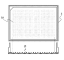
- FIG. 1 is a diagram illustrating an example of a culture container according to an embodiment.
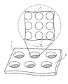
- FIG. 1 shows a part of a culture plate 3 having a plurality of culture vessels 1.
- the culture container 1 is provided with a plurality of recesses 10.
- the plurality of dents 10 are preferably arranged regularly from the viewpoint of manufacturing the culture vessel 1 and the efficiency of cell culture.
- the culture container 1 corresponds to one well of a well plate having a plurality of wells, for example.
- a well plate is an experimental / inspection instrument composed of a flat plate with a number of indentations (holes or wells), and each well is used as a test tube or petri dish.
- the number of wells includes, for example, 6, 24, 96, 384, and others.
- the bottom of the well is flat, round, or a combination of many elongated microtubes (deep well plate).
- the dent 10 forms a micro space, which is a minute space for culturing cells, it can also be called a micro container.
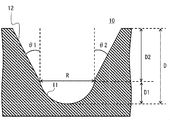
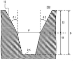
- FIG. 2 and 3 are diagrams illustrating examples of the shape of the recess according to the first embodiment.
- FIG. 2 shows a cross-sectional view when one recess 10 is viewed from the side
- FIG. 3 shows a view when one recess 10 is viewed from above.
- the dent 10 shown in FIG. 3 is a detailed configuration example of the dent 10 in the upper stage of FIG.
- Each recess 10 includes a bottom 11 and an opening 12.
- the bottom portion 11 is a portion that becomes the bottom of the culture vessel 1
- the opening portion 12 is a portion that is disposed on the top of the bottom portion 11.
- a portion where the bottom 11 and the opening 12 are in contact is referred to as a boundary.
- the length indicated by the arrow R corresponds to the boundary position.
- the position of the boundary is indicated by a two-dot broken line.
- the bottom part 11 and the opening part 12 are comprised by the continuous surface, and are manufactured as integral.
- the equivalent diameter R is a diameter of an inscribed circle inscribed in the bottom 11 of the recess 10. Here, it refers to the diameter of an inscribed circle inscribed at the boundary between the bottom 11 and the opening 12. More specifically, the equivalent diameter R refers to the diameter of an inscribed circle in the shape of a surface perpendicular to the direction of the height H of the recess 10 at the boundary.
- the depth D is the length from the inner bottom of the bottom 11 to the upper end of the recess 10. The upper end of the recess 10 is the same as the end (upper end) of the opening 12. The depth D is the depth of the space formed by the recess 10.
- FIG. 2 shows the depth D1 of the bottom 11 and the depth D2 of the opening 12 in addition to the depth D of the recess 10.
- the bottom 11 forms a space for culturing cells (first space).
- the bottom 11 has, for example, a hemispherical shape.
- a shape in which a spherical shape having an equivalent diameter R as a diameter is halved can be used.
- the shape of the bottom 11 is not limited to a hemisphere.
- Other specific examples will be described in the second embodiment.
- the opening 12 forms a space (second space) that works to assist cell culture and recovery.
- the opening 12 is configured by a wall having a taper angle of 1 degree or more and 20 degrees or less that surrounds the boundary from the bottom part 11 to the end part (tip) of the recess 10.
- the taper angle of the wall constituting the opening 12 is preferably 5 degrees or more and 15 degrees or less, and more preferably 10 degrees. The reason is that if the taper angle is too small, the cells cannot be transferred from the dent to the medium when collected, and conversely if too large, the cells are detached during the medium exchange.
- the taper angles are indicated by the symbols ⁇ 1 and ⁇ 2.
- the taper angles ⁇ 1 and ⁇ 2 are substantially the same.
- the boundary between the bottom 11 and the opening 12 is formed such that the equivalent diameter R is 50 ⁇ m or more and 1 mm or less.
- the equivalent diameter is preferably 50 ⁇ m or more and 500 ⁇ m or less, more preferably 100 ⁇ m or more and 500 ⁇ m or less.
- the equivalent diameter R is preferably 400 ⁇ m or more and less than 2 mm.
- the reason is that, as described above, at 300 ⁇ m, it can be considered that no nutrients reach the center and no necrosis occurs. Therefore, in order to obtain a spheroid having a diameter of 300 ⁇ m or more, it must be 400 ⁇ m or more.
- the depth D from the bottom to the end of the bottom is formed to be not less than 0.6 times and not more than 3 times the equivalent diameter R.
- the depth D is preferably an equivalent diameter R 0.7 times or more and 1.2 times or less, and more preferably 0, 8 to 1 times.
- the culture container 1 is flat between two adjacent dents 10.
- the distance between the two recesses 10 is preferably in the range of 5 ⁇ m to 50 ⁇ m.
- the distance between the two recesses 10 is preferably in the range of 5 ⁇ m to 50 ⁇ m.
- the distance between the two recesses 10 is preferably in the range of 5 ⁇ m to 50 ⁇ m.
- the distance between the two recesses 10 is preferably in the range of 5 ⁇ m to 50 ⁇ m.
- the distance between the two recesses 10 is preferably in the range of 5 ⁇ m to 50 ⁇ m. The reason is that in order to efficiently obtain a large amount of spheroids, it is preferable to increase the number of spheroids per unit area and culture at a high density. For that purpose, the smaller the upper surface of the wall on which spheroids are not formed, the better.
- the taper angle is small, if the wall is thin, cracks may easily occur due to vibration during cell seeding or medium exchange
- the case where two adjacent dents 10 contact may be sufficient.
- a part of the end portions of the two recesses 10 may be in contact with each other, and the slope of the taper angle of the opening 12 may be in contact with the mountain shape.
- the culture vessel 1 is preferably manufactured as follows.
- the culture vessel 1 is an acrylic resin, polylactic acid, polyglycolic acid, styrene resin, acrylic / styrene copolymer resin, polycarbonate resin, polyester resin, polyvinyl alcohol resin, ethylene / vinyl alcohol copolymer resin, A resin molded article made of one or a combination of a thermoplastic elastomer, a vinyl chloride resin, and a silicon resin is preferable.
- a functional group is formed on each recess 10 of the culture vessel 1 by a surface modification treatment method comprising any one of plasma treatment, glass coating, corona discharge, UV ozone treatment, or a combination thereof, and the water contact angle is 45 degrees. It is preferable to process so that it may become the following.
- a hydrophilic polymer chain that inhibits cell adhesion is preferably immobilized in each recess 10. More preferably, the hydrophilic polymer chain is fixed to each of the recesses 10 treated so that the water contact angle is 45 degrees or less.
- a phospholipid or a phospholipid / polymer complex is immobilized in each recess 10. This immobilization treatment is performed on each of the dents 10 processed so that the water contact angle is 45 degrees or less, each of the dents 10 on which the hydrophilic polymer chain is fixed, or each of the dents 10 in combination thereof. More preferably it is implemented.
- a functional group is formed on each recess 10 by a surface modification treatment method comprising any one of plasma treatment, glass coating, corona discharge, UV ozone treatment, or a combination thereof, so that the water contact angle is 45 degrees or less.
- a hydrophilic polymer chain that inhibits cell adhesion and a cell non-adherent surface on which any one polymer of phospholipid or phospholipid / polymer complex is immobilized Preferably there is.
- This process is more preferably performed together with the processes described above or a combination of the processes.
- the hydrophilic polymer chain described above is preferably polyhydroxyethyl methacrylate, and the average molecular weight of polyhydroxyethyl methacrylate is more preferably 100,000 or more.
- a cell culture method using the culture vessel 1 shown in FIGS. 1 to 3 will be described.
- Cell culture is performed by the following steps. a) Step of adding a medium in which cells are dispersed to the culture vessel 1 b) Step of culturing cells c) Step of replacing the medium d) Step of growing spheroids e) Step of floating the spheroids in the medium f) Step of recovering cells
- a) Step of adding a medium in which cells are dispersed to the culture vessel 1 b) Step of culturing cells c) Step of replacing the medium d) Step of growing spheroids e) Step of floating the spheroids in the medium f) Step of recovering cells
- Each of the steps described above is a step (cell culture step) in which cells are cultured from a) to d), and is divided from a step of recovering cells (cell recovery step) in e) and f). You can also.
- the spheroid is a three-dimensional state in which
- Step of adding the medium in which the cells are dispersed to the culture vessel 1 the following total number of cells are dispersed in the medium and added to the culture vessel 1.
- the lower limit of the total number of cells is equal to or more than the number (n) of the recesses 10 present in the culture vessel 1.
- the upper limit of the total number of cells is not more than the number obtained by multiplying the volume (v) of the dent 10 of the culture vessel 1 by the volume (v) of the cells to be seeded and the number of dents (n).
- the upper limit value of the total number of cells V / v ⁇ n.
- the volumes (V) of the plurality of recesses 10 are the same. If they are different, use the average value.
- the medium is adjusted according to the cells to be cultured.
- Step of culturing cells Cells are cultured in the culture vessel 1 for 12 hours or longer to form spheroids.
- the culture medium is added to the culture vessel 1, the cells dispersed in the culture medium are taken into the recesses 10 and cultured in each recess 10.
- One cell is preferably taken into each recess 10, and one spheroid is preferably formed in the space formed by the bottom 11.
- cells grow in the bottom 11 of the recess 10. If there is not at least one cell at the time of culture seeding, the cell will not move from the adjacent recess 10 during the culture, so that no spheroid is formed in the recess 10.
- spheroids are formed in all the recesses 10, and therefore it is preferable that at least one cell exists in the recesses 10. From the viewpoint of production efficiency, it is preferable that many spheroids can be recovered while reducing the initial number of cells as much as possible. Therefore, the smaller the number of cells present in the recess 10, the better. Therefore, it is preferable that one cell exists in the recess 10.
- Step of replacing the medium the medium in the culture vessel 1 is aspirated by 20% or more, and then the same amount of fresh medium is injected. The medium exchange is preferably performed at least once during cell culture.
- Steps a) to c) described above are performed a plurality of times to grow spheroids.
- inducing differentiation it is preferable to grow in a space formed by the bottom 11 of the dent 10 until the spheroids do not become large, and then change to a differentiation-inducing medium for differentiation.
- 60% or more of the total number of spheroids formed in the culture vessel 1 is more preferably a diameter within a range of plus or minus 5% of the average spheroid diameter.
- Step of floating spheroids in medium After growing spheroids to a desired size, the medium in culture vessel 1 is stirred and the cells cultured in each recess 11 are suspended in the medium. For example, it is carried out by stirring the medium. Specifically, the medium is agitated by (1) shaking the culture vessel 1 to stir the medium, (2) aspirating and discharging the medium (pipetting operation) and stirring the medium (3 ) Install a stirring blade in the culture vessel 1 and stir the medium; (4) Add a stir bar to the culture vessel 1 and stir the medium; (5) At least two of the above (1) to (4) Any of the methods of combining and stirring the media can be used. f) Step of collecting cells The medium containing the cells in the culture vessel 1 is sucked with a suction machine, and the cells (spheroids) suspended in the medium are collected.
- Embodiment 1 in addition to being able to perform cell seeding, medium exchange, and recovery in the same container, the description has been made regarding the nutrient container that can recover spheroids.
- spheroids having a desired size can be formed on the bottom 11.
- the cultured spheroid can be efficiently recovered.
- the recess 10 has an opening 12 in addition to the bottom 11 to facilitate maintaining a state in which cells adhere to or float on the bottom 11 but are not detached when the medium is sucked in the medium exchange. It can be expected to suppress detachment of cells from the bottom 11.
- the flow of the medium is easily generated by the opening 12 when the medium in the bottom portion 11 is sucked and discharged.
- a hemispherical shape for the bottom portion 11 it can be expected to contribute to making the shape and size of the spheroid uniform.
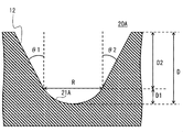
- FIG. 4 to FIG. 7 show examples of the shape of the recess according to this embodiment. 4 to 7 show the recesses 20A to 20D having bottom portions 21A to 21D different from the bottom portion 11 of the first embodiment, but the opening 12 can be realized in the same shape as that of the first embodiment, and thus the same ones are combined.
- the example of a shape of a dent is shown.
- FIG. 4 and 5 show an example in which the hemispherical shape used for the bottom portion is different from that of the first embodiment in which a hemispherical shape in which the sphere is halved is used for the bottom portion 11.
- FIG. 4 shows a bottom 21A that uses less than half of a sphere. In other words, the bottom 21A uses a hemispherical part.
- FIG. 5 shows a bottom 21B using a cylindrical shape with a hemispherical bottom. In the case of the shape of the bottom portion 21B shown in FIG. 5, if the tube portion becomes long, the cells will not float from the bottom portion 21B to the culture medium when the cells are collected. Therefore, it is preferable to adjust the length of the tube portion.
- the depth (height) of the bottom 21B and the opening 12 be the same ratio (1: 1).
- FIG. 6 shows a bottom 21C using a truncated cone. When the bottom is flat, light refraction and interference can be reduced, which is useful for microscopic observation.
- FIG. 7 shows an example of the shape of the recess 20D in which the bottom 21D is linear, in other words, the bottom 21D does not form a space.
- the dent 20D is advantageous in that the manufacturing process of the culture container is easy, although the efficiency of culturing and collecting cells is inferior to that of other shape culture containers.
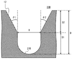
- Embodiment 3 In each of the above-described embodiments, the mode in which the shape of the opening 12 is circular or substantially circular has been described. However, a culture vessel having an opening having another shape will be described.
- the shape of the end of the opening may be other shapes such as a hemispherical shape, a trapezoidal shape, or a tri inverted triangle.
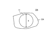
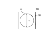
- the shape of the boundary where the opening contacts the bottom (the boundary of the opening) needs to be the same shape as the boundary of the bottom. 8 and 9 show dents 30 ⁇ / b> A and 30 ⁇ / b> B having end shapes different from those of the opening 12 of the first embodiment.
- the same bottom portion 11 as that of the first embodiment is shown, but it can be combined with any of the bottom portions 21A to 21D of the second embodiment, and may be a bottom portion having another shape.
- the shape of a bottom part and an opening part should just be a combination which can form a slope continuously in the boundary.
- FIG. 8 shows an example of a shape in which the end of the opening 32A draws a curve.
- FIG. 8 is a view of the recess 30A as viewed from above, in which the end of the bottom 11 is indicated by a circle having an equivalent diameter R, and the outer periphery of the opening 32A is indicated by a curve.
- the end of the opening 32A is a curved line that is not symmetrical left and right and up and down, but may have a shape that is symmetrical left and right or up and down.
- FIG. 9 shows an example in which the end of the opening 32B is rectangular. Although FIG. 9 shows an example of a square, other polygons, combinations of curves and straight lines may be used.
- FIG. 9 shows an example of a square, other polygons, combinations of curves and straight lines may be used.
- the taper angle is important. 8 and 9, the taper angle has a different value depending on the shape of the openings 32A and 32B. This is because the slopes of the slopes forming the walls differ depending on the shapes of the openings 32A and 32B.
- each shape of the opening shown in this embodiment can be combined with each shape of the bottom 11 of Embodiment 1 or the bottom described in Embodiment 2.
- shapes and combinations other than the bottom shown in the above-described embodiments are possible. Since the cell culture method using the culture container of the present embodiment is the same as that of the first embodiment, description thereof is omitted.
- the culture container of the present embodiment can also achieve the same effects as those of the first embodiment.
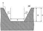
- FIG. 10 is a schematic view showing a configuration example using a flask-shaped culture flask.
- FIG. 11 is a schematic diagram showing a configuration example using a frame of a culture plate.
- FIG. 12 is a schematic diagram showing a configuration example in which the culture plate shown in FIG. 11 is used in a stack shape.
- the bottom surface of the culture flask 4 is defined as a culture surface 4A (culture bottom surface). Since the culture surface 4A corresponds to the culture vessel 1 shown in FIG. 1, it can also be called a culture vessel.
- the culture surface 4A is a unit using the same medium as in the culture container 1 of FIG.
- the culture flask 4 has a cap 4B. What is necessary is just to design the area of 4 A of culture surfaces according to a use.
- General culture flask there is 25,75,225cm 2.
- a plurality of recesses 40 are formed on the culture surface 4 ⁇ / b> A of the culture flask 4.
- the shaded portion of the bottom surface of the culture flask 4 is designed as the culture surface 4A, and a plurality of recesses 40 are formed on the culture surface 4A.
- the shape of the recess 40 may be any of the above embodiments.
- FIG. 11 shows an example in which only the frame of the culture plate is used.
- the culture vessel 1 (well) is formed on the culture plate 3.
- the bottom surface of the culture plate 5 is defined as a culture surface 5A (culture bottom surface). Since the culture surface 5A corresponds to the culture container 1 shown in FIG. 1, it can also be called a culture container.
- the culture surface 5A is a unit using the same medium.
- FIG. 12 shows a configuration example of a cell stack configuration in which a plurality of culture plates 5 shown in FIG. 11 are stacked, in other words, a multi-stage configuration example.
- FIG. 12 shows an example in which the culture plates 5 shown in FIG. 11 are stacked, the culture plates 3 shown in FIG. 1 may be stacked.
- a container for storing a stack of a plurality of culture containers and providing a mechanism for exchanging the medium is omitted.
- a container for storing a plurality of culture plates for example, a general stack-shaped culture container can be used. The description is omitted here.
- the boundary between the bottom and the opening is shown to be parallel to the bottom of the culture vessel, but it is not necessarily parallel to the bottom.
- the boundary may be inclined with respect to the bottom, and the boundary may be formed to draw a curve. It is sufficient that a sufficient space can be formed so that spheroids are formed at the bottom 11.
- Example The following Examples and Comparative Examples were tested for cell aggregate culture containers and collection methods.
- (1) Culture container The culture container shown in Table 1 was used.
- the culture container of the example was prepared by forming a well (culture container 1) having a recess 10 shown in FIG. 1-3 on a culture plate.
- the micro container corresponds to the recess 10 in FIGS. 1-3, and the micro space is a space formed by the recess 10 (micro space). It can be said that the number of microspaces per well is the number of recesses per well.
- the spheroid residual rate was calculated by the following formula.
- Spheroid residual rate (%) number of spheroids ⁇ 100 / number of microspaces Several hours after cell seeding (zero day), spheroid-like lumps were formed in each culture container in 90% or more of microspace. The value obtained by dividing the number of spheroids after medium change on day 10 and day 20 by the number of spheroids on day 0 was defined as the spheroid residual rate.
- (4) Recovery method After completion of the culture, the solution was stirred using a pipette (manufacturer, model number), and the floating spheroids were recovered.
- a 24-well plate contains 500 ⁇ L to 1 mL of medium, it is suitable to use a pipette that can aspirate a maximum of 1 mL of medium.
- Recovery efficiency Images were acquired with a confocal laser microscope before and after recovery.
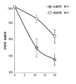
- FIG. 13 shows the remaining rate of spheroids at the time of medium exchange.
- the vertical axis represents the spheroid survival rate (Sphere Number), and the horizontal axis represents the number of days of culture.
- FIG. 13 shows data from the start of culture to 20 days. As shown in FIG. 13, it is shown that the comparative example is greatly reduced from the embodiment. After culturing for 20 days, the spheroid survival rate in the Examples was 60% or more, which was 1.5 times that of the Comparative Examples.
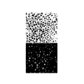
- FIG. 14 the image of the spheroid before and behind the culture medium exchange of an Example and a comparative example is shown.
- FIG. 14 shows images of spheroids in the culture container on the fourth day of culture and before and after the second medium change.
- the left side of FIG. 14 is a photograph of an example (Kuraray p-HEMA), and the right side is a photograph of a comparative example (Iwaki MPC).
- the upper image shows the image before the medium exchange
- the lower image (below the arrow) shows the image after the medium exchange. More specifically, the lower image shows a state in which half of the medium is exchanged (half quantity exchange) from the state before the medium exchange, and after two medium exchanges.
- the part that appears white in the image is a spheroid. Spheroids are confirmed throughout the culture medium exchange. After the medium exchange, there is no significant difference between before and after the medium exchange in the examples, and most of the spheroids remain, but only about half remains in the comparative example.
- FIG. 15 shows images before and after the cells of Examples were collected.
- the left side (BEFORE) is an image before collection
- the right side (AFTER) is an image after collection.
- the upper part shows an image of the entire culture container, and the lower part shows an enlarged image of a part of the culture container.
- recovering from the culture container of an Example is shown in FIG.
- a spheroid is a dot-like one in a black circular micro container (dent). There is no dot-like image after collection, and almost 100% is collected.
- recovery was a favorable spheroid form, and the spheroid was not destroyed by collection
Abstract
Description
さらに、一実施形態の培養用において、前記培養容器が、アクリル系樹脂、ポリ乳酸、ポリグリコール酸、スチレン系樹脂、アクリル・スチレン系共重合樹脂、ポリカーボネート系樹脂、ポリエステル系樹脂、ポリビニルアルコール系樹脂、エチレン・ビニルアルコール系共重合樹脂、熱可塑性エラストマー、塩化ビニル系樹脂、及びシリコン樹脂のうちの1つまたはこれらの組み合わせからなる樹脂成形品であることが好ましい。前記凹みへ、プラズマ処理、ガラスコート、コロナ放電、UVオゾン処理のいいずれかまたはこれら組み合わせからなる表面改質処理方法により官能基を形成させて、水接触角が45度以下になるように処理されたことが好ましい。
前記凹みへ、細胞接着を阻害する親水性のポリマー鎖が固定化されていることが好ましい。
前記凹みへ、リン脂質、または、リン脂質・高分子複合体が固定化されていることが好ましい。
前記凹みへ、プラズマ処理、ガラスコート、コロナ放電、UVオゾン処理のいいずれかまたはこれら組み合わせからなる表面改質処理方法により官能基を形成させて、水接触角を45度以下になるように処理した後、細胞接着を阻害する親水性のポリマー鎖、及び、リン脂質、または、リン脂質・高分子複合体のうちのいずれか一つのポリマーが固定化されている細胞非接着表面であることが好ましい。
前記親水性のポリマー鎖がポリヒドロキシエチルメタクリレートであることが好ましく、前記ポリヒドロキシエチルメタクリレートの平均分子量が10万以上であることがより好ましい。 Further, in the culture vessel of one embodiment, the shape of the end is preferably a hemispherical shape, a trapezoidal shape, or an inverted triangle, and a space between two adjacent recesses is flat, and the two recesses are The distance is preferably 5 μm to 50 μm.
Further, in one embodiment of the culture, the culture vessel may be an acrylic resin, polylactic acid, polyglycolic acid, styrene resin, acrylic / styrene copolymer resin, polycarbonate resin, polyester resin, polyvinyl alcohol resin. It is preferably a resin molded product made of one or a combination of ethylene / vinyl alcohol copolymer resin, thermoplastic elastomer, vinyl chloride resin, and silicon resin. A functional group is formed on the recess by a surface modification treatment method comprising plasma treatment, glass coating, corona discharge, UV ozone treatment, or a combination thereof, so that the water contact angle is 45 degrees or less. It is preferred that
It is preferable that a hydrophilic polymer chain that inhibits cell adhesion is immobilized in the recess.
It is preferable that a phospholipid or a phospholipid / polymer complex is immobilized in the recess.
A functional group is formed on the recess by a surface modification treatment method comprising any one of plasma treatment, glass coating, corona discharge, UV ozone treatment, or a combination thereof, so that the water contact angle is 45 degrees or less. Then, the hydrophilic polymer chain that inhibits cell adhesion and the cell non-adherent surface on which any one polymer of phospholipid or phospholipid / polymer complex is immobilized. preferable.
It is preferable that the hydrophilic polymer chain is polyhydroxyethyl methacrylate, and it is more preferable that the average molecular weight of the polyhydroxyethyl methacrylate is 100,000 or more.
分化誘導させる場合は、前記空間にスフェロイドを形成させた状態で誘導することが好ましい。
前記培養容器内に形成されたスフェロイドの総数の60%以上が、平均スフェロイド直径のプラスマイナス5%の範囲内の直径であることが好ましい。
前記培地を攪拌することで前記凹み内の細胞を回収することが好ましく、前記培地の攪拌が、前記培養容器を振とうして前記培地を攪拌すること、前記培地を吸引及び排出して前記培地を攪拌すること、前記培養容器に攪拌羽を設置し培地を攪拌すること、前記培養容器に攪拌子をいれ培地を攪拌すること、またはこれら組みあわせからなる方法のいずれかであることがより好ましい。
前記培地を少なくとも1回以上交換し、交換する培地の割合が20%以上であることが好ましい。
本発明の一実施形態の係る培養方法の他の一態様は、上述したいずれかの培養容器を用いる。そして、培養方法は、以下の各工程を実施することにより細胞を播種、培養、培地交換、回収する。
a)培養容器に存在する凹みの数(n)と同数以上、前記凹みの体積(V)を播種する細胞の体積(v)で割った値に前記凹みの数(n)を掛けた数以下の細胞数を培地に分散させ、前記培地を培養容器に添加する工程、
b)12時間以上前記培養容器内で前記細胞を培養してスフェロイドを形成させる工程、
c)前記培地を20%以上吸引した後、同量の新鮮な培地を注入する工程、
d)前記a)からc)の工程を複数回行い、スフェロイドを成長させる工程、
e)前記スフェロイドを所望の大きさに成長させた後、前記培地を攪拌して各凹み内の細胞を前記培地中に浮遊させる工程、及び
f)前記培地ごと前記細胞を吸引機にて吸い取り前記細胞を回収する工程。 In one aspect of the culture method according to an embodiment of the present invention, it is preferable to form one spheroid per one space formed by the recess, and to grow (proliferate) the spheroid by forming a spheroid in the space. It is more preferable.
In order to induce differentiation, it is preferable to induce in a state where spheroids are formed in the space.
It is preferable that 60% or more of the total number of spheroids formed in the culture vessel has a diameter within a range of plus or minus 5% of the average spheroid diameter.
It is preferable to collect the cells in the dent by stirring the medium, and stirring the medium is performed by shaking the culture vessel and stirring the medium, and sucking and discharging the medium. More preferably, the method is a method of stirring a medium, stirring a medium by installing a stirring blade in the culture container, stirring a medium by adding a stirring bar to the culture container, or a combination thereof. .
It is preferable that the medium is replaced at least once, and the ratio of the medium to be replaced is 20% or more.
Another aspect of the culture method according to one embodiment of the present invention uses any of the culture vessels described above. In the culture method, the cells are seeded, cultured, medium exchanged, and recovered by performing the following steps.
a) The number equal to or greater than the number of depressions (n) present in the culture vessel, and the number obtained by dividing the volume (V) of the depressions by the volume (v) of cells to be seeded multiplied by the number of depressions (n) Dispersing the number of cells in a medium and adding the medium to a culture vessel,
b) culturing the cells in the culture vessel for 12 hours or more to form spheroids;
c) A step of injecting the same amount of fresh medium after aspirating 20% or more of the medium;
d) performing the steps a) to c) multiple times to grow spheroids,
e) a step of growing the spheroids to a desired size, and then stirring the medium to suspend cells in each dent in the medium; and f) sucking the cells together with the medium with an aspirator Recovering cells.
<培養容器>
図1は、一実施形態の培養容器の一例を示す図である。図1では、複数の培養容器1を有する培養プレート3の一部分を示す。図1の上段には、培養容器1の底に形成される複数の凹み10の一部分を、培養プレート3の上からみた図を示す。培養容器1は、複数の凹み10が配置される。複数の凹み10は、培養容器1の製造や細胞培養の効率の観点から、規則的に配置されることが好ましい。培養容器1は例えば複数のウェルを有するウェルプレートの一つのウェルに相当する。言い換えると、ウェルプレートの各ウェルに複数の凹み10が配置されることになる。
ウェルプレートは、多数のくぼみ(穴またはウェル)のついた平板からなる実験・検査器具であり、各ウェルを試験管あるいはシャーレとして利用するものをいう。ウェルの数には例えば、6、24、96、384などがあり、それ以上の数のものもある。ウェルの底は平らなもの、丸いもののほか、細長いマイクロチューブを多数組み合わせた形式のもの(ディープウェルプレート)もある。
また、凹み10は、細胞を培養するための微小な空間であるマイクロ空間を形成することから、マイクロ容器ということもできる。
<Culture container>
FIG. 1 is a diagram illustrating an example of a culture container according to an embodiment. FIG. 1 shows a part of a
A well plate is an experimental / inspection instrument composed of a flat plate with a number of indentations (holes or wells), and each well is used as a test tube or petri dish. The number of wells includes, for example, 6, 24, 96, 384, and others. The bottom of the well is flat, round, or a combination of many elongated microtubes (deep well plate).
Moreover, since the
各凹み10は、底部11と開口部12とから構成される。底部11は、培養容器1の底になる部分であり、開口部12は、底部11の上部に配置される部分である。底部11と開口部12とが接する部分を境界と記載する。図2では、符号Rの矢印で示す長さの部分が境界の位置に対応する。また、図3では、境界の位置を2点破線で示している。ただし、底部11と開口部12とは連続した面で構成され、一体として製造される。 2 and 3 are diagrams illustrating examples of the shape of the recess according to the first embodiment. FIG. 2 shows a cross-sectional view when one
Each
相当直径Rは、凹み10の底部11に内接する内接円の直径をいう。ここでは、底部11と開口部12との境界において内接する内接円の直径をいう。より詳しくは、相当直径Rは、境界における、凹み10の高さHの方向と垂直になる面の形状の内接円の直径をいう。
深さDは、底部11の内側の底から凹み10の上端までの長さである。凹みの10の上端は、開口部12の端部(上端)と同じである。深さDは、凹み10が形成する空間の深さである。言い換えると、底部11が形成する空間の底から開口部12が形成する空間の上端までの深さである。図2では凹み10の深さDに加え、底部11の深さD1及び開口部12の深さD2を示している。 2 and 3, the equivalent diameter R and the depth (height) D are shown for the plurality of
The equivalent diameter R is a diameter of an inscribed circle inscribed in the bottom 11 of the
The depth D is the length from the inner bottom of the bottom 11 to the upper end of the
開口部12は、細胞の培養及び回収を補助するように働く空間(第2空間)を形成する。開口部12は、底部11との境界から凹み10の端部(先端)までを囲むテーパ角が1度以上20度以下の壁で構成される。開口部12を構成する壁のテーパ角が5度以上15度以下であることが好ましく、10度がより好ましい。その理由は、テーパ角が小さすぎると回収する際に細胞が凹みから培地に移行できず、逆に大きすぎると培地交換中に細胞が離脱するからである。 The bottom 11 forms a space for culturing cells (first space). The bottom 11 has, for example, a hemispherical shape. For example, a shape in which a spherical shape having an equivalent diameter R as a diameter is halved can be used. The shape of the bottom 11 is not limited to a hemisphere. Other specific examples will be described in the second embodiment.
The opening 12 forms a space (second space) that works to assist cell culture and recovery. The
底部11と開口部12との境界は、相当直径Rが50μm以上1mm以下となるように形成される。スフェロイドの中心部まで栄養を供給したい場合は相当直径50μm以上500μm以下が好ましく、より好ましくは100μm以上500μm以下が好ましい。その理由は、栄養分や酸素は拡散によってのみ細胞内に移行するが、中心部が壊死しない大きさは300μmといわれており(Efrem Curcio et al.,"Mass transfer and metabolic reactions in hepatocyte spheroids cultured in rotating wall gas-permeable membrane system", Biomaterials 28 (2007) 5487-5497)、その大きさ以上にならないようにするためには、上記直径が好ましい。
逆にガン細胞のように細胞の中心部にネクローシスを作製したいような場合(Franziska Hirschhaeuser et al.,"Multicellular tumor spheroids: An underestimated tool is catching up again", Journal of Biotechnology 148 (2010) 3-15, Fig1)には、相当直径Rが400μm以上2mm未満であることが好ましい。その理由は、上述したように300μmでは中心部まで栄養がいきわたりネクローシスを起こさない場合考えられるからである。よって300μm以上の直径のスフェロイドを得るためには、400μm以上なければならない。
加えて、底部の底から端部までの深さDが相当直径Rの0.6倍以上3倍以下となるように形成される。好ましくは、深さDが相当直径R0.7倍以上1.2倍以下であり、より好ましくは、0、8~1倍である。 In FIG. 2, the taper angles are indicated by the symbols θ1 and θ2. In the example of the shape of the
The boundary between the bottom 11 and the
Conversely, when it is desired to create necrosis at the center of a cell like a cancer cell (Franziska Hirschhaeuser et al., “Multicellular tumor spheroids: An underestimated tool is catching up again”, Journal of Biotechnology 148 (2010) 3-15 , Fig1), the equivalent diameter R is preferably 400 μm or more and less than 2 mm. The reason is that, as described above, at 300 μm, it can be considered that no nutrients reach the center and no necrosis occurs. Therefore, in order to obtain a spheroid having a diameter of 300 μm or more, it must be 400 μm or more.
In addition, the depth D from the bottom to the end of the bottom is formed to be not less than 0.6 times and not more than 3 times the equivalent diameter R. The depth D is preferably an equivalent diameter R 0.7 times or more and 1.2 times or less, and more preferably 0, 8 to 1 times.
これに対して、隣り合う二つの凹み10が接触する場合であってもよい。例えば、二つの凹み10の端部の一部分が接触し、開口部12のテーパ角の斜面が接触して山形の形状となっていてもよい。 Moreover, it is preferable that the
On the other hand, the case where two
培養容器1が、アクリル系樹脂、ポリ乳酸、ポリグリコール酸、スチレン系樹脂、アクリル・スチレン系共重合樹脂、ポリカーボネート系樹脂、ポリエステル系樹脂、ポリビニルアルコール系樹脂、エチレン・ビニルアルコール系共重合樹脂、熱可塑性エラストマー、塩化ビニル系樹脂、及びシリコン樹脂のうちの1つまたはこれらの組み合わせからなる樹脂成形品であることが好ましい。 In addition to the shape described above, the
The
加えて、各凹み10へ、細胞接着を阻害する親水性のポリマー鎖が固定化されていることが好ましい。親水性のポリマー鎖は、上述した水接触角が45度以下になるように処理された各凹み10へ固定化されることがより好ましい。
さらに加えて、各凹み10へ、リン脂質、または、リン脂質・高分子複合体が固定化されていることが好ましい。この固定化の処理は、上述した水接触角が45度以下になるように処理された各凹み10、親水性のポリマー鎖が固定化された各凹み10、またはこれらを組合せた各凹み10へ実施されることがより好ましい。 A functional group is formed on each
In addition, a hydrophilic polymer chain that inhibits cell adhesion is preferably immobilized in each
In addition, it is preferable that a phospholipid or a phospholipid / polymer complex is immobilized in each
また、上述した親水性のポリマー鎖がポリヒドロキシエチルメタクリレートであることが好ましく、さらに、ポリヒドロキシエチルメタクリレートの平均分子量が10万以上であることがより好ましい。 Furthermore, a functional group is formed on each
The hydrophilic polymer chain described above is preferably polyhydroxyethyl methacrylate, and the average molecular weight of polyhydroxyethyl methacrylate is more preferably 100,000 or more.
次に、図1乃至3に示す培養容器1を用いる細胞の培養方法について説明する。
細胞の培養は次の各工程により実施する。
a)細胞を分散させた培地を培養容器1へ添加する工程
b)細胞を培養する工程
c)培地を交換する工程
d)スフェロイドを成長させる工程
e)スフェロイドを培地の中に浮遊させる工程
f)細胞を回収する工程
上述した各工程は、a)からd)が細胞を培養する工程(細胞培養工程)であり、e)、f)が細胞を回収する工程(細胞回収工程)と区分することもできる。
ここで、スフェロイドは、細胞が多数凝集して細胞塊を形成し、3次元状態になったものである。 <Culture method>
Next, a cell culture method using the
Cell culture is performed by the following steps.
a) Step of adding a medium in which cells are dispersed to the culture vessel 1 b) Step of culturing cells c) Step of replacing the medium d) Step of growing spheroids e) Step of floating the spheroids in the medium f) Step of recovering cells Each of the steps described above is a step (cell culture step) in which cells are cultured from a) to d), and is divided from a step of recovering cells (cell recovery step) in e) and f). You can also.
Here, the spheroid is a three-dimensional state in which many cells aggregate to form a cell mass.
a)細胞を分散させた培地を培養容器1へ添加する工程
細胞を培養する準備を行う工程であり、培地に以下の総数の細胞を分散させ、培養容器1へ添加する。
総細胞数の下限は、培養容器1に存在する凹み10の数(n)と同数以上とする。
総細胞数の上限は、培養容器1が有する凹み10の体積(V)を播種する細胞の体積(v)で割った値に、凹みの数(n)を掛けた数以下とする。記号を用いた数式で表すと、細胞総数の上限値=V/v×n、と表すことができる。ここで、複数の凹み10の体積(V)は同じであることを前提とする。異なる場合には平均値を用いる。
培地は培養する細胞に応じて調整する。 Each step will be described below.
a) Step of adding the medium in which the cells are dispersed to the
The lower limit of the total number of cells is equal to or more than the number (n) of the
The upper limit of the total number of cells is not more than the number obtained by multiplying the volume (v) of the
The medium is adjusted according to the cells to be cultured.
培養容器1内で12時間以上、細胞を培養し、スフェロイドを形成させる。培地に分散させた細胞は、培養容器1へ培地を添加すると、凹み10へ取り込まれ、各凹み10内で培養される。各凹み10に細胞が1個取り込まれることが好ましく、底部11が形成する空間に1個のスフェロイドが形成されることが好ましい。各凹み10内では、細胞が凹み10の底部11内で増殖する。培養播種時に細胞が最低1個なければ、培養中に隣の凹み10から細胞が移動することはないので、その凹み10にスフェロイドは形成されない。スフェロイドを高密度に培養するためには、全ての凹み10にスフェロイドが形成されることが好ましいことから、最低1個の細胞が凹み10に存在することが好ましい。生産効率の観点から、初期の細胞数はできる限り少なくする一方で多くのスフェロイドを回収できることが好ましいため、凹み10に存在する細胞数は少ないほどよい。そのため、1個の細胞が凹み10に存在することが好ましい。
c)培地を交換する工程
培地交換では、培養容器1内の培地を20%以上吸引した後、同量の新鮮な培地を注入する。培地交換は、細胞培養中少なくとも1回以上実施されることが好ましい。
d)スフェロイドを成長させる工程
上述したa)からc)の工程を複数回行い、スフェロイドを成長させる。分化誘導させる場合は、凹み10の底部11が形成する空間において、スフェロイドが大きくならない状態まで成長させた後、分化誘導培地に交換して分化させることが好ましい。加えて、培養容器1内に形成されたスフェロイドの総数の60%以上が、平均スフェロイド直径のプラスマイナス5%の範囲内の直径であることがより好ましい。 b) Step of culturing cells Cells are cultured in the
c) Step of replacing the medium In the medium replacement, the medium in the
d) Step of growing spheroids Steps a) to c) described above are performed a plurality of times to grow spheroids. In the case of inducing differentiation, it is preferable to grow in a space formed by the bottom 11 of the
スフェロイドを所望の大きさに成長させた後、培養容器1の培地を攪拌して各凹み11内で培養した細胞を培地中に浮遊させる。例えば、培地を攪拌することによって実施する。具体的には、培地の攪拌は、(1)培養容器1を振とうして培地を攪拌すること、(2)培地を吸引及び排出(ピペッティング操作)して培地を攪拌すること、(3)培養容器1に攪拌羽を設置し培地を攪拌すること、(4)培養容器1に攪拌子をいれ培地を攪拌すること、(5)上述した(1)から(4)の二つ以上を組合せて培地を攪拌すること、のうちのいずれかの方法を用いることができる。
f)細胞を回収する工程
培養容器1内の細胞含む培地を吸引機にて吸い取り、培地に浮遊させた細胞(スフェロイド)を回収する。 e) Step of floating spheroids in medium After growing spheroids to a desired size, the medium in
f) Step of collecting cells The medium containing the cells in the
実施形態1の培養容器1を用いて細胞を培養することにより、底部11に所望の大きさのスフェロイドを形成することができる。そして、培養したスフェロイドを効率よく回収することができる。具体的には、凹み10が底部11に加え、開口部12を有することにより、培地交換では培地を吸い取るときに細胞が底部11に接着または浮遊しているが離脱しない状態を維持しやすくし、底部11からの細胞の離脱を抑制することが期待できる。一方、細胞の回収では、底部11の培地を吸引及び排出するときに、開口部12により培地の流れを生じやすくすることが期待できる。加えて、底部11に半球状の形状を用いることにより、スフェロイドの形状、大きさを均一にすることに寄与することが期待できる。 As described above, in
By culturing cells using the
実施形態1では、底部11が半球状の形状を有する構成例を説明したが、実施形態2では他の形状について説明する。底部は、球形状の一部分からなる形状、円錐台の形状、あるいは、線状から形成されている態様であってもよい。底部が線状とは、実質的な底部がなく、凹みが開口部のみから形成されている態様である。図4から図7に本実施形態の凹みの形状例を示す。図4から図7は、実施形態1の底部11と異なる底部21A~21Dを有する凹み20A~20Dを示すが、開口部12については実施形態1と同様の形状で実現できるため同じものを組み合わせた凹みの形状例を示す。 Embodiment 2. FIG.
In the first embodiment, the configuration example in which the
図6は、円錐台を用いる底部21Cを示す。底部が平らである場合、光の屈折・干渉が軽減でき顕微鏡観察を行う際には有用である。
図7は、底部21Dが線状、言い換えると底部21Dが空間を形成しない凹み20Dの形状例を示す。凹み20Dは、他の形状の培養容器に比べて細胞の培養・回収の効率は劣るものの、培養容器の製造工程が容易であるという利点がある。 4 and 5 show an example in which the hemispherical shape used for the bottom portion is different from that of the first embodiment in which a hemispherical shape in which the sphere is halved is used for the
FIG. 6 shows a bottom 21C using a truncated cone. When the bottom is flat, light refraction and interference can be reduced, which is useful for microscopic observation.
FIG. 7 shows an example of the shape of the
本実施形態の培養容器を用いる細胞の培養方法は実施形態1と同様であるため説明を省略する。
本実施形態の培養容器も実施形態1と同様の効果を奏することができる。 In addition, although this embodiment demonstrated the case where the
Since the cell culture method using the culture container of the present embodiment is the same as that of the first embodiment, description thereof is omitted.
The culture container of the present embodiment can also achieve the same effects as those of the first embodiment.
上述した各実施形態では、開口部12の形状を円形または略円形である態様を説明したが、他の形状の開口部を有する培養容器について説明する。開口部の端部の形状は、半球状、台形、または三逆三角形等の他の形状であってもよい。一方、開口部が底部と接する境界の形状(開口部の境界部分)は、底部の境界部分と同じ形状であることが必要である。図8,9は、実施形態1の開口部12と異なる端部の形状を有する凹み30A、30Bを表している。図8,9では、実施形態1と同じ底部11を表しているが、実施形態2の底部21A~21Dのいずれかと組み合わせることも可能であり、他の形状の底部であってもよい。底部及び開口部の形状は、その境界において斜面が連続して形成できる組合せであればよい。
In each of the above-described embodiments, the mode in which the shape of the
図8,9の形状例では、テーパ角は開口部32A、32Bの形状に応じて異なる値となる。これは、開口部32A、32Bの形状に応じて、壁を形成する斜面の傾斜が異なるからである。 FIG. 8 shows an example of a shape in which the end of the
8 and 9, the taper angle has a different value depending on the shape of the
本実施形態の培養容器を用いる細胞の培養方法は実施形態1と同様であるため説明を省略する。
本実施形態の培養容器も実施形態1と同様の効果を奏することができる。 Each shape of the opening shown in this embodiment can be combined with each shape of the bottom 11 of
Since the cell culture method using the culture container of the present embodiment is the same as that of the first embodiment, description thereof is omitted.
The culture container of the present embodiment can also achieve the same effects as those of the first embodiment.
図1では、一実施形態の培養容器1を培養プレート3(ウェルプレート)に配置した態様を説明した。一実施形態の培養容器1は、図1の培養プレート3以外の容器(器具)にも形成することができる。図10から12に実施形態4の培養容器の構成例を示す。図10は、フラスコ形状の培養フラスコを用いる構成例を示す概略図である。図11は、培養プレートの枠を用いる構成例を示す概略図である。図12は、図11に示す培養プレートをスタック形状にして用いる構成例を示す概略図である。
In FIG. 1, the aspect which has arrange | positioned the
図11では、培養プレートの枠のみを用いる例である。図1では培養プレート3に培養容器1(ウェル)が形成されているが、図11では培養プレート5の底の面を培養面5A(培養底面)とする。培養面5Aは、図1に示す培養容器1に相当するため、培養容器ということもできる。培養面5Aは、同じ培地を用いる単位となる。培養面5Aの構成例(概略断面図)を図11の下段に示している。例えば、培養プレート5の底の面のうち、網掛け部分を培養面5Aとして設計し、培養面5Aに複数の凹み50を形成する。図11に示す凹み50は、模式的に示したものであり、凹み50の数、大きさい等は、用途に応じて設計されるものである。凹み50の形状(底部及び開口部の形状)は、上記各実施形態のいずれであってもよい。
図12に、図11に示す培養プレート5を複数積み上げて構成するセルスタック形態の構成例、言い換えると多段式の構成例を示す。より大面積化し閉鎖系で培養する場合には、セルスタック形態を用いるのが一般的である。図12では、図11に示す培養プレート5を積み上げた例を示したが、図1に示す培養プレート3を積み上げてもよい。図12中、複数の培養容器を積み上げたものを収納し、培地を交換するための仕組みを提供する容器については、省略している。複数の培養プレートを収納する容器は、例えば、一般的なスタック形状の培養容器を用いることができる。ここでは説明を省略する。 In FIG. 10, the bottom surface of the
FIG. 11 shows an example in which only the frame of the culture plate is used. In FIG. 1, the culture vessel 1 (well) is formed on the
FIG. 12 shows a configuration example of a cell stack configuration in which a plurality of
上記各実施形態では、底部と開口部との境界を培養容器の底と平行するようにあらわしているが、必ずしも底と平行でなくてもよい。例えば、境界が底に対して傾斜していてもよく、境界が曲線を描くように形成されていてもよい。底部11においてスフェロイドが形成されるように十分な空間が形成できればよい。 Other embodiments.
In each of the above embodiments, the boundary between the bottom and the opening is shown to be parallel to the bottom of the culture vessel, but it is not necessarily parallel to the bottom. For example, the boundary may be inclined with respect to the bottom, and the boundary may be formed to draw a curve. It is sufficient that a sufficient space can be formed so that spheroids are formed at the bottom 11.
細胞凝集体の培養容器及び回収方法について、次の実施例、比較例の試験を行った。
(1)培養容器
表1に示す培養容器を用いた。 [Example]
The following Examples and Comparative Examples were tested for cell aggregate culture containers and collection methods.
(1) Culture container The culture container shown in Table 1 was used.
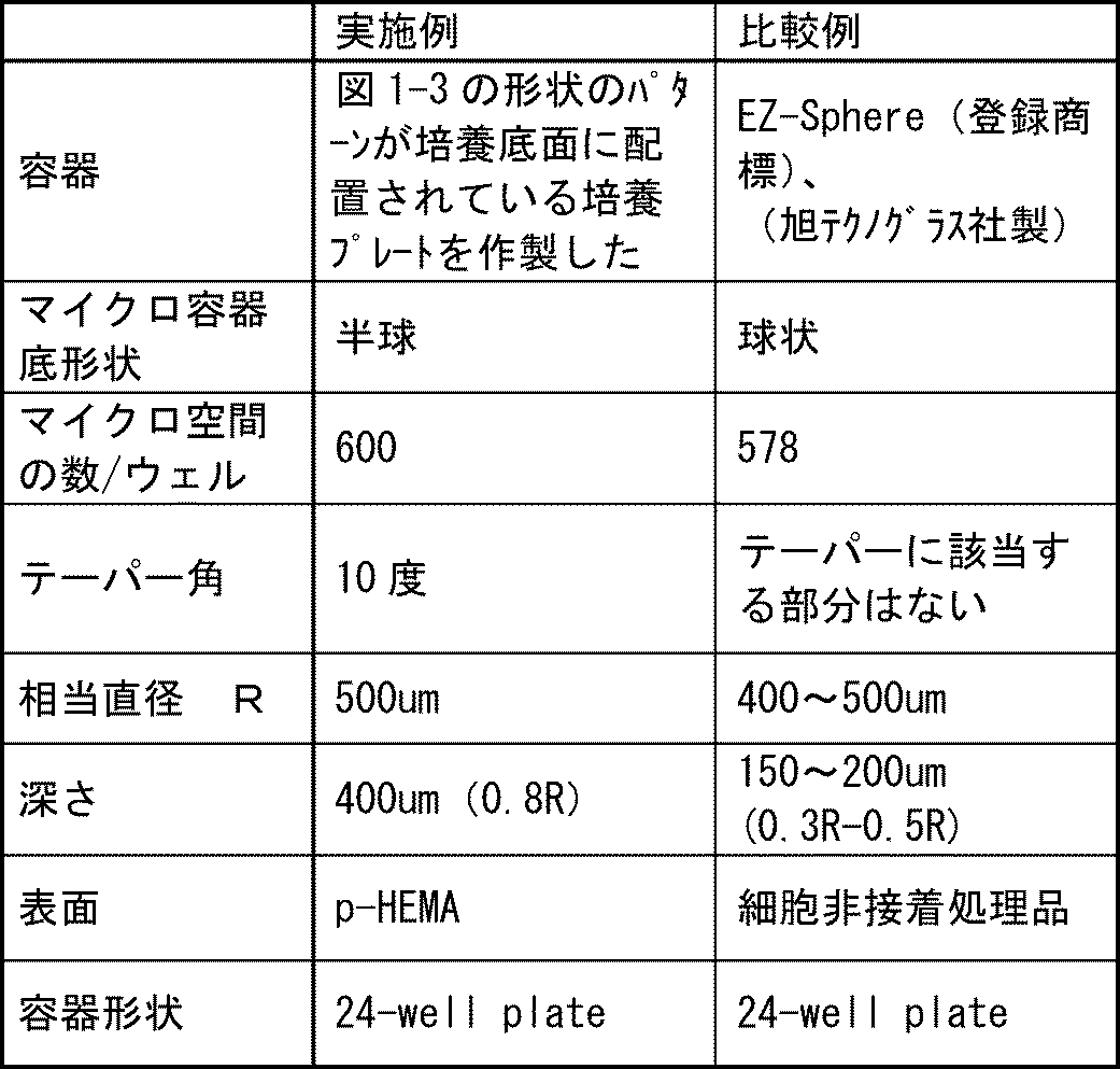
表1中、マイクロ容器は、図1-3の凹み10に相当し、マイクロ空間は、凹み10(マイクロ空間)が形成する空間である。一ウェル当たりのマイクロ空間の数は、一ウェル当たりの凹みの数であるともいえる。 The culture container of the example was prepared by forming a well (culture container 1) having a
In Table 1, the micro container corresponds to the
後述する残存率及び回収率を画像解析により算出するため、GFPで蛍光標識した内胚葉細胞を用いた。この内胚葉細胞、血管内皮細胞とヒト間葉系幹細胞各々10:5-10:2の割合で混合し、内皮細胞培地キット-2:EGM-2 BulletKit(製品コード CC-3162:Lonza)で30日間培養を行った。培地は2日に1回交換した。
(3)スフェロイド残存率の測定
共焦点レーザ顕微鏡を用い、ウェル全体を観察、画像解析ソフトを用いてスフェロイドを認識させ、その数をカウントし、スフェロイド数とした。以下の式でスフェロイド残存率を計算した。
スフェロイド残存率(%)=スフェロイド数×100/マイクロ空間の数
細胞播種後数時間後(ゼロ日)、いずれの培養容器も90%以上のマイクロ空間でスフェロイド様の塊が形成されていた。培養10日目、20日目の培地交換後のスフェロイドの数をゼロ日目のスフェロイド数で其々割った値をスフェロイド残存率とした。
(4)回収方法
培養終了後、ピペット(メーカ、型番)を用いて溶液を攪拌し、浮遊してきたスフェロイドを回収した。例えば、24ウェルプレートでは500μL~1mLの培地が入っているので、最大1mLの培地が吸引できるピペットを用いるのが適している。
(5)回収効率
回収前後に共焦点レーザ顕微鏡で画像を取得した。 (2) Culture method In order to calculate the residual rate and recovery rate described later by image analysis, endodermal cells fluorescently labeled with GFP were used. These endoderm cells, vascular endothelial cells, and human mesenchymal stem cells were mixed at a ratio of 10: 5-10: 2, respectively, and were mixed with endothelial cell medium kit-2: EGM-2 BulletKit (product code CC-3162: Lonza). The culture was performed for a day. The medium was changed once every two days.
(3) Measurement of residual rate of spheroids Using a confocal laser microscope, the whole well was observed, spheroids were recognized using image analysis software, and the number was counted to obtain the number of spheroids. The spheroid residual rate was calculated by the following formula.
Spheroid residual rate (%) = number of spheroids × 100 / number of microspaces Several hours after cell seeding (zero day), spheroid-like lumps were formed in each culture container in 90% or more of microspace. The value obtained by dividing the number of spheroids after medium change on
(4) Recovery method After completion of the culture, the solution was stirred using a pipette (manufacturer, model number), and the floating spheroids were recovered. For example, since a 24-well plate contains 500 μL to 1 mL of medium, it is suitable to use a pipette that can aspirate a maximum of 1 mL of medium.
(5) Recovery efficiency Images were acquired with a confocal laser microscope before and after recovery.
図13に培地交換時のスフェロイドの残存率を示す。縦軸にスフェロイドの残存率(Sphere Number)を、横軸に培養日数を示す。
図13では、培養開始から20日までのデータを示す。図13に示すように、実施例より比較例が大きく減少していることが示されている。20日培養した後、実施例は、スフェロイドの残存率が60%以上となり、比較例の1.5倍に向上した。 (6) Results FIG. 13 shows the remaining rate of spheroids at the time of medium exchange. The vertical axis represents the spheroid survival rate (Sphere Number), and the horizontal axis represents the number of days of culture.
FIG. 13 shows data from the start of culture to 20 days. As shown in FIG. 13, it is shown that the comparative example is greatly reduced from the embodiment. After culturing for 20 days, the spheroid survival rate in the Examples was 60% or more, which was 1.5 times that of the Comparative Examples.
画像中、白色に見える部分がスフェロイドである。培地交換前は全体にスフェロイドが確認される。培地交換後は、実施例では培地交換前後で大きな違いはなく、ほとんどのスフェロイドが残存しているが、比較例では約半分程度しか残存していない。 In FIG. 14, the image of the spheroid before and behind the culture medium exchange of an Example and a comparative example is shown. FIG. 14 shows images of spheroids in the culture container on the fourth day of culture and before and after the second medium change. The left side of FIG. 14 is a photograph of an example (Kuraray p-HEMA), and the right side is a photograph of a comparative example (Iwaki MPC). Further, the upper image shows the image before the medium exchange, and the lower image (below the arrow) shows the image after the medium exchange. More specifically, the lower image shows a state in which half of the medium is exchanged (half quantity exchange) from the state before the medium exchange, and after two medium exchanges.
The part that appears white in the image is a spheroid. Spheroids are confirmed throughout the culture medium exchange. After the medium exchange, there is no significant difference between before and after the medium exchange in the examples, and most of the spheroids remain, but only about half remains in the comparative example.
黒色の円形のマイクロ容器(凹み)内の点状のものがスフェロイドである。回収後の画像には点状のものがなく、ほぼ100%回収できている。また、図16に示すように、回収後の細胞の形態は良好なスフェロイドの形をしており、回収操作によりスフェロイドが破壊されることはなかった。 FIG. 15 shows images before and after the cells of Examples were collected. In FIG. 15, the left side (BEFORE) is an image before collection, and the right side (AFTER) is an image after collection. The upper part shows an image of the entire culture container, and the lower part shows an enlarged image of a part of the culture container. The photograph of the spheroid after collect | recovering from the culture container of an Example is shown in FIG.
A spheroid is a dot-like one in a black circular micro container (dent). There is no dot-like image after collection, and almost 100% is collected. Moreover, as shown in FIG. 16, the form of the cell after collection | recovery was a favorable spheroid form, and the spheroid was not destroyed by collection | recovery operation.
3、5 培養プレート
4A、5A 培養面
5 培養フラスコ
10、20A~20D、30A、30B、40、50 凹み
11、21A~21D 底部
12、32A、32B 開口部 1
Claims (19)
- 底部と開口部とからなる複数の凹みが配列し、
前記底部が、半球状と円錐台とのいずれかの形状を有し、
前記開口部が、前記底部との境界から前記凹みの端部までを囲むテーパ角1度以上20度以下の壁で構成され、
前記境界の相当直径が50μm以上2mm以下であり、前記底部の底から前記端部までの深さが前記相当直径の0.6倍以上3倍以下であり、
前記開口部を構成する壁が前記底部と連続する面を形成し、かつ、前記連続する面の傾斜が前記境界で変化する培養容器。 A plurality of dents consisting of a bottom and an opening are arranged,
The bottom has a shape of either a hemisphere or a truncated cone;
The opening is constituted by a wall having a taper angle of 1 degree or more and 20 degrees or less surrounding a boundary with the bottom part to an end part of the dent,
The equivalent diameter of the boundary is 50 μm or more and 2 mm or less, and the depth from the bottom of the bottom to the end is 0.6 to 3 times the equivalent diameter,
A culture vessel in which a wall constituting the opening forms a surface continuous with the bottom, and the inclination of the continuous surface changes at the boundary. - 前記端部の形状が半球状、台形、または逆三角形のいずれかであることを特徴とする請求項1記載の培養容器。 2. The culture vessel according to claim 1, wherein the shape of the end is hemispherical, trapezoidal, or inverted triangular.
- 隣り合う二つの凹みの間が平坦であり、前記二つの凹みの距離が5μmから50μmであることを特徴とする請求項1または2記載の培養容器。 The culture vessel according to claim 1 or 2, wherein a space between two adjacent recesses is flat, and a distance between the two recesses is 5 µm to 50 µm.
- 前記培養容器が、アクリル系樹脂、ポリ乳酸、ポリグリコール酸、スチレン系樹脂、アクリル・スチレン系共重合樹脂、ポリカーボネート系樹脂、ポリエステル系樹脂、ポリビニルアルコール系樹脂、エチレン・ビニルアルコール系共重合樹脂、熱可塑性エラストマー、塩化ビニル系樹脂、及びシリコン樹脂のうちの1つまたはこれらの組み合わせからなる樹脂成形品であることを特徴とする請求項1乃至3のいずれか一項に記載の培養容器。 The culture vessel is an acrylic resin, polylactic acid, polyglycolic acid, styrene resin, acrylic / styrene copolymer resin, polycarbonate resin, polyester resin, polyvinyl alcohol resin, ethylene / vinyl alcohol copolymer resin, The culture container according to any one of claims 1 to 3, wherein the culture container is a resin molded product made of one of a thermoplastic elastomer, a vinyl chloride resin, and a silicon resin, or a combination thereof.
- 前記凹みへ、プラズマ処理、ガラスコート、コロナ放電、UVオゾン処理のいいずれかまたはこれら組み合わせからなる表面改質処理方法により官能基を形成させて、水接触角が45度以下になるように処理されたことを特徴とする請求項1乃至4のいずれか一項に記載の培養容器。 A functional group is formed on the recess by a surface modification treatment method comprising plasma treatment, glass coating, corona discharge, UV ozone treatment, or a combination thereof, so that the water contact angle is 45 degrees or less. The culture container according to any one of claims 1 to 4, wherein the culture container is formed.
- 前記凹みへ、細胞接着を阻害する親水性のポリマー鎖が固定化されていることを特徴とする請求項1乃至5のいずれか一項に記載の培養容器。 The culture vessel according to any one of claims 1 to 5, wherein a hydrophilic polymer chain that inhibits cell adhesion is immobilized in the recess.
- 前記凹みへ、リン脂質、または、リン脂質・高分子複合体が固定化されていることを特徴とする請求項1乃至6のいずれか一項に記載の培養容器。 The culture container according to any one of claims 1 to 6, wherein a phospholipid or a phospholipid / polymer complex is immobilized in the recess.
- 前記凹みへ、プラズマ処理、ガラスコート、コロナ放電、UVオゾン処理のいいずれかまたはこれら組み合わせからなる表面改質処理方法により官能基を形成させて、水接触角を45度以下になるように処理した後、細胞接着を阻害する親水性のポリマー鎖、及び、リン脂質、または、リン脂質・高分子複合体のうちのいずれか一つのポリマーが固定化されている細胞非接着表面であることを特徴とする請求項1乃至7のいずれか一項に記載の培養容器。 A functional group is formed on the recess by a surface modification treatment method comprising any one of plasma treatment, glass coating, corona discharge, UV ozone treatment, or a combination thereof, so that the water contact angle is 45 degrees or less. After that, the hydrophilic polymer chain that inhibits cell adhesion and the phospholipid or phospholipid / polymer complex is one of the non-cell-adherent surfaces on which the polymer is immobilized. The culture container according to any one of claims 1 to 7, wherein the culture container is characterized in that
- 前記親水性のポリマー鎖がポリヒドロキシエチルメタクリレートであることを特徴とする請求項8記載の培養容器。 The culture vessel according to claim 8, wherein the hydrophilic polymer chain is polyhydroxyethyl methacrylate.
- 前記ポリヒドロキシエチルメタクリレートの平均分子量が10万以上であることを特徴とする請求項9記載の培養容器。 The culture container according to claim 9, wherein the polyhydroxyethyl methacrylate has an average molecular weight of 100,000 or more.
- 前記請求項1乃至10のいずれか一項に記載の培養容器を用い、
総細胞数が、前記培養容器が有する前記凹みの数N以上であり、前記凹みが形成する空間の体積V1を播種する細胞の体積V2で割った値に前記凹みの数Nを掛けた数以下である細胞を培地に分散させ、
前記培地を前記培養容器に添加する培養方法。 Using the culture container according to any one of claims 1 to 10,
The total number of cells is equal to or greater than the number N of the recesses of the culture vessel, and is equal to or less than the value obtained by dividing the volume V1 of the space formed by the recesses by the volume V2 of the seeded cells and the number N of the recesses. Are dispersed in the medium,
A culture method of adding the medium to the culture vessel. - 前記凹みが形成する空間1個につき1個のスフェロイドを形成させることを特徴とする請求項11記載の培養方法。 The culture method according to claim 11, wherein one spheroid is formed per one space formed by the recess.
- 前記空間にスフェロイドを形成させてスフェロイドを成長させることを特徴とする請求項12記載の培養方法。 The culture method according to claim 12, wherein spheroids are grown in the space to grow spheroids.
- 前記空間にスフェロイドを形成させて分化誘導することを特徴とする請求項12または13に記載の培養方法。 The culture method according to claim 12 or 13, wherein differentiation is induced by forming spheroids in the space.
- 前記培養容器内に形成されたスフェロイドの総数の60%以上が、平均スフェロイド直径のプラスマイナス5%の範囲内の直径であることを特徴とする請求項11乃至14のいずれか一項に記載の培養方法。 The total number of spheroids formed in the culture vessel is 60% or more, and the diameter is within a range of plus or minus 5% of the average spheroid diameter. Culture method.
- 前記培地を攪拌することで前記凹み内の細胞を回収することを特徴とする請求項11乃至15のいずれか一項に記載の培養方法。 The culture method according to any one of claims 11 to 15, wherein the cells in the recess are collected by stirring the medium.
- 前記培地の攪拌が、前記培養容器を振とうして前記培地を攪拌すること、前記培地を吸引及び排出して前記培地を攪拌すること、前記培養容器に攪拌羽を設置し培地を攪拌すること、前記培養容器に攪拌子をいれ培地を攪拌すること、またはこれら組みあわせからなる方法のいずれかであることを特徴とする請求項16記載の培養方法。 Agitation of the culture medium involves shaking the culture container to stir the culture medium, aspirating and discharging the culture medium to stir the culture medium, and stirring the culture medium by installing stirring blades in the culture container. The culture method according to claim 16, which is one of a method comprising adding a stirring bar to the culture vessel and stirring the medium, or a combination thereof.
- 前記培地を少なくとも1回以上交換し、交換する培地の割合が20%以上であることを特徴とする請求項11乃至17のいずれか一項に記載の培養方法。 The culture method according to any one of claims 11 to 17, wherein the medium is exchanged at least once, and a ratio of the medium to be exchanged is 20% or more.
- 前記請求項1乃至10のいずれか一項に記載の培養容器を用い、
a)培養容器に存在する凹みの数nと同数以上、前記凹みの体積Vを播種する細胞の体積vで割った値に前記凹みの数nを掛けた数以下の細胞数を培地に分散させ、前記培地を培養容器に添加する工程、
b)12時間以上前記培養容器内で前記細胞を培養してスフェロイドを形成させる工程、
c)前記培地を20%以上吸引した後、同量の新鮮な培地を注入する工程、
d)前記a)からc)の工程を複数回行い、スフェロイドを成長させる工程、
e)前記スフェロイドを所望の大きさに成長させた後、前記培地を攪拌して各凹み内の細胞を前記培地の中に浮遊させる工程、及び
f)前記培地ごと前記細胞を吸引機にて吸い取り前記細胞を回収する工程、
を実施することにより細胞を播種、培養、培地交換、回収する培養方法。 Using the culture container according to any one of claims 1 to 10,
a) Disperse in the medium the number of cells equal to or greater than the number n of dents present in the culture vessel, but not more than the value obtained by dividing the volume V of the dents by the volume v of cells to be seeded and the number n of the dents. Adding the medium to the culture vessel;
b) culturing the cells in the culture vessel for 12 hours or more to form spheroids;
c) A step of injecting the same amount of fresh medium after aspirating 20% or more of the medium;
d) performing the steps a) to c) multiple times to grow spheroids,
e) a step of growing the spheroids to a desired size and then stirring the medium to suspend the cells in each dent in the medium; and f) sucking the cells together with the medium with an aspirator. Collecting the cells;
A culture method for seeding, culturing, exchanging the medium, and collecting the cells by carrying out the above.
Priority Applications (12)
| Application Number | Priority Date | Filing Date | Title |
|---|---|---|---|
| SG11201509870QA SG11201509870QA (en) | 2013-06-07 | 2014-06-05 | Culture vessel and culture method |
| AU2014276229A AU2014276229B2 (en) | 2013-06-07 | 2014-06-05 | Culture vessel and culture method |
| CA2914463A CA2914463C (en) | 2013-06-07 | 2014-06-05 | Culture chamber and culture method |
| BR112015030041-3A BR112015030041B1 (en) | 2013-06-07 | 2014-06-05 | culture chamber and culture method |
| EP14808113.6A EP3006553B1 (en) | 2013-06-07 | 2014-06-05 | Culture vessel and culture method |
| US14/896,251 US10494593B2 (en) | 2013-06-07 | 2014-06-05 | Culture chamber and culture method |
| KR1020167000067A KR102359408B1 (en) | 2013-06-07 | 2014-06-05 | Culture vessel and culture method |
| CN201480032635.8A CN105308170B (en) | 2013-06-07 | 2014-06-05 | Culture vessel and cultural method |
| JP2015521306A JPWO2014196204A1 (en) | 2013-06-07 | 2014-06-05 | Culture container and culture method |
| US16/668,701 US11473046B2 (en) | 2013-06-07 | 2019-10-30 | Culture chamber and culture method |
| AU2020201221A AU2020201221B2 (en) | 2013-06-07 | 2020-02-20 | Culture vessel and culture method |
| US18/521,821 US20240093134A1 (en) | 2013-06-07 | 2023-11-28 | Culture chamber and culture method |
Applications Claiming Priority (2)
| Application Number | Priority Date | Filing Date | Title |
|---|---|---|---|
| JP2013120915 | 2013-06-07 | ||
| JP2013-120915 | 2013-06-07 |
Related Child Applications (3)
| Application Number | Title | Priority Date | Filing Date |
|---|---|---|---|
| US14/896,251 A-371-Of-International US10494593B2 (en) | 2013-06-07 | 2014-06-05 | Culture chamber and culture method |
| US16/668,701 Division US11473046B2 (en) | 2013-06-07 | 2019-10-30 | Culture chamber and culture method |
| US16/668,701 Continuation US11473046B2 (en) | 2013-06-07 | 2019-10-30 | Culture chamber and culture method |
Publications (1)
| Publication Number | Publication Date |
|---|---|
| WO2014196204A1 true WO2014196204A1 (en) | 2014-12-11 |
Family
ID=52007861
Family Applications (1)
| Application Number | Title | Priority Date | Filing Date |
|---|---|---|---|
| PCT/JP2014/002993 WO2014196204A1 (en) | 2013-06-07 | 2014-06-05 | Culture vessel and culture method |
Country Status (10)
| Country | Link |
|---|---|
| US (4) | US10494593B2 (en) |
| EP (1) | EP3006553B1 (en) |
| JP (3) | JPWO2014196204A1 (en) |
| KR (1) | KR102359408B1 (en) |
| CN (1) | CN105308170B (en) |
| AU (2) | AU2014276229B2 (en) |
| BR (1) | BR112015030041B1 (en) |
| CA (1) | CA2914463C (en) |
| SG (1) | SG11201509870QA (en) |
| WO (1) | WO2014196204A1 (en) |
Cited By (21)
| Publication number | Priority date | Publication date | Assignee | Title |
|---|---|---|---|---|
| JP2015167518A (en) * | 2014-03-07 | 2015-09-28 | 大日本印刷株式会社 | Cell cultivation vessel |
| WO2016069885A1 (en) * | 2014-10-29 | 2016-05-06 | Corning Incorporated | Perfusion bioreactor platform |
| CN106047690A (en) * | 2015-04-16 | 2016-10-26 | 爱科来株式会社 | Cell culture device |
| JP2017153388A (en) * | 2016-02-29 | 2017-09-07 | 米満 吉和 | Spheroids of the same size arranged regularly, and their use |
| US9790465B2 (en) | 2013-04-30 | 2017-10-17 | Corning Incorporated | Spheroid cell culture well article and methods thereof |
| WO2018123663A1 (en) * | 2016-12-28 | 2018-07-05 | Agcテクノグラス株式会社 | Cell culture substrate and method for producing same |
| JP2018174824A (en) * | 2017-04-14 | 2018-11-15 | 株式会社クラレ | Method for wetting surface of micro-pattern |
| WO2019151114A1 (en) | 2018-02-01 | 2019-08-08 | Agc株式会社 | Cell culture container |
| JP2019193666A (en) * | 2014-05-30 | 2019-11-07 | コーニング インコーポレイテッド | Culture method |
| WO2020166452A1 (en) * | 2019-02-13 | 2020-08-20 | 古河電気工業株式会社 | Storage container |
| WO2021166977A1 (en) * | 2020-02-19 | 2021-08-26 | 凸版印刷株式会社 | Method for pretreatment of cell transplantation, device for pretreatment of cell transplantation, and unit for pretreatment of cell transplantation |
| JP2022501002A (en) * | 2018-07-13 | 2022-01-06 | コーニング インコーポレイテッド | Microcavity dish with sidewalls including liquid medium delivery surface |
| WO2022024886A1 (en) * | 2020-07-27 | 2022-02-03 | 株式会社コーセー | Holocrine regulators evaluation and/or selection method |
| US11345880B2 (en) | 2017-07-14 | 2022-05-31 | Corning Incorporated | 3D cell culture vessels for manual or automatic media exchange |
| US11584906B2 (en) | 2017-07-14 | 2023-02-21 | Corning Incorporated | Cell culture vessel for 3D culture and methods of culturing 3D cells |
| US11661574B2 (en) | 2018-07-13 | 2023-05-30 | Corning Incorporated | Fluidic devices including microplates with interconnected wells |
| US11732227B2 (en) | 2018-07-13 | 2023-08-22 | Corning Incorporated | Cell culture vessels with stabilizer devices |
| WO2023176949A1 (en) * | 2022-03-17 | 2023-09-21 | 日産化学株式会社 | Cell culture container having high cell utilization efficiency |
| US11767499B2 (en) | 2017-07-14 | 2023-09-26 | Corning Incorporated | Cell culture vessel |
| US11857970B2 (en) | 2017-07-14 | 2024-01-02 | Corning Incorporated | Cell culture vessel |
| US11970682B2 (en) | 2022-05-03 | 2024-04-30 | Corning Incorporated | 3D cell culture vessels for manual or automatic media exchange |
Families Citing this family (16)
| Publication number | Priority date | Publication date | Assignee | Title |
|---|---|---|---|---|
| WO2018047707A1 (en) * | 2016-09-06 | 2018-03-15 | 学校法人慶應義塾 | Method for measuring ultraviolet protection effect or infrared protection effect of aqueous composition containing ultraviolet absorbing agent or infrared blocking agent, and apparatus for preparing measurement sample |
| KR101949856B1 (en) * | 2017-06-28 | 2019-02-20 | 한국과학기술원 | Well plate, method of preparing the same, and method for culturing a cell using the same |
| EP3660139A4 (en) * | 2017-07-22 | 2021-04-21 | Toyo Seikan Group Holdings, Ltd. | Culture vessel, method for manufacturing culture vessel, laminated structure, and method for producing laminated structure |
| KR101965900B1 (en) * | 2017-11-03 | 2019-04-05 | 한국화학연구원 | Spheroid forming and counting apparatus and manufacturing method thereof, and counting method and culturing method of spheroid using the same |
| WO2019092813A1 (en) * | 2017-11-08 | 2019-05-16 | 株式会社Ihi | Connection unit for cell culture device, incubator device, and cell culture device |
| US20190382701A1 (en) * | 2018-06-18 | 2019-12-19 | SageMedic Corporation | System for Obtaining 3D Micro-Tissues |
| JP7271903B2 (en) * | 2018-10-20 | 2023-05-12 | 東洋製罐グループホールディングス株式会社 | Sphere culture member, culture vessel, perforated member processing method, and washing vessel |
| KR102237425B1 (en) * | 2018-12-26 | 2021-04-08 | 주식회사 넥스트앤바이오 | A method for providing the information for diagnosing of drug and/or radiation resistance in a cancer subject |
| JP7057878B2 (en) * | 2019-10-25 | 2022-04-21 | 東洋製罐グループホールディングス株式会社 | Adhesive cell culture equipment, culture vessel, cell detachment method, and method for manufacturing adhesive cell culture equipment |
| EP4174173A4 (en) * | 2020-06-25 | 2024-03-27 | Next & Bio Inc | Brain organoid manufacturing method |
| JP2023538208A (en) * | 2020-06-25 | 2023-09-07 | ネクストアンドバイオ インコーポレイテッド | Standard organoid production method |
| CN114514312A (en) * | 2020-06-25 | 2022-05-17 | 株式会社下一代生物 | Method for proliferating stem cells in large quantities without using hydrogel |
| WO2022097984A1 (en) * | 2020-11-05 | 2022-05-12 | (주)에스엔이바이오 | Method for producing extracellular vesicles derived from three-dimensional spheroid-type cell aggregate |
| CN113189317A (en) * | 2021-04-29 | 2021-07-30 | 广东省人民医院 | Experimental device for three-dimensional static culture of artificial blood vessel and use method thereof |
| KR20230108960A (en) * | 2022-01-12 | 2023-07-19 | 전남대학교산학협력단 | Preparing methods for inner ear organoids |
| CN115141752A (en) * | 2022-06-28 | 2022-10-04 | 上海划创科技发展有限公司 | Cell culture and in-situ detection container, preparation method and cell in-situ detection method |
Citations (6)
| Publication number | Priority date | Publication date | Assignee | Title |
|---|---|---|---|---|
| JPH08131153A (en) | 1994-09-16 | 1996-05-28 | Sumitomo Bakelite Co Ltd | Cell culture container, its production and culture |
| JP2001509272A (en) * | 1997-01-17 | 2001-07-10 | コーニング インコーポレイテッド | Multi-well plate |
| WO2008130025A1 (en) * | 2007-04-18 | 2008-10-30 | Public University Corporation Yokohama City University | Hepatocyte culture container and hepatocyte culture method |
| JP2010088347A (en) | 2008-10-08 | 2010-04-22 | Tohoku Univ | Method and container for spheroid culture |
| WO2012036011A1 (en) | 2010-09-14 | 2012-03-22 | 旭硝子株式会社 | Culture substrate |
| WO2013042360A1 (en) | 2011-09-20 | 2013-03-28 | 株式会社クラレ | Adherent cell culture method |
Family Cites Families (13)
| Publication number | Priority date | Publication date | Assignee | Title |
|---|---|---|---|---|
| EP0101749A1 (en) | 1982-08-26 | 1984-03-07 | Westerwald AG für Silikatindustrie | Wall composed of glass blocks |
| WO2002095339A1 (en) * | 2001-05-24 | 2002-11-28 | Hitachi, Ltd. | Heating resistor type flow measuring device |
| GB0200721D0 (en) * | 2002-01-14 | 2002-02-27 | Univ Bristol | Toxicity test |
| JP2005027598A (en) * | 2003-07-09 | 2005-02-03 | Kitakyushu Foundation For The Advancement Of Industry Science & Technology | Cell culture chip and incubator and method for culturing cell by using those, cell-carrying module carrying spherical cell tissue body and spherical cell tissue body |
| NL2000159C2 (en) * | 2006-07-24 | 2008-01-25 | Stork Pmt | Device, method and production line for conditioning slaughtered poultry. |
| US20080227664A1 (en) * | 2007-03-16 | 2008-09-18 | Canon Kabushiki Kaisha | Cell array structural body and cell array |
| ES2363406T3 (en) * | 2007-06-29 | 2011-08-03 | Unisense Fertilitech A/S | DEVICE, SYSTEM AND METHOD FOR MONITORING AND / OR CULTIVATING MICROSCOPIC OBJECTS. |
| US8533234B2 (en) * | 2008-10-07 | 2013-09-10 | Aspect Software, Inc. | Custom data display |
| JP4724854B2 (en) * | 2009-02-09 | 2011-07-13 | 大日本印刷株式会社 | Cell culture vessel |
| US8278511B2 (en) * | 2009-07-16 | 2012-10-02 | Monsanto Technology Llc | Soybean variety A1016184 |
| WO2011083768A1 (en) * | 2010-01-08 | 2011-07-14 | 住友ベークライト株式会社 | Culture vessel for formation of aggregated cell mass |
| JP2012157267A (en) * | 2011-01-31 | 2012-08-23 | Hitachi Maxell Ltd | Plate member having fine pattern |
| JP2012210166A (en) | 2011-03-30 | 2012-11-01 | Sumitomo Bakelite Co Ltd | Culture vessel for forming embryoid body |
-
2014
- 2014-06-05 BR BR112015030041-3A patent/BR112015030041B1/en active IP Right Grant
- 2014-06-05 WO PCT/JP2014/002993 patent/WO2014196204A1/en active Application Filing
- 2014-06-05 JP JP2015521306A patent/JPWO2014196204A1/en active Pending
- 2014-06-05 US US14/896,251 patent/US10494593B2/en active Active
- 2014-06-05 CN CN201480032635.8A patent/CN105308170B/en active Active
- 2014-06-05 EP EP14808113.6A patent/EP3006553B1/en active Active
- 2014-06-05 KR KR1020167000067A patent/KR102359408B1/en active IP Right Grant
- 2014-06-05 CA CA2914463A patent/CA2914463C/en active Active
- 2014-06-05 AU AU2014276229A patent/AU2014276229B2/en active Active
- 2014-06-05 SG SG11201509870QA patent/SG11201509870QA/en unknown
-
2019
- 2019-09-17 JP JP2019168253A patent/JP6892486B2/en active Active
- 2019-10-30 US US16/668,701 patent/US11473046B2/en active Active
-
2020
- 2020-02-20 AU AU2020201221A patent/AU2020201221B2/en active Active
-
2021
- 2021-05-27 JP JP2021088960A patent/JP7219303B2/en active Active
-
2022
- 2022-09-30 US US17/957,743 patent/US11866682B1/en active Active
-
2023
- 2023-11-28 US US18/521,821 patent/US20240093134A1/en active Pending
Patent Citations (6)
| Publication number | Priority date | Publication date | Assignee | Title |
|---|---|---|---|---|
| JPH08131153A (en) | 1994-09-16 | 1996-05-28 | Sumitomo Bakelite Co Ltd | Cell culture container, its production and culture |
| JP2001509272A (en) * | 1997-01-17 | 2001-07-10 | コーニング インコーポレイテッド | Multi-well plate |
| WO2008130025A1 (en) * | 2007-04-18 | 2008-10-30 | Public University Corporation Yokohama City University | Hepatocyte culture container and hepatocyte culture method |
| JP2010088347A (en) | 2008-10-08 | 2010-04-22 | Tohoku Univ | Method and container for spheroid culture |
| WO2012036011A1 (en) | 2010-09-14 | 2012-03-22 | 旭硝子株式会社 | Culture substrate |
| WO2013042360A1 (en) | 2011-09-20 | 2013-03-28 | 株式会社クラレ | Adherent cell culture method |
Non-Patent Citations (5)
| Title |
|---|
| C'ELINE LIU BAUWENS ET AL.: "Control of Human Embryonic Stem Cell Colony and Aggregate Size Heterogeneity Influences Differentiation Trajectories", STEM CELL, 2008, pages 2300 - 2310 |
| EFREM CURCIO ET AL.: "Mass transfer and metabolic reactions in hepatocyte spheroids cultured in rotating wall gas-permeable membrane system", BIOMATERIALS, vol. 28, 2007, pages 5487 - 5497, XP022308847, DOI: doi:10.1016/j.biomaterials.2007.08.033 |
| FRANZISKA HIRSCHHAEUSER ET AL.: "Multicellular tumor spheroids: An underestimated tool is catching up again", JOURNAL OF BIOTECHNOLOGY, vol. 148, 2010, pages 3 - 15, XP027096372 |
| JUERGEN FRIEDRICHL ET AL.: "Spheroid-based drug screen: considerations and practical approach", PROTOCOL, 12 February 2009 (2009-02-12), pages 309 - 324, XP055150985, DOI: doi:10.1038/nprot.2008.226 |
| See also references of EP3006553A4 |
Cited By (30)
| Publication number | Priority date | Publication date | Assignee | Title |
|---|---|---|---|---|
| US11441121B2 (en) | 2013-04-30 | 2022-09-13 | Corning Incorporated | Spheroid cell culture article and methods thereof |
| US9790465B2 (en) | 2013-04-30 | 2017-10-17 | Corning Incorporated | Spheroid cell culture well article and methods thereof |
| JP2015167518A (en) * | 2014-03-07 | 2015-09-28 | 大日本印刷株式会社 | Cell cultivation vessel |
| JP2019193666A (en) * | 2014-05-30 | 2019-11-07 | コーニング インコーポレイテッド | Culture method |
| JP7125717B2 (en) | 2014-05-30 | 2022-08-25 | コーニング インコーポレイテッド | Culture method |
| US10752879B2 (en) | 2014-05-30 | 2020-08-25 | Corning Incorporated | Culture method and cell cluster |
| WO2016069885A1 (en) * | 2014-10-29 | 2016-05-06 | Corning Incorporated | Perfusion bioreactor platform |
| US11613722B2 (en) | 2014-10-29 | 2023-03-28 | Corning Incorporated | Perfusion bioreactor platform |
| CN106047690A (en) * | 2015-04-16 | 2016-10-26 | 爱科来株式会社 | Cell culture device |
| JP2017153388A (en) * | 2016-02-29 | 2017-09-07 | 米満 吉和 | Spheroids of the same size arranged regularly, and their use |
| WO2017150366A1 (en) * | 2016-02-29 | 2017-09-08 | 米満 吉和 | Regularly arranged spheroids having uniform size, and use thereof |
| WO2018123663A1 (en) * | 2016-12-28 | 2018-07-05 | Agcテクノグラス株式会社 | Cell culture substrate and method for producing same |
| JP2018174824A (en) * | 2017-04-14 | 2018-11-15 | 株式会社クラレ | Method for wetting surface of micro-pattern |
| US11584906B2 (en) | 2017-07-14 | 2023-02-21 | Corning Incorporated | Cell culture vessel for 3D culture and methods of culturing 3D cells |
| US11345880B2 (en) | 2017-07-14 | 2022-05-31 | Corning Incorporated | 3D cell culture vessels for manual or automatic media exchange |
| US11857970B2 (en) | 2017-07-14 | 2024-01-02 | Corning Incorporated | Cell culture vessel |
| US11767499B2 (en) | 2017-07-14 | 2023-09-26 | Corning Incorporated | Cell culture vessel |
| WO2019151114A1 (en) | 2018-02-01 | 2019-08-08 | Agc株式会社 | Cell culture container |
| JP7171695B2 (en) | 2018-07-13 | 2022-11-15 | コーニング インコーポレイテッド | A microcavity dish having sidewalls containing a liquid medium delivery surface |
| JP2022501002A (en) * | 2018-07-13 | 2022-01-06 | コーニング インコーポレイテッド | Microcavity dish with sidewalls including liquid medium delivery surface |
| US11661574B2 (en) | 2018-07-13 | 2023-05-30 | Corning Incorporated | Fluidic devices including microplates with interconnected wells |
| US11732227B2 (en) | 2018-07-13 | 2023-08-22 | Corning Incorporated | Cell culture vessels with stabilizer devices |
| US11912968B2 (en) | 2018-07-13 | 2024-02-27 | Corning Incorporated | Microcavity dishes with sidewall including liquid medium delivery surface |
| JP2020129981A (en) * | 2019-02-13 | 2020-08-31 | 古河電気工業株式会社 | Storage container |
| WO2020166452A1 (en) * | 2019-02-13 | 2020-08-20 | 古河電気工業株式会社 | Storage container |
| JP7019618B2 (en) | 2019-02-13 | 2022-02-15 | 古河電気工業株式会社 | Containment vessel |
| WO2021166977A1 (en) * | 2020-02-19 | 2021-08-26 | 凸版印刷株式会社 | Method for pretreatment of cell transplantation, device for pretreatment of cell transplantation, and unit for pretreatment of cell transplantation |
| WO2022024886A1 (en) * | 2020-07-27 | 2022-02-03 | 株式会社コーセー | Holocrine regulators evaluation and/or selection method |
| WO2023176949A1 (en) * | 2022-03-17 | 2023-09-21 | 日産化学株式会社 | Cell culture container having high cell utilization efficiency |
| US11970682B2 (en) | 2022-05-03 | 2024-04-30 | Corning Incorporated | 3D cell culture vessels for manual or automatic media exchange |
Also Published As
| Publication number | Publication date |
|---|---|
| SG11201509870QA (en) | 2016-02-26 |
| CN105308170A (en) | 2016-02-03 |
| CN105308170B (en) | 2018-04-10 |
| US20240093134A1 (en) | 2024-03-21 |
| AU2020201221B2 (en) | 2021-12-23 |
| US20160137962A1 (en) | 2016-05-19 |
| EP3006553A4 (en) | 2017-01-04 |
| EP3006553B1 (en) | 2020-09-02 |
| JP2020000255A (en) | 2020-01-09 |
| BR112015030041B1 (en) | 2021-01-12 |
| US20200063080A1 (en) | 2020-02-27 |
| US11473046B2 (en) | 2022-10-18 |
| JP6892486B2 (en) | 2021-06-23 |
| KR20160017036A (en) | 2016-02-15 |
| JP2021129590A (en) | 2021-09-09 |
| KR102359408B1 (en) | 2022-02-07 |
| CA2914463A1 (en) | 2014-12-11 |
| US10494593B2 (en) | 2019-12-03 |
| JP7219303B2 (en) | 2023-02-07 |
| US11866682B1 (en) | 2024-01-09 |
| BR112015030041A2 (en) | 2017-07-25 |
| EP3006553A1 (en) | 2016-04-13 |
| JPWO2014196204A1 (en) | 2017-02-23 |
| AU2014276229A1 (en) | 2016-01-21 |
| CA2914463C (en) | 2023-01-03 |
| AU2020201221A1 (en) | 2020-03-12 |
| AU2014276229B2 (en) | 2019-11-21 |
Similar Documents
| Publication | Publication Date | Title |
|---|---|---|
| JP7219303B2 (en) | Culture method | |
| CN201193228Y (en) | Three-dimensional cell-culturing insert, manufacturing equipment thereof and kit | |
| Bruzewicz et al. | Fabrication of a modular tissue construct in a microfluidic chip | |
| CN109689366B (en) | Three-dimensional bioreactor for cell expansion and related applications | |
| JP2016093149A (en) | Cell culture apparatus, and cell culture method | |
| JPWO2013042360A1 (en) | Method for culturing adherent cells | |
| CN108474140A (en) | Mass cell production system | |
| CN102719391A (en) | Diphasic porous three-dimensional cell culture scaffold | |
| WO2015129263A1 (en) | Device for fabricating spheroid, and spheroid recovery method and manufacturing method | |
| JP2016103982A (en) | Cell culture container, cell culture device, and cell culture method | |
| CN103608451A (en) | Culture method, group of mature adipocytes, and drug screening method | |
| Sun et al. | A superhydrophobic chip integrated with an array of medium reservoirs for long-term hanging drop spheroid culture | |
| JP2011507499A (en) | Dynamic system for cell culture in 3D support | |
| CN202643702U (en) | Biphase porous three-dimensional cell culture support | |
| US20200148989A1 (en) | Cell culture vessel for 3d culture and methods of culturing 3d cells | |
| CN218811793U (en) | Hanging drop culture plate and hanging drop culture device | |
| EP4289932A1 (en) | Cell cultivation by using removable top-loaded chambers in cell culture plates | |
| CN117025391A (en) | Hanging drop culture plate and hanging drop culture device | |
| US20230112108A1 (en) | Microcarrier based-4 dimensional cell culture apparatus and method for monitoring cell culture using the same | |
| KR20240049382A (en) | Bioreactor for organoid production | |
| JP5636207B2 (en) | Method for measuring the number of microbial cells in a microbial aggregate film |
Legal Events
| Date | Code | Title | Description |
|---|---|---|---|
| WWE | Wipo information: entry into national phase |
Ref document number: 201480032635.8 Country of ref document: CN |
|
| 121 | Ep: the epo has been informed by wipo that ep was designated in this application |
Ref document number: 14808113 Country of ref document: EP Kind code of ref document: A1 |
|
| ENP | Entry into the national phase |
Ref document number: 2015521306 Country of ref document: JP Kind code of ref document: A |
|
| WWE | Wipo information: entry into national phase |
Ref document number: P1593/2015 Country of ref document: AE |
|
| ENP | Entry into the national phase |
Ref document number: 2914463 Country of ref document: CA |
|
| WWE | Wipo information: entry into national phase |
Ref document number: 14896251 Country of ref document: US |
|
| NENP | Non-entry into the national phase |
Ref country code: DE |
|
| WWE | Wipo information: entry into national phase |
Ref document number: 2014808113 Country of ref document: EP |
|
| REG | Reference to national code |
Ref country code: BR Ref legal event code: B01A Ref document number: 112015030041 Country of ref document: BR |
|
| ENP | Entry into the national phase |
Ref document number: 20167000067 Country of ref document: KR Kind code of ref document: A |
|
| ENP | Entry into the national phase |
Ref document number: 2014276229 Country of ref document: AU Date of ref document: 20140605 Kind code of ref document: A |
|
| ENP | Entry into the national phase |
Ref document number: 112015030041 Country of ref document: BR Kind code of ref document: A2 Effective date: 20151130 |