WO2014148647A1 - 肝臓組織型スフェロイド - Google Patents
肝臓組織型スフェロイド Download PDFInfo
- Publication number
- WO2014148647A1 WO2014148647A1 PCT/JP2014/058152 JP2014058152W WO2014148647A1 WO 2014148647 A1 WO2014148647 A1 WO 2014148647A1 JP 2014058152 W JP2014058152 W JP 2014058152W WO 2014148647 A1 WO2014148647 A1 WO 2014148647A1
- Authority
- WO
- WIPO (PCT)
- Prior art keywords
- spheroid
- spheroids
- hepatocytes
- liver tissue
- cells
- Prior art date
- Legal status (The legal status is an assumption and is not a legal conclusion. Google has not performed a legal analysis and makes no representation as to the accuracy of the status listed.)
- Ceased
Links
Images



Classifications
-
- C—CHEMISTRY; METALLURGY
- C12—BIOCHEMISTRY; BEER; SPIRITS; WINE; VINEGAR; MICROBIOLOGY; ENZYMOLOGY; MUTATION OR GENETIC ENGINEERING
- C12N—MICROORGANISMS OR ENZYMES; COMPOSITIONS THEREOF; PROPAGATING, PRESERVING, OR MAINTAINING MICROORGANISMS; MUTATION OR GENETIC ENGINEERING; CULTURE MEDIA
- C12N5/00—Undifferentiated human, animal or plant cells, e.g. cell lines; Tissues; Cultivation or maintenance thereof; Culture media therefor
- C12N5/06—Animal cells or tissues; Human cells or tissues
- C12N5/0602—Vertebrate cells
- C12N5/067—Hepatocytes
- C12N5/0671—Three-dimensional culture, tissue culture or organ culture; Encapsulated cells
-
- C—CHEMISTRY; METALLURGY
- C12—BIOCHEMISTRY; BEER; SPIRITS; WINE; VINEGAR; MICROBIOLOGY; ENZYMOLOGY; MUTATION OR GENETIC ENGINEERING
- C12N—MICROORGANISMS OR ENZYMES; COMPOSITIONS THEREOF; PROPAGATING, PRESERVING, OR MAINTAINING MICROORGANISMS; MUTATION OR GENETIC ENGINEERING; CULTURE MEDIA
- C12N2502/00—Coculture with; Conditioned medium produced by
- C12N2502/13—Coculture with; Conditioned medium produced by connective tissue cells; generic mesenchyme cells, e.g. so-called "embryonic fibroblasts"
- C12N2502/1347—Smooth muscle cells
-
- C—CHEMISTRY; METALLURGY
- C12—BIOCHEMISTRY; BEER; SPIRITS; WINE; VINEGAR; MICROBIOLOGY; ENZYMOLOGY; MUTATION OR GENETIC ENGINEERING
- C12N—MICROORGANISMS OR ENZYMES; COMPOSITIONS THEREOF; PROPAGATING, PRESERVING, OR MAINTAINING MICROORGANISMS; MUTATION OR GENETIC ENGINEERING; CULTURE MEDIA
- C12N2502/00—Coculture with; Conditioned medium produced by
- C12N2502/13—Coculture with; Conditioned medium produced by connective tissue cells; generic mesenchyme cells, e.g. so-called "embryonic fibroblasts"
- C12N2502/1352—Mesenchymal stem cells
- C12N2502/1358—Bone marrow mesenchymal stem cells (BM-MSC)
-
- C—CHEMISTRY; METALLURGY
- C12—BIOCHEMISTRY; BEER; SPIRITS; WINE; VINEGAR; MICROBIOLOGY; ENZYMOLOGY; MUTATION OR GENETIC ENGINEERING
- C12N—MICROORGANISMS OR ENZYMES; COMPOSITIONS THEREOF; PROPAGATING, PRESERVING, OR MAINTAINING MICROORGANISMS; MUTATION OR GENETIC ENGINEERING; CULTURE MEDIA
- C12N2502/00—Coculture with; Conditioned medium produced by
- C12N2502/14—Coculture with; Conditioned medium produced by hepatocytes
Definitions
- the present invention relates to a liver tissue spheroid and a method for producing the same.
- liver synthesizes various proteins and metabolizes harmful substances, and is an essential organ for life support.
- Liver transplantation is currently the most effective for severe liver disease, but there are serious donor shortages and risks from surgical invasion. Liver regenerative medicine is expected as a new treatment alternative.
- hepatocyte transplantation has been carried out, and although certain effects have been observed depending on the disease, the survival rate of transplanted hepatocytes is low and no permanent effect has been obtained.
- the cells are scattered one by one, so that the interaction due to cell adhesion is lost, or rejection is mediated by natural immunity or cellular immunity by directly injecting cells into the bloodstream. We have problems such as being easy to receive.
- hepatocyte transplantation has a limited number of donor cells. From these viewpoints, a hepatic tissue engineering approach has been examined in which hepatocytes are engrafted outside the liver (ectopic site) to form a small liver tissue. This approach is highly expected as the next generation therapy.
- hepatocytes are present in a highly organized state with other cells and extracellular matrix, and their functions are maintained. In order to obtain good engraftment of hepatocytes at an ectopic site, it is necessary to construct an advanced three-dimensional cellular tissue closer to the living liver. That is, it is necessary to promote adhesion between hepatocytes and other cells. It is also important to build a blood vessel network that supplies nutrients to cells.
- tissue engineering approaches using cell sheets and scaffolds have been attempted (Non-patent Document 1), but the results are still not sufficient. It is desired to construct a liver tissue that overcomes these problems, has high functional efficiency, and is sufficiently large.
- An object of the present invention is to provide a method for constructing a liver tissue only with cells.
- the present invention has been completed. That is, the present invention is as follows. (1) A liver tissue spheroid formed from a mixture of hepatocytes, vascular endothelial cells and stem cells. (2) The mixing ratio of hepatocytes, vascular endothelial cells and stem cells is 30 to 100, preferably 40 to 60 for vascular endothelial cells with respect to hepatocytes 100, and 20 to 70, preferably 30 to 50 for stem cells. The liver tissue type spheroid according to (1).
- the abundance ratio of spheroids formed from hepatocytes and stem cells to spheroids derived from vascular endothelial cells is 5 to 10 spheroids derived from vascular endothelial cells relative to spheroids 100 formed from hepatocytes and stem cells.
- liver tissue type spheroids A method for producing a liver tissue type three-dimensional structure, comprising blending or laminating the liver tissue type spheroid according to any one of (1) to (5).
- the method according to (6), wherein the spheroids are stacked using a support including a substrate and a filamentous body or needle-like body disposed substantially perpendicular to the substrate.
- An artificial liver tissue comprising the liver tissue-type spheroid according to any one of (1) to (5) or a three-dimensional structure produced by the method according to (6) or (7).
- a liver tissue spheroid and a method for producing the same are provided. Moreover, a liver tissue type solid structure can be manufactured by mix
- the tissue produced by the method of the present invention has a function similar to the liver function in vivo, and can be used as an artificial liver tissue. Therefore, the present invention is extremely useful in that it can be used for regenerative medicine.
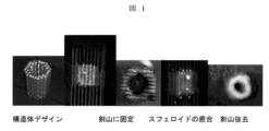
- FIG. 1 is a diagram showing the results of producing a three-dimensional structure using the method of the present invention.
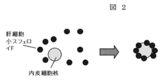
- FIG. 2 is a conceptual diagram of a process for preparing a bilayer spheroid by seeding hepatocyte small spheroids into endothelial cell nuclei.
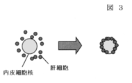
- FIG. 3 is a conceptual diagram of a process for preparing a bilayer spheroid by seeding hepatocyte small spheroids into the endothelial cell nucleus.
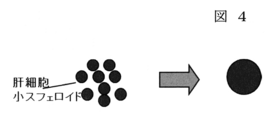
- FIG. 4 is a conceptual diagram of a process of producing large spheroids using hepatocyte small spheroids.
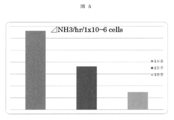
- FIG. 5 is a diagram showing the ammonia metabolic activity of hepatocytes.
- FIG. 1 is a diagram showing the results of producing a three-dimensional structure using the method of the present invention.
- FIG. 2 is a conceptual diagram of a process for preparing a bilayer spheroid by seeding hepatocyte small spheroids into endo
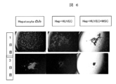
- FIG. 6 is a diagram showing a comparison of spheroid formation by co-culture.
- FIG. 7 is a cross-sectional view of a structure made of a Hep + HUVEC bilayer spheroid. It is a figure which shows E dyeing
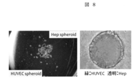
- FIG. 8 is a diagram showing a two-layer spheroid.
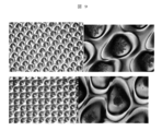
- FIG. 9 is a diagram showing spheroid formation after cells are seeded in a culture dish for producing small spheroids.
- FIG. 10 is a cross-sectional view of H.D. E. It is a figure which shows a dyeing
- FIG. 11 is a diagram comparing albumin secretion per spheroid.
- the present invention relates to a liver tissue spheroid formed from a mixture of hepatocytes, vascular endothelial cells, and stem cells.
- the present invention is a method for producing a liver tissue type three-dimensional structure characterized in that spheroids are formed from a mixture of hepatocytes, vascular endothelial cells and stem cells, and the spheroids are blended or laminated.
- the liver tissue type spheroid or three-dimensional structure produced by the method of the present invention can be used as an artificial liver tissue.
- the present inventor has paid attention to the formation of spheroids and has developed a technique for forming a more three-dimensional and highly functional tissue by combining cells specific to a target organ in order to have a function closer to a living body.
- the main cell is a hepatocyte.
- This main cell is referred to as “main cell” in this specification.
- spheroids or three-dimensional structures formed using hepatocytes as main cells are referred to as “liver tissue-type spheroids” and “liver tissue-type three-dimensional structures”, respectively.
- the above-mentioned main cells (hepatocytes), vascular endothelial cells, and stem cells are mixed to produce a liver tissue spheroid.
- liver tissue spheroids By blending or laminating the spheroids by an arbitrary method, the spheroids are three-dimensionally fused to obtain a liver tissue type three-dimensional structure. 1. Formation of liver tissue spheroids (1) In the present invention, a mixture of cells is prepared by mixing the hepatocytes, vascular endothelial cells, and stem cells, and spheroids are formed using the mixture. Therefore, one spheroid includes hepatocytes, vascular endothelial cells and stem cells as its constituent cells. The mixing ratio of cells for producing spheroids is as follows.
- the vascular endothelial cells are 30 to 100, preferably 40 to 60, and the stem cells are 20 to 70, preferably 30 to 50.
- hepatocytes can be obtained by refluxing collagenase, which is a cell-isolating enzyme, into blood vessels in living body tissue.
- the kind of vascular endothelial cell is not particularly limited, and examples thereof include human umbilical vein vascular endothelial cells (HUVEC), carotid artery endothelial cells, and hepatic sinusoidal endothelial cells.
- SAm cells are not particularly limited, and any stem cell can be used.
- Examples include mesenchymal stem cells, hematopoietic hepatocytes, iPS, and the like, and mesenchymal stem cells are preferable.
- mesenchymal stem cells By mixing mesenchymal stem cells, further enhancement of spheroid function can be expected.
- other cells different from these cells can be mixed. Examples of other cells include fibroblasts and stellate cells. In this case, the mixing ratio is 20 to 80, preferably 30 to 50 with respect to the hepatocytes 100.
- mesenchymal stem cells further enhancement of spheroid function can be expected.
- the cells When the cells thus obtained are cultured on a plate subjected to a water-repellent treatment or a cell non-adhesion treatment, for example, a plate treated with Teflon (registered trademark), the cells seek each other for a scaffold. Adhering to each other, spheroids that are cell aggregates are formed. The culture time until spheroids are formed is 6 to 48 hours, preferably 12 to 48 hours. As another spheroid formation method, a container manufactured by Iwaki can be used, or a low-adhesive 10 cm or 6 cm dish can be used.
- the culture medium for forming spheroids is a standard culture medium usually used for animal cell culture, such as Dulbecco's MEM medium (DMEM / High glucose), Dulbecco's MEM / Ham F12 medium, RPMI-1640 medium, etc. Serum can be added to this.
- Dulbecco's MEM medium DMEM / High glucose
- Dulbecco's MEM / Ham F12 medium RPMI-1640 medium, etc. Serum can be added to this.
- ECM endothelial cell medium: Sciencecell
- vascular endothelial cell growth factor to the medium.
- spheroids formed from hepatocytes and stem cells can be fused with spheroids formed from vascular endothelial cells.
- Spheroids formed from hepatocytes and stem cells includes both spheroids prepared by previously combining spheroids prepared from hepatocytes and spheroids prepared from stem cells, and hepatocytes and stem cells mixed. Any of the spheroids of the aspect containing a cell is included.
- spheroids derived from vascular endothelial cells and stem cells are produced. What is necessary is just to fuse each spheroid.
- the spheroid formation method (container, culture conditions, etc.) is the same as described above. In this case, the ratio of cells described in the section “Formation of liver tissue spheroids (1)” can be applied to the abundance ratio of spheroids.
- Spheroids are 30 to 100, preferably 40 to 60
- stem cell-derived spheroids are 20 to 70, preferably 30 to 50.
- the blending order and blending position of hepatocyte-derived spheroids, vascular endothelial cell-derived spheroids, and stem cell-derived spheroids can be adjusted.
- a small spheroid derived from vascular endothelial cells is prepared, and this spheroid is used as an inner core, and (a) hepatocyte-derived spheroid and stem cell-derived spheroid are seeded around it, or (b) hepatocytes and stem cells It is set as the layered spheroid which seed
- liver tissue type mold spheroid which the liver function improved more can be obtained.
- the abundance ratio of each spheroid is the same as that in the case of (a) in the case of individually producing and fusing spheroids, and in the case of (b), it is formed from a mixture of hepatocytes and stem cells.
- the number of spheroids derived from vascular endothelial cells is 5 to 10 with respect to the obtained spheroid 100.
- Whether the liver tissue type spheroid has a predetermined function can be evaluated by an arbitrary method. For example, liver function may be evaluated using albumin secretion, ammonia metabolism, urea production, drug metabolism, and the like as indices. 3.
- a liver tissue type three-dimensional structure can be produced by blending or laminating the spheroids formed as described above.
- the method of blending or laminating spheroids in three dimensions is not particularly limited. For example, when spheroids are put in a tube or the like and cultured, the spheroids are fused to form a larger spheroid mass. Further, for example, a three-dimensional structure can be produced by adopting the method described in WO2008 / 123614.
- a support including a substrate and a filamentous body or needle-like body (hereinafter simply referred to as “needle-like body”) for penetrating a cell mass (spheroid) is described. If a support body is used, a spheroid can be arrange
- the needle-like body is provided substantially perpendicular to the substrate, and the spheroids can be stacked in a skewered shape. By controlling the number of spheroids that pierce each needle (needle of each coordinate on the substrate), the spheroids are arranged in an arbitrary three-dimensional space.
- the support is removed, whereby a three-dimensional cell construct having an arbitrary shape as a whole can be obtained.
- Spheroids are known to fuse when left in close proximity, but by using the above-mentioned support, the shape of the structure formed by spheroid fusion is controlled, and spheroids are desired in any three-dimensional space.
- the abundance ratio of each single type of cell-derived spheroid is the ratio of cells when spheroids are formed by mixing multiple types of cells.
- the mixing ratio can be applied.
- the present invention will be described more specifically with reference to examples. However, the present invention is not limited to these examples.
- hepatocytes were isolated from Wistar rats using the two-step collagenase reflux method. Centrifugation was performed several times at 50 g for 2 minutes, and hepatocytes having a survival rate of 80% or more were used. In addition, in order to raise a survival rate, it can also centrifuge using Percol.
- the composition of the hepatocyte medium is as follows. DMEM with glucose supplemented with 5 ⁇ g / L EGF, 10 mg / L insulin, 60 mg / L Plorine, 3.7 g / L, Dexamethasone 0.1 ⁇ M, NaHCO 3 , 5.985 g / L HBSes.
- vascular endothelial cells Human normal umbilical vein endothelial cells (HUVEC) were purchased from Lonza. Medium for HUVEC: Lonza EBM-2 (3) Preparation of stem cells Adipose-derived stem cells (MSC) were isolated from rats as stem cells. Alternatively, Lonza ADSC was used. (4) Production of Spheroids The cells produced in the above (1) to (3) were seeded on a Sumilon 96-well U bottom plate (Prime surface). The cells were cultured in an incubator at 37 ° C. and 5% CO 2 until spherical spheroids having a diameter of about 500 ⁇ m were formed.
- HUVEC Human normal umbilical vein endothelial cells
- MSC Adipose-derived stem cells
- the medium was changed every 24 hours.
- Small spheroids were prepared by seeding cells in EZ-Sphere 100 mm dish, 14000 well / dish (IWAKI). The cells were cultured in an incubator at 37 ° C. and 5% CO 2 until small spheroids having a diameter of about 50 ⁇ m were formed.
- the present inventor has automated the Scaffold free spheroid lamination technique, and has produced a three-dimensional liver structure (Biorapid prototyping system: BRP system).
- the BRP system uses a robot system to automatically fix spheroids, which are about 500 ⁇ m in diameter, to the sword mountain based on 3D design data, and remove the sword mountain after spheroid fusion.
- the spheroid produced as described above was fixed to the sword mountain in a tube having a diameter of 5 mm and a height of 10 mm using a BRP system. It was made circular with 48 spheroids per layer and laminated into 15 layers (FIG. 1). The total number of spheroids is 720.
- the tubular structure was cultured using a bioreactor for 2 days until fusion between spheroids was obtained. When a fusion of spheroids was obtained, Kenzan was removed, and a tubular three-dimensional hepatocyte structure was obtained. Further, this was continued in reflux culture using a bioreactor.
- hepatocyte small spheroids having a diameter of about 50 ⁇ m were prepared in a culture dish for forming small spheroids.
- About 20 hepatocyte small spheroids are seeded around HUVEC spheroids as a nucleus to form a bilayer spheroid in which HUVEC spheroids are arranged in the center and hepatocyte small spheroids are arranged in the outer layer (final cell number Hep: HUVEC: MSC 10000: 5000: 40000 / spheroid) (FIG. 2).
- HUVEC 3000 / well was seeded on a 96-well spheroid plate, and a spheroid having a diameter of 400 ⁇ m was first prepared.
- 10000 hepatocytes, 2000 HUVECs, and 4000 MSCs were seeded around HUVEC spheroids as nuclei.
- a spheroid having a diameter of 400 ⁇ m was produced by HUVEC.
- a HUVEC spheroid was used as a nucleus, and hepatocytes and MSC were seeded around the spheroid to form a bilayer spheroid in which hepatocytes were arranged in the outer layer. Spheroid formation took 3-4 days.
- a three-dimensional structure was produced using the spheroids produced by this method (FIG. 7).
- small spheroids of hepatocytes were prepared using a culture dish for making small spheroids (EZ-sphere).
- the small spheroids were fused with HUVEC spheroids to produce bilayer spheroids (FIG. 8).
- the speed of spheroid formation in the culture dish for small spheroid production was compared by co-culture with only hepatocytes and other non-parenchymal cells (FIG. 9). As a result, it took 2 days to form small spheroids only with hepatocytes.
- Small spheroids were formed in one day when seeded with hepatocytes + HUVEC + MSC.
- the upper panel is a spheroid on the first day after seeding hepatocytes in a small spheroid-producing culture dish
- the lower panel is a spheroid on the first day after seeding hepatocytes + HUVEC + MSC in a small spheroid-producing culture dish.
- Group I A spheroid having a diameter of 400 ⁇ m was first prepared using HUVEC.
- hepatocyte small spheroids having a diameter of about 5 ⁇ m were prepared by Hep + HUVEC + MSC.
- a HUVEC spheroid was used as a nucleus, a hepatocyte small spheroid was seeded around it, and a bilayer spheroid in which a hepatocyte small spheroid was arranged in the outer layer was obtained.
- Group II Spheroids having a diameter of 400 ⁇ m were first prepared using HUVEC.
- a HUVEC spheroid was used as a nucleus, and hepatocytes, HUVEC, and MSC were seeded around it to obtain a bilayer spheroid in which hepatocytes were arranged in the outer layer.
- Group III Small hepatocyte spheroids with a diameter of about 50 ⁇ m were prepared with Hep + HUVEC + MSC in a culture dish for small spheroid formation. The hepatocyte small spheroids were further fused on a spheroid plate to obtain a spheroid having a diameter of 500 ⁇ m.
- Hep spheroids The survival rate of hepatocytes in the structure was 80% for Group I, 20% for Group II, and 60% for Group III (FIG. 10).
- each panel is as follows.
- Upper panel H. of cross section of group I (Hep spheroids on EC spheroid) structure.
- E staining 80% hepatocyte viability
- Middle panel H.C of cross section of group II (Hep on EC spheroid) structure.
- H. H. of cross section of group I Hep on EC spheroid
- group III Hep spheroids
- E staining 60% hepatocyte survival rate
- the amount of albumin in the supernatant of the culture medium of the structure was measured, converted into the amount of albumin secreted per spheroid, and compared. (FIG. 11). This was maintained at the same level as when the culture was continued with spheroids alone.
- the structure of group I was H.
- HE staining hepatocytes adhered to each other, and a cord-like structure similar to that of living liver tissue was constructed.
- the method of the present invention is an optimal method for producing spheroids and structures, as shown in the method for producing Group I.
Landscapes
- Health & Medical Sciences (AREA)
- Engineering & Computer Science (AREA)
- Life Sciences & Earth Sciences (AREA)
- Biomedical Technology (AREA)
- Biotechnology (AREA)
- Genetics & Genomics (AREA)
- Bioinformatics & Cheminformatics (AREA)
- Wood Science & Technology (AREA)
- Zoology (AREA)
- Chemical & Material Sciences (AREA)
- Organic Chemistry (AREA)
- Cell Biology (AREA)
- Gastroenterology & Hepatology (AREA)
- Microbiology (AREA)
- Biochemistry (AREA)
- General Engineering & Computer Science (AREA)
- General Health & Medical Sciences (AREA)
- Micro-Organisms Or Cultivation Processes Thereof (AREA)
Abstract
肝細胞と、血管内皮細胞及び幹細胞から選ばれる少なくとも1種の細胞との混合物から形成された肝臓組織型スフェロイド、及び前記スフェロイドを配合又は積層することを特徴とする肝臓組織型立体構造体の製造方法。
Description
本発明は、肝臓組織型スフェロイド及びその製造方法に関する。
肝臓は様々な蛋白質の合成や有害物質の代謝を行い、生命維持に必須の臓器である。重篤な肝疾患に対しては、肝臓移植が現在最も効果的であるが、深刻なドナー不足や手術侵襲によるリスクといった問題がある。これに代わる新たな治療法として肝再生医療が期待されている。
近年、肝細胞移植が行われるようになり、疾患によって一定の効果がみられているものの、移植した肝細胞の生着率は低く、永続的効果は得られていない。肝細胞移植は、細胞が一つ一つバラバラである為、細胞接着による相互作用を失ってしまう点、あるいは、血流内へ細胞を直接注入することにより自然免疫や細胞性免疫を介する拒絶反応を受けやすい点などの問題を抱えている。さらに、肝細胞移植はドナー細胞数等に限りがある。
これらの観点から、肝外(異所性部位)へ肝細胞を生着させて小肝組織を形成する肝組織工学的アプローチが検討されている。このアプローチは、次世代の治療法としての期待が高い。
一方、生体内において、肝実質細胞は他の細胞および細胞外マトリックスと高度に組織化された状態で存在し、その機能の維持がなされている。異所性部位で肝細胞の良好な生着を得るためには、より生体肝に近い高度な立体的細胞組織の構築が必要である。すなわち肝細胞相互および他の細胞の接着を促進させることが必要とされる。また、細胞へ栄養供給を行う血管ネットワークを構築することが重要である。これに対し近年、細胞シートやscaffoldを用いた組織工学的アプローチが試みられているが(非特許文献1)、その成果はまだ十分でない。
これらの課題を克服し、機能効率がよく、充分な大きさの肝組織の構築が望まれている。
近年、肝細胞移植が行われるようになり、疾患によって一定の効果がみられているものの、移植した肝細胞の生着率は低く、永続的効果は得られていない。肝細胞移植は、細胞が一つ一つバラバラである為、細胞接着による相互作用を失ってしまう点、あるいは、血流内へ細胞を直接注入することにより自然免疫や細胞性免疫を介する拒絶反応を受けやすい点などの問題を抱えている。さらに、肝細胞移植はドナー細胞数等に限りがある。
これらの観点から、肝外(異所性部位)へ肝細胞を生着させて小肝組織を形成する肝組織工学的アプローチが検討されている。このアプローチは、次世代の治療法としての期待が高い。
一方、生体内において、肝実質細胞は他の細胞および細胞外マトリックスと高度に組織化された状態で存在し、その機能の維持がなされている。異所性部位で肝細胞の良好な生着を得るためには、より生体肝に近い高度な立体的細胞組織の構築が必要である。すなわち肝細胞相互および他の細胞の接着を促進させることが必要とされる。また、細胞へ栄養供給を行う血管ネットワークを構築することが重要である。これに対し近年、細胞シートやscaffoldを用いた組織工学的アプローチが試みられているが(非特許文献1)、その成果はまだ十分でない。
これらの課題を克服し、機能効率がよく、充分な大きさの肝組織の構築が望まれている。
Da Yoon No et al.,PLOS ONE,Volume 7,December 2012,p1−8
本発明は、肝臓組織を細胞のみで構築するための方法を提供することを目的とする。
本発明者は、上記課題を解決するため鋭意研究を行った結果、肝細胞、内皮細胞及び幹細胞の混合物を用いることにより、生体内での機能に近似した肝臓組織型スフェロイドを作製することに成功し、本発明を完成するに至った。
すなわち、本発明は以下の通りである。
(1)肝細胞と血管内皮細胞と幹細胞との混合物から形成された肝臓組織型スフェロイド。
(2)肝細胞と血管内皮細胞と幹細胞との混合比が、肝細胞100に対して血管内皮細胞が30~100、好ましくは40~60であり、幹細胞が20~70、好ましくは30~50である(1)に記載の肝臓組織型スフェロイド。
(3)肝細胞及び幹細胞から形成されたスフェロイドと、血管内皮細胞から形成されたスフェロイドとを融合してなる、肝臓組織型スフェロイド。
(4)肝細胞及び幹細胞から形成されたスフェロイドと、血管内皮細胞由来のスフェロイドとの存在比が、肝細胞及び幹細胞から形成されたスフェロイド100に対して血管内皮細胞由来のスフェロイドが5~10である(3)に記載の肝臓組織型スフェロイド。
(5)血管内皮細胞から形成されたスフェロイドが内核に、肝細胞及び幹細胞から形成されたスフェロイドが前記血管内皮細胞から形成されたスフェロイドの周囲に存在する、(3)又は(4)に記載の肝臓組織型スフェロイド。
(6)前記(1)~(5)のいずれか1項に記載の肝臓組織型スフェロイドを配合又は積層することを特徴とする肝臓組織型立体構造体の製造方法。
(7)スフェロイドの積層は、基板と、該基板に略垂直に配置させた糸状体又は針状体とを備える支持体を用いて行なうものである(6)に記載の方法。
(8)前記(1)~(5)のいずれか1項に記載の肝臓組織型スフェロイド、又は(6)若しくは(7)に記載の方法により製造された立体構造体からなる人工肝臓組織。
すなわち、本発明は以下の通りである。
(1)肝細胞と血管内皮細胞と幹細胞との混合物から形成された肝臓組織型スフェロイド。
(2)肝細胞と血管内皮細胞と幹細胞との混合比が、肝細胞100に対して血管内皮細胞が30~100、好ましくは40~60であり、幹細胞が20~70、好ましくは30~50である(1)に記載の肝臓組織型スフェロイド。
(3)肝細胞及び幹細胞から形成されたスフェロイドと、血管内皮細胞から形成されたスフェロイドとを融合してなる、肝臓組織型スフェロイド。
(4)肝細胞及び幹細胞から形成されたスフェロイドと、血管内皮細胞由来のスフェロイドとの存在比が、肝細胞及び幹細胞から形成されたスフェロイド100に対して血管内皮細胞由来のスフェロイドが5~10である(3)に記載の肝臓組織型スフェロイド。
(5)血管内皮細胞から形成されたスフェロイドが内核に、肝細胞及び幹細胞から形成されたスフェロイドが前記血管内皮細胞から形成されたスフェロイドの周囲に存在する、(3)又は(4)に記載の肝臓組織型スフェロイド。
(6)前記(1)~(5)のいずれか1項に記載の肝臓組織型スフェロイドを配合又は積層することを特徴とする肝臓組織型立体構造体の製造方法。
(7)スフェロイドの積層は、基板と、該基板に略垂直に配置させた糸状体又は針状体とを備える支持体を用いて行なうものである(6)に記載の方法。
(8)前記(1)~(5)のいずれか1項に記載の肝臓組織型スフェロイド、又は(6)若しくは(7)に記載の方法により製造された立体構造体からなる人工肝臓組織。
本発明により、肝臓組織型スフェロイド並びにその製造方法が提供される。また、前記スフェロイドを配合又は積層することにより肝臓組織型立体構造体を製造することができる。本発明の方法により製造された組織は、生体内における肝機能に類似した機能を有しており、人工肝臓組織として使用することができる。従って、本発明は、再生医療に利用できる点で極めて有用である。
図1は、本発明の方法を用いて立体構造体を作製した結果を示す図である。
図2は、内皮細胞核に肝細胞小スフェロイドを播種して2層性のスフェロイドを作製する工程の概念図である。
図3は、内皮細胞核に肝細胞小スフェロイドを播種して2層性のスフェロイドを作製する工程の概念図である。
図4は、肝細胞小スフェロイドを用いて大スフェロイドを作製する工程の概念図である。
図5は、肝細胞のアンモニア代謝活性を示す図である。
図6は、共培養によるスフェロイド形成の比較を示す図である。
図7は、Hep+HUVECの2層性のスフェロイドで作製した構造体断面のH.E染色結果を示す図である。
図8は、2層性のスフェロイドを示す図である。
図9は、小スフェロイド作製用培養皿に細胞を播種した後のスフェロイド形成を示す図である。
図10は、立体構造体断面のH.E.染色結果を示す図である。
図11は、スフェロイド1個当たりのアルブミン分泌量を比較した図である。
図2は、内皮細胞核に肝細胞小スフェロイドを播種して2層性のスフェロイドを作製する工程の概念図である。
図3は、内皮細胞核に肝細胞小スフェロイドを播種して2層性のスフェロイドを作製する工程の概念図である。
図4は、肝細胞小スフェロイドを用いて大スフェロイドを作製する工程の概念図である。
図5は、肝細胞のアンモニア代謝活性を示す図である。
図6は、共培養によるスフェロイド形成の比較を示す図である。
図7は、Hep+HUVECの2層性のスフェロイドで作製した構造体断面のH.E染色結果を示す図である。
図8は、2層性のスフェロイドを示す図である。
図9は、小スフェロイド作製用培養皿に細胞を播種した後のスフェロイド形成を示す図である。
図10は、立体構造体断面のH.E.染色結果を示す図である。
図11は、スフェロイド1個当たりのアルブミン分泌量を比較した図である。
以下、本発明を詳細に説明する。
本発明は、肝細胞と、血管内皮細胞と、幹細胞との混合物から形成された肝臓組織型スフェロイドに関する。また、本発明は、肝細胞と、血管内皮細胞と幹細胞との混合物からスフェロイドを形成させ、このスフェロイドを配合又は積層することを特徴とする肝臓組織型立体構造体の製造方法である。
本発明の方法により製造された肝臓組織型スフェロイド又は立体構造体は、人工肝臓組織として利用することができる。
本発明者は、前記スフェロイド形成に着目し、より生体に近い機能を持たせるため目的とする臓器特有の細胞を組み合わせることにより、より立体的かつ高機能な組織を形成する技術を開発した。
本発明において、主体となる細胞は肝細胞である。この主体となる細胞を、本明細書では「主体細胞」という。主体細胞として肝細胞を用いて形成されたスフェロイド又は立体構造体を、本発明ではそれぞれ「肝臓組織型スフェロイド」、「肝臓組織型立体構造体」という。
本発明においては、上記主体細胞(肝細胞)と、血管内皮細胞と、幹細胞とを混合して肝臓組織型スフェロイドを作製する。このスフェロイドを任意の方法で配合又は積層することにより、スフェロイド同士が立体的に融合して、肝臓組織型立体構造体を得ることができる。
1.肝臓組織型スフェロイドの形成(1)
本発明は、上記肝細胞と、血管内皮細胞と、幹細胞とを混合して細胞の混合物を作製し、この混合物を用いてスフェロイドを形成させるというものである。従って、1個のスフェロイドの中には、その構成細胞として、肝細胞、並びに血管内皮細胞及び幹細胞が含まれる。
スフェロイドを作製する細胞の混合比は以下の通りである。
肝細胞100に対し、血管内皮細胞は30~100、好ましくは40~60であり、幹細胞は20~70、好ましくは30~50である。
ここで、肝細胞は生体の肝臓組織に細胞単離酵素であるコラゲナーゼを血管内に還流することにより得ることができる。
血管内皮細胞の種類は特に限定されるものではなく、ヒト臍帯静脈血管内皮細胞(HUVEC)、頸動脈内皮細胞、肝類洞内皮細胞などが挙げられる。
また、幹細胞としては、特に限定されるものではなく任意の幹細胞を用いることができ、例えば間葉系幹細胞、造血系肝細胞、iPSなどが挙げられ、間葉系幹細胞が好ましい。間葉系幹細胞を混合することにより、さらにスフェロイドの高機能化が期待できる。
また、本発明においては、血管内皮細胞及び幹細胞のほかに、これらの細胞とは異なる他の細胞を混合することもできる。他の細胞としては、例えば線維芽細胞、星細胞などが挙げられる。この場合の混合比率は、肝細胞100に対して20~80、好ましくは30~50とする。間葉系幹細胞を混合することにより、さらにスフェロイドの高機能化が期待できる。
このようにして得られた細胞混合物を、撥水処理又は細胞非接着処理を施したプレート、例えばテフロン(登録商標)加工されたプレート上で細胞を培養すると、細胞は足場を求めて、お互いに接着し合い、細胞凝集塊であるスフェロイドが形成される。スフェロイドが形成されるまでの培養時間は、6~48時間、好ましくは12~48時間である。
その他のスフェロイド形成方法としてIwaki社製の容器を使用することもでき、あるいは低接着性の10cm又は6cmディッシュを使用することもできる。
スフェロイドを形成させるための培養液は、動物細胞培養用に通常使用される標準培養液、例えばダルベッコMEM培地(DMEM/High glucose)、ダルベッコMEM/ハムF12培地、RPMI−1640培地などを使用し、これに血清を添加することができる。血管内皮細胞と混合してスフェロイドを形成するときは、ECM(endothelial cell medium:Sciencell)を使用するか、培地に血管内皮細胞増殖因子を添加することが好ましい。
2.肝臓組織型スフェロイドの形成(2)
本発明においては、上記のように1つのスフェロイド中に複数種類の細胞が混合されたものを使用することもできるが、1つのスフェロイドに単一種類の細胞のみが存在するようにして、複数種類の細胞の分だけ別のスフェロイドを形成することもできる。
この場合、肝細胞と幹細胞から形成されたスフェロイドと、血管内皮細胞から形成されたスフェロイドとを融合させることも可能である。「肝細胞と幹細胞から形成されたスフェロイド」には、肝細胞から作製したスフェロイドと、幹細胞から作製したスフェロイドとを予め融合させた態様のスフェロイド、及び肝細胞と幹細胞とを混合して作製した両細胞を含む態様のスフェロイドのいずれをも含む。
例えば、肝細胞のみから形成させたスフェロイド(肝細胞由来のスフェロイドともいう。他のスフェロイドも同様に表現する。)のほかに、血管内皮細胞由来のスフェロイド及び幹細胞由来のスフェロイドを作製して、これらの各スフェロイド同士を融合させればよい。
スフェロイドの形成方法(容器や培養条件など)は前記と同様である。
この場合のスフェロイドの存在比率は、前記「肝臓組織型スフェロイドの形成(1)」の項で記載した細胞の比率を適用することができ、肝細胞由来のスフェロイド100に対し、血管内皮細胞由来のスフェロイドは30~100、好ましくは40~60であり、幹細胞由来のスフェロイドは20~70、好ましくは30~50である。
本発明においては、肝臓組織の機能を高めるために、肝細胞由来スフェロイドと血管内皮細胞由来スフェロイドと幹細胞由来スフェロイドとの配合順序や配合位置を調整することができる。
例えば、先ず血管内皮細胞由来の小スフェロイドを作製し、このスフェロイドを内核として、(a)その周囲に肝細胞由来スフェロイド及び幹細胞由来スフェロイドをそれぞれ播種するか、あるいは(b)肝細胞と幹細胞との混合物から形成されたスフェロイドを播種する層状のスフェロイドとする。これにより、より肝機能が向上した肝臓組織型スフェロイドを得ることができる。各スフェロイドの存在比は、(a)の態様の場合は、前記個別にスフェロイドを作製して融合させるときと同様であり、(b)の態様の場合は、肝細胞と幹細胞との混合物から形成されたスフェロイド100に対して血管内皮細胞由来のスフェロイドが5~10である。
肝臓組織型スフェロイドが所定の機能を有しているかどうかは、任意の手法により評価することができる。例えば、アルブミンの分泌、アンモニア代謝、尿素の産生、薬物の代謝などを指標として肝機能を評価すればよい。
3.肝臓組織型又は血管組織型立体構造体の構築
本発明においては、前記のように形成させたスフェロイドを配合又は積層することにより、肝臓組織型立体構造体を作製することができる。
スフェロイドを立体的に配合又は積層する方法は、特に限定されるものではない。例えば、チューブなどにスフェロイドを入れて培養すると、スフェロイド同士が融合してさらに大きなスフェロイドの塊となる。
また、例えばWO2008/123614号公報に記載の方法を採用して立体構造体を作製することもできる。WO2008/123614号公報には、基板と、細胞塊(スフェロイド)を貫通させるための糸状体又は針状体(以下、単に「針状体」という)とを備える支持体が記載されており、この支持体を用いると、スフェロイドを任意の空間に配置することができる。上記支持体において、針状体は、基板に略垂直に備えられており、スフェロイドを串刺し状にして積層することができる。各針状体(基板上の各座標の針状体)に突き刺すスフェロイドの個数を制御することで、スフェロイドを任意の3次元空間に配置させる。スフェロイド同士が融合/結合して構造体が形成された後に、支持体を抜去することによって、スフェロイドのみからなり、しかも、全体として任意形状の立体的細胞構築物を得ることができる。スフェロイドは、近接した状態で放置すると融合することが知られているが、上記支持体を用いることによって、スフェロイドの融合により形成される構築物の形状を制御し、任意の3次元空間にスフェロイドを所望のとおりに配置することができる。
任意の3次元空間にスフェロイドを配置させた後は、所定時間培養することにより、目的の組織を得ることができる。
単一種類の細胞により形成させたスフェロイドを複数種類配合又は積層する場合において、それぞれの単一種類の細胞由来のスフェロイドの存在比は、複数種類の細胞の混合によりスフェロイドを形成させるときの細胞の混合比率に準じることができる。あるいは、肝細胞:血管内皮細胞:肝細胞=2:1:1という比率にしてもよい。
このようにして構築された肝臓組織型立体構造体は、アルブミン分泌、尿素の産生などが行なわれることから、得られた立体構造体は実際の肝臓の機能を再現したと言える。
従って、本発明の肝臓組織型スフェロイド又は立体構造体は、人工肝臓組織として利用することができる。
以下、実施例により本発明をさらに具体的に説明する。但し、本発明はこれら実施例に限定されるものではない。
本発明は、肝細胞と、血管内皮細胞と、幹細胞との混合物から形成された肝臓組織型スフェロイドに関する。また、本発明は、肝細胞と、血管内皮細胞と幹細胞との混合物からスフェロイドを形成させ、このスフェロイドを配合又は積層することを特徴とする肝臓組織型立体構造体の製造方法である。
本発明の方法により製造された肝臓組織型スフェロイド又は立体構造体は、人工肝臓組織として利用することができる。
本発明者は、前記スフェロイド形成に着目し、より生体に近い機能を持たせるため目的とする臓器特有の細胞を組み合わせることにより、より立体的かつ高機能な組織を形成する技術を開発した。
本発明において、主体となる細胞は肝細胞である。この主体となる細胞を、本明細書では「主体細胞」という。主体細胞として肝細胞を用いて形成されたスフェロイド又は立体構造体を、本発明ではそれぞれ「肝臓組織型スフェロイド」、「肝臓組織型立体構造体」という。
本発明においては、上記主体細胞(肝細胞)と、血管内皮細胞と、幹細胞とを混合して肝臓組織型スフェロイドを作製する。このスフェロイドを任意の方法で配合又は積層することにより、スフェロイド同士が立体的に融合して、肝臓組織型立体構造体を得ることができる。
1.肝臓組織型スフェロイドの形成(1)
本発明は、上記肝細胞と、血管内皮細胞と、幹細胞とを混合して細胞の混合物を作製し、この混合物を用いてスフェロイドを形成させるというものである。従って、1個のスフェロイドの中には、その構成細胞として、肝細胞、並びに血管内皮細胞及び幹細胞が含まれる。
スフェロイドを作製する細胞の混合比は以下の通りである。
肝細胞100に対し、血管内皮細胞は30~100、好ましくは40~60であり、幹細胞は20~70、好ましくは30~50である。
ここで、肝細胞は生体の肝臓組織に細胞単離酵素であるコラゲナーゼを血管内に還流することにより得ることができる。
血管内皮細胞の種類は特に限定されるものではなく、ヒト臍帯静脈血管内皮細胞(HUVEC)、頸動脈内皮細胞、肝類洞内皮細胞などが挙げられる。
また、幹細胞としては、特に限定されるものではなく任意の幹細胞を用いることができ、例えば間葉系幹細胞、造血系肝細胞、iPSなどが挙げられ、間葉系幹細胞が好ましい。間葉系幹細胞を混合することにより、さらにスフェロイドの高機能化が期待できる。
また、本発明においては、血管内皮細胞及び幹細胞のほかに、これらの細胞とは異なる他の細胞を混合することもできる。他の細胞としては、例えば線維芽細胞、星細胞などが挙げられる。この場合の混合比率は、肝細胞100に対して20~80、好ましくは30~50とする。間葉系幹細胞を混合することにより、さらにスフェロイドの高機能化が期待できる。
このようにして得られた細胞混合物を、撥水処理又は細胞非接着処理を施したプレート、例えばテフロン(登録商標)加工されたプレート上で細胞を培養すると、細胞は足場を求めて、お互いに接着し合い、細胞凝集塊であるスフェロイドが形成される。スフェロイドが形成されるまでの培養時間は、6~48時間、好ましくは12~48時間である。
その他のスフェロイド形成方法としてIwaki社製の容器を使用することもでき、あるいは低接着性の10cm又は6cmディッシュを使用することもできる。
スフェロイドを形成させるための培養液は、動物細胞培養用に通常使用される標準培養液、例えばダルベッコMEM培地(DMEM/High glucose)、ダルベッコMEM/ハムF12培地、RPMI−1640培地などを使用し、これに血清を添加することができる。血管内皮細胞と混合してスフェロイドを形成するときは、ECM(endothelial cell medium:Sciencell)を使用するか、培地に血管内皮細胞増殖因子を添加することが好ましい。
2.肝臓組織型スフェロイドの形成(2)
本発明においては、上記のように1つのスフェロイド中に複数種類の細胞が混合されたものを使用することもできるが、1つのスフェロイドに単一種類の細胞のみが存在するようにして、複数種類の細胞の分だけ別のスフェロイドを形成することもできる。
この場合、肝細胞と幹細胞から形成されたスフェロイドと、血管内皮細胞から形成されたスフェロイドとを融合させることも可能である。「肝細胞と幹細胞から形成されたスフェロイド」には、肝細胞から作製したスフェロイドと、幹細胞から作製したスフェロイドとを予め融合させた態様のスフェロイド、及び肝細胞と幹細胞とを混合して作製した両細胞を含む態様のスフェロイドのいずれをも含む。
例えば、肝細胞のみから形成させたスフェロイド(肝細胞由来のスフェロイドともいう。他のスフェロイドも同様に表現する。)のほかに、血管内皮細胞由来のスフェロイド及び幹細胞由来のスフェロイドを作製して、これらの各スフェロイド同士を融合させればよい。
スフェロイドの形成方法(容器や培養条件など)は前記と同様である。
この場合のスフェロイドの存在比率は、前記「肝臓組織型スフェロイドの形成(1)」の項で記載した細胞の比率を適用することができ、肝細胞由来のスフェロイド100に対し、血管内皮細胞由来のスフェロイドは30~100、好ましくは40~60であり、幹細胞由来のスフェロイドは20~70、好ましくは30~50である。
本発明においては、肝臓組織の機能を高めるために、肝細胞由来スフェロイドと血管内皮細胞由来スフェロイドと幹細胞由来スフェロイドとの配合順序や配合位置を調整することができる。
例えば、先ず血管内皮細胞由来の小スフェロイドを作製し、このスフェロイドを内核として、(a)その周囲に肝細胞由来スフェロイド及び幹細胞由来スフェロイドをそれぞれ播種するか、あるいは(b)肝細胞と幹細胞との混合物から形成されたスフェロイドを播種する層状のスフェロイドとする。これにより、より肝機能が向上した肝臓組織型スフェロイドを得ることができる。各スフェロイドの存在比は、(a)の態様の場合は、前記個別にスフェロイドを作製して融合させるときと同様であり、(b)の態様の場合は、肝細胞と幹細胞との混合物から形成されたスフェロイド100に対して血管内皮細胞由来のスフェロイドが5~10である。
肝臓組織型スフェロイドが所定の機能を有しているかどうかは、任意の手法により評価することができる。例えば、アルブミンの分泌、アンモニア代謝、尿素の産生、薬物の代謝などを指標として肝機能を評価すればよい。
3.肝臓組織型又は血管組織型立体構造体の構築
本発明においては、前記のように形成させたスフェロイドを配合又は積層することにより、肝臓組織型立体構造体を作製することができる。
スフェロイドを立体的に配合又は積層する方法は、特に限定されるものではない。例えば、チューブなどにスフェロイドを入れて培養すると、スフェロイド同士が融合してさらに大きなスフェロイドの塊となる。
また、例えばWO2008/123614号公報に記載の方法を採用して立体構造体を作製することもできる。WO2008/123614号公報には、基板と、細胞塊(スフェロイド)を貫通させるための糸状体又は針状体(以下、単に「針状体」という)とを備える支持体が記載されており、この支持体を用いると、スフェロイドを任意の空間に配置することができる。上記支持体において、針状体は、基板に略垂直に備えられており、スフェロイドを串刺し状にして積層することができる。各針状体(基板上の各座標の針状体)に突き刺すスフェロイドの個数を制御することで、スフェロイドを任意の3次元空間に配置させる。スフェロイド同士が融合/結合して構造体が形成された後に、支持体を抜去することによって、スフェロイドのみからなり、しかも、全体として任意形状の立体的細胞構築物を得ることができる。スフェロイドは、近接した状態で放置すると融合することが知られているが、上記支持体を用いることによって、スフェロイドの融合により形成される構築物の形状を制御し、任意の3次元空間にスフェロイドを所望のとおりに配置することができる。
任意の3次元空間にスフェロイドを配置させた後は、所定時間培養することにより、目的の組織を得ることができる。
単一種類の細胞により形成させたスフェロイドを複数種類配合又は積層する場合において、それぞれの単一種類の細胞由来のスフェロイドの存在比は、複数種類の細胞の混合によりスフェロイドを形成させるときの細胞の混合比率に準じることができる。あるいは、肝細胞:血管内皮細胞:肝細胞=2:1:1という比率にしてもよい。
このようにして構築された肝臓組織型立体構造体は、アルブミン分泌、尿素の産生などが行なわれることから、得られた立体構造体は実際の肝臓の機能を再現したと言える。
従って、本発明の肝臓組織型スフェロイド又は立体構造体は、人工肝臓組織として利用することができる。
以下、実施例により本発明をさらに具体的に説明する。但し、本発明はこれら実施例に限定されるものではない。
肝臓組織型スフェロイドの作製
(1)肝細胞の調製
Wistarラットよりtwo−step collagenase還流法を用いて成熟肝細胞を単離した。
50gで2分の遠心を数回行い、生存率80%以上の肝細胞を使用した。なお、生存率を上げるためにPercolを用いて遠心を行うこともできる。
肝細胞用培地組成は以下の通りである。
DMEM with low glucose supplemented with 5μg/L EGF,10mg/L insuline,60mg/L Plorine,3.7g/L,Dexamethasone 0.1μM,NaHCO3,5.985g/L HEPES,10% FBS and antibiotics(58.5mg/L penicilline,10mg/L streptmycin)
(2)血管内皮細胞の調製
ヒト正常臍帯静脈内皮細胞(HUVEC)をLonza社より購入した。
HUVEC用培地:Lonza EBM−2
(3)幹細胞の調製
幹細胞として脂肪由来幹細胞(MSC)をラットより単離した。あるいは、Lonza社製ADSCを使用した。
(4)スフェロイドの作製
スミロン 96well U底プレート(Prime surface)に上記(1)~(3)で作製した細胞を播種した。直径約500μmの球形のスフェロイドが形成されるまで、37℃、5%CO2のインキュベーター内で培養した。培地交換は24時間毎に行なった。
小スフェロイド作製は、EZ−Sphere 100mm dish,14000well/dish(IWAKI)に細胞を播種した。直径約50μmの小スフェロイドが形成されるまで、37℃、5%CO2のインキュベーター内で培養した。
(5)立体構造体の作製
本発明者は、Scaffold freeスフェロイド積層技術を自動化し、これを用いて立体的肝構造体の作製を行った(バイオラピッドプロトタイピングシステム:BRP system)。BRP systemはロボットシステムにより直径500μm程度の細胞凝集塊であるスフェロイドを、3Dデザインデータを元に自動的に剣山に固定し、スフェロイド癒合後に剣山を抜去することで、細胞のみで(scaffold free)様々な形・大きさの立体組織を作成するシステムである(WO2008/123614号公報)。
前記のとおり作製したスフェロイドを、BRP systemを用いて、直径5mm、高さ10mmの管状に剣山に固定した。1層辺り48個のスフェロイドで円形にし、これを15層に積層した(図1)。合計スフェロイド数720個。
スフェロイド同士の癒合が得られるまで、バイオリアクターを用いて管状構造体を2日間培養した。スフェロイド同士の癒合物が得られたところで剣山を抜去し、管状の立体的肝細胞構造体を得た。さらにこれを、バイオリアクターを用いて還流培養を継続した。
(6)立体構造体の機能測定
アンモニア測定:培養上澄みのアンモニア濃度をアンモニア−テストワコーを用いて、吸光度より測定した。
アルブミン濃度の測定:培養上澄みのアルブミン濃度をRat Albumin ELISA Quantitaion Set(BethylLaboratoies,INC)を用いてELISA法にて測定した。
(7)共培養によるスフェロイド形成の比較
播種細胞腫と細胞数
Group1:肝細胞 1x10⁴/well
Group2:肝細胞 1x10⁴/well+HUVEC 1x10⁴/well
Group3:肝細胞 1x10⁴/well+HUVEC 1x10⁴/well+MSC 1x10⁴/well
共培養による小スフェロイド形成の比較
肝細胞のみ:肝細胞 7x10⁶/dish=500/sphere
肝細胞+HUVEC+MSC:肝細胞 7x10⁶/dish:HUVEC 1.4x10⁶/dis:MSC 2.8x10⁶/dish=500/sphere:100/sphere:200/sphere
(8)スフェロイドの作製方法による構造体中の肝細胞の生存率およびアルブミンの分泌機能の比較
I群
HUVEC3000個/wellを96 well spheroid plateに播種し、先に直径400μmのスフェロイドを作製した。次に、小スフェロイド形成用培養皿で直径約50μmの肝細胞小スフェロイド(細胞数Hep:HUVEC:MSC=500:100:200/spheroid)を作製した。
HUVECのスフェロイドを核として、その周囲に肝細胞小スフェロイド約20個を播種し、中心部にHUVECスフェロイド、外層に肝細胞小スフェロイドが配置される2層性のスフェロイドとした(最終細胞数Hep:HUVEC:MSC=10000:5000:40000/spheroid)(図2)。
II群
HUVEC3000個/wellを96 well spheroid plateに播種し、先に直径400μmのスフェロイドを作製した。次にHUVECのスフェロイドを核として、その周囲に肝細胞10000個、HUVEC2000個、MSC4000個を播種した。これにより、中心部にHUVECスフェロイド、外層に肝細胞が配置される2層性のスフェロイドとした(最終細胞数 肝細胞(Hepともいう):HUVEC:MSC=10000:5000:4000/spheroid)(図3)。
III群
小スフェロイド形成用培養皿で直径約50μmの肝細胞小スフェロイド(細胞数Hep:HUVEC:MSC=500:250:200/spheroid)を作製した。肝細胞小スフェロイドを96 well spheroid plateに約20個/wellで播種し、大きなスフェロイドを作製した(最終細胞数Hep:HUVEC:MSC=10000:5000:40000/spheroid)(図4)。
(9)結果
まず、肝細胞のみでスフェロイド作製を試みたところ、スフェロイド形成には5日間を要した。さらに、培養3日目にはアンモニアの代謝活性が低下してしまうことが分かった(図5)。
そこで、スフェロイド形成の促進を図るために、他の非実質細胞との共培養を行いスフェロイド形成の速さを比較した(図6)。
Group1:肝細胞のみではスフェロイド形成に5日要した。
Group2:肝細胞+HUVECの2種類の細胞ではスフェロイド形成に3日要した。
Group3:肝細胞+HUVEC+MSCの3種類の細胞では1日目より癒合が進み、2~3日でスフェロイド形成した。
(1)肝細胞の調製
Wistarラットよりtwo−step collagenase還流法を用いて成熟肝細胞を単離した。
50gで2分の遠心を数回行い、生存率80%以上の肝細胞を使用した。なお、生存率を上げるためにPercolを用いて遠心を行うこともできる。
肝細胞用培地組成は以下の通りである。
DMEM with low glucose supplemented with 5μg/L EGF,10mg/L insuline,60mg/L Plorine,3.7g/L,Dexamethasone 0.1μM,NaHCO3,5.985g/L HEPES,10% FBS and antibiotics(58.5mg/L penicilline,10mg/L streptmycin)
(2)血管内皮細胞の調製
ヒト正常臍帯静脈内皮細胞(HUVEC)をLonza社より購入した。
HUVEC用培地:Lonza EBM−2
(3)幹細胞の調製
幹細胞として脂肪由来幹細胞(MSC)をラットより単離した。あるいは、Lonza社製ADSCを使用した。
(4)スフェロイドの作製
スミロン 96well U底プレート(Prime surface)に上記(1)~(3)で作製した細胞を播種した。直径約500μmの球形のスフェロイドが形成されるまで、37℃、5%CO2のインキュベーター内で培養した。培地交換は24時間毎に行なった。
小スフェロイド作製は、EZ−Sphere 100mm dish,14000well/dish(IWAKI)に細胞を播種した。直径約50μmの小スフェロイドが形成されるまで、37℃、5%CO2のインキュベーター内で培養した。
(5)立体構造体の作製
本発明者は、Scaffold freeスフェロイド積層技術を自動化し、これを用いて立体的肝構造体の作製を行った(バイオラピッドプロトタイピングシステム:BRP system)。BRP systemはロボットシステムにより直径500μm程度の細胞凝集塊であるスフェロイドを、3Dデザインデータを元に自動的に剣山に固定し、スフェロイド癒合後に剣山を抜去することで、細胞のみで(scaffold free)様々な形・大きさの立体組織を作成するシステムである(WO2008/123614号公報)。
前記のとおり作製したスフェロイドを、BRP systemを用いて、直径5mm、高さ10mmの管状に剣山に固定した。1層辺り48個のスフェロイドで円形にし、これを15層に積層した(図1)。合計スフェロイド数720個。
スフェロイド同士の癒合が得られるまで、バイオリアクターを用いて管状構造体を2日間培養した。スフェロイド同士の癒合物が得られたところで剣山を抜去し、管状の立体的肝細胞構造体を得た。さらにこれを、バイオリアクターを用いて還流培養を継続した。
(6)立体構造体の機能測定
アンモニア測定:培養上澄みのアンモニア濃度をアンモニア−テストワコーを用いて、吸光度より測定した。
アルブミン濃度の測定:培養上澄みのアルブミン濃度をRat Albumin ELISA Quantitaion Set(BethylLaboratoies,INC)を用いてELISA法にて測定した。
(7)共培養によるスフェロイド形成の比較
播種細胞腫と細胞数
Group1:肝細胞 1x10⁴/well
Group2:肝細胞 1x10⁴/well+HUVEC 1x10⁴/well
Group3:肝細胞 1x10⁴/well+HUVEC 1x10⁴/well+MSC 1x10⁴/well
共培養による小スフェロイド形成の比較
肝細胞のみ:肝細胞 7x10⁶/dish=500/sphere
肝細胞+HUVEC+MSC:肝細胞 7x10⁶/dish:HUVEC 1.4x10⁶/dis:MSC 2.8x10⁶/dish=500/sphere:100/sphere:200/sphere
(8)スフェロイドの作製方法による構造体中の肝細胞の生存率およびアルブミンの分泌機能の比較
I群
HUVEC3000個/wellを96 well spheroid plateに播種し、先に直径400μmのスフェロイドを作製した。次に、小スフェロイド形成用培養皿で直径約50μmの肝細胞小スフェロイド(細胞数Hep:HUVEC:MSC=500:100:200/spheroid)を作製した。
HUVECのスフェロイドを核として、その周囲に肝細胞小スフェロイド約20個を播種し、中心部にHUVECスフェロイド、外層に肝細胞小スフェロイドが配置される2層性のスフェロイドとした(最終細胞数Hep:HUVEC:MSC=10000:5000:40000/spheroid)(図2)。
II群
HUVEC3000個/wellを96 well spheroid plateに播種し、先に直径400μmのスフェロイドを作製した。次にHUVECのスフェロイドを核として、その周囲に肝細胞10000個、HUVEC2000個、MSC4000個を播種した。これにより、中心部にHUVECスフェロイド、外層に肝細胞が配置される2層性のスフェロイドとした(最終細胞数 肝細胞(Hepともいう):HUVEC:MSC=10000:5000:4000/spheroid)(図3)。
III群
小スフェロイド形成用培養皿で直径約50μmの肝細胞小スフェロイド(細胞数Hep:HUVEC:MSC=500:250:200/spheroid)を作製した。肝細胞小スフェロイドを96 well spheroid plateに約20個/wellで播種し、大きなスフェロイドを作製した(最終細胞数Hep:HUVEC:MSC=10000:5000:40000/spheroid)(図4)。
(9)結果
まず、肝細胞のみでスフェロイド作製を試みたところ、スフェロイド形成には5日間を要した。さらに、培養3日目にはアンモニアの代謝活性が低下してしまうことが分かった(図5)。
そこで、スフェロイド形成の促進を図るために、他の非実質細胞との共培養を行いスフェロイド形成の速さを比較した(図6)。
Group1:肝細胞のみではスフェロイド形成に5日要した。
Group2:肝細胞+HUVECの2種類の細胞ではスフェロイド形成に3日要した。
Group3:肝細胞+HUVEC+MSCの3種類の細胞では1日目より癒合が進み、2~3日でスフェロイド形成した。
立体構造体の作製
先ず、HUVECで直径400μmのスフェロイドを作製した。次にHUVECのスフェロイドを核として、その周囲に肝細胞とMSCを播種し、外層に肝細胞が配置される2層性のスフェロイドとした。スフェロイド形成には3~4日を要した。
この方法で作製したスフェロイドを用いて、立体構造体を作製した(図7)。図7は、そのH.E.染色図である。このときの肝細胞の生存率は約30%であった。
上記立体構造体においては、肝細胞の接着に時間を要することから、さらに肝細胞同士の接着を促進するため、まず小スフェロイド作成用培養皿(EZ−sphere)で肝細胞の小スフェロイドを作製し、この小スフェロイドをHUVECスフェロイドに癒合させて2層性のスフェロイドを作製した(図8)。
また、小スフェロイド作製用培養皿でのスフェロイド形成の速さを肝細胞のみと他の非実質細胞との共培養で比較した(図9)。
その結果、肝細胞のみでは小スフェロイド形成に2日を要した。
肝細胞+HUVEC+MSCを播種した場合小スフェロイドは1日で形成された。
図9において、上パネルは肝細胞を小スフェロイド作製用培養皿に播種後1日目のスフェロイドであり、下パネルは肝細胞+HUVEC+MSCを小スフェロイド作製用培養皿に播種後1日目のスフェロイドである。
次に、以下のI群~III群に分けて立体構造体中の肝細胞の生存率およびアルブミンの分泌機能の比較を行った。
I群
HUVECで先に直径400μmのスフェロイドを作製した。小スフェロイド形成用培養皿を用いて、直径約5μmの肝細胞小スフェロイドをHep+HUVEC+MSCで作製した。HUVECのスフェロイドを核として、その周囲に肝細胞小スフェロイドを播種し、外層に肝細胞小スフェロイドが配置される2層性のスフェロイドとした。(Hep spheroids on EC spheroid)
II群
HUVECで先に直径400μmのスフェロイドを作製した。HUVECのスフェロイドを核として、その周囲に肝細胞,HUVEC,MSCを播種し、外層に肝細胞が配置される2層性のスフェロイドとした。(Hep on EC spheroid)
III群
小スフェロイド形成用培養皿で直径約50μmの肝細胞小スフェロイドをHep+HUVEC+MSCで作製。肝細胞小スフェロイドをさらにスフェロイドプレート上で癒合させ直径500μmのスフェロイドとした。(Hep spheroids)
構造体中の肝細胞の生存率はI群80%、II群20%、III群60%であった(図10)。
図10において各パネルは以下の通りである。
上パネル:I群(Hep spheroids on EC spheroid)構造体断面のH.E染色:肝細胞生存率80%
中パネル:II群(Hep on EC spheroid)構造体断面のH.E染色:肝細胞生存率20%
下パネル:III群(Hep spheroids)構造体断面のH.E染色:肝細胞生存率60%
また、構造体培養液上澄み中のアルブミン量を測定し、スフェロイド1つ辺りのアルブミン分泌量に換算し、比較したところ、I群が最も高く積層培養8日目までアルブミン分泌機能は維持されていた(図11)。これは、スフェロイド単独で培養を継続した場合と同等に維持されていた。
また、I群の構造体を積層培養8日目にH.E染色、HUVECの蛍光染色および抗アルブミン抗体で免疫染色した。HE染色では、肝細胞相互に接着し、生体肝組織に類似した索状構造が構築されていた。HUVECの蛍光染色の結果、HUVECは自動的にソーティングが起こり、スフェロイドの中央にあったHUVECが構造体表面および肝細胞間に配置されていた。さらにアルブミンの免疫染色では、肝細胞が染色され、肝細胞の生存とアルブミンの分泌が確認できた。
以上の結果から、I群の作製方法に示される通り、本発明の方法は、スフェロイドおよび構造体を作製するために最適な方法であると言える。
先ず、HUVECで直径400μmのスフェロイドを作製した。次にHUVECのスフェロイドを核として、その周囲に肝細胞とMSCを播種し、外層に肝細胞が配置される2層性のスフェロイドとした。スフェロイド形成には3~4日を要した。
この方法で作製したスフェロイドを用いて、立体構造体を作製した(図7)。図7は、そのH.E.染色図である。このときの肝細胞の生存率は約30%であった。
上記立体構造体においては、肝細胞の接着に時間を要することから、さらに肝細胞同士の接着を促進するため、まず小スフェロイド作成用培養皿(EZ−sphere)で肝細胞の小スフェロイドを作製し、この小スフェロイドをHUVECスフェロイドに癒合させて2層性のスフェロイドを作製した(図8)。
また、小スフェロイド作製用培養皿でのスフェロイド形成の速さを肝細胞のみと他の非実質細胞との共培養で比較した(図9)。
その結果、肝細胞のみでは小スフェロイド形成に2日を要した。
肝細胞+HUVEC+MSCを播種した場合小スフェロイドは1日で形成された。
図9において、上パネルは肝細胞を小スフェロイド作製用培養皿に播種後1日目のスフェロイドであり、下パネルは肝細胞+HUVEC+MSCを小スフェロイド作製用培養皿に播種後1日目のスフェロイドである。
次に、以下のI群~III群に分けて立体構造体中の肝細胞の生存率およびアルブミンの分泌機能の比較を行った。
I群
HUVECで先に直径400μmのスフェロイドを作製した。小スフェロイド形成用培養皿を用いて、直径約5μmの肝細胞小スフェロイドをHep+HUVEC+MSCで作製した。HUVECのスフェロイドを核として、その周囲に肝細胞小スフェロイドを播種し、外層に肝細胞小スフェロイドが配置される2層性のスフェロイドとした。(Hep spheroids on EC spheroid)
II群
HUVECで先に直径400μmのスフェロイドを作製した。HUVECのスフェロイドを核として、その周囲に肝細胞,HUVEC,MSCを播種し、外層に肝細胞が配置される2層性のスフェロイドとした。(Hep on EC spheroid)
III群
小スフェロイド形成用培養皿で直径約50μmの肝細胞小スフェロイドをHep+HUVEC+MSCで作製。肝細胞小スフェロイドをさらにスフェロイドプレート上で癒合させ直径500μmのスフェロイドとした。(Hep spheroids)
構造体中の肝細胞の生存率はI群80%、II群20%、III群60%であった(図10)。
図10において各パネルは以下の通りである。
上パネル:I群(Hep spheroids on EC spheroid)構造体断面のH.E染色:肝細胞生存率80%
中パネル:II群(Hep on EC spheroid)構造体断面のH.E染色:肝細胞生存率20%
下パネル:III群(Hep spheroids)構造体断面のH.E染色:肝細胞生存率60%
また、構造体培養液上澄み中のアルブミン量を測定し、スフェロイド1つ辺りのアルブミン分泌量に換算し、比較したところ、I群が最も高く積層培養8日目までアルブミン分泌機能は維持されていた(図11)。これは、スフェロイド単独で培養を継続した場合と同等に維持されていた。
また、I群の構造体を積層培養8日目にH.E染色、HUVECの蛍光染色および抗アルブミン抗体で免疫染色した。HE染色では、肝細胞相互に接着し、生体肝組織に類似した索状構造が構築されていた。HUVECの蛍光染色の結果、HUVECは自動的にソーティングが起こり、スフェロイドの中央にあったHUVECが構造体表面および肝細胞間に配置されていた。さらにアルブミンの免疫染色では、肝細胞が染色され、肝細胞の生存とアルブミンの分泌が確認できた。
以上の結果から、I群の作製方法に示される通り、本発明の方法は、スフェロイドおよび構造体を作製するために最適な方法であると言える。
Claims (8)
- 肝細胞と血管内皮細胞と幹細胞との混合物から形成された肝臓組織型スフェロイド。
- 肝細胞と血管内皮細胞と幹細胞との混合比が、肝細胞100に対して血管内皮細胞が40~60、幹細胞が30~50である請求項1に記載の肝臓組織型スフェロイド。
- 肝細胞及び幹細胞から形成されたスフェロイドと、血管内皮細胞から形成されたスフェロイドとを融合してなる、肝臓組織型スフェロイド。
- 肝細胞及び幹細胞から形成されたスフェロイドと、血管内皮細胞由来のスフェロイドとの存在比が、肝細胞及び幹細胞から形成されたスフェロイド100に対して血管内皮細胞由来のスフェロイドが5~10である請求項3に記載の肝臓組織型スフェロイド。
- 血管内皮細胞から形成されたスフェロイドが内核に、肝細胞及び幹細胞から形成されたスフェロイドが前記血管内皮細胞から形成されたスフェロイドの周囲に存在する、請求項3又は4に記載の肝臓組織型スフェロイド。
- 請求項1~5のいずれか1項に記載の肝臓組織型スフェロイドを配合又は積層することを特徴とする肝臓組織型立体構造体の製造方法。
- スフェロイドの積層は、基板と、該基板に略垂直に配置させた糸状体又は針状体とを備える支持体を用いて行なうものである請求項6に記載の方法。
- 請求項1~5のいずれか1項に記載の肝臓組織型スフェロイド、又は請求項6若しくは7に記載の方法により製造された立体構造体からなる人工肝臓組織。
Applications Claiming Priority (2)
| Application Number | Priority Date | Filing Date | Title |
|---|---|---|---|
| JP2013056835A JP2016105700A (ja) | 2013-03-19 | 2013-03-19 | 肝臓組織型スフェロイド |
| JP2013-056835 | 2013-03-19 |
Publications (1)
| Publication Number | Publication Date |
|---|---|
| WO2014148647A1 true WO2014148647A1 (ja) | 2014-09-25 |
Family
ID=51580315
Family Applications (1)
| Application Number | Title | Priority Date | Filing Date |
|---|---|---|---|
| PCT/JP2014/058152 Ceased WO2014148647A1 (ja) | 2013-03-19 | 2014-03-18 | 肝臓組織型スフェロイド |
Country Status (2)
| Country | Link |
|---|---|
| JP (1) | JP2016105700A (ja) |
| WO (1) | WO2014148647A1 (ja) |
Cited By (8)
| Publication number | Priority date | Publication date | Assignee | Title |
|---|---|---|---|---|
| CN106039415A (zh) * | 2015-04-07 | 2016-10-26 | 四川蓝光英诺生物科技股份有限公司 | 制备包含氧化的海藻酸盐的生物砖的方法及由此制备的生物砖 |
| CN106039413A (zh) * | 2015-04-07 | 2016-10-26 | 四川蓝光英诺生物科技股份有限公司 | 制备包含内皮细胞的生物砖的方法以及由此制备的生物砖 |
| WO2017200111A1 (ja) * | 2016-05-19 | 2017-11-23 | 株式会社サイフューズ | ヒト由来細胞を含むヘテロスフェロイド |
| WO2019087988A1 (ja) | 2017-10-30 | 2019-05-09 | 公立大学法人横浜市立大学 | 構造体と細胞塊を連結した構築物 |
| WO2020246572A1 (ja) * | 2019-06-07 | 2020-12-10 | 株式会社日本触媒 | 細胞及び/又はその集合体の回収方法 |
| JP2021148618A (ja) * | 2020-03-19 | 2021-09-27 | 株式会社日本触媒 | 基材に接着されたスフェロイドを観察する方法 |
| JP2021151218A (ja) * | 2020-03-19 | 2021-09-30 | 株式会社日本触媒 | スフェロイド及びその作製方法 |
| CN119101678A (zh) * | 2024-08-06 | 2024-12-10 | 南京鼓楼医院 | 一种用于肝脏再生的仿生血管化iPSC-肝细胞球体及其制备方法 |
Families Citing this family (1)
| Publication number | Priority date | Publication date | Assignee | Title |
|---|---|---|---|---|
| US20220145260A1 (en) | 2019-03-20 | 2022-05-12 | Sapporo Medical University | Method for culturing hepatic epithelioid tissue having a structure of connections between hepatocytes and bile duct epithelial cell |
Citations (2)
| Publication number | Priority date | Publication date | Assignee | Title |
|---|---|---|---|---|
| WO2008123614A1 (ja) * | 2007-03-30 | 2008-10-16 | Kyushu University, National University Corporation | 細胞の立体構造体の製造方法 |
| KR20120125196A (ko) * | 2011-05-06 | 2012-11-14 | 부산대학교 산학협력단 | 허혈성 질환에서 신생혈관형성을 위한 3차원적인 줄기세포 하이브리드 융합 세포체의 제조방법 |
-
2013
- 2013-03-19 JP JP2013056835A patent/JP2016105700A/ja active Pending
-
2014
- 2014-03-18 WO PCT/JP2014/058152 patent/WO2014148647A1/ja not_active Ceased
Patent Citations (2)
| Publication number | Priority date | Publication date | Assignee | Title |
|---|---|---|---|---|
| WO2008123614A1 (ja) * | 2007-03-30 | 2008-10-16 | Kyushu University, National University Corporation | 細胞の立体構造体の製造方法 |
| KR20120125196A (ko) * | 2011-05-06 | 2012-11-14 | 부산대학교 산학협력단 | 허혈성 질환에서 신생혈관형성을 위한 3차원적인 줄기세포 하이브리드 융합 세포체의 제조방법 |
Non-Patent Citations (3)
| Title |
|---|
| CHOU, M.-J. ET AL.: "Application of open porous poly(D,L-lactide-co-glycolide) microspheres and the strategy of hydrophobic seeding in hepatic tissue cultivation", J. BIOMED. MATER. RES. PART A, vol. 101A, 18 March 2013 (2013-03-18), pages 2862 - 2869 * |
| YUSUKE YANAGI ET AL.: "Bio Rapid Prototyping System (BRP system) o Mochiita (scaffold free) Kanzo Kozotai no Kochiku", REGENERATIVE MEDICINE, vol. 11, 1 June 2012 (2012-06-01), pages 247 * |
| YUSUKE YANAGI ET AL.: "Bio Rapid Prototyping System (BRP system) o Mochiita Kinoteki Sanjigen Kan Kozotai no Kochiku", REGENERATIVE MEDICINE, vol. 12, 28 February 2013 (2013-02-28), pages 153 * |
Cited By (14)
| Publication number | Priority date | Publication date | Assignee | Title |
|---|---|---|---|---|
| CN106039413B (zh) * | 2015-04-07 | 2021-04-06 | 四川蓝光英诺生物科技股份有限公司 | 制备包含内皮细胞的生物砖的方法以及由此制备的生物砖 |
| CN106039413A (zh) * | 2015-04-07 | 2016-10-26 | 四川蓝光英诺生物科技股份有限公司 | 制备包含内皮细胞的生物砖的方法以及由此制备的生物砖 |
| CN106039415A (zh) * | 2015-04-07 | 2016-10-26 | 四川蓝光英诺生物科技股份有限公司 | 制备包含氧化的海藻酸盐的生物砖的方法及由此制备的生物砖 |
| CN106039415B (zh) * | 2015-04-07 | 2021-04-06 | 四川蓝光英诺生物科技股份有限公司 | 制备包含氧化的海藻酸盐的生物砖的方法及由此制备的生物砖 |
| WO2017200111A1 (ja) * | 2016-05-19 | 2017-11-23 | 株式会社サイフューズ | ヒト由来細胞を含むヘテロスフェロイド |
| WO2019087988A1 (ja) | 2017-10-30 | 2019-05-09 | 公立大学法人横浜市立大学 | 構造体と細胞塊を連結した構築物 |
| JPWO2020246572A1 (ja) * | 2019-06-07 | 2020-12-10 | ||
| WO2020246572A1 (ja) * | 2019-06-07 | 2020-12-10 | 株式会社日本触媒 | 細胞及び/又はその集合体の回収方法 |
| JP7263512B2 (ja) | 2019-06-07 | 2023-04-24 | 株式会社日本触媒 | 細胞及び/又はその集合体の回収方法 |
| JP2021148618A (ja) * | 2020-03-19 | 2021-09-27 | 株式会社日本触媒 | 基材に接着されたスフェロイドを観察する方法 |
| JP2021151218A (ja) * | 2020-03-19 | 2021-09-30 | 株式会社日本触媒 | スフェロイド及びその作製方法 |
| JP7558670B2 (ja) | 2020-03-19 | 2024-10-01 | 株式会社日本触媒 | 基材に接着されたスフェロイドを観察する方法 |
| JP7672796B2 (ja) | 2020-03-19 | 2025-05-08 | 株式会社日本触媒 | スフェロイド及びその作製方法 |
| CN119101678A (zh) * | 2024-08-06 | 2024-12-10 | 南京鼓楼医院 | 一种用于肝脏再生的仿生血管化iPSC-肝细胞球体及其制备方法 |
Also Published As
| Publication number | Publication date |
|---|---|
| JP2016105700A (ja) | 2016-06-16 |
Similar Documents
| Publication | Publication Date | Title |
|---|---|---|
| WO2014148647A1 (ja) | 肝臓組織型スフェロイド | |
| Zhang et al. | Can we engineer a human cardiac patch for therapy? | |
| Bray et al. | Addressing patient specificity in the engineering of tumor models | |
| Liu et al. | hESCs‐derived early vascular cell spheroids for cardiac tissue vascular engineering and myocardial infarction treatment | |
| Strobel et al. | Vascularized tissue organoids | |
| Cortini et al. | New advances in the study of bone tumors: a lesson from the 3D environment | |
| Lippi et al. | Human cell modeling for cardiovascular diseases | |
| Zhang et al. | Microfluidic bioprinting for engineering vascularized tissues and organoids | |
| Wang et al. | Fibrin hydrogels for endothelialized liver tissue engineering with a predesigned vascular network | |
| Yin et al. | Structurally and mechanically tuned macroporous hydrogels for scalable mesenchymal stem cell–extracellular matrix spheroid production | |
| US20160022870A1 (en) | Cardiac or vascular tissue spheroid | |
| Sakabe et al. | Organoid medicine in hepatology | |
| Wang et al. | Mitochondria transplantation to bone marrow stromal cells promotes angiogenesis during bone repair | |
| Seguret et al. | Cardiac organoids to model and heal heart failure and cardiomyopathies | |
| Kitsuka et al. | Advances in cardiac tissue engineering | |
| Siapati et al. | Liver regeneration by hematopoietic stem cells: have we reached the end of the road? | |
| Maity et al. | Recent developments in glioblastoma‐on‐a‐chip for advanced drug screening applications | |
| CN103740644B (zh) | 一种基于3d培养体系的扩增造血干细胞的方法 | |
| Hassan et al. | Cancer stem cell microenvironment models with biomaterial scaffolds in vitro | |
| Smandri et al. | ECM-derived biomaterials for regulating tissue multicellularity and maturation | |
| Wang et al. | Vascularizing organoids to promote long-term organogenesis on a chip | |
| Li et al. | Engineered tissue for cardiac regeneration: current status and future perspectives | |
| Watanabe et al. | Bridging the gap: advances and challenges in heart regeneration from in vitro to in vivo applications | |
| Kojima et al. | Decellularized organ-derived scaffold is a promising carrier for human induced pluripotent stem cells-derived hepatocytes | |
| Okudaira et al. | Fabrication of a fiber-type hepatic tissue by bottom-up method using multilayer spheroids |
Legal Events
| Date | Code | Title | Description |
|---|---|---|---|
| 121 | Ep: the epo has been informed by wipo that ep was designated in this application |
Ref document number: 14768892 Country of ref document: EP Kind code of ref document: A1 |
|
| NENP | Non-entry into the national phase |
Ref country code: DE |
|
| 122 | Ep: pct application non-entry in european phase |
Ref document number: 14768892 Country of ref document: EP Kind code of ref document: A1 |
|
| NENP | Non-entry into the national phase |
Ref country code: JP |