WO2014084027A1 - 細胞膜透過性の線維芽細胞増殖因子の医療用途 - Google Patents
細胞膜透過性の線維芽細胞増殖因子の医療用途 Download PDFInfo
- Publication number
- WO2014084027A1 WO2014084027A1 PCT/JP2013/080382 JP2013080382W WO2014084027A1 WO 2014084027 A1 WO2014084027 A1 WO 2014084027A1 JP 2013080382 W JP2013080382 W JP 2013080382W WO 2014084027 A1 WO2014084027 A1 WO 2014084027A1
- Authority
- WO
- WIPO (PCT)
- Prior art keywords
- amino acid
- acid sequence
- fgf1
- chimeric protein
- cpp
- Prior art date
- Legal status (The legal status is an assumption and is not a legal conclusion. Google has not performed a legal analysis and makes no representation as to the accuracy of the status listed.)
- Ceased
Links
Images


Classifications
-
- C—CHEMISTRY; METALLURGY
- C07—ORGANIC CHEMISTRY
- C07K—PEPTIDES
- C07K14/00—Peptides having more than 20 amino acids; Gastrins; Somatostatins; Melanotropins; Derivatives thereof
- C07K14/435—Peptides having more than 20 amino acids; Gastrins; Somatostatins; Melanotropins; Derivatives thereof from animals; from humans
- C07K14/475—Growth factors; Growth regulators
- C07K14/50—Fibroblast growth factor [FGF]
-
- A—HUMAN NECESSITIES
- A61—MEDICAL OR VETERINARY SCIENCE; HYGIENE
- A61P—SPECIFIC THERAPEUTIC ACTIVITY OF CHEMICAL COMPOUNDS OR MEDICINAL PREPARATIONS
- A61P1/00—Drugs for disorders of the alimentary tract or the digestive system
-
- A—HUMAN NECESSITIES
- A61—MEDICAL OR VETERINARY SCIENCE; HYGIENE
- A61P—SPECIFIC THERAPEUTIC ACTIVITY OF CHEMICAL COMPOUNDS OR MEDICINAL PREPARATIONS
- A61P13/00—Drugs for disorders of the urinary system
- A61P13/12—Drugs for disorders of the urinary system of the kidneys
-
- A—HUMAN NECESSITIES
- A61—MEDICAL OR VETERINARY SCIENCE; HYGIENE
- A61P—SPECIFIC THERAPEUTIC ACTIVITY OF CHEMICAL COMPOUNDS OR MEDICINAL PREPARATIONS
- A61P15/00—Drugs for genital or sexual disorders; Contraceptives
-
- A—HUMAN NECESSITIES
- A61—MEDICAL OR VETERINARY SCIENCE; HYGIENE
- A61P—SPECIFIC THERAPEUTIC ACTIVITY OF CHEMICAL COMPOUNDS OR MEDICINAL PREPARATIONS
- A61P17/00—Drugs for dermatological disorders
-
- A—HUMAN NECESSITIES
- A61—MEDICAL OR VETERINARY SCIENCE; HYGIENE
- A61P—SPECIFIC THERAPEUTIC ACTIVITY OF CHEMICAL COMPOUNDS OR MEDICINAL PREPARATIONS
- A61P17/00—Drugs for dermatological disorders
- A61P17/02—Drugs for dermatological disorders for treating wounds, ulcers, burns, scars, keloids, or the like
-
- A—HUMAN NECESSITIES
- A61—MEDICAL OR VETERINARY SCIENCE; HYGIENE
- A61P—SPECIFIC THERAPEUTIC ACTIVITY OF CHEMICAL COMPOUNDS OR MEDICINAL PREPARATIONS
- A61P17/00—Drugs for dermatological disorders
- A61P17/14—Drugs for dermatological disorders for baldness or alopecia
-
- A—HUMAN NECESSITIES
- A61—MEDICAL OR VETERINARY SCIENCE; HYGIENE
- A61P—SPECIFIC THERAPEUTIC ACTIVITY OF CHEMICAL COMPOUNDS OR MEDICINAL PREPARATIONS
- A61P25/00—Drugs for disorders of the nervous system
-
- A—HUMAN NECESSITIES
- A61—MEDICAL OR VETERINARY SCIENCE; HYGIENE
- A61P—SPECIFIC THERAPEUTIC ACTIVITY OF CHEMICAL COMPOUNDS OR MEDICINAL PREPARATIONS
- A61P27/00—Drugs for disorders of the senses
- A61P27/02—Ophthalmic agents
-
- A—HUMAN NECESSITIES
- A61—MEDICAL OR VETERINARY SCIENCE; HYGIENE
- A61P—SPECIFIC THERAPEUTIC ACTIVITY OF CHEMICAL COMPOUNDS OR MEDICINAL PREPARATIONS
- A61P27/00—Drugs for disorders of the senses
- A61P27/16—Otologicals
-
- A—HUMAN NECESSITIES
- A61—MEDICAL OR VETERINARY SCIENCE; HYGIENE
- A61P—SPECIFIC THERAPEUTIC ACTIVITY OF CHEMICAL COMPOUNDS OR MEDICINAL PREPARATIONS
- A61P3/00—Drugs for disorders of the metabolism
- A61P3/08—Drugs for disorders of the metabolism for glucose homeostasis
- A61P3/10—Drugs for disorders of the metabolism for glucose homeostasis for hyperglycaemia, e.g. antidiabetics
-
- A—HUMAN NECESSITIES
- A61—MEDICAL OR VETERINARY SCIENCE; HYGIENE
- A61P—SPECIFIC THERAPEUTIC ACTIVITY OF CHEMICAL COMPOUNDS OR MEDICINAL PREPARATIONS
- A61P35/00—Antineoplastic agents
-
- A—HUMAN NECESSITIES
- A61—MEDICAL OR VETERINARY SCIENCE; HYGIENE
- A61P—SPECIFIC THERAPEUTIC ACTIVITY OF CHEMICAL COMPOUNDS OR MEDICINAL PREPARATIONS
- A61P35/00—Antineoplastic agents
- A61P35/04—Antineoplastic agents specific for metastasis
-
- A—HUMAN NECESSITIES
- A61—MEDICAL OR VETERINARY SCIENCE; HYGIENE
- A61P—SPECIFIC THERAPEUTIC ACTIVITY OF CHEMICAL COMPOUNDS OR MEDICINAL PREPARATIONS
- A61P39/00—General protective or antinoxious agents
-
- A—HUMAN NECESSITIES
- A61—MEDICAL OR VETERINARY SCIENCE; HYGIENE
- A61P—SPECIFIC THERAPEUTIC ACTIVITY OF CHEMICAL COMPOUNDS OR MEDICINAL PREPARATIONS
- A61P43/00—Drugs for specific purposes, not provided for in groups A61P1/00-A61P41/00
-
- A—HUMAN NECESSITIES
- A61—MEDICAL OR VETERINARY SCIENCE; HYGIENE
- A61P—SPECIFIC THERAPEUTIC ACTIVITY OF CHEMICAL COMPOUNDS OR MEDICINAL PREPARATIONS
- A61P9/00—Drugs for disorders of the cardiovascular system
- A61P9/10—Drugs for disorders of the cardiovascular system for treating ischaemic or atherosclerotic diseases, e.g. antianginal drugs, coronary vasodilators, drugs for myocardial infarction, retinopathy, cerebrovascula insufficiency, renal arteriosclerosis
-
- C—CHEMISTRY; METALLURGY
- C07—ORGANIC CHEMISTRY
- C07K—PEPTIDES
- C07K14/00—Peptides having more than 20 amino acids; Gastrins; Somatostatins; Melanotropins; Derivatives thereof
- C07K14/435—Peptides having more than 20 amino acids; Gastrins; Somatostatins; Melanotropins; Derivatives thereof from animals; from humans
- C07K14/475—Growth factors; Growth regulators
- C07K14/50—Fibroblast growth factor [FGF]
- C07K14/501—Fibroblast growth factor [FGF] acidic FGF [aFGF]
-
- C—CHEMISTRY; METALLURGY
- C07—ORGANIC CHEMISTRY
- C07K—PEPTIDES
- C07K14/00—Peptides having more than 20 amino acids; Gastrins; Somatostatins; Melanotropins; Derivatives thereof
- C07K14/435—Peptides having more than 20 amino acids; Gastrins; Somatostatins; Melanotropins; Derivatives thereof from animals; from humans
- C07K14/475—Growth factors; Growth regulators
- C07K14/50—Fibroblast growth factor [FGF]
- C07K14/503—Fibroblast growth factor [FGF] basic FGF [bFGF]
-
- C—CHEMISTRY; METALLURGY
- C07—ORGANIC CHEMISTRY
- C07K—PEPTIDES
- C07K14/00—Peptides having more than 20 amino acids; Gastrins; Somatostatins; Melanotropins; Derivatives thereof
- C07K14/435—Peptides having more than 20 amino acids; Gastrins; Somatostatins; Melanotropins; Derivatives thereof from animals; from humans
- C07K14/705—Receptors; Cell surface antigens; Cell surface determinants
-
- A—HUMAN NECESSITIES
- A61—MEDICAL OR VETERINARY SCIENCE; HYGIENE
- A61K—PREPARATIONS FOR MEDICAL, DENTAL OR TOILETRY PURPOSES
- A61K38/00—Medicinal preparations containing peptides
-
- C—CHEMISTRY; METALLURGY
- C07—ORGANIC CHEMISTRY
- C07K—PEPTIDES
- C07K2319/00—Fusion polypeptide
- C07K2319/01—Fusion polypeptide containing a localisation/targetting motif
- C07K2319/10—Fusion polypeptide containing a localisation/targetting motif containing a tag for extracellular membrane crossing, e.g. TAT or VP22
Definitions
- the present invention relates to a cell membrane-permeable fibroblast growth factor. More specifically, the present invention relates to a chimeric protein obtained by fusing a cell membrane permeation peptide (hereinafter abbreviated as CPP) to a fibroblast growth factor (hereinafter abbreviated as FGF), or a pharmaceutical use or a cell culture use thereof.
- CPP cell membrane permeation peptide
- FGF fibroblast growth factor
- FGF is a physiologically active substance that stimulates cell growth in mammals, and currently 23 members classified into 7 subfamilies have been identified. Many members of FGF exert physiological activity through signal transduction that occurs by interacting with a fibroblast growth factor receptor (hereinafter abbreviated as FGFR) and activating tyrosine kinase in the intracellular domain (non-patented). (Refer to the introduction of documents 1 to 24).
- FGFR fibroblast growth factor receptor
- the FGFR family includes four types, FGFR1 to FGFR4, and FGFR1 to FGFR3 have FGFR1a, FGFR1b and FGFR1c, FGFR2a, FGFR2b and FGFR2c, respectively, and subgroups of FGFR3a, FGFR3b and FGFR3c (for example, 1 And 17).
- FGFR1a FGFR1b and FGFR1c
- FGFR2a FGFR2b and FGFR2c
- subgroups of FGFR3a, FGFR3b and FGFR3c for example, 1 And 17.
- the b subgroup is expressed in epithelial tissues and the like
- the c subgroup is expressed in mesenchymal tissues and the like (for example, Non-Patent Documents 1 and 17).
- FGF1 (sometimes called acidic fibroblast growth factor) belongs to the same subfamily (FGF1 subfamily) as FGF2 (sometimes called basic fibroblast growth factor) and is similar to FGF2 Have physiological activity.
- FGF2 has a weak interaction with FGFR2b specifically expressed in epithelial cells, whereas FGF1 has a feature that it can interact with all FGFRs (Non-patent Document 1).
- FGF1 is also known to interact with CSNK2B, CSNK2A2, HSPA9, S100A13, casein kinase 2, and FIBP (Non-patent Documents 25 to 29).
- FGF1 is involved in various physiological activities in various mesodermal-derived tissues and neuroectodermal tissues such as brain, eyes, kidneys, placenta and adrenal tissues not only in the developmental stage but also in adults.
- Possible treatment of ischemic heart disease (Non-patent document 11), Angiogenesis in severe lower limb ischemia (Non-patent document 12), Healing skin ulcer in diabetic mice (Non-patent document 13), Tympanic membrane perforation Treatment (Non-Patent Document 14), Prevention and treatment of intestinal tract damage due to radiation (Non-Patent Document 2), Prevention of hair follicle damage due to radiation (Non-Patent Document 15), Maintenance of stem cells (Non-Patent Document 16), and Inhibition of cancer cell migration and invasion (Non-patent Document 17) has been studied.
- FGF1 is unstable and cannot exhibit physiological activity unless it forms a complex with heparin or heparan sulfate (HS).
- HS heparan sulfate
- Non-Patent Documents 3 to 5 For example, Wiedlocha et al., In an experiment using CAAX-labeled FGF1, that FGF1 translocates into the nucleus and stimulates DNA synthesis, and that FGFR1 translocation into cells requires binding of FGF1 to FGFR. (Non-Patent Document 3). In addition, Imamura et al.
- Non-patent Document 5 Added FGF1 lacking a nuclear translocation sequence and FGF1 restored from the nuclear translocation sequence to LE-II cells under conditions that allow interaction with FGFR, and FGF1 lacking the nuclear translocation sequence exhibits cell division activity. Although not demonstrated, it is reported that FGF1 having a nuclear translocation sequence has cell division activity (Non-patent Document 5). In addition, Wiedlocha et al. Reported that DNA synthesis was promoted when a chimeric protein in which diphtheria toxin A was fused to FGF1 was prepared and transferred into the cell via the diphtheria toxin A receptor. (Non-Patent Document 4). This report suggests that FGF1 translocation into the nucleus has some relationship with cell division activity or cell proliferation.
- non-patent Documents 4 and 24 Regarding the action mechanism of FGF2, non-patent document 23 and the like have reported signal transduction and intracellular translocation through FGFR, and action in cells, as in FGF1.
- FGF1 or FGF2 Previous studies on the pharmacological or biological activity of FGF1 or FGF2 are based on the mechanism of action of FGF1 or FGF2. That is, on the premise that cells of the lesion or damaged tissue express FGFR, FGF1 or FGF2 interacts with FGFR, and through FGFR-mediated signal transduction and FGF1 or FGF2 intracellular translocation. It is intended to generate the desired activity. However, at present, the mechanism of action of the anti-apoptotic effect has not been clarified.
- Non-Patent Document 22 For example, Meyer et al. Report that in keratinocytes lacking FGFR1 and 2, keratinocyte migration is delayed and wound skin is slowed, and the presence of FGFR1 or FGFR2 is concluded to be essential for wound skin healing. (Non-Patent Document 22).
- FGF1 has an advantage over other FGF family members for the prevention and treatment of intestinal damage due to radiation in relation to the profile of FGFR expression in the jejunum before and after radiation ( Non-patent document 2).
- Non-patent Document 11 Palmen et al. Report that FGF1 is effective for functional recovery in ischemic heart disease, and this action is caused by an intracellular signal transduction system via FGFR.
- Non-patent Document 12 Nikol et al. Reported that when NV1FGF was administered intramuscularly to patients with severe ischemic limbs, the risk of cleavage was significantly reduced (Non-patent Document 12). However, with regard to ulcer healing, it has been reported that the administration group had no significant difference from the non-administration group.
- Non-Patent Document 14 the test results of administering FGF1 to perforated eardrum.
- Non-patent Document 17 Liu et al. Focused on the fact that FGFR1c is predominantly expressed in tumor cells but the expression of FGFR1b is low, and forcibly overexpressing FGFR1b in a pancreatic cancer cell line before administering FGF1 etc. It is reported that cell proliferation, migration and invasion are suppressed (Non-patent Document 17).
- Non-patent Document 15 when Fu or the like was injected into an animal model with FGF1 or FGF1 (28-154) lacking a nuclear translocation domain, FGF1 lacking the nuclear translocation domain has an anti-apoptotic effect than FGF1 having the same domain. Is reported to have increased (Non-patent Document 21). On the other hand, Rodriguez et al.
- Non-patent document 20 Showed that neuronal differentiation and anti-apoptotic ability increased when FGF1 nuclear translocation was observed in a test in which FGF1 expression vector was introduced into PC12 cells and FGF1 was expressed in cells with dexamethasone.
- Non-patent document 20 As described above, the anti-apoptotic effect has been reported to occur when FGF1 moves into the nucleus regardless of the interaction with FGFR. On the other hand, after FGF1 moves into the cell due to the interaction with FGFR. Rather, there are reports that those who do not move into the nucleus will increase. Therefore, the current mechanism of action of the anti-apoptotic effect of FGF1 is not clear. However, even in a test for confirming the anti-apoptotic effect of FGF1, it is usually performed under conditions that presuppose the interaction between FGF1 and FGFR.
- FGF11 subfamily members unlike other FGF family members including FGF1 and 2, have the unique property of not interacting with FGFR.
- FGF11 to 14 belong to this subfamily, and their amino acid sequences are also known (Patent Documents 1 to 6). However, it has not been well understood how these FGFs can translocate into cells or whether they are involved in some physiological action in cells (Non-patent Document 24).
- the present inventors can transfer FGF12 from the outside of the cell into the cell without depending on FGFR, and a cell membrane permeation peptide domain (hereinafter sometimes abbreviated as CPP domain) responsible for the intracellular transfer is located in the center (
- CPP domain a cell membrane permeation peptide domain responsible for the intracellular transfer
- CPP-M domain sometimes referred to as CPP-M domain
- CPP-C domain the C-terminal part
- Non-patent Document 8 shows that similar members are present in other members of the FGF11 subfamily, but FGF1 does not have a CPP-C domain and that this domain promotes intracellular translocation of FGF12. It was.
- This report also showed that a peptide consisting of the CPP-C domain of FGF12 was fused to FGF1, and that the resulting chimeric protein could be transferred into cells without depending on FGFR.
- FGF12 itself has anti-apoptotic activity, and further, the FGF12 fragment lacking 140-181 amino acid residues lacks intracellular translocation properties and anti-apoptotic activity. It was also shown that radiation-induced apoptosis is significantly reduced when the internalization property is restored (Non-patent Document 8). Further, in subsequent studies, the present inventors proliferated and differentiated small intestinal epithelial cells by intracellular expression of a peptide consisting of 30 amino acids derived from FGF12 containing either the CPP-M domain or the CPP-C domain. It was reported that apoptosis was suppressed (Non-patent Document 18).
- Fibroblast growth factor (FGF) homologous factors share structural but not functional homology with FGFl.2003 34. 34236. Nakayama F, Yasuda T, Umeda S, Asada M, Imamura T, Whyke V, Akashi M. Fibroblast growth factor-12 (FGF12) translocation into intestinal epithelial cells is dependent on a novel ment in involve in vivo role of exogenous FGF12. J. Biol. Chem.
- Zakrzewska M Krowarsch D, Wiedlocha A, Olsnes S, Otlewski J. Highly stable mutants of human fibroblast growth factor-1 exhibit prolonged biological action. J. Mol. Biol. 2005; 352: 860-875. Zakrzewska M, Wiedlocha A, Szlachcic A, Krowarsch D, Otlewski J, Olsnes S. Increased proteincrestability of FGF1 can compensate for its reduced affinity for he-25.403: J. 388-25 Palmen M, Daemen MJ, De Windt LJ, Willems J, Dassen WR, Heeneman S, Zimmermann R, Van Bilsen M, Doevendans PA.
- fibroblast growth factor protein is a determinant factor in regulating self-renewal, differentiation, and reprogramming in human pluricells 630.
- FGF1 and FGF2 are considered to exert their physiological activities through signal transduction and intracellular translocation through interaction with FGFR, and the interaction of FGFR on the cell surface that is the partner of the interaction.
- the physiological activities of FGF1 and FGF2 are also affected by factors such as the expression level and expression profile. Therefore, FGF1 and FGF2 have physiological effects in blood cells such as lymphocytes with low FGFR expression, and in tissues where FGF receptor expression is reduced due to various factors such as burns, radiation, blood flow disorders, and infection. Cannot fully demonstrate.
- Non-Patent Document 2 Sugawara et al. Reported that the expression level of FGFR2b in the jejunum of mice decreased temporarily after whole-body irradiation with gamma rays (Non-Patent Document 2), and Mellin et al. was a diabetic skin ulcer model.
- transfer level of FGFR falls and this is a cause of delaying wound healing (nonpatent literature 13)
- neither report has shown the solution with respect to the problem accompanying the low expression of FGFR. Therefore, there is no report presenting a fundamental solution to this problem other than forcibly expressing FGFR in tumor cells (Non-patent Document 17).
- FGF1 cannot exert an effect on blood cells such as lymphocytes and granulocytes where FGFR expression is low, but the above report does not mention this problem at all.
- FGF1 or FGF2 when FGFR is expressed on the cell surface, FGF1 or FGF2 can be transferred into the cell via FGFR and signal transmission can be performed via FGFR. There seems to be no need to move in. However, it would be beneficial if a means could be provided that could further enhance the physiological activity of FGF1 or FGF2 via FGFR. In addition, there seems to be no report to date using FGF1 or FGF2 as a means for protecting stem cells from the effects of radiation, chemotherapy, or the like. Therefore, if such treatment is possible with FGF1 or FGF2, recovery after treatment with radiation therapy or chemotherapy can be promoted, or a new option for reducing side effects can be provided.
- the present invention in one embodiment thereof, provides a chimeric protein in which FGF1 or FGF2 is fused with a CPP containing the CPP-C domain of any one of FGF11, FGF12, FGF13, and FGF14.
- the present invention provides a DNA molecule containing a DNA sequence encoding FGF1 or FGF2 and a DNA sequence encoding CPP-C, or a vector containing these DNA sequences.
- the present invention provides a pharmaceutical composition comprising the chimeric protein, DNA molecule, or vector as an active ingredient.
- the present invention also provides a physiological phenomenon involving FGF1 or FGF2, which comprises the step of administering a therapeutically effective amount of the chimeric protein, DNA molecule, vector or composition to a subject in need thereof.
- the present invention provides a method for preventing or treating various diseases or symptoms caused by.
- the present invention also provides, in yet another embodiment, the use of the chimeric protein, DNA molecule, vector or composition for preparing a pharmaceutical or cell culture medium.
- the method, pharmaceutical composition, chimeric protein and the like according to the present invention are not limited to these, but include, for example, cell maintenance or proliferation, stem cell protection, cell apoptosis suppression, It can be used to promote migration, to inhibit tumor cell proliferation or metastasis, or to restore the function of ischemic tissue. More specifically, the method, pharmaceutical composition or chimeric protein of the present invention can be used for, for example, promoting wound healing, preventing or treating intestinal disorders by radiation or chemotherapy, preventing or treating alopecia by radiation or chemotherapy, or the like.
- the CPP-FGF1 or CPP-FGF2 chimeric protein used as an active ingredient in the present invention can move into cells with higher efficiency than natural FGF1 or FGF2. It is thought that it does not involve FGFR. Conventionally, various biological or pharmacological activities by FGF1 or FGF2 are not expressed simply by transferring FGF1 or the like into the nucleus, but intracellular transfer of FGF1 or the like through FGFR and signal transduction through FGFR. Although thought to be necessary, the CPP-FGF1 or CPP-FGF2 chimeric protein used as an active ingredient in the present invention is not limited to FGFR, but is transferred into cells without being mediated by FGF1 or FGF2. Can exhibit pharmacological or pharmacological activity.
- the pharmaceutical composition according to the present invention provides a symptom or disease in which cells of the lesion or damaged tissue to be treated do not express all or part of FGFR or only at low levels, or for some reason FGF1 or It is particularly useful for the treatment or prevention of symptoms in which FGF2 cannot migrate into cells or in which FGF1 or the like cannot interact with FGFR. Under such conditions, natural FGF1 and the like cannot fully exert their biological or pharmacological activity, but the present invention can provide a fundamental solution to this problem.
- the present invention also provides a new means for suppressing the growth and metastasis of tumor cells.
- Tumor cells have a low expression level of FGFR1b and treatment with natural FGF1 or FGF2 does not provide a sufficient therapeutic effect.
- the conventional treatment using FGF involves administering FGF1 after forcibly expressing FGFR1b in tumor cells.
- the present invention also provides a means having a greater effect than a conventional method using FGF1 or FGF2 for a symptom or disease in which cells of a lesion or damaged tissue express FGFR.
- FGF1 or FGF2 can be transferred into the cell via FGFR and signal transduction can be generated via FGFR.
- CPP-FGF1 or CPP-FGF2 chimeric proteins unexpectedly exhibit higher biological or pharmacological activity than native FGF1 or FGF2.
- CPP-FGF1 or CPP-FGF2 chimeric protein also has the effect of protecting stem cells from radiation and chemotherapy. Therefore, the present invention provides a new option for promoting recovery after treatment with radiation therapy or chemotherapy, or reducing side effects.
- FGF Fibroblast growth factor
- FGF1 Fibroblast growth factor 1 (however, in this specification, it may generically include mutants described later)
- FGF2 Fibroblast growth factor 2 (however, in this specification, it may generically include mutants described later)
- FGF11 Fibroblast growth factor 11 (however, in the present specification, the term may include the mutants described later)
- FGF12 Fibroblast growth factor 12 (However, in this specification, it may be named generically including the variant mentioned later.)
- FGF13 Fibroblast growth factor 13 (However, in this specification, it may generically include mutants described later)
- FGF14 Fibroblast growth factor 14 (however, in the present specification, it may be collectively referred to as mutants described later)
- Variant FGF1 represented by any of the amino acid sequences shown in SEQ ID NOs
- CPP cell membrane permeation peptide
- CPP-C domain cell membrane permeation peptide domain present in the C-terminal region of the FGF11 subfamily member
- CPP-M domain cell membrane permeation peptide domain
- CPP-C present in the center of the FGF11 subfamily member special mention Unless otherwise indicated, a peptide CPP-FGF1 chimeric protein comprising an amino acid sequence in which the FGF11 subfamily CPP-C domain or a part of amino acids thereof is substituted or deleted and having membrane permeability: a chimera in which CPP-C is fused to FGF1 Protein
- CPP-FGF2 chimeric protein A chimeric protein in which CPP-C is fused to FGF2.
- the CPP-FGF1 chimeric protein and the CPP-FGF2 chimeric protein are sometimes collectively referred to simply as a chimeric protein.
- FGFR fibroblast growth factor receptor
- FACS flow cytometry hydrophilic amino acid: as used herein includes at least arginine, aspartic acid, glutamic acid, histidine, and lysine.
- Hydrophobic amino acids As used herein, includes at least alanine, cysteine, isoleucine, leucine, methionine, phenylalanine, tryptophan, valine, proline, and glycine.
- Neutral amino acids As used herein, includes at least asparagine, glutamine, tyrosine, threonine, and serine.
- FIG. 1A is a schematic diagram schematically showing the structure of a CPP-FGF1 chimeric protein prepared and used in the examples of the present application.
- FIG. 1B shows an alignment between CPP-C domainins of FGF11, FGF12, FGF13, and FGF14.
- the amino acid shown in italics in FIG. 1B is an amino acid different from the corresponding amino acid of FGF12.
- FIG. 1C is a diagram showing alignment between CPP-C domenins of FGF11, FGF12, FGF13 and FGF14, with emphasis on the amino acid sequence pattern.
- FIG. 1A is a schematic diagram schematically showing the structure of a CPP-FGF1 chimeric protein prepared and used in the examples of the present application.
- FIG. 1B shows an alignment between CPP-C domainins of FGF11, FGF12, FGF13, and FGF14.
- the amino acid shown in italics in FIG. 1B is an amino acid different from the corresponding amino acid of
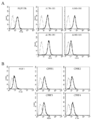
- FIG. 2 is a histogram obtained by measuring the fluorescence intensity of the IEC6 cell line before and after the addition of each fluorescently labeled FGF by FACS.
- FIG. 2A is a histogram for each fragment of FGF12B and a fragment thereof, and
- FIG. 2B is a histogram for FGF1 and each CPP-FGF1 chimeric protein.
- FIG. 3 is a graph showing the apoptosis rate of cells when the IEC6 cell line is cultured with each FGF and then irradiated with X-rays.
- FIG. 4A shows each FGF12B fragment consisting of 30 amino acids derived from different regions of FGF12B.
- FIG. 4B is a graph showing the apoptosis rate of cells when the IEC6 cell line is cultured with each FGF12B fragment and then irradiated with X-rays.
- FIG. 4C is a graph in which the fluorescence positive rate of the IEC6 cell line after adding each fluorescently labeled FGF12B fragment was measured over time by FACS.
- FIG. 4A shows each FGF12B fragment consisting of 30 amino acids derived from different regions of FGF12B.
- FIG. 4B is a graph showing the apoptosis rate of cells when the IEC6 cell line is cultured with each FGF12B fragment and then irradiated with X-ray
- FIG. 4D is a graph showing the average value of the crypt survival rate of each group when each FGF12B fragment or physiological saline was administered into the peritoneal cavity.
- FIG. 5A is a photomicrograph (200 ⁇ ) of immunohistochemical staining of the hair follicle valve region of a mouse that was administered intraperitoneally with each FGF or physiological saline after hair removal and then irradiated with ⁇ rays throughout the body.
- FIG. 5B is a graph showing the average value of the number of apoptosis per hair follicle valve in each administration group calculated by the TUNEL assay.
- FIG. 6A is a photomicrograph (200 ⁇ ) obtained by immunohistochemically staining the crypts of the small intestine of mice that were each intraperitoneally administered with each FGF or physiological saline and then irradiated whole body with ⁇ rays.
- FIG. 6B is a graph showing the average number of apoptosis per crypt in each administration group calculated by the TUNEL assay.
- FIG. 7A shows that a cross section of the small intestine of a mouse in which ⁇ -rays were irradiated to the whole body, each FGF or physiological saline was intraperitoneally administered, and BrdU was intraperitoneally administered 3.5 days after irradiation, was immunohistochemically stained with anti-BrdU. Is a micrograph (400 times).
- FIG. 7B is a graph showing the average value of the crypt survival rate in each administration group.
- FIG. 8A shows that ⁇ -rays are irradiated to the whole body, each FGF or physiological saline is intraperitoneally administered, and a transverse section of a mouse small intestine epithelial tissue immunized with anti-BrdU 3.5 days after irradiation is intraperitoneally administered. It is the microscope picture (200 times) which carried out the tissue dyeing
- FIG. 8B is a graph showing the average value of the crypt length in each administration group.
- FIG. 9 is a photomicrograph (400 magnifications) of immunohistochemical staining of the tissue of the hair follicle valve region of mice that were each intraperitoneally administered with FGF after hair removal and then ⁇ -irradiated with anti-Keratin15 antibody.
- FIG. 10A is a histogram obtained by measuring the fluorescence intensities of human pancreatic cancer cell lines MIAPaCa-2 and PANC-1 before and after adding each fluorescently labeled FGF by FACS.
- FIG. 10B is a graph showing the relationship between the absorbance of formazan (absorbance difference with respect to control) and the concentrations of FGF1 and CPPF2 that increase with cell growth of human pancreatic cancer cell lines MIAPaCa-2 and PANC-1.
- FIG. 10A is a histogram obtained by measuring the fluorescence intensities of human pancreatic cancer cell lines MIAPaCa-2 and PANC-1 before and after adding each fluorescently labeled FGF by FACS.
- FIG. 11A is a photograph of a medium obtained by culturing PANC-1 in a medium without FGF, or a medium with each FGF added, followed by fixed staining with methylene blue / methanol.
- FIG. 11B is a graph showing the average number of colonies in each group stained by the fixed staining shown in FIG. 11A.
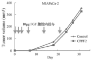
- FIG. 12 is a graph showing the increase in subcutaneous tumor volume over time in mice in which MIAPaCa-2 was subcutaneously implanted into the mouse thigh and each FGF or physiological saline was then administered intraperitoneally.
- FIG. 13A is a photomicrograph (50 ⁇ ) of a filter in which cells infiltrated into the gel by an invasion assay are fixedly stained with DiffQuick.
- FIG. 13B is a graph showing the average value of the infiltrating cell rate of each group determined by the invasion assay.
- the present invention provides a chimeric protein obtained by fusing FGF1 or FGF2 with a CPP containing the CPP-C domain of the FGF11 subfamily member, a DNA sequence encoding FGF1 or FGF2, and a DNA molecule containing a DNA sequence encoding CPP-C, or
- the present invention relates to a pharmaceutical composition comprising a vector containing the DNA sequence as an active ingredient, and medical use of the chimeric protein and the like.
- FGF1 FGF1 is a physiologically active substance known in mammals such as humans, mice, rats, cows, and horses.
- Human FGF1 has an amino acid sequence represented by SEQ ID NO: 1
- mouse FGF1 includes Some have the amino acid sequence represented by SEQ ID NO: 2.
- rat FGF1 includes an amino acid sequence represented by SEQ ID NO: 3
- bovine FGF1 includes an amino acid sequence represented by SEQ ID NO: 4
- horse FGF1 includes SEQ ID NO: 5
- the chimeric protein may be composed of FGF1 derived from any mammal, and can be selected according to the animal to be treated, for example.
- the amino acid sequence identity between these FGF1 animals is 90% or more, and the sequence identity of the amino acid sequences of FGF1 derived from other animals with respect to the amino acid sequence of human FGF1 is 92% or more. Therefore, even if it is a variant in which some amino acids in the above amino acid sequence are different, if they are composed of amino acid sequences having 90% or more sequence identity, they have the same biological or pharmacological activity. It is understood that something exists. From this viewpoint, it is preferably 70% or more, more preferably 80% or more, still more preferably 90% or more, particularly preferably, with respect to any FGF1 amino acid sequence represented by SEQ ID NOs: 1 to 5.
- the FGF1 mutant used for medical use targeting humans is preferably 70% or more, more preferably 80% or more, with respect to the amino acid sequence represented by SEQ ID NO: 1. More preferably, it is composed of an amino acid sequence having a sequence identity of 90% or more, particularly preferably 95% or more.
- the amino acid sequence present in the N-terminal region of complete FGF1 contributes to nuclear translocation of FGF1, and at least part of biological or pharmacological activities such as cell proliferation of FGF1 is translocated into the nuclear nucleus of FGF1. Is considered necessary (Non-Patent Documents 3 to 5). Therefore, it is preferable to maintain the amino acids at positions 22 to 28 in the amino acid sequence represented by SEQ ID NOs: 1 to 5.
- this nuclear translocation sequence of FGF1 can be replaced with a nuclear translocation sequence derived from another source, for example, a nuclear translocation sequence derived from yeast histone 2B (MGKKRKSKAK) or the like (non-patented). Reference 5).
- this nuclear translocation sequence is considered to maintain the nuclear translocation activity even when one to several amino acids are replaced with the same hydrophilic or hydrophobic amino acid.
- Non-patent Documents 6 and 10 since substitution of 127 Lys and 133 Lys in the amino acid sequences of SEQ ID NOs: 1 to 5 is considered to affect the binding of FGF1 to heparin, activation of FGFR or DNA synthesis (Non-patent Documents 6 and 10), It is also preferred to maintain the amino acid at position.
- the chimeric protein of the present invention is relatively stable and can exhibit a desired activity even if 127 is substituted.
- An amino acid substitution known to contribute to stabilization or optimization of the FGF1 conformation may be introduced.
- Gln at position 55 of the amino acid sequence represented by SEQ ID NOs: 1 to 5 is converted to Pro.
- Ser at position 62 can be replaced with IIe
- His at position 108 can be replaced with Gly
- Lys at position 127 can be replaced with Asn (Non-Patent Documents 9, 10 and 19).
- Such substitution may be only one amino acid or a plurality of amino acids, but the stability is improved by substituting these amino acids at all positions.
- the chimeric protein used in the present invention is relatively stable and can be transferred into cells without introducing such amino acid substitution, as demonstrated in the examples described later.
- amino acids other than those which are desirably maintained above may be substituted with other amino acids within the range having the above-described sequence identity.
- the number of amino acids to be substituted is preferably less than 10, more preferably less than 8, and still more preferably less than 5.
- FGF1 the activity of FGF1 is maintained even in a mutant lacking all or part of the C-terminal region of complete FGF1 or a variant in which another amino acid sequence is inserted in the middle of the region and the region is divided. It is done. Therefore, for example, it may be a mutant lacking all or part of the amino acids in the FGF1C terminal region of 152 to 155 in the amino acid sequence represented by SEQ ID NOs: 1 to 5, and other amino acids may be inserted in the middle of the amino acid in the FGF1C terminal region. A mutant having the amino acid sequence inserted therein may be used.
- FGF1 in which an amino acid sequence derived from another source such as CPP is inserted between the amino acid sequences 150 and 151 represented by SEQ ID NOs: 1 to 5 and the C-terminal region is divided. Mutants can be mentioned.
- the amino acid sequence of 1 to 150 represented by any one of SEQ ID NOs: 1 to 150 preferably has 90% or more sequence identity, and more preferably 95% or more sequence identity.
- FGF2 FGF2 is also a physiologically active substance known in mammals such as humans, mice, rats, cows, and horses.
- Human FGF2 has an amino acid sequence represented by SEQ ID NO: 6, and mouse FGF2 includes Some have the amino acid sequence represented by SEQ ID NO: 7.
- rat FGF2 has an amino acid sequence represented by SEQ ID NO: 8
- bovine FGF2 has an amino acid sequence represented by SEQ ID NO: 9
- horse FGF2 has SEQ ID NO: 10
- the chimeric protein may be composed of FGF2 derived from any mammal, and can be selected according to the animal to be treated, for example.
- amino acids 134 to 288 of the amino acid sequence represented by SEQ ID NO: 6 and the amino acid sequences represented by SEQ ID NOs: 7 to 10 have a sequence identity of 95% or more of each other.
- a protein comprising an amino acid sequence having a sequence identity of 80% or more, preferably 90% or more, more preferably 95% or more with respect to any of the amino acid sequences is represented by any one of SEQ ID NOs: 6 to 10. Even if some amino acids in the amino acid sequence are substituted or deleted, or other amino acids are added, it is considered that FGF2 activity is exerted.
- mutants lacking all or part of the C-terminal region of complete FGF2 or mutants in which other amino acid sequences are inserted in the middle of the region to disrupt the region Activity is believed to be maintained.
- amino acids 283 to 288 of the amino acid sequence represented by SEQ ID NO: 6 amino acids 149 to 154 of the amino acid sequence represented by SEQ ID NOs: 7 to 9, or 150 to 155 of the amino acid sequence represented by SEQ ID NO: 10 It may be a mutant lacking all or part of the amino acid, or a mutant in which another amino acid sequence is inserted in the middle of the amino acid in the FGF1C terminal region.
- Typical examples include between 282 and 283 of the amino acid sequence represented by SEQ ID NO: 6, between 148 and 149 of the amino acid sequence represented by SEQ ID NOs: 7-9, or the amino acid represented by SEQ ID NO: 10.
- An FGF2 variant in which an amino acid sequence derived from another source such as CPP is inserted between the sequences 149 and 150 to cleave the C-terminal region can be mentioned.
- FGF2 amino acids 1 to 155 of SEQ ID NOs: 1 to 5
- FGF1 amino acids 1 to 155 of SEQ ID NOs: 1 to 5
- CPP The chimeric protein used as an active ingredient in the present invention has a structure in which FGF1 or FGF2 is fused with CPP (CPP-C) containing the FGF11 subfamily CPP-C domain.
- CPP CPP
- a chimeric protein in which CPP is fused to FGF1 or the like a chimeric protein in which diphtheria toxin A is fused to FGF1 is known. Even if this chimeric protein is administered to transfer FGF1 into cells, DNA synthesis However, it was understood that FGFR must be involved in cell division and proliferation (Non-patent Documents 4 and 24). In a chimeric protein in which CPP-C is fused to FGF1, FGF1 has various physiological activities. Demonstrated.
- CPP-C can be obtained from mammals such as humans, mice, rats, cows and horses, and can be appropriately selected according to the subject of administration or purpose of use of the chimeric protein.
- the CPP-C domains of human FGF11 to 14 are represented by the amino acid sequences shown in SEQ ID NOs: 11, 12, 13, and 14, respectively.
- the CPP-C domains of mouse FGF11-14 are represented by the amino acid sequences represented by SEQ ID NOs: 15, 16, 17, and 18, respectively.
- the CPP-C domains of rat FGF11 to 14 are represented by the amino acid sequences shown in SEQ ID NOs: 19, 20, 21, and 22, respectively
- the CPP-C domains of bovine FGF11 to 14 are respectively SEQ ID NO: 23.
- 24, 25, and 26 and the CPP-C domains of equine FGF11, FGF13, and 14 are represented by the amino acid sequences shown in SEQ ID NOs: 27, 28, and 29, respectively.
- sequence identity between animals of the FGF11 subfamily CPP-C domain is 80-100% for FGF11, 100% for FGF12, 100% for FGF13, and 100% for FGF14.
- sequence difference between FGF11 subfamily CPP-C domains in humans is as shown in FIG. 1B, which has 60 to 80% sequence identity and 2 to 4 amino acids are mutually linked. Different.
- FIG. 1B has 60 to 80% sequence identity and 2 to 4 amino acids are mutually linked. Different.
- the sequence pattern of hydrophilic amino acids or neutral amino acids and hydrophobic amino acids is common among the FGF11 subfamily in humans, and the amino acid sequence N constituting the CPP-C domainin
- the third and ninth amino acids from the terminal side are hydrophilic, the seventh amino acid is neutral, the eighth is hydrophilic or neutral, and all other sites are hydrophobic.
- sequence pattern of CPP-C domenin represented by any of SEQ ID NOs: 11 to 29 and hydrophilic amino acids or neutral amino acids and hydrophobic amino acids, preferably sequence patterns of hydrophilic amino acids, neutral amino acids and hydrophobic amino acids And FGF11 subfamily CPP-C mutants having a sequence identity of 60% or more, preferably 80% or more, and more preferably 90% or more in common. Conceivable. However, it is preferable to substitute between amino acids that are closer in polarity. For example, a peptide containing CPP-C domenin composed of the following amino acids is preferable.
- Proline or leucine preferably proline
- the CPP constituting the chimeric protein may be one in which one or more amino acids are further added to both ends or one of the amino acid sequences constituting the CPP-C domain.
- CPP- consisting of more than 10 and not more than 40 amino acids.
- CPP-C derived entirely from any of FGFs 11 to 14 of various mammals and can be CPP-C consisting of more than 10 consecutive amino acids.
- the smaller the number of such additional amino acids the greater the cell membrane permeation effect. Therefore, CPP-C containing an additional amino acid is preferably composed of 40 or less, more preferably 25 or less, even more preferably 20 or less, and still more preferably 15 or less amino acid residues, and particularly preferably CPP-C domainin.
- CPP-C when CPP-C is derived from human FGF 11-14 as a whole, it contains the amino acid sequence represented by any one of SEQ ID NOs: 11-14, and preferably 40 or less amino acids, More preferably, it is composed of 25 or less consecutive amino acids, more preferably 20 or less consecutive amino acids, even more preferably 15 or less consecutive amino acids, and particularly preferably an amino acid represented by any one of SEQ ID NOS: 11 to 14 Consists only of arrays.
- the amino acid sequence constituting the CPP has a partial, preferably within a few, while maintaining the sequence pattern of the hydrophobic amino acid or neutral amino acid of the CPP-C domain and the hydrophilic amino acid as described above. Amino acids may be substituted.
- the chimeric protein according to the present invention is the one in which CPP-C is fused to FGF1 or FGF2, but they may be directly bound to each other and bound via a linking moiety consisting of a peptide. May be.
- the linking moiety consisting of a peptide is preferably composed of a hydrophilic amino acid such as aspartic acid or glutamic acid. Further, from the viewpoint of the three-dimensional structure, a linking moiety consisting of less than 10 amino acids is preferred, and a linking moiety consisting of less than 3 amino acids is more preferred.
- CPP-C can be bound to the N-terminal side of FGF1 when no other peptide is linked, but it is usually bound to the C-terminal side or inserted in the middle of the amino acid sequence of the C-terminal region. Is done. More specifically, for example, the FGF1 mutant obtained by cleaving the C-terminal side at any position of 151 to 155 of the amino acid sequences shown in SEQ ID NOs: 1 to 5, or the complete FGF1 or C-terminal region is completely CPPs can be bound to the C-terminus of the maintained FGF1 variant via or without a linking moiety. Further, for example, CPP-C can be inserted at any position of 151 to 155 of the amino acid sequences shown in SEQ ID NOs: 1 to 5 with or without one or two linking moieties.
- amino acids 283 to 288 of the amino acid sequence represented by SEQ ID NO: 6, amino acids 149 to 154 of the amino acid sequence represented by SEQ ID NOs: 7 to 9, or 150 to 150 of the amino acid sequence represented by SEQ ID NO: 10 An FGF2 mutant obtained by cleaving the C-terminal side at an arbitrary position of 155 amino acids, or a complete FGF2 or FGF2 mutant in which the C-terminal region is completely maintained, via a linking moiety or via CPP can be combined without Further, for example, amino acids 283 to 288 of the amino acid sequence represented by SEQ ID NO: 6, amino acids 149 to 154 of the amino acid sequence represented by SEQ ID NOs: 7 to 9, or 150 of the amino acid sequence represented by SEQ ID NO: 10 CPP-C can be inserted at any position of ⁇ 155 amino acids, with or without one or two linking moieties.
- Such a configuration is preferable in that the original function of FGF1 or FGF2 can be maintained because CPP-C can be introduced while maintaining an amino acid sequence highly homologous to the original FGF1 or FGF2.
- FIG. 1A schematically shows the structure of a CPP-FGF1 chimeric protein according to a preferred embodiment of the present invention.
- the amino acid sequence of FGF1 is split between 150 and 151, and the FGF11 subfamily member CPP-C is inserted at that position via the EcoRI and SalI cleavage sequences.
- CPP-C is composed of only 10 amino acids constituting the CPP-C domain, and the amino acid sequence of 1-150 of FGF1 is maintained. Therefore, it is considered that the cell membrane permeability is high and the biological or pharmacological activity of FGF1 is completely maintained. In fact, as demonstrated in the examples described later, various pharmacological actions can be exhibited at a high level. Specific amino acid sequences of such chimeric proteins are shown in SEQ ID NOs: 30 to 33.
- DNA encoding FGF1 or FGF2 is replicated by synthesis or polymerase chain reaction (PCR).
- PCR polymerase chain reaction
- a restriction enzyme cleavage site is added to an appropriate site of this DNA and cleaved with a restriction enzyme.
- a single-stranded DNA fragment that encodes CPP and also has a corresponding restriction enzyme cleavage end is synthesized and made into double-stranded by annealing. Thereafter, a DNA fragment encoding CPP is inserted and bound to the cleavage site of DNA encoding FGF1 or FGF2 using DNA ligase.
- DNA ligase One or two restriction enzymes can be used.
- any vector can be used as long as it can be replicated and maintained in the host. For example, plasmids derived from E.
- coli pBR322, pBR325, pUC12, pET-3
- plasmids derived from Bacillus subtilis bacteriophages such as ⁇ phage and their derivatives
- animal viruses such as retroviruses, adenoviruses and vaccinia viruses, insect viruses, etc.
- the gene of the chimeric protein may have ATG as a translation initiation codon at the 5 ′ end, and may have TAA, TGA, or ATG as a translation termination codon at the 3 ′ end.
- the promoter may be any as long as it is appropriate for the host used for gene expression.
- Escherichia coli for example, BL21, BL21 (DE3), BL21 (DE3) pLysS, BL21 (DE3) pLysE
- Bacillus subtilis for example, Bacillus subtilis DB305
- yeast for example, Pichia pastoris, Saccharomyces cerevisiae cells
- COS cell CHO cell, BHK cell, NIH3T2 cell, HUVE cell, LEII cell
- an applicable method may be selected depending on each host. For example, when Escherichia coli is a host, a temperature shock method or electroporation is performed on a competent cell prepared by the calcium method or other methods. Recombinant DNA or vectors can be introduced by the method.
- a transformant carrying a vector containing a recombinant DNA encoding the CPP-FGF1 chimeric protein is obtained, and by culturing this transformant, a CPP-FGF1 chimeric protein is produced.
- an appropriate medium may be selected according to the host. For example, when Escherichia coli is the host, LB medium is used, and when yeast is used, YPD medium is used.
- the culture conditions may be appropriately selected depending on each host. For example, when Escherichia coli is the host, the culture is performed at about 30 to 37 ° C. for about 3 to 24 hours. Stirring can be added.
- Examples of the method for destroying cultured cells or cells after culture and eluting the chimeric protein include homogenizer, French press, ultrasonic wave, lysozyme, and freeze-thaw.
- Purification of the chimeric protein can be carried out by a known separation method and purification method from the soluble fraction, alone or in combination. Such separation or purification methods include salting out, solvent precipitation, dialysis, ultrafiltration, gel filtration, SDS-polyacrylamide gel electrophoresis, ion exchange chromatography, affinity chromatography, reverse phase high performance liquid chromatography, And isoelectric focusing.
- a method for isolation using the heparin-binding property can be mentioned. Specifically, for example, it can be separated and purified by adsorbing the chimeric protein on heparin sepharose chromatography and eluting with a sodium chloride gradient.
- the chimeric protein obtained as described above is preferably preferably refrigerated or frozen at 4 ° C. or lower. Moreover, as long as the activity is not lost, dialysis can be performed and substitution with an appropriate solvent is possible. Furthermore, it can also be freeze-dried to obtain a dry powder.
- a recombinant DNA encoding the above-described chimeric protein or a vector having such a recombinant DNA can also be used as an active ingredient.
- the above-described chimeric protein can be expressed in the body using such a recombinant DNA or vector, and the desired treatment can be performed.
- the recombinant DNA is preferably at least 60%, preferably at least 60% of the DNA sequence encoding the amino acid sequence of FGF1 represented by any of SEQ ID NOs: 1 to 5 or FGF2 represented by any of SEQ ID NOs: 6 to 10.
- FGF1 represented by any of SEQ ID NOs: 1 to 5
- FGF2 represented by any of SEQ ID NOs: 6 to 10.
- a typical example is a recombinant DNA comprising a hydrophobic amino acid or a DNA sequence that encodes an amino acid sequence having the same sequence pattern of a neutral amino acid and a hydrophilic amino acid.
- the vector may be one generally used for gene therapy, and examples thereof include adenovirus, retrovirus, Sendai virus, plasmid, and the like, and a suitable one can be selected according to the purpose. In particular, Sendai virus is preferred.
- Examples of the method for introducing and expressing the chimeric DNA according to the present invention into a living body include membrane-fused liposomes and nanoparticles.
- the CPP-FGF1 chimeric protein according to the present invention has FGF1 as a main component, it is effective for symptoms or diseases that can be prevented or treated with natural FGF1. Therefore, cell division, cell proliferation, anti-apoptosis, stem cell in various tissues such as brain, central nervous system, kidney, placenta, adrenal gland, skin, hair, eardrum, eye, intestinal tract etc. It is effective for various medical uses involving physiological actions such as protection and angiogenesis.
- the chimeric protein of the present invention includes, but is not limited to, brain, central nervous system, kidney, placenta, adrenal gland, skin, hair, due to radiation, chemotherapy, physical intervention, apoptosis or other causes.
- Ischemic symptoms or diseases such as tympanic membranes, digestive tracts such as eyes, intestinal tract, reproductive tissues such as ovaries, degeneration, ulcers, necrosis, injury or disorder, or leg limb ischemic disease or ischemic coronary artery disease, or Prevention or prevention of proliferation or metastasis of tumor cells such as lung cancer, stomach cancer, colon cancer, pancreatic cancer, renal cell cancer, squamous cell carcinoma, malignant melanoma, endometrial cancer, ovarian cancer, bladder cancer, ureteral cancer, angiosarcoma, etc. It is effective for treatment.
- tumor cells such as lung cancer, stomach cancer, colon cancer, pancreatic cancer, renal cell cancer, squamous cell carcinoma, malignant melanoma, endometrial cancer, ovarian cancer, bladder cancer, ureteral cancer, angiosarcoma, etc. It is effective for treatment.
- the CPP-FGF1 or CPP-FGF2 chimeric protein according to the present invention can move into cells without depending on FGFR and exhibit biological or pharmacological activity by FGF1 or FGF2.
- the chimeric protein of the present invention is particularly suitable for blood cells such as lymphocytes with low FGFR expression, tissues with reduced FGFR expression due to various factors such as burns, radiation, blood flow disorders, and infections. It is effective for the prevention or treatment of tumors whose expression profile is different from that of normal tissues, or symptoms in which FGF1 or FGF2 cannot move into cells or interact with FGFR for some reason.
- the composition of the present invention is free from such a symptom or disease.
- a higher preventive or therapeutic effect can be brought about.
- diseases or symptoms include, for example, damage to skin tissue due to burns, damage to tissues such as the intestine due to radiation or chemotherapy, and apoptosis induced by radiation such as alopecia due to radiation or chemotherapy.
- Tissue loss ischemic symptoms or diseases such as lower limb ischemic disease or ischemic coronary artery disease, diabetic skin ulcer or diabetic gangrene, or lung cancer, stomach cancer, colon cancer, pancreatic cancer, renal cell cancer, squamous cell cancer, Examples include prevention or treatment of proliferation or metastasis of tumor cells such as malignant melanoma, endometrial cancer, ovarian cancer, bladder cancer, ureteral cancer, and hemangiosarcoma.
- tumor cells such as malignant melanoma, endometrial cancer, ovarian cancer, bladder cancer, ureteral cancer, and hemangiosarcoma.
- the pharmaceutical composition containing the chimeric protein or the like according to the present invention is not particularly limited with respect to the other components.
- a pharmaceutical preparation can be produced using a pharmaceutically acceptable solvent, diluent, excipient, carrier, adjuvant and the like. According to the conventional method, it can be prepared into dosage forms such as liquids, injections, powders, granules, tablets, suppositories, ointments, intestinal solvents or capsules.
- the pharmaceutical composition according to the present invention is not particularly limited with respect to the administration route, and may be administered orally or parenterally such as intravascular, subcutaneous, intraperitoneal, intratumoral, etc., depending on the indication or dosage form. it can.
- the dosage of the pharmaceutical composition according to the present invention is appropriately changed depending on the dosage form, administration route, and symptoms.
- the chimeric protein when administered intravenously to mammals including humans, is administered at 0 per day. It is preferably about 0.001 to 1 mg / kg body weight, and when administered by subcutaneous injection, the chimeric protein is preferably about 0.01 to 10 mg / kg body weight per day.
- the pharmaceutical composition of the present invention may contain an active ingredient in addition to the CPP-FGF1 chimeric protein.
- an additional active ingredient include cytokines such as G-CSF, VEGF, HGF, EGF and the like. Other cell growth factors or molecular targeted drugs targeting them can be mentioned.
- active ingredients to be used in combination are selected according to the indication. For example, in the case of tumor treatment, a molecular target drug or the like can be combined. In the prevention or treatment of radiation damage, cytokines and proliferation Factors can be combined.
- FGF1, FGF12B and FGF12B fragment FGF1 having the amino acid sequence shown in SEQ ID NO: 1 was prepared according to the method described in Non-Patent Document 8.
- FGF12B and FGF12B fragment were also prepared by the procedure described in Non-Patent Document 8.
- the amino acid sequence of FGF12B is shown in SEQ ID NO: 34.
- Chimeric protein A chimeric protein (hereinafter abbreviated as CPPF1, CPPF2, CPPF3, and CPPF4, respectively) obtained by fusing each CPP-C derived from the FGF11 subfamily FGF11, FGF12, FGF13, and FGF14 to FGF1, is described in Non-Patent Document 8. Prepared according to the method described. The related description of Non-Patent Document 8 is incorporated herein by reference. The structure of each chimeric protein is shown in FIG. 1A, and the amino acid sequences of the chimeric proteins are shown in SEQ ID NOs: 30-33. 3.
- rat small intestinal cell line IEC6 was used as a test cell, and 1 ⁇ 10 5 cells were seeded per well in a 24-well plate.
- DMEM medium containing 5% FCS and 4 ⁇ g / ml insulin was added to each well and cultured for 6 hours to attach the cells to the plate.
- fluorescently labeled FGF12B, each FGF12B fragment ( ⁇ 170-181, ⁇ 160-181, ⁇ 150-181, and ⁇ 140-181) with the C-terminal additionally trimmed by 10 residues, FGF1, CPPF1, CPPF2, CPPF3, and CPPF4 Were added to the plate so that each would be 1 ⁇ g / ml.
- the cells were detached from the plate with trypsin, the fluorescence intensity was measured with FACS, and the amount of FGF migrated into the cells was measured.
- FIG. 2A shows a FACS histogram before and after the addition of FGF12B or each FGF12B fragment
- FIG. 2B shows a FACS histogram before and after the addition of FGF1 or each CPP-FGF1 chimeric protein.
- a dotted line is a FACS histogram of a cell before adding each FGF
- a solid line is a FACS histogram of a cell after adding each FGF.
- FGF12B fragments ( ⁇ 170-181, ⁇ 160-181, and ⁇ 150-181) that retain amino acid residues 1-149 remain strong even with deletion of 10 residues from the C-terminus of FGF12B. Met. However, among these, the fluorescence intensity was maximized with the fragment obtained by cleaving the shortest amino acid residues 150-181. On the other hand, the fluorescence intensity decreased sharply in the fragment obtained by cleaving amino acid residues 140-181.
- amino acid residues 140 to 149 of FGF12B are CPP-C domains.
- amino acids are added before and after this CPP-C domain, the intracellular translocation ability is retained. was shown to be. It was also inferred that the smaller the number of amino acids added before and after the CPP-C domain, the higher the ability to move into the cell.
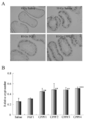
- the rat small intestinal cell line IEC6 not expressing FGFR was used as a test cell, and 3 ⁇ 10 4 cells were seeded in each 3.5 cm dish, and each dish contained 5% FCS and 4 ⁇ g / ml insulin. DMEM medium was added. Each dish was placed in an incubator with an atmosphere of 37 ° C. and 5% CO 2 and cultured for 16 hours.
- heparin was added to each medium at a concentration of 5 ⁇ g / ml, FGF was not added in the control group, and FGF1, CPPF1, CPPF2, CPPF3, and CPPF4 were added to each test group at a concentration of 100 ng / ml, After further incubation for 24 hours, X-rays were irradiated with 20 Gy. After 24 hours of irradiation, the cells were fixed with 2% glutaraldehyde, subjected to nuclear staining with 20 ⁇ g / ml Hoechst 33258, and 10 fields of one field of 200 cells or more were examined with an inverted fluorescence microscope, and the number of cells with nuclear condensation was calculated.
- This nucleus-aggregated cell was regarded as a cell in which apoptosis was induced by X-ray irradiation, and the ratio of the number of nucleus-aggregated cells to the total number of cells examined in each field was evaluated as the apoptosis rate.
- FIG. 3 shows the average value of apoptotic rate +/ ⁇ standard deviation (SD) of the control group and each test group.
- SD standard deviation
- the apoptosis rate reached about 45%. Further, in the test group to which FGF1 was added, no significant decrease in the apoptosis rate was observed with respect to the control group. On the other hand, in the test groups to which CPP-FGF1 chimeric protein (CPPF1, CPPF2, CPPF3, and CPPF4) was added, the apoptosis rate was significantly reduced compared to the control group. This demonstrates that FGF1 cannot effectively suppress apoptosis of cells that do not express FGFR, but the CPP-FGF1 chimeric protein can suppress apoptosis even in such cells. It was.
- FGF1 which is expected to suppress apoptosis through FGFR, did not show a significant difference from the control, is consistent with the absence of FGFR expression in IEC6 cells.
- the ability of the CPP-FGF1 chimeric protein to suppress apoptosis is likely due to the property that the CPP-FGF1 chimeric protein can move into cells without depending on the expression of FGFR.
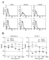
- FGF12B and each FGF12 fragment shown in FIG. 4A were used as FGF.
- the P8 fragment contains CPP-M, and the P11 and P12 fragments contain CPP-C.
- rat small intestinal cell line IEC6 was also used in this test.
- the test procedure is the same as the evaluation related to the apoptosis-inhibiting effect described above.
- 4B represents the mean value of apoptotic rate +/ ⁇ standard deviation (SD) of the control group and each test group.
- SD standard deviation
- the apoptosis rate reached about 45%. Moreover, in the test groups to which P8, P10, and P12 were added, the apoptosis rate was significantly reduced compared to the control group. On the other hand, in the test group to which P11 containing CPP-C was added, the apoptosis rate was not significantly decreased compared to the control group. This demonstrated that P12 consisting of 30 amino acids containing CPP-C suppresses apoptosis, but CPP-C itself consisting of 10 amino acids cannot suppress apoptosis. Furthermore, peptides containing central CPP-M domainin were also demonstrated to inhibit apoptosis.
- FIG. 4C shows the ability of the C-terminal peptide of FGF12B to translocate into the cell. It is the graph which measured the fluorescence positive rate of the IEC6 cell line after adding each fluorescently labeled peptide by the density
- P12 containing CPP-C migrated into the cell with a peak at 24 hours.
- P11 containing CPP-C had a lower fluorescence positive rate than P12, it migrated into the cell at the peak of 24 hours like P12.
- P10 and P13 had a very low fluorescence positive rate after 24 hours.
- FIG. 4D is a graph showing the average value of the crypt survival rate in the peptide or physiological saline peritoneal administration group.
- 0.5 ml of 5% mouse serum-containing physiological saline was administered to the abdominal cavity of the mice.
- 100 ⁇ g of P8, P10, and P12 were respectively administered.
- the solution was diluted with 0.5 ml of physiological saline containing 5% mouse serum and administered to the abdominal cavity of the mouse. Twenty-four hours later, 10 Gy gamma rays were whole-body irradiated to each group of mice at a dose rate of 0.5 Gy / min.
- mice were euthanized 3.5 days after irradiation and jejunum was collected. After fixing the jejunum with 10% formalin, paraffin-embedded sections were prepared, and the sections were stained with HE. A crypt having 10 or more crypt cells was judged to be viable by a microscope, and the number of crypts per cross section was counted for 10 intestinal cross sections, and the average value was calculated. Furthermore, this average value was divided by the average value of the number of crypts per cross section of the non-irradiated group to obtain a relative value (crypto survival rate). The average value of the crypt survival rate of 3 mice in each group +/ ⁇ standard deviation (SD) is shown.
- SD standard deviation
- jejunal crypt survival was significantly higher than that in the control group, but in the group administered P10, jejunal crypt survival was not significantly higher than that in the control group. It was.
- hair follicle damage prevention effect In this test, the prevention effect of CPP-FGF1 chimeric protein against hair loss and hair follicle damage caused by radiation was evaluated. Hair follicles actively undergo cell division during the growth phase and are highly sensitive to radiation during this period. For this reason, when the hair follicle is irradiated with radiation at this time, apoptosis is likely to be caused, but this apoptosis is an index of hair follicle damage. Therefore, the inhibitory effect of hair follicle damage was evaluated by measuring the inhibitory effect of CPP-FGF1 chimeric protein on radiation-induced apoptosis in growing mouse hair follicles.
- Hair removal was performed from the back of male BALB / c mice 51-53 days old, and the resting hair follicles were induced to the growth phase.
- 0.5 ml of 5% mouse serum-containing physiological saline was administered to the abdominal cavity of the mouse, and in the test group, 100 ⁇ g of FGF1, FGF12, CPPF1, CPPF2, CPPF3 and CPPF4, respectively.
- the solution was diluted with 0.5 ml of physiological saline containing 5% mouse serum and administered to the abdominal cavity of the mouse. 24 hours later, whole body irradiation was performed with 12 Gy of gamma rays at a dose rate of 0.5 Gy / min.
- mice were euthanized 24 hours after irradiation, skin was collected, fixed with 10% formalin, paraffin-embedded sections were prepared, and TUNEL assay was performed. TUNEL positive cells were regarded as apoptotic cells, and the number of apoptosis for each hair follicle bulb was calculated over 3 fields of view.
- FIG. 5A is a photomicrograph (200 ⁇ ) of immunohistochemical staining of the hair follicle valve region of each group of mice by TUNEL assay, and the arrows in the figure indicate TUNEL positive cells (ie, apoptotic cells).
- FIG. 5B shows the mean value +/ ⁇ standard deviation (SD) of the number of apoptosis per hair follicle bulb of 3 or more fields in each group, and *** in FIG. 5 indicates physiological saline containing 5% mouse serum. The test group which became P ⁇ 0.001 by the multiple test with respect to the control group which administered water is shown.
- SD standard deviation
- mice In 8-week-old male BALB / c mice, in the control group, 0.5 ml of physiological saline containing 5% mouse serum was administered to the abdominal cavity of the mice. In the test group, 100 ⁇ g of FGF1, FGF12, CPPF1, CPPF2, CPPF3, and CPPF4 were diluted with 0.5 ml of 5% mouse serum-containing saline and administered to the abdominal cavity of mice. After 24 hours, each mouse was whole-body irradiated with 12 Gy gamma rays at a dose rate of 0.5 Gy / min.
- mice were euthanized 24 hours after irradiation, the small intestine was collected, fixed with 10% formalin, paraffin-embedded sections were prepared, and a TUNEL assay was performed. TUNEL positive cells were regarded as apoptotic cells, and the number of apoptosis per crypt was calculated in 10 fields.
- FIG. 6A is a photomicrograph of immunohistochemical staining of small intestine crypts in each group of mice by TUNEL assay, and arrows in the figure indicate TUNEL positive cells (ie, apoptotic cells).
- FIG. 6B shows the mean value +/ ⁇ standard deviation (SD) of the number of TUNEL positive cells per crypt of 10 fields in each group, and *** in FIG. 6 indicates physiological saline containing 5% mouse serum. The test group which became P ⁇ 0.001 by the multiple test with respect to the control group which administered No. is shown.
- SD standard deviation
- the apoptosis reduction rate compared with the control group was only 18.1% in the FGF1 administration group, but 66% in the CPPF1 administration group, 63.1% in the CPPF2 administration group, 64.2% in the CPPF3 administration group, and CPPF4
- the CPP-FGF1 administration group significantly decreased apoptosis compared with the FGF1 administration group (P ⁇ 0.001).
- the apoptosis reduction rate of the FGF12 administration group with respect to the control group was 50.5%, and the apoptosis was significantly reduced compared to the FGF12 group. This demonstrated that the CPP-FGF chimeric protein has a higher protective effect against radiation-induced small intestine damage than FGF1 and FGF12.
- Evaluation 1 on the effect of promoting the recovery of damaged small intestine In this test, the effect of promoting the recovery of the small intestine damaged by radiation of the CPP-FGF1 chimeric protein was evaluated using the number of crypts regenerated after irradiation as an index.
- mice 8 weeks old male BALB / c mice were used, and 10 Gy gamma rays were first irradiated to the mice of each group at a dose rate of 0.5 Gy / min. 24 hours later, in the control group, 0.5 ml of physiological saline containing 5% mouse serum was administered to the abdominal cavity of the mouse, and in the test group, 10 ⁇ g of FGF1, CPPF1, CPPF2, CPPF3, and CPPF4 were each added in 0.5 ml. was diluted with physiological saline containing 5% mouse serum and administered to the abdominal cavity of mice.
- BrdU labeling solution was injected intraperitoneally 3.5 days after irradiation, BrdU was taken up into the cell dividing cells, the mouse was euthanized 2 hours later, and the jejunum was collected. After fixing the jejunum with 10% formalin, paraffin-embedded sections were prepared, and the sections were immunohistologically stained with an anti-BrdU antibody and then stained with hematoxylin.
- FIG. 7A is a photomicrograph of a cross-section of the intestine showing a crypt containing cells that have incorporated BrdU and bound with an anti-BrdU antibody.
- Cryptograms containing 10 or more anti-BrdU antibody positive cells were judged to be viable by a microscope, and the number of crypts per cross section was counted for 10 intestinal cross sections, and the average value was calculated. Furthermore, this average value was divided by the average value of the number of crypts per cross section of the non-irradiated group to obtain a relative value (crypto survival rate).
- FIG. 7B shows the mean value of the crypt survival rate of three mice in each group +/ ⁇ standard deviation (SD), and ** in the figure is administered 5% mouse serum-containing physiological saline.
- the jejunum crypt survival rate was only 0.26 by 10 Gy whole-body gamma irradiation, and it was not significantly increased even in the FGF1 administration group.
- the jejunum crypt survival rate was 0.45, 0.48, 0.48, and 0.51, respectively. It was significantly higher than the control group as well as the FGF1 administration group (P ⁇ 0.05). This result also demonstrates that the CPP-FGF1 chimeric protein has an extremely high recovery promoting effect on the small intestine damaged by radiation compared to FGF1.
- mice 8 weeks old male BALB / c mice were used, and 10 Gy gamma rays were irradiated to the mice of each group at a dose rate of 0.5 Gy / min. 24 hours later, in the control group, 0.5 ml of physiological saline containing 5% mouse serum was administered to the abdominal cavity of the mouse, and in the test group, 10 ⁇ g of FGF1, CPPF1, CPPF2, CPPF3, and CPPF4 were each added in 0.5 ml. was diluted with physiological saline containing 5% mouse serum and administered to the abdominal cavity of mice.
- BrdU labeling solution was injected intraperitoneally 3.5 days after irradiation, BrdU was taken up into the cell dividing cells, the mouse was euthanized 2 hours later, and the jejunum was collected. After fixing the jejunum with 10% formalin, paraffin-embedded sections were prepared, and the sections were immunohistologically stained with an anti-BrdU antibody and then stained with hematoxylin.
- FIG. 8A is a photomicrograph of the small intestinal epithelium stained with immunohistochemistry showing crypts having cells to which BrdU was incorporated and anti-BrdU antibody was bound in each group.
- Three images of each group of tissue images were taken with a microscope, the length of 10 crypts was measured for each image, the average value was obtained for each group, and the relative value to the control group administered with physiological saline based on this average value was calculated. Calculated.
- FIG. 8B shows the average relative value +/ ⁇ standard deviation (SD) of the crypt length of each group, and *** in the figure is P ⁇ 0.001 by multiple testing with respect to the control group. A test group is shown.
- SD standard deviation
- the FGF1 administration group had significantly longer jejunal crypts 3.5 days after 10 Gy whole-body gamma irradiation compared to the control group.
- jejunal crypt was not less than twice as long as that in the control group, but also in comparison with the FGF1 administration group. Even so, it was significantly longer (P ⁇ 0.01 to 0.001). This result also demonstrated that CPP-C fusion FGF had a higher recovery promoting effect on the small intestine damaged by radiation than FGF1.
- Hair removal was performed from the back of male BALB / c mice 51-53 days old, and the resting hair follicles were induced to the growth phase.
- 0.5 ml of 5% mouse serum-containing physiological saline was administered to the abdominal cavity of the mouse.
- 100 ⁇ g of FGF1, CPPF1, CPPF2, CPPF3, and CPPF4 were each 0
- the solution was diluted with 5 ml of 5% mouse serum-containing physiological saline and administered to the abdominal cavity of the mouse. Twenty-four hours later, 12 Gy of gamma rays was whole-body irradiated at a dose rate of 0.5 Gy / min.
- mice were euthanized 24 hours after irradiation, skin was collected and fixed with 10% formalin. Paraffin-embedded sections were prepared, and immunohistochemical staining was performed with an antibody against Keratin 15 which is a marker of hair follicle stem cells.
- FIG. 9 shows photomicrographs of hair follicle bulge regions stained with immunohistochemistry in the non-irradiated group, the control group administered with 5% mouse serum-containing physiological saline, and the arrows indicate Keratin15 positive hair follicle stem cells. Indicates. In the control group administered with 5% mouse serum-containing physiological saline and irradiated with 12 Gy of whole body gamma rays, keratin15-positive hair follicle stem cells in the hair follicle bulge region decreased compared to the non-irradiated group. In the FGF1 administration group, hair follicle stem cells were decreased by irradiation.
- the number of hair follicle stem cells in the bulge region was significantly larger than that in the control group as well as the FGF1 administration group, and hair follicle stem cells The number reached a level higher than the non-irradiated control group.
- This result demonstrates that the CPP-FGF1 chimeric protein has a higher effect of protecting and maintaining hair follicle stem cells against radiation compared to FGF1.
- WST-1 which is a stable tetrazolium salt, is degraded to soluble formazan on the surface of cells having metabolic activity, and thus directly correlates with the number of cells having metabolic activity in culture. Therefore, the amount of formazan before and after each FGF administration was measured by absorbance at 450 nm to evaluate the effect of suppressing tumor cell growth.
- FIG. 10B is a graph showing the relationship between the concentrations of FGF1 and CPPF2 and the amount of formazan that increases with cell proliferation, and the vertical axis shows the difference in absorbance with respect to the OD450 value of the control. Therefore, the higher the value, the higher the level of cell proliferation relative to the control group.
- Each 6 cm dish is seeded with 100 human pancreatic cancer cell lines PANC-1 cells, DMEM medium containing 10% FCS and 5 ⁇ g / ml heparin is added to the dish, FGF is not added in the control group, and in the test group, Furthermore, after each FGF was added so that it might become 100 ng / ml, the culture solution of each group was cultured for 13 days. Thereafter, the cells were fixedly stained with 1% methylene blue / 30% methanol, and the number of colonies of 50 cells or more stained with each group of dishes was calculated to evaluate the proliferation ability of cancer cells.
- FIG. 11A is a photograph showing the medium after staining of the control group and the group to which each FGF was added.
- FIG. 11B shows the mean value +/ ⁇ standard deviation (SD) of the number of colonies in each group.
- SD standard deviation
- the colony reduction rate relative to the control group was 37.8% in the CPPF1 addition group, 48.6% in the CPPF2 addition group, 40.5% in the CPPF3 addition group, 45.9% in the CPPF4 addition group, and any CPP-FGF1.
- the number of colonies decreased by about 40%.
- the number of colonies was significantly reduced in the group to which CPP-FGF1 chimeric protein was added (P ⁇ 0.05 to 0.01). This result demonstrated that the CPP-FGF1 chimeric protein can significantly suppress cancer growth more than FGF1.
- mice were conducted with consideration for animal ethics based on a pre-approved animal experiment plan.
- 1 ⁇ 10 6 human pancreatic cancer cell line MIAPaCa-2 was suspended in 10 ⁇ l of phosphate buffered saline (PBS) and injected subcutaneously into the right thigh of a 7 week old male SCID mouse.
- PBS phosphate buffered saline
- 0.5 ml of 5% mouse serum-containing physiological saline was administered to the abdominal cavity of the mice for 1 hour, 24 hours, 48 hours, 7 days, 14 days, and 21 days later.
- FIG. 12 shows the time-dependent volume change of the right femoral subcutaneous tumor in the control group and the test group.
- the arrows indicate the timing of intraperitoneal administration.
- the average tumor volume was always smaller than that in the control group from day 18 to day 31. From this result, it was shown that CPP-FGF1 chimeric protein has an effect of suppressing tumor cell mass formation.
- Cancer cells have the property of secreting proteases when cell metastasis occurs, destroying the basement membrane and migrating, and using an invasion assay that utilizes the characteristics of these cancer cells, the CPP-FGF1 chimeric protein has the effect of inhibiting cancer cell invasion Evaluated.
- a filter of the Boyden chamber of a 24-well plate was coated with 20 ⁇ L of 66 ⁇ g of Matrigel and gelled. Thereafter, 650 ⁇ L of DMEM culture medium containing 10% FCS was added to the lower well. Meanwhile, 1.5 ⁇ 10 5 MIAPaCa-2 cells or PANC-1 cells were suspended in a DMEM culture solution containing 100 ⁇ L of 0.35% BSA, and this suspension was added to the upper well. Subsequently, 5 ⁇ g / ml heparin was added to the culture medium of the lower well and the upper well, FGF was not added to the wells of the control group, and FGF1 and CPPF2 were added to the wells of the test group to 100 ng / mL, respectively.
- the plate was placed in an incubator at 37 ° C. and 5% CO 2 and cultured for 24 hours to induce invasion of the cancer cells into the gel.
- the infiltrated cells were fixedly stained with DiffQuick (manufactured by Sysmex) together with the filter of the chamber, and the number of stained cells was calculated and used as the number of infiltrating cells.
- the test was performed by assigning four chambers to each group, obtaining an average value thereof, and determining the ratio of the average value of the number of infiltrating cells in each group to the number of cells suspended in the culture solution, and obtaining the infiltrating cell rate.
- FIG. 13A is a photomicrograph of a filter in which cells infiltrated into the gel are fixedly stained with DiffQuick.
- FIG. 13B shows the mean value +/ ⁇ standard deviation (SD) of the infiltrating cell ratio of each group, and in the figure, ** is a test group in which P ⁇ 0.01 by multiple testing with respect to the control group. *** indicates a test group in which P ⁇ 0.001.
- SD standard deviation
- MIAPaCa-2 cells infiltrated 2.34% in the control group, but infiltrated 1.52% in the FGF1 addition group and 1.03% in the CPPF2 addition group.
- PANC-1 cells infiltrated at a rate of 1.27% in the control group, whereas 0.81% in the FGF1-added group and 0.26% infiltrated in the CPPF2-added group.
- FGF1 suppressed invasion of cancer cells by 35% in MIAPaCa cells and 36% by PANC-1
- CPP-C fusion FGF (CPPF2) showed 56% in MIAPaCa cells.
- CPP-FGF1 chimeric protein further reduced the invasive ability of cancer cells and further suppressed cancer metastasis compared to FGF1.
Landscapes
- Health & Medical Sciences (AREA)
- Chemical & Material Sciences (AREA)
- Life Sciences & Earth Sciences (AREA)
- Organic Chemistry (AREA)
- General Health & Medical Sciences (AREA)
- Medicinal Chemistry (AREA)
- Pharmacology & Pharmacy (AREA)
- Nuclear Medicine, Radiotherapy & Molecular Imaging (AREA)
- General Chemical & Material Sciences (AREA)
- Animal Behavior & Ethology (AREA)
- Chemical Kinetics & Catalysis (AREA)
- Public Health (AREA)
- Veterinary Medicine (AREA)
- Engineering & Computer Science (AREA)
- Bioinformatics & Cheminformatics (AREA)
- Toxicology (AREA)
- Gastroenterology & Hepatology (AREA)
- Zoology (AREA)
- Proteomics, Peptides & Aminoacids (AREA)
- Molecular Biology (AREA)
- Genetics & Genomics (AREA)
- Biophysics (AREA)
- Biochemistry (AREA)
- Dermatology (AREA)
- Diabetes (AREA)
- Immunology (AREA)
- Cell Biology (AREA)
- Urology & Nephrology (AREA)
- Endocrinology (AREA)
- Cardiology (AREA)
- Heart & Thoracic Surgery (AREA)
- Neurosurgery (AREA)
- Biomedical Technology (AREA)
- Reproductive Health (AREA)
- Ophthalmology & Optometry (AREA)
- Oncology (AREA)
- Neurology (AREA)
- Vascular Medicine (AREA)
- Obesity (AREA)
- Hematology (AREA)
Priority Applications (2)
| Application Number | Priority Date | Filing Date | Title |
|---|---|---|---|
| JP2014510567A JP5818977B2 (ja) | 2012-11-28 | 2013-11-11 | 細胞膜透過性の線維芽細胞増殖因子の医療用途 |
| US14/647,634 US20150299280A1 (en) | 2012-11-28 | 2013-11-11 | Medical treatment use of cell-membrane-permeable fibroblast growth factor |
Applications Claiming Priority (2)
| Application Number | Priority Date | Filing Date | Title |
|---|---|---|---|
| JP2012-259816 | 2012-11-28 | ||
| JP2012259816 | 2012-11-28 |
Publications (1)
| Publication Number | Publication Date |
|---|---|
| WO2014084027A1 true WO2014084027A1 (ja) | 2014-06-05 |
Family
ID=50827673
Family Applications (1)
| Application Number | Title | Priority Date | Filing Date |
|---|---|---|---|
| PCT/JP2013/080382 Ceased WO2014084027A1 (ja) | 2012-11-28 | 2013-11-11 | 細胞膜透過性の線維芽細胞増殖因子の医療用途 |
Country Status (3)
| Country | Link |
|---|---|
| US (1) | US20150299280A1 (https=) |
| JP (2) | JP5818977B2 (https=) |
| WO (1) | WO2014084027A1 (https=) |
Cited By (3)
| Publication number | Priority date | Publication date | Assignee | Title |
|---|---|---|---|---|
| JP2016196440A (ja) * | 2015-04-06 | 2016-11-24 | 公立大学法人大阪市立大学 | サイトグロビン発現増強剤 |
| US10385113B2 (en) | 2016-03-30 | 2019-08-20 | The Board Of Trustees Of The University Of Arkansas | Engineered FGF compositions and methods of use thereof |
| US11267855B2 (en) | 2018-03-16 | 2022-03-08 | The Board Of Trustees Of The University Of Arkansas | Engineered FGF1 and FGF2 compositions and methods of use thereof |
Families Citing this family (3)
| Publication number | Priority date | Publication date | Assignee | Title |
|---|---|---|---|---|
| JP7682470B2 (ja) * | 2019-12-16 | 2025-05-26 | Jsr株式会社 | オルガノイドの製造方法 |
| CN114874990A (zh) * | 2021-02-05 | 2022-08-09 | 中国科学院苏州纳米技术与纳米仿生研究所 | 一种功能化外泌体及其制备方法和应用 |
| CZ309550B6 (cs) * | 2021-06-15 | 2023-04-05 | Enantis s.r.o | Termostabilní polypeptid na bázi FGF18 a jeho použití |
-
2013
- 2013-11-11 WO PCT/JP2013/080382 patent/WO2014084027A1/ja not_active Ceased
- 2013-11-11 JP JP2014510567A patent/JP5818977B2/ja not_active Expired - Fee Related
- 2013-11-11 US US14/647,634 patent/US20150299280A1/en not_active Abandoned
-
2015
- 2015-09-29 JP JP2015191288A patent/JP6183757B2/ja not_active Expired - Fee Related
Non-Patent Citations (6)
Cited By (3)
| Publication number | Priority date | Publication date | Assignee | Title |
|---|---|---|---|---|
| JP2016196440A (ja) * | 2015-04-06 | 2016-11-24 | 公立大学法人大阪市立大学 | サイトグロビン発現増強剤 |
| US10385113B2 (en) | 2016-03-30 | 2019-08-20 | The Board Of Trustees Of The University Of Arkansas | Engineered FGF compositions and methods of use thereof |
| US11267855B2 (en) | 2018-03-16 | 2022-03-08 | The Board Of Trustees Of The University Of Arkansas | Engineered FGF1 and FGF2 compositions and methods of use thereof |
Also Published As
| Publication number | Publication date |
|---|---|
| US20150299280A1 (en) | 2015-10-22 |
| JP6183757B2 (ja) | 2017-08-23 |
| JP2016029071A (ja) | 2016-03-03 |
| JPWO2014084027A1 (ja) | 2017-01-05 |
| JP5818977B2 (ja) | 2015-11-18 |
Similar Documents
| Publication | Publication Date | Title |
|---|---|---|
| JP6183757B2 (ja) | 細胞膜透過性の線維芽細胞増殖因子の医療用途 | |
| JP7339372B2 (ja) | 代謝障害を処置するための融合タンパク質 | |
| JP6110304B2 (ja) | Fgf21関連障害を処置する方法 | |
| JP5414078B2 (ja) | 高機能化キメラ蛋白質を含有する細胞増殖促進用培地 | |
| US8324160B2 (en) | Chimeric polypeptides and uses thereof | |
| KR20090045940A (ko) | 세포자멸사의 억제를 위한 약학 조성물, 및 이를 전달하는 방법 | |
| JP2015091850A (ja) | キメラのコイルドコイル(二重コイル)分子を使用する方法 | |
| CN109535243B (zh) | 人肝细胞生长因子突变体及其应用 | |
| KR20120034927A (ko) | 피부투과성 인간 상피세포 성장인자 및 그 생산방법 | |
| Barbon et al. | In vitro assessment of TAT—Ciliary Neurotrophic Factor therapeutic potential for peripheral nerve regeneration | |
| EP3125921B1 (en) | Fgf21 variants for use in treating hiv-haart induced partial lipodystrophy | |
| US12447199B2 (en) | System and methods to promote cellular regeneration following ischemia | |
| AU2016203988B2 (en) | Protein to promote blood vessel growth and uses thereof | |
| US20110092441A1 (en) | Cell-permeable endostatin recombinant protein, a polynucleotide encoding the same, and an anti-cancer preparation containing the same as an active component | |
| JP4537206B2 (ja) | 血管新生を阻害するharp因子のペプチドフラグメント | |
| AU2008256550B2 (en) | VEGF-D mutants and their use | |
| PT90118B (pt) | Processo para a preparacao de novos polipeptidos exibindo actividade de factor de crescimento e de sequencias de acidos nucleicos que codificam esses polipeptidos | |
| JP6944875B2 (ja) | Met受容体アゴニストタンパク質 | |
| US10125183B2 (en) | Decoy peptides inhibiting binding of AMIGO2 and 3-phosphoinositide-dependent kinase 1 | |
| Boettcher et al. | Patent: Methods of Treating FGF21-Associated Disorders | |
| CN107531802A (zh) | 来自肝细胞生长因子/散射因子(hgf/sf)的kringle结构域的多聚体化合物 |
Legal Events
| Date | Code | Title | Description |
|---|---|---|---|
| ENP | Entry into the national phase |
Ref document number: 2014510567 Country of ref document: JP Kind code of ref document: A |
|
| 121 | Ep: the epo has been informed by wipo that ep was designated in this application |
Ref document number: 13858885 Country of ref document: EP Kind code of ref document: A1 |
|
| WWE | Wipo information: entry into national phase |
Ref document number: 14647634 Country of ref document: US |
|
| NENP | Non-entry into the national phase |
Ref country code: DE |
|
| 122 | Ep: pct application non-entry in european phase |
Ref document number: 13858885 Country of ref document: EP Kind code of ref document: A1 |