WO2012053035A1 - インプラント用材料、その製造方法及び骨細胞との親和性向上方法 - Google Patents
インプラント用材料、その製造方法及び骨細胞との親和性向上方法 Download PDFInfo
- Publication number
- WO2012053035A1 WO2012053035A1 PCT/JP2010/006247 JP2010006247W WO2012053035A1 WO 2012053035 A1 WO2012053035 A1 WO 2012053035A1 JP 2010006247 W JP2010006247 W JP 2010006247W WO 2012053035 A1 WO2012053035 A1 WO 2012053035A1
- Authority
- WO
- WIPO (PCT)
- Prior art keywords
- functional group
- group containing
- abundance ratio
- less
- carbonaceous film
- Prior art date
- Legal status (The legal status is an assumption and is not a legal conclusion. Google has not performed a legal analysis and makes no representation as to the accuracy of the status listed.)
- Ceased
Links
Images


Classifications
-
- A—HUMAN NECESSITIES
- A61—MEDICAL OR VETERINARY SCIENCE; HYGIENE
- A61C—DENTISTRY; APPARATUS OR METHODS FOR ORAL OR DENTAL HYGIENE
- A61C8/00—Means to be fixed to the jaw-bone for consolidating natural teeth or for fixing dental prostheses thereon; Dental implants; Implanting tools
- A61C8/0012—Means to be fixed to the jaw-bone for consolidating natural teeth or for fixing dental prostheses thereon; Dental implants; Implanting tools characterised by the material or composition, e.g. ceramics, surface layer, metal alloy
-
- A—HUMAN NECESSITIES
- A61—MEDICAL OR VETERINARY SCIENCE; HYGIENE
- A61C—DENTISTRY; APPARATUS OR METHODS FOR ORAL OR DENTAL HYGIENE
- A61C8/00—Means to be fixed to the jaw-bone for consolidating natural teeth or for fixing dental prostheses thereon; Dental implants; Implanting tools
- A61C8/0012—Means to be fixed to the jaw-bone for consolidating natural teeth or for fixing dental prostheses thereon; Dental implants; Implanting tools characterised by the material or composition, e.g. ceramics, surface layer, metal alloy
- A61C8/0013—Means to be fixed to the jaw-bone for consolidating natural teeth or for fixing dental prostheses thereon; Dental implants; Implanting tools characterised by the material or composition, e.g. ceramics, surface layer, metal alloy with a surface layer, coating
-
- A—HUMAN NECESSITIES
- A61—MEDICAL OR VETERINARY SCIENCE; HYGIENE
- A61L—METHODS OR APPARATUS FOR STERILISING MATERIALS OR OBJECTS IN GENERAL; DISINFECTION, STERILISATION OR DEODORISATION OF AIR; CHEMICAL ASPECTS OF BANDAGES, DRESSINGS, ABSORBENT PADS OR SURGICAL ARTICLES; MATERIALS FOR BANDAGES, DRESSINGS, ABSORBENT PADS OR SURGICAL ARTICLES
- A61L27/00—Materials for grafts or prostheses or for coating grafts or prostheses
- A61L27/02—Inorganic materials
- A61L27/04—Metals or alloys
- A61L27/06—Titanium or titanium alloys
-
- A—HUMAN NECESSITIES
- A61—MEDICAL OR VETERINARY SCIENCE; HYGIENE
- A61L—METHODS OR APPARATUS FOR STERILISING MATERIALS OR OBJECTS IN GENERAL; DISINFECTION, STERILISATION OR DEODORISATION OF AIR; CHEMICAL ASPECTS OF BANDAGES, DRESSINGS, ABSORBENT PADS OR SURGICAL ARTICLES; MATERIALS FOR BANDAGES, DRESSINGS, ABSORBENT PADS OR SURGICAL ARTICLES
- A61L27/00—Materials for grafts or prostheses or for coating grafts or prostheses
- A61L27/28—Materials for coating prostheses
- A61L27/30—Inorganic materials
- A61L27/303—Carbon
-
- A—HUMAN NECESSITIES
- A61—MEDICAL OR VETERINARY SCIENCE; HYGIENE
- A61F—FILTERS IMPLANTABLE INTO BLOOD VESSELS; PROSTHESES; DEVICES PROVIDING PATENCY TO, OR PREVENTING COLLAPSING OF, TUBULAR STRUCTURES OF THE BODY, e.g. STENTS; ORTHOPAEDIC, NURSING OR CONTRACEPTIVE DEVICES; FOMENTATION; TREATMENT OR PROTECTION OF EYES OR EARS; BANDAGES, DRESSINGS OR ABSORBENT PADS; FIRST-AID KITS
- A61F2/00—Filters implantable into blood vessels; Prostheses, i.e. artificial substitutes or replacements for parts of the body; Appliances for connecting them with the body; Devices providing patency to, or preventing collapsing of, tubular structures of the body, e.g. stents
- A61F2/02—Prostheses implantable into the body
- A61F2/30—Joints
- A61F2/30767—Special external or bone-contacting surface, e.g. coating for improving bone ingrowth
- A61F2002/3093—Special external or bone-contacting surface, e.g. coating for improving bone ingrowth for promoting ingrowth of bone tissue
-
- A—HUMAN NECESSITIES
- A61—MEDICAL OR VETERINARY SCIENCE; HYGIENE
- A61L—METHODS OR APPARATUS FOR STERILISING MATERIALS OR OBJECTS IN GENERAL; DISINFECTION, STERILISATION OR DEODORISATION OF AIR; CHEMICAL ASPECTS OF BANDAGES, DRESSINGS, ABSORBENT PADS OR SURGICAL ARTICLES; MATERIALS FOR BANDAGES, DRESSINGS, ABSORBENT PADS OR SURGICAL ARTICLES
- A61L2400/00—Materials characterised by their function or physical properties
- A61L2400/18—Modification of implant surfaces in order to improve biocompatibility, cell growth, fixation of biomolecules, e.g. plasma treatment
Definitions
- the present disclosure relates to an implant material, a method for producing the same, and a method for improving affinity with bone cells, and more particularly, to a material that requires affinity for bone cells such as artificial tooth roots and dentures and a method for producing the same.
- titanium and titanium alloys are used as a base material for implants embedded in living bodies such as artificial tooth roots because they are excellent in biocompatibility, corrosion resistance and mechanical strength.
- an artificial dental root made of titanium or titanium alloy is directly fixed to the jawbone, the metabolic balance of bone tissue regeneration of the jawbone may be lost, and the artificial root may loosen or the jawbone may be destroyed.
- peri-implantitis As a cause of the failure of such artificial tooth root implantation, there is peri-implantitis in which osteoclasts are induced around the artificial tooth root and bone destruction occurs. Peri-implantitis may result from microbial stimulation such as bacterial infection and mechanical stimulation such as excessive bite force, but also occurs due to osteoclast activation by the artificial tooth root itself.
- osteoclasts Activation of osteoclasts by artificial tooth roots also occurs at the initial implantation of the implant. Osteoclasts that appear at the time of initial implantation interfere with the osseointegration of the implant and cause the implant to fail.
- osteoclasts Such activation of osteoclasts is thought to be caused by the fact that titanium and titanium alloys do not have sufficient affinity with bone cells. On the surface of a material having insufficient affinity for bone cells, differentiation from osteoclast precursor cells to osteoclasts is promoted, thereby causing bone destruction. Titanium and titanium alloys are known to have relatively high affinity for bone cells. However, it cannot be said that the affinity is sufficient, and the implantation of the artificial tooth root fails due to the condition of the jaw bone of the person to be implanted, the condition in the oral cavity, and the like.
- DLC film diamond-like thin film
- the DLC film has carbon as a main component and has a smooth and inert surface, so that it is excellent in compatibility with a living body. For this reason, it is thought that it is excellent also in the affinity with a bone cell.
- the affinity between the implant and the bone cells needs to be examined not only from the suppression of osteoclast induction but also from the viewpoint of promoting differentiation into osteoblasts. Even if the differentiation from osteoclast precursor cells to osteoclasts can be suppressed, if the differentiation from immature osteoblasts to osteoblasts is suppressed or the proliferation of osteoblasts is inhibited, the osseointegration of the implant Is not promoted. To date, little is known about how the DLC membrane affects osteoblast differentiation.
- the present disclosure is based on the knowledge of the effect of the DLC membrane on the differentiation into osteoblasts obtained by the inventors of the present application, and suppresses the induction of osteoclasts and promotes the differentiation into osteoblasts.
- An object of the present invention is to realize a material for implants.
- the implant material according to the present disclosure includes a base material and a carbonaceous thin film formed on the surface of the base material, and the carbonaceous thin film includes at least a functional group containing oxygen and a functional group containing nitrogen.
- the ratio of the functional group containing oxygen is 4% or less, and the value obtained by dividing the abundance ratio of the functional group containing nitrogen by the abundance ratio of the functional group containing oxygen is 10 or less.
- the implant material of the present disclosure can suppress differentiation into osteoclasts and promote differentiation into osteoblasts, thus realizing an implant material with excellent affinity with bone tissue it can.
- the base material may be a metal.
- a base material consists of titanium or a titanium alloy.
- the base material may be an artificial tooth root, a denture, a crown restoration, an artificial bone, or an artificial joint.
- the zeta potential on the surface of the carbonaceous film may be ⁇ 50 mV or more and less than 0 mV.
- the method for producing an implant material according to the present disclosure includes a step (a) of preparing a base material, and a step of forming a carbonaceous film on the surface of the base material by a chemical vapor deposition method using a gas containing a hydrocarbon. (B) and a step (c) for maintaining the substrate in a vacuum state until the dangling bonds on the surface of the carbonaceous film formed on the surface of the substrate are stabilized, and a function including oxygen in the carbonaceous film
- the abundance ratio of the group is 4% or less, and the value obtained by dividing the abundance ratio of the functional group containing nitrogen by the abundance ratio of the functional group containing oxygen is 10 or less.
- the method for producing an implant material according to the present disclosure may further include a step (d) of irradiating the carbonaceous film with plasma of a basic nitrogen-containing compound after the step (c).
- a carbonaceous film having at least one of a functional group containing oxygen and a functional group containing nitrogen is formed on the surface of a base material,
- the abundance ratio of the group is 4% or less, and a value obtained by dividing the abundance ratio of the functional group containing nitrogen by the abundance ratio of the functional group containing oxygen is 10 or less.
- an implant material that suppresses induction into osteoclasts and promotes differentiation into osteoblasts can be realized.
- FIG. 6 is a cross-sectional view illustrating an exemplary implant material. It is a graph which shows the relationship between the surface state of a base material, and the proliferation of an osteoblast. It is a graph which shows the relationship between the surface state of a base material, and the differentiation to an osteoblast. It is a graph which shows the relationship between the surface state of a base material, and the differentiation to an osteoblast. It is a graph which shows the relationship between the surface state of a base material, and the differentiation to an osteoclast.
- the implant includes not only an artificial tooth root but also a denture, a crown restoration material, a denture restoration material, and the like.
- it also includes instruments that need to be compatible with bone cells, such as artificial bones and artificial joints, that are implanted in a living body.
- the carbonaceous film is a film containing sp 2 carbon-carbon bonds (graphite bonds) and sp 3 carbon-carbon bonds (diamond bonds) represented by diamond-like films (DLC films). It may be an amorphous film such as a DLC film or a crystalline film such as a diamond film. Typically, it contains sp 2 carbon-hydrogen bonds and sp 3 carbon-hydrogen bonds, but hydrogen is not an essential component. Further, silicon (Si), fluorine (F), or the like may be added.
- FIG. 1 shows a cross-sectional configuration of the implant material according to the present embodiment.
- a carbonaceous film 20 having a film thickness of about 0.005 ⁇ m to 3 ⁇ m is formed on the surface of the substrate 10.
- At least one of a functional group containing oxygen typified by a carboxyl group and a functional group containing nitrogen typified by an amino group is present on the surface of the carbonaceous film 20.
- the abundance ratio of the functional group containing oxygen in the carbonaceous film 20 is 4% or less. Further, the value obtained by dividing the abundance ratio of the functional group containing nitrogen by the abundance ratio of the functional group containing oxygen is 10 or less.
- the abundance ratio of the functional group containing oxygen refers to the peak area of the 1s (O1s) peak of oxygen obtained by X-ray photoelectron spectroscopy as the peak area of the 1s (C1s) peak of carbon.
- the abundance ratio of the functional group containing nitrogen is a value obtained by dividing the peak area of the 1s (N1s) peak of nitrogen by the peak area of the 1s (C1s) peak of carbon.
- Affinity with bone cells refers to both the promotion of differentiation from immature osteoblasts to osteoblasts, the promotion of osteoblast proliferation, and the suppression of induction from osteoclast precursor cells to osteoclasts. It is necessary to evaluate from.
- a carbonaceous film is formed on the surface of the substrate, and the abundance ratio of functional groups including oxygen such as carboxyl groups on the surface of the carbonaceous film is 4% or less, and the abundance ratio of functional groups including nitrogen such as amino groups is carboxyl.
- platelets have a great influence on treatment results.
- treatment is more reliably performed by immersing the implant in plasma containing platelets (Platelet-Rich Plasma) obtained from the patient's blood before the implant is placed, and adsorbing the platelet to the surface of the implant. It has been.
- the abundance ratio of amino group and carboxyl group on the surface of the carbonaceous film affects the zeta potential on the surface of the carbonaceous film, and the value of the zeta potential zeta potential on the surface of the carbonaceous film is ⁇ 50 mV or less. In this case, it is known that platelets hardly adsorb.
- the abundance ratio of the carboxyl group which is a functional group containing oxygen is 4% or less and the abundance ratio of the amino group which is a functional group containing nitrogen / the abundance ratio of the carboxyl group which is a functional group containing oxygen is 10 or less
- the value of the potential is about ⁇ 60 mV to about 0 mV.
- the zeta potential on the surface of the carbonaceous film is preferably ⁇ 50 mV or more and 0 mV or less.
- the implant material of the present embodiment covers the surface of the base material with a carbonaceous film, and therefore exhibits an excellent affinity for bone tissue regardless of the material of the base material.
- the base material may be any material as long as it satisfies characteristics such as strength.
- metals such as titanium and titanium alloys, resins, ceramics, and the like can be used.
- it can be applied to various implants that require affinity for bone cells such as artificial tooth roots, dentures, artificial bones, and artificial joints.
- the carbonaceous film may be formed by a chemical vapor deposition method (CVD method). Further, it may be formed by sputtering, plasma ion implantation, ion plating, arc ion plating, ion beam vapor deposition, laser ablation, or the like other than CVD.
- CVD method chemical vapor deposition method
- the purity of the argon gas used for plasma generation is set to 99.9999% or more, and after the film formation is finished, the substrate temperature becomes room temperature, preferably at least 10 minutes. May be left in a vacuum for 60 minutes or longer.
- the carbonaceous film may be irradiated with plasma of a basic nitrogen-containing compound.
- basic nitrogen-containing compounds include ammonia and organic amines represented by the general formula NR 1 R 2 R 3 (where R 1 , R 2 and R 3 are hydrogen, —CH 3 , —C 2 H 5 , —C 3 H 7 or —C 4 H 8 , and R 1 , R 2 and R 3 may be the same or different from each other.) Or benzylamine and its secondary and tertiary amines Etc. may be used.
- ammonia is preferable because of cost and ease of handling.
- the ultimate vacuum in the chamber during plasma irradiation may be about 0.01 Pa to about 500 Pa. However, it is preferable that the ultimate vacuum is higher without being affected by oxygen in the air, and may be about 5 ⁇ 10 ⁇ 3 Pa.
- oxygen plasma or plasma of gas containing oxygen may be irradiated. Further, the ultimate vacuum at the time of irradiation with plasma such as ammonia may be lowered.
- a plasma irradiation apparatus having any structure may be used.
- any type of discharge may be used.
- Plasma irradiation conditions are not particularly limited.
- the power source for generating plasma various power source frequencies such as a commercial frequency (50 Hz or 60 Hz), a high frequency (radio frequency), or a microwave region can be used.
- the pressure control method and supply structure of the source gas are not particularly limited. However, if plasma irradiation conditions with a very high etching rate are used, the carbonaceous thin film may be damaged.
- the thickness of the carbonaceous film is not particularly limited, but is preferably in the range of 0.005 ⁇ m to 3 ⁇ m, more preferably in the range of 0.01 ⁇ m to 1 ⁇ m.
- the carbonaceous film can be directly formed on the surface of the base material, but an intermediate layer is provided between the base material and the carbonaceous thin film in order to more firmly adhere the base material and the carbonaceous thin film. Also good.
- the material for the intermediate layer various materials can be used depending on the type of substrate, but silicon (Si) and carbon (C), titanium (Ti) and carbon (C), or chromium (Cr) and carbon.
- a known film such as an amorphous film made of (C) can be used.
- the thickness is not particularly limited, but is preferably in the range of 0.005 ⁇ m to 0.3 ⁇ m, and more preferably in the range of 0.01 ⁇ m to 0.1 ⁇ m.
- the intermediate layer may be formed using, for example, a sputtering method, a CVD method, a plasma CVD method, a thermal spraying method, an ion plating method, an arc ion plating method, or the like.
- a carbonaceous film made of a DLC film was formed on the surface of a base material made of pure titanium (JIS type 2).
- a substrate with a diameter of 20 mm was used for cell culture.
- the DLC film was formed using a chemical vapor deposition (CVD) method. Specifically, C 2 H 2 was introduced into the chamber on which the substrate was placed so that the flow rate was 150 sccm (cm 3 / min, but 1 atm, 0 ° C.) and the pressure was 24.67 Pa (35 mTorr).
- a high frequency power of 100 W to 500 W was applied to the RF electrode.
- the DLC film having a low abundance ratio of oxygen-containing functional groups can be obtained by leaving it in a vacuum.
- Table 1 shows the abundance ratio of the functional group containing oxygen and the abundance ratio of the functional group containing nitrogen when the DLC film is left in a vacuum for 10 minutes after being formed and in a vacuum for 120 minutes.
- the functional group containing oxygen is regarded as a carboxyl group (COOH), and the functional group containing nitrogen is regarded as an amino group (NH 2 ) for evaluation.
- the abundance ratio of the functional group containing oxygen can be reduced to 4% or less by leaving it in a vacuum for 10 minutes or more after film formation. In addition, the longer the time of leaving in vacuum, the smaller the abundance ratio of the functional group containing oxygen.
- Plasma irradiation was performed to further introduce functional groups into the carbonaceous film.
- Plasma irradiation was performed by a parallel plate type plasma irradiation apparatus. After setting the base material obtained above in the chamber of the plasma irradiation apparatus, the pressure in the chamber was evacuated to 2 Pa or less. Next, ammonia or oxygen was introduced into the chamber at a predetermined flow rate, and plasma was generated by applying high frequency power of 30 W between the parallel plate electrodes. The gas flow rate was adjusted by a mass flow controller, and the pressure in the chamber during plasma irradiation was 130 Pa. The high frequency power was applied using a high frequency power source connected via a matching box. Ammonia was used to introduce a functional group containing nitrogen, and oxygen was used to introduce a functional group containing oxygen. The plasma irradiation time was 15 seconds.
- the abundance ratio of functional groups in the carbonaceous film was evaluated by X-ray photoelectron spectroscopy (XPS) measurement.
- XPS X-ray photoelectron spectroscopy
- An aluminum K ⁇ ray was used as the X-ray source, the acceleration voltage was 10.0 KV, and the emission current was 10 mA.
- the incident angle of X-rays was 45 degrees, and the state from the surface to a depth of about 4 nm was measured.
- the abundance ratio of the functional group containing nitrogen was the ratio of the area of the 1s (N1s) peak of nitrogen and the area of the carbon (C) 1s peak obtained in XPS measurement. It is not clear what state nitrogen is present on the surface of the carbonaceous film. However, it is considered that functional groups containing nitrogen (nitrogen functional groups) such as amino groups and amide groups are formed. Although detailed analysis of the nitrogenous functional group is difficult, it is considered that it exists mainly as an amino group because an N1s peak appears in the vicinity of 399 eV in XPS measurement.
- the ratio of the area of the N1s peak to the area of the C1s peak will be described as the abundance ratio [NH 2 ] of the amino group.
- the abundance ratio of the functional group containing oxygen was the ratio of the area of the 1s (O1s) peak of oxygen and the area of the C1s peak obtained in the XPS measurement.
- a hydroxyl group other than a carboxyl group may be formed as a functional group containing oxygen, but in the following, the ratio of the area of the O1s peak to the area of the C1s peak is referred to as a carboxyl group abundance ratio [COOH]. .
- the abundance ratio obtained from the ratio of peak areas is atomic% (at%).
- MC3T3-E1 cell mouse osteoblast-like cell line MC3T3-E1 cell
- MC3T3-E1 cell mouse osteoblast-like cell line
- FBS fetal bovine serum
- L-glutamine L-glutamine
- mixed antibiotics Invitrogen
- 50 ⁇ g / ml ascorbic acid was used as the culture medium.
- the culture temperature was 37 ° C., and the culture was performed in a 5% carbon dioxide atmosphere.
- the wavelength of absorbance measurement for evaluating proliferation was 490 nm.
- the sample and osteoclast precursor cells were contacted in the presence of an osteoclast differentiation inducing factor (Receptor Activator of NF-kB Ligand: RANKL) and cultured at 37 ° C.
- an osteoclast differentiation inducing factor Receptor Activator of NF-kB Ligand: RANKL
- RANKL osteoclast differentiation inducing factor
- As the osteoclast precursor cells cell line RAW264.7 cells (TIB-71, ATCC) that have been established to differentiate into osteoclasts in the presence of RANKL were used. Cells were seeded at 5 ⁇ 10 3 cells per well.
- Sample 1 is a pure titanium base material on which a DLC film provided for control is not formed.
- Sample 2 is a DLC film not subjected to plasma irradiation.
- Sample 3 is a DLC film in which the amount of carboxyl groups introduced is increased by plasma irradiation.
- Sample 4 is a DCL film in which the amount of amino groups introduced is increased by plasma irradiation.
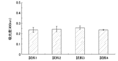
- FIG. 2 shows the growth characteristics of MC3T3-E1 cells.
- the vertical axis indicates the absorbance at 490 nm, and the higher the absorbance, the more the MC3T3-E1 cells are proliferating.
- the proliferation of MC3T3-E1 cells did not decrease even when a DLC film was formed.
- the proliferation of MC3T3-E1 was not affected by the amount of functional groups introduced into the DLC membrane.
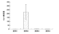
- FIG. 3 and 4 show the differentiation characteristics of MC3T3-E1 cells.
- the vertical axis indicates the mRNA expression level of Runx2, and the larger the value, the more the Runx2 is expressed, indicating that the differentiation of MC3T3-E1 cells is promoted.
- the vertical axis represents the amount of CoI-I mRNA expression. The larger the value, the more CoI-I is expressed and the differentiation of MC3T3-E1 cells is promoted.
- Sample 2 the expression levels of Runx2 and CoI-I increased about 200 times that of pure titanium. However, in sample 3 with [COOH] exceeding 4% and sample 4 with [NH 2 ] / [COOH] exceeding 10, no effect of promoting differentiation into osteoblasts was observed.
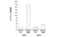
- FIG. 5 shows the expression characteristics of cathepsin K, which is an index of differentiation into osteoclasts.
- RANKL which is a differentiation promoting factor
- the implant material and the manufacturing method thereof according to the present disclosure can realize an implant material that suppresses induction into osteoclasts and promotes differentiation into osteoblasts, and particularly bone cells such as artificial tooth roots and dentures. It is useful as a dental material that requires an affinity for and a manufacturing method thereof.
Landscapes
- Health & Medical Sciences (AREA)
- Veterinary Medicine (AREA)
- General Health & Medical Sciences (AREA)
- Chemical & Material Sciences (AREA)
- Animal Behavior & Ethology (AREA)
- Oral & Maxillofacial Surgery (AREA)
- Public Health (AREA)
- Epidemiology (AREA)
- Life Sciences & Earth Sciences (AREA)
- Transplantation (AREA)
- Inorganic Chemistry (AREA)
- Medicinal Chemistry (AREA)
- Dermatology (AREA)
- Engineering & Computer Science (AREA)
- Ceramic Engineering (AREA)
- Orthopedic Medicine & Surgery (AREA)
- Dentistry (AREA)
- Materials For Medical Uses (AREA)
Abstract
インプラント用材料は、基材10と、基材10の表面に形成された炭素質薄膜20とを備えている。炭素質薄膜20は、酸素を含む官能基及び窒素を含む官能基の少なくとも一方を有する。酸素を含む官能基の存在比は4%以下であり、窒素を含む官能基の存在比/酸素を含む官能基の存在比は10以下である。
Description
本開示は、インプラント用材料、その製造方法及び骨細胞との親和性向上方法に関し、特に、人工歯根及び義歯等の骨細胞との親和性が必要な材料及びその製造方法に関する。
現在、人工歯根等をはじめとする生体内に埋め込むインプラントの基材としてチタン及びチタン合金が生体適合性、耐食性及び機械的強度に優れているという理由から使用されている。しかし、チタン及びチタン合金からなる人工歯根を直接顎骨に固定した場合には、顎骨の骨組織再生の代謝バランスが崩れ、人工歯根のゆるみが生じたり、顎骨破壊が生じたりするおそれがあることが知られている。
このような人工歯根の埋め込みが失敗する原因として、人工歯根の周囲において破骨細胞が誘導され、骨破壊が生じるインプラント周囲炎がある。インプラント周囲炎は、細菌感染等の微生物刺激及び過度の咬合力等の機械的刺激に起因する場合もあるが、人工歯根自体による破骨細胞の活性化によっても生じる。
人工歯根による破骨細胞の活性化は、インプラント初期埋入時にも発生する。初期埋入時に出現した破骨細胞は、インプラントのオッセオインテグレーションを妨げ、インプラントが失敗に至る原因となる。
このような破骨細胞の活性化は、チタン及びチタン合金が骨細胞と十分な親和性を有していないことにより生じると考えられる。骨細胞との親和性が不十分な材料の表面では、破骨前駆細胞から破骨細胞への分化が促進され、これにより骨破壊が生じてしまう。チタン及びチタン合金は比較的骨細胞との親和性が高い材料であることが知られている。しかし、親和性が十分とはいえず、埋め込み対象者の顎骨の状態及び口腔内の状態等によって人工歯根の埋め込みが失敗してしまう。
人工歯根等のインプラントと骨細胞との親和性を向上させる方法としてダイヤモンド様薄膜(DLC膜)による被覆が試みられている(例えば、特許文献1を参照。)。DLC膜は、炭素を主成分とし、表面が平滑で不活性であるため生体との適合性に優れている。このため、骨細胞との親和性にも優れていると考えられる。
しかしながら、インプラントと骨細胞との親和性は、破骨細胞の誘導抑制だけでなく、骨芽細胞への分化の促進という観点からも検討する必要がある。破骨前駆細胞から破骨細胞への分化を抑制できたとしても、未熟骨芽細胞から骨芽細胞への分化が抑制されたり、骨芽細胞の増殖が阻害されたりすると、インプラントのオッセオインテグレーションは促進されない。これまで、DLC膜が骨芽細胞への分化にどのような影響を与えるかについてはほとんど知られていない。
本開示は、本願発明者らの検討により得られた骨芽細胞への分化にDLC膜が与える影響に関する知見に基づき、破骨細胞への誘導を抑制すると共に、骨芽細胞への分化を促進するインプラント用材料を実現できるようにすることを目的とする。
具体的に、本開示に係るインプラント用材料は、基材と、基材の表面に形成された炭素質薄膜とを備え、炭素質薄膜は、酸素を含む官能基及び窒素を含む官能基の少なくとも一方を有し、酸素を含む官能基の存在比は4%以下であり、窒素を含む官能基の存在比を酸素を含む官能基の存在比により除した値は10以下である。
本願発明者らの検討によれば、酸素を含む官能基及び窒素を含む官能基の少なくとも一方を有し、酸素を含む官能基の存在比が4%以下であり、窒素を含む官能基の存在比/酸素を含む官能基の存在比が10以下である炭素質膜の表面において骨芽細胞への分化が生じやすく、破骨細胞への分化が生じにくくなる。このため、本開示のインプラント用材料は、破骨細胞への分化を抑制できると共に、骨芽細胞への分化を促進することができるため、骨組織との親和性に優れたインプラント用材料を実現できる。
本開示のインプラント用材料において、基材は、金属とすればよい。また、基材はチタン又はチタン合金からなることが好ましい。
本開示のインプラント用材料において、基材は、人工歯根、義歯、歯冠修復物、人工骨又は人工関節とすればよい。
本開示のインプラント用材料において、炭素質膜の表面におけるゼータ電位は、-50mV以上且つ0mV未満とすればよい。
本開示に係るインプラント用材料の製造方法は、基材を準備する工程(a)と、炭化水素を含むガスを用いた化学気相堆積法により、基材の表面に炭素質膜を形成する工程(b)と、基材の表面に形成された炭素質膜の表面のダングリングボンドが安定するまで基材を真空状態に保持する工程(c)とを備え、炭素質膜における酸素を含む官能基の存在比を4%以下とし、窒素を含む官能基の存在比を酸素を含む官能基の存在比により除した値が10以下とする。
本開示のインプラント用材料の製造方法は、工程(c)よりも後に、炭素質膜に塩基性窒素含有化合物のプラズマを照射する工程(d)をさらに備えていてもよい。
本開示に係るインプラント用材料の骨細胞との親和性向上方法は、基材の表面に酸素を含む官能基及び窒素を含む官能基の少なくとも一方を有する炭素質膜を形成し、酸素を含む官能基の存在比は4%以下とし、窒素を含む官能基の存在比を酸素を含む官能基の存在比により除した値を10以下とすることを特徴とする。
本開示に係る炭素質薄膜及びその製造方法によれば、破骨細胞への誘導を抑制すると共に、骨芽細胞への分化を促進するインプラント用材料を実現できる。
本実施形態においてインプラントとは、人工歯根だけでなく、義歯、歯冠修復材料及び義歯修復材料等を含む。また、歯科用だけでなく、生体内に埋め込まれる、人工骨及び人工関節等の骨細胞と親和性が必要とされる器具を含む。
炭素質膜とは、ダイヤモンド様膜(DLC膜)に代表されるsp2炭素-炭素結合(グラファイト結合)及びsp3炭素-炭素結合(ダイヤモンド結合)を含む膜である。DLC膜のようなアモルファス状態の膜であっても、ダイヤモンド膜のような結晶状態の膜であってもよい。通常、sp2炭素-水素結合及びsp3炭素-水素結合を含んでいるが、水素は必須の構成要素ではない。また、シリコン(Si)又はフッ素(F)等が添加されていてもよい。
図1は、本実施形態に係るインプラント用材料の断面構成を示している。基材10の表面に膜厚が0.005μm~3μm程度の炭素質膜20が形成されている。炭素質膜20の表面には、カルボキシル基に代表される酸素を含む官能基及びアミノ基に代表される窒素を含む官能基の少なくとも一方が存在している。本実施形態において、炭素質膜20における酸素を含む官能基の存在比は4%以下である。また、窒素を含む官能基の存在比を酸素を含む官能基の存在比で割った値は10以下である。なお、後で詳細に説明するが、酸素を含む官能基の存在比とは、X線光電子分光により求めた酸素の1s(O1s)ピークのピーク面積を炭素の1s(C1s)ピークのピーク面積で割った値であり、窒素を含む官能基の存在比とは、窒素の1s(N1s)ピークのピーク面積を炭素の1s(C1s)ピークのピーク面積で割った値である。
インプラント用材料は、骨細胞との親和性が非常に重要である。骨細胞との親和性とは、未熟骨芽細胞から骨芽細胞への分化の促進及び骨芽細胞の増殖の促進と、破骨前駆細胞から破骨細胞への誘導の抑制との両方の観点から評価する必要がある。
基材の表面に炭素質膜を形成し且つ炭素質膜の表面におけるカルボキシル基等の酸素を含む官能基の存在比を4%以下とし且つアミノ基等の窒素を含む官能基の存在比をカルボキシル基等の酸素を含む官能基の存在比で割った値を10以下とすることにより、骨芽細胞への分化の促進及び骨芽細胞の増殖の促進と、破骨細胞への誘導の抑制とを実現することができることを本願発明者らは見出した。
酸素を含む官能基の存在比を4%以下とし且つ窒素を含む官能基の存在比/酸素を含む官能基の存在比を10以下とした場合に、未熟骨芽細胞から骨芽細胞への分化が促進されると共に、破骨細胞への誘導が抑制される理由は明確ではない。しかし、酸素を含む官能基の存在比が4%以下で窒素を含む官能基の存在比/酸素を含む官能基の存在比が10よりも大きい場合に及び窒素を含む官能基の存在比/酸素を含む官能基の存在比が10以下で酸素を含む官能基の存在比が4%よりも高い場合には、骨芽細胞は増殖するが骨芽細胞への分化の促進は認められない。このことから、骨細胞の親和性は炭素質膜の表面におけるカルボキシル基とアミノ基とのバランスによって制御されると考えられる。
歯科インプラントの治療において、血小板が治療成績に大きな影響を与えることが知られている。例えば、インプラントの埋入前に、患者の血液より得た血小板を含む血漿(Platelet-Rich Plasma)にインプラントを浸漬し、インプラントの表面に血小板を吸着させると治療がより確実に行われることが知られている。一方、炭素質膜の表面におけるアミノ基の存在比及びカルボキシル基の存在比は、炭素質膜の表面のゼータ電位に影響を与え、炭素質膜の表面におけるゼータ電位ゼータ電位の値が-50mV以下の場合にはほとんど血小板が吸着しないことが知られている。酸素を含む官能基であるカルボキシル基の存在比が4%以下で且つ窒素を含む官能基であるアミノ基の存在比/酸素を含む官能基であるカルボキシル基の存在比が10以下の場合におけるゼータ電位の値は-60mV程度~0mV程度となる。このことから、炭素質膜の表面のゼータ電位は、-50mV以上且つ0mV以下であることが好ましい。
本実施形態のインプラント用材料は基材の表面を炭素質膜により覆うため、基材がどのような材質であっても優れた骨組織との親和性を示す。このため、基材は強度等の特性を満たしていればどのような材質であってもよい。例えば、チタン及びチタン合金等をはじめとする金属、樹脂又はセラミックス等を用いることができる。また、人工歯根をはじめ、義歯、人工骨及び人工関節等の骨細胞と親和性が必要とされる種々のインプラントに適用することが可能である。
炭素質膜は、化学気相堆積法(CVD法)により形成すればよい。またCVD法以外の、スパッタ法、プラズマイオン注入法、イオンプレーティング法、アークイオンプレーティング法、イオンビーム蒸着法又はレーザーアブレーション法等により形成してもよい。
炭素質膜の成膜中及び成膜後にチャンバ内に存在する酸素及び水分は、炭素質膜の表面にカルボキシル基等の酸素を含む官能基を生成する原因となる。このため、酸素を含む官能基の存在比を4%以下とする場合には、成膜の際に高純度の原料ガスを用い、さらに吸着脱水装置等を介して原料ガスを供給することが好ましい。また、成膜完了後における酸素との結合を抑制するために、成膜完了直後は一定時間真空中に保持し、炭素質膜のダングリングボンドを安定化することが好ましい。例えばプラズマCVD法により炭素質膜を成膜する場合、プラズマ生成に用いるアルゴンガスの純度を99.9999%以上とし、成膜終了後は基板の温度が常温となった後、少なくとも10分間、好ましくは60分以上真空中に放置すればよい。
炭素質膜へアミノ基等の窒素を含む官能基を導入する必要がある場合には、塩基性窒素含有化合物のプラズマを炭素質膜に照射すればよい。塩基性窒素含有化合物としては、アンモニアをはじめとして、一般式がNR1R2R3により示される有機アミン類(但し、R1、R2及びR3は水素、-CH3、-C2H5、-C3H7又は-C4H8であり、R1、R2及びR3は互いに同一であっても、異なっていてもよい。)又はベンジルアミン及びその2級、3級アミン等を用いればよい。但し、アンモニアがコスト、取り扱いの容易さから好ましい。なお、プラズマ照射時におけるチャンバ内の到達真空度は、0.01Pa程度~500Pa程度とすればよい。但し、到達真空度が高い方が、空気中の酸素の影響を受けることがなく好ましく、5×10⁻³Pa程度としてもよい。
炭素質膜に酸素を含む官能基をさらに導入する必要がある場合には、酸素プラズマ又は酸素を含むガスのプラズマ等を照射すればよい。また、アンモニア等のプラズマを照射する際の到達真空度を低くしてもよい。
なお、プラズマ照射装置は、どのような構造のものを用いてもよい。また、放電形式についても、どのようなものを用いてもよく、例えば平行平板方式、アフターグロー放電方式、電磁誘導型及び有磁場型等を用いればよい。プラズマ照射条件は特に限定されない。例えば、プラズマ発生用の電源としては、商用周波数(50Hz又は60Hz)、高周波(ラジオ周波数)又はマイクロ波領域等の各種の電源周波数を用いることができる。さらに、原料ガスの圧力制御方法や供給構造についても特に限定するものではない。しかし、であまりエッチングレートが大きいプラズマ照射条件を用いると、炭素質薄膜にダメージを与えるおそれがある。
炭素質膜の厚さは特に限定されるものではないが、0.005μm~3μmの範囲が好ましく、より好ましくは0.01μm~1μmの範囲である。
また、炭素質膜は基材の表面に直接形成することができるが、基材と炭素質薄膜とをより強固に密着させるために、基材と炭素質薄膜との間に中間層を設けてもよい。中間層の材質としては、基材の種類に応じて種々のものを用いることができるが、珪素(Si)と炭素(C)、チタン(Ti)と炭素(C)又はクロム(Cr)と炭素(C)からなるアモルファス膜等の公知のものを用いることができる。その厚みは特に限定されるものではないが、0.005μm~0.3μmの範囲が好ましく、より好ましくは0.01μm~0.1μmの範囲である。中間層は、例えば、スパッタ法、CVD法、プラズマCVD法、溶射法、イオンプレーティング法又はアークイオンプレーティング法等を用いて形成すればよい。
(一実施例)
以下に、本実施形態のインプラント用材料について実施例を用いてさらに詳細に説明する。
以下に、本実施形態のインプラント用材料について実施例を用いてさらに詳細に説明する。
-炭素質膜の形成-
純チタン(JIS2種)からなる基材の表面にDLC膜からなる炭素質膜を形成した。細胞培養の際には直径20mmの基材を用いた。DLC膜は化学気相堆積(CVD)法を用いて形成した。具体的には、基材を載置したチャンバ内にC2H2を流量が150sccm(cm3/分、但し1気圧、0℃)で、圧力が24.67Pa(35mTorr)となるように導入し、RF電極に100W~500Wの高周波電力を印加した。
純チタン(JIS2種)からなる基材の表面にDLC膜からなる炭素質膜を形成した。細胞培養の際には直径20mmの基材を用いた。DLC膜は化学気相堆積(CVD)法を用いて形成した。具体的には、基材を載置したチャンバ内にC2H2を流量が150sccm(cm3/分、但し1気圧、0℃)で、圧力が24.67Pa(35mTorr)となるように導入し、RF電極に100W~500Wの高周波電力を印加した。
DLC膜成膜後に、真空中に放置することにより酸素を含む官能基の存在比が低いDLC膜とすることができる。表1は、DLC膜を成膜後10分間真空中に放置した場合と、120分間真空中に放置した場合の酸素を含む官能基の存在比及び窒素を含む官能基の存在比を示している。なお、酸素を含む官能基はカルボキシル基(COOH)であるとみなし、窒素を含む官能基はアミノ基(NH2)であるとみなして評価している。成膜後10分以上真空中に放置することにより酸素を含む官能基の存在比を4%以下とすることができる。また、真空中に放置する時間が長いほど酸素を含む官能基の存在比を小さくすることができる。

-官能基の導入-
炭素質膜に官能基をさらに導入するためにプラズマ照射を行った。プラズマ照射は平行平板型のプラズマ照射装置により行った。プラズマ照射装置のチャンバ内に上記で得られた基材をセットした後、チャンバ内の圧力を2Pa以下まで排気した。次に、チャンバ内にアンモニア又は酸素を所定の流量で導入し、平行平板電極の間に30Wの高周波電力を印加することによりプラズマを発生させた。ガス流量の調整はマスフローコントローラにより行い、プラズマ照射時のチャンバ内圧力は130Paとした。高周波電力は、マッチングボックスを介して接続された高周波電源を用いて印加した。窒素を含む官能基の導入にはアンモニアを用い、酸素を含む官能基の導入には酸素を用いた。プラズマ照射時間は15秒とした。
炭素質膜に官能基をさらに導入するためにプラズマ照射を行った。プラズマ照射は平行平板型のプラズマ照射装置により行った。プラズマ照射装置のチャンバ内に上記で得られた基材をセットした後、チャンバ内の圧力を2Pa以下まで排気した。次に、チャンバ内にアンモニア又は酸素を所定の流量で導入し、平行平板電極の間に30Wの高周波電力を印加することによりプラズマを発生させた。ガス流量の調整はマスフローコントローラにより行い、プラズマ照射時のチャンバ内圧力は130Paとした。高周波電力は、マッチングボックスを介して接続された高周波電源を用いて印加した。窒素を含む官能基の導入にはアンモニアを用い、酸素を含む官能基の導入には酸素を用いた。プラズマ照射時間は15秒とした。
-官能基存在比の評価-
炭素質膜における官能基の存在比はX線光電子分光(XPS)測定により評価した。X線源にはアルミニウムKα線を用い、加速電圧は10.0KV、エミッション電流は10mAとした。X線の入射角度は45度とし、表面から4nm程度の深さまでの状態について測定した。
炭素質膜における官能基の存在比はX線光電子分光(XPS)測定により評価した。X線源にはアルミニウムKα線を用い、加速電圧は10.0KV、エミッション電流は10mAとした。X線の入射角度は45度とし、表面から4nm程度の深さまでの状態について測定した。
窒素を含む官能基の存在比は、XPS測定において得られた窒素の1s(N1s)ピークの面積と炭素(C)1sピークの面積との比率とした。炭素質膜の表面に存在する窒素がどのような状態となっているかは明確ではない。しかし、アミノ基及びアミド基等の窒素を含む官能基(窒素性官能基)を形成していると考えられる。窒素性官能基の詳細な分析は困難であるが、XPS測定において、399eV付近にN1sピークが出現していることからも主にアミノ基として存在していると考えられる。以下においては、N1sピークの面積とC1sピークの面積との比率をアミノ基の存在比[NH2]として説明する。
酸素を含む官能基の存在比は、XPS測定において得られた酸素の1s(O1s)ピークの面積とC1sピークの面積との比率とした。酸素を含む官能基としてカルボキシル基以外の水酸基等も形成されている可能性があるが、以下においては、O1sピークの面積とC1sピークの面積との比率をカルボキシル基の存在比[COOH]とする。
なお、XPS測定において、ピーク面積の比率により求めた存在比は、原子%(at%)となる。
-骨芽細胞の評価-
直径20mmのウェル内において試料とマウス骨芽細胞様細胞株MC3T3-E1細胞(以下、MC3T3-E1細胞という。)とを接触させて培養した。細胞は1ウェル当たり5×10⁴個播種した。培養培地には10%ウシ胎仔血清(FBS)、L-グルタミン、混合抗生物質(Invitrogen社製)及び50μg/mlのアスコルビン酸を含有したα変法イーグル培地(α-MEM)を用いた。培養温度は37℃とし、5%二酸化炭素雰囲気で培養した。
直径20mmのウェル内において試料とマウス骨芽細胞様細胞株MC3T3-E1細胞(以下、MC3T3-E1細胞という。)とを接触させて培養した。細胞は1ウェル当たり5×10⁴個播種した。培養培地には10%ウシ胎仔血清(FBS)、L-グルタミン、混合抗生物質(Invitrogen社製)及び50μg/mlのアスコルビン酸を含有したα変法イーグル培地(α-MEM)を用いた。培養温度は37℃とし、5%二酸化炭素雰囲気で培養した。
細胞を1日培養した後、細胞数をMTS(Multiple target screening)法を用いて、細胞の増殖性を評価した。増殖性を評価するための吸光度測定の波長は490nmとした。
細胞を7日間培養した後、骨分化マーカー遺伝子であるRunx2及びTypeIコラーゲン(CoI-I)の発現について評価した。具体的には、培養したMC3T3-E1細胞からTRIzol試薬(Invitrogen社製)を用いてRNAを抽出した後、ReverTra Ace reverse transcriptase(東洋紡社製)を用いてcDNAを作成した。作成したcDNAを、定量RT-PCR(Rela-time Quantitative Reverse Transcriptase-Polymerase Chain Reaction)法により評価した。
-骨破壊細胞の分化の評価-
ウェル内において試料と破骨前駆細胞とを破骨細胞分化誘導因子(Receptor Activator of NF-kB Ligand:RANKL)の存在下において接触させ、37℃で細胞培養した。破骨前駆細胞は、RANKLの存在により破骨細胞へと分化することが確立されているセルラインRAW264.7細胞 (TIB-71, ATCC)を用いた。細胞は1ウェル当たり5×10³個播種した。
ウェル内において試料と破骨前駆細胞とを破骨細胞分化誘導因子(Receptor Activator of NF-kB Ligand:RANKL)の存在下において接触させ、37℃で細胞培養した。破骨前駆細胞は、RANKLの存在により破骨細胞へと分化することが確立されているセルラインRAW264.7細胞 (TIB-71, ATCC)を用いた。細胞は1ウェル当たり5×10³個播種した。
分化関連遺伝子であるカテプシンK(cathepsin K)の発現を定量RTPCR法を用いて定量することにより、破骨細胞への分化を評価した。
-測定結果-
表2に示す4つの試料について測定を行った。試料1はコントロールのために設けたDLC膜を形成していない純チタンの基材である。試料2はプラズマ照射を行っていないDLC膜である。試料3は、プラズマ照射によりカルボキシル基の導入量を多くしたDLC膜である。試料4は、プラズマ照射によりアミノ基の導入量を多くしたDCL膜である。
表2に示す4つの試料について測定を行った。試料1はコントロールのために設けたDLC膜を形成していない純チタンの基材である。試料2はプラズマ照射を行っていないDLC膜である。試料3は、プラズマ照射によりカルボキシル基の導入量を多くしたDLC膜である。試料4は、プラズマ照射によりアミノ基の導入量を多くしたDCL膜である。
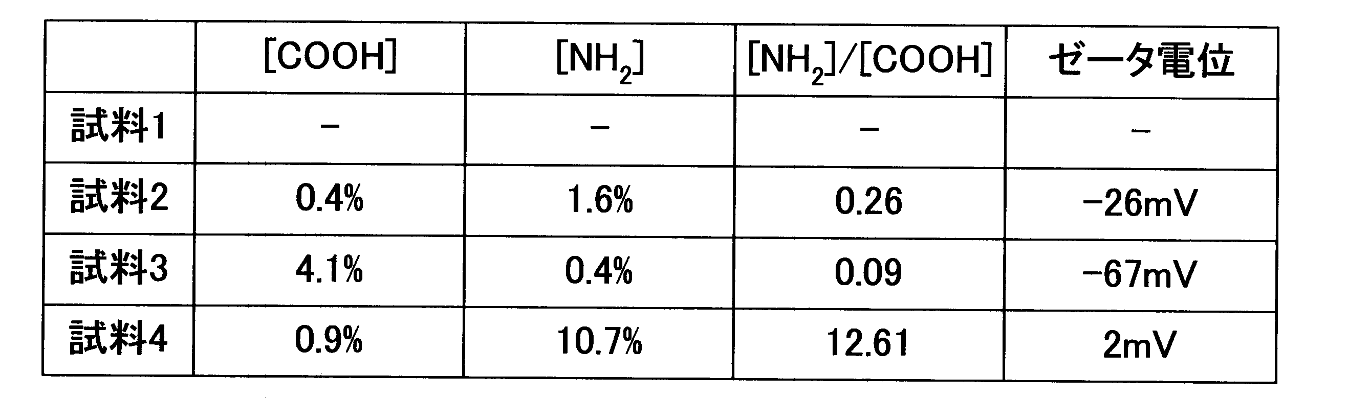
図2は、MC3T3-E1細胞の増殖特性を示している。図2において縦軸は490nmの吸光度を示し、吸光度が高いほどMC3T3-E1細胞が増殖していることを示す。図2に示すように、DLC膜を形成してもMC3T3-E1細胞の増殖性は低下しなかった。また、DLC膜に導入した官能基の量によってもMC3T3-E1の増殖性は影響を受けなかった。
図3及び図4は、MC3T3-E1細胞の分化特性を示している。図3において縦軸はRunx2のmRNA発現量を示し、値が大きいほどRunx2が発現し、MC3T3-E1細胞の分化が促進されていることを示している。図4において縦軸はCoI-IのmRNA発現量を示し、値が大きいほどCoI-Iが発現し、MC3T3-E1細胞の分化が促進されていることを示している。試料2においては、Runx2及びCoI-Iの発現量が、純チタンの場合の約200倍に増加した。しかし、[COOH]が4%を越える試料3及び[NH2]/[COOH]が10を越える試料4では、骨芽細胞への分化を促進する効果は認められなかった。
図5は、破骨細胞への分化の指標であるカテプシンKの発現特性を示している。試料1及び試料2のいずれにおいても、分化促進因子であるRANKLが存在していない場合には、カテプシンKの発現量はわずかであり、破骨前駆細胞から破骨細胞への分化は抑制されている。しかし、RANKLの存在下においては、試料1の場合カテプシンKの発現量がRANKLが存在しない場合の9倍以上に増加し、破骨前駆細胞から破骨細胞への分化が活発に生じた。一方、[COOH]が4%以下で且つ[NH2]/[COOH]が10以下の炭素質膜を有する試料2においては、カテプシンKの発現量の増加はわずかであった。このことから、[COOH]が4%以下で且つ[NH2]/[COOH]が10以下の炭素質膜は、破骨前駆細胞から破骨細胞への分化を抑制できることが明らかとなった。
本開示に係るインプラント用材料及びその製造方法は、破骨細胞への誘導を抑制すると共に、骨芽細胞への分化を促進するインプラント用材料を実現でき、特に、人工歯根及び義歯等の骨細胞との親和性が必要な歯科用材料及びその製造方法等として有用である。
10 基材
20 炭素質膜
20 炭素質膜
Claims (8)
- 基材と、
前記基材の表面に形成された炭素質膜とを備え、
前記炭素質膜は、酸素を含む官能基及び窒素を含む官能基の少なくとも一方を有し、
前記酸素を含む官能基の存在比は4%以下であり、
前記窒素を含む官能基の存在比を前記酸素を含む官能基の存在比により除した値は10以下であるインプラント用材料。 - 前記基材は、金属からなる請求項1に記載のインプラント用材料。
- 前記基材は、チタン又はチタン合金からなる請求項1に記載のインプラント用材料。
- 前記基材は、人工歯根、義歯、歯冠修復物、人工骨又は人工関節である請求項1に記載のインプラント用材料。
- 前記炭素質膜の表面におけるゼータ電位は、-50mV以上且つ0mV未満である請求項1に記載のインプラント用材料。
- 基材を準備する工程(a)と、
炭化水素を含むガスを用いた化学気相堆積法により、前記基材の表面に炭素質膜を形成する工程(b)と、
前記基材の表面に形成された炭素質膜の表面のダングリングボンドが安定するまで前記基材を真空状態に保持する工程(c)とを備え、
前記炭素質膜における酸素を含む官能基の存在比を4%以下とし、窒素を含む官能基の存在比を前記酸素を含む官能基の存在比により除した値を10以下とするインプラント用材料の製造方法。 - 前記工程(c)よりも後に、前記炭素質膜に塩基性窒素含有化合物のプラズマを照射する工程(d)をさらに備えている請求項6に記載のインプラント用材料の製造方法。
- 基材の表面に、酸素を含む官能基及び窒素を含む官能基の少なくとも一方を有する炭素質膜を形成し、
前記酸素を含む官能基の存在比を4%と以下とし且つ前記窒素を含む官能基の存在比を前記酸素を含む官能基の存在比により除した値を10以下とするインプラント材料の骨細胞との親和性向上方法。
Priority Applications (1)
| Application Number | Priority Date | Filing Date | Title |
|---|---|---|---|
| PCT/JP2010/006247 WO2012053035A1 (ja) | 2010-10-21 | 2010-10-21 | インプラント用材料、その製造方法及び骨細胞との親和性向上方法 |
Applications Claiming Priority (1)
| Application Number | Priority Date | Filing Date | Title |
|---|---|---|---|
| PCT/JP2010/006247 WO2012053035A1 (ja) | 2010-10-21 | 2010-10-21 | インプラント用材料、その製造方法及び骨細胞との親和性向上方法 |
Publications (1)
| Publication Number | Publication Date |
|---|---|
| WO2012053035A1 true WO2012053035A1 (ja) | 2012-04-26 |
Family
ID=45974775
Family Applications (1)
| Application Number | Title | Priority Date | Filing Date |
|---|---|---|---|
| PCT/JP2010/006247 Ceased WO2012053035A1 (ja) | 2010-10-21 | 2010-10-21 | インプラント用材料、その製造方法及び骨細胞との親和性向上方法 |
Country Status (1)
| Country | Link |
|---|---|
| WO (1) | WO2012053035A1 (ja) |
Citations (2)
| Publication number | Priority date | Publication date | Assignee | Title |
|---|---|---|---|---|
| JP2009137888A (ja) * | 2007-12-06 | 2009-06-25 | Hiroshima Univ | インプラント用材料及びその製造方法 |
| WO2010092616A1 (ja) * | 2009-02-10 | 2010-08-19 | 国立大学法人広島大学 | インプラント用材料及びその製造方法 |
-
2010
- 2010-10-21 WO PCT/JP2010/006247 patent/WO2012053035A1/ja not_active Ceased
Patent Citations (2)
| Publication number | Priority date | Publication date | Assignee | Title |
|---|---|---|---|---|
| JP2009137888A (ja) * | 2007-12-06 | 2009-06-25 | Hiroshima Univ | インプラント用材料及びその製造方法 |
| WO2010092616A1 (ja) * | 2009-02-10 | 2010-08-19 | 国立大学法人広島大学 | インプラント用材料及びその製造方法 |
Non-Patent Citations (4)
| Title |
|---|
| KUMARI T.V. ET AL.: "In vitro cytocompatibility studies of Diamond Like Carbon coatings on titanium.", BIO-MEDICAL MATERIALS AND ENGINEERING, vol. 12, no. 4, 2002, pages 329 - 338 * |
| OKAMOTO K. ET AL.: "Differentiation Capability Evaluation of Osteoblast by Functionalized DLC Thin Films with Plasma Processing", JOURNAL OF PHOTOPOLYMER SCIENCE AND TECHNOLOGY, vol. 23, no. 4, 6 July 2010 (2010-07-06), pages 591 - 594 * |
| SATOSHI IWATA ET AL.: "DLC Coating shita Titanium ga Kotsuga Saiboyo Saibokabu MC3T3-E1 Saibo no Bunka ni Ataeru Eikyo", JOURNAL OF JAPANESE SOCIETY OF ORAL IMPLANTOLOGY, vol. 22, 25 August 2009 (2009-08-25), pages 231, P-1-6 * |
| YASUHIRO OKADA ET AL.: "Kakushu DLC Kiban Jo deno Kotsugayo Saibo no Zoshoku to Bunka ni Tsuite", POLYMER PREPRINTS, JAPAN(CD-ROM), DISKL, vol. 59, no. 2, 1 September 2010 (2010-09-01), JAPAN, pages ROMBUNNO.1PC067 * |
Similar Documents
| Publication | Publication Date | Title |
|---|---|---|
| Poon et al. | Carbon plasma immersion ion implantation of nickel–titanium shape memory alloys | |
| JP6029865B2 (ja) | インプラント、その製造方法及び骨代謝の制御方法 | |
| KR101846038B1 (ko) | 임플란트용 재료 | |
| JPWO2009060602A1 (ja) | 炭素質薄膜及びその製造方法 | |
| CN102286767B (zh) | 一种镁合金生物植入材料表面的复合涂层及其制备方法 | |
| RU2571559C1 (ru) | Способ изготовления внутрикостного стоматологического имплантата с углеродным нанопокрытием | |
| Rahimi et al. | Surface modifications of dental implant and its clinical performance: a review | |
| US9138507B2 (en) | Method for manufacturing an implant material | |
| JP5403542B2 (ja) | インプラントの製造方法 | |
| RU2697855C1 (ru) | Способ нанесения покрытия на устройства и инструменты для остеосинтеза, ортопедические имплантаты из металла | |
| JP5327934B2 (ja) | インプラント用材料及びその製造方法 | |
| WO2012053035A1 (ja) | インプラント用材料、その製造方法及び骨細胞との親和性向上方法 | |
| JP2009153586A (ja) | 抗血栓性材料及びその製造方法 | |
| Takechi et al. | In vitro investigation of the cell compatibility and antibacterial properties of titanium treated with calcium and ozone | |
| JP6371611B2 (ja) | インプラント及びその製造方法 | |
| CN104962970A (zh) | 一种医用镁合金的表面改性方法 | |
| JP7677739B2 (ja) | セラミックコーティング層を有するインプラント | |
| RU2784152C1 (ru) | Способ получения коррозионно-стойкого биосовместимого покрытия из нитрида титана методом магнетронного напыления на изделии из биоразлагаемого сплава на основе магния | |
| KOMASA et al. | Effect of Silicon Nitride Coating on Titanium Surface on the Expression Levels of Early Hard Tissue Differentiation-Induced Genes | |
| EA022035B1 (ru) | Способ получения гибридного покрытия на металлических имплантатах | |
| JP4272845B2 (ja) | 人工歯根 | |
| US20230091812A1 (en) | White, bacteria-resistant, biocompatible, adherent coating for implants, screws and plates integrated in hard and soft tissue and production method | |
| JP2023012410A (ja) | パリレンを用いたインプラントのコーティング方法 | |
| JP2017209303A (ja) | インプラント | |
| CN120905672A (zh) | 一种钛合金植体表面改性的制备方法及应用 |
Legal Events
| Date | Code | Title | Description |
|---|---|---|---|
| 121 | Ep: the epo has been informed by wipo that ep was designated in this application |
Ref document number: 10858593 Country of ref document: EP Kind code of ref document: A1 |
|
| NENP | Non-entry into the national phase |
Ref country code: DE |
|
| 122 | Ep: pct application non-entry in european phase |
Ref document number: 10858593 Country of ref document: EP Kind code of ref document: A1 |
|
| NENP | Non-entry into the national phase |
Ref country code: JP |