WO2012015050A1 - アミロイドβペプチド蓄積を伴う疾患の診断の為の可溶型アミロイドβ前駆体タンパク質770β切断産物の検出方法 - Google Patents
アミロイドβペプチド蓄積を伴う疾患の診断の為の可溶型アミロイドβ前駆体タンパク質770β切断産物の検出方法 Download PDFInfo
- Publication number
- WO2012015050A1 WO2012015050A1 PCT/JP2011/067530 JP2011067530W WO2012015050A1 WO 2012015050 A1 WO2012015050 A1 WO 2012015050A1 JP 2011067530 W JP2011067530 W JP 2011067530W WO 2012015050 A1 WO2012015050 A1 WO 2012015050A1
- Authority
- WO
- WIPO (PCT)
- Prior art keywords
- amyloid
- antibody
- precursor protein
- disease
- app
- Prior art date
- Legal status (The legal status is an assumption and is not a legal conclusion. Google has not performed a legal analysis and makes no representation as to the accuracy of the status listed.)
- Ceased
Links
Images



Classifications
-
- G—PHYSICS
- G01—MEASURING; TESTING
- G01N—INVESTIGATING OR ANALYSING MATERIALS BY DETERMINING THEIR CHEMICAL OR PHYSICAL PROPERTIES
- G01N33/00—Investigating or analysing materials by specific methods not covered by groups G01N1/00 - G01N31/00
- G01N33/48—Biological material, e.g. blood, urine; Haemocytometers
- G01N33/50—Chemical analysis of biological material, e.g. blood, urine; Testing involving biospecific ligand binding methods; Immunological testing
- G01N33/68—Chemical analysis of biological material, e.g. blood, urine; Testing involving biospecific ligand binding methods; Immunological testing involving proteins, peptides or amino acids
- G01N33/6893—Chemical analysis of biological material, e.g. blood, urine; Testing involving biospecific ligand binding methods; Immunological testing involving proteins, peptides or amino acids related to diseases not provided for elsewhere
- G01N33/6896—Neurological disorders, e.g. Alzheimer's disease
-
- G—PHYSICS
- G01—MEASURING; TESTING
- G01N—INVESTIGATING OR ANALYSING MATERIALS BY DETERMINING THEIR CHEMICAL OR PHYSICAL PROPERTIES
- G01N2333/00—Assays involving biological materials from specific organisms or of a specific nature
- G01N2333/435—Assays involving biological materials from specific organisms or of a specific nature from animals; from humans
- G01N2333/46—Assays involving biological materials from specific organisms or of a specific nature from animals; from humans from vertebrates
- G01N2333/47—Assays involving proteins of known structure or function as defined in the subgroups
- G01N2333/4701—Details
- G01N2333/4709—Amyloid plaque core protein
-
- G—PHYSICS
- G01—MEASURING; TESTING
- G01N—INVESTIGATING OR ANALYSING MATERIALS BY DETERMINING THEIR CHEMICAL OR PHYSICAL PROPERTIES
- G01N2800/00—Detection or diagnosis of diseases
- G01N2800/28—Neurological disorders
- G01N2800/2814—Dementia; Cognitive disorders
- G01N2800/2821—Alzheimer
Definitions
- the present invention relates to a method for detecting soluble amyloid ⁇ precursor protein 770 ⁇ . More specifically, a method for detecting a cleavage product of amyloid ⁇ precursor protein 770 ⁇ in a biological sample derived from a subject suspected of having a disease associated with accumulation of amyloid ⁇ peptide, and accumulating amyloid ⁇ peptide used in the method
- the present invention relates to a diagnostic agent for a disease and a diagnostic kit.
- AD Alzheimer's disease
- a ⁇ amyloid ⁇ peptide
- a ⁇ may be abbreviated as amyloid ⁇ precursor protein (hereinafter referred to as “APP”) by sequential enzymatic reaction of ⁇ secretase (BACE1) (Non-patent Documents 3 to 6) and ⁇ -secretase (Non-patent Document 7). ). Because most early-onset familial AD patients have gene mutations that affect A ⁇ production mechanisms or aggregation, and neurites associated with A ⁇ aggregation are often damaged (Non-patent Document 2), The process of A ⁇ aggregation and accumulation in the brain is thought to be strongly related to the onset of AD.
- APP amyloid ⁇ precursor protein
- Non-patent Document 10 APP-like proteins 1 and 2 (APLP1 and APLP2 respectively).
- APLP1 and APLP2 APP-like proteins 1 and 2
- triple knockout mice lacking all three of these APP family members ie, APP, APLP1 and APLP2 died shortly after birth (11), suggesting an overlapping function of APP family proteins. ing.
- APP has three types of alternative splicing isoforms, APP695, APP751, and APP770 (Non-patent Documents 12 and 13).
- APP751 further includes a Kunitz-type protease inhibitor (KPI) region
- APP770 includes an OX2 region in addition to the KPI region.
- APP695 is expressed most abundantly in neurons, whereas APP751 and APP770 show a more universal expression pattern (Non-Patent Document 14).
- Secreted APP containing the KPI region also known as protease nexin 2
- the function of the OX2 region available information is very limited.
- markers directly related to pathological changes include a decrease in one type of A ⁇ (A ⁇ 1-42) consisting of 42 amino acids in cerebrospinal fluid, and an increase in (phosphorylated) tau protein.
- a ⁇ is known to change its value only after the symptoms of Alzheimer's disease become serious, and cannot be used as an early diagnostic marker.
- phosphorylated tau is used as a biomarker, neuronal cell death has already progressed at the time when phosphorylated tau is elevated, and even if treatment is started at this time, complete recovery of nerve function is not possible. I can't hope.
- these markers are mainly measured with cerebrospinal fluid (lumbar puncture fluid), special procedures are required to collect these samples, which can be a burden on patients and can be a mass screening method. Absent.
- AD symptoms are closely related to the accumulation of A ⁇ in the brain parenchyma and vascular wall in the brain (Non-patent Documents 16 to 18).
- the accumulation of A ⁇ in the cerebral vasculature is related to cerebrovascular It is also considered as the etiology of amyloid angiopathy (hereinafter sometimes abbreviated as “CAA”).
- CAA is recognized in over 80% of AD patients and is found in 10-40% of elderly people who are not AD (Non-patent Document 19).
- Non-Patent Document 20 It is also known that CAA can cause cerebrovascular dementia disorder, stroke, and intracerebral hemorrhage. Furthermore, in AD treatment using a vaccine against A ⁇ , CAA has also been shown to possibly affect CAA as a cause of side effects (Non-patent Documents 21 and 22). Therefore, CAA indicating the state of A ⁇ accumulation in the cerebral blood vessels The need for diagnosis is widely pointed out.
- Non-Patent Document 23 Although the important role of cerebral vascular smooth muscle cells in the removal of A ⁇ in blood vessels has recently been highlighted (Non-Patent Document 23), there is no precedent for detailed analysis of APP of vascular endothelial cells, and accumulated vascular A ⁇ Where it is produced remains unclear.
- An object of the present invention is to provide a biomarker detection method that enables early diagnosis of diseases associated with accumulation of amyloid ⁇ peptide such as Alzheimer's disease.
- the present invention has focused on the accumulation of A ⁇ in the cerebral blood vessels, which is a cause of cerebrovascular amyloid angiopathy and correlates with symptoms of Alzheimer's disease.
- the present inventors believe that if nerve cell-derived APP and vascular endothelial cell-derived APP can be distinguished from each other, it is possible to discriminate between A ⁇ production in nerve cells and A ⁇ production in vascular endothelial cells.
- the analysis was performed.
- APP770 which is a unique splicing variant of APP, can be specifically detected in vascular endothelial cells that have not been focused from the viewpoint of A ⁇ production, and amyloid ⁇ 40 and 42 are produced from APP770 of vascular endothelial cells. It was discovered for the first time in the world that the cleavage product of APP770 is an index of amyloid accumulation in the cerebral blood vessels.
- the present invention by specifically detecting the cleavage product of APP770, cerebrovascular amyloid angiopathy in which A ⁇ accumulation in the cerebral blood vessels is pathogenic, or Alzheimer's disease in which A ⁇ accumulation in the cerebral blood vessels is correlated.
- cerebrovascular amyloid angiopathy in which A ⁇ accumulation in the cerebral blood vessels is pathogenic
- Alzheimer's disease in which A ⁇ accumulation in the cerebral blood vessels is correlated.
- soluble APP770 ⁇ which is the N-terminal fragment of BACE1 cleavage product of APP770, is cerebral vascular amyloid angio It has been found that it becomes a biomarker for diseases associated with A ⁇ accumulation, such as Paty and Alzheimer's disease, and has completed the present invention.
- the present invention provides the following. [1] The following steps: (1) a step of contacting a biological sample derived from a subject with an antibody that recognizes soluble amyloid ⁇ precursor protein ⁇ or an amyloid ⁇ precursor protein 770-specific antibody; and (2) formed in step (1). Detecting soluble amyloid ⁇ precursor protein 770 ⁇ in the complex; A method for detecting soluble amyloid ⁇ precursor protein 770 ⁇ derived from vascular endothelial cells.
- Step (2) is (2a) An antibody that recognizes the soluble amyloid ⁇ precursor protein 770 ⁇ in the complex formed in step (1) and the other soluble amyloid ⁇ precursor protein ⁇ that was not used in step (1) Or contacting with an amyloid ⁇ precursor protein 770-specific antibody; and (2b) detecting the complex formed in step (2a); The detection method according to [1].
- [3] The method according to [1] or [2], wherein the biological sample is blood, plasma or serum.
- [4] The method according to [1] or [2], wherein the biological sample is cerebrospinal fluid.
- [5] The method according to [1] or [2], wherein the biological sample is a culture supernatant of vascular endothelial cells.
- steps (1) and (2) the following steps: (3) Contacting a biological sample derived from a subject suspected of having a disease associated with accumulation of amyloid ⁇ peptide with an antibody that recognizes soluble amyloid ⁇ precursor protein ⁇ or a soluble amyloid ⁇ precursor protein 695 ⁇ -specific antibody And (4) detecting soluble amyloid ⁇ precursor protein 695 ⁇ in the complex formed in step (3); And the method according to any one of [1] to [5], including detection of soluble amyloid ⁇ precursor protein 695 ⁇ .
- Step (3) either one of an antibody that recognizes soluble amyloid ⁇ precursor protein ⁇ or an amyloid ⁇ precursor protein 695-specific antibody is used,
- Step (4) is (4a) An antibody that recognizes the soluble amyloid ⁇ precursor protein 695 ⁇ in the complex formed in step (3) and the other soluble amyloid ⁇ precursor protein ⁇ not used in step (3) Or a step of contacting with a soluble amyloid ⁇ precursor protein 695 ⁇ -specific antibody; and (4b) a step of detecting the complex formed in step (4a); The detection method according to [6].
- the biological sample in step (1) is blood, plasma, serum, vascular endothelial cell culture supernatant or cerebrospinal fluid, and the biological sample in step (3) is cerebrospinal fluid [6] or [7 ] Method.
- the method according to any one of [1] to [9], wherein the subject is suspected of having a disease associated with accumulation of amyloid ⁇ peptide.
- the disease is at least one of cerebrovascular amyloid angiopathy, cerebrovascular dementia, cerebral infarction, and Alzheimer's disease.
- a diagnostic agent for diseases associated with accumulation of amyloid ⁇ peptide comprising an antibody that recognizes soluble amyloid ⁇ precursor protein ⁇ and / or an amyloid ⁇ precursor protein 770-specific antibody as an active ingredient.
- a diagnostic kit for a disease associated with accumulation of amyloid ⁇ peptide comprising an antibody recognizing soluble amyloid ⁇ precursor protein ⁇ and an amyloid ⁇ precursor protein 770-specific antibody.
- Step (2) is (2a) An antibody that recognizes the soluble amyloid ⁇ precursor protein 770 ⁇ in the complex formed in step (1) and the other soluble amyloid ⁇ precursor protein ⁇ that was not used in step (1) Or contacting with an amyloid ⁇ precursor protein 770-specific antibody; and (2b) detecting the complex formed in step (2a); The detection method according to [18].
- the biological sample is cerebrospinal fluid.
- Step (3) either an antibody that recognizes soluble amyloid ⁇ precursor protein ⁇ or an amyloid ⁇ precursor protein 695-specific antibody is used
- Step (4) is (4a) An antibody that recognizes the soluble amyloid ⁇ precursor protein 695 ⁇ in the complex formed in step (3) and the other soluble amyloid ⁇ precursor protein ⁇ not used in step (3) Or a step of contacting with a soluble amyloid ⁇ precursor protein 695 ⁇ -specific antibody; and (4b) a step of detecting the complex formed in step (4a); The detection method according to [23].
- the biological sample in step (1) is blood, plasma serum, vascular endothelial cell culture supernatant or cerebrospinal fluid, and the biological sample in step (3) is cerebrospinal fluid [23] or [24] The method described in 1.
- the disease associated with accumulation of amyloid ⁇ peptide is at least one of cerebrovascular amyloid angiopathy, cerebrovascular dementia, cerebral infarction, and Alzheimer's disease [1] to any one of [26] The method described.
- the detection of soluble amyloid ⁇ precursor protein 770 ⁇ is used to determine the possibility of afflicting at least one disease of cerebral vascular amyloid angiopathy, cerebral vascular dementia, or cerebral infarction [18] to [26 ] The method as described in any one of. [29] The following steps: (1 ′) contacting a biological sample derived from a subject suspected of a disease associated with accumulation of amyloid ⁇ peptide with an antibody recognizing amyloid ⁇ precursor protein 770-specific or soluble amyloid ⁇ precursor protein ⁇ ; And (2 ′) the soluble amyloid ⁇ precursor protein 770 in the complex formed in step (1 ′) is detected (preferably quantitatively detected). For detecting type amyloid ⁇ precursor protein 770 ⁇ .
- the detection method of the present invention uses sAPP770 ⁇ produced in vascular endothelial cells as a measurement target as a biomarker of a disease associated with accumulation of amyloid ⁇ peptide, and thus various methods including blood, serum, plasma, or cerebrospinal fluid Detection can be easily performed with high sensitivity using a body fluid.
- various methods including blood, serum, plasma, or cerebrospinal fluid Detection can be easily performed with high sensitivity using a body fluid.
- diseases such as cerebral vascular amyloid angiopathy in which A ⁇ accumulation in the cerebral blood vessels is caused, and brain parenchyma
- Alzheimer's disease in which A ⁇ accumulation in the region is a cause, each can be diagnosed as an individual disease target, or both diseases can be diagnosed with suspected co-occurrence.
- the amount of sAPP770 ⁇ is considered to reflect the production amount of amyloid ⁇ peptide. That is, using the results obtained by the detection method of the present invention, whether or not the subject suffers from the disease, whether or not the subject is likely to develop the disease, is an early stage such as before onset or early onset But it can be determined. When blood, serum, or plasma is used, the burden on the patient can be further reduced compared to cerebrospinal fluid.
- the diagnostic kit and diagnostic agent of the present invention are suitable for the above-described detection method of the present invention, and enable efficient diagnosis of a disease associated with accumulation of amyloid ⁇ peptide.
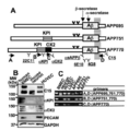
- FIG. 1A Schematic representation of three types of APP splice variants, APP695, APP751, and APP770. A series of anti-APP antibodies and the recognition sites for primers A, B, C and D used to analyze APP transcripts are shown.
- FIG. 1B Cell lysate of human cerebral vascular endothelial cells (BMEC), mouse primary neurons and human umbilical cord blood-derived vascular endothelial cells (HUVEC) using anti-APP C15, anti-KPI, anti-OX2, anti-PECAM and anti-GAPDH antibodies ( Western blot analysis of APP at 20 ⁇ g protein).
- BMEC human cerebral vascular endothelial cells
- HAVEC human umbilical cord blood-derived vascular endothelial cells
- FIG. 2 shows a series of lectin precipitates of endothelial APP.
- BMEC lysates 100 ⁇ g were incubated with SSA-, MAA-, E4-PHA-, ConA-, RCA120- or Jacalin-agarose for 16 hours at 4 ° C.
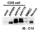
- FIG. 6 shows analysis of APP695, APP751 and APP770 expressed in COS cells. Human APP695, APP751, and APP770 were individually overexpressed in COS cells. The obtained cell lysate (1 ⁇ g) was analyzed by Western blot using anti-APP C15 antibody together with BMEC lysate (5 ⁇ g) and neuronal lysate (10 ⁇ g). It is a figure which shows glycosidase digestion of brain endothelial APP.
- FIG. 6 shows analysis of APP695, APP751 and APP770 expressed in COS cells. Human APP695, APP751, and APP770 were individually overexpressed in COS cells. The obtained cell lysate (1 ⁇ g) was analyzed by Western blot using anti-APP C15 antibody together with BMEC lysate (5 ⁇ g) and neuronal lysate (10 ⁇ g). It is a figure which shows glycosidase digestion of brain endothelial APP.
- FIG. 4A shows that high molecular weight type and low molecular weight type APP770 (APP-H and APP-L, respectively) have N-linked sugar chains.
- FIG. 4B shows that APP-H has a sialylated core type 1 O-linked sugar chain.
- FIG. 4C Schematic diagram of APP770 mutant used in this study.
- FIG. 4D shows changes in mobility of SDS-PAGE of wild-type APP770 and its mutants expressed in COS cells. Black and gray arrowheads indicate APP-H and APP-L, respectively. It is a figure which shows the analysis of sAPP secreted from BMEC.
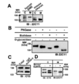
- FIG. 5A Intact APP and soluble secreted sAPP pulled down using heparin-agarose from cell lysates (C, 6 ⁇ g) and medium (M) of BMEC and mouse primary neurons, Western blot using anti-APP 22C11 antibody. Respectively.
- FIG. 5B sAPP pulled down from BMEC with heparin-agarose was incubated in the presence or absence of PNGase, sialidase and O-glycosidase and then analyzed by Western blot using anti-APP 22C11 antibody.
- FIG. 5B sAPP pulled down from BMEC with heparin-agarose was incubated in the presence or absence of PNGase, sialidase and O-glycosidase and then analyzed by Western blot using anti-APP 22C11 antibody.
- FIG. 5C After cell surface biotinylation of BMEC, biotinylated cell surface proteins were precipitated using streptavidin-sepharose and then analyzed by Western blot using anti-APP C15 antibody.
- FIG. 5D BMEC is cultured in the presence or absence of benzyl GalNAc, then intact APP and sAPP in cell lysate (C) and medium (M), anti-APP C15 antibody and anti-APP 22C11 antibody, respectively. And analyzed by Western blot.
- FIG. 4 shows APP770 expression in human cerebral blood vessels, cleavage at ⁇ and ⁇ sites, and secretion into CSF.
- FIG. 6A sAPP770 secreted from BMEC was analyzed by Western blot using anti-APP 22C11, anti-OX2, anti-sAPP ⁇ and anti-sAPP ⁇ antibodies.
- FIG. 6C Human brain sections embedded in paraffin were analyzed by hematoxylin-eosin staining (upper panel a) and immunostaining with anti-OX2 antibody (lower panel b).
- Gray and black arrowheads in a indicate nuclei of endothelial cells and smooth muscle cells, respectively.
- the arrow in b indicates the OX2 immunoreactive endothelium. Scale bar: 20 ⁇ m.
- FIG. 6D sAPP in human CSF samples (0.5 ml each) was pulled down using heparin-agarose (30 ⁇ l) and then analyzed by Western blot using anti-APP 22C11, anti-OX2 and anti-sAPP ⁇ antibodies.
- Gray and black arrowheads indicate sAPP ⁇ and sAPP770 ⁇ other than sAPP770 ⁇ , respectively.
- amyloid ⁇ precursor protein means a molecule that is cleaved by ⁇ secretase (BACE1) to produce amyloid ⁇ peptide (also referred to herein as A ⁇ peptide or simply A ⁇ ).
- BACE1 ⁇ secretase
- a ⁇ peptide also referred to herein as A ⁇ peptide or simply A ⁇ .
- APP it is desirable to use an APP derived from the same species as the target depending on the target to be detected.
- three types of splicing variants, APP695, APP751, and APP770 are known for human APP.
- the amino acid sequence of APP and the base sequence encoding it have been published in several animals. In humans, the amino acid sequences (GenBank Accession No. NP_958817, NP_958816 and NP_000475) and the base sequences (GenBank Accession No. NM_N2014. 1 and NM_000484.2) have been published and can be isolated by methods known per se.
- the amino acid sequence and base sequence of APP derived from animals other than humans can also be easily obtained by searching the genome database of various animals, and based on this information, it can be obtained by a method known per se. Can be separated.
- human APP will be described as an example.
- FIG. 1A shows the structure of the splicing variant of APP.
- APP751 further includes a Kunitz-type protease inhibitor (KPI) region
- APP770 includes an OX2 region in addition to the KPI region.
- KPI Kunitz-type protease inhibitor
- These three splicing variants have the same amino acid sequence in a region other than the KPI region and the OX2 region (including the C-terminal portion including the A ⁇ region and the transmembrane region).
- the OX2 region is thus a characteristic sequence that can be used to distinguish APP770 from the other two variants.
- the amino acid sequence of human APP770 and the nucleotide sequence encoding it are shown in SEQ ID NOs: 2 and 1, respectively.
- the KPI region corresponds to the amino acid sequence at positions 288 to 344
- the OX2 region corresponds to the amino acid sequence at positions 345 to 364
- the A ⁇ 40 (42) region corresponds to the amino acid sequence at positions 672 to 711 (713).
- soluble amyloid ⁇ precursor protein ⁇ means an N-terminal fragment of APP produced by cleavage by ⁇ -secretase (referred to as ⁇ -cleavage). Is not included.
- the ⁇ -cleavage products produced from APP695, APP751 and APP770, which are APP splicing variants, are called sAPP695 ⁇ , sAPP751 ⁇ and sAPP770 ⁇ , respectively.
- the amino acid sequence of human sAPP770 ⁇ corresponds to the 18th to 671th amino acid sequence in the sequence of SEQ ID NO: 2.
- soluble amyloid ⁇ precursor protein ⁇ means an N-terminal fragment of APP produced by cleavage of APP by ⁇ -secretase known as A ⁇ non-production pathway (called ⁇ -cleavage).
- ⁇ site means the cleavage site.
- endothelial cells refer to all vascular endothelial cells present in the living body, and particularly mean vascular endothelial cells present in all circulatory systems from the heart to capillaries.
- the endothelial cells are specifically flat cells covering the inner wall of the blood vessel, and preferably include vascular endothelial cells such as cerebral vascular endothelial cells, umbilical vein vascular endothelial cells, and sinusoidal endothelial cells. .
- an antibody includes all immunoglobulin classes and subclasses and forms of functional fragments of an antibody.
- the antibody is produced using a gene recombination technique.
- the resulting antibodies, antibody fragments, and binding fragments thereof include but are not limited to.
- polyclonal antibodies and monoclonal antibodies are included, but antibody preparations containing different antibodies against different epitopes from polyclonal antibodies.
- Monoclonal antibodies are antibodies obtained from a substantially homogeneous antibody population (including antibody fragments). ).
- the binding fragment means a partial region of the aforementioned antibody.
- F (ab ′) 2 Fab ′, Fab, Fv (variable fragment of antibody), sFv, dsFv (disulphide stabilized Fv ), DAb (single domain antibody) and the like
- F (ab ′) 2 Fab ′, Fab, Fv (variable fragment of antibody), sFv, dsFv (disulphide stabilized Fv ), DAb (single domain antibody) and the like
- a disease accompanied by accumulation of amyloid ⁇ peptide refers to a disease or condition in which a causal relationship between A ⁇ level and the disease in vivo is suggested.
- a ⁇ accumulates in the brain parenchyma (referred to as senile plaques) or cerebrovascular (referred to as cerebral vascular amyloid angiopathy (CAA)).
- senile plaques referred to as senile plaques
- cerebrovascular referred to as cerebral vascular amyloid angiopathy (CAA)
- CAA cerebral vascular amyloid angiopathy
- a ⁇ level in the living body is considered to increase as the aging progresses, a state in which the A ⁇ level in the living body is high can also be caused by a normal aging phenomenon, but a more rapid increase is seen in Alzheimer's disease. Therefore, diseases associated with amyloid ⁇ peptide accumulation typically have Alzheimer's disease, cerebral vascular amyloid angiopathy, cerebral vascular dementia, other dementia, brain, regardless of whether symptoms are manifested or not. Infarcts and the like are included. Since it has been reported that A ⁇ accumulation in cerebral blood vessels is found in the early stage of Alzheimer's disease, an increase in soluble amyloid ⁇ precursor protein 770 ⁇ may occur prior to an increase in highly toxic A ⁇ oligomers There is sex.
- Method for detecting soluble amyloid ⁇ precursor protein 770 ⁇ comprises the following steps: (1) contacting a biological sample derived from a subject suspected of having a disease associated with accumulation of amyloid ⁇ peptide with an antibody that recognizes soluble amyloid ⁇ precursor protein ⁇ or an amyloid ⁇ precursor protein 770-specific antibody; And (2) detecting (preferably quantitatively detecting) soluble amyloid ⁇ precursor protein 770 ⁇ in the complex formed in step (1); (Hereinafter also referred to as detection method I).
- a biological sample derived from a subject suspected of having a disease associated with accumulation of amyloid ⁇ peptide is contacted with an antibody specific for amyloid ⁇ precursor protein 770 or an antibody that recognizes soluble amyloid ⁇ precursor protein ⁇ . It is a process to make.
- the subject in the step (1) is a subject to determine whether or not the patient is suffering from a disease accompanied by accumulation of amyloid ⁇ peptide, and means a mammal suspected of suffering from the disease.
- Mammals include, for example, rodents such as mice, rats, hamsters, guinea pigs, laboratory animals such as rabbits, pets such as dogs and cats, domestic animals such as cows, pigs, goats, horses and sheep, monkeys, orangutans, and Examples include primates such as chimpanzees and humans, with humans being particularly preferred.
- a disease model such as a chronic cerebral ischemia model can be prepared, and fluctuations in soluble amyloid ⁇ precursor protein 770 ⁇ can be observed.
- Such a subject may or may not exhibit cognitive impairment and may be treated for a disease involving accumulation of amyloid ⁇ peptide, such as Alzheimer's disease.
- the biological sample can be collected from the subject using a known method.
- various body fluids such as blood, plasma, serum, lymph fluid and cerebrospinal fluid can be used.
- the biological sample of the present invention is blood, plasma or serum.
- the use of these samples is preferable in applying the method of the present invention in mass screening because no special technique is required for collection and the burden on the subject is small.
- the biological sample of the present invention includes cerebrospinal fluid. Cerebrospinal fluid fills and circulates brain tissue and is thought to reflect the state of the brain. It is also used in the measurement of a conventionally known biomarker for a brain state, and is suitably used as a biological sample also in the present invention.
- the biological sample of the present invention is a culture supernatant of vascular endothelial cells.
- the sensitivity in the detection method of the present invention can be enhanced by culturing and proliferating vascular endothelial cells.
- the vascular endothelial cells include cerebral vascular endothelial cells, umbilical cord blood-derived vascular endothelial cells, and liver sinusoidal endothelial cells.
- these cells may be derived from stem cells such as embryonic stem cells, tissue stem cells, and cells to which pluripotency is imparted by genetic manipulation or the like (for example, iPS cells).
- Vascular endothelial cells can be collected from blood vessels surgically removed from the living body, blood vessels for transplantation prepared from ES cells or bone marrow cells, and the like.
- vascular endothelial progenitor cells present in blood peripheral blood, umbilical cord blood, etc.
- fat can be differentiated and proliferated (see, for example, International Publication No. WO2006 / 090882, Japanese Patent No. 4217262, etc.).
- Culture of vascular endothelial cells can be performed using known methods and reagents.
- a biological sample derived from a subject is contacted with an antibody recognizing amyloid ⁇ precursor protein 770 (APP770) -specific antibody or soluble amyloid ⁇ precursor protein ⁇ (sAPP ⁇ ), and soluble amyloid ⁇ precursor protein 770 ⁇ A complex with the antibody is formed.
- APP770 amyloid ⁇ precursor protein 770
- sAPP ⁇ soluble amyloid ⁇ precursor protein ⁇
- the “amyloid ⁇ precursor protein 770 (APP770) -specific antibody” in the present invention has the ability to bind to APP770, and splicing variants other than APP770 of amyloid ⁇ precursor protein (APP) (APP695, APP751 are known). It is an antibody that does not bind to.
- APP770-specific antibodies have the ability to bind also to soluble amyloid ⁇ precursor protein 770 ⁇ . Also, it can be specific to APP770 from any mammal, but is preferably specific to APP770 from the same species as the subject.
- Such an antibody can be prepared by a method known per se on the basis of the difference in amino acid sequence between APP770 and other splicing variants, or the state of glycosylation.
- an antigen peptide sequence is designed in the OX2 region that exists on the amino acid sequence of APP770 but does not exist on the amino acid sequences of APP695 and APP751, and an antibody is produced using this peptide to produce APP770.
- Specific antibodies can be obtained.
- Such antibodies are also commercially available (eg, anti-OX2 antibody (Chemicon)).
- an antibody recognizing soluble amyloid ⁇ precursor protein ⁇ means an antibody capable of binding to all or at least sAPP ⁇ including sAPP695 ⁇ , sAPP751 ⁇ and sAPP770 ⁇ .
- Such an antibody can be prepared by a method known per se on the basis of information such as the amino acid sequence common to sAPP ⁇ or the sugar chain addition state. Specifically, for example, by designing an antigen peptide sequence around the ⁇ -secretase cleavage site ( ⁇ site) common to all APP variants and using this peptide to produce an antibody, sAPP ⁇ is not recognized. An antibody that specifically recognizes sAPP ⁇ can be obtained.
- an antibody that does not recognize sAPP695 ⁇ but recognizes sAPP751 ⁇ and sAPP770 ⁇ can also be obtained by designing an antigenic peptide in the KPI region.
- Such antibodies are also commercially available (eg, mouse monoclonal anti-APP 22C11 antibody (Chemicon), anti-human sAPP ⁇ antibody (IBL Co.), and anti-KPI antibody (Chemicon)).
- an antibody recognizing soluble amyloid ⁇ precursor protein ⁇ an antibody that also recognizes ⁇ -cleaved APP (for example, anti-APP 22C11 antibody (Chemicon)) is also used.
- an antibody that recognizes the ⁇ site of soluble amyloid ⁇ precursor protein ⁇ for example, anti-human sAPP ⁇ antibody (IBL Co.) is preferably used.
- an antibody that specifically recognizes sAPP695 ⁇ can be used for the purpose of discriminating A ⁇ production in vascular endothelial cells as compared to A ⁇ production in nerve cells, and the step (1
- the following steps (3) Contacting a biological sample derived from a subject suspected of having a disease associated with accumulation of amyloid ⁇ peptide with an antibody that recognizes soluble amyloid ⁇ precursor protein ⁇ or a soluble amyloid ⁇ precursor protein 695 ⁇ -specific antibody And (4) detecting soluble amyloid ⁇ precursor protein 695 ⁇ in the complex formed in step (3); Soluble amyloid ⁇ precursor protein 695 ⁇ can also be detected.
- the “sAPP695 ⁇ -specific antibody” means an antibody capable of binding to the sAPP695 ⁇ and not binding to sAPP770 ⁇ and sAPP751 ⁇ .
- Such an antibody can be prepared by a method known per se based on information such as the amino acid sequence of sAPP695 ⁇ or the state of sugar chain addition.
- an antibody that specifically recognizes sAPP695 ⁇ is linked to the 287th and 365th amino acids by removing the 288th to 364th amino acid sequences in which the KPI region and OX2 region are present in the sequence of SEQ ID NO: 2.
- Any antibody that recognizes a 40 amino acid sequence including a portion, preferably a 30 amino acid sequence, more preferably a 10 to 20 amino acid sequence may be used.
- antibodies that specifically recognize sAPP695 ⁇ without recognizing sAPP770 ⁇ and sAPP751 ⁇ can be obtained. Such antibodies also have the ability to specifically bind APP695 and not bind APP770 and APP751.
- an antibody that specifically recognizes sAPP751 ⁇ can be used as appropriate.
- the “sAPP751 ⁇ -specific antibody” means an antibody capable of binding to the sAPP751 ⁇ and not binding to sAPP770 ⁇ and sAPP695 ⁇ .
- Such an antibody can be prepared by a method known per se based on information such as the amino acid sequence of sAPP751 ⁇ or the state of sugar chain addition.
- an antibody specifically recognizing sAPP751 ⁇ is a part of the sequence of SEQ ID NO: 2 from which the 345 to 364th amino acid sequence of the OX2 region is removed and the KPI region is present, that is, the 344th and 365th positions.
- Any antibody that recognizes a sequence of 40 amino acids, preferably a sequence of 30 amino acids, more preferably a sequence of 10 to 20 amino acids, including a portion to which the second amino acid is linked may be used.
- antigen peptides of these sequences and producing antibodies using these peptides, it is possible to obtain antibodies that specifically recognize sAPP751 ⁇ without recognizing sAPP770 ⁇ and sAPP695 ⁇ .
- Such antibodies also have the ability to specifically bind APP751 and not bind APP770 and APP695.
- the APP770-specific antibody, sAPP695 ⁇ -specific antibody, sAPP751 ⁇ -specific antibody, and sAPP ⁇ -recognizing antibody may be either a polyclonal antibody or a monoclonal antibody.
- the antibodies include, but are not limited to, antibodies that can be produced using gene recombination techniques, antibody fragments, and binding fragments thereof.
- the binding fragment means a partial region of the aforementioned antibody, and specifically, for example, F (ab ′) 2 , Fab ′, Fab, Fv (variable fragment of antibody), sFv, dsFv (disulphide stabilized Fv).
- dAb single domain antibody
- the antibody may be directly or indirectly labeled with a labeling substance.
- labeling substances include fluorescent substances (eg, FITC, rhodamine), radioactive substances (eg, 32 P, 35 S, 14 C, 3 H), enzymes (eg, alkaline phosphatase, peroxidase), colored particles (eg, metal colloids) Particles, colored latex), biotin and the like.
- Solid phase includes plates (eg, microwell plates), tubes, beads (eg, plastic beads, magnetic beads), chromatographic carriers (eg, water-absorbing substrates such as nitrocellulose membranes, Sepharose), membranes (Eg, nitrocellulose membrane, PVDF membrane), gel (eg, polyacrylamide gel), metal membrane (eg, gold membrane) and the like.
- plates, beads, chromatographic carriers and membranes are preferably used, and plates are most preferably used from the viewpoint of easy handling.
- the bond include covalent bond, ionic bond, physical adsorption, and the like, and are not particularly limited. However, covalent bond and / or physical adsorption are preferable because sufficient bond strength can be obtained.
- the solid phase may be directly bonded to the solid phase or indirectly bonded to the solid phase using a substance known per se.
- a phosphate buffer solution such as bovine serum albumin (BSA) or bovine milk protein is brought into contact with the solid phase, and the solid surface portion not coated with the antibody is removed. Blocking with BSA, bovine milk protein or the like is generally performed.
- BSA bovine serum albumin
- sAPP770 ⁇ contact between an APP770-specific antibody, an sAPP695 ⁇ -specific antibody, an sAPP751 ⁇ -specific antibody or an antibody that recognizes sAPP ⁇ and a biological sample derived from a subject suspected of having a disease associated with the accumulation of A ⁇ is caused by sAPP770 ⁇ in the biological sample.
- the contact can be made, for example, by adding a biological sample to a plate on which an antibody that recognizes APP770 or an antibody that recognizes sAPP ⁇ is immobilized.
- the biological sample may be separated by means such as SDS-PAGE, transferred to a membrane and fixed, and then contacted with an antibody.
- the time for maintaining such contact should be sufficient to form a complex by combining the antibody with sAPP770 ⁇ or the like contained in a biological sample derived from a subject suspected of having a disease associated with A ⁇ accumulation. Although not particularly limited, it is usually several seconds to several tens of hours.
- the temperature condition for the contact is usually 4 ° C. to 50 ° C., preferably 4 ° C. to 37 ° C., most preferably room temperature of about 15 ° C. to 30 ° C.
- the pH condition for carrying out the reaction is preferably 5.0 to 9.0, particularly preferably the neutral range of 6.0 to 8.0.
- Step (2) is a step of detecting soluble amyloid ⁇ precursor protein 770 ⁇ in the complex formed in the above step (1).
- any method capable of detecting soluble amyloid ⁇ precursor protein 770 ⁇ can be used.
- a method of detecting sAPP770 ⁇ based on its molecular weight or a method of detecting using an antibody is used.
- a step of detecting sAPP695 ⁇ can be included for the purpose of discriminating A ⁇ production in vascular endothelial cells as compared with A ⁇ production in nerve cells.
- detection of sAPP770 ⁇ can be treated as an index of A ⁇ production in vascular endothelial cells
- detection of sAPP695 ⁇ can be treated as an index of A ⁇ production in nerve cells.
- any method capable of detecting sAPP695 ⁇ can be used.
- a method of detecting sAPP695 ⁇ based on its molecular weight or a method of detecting using an antibody is used.
- a step of appropriately detecting sAPP751 ⁇ can be included as a further control.
- any method capable of detecting sAPP751 ⁇ can be used.
- a method of detecting sAPP751 ⁇ based on its molecular weight or a method of detecting using an antibody is used.
- Methods for detecting sAPP770 ⁇ , sAPP695 ⁇ or sAPP751 ⁇ based on their molecular weight include gel electrophoresis (eg, SDS-PAGE), various separation and purification methods (eg, size exclusion chromatography, ion exchange chromatography, hydrophobic chromatography).
- mass spectrometer eg, double focusing mass spectrometer, quadrupole analyzer, time-of-flight mass spectrometer (TOF MS), Fourier transform mass spectrometer, It can be used for measurement by an ion cyclotron mass spectrometer, etc., as well as a method for combining them, and by comparing the molecular weight of a predetermined marker peptide and detecting a band, spot or peak of the target molecular weight. However, it is not limited to these.
- the complex Prior to detection of sAPP770 ⁇ (or sAPP695 ⁇ or sAPP751 ⁇ ), the complex may be isolated from the reaction solution in step (1) (or (3)). Such isolation can be performed, for example, by an isolation method utilizing affinity for heparin or immunoprecipitation, and an appropriate label added to the antibody used in step (1) (or (3)). (For example, biotin etc.) may be used.
- the detection may be performed in a state where a complex is formed.
- the formed complex may be isolated from a sample, and the complex may be dissociated to release sAPP770 ⁇ , sAPP695 ⁇ , or sAPP751 ⁇ .
- Measurement using a mass spectrometer is performed as follows, for example.
- the complex formed in the step (1) or (3) is isolated by a method (for example, immunoprecipitation method) using a label attached to the antibody used in the step (1) or (3).
- the isolated complex may be fragmented by digestion with a protease such as trypsin. Digestion with a protease having high sequence specificity makes it possible to identify proteins based on the pattern of the resulting peptide.
- the sample is then subjected to ionization and mass spectrometry.
- suitable ionization methods eg, electron impact ionization method, field desorption method, secondary ionization method, fast atom collision method, matrix-assisted laser desorption ionization (MALDI) method, electrospray ionization (ESI) method
- MALDI matrix-assisted laser desorption ionization
- ESI electrospray ionization
- the combination of the ionization method and the mass spectrometry include MALDI-TOF MS, ESI-MS, and the like.
- tandem mass spectrometry MS / MS
- MS tandem mass spectrometry
- sAPP770 ⁇ When sAPP770 ⁇ is detected in step (2), it can be determined that the subject is suffering from a disease associated with accumulation of A ⁇ or has a high probability of developing the disease. When sAPP770 ⁇ is not detected or is below the detection limit, it can be determined that the subject does not suffer from a disease associated with accumulation of A ⁇ or has a low probability of developing the disease.
- the disease is preferably cerebrovascular amyloid angiopathy, cerebrovascular dementia, or cerebral infarction, but it may be Alzheimer's disease and is not limited to these diseases.
- a disease associated with accumulation of A ⁇ in nerve cells preferably Alzheimer's disease, or It can be determined that the probability of developing a disease is high.
- the target disease in the detection of sAPP770 ⁇ is Alzheimer's disease, the determination can determine the onset state of Alzheimer's disease in more detail.
- the step (2) of the present invention preferably comprises: (2a) An antibody that recognizes the soluble amyloid ⁇ precursor protein 770 ⁇ in the complex formed in step (1) and the other soluble amyloid ⁇ precursor protein ⁇ that was not used in step (1) Or contacting with an amyloid ⁇ precursor protein 770-specific antibody; and (2b) detecting the complex formed in step (2a); It is.
- the step (4) is preferably (4a) An antibody that recognizes the soluble amyloid ⁇ precursor protein 695 ⁇ in the complex formed in step (3) and the other soluble amyloid ⁇ precursor protein ⁇ not used in step (3) Or a step of contacting with a soluble amyloid ⁇ precursor protein 695 ⁇ -specific antibody; and (4b) a step of detecting the complex formed in step (4a); It is.
- the complex formed in the step (1) can be contacted with the antibody in the state of the complex or in a state where the complex is dissociated and sAPP770 ⁇ is released.
- the contact method with sAPP770 ⁇ and the contact conditions the corresponding description of the labeling of the antibody, the contact method with the biological sample and the contact conditions in the above step (1) can be applied.
- Step (2b) detects a complex composed of the complex formed in step (1) and the antibody used in step (2a), or a complex composed of sAPP770 ⁇ and the antibody used in step (2a). It is a process.
- Detection is performed by detecting sAPP770 ⁇ contained in the complex or the antibody used in step (2a). Such detection can be performed in the state of the complex or in a state where the complex is dissociated and the soluble amyloid ⁇ precursor protein 770 ⁇ is released.
- the detection can be performed by enzyme immunoassay (EIA method), fluorescence immunoassay (FIA), immunochromatography, Western blotting, radioimmunoassay and the like, and these analysis methods are well known to those skilled in the art.
- EIA method enzyme immunoassay
- FIA fluorescence immunoassay
- immunochromatography Western blotting
- radioimmunoassay radioimmunoassay
- a sandwich ELISA using an APP770-specific antibody and an antibody that recognizes soluble amyloid ⁇ precursor protein 770 ⁇ is used as the detection method of the present invention. It is preferable to carry out by the method.
- a sandwich ELISA method is suitable for practicing the present invention because of its high specificity for an antigen.
- the sandwich ELISA method is performed as follows. First, the antibody of step (1) is immobilized on the well surface of an ELISA plate. Next, after blocking to prevent nonspecific adsorption to the well surface, a sample is added, and sAPP770 ⁇ in the sample is contacted with the antibody to form a complex. After the protein that has not bound to the antibody is removed by washing, the labeled antibody in the step (2a) is added to the well, brought into contact with sAPP770 ⁇ to form a complex, and detection and quantification are performed using the label.
- an enzyme-labeled antibody can be used.
- enzymes used for labeling include peroxidase, alkaline phosphatase, glucose oxidase, ⁇ -galactosidase and the like.
- the substrate agent used for the detection of the enzyme an appropriate one is selected according to the selected labeling enzyme. For example, when peroxidase is selected as the enzyme, o-phenylenediamine (OPD), tetramethylbenzidine (TMB) and the like are used.
- OPD o-phenylenediamine
- TMB tetramethylbenzidine
- alkaline phosphatase is selected, p-nitrophenyl phosphate (PNPP) and the like are used. used.
- the reaction stop solution and substrate solution conventionally known ones can be appropriately used without particular limitation depending on the selected enzyme.
- a method using avidin-biotin reaction is applicable as a kind of sandwich ELISA.
- sAPP770 ⁇ in a sample is captured with the antibody used in the solid phase step (1), and the antigen-antibody reaction between the captured sAPP770 ⁇ and the antibody used in the step (2a) labeled with biotin. To do.
- enzyme-labeled streptavidin is added to cause the avidin-biotin reaction.
- sAPP770 ⁇ is detected by detecting this enzyme.
- the antibody used in step (2a) labeled with biotin can be produced by binding biotin and the antibody used in step (2a) by a method known per se.
- labeling can be performed, for example, using a commercially available biotin labeling kit.
- a commercially available enzyme-labeled streptavidin can also be used.
- a method using a secondary antibody is also applicable.
- antibodies derived from different animal species are used for the antibody used in step (1) and the antibody used in step (2a), respectively, and a secondary antibody that recognizes the Ig domain of the antibody used in step (2a); A complex is formed with the antibody used in 2a).
- an enzyme-labeled secondary antibody and detecting this enzyme the presence of sAPP770 ⁇ in the complex is discriminated.
- a commercially available enzyme-labeled secondary antibody can be used.
- Such a method using a secondary antibody can enhance the sensitivity of the detection method of the present invention.
- sAPP770 ⁇ is detected by the same method as the sandwich ELISA method described above by replacing the enzyme used as a label in the EIA method with a fluorescent substance.
- a fluorescent substance chemical substances such as APC, PE, Cy2, Cy3, Cy5, ECD, FITC, PerCP, Alexa (registered trademark) Fluor, fluorescein, and rhodamine can be preferably used. Labeling with these chemical substances can be carried out by a method known per se. The fluorescence can be detected using a commercially available measuring instrument, a fluorescence microscope, or the like.
- the antibody of the step (2a) solidified in a line on a water-absorbing substrate such as a nitrocellulose membrane is developed on the membrane.
- a complex is formed between the biological sample and the labeled antibody in the step (1).
- the labeled antibody in step (1) may be pre-mixed with the biological sample or on the water-absorbing substrate so that the biological sample contacts the biological sample before contacting the antibody used in step (2a). It may be supplied.
- the formation of the complex can be detected by a technique corresponding to the label. For example, when colloidal gold particles are used as a label, a region where the labeled antibody is accumulated exhibits a red color, and is thus detected.
- Western blotting can also be selected as the detection method in step (2b).
- Western blotting also called immunoblotting is a method for detecting specific proteins in a sample. Gel electrophoresis is performed to separate proteins by length of polypeptides under denaturing conditions or by three-dimensional structure of proteins under non-denaturing conditions. The protein is then transferred to a membrane (typically nitrocellulose or PVDF) and immobilized, after which the target protein is detected using a specific antibody.
- a membrane typically nitrocellulose or PVDF
- detection of sAPP695 ⁇ is performed in steps (2a) and (2b) in steps (4a) and (4b). It can be performed in the same manner as the detection of sAPP770 ⁇ . As a further control, the detection of sAPP751 ⁇ can be performed in the same manner as the detection of sAPP770 ⁇ in steps (2a) and (2b).
- the detection step in the present invention is preferably a step of quantitatively detecting sAPP770 ⁇ .
- the quantification can be performed by a method known per se, such as quantifying the signal of the biological sample detected based on the label used using a calibration curve created using a standard sample of sAPP770 ⁇ .
- the standard sample of sAPP770 ⁇ may be sAPP770 ⁇ prepared from a biological sample, or may be prepared by a genetic engineering technique known per se based on gene information encoding sAPP770 ⁇ .
- sAPP770 ⁇ in the above steps (2a) and (2b) is performed. SAPP695 ⁇ can be handled in the same manner as described above. The same applies to the case of detecting sAPP751 ⁇ .
- the result obtained from the method of the present invention it is possible to diagnose whether or not the patient is afflicted with a disease associated with the accumulation of A ⁇ , and whether the disease is likely to develop.
- a diagnosis is made by comparing the level of sAPP770 ⁇ in a biological sample from a subject to the concentration range of sAPP770 ⁇ in a control sample from a healthy or young subject (which can be examined by statistical studies). be able to. Diagnosis can also be made by comparing the same subject with previously measured levels.
- the standard sAPP770 ⁇ level is an appropriate standard set according to the purpose. sAPP770 ⁇ can be observed to increase quantitatively with aging even in healthy individuals. Therefore, when “average value + 2 standard deviations” of healthy subjects whose age group matched with the disease control group is set as the cut off value and exceeds the cut off value, there is a possibility that the disease is associated with accumulation of amyloid ⁇ peptide Is diagnosed as high.
- the effect of treatment can be evaluated for a patient who is treating a disease associated with A ⁇ accumulation.
- a biological sample is collected from such a patient before treatment, during treatment and / or after treatment, and the change in the concentration of sAPP770 ⁇ is examined to know the effect of the treatment. For example, if the concentration of sAPP770 ⁇ in a later biological sample is lower than that in the previous biological sample, the treatment can be evaluated as being effective.
- some examples of therapeutic agents for dementia are known to cause CAA, and it is possible to predict the risk of CAA in patients administered with therapeutic agents using the method of the present invention. .
- a ⁇ production in vascular endothelial cells can be distinguished from A ⁇ production in nerve cells, and in this case, A ⁇ production in vascular endothelial cells is determined by sAPP770 ⁇ level in a biological sample.
- a ⁇ production in nerve cells can be determined using the sAPP695 ⁇ level in a biological sample as an index.
- SAPP770 ⁇ level which is an indicator of A ⁇ production in vascular endothelial cells, is a disease associated with A ⁇ accumulation, preferably cerebrovascular amyloid angiopathy, cerebrovascular dementia, cerebral infarction or Alzheimer's disease, more preferably cerebrovascular It is used for the determination that it is suffering from cerebrovascular amyloid angiopathy, cerebrovascular dementia, cerebral infarction caused by A ⁇ accumulation or having a high probability of developing the disease.
- the sAPP695 ⁇ level which is an index of A ⁇ production in nerve cells, is used to determine that a disease associated with A ⁇ accumulation in the brain parenchyma, preferably Alzheimer's disease, or a high probability of developing the disease.
- cerebrovascular amyloid angiopathy, cerebral vascular dementia or cerebral infarction and Alzheimer's disease are targeted as target diseases, or a combination of these diseases, and these diseases are determined as targets alone or these diseases Can be determined as a target, or the possibility can be determined.
- the diagnosis can determine the onset state of Alzheimer's disease in more detail.
- the present invention can be used in combination with a known diagnosis of Alzheimer's disease.
- a diagnosis of Alzheimer's disease other than the present invention clinical diagnosis using NINCDS-ADRDA and DSM-IV as diagnostic criteria, amyloid ⁇ peptide in body fluids Diagnostic method using 42 decrease as an index, diagnosing method using increase in tau or phosphorylated tau in body fluid as an index, diagnostic method using increase / decrease in sugar chain metabolites due to BACE1 activity in body fluid, A ⁇ by amyloid imaging Examples include, but are not limited to, image diagnosis using aggregation as an index and image diagnosis using partial brain atrophy as an index.
- the present invention increases microhemorrhage with cerebrovascular amyloid angiopathy with certain antibodies reported in immunotherapy of Alzheimer's disease using antibodies against amyloid ⁇ peptide. It is also possible to predict the risk of side effects (Hartman, RE, et al., J. Neurosci., 25, 6213-6220 (2005), Racke, MM, et.al., J. Neurosci., 25, 629-636 (2005).).
- sAPP770 ⁇ can be used as a diagnostic marker for the pathological condition of Alzheimer's disease because of the possibility that A ⁇ accumulation in vascular endothelial cells has shifted to the brain parenchyma and affects the symptoms of Alzheimer's disease. This is based on the finding that emboli leak out of the blood vessels during reopening of brain microvessels (Nature. 2010 May 27; 465 (7297): 478-82.). That is, from this finding, A ⁇ accumulation also exists as an embolus, and therefore, A ⁇ leaks out of the blood vessel during reopening of the brain microvasculature. Indicates potential impact.
- the detection of sAPP770 ⁇ in the present invention makes it possible to diagnose Alzheimer's disease based on the possibility of accumulation of A ⁇ migrated from cerebral microvessels or the correlation with cerebral vascular amyloid angiopathy. Since the diagnosis of Alzheimer's disease by detecting sAPP695 ⁇ focuses on A ⁇ accumulation in the brain parenchyma produced from nerve cells, the diagnosis of Alzheimer's disease by detecting sAPP770 ⁇ is based on a principle different from the diagnosis by detecting sAPP695 ⁇ . Allows diagnosis of disease. This also means that the diagnosis of Alzheimer's disease combining the detection of sAPP770 ⁇ and the detection of sAPP695 ⁇ can diagnose the disease state of Alzheimer's disease in more detail than when each is performed alone.
- the method for detecting soluble amyloid ⁇ precursor protein 770 ⁇ replaces steps (1) and (2) with the following steps: (1 ′) contacting a biological sample derived from a subject suspected of a disease associated with accumulation of amyloid ⁇ peptide with an antibody recognizing amyloid ⁇ precursor protein 770-specific or soluble amyloid ⁇ precursor protein ⁇ ; And (2 ′) a detection method comprising a step of detecting (preferably quantitatively detecting) soluble amyloid ⁇ precursor protein 770 in the complex formed in step (1 ′) (hereinafter referred to as a detection method) Also referred to as detection method II).
- Step (1 ') is the same as step (1) of detection method I.
- the soluble amyloid ⁇ precursor protein 770 means an amyloid ⁇ precursor protein 770 cleavage product (N-terminal fragment of APP770) containing at least sAPP770 ⁇ . That is, in the step (2 '), other amyloid ⁇ precursor protein 770 cleavage product (N-terminal fragment of APP770) (eg, sAPP770 ⁇ ) is also detected together with sAPP770 ⁇ .
- sAPP770 ⁇ amyloid ⁇ precursor protein 770 cleavage product
- sAPP770 ⁇ amyloid ⁇ precursor protein 770 cleavage product
- the disease of the subject is substantially diagnosed by detecting (preferably quantitatively detecting) soluble amyloid ⁇ precursor protein 770 in step (2 ′). Or the risk can be predicted.
- the description about the detection method I can be applied to the detection method II.
- the present invention relates to a diagnostic agent for diseases associated with accumulation of amyloid ⁇ peptide.
- the present invention relates to an amyloid ⁇ peptide comprising an antibody specific for amyloid ⁇ precursor protein 770 and / or an antibody recognizing soluble amyloid ⁇ precursor protein ⁇ as an active ingredient.
- the antibody that is an active ingredient of the diagnostic agent of the present invention is an antibody that recognizes soluble amyloid ⁇ precursor protein ⁇ and / or amyloid ⁇ described in “1. Method for detecting soluble amyloid ⁇ precursor protein 770 ⁇ ”. It is a precursor protein 770 specific antibody.
- the diagnostic agent of the present invention may consist only of the above-described antibody or may contain a pharmaceutically acceptable carrier.
- a pharmaceutically acceptable carrier when preparing the diagnostic agent of the present invention as a solution, various carriers conventionally used as pharmaceutical materials, such as diluents, solvents, solubilizers, suspending agents, isotonicity, etc. An agent, a buffering agent, etc. may be included.
- a surfactant such as Tween 20 (registered trademark) in order to prevent antibody aggregation.
- Tween 20 registered trademark
- the diagnostic agent for diseases associated with accumulation of A ⁇ is a diagnostic agent for cerebrovascular amyloid angiopathy, cerebrovascular dementia, cerebral infarction or Alzheimer's disease.
- the present invention relates to a kit for diagnosing a disease associated with accumulation of amyloid ⁇ peptide.
- the present invention relates to accumulation of amyloid ⁇ peptide comprising as an active ingredient an antibody recognizing amyloid ⁇ precursor protein 770-specific antibody and soluble amyloid ⁇ precursor protein ⁇ .
- a kit for diagnosing a disease associated with is provided.
- the antibody which is an active ingredient of the diagnostic kit of the present invention is the antibody described in “1. Method for detecting soluble amyloid ⁇ precursor protein 770 ⁇ ”.
- the diagnostic kit of the present invention may contain an antibody such as a sAPP695 ⁇ -specific antibody, a sAPP751 ⁇ -specific antibody, or a reagent in addition to the above-mentioned antibody. They may be together or stored in separate containers. Examples of antibodies or reagents include secondary antibodies, substrate agents, labeling substances (eg, fluorescent dyes, enzymes), solid phases, reactions, and the like described in “1. Method for detecting soluble amyloid ⁇ precursor protein 770 ⁇ ”. In addition to the container, a buffer for diluting the treatment solution and the antibody, a positive control (eg, recombinant sAPP770 ⁇ ), a negative control, an instruction describing the protocol, and the like can be mentioned. These elements can be mixed in advance if necessary.
- an antibody such as a sAPP695 ⁇ -specific antibody, a sAPP751 ⁇ -specific antibody, or a reagent in addition to the above-mentioned antibody. They may be together or stored in separate containers. Examples of antibodies or
- the antibody may be immobilized in advance, and the antibody may be labeled in advance.
- the solid phase that can be used in the diagnostic kit of the present invention is not particularly limited, and examples thereof include polymers such as polystyrene, glass beads, magnetic particles, microplates, filter paper for immunochromatography, glass filters, and insoluble carriers. Can do. Preferably, it is a microplate used for sandwich ELISA.
- the form of the diagnostic kit of the present invention is not particularly limited, but for the purpose of simple diagnosis, it can be an integrated diagnostic kit in which the components of the diagnostic kit of the present invention are integrated.
- Examples of the integrated diagnostic kit include a cassette type using an immunochromatography method.
- the diagnostic kit of the present invention makes it easy and highly accurate to diagnose diseases associated with A ⁇ accumulation.
- the diagnostic kit for a disease associated with accumulation of A ⁇ of the present invention is used as a diagnostic kit for cerebrovascular amyloid angiopathy, cerebrovascular dementia, cerebral infarction, or Alzheimer's disease.
- tissue culture media and reagents including DMEM
- DMEM tissue culture media and reagents
- PNGase recombinant peptide N-glycosidase F
- O-glycosidase From Roche Arthrobacter ureafaciens sialidase from Nacalai Tesque
- Protein A-Sepharose Fast Flow from GE Healthcare
- Protein Molecular Weight Standard from Bio-Rad
- a series of lectin-bound agarose from Seikagaku Corporation
- a ⁇ 40 from Peptide Institute
- BCA protein assay reagents and sulfo-NHS-LC-biotin are from Thermo Fisher Scientific Inc .
- all other chemicals are from Sigma or Wako Chemicals.
- Oligo DNA primers were obtained from Invitrogen. The primers used are shown in Table 1.
- Anti-APP rabbit polyclonal antibody (C15 antibody) against endogenous membrane-bound (intact) APP was provided by Dr. Takashi Maruyama (Saitama Medical University, Saitama, Japan).
- the commercially available antibodies used are as follows: mouse monoclonal anti-APP 22C11 antibody (Chemicon); anti-human sAPP ⁇ antibody (6E10; Signet Laboratories); anti-GAPDH antibody (Chemicon); anti-KPI antibody (Chemicon) ); Anti-OX2 antibody (Chemicon); anti-human sAPP ⁇ antibody (IBL Co.); goat polyclonal anti-PECAM antibody (Santa Cruz Biotechnology). Clinical research was approved by the Ethics Committee of RIKEN and Fukushima Medical University. All animal experiments were conducted according to the guidelines for animal experiments at RIKEN.
- Human APP770 FLAG-pEF is constructed by inserting the human APP770 sequence amplified by PCR (using primers 1 and 2) into the SalI and HindIII sites (of the vector), and the FLAG region fragment consists of primers 3 and 4 in HindIII. And by annealing to the XbaI site.
- a ViraPower adenovirus expression system (Invitrogen) was used according to the manufacturer's protocol to generate a recombinant adenovirus carrying human APP-FLAG.
- a series of APP770-pcDNA3.1 mutants were generated using the QuickChange Site-Directed Mutagenesis Kit (Stratagene).
- APP S346,348A was first constructed, and then APP S346,348A was used as a template.
- APP OX2All mutants were generated using APP SOX2A forward and reverse as primers.
- et uses APP OX2All variant as a template, using the APP T291,292A forward and reverse and APP T651A forward and reverse as primers to replace the Thr291, Thr292 and Thr651 to Ala.
- Human cerebral vascular endothelial cells (BMEC, Applied Cell Biology Research Institute) were cultured in CS-C complete medium with or without 10% FBS and used within 4 passages.
- HUVEC (TaKaRa) was cultured in EMB-2 (TaKaRa) containing 2% FBS and EGM TM -2 SingleQuots and used within 4 passages.
- Primary liver sinusoidal endothelial cells (LSEC) were prepared from mouse liver using CD146 MicroBeads (Miltenyi Biotec) according to the manufacturer's protocol.
- COS-7 cells were cultured in DMEM containing 10% FBS.
- PCR analysis of APP transcripts Isolate total RNA from cells using Sepasol reagent (Nacalai Tesque, Inc.), and use 5 ⁇ g of isolated RNA to generate random hexamers using Super Script III RT Kit (Invitrogen) according to the manufacturer's protocol. Used for reverse transcription. The obtained cDNA samples were subjected to PCR analysis using primers A and D for APP695, APP751 and APP770, primers B and D for APP751 and APP770, and primers C and D for APP770. PCR was performed for 28 cycles (95 ° C. for 40 seconds, 56 ° C. for 40 seconds, 72 ° C. for 90 seconds).
- a ⁇ 40 and A ⁇ 42 in the medium are as follows: Human Amyloid ⁇ (1-40) or (1-42) Assay Kit (IBL Co.) for BMEC, Human / Rat ⁇ Amyloid (40) or (42 ) Determined using ELISA Kit (Wako Chemicals).
- Cell surface biotinylation BMEC grown in 10 cm culture dishes were labeled with Sulfo-NHS-LC-Biotin at 4 ° C. for 30 minutes. The cells were washed three times with PBS (pH 8.0) containing 0.1 M glycine and once with PBS, and then cell lysate was prepared. For further analysis, biotinylated cell surface proteins were pulled down with streptavidin-Sepharose (GE Healthcare).
- Example 1 This example illustrates cell type specific expression of three isoforms by alternative splicing of APP.
- the human brain expresses APP mRNA isoforms, APP695, APP751, and APP770 with three alternative splices (Nature 1988 331 (6156): 525-527; Nature 1988 331 (6156): 528-530) (FIG. 1A).
- neurons are expressed exclusively in APP695 (Proc Natl Acad Sci USA 1993 90 (20): 9513-9517; Proc Natl Acad Sci USA 1990 87 (4): 1561-1565; Proc Natl Acad Sci USA 1989 86 ( 16): 6338-6342).
- the present inventors focused on brain endothelial cells and analyzed established brain endothelial APP using established primary human brain microvascular endothelial cells (BMEC).
- BMEC primary human brain microvascular endothelial cells
- Western blot analysis using an anti-APP C-terminal antibody (C15) showed that BMECs expressed comparable or rather high levels of APP compared to primary neurons (FIG. 1B).
- Endothelial APP showed two separate bands with different gel mobilities after SDS-PAGE.
- the anti-KPI antibody and anti-OX2 antibody also detected two endothelial APPs and concluded that the endothelial cells express APP770.
- Human umbilical cord blood-derived endothelial cells also expressed two types of APP, although the expression level was significantly lower than that in BMEC, indicating that APP770 is widely expressed in endothelial cells. It was.
- RNA from endothelial cells and primary neurons.
- the reverse transcribed cDNA sample was then analyzed by PCR using a series of oligonucleotide primers to detect APP695, APP751 and APP770.
- FIG. 1C neurons expressed APP695, whereas endothelial cells expressed APP770 and did not express APP695. From these results, cell type-specific APP expression was confirmed.
- Example 2 the sugar chain addition state of APP770 will be described.
- APP may have two N-linked and multiple O-linked sugar chains (Biochim Biophys Acta 1999 1472 (1-2): 344-358; J Proteome Res 2009 8 (2): 631- 642; J Biol Chem 1998 273 (11): 6277-6284), the present inventors predicted that high molecular weight APP770 (APP-H) was highly glycosylated. Based on the results of a series of lectin pull-down assays (FIG.
- APP-H Since APP-H has an affinity for the SSA lectin that recognizes Sia ⁇ 2,6Gal / GalNAc, the present inventors expect that APP-H contains an O-linked sugar chain having a sialic acid residue. did. After sialidase treatment, the mobility of APP-H changed significantly, approaching that of APP-L (FIG. 4B). After treatment with sialidase and O-glycosidase, the mobility of APP-H was completely consistent with that of APP-L. Since O-glycosidase only cleaves unsubstituted core type 1 O-linked sugar chains, we conclude that APP-H has a sialylated core type 1 O-linked sugar chain. It was.
- Example 3 the glycosylated state of sAPP secreted from cerebral vascular endothelial cell BMEC will be described.
- the inventors performed Western blot analysis of BMEC lysates and media samples using anti-APP 22C11 antibody (which recognizes the N-terminal region of APP).
- FIG. 5A a single sAPP band was detected in the medium with a mobility between the two APP (APP-H and APP-L) bands found in the cell lysate. This suggests that sAPP is derived from APP-H only.
- sAPP is sensitive to both sialidase and O-glycosidase treatment (FIG.
- Example 4 it is explained that APP770 is expressed in human cerebral blood vessels, cleaved at ⁇ and ⁇ sites, and secreted into CSF. Since there is limited information regarding the processing level of APP770 regarding the expression level and activity of endothelial ⁇ -secretase and ⁇ -secretase, we analyzed soluble sAPP770 secreted from BMEC. When specific antibodies against sAPP ⁇ and sAPP ⁇ were used, both sAPP ⁇ and sAPP ⁇ were detected (FIG. 6A). Since endothelial cells have an amyloidogenic ⁇ -secretase pathway, it can be considered that these cells also have ⁇ -secretase activity and produce A ⁇ peptides.
- BMEC medium overexpressing APP770 both A ⁇ 40 and A ⁇ 42 were detected, and the endothelial A ⁇ 42 / A ⁇ 40 ratio was similar to that in neurons (FIG. 6B).
- the present inventors first analyzed cerebral cortex sections using anti-OX2 antibody to determine the localization of APP770. The luminal regions of endothelial cells of veins and venules were stained with anti-OX2 antibody, but smooth muscle cells were not stained (FIG. 6C). No immunohistochemical signals were observed in the arachnoid blood vessels.
- CSF contains sAPP770 ⁇ ( ⁇ -secretase cleavage product on the N-terminal side of APP770).
- SAPP was pulled down from CSF using heparin-agarose and then immunostained using anti-APP22C11, anti-OX2 and anti-sAPP ⁇ antibodies. Two bands were detected by the anti-APP22C11 antibody, and only the upper band was detected by the anti-OX2 antibody (FIG. 6D). This indicates that the upper band is derived from APP770.
- Both upper and lower bands detected by anti-sAPP ⁇ antibody indicate that both forms contain ⁇ -secretase cleavage products.
- soluble amyloid ⁇ precursor protein 770 ⁇ derived from endothelial cells can be detected.
- the data obtained can be used to diagnose diseases associated with amyloid ⁇ peptide accumulation, such as Alzheimer's disease.
- the diagnostic agent and diagnostic kit of the present invention it is possible to easily and accurately diagnose whether or not the patient is suffering from a disease accompanied by accumulation of amyloid ⁇ peptide.
Landscapes
- Life Sciences & Earth Sciences (AREA)
- Health & Medical Sciences (AREA)
- Engineering & Computer Science (AREA)
- Biomedical Technology (AREA)
- Hematology (AREA)
- Chemical & Material Sciences (AREA)
- Urology & Nephrology (AREA)
- Molecular Biology (AREA)
- Immunology (AREA)
- Proteomics, Peptides & Aminoacids (AREA)
- Medicinal Chemistry (AREA)
- Microbiology (AREA)
- Biotechnology (AREA)
- Neurosurgery (AREA)
- Neurology (AREA)
- Food Science & Technology (AREA)
- Cell Biology (AREA)
- Physics & Mathematics (AREA)
- Analytical Chemistry (AREA)
- Biochemistry (AREA)
- General Health & Medical Sciences (AREA)
- General Physics & Mathematics (AREA)
- Pathology (AREA)
- Peptides Or Proteins (AREA)
- Investigating Or Analysing Biological Materials (AREA)
Abstract
Description
[1]下記の工程:
(1)被験者由来の生体試料と、可溶型アミロイドβ前駆体タンパク質βを認識する抗体又はアミロイドβ前駆体タンパク質770特異的抗体とを接触させる工程;及び
(2)工程(1)で形成された複合体中の可溶型アミロイドβ前駆体タンパク質770βを検出する工程;
を含む、血管内皮細胞由来の可溶型アミロイドβ前駆体タンパク質770βの検出方法。
[2]工程(1)において、可溶型アミロイドβ前駆体タンパク質βを認識する抗体又はアミロイドβ前駆体タンパク質770特異的抗体のいずれか一方を使用し、
工程(2)が、
(2a)工程(1)で形成された複合体中の可溶型アミロイドβ前駆体タンパク質770βと、工程(1)で使用しなかった他方の可溶型アミロイドβ前駆体タンパク質βを認識する抗体又はアミロイドβ前駆体タンパク質770特異的抗体とを接触させる工程;及び
(2b)工程(2a)で形成された複合体を検出する工程;
である、[1]に記載の検出方法。
[3]生体試料が血液、血漿又は血清である[1]又は[2]に記載の方法。
[4]生体試料が脳脊髄液である[1]又は[2]に記載の方法。
[5]生体試料が血管内皮細胞の培養上清である[1]又は[2]に記載の方法。
[6] 工程(1)、(2)に加えて、下記の工程:
(3)アミロイドβペプチドの蓄積を伴う疾患が疑われる被験者由来の生体試料と、可溶型アミロイドβ前駆体タンパク質βを認識する抗体又は可溶型アミロイドβ前駆体タンパク質695β特異的抗体とを接触させる工程;及び
(4)工程(3)で形成された複合体中の可溶型アミロイドβ前駆体タンパク質695βを検出する工程;
を含み、可溶型アミロイドβ前駆体タンパク質695βの検出も含めた、[1]から[5]のいずれか1つに記載の方法。
[7]工程(3)において、可溶型アミロイドβ前駆体タンパク質βを認識する抗体又はアミロイドβ前駆体タンパク質695特異的抗体のいずれか一方を使用し、
工程(4)が、
(4a)工程(3)で形成された複合体中の可溶型アミロイドβ前駆体タンパク質695βと、工程(3)で使用しなかった他方の可溶型アミロイドβ前駆体タンパク質βを認識する抗体又は可溶型アミロイドβ前駆体タンパク質695β特異的抗体とを接触させる工程;及び
(4b)工程(4a)で形成された複合体を検出する工程;
である、[6]に記載の検出方法。
[8] 工程(1)における生体試料が血液、血漿、血清、血管内皮細胞の培養上清又は脳脊髄液であり、工程(3)における生体試料が脳脊髄液である[6]又は[7]に記載の方法。
[9] アミロイドβ前駆体タンパク質770特異的抗体がOX2ドメインを認識する抗体である[1]から[8]のいずれか1つに記載の方法。
[10]被験者がアミロイドβペプチドの蓄積を伴う疾患の罹患を疑われる、[1]から[9]のいずれか1つに記載の方法。
[11]疾患が脳血管アミロイドアンギオパチー、脳血管性痴呆症、脳梗塞、アルツハイマー病の少なくともいずれか一つである[10]に記載の方法。
[12]被験者が脳血管アミロイドアンギオパチー、脳血管性痴呆症、脳梗塞の少なくともいずれか一つと、アルツハイマー病の併発を疑われる[1]から[9]のいずれか1つに記載の方法。
[13]可溶型アミロイドβ前駆体タンパク質βを認識する抗体及び/又はアミロイドβ前駆体タンパク質770特異的抗体を有効成分とする、アミロイドβペプチドの蓄積を伴う疾患の診断薬。
[14]可溶型アミロイドβ前駆体タンパク質βを認識する抗体及びアミロイドβ前駆体タンパク質770特異的抗体を含む、アミロイドβペプチドの蓄積を伴う疾患の診断用キット。
[15]可溶型アミロイドβ前駆体タンパク質695β特異的抗体を更に含む、[14]の診断用キット。
[16]疾患が脳血管アミロイドアンギオパチー、脳血管性痴呆症、脳梗塞、アルツハイマー病の少なくともいずれか一つを対象とする[13]に記載の診断薬又は[14]若しくは[15]に記載のキット。
[17]疾患が、脳血管アミロイドアンギオパチー、脳血管性痴呆症、脳梗塞の少なくともいずれか一つと、アルツハイマー病の併発を対象とする[16]に記載の診断薬又はキット。
[18]下記の工程:
(1)被験者より採取された生体試料と、可溶型アミロイドβ前駆体タンパク質βを認識する抗体又はアミロイドβ前駆体タンパク質770特異的抗体とを接触させる工程;及び
(2)工程(1)で形成された複合体中の可溶型アミロイドβ前駆体タンパク質770βを検出する工程;
を含む、血管内皮細胞由来の可溶型アミロイドβ前駆体タンパク質770βを検出してアミロイドβペプチドの蓄積を伴う疾患に罹患している可能性を判定する方法。
[19]工程(1)において、可溶型アミロイドβ前駆体タンパク質βを認識する抗体又はアミロイドβ前駆体タンパク質770特異的抗体のいずれか一方を使用し、
工程(2)が、
(2a)工程(1)で形成された複合体中の可溶型アミロイドβ前駆体タンパク質770βと、工程(1)で使用しなかった他方の可溶型アミロイドβ前駆体タンパク質βを認識する抗体又はアミロイドβ前駆体タンパク質770特異的抗体とを接触させる工程;及び
(2b)工程(2a)で形成された複合体を検出する工程;
である、[18]に記載の検出方法。
[20]生体試料が血液、血漿又は血清である[18]又は[19]に記載の方法。
[21]生体試料が脳脊髄液である[18]又は[19]に記載の方法。
[22]生体試料が血管内皮細胞の培養上清である[18]又は[19]に記載の方法。
[23]工程(1)、(2)に加えて、下記の工程:
(3)被験者より採取された生体試料と、可溶型アミロイドβ前駆体タンパク質βを認識する抗体又は可溶型アミロイドβ前駆体タンパク質695β特異的抗体とを接触させる工程;及び
(4)工程(3)で形成された複合体中の可溶型アミロイドβ前駆体タンパク質695βを検出する工程;
を含み、可溶型アミロイドβ前駆体タンパク質695βの検出も含めた、[18]から[22]のいずれか1項に記載の方法。
[24]工程(3)において、可溶型アミロイドβ前駆体タンパク質βを認識する抗体又はアミロイドβ前駆体タンパク質695特異的抗体のいずれか一方を使用し、
工程(4)が、
(4a)工程(3)で形成された複合体中の可溶型アミロイドβ前駆体タンパク質695βと、工程(3)で使用しなかった他方の可溶型アミロイドβ前駆体タンパク質βを認識する抗体又は可溶型アミロイドβ前駆体タンパク質695β特異的抗体とを接触させる工程;及び
(4b)工程(4a)で形成された複合体を検出する工程;
である、[23]に記載の検出方法。
[25]工程(1)における生体試料が血液、血漿血清、血管内皮細胞の培養上清又は脳脊髄液であり、工程(3)における生体試料が脳脊髄液である[23]又は[24]に記載の方法。
[26]アミロイドβ前駆体タンパク質770特異的抗体がOX2ドメインを認識する抗体である[18]から[25]のいずれか1項に記載の方法。
[27]アミロイドβペプチドの蓄積を伴う疾患が脳血管アミロイドアンギオパチー、脳血管性痴呆症、脳梗塞、アルツハイマー病の少なくとも何れか一つである[18]から[26]のいずれか1項に記載の方法。
[28]可溶型アミロイドβ前駆体タンパク質770βの検出から脳血管アミロイドアンギオパチー、脳血管性痴呆症、脳梗塞の少なくともいずれか一つの疾患の罹患の可能性を判定する[18]から[26]のいずれか1つに記載の方法。
[29]下記の工程:
(1’)アミロイドβペプチドの蓄積を伴う疾患が疑われる被験者由来の生体試料と、アミロイドβ前駆体タンパク質770特異的抗体又は可溶型アミロイドβ前駆体タンパク質βを認識する抗体を接触させる工程;及び
(2’)工程(1’)で形成された複合体中の可溶型アミロイドβ前駆体タンパク質770を検出する(好ましくは定量的に検出する)工程
を含む、血管内皮細胞由来の可溶型アミロイドβ前駆体タンパク質770βの検出方法。
本発明の診断用キット及び診断薬は、上記の本発明の検出方法に適しており、アミロイドβペプチドの蓄積を伴う疾患の、効率的な診断を可能にする。
本発明において、アミロイドβ前駆体タンパク質(APP)とは、βセクレターゼ(BACE1)により切断され、アミロイドβペプチド(本明細書中、Aβペプチド、又は単にAβとも言う)を産生する分子を意味する。APPとしては、検出する対象に応じて、対象と同じ種由来のAPPを用いることが望ましい。一例として、ヒトAPPには3種類のスプライシングバリアント、APP695、APP751及びAPP770が知られている。
また、可溶型アミロイドβ前駆体タンパク質α(sAPPα)とは、Aβ非産生経路として知られるαセクレターゼによるAPPの切断(α切断と呼ばれる)で産生されたAPPのN末端側の断片を意味し、α部位はその切断部位を意味する。
生体内におけるAβレベルが高い対象においては、典型的には、Aβの脳実質部分への蓄積(老人斑と呼ばれる)や脳血管内への蓄積(脳血管アミロイドアンギオパチー(CAA)と呼ばれる)がみられる。さらに神経細胞の破壊、脳血管障害などが起こり、これらが進行した状態においては認知症として症状が顕在化する。加齢が進むに従って生体内のAβレベルは上昇すると考えられることから、生体内におけるAβレベルが高い状態は、正常な老化現象によっても起こり得るが、アルツハイマー病においてはより急速な上昇がみられる。したがって、アミロイドβペプチドの蓄積を伴う疾患には、典型的には、症状が顕在化しているかいないかに関わらず、アルツハイマー病、脳血管アミロイドアンギオパチー、脳血管性痴呆症、その他の認知症、脳梗塞等が含まれる。脳血管内のAβ蓄積は、アルツハイマー病の初期段階に見出される、との報告もあることから、可溶型アミロイドβ前駆体タンパク質770βの増加は、毒性の高いAβオリゴマーの増加に先立って起きる可能性がある。
本発明の内皮細胞由来の可溶型アミロイドβ前駆体タンパク質770βの検出方法は、下記の工程:
(1)アミロイドβペプチドの蓄積を伴う疾患が疑われる被験者由来の生体試料と、可溶型アミロイドβ前駆体タンパク質βを認識する抗体又はアミロイドβ前駆体タンパク質770特異的抗体とを接触させる工程;及び
(2)工程(1)で形成された複合体中の可溶型アミロイドβ前駆体タンパク質770βを検出する(好ましくは定量的に検出する)工程;
を含むことを特徴とする(以下、検出方法Iとも言う)。
(3)アミロイドβペプチドの蓄積を伴う疾患が疑われる被験者由来の生体試料と、可溶型アミロイドβ前駆体タンパク質βを認識する抗体又は可溶型アミロイドβ前駆体タンパク質695β特異的抗体とを接触させる工程;及び
(4)工程(3)で形成された複合体中の可溶型アミロイドβ前駆体タンパク質695βを検出する工程;
を含み、可溶型アミロイドβ前駆体タンパク質695βも検出することができる。
「sAPP695β特異的抗体」とは、上記sAPP695βと結合する能力があり、且つsAPP770β及びsAPP751βに結合しない抗体を意味する。このような抗体は、sAPP695βのアミノ酸配列、又は糖鎖付加状態等の情報に基づき、自体公知の方法により調製することができる。具体的には例えば、sAPP695βを特異的に認識する抗体は、配列番号2の配列中、KPI領域とOX2領域が存在する288~364番目のアミノ酸配列が除かれて287番目と365番目が連結した部分を含む40アミノ酸の配列、好ましくは30アミノ酸の配列、より好ましくは10から20アミノ酸の配列を認識する抗体であれば良い。これらの配列の抗原ペプチドを設計して当該ペプチドを用いて抗体を作製することで、sAPP770β及びsAPP751βは認識せずにsAPP695βを特異的に認識する抗体を得ることができる。このような抗体は、APP695を特異的に結合し、APP770及びAPP751には結合しない能力も併せ持つ。
また、神経細胞でのAβ産生と対比することを目的としてsAPP695β、更にはsAPP751βを検出した場合には、神経細胞でのAβの蓄積を伴う疾患、好適にはアルツハイマー病に罹患しているか又は該疾患を発症する蓋然性が高いと判定することができる。sAPP770βの検出での対象疾患がアルツハイマー病であった場合、前記判定はアルツハイマー病の発症状態をより詳細に判定することができる。
(2a)工程(1)で形成された複合体中の可溶型アミロイドβ前駆体タンパク質770βと、工程(1)で使用しなかった他方の可溶型アミロイドβ前駆体タンパク質βを認識する抗体又はアミロイドβ前駆体タンパク質770特異的抗体とを接触する工程;及び
(2b)工程(2a)で形成された複合体を検出する工程;
である。
また、sAPP695βを抗体を利用して検出する方法を採用する場合、前記工程(4)は、好ましくは、
(4a)工程(3)で形成された複合体中の可溶型アミロイドβ前駆体タンパク質695βと、工程(3)で使用しなかった他方の可溶型アミロイドβ前駆体タンパク質βを認識する抗体又は可溶型アミロイドβ前駆体タンパク質695β特異的抗体とを接触させる工程;及び
(4b)工程(4a)で形成された複合体を検出する工程;
である。
抗体の標識、sAPP770βとの接触方法及び接触条件についての説明は、上記工程(1)における抗体の標識、生体試料との接触方法及び接触条件ついての対応する記載を適用することができる。
蛍光物質としては、APC、PE、Cy2、Cy3、Cy5、ECD、FITC、PerCP、Alexa(登録商標)Fluor、フルオレセイン、ローダミンなどの化学物質を好ましく利用することができる。これらの化学物質による標識は、自体公知の方法で行なうことができる。
蛍光は、市販の測定機器、蛍光顕微鏡などを用いて検出することができる。
神経細胞でのAβ産生と対比して血管内皮細胞でのAβ産生につき判別する目的の為に、検出対象としてsAPP695βの検出を加えた場合は、上述の工程(2a)及び(2b)でのsAPP770βの扱いと同様にsAPP695βを扱うことができる。更にはsAPP751βを検出する場合も同様である。
血管内皮細胞でのAβ産生の指標となるsAPP770βレベルは、Aβ蓄積を伴う疾患、好適には脳血管アミロイドアンギオパチー、脳血管性痴呆症、脳梗塞若しくはアルツハイマー病、より好ましくは脳血管内でのAβ蓄積を病因とする脳血管アミロイドアンギオパチー、脳血管性痴呆症、脳梗塞に罹患しているか又は該疾患を発症する蓋然性が高いとする判定に利用される。神経細胞でのAβ産生の指標となるsAPP695βレベルは、脳実質部分でのAβ蓄積を伴う疾患、好適にはアルツハイマー病に罹患しているか又は該疾患を発症する蓋然性が高いとする判定に利用される。
従って、本発明でのsAPP770βの検出は、脳微小血管から移行したAβの蓄積する可能性、或いは脳血管アミロイドアンギオパチーとの相関よりアルツハイマー病の診断を可能にするものである。sAPP695βの検出によるアルツハイマー病の診断は神経細胞から産生された脳実質部分でのAβ蓄積に着目しているので、sAPP770βの検出によるアルツハイマー病の診断は、sAPP695βの検出による診断とは異なる原理によってアルツハイマー病の診断を可能とする。これは、sAPP770βの検出とsAPP695βの検出を併せたアルツハイマー病の診断は、それぞれを単独で行うよりも詳細にアルツハイマー病の病状を診断できることも意味する。
(1’)アミロイドβペプチドの蓄積を伴う疾患が疑われる被験者由来の生体試料と、アミロイドβ前駆体タンパク質770特異的抗体又は可溶型アミロイドβ前駆体タンパク質βを認識する抗体を接触させる工程;及び
(2’)工程(1’)で形成された複合体中の可溶型アミロイドβ前駆体タンパク質770を検出する(好ましくは定量的に検出する)工程
を含む、検出方法であり得る(以下、検出方法IIとも言う)。
本発明は、アミロイドβ前駆体タンパク質770特異的抗体及び/又は可溶型アミロイドβ前駆体タンパク質βを認識する抗体を有効成分とする、アミロイドβペプチドの蓄積を伴う疾患の診断薬を提供する。本発明の診断薬の有効成分である抗体は、「1.可溶型アミロイドβ前駆体タンパク質770βの検出方法」に記載の可溶型アミロイドβ前駆体タンパク質βを認識する抗体及び/又はアミロイドβ前駆体タンパク質770特異的抗体である。
本発明は、アミロイドβ前駆体タンパク質770特異的抗体及び可溶型アミロイドβ前駆体タンパク質βを認識する抗体を有効成分とする、アミロイドβペプチドの蓄積を伴う疾患の診断用キットを提供する。本発明の診断用キットの有効成分である抗体は、「1.可溶型アミロイドβ前駆体タンパク質770βの検出方法」に記載の抗体である。
本研究において使用した材料の供給源は次の通りである:組織培養培地及び試薬(DMEMを含む)はInvitrogenから;組換えペプチドN-グリコシダーゼF(PNGase)はNew England BioLabsから;O-グリコシダーゼはRocheから;Arthrobacter ureafaciensシアリダーゼはナカライテスクから;プロテインA-セファロースFast FlowはGE Healthcareから;タンパク質分子量スタンダードはBio-Radから;一連のレクチン結合アガロースは生化学工業株式会社から;Aβ40はPeptide Instituteから;BCAプロテインアッセイ試薬及びsulfo-NHS-LC-biotinはThermo Fisher Scientific Inc.から;他の全ての化学製品はSigma又はWako Chemicalsから。オリゴDNAプライマーは、Invitrogenより入手した。使用したプライマーを表1に示す。内因性膜結合(インタクトな)APPに対する抗APPウサギポリクローナル抗体(C15抗体)は、丸山敬先生(埼玉医科大学、埼玉、日本)から提供していただいた。使用した商業的に入手可能な抗体は次の通りである:マウスモノクローナル抗-APP 22C11抗体(Chemicon);抗ヒトsAPPα抗体(6E10;Signet Laboratories);抗GAPDH抗体(Chemicon);抗KPI抗体(Chemicon);抗OX2抗体(Chemicon);抗ヒトsAPPβ抗体(IBL Co.);ヤギポリクローナル抗PECAM抗体(Santa Cruz Biotechnology)。臨床研究は、理化学研究所及び福島県立医科大学の倫理委員会の承認を受けた。全ての動物実験は、理化学研究所の動物実験のためのガイドラインに従って実施した。

ヒトAPP770 FLAG-pEFは、PCR(プライマー1及び2を使用)により増幅したヒトAPP770配列を(ベクターの)SalI及びHindIII部位に挿入することにより構築し、FLAG領域断片は、プライマー3及び4をHindIII及びXbaI部位にアニールさせることにより作製した。ViraPowerアデノウイルス発現系(Invitrogen)を製造者のプロトコールに従って使用し、ヒトAPP-FLAGを保持する組換えアデノウイルスを作製した。一連のAPP770-pcDNA3.1変異体は、QuickChange Site-Directed Mutagenesis Kit(Stratagene)を使用して作製した。APPOX2All変異体(OX2領域において全ての可能性のあるO型糖鎖付加部位を欠失させた)を作製するために、まずAPPS346,348Aを構築し、その後、APPS346,348Aを鋳型として使用し、プライマーとしてAPPSOX2Aforward及びreverseを使用して、APPOX2All変異体を作製した。APPAll変異体(APP770領域全体において全ての報告されているO型糖鎖付加部位(J Proteome Res 2009 8(2):631-642)を欠失させた)を作製するために、本発明者らは鋳型としてAPPOX2All変異体を使用し、プライマーとしてAPPT291,292Aforward及びreverse並びにAPPT651Aforward及びreverseを使用して、Thr291、Thr292及びThr651をAlaに置換した。ヒト脳血管内皮細胞(BMEC、Applied Cell Biology Research Institute)は、10% FBSを含む又は含まないCS-C完全培地で培養し、継代4代以内に使用した。HUVEC(TaKaRa)は、2% FBS及びEGMTM-2 SingleQuotsを含むEMB-2(TaKaRa)で培養し、継代4代以内に使用した。初代肝臓類洞内皮細胞(LSEC)は、CD146 MicroBeads(Miltenyi Biotec)を製造者のプロトコールに従って使用してマウス肝臓から調製した。COS-7細胞は、10%FBSを含むDMEMで培養した。
非ケトン性高浸透圧性昏睡により死亡した患者(60歳)を本試験に採用した。CSF試料は、ADを有する患者から採取した。本試験は、福島県立医科大学の倫理委員会による承認を受けた(No.613)。
Sepasol試薬(ナカライテスク株式会社)を使用して、細胞から全RNAを単離し、5μgの単離したRNAを、Super Script III RT Kit(Invitrogen)を製造者のプロトコールに従って使用してランダムヘキサマーを用いて逆転写した。得られたcDNA試料を、APP695、APP751及びAPP770についてはプライマーA及びD、APP751及びAPP770についてはプライマーB及びD、並びにAPP770についてはプライマーC及びDを用いてPCR解析に供した。PCRは、28サイクル実施した(95℃で40秒、56℃で40秒、72℃で90秒)。
脳組織を、リン酸緩衝15%ホルマリン溶液で固定し、パラフィルムに包埋した。1組の連続切片(厚さ5μm)について、一方をヘマトキシリン及びエオシンで染色した。他方を37℃で一晩、抗OX2抗体(1:100)でインキュベートした後、ビオチン化抗ウサギIgG(1:200)でインキュベートした。結合した抗原を、アビジン・ビオチン-ペルオキシダーゼ複合体法(ABCキット;Vector Laboratories)を用いて可視化した。
細胞ライセートを、プロテアーゼインヒビターカクテル(Roche)を含むT-PER緩衝液中で可溶化し、SDS-PAGE(5~20%勾配ゲル)に供した後、ニトロセルロース膜に移した。次いで膜を、抗APP 22C11(1:1,000希釈)、抗APP C15(1:1,000希釈)、抗sAPPβ(1:500希釈)、抗sAPPα(1:1,000希釈)、抗KPI(1:250希釈)及び抗OX2抗体(1:250希釈)とインキュベートした。適切な、西洋ワサビペルオキシダーゼ-ロバ抗ヤギIgG(Jackson ImmunoResearch Laboratories)、抗マウス及び抗ウサギIgG(GE Healthcare)抗体を、二次抗体(1:1,000希釈)として使用した。化学発光基質(Thermo Fisher Scientific Inc.)を、結合した抗体の検出に使用した。ローディングコントロールとして、同じ膜を使用して、抗GAPDH抗体(1:250希釈;Chemicon)とインキュベートすることにより、GAPDHを検出した。検出されたシグナルを、Luminoimage Analyzer LAS-1000 PLUS(富士フイルム)を使用して定量した。BMECからの培地は、SSA-又はConA-アガロースと16時間インキュベートした。ヒトCSF(0.2~0.5ml)又は培養した細胞からの培地は、ヘパリン-セファロース(Thermo Fisher Scientific Inc.)とインキュベートした。沈殿は、SDS-PAGEの前に、PBSで3回洗浄した。
BMECを、APP770-FLAG過剰発現用アデノウイルス調製物で感染させ、Opti-MEMで8時間培養した。培地中のAβ40及びAβ42のレベルは、BMECについてはHuman Amyloid β(1-40) or (1-42) Assay Kit (IBL Co.)を、マウスニューロンについてはHuman/Rat βAmyloid (40) or (42) ELISA Kit(Wako Chemicals)を使用して決定した。
細胞ライセート又は培地からヘパリンにより沈殿させた試料を、Arthrobacter ureafaciensシアリダーゼ(4μU)及び/又はO-グリカナーゼ(2mU)の存在下又は非存在下で18時間インキュベートした。
10cm培養皿で増殖させたBMECを、Sulfo-NHS-LC-Biotinで4℃にて30分間標識した。0.1Mグリシンを含むPBS(pH8.0)で3回、PBSで1回、細胞を洗浄した後、細胞ライセートを調製した。更なる分析のために、ビオチン化した細胞表面タンパク質を、ストレプトアビジン-セファロース(GE Healthcare)でプルダウンした。
10cm組織培養皿で増殖させたサブコンフルエントなBMECを、ベンジルGalNAc(2mM)の存在下で18時間インキュベートし、その後、更なる分析のために、細胞ライセート及び培地試料を調製した。
本実施例に、APPの選択的スプライシングによる3種類のアイソフォームの細胞型特異的な発現を説明する。
ヒト脳は、3種類の選択的スプライスによるAPP mRNAアイソフォーム、APP695、APP751及びAPP770を発現する(Nature 1988 331(6156):525-527; Nature 1988 331(6156):528-530)(図1A)が、ニューロンはAPP695に限って発現する(Proc Natl Acad Sci U S A 1993 90(20):9513-9517; Proc Natl Acad Sci U S A 1990 87(4):1561-1565; Proc Natl Acad Sci U S A 1989 86(16):6338-6342)。これらの情報から、脳において細胞型特異的なAPPのスプライシング現象が起こることが示唆される。本実施例において、本発明者らは脳内皮細胞に着目し、確立した初代ヒト脳微小血管内皮細胞(BMEC)を使用し、脳内皮APPを解析した。抗APP C末端抗体(C15)を使用したウェスタンブロット解析により、BMECが、初代ニューロンと比較して同等のレベルの、又はむしろ高いレベルのAPPを発現することを示した(図1B)。内皮APPは、SDS-PAGE後のゲル移動度の異なる、2つの別々のバンドを示した。抗KPI抗体及び抗OX2抗体も、2つの内皮型APPを検出したことから、内皮細胞がAPP770を発現すると結論付けた。抗KPI抗体も抗OX2抗体も、ニューロンのAPPを検出しなかったことから、ニューロンはAPP695のみを発現するという以前の報告(Proc Natl Acad Sci U S A 1993 90(20):9513-9517; Proc Natl Acad Sci U S A 1990 87(4):1561-1565)が確認された。抗KPI抗体又は抗OX2抗体でBMECライセートを分析した場合には、2つのAPPのバンドの間にさらに1つのバンドが見出された。このバンドは、APPのC末端配列を欠く、APP又はsAPPのプロセスされた形態である可能性が高い。ヒト臍帯血由来血管内皮細胞(HUVEC)も、発現レベルはBMECにおけるものよりも顕著に低かったものの、2種類の型のAPPを発現し、APP770が内皮細胞において広く発現していることが示された。
APP転写産物を分析するために、本発明者らは、内皮細胞及び初代ニューロンからRNAを単離した。次いで、逆転写したcDNA試料を、一連のオリゴヌクレオチドプライマーを使用するPCRにより分析し、APP695、APP751及びAPP770を検出した。図1Cに示すように、ニューロンがAPP695を発現したのに対し、内皮細胞はAPP770を発現し、APP695を発現しなかった。これらの結果から、細胞型特異的APP発現が確認された。
本実施例に、APP770の糖鎖付加状態について説明する。
APPは、2つのN結合型、及び複数のO結合型糖鎖を有する可能性がある(Biochim Biophys Acta 1999 1472(1-2):344-358; J Proteome Res 2009 8(2):631-642; J Biol Chem 1998 273(11):6277-6284)ことから、本発明者らは、高分子量APP770(APP-H)は高度に糖鎖付加されていると予想した。一連のレクチンプルダウンアッセイの結果(図2)に基づき、APP-H及び低分子量APP770(APP-L)を、それぞれSambucus sieboldiana凝集素(SSA)-及びコンカナバリンA(ConA)-アガロースにより分離し、ペプチドN-グリコシダーゼ(PNGase)で消化し、N結合型糖鎖を除去した。図4Aに示すように、PNGase処理後、2つの形態のAPPはいずれも、処理前よりもSDS-PAGEゲル内でわずかに早く移動した。したがって、両形態がN結合型糖鎖を有すること、及び両形態の分子量の違いはN結合型糖鎖のみでは説明できないことが示された。
APP-Hは、Siaα2,6Gal/GalNAcを認識するSSAレクチンに対して親和性を有することから、本発明者らは、APP-Hがシアル酸残基を有するO結合型糖鎖を含むと予想した。シアリダーゼ処理の後、APP-Hの移動度は顕著に変化し、APP-Lのものに近くなった(図4B)。シアリダーゼとO-グリコシダーゼでの処理の後、APP-Hの移動度はAPP-Lのものと完全に一致した。O-グリコシダーゼは置換されていないコア1型O結合型糖鎖のみを切断することから、本発明者らは、APP-Hがシアル酸化されたコア1型O結合型糖鎖を有すると結論付けた。COS細胞においてAPP695、APP751又はAPP770を過剰発現させた場合、APPの2つの形態の間の移動度の差は、APP770の場合には常に明確に見られた(図3)。これらのデータは、OX2領域におけるO結合型糖鎖の存在を示唆する。
そこで本発明者らは、OX2領域内の各Ser/Thr残基を個別にAlaに置換した、一連のAPP770変異体を作製した(図4C)。次いで、野生型APP770及びその変異体を、COS細胞において過剰発現させた。O結合型糖鎖付加機構はCOS細胞においては十分には発達していないが、本発明者らは、低レベルのAPP-Hを検出した(図4D)。Ser346、Ser348及びThr352における変異は、SDS-PAGEゲルにおけるAPP変異体の移動度には影響しなかったが、APPT353A変異体(Thr353をAlaに置換した)は、野生型APPよりも速く移動した(図4D)。これらの結果は、O結合型糖鎖がThr353に付加されていることを示唆する。
APPT353A及びAPPOX2all(OX2領域内の全てのSer/Thr残基をAlaへと変異させた)の両方がなおも2つの別々のバンドを示したという知見は、APP770のOX2領域以外の領域にO結合型糖鎖付加部位がさらに存在することを示唆する。Perdivaraら(J Proteome Res 2009 8(2):631-642)は最近、APP695が、Thr291、Thr292及びThr576においてO結合型糖鎖で修飾されることを示した。そこで本発明者らは次に、APP770変異体APPall(APPOX2allからさらに、Thr291、Thr292、Thr651(APP695についてはThr576)及びThr652をAlaに置換した)を作製した。APPallはほぼ単一のバンドを示し(図4D)、このことは本発明者らが、APPall変異体においてAPP770の全てのO結合型糖鎖付加部位を変異させたことを示す。
本実施例に、脳血管内皮細胞BMECから分泌されるsAPPの糖鎖付加状態について説明する。
本発明者らは、内皮細胞におけるAPP代謝産物を分析するために、抗APP 22C11抗体(APPのN末端領域を認識する)を用いて、BMECライセート及び培地試料のウェスタンブロット解析を行なった。図5Aに示すように、培地において、細胞ライセートにおいて見られた2つのAPP(APP-H及びAPP-L)のバンドの間の移動度を有する単一のsAPPのバンドを検出した。このことはsAPPがAPP-Hのみに由来することを示唆する。実際、sAPPは、シアリダーゼ及びO-グリコシダーゼ処理の両方に感受性であり(図5B)、このことはsAPPが、シアル酸化されたコア1型O結合型糖鎖鎖を含むことを示す。
本発明者らが、O結合型糖鎖を有さないsAPPを検出できなかったことは、注目すべき点である。α部位におけるAPPの切断は細胞表面において起こるように見える一方、β部位における切断はエンドサイトーシス経路の間に起こる(J Cell Biol 2003 160(1):113-123)という一般的側面を考慮すると、O結合型糖鎖付加のないAPPは細胞表面に移動できず、α-セクレターゼとβ-セクレターゼのいずれにも接触できないという可能性も考えられた。しかしながら、細胞表面ビオチン化実験により、APP-H及びAPP-Lの両方が細胞表面に到達することが示された(図5C)。さらに、ベンジルGalNAc(O結合型糖鎖鎖伸長の阻害剤)は、培地へのsAPPの分泌を阻害しなかった(図5D)。これらのことは、APP770が分泌経路に入るためには、APP770に1つのGalNAcが付加されれば十分であることを示唆する。
本実施例において、APP770がヒト脳血管において発現し、α及びβ部位において切断され、CSFへと分泌されることを説明する。
APP770のプロセシングについて、内皮のαセクレターゼ及びβセクレターゼの発現レベル及び活性に関しては限られた情報しか存在しないことから、本発明者らはBMECから分泌される可溶性sAPP770を解析した。sAPPα及びsAPPβに対する特異的抗体を使用したところ、sAPPα及びsAPPβの両方が検出された(図6A)。
内皮細胞にはアミロイド産生性のβセクレターゼ経路が存在することから、これらの細胞がγセクレターゼ活性も有し、Aβペプチドを産生すると考えることができる。APP770を過剰発現するBMECの培地において、Aβ40及びAβ42の両方が検出され、内皮のAβ42/Aβ40の比は、ニューロンにおけるものと同様であった(図6B)。
APP770が脳血管において実際に発現しているか否かを明らかにするために、本発明らはまず大脳皮質切片を抗OX2抗体を用いて分析し、APP770の局在を決定した。静脈及び細静脈の内皮細胞の内腔領域は抗OX2抗体で染色されたが、平滑筋細胞は染色されなかった(図6C)。くも膜の血管においては免疫組織化学的シグナルは観察されなかった。
次に、CSFがsAPP770β(APP770のN末端側のβセクレターゼ切断産物)を含むか否か調べた。ヘパリン-アガロースを用いてsAPPをCSFからプルダウンし、次いで抗APP22C11抗体、抗OX2抗体及び抗sAPPβ抗体を用いて免疫染色を行なった。抗APP22C11抗体により2つのバンドが検出され、それらのうち上のバンドのみが抗OX2抗体により検出された(図6D)。このことは、上のバンドがAPP770由来であることを示す。上下のバンドが両方とも抗sAPPβ抗体により検出されたことは、両形態ともにβセクレターゼ切断産物を含むことを示す。これらの結果は、対象から採取したCSFが、生体内の内皮細胞によるsAPP770βの産生の有無を調べるための試料として採用され得ることを示す。
Claims (17)
- 下記の工程:
(1)被験者由来の生体試料と、可溶型アミロイドβ前駆体タンパク質βを認識する抗体又はアミロイドβ前駆体タンパク質770特異的抗体とを接触させる工程;及び
(2)工程(1)で形成された複合体中の可溶型アミロイドβ前駆体タンパク質770βを検出する工程;
を含む、血管内皮細胞由来の可溶型アミロイドβ前駆体タンパク質770βの検出方法。 - 工程(1)において、可溶型アミロイドβ前駆体タンパク質βを認識する抗体又はアミロイドβ前駆体タンパク質770特異的抗体のいずれか一方を使用し、
工程(2)が、
(2a)工程(1)で形成された複合体中の可溶型アミロイドβ前駆体タンパク質770βと、工程(1)で使用しなかった他方の可溶型アミロイドβ前駆体タンパク質βを認識する抗体又はアミロイドβ前駆体タンパク質770特異的抗体とを接触させる工程;及び
(2b)工程(2a)で形成された複合体を検出する工程;
である、請求項1に記載の検出方法。 - 生体試料が血液、血漿又は血清である請求項1又は2に記載の方法。
- 生体試料が脳脊髄液である請求項1又は2に記載の方法。
- 生体試料が血管内皮細胞の培養上清である請求項1又は2に記載の方法。
- 工程(1)、(2)に加えて、下記の工程:
(3)アミロイドβペプチドの蓄積を伴う疾患が疑われる被験者由来の生体試料と、可溶型アミロイドβ前駆体タンパク質βを認識する抗体又は可溶型アミロイドβ前駆体タンパク質695β特異的抗体とを接触させる工程;及び
(4)工程(3)で形成された複合体中の可溶型アミロイドβ前駆体タンパク質695βを検出する工程;
を含み、可溶型アミロイドβ前駆体タンパク質695βの検出も含めた、請求項1から5のいずれか1項に記載の方法。 - 工程(3)において、可溶型アミロイドβ前駆体タンパク質βを認識する抗体又はアミロイドβ前駆体タンパク質695特異的抗体のいずれか一方を使用し、
工程(4)が、
(4a)工程(3)で形成された複合体中の可溶型アミロイドβ前駆体タンパク質695βと、工程(3)で使用しなかった他方の可溶型アミロイドβ前駆体タンパク質βを認識する抗体又は可溶型アミロイドβ前駆体タンパク質695β特異的抗体とを接触させる工程;及び
(4b)工程(4a)で形成された複合体を検出する工程;
である、請求項6に記載の検出方法。 - 工程(1)における生体試料が血液、血漿、血清、血管内皮細胞の培養上清又は脳脊髄液であり、工程(3)における生体試料が脳脊髄液である請求項6又は7に記載の方法。
- アミロイドβ前駆体タンパク質770特異的抗体がOX2ドメインを認識する抗体である請求項1から8のいずれか1項に記載の方法。
- 被験者がアミロイドβペプチドの蓄積を伴う疾患の罹患を疑われる、請求項1から9のいずれか1項に記載の方法。
- 疾患が脳血管アミロイドアンギオパチー、脳血管性痴呆症、脳梗塞、アルツハイマー病の少なくともいずれか一つである請求項10に記載の方法。
- 被験者が脳血管アミロイドアンギオパチー、脳血管性痴呆症、脳梗塞の少なくともいずれか一つと、アルツハイマー病の併発を疑われる請求項1から9のいずれか1項に記載の方法。
- 可溶型アミロイドβ前駆体タンパク質βを認識する抗体及び/又はアミロイドβ前駆体タンパク質770特異的抗体を有効成分とする、アミロイドβペプチドの蓄積を伴う疾患の診断薬。
- 可溶型アミロイドβ前駆体タンパク質βを認識する抗体及びアミロイドβ前駆体タンパク質770特異的抗体を含む、アミロイドβペプチドの蓄積を伴う疾患の診断用キット。
- 可溶型アミロイドβ前駆体タンパク質695β特異的抗体を更に含む、請求項14の診断用キット。
- 疾患が脳血管アミロイドアンギオパチー、脳血管性痴呆症、脳梗塞、アルツハイマー病の少なくともいずれか一つを対象とする請求項13に記載の診断薬又は請求項14若しくは15に記載のキット。
- 疾患が、脳血管アミロイドアンギオパチー、脳血管性痴呆症、脳梗塞の少なくともいずれか一つと、アルツハイマー病の併発を対象とする請求項16に記載の診断薬又はキット。
Priority Applications (2)
| Application Number | Priority Date | Filing Date | Title |
|---|---|---|---|
| JP2012526602A JP5871279B2 (ja) | 2010-07-29 | 2011-07-29 | アミロイドβペプチド蓄積を伴う疾患の診断の為の可溶型アミロイドβ前駆体タンパク質770β切断産物の検出方法 |
| US13/813,073 US20130203184A1 (en) | 2010-07-29 | 2011-07-29 | Method for detection of cleavage product of soluble amyloid-b precursor protein 770b for diagnosis of diseases associated with accumulation of amyloid-b peptide |
Applications Claiming Priority (2)
| Application Number | Priority Date | Filing Date | Title |
|---|---|---|---|
| JP2010-171122 | 2010-07-29 | ||
| JP2010171122 | 2010-07-29 |
Publications (1)
| Publication Number | Publication Date |
|---|---|
| WO2012015050A1 true WO2012015050A1 (ja) | 2012-02-02 |
Family
ID=45530248
Family Applications (1)
| Application Number | Title | Priority Date | Filing Date |
|---|---|---|---|
| PCT/JP2011/067530 Ceased WO2012015050A1 (ja) | 2010-07-29 | 2011-07-29 | アミロイドβペプチド蓄積を伴う疾患の診断の為の可溶型アミロイドβ前駆体タンパク質770β切断産物の検出方法 |
Country Status (3)
| Country | Link |
|---|---|
| US (1) | US20130203184A1 (ja) |
| JP (1) | JP5871279B2 (ja) |
| WO (1) | WO2012015050A1 (ja) |
Cited By (4)
| Publication number | Priority date | Publication date | Assignee | Title |
|---|---|---|---|---|
| WO2013183720A1 (ja) | 2012-06-06 | 2013-12-12 | 独立行政法人理化学研究所 | 急性冠症候群のマーカー及びその利用 |
| WO2014007367A1 (ja) * | 2012-07-05 | 2014-01-09 | 国立大学法人東京大学 | 同一試料中の2種の物質を検出又は測定する方法 |
| JP2017053874A (ja) * | 2016-12-22 | 2017-03-16 | 国立研究開発法人理化学研究所 | 急性冠症候群のマーカー及びその利用 |
| WO2020261608A1 (ja) * | 2019-06-28 | 2020-12-30 | 株式会社島津製作所 | アミロイドβの脳内蓄積状態評価方法及び評価装置 |
Citations (2)
| Publication number | Priority date | Publication date | Assignee | Title |
|---|---|---|---|---|
| JPH04126095A (ja) * | 1990-09-14 | 1992-04-27 | Asahi Chem Ind Co Ltd | 新規モノクローナル抗体及びそれを産生するハイブリドーマ |
| JP2002538168A (ja) * | 1999-03-03 | 2002-11-12 | バイロイター,コンラート | APPの銅結合部位に結合する、および/またはアミロイドAβペプチドの放出に対する阻害効果を発揮する銅アゴニスト |
Family Cites Families (3)
| Publication number | Priority date | Publication date | Assignee | Title |
|---|---|---|---|---|
| ATE182900T1 (de) * | 1992-03-20 | 1999-08-15 | Innogenetics Nv | Mutierte form von dem beta-amyloidprecursor proteine gen |
| AU2001269127A1 (en) * | 2000-07-06 | 2002-01-14 | Vlaams Interuniversitair Instituut Voor Biotechnologie Vzw | A novel app mutation associated with an unusual alzheimer's disease pathology |
| WO2010046332A1 (en) * | 2008-10-20 | 2010-04-29 | INSERM (Institut National de la Santé et de la Recherche Médicale) | Method for detecting soluble amyloid precursor protein (app) alpha and/or soluble app beta |
-
2011
- 2011-07-29 JP JP2012526602A patent/JP5871279B2/ja active Active
- 2011-07-29 US US13/813,073 patent/US20130203184A1/en not_active Abandoned
- 2011-07-29 WO PCT/JP2011/067530 patent/WO2012015050A1/ja not_active Ceased
Patent Citations (2)
| Publication number | Priority date | Publication date | Assignee | Title |
|---|---|---|---|---|
| JPH04126095A (ja) * | 1990-09-14 | 1992-04-27 | Asahi Chem Ind Co Ltd | 新規モノクローナル抗体及びそれを産生するハイブリドーマ |
| JP2002538168A (ja) * | 1999-03-03 | 2002-11-12 | バイロイター,コンラート | APPの銅結合部位に結合する、および/またはアミロイドAβペプチドの放出に対する阻害効果を発揮する銅アゴニスト |
Non-Patent Citations (7)
| Title |
|---|
| HAYASHI Y ET AL.: "Alzheimer amyloid protein precursor enhances proliferation of neural stem cells from fetal rat brain", BIOCHEM BIOPHYS RES COMMUN, vol. 205, no. 1, 1994, pages 936 - 943, XP024765696, DOI: doi:10.1006/bbrc.1994.2755 * |
| KENJI NAKAJIMA ET AL.: "Parkinson-byo to Chiho Bunshi Seibutsugaku-teki Kento Alzheimer-gata Chiho tono Kanren", PROCEEDINGS OF THE ANNUAL MEETING OF THE JAPANESE RESEARCH GROUP ON SENILE DEMENTIA, vol. 11, 1999, pages 20 - 22 * |
| KITAZUME SHINOBU ET AL.: "Brain Endothelial Cells Produce Amyloid from Amyloid Precursor Protein 770 and Preferentially Secrete the O-Glycosylated Form", J BIOL CHEM, vol. 285, no. 51, 17 December 2010 (2010-12-17), pages 40097 - 40103, XP055237973, DOI: doi:10.1074/jbc.M110.144626 * |
| MARK R. PALMERT ET AL.: "The Beta-amyloid protein precursor of Alzheimer disease has soluble derivatives found in human brain and cerebrospinal fluid", PROC NATL ACAD SCI USA, vol. 86, 1989, pages 6338 - 6342 * |
| PODLISNY M B ET AL.: "Detection of soluble forms of the Beta-amyloid precursor protein in human plasma", BIOCHEM BIOPHYS RES COMMUN, vol. 167, no. 3, 1990, pages 1094 - 1101, XP024769992, DOI: doi:10.1016/0006-291X(90)90635-Z * |
| SACHIO TAKASHIMA: "Specific expression of KPI-containing substance in the cerebral blood vessels of patients with Down's syndrome.", CHOJU KAGAKU SOGO KENKYU, vol. 1993, no. 1, 1994, pages 227 - 229 * |
| SIGENOBU NAKAMURA: "Tokushu/Bunshi Men'eki Shindan '89 II Rinsho Shoryoiki deno Bunshi Igaku no Shinpo Shinkei 2 Alzheimer's disease", MED IMMUNOL, vol. 18, no. 4, 1989, pages 555 - 562 * |
Cited By (7)
| Publication number | Priority date | Publication date | Assignee | Title |
|---|---|---|---|---|
| WO2013183720A1 (ja) | 2012-06-06 | 2013-12-12 | 独立行政法人理化学研究所 | 急性冠症候群のマーカー及びその利用 |
| JP2013253848A (ja) * | 2012-06-06 | 2013-12-19 | Institute Of Physical & Chemical Research | 急性冠症候群のマーカー及びその利用 |
| EP2860527A4 (en) * | 2012-06-06 | 2016-02-10 | Riken | MARKERS FOR ACUTE CORONARY SYNDROME AND USE THEREOF |
| WO2014007367A1 (ja) * | 2012-07-05 | 2014-01-09 | 国立大学法人東京大学 | 同一試料中の2種の物質を検出又は測定する方法 |
| JPWO2014007367A1 (ja) * | 2012-07-05 | 2016-06-02 | 国立大学法人 東京大学 | 同一試料中の2種の物質を検出又は測定する方法 |
| JP2017053874A (ja) * | 2016-12-22 | 2017-03-16 | 国立研究開発法人理化学研究所 | 急性冠症候群のマーカー及びその利用 |
| WO2020261608A1 (ja) * | 2019-06-28 | 2020-12-30 | 株式会社島津製作所 | アミロイドβの脳内蓄積状態評価方法及び評価装置 |
Also Published As
| Publication number | Publication date |
|---|---|
| JPWO2012015050A1 (ja) | 2013-09-12 |
| US20130203184A1 (en) | 2013-08-08 |
| JP5871279B2 (ja) | 2016-03-01 |
Similar Documents
| Publication | Publication Date | Title |
|---|---|---|
| Hondius et al. | Proteomics analysis identifies new markers associated with capillary cerebral amyloid angiopathy in Alzheimer’s disease | |
| JP5281397B2 (ja) | 脳損傷関連障害の診断法 | |
| CN110187119B (zh) | 子痫前期检测和治疗的方法和组合物 | |
| US20210011032A1 (en) | Methods and reagents for improved detection of amyloid beta peptides | |
| Tanaka et al. | ITIH4 and Gpx3 are potential biomarkers for amyotrophic lateral sclerosis | |
| US20200150133A1 (en) | Biomarker for alzheimer's disease | |
| KR101883515B1 (ko) | 알츠하이머 병의 진단약 및 진단 방법 | |
| KR101716114B1 (ko) | 퇴행성 신경질환 진단용 조성물 | |
| JP5871279B2 (ja) | アミロイドβペプチド蓄積を伴う疾患の診断の為の可溶型アミロイドβ前駆体タンパク質770β切断産物の検出方法 | |
| JP2025060925A (ja) | シナプス機能不全に起因する、又はシナプス機能不全を付随する疾病の判定方法 | |
| Yamaguchi et al. | Investigation of annexin A5 as a biomarker for Alzheimer's disease using neuronal cell culture and mouse model | |
| JP4933159B2 (ja) | アルツハイマー病の診断方法 | |
| CN105510592A (zh) | 梗阻性肾病的蛋白标志物 | |
| WO2012140148A2 (en) | Junction plakoglobin for diagnosis of cardiovascular diseases | |
| JP7280610B2 (ja) | 細胞内コレステロールレベルに関連する疾患の、診断用マーカー、診断を補助する方法、診断のためにデータを収集する方法、罹患可能性を評価する方法、及び診断用キット | |
| JP6405549B2 (ja) | 急性冠症候群のマーカー及びその利用 | |
| EP2653872B1 (en) | Biomarker for amyloid-beta -related neurological disorders | |
| JP6093943B2 (ja) | 急性冠症候群のマーカー及びその利用 | |
| JP2014070037A (ja) | アルツハイマー病の診断薬および診断方法 | |
| Kimura et al. | Milk fat globule-EGF-factor 8 is induced from neuronal cells upon stimulation of Aβ oligomer and specifically localizes in amyloid plaques in the brain of mouse model for Alzheimer’s disease | |
| WO2025059281A1 (en) | Enriching neuron-specific extracellular vesicles, and their use in identifying and treating subjects with neurodegenerative disorders | |
| JP2025041218A (ja) | Limch1を含むバイオマーカー | |
| WO2021245413A1 (en) | Methods of determining cancer | |
| WO2008148490A1 (en) | Hsp27 as biomarker for alzheimer's disease | |
| Ikeda | Hirotaka Tanaka, Masamitsu Shimazawa, Masafumi Takata, Hideo Kaneko, Kazuhiro Tsuruma, Tsunehiko |
Legal Events
| Date | Code | Title | Description |
|---|---|---|---|
| 121 | Ep: the epo has been informed by wipo that ep was designated in this application |
Ref document number: 11812639 Country of ref document: EP Kind code of ref document: A1 |
|
| WWE | Wipo information: entry into national phase |
Ref document number: 2012526602 Country of ref document: JP |
|
| NENP | Non-entry into the national phase |
Ref country code: DE |
|
| WWE | Wipo information: entry into national phase |
Ref document number: 13813073 Country of ref document: US |
|
| 122 | Ep: pct application non-entry in european phase |
Ref document number: 11812639 Country of ref document: EP Kind code of ref document: A1 |