WO2011102483A1 - ヒトTGF-βのLAPに結合する抗体 - Google Patents
ヒトTGF-βのLAPに結合する抗体 Download PDFInfo
- Publication number
- WO2011102483A1 WO2011102483A1 PCT/JP2011/053559 JP2011053559W WO2011102483A1 WO 2011102483 A1 WO2011102483 A1 WO 2011102483A1 JP 2011053559 W JP2011053559 W JP 2011053559W WO 2011102483 A1 WO2011102483 A1 WO 2011102483A1
- Authority
- WO
- WIPO (PCT)
- Prior art keywords
- tgf
- lap
- antibody
- seq
- binds
- Prior art date
- Legal status (The legal status is an assumption and is not a legal conclusion. Google has not performed a legal analysis and makes no representation as to the accuracy of the status listed.)
- Ceased
Links
Images





Classifications
-
- C—CHEMISTRY; METALLURGY
- C07—ORGANIC CHEMISTRY
- C07K—PEPTIDES
- C07K16/00—Immunoglobulins [IG], e.g. monoclonal or polyclonal antibodies
- C07K16/18—Immunoglobulins [IG], e.g. monoclonal or polyclonal antibodies against material from animals or humans
- C07K16/22—Immunoglobulins [IG], e.g. monoclonal or polyclonal antibodies against material from animals or humans against growth factors ; against growth regulators
-
- A—HUMAN NECESSITIES
- A61—MEDICAL OR VETERINARY SCIENCE; HYGIENE
- A61P—SPECIFIC THERAPEUTIC ACTIVITY OF CHEMICAL COMPOUNDS OR MEDICINAL PREPARATIONS
- A61P1/00—Drugs for disorders of the alimentary tract or the digestive system
- A61P1/16—Drugs for disorders of the alimentary tract or the digestive system for liver or gallbladder disorders, e.g. hepatoprotective agents, cholagogues, litholytics
-
- A—HUMAN NECESSITIES
- A61—MEDICAL OR VETERINARY SCIENCE; HYGIENE
- A61P—SPECIFIC THERAPEUTIC ACTIVITY OF CHEMICAL COMPOUNDS OR MEDICINAL PREPARATIONS
- A61P11/00—Drugs for disorders of the respiratory system
-
- A—HUMAN NECESSITIES
- A61—MEDICAL OR VETERINARY SCIENCE; HYGIENE
- A61P—SPECIFIC THERAPEUTIC ACTIVITY OF CHEMICAL COMPOUNDS OR MEDICINAL PREPARATIONS
- A61P13/00—Drugs for disorders of the urinary system
- A61P13/12—Drugs for disorders of the urinary system of the kidneys
-
- A—HUMAN NECESSITIES
- A61—MEDICAL OR VETERINARY SCIENCE; HYGIENE
- A61P—SPECIFIC THERAPEUTIC ACTIVITY OF CHEMICAL COMPOUNDS OR MEDICINAL PREPARATIONS
- A61P9/00—Drugs for disorders of the cardiovascular system
- A61P9/10—Drugs for disorders of the cardiovascular system for treating ischaemic or atherosclerotic diseases, e.g. antianginal drugs, coronary vasodilators, drugs for myocardial infarction, retinopathy, cerebrovascula insufficiency, renal arteriosclerosis
Definitions
- the present invention relates to an antibody that binds to LAP of human TGF- ⁇ and use thereof. More specifically, the present invention relates to an antibody that specifically binds to LAP of a specific isoform of human TGF- ⁇ and has an activity of suppressing the activation of the TGF- ⁇ isoform.
- TGF- ⁇ Transforming Growth Factor- ⁇ strongly promotes extracellular matrix production of mesenchymal cells and suppresses proliferation of epithelial cells, thereby causing liver fibrosis / cirrhosis, lung fibrosis, renal fibrosis / It is a homodimeric multifunctional cytokine with a molecular weight of 25 kD that shows a variety of biological activities, such as suppressing the action of immunocompetent cells while forming the pathology of sclerosing diseases such as renal failure, dermal fibrosis, and arteriosclerosis.
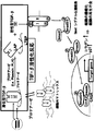
- TGF- ⁇ is produced as an inactive latent type having a molecular weight of about 300 kD that cannot bind to the receptor, and is activated on the surface of the target cell or its surroundings to become an active type that can bind to the receptor. As a result, the function can be exhibited for the first time (FIG. 1).
- TGF- ⁇ 1 is first produced as a precursor protein consisting of 391 amino acids. Thereafter, the precursor protein 279Arg and 280Ala are cleaved by the action of a furin-like protease in the Golgi.
- TGF- ⁇ 1 having a molecular weight of 25 kD.
- TGF- ⁇ 2 and TGF- ⁇ 3 have the same structure and share the same Arg-Ala sequence.
- the remaining cleaved amino terminal portion is called LAP (latency associated protein).
- LAP is also dimerized (molecular weight 75 kD), and after it is cleaved from active TGF- ⁇ , it traps active TGF- ⁇ by non-covalent bond, and latent TGF- ⁇ small complex (small latent TGF- ⁇ ) ⁇ complex (SLC) is formed.
- LAP remains a structure that cannot bind active TGF- ⁇ to the receptor, that is, latent form.
- a latent TGF- ⁇ complex (LLC) is bound to the end of the LAP dimer by a protein with a molecular weight of about 200 kD called LTBP (latent TGF- ⁇ binding protein).
- LTBP latent TGF- ⁇ binding protein
- TGF- ⁇ 2 and TGF- ⁇ 3 have the same structure.
- LTBP is similar in structure to fibrillin, a kind of extracellular matrix protein, and LLC is pooled in the extracellular matrix via LTBP (FIG. 1).
- Activating TGF- ⁇ is a reaction that dissociates and releases active TGF- ⁇ trapped in LLC by preventing noncovalent association between LAP and active TGF- ⁇ by some method. Activation of TGF- ⁇ occurs on or near the target cell surface after LLC is released from the matrix (FIG. 1). In the physiological TGF- ⁇ activation reaction, the structure of LAP is changed by binding to thrombospondin or integrin, resulting in an adhesion activation reaction that releases active TGF- ⁇ and limited degradation of LAP by protease. Then, it is known that the cleavage activation reaction releases the retained active TGF- ⁇ (FIG. 1).
- TGF- ⁇ activation As the mechanism of TGF- ⁇ activation is gradually elucidated in this way, TGF- ⁇ activation is detected for the purpose of diagnosis, treatment or prevention of the above-mentioned diseases associated with TGF- ⁇ . Attempts have been made to develop antibodies for suppressing the activation of TGF- ⁇ .
- an antibody that recognizes the cut surface of latent TGF- ⁇ by plasmin and plasma kallikrein (between 56Tys-57Leu and 58Arg-59Leu of latent TGF- ⁇ , respectively) It has been developed (Patent Document 1). Since this antibody does not bind to intact LAP, it has the property of binding specifically to its cut surface, so that it can be used to detect TGF- ⁇ activation reaction. However, it does not suppress the TGF- ⁇ activation reaction itself.
- TGF- ⁇ an antibody that binds to LAP and suppresses the activation of TGF- ⁇ has also been developed (Patent Document 2).
- Patent Document 2 an antibody that binds to LAP and suppresses the activation of TGF- ⁇ has also been developed.
- TGF- ⁇ When TGF- ⁇ is activated in co-culture of bovine aortic endothelial cells and bovine aortic smooth muscle cells in a pseudo-wound state, the migration of bovine aortic smooth muscle cells is suppressed. It is said to have the activity of restoring muscle cell migration.
- TGF- ⁇ has isoforms from TGF- ⁇ 1 to TGF- ⁇ 3 that exhibit almost the same biological activity. Therefore, when treating or preventing diseases targeting TGF- ⁇ , it is extremely important to suppress only the production of specific TGF- ⁇ isoforms that are abnormally produced during pathogenesis from the viewpoint of reducing side effects. is there.
- the present invention has been made in view of such circumstances, and an object thereof is to specifically bind to a specific isoform of TGF- ⁇ and to have an activity of suppressing the activation of the isoform. Is to provide.
- a further object of the present invention is to treat diseases caused by the activation of TGF- ⁇ , such as liver fibrosis / cirrhosis, pulmonary fibrosis, renal fibrosis / renal failure, dermal fibrosis, comprising such an antibody as an active ingredient.
- An object of the present invention is to provide a drug for treating or preventing sclerotic diseases such as arteriosclerosis.
- the present inventors focused on the fact that LAP in latent TGF- ⁇ is cleaved by plasmin and plasma kallikrein, thereby generating active TGF- ⁇ .
- plasmin and plasma kallikrein thereby generating active TGF- ⁇ .
- a specific isoform of TGF- ⁇ An attempt was made to prepare a monoclonal antibody that specifically binds to LAP.
- peptides corresponding to TGF- ⁇ 1 and TGF- ⁇ 2 LAP were prepared by genetic recombination or synthesis, and hybridomas were prepared using this peptide as an immunogen.
- clones producing a monoclonal antibody that binds only to either TGF- ⁇ 1 or TGF- ⁇ 2 LAP were selected from the prepared hybridomas by ELISA.
- the isoform-specific monoclonal antibodies produced by these clones the reactivity to the site on LAP that is cleaved by plasmin or plasma kallikrein and its neighboring region was evaluated, and the epitope region on those LAPs was identified.
- the effect of the thus obtained isoform-specific monoclonal antibody on TGF- ⁇ activation was examined. Specifically, the inhibitory activity of these monoclonal antibodies against the cleavage of LAP by plasma kallikrein was evaluated. As a result, the present inventors have obtained, among isoform-specific monoclonal antibodies, a plurality of antibodies having an activity of suppressing the activation of the isoform, and an antibody having an activity of suppressing fibrosis of the liver or kidney. I succeeded in finding it. Furthermore, as a result of detailed analysis of the correlation between the epitope of these antibodies and the inhibitory activity of TGF- ⁇ activation, the inventors have succeeded in finding an epitope useful for inhibiting TGF- ⁇ activation.
- the present invention relates to an antibody that specifically binds to LAP of a specific isoform of TGF- ⁇ and has an activity of suppressing the activation of the isoform, and use thereof.
- the antibody according to (1) which substantially binds to LAP of human TGF- ⁇ 1 and does not substantially bind to LAP of human TGF- ⁇ 2.
- the antibody according to (2) which substantially binds to a region consisting of the amino acid sequence described in SEQ ID NO: 5 in LAP of human TGF- ⁇ 1.
- the antibody according to (2) which substantially binds to the peptide consisting of the amino acid sequence set forth in SEQ ID NO: 7 and does not substantially bind to the peptide consisting of the amino acid sequence set forth in SEQ ID NO: 9-11 .
- the antibody according to (1) or (2) which has an activity of suppressing liver or kidney fibrosis.
- the antibody according to (1) which substantially binds to LAP of human TGF- ⁇ 2 and does not substantially bind to LAP of human TGF- ⁇ 1.
- the antibody according to (6) which substantially binds to a region consisting of the amino acid sequence set forth in SEQ ID NO: 6 in LAP of human TGF- ⁇ 2.
- a drug for treating or preventing a disease caused by activation of TGF- ⁇ , comprising the antibody according to any one of (1) to (9) as an active ingredient.
- an antibody that specifically binds to LAP of a specific isoform of TGF- ⁇ and has an activity of suppressing activation of the isoform is provided.
- diseases caused by activation of TGF- ⁇ such as liver fibrosis / cirrhosis, pulmonary fibrosis, renal fibrosis / renal failure, dermal fibrosis, arteriosclerosis and other sclerosing diseases Treatment and prevention are possible.
- the antibody of the present invention is specific to a specific isoform of TGF- ⁇ , low side effects can be expected when administered to a living body.
- the active TGF- ⁇ is not targeted, but the stage in which the active TGF- ⁇ is generated from the inactive latent TGF- ⁇ is inhibited, treatment / prevention at an earlier stage can be expected.
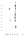
- lane 1 is LAP only
- lane 2 is LAP + PLK
- lane 3 is LAP + PLK + aprotinin
- lane 4 is LAP + PLK + clone 476
- lane 5 is LAP + PLK + clone 727
- lane 6 is LAP + PLK + clone 736
- Lane 7 shows LAP + PLK + clone 467
- Lane 8 shows LAP + PLK + clone 481
- Lane 9 shows LAP + PLK + anti-LAP antibody.
- “ ⁇ ” indicates a LAP fragment
- “*” indicates a fragment of the used antibody. It is a schematic diagram of the epitope analysis of each monoclonal antibody.
- the present invention provides an antibody that specifically binds to LAP of a specific isoform of human TGF- ⁇ and has an activity of suppressing the activation of the isoform.
- “specific” means substantially binding to LAP of a specific isoform of human TGF- ⁇ and not substantially binding to LAP of another isoform.
- the “antibody” in the present invention includes all classes and subclasses of immunoglobulin.
- “Antibody” includes polyclonal antibodies and monoclonal antibodies, and also includes forms of functional fragments of antibodies.
- Polyclonal antibodies are antibody preparations comprising different antibodies directed against different epitopes.
- the “monoclonal antibody” means an antibody (including an antibody fragment) obtained from a substantially homogeneous antibody population. In contrast to polyclonal antibodies, monoclonal antibodies are those that recognize a single determinant on an antigen.
- the antibody of the present invention is preferably a monoclonal antibody.
- An antibody of the invention is an antibody that has been separated and / or recovered from a component of the natural environment, ie, an “isolated” antibody.
- TGF- ⁇ strongly promotes extracellular matrix production of mesenchymal cells and suppresses proliferation of epithelial cells, thereby causing liver fibrosis / cirrhosis, lung fibrosis, renal fibrosis / renal failure, dermal fibrosis It is a homodimeric multifunctional cytokine with a molecular weight of 25 kD that exhibits various biological activities, such as inhibiting the action of immunocompetent cells while forming the pathology of sclerosing diseases such as arteriosclerosis.
- the primary structures of “human TGF- ⁇ 1” and “human TGF- ⁇ 2” to which the antibody of the present invention binds are already known.
- a typical amino acid sequence of “human TGF- ⁇ 1” in the present invention is shown in SEQ ID NO: 2, and a typical base sequence of DNA encoding the amino acid sequence is shown in SEQ ID NO: 1.
- a typical amino acid sequence of “human TGF- ⁇ 2” in the present invention is shown in SEQ ID NO: 4
- a typical base sequence of DNA encoding the amino acid sequence is shown in SEQ ID NO: 3.
- “human TGF- ⁇ 1” and “human TGF- ⁇ 2” in addition to those having such typical amino acid sequences, those having naturally mutated amino acids may also exist.
- “Human TGF- ⁇ 1” and “human TGF- ⁇ 2” of the present invention include such mutants.
- Such a variant consists of an amino acid sequence in which one or more amino acids are substituted, deleted, inserted or added in the amino acid sequence represented by SEQ ID NO: 2 or 4.
- the number of mutations that occur in nature is generally considered to be within 10 amino acids (eg, within 5 amino acids, within 3 amino acids, 1 amino acid).
- LAP means a dimeric prepeptide region that is non-covalently associated with active TGF- ⁇ in a latent TGF- ⁇ molecule.
- activation of TGF- ⁇ means that TGF- ⁇ binds to its receptor and becomes capable of exerting an action.
- LAP of latent TGF- ⁇ is cleaved by a protease to inhibit the association of LAP with active TGF- ⁇ , and active TGF- ⁇ is converted from inactive latent TGF- ⁇ complex. This means that activated TGF- ⁇ is produced upon release.
- One preferred embodiment of the antibody of the present invention is an antibody that binds substantially to the LAP of human TGF- ⁇ 1 and does not substantially bind to the LAP of human TGF- ⁇ 2. More preferably, it is an antibody that substantially binds to a site cleaved by a protease (plasmin, plasma kallikrein) or a nearby region (hereinafter simply referred to as “protease cleavage region”) in LAP of human TGF- ⁇ 1, An antibody that substantially binds to a region consisting of the amino acid sequence set forth in SEQ ID NO: 5 in LAP of human TGF- ⁇ 1 (“TGF- ⁇ 1-LAP” in FIG. 5).
- “substantially binds” means that the absorbance value at 490 nm is 0.2 or more in the ELISA experiment of this example (see FIGS. 6 and 7).
- the peptide consisting of the amino acid sequence set forth in SEQ ID NO: 7 (“LAP1-1” in FIG. 5) substantially binds.
- An antibody that does not substantially bind to a peptide consisting of the amino acid sequence set forth in SEQ ID NOs: 9 to 11 (“LAP1-3 to LAP1-5” in FIG. 5) is cleaved by plasma kallikrein with TGF- ⁇ 1-LAP, That is, it was found that the effect of suppressing the activation reaction of TGF- ⁇ 1 was high (Table 1, FIG. 4, FIG. 6A).
- TGF- ⁇ 1-LAP human TGF- ⁇ 1
- Another preferred embodiment of the antibody of the present invention is an antibody that specifically binds to TGF- ⁇ 1-LAP and has an activity of suppressing fibrosis of the liver or kidney.
- An antibody having an activity of suppressing liver fibrosis substantially binds to a peptide consisting of the amino acid sequence set forth in SEQ ID NO: 7 (“LAP1-1” in FIG. 5), for example, SEQ ID NO: 9
- antibodies having an activity of suppressing renal fibrosis include, for example, peptides substantially consisting of the amino acid sequences set forth in SEQ ID NOs: 17 and 18 (“LAP1-0” and “LAP1-6” in FIG. 7). Is an antibody that does not substantially bind to peptides having the amino acid sequences set forth in SEQ ID NOs: 7 to 11 ("LAP1-1 to LAP1-5" in FIG. 7).
- the activity of suppressing liver fibrosis is, for example, an experiment using a non-alcoholic steatohepatitis (NASH) model mouse fed with a methionine / choline-deficient diet (MCDD) described in Example 10 and Example 11 It can be evaluated by an experiment using a model mouse of non-alcoholic steatohepatitis (NASH) fed with a choline-deficient amino acid substitution diet (CDAA diet) as described.
- the activity which suppresses the fibrosis of a kidney is the experiment using the unilateral ureteral ligation (UUO) renal fibrosis model mouse described in Example 12, for example, or the unilateral ureteral ligation (UUO) described in Example 13. It can be evaluated by experiments using renal fibrosis model mice.
- Another preferred embodiment of the antibody of the present invention is an antibody that substantially binds to LAP of human TGF- ⁇ 2 and does not substantially bind to LAP of human TGF- ⁇ 1. More preferably, it is an antibody that substantially binds to a region on human TGF- ⁇ 2 corresponding to the protease cleavage region of human TGF- ⁇ 1, for example, the amino acid described in SEQ ID NO: 6 in the LAP of human TGF- ⁇ 2 It is an antibody that substantially binds to a region consisting of a sequence (“TGF- ⁇ 2-LAP” in FIG. 5).
- the most preferred embodiment of the antibody of the present invention is an antibody produced by a hybridoma identified by accession number NITE BP-873, accession number NITE BP-881, or accession number NITE BP-882. These antibodies specifically bind to human TGF- ⁇ 1 LAP and have the activity of inhibiting the cleavage of human TGF- ⁇ 1 LAP by plasma kallikrein and the activity of suppressing liver or kidney fibrosis. (Table 1, FIG. 4, FIG. 5, FIG. 6A, FIGS. 7 to 11).
- the present invention includes an antibody that binds to the epitope of the antibody produced by the hybridoma and has an activity of inhibiting the cleavage of human TGF- ⁇ 1 LAP by plasma kallikrein.
- the epitope of the antibody can be determined by a well-known method such as examining the binding to an overlapping synthetic oligopeptide obtained from the amino acid sequence of LAP (eg, Example, Ed Harlow and D. Lane, Using this application).
- a peptide library by phage display can also be used for epitope mapping. Whether two antibodies bind to the same or sterically overlapping epitopes can be determined by competition assays.
- the activity of the antibody to suppress the cleavage of LAP can be evaluated by the method shown in Example 8. In Example 8, plasma kallikrein is used as a protease. However, when other proteases are used, evaluation can be performed by the method according to Example 8.
- the antibodies of the present invention include chimeric antibodies, humanized antibodies, human antibodies, and functional fragments of these antibodies.
- a human as a pharmaceutical
- a chimeric antibody, a humanized antibody, or a human antibody is desirable from the viewpoint of reducing side effects.
- a “chimeric antibody” is an antibody in which a variable region of a certain antibody is linked to a constant region of a heterogeneous antibody.
- a chimeric antibody for example, immunizes an antigen to a mouse, cuts out an antibody variable region (variable region) that binds to the antigen from the mouse monoclonal antibody gene, and binds to an antibody constant region (constant region) gene derived from human bone marrow.
- the “humanized antibody” is an antibody obtained by transplanting the gene sequence of the antigen-binding site (CDR) of a non-human-derived antibody to a human antibody gene (CDR grafting), and its production method is publicly known. (See, for example, EP239400, EP125023, WO90 / 07861, WO96 / 02576).
- a “human antibody” is an antibody derived from all regions. In the production of human antibodies, it is possible to use a transgenic animal (for example, a mouse) that can produce a repertoire of human antibodies by immunization. Methods for producing human antibodies are known (for example, Nature, 362: 255-258 (1992), Intern. Rev.
- the antibody in the present invention includes a part (partial fragment) of an antibody that specifically recognizes LAP of a specific isoform of human TGF- ⁇ (functional fragment). Specifically, Fab, Fab ′, F (ab ′) 2, variable region fragment (Fv), disulfide bond Fv, single chain Fv (scFv), sc (Fv) 2, diabody, multispecific antibody, And polymers thereof.
- Fab means a monovalent antigen-binding fragment of an immunoglobulin composed of one light chain and part of a heavy chain. It can be obtained by papain digestion of antibodies and by recombinant methods. “Fab ′” differs from Fab by the addition of a few residues at the carboxy terminus of the heavy chain CH1 domain, including one or more cysteines in the hinge region of the antibody. “F (ab ') 2” means a divalent antigen-binding fragment of an immunoglobulin that consists of both light chains and parts of both heavy chains.
- “Variable region fragment (Fv)” is the smallest antibody fragment with complete antigen recognition and binding sites. Fv is a dimer in which a heavy chain variable region and a light chain variable region are strongly linked by a non-covalent bond. “Single-chain Fv (sFv)” comprises the heavy and light chain variable regions of an antibody, and these regions are present in a single polypeptide chain. “Sc (Fv) 2” is a chain formed by joining two heavy chain variable regions and two light chain variable regions with a linker or the like.
- a “diabody” is a small antibody fragment having two antigen-binding sites, the fragment comprising a heavy chain variable region bound to a light chain variable region in the same polypeptide chain, each region comprising a separate It forms a pair with the complementary region of the strand.
- a “multispecific antibody” is a monoclonal antibody that has binding specificities for at least two different antigens. For example, it can be prepared by co-expression of two immunoglobulin heavy / light chain pairs where the two heavy chains have different specificities.
- the antibodies of the present invention reduce the desired activity (activity that specifically binds to LAP of a particular isoform of TGF- ⁇ , activity that inhibits cleavage of LAP by proteases, and / or other biological properties)
- an antibody whose amino acid sequence is modified is included.
- Amino acid sequence variants of the antibody can be produced by introducing mutations into the DNA encoding the antibody chain or by peptide synthesis. Such modifications include, for example, residue substitution, deletion, addition and / or insertion within the amino acid sequence of the antibody.
- the site where the amino acid sequence of the antibody is modified may be the constant region of the heavy chain or light chain of the antibody as long as it has an activity equivalent to that of the antibody before modification, and the variable region (framework region and CDR).
- Modification of amino acids other than CDR is considered to have a relatively small effect on the binding affinity with the antigen.
- the amino acid of the CDR is modified to screen for antibodies with increased affinity for the antigen.
- Methods are known (PNAS, 102: 8466-8471 (2005), Protein Engineering, Design & Selection, 21: 485-493 (2008), International Publication No. 2002/051870, J. Biol. Chem., 280: 24880-24887 (2005), Protein Engineering, Design & Selection, 21: 345-351 (2008)).
- the number of amino acids to be modified is preferably within 10 amino acids, more preferably within 5 amino acids, and most preferably within 3 amino acids (eg, within 2 amino acids, 1 amino acid).
- the amino acid modification is preferably a conservative substitution.
- conservative substitution means substitution with another amino acid residue having a chemically similar side chain. Groups of amino acid residues having chemically similar amino acid side chains are well known in the technical field to which the present invention belongs.
- acidic amino acids for example, acidic amino acids (aspartic acid and glutamic acid), basic amino acids (lysine, arginine, histidine), neutral amino acids, amino acids with hydrocarbon chains (glycine, alanine, valine, leucine, isoleucine, proline), hydroxy groups Amino acids with amino acids (serine / threonine), amino acids with sulfur (cysteine / methionine), amino acids with amide groups (asparagine / glutamine), amino acids with imino groups (proline), amino acids with aromatic groups (phenylalanine / tyrosine / (Tryptophan).
- basic amino acids lysine, arginine, histidine
- neutral amino acids amino acids with hydrocarbon chains (glycine, alanine, valine, leucine, isoleucine, proline), hydroxy groups Amino acids with amino acids (serine / threonine), amino acids with sulfur (cystein
- the antibody of the present invention is a polyclonal antibody
- an immunized animal is immunized with an antigen, and purified from the antiserum by conventional means (eg, salting out, centrifugation, dialysis, column chromatography, etc.) can do.
- Monoclonal antibodies can be prepared by a hybridoma method or a recombinant DNA method.
- LAP prepared by genetic recombination, or synthesized LAP or a partial peptide thereof, or a cell expressing these can be used.
- the antigen is preferably a highly purified peptide.
- an affinity column for this tag can be used in the purification process.
- the hybridoma method typically includes the Kohler and Milstein method (Kohler & Milstein, Nature, 256: 495 (1975)).
- the antibody-producing cells used in the cell fusion step in this method are spleen cells, lymph node cells, peripheral blood leukocytes, etc. of animals immunized with antigen (eg, mouse, rat, hamster, rabbit, monkey, goat) . It is also possible to use antibody-producing cells obtained by allowing an antigen to act on the above-mentioned cells or lymphocytes previously isolated from an unimmunized animal in a medium.
- the myeloma cells various known cell lines can be used.
- the antibody-producing cells and myeloma cells may be of different animal species as long as they can be fused, but are preferably of the same animal species.
- Hybridomas are produced, for example, by cell fusion between spleen cells obtained from mice immunized with antigen and mouse myeloma cells. Thereafter, by screening the obtained hybridoma clones, a clone producing a monoclonal antibody specific for LAP of a specific isoform of TGF- ⁇ can be obtained.
- monoclonal antibodies for example, culture supernatant, purified antibody
- LAP of each isoform of TGF- ⁇ for example, LAP of TGF- ⁇ 1, LAP of TGF- ⁇ 2
- a clone producing a monoclonal antibody that specifically reacts with LAP of any isoform may be selected.
- isoform-specific monoclonal antibodies the reactivity of the LAP to the site that is cleaved by protease and its neighboring region can be evaluated, and the epitope region on the LAP of these antibodies can be identified.
- the monoclonal antibody can be obtained by culturing the hybridoma or from the ascites of the mammal to which the hybridoma has been administered.
- immunogen and the synthetic peptide for screening include “EAIRGQILSKLRLASPPSQGEVPPGPLPEAVLAL (SEQ ID NO: 5)” as TGF- ⁇ 1-LAP and “EAIRGQILSKLKLTSPPEDYPEPEEVPPEVISI (SEQ ID NO: 6)” as TGF- ⁇ 2-LAP.
- TGF- ⁇ 1-LAP examples include TGF- ⁇ 1-LAP as “LSKLRLASPP (SEQ ID NO: 7)”, “KLRLASPPSQ (SEQ ID NO: 8)”, “RLASPPSQEVPPGP (SEQ ID NO: 9)”, “ASPPSQGEVPPG (SEQ ID NO: 10) ”,“ PPSQGEVPPGPL (SEQ ID NO: 11) ”,“ EAIRGQIL (SEQ ID NO: 17) ”, and“ PEAVLAL (SEQ ID NO: 18) ”.
- the DNA encoding the antibody of the present invention is cloned from a hybridoma, B cell or the like and incorporated into an appropriate vector, which is then introduced into a host cell (eg, mammalian cell line, E. coli, yeast cell, insect cell, It is a technique for introducing the antibody of the present invention as a recombinant antibody by introducing it into a plant cell (e.g., PJDelves, Antibody Production: Essential Technologys, 1997 WILEY, P. Shepherd and C. Dean Monoclonal Antibodies, 2000 OXFORD UNIVERSITY PRESS, Vandamme AM et al., Eur. J. Biochem. 192: 767-775 (1990)).
- a host cell e.g., mammalian cell line, E. coli, yeast cell, insect cell.
- DNA encoding the heavy chain or the light chain may be separately incorporated into an expression vector to transform the host cell.
- Host cells may be transformed into a single expression vector (see WO94 / 11523).
- the antibody of the present invention can be obtained in a substantially pure and uniform form by culturing the above host cell, separating and purifying it from the host cell or culture medium. For the separation and purification of the antibody, the methods used in the usual purification of polypeptides can be used.
- transgenic animals such as cows, goats, sheep or pigs
- transgenic animal production technology a large amount of monoclonal antibody derived from the antibody gene is produced from the milk of the transgenic animal. It is also possible to obtain.
- the present invention also provides a DNA encoding the antibody of the present invention, a vector containing the DNA, a host cell holding the DNA, and a method for producing the antibody comprising culturing the host cell and recovering the antibody. To do.
- the present invention comprises an agent for treating or preventing a disease caused by activation of TGF- ⁇ comprising the antibody of the present invention as an active ingredient, and a therapeutically or prophylactically effective amount of the antibody of the present invention.
- the present invention also provides a method for treating or preventing a disease caused by activation of TGF- ⁇ , comprising a step of administering to a patient.
- the drug containing the antibody of the present invention as an active ingredient can be used in the form of a composition containing the antibody of the present invention and an optional component such as physiological saline, sucrose aqueous solution or phosphate buffer.
- the agent of the present invention may be formed into a liquid or lyophilized form as necessary, and optionally a pharmaceutically acceptable carrier or medium, such as a stabilizer, preservative, isotonic agent and the like. Can also be included.
- Examples of the pharmaceutically acceptable carrier include mannitol, lactose, saccharose, human albumin and the like in the case of a lyophilized preparation.
- mannitol lactose
- saccharose human albumin
- human albumin human albumin
- the buffer solution and aluminum hydroxide include, but are not limited to.
- the administration method of the drug of the present invention varies depending on the age, weight, sex, health status, etc. of the administration subject, but either parenteral administration (eg, intravenous administration, subcutaneous administration, arterial administration, local administration) or oral administration It can be administered by the route of administration.
- parenteral administration eg, intravenous administration, subcutaneous administration, arterial administration, local administration
- oral administration It can be administered by the route of administration.
- a preferred method of administration is parenteral administration.
- the dose of the drug of the present invention may vary depending on the patient's age, weight, sex, health status, degree of disease progression and the components of the drug to be administered. 0.001 to 1000 mg per kg per day, preferably 0.01 to 10 mg. Usually, it is administered at intervals of 1 week to 1 month, but is not limited to this administration interval.
- the antibody of the present invention can be applied not only to treatment and prevention of the above-mentioned diseases but also to diagnosis.
- the antibody of the present invention may be labeled.
- a label for example, a radioactive substance, a fluorescent dye, a chemiluminescent substance, an enzyme, and a coenzyme can be used.
- radioisotope, fluorescein, rhodamine, dansyl chloride, luciferase, peroxidase, alkaline phosphatase examples include lysozyme and biotin / avidin.
- the antibody of the present invention can be obtained in any dosage form by employing any suitable means.
- the antibody titer of the purified antibody can be measured and appropriately diluted with PBS (phosphate buffer containing physiological saline) or the like, and then 0.1% sodium azide or the like can be added as a preservative.
- the antibody titer of a substance obtained by adsorbing the antibody of the present invention on latex or the like can be obtained, diluted appropriately, and added with a preservative.
- Transformant 1 and transformant 2 were cultured in 5 mL of LB medium + Amp50 for 12 to 15 hours (preculture), and the total amount of this culture solution was added to 500 mL of LB medium + Amp50 (1% inoculation) . After 3 hours from the start of the culture (stage where OD600 reached 0.2 to 0.3), IPTG having a final concentration of 0.1 mM was added to transformant 1, and IPTG having a final concentration of 0.05 mM was added to transformant 2. Thereafter, the culture temperature was set to 16 ° C. and the culture was continued for 12 to 15 hours.
- the cultured cells were collected by centrifugation at 3000 rpm, 4 ° C. for 15 minutes and washed with sterilized water.
- 5 mL of the above-mentioned microbial cell disruption buffer was added to 1 g of wet microbial cells, and shaken for 15 minutes with a vortex mixer or the like. After completion of the shaking, centrifugation was performed at 21000 rpm, 4 ° C. for 15 minutes, and the solution fraction was collected. This fraction was subjected to separation and purification using a GST fusion protein purification gel (GE Healthcare, etc.).
- the transferred PVDF membrane was equilibrated with TBS-T (2.48 mM Tris-HCl, 137 mM sodium chloride, 0.27 mM potassium chloride, 0.05% Tween20 (pH 7.4)) for 10 minutes, and contained 5% skim milk. Blocking (30 minutes to 1 hour) was performed with TBS-T. Thereafter, the PVDF membrane was thoroughly washed with TBS-T.
- Anti-human TGF- ⁇ 2 antibody (SANTA CRUZ BIOTECHNOLOGY, model number sc-90) was prepared at a concentration of 1 ⁇ g / mL, and this was reacted as a primary antibody with a PVDF membrane at 37 ° C. for 1 hour.
- TGF- ⁇ 1-LAP might be degraded during the preparation process.
- TGF- ⁇ 2-LAP a plurality of signals were detected in addition to the target signal (FIG. 3 (B), lane 3). It was suggested that TGF- ⁇ 2-LAP may be degraded during the preparation process.
- the target peptide is contained in the sample. Therefore, it was considered that these samples can be sufficiently used as an immunogen. Thus, antibody production was attempted using these samples.
- Example 2 Preparation of peptide antigens TGF- ⁇ 1-LAP and TGF- ⁇ 2-LAP by solid phase synthesis
- Amino acid sequence from 47th glutamic acid residue to 79th alanine residue of TGF- ⁇ 1-LAP, and The amino acid sequence from the 47th glutamic acid residue to the 79th isoleucine residue of TGF- ⁇ 2-LAP may contain an activated LAP cleavage sequence.
- Various peptides consisting of sequences in which a cysteine residue was added to the N-terminus of these sequences were synthesized by the Fmoc solid phase synthesis method (Applied Biosystems).
- the final peptide was prepared by deprotection and excision from the resin using a cleave cocktail containing 95% TFA.
- mcKLH PIERCE
- PIERCE mcKLH
- Example 3 Preparation of Epitope Analysis Peptides ⁇ 1-LAP and ⁇ 2-LAP by Solid Phase Synthesis As TGF- ⁇ 1-LAP, “LSKLRLASPP (SEQ ID NO: 7)”, “KLRLASPPSQ (SEQ ID NO: 8)”, “RLASPPSQEVPPGP” (SEQ ID NO: 9) ”,“ ASPPSQGEVPPG (SEQ ID NO: 10) ”, and“ PPSQGEVPPGPL (SEQ ID NO: 11) ”, a peptide comprising a sequence having a cysteine residue added thereto is designated as TGF- ⁇ 2-LAP.
- Example 4 Production of monoclonal antibodies against recombinant TGF- ⁇ 1-LAP and recombinant TGF- ⁇ 2-LAP (1) Production of hybridoma Using affinity-purified recombinant TGF- ⁇ 1-LAP or recombinant TGF- ⁇ 2-LAP, female Balb / c mice were immunized by intraperitoneal injection. Initially, 50 ug of recombinant protein in Freund's complete adjuvant was administered per animal, and thereafter 25 ug to 50 ug of recombinant protein or KLH-binding synthetic peptide in RIBI adjuvant was administered. Immunizations were performed at 2-3 week intervals.
- mice 14 days after the last immunization, the mice were boosted with saline phosphate buffer (PBS) containing 50 ug of recombinant protein. Three days after the booster, the spleen was removed from the mouse, and the spleen cell of the mouse having a high antibody titer was fused with P3.X63-Ag8.653 mouse myeloma cell using polyethylene glycol 4000. Hybridoma cells were selected on microtiter plates in HAT medium using known Kohler and Milstein techniques.
- PBS saline phosphate buffer
- ELISA was used for determination of antibody titer in serum.
- a microtiter plate was first coated with a recombinant protein or synthetic peptide.
- serially diluted serum was added to the wells blocked with 1% BSA / TBS-0.05% Tween20 and incubated.
- the antibody bound to the plate was detected with a peroxidase-conjugated antibody against mouse immunoglobulin.
- Hybridoma screening Hybridomas prepared using recombinant TGF- ⁇ 1-LAP as an immunogen were evaluated for the reactivity of the culture supernatant with recombinant TGF- ⁇ 1-LAP and recombinant TGF- ⁇ 2-LAP.
- Hybridomas producing a culture supernatant that reacts only with ⁇ 1-LAP were selected.
- hybridomas prepared using recombinant TGF- ⁇ 2-LAP as an immunogen the reactivity of the culture supernatant with recombinant TGF- ⁇ 1-LAP and recombinant TGF- ⁇ 2-LAP was evaluated, and recombinant TGF- ⁇ 2-LAP only Hybridomas that produce culture supernatants that react with the above were selected.
- an ELISA method and an immunoprecipitation ELISA method using immunoprecipitation were used.
- Recombinant TGF- ⁇ 1-LAP or recombinant TGF- ⁇ 2-LAP (concentration of 1 ug / ml in 50 mM carbonate buffer) was fixed to a 96-well microtiter plate (NUNC) for 2 hours at room temperature. The plate is then washed with TBS / 0.05% Tween 20, and the free adsorbed portion of the plate surface is blocked with 0.25% skim milk phosphate buffer / 0.05% Tween 20 (30 minutes at room temperature) and again TBS / 0.05% Washed with Tween20. The culture supernatant of each hybridoma was added to the well and allowed to react at room temperature for 1 hour.
- the plates were then washed with TBS / 0.05% Tween 20, and then peroxidase-conjugated mouse anti-IgG antibody diluted in phosphate buffer / 0.05% Tween 20 was added at 50 uL / well. After incubating at room temperature for 1 hour, the plate was washed with TBS / 0.05% Tween 20, and the substrate solution (citrate buffer (pH 5), 0.05% O-phenylenediamine, 0.03% H 2 O 2 ) was added to the well at 100 uL / well. added. After 10 to 15 minutes, the reaction was stopped with 2N sulfuric acid, and the absorbance at 490 nm was measured with an intelligible spectrophotometer.
- substrate solution citrate buffer (pH 5), 0.05% O-phenylenediamine, 0.03% H 2 O 2
- Example 5 Production of monoclonal antibodies against synthetic peptides TGF- ⁇ 1-LAP and TGF- ⁇ 2-LAP (1) Immunization Using synthetic peptides TGF- ⁇ 1-LAP or TGF- ⁇ 2-LAP bound to KLH, Balb / c mice were immunized by intraperitoneal injection. For the first time, 50 ug of KLH-binding synthetic peptide in Freund's complete adjuvant was administered per animal, and thereafter, 25 ug to 50 ug of KLH-binding synthetic peptide in RIBI adjuvant were administered. Immunizations were performed at 2-3 week intervals.
- mice 14 days after the last immunization, the mice were boosted with a saline phosphate buffer (PBS) containing 50 ug of KLH-binding synthetic peptide.
- PBS saline phosphate buffer
- the spleen was removed from the mouse, and the spleen cell of the mouse having a high antibody titer was fused with P3.X63-Ag8.653 mouse myeloma cell using polyethylene glycol 4000.
- Hybridoma cells were selected on microtiter plates in HAT medium using known Kohler and Milstein techniques.
- ELISA was used for determination of antibody titer in serum.
- a microtiter plate was first coated with a recombinant protein or synthetic peptide.
- serially diluted serum was added to the wells blocked with 1% BSA / TBS-0.05% Tween20 and incubated.
- the antibody bound to the plate was detected with a peroxidase-conjugated antibody against mouse immunoglobulin.
- hybridomas prepared using KLH-binding synthetic peptide TGF- ⁇ 2-LAP as an immunogen the reactivity of the culture supernatant with OVA-binding synthetic peptide TGF- ⁇ 1-LAP or OVA-binding synthetic peptide TGF- ⁇ 2-LAP was evaluated. Then, a hybridoma producing a culture supernatant that reacts only with the OVA-binding synthetic peptide TGF- ⁇ 2-LAP was selected. The ELISA method was used for this selection. Details of the ELISA method are the same as in Example 4.
- Example 6 Analysis of selectivity of monoclonal antibody against TGF- ⁇ 1-LAP and TGF- ⁇ 2-LAP (Part 1) Among the hybridomas prepared in Example 4, for hybridomas prepared using recombinant TGF- ⁇ 1-LAP as an immunogen, the OVA-bound TGF- ⁇ 1-LAP synthetic peptide or OVA-bound TGF- ⁇ 2-LAP synthetic peptide of the culture supernatant was used. The strain reacting with the OVA-bound TGF- ⁇ 2-LAP synthetic peptide was selected by ELISA and discarded.
- the hybridomas prepared in Example 4 the hybridomas prepared using recombinant TGF- ⁇ 2-LAP as an immunogen, the OVA-bound TGF- ⁇ 1-LAP synthetic peptide or OVA-bound TGF- ⁇ 2-LAP of the culture supernatant was used. The reactivity with the synthetic peptide was evaluated, and a strain that reacts with the OVA-bound TGF- ⁇ 1-LAP synthetic peptide was selected by ELISA and discarded. Thereby, a hybridoma cell producing an antibody specific to the immunogen was obtained.
- OVA-bound TGF- ⁇ 1-LAP synthetic peptide or OVA-bound TGF- ⁇ 2-LAP synthetic peptide (50 mM carbonate buffer at a concentration of 1 ug / ml) to a 96-well microtiter plate (NUNC) for 2 hours at room temperature. went. The plate is then washed with TBS / 0.05% Tween20 and the free adsorbed portion of the plate surface is blocked with 0.25% skim milk phosphate buffer / 0.05% Tween20 (30 minutes at room temperature) and again with TBS / 0.05% Tween20. Washed with.
- a hybridoma supernatant or antibody solution at a concentration of 1 ug / ml was added to each well and allowed to react at room temperature for 1 hour.
- the plates were then washed with TBS / 0.05% Tween 20, and then peroxidase-conjugated mouse anti-IgG antibody diluted in phosphate buffer / 0.05% Tween 20 was added at 50 uL / well. After incubating at room temperature for 1 hour, the plate was washed with TBS / 0.05% Tween 20, and the substrate solution (citrate buffer (pH 5), 0.05% O-phenylenediamine, 0.03% H 2 O 2 ) was added to the well at 100 uL / well. added. After 10 to 15 minutes, the reaction was stopped with 2N sulfuric acid, and the absorbance at 490 nm was measured with a spectrophotometer.
- the selected hybridomas were further analyzed by ELISA using the epitope analysis peptide.
- the hybridoma culture supernatant prepared using recombinant TGF- ⁇ 1-LAP as an immunogen was evaluated for its reactivity with TGF- ⁇ 1-LAP epitope analysis peptide and TGF- ⁇ 2-LAP epitope analysis peptide. -Those that did not react with the peptide for LAP epitope analysis were selected.
- the hybridoma culture supernatant prepared using recombinant TGF- ⁇ 2-LAP as an immunogen was evaluated for its reactivity with TGF- ⁇ 1-LAP epitope analysis peptide and TGF- ⁇ 2-LAP epitope analysis peptide. -Those that did not react with the peptide for ⁇ 1-LAP epitope analysis were selected.
- hybridomas prepared using KLH-binding synthetic peptide TGF- ⁇ 1-LAP as an immunogen were obtained from recombinant TGF- ⁇ 1-LAP or recombinant TGF- ⁇ 2-LAP in the culture supernatant.
- the hybridomas that react with the recombinant TGF- ⁇ 2-LAP were selected and discarded.
- hybridomas prepared using KLH-binding synthetic peptide TGF- ⁇ 2-LAP as an immunogen were compared with recombinant TGF- ⁇ 1-LAP or recombinant TGF- ⁇ 2-LAP in the culture supernatant.
- the hybridomas that react with the recombinant TGF- ⁇ 1-LAP were selected and discarded. Thereby, a hybridoma cell producing an antibody specific to the immunogen was obtained.
- an ELISA method and an immunoprecipitation ELISA method using immunoprecipitation were used.
- Recombinant TGF- ⁇ 1-LAP or recombinant TGF- ⁇ 2-LAP (concentration of 1 ug / ml in 50 mM carbonate buffer) was fixed to a 96-well microtiter plate (NUNC) for 2 hours at room temperature. The plate is then washed with TBS / 0.05% Tween20 and the free adsorbed portion of the plate surface is blocked with 0.25% skim milk phosphate buffer / 0.05% Tween20 (30 minutes at room temperature) and again with TBS / 0.05% Tween20. Washed with. A hybridoma supernatant or antibody solution at a concentration of 1 ug / ml was added to each well and allowed to react at room temperature for 1 hour.
- the plates were then washed with TBS / 0.05% Tween 20, and then 50 uL / well of peroxidase-conjugated mouse anti-IgG antibody diluted in phosphate buffer / 0.05% Tween 20 was added. After incubating at room temperature for 1 hour, the plate was washed with TBS / 0.05% Tween 20, and the substrate solution (citrate buffer (pH 5), 0.05% O-phenylenediamine, 0.03% H 2 O 2 ) was added to the well at 100 uL / well. added. After 10 to 15 minutes, the reaction was stopped with 2N sulfuric acid, and the absorbance at 490 nm was measured with an intelligible spectrophotometer.
- substrate solution citrate buffer (pH 5), 0.05% O-phenylenediamine, 0.03% H 2 O 2
- the selected hybridomas were further analyzed by ELISA using the epitope analysis peptide.
- the hybridoma culture supernatant prepared using the KLH-binding synthetic peptide TGF- ⁇ 1-LAP as an immunogen was evaluated for its reactivity with TGF- ⁇ 1-LAP and TGF- ⁇ 2-LAP epitope analysis peptides, and TGF- ⁇ 2- Those that did not react with the peptide for LAP epitope analysis were selected.
- the hybridoma culture supernatant prepared using the KLH-binding synthetic peptide TGF- ⁇ 2-LAP as an immunogen was evaluated for its reactivity with TGF- ⁇ 1-LAP and TGF- ⁇ 2-LAP epitope analysis peptides. Those that did not react with the peptide for ⁇ 1-LAP epitope analysis were selected.
- Example 7 Purification of Isomer-Selective Monoclonal Antibody 100 mL of the antibody-producing hybridoma culture supernatant was filtered through a 0.22 ⁇ m filter to remove insoluble matters. The filtered culture supernatant was passed through a column packed with 1 mL of Protein G-sepharose 4B (GE Helthecare) according to a conventional method to adsorb antibody components onto the column. Non-specifically adsorbed components were removed from the column, and then the monoclonal antibody was released by placing the column under acidic conditions. The recovered monoclonal antibody was used as a purified antibody. The obtained purified antibody was dialyzed against a 100-fold amount of PBS buffer.
- Protein G-sepharose 4B GE Helthecare
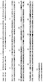
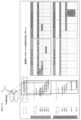
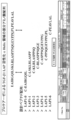
- FIG. 5 the schematic diagram of the epitope analysis of the clone obtained by the above Example is shown in FIG. 5, and the summary of the binding specificity of each clone is shown in FIG.
- Example 8 Analysis of selectivity of monoclonal antibody against TGF- ⁇ 1-LAP (part 2) Furthermore, peptides comprising a sequence in which a cysteine residue is added to the N-terminus of “EAIRGQIL (SEQ ID NO: 17)” and “PEAVLAL (SEQ ID NO: 18)” as TGF- ⁇ 1-LAP (respectively “LAP1-0” and “LAP1-0”) (Hereinafter referred to as “LAP1-6”), and in the same manner as in Example 6, the reactivity of the obtained monoclonal antibody with the peptide for TGF- ⁇ 1-LAP epitope analysis was evaluated. As a result, clone 481 showed reactivity to LAP1-0 and LAP1-6 (FIG. 7).
- aprotinin manufactured by Sigma
- an inhibitor of plasmin and plasma kallikrein final concentration 200 ⁇ g / ml
- a commercially available monoclonal anti-LAP antibody manufactured by R & D
- the inhibitory activity of the monoclonal antibody was evaluated by comparison with each control.
- FIG. 4 shows the results of evaluation by Western blot for five types of monoclonal antibodies.
- human recombinant LAP (lane 1) is cleaved with plasma kallikrein, a 30 kD LAP fragment is observed (lane 2).
- Aprotinin an inhibitor of plasma kallikrein, completely inhibited this cleavage (lane 3).
- the commercially available anti-LAP antibody showed no inhibitory activity (lane 9).
- a moderate inhibitory effect was observed in clone 467 (lane 7) and a weak inhibitory effect in clone 481 (lane 8).
- Table 1 summarizes the results of evaluation for each antibody using the same method.
- mice Male, 9 weeks old, Nihon Charles River Co., Ltd.
- mice were acclimated for about 1 week and then divided into 2 groups (12 mice each) according to their body weight.
- the other was used as a solvent control group (non-administration group of clone 1196) and fed MCDD for 12 weeks.
- the liver was removed from each group of mice, tissue sections were prepared, and Sirius red staining was performed to evaluate the degree of liver fibrosis. Specifically, it is as follows.
- the number of experimental animals is 3-4 per cage (W188mm x D297mm x H128mm), the temperature is 20-25 ° C, the humidity is 45-70%, the ventilation rate is 13 times / hour, and the lighting time is 12 hours (7 : 00-19: 00) was kept in an animal breeding room adjusted to the environment.
- solid material CRF-1 Oriental Yeast Industry
- F2MCD Oriental Yeast Industry
- Clone 1196 is diluted in Ca 2+ and Mg 2+ free phosphate buffered saline (PBS; phosphate buffered saline; Sigma) to give 1 mg / 5 ml at the time of administration. Thereafter, it was administered via the tail vein so as to be 1 mg / kg 12 times per week.
- PBS phosphate buffered saline
- Ca 2+ and Mg 2+ -free PBS were administered into the tail vein (5 ml / kg).
- the animals were laparotomized under anesthesia with isoflurane (Foren; Abbott Japan).
- the portion of the outer left lobe below the origin of the extrahepatic bile duct was cut out to a width of about 5 mm, fixed in 4% paraformaldehyde solution at 4 ° C. overnight, and a paraffin-embedded specimen was prepared. Staining was performed using sections sliced into 4 ⁇ l from paraffin-embedded liver tissue specimens. For HE staining, a double Meyer's hematoxylin solution and a 1% eosin solution (Muto Chemical) were used. For fiber staining with Sirius red, deparaffinized sections were immersed in a saturated picric acid solution containing 0.1% Sirius red for 30 minutes.
- clone 1196 markedly suppressed fibrosis around the leaflets (FIG. 8, upper right) induced by MCDD diet load (FIG. 8, lower right).
- the effect of clone 1196 administration was not observed on the formation of lipid droplets in the liver (FIG. 8, upper left, lower left).
- clone 1196 had no effect on body weight loss due to MCDD diet load (approximately 4 g loss after one week after starting MCDD diet, and then gradually decreasing by approximately 6 g until the end of the experiment). .
- Example 11 Examination of anti-fibrotic action of each monoclonal antibody using a non-alcoholic steatohepatitis (NASH) model mouse fed a choline-deficient amino acid substitution diet (CDAA diet) KK-Ay mice
- NASH non-alcoholic steatohepatitis
- CDAA diet choline-deficient amino acid substitution diet
- KK-Ay mice This is a type II diabetes model mouse that expresses hyperglycemia from an early stage by introducing the obesity gene Ay into KK mice that spontaneously develop impaired glucose tolerance.
- this model mouse exhibits a pathological condition very similar to human metabolic syndrome, such as leptin resistance and hypoadiponectinemia.
- KK-Ay mice spontaneously develop mild steatohepatitis even when bred on normal diet, dietary burdens such as choline-deficient L-amino acid-sefined diet (CDAA diet) Highly steatohepatitis is caused by applying.
- CDAA diet choline-deficient L-amino acid-sefined diet
- NASH non-alcoholic steatohepatitis
- TGF- ⁇ 1 activation inhibitory antibody was diluted in Japanese Pharmacopoeia saline to 1 mg / 5 ml at the time of administration, and was administered into the tail vein over 6 weeks from the 9th week of feeding CDAA feed. Specifically, clones 94, 467, and 1196 were administered twice a week, and clones 481, 536, and 759 were administered once a week at 1 mg / kg. In the solvent control group, Japanese Pharmacopoeia saline was administered into the tail vein (5 ml / kg).
- liver fibrosis associated with NASH in CDAA group mice was evaluated by the amount of hadoxyproline in the liver. Specifically, it is as follows. To the liver tissue thawed on ice, 1 ml of 2N NaOH was added and heated in a 65 ° C. bath for 10 minutes to completely dissolve the tissue. 500 ⁇ l was transferred to a new tube and hydrolyzed in an autoclave (121 ° C., 20 minutes). An equal amount of 6N HCL was added, and hydrolysis was further performed in an autoclave (121 ° C., 20 minutes).
- 0.1M Chloramine T reagent (Wako Pure Chemical Industries, Ltd.) was added in an amount equivalent to the sample and allowed to stand at room temperature for 25 minutes, then the Ehrlich reagent was added in an amount equivalent to the sample and heated in a 65 ° C. bath for 20 minutes. Cooled rapidly in ice. Thereafter, the mixture was centrifuged (12000 rpm, 10 minutes), the absorbance of the supernatant was measured at a wavelength of 590 nm, a calibration curve was drawn from the standard, and the amount of hydroxyproline in the tissue was calculated.
- the amount of protein was quantified using a BCAassay kit (Thermo Scientific), and the value obtained by correcting the amount of hydroxyproline with the amount of protein was determined.
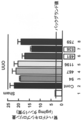
- clone 467 increased the amount of liver hydroxyproline increased by CDAA diet load 5.3 ⁇ g / mg protein [Difference between CSAA diet group (3.3 ⁇ 0.6 ⁇ g / mg protein) (lane 1) and CDAA diet load solvent control group (8.6 ⁇ 0.9 ⁇ g / mg protein) (lane 2)] decreased by about 20% to 4.1 ⁇ g / Clone 1196 has approximately 30 mg protein [difference between CSAA diet group (3.3 ⁇ 0.6 ⁇ g / mg protein) (lane 1) and CDAA diet-loaded clone 467 group (7.4 ⁇ 0.9 ⁇ g / mg protein) (lane 4)].
- Example 12 Examination of antifibrotic action of each monoclonal antibody using unilateral ureteral ligation (UUO) renal fibrosis model mouse
- the unilateral ureteral ligation (UUO) model is prepared by ligating the unilateral ureter.
- urine accumulates in the renal pelvis, resulting in hydronephrosis and causing renal interstitial fibrosis.
- a UUO model was created by ligating the left ureter of mice, and the inhibitory effect of six TGF- ⁇ 1 activation-inhibiting antibody clones on renal interstitial fibrosis was evaluated.
- Cd1 mice male, 8 weeks old, Nippon Charles River Co., Ltd.
- mice Male, 8 weeks old, Nippon Charles River Co., Ltd.
- a test antibody or a control antibody negative IgG
- negative IgG ChiromPure Mouse IgG, whole molecule
- the laboratory animals are kept in a polymethylpentene cage (W154mm x D259mm x H137mm) after the arrival and before the model is made, and 3 to 4 animals are kept per cage.
- a polymethylpentene cage (W110x) D260 ⁇ H140 mm), and one animal was raised per cage.
- the animal breeding room was adjusted to an environment with a temperature of 23.9-25.1 ° C, humidity of 54-65%, ventilation rate of 10-20 times / hour, and lighting time of 12 hours (7: 00-19: 00) I went there.
- the solid material CRF-1 Oriental Yeast Industry
- TGF- ⁇ 1 activation inhibitory antibody and control antibody are diluted in Japanese Pharmacopoeia physiological saline to 1mg / 5ml at the time of administration, and tail vein is 1mg / kg on day 1, 6 and 11 after model preparation It was administered internally.
- Japanese Pharmacopoeia saline was administered into the tail vein (5 ml / kg).
- kidney hydroxyproline One-fourth of the extracted left kidney was frozen with liquid nitrogen and used as a sample for measuring the amount of kidney hydroxyproline.
- 1 ml of 2N NaOH was added and heated in a 65 ° C. bath for 10 minutes to completely dissolve the tissue.
- 500 ⁇ l was transferred to a new tube and hydrolyzed in an autoclave (121 ° C., 20 minutes).
- An equal amount of 6N HCL was added, and hydrolysis was further carried out in an autoclave (121 ° C., 20 minutes).
- 4N KCL with 10 mg / ml activated carbon and acetic acid (2.2 M) -citric acid (0.48 M) buffer (pH 6.5) were added in equal amounts and mixed well.
- the amount of renal hydroxyproline in the left kidney (ureter ligation side) in the Sham group was 2.5 ⁇ 0.2 ⁇ g / mg protein.
- the UUO model negative IgG group 22.3 ⁇ 1.8 ⁇ g / mg protein and a significant increase of 19.8 ⁇ g / mg protein were observed compared to the Sham group, indicating that renal fibrosis has progressed. confirmed.
- clone 481 increased the amount of renal hydroxyproline increased by 19.8 ⁇ g / mg protein [Sham group (2.5 ⁇ 0.2 ⁇ g / mg protein) (lane 1) and UUO model negative IgG group (22.3 ⁇ 1.8 ⁇ g) by making UUO model.
- Example 13 Examination of the effect of clones 481 and 536 on renal tissue collagen gene expression using a mouse model of renal fibrosis caused by unilateral ureteral ligation (UUO) From renal tissue stored in RNALater, RNA purification kit Rneasy Mini MRNA was extracted using Kit (manufactured by QIAGEN), and the concentration was determined using a spectrophotometer (Nano Drop). Subsequently, RT reaction was performed according to the attached document using PrimeScript TM RT reagent Kit (manufactured by TAKARA) using this mRNA as a template.
- UUO unilateral ureteral ligation
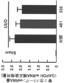
- clone 481 increased the relative amount of renal type I collagen mRNA by UUO model production 2.29 [Sham group (0.01 ⁇ 0.01) (lane 1) and UUO model negative IgG group (2.30 ⁇ 0.29) (lane 2)
- Clone 536 decreased by approximately 16% to 2.03 [difference between Sham group (0.01 ⁇ 0.01) (lane 1) and UUO clone 481 group (2.04 ⁇ 0.24) (lane 3)] 1.93 [Difference between Sham group (0.01 ⁇ 0.01) (lane 1) and UUO clone 536 group (1.94 ⁇ 0.27) (lane 4)]
- the anti-renal fibrosis inhibitory activity of both antibodies was confirmed. (FIG. 11).
- the monoclonal antibody of the present invention specifically binds to LAP of a specific isoform of TGF- ⁇ and has an activity of suppressing the activation of the isoform. For this reason, it is used for the treatment or prevention of diseases caused by TGF- ⁇ activation, such as liver fibrosis / cirrhosis, pulmonary fibrosis, renal fibrosis / renal failure, dermal fibrosis, arteriosclerosis, etc. be able to.
- the isoform specificity of the antibody of the present invention leads to reduction of side effects when administered to a patient as a medicine.
- the antibody of the present invention does not target active TGF- ⁇ , but inhibits the stage in which active TGF- ⁇ is produced from inactive latent TGF- ⁇ .
- the antibody of the present invention is medically useful.
- the monoclonal antibody of the present invention can be applied as a diagnostic reagent or a research reagent for isoform-specific detection / selection of TGF- ⁇ .
Landscapes
- Health & Medical Sciences (AREA)
- Chemical & Material Sciences (AREA)
- Organic Chemistry (AREA)
- Life Sciences & Earth Sciences (AREA)
- General Health & Medical Sciences (AREA)
- Engineering & Computer Science (AREA)
- Bioinformatics & Cheminformatics (AREA)
- Medicinal Chemistry (AREA)
- Veterinary Medicine (AREA)
- General Chemical & Material Sciences (AREA)
- Chemical Kinetics & Catalysis (AREA)
- Nuclear Medicine, Radiotherapy & Molecular Imaging (AREA)
- Public Health (AREA)
- Pharmacology & Pharmacy (AREA)
- Animal Behavior & Ethology (AREA)
- Urology & Nephrology (AREA)
- Proteomics, Peptides & Aminoacids (AREA)
- Vascular Medicine (AREA)
- Pulmonology (AREA)
- Gastroenterology & Hepatology (AREA)
- Biochemistry (AREA)
- Heart & Thoracic Surgery (AREA)
- Cardiology (AREA)
- Biophysics (AREA)
- Genetics & Genomics (AREA)
- Molecular Biology (AREA)
- Immunology (AREA)
- Peptides Or Proteins (AREA)
Abstract
TGF-β1およびTGF-β2のLAPに相当するペプチドに対するモノクローナル抗体を調製し、その中から、いずれかのアイソフォームに特異的に結合するモノクローナル抗体を選抜した。選抜した抗体のLAP上のエピトープ領域を同定した。さらに、選抜した抗体の中に、プロテアーゼによるLAPの切断に対する抑制活性を示す複数の抗体、並びに肝臓または腎臓の線維化を抑制する活性を有する抗体を見出した。
Description
本発明は、ヒトTGF-βのLAPに結合する抗体およびその用途に関する。より詳しくは、ヒトTGF-βの特定のアイソフォームのLAPに特異的に結合し、かつ、当該TGF-βアイソフォームの活性化を抑制する活性を持つ抗体に関する。
TGF-β(Transforming Growth Factor-β)は、間葉系細胞の細胞外マトリックス産生を強力に促すとともに上皮系細胞の増殖を抑制することにより、肝線維化/肝硬変、肺線維化、腎線維化/腎不全、皮膚線維症、動脈硬化などの硬化性疾患の病態を形成する一方、免疫担当細胞の働きを抑制するなど、多彩な生物活性を示す分子量25kDのホモダイマー多機能性サイトカインである。
TGF-βは、受容体に結合できない分子量約300kDの不活性な潜在型として産生され、標的細胞表面やその周囲で活性化されて受容体に結合できる活性型となる。これにより、始めてその作用を発揮できるようになる(図1)。TGF-β1は、まず、391個のアミノ酸からなる前駆体タンパク質として生成される。その後、ゴルジにおいてフューリン様プロテアーゼの働きで、前駆体タンパク質の279Argと280Alaの間が切断される。そして、カルボキシル末端側の112個のアミノ酸からなる部分がジスルフィド結合により2量体化することにより、分子量25kDの活性型TGF-β1が生成する。TGF-β2とTGF-β3も同じ構造で、Arg-Ala配列は共通している。切断された残りのアミノ末端側の部分は、LAP(latency associated protein)と呼ばれる。LAPもまた2量体化し(分子量75kD)、活性型TGF-βから切り離された後も非共有結合で活性型TGF-βをトラップして、潜在型TGF-β小複合体(small latent TGF-β complex;SLC)を形成する。こうしてLAPは、活性型TGF-βを受容体に結合できない構造、すなわち潜在型に留めている。さらに多くの場合、LAPダイマーの端には、LTBP(latent TGF-β binding protein)という分子量約200kDのタンパク質が結合して、潜在型TGF-β大複合体(Large latent TGF-β complex;LLC)を形成する。TGF-β2とTGF-β3も同様の構造をとっている。LTBPは、細胞外マトリックスタンパク質の1種であるフィブリリンと構造が似ており、LLCは、LTBPを介して細胞外マトリックスにプールされている(図1)。
TGF-βの活性化は、なんらかの方法でLAPと活性型TGF-β間の非共有結合による会合を妨げることによってLLCにトラップされている活性型TGF-βを解離、放出する反応である。TGF-βの活性化は、マトリックスからLLCが放出されたのちに、標的細胞表面やその近傍において起こる(図1)。生理的なTGF-β活性化反応には、トロンボスポンジンやインテグリンと結合することによってLAPの構造が変化する結果、活性型TGF-βを放出する接着活性化反応と、プロテアーゼによってLAPが限定分解されて、保持していた活性型TGF-βを放出する切断活性化反応とが知られている(図1)。
このようにTGF-βの活性化の機構が徐々に解明されてきたことに伴い、TGF-βが関連する上記疾患の診断、治療あるいは予防などの目的で、TGF-βの活性化を検出するための抗体の開発やTGF-βの活性化を抑制する抗体の開発が試みられてきた。
例えば、TGF-βの活性化過程における、潜在型TGF-βのプラスミンと血漿カリクレインによる切断面(それぞれ、潜在型TGF-βの56Lys-57Leuの間、58Arg-59Leuの間)を認識する抗体が開発されている(特許文献1)。この抗体は、インタクトなLAPには結合せず、その切断面に特異的に結合するという性質を有していることから、これを用いることにより、TGF-β活性化反応の検出が可能であるとされているが、TGF-β活性化反応自体は抑制しない。
一方、LAPに結合し、TGF-βの活性化を抑制する抗体の開発もなされている(特許文献2)。疑似創傷状態にしたウシ大動脈内皮細胞とウシ大動脈平滑筋細胞との共存培養において、TGF-βが活性化した場合、ウシ大動脈平滑筋細胞の遊走が抑制されるが、この抗体は、ウシ大動脈平滑筋細胞の遊走を回復させる活性を持つとされている。
しかしながら、TGF-βには、ほとんど同じ生物活性を示すTGF-β1からTGF-β3までのアイソフォームが存在する。このため、TGF-βを標的として病気の治療や予防を行う場合、副作用低減等の観点から、病態形成時に異常産生される特定のTGF-βアイソフォームの生成のみを抑制することが極めて重要である。
これまでに、ヒトTGF-βの特定のアイソフォームのみを認識する抗体は知られているが(例えば、特許文献3)、このような特異性を有するとともに、当該特定のアイソフォームの活性化を抑制する活性をも持つ抗体については、いまだ開発されていない。
F.Okada, et al., J. Biochem. 106:304-310(1989)
本発明はこのような状況に鑑みてなされたものであり、その目的は、TGF-βの特定のアイソフォームに特異的に結合し、かつ、当該アイソフォームの活性化を抑制する活性を有する抗体を提供することにある。さらなる本発明の目的は、このような抗体を有効成分とする、TGF-βの活性化に起因する疾患、例えば、肝線維化/肝硬変、肺線維化、腎線維化/腎不全、皮膚線維症、動脈硬化などの硬化性疾患、を治療または予防するための薬剤を提供することにある。
本発明者らは、潜在型TGF-βにおけるLAPが、プラスミンや血漿カリクレインによる切断を受け、これにより活性型TGF-βを生成することに着目し、まず、TGF-βの特定のアイソフォームのLAPに特異的に結合するモノクローナル抗体の調製を試みた。
具体的には、TGF-β1およびTGF-β2のLAPに相当するペプチドを遺伝子組み換えまたは合成により調製し、これを免疫原としてハイブリドーマを調製した。次いで、調製したハイブリドーマから、ELISA法により、TGF-β1およびTGF-β2のいずれかのLAPにのみ結合するモノクローナル抗体を産生するクローンを選抜した。さらに、これらクローンが産生するアイソフォーム特異的モノクローナル抗体について、プラスミンや血漿カリクレインにより切断を受けるLAP上の部位およびその近傍領域に対する反応性を評価し、それらのLAP上のエピトープ領域を特定した。
次に、こうして得られたアイソフォーム特異的モノクローナル抗体について、TGF-β活性化に与える影響を検討した。具体的には、血漿カリクレインによるLAPの切断に対する、これらモノクローナル抗体の抑制活性を評価した。その結果、本発明者らは、アイソフォーム特異的モノクローナル抗体の中から、当該アイソフォームの活性化を抑制する活性を有する複数の抗体、並びに肝臓または腎臓の線維化を抑制する活性を有する抗体を見出すことに成功した。さらに、これら抗体のエピトープとTGF-βの活性化の抑制活性との相関を詳細に分析した結果、TGF-βの活性化の抑制に有用なエピトープを見出すことに成功した。
従って、本発明は、TGF-βの特定のアイソフォームのLAPに特異的に結合し、かつ、当該アイソフォームの活性化を抑制する活性を有する抗体、およびその利用に関し、より詳しくは、下記発明を提供するものである。
(1) ヒトTGF-βの特定のアイソフォームのLAPに特異的に結合し、かつ、当該アイソフォームの活性化を抑制する活性を有する抗体。
(2) ヒトTGF-β1のLAPに実質的に結合し、ヒトTGF-β2のLAPに実質的に結合しない、(1)に記載の抗体。
(3) ヒトTGF-β1のLAPにおける配列番号:5に記載のアミノ酸配列からなる領域に実質的に結合する、(2)に記載の抗体。
(4) 配列番号:7に記載のアミノ酸配列からなるペプチドに実質的に結合し、配列番号:9~11に記載のアミノ酸配列からなるペプチドに実質的に結合しない、(2)に記載の抗体。
(5) 肝臓または腎臓の線維化を抑制する活性を有する、(1)または(2)に記載の抗体。
(6) ヒトTGF-β2のLAPに実質的に結合し、ヒトTGF-β1のLAPに実質的に結合しない(1)に記載の抗体。
(7) ヒトTGF-β2のLAPにおける配列番号:6に記載のアミノ酸配列からなる領域に実質的に結合する、(6)に記載の抗体。
(8) 受託番号NITE BP-873、受託番号NITE BP-881、または受託番号NITE BP-882で特定されるハイブリドーマにより産生される抗体。
(9) (8)に記載の抗体が結合するヒトTGF-β1のLAP上のエピトープに結合する抗体。
(10) (1)から(9)のいずれかに記載の抗体をコードするDNA。
(11) (1)から(9)のいずれかに記載の抗体を産生する、または、(10)に記載のDNAを含む、ハイブリドーマ。
(12) 受託番号NITE BP-873、受託番号NITE BP-881、または受託番号NITE BP-882で特定されるハイブリドーマ。
(13) (1)から(9)のいずれかに記載の抗体を有効成分とする、TGF-βの活性化に起因する疾患の治療または予防のための薬剤。
(1) ヒトTGF-βの特定のアイソフォームのLAPに特異的に結合し、かつ、当該アイソフォームの活性化を抑制する活性を有する抗体。
(2) ヒトTGF-β1のLAPに実質的に結合し、ヒトTGF-β2のLAPに実質的に結合しない、(1)に記載の抗体。
(3) ヒトTGF-β1のLAPにおける配列番号:5に記載のアミノ酸配列からなる領域に実質的に結合する、(2)に記載の抗体。
(4) 配列番号:7に記載のアミノ酸配列からなるペプチドに実質的に結合し、配列番号:9~11に記載のアミノ酸配列からなるペプチドに実質的に結合しない、(2)に記載の抗体。
(5) 肝臓または腎臓の線維化を抑制する活性を有する、(1)または(2)に記載の抗体。
(6) ヒトTGF-β2のLAPに実質的に結合し、ヒトTGF-β1のLAPに実質的に結合しない(1)に記載の抗体。
(7) ヒトTGF-β2のLAPにおける配列番号:6に記載のアミノ酸配列からなる領域に実質的に結合する、(6)に記載の抗体。
(8) 受託番号NITE BP-873、受託番号NITE BP-881、または受託番号NITE BP-882で特定されるハイブリドーマにより産生される抗体。
(9) (8)に記載の抗体が結合するヒトTGF-β1のLAP上のエピトープに結合する抗体。
(10) (1)から(9)のいずれかに記載の抗体をコードするDNA。
(11) (1)から(9)のいずれかに記載の抗体を産生する、または、(10)に記載のDNAを含む、ハイブリドーマ。
(12) 受託番号NITE BP-873、受託番号NITE BP-881、または受託番号NITE BP-882で特定されるハイブリドーマ。
(13) (1)から(9)のいずれかに記載の抗体を有効成分とする、TGF-βの活性化に起因する疾患の治療または予防のための薬剤。
本発明により、TGF-βの特定のアイソフォームのLAPに特異的に結合し、かつ、当該アイソフォームの活性化を抑制する活性を有する抗体が提供された。本発明の抗体を用いれば、TGF-βの活性化に起因する疾患、例えば、肝線維化/肝硬変、肺線維化、腎線維化/腎不全、皮膚線維症、動脈硬化などの硬化性疾患の治療や予防が可能となる。本発明の抗体は、TGF-βの特定のアイソフォームに特異的であるため、生体に投与した場合に、低い副作用が期待できる。また、活性型TGF-βを標的とするのではなく、不活性潜在型TGF-βから活性型TGF-βが生成する段階を阻害するため、より早い段階での治療・予防が期待できる。
本発明は、ヒトTGF-βの特定のアイソフォームのLAPに特異的に結合し、かつ、当該アイソフォームの活性化を抑制する活性を有する抗体を提供する。ここで「特異的」とは、ヒトTGF-βの特定のアイソフォームのLAPに実質的に結合し、他のアイソフォームのLAPに実質的に結合しないことを意味する。
本発明における「抗体」は、免疫グロブリンのすべてのクラスおよびサブクラスを含む。「抗体」には、ポリクローナル抗体、モノクローナル抗体が含まれ、また、抗体の機能的断片の形態も含む意である。「ポリクローナル抗体」は、異なるエピトープに対する異なる抗体を含む抗体調製物である。また、「モノクローナル抗体」とは、実質的に均一な抗体の集団から得られる抗体(抗体断片を含む)を意味する。ポリクローナル抗体とは対照的に、モノクローナル抗体は、抗原上の単一の決定基を認識するものである。本発明の抗体は、好ましくはモノクローナル抗体である。本発明の抗体は、自然環境の成分から分離、および/または回収された抗体、すなわち、「単離された」抗体である。
TGF-βは、間葉系細胞の細胞外マトリックス産生を強力に促すとともに上皮系細胞の増殖を抑制することにより、肝線維化/肝硬変、肺線維化、腎線維化/腎不全、皮膚線維症、動脈硬化などの硬化性疾患の病態を形成する一方、免疫担当細胞の働きを抑制するなど、多彩な生物活性を示す分子量25kDのホモダイマー多機能性サイトカインである。本発明の抗体が結合する「ヒトTGF-β1」および「ヒトTGF-β2」については、既にその一次構造が公知である。本発明における「ヒトTGF-β1」の典型的なアミノ酸配列を配列番号:2に、当該アミノ酸配列をコードするDNAの典型的な塩基配列を配列番号:1に示す。また、本発明における「ヒトTGF-β2」の典型的なアミノ酸配列を配列番号:4に、当該アミノ酸配列をコードするDNAの典型的な塩基配列を配列番号:3に示す。「ヒトTGF-β1」および「ヒトTGF-β2」については、このような典型的なアミノ酸配列を有するもの以外に、天然においてアミノ酸が変異したものも存在しうる。本発明の「ヒトTGF-β1」および「ヒトTGF-β2」には、このような変異体が含まれる。このような変異体は、配列番号:2または4で表されるアミノ酸配列において、1もしくは複数個のアミノ酸が置換、欠失、挿入もしくは付加されたアミノ酸配列からなるものである。天然において生じる変異数は、一般的には、10アミノ酸以内(例えば、5アミノ酸以内、3アミノ酸以内、1アミノ酸)であると考えられる。
本発明において「LAP」とは、潜在型TGF-β分子中において、活性型TGF-βと非共有結合的に会合している二量体のプレペプチド領域を意味する。
本発明において、TGF-βの「活性化」とは、TGF-βが、その受容体に結合し、作用を発揮できる状態になることを意味する。典型的には、潜在型TGF-βのLAPが、プロテアーゼによる切断を受けて、LAPと活性型TGF-βの会合が阻害され、不活性潜在型TGF-β複合体から活性型TGF-βが遊離して、活性型TGF-βが生成することを意味する。
本発明の抗体の一つの好ましい態様は、ヒトTGF-β1のLAPに実質的に結合し、ヒトTGF-β2のLAPに実質的に結合しない抗体である。より好ましくは、ヒトTGF-β1のLAPにおけるプロテアーゼ(プラスミン、血漿カリクレイン)により切断される部位または近傍の領域(以下、単に「プロテアーゼ切断領域」と称する)に実質的に結合する抗体であり、例えば、ヒトTGF-β1のLAPにおける配列番号:5に記載のアミノ酸配列からなる領域(図5における「TGF-β1-LAP」)に実質的に結合する抗体である。
なお、本発明において「実質的に結合する」とは、本実施例におけるELISA実験において、490nmの吸光度の値が0.2以上となることを意味する(図6、7参照)。
なお、本発明において「実質的に結合する」とは、本実施例におけるELISA実験において、490nmの吸光度の値が0.2以上となることを意味する(図6、7参照)。
本発明においては、TGF-β1-LAPに特異的に結合する抗体のうち、配列番号:7に記載のアミノ酸配列からなるペプチド(図5における「LAP1-1」)には実質的に結合するが、配列番号:9~11に記載のアミノ酸配列からなるペプチド(図5における「LAP1-3~LAP1-5」)には実質的に結合しない抗体が、血漿カリクレインによるTGF-β1-LAPの切断、すなわちTGF-β1の活性化反応の抑制効果が高いことが見出された(表1、図4、図6A)。従って、ヒトTGF-β1のLAPにおける配列番号:5に記載のアミノ酸配列からなる領域(図5における「TGF-β1-LAP」)に実質的に結合する抗体のうち、特に、このようなペプチド特異性を有する抗体は、本発明において好適に用いることができる。
本発明の抗体の他の好ましい態様は、TGF-β1-LAPに特異的に結合する抗体であって、肝臓または腎臓の線維化を抑制する活性を有する抗体である。肝臓の線維化を抑制する活性を有する抗体は、例えば、配列番号:7に記載のアミノ酸配列からなるペプチド(図5における「LAP1-1」)には実質的に結合するが、配列番号:9~11に記載のアミノ酸配列からなるペプチド(図5における「LAP1-3~LAP1-5」)には実質的に結合しない抗体である。また、腎臓の線維化を抑制する活性を有する抗体は、例えば、配列番号:17および18に記載のアミノ酸配列からなるペプチド(図7における「LAP1-0」および「LAP1-6」)には実質的に結合するが、配列番号:7~11に記載のアミノ酸配列からなるペプチド(図7における「LAP1-1~LAP1-5」)には実質的に結合しない抗体である。肝臓の線維化を抑制する活性は、例えば、 実施例10に記載のメチオニン・コリン欠乏食(MCDD)を給与した非アルコール性脂肪性肝炎(NASH)のモデルマウスを用いた実験や実施例11に記載のてコリン欠乏アミノ酸置換食(CDAA diet)を給与した非アルコール性脂肪性肝炎(NASH)のモデルマウスを用いた実験により評価することができる。また、腎臓の線維化を抑制する活性は、例えば、実施例12に記載の片側尿管結紮(UUO)腎線維化モデルマウスを用いた実験や実施例13に記載の片側尿管結紮(UUO)による腎線維化モデルマウスを用いた実験により評価することができる。
本発明の抗体の他の好ましい態様は、ヒトTGF-β2のLAPに実質的に結合し、ヒトTGF-β1のLAPに実質的に結合しない抗体である。より好ましくは、上記ヒトTGF-β1のプロテアーゼ切断領域に対応するヒトTGF-β2上の領域に実質的に結合する抗体であり、例えば、ヒトTGF-β2のLAPにおける配列番号:6に記載のアミノ酸配列からなる領域(図5における「TGF-β2-LAP」)に実質的に結合する抗体である。
本発明の抗体の最も好ましい態様は、受託番号NITE BP-873、受託番号NITE BP-881、または受託番号NITE BP-882で特定されるハイブリドーマにより産生される抗体である。これら抗体は、ヒトTGF-β1のLAPに特異的に結合するとともに、血漿カリクレインによるヒトTGF-β1のLAPの切断を抑制する活性を有し、かつ、肝臓または腎臓の線維化を抑制する活性を有する(表1、図4、図5、図6A、図7~11)。
一旦、これら特定のハイブリドーマが産生する抗体が得られれば、当業者であれば、その抗体が認識するLAP上のペプチド領域(エピトープ)を特定して、その領域に結合し、かつ、同様にLAPの切断を抑制する活性を示す種々の抗体を作製することができる。従って、本発明は、上記ハイブリドーマが産生する抗体のエピトープに結合し、かつ、血漿カリクレインによるヒトTGF-β1のLAPの切断を抑制する活性を有する抗体が含まれる。抗体のエピトープは、LAPのアミノ酸配列から得られたオーバーラップする合成オリゴペプチドへの結合を調べるなどの周知の方法によって決定することができる(例えば、本願実施例、Ed Harlow and D.Lane, Using Antibodies, a Laboratory Manual, Cold Spring Harbor Laboratory Press、米国特許4708871号、を参照のこと)。ファージディスプレイによるペプチドライブラリーをエピトープマッピングに用いることもできる。二つの抗体が同一または立体的に重なり合ったエピトープと結合するかどうかは、競合アッセイ法により決定することができる。抗体における、LAPの切断を抑制する活性は、実施例8に示す方法で評価することが可能である。実施例8においては、プロテアーゼとして血漿カリクレインを用いているが、他のプロテアーゼを用いる場合にも、実施例8に準じた方法で評価することが可能である。
本発明の抗体には、キメラ抗体、ヒト化抗体、ヒト抗体、および、これら抗体の機能的断片が含まれる。本発明の抗体を医薬としてヒトに投与する場合は、副作用低減の観点から、キメラ抗体、ヒト化抗体、あるいはヒト抗体が望ましい。
本発明において「キメラ抗体」とは、ある種の抗体の可変領域とそれとは異種の抗体の定常領域とを連結した抗体である。キメラ抗体は、例えば、抗原をマウスに免役し、そのマウスモノクローナル抗体の遺伝子から抗原と結合する抗体可変部(可変領域)を切り出して、ヒト骨髄由来の抗体定常部(定常領域)遺伝子と結合し、これを発現ベクターに組み込んで宿主に導入して産生させることにより取得することができる(例えば、特開平8-280387号公報、米国特許第4816397号公報、米国特許第4816567号公報、米国特許第5807715号公報)。また、本発明において「ヒト化抗体」とは、非ヒト由来の抗体の抗原結合部位(CDR)の遺伝子配列をヒト抗体遺伝子に移植(CDRグラフティング)した抗体であり、その作製方法は、公知である(例えば、EP239400、EP125023、WO90/07861、WO96/02576参照)。本発明において、「ヒト抗体」とは、すべての領域がヒト由来の抗体である。ヒト抗体の作製においては、免疫することで、ヒト抗体のレパートリーを生産することが可能なトランスジェニック動物(例えばマウス)を利用することが可能である。ヒト抗体の作製手法は、公知である(例えば、Nature, 362:255-258(1992)、Intern. Rev. Immunol, 13:65-93(1995)、J. Mol. Biol, 222:581-597(1991)、Nature Genetics, 15:146-156(1997)、Proc. Natl. Acad. Sci. USA, 97:722-727(2000)、特開平10-146194号公報、特開平10-155492号公報、特許2938569号公報、特開平11-206387号公報、特表平8-509612号公報、特表平11-505107号公報)。
本発明における抗体には、抗体の一部分(部分断片)であって、ヒトTGF-βの特定のアイソフォームのLAPを特異的に認識するもの(機能的断片)が含まれる。具体的には、Fab、Fab’、F(ab’)2、可変領域断片(Fv)、ジスルフィド結合Fv、一本鎖Fv(scFv)、sc(Fv)2、ダイアボディー、多特異性抗体、およびこれらの重合体などが挙げられる。
ここで「Fab」とは、1つの軽鎖および重鎖の一部からなる免疫グロブリンの一価の抗原結合断片を意味する。抗体のパパイン消化によって、また、組換え方法によって得ることができる。「Fab'」は、抗体のヒンジ領域の1つまたはそれより多いシステインを含めて、重鎖CH1ドメインのカルボキシ末端でのわずかの残基の付加によって、Fabとは異なる。「F(ab’)2」とは、両方の軽鎖と両方の重鎖の部分からなる免疫グロブリンの二価の抗原結合断片を意味する。
「可変領域断片(Fv)」は、完全な抗原認識および結合部位を有する最少の抗体断片である。Fvは、重鎖可変領域および軽鎖可変領域が非共有結合により強く連結されたダイマーである。「一本鎖Fv(sFv)」は、抗体の重鎖可変領域および軽鎖可変領域を含み、これらの領域は、単一のポリペプチド鎖に存在する。「sc(Fv)2」は、2つの重鎖可変領域および2つの軽鎖可変領域をリンカー等で結合して一本鎖にしたものである。「ダイアボディー」とは、二つの抗原結合部位を有する小さな抗体断片であり、この断片は、同一ポリペプチド鎖の中に軽鎖可変領域に結合した重鎖可変領域を含み、各領域は別の鎖の相補的領域とペアを形成している。「多特異性抗体」は、少なくとも2つの異なる抗原に対して結合特異性を有するモノクローナル抗体である。例えば、二つの重鎖が異なる特異性を持つ二つの免疫グロブリン重鎖/軽鎖対の同時発現により調製することができる。
本発明の抗体には、望ましい活性(TGF-βの特定のアイソフォームのLAPに特異的に結合する活性、プロテアーゼによるLAPの切断を抑制する活性、および/または他の生物学的特性)を減少させることなく、そのアミノ酸配列が修飾された抗体が含まれる。抗体のアミノ酸配列変異体は、抗体鎖をコードするDNAへの変異導入によって、またはペプチド合成によって作製することができる。そのような修飾には、例えば、抗体のアミノ酸配列内の残基の置換、欠失、付加および/または挿入を含む。抗体のアミノ酸配列が改変される部位は、改変される前の抗体と同等の活性を有する限り、抗体の重鎖または軽鎖の定常領域であってもよく、また、可変領域(フレームワーク領域およびCDR)であってもよい。CDR以外のアミノ酸の改変は、抗原との結合親和性への影響が相対的に少ないと考えられるが、現在では、CDRのアミノ酸を改変して、抗原へのアフィニティーが高められた抗体をスクリーニングする手法が公知である(PNAS, 102:8466-8471(2005)、Protein Engineering, Design & Selection, 21:485-493(2008)、国際公開第2002/051870号、J. Biol. Chem., 280:24880-24887(2005)、Protein Engineering, Design & Selection, 21:345-351(2008))。
改変されるアミノ酸数は、好ましくは、10アミノ酸以内、より好ましくは5アミノ酸以内、最も好ましくは3アミノ酸以内(例えば、2アミノ酸以内、1アミノ酸)である。アミノ酸の改変は、好ましくは、保存的な置換である。本発明において「保存的な置換」とは、化学的に同様な側鎖を有する他のアミノ酸残基で置換することを意味する。化学的に同様なアミノ酸側鎖を有するアミノ酸残基のグループは、本発明の属する技術分野でよく知られている。例えば、酸性アミノ酸(アスパラギン酸およびグルタミン酸)、塩基性アミノ酸(リシン・アルギニン・ヒスチジン)、中性アミノ酸においては、炭化水素鎖を持つアミノ酸(グリシン・アラニン・バリン・ロイシン・イソロイシン・プロリン)、ヒドロキシ基を持つアミノ酸(セリン・トレオニン)、硫黄を含むアミノ酸(システイン・メチオニン)、アミド基を持つアミノ酸(アスパラギン・グルタミン)、イミノ基を持つアミノ酸(プロリン)、芳香族基を持つアミノ酸(フェニルアラニン・チロシン・トリプトファン)で分類することができる。
本発明の抗体は、ポリクローナル抗体であれば、抗原で免疫動物を免疫し、その抗血清から、従来の手段(例えば、塩析、遠心分離、透析、カラムクロマトグラフィーなど)によって、精製して取得することができる。また、モノクローナル抗体は、ハイブリドーマ法や組換えDNA法によって作製することができる。抗原としては、遺伝子組み換えにより調製された、または、合成されたLAPまたはその部分ペプチド、またはこれらを発現する細胞などを用いることができる。抗原は、高純度で精製されたペプチドであることが好ましい。ヒスチジンタグ等のアフィニティタグを融合したペプチドとして調製する場合には、精製過程においては、このタグに対するアフィニティカラムを用いることができる。
ハイブリドーマ法としては、代表的には、コーラーおよびミルスタインの方法(Kohler & Milstein, Nature, 256:495(1975))が挙げられる。この方法における細胞融合工程に使用される抗体産生細胞は、抗原で免疫された動物(例えば、マウス、ラット、ハムスター、ウサギ、サル、ヤギ)の脾臓細胞、リンパ節細胞、末梢血白血球などである。免疫されていない動物から予め単離された上記の細胞またはリンパ球などに対して、抗原を培地中で作用させることによって得られた抗体産生細胞も使用することが可能である。ミエローマ細胞としては公知の種々の細胞株を使用することが可能である。抗体産生細胞およびミエローマ細胞は、それらが融合可能であれば、異なる動物種起源のものでもよいが、好ましくは、同一の動物種起源のものである。ハイブリドーマは、例えば、抗原で免疫されたマウスから得られた脾臓細胞と、マウスミエローマ細胞との間の細胞融合により産生される。その後、得られたハイブリドーマクローンのスクリーニングにより、TGF-βの特定のアイソフォームのLAPに特異的なモノクローナル抗体を産生するクローンを得ることができる。スクリーニングにおいては、例えば、得られたハイブリドーマが産生するモノクローナル抗体(例えば、培養上清、精製抗体)について、TGF-βの各アイソフォームのLAP(例えば、TGF-β1のLAP、TGF-β2のLAP)への反応性を評価し、いずれかのアイソフォームのLAPに特異的に反応するモノクローナル抗体を産生するクローンを選抜すればよい。さらに、アイソフォーム特異的モノクローナル抗体については、LAPにおけるプロテアーゼにより切断を受ける部位およびその近傍領域に対する反応性を評価し、それら抗体のLAP上のエピトープ領域を同定することができる。モノクローナル抗体は、ハイブリドーマを培養することにより、また、ハイブリドーマを投与した哺乳動物の腹水から、取得することができる。
免疫原およびスクリーニング用の合成ペプチドの具体例としては、TGF-β1-LAPとして「EAIRGQILSKLRLASPPSQGEVPPGPLPEAVLAL(配列番号:5)」が、TGF-β2-LAPとして「EAIRGQILSKLKLTSPPEDYPEPEEVPPEVISI(配列番号:6)」が挙げられる。エピトープ解析用の合成ペプチドの具体例としては、TGF-β1-LAPとして「LSKLRLASPP(配列番号:7)」、「KLRLASPPSQ(配列番号:8)」、「RLASPPSQEVPPGP(配列番号:9)」、「ASPPSQGEVPPG(配列番号:10)」、「PPSQGEVPPGPL(配列番号:11)」、「EAIRGQIL(配列番号:17)」、および「PEAVLAL(配列番号:18)」が挙げられる。TGF-β2-LAPとして「KLKLTSPPED(配列番号:12)」、「KLTSPPEDYP(配列番号:13)」、「TSPPEDYPEP(配列番号:14)」、「PPEDYPEPEE(配列番号:15)」、および「EDYPEPEEVP(配列番号:16)」が挙げられる。本実施例に示すように、これらペプチドのN末端にシステインを付加したペプチドを、Fmoc固相合成法により合成することが可能である。
組換えDNA法は、上記本発明の抗体をコードするDNAをハイブリドーマやB細胞等からクローニングし、適当なベクターに組み込んで、これを宿主細胞(例えば哺乳類細胞株、大腸菌、酵母細胞、昆虫細胞、植物細胞など)に導入し、本発明の抗体を組換え抗体として産生させる手法である(例えば、P.J.Delves, Antibody Production: Essential Techniques, 1997 WILEY、P.Shepherd and C. Dean Monoclonal Antibodies, 2000 OXFORD UNIVERSITY PRESS、Vandamme A.M. et al., Eur. J. Biochem. 192:767-775(1990))。本発明の抗体をコードするDNAの発現においては、重鎖または軽鎖をコードするDNAを別々に発現ベクターに組み込んで宿主細胞を形質転換してもよく、重鎖および軽鎖をコードするDNAを単一の発現ベクターに組み込んで宿主細胞を形質転換してもよい(WO94/11523号公報参照)。本発明の抗体は、上記宿主細胞を培養し、宿主細胞内または培養液から分離・精製し、実質的に純粋で均一な形態で取得することができる。抗体の分離・精製は、通常のポリペプチドの精製で使用されている方法を使用することができる。トランスジェニック動物作製技術を用いて、抗体遺伝子が組み込まれたトランスジェニック動物(ウシ、ヤギ、ヒツジまたはブタなど)を作製すれば、そのトランスジェニック動物のミルクから、抗体遺伝子に由来するモノクローナル抗体を大量に取得することも可能である。
本発明は、上記本発明の抗体をコードするDNA、当該DNAを含むベクター、当該DNAを保持する宿主細胞、および当該宿主細胞を培養し、抗体を回収することを含む抗体の生産方法をも提供するものである。
本発明の抗体は、アイソフォーム特異的にTGF-βの活性化を抑制することから、例えば、肝線維化/肝硬変、肺線維化、腎線維化/腎不全、皮膚線維症、動脈硬化などの硬化性疾患の治療または予防に利用することができる。従って、本発明は、本発明の抗体を有効成分とするTGF-βの活性化に起因する疾患の治療または予防のための薬剤、および、本発明の抗体の治療上または予防上の有効量を、患者に投与する工程を含む、TGF-βの活性化に起因する疾患の治療または予防の方法をも提供するものである。
本発明の抗体を有効成分とする薬剤は、本発明の抗体と任意の成分、例えば生理食塩水、葡萄糖水溶液または燐酸塩緩衝液などを含有する組成物の形態で使用することができる。本発明の薬剤は、必要に応じて液体または凍結乾燥した形態で製形化しても良く、任意に薬学的に許容される担体もしくは媒体、例えば、安定化剤、防腐剤、等張化剤などを含有させることもできる。
薬学的に許容される担体としては、凍結乾燥した製剤の場合、マンニトール、ラクトース、サッカロース、ヒトアルブミンなどを例として挙げることができ、液状製剤の場合には、生理食塩水、注射用水、燐酸塩緩衝液、水酸化アルミニウムなどを例として挙げることができるが、これらに限定されるものではない。
本発明の薬剤の投与方法は、投与対象の年齢、体重、性別、健康状態などにより異なるが、非経口投与(例えば、静脈投与、皮下投与、動脈投与、局所投与)、経口投与のいずれかの投与経路で投与することができる。好ましい投与方法は、非経口投与である。本発明の薬剤の投与量は、患者の年齢、体重、性別、健康状態、病気の進行の程度および投与する薬剤の成分により変動しうるが、一般的に静脈内投与の場合、成人には体重1kg当たり1日0.001~1000mg、好ましくは0.01~10mgである。通常、1週間から1ヶ月間隔で投与するが、この投与間隔に限定されるものではない。
本発明の抗体は、上記疾患の治療や予防のみならず、診断への応用も考えられる。本発明の抗体を診断に用いる場合あるいはTGF-βの特定のアイソフォームの検出に用いる場合、本発明の抗体は、標識したものであってもよい。標識としては、例えば、放射性物質、蛍光色素、化学発光物質、酵素、補酵素を用いることが可能であり、具体的には、ラジオアイソトープ、フルオレセイン、ローダミン、ダンシルクロリド、ルシフェラーゼ、ペルオキシダーゼ、アルカリフォスファターゼ、リゾチーム、ビオチン/アビジンなどが挙げられる。本発明の抗体を診断剤として調剤するには、合目的な任意の手段を採用して任意の剤型でこれを得ることができる。例えば、精製した抗体についてその抗体価を測定し、適当にPBS(生理食塩を含むリン酸緩衝液)等で希釈した後、0.1%アジ化ナトリウム等を防腐剤として加えることができる。また、例えば、ラテックス等に本発明の抗体を吸着させたものについて抗体価を求め、適当に希釈し、防腐剤を添加して用いることもできる。
以下、本発明を実施例により、さらに詳細に説明するが、本発明はこれら実施例に制限されるものではない。
[実施例1] 組み換え抗原TGF-β1-LAPおよびTGF-β2-LAPの組換え大腸菌による調製
pGEX-6P-1/TGF-β1(プラスミド1)とpGEX-6P-1/TGF-β2(プラスミド2)をそれぞれ100ng用いて、ヒートショック法により、Rosetta-gamiB(DE3)(Novagen社)の形質転換を実施した。形質転換体の選抜には、LB培地+Amp50寒天培地を使用した。形質転換体のコロニーを、LB培地+Amp50において12~15時間培養を行って増殖させた(前培養)。培養液200μLを10mLのLB+Amp50培地に添加し(2%植菌)、培養開始から4時間後、イソプロピルβ-チオガラクトピラノシド(IPTG)を終濃度0.3mMとなるように添加した。さらに3時間37℃にて培養し、培養菌体を回収した。回収した菌体を菌体破砕用緩衝液(0.1m MEDTA、1% Triton-X100、1mg/mL リゾチーム、DNaseIを含むPBS緩衝液)500μLに懸濁し、1時間振とう撹拌することで、水溶性のタンパク質を抽出した。その後、SDS-PAGEを実施した(DRC社、Perfect NT Gel 15~25%、泳動条件500V、50mA、60分)。簡易的にタンパク質の発現を確認した結果、TGF-β1およびTGF-β2ともに、Rosetta-gamiB(DE3)における発現が確認された。このため、組み換えタンパク質の調製においては、プラスミド1またはプラスミド2を保有するRosetta-gamiB(DE3)形質転換体(以降、それぞれ「形質転換体1」、「形質転換体2」と称する)を用いた。
pGEX-6P-1/TGF-β1(プラスミド1)とpGEX-6P-1/TGF-β2(プラスミド2)をそれぞれ100ng用いて、ヒートショック法により、Rosetta-gamiB(DE3)(Novagen社)の形質転換を実施した。形質転換体の選抜には、LB培地+Amp50寒天培地を使用した。形質転換体のコロニーを、LB培地+Amp50において12~15時間培養を行って増殖させた(前培養)。培養液200μLを10mLのLB+Amp50培地に添加し(2%植菌)、培養開始から4時間後、イソプロピルβ-チオガラクトピラノシド(IPTG)を終濃度0.3mMとなるように添加した。さらに3時間37℃にて培養し、培養菌体を回収した。回収した菌体を菌体破砕用緩衝液(0.1m MEDTA、1% Triton-X100、1mg/mL リゾチーム、DNaseIを含むPBS緩衝液)500μLに懸濁し、1時間振とう撹拌することで、水溶性のタンパク質を抽出した。その後、SDS-PAGEを実施した(DRC社、Perfect NT Gel 15~25%、泳動条件500V、50mA、60分)。簡易的にタンパク質の発現を確認した結果、TGF-β1およびTGF-β2ともに、Rosetta-gamiB(DE3)における発現が確認された。このため、組み換えタンパク質の調製においては、プラスミド1またはプラスミド2を保有するRosetta-gamiB(DE3)形質転換体(以降、それぞれ「形質転換体1」、「形質転換体2」と称する)を用いた。
形質転換体1と形質転換体2を、5mLのLB培地+Amp50において、12~15時間培養し(前培養)、この培養液全量を500mLのLB培地+Amp50に添加した(1%植菌)。培養開始から3時間後(OD600が0.2~0.3に到達した段階)、形質転換体1には終濃度0.1mMのIPTGを添加し、形質転換体2には終濃度0.05mMのIPTGを添加した。その後、培養温度を16℃にして、12~15時間培養を継続した。
培養菌体を3000rpm、4℃、15分の遠心分離で集菌し、滅菌水などで洗浄した。回収した形質転換体1は約3gの湿菌体(1.5L培養分)に、回収した形質転換体2は約8gの湿菌体(4L培養分)に、それぞれ量を調製し、凍結融解処理を2~3回実施した。その後、前記の菌体破砕用緩衝液を湿菌体量1gに対して5mL添加し、ボルテックスミキサーなどで15分間振とうした。振とう終了後、21000rpm、4℃、15分の遠心分離を実施して、溶液画分を回収した。この画分を、GST融合タンパク質精製用ゲル(GEヘルスケア社など)を用いた分離・精製に供した。分離・精製は、製品に添付されているマニュアルのプロトコルに従って実施した。精製度の確認においては、Perfect NT Gel 15-25%(DRC社など)を用い、500V、50mA、60分の条件で電気泳動を実施した。その結果、TGF-β1-LAPについては、元の分子量41.2kDa(ただし30番目の残基から390番目の残基までの361アミノ酸)にGSTタグの分子量を加算した約66kDaの位置にメインバンドが検出された(図3(A)、レーン1と2)。TGF-β2-LAPにおいても、元の分子量47.7kDaにGSTタグの分子量を加算した約73kDaの位置に、バンドが検出された(図3(A)、レーン3と4)。
また、TGF-β1-LAPおよびTGF-β2-LAPについては、ウェスタンブロッティングによる検出も試みた。各タンパク質を200ng/レーンとなるように濃度を調製し、SDS-PAGEを実施した。メタノールで活性化したPVDF膜をTransfer buffer(4.79mM Tris、3.86mM グリシン、0.128mM SDS)に、振とうしながら、10分間浸した。TRANS-BLOT SD SEMI-DRY TRANSFER CELL(BIORAD社)を用い、14V、最高アンペア、120分の条件で、PVDF膜への転写を行なった。転写されたPVDF膜をTBS-T(2.48mM Tris-HCl、137mM 塩化ナトリウム、0.27mM 塩化カリウム、0.05% Tween20(pH7.4))で10分間振とうして平衡化し、5%のスキムミルクを含むTBS-Tでブロッキング(30分~1時間)を行った。その後、PVDF膜をTBS-Tで十分に洗浄した。抗ヒトTGF-β2抗体(SANTA CRUZ BIOTECHNOLOGY社、型番sc-90)を1μg/mLの濃度に調製し、これを1次抗体として、PVDF膜に、37℃で1時間、反応させた。その後、ブロッキング溶液にて15000倍に希釈したパーオキシターゼ結合抗ウサギIgG抗体を2次抗体として用い、一次抗体が結合したPVDF膜に対し、37℃で30分反応させた。Immobilon TM western chemiluminescent HRP substrate(ミリポア社)を2次抗体反応後のPVDF膜に作用させ、その発色を、LAS3000(富士フイルム社)を用いて検出した。その結果、TGF-β1-LAPにおいては、ほぼ目的の位置にシグナルが検出された(図3(B)、レーン1と2)。しかしながら、30kDa超の位置にもシグナルが検出されていることから、調製過程でTGF-β1-LAPが分解を受けている可能性が示唆された。TGF-β2-LAPにおいても、目的のシグナル以外に、複数のシグナルが検出された(図3(B)、レーン3)。調製過程でTGF-β2-LAPが分解を受けている可能性が示唆された。しかしながら、SDS-PAGEの結果と併せて考察すると、目的のペプチドがサンプル中に含まれていることが明らかであることから、これらサンプルは、免疫原として十分に使用できると考えた。そこで、これらサンプルを用いて抗体作製を試みた。
[実施例2] ペプチド抗原TGF-β1-LAPおよびTGF-β2-LAPの固相合成法による調製 TGF-β1-LAPの47番目のグルタミン酸残基から79番目のアラニン残基までのアミノ酸配列、およびTGF-β2-LAPの47番目のグルタミン酸残基から79番目のイソロイシン残基までのアミノ酸配列は、活性化LAP切断配列を含む可能性がある。これら配列のN末端にシステイン残基が付加された配列からなる種々のペプチドを、Fmoc固相合成法(アプライドバイオシステムズ社製)にて合成した。最終的なペプチドは、95% TFAを含むクリベージカクテルによって、脱保護およびレジンからの切り出しを行い、調製した。合成ペプチドに対して抗原性を付与するために、キャリアタンパク質としてmcKLH(PIERCE社製)を結合させた。
[実施例3] エピトープ解析用ペプチドβ1-LAPおよびβ2-LAPの固相合成による調製
TGF-β1-LAPとして「LSKLRLASPP(配列番号:7)」、「KLRLASPPSQ(配列番号:8)」、「RLASPPSQEVPPGP(配列番号:9)」、「ASPPSQGEVPPG(配列番号:10)」、「PPSQGEVPPGPL(配列番号:11)」のN末端にシステイン残基が付与された配列からなるペプチドを、TGF-β2-LAPとして、「KLKLTSPPED(配列番号:12)」、「KLTSPPEDYP(配列番号:13)」、「TSPPEDYPEP(配列番号:14)」、「PPEDYPEPEE(配列番号:15)」、「EDYPEPEEVP(配列番号:16)」のN末端にシステイン残基が付与された配列からなるペプチドを、上記と同様にして調製した。合成ペプチドに対し、抗原性を付与するために、キャリアタンパク質としてOVA(PIERCE社製)を結合させた。
TGF-β1-LAPとして「LSKLRLASPP(配列番号:7)」、「KLRLASPPSQ(配列番号:8)」、「RLASPPSQEVPPGP(配列番号:9)」、「ASPPSQGEVPPG(配列番号:10)」、「PPSQGEVPPGPL(配列番号:11)」のN末端にシステイン残基が付与された配列からなるペプチドを、TGF-β2-LAPとして、「KLKLTSPPED(配列番号:12)」、「KLTSPPEDYP(配列番号:13)」、「TSPPEDYPEP(配列番号:14)」、「PPEDYPEPEE(配列番号:15)」、「EDYPEPEEVP(配列番号:16)」のN末端にシステイン残基が付与された配列からなるペプチドを、上記と同様にして調製した。合成ペプチドに対し、抗原性を付与するために、キャリアタンパク質としてOVA(PIERCE社製)を結合させた。
[実施例4] 組み換えTGF-β1-LAPおよび組み換えTGF-β2-LAPに対するモノクローナル抗体の作製
(1)ハイブリドーマの作製
アフィニティ精製した組み換えTGF-β1-LAPまたは組み換えTGF-β2-LAPを用いて、メスのBalb/cマウスを腹腔注射により免疫した。初回は、一匹あたりFreundの完全アジュバント中の50ugの組み換えタンパク質を投与し、以降は、RIBIアジュバント中の、25ug~50ugの組み換えタンパク質もしくはKLH結合合成ペプチドを投与した。免疫はそれぞれ2~3週間隔で行った。最後の免疫から14日後に、50ugの組み換えタンパク質を含む食塩加リン酸バッファー(PBS)で追加免疫した。追加免疫から3日後にマウスから脾臓を取り出し、抗体価の高いマウスの脾臓細胞をポリエチレングリコール4000を用いてP3.X63-Ag8.653マウス骨髄腫細胞と融合させた。公知のKohlerおよびMilsteinの技術を用いて、ハイブリドーマ細胞をHAT培地中のマイクロタイタープレートで選択した。
(1)ハイブリドーマの作製
アフィニティ精製した組み換えTGF-β1-LAPまたは組み換えTGF-β2-LAPを用いて、メスのBalb/cマウスを腹腔注射により免疫した。初回は、一匹あたりFreundの完全アジュバント中の50ugの組み換えタンパク質を投与し、以降は、RIBIアジュバント中の、25ug~50ugの組み換えタンパク質もしくはKLH結合合成ペプチドを投与した。免疫はそれぞれ2~3週間隔で行った。最後の免疫から14日後に、50ugの組み換えタンパク質を含む食塩加リン酸バッファー(PBS)で追加免疫した。追加免疫から3日後にマウスから脾臓を取り出し、抗体価の高いマウスの脾臓細胞をポリエチレングリコール4000を用いてP3.X63-Ag8.653マウス骨髄腫細胞と融合させた。公知のKohlerおよびMilsteinの技術を用いて、ハイブリドーマ細胞をHAT培地中のマイクロタイタープレートで選択した。
血清中の抗体価の決定には、ELISA法を用いた。この検定においては、まず、マイクロタイタープレートを、組み換えタンパク質または合成ペプチドでコートした。次いで1% BSA/TBS-0.05% Tween20でブロッキングしたウェルに、連続希釈した血清を添加し、インキュベートした。プレートに結合した抗体をマウス免疫グロブリンに対するパーオキシターゼ結合抗体で検出した。
(2)ハイブリドーマのスクリーニング
組み換えTGF-β1-LAPを免疫原として調製したハイブリドーマについて、その培養上清の組み換えTGF-β1-LAPおよび組み換えTGF-β2-LAPとの反応性を評価し、組み換えTGF-β1-LAPのみに反応する培養上清を産生するハイブリドーマを選抜した。一方、組み換えTGF-β2-LAPを免疫原として調製したハイブリドーマについて、その培養上清の組み換えTGF-β1-LAPおよび組み換えTGF-β2-LAPとの反応性を評価し、組み換えTGF-β2-LAPのみに反応する培養上清を産生するハイブリドーマを選抜した。この選抜には、下記の通り、ELISA法および免疫沈降を利用した免疫沈降ELISA法を用いた。
(2)ハイブリドーマのスクリーニング
組み換えTGF-β1-LAPを免疫原として調製したハイブリドーマについて、その培養上清の組み換えTGF-β1-LAPおよび組み換えTGF-β2-LAPとの反応性を評価し、組み換えTGF-β1-LAPのみに反応する培養上清を産生するハイブリドーマを選抜した。一方、組み換えTGF-β2-LAPを免疫原として調製したハイブリドーマについて、その培養上清の組み換えTGF-β1-LAPおよび組み換えTGF-β2-LAPとの反応性を評価し、組み換えTGF-β2-LAPのみに反応する培養上清を産生するハイブリドーマを選抜した。この選抜には、下記の通り、ELISA法および免疫沈降を利用した免疫沈降ELISA法を用いた。
組み換えTGF-β1-LAPまたは組み換えTGF-β2-LAP(50mM 炭酸バッファー中で、1ug/mlの濃度)を96ウェルマイクロタイタープレート(NUNC社)に室温で2時間固定処理を行った。次いで、プレートをTBS/0.05% Tween 20で洗浄し、プレート表面のフリーの吸着部分を0.25% スキムミルク リン酸Buffer/0.05% Tween20を用いてブロックし(室温で30分)、再び、TBS/0.05% Tween20で洗浄した。それぞれのハイブリドーマの培養上清をウェルに加え、室温で1時間反応させた。その後、プレートをTBS/0.05% Tween20で洗浄し、次いで、リン酸Buffer/0.05% Tween20中に希釈したパーオキシターゼ結合マウス抗IgG抗体を50uL/ウェルで加えた。室温で1時間インキュベートした後、プレートをTBS/0.05% Tween20で洗浄し、基質溶液(クエン酸バッファー(pH5)、0.05% O-フェニレンジアミン、0.03% H2O2)をウェルに100uL/ウェルで加えた。10~15分後、2N硫酸で反応を停止させ、了解分光光度計で490nmの吸光度を測定した。免疫沈降ELISA法では、抗原、Sepharose4Bビーズ、およびハイブリドーマの培養上清の混合液(各50μL)を30分間撹拌し、これにより得られた上清をサンプルとした。
[実施例5] 合成ペプチドTGF-β1-LAPおよびTGF-β2-LAPに対するモノクローナル抗体の作製
(1)免疫
KLHと結合した合成ペプチドTGF-β1-LAPもしくはTGF-β2-LAPを用いて、メスのBalb/cマウスを腹腔注射により免疫した。初回は、一匹あたり、Freundの完全アジュバント中の50ugのKLH結合合成ペプチドを投与し、以降は、RIBIアジュバント中の25ug~50ugのKLH結合合成ペプチドを投与した。免疫は、それぞれ2~3週間隔で行った。最後の免疫から14日後に、50ugのKLH結合合成ペプチドを含む食塩加リン酸バッファー(PBS)で追加免疫した。追加免疫から3日後にマウスから脾臓を取り出し、抗体価の高いマウスの脾臓細胞をポリエチレングリコール4000を用いてP3.X63-Ag8.653マウス骨髄腫細胞と融合させた。公知のKohlerおよびMilsteinの技術を用いて、ハイブリドーマ細胞をHAT培地中のマイクロタイタープレートで選択した。
(1)免疫
KLHと結合した合成ペプチドTGF-β1-LAPもしくはTGF-β2-LAPを用いて、メスのBalb/cマウスを腹腔注射により免疫した。初回は、一匹あたり、Freundの完全アジュバント中の50ugのKLH結合合成ペプチドを投与し、以降は、RIBIアジュバント中の25ug~50ugのKLH結合合成ペプチドを投与した。免疫は、それぞれ2~3週間隔で行った。最後の免疫から14日後に、50ugのKLH結合合成ペプチドを含む食塩加リン酸バッファー(PBS)で追加免疫した。追加免疫から3日後にマウスから脾臓を取り出し、抗体価の高いマウスの脾臓細胞をポリエチレングリコール4000を用いてP3.X63-Ag8.653マウス骨髄腫細胞と融合させた。公知のKohlerおよびMilsteinの技術を用いて、ハイブリドーマ細胞をHAT培地中のマイクロタイタープレートで選択した。
血清中の抗体価の決定には、ELISA法を用いた。この検定においては、まず、マイクロタイタープレートを、組み換えタンパク質または合成ペプチドでコートした。次いで1% BSA/TBS-0.05% Tween20でブロッキングしたウェルに、連続希釈した血清を添加し、インキュベートした。プレートに結合した抗体をマウス免疫グロブリンに対するパーオキシターゼ結合抗体で検出した。
(2)スクリーニング
KLH結合合成ペプチドTGF-β1-LAPを免疫原として調製したハイブリドーマについて、その培養上清のOVA結合合成ペプチドTGF-β1-LAPまたはOVA結合合成ペプチドTGF-β2-LAPとの反応性を評価し、OVA結合合成ペプチドTGF-β1-LAPのみに反応する培養上清を産生するハイブリドーマを選抜した。一方、KLH結合合成ペプチドTGF-β2-LAPを免疫原として調製したハイブリドーマについて、その培養上清のOVA結合合成ペプチドTGF-β1-LAPまたはOVA結合合成ペプチドTGF-β2-LAPとの反応性を評価し、OVA結合合成ペプチドTGF-β2-LAPのみに反応する培養上清を産生するハイブリドーマを選抜した。この選抜には、ELISA法を用いた。ELISA法の詳細は、実施例4と同様である。
(2)スクリーニング
KLH結合合成ペプチドTGF-β1-LAPを免疫原として調製したハイブリドーマについて、その培養上清のOVA結合合成ペプチドTGF-β1-LAPまたはOVA結合合成ペプチドTGF-β2-LAPとの反応性を評価し、OVA結合合成ペプチドTGF-β1-LAPのみに反応する培養上清を産生するハイブリドーマを選抜した。一方、KLH結合合成ペプチドTGF-β2-LAPを免疫原として調製したハイブリドーマについて、その培養上清のOVA結合合成ペプチドTGF-β1-LAPまたはOVA結合合成ペプチドTGF-β2-LAPとの反応性を評価し、OVA結合合成ペプチドTGF-β2-LAPのみに反応する培養上清を産生するハイブリドーマを選抜した。この選抜には、ELISA法を用いた。ELISA法の詳細は、実施例4と同様である。
[実施例6] TGF-β1-LAPおよびTGF-β2-LAPに対するモノクローナル抗体の選択性の解析(その1)
実施例4において調製されたハイブリドーマのうち、組み換えTGF-β1-LAPを免疫原として調製したハイブリドーマについて、その培養上清のOVA結合TGF-β1-LAP合成ペプチドまたはOVA結合TGF-β2-LAP合成ペプチドとの反応性を評価し、OVA結合TGF-β2-LAP合成ペプチドと反応する株を、ELISA法により選別し、廃棄した。一方、実施例4において調製されたハイブリドーマのうち、組み換えTGF-β2-LAPを免疫原として調製したハイブリドーマについて、その培養上清のOVA結合TGF-β1-LAP合成ペプチドまたはOVA結合TGF-β2-LAP合成ペプチドとの反応性を評価し、OVA結合TGF-β1-LAP合成ペプチドと反応する株を、ELISA法により選別し、廃棄した。これにより、免疫原に特異的な抗体を産生するハイブリドーマ細胞を得た。
実施例4において調製されたハイブリドーマのうち、組み換えTGF-β1-LAPを免疫原として調製したハイブリドーマについて、その培養上清のOVA結合TGF-β1-LAP合成ペプチドまたはOVA結合TGF-β2-LAP合成ペプチドとの反応性を評価し、OVA結合TGF-β2-LAP合成ペプチドと反応する株を、ELISA法により選別し、廃棄した。一方、実施例4において調製されたハイブリドーマのうち、組み換えTGF-β2-LAPを免疫原として調製したハイブリドーマについて、その培養上清のOVA結合TGF-β1-LAP合成ペプチドまたはOVA結合TGF-β2-LAP合成ペプチドとの反応性を評価し、OVA結合TGF-β1-LAP合成ペプチドと反応する株を、ELISA法により選別し、廃棄した。これにより、免疫原に特異的な抗体を産生するハイブリドーマ細胞を得た。
OVA結合TGF-β1-LAP合成ペプチドまたはOVA結合TGF-β2-LAP合成ペプチド(50mM 炭酸バッファー中で、1ug/mlの濃度)を96ウェルマイクロタイタープレート(NUNC社)に室温で2時間固定処理を行った。次いで、プレートをTBS/0.05% Tween20で洗浄し、プレート表面のフリーの吸着部分を0.25% スキムミルク リン酸Buffer/0.05% Tween20を用いてブロックし(室温で30分)、再び、TBS/0.05% Tween20で洗浄した。1ug/mlの濃度のハイブリドーマ上清または抗体溶液をそれぞれウェルに加え、室温で1時間反応させた。その後、プレートをTBS/0.05% Tween20で洗浄し、次いで、リン酸Buffer/0.05% Tween20中に希釈したパーオキシターゼ結合マウス抗IgG抗体を50uL/ウェルで加えた。室温で1時間インキュベートした後、プレートをTBS/0.05% Tween20で洗浄し、基質溶液(クエン酸バッファー(pH5)、0.05% O-フェニレンジアミン、0.03% H2O2)をウェルに100uL/ウェルで加えた。10~15分後、2N硫酸で反応を停止させ、分光光度計で490nmの吸光度を測定した。
選別されたハイブリドーマについて、さらにエピトープ解析用ペプチドを用いて同様にELISA法にて解析した。組み換えTGF-β1-LAPを免疫原として調製したハイブリドーマの培養上清については、TGF-β1-LAPエピトープ解析用ペプチドおよびTGF-β2-LAPエピトープ解析用ペプチドとの反応性を評価し、TGF-β2-LAPエピトープ解析用ペプチドと反応しないものを選択した。一方、組み換えTGF-β2-LAPを免疫原として調製したハイブリドーマの培養上清については、TGF-β1-LAPエピトープ解析用ペプチドおよびTGF-β2-LAPエピトープ解析用ペプチドとの反応性を評価し、TGF-β1-LAPエピトープ解析用ペプチドと反応しないものを選択した。
また、実施例5で調製されたハイブリドーマのうち、KLH結合合成ペプチドTGF-β1-LAPを免疫原として調製したハイブリドーマについて、その培養上清の組み換えTGF-β1-LAPまたは組み換えTGF-β2-LAPとの反応性を評価し、組み換えTGF-β2-LAPと反応するハイブリドーマを選別し、廃棄した。一方、実施例5で調製されたハイブリドーマのうち、KLH結合合成ペプチドTGF-β2-LAPを免疫原として調製したハイブリドーマについて、その培養上清の組み換えTGF-β1-LAPまたは組み換えTGF-β2-LAPとの反応性を評価し、組み換えTGF-β1-LAPと反応するハイブリドーマを選別し、廃棄した。これにより、免疫原に特異的な抗体を産生するハイブリドーマ細胞を得た。この選抜には、下記の通り、ELISA法および免疫沈降を利用した免疫沈降ELISA法を用いた。
組み換えTGF-β1-LAPまたは組み換えTGF-β2-LAP(50mM 炭酸バッファー中で、1ug/mlの濃度)を96ウェルマイクロタイタープレート(NUNC社)に室温で2時間固定処理を行った。次いで、プレートをTBS/0.05% Tween20で洗浄し、プレート表面のフリーの吸着部分を0.25% スキムミルク リン酸Buffer/0.05% Tween20を用いてブロックし(室温で30分)、再び、TBS/0.05% Tween20で洗浄した。1ug/mlの濃度のハイブリドーマ上清または抗体溶液をそれぞれウェルに加え、室温で1時間反応させた。その後、プレートをTBS/0.05% Tween20で洗浄し、次いで、リン酸Buffer/0.05% Tween20中に希釈したパーオキシターゼ結合マウス抗IgG抗体を50uL/ウェル加えた。室温で1時間インキュベートした後、プレートをTBS/0.05% Tween20で洗浄し、基質溶液(クエン酸バッファー(pH5)、0.05% O-フェニレンジアミン、0.03% H2O2)をウェルに100uL/ウェルで加えた。10~15分後、2N硫酸で反応を停止させ、了解分光光度計で490nmの吸光度を測定した。免疫沈降ELISA法では、抗原、Sepharose4Bビーズ、およびハイブリドーマの培養上清の混合液(各50μL)を30分間撹拌し、これにより得られた上清をサンプルとした。
選別されたハイブリドーマについて、さらにエピトープ解析用ペプチドを用いて同様にELISA法にて解析した。KLH結合合成ペプチドTGF-β1-LAPを免疫原として調製したハイブリドーマの培養上清については、TGF-β1-LAPおよびTGF-β2-LAPエピトープ解析用ペプチドとの反応性を評価し、TGF-β2-LAPエピトープ解析用ペプチドと反応しないものを選択した。一方、KLH結合合成ペプチドTGF-β2-LAPを免疫原として調製したハイブリドーマの培養上清については、TGF-β1-LAPおよびTGF-β2-LAPエピトープ解析用ペプチドとの反応性を評価し、TGF-β1-LAPエピトープ解析用ペプチドと反応しないものを選択した。
[実施例7] アイソマー選択的なモノクローナル抗体の精製
抗体産生ハイブリドーマ培養上清の100mLを0.22μmフィルターでろ過して不溶物を除去した。ろ過した培養上清を、常法に従って、ProteinG-sepharose4B(GE Helthecare)1mLを充填したカラムに通し、抗体成分をカラムに吸着させた。カラムから、非特異吸着分を除去し、その後、カラムを酸性条件におくことで、モノクローナル抗体を遊離させた。こうして回収したモノクローナル抗体を精製抗体とした。得られた精製抗体は、100倍量のPBS緩衝液で透析した。
抗体産生ハイブリドーマ培養上清の100mLを0.22μmフィルターでろ過して不溶物を除去した。ろ過した培養上清を、常法に従って、ProteinG-sepharose4B(GE Helthecare)1mLを充填したカラムに通し、抗体成分をカラムに吸着させた。カラムから、非特異吸着分を除去し、その後、カラムを酸性条件におくことで、モノクローナル抗体を遊離させた。こうして回収したモノクローナル抗体を精製抗体とした。得られた精製抗体は、100倍量のPBS緩衝液で透析した。
なお、以上の実施例により得られたクローンのエピトープ解析の模式図を図5に、各クローンの結合特異性のまとめを図6に示す。
[実施例8] TGF-β1-LAPに対するモノクローナル抗体の選択性の解析(その2)
さらに、TGF-β1-LAPとして「EAIRGQIL(配列番号:17)」および「PEAVLAL(配列番号:18)」のN末端にシステイン残基が付与された配列からなるペプチド(それぞれ「LAP1-0」および「LAP1-6」と称する)を調製し、実施例6と同様に、得られたモノクローナル抗体についてTGF-β1-LAPエピトープ解析用ペプチドとの反応性を評価した。その結果、クローン481は、LAP1-0およびLAP1-6に対して反応性を示した(図7)。
さらに、TGF-β1-LAPとして「EAIRGQIL(配列番号:17)」および「PEAVLAL(配列番号:18)」のN末端にシステイン残基が付与された配列からなるペプチド(それぞれ「LAP1-0」および「LAP1-6」と称する)を調製し、実施例6と同様に、得られたモノクローナル抗体についてTGF-β1-LAPエピトープ解析用ペプチドとの反応性を評価した。その結果、クローン481は、LAP1-0およびLAP1-6に対して反応性を示した(図7)。
[実施例9] 得られたモノクローナル抗体の生化学的試験
得られたモノクローナル抗体について、血漿カリクレインによるヒト組換えLAP(TGF-β1)の切断(TGF-β1の活性化)に対する阻害活性を検討した。ヒト組換えLAP(TGF-β1)(R&D社製、終濃度3.3μg/ml)と各モノクローナル抗体(終濃度10μg/ml)(LAPモノマー:抗体のモル比=1:0.7)をリン酸緩衝生理食塩水中で、37度で10分間インキュベーションした。その後、血漿カリクレイン(Sigma社製)を添加し(終濃度33μg/ml)、37度で45分間切断反応を行った。次に、この反応溶液を13.5%のポリアクリルアミドゲルで電気泳動した。その後、53mAで2時間の条件で、メタノールで活性化したPVDF膜(Millipore社製)への転写処理を行った。転写されたPVDF膜を5%のスキムミルクを含むリン酸緩衝生理食塩水で30分間ブロッキング処理した。その後、血漿カリクレインによるヒト組換えLAP(TGF-β1)分解産物(L59~)を特異的に検出する抗L59抗体(1μg/ml)を一次抗体として用い、PVDF膜と4℃で一晩反応させた。反応後、ブロッキング溶液に希釈したパーオキシターゼ結合抗マウスIgG抗体(終濃度 0.4μg/ml)を2次抗体として用い、一次抗体が結合したPVDF膜に対し、25℃で1時間反応させた。抗体の検出には、ECL Plus Western Blotting Detection Reagents(GE Healthcare社製)を用いた。なお、プラスミンおよび血漿カリクレインの阻害剤であるアプロチニン(Sigma社製)(終濃度200μg/ml)を陽性対照、市販のモノクローナル抗LAP抗体(R&D社製)(終濃度10μg/ml)を陰性対照として用いた。モノクローナル抗体の阻害活性は、各対照との比較で評価した。
得られたモノクローナル抗体について、血漿カリクレインによるヒト組換えLAP(TGF-β1)の切断(TGF-β1の活性化)に対する阻害活性を検討した。ヒト組換えLAP(TGF-β1)(R&D社製、終濃度3.3μg/ml)と各モノクローナル抗体(終濃度10μg/ml)(LAPモノマー:抗体のモル比=1:0.7)をリン酸緩衝生理食塩水中で、37度で10分間インキュベーションした。その後、血漿カリクレイン(Sigma社製)を添加し(終濃度33μg/ml)、37度で45分間切断反応を行った。次に、この反応溶液を13.5%のポリアクリルアミドゲルで電気泳動した。その後、53mAで2時間の条件で、メタノールで活性化したPVDF膜(Millipore社製)への転写処理を行った。転写されたPVDF膜を5%のスキムミルクを含むリン酸緩衝生理食塩水で30分間ブロッキング処理した。その後、血漿カリクレインによるヒト組換えLAP(TGF-β1)分解産物(L59~)を特異的に検出する抗L59抗体(1μg/ml)を一次抗体として用い、PVDF膜と4℃で一晩反応させた。反応後、ブロッキング溶液に希釈したパーオキシターゼ結合抗マウスIgG抗体(終濃度 0.4μg/ml)を2次抗体として用い、一次抗体が結合したPVDF膜に対し、25℃で1時間反応させた。抗体の検出には、ECL Plus Western Blotting Detection Reagents(GE Healthcare社製)を用いた。なお、プラスミンおよび血漿カリクレインの阻害剤であるアプロチニン(Sigma社製)(終濃度200μg/ml)を陽性対照、市販のモノクローナル抗LAP抗体(R&D社製)(終濃度10μg/ml)を陰性対照として用いた。モノクローナル抗体の阻害活性は、各対照との比較で評価した。
図4に5種類のモノクローナル抗体について、ウエスタンブロットにより評価した結果を示す。ヒト組換えLAP(レーン1)を血漿カリクレインで切断すると30kDのLAP断片が観察される(レーン2)。血漿カリクレインの阻害剤であるアプロチニンは、この切断を完全に阻害した(レーン3)。市販の抗LAP抗体には、阻害活性は全く見られなかった(レーン9)。試した5種類の抗体(レーン4-8)の中では、クローン467(レーン7)に中程度の阻害効果、クローン481(レーン8)に弱い阻害効果が観察された。表1に、同様の方法を用いて各抗体について評価した結果をまとめた。クローン94、クローン467、クローン1196、クローン1329に中程度の阻害活性が認められた。表中、「+++」は強い阻害を、「++」は中程度の阻害を、「+」は弱い阻害を、「-」は阻害なし、それぞれ示す。

なお、TGF-β1の活性化に対する阻害活性が認められたクローンについては、2010年1月26日(原寄託日)に、独立行政法人製品評価技術基盤機構 特許微生物寄託センター(NPMD)(郵便番号292-0818 千葉県木更津市かずさ鎌足2-5-8)に寄託した。寄託したクローンのクローン番号と付与された受託番号は、下記の通りである。
クローン481/受託番号NITE BP-881
クローン536/受託番号NITE BP-882
クローン1196/受託番号NITE BP-873
[実施例10] メチオニン・コリン欠乏食(MCDD)を給与した非アルコール性脂肪性肝炎(NASH)のモデルマウスを用いたクローン1196の抗線維化作用の検討
メチオニン・コリン欠乏食(MCDD)負荷による、非アルコール性の脂肪性肝炎(NASH)のモデル動物における脂肪性肝炎の進展に伴う肝線維化に対する、クローン1196の抑制効果を評価した。C57BL/6系マウス(雄性、9週齢、日本チャールス・リバー株式会社)を約1週間の馴化期間の後、体重により2群(各群12匹)に分け、一方をクローン1196の投与群とし、他方を溶媒コントロール群(クローン1196の非投与群)として、12週間、MCDD給餌を行った。その後、各群のマウスから肝臓を摘出し、組織切片を作製し、シリウスレッド染色を行い、肝線維化の程度を評価した。具体的には、以下の通りである。
クローン481/受託番号NITE BP-881
クローン536/受託番号NITE BP-882
クローン1196/受託番号NITE BP-873
[実施例10] メチオニン・コリン欠乏食(MCDD)を給与した非アルコール性脂肪性肝炎(NASH)のモデルマウスを用いたクローン1196の抗線維化作用の検討
メチオニン・コリン欠乏食(MCDD)負荷による、非アルコール性の脂肪性肝炎(NASH)のモデル動物における脂肪性肝炎の進展に伴う肝線維化に対する、クローン1196の抑制効果を評価した。C57BL/6系マウス(雄性、9週齢、日本チャールス・リバー株式会社)を約1週間の馴化期間の後、体重により2群(各群12匹)に分け、一方をクローン1196の投与群とし、他方を溶媒コントロール群(クローン1196の非投与群)として、12週間、MCDD給餌を行った。その後、各群のマウスから肝臓を摘出し、組織切片を作製し、シリウスレッド染色を行い、肝線維化の程度を評価した。具体的には、以下の通りである。
実験動物は、飼育ケージ(W188mm×D297mm×H128mm)当たり3~4匹で、温度が20~25℃、湿度が45~70%、換気回数が13回以上/時間、照明時間が12時間(7:00~19:00)の環境に調節された動物飼育室で飼育した。馴化期間は固形資料CRF-1(オリエンタル酵母工業)を、実験開始後はMCDDとしてF2MCD(オリエンタル酵母工業)を自由摂取させた。飲水はフィルター濾過水を自由摂取させた。クローン1196は、投与時1mg/5mlになるようにCa2+とMg2+フリーのリン酸緩衝生理食塩水(PBS;phosphate buffered saline;シグマ社)に希釈し、MCDD給餌開始時を1回目として、以後1週間ごとに12回、1mg/kgとなるよう尾静脈内投与した。溶媒コントロール群には、Ca2+とMg2+フリーのPBSを尾静脈内投与(5ml/kg)した。12回目の投与から1週間後に動物をイソフルラン(フォーレン;アボットジャパン株式会社)麻酔下で開腹した。放血死後、外側左葉の肝外胆管の起始部より下の部分を約5mmの幅で切り取り、4%パラホルムアルデヒド溶液中で4℃で一晩固定した後、パラフィン包埋標本を作製した。パラフィン包埋肝組織標本より4μlに薄切した切片を用いて染色を行った。HE染色には2倍法マイヤー・ヘマトキシリン液と1%エオジン液(武藤化学)を用いた。シリウスレッドによる線維染色は、0.1%シリウスレッドを含む飽和ピクリン酸溶液に脱パラフィンした切片を30分間浸漬した。
その結果、クローン1196は、MCDD食負荷により誘導される小葉周囲の線維化(図8、右上)を顕著に抑制した(図8、右下)。このとき、肝臓の脂肪滴形成には、クローン1196投与による影響は見られなかった(図8、左上、左下)。また、MCDD食負荷による体重減少(MCDD食給餌開始後1週間で約4g減、以後も実験終了時までかけてさらに緩やかに約6g減)に対しても、クローン1196の影響は認められなかった。
[実施例11] コリン欠乏アミノ酸置換食(CDAA diet)を給与した非アルコール性脂肪性肝炎(NASH)のモデルマウスを用いた各モノクローナル抗体の抗線維化作用の検討
KK-Ayマウスは、肥満・耐糖能異常を自然発症するKKマウスに肥満遺伝子Ayを導入することにより、早期から高血糖を発現するII型糖尿病モデルマウスである。このモデルマウスは、インスリン抵抗性に加えてレプチン抵抗性や低アディポネクチン血症など、ヒトのメタボリックシンドロームに極めて類似した病態を呈する。KK-Ayマウスは、通常の餌で飼育しても軽度の脂肪性肝炎を自然発症するが、コリン欠乏アミノ酸置換食(CDAA diet:choline-deficient L-amino acid-sefined diet)などの食餌性負荷をかけることにより高度の脂肪性肝炎が惹起される。この比較的新しい非アルコール性の脂肪性肝炎(NASH)モデルを用いて、脂肪性肝炎の進展に伴う肝線維化に対するTGF-β1活性化抑制抗体クローン6種の抑制効果を評価した。KK-Ay/Ta Jcl系マウス(雄性、6週齢、日本クレア株式会社)を1週間の馴化期間の後、体重により8群(各群8匹)に分け、7群にはCDAA飼料を、1群にはネガティブコントロールとしてコリン添加アミノ酸置換食(CSAA diets:choline-supplemented L-amino acid-sefined diets)飼料を、15週間、摂餌させた。実験動物は、飼育ケージ(W110mm×D260mm×H140mm)当たり1匹で、温度が23.6~25.7℃、湿度が45~70%、換気回数が10~20回/時間、照明時間が12時間(7:00~19:00)の環境に調節された動物飼育室で飼育した。馴化期間は固形資料CRF-1(オリエンタル酵母工業)を、実験開始後はCDAA食としてA09042101(Research Diets Inc.)、CSAA食としてD09042003(Research Diets Inc.)を自由摂取させた。飲水は次亜塩素酸ソーダを添加した(約2ppm)井戸水を自由摂取させた。TGF-β1活性化抑制抗体は、投与時1mg/5mlになるように日本薬局方生理食塩水に希釈し、CDAA飼料摂餌9週目より6週間に渡って尾静脈内投与した。具体的には、クローン94、467、1196については週2回、クローン481、536、759については週1回、1mg/kgで投与した。溶媒コントロール群には、日本薬局方生理食塩水を尾静脈内投与(5ml/kg)した。被験抗体の最終投与から1週間後に動物をペントバルビタールナトリウム(イムノペンチル;約50mg/kg,i.p.;共立製薬株式会社)麻酔下で開腹し、放血死後、外側左葉の肝外胆管の起始部より下の部分を約1mmの幅で切り取り、液体窒素で凍結後、-20℃で保存した。
KK-Ayマウスは、肥満・耐糖能異常を自然発症するKKマウスに肥満遺伝子Ayを導入することにより、早期から高血糖を発現するII型糖尿病モデルマウスである。このモデルマウスは、インスリン抵抗性に加えてレプチン抵抗性や低アディポネクチン血症など、ヒトのメタボリックシンドロームに極めて類似した病態を呈する。KK-Ayマウスは、通常の餌で飼育しても軽度の脂肪性肝炎を自然発症するが、コリン欠乏アミノ酸置換食(CDAA diet:choline-deficient L-amino acid-sefined diet)などの食餌性負荷をかけることにより高度の脂肪性肝炎が惹起される。この比較的新しい非アルコール性の脂肪性肝炎(NASH)モデルを用いて、脂肪性肝炎の進展に伴う肝線維化に対するTGF-β1活性化抑制抗体クローン6種の抑制効果を評価した。KK-Ay/Ta Jcl系マウス(雄性、6週齢、日本クレア株式会社)を1週間の馴化期間の後、体重により8群(各群8匹)に分け、7群にはCDAA飼料を、1群にはネガティブコントロールとしてコリン添加アミノ酸置換食(CSAA diets:choline-supplemented L-amino acid-sefined diets)飼料を、15週間、摂餌させた。実験動物は、飼育ケージ(W110mm×D260mm×H140mm)当たり1匹で、温度が23.6~25.7℃、湿度が45~70%、換気回数が10~20回/時間、照明時間が12時間(7:00~19:00)の環境に調節された動物飼育室で飼育した。馴化期間は固形資料CRF-1(オリエンタル酵母工業)を、実験開始後はCDAA食としてA09042101(Research Diets Inc.)、CSAA食としてD09042003(Research Diets Inc.)を自由摂取させた。飲水は次亜塩素酸ソーダを添加した(約2ppm)井戸水を自由摂取させた。TGF-β1活性化抑制抗体は、投与時1mg/5mlになるように日本薬局方生理食塩水に希釈し、CDAA飼料摂餌9週目より6週間に渡って尾静脈内投与した。具体的には、クローン94、467、1196については週2回、クローン481、536、759については週1回、1mg/kgで投与した。溶媒コントロール群には、日本薬局方生理食塩水を尾静脈内投与(5ml/kg)した。被験抗体の最終投与から1週間後に動物をペントバルビタールナトリウム(イムノペンチル;約50mg/kg,i.p.;共立製薬株式会社)麻酔下で開腹し、放血死後、外側左葉の肝外胆管の起始部より下の部分を約1mmの幅で切り取り、液体窒素で凍結後、-20℃で保存した。
CDAA群マウスのNASHに伴う肝線維化に及ぼす抑制効果は、肝臓中のハドロキシプロリン量によって評価した。具体的には、以下の通りである。氷上で解凍した肝組織に2N NaOH 1mlを加え、65℃の温浴中で10分間加温し、組織を完全に溶解した。500μlを新しいチューブに移し、オートクレーブにて加水分解を行った(121℃、20分間)。6N HCLを等量加え、さらにオートクレーブにて加水分解を行った(121℃、20分間)。10mg/ml活性炭入りの4NのKCL、酢酸(2.2M)-クエン酸(0.48M)緩衝液(pH6.5)を各等量加え、よく混和した。遠心分離(12000rpmで10分間)した後、上清400μlを日比色定量用のサンプルとして新しいチューブに分取し、市販のtrans-4-Hydroxy-L-prolineを用いてスタンダードを調製し、以下の通り、定色反応を行った。0.1M クロラミンT試薬(和光純薬工業株式会社)をサンプルと等量加え、室温で25分間放置した後、エールリッヒ試薬をサンプルと等量加え、65℃の温浴で20分間加温した。氷中にて急速に冷却した。その後、遠心分離(12000rpm、10分間)し、上清の吸光度を590nmの波長で測定し、スタンダードから検量線を描き、組織中のハイドロキシプロリン量を算出した。また、2N NaOHに溶解した組織液を1/50に希釈した後、BCAassayキット(Thermo Scientific)を用いてタンパク量を定量し、ハイドロキシプロリン量をタンパク量で補正した値を求めた。
その結果、先にウエスタンブロットにより、血漿カリクレインによるLAP切断に対して強い抑制効果を示したクローン94、467、1196のうち、クローン467がCDAA食負荷による肝ハイドロキシプロリンの増加量5.3μg/mgタンパク質[CSAA食群(3.3±0.6μg/mgタンパク質)(レーン1)とCDAA食負荷溶媒コントロール群(8.6±0.9μg/mgタンパク質)(レーン2)との差]を約20%減の4.1μg/mgタンパク質[CSAA食群(3.3±0.6μg/mgタンパク質)(レーン1)とCDAA食負荷クローン467群(7.4±0.9μg/mgタンパク質)(レーン4)との差]に、クローン1196が約30%減の3.7μg/mgタンパク質[CSAA食群(3.3±0.6μg/mgタンパク質)(レーン1)とCDAA食負荷クローン1196群(6.9±0.4μg/mgタンパク質)(レーン5)との差]までに抑制した(図9)。これに対して、クローン94(レーン3)を含め他の被験抗体クローンは、抑制効果を示さなかった。CDAA食負荷による体重減少(15週目において、CSAA群が56gであるのに比べて、CDAA溶媒コントロール群では約8g減の48g)に対しては、すべての被験抗体で有意な影響を認められなかった(15週目において、45~49g)。
[実施例12] 片側尿管結紮(UUO)腎線維化モデルマウスを用いた各モノクローナル抗体の抗線維化作用の検討
片側尿管結紮(UUO)モデルは、片側の尿管を結紮することで水腎盂内に尿が貯積して水腎症となり、腎間質性線維化を引き起こすモデルである。本試験では、マウスの左側尿管を結紮することによりUUOモデルを作製し、腎間質性線維化に対するTGF-β1活性化抑制抗体クローン6種の抑制効果を評価した。Crlj:Cd1(ICR)系マウス(雄性、8週齢、日本チャールス・リバー株式会社)を1週間の馴化期間の後、体重により8群(各群8~10匹)に分け、モデル作製後1、6および11日に、被験抗体もしくはコントロール抗体(ネガティブIgG)(ChromPure Mouse IgG,whole molecule)を静脈内に投与した。実験動物は、入荷以降モデル作製前は、ポリメチルペンテン製飼育ケージ(W154mm×D259mm×H137mm)を用い、1ケージ当たり3~4匹飼育し、モデル作製以降はポリメチルペンテン製飼育ケージ(W110×D260×H140mm)を用い、1ケージ当たり1匹で飼育した。飼育は、温度が23.9~25.1℃、湿度が54~65%、換気回数が10~20回/時間、照明時間が12時間(7:00~19:00)の環境に調節された動物飼育室で行った。飼料としては、固形資料CRF-1(オリエンタル酵母工業)を自由摂取させた。飲水は次亜塩素酸ソーダを添加した(約2ppm)井戸水を自由摂取させた。片側尿管結紮(UUO)モデルは、以下の通り作製した。マウス(9週齢)を2%イソフルラン(麻酔背景;笑気:酸素=7:3)による吸入麻酔下で背位に固定して開腹し、左側の尿管を露出させた。尿管の腎臓側と膀胱側の2箇所を5-0の絹糸で結紮し、結紮した尿管の腎臓から膀胱の間を切断し、術野を縫合した。その後、注射用ビクシリン(R)S(アンピシリンナトリウム・クロキシリンナトリウム 各50mg力価)を1mlの日本薬局方注射用水に溶解したものを、切開部(縫合部)に注射筒を用いて1~2滴滴下した。擬手術(Shamオペ)動物は開腹のみを実施して縫合した。TGF-β1活性化抑制抗体およびコントロール抗体は、投与時1mg/5mlになるように日本薬局方生理食塩水に希釈し、モデル作製後1、6および11日に、1mg/kgとなるよう尾静脈内投与した。Sham群には、日本薬局方生理食塩水を尾静脈内投与(5ml/kg)した。モデル作製後14日に動物を2%イソフルラン(麻酔背景;笑気:酸素=7:3)による吸入麻酔下で開腹し、放血死後、左右の腎臓を摘出した。摘出した左腎臓の1/4を液体窒素にて凍結処理し、腎臓中ハドロキシプロリン量測定用サンプルとした。氷上で解凍した腎組織に、2N NaOH 1mlを加え、65℃の温浴中で10分間加温し、組織を完全に溶解した。500μlを新しいチューブに移し、オートクレーブにて加水分解を行った(121℃、20分間)。6N HCLを等量加え、さらにオートクレーブにて加水分解を行った(121℃、20分間)。10mg/ml活性炭入りの4NのKCL、酢酸(2.2M)-クエン酸(0.48M)緩衝液(pH6.5)を各等量加え、よく混和した。遠心分離(12000rpmで10分間)した後、上清400μlを日比色定量用のサンプルとして新しいチューブに分取し、市販のtrans-4-Hydroxy-L-prolineを用いてスタンダードを調製し、以下の通り、定色反応を行った。0.1M クロラミンT試薬(和光純薬工業株式会社)をサンプルと等量加え、室温で25分間放置した後、エールリッヒ試薬をサンプルと等量加え、65℃の温浴で20分間加温した。氷中にて急速に冷却した後、遠心分離(12000rpm、10分間)し、上清の吸光度を590nmの波長で測定し、スタンダードから検量線を描き、組織中のハイドロキシプロリン量を算出した。また、2N NaOHに溶解した組織液を1/50に希釈した後、BCAassayキット(Thermo Scientific)を用いてタンパク量を定量し、ハイドロキシプロリン量をタンパク量で補正した値を求めた。
片側尿管結紮(UUO)モデルは、片側の尿管を結紮することで水腎盂内に尿が貯積して水腎症となり、腎間質性線維化を引き起こすモデルである。本試験では、マウスの左側尿管を結紮することによりUUOモデルを作製し、腎間質性線維化に対するTGF-β1活性化抑制抗体クローン6種の抑制効果を評価した。Crlj:Cd1(ICR)系マウス(雄性、8週齢、日本チャールス・リバー株式会社)を1週間の馴化期間の後、体重により8群(各群8~10匹)に分け、モデル作製後1、6および11日に、被験抗体もしくはコントロール抗体(ネガティブIgG)(ChromPure Mouse IgG,whole molecule)を静脈内に投与した。実験動物は、入荷以降モデル作製前は、ポリメチルペンテン製飼育ケージ(W154mm×D259mm×H137mm)を用い、1ケージ当たり3~4匹飼育し、モデル作製以降はポリメチルペンテン製飼育ケージ(W110×D260×H140mm)を用い、1ケージ当たり1匹で飼育した。飼育は、温度が23.9~25.1℃、湿度が54~65%、換気回数が10~20回/時間、照明時間が12時間(7:00~19:00)の環境に調節された動物飼育室で行った。飼料としては、固形資料CRF-1(オリエンタル酵母工業)を自由摂取させた。飲水は次亜塩素酸ソーダを添加した(約2ppm)井戸水を自由摂取させた。片側尿管結紮(UUO)モデルは、以下の通り作製した。マウス(9週齢)を2%イソフルラン(麻酔背景;笑気:酸素=7:3)による吸入麻酔下で背位に固定して開腹し、左側の尿管を露出させた。尿管の腎臓側と膀胱側の2箇所を5-0の絹糸で結紮し、結紮した尿管の腎臓から膀胱の間を切断し、術野を縫合した。その後、注射用ビクシリン(R)S(アンピシリンナトリウム・クロキシリンナトリウム 各50mg力価)を1mlの日本薬局方注射用水に溶解したものを、切開部(縫合部)に注射筒を用いて1~2滴滴下した。擬手術(Shamオペ)動物は開腹のみを実施して縫合した。TGF-β1活性化抑制抗体およびコントロール抗体は、投与時1mg/5mlになるように日本薬局方生理食塩水に希釈し、モデル作製後1、6および11日に、1mg/kgとなるよう尾静脈内投与した。Sham群には、日本薬局方生理食塩水を尾静脈内投与(5ml/kg)した。モデル作製後14日に動物を2%イソフルラン(麻酔背景;笑気:酸素=7:3)による吸入麻酔下で開腹し、放血死後、左右の腎臓を摘出した。摘出した左腎臓の1/4を液体窒素にて凍結処理し、腎臓中ハドロキシプロリン量測定用サンプルとした。氷上で解凍した腎組織に、2N NaOH 1mlを加え、65℃の温浴中で10分間加温し、組織を完全に溶解した。500μlを新しいチューブに移し、オートクレーブにて加水分解を行った(121℃、20分間)。6N HCLを等量加え、さらにオートクレーブにて加水分解を行った(121℃、20分間)。10mg/ml活性炭入りの4NのKCL、酢酸(2.2M)-クエン酸(0.48M)緩衝液(pH6.5)を各等量加え、よく混和した。遠心分離(12000rpmで10分間)した後、上清400μlを日比色定量用のサンプルとして新しいチューブに分取し、市販のtrans-4-Hydroxy-L-prolineを用いてスタンダードを調製し、以下の通り、定色反応を行った。0.1M クロラミンT試薬(和光純薬工業株式会社)をサンプルと等量加え、室温で25分間放置した後、エールリッヒ試薬をサンプルと等量加え、65℃の温浴で20分間加温した。氷中にて急速に冷却した後、遠心分離(12000rpm、10分間)し、上清の吸光度を590nmの波長で測定し、スタンダードから検量線を描き、組織中のハイドロキシプロリン量を算出した。また、2N NaOHに溶解した組織液を1/50に希釈した後、BCAassayキット(Thermo Scientific)を用いてタンパク量を定量し、ハイドロキシプロリン量をタンパク量で補正した値を求めた。
その結果、Sham群における左腎臓(尿管結紮側)中の腎臓中ハドロキシプロリン量は、2.5±0.2μg/mgタンパク質であった。これに対して、UUOモデルネガティブIgG群では、22.3±1.8μg/mgタンパク質と、Sham群と比較して19.8μg/mgタンパク質の有意な増加が認められ、腎線維化が進行していることが確認された。被験抗体のうち、クローン481がUUOモデル作製による腎ハイドロキシプロリンの増加量19.8μg/mgタンパク質[Sham群(2.5±0.2μg/mgタンパク質)(レーン1)とUUOモデルネガティブIgG群(22.3±1.8μg/mgタンパク質)(レーン2)との差]を約24%減の15.0μg/mgタンパク質[Sham群(2.5±0.2μg/mgタンパク質)(レーン1)とUUOクローン481群(17.5±1.3μg/mgタンパク質)(レーン7)との差]に、クローン536が約41%減の11.7μg/mgタンパク質[Sham群(2.5±0.2μg/mgタンパク質)(レーン1)とUUOクローン536群(14.2±0.6μg/mgタンパク質)(レーン8)との差]に、それぞれ有意に抑制した(図10)。これに対して、他の被験抗体クローンは、有意な抑制効果を示さなかった。各抗体投与群はいずれの群も、コントロール抗体群と比較して、UUOモデル作製による右腎臓(非尿管結紮側)での有意な腎重量増加、および左腎臓(尿管結紮側)での有意な腎重量減少に対して影響を与えなかった。
[実施例13] 片側尿管結紮(UUO)による腎線維化モデルマウスを用いたクローン481と536の腎組織中コラーゲン遺伝子発現に対する影響の検討
RNALater中で保存した腎組織から、RNA精製キットRneasy Mini Kit(QIAGEN社製)を用いてmRNAを抽出し、濃度を分光光度計(Nano Drop)を用いて求めた。次いでこのmRNAをテンプレートとして、PrimeScriptTM RT reagent Kit(TAKARA社製)を用い、添付文書に従ってRT反応を行った。さらにSYBR(R) Premix EX TaqTMR(TAKARA社製)を用い、添付文書に従って反応液を調製し、I型コラーゲン(CollagenIα1)および内部標準であるGAPDHの各プライマー(インビトロジェン社製)を用いてqPCR反応を行い、mRNA発現量を定量した。I型コラーゲンmRNA発現量をGAPDH遺伝子発現量で補正し、グラフ化した。
RNALater中で保存した腎組織から、RNA精製キットRneasy Mini Kit(QIAGEN社製)を用いてmRNAを抽出し、濃度を分光光度計(Nano Drop)を用いて求めた。次いでこのmRNAをテンプレートとして、PrimeScriptTM RT reagent Kit(TAKARA社製)を用い、添付文書に従ってRT反応を行った。さらにSYBR(R) Premix EX TaqTMR(TAKARA社製)を用い、添付文書に従って反応液を調製し、I型コラーゲン(CollagenIα1)および内部標準であるGAPDHの各プライマー(インビトロジェン社製)を用いてqPCR反応を行い、mRNA発現量を定量した。I型コラーゲンmRNA発現量をGAPDH遺伝子発現量で補正し、グラフ化した。
その結果、Sham群における左腎臓(尿管結紮側)中の腎臓中I型コラーゲンmRNA相対量は、0.01±0.01であるときに、UUOモデルネガティブIgG群では、2.30±0.29と、Sham群と比較して有意な増加が認められ、腎線維化が進行していることが確認された。被験抗体のうち、クローン481がUUOモデル作製による腎I型コラーゲンmRNA相対量の増加量2.29[Sham群(0.01±0.01)(レーン1)とUUOモデルネガティブIgG群(2.30±0.29)(レーン2)との差]を約11%減の2.03[Sham群(0.01±0.01)(レーン1)とUUOクローン481群(2.04±0.24)(レーン3)との差]に、クローン536が約16%減の1.93[Sham群(0.01±0.01)(レーン1)とUUOクローン536群(1.94±0.27)(レーン4)との差]に、それぞれ抑制し、両抗体の抗腎線維化抑制活性が確認された(図11)。
本発明のモノクローナル抗体は、TGF-βの特定のアイソフォームのLAPに特異的に結合し、かつ、当該アイソフォームの活性化を抑制する活性を有する。このため、TGF-βの活性化に起因する疾患、例えば、肝線維化/肝硬変、肺線維化、腎線維化/腎不全、皮膚線維症、動脈硬化などの硬化性疾患の治療または予防に用いることができる。本発明の抗体におけるアイソフォーム特異性は、医薬として患者に投与した場合における、副作用の低減につながる。また、本発明の抗体は、活性型TGF-βを標的とするのではなく、不活性潜在型TGF-βから活性型TGF-βが生成する段階を阻害するため、より早い段階での治療・予防が期待できる。このように本発明の抗体は、医療上有用である。また、本発明のモノクローナル抗体は、診断薬として、あるいは、アイソフォーム特異的なTGF-βの検出・選別などのための研究用試薬としての応用も可能である。
Claims (13)
- ヒトTGF-βの特定のアイソフォームのLAPに特異的に結合し、かつ、当該アイソフォームの活性化を抑制する活性を有する抗体。
- ヒトTGF-β1のLAPに実質的に結合し、ヒトTGF-β2のLAPに実質的に結合しない、請求項1に記載の抗体。
- ヒトTGF-β1のLAPにおける配列番号:5に記載のアミノ酸配列からなる領域に実質的に結合する、請求項2に記載の抗体。
- 配列番号:7に記載のアミノ酸配列からなるペプチドに実質的に結合し、配列番号:9~11に記載のアミノ酸配列からなるペプチドに実質的に結合しない、請求項2に記載の抗体。
- 肝臓または腎臓の線維化を抑制する活性を有する、請求項1または2に記載の抗体。
- ヒトTGF-β2のLAPに実質的に結合し、ヒトTGF-β1のLAPに実質的に結合しない請求項1に記載の抗体。
- ヒトTGF-β2のLAPにおける配列番号:6に記載のアミノ酸配列からなる領域に実質的に結合する、請求項6に記載の抗体。
- 受託番号NITE BP-873、受託番号NITE BP-881、または受託番号NITE BP-882で特定されるハイブリドーマにより産生される抗体。
- 請求項8に記載の抗体が結合するヒトTGF-β1のLAP上のエピトープに結合する抗体。
- 請求項1から9のいずれかに記載の抗体をコードするDNA。
- 請求項1から9のいずれかに記載の抗体を産生する、または、請求項10に記載のDNAを含む、ハイブリドーマ。
- 受託番号NITE BP-873、受託番号NITE BP-881、または受託番号NITE BP-882で特定されるハイブリドーマ。
- 請求項1から9のいずれかに記載の抗体を有効成分とする、TGF-βの活性化に起因する疾患の治療または予防のための薬剤。
Applications Claiming Priority (2)
| Application Number | Priority Date | Filing Date | Title |
|---|---|---|---|
| JP2010034449 | 2010-02-19 | ||
| JP2010-034449 | 2010-02-19 |
Publications (1)
| Publication Number | Publication Date |
|---|---|
| WO2011102483A1 true WO2011102483A1 (ja) | 2011-08-25 |
Family
ID=44483068
Family Applications (1)
| Application Number | Title | Priority Date | Filing Date |
|---|---|---|---|
| PCT/JP2011/053559 Ceased WO2011102483A1 (ja) | 2010-02-19 | 2011-02-18 | ヒトTGF-βのLAPに結合する抗体 |
Country Status (1)
| Country | Link |
|---|---|
| WO (1) | WO2011102483A1 (ja) |
Cited By (17)
| Publication number | Priority date | Publication date | Assignee | Title |
|---|---|---|---|---|
| WO2014182676A2 (en) | 2013-05-06 | 2014-11-13 | Scholar Rock, Inc. | Compositions and methods for growth factor modulation |
| WO2017156500A1 (en) * | 2016-03-11 | 2017-09-14 | Scholar Rock, Inc. | Tgfb1-binding immunoglobulins and use thereof |
| WO2018043734A1 (en) * | 2016-09-05 | 2018-03-08 | Chugai Seiyaku Kabushiki Kaisha | Anti-tgf-beta 1 antibodies and methods of use |
| US10017567B2 (en) | 2015-01-14 | 2018-07-10 | The Brigham And Women's Hospital, Inc. | Treatment of cancer with anti-LAP monoclonal antibodies |
| WO2018129329A1 (en) * | 2017-01-06 | 2018-07-12 | Scholar Rock, Inc. | ISOFORM-SPECIFIC, CONTEXT-PERMISSIVE TGFβ1 INHIBITORS AND USE THEREOF |
| WO2019045086A1 (ja) * | 2017-08-29 | 2019-03-07 | 林化成株式会社 | 活性型もしくは潜在型TGF−β1特異的抗体の用途 |
| WO2019163927A1 (en) * | 2018-02-23 | 2019-08-29 | Chugai Seiyaku Kabushiki Kaisha | Cross-species anti-latent tgf-beta 1 antibodies and methods of use |
| WO2020160291A3 (en) * | 2019-01-30 | 2020-09-10 | Scholar Rock, Inc. | LTBP COMPLEX-SPECIFIC INHIBITORS OF TGFβ AND USES THEREOF |
| KR20210039328A (ko) | 2019-08-28 | 2021-04-09 | 추가이 세이야쿠 가부시키가이샤 | 종교차성 항잠재형 TGF-β1 항체 및 사용 방법 |
| US11130802B2 (en) | 2018-10-10 | 2021-09-28 | Tilos Therapeutics, Inc. | Anti-lap antibody variants |
| US11230601B2 (en) | 2017-10-10 | 2022-01-25 | Tilos Therapeutics, Inc. | Methods of using anti-lap antibodies |
| US11261244B2 (en) | 2019-07-19 | 2022-03-01 | Riken | Antibody against human TGF-β LAP degradate, and use thereof |
| KR20220122541A (ko) | 2021-02-26 | 2022-09-02 | 추가이 세이야쿠 가부시키가이샤 | 종교차성 항잠재형 TGF-β1 항체의 사용 |
| WO2023288277A1 (en) | 2021-07-14 | 2023-01-19 | Scholar Rock, Inc. | Ltbp complex-specific inhibitors of tgfb1 and uses thereof |
| JP2023061346A (ja) * | 2021-02-26 | 2023-05-01 | 中外製薬株式会社 | 種交差性抗潜在型TGF-β1抗体の使用 |
| CN117510630A (zh) * | 2020-03-19 | 2024-02-06 | 基因泰克公司 | 同种型选择性抗TGF-β抗体及使用方法 |
| KR20250148627A (ko) | 2023-02-10 | 2025-10-14 | 추가이 세이야쿠 가부시키가이샤 | 항-잠복성 tgf-베타 1 항체 및 이용 방법 |
Citations (7)
| Publication number | Priority date | Publication date | Assignee | Title |
|---|---|---|---|---|
| JP2003252792A (ja) * | 2002-03-04 | 2003-09-10 | Inst Of Physical & Chemical Res | 肝再生促進剤 |
| WO2005023870A1 (ja) * | 2003-09-04 | 2005-03-17 | Riken | TGF-β活性化制御領域の切断面を認識する抗体 |
| WO2005039570A1 (ja) * | 2003-10-28 | 2005-05-06 | Riken | TGF-β情報伝達経路阻害剤 |
| JP2006523179A (ja) * | 2002-07-16 | 2006-10-12 | イエール・ユニバーシテイ | Behabおよび原発性cns腫瘍に関する組成物、方法およびキット |
| JP2008247900A (ja) * | 2007-03-08 | 2008-10-16 | Institute Of Physical & Chemical Research | TGF−β活性化反応阻害物質 |
| JP2008289478A (ja) * | 2001-04-24 | 2008-12-04 | Johns Hopkins Univ | 筋重量を増加させるためのフォリスタチンの使用方法 |
| WO2009119455A1 (ja) * | 2008-03-27 | 2009-10-01 | 国立大学法人東北大学 | がん細胞運動およびがん細胞浸潤抑制剤 |
-
2011
- 2011-02-18 WO PCT/JP2011/053559 patent/WO2011102483A1/ja not_active Ceased
Patent Citations (7)
| Publication number | Priority date | Publication date | Assignee | Title |
|---|---|---|---|---|
| JP2008289478A (ja) * | 2001-04-24 | 2008-12-04 | Johns Hopkins Univ | 筋重量を増加させるためのフォリスタチンの使用方法 |
| JP2003252792A (ja) * | 2002-03-04 | 2003-09-10 | Inst Of Physical & Chemical Res | 肝再生促進剤 |
| JP2006523179A (ja) * | 2002-07-16 | 2006-10-12 | イエール・ユニバーシテイ | Behabおよび原発性cns腫瘍に関する組成物、方法およびキット |
| WO2005023870A1 (ja) * | 2003-09-04 | 2005-03-17 | Riken | TGF-β活性化制御領域の切断面を認識する抗体 |
| WO2005039570A1 (ja) * | 2003-10-28 | 2005-05-06 | Riken | TGF-β情報伝達経路阻害剤 |
| JP2008247900A (ja) * | 2007-03-08 | 2008-10-16 | Institute Of Physical & Chemical Research | TGF−β活性化反応阻害物質 |
| WO2009119455A1 (ja) * | 2008-03-27 | 2009-10-01 | 国立大学法人東北大学 | がん細胞運動およびがん細胞浸潤抑制剤 |
Non-Patent Citations (1)
| Title |
|---|
| MURPHY-ULLRICH, J. E. ET AL.: "Activation of latent TGF-b by thrombospondin-1: mechanisms and physiology.", CYTOKINE AND GROWTH FACTOR REVIEWS, vol. 11, no. 1-2, 2000, pages 59 - 69 * |
Cited By (49)
| Publication number | Priority date | Publication date | Assignee | Title |
|---|---|---|---|---|
| WO2014182676A2 (en) | 2013-05-06 | 2014-11-13 | Scholar Rock, Inc. | Compositions and methods for growth factor modulation |
| US11827698B2 (en) | 2013-05-06 | 2023-11-28 | Scholar Rock, Inc. | Compositions and methods for growth factor modulation |
| US12454570B2 (en) | 2013-05-06 | 2025-10-28 | Scholar Rock, Inc. | Compositions and methods for growth factor modulation |
| EP2981822B1 (en) | 2013-05-06 | 2020-09-02 | Scholar Rock, Inc. | Compositions and methods for growth factor modulation |
| US12577296B2 (en) | 2013-05-06 | 2026-03-17 | Scholar Rock, Inc. | Compositions and methods for growth factor modulation |
| US10287347B2 (en) | 2015-01-14 | 2019-05-14 | The Brigham And Women's Hospital, Inc. | Treatment of cancer with anti-lap monoclonal antibodies |
| US11407823B2 (en) | 2015-01-14 | 2022-08-09 | The Brigham And Women's Hospital, Inc. | Treatment of cancer with anti-LAP monoclonal antibodies |
| US10017567B2 (en) | 2015-01-14 | 2018-07-10 | The Brigham And Women's Hospital, Inc. | Treatment of cancer with anti-LAP monoclonal antibodies |
| US11643459B2 (en) | 2016-03-11 | 2023-05-09 | Scholar Rock, Inc. | TGFβ1-binding immunoglobulins and use thereof |
| JP2022028680A (ja) * | 2016-03-11 | 2022-02-16 | スカラー ロック インコーポレイテッド | TGFβ1結合性免疫グロブリンおよびその使用 |
| JP2022031403A (ja) * | 2016-03-11 | 2022-02-18 | スカラー ロック インコーポレイテッド | TGFβ1結合性免疫グロブリンおよびその使用 |
| JP2019509737A (ja) * | 2016-03-11 | 2019-04-11 | スカラー ロック インコーポレイテッドScholar Rock,Inc. | TGFβ1結合性免疫グロブリンおよびその使用 |
| EP4169942A1 (en) * | 2016-03-11 | 2023-04-26 | Scholar Rock, Inc. | Tgfbeta1-binding immunoglobulins and use thereof |
| JP7794630B2 (ja) | 2016-03-11 | 2026-01-06 | スカラー ロック インコーポレイテッド | TGFβ1結合性免疫グロブリンおよびその使用 |
| CN109071646A (zh) * | 2016-03-11 | 2018-12-21 | 供石公司 | TGFβ1-结合免疫球蛋白及其用途 |
| WO2017156500A1 (en) * | 2016-03-11 | 2017-09-14 | Scholar Rock, Inc. | Tgfb1-binding immunoglobulins and use thereof |
| JP2023108057A (ja) * | 2016-03-11 | 2023-08-03 | スカラー ロック インコーポレイテッド | TGFβ1結合性免疫グロブリンおよびその使用 |
| WO2018043734A1 (en) * | 2016-09-05 | 2018-03-08 | Chugai Seiyaku Kabushiki Kaisha | Anti-tgf-beta 1 antibodies and methods of use |
| WO2018129329A1 (en) * | 2017-01-06 | 2018-07-12 | Scholar Rock, Inc. | ISOFORM-SPECIFIC, CONTEXT-PERMISSIVE TGFβ1 INHIBITORS AND USE THEREOF |
| JP2020503362A (ja) * | 2017-01-06 | 2020-01-30 | スカラー ロック インコーポレイテッドScholar Rock,Inc. | アイソフォーム特異的な、コンテクスト寛容TGFβ1阻害剤およびその使用 |
| CN110382530A (zh) * | 2017-01-06 | 2019-10-25 | 供石公司 | 亚型特异性、背景许可性TGFβ1抑制剂及其用途 |
| JP2022188226A (ja) * | 2017-01-06 | 2022-12-20 | スカラー ロック インコーポレイテッド | アイソフォーム特異的な、コンテクスト寛容TGFβ1阻害剤およびその使用 |
| JP7157744B2 (ja) | 2017-01-06 | 2022-10-20 | スカラー ロック インコーポレイテッド | アイソフォーム特異的な、コンテクスト寛容TGFβ1阻害剤およびその使用 |
| WO2019045086A1 (ja) * | 2017-08-29 | 2019-03-07 | 林化成株式会社 | 活性型もしくは潜在型TGF−β1特異的抗体の用途 |
| US11230601B2 (en) | 2017-10-10 | 2022-01-25 | Tilos Therapeutics, Inc. | Methods of using anti-lap antibodies |
| JP7315566B2 (ja) | 2018-02-23 | 2023-07-26 | 中外製薬株式会社 | 種交差性抗潜在型TGF-β1抗体および使用方法 |
| EP3755723A4 (en) * | 2018-02-23 | 2021-12-22 | Chugai Seiyaku Kabushiki Kaisha | CROSS SPECIES ANTI-LATENT TGF-BETA-1 ANTIBODIES AND METHOD OF USE |
| WO2019163927A1 (en) * | 2018-02-23 | 2019-08-29 | Chugai Seiyaku Kabushiki Kaisha | Cross-species anti-latent tgf-beta 1 antibodies and methods of use |
| JP2021514614A (ja) * | 2018-02-23 | 2021-06-17 | 中外製薬株式会社 | 種交差性抗潜在型TGF−β1抗体および使用方法 |
| US11130802B2 (en) | 2018-10-10 | 2021-09-28 | Tilos Therapeutics, Inc. | Anti-lap antibody variants |
| US12173059B2 (en) | 2019-01-30 | 2024-12-24 | Scholar Rock, Inc. | LTBP complex-specific inhibitors of TGFβ and uses thereof |
| US11365245B2 (en) | 2019-01-30 | 2022-06-21 | Scholar Rock, Inc. | LTBP complex-specific inhibitors of TGFβ and uses thereof |
| US11214614B2 (en) | 2019-01-30 | 2022-01-04 | Scholar Rock, Inc. | LTBP complex-specific inhibitors of TGFβ and uses thereof |
| WO2020160291A3 (en) * | 2019-01-30 | 2020-09-10 | Scholar Rock, Inc. | LTBP COMPLEX-SPECIFIC INHIBITORS OF TGFβ AND USES THEREOF |
| EP3689905A3 (en) * | 2019-01-30 | 2021-02-24 | Scholar Rock, Inc. | Ltbp complex-specific inhibitors of tgf-beta and uses thereof |
| US11261244B2 (en) | 2019-07-19 | 2022-03-01 | Riken | Antibody against human TGF-β LAP degradate, and use thereof |
| US11312767B2 (en) | 2019-08-28 | 2022-04-26 | Chugai Seiyaku Kabushiki Kaisha | Cross-species anti-latent TGF-beta 1 antibodies |
| KR20220043930A (ko) | 2019-08-28 | 2022-04-05 | 추가이 세이야쿠 가부시키가이샤 | 종교차성 항잠재형 TGF-β1 항체 및 사용 방법 |
| KR20210039328A (ko) | 2019-08-28 | 2021-04-09 | 추가이 세이야쿠 가부시키가이샤 | 종교차성 항잠재형 TGF-β1 항체 및 사용 방법 |
| US12371483B2 (en) | 2019-08-28 | 2025-07-29 | Chugai Seiyaku Kabushiki Kaisha | Cross-species anti-latent TGF-beta 1 antibodies and methods of use |
| KR20250093432A (ko) | 2019-08-28 | 2025-06-24 | 추가이 세이야쿠 가부시키가이샤 | 종교차성 항잠재형 TGF-β1 항체 및 사용 방법 |
| CN117510630A (zh) * | 2020-03-19 | 2024-02-06 | 基因泰克公司 | 同种型选择性抗TGF-β抗体及使用方法 |
| CN117510630B (zh) * | 2020-03-19 | 2025-03-04 | 基因泰克公司 | 同种型选择性抗TGF-β抗体及使用方法 |
| KR20240169581A (ko) | 2021-02-26 | 2024-12-03 | 추가이 세이야쿠 가부시키가이샤 | 종교차성 항잠재형 TGF-β1 항체의 사용 |
| KR20220122541A (ko) | 2021-02-26 | 2022-09-02 | 추가이 세이야쿠 가부시키가이샤 | 종교차성 항잠재형 TGF-β1 항체의 사용 |
| JP7280400B2 (ja) | 2021-02-26 | 2023-05-23 | 中外製薬株式会社 | 種交差性抗潜在型TGF-β1抗体の使用 |
| JP2023061346A (ja) * | 2021-02-26 | 2023-05-01 | 中外製薬株式会社 | 種交差性抗潜在型TGF-β1抗体の使用 |
| WO2023288277A1 (en) | 2021-07-14 | 2023-01-19 | Scholar Rock, Inc. | Ltbp complex-specific inhibitors of tgfb1 and uses thereof |
| KR20250148627A (ko) | 2023-02-10 | 2025-10-14 | 추가이 세이야쿠 가부시키가이샤 | 항-잠복성 tgf-베타 1 항체 및 이용 방법 |
Similar Documents
| Publication | Publication Date | Title |
|---|---|---|
| US11685789B2 (en) | CD47 antigen-binding molecules | |
| CN109310884B (zh) | 治疗或预防肝病的方法 | |
| WO2005023870A1 (ja) | TGF-β活性化制御領域の切断面を認識する抗体 | |
| US11981725B2 (en) | Antigen binding molecules targeting SARS-CoV-2 | |
| CA2711863C (en) | Antibody directed against pcrv | |
| US12473560B2 (en) | Antigen binding molecules targeting SARS-CoV-2 | |
| JP7656835B2 (ja) | 抗体又はその抗原結合性断片 | |
| JP5883443B2 (ja) | 新規の抗原結合タンパク質 | |
| US10494430B2 (en) | Anti-active GIP antibody | |
| CA2832391C (en) | Means and methods for diagnosing and treating multiple sclerosis | |
| TW202026313A (zh) | 阻礙血管內皮脂酶之酵素活性的人類化單株抗體 | |
| WO2007077934A1 (ja) | 抗ペリオスチン抗体およびそれを含有するペリオスチンが関与する疾患の予防または治療用医薬組成物 | |
| CN101522715B (zh) | 针对可溶型lox-1的单克隆抗体 | |
| CN109983029A (zh) | Erfe特异性抗体组合物和使用方法 | |
| WO2019219765A1 (en) | Antibodies targeting glycoprotein vi | |
| CN104105710A (zh) | 抗-spla2-v抗体以及其用途 | |
| JP5876833B2 (ja) | 抗1本鎖iv型コラーゲンポリペプチド抗体、並びに該抗体を含む医薬、及び腫瘍の診断薬、予防薬、又は治療薬 | |
| EP2691534B1 (en) | Adiponectin receptor c-terminal fragments (ctf)-immunoglobulin | |
| US20160009790A1 (en) | Anti-vasohibin 2 antibody | |
| JP4852773B2 (ja) | 肝再生・修復制御剤 | |
| US20240391996A1 (en) | Anti-growth hormone antibody | |
| HK40104653A (zh) | 抗生长激素抗体 | |
| HK40068066A (en) | Anti-nectin-4 antibodies and bispecific antibodies | |
| WO2023234406A1 (ja) | カンナビノイド1型受容体に結合する抗体 | |
| HK40068066B (en) | Anti-nectin-4 antibodies and bispecific antibodies |
Legal Events
| Date | Code | Title | Description |
|---|---|---|---|
| 121 | Ep: the epo has been informed by wipo that ep was designated in this application |
Ref document number: 11744769 Country of ref document: EP Kind code of ref document: A1 |
|
| NENP | Non-entry into the national phase |
Ref country code: DE |
|
| 122 | Ep: pct application non-entry in european phase |
Ref document number: 11744769 Country of ref document: EP Kind code of ref document: A1 |
|
| NENP | Non-entry into the national phase |
Ref country code: JP |