WO2011004449A1 - Appareil de chirurgie ultrasonique - Google Patents
Appareil de chirurgie ultrasonique Download PDFInfo
- Publication number
- WO2011004449A1 WO2011004449A1 PCT/JP2009/062315 JP2009062315W WO2011004449A1 WO 2011004449 A1 WO2011004449 A1 WO 2011004449A1 JP 2009062315 W JP2009062315 W JP 2009062315W WO 2011004449 A1 WO2011004449 A1 WO 2011004449A1
- Authority
- WO
- WIPO (PCT)
- Prior art keywords
- unit
- signal
- information
- ultrasonic
- cavitation
- Prior art date
Links
Images


Classifications
-
- A—HUMAN NECESSITIES
- A61—MEDICAL OR VETERINARY SCIENCE; HYGIENE
- A61B—DIAGNOSIS; SURGERY; IDENTIFICATION
- A61B17/00—Surgical instruments, devices or methods, e.g. tourniquets
- A61B17/32—Surgical cutting instruments
- A61B17/320068—Surgical cutting instruments using mechanical vibrations, e.g. ultrasonic
- A61B17/320092—Surgical cutting instruments using mechanical vibrations, e.g. ultrasonic with additional movable means for clamping or cutting tissue, e.g. with a pivoting jaw
-
- A—HUMAN NECESSITIES
- A61—MEDICAL OR VETERINARY SCIENCE; HYGIENE
- A61N—ELECTROTHERAPY; MAGNETOTHERAPY; RADIATION THERAPY; ULTRASOUND THERAPY
- A61N7/00—Ultrasound therapy
- A61N7/02—Localised ultrasound hyperthermia
- A61N7/022—Localised ultrasound hyperthermia intracavitary
-
- A—HUMAN NECESSITIES
- A61—MEDICAL OR VETERINARY SCIENCE; HYGIENE
- A61B—DIAGNOSIS; SURGERY; IDENTIFICATION
- A61B17/00—Surgical instruments, devices or methods, e.g. tourniquets
- A61B2017/00017—Electrical control of surgical instruments
- A61B2017/00022—Sensing or detecting at the treatment site
- A61B2017/00026—Conductivity or impedance, e.g. of tissue
-
- A—HUMAN NECESSITIES
- A61—MEDICAL OR VETERINARY SCIENCE; HYGIENE
- A61B—DIAGNOSIS; SURGERY; IDENTIFICATION
- A61B17/00—Surgical instruments, devices or methods, e.g. tourniquets
- A61B2017/00017—Electrical control of surgical instruments
- A61B2017/00022—Sensing or detecting at the treatment site
- A61B2017/00106—Sensing or detecting at the treatment site ultrasonic
-
- A—HUMAN NECESSITIES
- A61—MEDICAL OR VETERINARY SCIENCE; HYGIENE
- A61B—DIAGNOSIS; SURGERY; IDENTIFICATION
- A61B17/00—Surgical instruments, devices or methods, e.g. tourniquets
- A61B17/22—Implements for squeezing-off ulcers or the like on the inside of inner organs of the body; Implements for scraping-out cavities of body organs, e.g. bones; Calculus removers; Calculus smashing apparatus; Apparatus for removing obstructions in blood vessels, not otherwise provided for
- A61B17/22004—Implements for squeezing-off ulcers or the like on the inside of inner organs of the body; Implements for scraping-out cavities of body organs, e.g. bones; Calculus removers; Calculus smashing apparatus; Apparatus for removing obstructions in blood vessels, not otherwise provided for using mechanical vibrations, e.g. ultrasonic shock waves
- A61B2017/22005—Effects, e.g. on tissue
- A61B2017/22007—Cavitation or pseudocavitation, i.e. creation of gas bubbles generating a secondary shock wave when collapsing
- A61B2017/22008—Cavitation or pseudocavitation, i.e. creation of gas bubbles generating a secondary shock wave when collapsing used or promoted
-
- A—HUMAN NECESSITIES
- A61—MEDICAL OR VETERINARY SCIENCE; HYGIENE
- A61B—DIAGNOSIS; SURGERY; IDENTIFICATION
- A61B17/00—Surgical instruments, devices or methods, e.g. tourniquets
- A61B17/32—Surgical cutting instruments
- A61B17/320068—Surgical cutting instruments using mechanical vibrations, e.g. ultrasonic
- A61B2017/320069—Surgical cutting instruments using mechanical vibrations, e.g. ultrasonic for ablating tissue
-
- A—HUMAN NECESSITIES
- A61—MEDICAL OR VETERINARY SCIENCE; HYGIENE
- A61B—DIAGNOSIS; SURGERY; IDENTIFICATION
- A61B17/00—Surgical instruments, devices or methods, e.g. tourniquets
- A61B17/32—Surgical cutting instruments
- A61B17/320068—Surgical cutting instruments using mechanical vibrations, e.g. ultrasonic
- A61B2017/32007—Surgical cutting instruments using mechanical vibrations, e.g. ultrasonic with suction or vacuum means
-
- A—HUMAN NECESSITIES
- A61—MEDICAL OR VETERINARY SCIENCE; HYGIENE
- A61B—DIAGNOSIS; SURGERY; IDENTIFICATION
- A61B17/00—Surgical instruments, devices or methods, e.g. tourniquets
- A61B17/32—Surgical cutting instruments
- A61B17/320068—Surgical cutting instruments using mechanical vibrations, e.g. ultrasonic
- A61B17/320092—Surgical cutting instruments using mechanical vibrations, e.g. ultrasonic with additional movable means for clamping or cutting tissue, e.g. with a pivoting jaw
- A61B2017/320093—Surgical cutting instruments using mechanical vibrations, e.g. ultrasonic with additional movable means for clamping or cutting tissue, e.g. with a pivoting jaw additional movable means performing cutting operation
-
- A—HUMAN NECESSITIES
- A61—MEDICAL OR VETERINARY SCIENCE; HYGIENE
- A61B—DIAGNOSIS; SURGERY; IDENTIFICATION
- A61B17/00—Surgical instruments, devices or methods, e.g. tourniquets
- A61B17/32—Surgical cutting instruments
- A61B17/320068—Surgical cutting instruments using mechanical vibrations, e.g. ultrasonic
- A61B17/320092—Surgical cutting instruments using mechanical vibrations, e.g. ultrasonic with additional movable means for clamping or cutting tissue, e.g. with a pivoting jaw
- A61B2017/320095—Surgical cutting instruments using mechanical vibrations, e.g. ultrasonic with additional movable means for clamping or cutting tissue, e.g. with a pivoting jaw with sealing or cauterizing means
-
- A—HUMAN NECESSITIES
- A61—MEDICAL OR VETERINARY SCIENCE; HYGIENE
- A61N—ELECTROTHERAPY; MAGNETOTHERAPY; RADIATION THERAPY; ULTRASOUND THERAPY
- A61N7/00—Ultrasound therapy
- A61N2007/0039—Ultrasound therapy using microbubbles
-
- A—HUMAN NECESSITIES
- A61—MEDICAL OR VETERINARY SCIENCE; HYGIENE
- A61N—ELECTROTHERAPY; MAGNETOTHERAPY; RADIATION THERAPY; ULTRASOUND THERAPY
- A61N7/00—Ultrasound therapy
- A61N2007/0043—Ultrasound therapy intra-cavitary
Definitions
- the present invention relates to an ultrasonic surgical apparatus for treating living tissue.
- the ultrasonic coagulation and incision device generates frictional heat by grasping tissue with a probe that vibrates ultrasonically, and performs coagulation or incision.
- the ultrasonic coagulation / cutting device can be processed at a lower temperature than the electrosurgical device, and the degree of tissue damage is small. With a probe capable of high-frequency energization, hemostasis is easy.
- the ultrasonic suction device emulsifies and sucks only fragile tissues by ultrasonic vibration using the tissue selectivity of ultrasonic waves, and can expose highly elastic tissues such as blood vessels without crushing. .
- the ultrasonic lithotripter directly contacts the calculus with the ultrasonically oscillated probe to convert the ultrasonic vibration into impact and destroy the calculus.
- the state of cavitation is important in an ultrasonic surgical apparatus that performs treatment by cavitation generated by ultrasonic vibration.
- International Publication No. WO2005 / 094701 pamphlet discloses an ultrasonic irradiation method for detecting a cavitation state from a sound pressure signal in order to maintain a predetermined cavitation state.
- An object of the present invention is to provide an ultrasonic surgical apparatus with good operability that can acquire information on a living tissue to be treated.
- An ultrasonic surgical apparatus includes an ultrasonic transducer that generates ultrasonic vibration, a drive unit that supplies a drive signal to the ultrasonic transducer, and mechanically coupled to the ultrasonic transducer.
- a treatment unit for treating a living tissue via a liquid a detection unit for detecting a cavitation level signal corresponding to a cavitation state generated in the liquid by ultrasonic vibration of the treatment unit, and the cavitation level signal
- an information acquisition unit for acquiring information on the living tissue.
- the ultrasonic surgical apparatus 1 includes an ultrasonic suction type handpiece connected by a cable 42 via a device main body 20, a socket 21 of the apparatus main body 20 and a connector 49.
- 40 is a suction-type ultrasonic surgical apparatus having 40 and a foot switch 10 connected to the apparatus body 20.
- an ultrasonic transducer (hereinafter also referred to as “vibrator”) 35 and a base end portion 32 are mechanically coupled to the transducer 35, and the vibration generated by the transducer 35 is transmitted to the distal end portion 31.
- the distal end portion 31 is a treatment portion that performs treatment of a living tissue (hereinafter also referred to as “tissue”) 3 via the liquid 4, and may be detachable from the probe 30.
- the apparatus main body 20 of the ultrasonic surgical apparatus 1 includes a drive unit 22, a control unit 23, a setting unit 26, a detection unit 25, an information acquisition unit 27, a memory 24, a type determination unit 28, and a display unit. 29A and a notification unit 29B.
- the drive unit 22 outputs a current-controlled drive signal that drives the vibrator 35 under the control of the control unit 23.
- the control unit 23 that is a CPU controls the entire ultrasonic surgical apparatus 1 including the drive unit 22.
- the information acquisition unit 27 and the type determination unit 28 are independent components, but the information acquisition unit 27 and the type determination unit 28 may be integrated, or at least one of them is integrated with the control unit 23. It may be.
- the memory 24 may be integrated with the information acquisition unit 27 or the control unit 23.
- the detection unit 25 detects a cavitation level signal corresponding to the state of cavitation generated in the liquid 4 by the ultrasonic vibration of the tip 31.
- the information acquisition unit 27 acquires information on the tissue 3 based on the cavitation level signal detected by the detection unit 25 and the data stored in the memory 24.
- the type discriminating unit 28 discriminates the type of the organization 3 based on the information of the organization 3 acquired by the information acquisition unit 27.
- the display unit 29A and the notification unit 29B are for the operator to recognize the operating state of the apparatus main body unit 20 and the like, and are also a warning generation unit that generates a warning to the operator.
- the ultrasonic surgical apparatus disclosed in the publicly known international publication number WO2005 / 094701 pamphlet detects the cavitation state and controls to maintain the predetermined cavitation state.
- the ultrasonic surgical apparatus 1 is similar to a known ultrasonic surgical apparatus in that it detects the state of cavitation. However, the ultrasonic surgical apparatus 1 detects the cavitation state based on the specific frequency component signal of the drive signal, and acquires information on the tissue 3 being treated in real time based on the cavitation level signal corresponding to the cavitation state. .
- the drive unit 22 includes an oscillation circuit 22D, a multiplier 22A, an amplifier 22B, an output circuit 22C, a current-voltage detection circuit 22F, a PLL (Phase-Locked Loop) circuit 22E, and a differential amplifier 22G. And have.
- the oscillation signal generated by the oscillation circuit 22D is input to the multiplier 22A, the signal multiplied by the multiplier 22A is amplified by the amplifier 22B, and the amplified signal is output to the vibrator 35 via the output circuit 22C.
- the output circuit 22C is composed of, for example, a transformer, and the drive signal amplified by the amplifier 22B is input to the primary winding side, and the drive signal insulated from the drive signal on the primary winding side from the secondary winding side. Is output. Further, the primary winding of the output circuit 22C transformer detects the current of the drive signal flowing in the primary winding and the voltage at both ends thereof, and also detects the current phase and the voltage phase in order to detect the current and voltage phases. Connected with.
- the current phase signal ⁇ i and the voltage phase signal ⁇ v detected by the current / voltage detection circuit 22F are output to the PLL circuit 22E.
- the PLL circuit 22E outputs a control signal whose signal level (signal intensity) changes according to the phase difference between the current phase signal ⁇ i and the voltage phase signal ⁇ v to the oscillation circuit 22D.
- the oscillation circuit 22D is, for example, a voltage-controlled oscillation circuit (VCO: Voltage Controlled Oscillator) whose oscillation frequency changes according to the input signal level.
- VCO Voltage Controlled Oscillator
- the oscillation frequency of the oscillation circuit 22D is automatically adjusted by the closed loop using the PLL circuit 22E so that the phase difference between the current phase signal ⁇ i and the voltage phase signal ⁇ v becomes zero.
- the oscillation frequency when the phase difference between the current phase signal ⁇ i and the voltage phase signal ⁇ v is 0 is a frequency corresponding to the resonance frequency of the vibrator 35: fres (for example, 47 kHz). That is, the PLL circuit 22E automatically adjusts the oscillation frequency so that the vibrator 35 is driven by the drive signal having the resonance frequency.
- the differential amplifier 22G controls the drive signal level to be an output value set by the setting unit 26 or the foot SW 10 or a value controlled by the control unit 23 as described later.
- the distal end portion 31 of the ultrasonic surgical apparatus 1 is inserted into the living body 2 and is disposed close to the tissue 3 to be treated.
- the liquid 4 exists between the distal end portion 31 and the tissue 3.
- the liquid 4 is body fluid or water such as Ringer's solution supplied from a water supply unit (not shown) of the probe 30.
- the distal end portion 31 repeats a state where the distance from the tissue 3 is D1 (FIG. 2) and a state where the distance from the tissue 3 is D2 (FIG. 3) by ultrasonic vibration.
- the amplitude of the ultrasonic vibration is (D2-D1), and changes according to the control signal level.
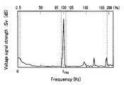
- FIG. 4 shows the frequency spectrum distribution of the voltage signal Sv of the drive signal when cavitation is not generated
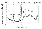
- FIG. 5 shows the frequency spectrum distribution of the voltage signal Sv when cavitation occurs. 4 and 5, the upper side indicates the frequency when the resonance frequency fres is 100%.
- the voltage signal Sv when cavitation is not generated, the voltage signal Sv does not have a large peak at frequencies other than the resonance frequency fres.
- the voltage signal Sv when cavitation occurs, the voltage signal Sv has a higher level at a frequency other than the resonance frequency fres than when no cavitation occurs. That is, when cavitation occurs, it has a subharmonic (SH) frequency peak that is a divisor, such as 1/2 or 1/4 of the resonance frequency fres, or a difference of the divisor, compared to when no cavitation occurs. Levels other than frequency are also higher than when no cavitation occurs. As the cavitation state becomes severe, the level of the voltage signal Sv is significantly different from that when no cavitation occurs, that is, the level increases.
- SH subharmonic
- the detection unit 25 can detect the cavitation state by detecting a signal excluding the vicinity of the resonance frequency fres of the voltage signal Sv of the drive signal as a cavitation level signal.
- a signal obtained by filtering the voltage signal Sv as a cavitation level signal and acquiring (integrating) only the frequency component of 95% of the resonance frequency fres from the frequency of 5% of the resonance frequency fres can be preferably used.
- the cavitation level signal may be a signal obtained by obtaining a frequency component excluding a frequency component of 5% before and after the resonance frequency fres. Further, a signal or peak intensity obtained from the subharmonic (SH) frequency component of the voltage signal Sv as the cavitation level signal can be preferably used.
- SH subharmonic
- the cavitation level signal is not limited to the voltage signal Sv but may be an impedance signal, or may be a current signal when a driving signal whose voltage is controlled by the driving unit 22 is used.
- the cavitation level signal intensity detected by the detection unit 25 varies depending on various conditions such as the type of the liquid 4, the amplitude of the ultrasonic vibration, or the state of the tissue 3, but the type of the liquid 4 and the amplitude of the ultrasonic vibration are different.
- the cavitation level signal strength represents the state of the tissue 3.
- the information acquisition part 27 can acquire the water content information of the tissue 3 based on the cavitation level signal intensity. That is, the cavitation level signal is larger when the moisture content of the tissue 3 is large than when it is small.
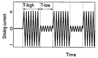
- the drive unit 22 switches the signal intensity of the drive signal supplied to the transducer 35 between high output and low output and outputs it.
- the drive signal with high output signal strength is the signal strength at which cavitation for performing treatment occurs
- the drive signal with low output signal strength is the signal strength at which cavitation does not occur
- the cavitation level signal This is the signal intensity for detecting the decay rate of.
- the high output signal supply time T-high and the low output signal supply time T-low shown in FIG. 6 are appropriately determined.
- the high output signal supply time T-high is 10 ms to 10 seconds
- T-high / (T-high + T-low) is 0.5 to 0.99. If the high output signal supply time T-high is equal to or longer than the above range, cavitation having a sufficient intensity for the detection unit 25 to detect the cavitation level signal is generated. Information can be acquired within a short time interval. Further, when T-high / (T-high + T-low) is greater than or equal to the above range, the treatment efficiency does not decrease, and when it is less than or equal to the above range, the accuracy of information acquired by the information acquisition unit 27 does not decrease. In FIG. 6, waveforms and the like are schematically shown.
- FIG. 7 corresponding to FIG. 6 shows the cavitation level signal when the diseased tissue A is treated
- FIG. 8 shows the cavitation level signal when the normal tissue B is treated.
- the cavitation level signal is a signal obtained (integrated) with a frequency component in the range of 5% to 95% of the resonance frequency fres of the drive voltage signal driven at constant current.
- the cavitation level signal increases because the cavitation bubble 4A is generated.
- the drive signal is switched to a low output, the cavitation bubble 4A is not newly generated and only collapses, so that the cavitation level signal is attenuated.
- the decay rate of the cavitation level signal is faster when the tissue A is treated (FIG. 7) than when the tissue B is treated (FIG. 8). This is because the hardness Hv-A of the tissue A is harder than the hardness Hv-B of the tissue B.
- the ultrasonic surgical apparatus 1 can acquire information on the tissue 3 based on the decay rate of the cavitation level signal.
- the ultrasonic surgical apparatus 1 may be based on the attenuation rate itself, or may use an attenuation rate.
- the attenuation rate is, for example, based on the time until the signal intensity becomes 0.1 when the cavitation level signal intensity immediately after switching the drive signal is 1.0 (10% attenuation time: T 0.1 ). Also good.
- tissue 3 is in inverse proportion to the water content of the structure
- the relationship between the attenuation rate and hardness of the cavitation level signal shown in FIG. 9 is acquired in advance and stored in the memory 24, whereby the information acquisition unit 27 is detected by the detection unit 25. Based on the cavitation level signal thus obtained, information on hardness, which is information on the tissue 3, is acquired. Further, the type discriminating unit 28 discriminates whether the tissue 3 is a normal tissue or a diseased tissue based on the hardness of the tissue 3 acquired by the information acquisition unit 27. For example, as shown in FIG. 9, when the hardness is harder than a predetermined hardness Hv-J, the type determination unit 28 determines that the tissue 3 is a lesioned tissue. The type discriminating unit 28 can also discriminate whether the tissue 3 is a muscle, a real organ, or a fat tissue.
- the ultrasonic operation apparatus 1 has been described in which the drive unit 22 switches and outputs the signal intensity of the drive signal supplied to the vibrator 35 between high output and low output.
- a drive signal may be intermittently supplied to the child 35. That is, when the vibrator 35 stops vibrating, it may take time to start the vibration again. If this time lag does not cause a problem, the drive signal may be supplied intermittently.
- the detection unit 25 uses the vibrator 35 as a sensor and the burst sound of the cavitation bubble 4A as a cavitation level signal when the ultrasonic vibrator 35 stops vibrating. , Sub-harmonic (SH) frequency component signals and the like are detected. Information on the tissue 3 is acquired from the decay rate of the cavitation level signal.
- SH Sub-harmonic
- control unit 23 controls the signal strength of the drive signal supplied by the drive unit 22 according to the information on the tissue 3 acquired by the information acquisition unit 27. That is, when the treatment for removing the lesion tissue is performed, the information acquisition unit 27 notifies the control unit 23 that the lesion tissue has been removed and the normal tissue has been exposed due to the slow decay rate of the cavitation level signal. Then, the control unit 23 controls the signal intensity of the drive signal of the high output signal supplied to the vibrator 35 to be small. For this reason, in the ultrasonic surgical apparatus 1, it is possible to prevent the normal tissue from being damaged.
- the control unit 23 may display the cavitation level signal detected by the detection unit 25 or the information on the tissue 3 acquired by the information acquisition unit 27 on the display unit 29A.
- control unit 23 controls the signal intensity of the drive signal to be small based on the information of the information acquisition unit 27, the display unit 29A and the notification unit 29B, which are also warning generation units, use characters, symbols, voice, light, vibration, or the like. A warning may be issued to the surgeon.
- the ultrasonic surgical apparatus 1 has good operability. Moreover, the ultrasonic surgical apparatus 1 has high safety.
- an ultrasonic surgical apparatus 1A according to a second embodiment of the present invention will be described. Since the ultrasonic surgical apparatus 1A according to the present embodiment is similar to the ultrasonic surgical apparatus 1 according to the first embodiment, components having the same functions are denoted by the same reference numerals and description thereof is omitted.
- the information acquisition unit of the ultrasonic surgical apparatus 1A according to the present embodiment acquires information on the distance D between the tissue 3 and the distal end portion 31 that is the treatment unit based on the strength of the cavitation level signal. As described with reference to FIGS. 2 and 3, the cavitation generation mechanism of the ultrasonic surgical apparatus 1A is significantly different from the cavitation generation mechanism generated by the ultrasonic waves radiated into the liquid.
- the ultrasonic surgical apparatus 1A for example, the relationship between the strength of the cavitation level signal and the distance D shown in FIG. Based on the detected cavitation level signal and the data stored in the memory 24, information on the distance from the tissue 3 to the tip is acquired.
- the control unit 23 of the ultrasonic surgical apparatus 1A reduces the signal strength of the drive signal that the drive unit 22 supplies to the vibrator 35 when the distal end portion 31 approaches the tissue 3 beyond a predetermined distance DL. To control. That is, when the cavitation level signal becomes larger than the predetermined strength SL, the information acquisition unit 27 acquires information that the distal end portion 31 exists at a distance closer to the tissue 3 than the predetermined distance DL. Based on this, the control unit 23 controls the drive unit 22. For this reason, in the ultrasonic surgical apparatus 1A, it is possible to prevent the blood vessel from being damaged. Note that the control unit 23 may increase the signal intensity again when it is detected that the tip 31 that has once approached is separated from the tissue 3 by a predetermined distance DL or more. For this reason, the ultrasonic surgical apparatus 1A has good operability.
- the ultrasonic surgical apparatus 1A based on the distance information acquired by the information acquisition unit 27, for example, by changing the number of LEDs that are turned on in the display unit 29A constituted by a plurality of LEDs, the operator can determine the distance D. It may be displayed easily. Further, when the distal end portion 31 is closer to the tissue 3 than a predetermined distance DL, a warning is given to the operator by letters, symbols, voice, light, vibration, or the like by the display unit 29A and the notification unit 29B that are also warning generation units. It may occur.
- the ultrasonic surgical apparatus 1A has good operability. Moreover, the ultrasonic surgical apparatus 1 has high safety.
- the ultrasonic surgical apparatus 1A performs the treatment inside the blood vessel, for example, arteriosclerosis in which a bulge (plaque) is generated inside the artery. It can also be used to treat the disease.
- a rotablator device can also be used for plaque removal. The rotablator device crushes the plaque by rotating at high speed a drill with a diamond at the tip of a guide wire inserted through the catheter of the endoscope device.
- the suction type ultrasonic surgical apparatus is safer than the rotablator apparatus, but it is not preferable that the distal end portion 31 is in contact with the blood vessel wall.
- the ultrasonic surgical apparatus 1A has a cavitation level signal corresponding to the state of cavitation generated in the liquid, that is, blood existing between the distal end portion 31 and the blood vessel inner wall that is the tissue 3, from the distal end portion 31 that is a treatment portion.
- the distance information to the inner wall of the blood vessel is acquired, and the control unit 23 controls the drive unit 22 according to the distance, so that the operability is excellent and the safety is also high.
- the ultrasonic surgical device 1 ⁇ / b> B is a scissors-type ultrasonic coagulation / cutting device having a device main body 20 and a handpiece 40 ⁇ / b> B connected to the device main body 20 and a cable 42.
- the ultrasonic surgical apparatus 1B further includes a high-frequency output unit 50 and a counter electrode 58 for flowing a high-frequency current from the treatment unit.
- the handpiece 40B includes the probe 30 that can apply a high-frequency current to the distal end portion 31 that is a treatment portion, and can perform a high-frequency current treatment.
- a cylindrical rod-shaped probe 30 is disposed inside the handpiece 40B, and an operation handle 43 for operating the grip portion 45 on the distal end side is disposed on the proximal end side.
- the grasping portion 45 is displaced in a direction to be pressed against the distal end portion 31 by an operation (closing operation) of grasping the operation handle 43 by the operator.
- the surgeon performs treatment on the tissue 3 (not shown in FIG. 14) grasped between the grasping portion 45 and the distal end portion 31 by frictional heat due to ultrasonic vibration.
- the high-frequency output unit 50 of the ultrasonic surgical apparatus 1 ⁇ / b> B has components similar to those of the apparatus body 20, acquires information on the tissue 3, and based on the acquired information on the tissue 3 3 adjusts the strength of the high-frequency current flowing through
- the high frequency output unit 50 includes a high frequency drive unit 52, a detection unit 55, an information acquisition unit 57, a memory 54, a control unit 53, a setting unit 56, a display unit 59A, and a notification unit 59B.
- the control unit 53 of the high frequency output unit 50 is connected to the control unit 23B of the apparatus main body unit 20 via a cable 42C.
- the control unit 53 controls the high frequency output unit 50, whereas the control unit 23B also performs overall control of the ultrasonic surgical apparatus 1B including the high frequency output unit 50.
- the high-frequency current from the high-frequency drive unit 52 is transmitted to the cable 42B via the connector 49A of the handpiece 40B connected to the socket 51 of the high-frequency output unit 50, reaches the distal end portion 31, flows through the tissue 3,
- the counter electrode 58 is reached.
- the high frequency drive unit 52 operates by setting of the setting unit 56 and control of the control unit 53.
- the detection unit 55 detects the electrical impedance of the tissue 3 between the distal end portion 31 and the counter electrode 58
- the information acquisition unit 57 is based on the impedance detected by the detection unit 55 and the data stored in the memory 54. Get information about organization 3.
- the display unit 59A and the notification unit 59B have functions similar to those of the display unit 29A and the notification unit 29B described above.
- the control unit 53 controls the power of the high-frequency current output from the high-frequency driving unit 52 based on the information acquired by the information acquisition unit 57.
- the ultrasonic surgical apparatus 1B not only has an ultrasonic treatment function and a high-frequency current treatment function of a known ultrasonic surgical apparatus or the like, but also an information acquisition function and high-frequency information on the tissue 3 being treated. And an information acquisition function using electric current.
- the information acquired by the information acquisition unit 57 of the high-frequency output unit 50 may not be the same as the information acquired by the information acquisition unit 27 of the apparatus main body unit 20.
- the information acquisition unit 57 of the high-frequency output unit 50 acquires information on the water content in the tissue 3 or type information indicating whether the tissue 3 is a muscle, a real organ, or a fat tissue.
- the information acquisition unit 57 can also acquire information such as the amount of energy administered to the tissue 3, the contact area between the tissue 3 and the distal end portion 31, and whether or not a discharge has occurred.
- the information acquisition unit 27 can also acquire information on a mechanical load such as a pressing force on the tip 31.
- the output (ultrasonic vibration / high frequency) to the probe 30 based on the stress that the tissue 3 acquired by the information acquisition unit 27 exerts on the distal end portion 31 and the contact / non-contact information with the distal end portion 31 of the tissue 3. It is also possible to control the current) and to protect the probe 30.
- the control unit 23B controls the ultrasonic vibration amplitude to be 30% of the maximum amplitude and the high frequency output to be 10 W, and the distal end portion 31 is in contact with the tissue 3. If so, the amplitude of the ultrasonic vibration is controlled to 70% of the maximum amplitude, and the high frequency output is controlled to 30 W.
- the information acquisition unit 27 can acquire a large amount of information with high accuracy when the moisture content of the tissue 3 is small, whereas the information acquisition unit 57 is a wet state when the tissue 3 has a high moisture content. In some cases, information acquisition is easy.
- FIG. 16 shows the relationship between the passage of time during treatment and the electrical resistance of the tissue 3, in other words, the moisture content.
- the tissue 3 is in “state A”, which is a state in which the water content is high and the electrical resistance is low.
- state A is a state in which the water content is high and the electrical resistance is low.
- the water in the tissue 3 is dehydrated, so the water content decreases, the electrical resistance increases, and the state becomes “state B”.
- the ultrasonic surgical apparatus 1B may acquire information on the tissue 3 using the information acquisition unit 57 in the “state A”, and may acquire information on the tissue 3 using the information acquisition unit 27 in the “state B”. preferable.
- the control unit 23 determines whether the information necessary for the treatment among the information acquisition unit 27 or the information acquisition unit 57 is easily acquired or can be acquired with high accuracy.
- At least one of the high-frequency driving unit 52 is controlled. Therefore, the ultrasonic surgical device 1B has better operability in addition to the effects of the known ultrasonic surgical device or the ultrasonic surgical device 1 or the like.
- the transducer 35 of the ultrasonic surgical apparatus to which the drive unit 22 intermittently supplies drive signals can also be used as a sensor.
- a pressing force is applied to the vibrator 35.
- information on the tissue with which the tip 31 is in contact such as hardness information or viscoelasticity information, can be obtained.
- the frequency of the transducer 35 can be swept with a small amplitude, and the impedance characteristics of the tissue can be analyzed to obtain information on the tissue on which the distal end portion 31 is performing treatment.
- an echo signal in which reverberation during driving or a burst sound of a cavitation bubble or the like is reflected back to the tissue 3 in a state where a driving current is not applied can be detected by the vibrator 35. Then, by analyzing the echo signal, the ultrasonic surgical apparatus acquires information on the boundary between the skeleton / organ and the muscle tissue or information on the acoustic impedance change surface such as the boundary between different organs as information on the tissue 3. be able to.
Landscapes
- Health & Medical Sciences (AREA)
- Life Sciences & Earth Sciences (AREA)
- Engineering & Computer Science (AREA)
- Surgery (AREA)
- Biomedical Technology (AREA)
- Nuclear Medicine, Radiotherapy & Molecular Imaging (AREA)
- Animal Behavior & Ethology (AREA)
- General Health & Medical Sciences (AREA)
- Public Health (AREA)
- Veterinary Medicine (AREA)
- Dentistry (AREA)
- Mechanical Engineering (AREA)
- Radiology & Medical Imaging (AREA)
- Heart & Thoracic Surgery (AREA)
- Medical Informatics (AREA)
- Molecular Biology (AREA)
- Surgical Instruments (AREA)
Abstract
La présente invention concerne un appareil de chirurgie ultrasonique (1) pourvu dun oscillateur ultrasonique (35) qui génère une oscillation ultrasonique, une unité de commande (22) qui transmet un signal de commande à loscillateur ultrasonique (35), une section dembout (31) qui est mécaniquement raccordée à loscillateur ultrasonique (35) et traite un tissu biomédical (3) via un liquide (4), une section de détection (25) qui détecte un signal de niveau de cavitation correspondant à létat de cavitation se produisant dans le liquide (4) par loscillation ultrasonique de la section dembout (31), et une section dacquisition dinformations (27) qui acquiert des informations relatives au tissu biomédical (3) sur la base du signal de niveau de cavitation.
Priority Applications (3)
| Application Number | Priority Date | Filing Date | Title |
|---|---|---|---|
| JP2011521721A JP5253576B2 (ja) | 2009-07-06 | 2009-07-06 | 超音波手術装置 |
| PCT/JP2009/062315 WO2011004449A1 (fr) | 2009-07-06 | 2009-07-06 | Appareil de chirurgie ultrasonique |
| US13/307,966 US20120136279A1 (en) | 2009-07-06 | 2011-11-30 | Ultrasound surgical apparatus |
Applications Claiming Priority (1)
| Application Number | Priority Date | Filing Date | Title |
|---|---|---|---|
| PCT/JP2009/062315 WO2011004449A1 (fr) | 2009-07-06 | 2009-07-06 | Appareil de chirurgie ultrasonique |
Related Child Applications (1)
| Application Number | Title | Priority Date | Filing Date |
|---|---|---|---|
| US13/307,966 Continuation US20120136279A1 (en) | 2009-07-06 | 2011-11-30 | Ultrasound surgical apparatus |
Publications (1)
| Publication Number | Publication Date |
|---|---|
| WO2011004449A1 true WO2011004449A1 (fr) | 2011-01-13 |
Family
ID=43428891
Family Applications (1)
| Application Number | Title | Priority Date | Filing Date |
|---|---|---|---|
| PCT/JP2009/062315 WO2011004449A1 (fr) | 2009-07-06 | 2009-07-06 | Appareil de chirurgie ultrasonique |
Country Status (3)
| Country | Link |
|---|---|
| US (1) | US20120136279A1 (fr) |
| JP (1) | JP5253576B2 (fr) |
| WO (1) | WO2011004449A1 (fr) |
Cited By (2)
| Publication number | Priority date | Publication date | Assignee | Title |
|---|---|---|---|---|
| WO2013158545A1 (fr) * | 2012-04-18 | 2013-10-24 | Ethicon Endo-Surgery, Inc. | Instrument chirurgical pourvu d'un capteur de densité tissulaire |
| WO2017171034A1 (fr) * | 2016-03-31 | 2017-10-05 | オリンパス株式会社 | Instrument chirurgical à ultrasons et procédé de traitement utilisant le dispositif chirurgical à ultrasons |
Families Citing this family (92)
| Publication number | Priority date | Publication date | Assignee | Title |
|---|---|---|---|---|
| US10835307B2 (en) | 2001-06-12 | 2020-11-17 | Ethicon Llc | Modular battery powered handheld surgical instrument containing elongated multi-layered shaft |
| US8182501B2 (en) | 2004-02-27 | 2012-05-22 | Ethicon Endo-Surgery, Inc. | Ultrasonic surgical shears and method for sealing a blood vessel using same |
| US20060079879A1 (en) | 2004-10-08 | 2006-04-13 | Faller Craig N | Actuation mechanism for use with an ultrasonic surgical instrument |
| US20070191713A1 (en) | 2005-10-14 | 2007-08-16 | Eichmann Stephen E | Ultrasonic device for cutting and coagulating |
| US7621930B2 (en) | 2006-01-20 | 2009-11-24 | Ethicon Endo-Surgery, Inc. | Ultrasound medical instrument having a medical ultrasonic blade |
| US8057498B2 (en) | 2007-11-30 | 2011-11-15 | Ethicon Endo-Surgery, Inc. | Ultrasonic surgical instrument blades |
| US8808319B2 (en) | 2007-07-27 | 2014-08-19 | Ethicon Endo-Surgery, Inc. | Surgical instruments |
| US8523889B2 (en) | 2007-07-27 | 2013-09-03 | Ethicon Endo-Surgery, Inc. | Ultrasonic end effectors with increased active length |
| US9044261B2 (en) | 2007-07-31 | 2015-06-02 | Ethicon Endo-Surgery, Inc. | Temperature controlled ultrasonic surgical instruments |
| US8430898B2 (en) | 2007-07-31 | 2013-04-30 | Ethicon Endo-Surgery, Inc. | Ultrasonic surgical instruments |
| US8512365B2 (en) | 2007-07-31 | 2013-08-20 | Ethicon Endo-Surgery, Inc. | Surgical instruments |
| US10010339B2 (en) | 2007-11-30 | 2018-07-03 | Ethicon Llc | Ultrasonic surgical blades |
| US9089360B2 (en) | 2008-08-06 | 2015-07-28 | Ethicon Endo-Surgery, Inc. | Devices and techniques for cutting and coagulating tissue |
| US8372100B2 (en) * | 2009-06-19 | 2013-02-12 | Olympus Medical Systems Corp. | Ultrasound surgical apparatus and calibration method therefor |
| US8650728B2 (en) | 2009-06-24 | 2014-02-18 | Ethicon Endo-Surgery, Inc. | Method of assembling a transducer for a surgical instrument |
| US8663220B2 (en) | 2009-07-15 | 2014-03-04 | Ethicon Endo-Surgery, Inc. | Ultrasonic surgical instruments |
| US11090104B2 (en) | 2009-10-09 | 2021-08-17 | Cilag Gmbh International | Surgical generator for ultrasonic and electrosurgical devices |
| US8469981B2 (en) | 2010-02-11 | 2013-06-25 | Ethicon Endo-Surgery, Inc. | Rotatable cutting implement arrangements for ultrasonic surgical instruments |
| US8951272B2 (en) | 2010-02-11 | 2015-02-10 | Ethicon Endo-Surgery, Inc. | Seal arrangements for ultrasonically powered surgical instruments |
| US9439668B2 (en) | 2012-04-09 | 2016-09-13 | Ethicon Endo-Surgery, Llc | Switch arrangements for ultrasonic surgical instruments |
| US11076880B2 (en) | 2012-06-11 | 2021-08-03 | Covidien Lp | Temperature estimation and tissue detection of an ultrasonic dissector from frequency response monitoring |
| US10677764B2 (en) | 2012-06-11 | 2020-06-09 | Covidien Lp | Temperature estimation and tissue detection of an ultrasonic dissector from frequency response monitoring |
| US20140005705A1 (en) | 2012-06-29 | 2014-01-02 | Ethicon Endo-Surgery, Inc. | Surgical instruments with articulating shafts |
| US9351754B2 (en) | 2012-06-29 | 2016-05-31 | Ethicon Endo-Surgery, Llc | Ultrasonic surgical instruments with distally positioned jaw assemblies |
| US9226767B2 (en) | 2012-06-29 | 2016-01-05 | Ethicon Endo-Surgery, Inc. | Closed feedback control for electrosurgical device |
| US9408622B2 (en) | 2012-06-29 | 2016-08-09 | Ethicon Endo-Surgery, Llc | Surgical instruments with articulating shafts |
| US9820768B2 (en) | 2012-06-29 | 2017-11-21 | Ethicon Llc | Ultrasonic surgical instruments with control mechanisms |
| US9198714B2 (en) | 2012-06-29 | 2015-12-01 | Ethicon Endo-Surgery, Inc. | Haptic feedback devices for surgical robot |
| US9393037B2 (en) | 2012-06-29 | 2016-07-19 | Ethicon Endo-Surgery, Llc | Surgical instruments with articulating shafts |
| US9326788B2 (en) | 2012-06-29 | 2016-05-03 | Ethicon Endo-Surgery, Llc | Lockout mechanism for use with robotic electrosurgical device |
| WO2014055906A1 (fr) | 2012-10-05 | 2014-04-10 | The Regents Of The University Of Michigan | Rétroaction par doppler couleur induite par des bulles lors d'une histotripsie |
| US9095367B2 (en) | 2012-10-22 | 2015-08-04 | Ethicon Endo-Surgery, Inc. | Flexible harmonic waveguides/blades for surgical instruments |
| US20140135804A1 (en) | 2012-11-15 | 2014-05-15 | Ethicon Endo-Surgery, Inc. | Ultrasonic and electrosurgical devices |
| US10226273B2 (en) | 2013-03-14 | 2019-03-12 | Ethicon Llc | Mechanical fasteners for use with surgical energy devices |
| WO2015003154A1 (fr) | 2013-07-03 | 2015-01-08 | Histosonics, Inc. | Limiteur de bras d'articulation pour un système de traitement par cavitation aux ultrasons |
| WO2015027164A1 (fr) | 2013-08-22 | 2015-02-26 | The Regents Of The University Of Michigan | Histotripsie au moyen d'impulsions d'ultrasons très courtes |
| GB2521228A (en) | 2013-12-16 | 2015-06-17 | Ethicon Endo Surgery Inc | Medical device |
| GB2521229A (en) * | 2013-12-16 | 2015-06-17 | Ethicon Endo Surgery Inc | Medical device |
| US9554854B2 (en) | 2014-03-18 | 2017-01-31 | Ethicon Endo-Surgery, Llc | Detecting short circuits in electrosurgical medical devices |
| US10092310B2 (en) | 2014-03-27 | 2018-10-09 | Ethicon Llc | Electrosurgical devices |
| US9737355B2 (en) | 2014-03-31 | 2017-08-22 | Ethicon Llc | Controlling impedance rise in electrosurgical medical devices |
| US9913680B2 (en) | 2014-04-15 | 2018-03-13 | Ethicon Llc | Software algorithms for electrosurgical instruments |
| US10285724B2 (en) | 2014-07-31 | 2019-05-14 | Ethicon Llc | Actuation mechanisms and load adjustment assemblies for surgical instruments |
| JP2017538545A (ja) * | 2014-12-15 | 2017-12-28 | ヴェセロン,インコーポレーテッド | 自動超音波非侵襲的血管再疎通処置および監視装置ならびに方法 |
| US10245095B2 (en) | 2015-02-06 | 2019-04-02 | Ethicon Llc | Electrosurgical instrument with rotation and articulation mechanisms |
| US11020140B2 (en) | 2015-06-17 | 2021-06-01 | Cilag Gmbh International | Ultrasonic surgical blade for use with ultrasonic surgical instruments |
| ES2948135T3 (es) | 2015-06-24 | 2023-08-31 | Univ Michigan Regents | Sistemas de terapia de histotripsia para el tratamiento del tejido cerebral |
| US11141213B2 (en) | 2015-06-30 | 2021-10-12 | Cilag Gmbh International | Surgical instrument with user adaptable techniques |
| US11051873B2 (en) | 2015-06-30 | 2021-07-06 | Cilag Gmbh International | Surgical system with user adaptable techniques employing multiple energy modalities based on tissue parameters |
| US10034704B2 (en) | 2015-06-30 | 2018-07-31 | Ethicon Llc | Surgical instrument with user adaptable algorithms |
| US11129669B2 (en) | 2015-06-30 | 2021-09-28 | Cilag Gmbh International | Surgical system with user adaptable techniques based on tissue type |
| US10357303B2 (en) | 2015-06-30 | 2019-07-23 | Ethicon Llc | Translatable outer tube for sealing using shielded lap chole dissector |
| US10194973B2 (en) | 2015-09-30 | 2019-02-05 | Ethicon Llc | Generator for digitally generating electrical signal waveforms for electrosurgical and ultrasonic surgical instruments |
| US10595930B2 (en) | 2015-10-16 | 2020-03-24 | Ethicon Llc | Electrode wiping surgical device |
| US11229471B2 (en) | 2016-01-15 | 2022-01-25 | Cilag Gmbh International | Modular battery powered handheld surgical instrument with selective application of energy based on tissue characterization |
| US10779849B2 (en) | 2016-01-15 | 2020-09-22 | Ethicon Llc | Modular battery powered handheld surgical instrument with voltage sag resistant battery pack |
| US11129670B2 (en) | 2016-01-15 | 2021-09-28 | Cilag Gmbh International | Modular battery powered handheld surgical instrument with selective application of energy based on button displacement, intensity, or local tissue characterization |
| US10555769B2 (en) | 2016-02-22 | 2020-02-11 | Ethicon Llc | Flexible circuits for electrosurgical instrument |
| US10456193B2 (en) | 2016-05-03 | 2019-10-29 | Ethicon Llc | Medical device with a bilateral jaw configuration for nerve stimulation |
| US10245064B2 (en) | 2016-07-12 | 2019-04-02 | Ethicon Llc | Ultrasonic surgical instrument with piezoelectric central lumen transducer |
| US10376305B2 (en) | 2016-08-05 | 2019-08-13 | Ethicon Llc | Methods and systems for advanced harmonic energy |
| USD847990S1 (en) | 2016-08-16 | 2019-05-07 | Ethicon Llc | Surgical instrument |
| US10420580B2 (en) | 2016-08-25 | 2019-09-24 | Ethicon Llc | Ultrasonic transducer for surgical instrument |
| US10952759B2 (en) | 2016-08-25 | 2021-03-23 | Ethicon Llc | Tissue loading of a surgical instrument |
| US11266430B2 (en) | 2016-11-29 | 2022-03-08 | Cilag Gmbh International | End effector control and calibration |
| EP3886737A4 (fr) | 2018-11-28 | 2022-08-24 | Histosonics, Inc. | Systèmes et procédés d'histotrypsie |
| US11684387B2 (en) | 2019-11-25 | 2023-06-27 | Covidien Lp | Methods and ultrasonic devices and systems for vessel sealing |
| US12023086B2 (en) | 2019-12-30 | 2024-07-02 | Cilag Gmbh International | Electrosurgical instrument for delivering blended energy modalities to tissue |
| US11944366B2 (en) | 2019-12-30 | 2024-04-02 | Cilag Gmbh International | Asymmetric segmented ultrasonic support pad for cooperative engagement with a movable RF electrode |
| US11937863B2 (en) | 2019-12-30 | 2024-03-26 | Cilag Gmbh International | Deflectable electrode with variable compression bias along the length of the deflectable electrode |
| US11779329B2 (en) | 2019-12-30 | 2023-10-10 | Cilag Gmbh International | Surgical instrument comprising a flex circuit including a sensor system |
| US11986201B2 (en) | 2019-12-30 | 2024-05-21 | Cilag Gmbh International | Method for operating a surgical instrument |
| US12064109B2 (en) | 2019-12-30 | 2024-08-20 | Cilag Gmbh International | Surgical instrument comprising a feedback control circuit |
| US12082808B2 (en) | 2019-12-30 | 2024-09-10 | Cilag Gmbh International | Surgical instrument comprising a control system responsive to software configurations |
| US11786291B2 (en) | 2019-12-30 | 2023-10-17 | Cilag Gmbh International | Deflectable support of RF energy electrode with respect to opposing ultrasonic blade |
| US11950797B2 (en) | 2019-12-30 | 2024-04-09 | Cilag Gmbh International | Deflectable electrode with higher distal bias relative to proximal bias |
| US11684412B2 (en) | 2019-12-30 | 2023-06-27 | Cilag Gmbh International | Surgical instrument with rotatable and articulatable surgical end effector |
| US11812957B2 (en) | 2019-12-30 | 2023-11-14 | Cilag Gmbh International | Surgical instrument comprising a signal interference resolution system |
| US11759251B2 (en) | 2019-12-30 | 2023-09-19 | Cilag Gmbh International | Control program adaptation based on device status and user input |
| US12076006B2 (en) | 2019-12-30 | 2024-09-03 | Cilag Gmbh International | Surgical instrument comprising an orientation detection system |
| US11452525B2 (en) | 2019-12-30 | 2022-09-27 | Cilag Gmbh International | Surgical instrument comprising an adjustment system |
| US11660089B2 (en) | 2019-12-30 | 2023-05-30 | Cilag Gmbh International | Surgical instrument comprising a sensing system |
| US20210196359A1 (en) | 2019-12-30 | 2021-07-01 | Ethicon Llc | Electrosurgical instruments with electrodes having energy focusing features |
| US11696776B2 (en) | 2019-12-30 | 2023-07-11 | Cilag Gmbh International | Articulatable surgical instrument |
| US11937866B2 (en) | 2019-12-30 | 2024-03-26 | Cilag Gmbh International | Method for an electrosurgical procedure |
| US12053224B2 (en) | 2019-12-30 | 2024-08-06 | Cilag Gmbh International | Variation in electrode parameters and deflectable electrode to modify energy density and tissue interaction |
| US11779387B2 (en) | 2019-12-30 | 2023-10-10 | Cilag Gmbh International | Clamp arm jaw to minimize tissue sticking and improve tissue control |
| US11911063B2 (en) | 2019-12-30 | 2024-02-27 | Cilag Gmbh International | Techniques for detecting ultrasonic blade to electrode contact and reducing power to ultrasonic blade |
| US20210196357A1 (en) | 2019-12-30 | 2021-07-01 | Ethicon Llc | Electrosurgical instrument with asynchronous energizing electrodes |
| CN113117262B (zh) * | 2019-12-30 | 2023-06-02 | 重庆融海超声医学工程研究中心有限公司 | 用于检测空化效应的装置、超声治疗设备 |
| JP2023513012A (ja) | 2020-01-28 | 2023-03-30 | ザ リージェンツ オブ ザ ユニバーシティー オブ ミシガン | ヒストトリプシー免疫感作のためのシステムおよび方法 |
| US20230059298A1 (en) * | 2021-08-18 | 2023-02-23 | Chevron U.S.A. Inc. | Cavitation detection system and method |
Citations (3)
| Publication number | Priority date | Publication date | Assignee | Title |
|---|---|---|---|---|
| JP2002537955A (ja) * | 1999-03-08 | 2002-11-12 | アンジオソニックス インコーポレーテッド | 二重変換機超音波溶解法および装置 |
| JP2005040222A (ja) * | 2003-07-24 | 2005-02-17 | Olympus Corp | 超音波処置装置 |
| JP2005506867A (ja) * | 2001-10-24 | 2005-03-10 | カッティング エッジ サージカル, インコーポレイテッド | 外科移植の間の骨内超音波 |
Family Cites Families (3)
| Publication number | Priority date | Publication date | Assignee | Title |
|---|---|---|---|---|
| US20040034340A1 (en) * | 1999-10-13 | 2004-02-19 | Spineco, Inc., An Ohio Corporation | Smart dissector |
| JPWO2005094701A1 (ja) * | 2004-03-31 | 2008-02-14 | 株式会社東京大学Tlo | 超音波照射方法及び超音波照射装置 |
| US8512365B2 (en) * | 2007-07-31 | 2013-08-20 | Ethicon Endo-Surgery, Inc. | Surgical instruments |
-
2009
- 2009-07-06 JP JP2011521721A patent/JP5253576B2/ja not_active Expired - Fee Related
- 2009-07-06 WO PCT/JP2009/062315 patent/WO2011004449A1/fr active Application Filing
-
2011
- 2011-11-30 US US13/307,966 patent/US20120136279A1/en not_active Abandoned
Patent Citations (3)
| Publication number | Priority date | Publication date | Assignee | Title |
|---|---|---|---|---|
| JP2002537955A (ja) * | 1999-03-08 | 2002-11-12 | アンジオソニックス インコーポレーテッド | 二重変換機超音波溶解法および装置 |
| JP2005506867A (ja) * | 2001-10-24 | 2005-03-10 | カッティング エッジ サージカル, インコーポレイテッド | 外科移植の間の骨内超音波 |
| JP2005040222A (ja) * | 2003-07-24 | 2005-02-17 | Olympus Corp | 超音波処置装置 |
Cited By (6)
| Publication number | Priority date | Publication date | Assignee | Title |
|---|---|---|---|---|
| WO2013158545A1 (fr) * | 2012-04-18 | 2013-10-24 | Ethicon Endo-Surgery, Inc. | Instrument chirurgical pourvu d'un capteur de densité tissulaire |
| AU2013249514B2 (en) * | 2012-04-18 | 2017-08-31 | Ethicon Endo-Surgery, Inc. | Surgical instrument with tissue density sensing |
| US9788851B2 (en) | 2012-04-18 | 2017-10-17 | Ethicon Llc | Surgical instrument with tissue density sensing |
| US10653437B2 (en) | 2012-04-18 | 2020-05-19 | Ethicon Llc | Surgical instrument with tissue density sensing |
| WO2017171034A1 (fr) * | 2016-03-31 | 2017-10-05 | オリンパス株式会社 | Instrument chirurgical à ultrasons et procédé de traitement utilisant le dispositif chirurgical à ultrasons |
| US10881425B2 (en) | 2016-03-31 | 2021-01-05 | Olympus Corporation | Ultrasonic surgical instrument and processing method for ultrasonic surgical device |
Also Published As
| Publication number | Publication date |
|---|---|
| JPWO2011004449A1 (ja) | 2012-12-13 |
| JP5253576B2 (ja) | 2013-07-31 |
| US20120136279A1 (en) | 2012-05-31 |
Similar Documents
| Publication | Publication Date | Title |
|---|---|---|
| JP5253576B2 (ja) | 超音波手術装置 | |
| JP4855541B2 (ja) | 超音波手術装置、前記超音波手術装置を具備する超音波手術システム及びキャビテーション利用方法 | |
| JP4741035B2 (ja) | 超音波手術装置、及び前記超音波手術装置のキャビテーション制御方法 | |
| US10639058B2 (en) | Ultrasonic surgical instrument with features for forming bubbles to enhance cavitation | |
| JP4950342B2 (ja) | 超音波手術装置、超音波手術システム及びキャビテーション抑制方法 | |
| US8740821B2 (en) | Medical apparatus | |
| JP5963505B2 (ja) | 超音波治療装置 | |
| ES2831160T3 (es) | Aparato quirúrgico ultrasónico | |
| JP4768883B2 (ja) | 超音波手術装置および超音波手術装置のキャリブレーション方法 | |
| JP3699825B2 (ja) | 超音波手術装置 | |
| JP4040914B2 (ja) | 超音波手術装置 | |
| JPH03151957A (ja) | 超音波処置装置 | |
| JP2004141397A (ja) | 超音波砕石装置及び超音波砕石装置を備えた砕石システム | |
| JP2002153482A (ja) | 超音波治療装置 | |
| JP4147064B2 (ja) | 超音波処置装置 | |
| CN117241746A (zh) | 用于控制治疗性超声波介入系统的方法 |
Legal Events
| Date | Code | Title | Description |
|---|---|---|---|
| 121 | Ep: the epo has been informed by wipo that ep was designated in this application |
Ref document number: 09847054 Country of ref document: EP Kind code of ref document: A1 |
|
| WWE | Wipo information: entry into national phase |
Ref document number: 2011521721 Country of ref document: JP |
|
| NENP | Non-entry into the national phase |
Ref country code: DE |
|
| 122 | Ep: pct application non-entry in european phase |
Ref document number: 09847054 Country of ref document: EP Kind code of ref document: A1 |