WO2010113615A1 - 医用画像処理装置、医用画像のグループ化方法及びプログラム - Google Patents
医用画像処理装置、医用画像のグループ化方法及びプログラム Download PDFInfo
- Publication number
- WO2010113615A1 WO2010113615A1 PCT/JP2010/054081 JP2010054081W WO2010113615A1 WO 2010113615 A1 WO2010113615 A1 WO 2010113615A1 JP 2010054081 W JP2010054081 W JP 2010054081W WO 2010113615 A1 WO2010113615 A1 WO 2010113615A1
- Authority
- WO
- WIPO (PCT)
- Prior art keywords
- medical image
- imaging
- input
- examination
- patient
- Prior art date
- Legal status (The legal status is an assumption and is not a legal conclusion. Google has not performed a legal analysis and makes no representation as to the accuracy of the status listed.)
- Ceased
Links
Images





Classifications
-
- A—HUMAN NECESSITIES
- A61—MEDICAL OR VETERINARY SCIENCE; HYGIENE
- A61B—DIAGNOSIS; SURGERY; IDENTIFICATION
- A61B6/00—Apparatus or devices for radiation diagnosis; Apparatus or devices for radiation diagnosis combined with radiation therapy equipment
- A61B6/56—Details of data transmission or power supply, e.g. use of slip rings
-
- A—HUMAN NECESSITIES
- A61—MEDICAL OR VETERINARY SCIENCE; HYGIENE
- A61B—DIAGNOSIS; SURGERY; IDENTIFICATION
- A61B6/00—Apparatus or devices for radiation diagnosis; Apparatus or devices for radiation diagnosis combined with radiation therapy equipment
- A61B6/52—Devices using data or image processing specially adapted for radiation diagnosis
- A61B6/5211—Devices using data or image processing specially adapted for radiation diagnosis involving processing of medical diagnostic data
- A61B6/5229—Devices using data or image processing specially adapted for radiation diagnosis involving processing of medical diagnostic data combining image data of a patient, e.g. combining a functional image with an anatomical image
- A61B6/5235—Devices using data or image processing specially adapted for radiation diagnosis involving processing of medical diagnostic data combining image data of a patient, e.g. combining a functional image with an anatomical image combining images from the same or different ionising radiation imaging techniques, e.g. PET and CT
-
- G—PHYSICS
- G16—INFORMATION AND COMMUNICATION TECHNOLOGY [ICT] SPECIALLY ADAPTED FOR SPECIFIC APPLICATION FIELDS
- G16H—HEALTHCARE INFORMATICS, i.e. INFORMATION AND COMMUNICATION TECHNOLOGY [ICT] SPECIALLY ADAPTED FOR THE HANDLING OR PROCESSING OF MEDICAL OR HEALTHCARE DATA
- G16H10/00—ICT specially adapted for the handling or processing of patient-related medical or healthcare data
- G16H10/60—ICT specially adapted for the handling or processing of patient-related medical or healthcare data for patient-specific data, e.g. for electronic patient records
-
- G—PHYSICS
- G16—INFORMATION AND COMMUNICATION TECHNOLOGY [ICT] SPECIALLY ADAPTED FOR SPECIFIC APPLICATION FIELDS
- G16H—HEALTHCARE INFORMATICS, i.e. INFORMATION AND COMMUNICATION TECHNOLOGY [ICT] SPECIALLY ADAPTED FOR THE HANDLING OR PROCESSING OF MEDICAL OR HEALTHCARE DATA
- G16H30/00—ICT specially adapted for the handling or processing of medical images
- G16H30/20—ICT specially adapted for the handling or processing of medical images for handling medical images, e.g. DICOM, HL7 or PACS
-
- G—PHYSICS
- G16—INFORMATION AND COMMUNICATION TECHNOLOGY [ICT] SPECIALLY ADAPTED FOR SPECIFIC APPLICATION FIELDS
- G16H—HEALTHCARE INFORMATICS, i.e. INFORMATION AND COMMUNICATION TECHNOLOGY [ICT] SPECIALLY ADAPTED FOR THE HANDLING OR PROCESSING OF MEDICAL OR HEALTHCARE DATA
- G16H30/00—ICT specially adapted for the handling or processing of medical images
- G16H30/40—ICT specially adapted for the handling or processing of medical images for processing medical images, e.g. editing
-
- A—HUMAN NECESSITIES
- A61—MEDICAL OR VETERINARY SCIENCE; HYGIENE
- A61B—DIAGNOSIS; SURGERY; IDENTIFICATION
- A61B6/00—Apparatus or devices for radiation diagnosis; Apparatus or devices for radiation diagnosis combined with radiation therapy equipment
- A61B6/50—Apparatus or devices for radiation diagnosis; Apparatus or devices for radiation diagnosis combined with radiation therapy equipment specially adapted for specific body parts; specially adapted for specific clinical applications
- A61B6/502—Apparatus or devices for radiation diagnosis; Apparatus or devices for radiation diagnosis combined with radiation therapy equipment specially adapted for specific body parts; specially adapted for specific clinical applications for diagnosis of breast, i.e. mammography
-
- A—HUMAN NECESSITIES
- A61—MEDICAL OR VETERINARY SCIENCE; HYGIENE
- A61B—DIAGNOSIS; SURGERY; IDENTIFICATION
- A61B6/00—Apparatus or devices for radiation diagnosis; Apparatus or devices for radiation diagnosis combined with radiation therapy equipment
- A61B6/52—Devices using data or image processing specially adapted for radiation diagnosis
- A61B6/5211—Devices using data or image processing specially adapted for radiation diagnosis involving processing of medical diagnostic data
- A61B6/5217—Devices using data or image processing specially adapted for radiation diagnosis involving processing of medical diagnostic data extracting a diagnostic or physiological parameter from medical diagnostic data
-
- A—HUMAN NECESSITIES
- A61—MEDICAL OR VETERINARY SCIENCE; HYGIENE
- A61B—DIAGNOSIS; SURGERY; IDENTIFICATION
- A61B6/00—Apparatus or devices for radiation diagnosis; Apparatus or devices for radiation diagnosis combined with radiation therapy equipment
- A61B6/52—Devices using data or image processing specially adapted for radiation diagnosis
- A61B6/5211—Devices using data or image processing specially adapted for radiation diagnosis involving processing of medical diagnostic data
- A61B6/5229—Devices using data or image processing specially adapted for radiation diagnosis involving processing of medical diagnostic data combining image data of a patient, e.g. combining a functional image with an anatomical image
- A61B6/5247—Devices using data or image processing specially adapted for radiation diagnosis involving processing of medical diagnostic data combining image data of a patient, e.g. combining a functional image with an anatomical image combining images from an ionising-radiation diagnostic technique and a non-ionising radiation diagnostic technique, e.g. X-ray and ultrasound
Definitions
- the present invention relates to a medical image processing apparatus, a medical image grouping method, and a program.
- One of the medical images obtained by examination imaging is a medical image (called mammography) in which the breast is imaged.
- CAD Computer-Aided Diagnosis / Detection
- the obtained mammography is image-analyzed to detect possible lesion areas such as tumors and microcalcification clusters, Information can be provided to the doctor.
- Mammography is often performed by changing the imaging direction for each of the left and right breasts, and a plurality of mammography images are generally obtained by one examination imaging.
- the doctor examines the left and right breast mammography side-by-side and interprets them to determine the presence or absence of the lesion and to confirm the position and state of the lesion.
- the lesion candidate may be detected by comparing the mammography of the left and right breasts.
- Patent Document 1 may not be able to group well.
- inspection imaging imaging that is different from the original schedule is often performed. Originally planned to shoot only in one direction, but it was judged that diagnosis was difficult in one direction, and when taking additional shots by switching to two-way shooting at the time of shooting, or when checking medical images after shooting, deficiencies were discovered, This is a case of re-shooting.
- medical images obtained by such unscheduled imaging are not registered in the reference table, they are grouped into separate examination groups.
- An object of the present invention is to enable grouping of medical images obtained by photographing other than scheduled photographing.
- the control unit for grouping the input medical image for each same examination of the same patient and storing it in a storage unit,
- the control means collates the input medical image with the supplementary information of the medical image that has already been grouped, so that the input medical image is additionally captured or If it is determined whether or not it was obtained by re-imaging, and if it is determined that it was obtained by additional imaging or re-imaging, the input medical image and the grouped medical image using the supplementary information are the same patient
- a medical image processing apparatus for regrouping every same examination.
- the control means determines that additional imaging is performed when the incidental information of the imaging region and the imaging direction is inconsistent with the incidental information of the imaging region and the imaging direction when the input medical image matches the patient and examination information of the medical image that has been grouped.
- the medical image processing apparatus according to claim 1, wherein the input medical images are classified and regrouped into a group in which the grouped medical images and the incidental information of the patient and examination match.
- the control means determines that re-imaging has been performed when the incidental information of the patient, examination, imaging region, and imaging direction of the input medical image and the medical image for which the grouping has been completed matches, and the grouping is completed.
- the medical image that is the medical image that has the input medical image and the incidental information of the patient, the examination, the imaging region, and the imaging direction coincides with the input medical image and is regrouped.
- a medical image processing apparatus is provided.
- a method for grouping medical images by a medical image processing apparatus Using the incidental information of the input medical image by the control means, grouping the input medical images for the same examination of the same patient and storing them in the storage means;
- the input medical image is additionally captured or re-imaged by collating the input medical image with the supplementary information of the medical image that has already been grouped.
- the control means determines the image has been obtained by additional imaging or re-imaging
- the input medical image and the grouped medical image are regrouped for the same examination of the same patient using the supplementary information.
- a process of A method for grouping medical images is provided.
- the input medical images are grouped for each same examination of the same patient and stored in the storage means,
- the input medical image is obtained by additional imaging or re-imaging by collating the input medical image with the supplementary information of the medical image that has already been grouped.
- a program for functioning as a server is provided.
- the medical image that has already been grouped and the input medical image can be regrouped together. it can.
- regrouping medical images can be correctly grouped and used for lesion candidate detection processing for each same examination of the same patient, and the accuracy of lesion candidate detection processing is improved.
- FIG. 1 shows a configuration of a medical image system 1 including a medical image processing apparatus 3 in the present embodiment.
- the medical image system 1 includes an imaging system 2, a medical image processing device 3, and an image server 4.
- the imaging system 2, the medical image processing apparatus 3, and the image server 4 are each connected to the network N1.
- the network N1 is, for example, a local area network (LAN) or the Internet, and DICOM (digital imaging and communication in medicine) is used as a communication standard.
- LAN local area network
- DICOM digital imaging and communication in medicine
- the photographing system 2 includes a photographing device 21, a reading device 22, and a console 23.
- the photographing device 21, the reading device 22, and the console 23 are each connected to the network N2.
- the network N2 is a LAN, for example, and DICOM is adopted as in the network N1.
- the imaging device 21 is a mammography imaging device.
- the imaging device 21 performs imaging processing according to instructions from the console 23.
- the photographing method of the photographing device 21 may be either a cassette method or an FPD (Flat Panel Detector) method.
- the cassette method is an imaging method using a cassette containing a volatile phosphor plate. The photographer replaces the cassette for each imaging, and the reading device 22 is loaded with the cassette to perform a medical image reading process. There is a need.
- the FPD method is an imaging method using an FPD in which detectors for converting X-ray energy into electric signals are provided in a matrix. Since a medical image is generated by the FPD, replacement and reading processing by the reading device 22 are unnecessary. In the case of the FPD method, the imaging device 21 transmits a medical image generated by the FPD to the console 23.
- the reading device 22 executes a cassette reading process and generates a medical image.
- the reading device 22 irradiates a phosphor plate built in the cassette with laser light, and photoelectrically converts excitation light from the phosphor plate to generate an image signal.
- the reading device 22 transmits to the console 23 a medical image obtained by performing A / D conversion and various signal processing on the image signal.
- the console 23 is a computer including, for example, a control unit, a display unit, an operation unit, a communication unit, and a storage unit, and is used for a photographing operation of a photographer.
- the console 23 holds order information in which information on a patient to be examined and information on examination imaging such as an imaging region and an imaging direction are defined, and displays a list of order information before imaging.
- the photographer can prepare for photographing according to the order information selected from the list, and can perform an operation of starting photographing using the console 23.
- the photographing apparatus 21 performs a photographing process. After the imaging process, a medical image is input to the console 23 from the reading device 22 in the cassette method, and a medical image is input from the imaging device 21 in the FPD method.
- the console 23 creates incidental information of the input medical image based on the order information in accordance with DICOM.
- the incidental information includes patient information such as an image number, a patient ID of a patient, a name, an age, a gender, and a receipt number issued at the time of receipt, and examination information such as an examination ID and examination name.
- the incidental information includes imaging date and time, type of imaging apparatus (for example, mammography, CR (Computed Radiography), MRI (Magnetic Resonance Imaging), CT (Computed Tomography)), imaging region, imaging direction, presence / absence of contrast medium Information on shooting is also included.
- the image number is identification information that is assigned to identify each medical image.
- the console 23 outputs a medical image with accompanying information to the medical image processing apparatus 3 and the image server 4.
- the medical image processing apparatus 3 executes a lesion candidate detection process on the medical image input from the console 23, and detects a lesion candidate area included in the medical image. Information about the detection result is transmitted to the image server 4.
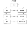
- FIG. 2 is a diagram illustrating a functional configuration of the medical image processing apparatus 3.
- the medical image processing apparatus 3 includes a control unit 31, an operation unit 32, a display unit 33, a communication unit 34, a first storage unit 35, a second storage unit 36, and a lesion candidate detection unit 37. It is configured.
- the control unit 31 is a control means including a CPU (Central Processing Unit) and a RAM (Random Access Memory).
- the control unit 31 executes various processes in cooperation with the program stored in the first storage unit 35. In the processing, the control unit 31 performs various calculations and centrally controls each unit of the medical image processing apparatus 3.
- the control unit 31 uses the incidental information of the input medical image to group the input medical images for each same examination of the same patient, and stores the grouped medical images in the second storage unit 36. .
- the control unit 31 collates the input medical image with the supplementary information of the medical image that has already been grouped, so that the input medical image is additionally captured or If it is determined whether or not it was obtained by re-imaging, and if it is determined that it was obtained by additional imaging or re-imaging, the input medical image and the grouped medical image using the supplementary information are the same patient Regroup for each identical examination.
- the operation unit 32 includes a keyboard and a mouse, generates operation signals corresponding to these operations, and outputs them to the control unit 31.
- a touch panel may be used.
- the display unit 33 includes a display, and displays an operation screen and a medical image according to display control of the control unit 31.
- the communication unit 34 includes a communication interface and communicates with external devices on the networks N1 and N2. For example, a medical image is received from the console 23 and information on detection results of lesion candidates is transmitted to the image server 4.
- the first storage unit 35 stores programs and files and data necessary for executing the programs.
- a hard disk can be used as the first storage unit 35.
- the second storage unit 36 is a storage unit that temporarily stores a medical image input to the medical image processing apparatus 3.
- a RAM can be used as the second storage unit 36.
- the second storage unit 36 stores a medical image DB (Data Base) stored in the second storage unit 36, and can manage the medical image stored by the DB.
- the DB is updated by the control unit 31 every time a medical image is stored.
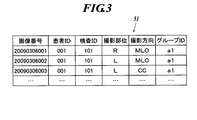
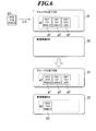
- 3 and 4 show an example of the DB.
- incidental information of medical images that have been grouped among the medical images stored in the second storage unit 36 is registered, and the new examination DB 52 shown in FIG. 4 has not been grouped yet.
- the incidental information of the medical image is registered.
- the registration destination of the incidental information is simply changed to the grouping completion DB 51 or the new examination DB 52, and the registration contents of the incidental information in the grouping completion DB 51 and the new examination DB 52 are the same. is there.
- the grouping completion DB 51 and the new examination DB 52 include the medical image image number, patient ID, examination ID, imaging region, and imaging direction incidental information stored in the second storage unit 36. Is registered.
- R in the imaging region indicates the left breast
- L indicates the right breast.
- the MLO in the shooting direction indicates the oblique direction
- CC indicates the front direction.
- group IDs of groups into which stored medical images are classified are registered.
- the grouping completion DB 51 indicates that the three images with the image numbers 20090306001 to 2009030603 are classified into the group with the same group ID “a1”.
- the lesion candidate detection unit 37 is a lesion candidate detection unit that analyzes a medical image and detects a region of a lesion candidate from the medical image.
- a method for detecting a lesion candidate is not particularly limited, and a method corresponding to the feature of a lesion part to be detected may be used.
- a method for detecting a candidate region of a tumor as a lesion a method using a Laplacian filter in addition to a method using an iris filter disclosed in Japanese Patent Application Laid-Open No. 10-91758 (a journal of the Institute of Electrical, Information and Communication Engineers) (D-II), Vol. J76-D-II, no. 2, pp241-249, 1993).
- a method is also disclosed in which medical images of left and right breasts are compared and false-positive candidate regions are deleted (Kasai et al., “Left and right breasts in tumor shadow automatic detection algorithm”. "Deleting false positive candidates by comparing images", Medical Imaging Technology, Vol. 16, No. 6, 1998).
- a method of detecting microcalcification clusters for example, a method using a morphological filter (The Institute of Electrical, Information and Communication Engineers (D-II), Vol.J71-D-II, no.7, pp1170-1176, 1992), Laplacian Filter (The Institute of Electrical, Information and Communication Engineers Journal (D-II), Vol. J71-D-II, no. 10, pp 1994-2001, 1998)
- D-II The Institute of Electrical, Information and Communication Engineers Journal
- D-II Vol. J71-D-II, no. 10, pp 1994-2001, 1998)
- a method using a triple ring filter can be mentioned.
- the microcalcification cluster appears as an image in which minute image portions having a density change in a substantially conical shape at a low density are aggregated (clustered).
- the lesion candidate detection unit 37 performs a filtering process on the medical image with a triple ring filter for each square fixed region.
- the triple ring filter is a ring filter in which the intensity component and direction component of the density gradient when the density change shows an ideal cone shape are set as a vector pattern. It is composed of three ring filters in which different vector patterns are set from the periphery to the center.
- An image region having a density change close to a conical shape is detected as a candidate region of a microcalcification cluster by the filtering process.
- the lesion candidate detection unit 37 performs the above filtering process on the left and right breast medical images having the same imaging direction as one set, and compares the filtered medical images on the left and right sides.
- the lesion candidate detection unit 37 compares the regions where the lesion candidates are detected on the left and right sides, and determines whether or not the feature amounts of the regions substantially match.
- the feature amount is, for example, contrast, average value of pixel values, standard deviation, area, circularity, and the like. If the feature amounts compared on the left and right sides are substantially the same, the lesion candidate detection unit 37 determines that the detected lesion candidate is false positive and deletes it from the detection result. On the other hand, if the feature amounts are not substantially the same, the lesion candidate detection unit 37 determines that the detected lesion candidate is true positive and outputs it as a detection result.
- the image server 4 stores and manages the medical image together with the detection result of the lesion candidate by the medical image processing apparatus 3.
- the image server 4 is incorporated in, for example, a PACS (Picture Archiving and Communication System), and distributes medical images and detection results of lesion candidates to an interpretation terminal (not shown).
- PACS Picture Archiving and Communication System
- the grouping process executed by the medical image processing apparatus 3 will be described with reference to FIG.
- the grouping process is a process of grouping medical images to be subjected to lesion candidate detection processing for each same examination of the same patient.
- the control unit 31 stores the input medical image in the second storage unit 36. Save (step S2).
- the control unit 31 searches the grouping completion DB 51 to determine whether there is a medical image that has been grouped and has a matching patient ID and examination ID (step S3).
- the control unit 31 determines that the input medical image is an image for a new examination (step S4).
- the same patient ID and examination ID as the input medical image are registered in the grouping completion DB 51, and there is a medical image in which the patient ID and examination ID match in the grouped medical image (step S3; Y)
- the control unit 31 determines whether or not the imaging part and the imaging direction of the medical image and the input medical image match (step S5).
- step S5; N If the imaging region and the imaging direction do not match (step S5; N), it is determined that the input medical image is an additionally captured image (step S6).
- step S5; Y the control unit 31 determines that the input medical image is a re-imaged medical image (step S7).
- the control unit 31 searches the new examination DB 52 to determine whether there is a medical image that has not been grouped and that has matching information of the patient ID and the examination ID (step S8).
- the same patient ID and examination ID as the inputted medical image are registered in the new examination DB 52 and there is a medical image with the matching patient ID and examination ID (step S8; Y), the control unit 31 has been inputted.
- the incidental information such as the image number of the medical image and the patient ID is registered in the new examination DB 52.
- control unit 31 registers the same group ID as the medical image in which the incidental information of the patient ID and the examination ID matches as the group ID of the input medical image in the new examination DB 52. Thereby, the input medical images are classified into the same group as the medical images in which the patient ID and the examination ID match in the new examination DB 52 (step S9).
- the control unit 31 When the same patient ID and examination ID as the inputted medical image are not registered in the new examination DB 52 and there is no medical image with the matching patient ID and examination ID (step S8; N), the control unit 31 has been inputted.
- the incidental information of the medical image is registered in the new examination DB 52, and is classified into a new group with a new group ID (step S10).
- FIG. 6 shows a specific example of grouping when it is determined as a new examination.
- a medical image g1 imaging site: R, imaging direction: MLO
- medical image g2 imaging site: L, imaging direction: MLO
- medical image g3 having a patient ID of 001 and an examination ID of 101.
- Image capturing part: L, image capturing direction: CC is classified into one group (group ID: a1) and registered in the grouping completion DB 51.
- a new medical image G1 patient ID: 002, examination ID: 102, imaging region: R, imaging direction CC
- the input medical image G1 and the medical images g1, g2, and g3 that have been grouped do not match the patient ID and the examination ID.
- a medical image whose patient ID and examination ID match the medical image G1 input also in the new examination DB 52 is not registered. Therefore, the input medical image G1 is classified into a new group (group ID: b1) and registered in the new examination DB 52.
- the medical image G1 matches the patient ID and examination ID, so the newly input medical image is the same group as the medical image G1.
- the ID is classified into a group of b1 and registered in the new inspection DB 52.
- the control unit 31 determines whether or not there is a new examination that has been grouped (step S11). For example, when a medical image with a patient ID different from a medical image already registered in the new examination DB 52 is input and newly registered in the new examination DB 52, grouping of the already registered medical images is completed. Judge. The same determination may be made based on the examination ID or the reception number instead of the patient ID. Alternatively, a table indicating in advance how many medical images are scheduled to be input for each patient ID and examination ID, and the number of medical images shown in this table are grouped and registered in the new examination DB 52. If so, it may be determined that the grouping has been completed.
- step S11; N If there is no new inspection for which grouping has been completed (step S11; N), this processing is terminated. On the other hand, when there is a new examination for which grouping has been completed (step S11; Y), the control unit 31 moves registration information of medical images belonging to the group for which grouping has been completed from the new examination DB 52 to the grouping completion DB 51 ( Step S12), the process ends. Thereafter, if there is a newly input medical image, the process starts from step S1.
- FIG. 7 shows a specific example of a new examination in which grouping is completed.
- the medical images G1 and G2 with the patient ID 002 and the examination ID 102 are classified into one group (group ID: b1) and registered in the new examination DB 52.
- group ID: b1 group ID: b1
- a new medical image Gn patient ID: 005, examination ID: 106, imaging region: R, imaging direction MLO
- the input medical image Gn does not have the same patient ID and examination ID as any of the medical images g1 to g3 registered in the grouping completion DB 51 and the medical images G1 and G2 registered in the new examination DB 52.
- the input medical image Gn is classified into a new group (group ID: b2) and registered in the new examination DB 52.
- group ID: b2 group ID
- the medical image Gn having a different patient ID from the already registered medical images G1 and G2 is registered in the new examination DB 52, it is determined that the grouping of the medical images G1 and G2 has been completed.
- the registration information is moved to the grouping completion DB 51 as shown in FIG.
- the control unit 31 registers the incidental information of the input medical image in the grouping completion DB 51, and registers the same group ID as the medical image in which the patient ID and the examination ID match in the grouping completion DB 51. To do.
- the input medical images are medical images that have been grouped, and are classified into the same group as the medical images having the same patient ID and examination ID, and are regrouped (step S13). After regrouping, this process ends. If there is a newly input medical image, the process starts from step S1.
- FIG. 8 shows a specific example when it is determined that additional shooting is performed.
- a medical image g1 imaging site: R, imaging direction: MLO
- medical image g2 imaging site: L, imaging direction: MLO
- medical image g3 having a patient ID of 001 and an examination ID of 101.
- Image capturing part: L, image capturing direction: CC is classified into one group (group ID: a1) and registered in the grouping completion DB 51.
- a new medical image g4 patient ID: 001, examination ID: 101, imaging region: R, imaging direction CC
- the input medical image g4 and grouped medical images g1, g2, and g3 have the same patient ID and examination ID, but the imaging site and imaging direction do not match. Accordingly, the input medical image g4 is assigned the same group ID: a1 as the medical images g1 to g3 and is registered in the grouping completion DB 51. Thereby, the group of group ID: a1 is regrouped.
- the control unit 31 registers the incidental information of the input medical image in the grouping completion DB 51, and the medical image in which the patient ID, examination ID, imaging region, and imaging direction match in the grouping completion DB 51. The same group ID is registered.
- the control unit 31 deletes from the grouping completion DB 51 the incidental information of the medical image that matches the input medical image with the patient ID, examination ID, imaging region, and imaging direction. Thereby, the input medical image and the medical image whose patient ID, examination ID, imaging region, and imaging direction match are replaced with the input medical image and regrouped (step S14). After regrouping, this process ends. If there is a newly input medical image, the process starts from step S1.
- FIG. 9 shows a specific example when it is determined that re-photographing is performed.
- a medical image g1 imaging region: R, imaging direction: MLO
- medical image g2 imaging region: L, imaging direction: MLO
- medical image g3 having a patient ID of 001 and an examination ID of 101.
- medical image g4 imaging region: R, imaging direction: CC
- group ID: a1 group ID: a1
- the input medical image g5 and the grouped medical image g4 have the same patient ID, examination ID, imaging region, and imaging direction. Therefore, the input medical image g5 is replaced with the medical image g4 and registered in the grouping completion DB 51. Thereby, the group of group ID: a1 is regrouped.
- the medical images input to the medical image processing apparatus 3 are grouped, but no new medical image is input even after a predetermined time has elapsed (steps S1; N, S15; Y).
- the control unit 31 closes reception of new examinations.
- the control unit 31 determines that grouping of medical images currently registered in the new examination DB 52 has been completed, and registers registration information of medical images belonging to the group for which grouping has been completed from the new examination DB 52 to the grouping completion DB 51. Transfer (step S12), the process is terminated. Thereafter, if there is a newly input medical image, the process starts from step S1.
- the control unit 31 After the grouping process, the control unit 31 outputs the medical image stored in the second storage unit 36 to the lesion candidate detection unit 37. At this time, the control unit 31 refers to the group ID of the grouping completion DB 51, reads out medical images belonging to the same group, that is, medical images with the same patient and examination from the second storage unit 36, and detects lesion candidates in units of groups. To the unit 37.
- the lesion candidate detection unit 37 uses medical images in units of groups for the lesion candidate detection process.
- the control unit 31 displays the regrouped medical images. It outputs to the lesion candidate detection part 37 in a group unit.
- the lesion candidate detection unit 37 executes the lesion candidate detection process again on the regrouped medical images.
- the supplementary information of the medical image that has been grouped and registered in the grouping completion DB 51 by the control unit 31 and the incidental ID of the patient ID and examination ID of the input medical image The information is checked, and if it does not match, it is determined as a new inspection.
- the control unit 31 collates the incidental information of the medical image registered in the new examination DB 52 with the incidental information of the patient ID and examination ID of the input medical image. If they match, the control unit 31 classifies the input images into the corresponding group of medical images, and if they do not match, classifies the images into a new group and groups them. As a result, medical images captured and input as scheduled can be grouped for each same examination of the same patient.
- the control unit 31 determines that the image has been re-photographed. In this case, the control unit 31 classifies the input medical images into a group of medical images that have been grouped and have the same patient ID and examination ID, and regroups them.
- the control unit 31 judges that it was re-photographed. In this case, the control unit 31 classifies the input medical images into groups of medical images that have been grouped and have the same patient ID, examination ID, imaging region, and imaging direction, and regroups them. To do.
- the medical image that has already been grouped and the input medical image can be regrouped together, and the same Medical images can be correctly grouped for each identical examination of a patient.
- examination imaging is performed as scheduled, if a medical image is input within a predetermined time due to a network failure or the like, the patient and examination may not be grouped into the same group as a new examination.
- a delayed medical image is treated as additional imaging, but is regrouped with a medical image that has already been grouped. Medical images can be correctly grouped. Since the regrouped medical images are output to the lesion candidate detection unit 37 and reprocessed in units of groups, the left and right breasts can be compared when detecting the lesion candidates, and the accuracy of the lesion candidate detection processing is improved. To do.
- the medical image processing apparatus 3 performs the grouping process, but the console 23 or the image server 4 may perform the grouping process.
- the console 23 groups medical images generated by the imaging device 21 or the reading device 22, and the medical images are grouped as targets of lesion candidate detection processing by the lesion candidate detection unit 37. It transmits to the medical image processing apparatus 3.
- the image server 4 groups medical images input from the console 23 and similarly transmits them to the medical image processing apparatus 3 in units of groups.
- medical images that have been transmitted once may be transmitted again in units of regrouped groups.
- the lesion candidate detection unit 37 of the medical image processing apparatus 3 executes lesion candidate detection processing using the transmitted group-unit medical image.
- control unit 31 may refer to the incidental information of the input medical image, and the grouping process may be performed only for the medical image whose mammography type is the imaging device.
- the grouping process shown in FIG. 10 may be executed instead of the grouping process shown in FIG. In FIG. 10, the same step numbers are assigned to the processing portions having the same contents as the processing shown in FIG.
- a non-volatile memory such as a ROM and a flash memory
- a portable recording medium such as a CD-ROM
- a carrier wave carrier wave is also applied to the present invention as a medium for providing program data according to the present invention via a communication line.
Landscapes
- Health & Medical Sciences (AREA)
- Engineering & Computer Science (AREA)
- Medical Informatics (AREA)
- Life Sciences & Earth Sciences (AREA)
- Public Health (AREA)
- General Health & Medical Sciences (AREA)
- Nuclear Medicine, Radiotherapy & Molecular Imaging (AREA)
- Radiology & Medical Imaging (AREA)
- Epidemiology (AREA)
- Primary Health Care (AREA)
- Biophysics (AREA)
- Pathology (AREA)
- Biomedical Technology (AREA)
- Heart & Thoracic Surgery (AREA)
- Molecular Biology (AREA)
- Surgery (AREA)
- Animal Behavior & Ethology (AREA)
- Optics & Photonics (AREA)
- High Energy & Nuclear Physics (AREA)
- Physics & Mathematics (AREA)
- Veterinary Medicine (AREA)
- Computer Networks & Wireless Communication (AREA)
- Computer Vision & Pattern Recognition (AREA)
- Measuring And Recording Apparatus For Diagnosis (AREA)
- Processing Or Creating Images (AREA)
- Medical Treatment And Welfare Office Work (AREA)
- Apparatus For Radiation Diagnosis (AREA)
- Magnetic Resonance Imaging Apparatus (AREA)
Abstract
Description
例えば、検査撮影においては当初の予定とは異なる撮影が行われることもしばしばある。当初1方向のみ撮影する予定だったが、1方向では診断が困難と判断し、撮影時に2方向の撮影に切り替えて追加撮影する場合や、撮影後に医用画像を確認したところ、不備が発見され、再撮影する場合である。上記特許文献1に記載のシステムによれば、このような予定外の撮影によって得られた医用画像は参照テーブルに登録されていないため、別検査のグループにまとめられてしまう。
本来、同じ患者、同じ検査に属する医用画像が別々にまとめられたり、何枚か欠けた状態でまとめられたりすると、左右比較に用いるべき医用画像が欠けるため、病変候補の検出処理の精度低下をもたらす。また、検出結果についても別検査として出力されるため、医師も効率的な読影ができない。
入力された医用画像の付帯情報を用いて、同一患者の同一検査毎に前記入力された医用画像をグループ化して記憶手段に保存する制御手段を備え、
前記制御手段は、新たに医用画像が入力されると、当該入力された医用画像と、既にグループ化が完了した医用画像の付帯情報を照合することによって、前記入力された医用画像が追加撮影又は再撮影によって得られたか否かを判断し、追加撮影又は再撮影によって得られたと判断した場合、前記付帯情報を用いて前記入力された医用画像と前記グループ化が完了した医用画像とを同一患者の同一検査毎に再グループ化する医用画像処理装置が提供される。
前記制御手段は、前記入力された医用画像と前記グループ化が完了した医用画像の患者及び検査の付帯情報が一致し、撮影部位及び撮影方向の付帯情報が不一致だった場合、追加撮影されたと判断し、前記グループ化が完了した医用画像と患者及び検査の付帯情報が一致するグループに、前記入力された医用画像を分類し、再グループ化する請求項1に記載の医用画像処理装置が提供される。
前記制御手段は、前記入力された医用画像と前記グループ化が完了した医用画像の患者、検査、撮影部位及び撮影方向の付帯情報が一致した場合、再撮影されたと判断し、前記グループ化が完了した医用画像であって、前記入力された医用画像と患者、検査、撮影部位及び撮影方向の付帯情報が一致する医用画像を、前記入力された医用画像に置き換えて再グループ化する請求項1又は2に記載の医用画像処理装置が提供される。
前記制御手段は、医用画像が再グループ化されると、当該再グループ化された医用画像をグループ単位で病変候補検出手段に出力する請求項1~3の何れか一項に記載の医用画像処理装置が提供される。
医用画像処理装置による医用画像のグループ化方法であって、
制御手段により、入力された医用画像の付帯情報を用いて、同一患者の同一検査毎に前記入力された医用画像をグループ化して記憶手段に保存する工程と、
制御手段により、新たに医用画像が入力されると、当該入力された医用画像と、既にグループ化が完了した医用画像の付帯情報を照合することによって、前記入力された医用画像が追加撮影又は再撮影によって得られたか否かを判断する工程と、
制御手段により、追加撮影又は再撮影によって得られたと判断した場合、前記付帯情報を用いて前記入力された医用画像と前記グループ化が完了した医用画像とを同一患者の同一検査毎に再グループ化する工程と、
を含む医用画像のグループ化方法が提供される。
コンピュータを、
入力された医用画像の付帯情報を用いて、同一患者の同一検査毎に前記入力された医用画像をグループ化して記憶手段に保存し、
新たに医用画像が入力されると、当該入力された医用画像と、既にグループ化が完了した医用画像の付帯情報を照合することによって、前記入力された医用画像が追加撮影又は再撮影によって得られたか否かを判断し、
追加撮影又は再撮影によって得られたと判断した場合、前記付帯情報を用いて前記入力された医用画像と前記グループ化が完了した医用画像とを同一患者の同一検査毎に再グループ化する制御手段、
として機能させるためのプログラムが提供される。
撮影装置21の撮影方式はカセッテ方式、FPD(Flat Panel Detector)方式の何れであってもよい。カセッテ方式は、揮尽性の蛍光体プレートを内蔵するカセッテを用いる撮影方式であり、撮影毎に撮影者がカセッテを交換し、読取装置22にカセッテを装填して医用画像の読取処理を行わせる必要がある。
コンソール23は、付帯情報が付帯された医用画像を、医用画像処理装置3及び画像サーバ4に出力する。
図2に示すように、医用画像処理装置3は、制御部31、操作部32、表示部33、通信部34、第1記憶部35、第2記憶部36、病変候補検出部37を備えて構成されている。
表示部33はディスプレイを備え、制御部31の表示制御に従って操作画面や医用画像を表示する。
微小石灰化クラスタは、低濃度で略円錐形状の濃度変化を有する微小な画像部分が集合(クラスタ化)した画像として現れる。このような濃度特性に基づき、病変候補検出部37は医用画像に対し、正方形の一定領域毎に3重リングフィルタによるフィルタ処理を行う。3重リングフィルタは濃度変化が理想的な円錐形状を示す場合の濃度勾配の強度成分及び方向成分がベクトルパターンとして設定されたリングフィルタである。周辺から中心にかけて異なるベクトルパターンが設定された3つのリングフィルタから構成される。フィルタ処理により円錐形状に近い濃度変化を有する画像領域が、微小石灰化クラスタの候補領域として検出される。
図5を参照して、医用画像処理装置3により実行されるグループ化処理について説明する。グループ化処理は、病変候補の検出処理の対象となる医用画像を、同一患者の同一検査毎にグループ化する処理である。
図5に示すように、コンソール23又は撮影装置21から医用画像処理装置3に医用画像が入力されると(ステップS1;Y)、制御部31は入力された医用画像を第2記憶部36に保存する(ステップS2)。次いで、制御部31はグループ化完了DB51を検索し、グループ化が完了した医用画像であって患者ID及び検査IDが一致する医用画像の有無を判断する(ステップS3)。
新規検査の画像と判断された場合、制御部31は新規検査DB52を検索し、患者ID及び検査IDの付帯情報が一致するグループ化が未完了の医用画像の有無を判断する(ステップS8)。新規検査DB52に、入力された医用画像と同じ患者ID及び検査IDが登録されており、患者ID及び検査IDが一致する医用画像が有る場合(ステップS8;Y)、制御部31は入力された医用画像の画像番号、患者IDといった付帯情報を新規検査DB52に登録する。また、制御部31は入力された医用画像のグループIDとして、患者ID及び検査IDの付帯情報が一致する医用画像と同じグループIDを、新規検査DB52に登録する。これにより、入力された医用画像は、新規検査DB52において患者ID及び検査IDが一致する医用画像と同じグループに分類され、グループ化される(ステップS9)。
図6に示すように、患者IDが001、検査IDが101の医用画像g1(撮影部位:R、撮影方向:MLO)、医用画像g2(撮影部位:L、撮影方向:MLO)、医用画像g3(撮影部位:L、撮影方向:CC)が、1つのグループ(グループID:a1)に分類されてグループ化完了DB51に登録されている。新規検査DB52には登録されている医用画像が無い。ここへ新たに医用画像G1(患者ID:002、検査ID:102、撮影部位:R、撮影方向CC)が入力されたとする。入力された医用画像G1と、グループ化が完了した医用画像g1、g2、g3とは、患者ID及び検査IDが不一致である。新規検査DB52にも入力された医用画像G1と患者ID及び検査IDが一致する医用画像が登録されていない。よって、入力された医用画像G1は新規のグループ(グループID:b1)に分類されて新規検査DB52に登録される。
図7に示すように、患者IDが002、検査IDが102の医用画像G1、G2が1つのグループ(グループID:b1)に分類されて新規検査DB52に登録されている。ここへ新たに医用画像Gn(患者ID:005、検査ID:106、撮影部位:R、撮影方向MLO)が入力されたとする。入力された医用画像Gnはグループ化完了DB51に登録されている医用画像g1~g3、新規検査DB52に登録されている医用画像G1、G2の何れとも患者ID、検査IDが一致しない。よって、入力された医用画像Gnは新規のグループ(グループID:b2)に分類されて新規検査DB52に登録される。このとき、既に登録されていた医用画像G1、G2とは患者IDが異なる医用画像Gnが新規検査DB52に登録されたので、医用画像G1、G2についてはグループ化が完了したと判断され、図7に示すようにグループ化完了DB51にその登録情報が移動する。
追加撮影と判断された場合、制御部31は入力された医用画像の付帯情報をグループ化完了DB51に登録し、グループ化完了DB51において患者ID及び検査IDが一致する医用画像と同じグループIDを登録する。これにより、入力された医用画像はグループ化が完了した医用画像であって、患者ID及び検査IDが一致する医用画像と同じグループに分類され、再グループ化が行われる(ステップS13)。再グループ化後、本処理を終了する。新たに入力される医用画像が有れば、ステップS1から処理が開始される。
図8に示すように、患者IDが001、検査IDが101の医用画像g1(撮影部位:R、撮影方向:MLO)、医用画像g2(撮影部位:L、撮影方向:MLO)、医用画像g3(撮影部位:L、撮影方向:CC)が、1つのグループ(グループID:a1)に分類されてグループ化完了DB51に登録されている。ここへ新たに医用画像g4(患者ID:001、検査ID:101、撮影部位:R、撮影方向CC)が入力されたとする。入力された医用画像g4とグループ化が完了した医用画像g1、g2、g3とは患者ID及び検査IDが一致するが、撮影部位及び撮影方向は不一致である。よって、入力された医用画像g4は医用画像g1~g3と同じグループID:a1が付与されてグループ化完了DB51に登録される。これにより、グループID:a1のグループが再グループ化される。
再撮影と判断された場合、制御部31は入力された医用画像の付帯情報をグループ化完了DB51に登録し、グループ化完了DB51において患者ID、検査ID、撮影部位及び撮影方向が一致する医用画像と同じグループIDを登録する。次いで、制御部31は入力された医用画像と患者ID、検査ID、撮影部位及び撮影方向が一致する医用画像の付帯情報をグループ化完了DB51から削除する。これにより、入力された医用画像と患者ID、検査ID、撮影部位及び撮影方向が一致する医用画像が、入力された医用画像に置き換えられ、再グループ化される(ステップS14)。再グループ化後、本処理を終了する。新たに入力される医用画像が有れば、ステップS1から処理が開始される。
図9に示すように、患者IDが001、検査IDが101の医用画像g1(撮影部位:R、撮影方向:MLO)、医用画像g2(撮影部位:L、撮影方向:MLO)、医用画像g3(撮影部位:L、撮影方向:CC)、医用画像g4(撮影部位:R、撮影方向:CC)が、1つのグループ(グループID:a1)に分類されてグループ化完了DB51に登録されている。ここへ新たに医用画像g5(患者ID:001、検査ID:101、撮影部位:R、撮影方向CC)が入力されたとする。入力された医用画像g5とグループ化が完了した医用画像g4とは患者ID、検査ID、撮影部位及び撮影方向が一致する。よって、入力された医用画像g5は医用画像g4と置き換えられてグループ化完了DB51に登録される。これにより、グループID:a1のグループが再グループ化される。
これにより、予定通りに撮影されて入力された医用画像を、同一患者の同一検査毎にグループ化することができる。
再グループ化された医用画像はグループ単位で病変候補検出部37に出力され、再処理されるので、病変候補の検出にあたり左右乳房の比較を行うことができ、病変候補の検出処理の精度が向上する。
例えば、上記実施形態では医用画像処理装置3においてグループ化処理を行っていたが、コンソール23や画像サーバ4によってグループ化処理を行ってもよい。コンソール23によってグループ化処理する場合、コンソール23は撮影装置21や読取装置22により生成された医用画像をグループ化し、病変候補検出部37による病変候補の検出処理の対象として、医用画像をグループ単位で医用画像処理装置3に送信する。画像サーバ4によってグループ化処理する場合、画像サーバ4はコンソール23から入力された医用画像をグループ化し、同様にグループ単位で医用画像処理装置3に送信する。何れの場合も追加撮影や再撮影によって再グループ化された場合には、一度送信した医用画像でも、再グループ化されたグループ単位で医用画像を再度送信すればよい。医用画像処理装置3の病変候補検出部37は、送信されたグループ単位の医用画像を用いて病変候補の検出処理を実行する。
図10に示すグループ化処理では、入力された医用画像が新規検査、追加撮影又は再撮影の何れかであるかを判断して、グループ化又は再グループ化した後(ステップS9、S13、S14)、ステップS1の処理に戻る。所定時間経過しても新たに入力される医用画像が無ければ(ステップS1;N、S15;Y)、新規検査DB52からグループ化完了DB51に医用画像の登録情報が移動され(ステップS12)、本処理を終了する。つまり、図10に示すグループ化処理では、新規撮影、追加撮影又は再撮影の何れであっても、所定時間内に入力された同一患者及び同一検査の医用画像群についてグループ化又は再グループ化する。
また、本発明に係るプログラムのデータを通信回線を介して提供する媒体として、キャリアウエーブ(搬送波)も本発明に適用される。
2 撮影システム
21 撮影装置
22 読取装置
23 コンソール
3 医用画像処理装置
31 制御部
36 第2記憶部
37 病変候補検出部
4 画像サーバ
51 グループ化完了DB
52 新規検査DB
Claims (6)
- 入力された医用画像の付帯情報を用いて、同一患者の同一検査毎に前記入力された医用画像をグループ化して記憶手段に保存する制御手段を備え、
前記制御手段は、新たに医用画像が入力されると、当該入力された医用画像と、既にグループ化が完了した医用画像の付帯情報を照合することによって、前記入力された医用画像が追加撮影又は再撮影によって得られたか否かを判断し、追加撮影又は再撮影によって得られたと判断した場合、前記付帯情報を用いて前記入力された医用画像と前記グループ化が完了した医用画像とを同一患者の同一検査毎に再グループ化する医用画像処理装置。 - 前記制御手段は、前記入力された医用画像と前記グループ化が完了した医用画像の患者及び検査の付帯情報が一致し、撮影部位及び撮影方向の付帯情報が不一致だった場合、追加撮影されたと判断し、前記グループ化が完了した医用画像と患者及び検査の付帯情報が一致するグループに、前記入力された医用画像を分類し、再グループ化する請求項1に記載の医用画像処理装置。
- 前記制御手段は、前記入力された医用画像と前記グループ化が完了した医用画像の患者、検査、撮影部位及び撮影方向の付帯情報が一致した場合、再撮影されたと判断し、前記グループ化が完了した医用画像であって、前記入力された医用画像と患者、検査、撮影部位及び撮影方向の付帯情報が一致する医用画像を、前記入力された医用画像に置き換えて再グループ化する請求項1又は2に記載の医用画像処理装置。
- 前記制御手段は、医用画像が再グループ化されると、当該再グループ化された医用画像をグループ単位で病変候補検出手段に出力する請求項1~3の何れか一項に記載の医用画像処理装置。
- 医用画像処理装置による医用画像のグループ化方法であって、
制御手段により、入力された医用画像の付帯情報を用いて、同一患者の同一検査毎に前記入力された医用画像をグループ化して記憶手段に保存する工程と、
制御手段により、新たに医用画像が入力されると、当該入力された医用画像と、既にグループ化が完了した医用画像の付帯情報を照合することによって、前記入力された医用画像が追加撮影又は再撮影によって得られたか否かを判断する工程と、
制御手段により、追加撮影又は再撮影によって得られたと判断した場合、前記付帯情報を用いて前記入力された医用画像と前記グループ化が完了した医用画像とを同一患者の同一検査毎に再グループ化する工程と、
を含む医用画像のグループ化方法。 - コンピュータを、
入力された医用画像の付帯情報を用いて、同一患者の同一検査毎に前記入力された医用画像をグループ化して記憶手段に保存し、
新たに医用画像が入力されると、当該入力された医用画像と、既にグループ化が完了した医用画像の付帯情報を照合することによって、前記入力された医用画像が追加撮影又は再撮影によって得られたか否かを判断し、
追加撮影又は再撮影によって得られたと判断した場合、前記付帯情報を用いて前記入力された医用画像と前記グループ化が完了した医用画像とを同一患者の同一検査毎に再グループ化する制御手段、
として機能させるためのプログラム。
Priority Applications (2)
| Application Number | Priority Date | Filing Date | Title |
|---|---|---|---|
| JP2011507077A JP5692064B2 (ja) | 2009-04-03 | 2010-03-11 | 医用画像処理装置、医用画像のグループ化方法及びプログラム |
| US13/262,826 US20120041785A1 (en) | 2009-04-03 | 2010-03-11 | Medical image processing device, medical image grouping method, and program |
Applications Claiming Priority (2)
| Application Number | Priority Date | Filing Date | Title |
|---|---|---|---|
| JP2009-091128 | 2009-04-03 | ||
| JP2009091128 | 2009-04-03 |
Publications (1)
| Publication Number | Publication Date |
|---|---|
| WO2010113615A1 true WO2010113615A1 (ja) | 2010-10-07 |
Family
ID=42827915
Family Applications (1)
| Application Number | Title | Priority Date | Filing Date |
|---|---|---|---|
| PCT/JP2010/054081 Ceased WO2010113615A1 (ja) | 2009-04-03 | 2010-03-11 | 医用画像処理装置、医用画像のグループ化方法及びプログラム |
Country Status (3)
| Country | Link |
|---|---|
| US (1) | US20120041785A1 (ja) |
| JP (1) | JP5692064B2 (ja) |
| WO (1) | WO2010113615A1 (ja) |
Cited By (5)
| Publication number | Priority date | Publication date | Assignee | Title |
|---|---|---|---|---|
| JP2013048746A (ja) * | 2011-08-31 | 2013-03-14 | Fujifilm Corp | 撮影条件決定支援装置及び撮影条件決定支援方法 |
| JP2014229019A (ja) * | 2013-05-21 | 2014-12-08 | 株式会社東芝 | 医用診断装置および医用情報処理装置 |
| JP2015198765A (ja) * | 2014-04-08 | 2015-11-12 | コニカミノルタ株式会社 | 診断提供用医用画像システム |
| WO2018042738A1 (ja) * | 2016-08-29 | 2018-03-08 | オリンパス株式会社 | プロセッサ、管理装置、及び医療システム |
| KR101910822B1 (ko) * | 2017-04-12 | 2018-10-24 | 고려대학교 산학협력단 | 복수의 의료 영상에서의 동일 병변 영역 추적 장치 및 방법 |
Families Citing this family (3)
| Publication number | Priority date | Publication date | Assignee | Title |
|---|---|---|---|---|
| JP6106955B2 (ja) * | 2012-05-30 | 2017-04-05 | 日本電気株式会社 | 情報処理装置、および携帯通信端末、それらの制御方法ならびに制御プログラム |
| US10282838B2 (en) * | 2017-01-09 | 2019-05-07 | General Electric Company | Image analysis for assessing image data |
| JP7770826B2 (ja) * | 2021-09-15 | 2025-11-17 | キヤノン株式会社 | 医用情報処理装置、医用情報処理方法及びプログラム |
Citations (7)
| Publication number | Priority date | Publication date | Assignee | Title |
|---|---|---|---|---|
| JPH11342113A (ja) * | 1998-03-31 | 1999-12-14 | Fuji Photo Film Co Ltd | 画像確認方法 |
| JP2004533068A (ja) * | 2001-06-07 | 2004-10-28 | コーニンクレッカ フィリップス エレクトロニクス エヌ ヴィ | 検査を統合するための方法及びコンピュータ読み取り可能媒体 |
| JP2004337232A (ja) * | 2003-05-13 | 2004-12-02 | Canon Inc | 画像管理方法及び装置並びにプログラム |
| JP2005261839A (ja) * | 2004-03-22 | 2005-09-29 | Konica Minolta Medical & Graphic Inc | 撮影画像生成システム |
| JP2006338523A (ja) * | 2005-06-03 | 2006-12-14 | Nidek Co Ltd | 医療情報管理システム |
| JP4065472B2 (ja) * | 1999-04-27 | 2008-03-26 | キヤノン株式会社 | 画像処理装置およびその方法、記憶媒体 |
| JP4209039B2 (ja) * | 1998-08-20 | 2009-01-14 | 富士フイルム株式会社 | 異常陰影検出処理システム |
Family Cites Families (2)
| Publication number | Priority date | Publication date | Assignee | Title |
|---|---|---|---|---|
| US20070232885A1 (en) * | 2006-03-03 | 2007-10-04 | Thomas Cook | Medical imaging examination review and quality assurance system and method |
| CN101742960B (zh) * | 2007-07-03 | 2012-06-20 | 艾高特有限责任公司 | 记录访问和管理 |
-
2010
- 2010-03-11 US US13/262,826 patent/US20120041785A1/en not_active Abandoned
- 2010-03-11 WO PCT/JP2010/054081 patent/WO2010113615A1/ja not_active Ceased
- 2010-03-11 JP JP2011507077A patent/JP5692064B2/ja active Active
Patent Citations (7)
| Publication number | Priority date | Publication date | Assignee | Title |
|---|---|---|---|---|
| JPH11342113A (ja) * | 1998-03-31 | 1999-12-14 | Fuji Photo Film Co Ltd | 画像確認方法 |
| JP4209039B2 (ja) * | 1998-08-20 | 2009-01-14 | 富士フイルム株式会社 | 異常陰影検出処理システム |
| JP4065472B2 (ja) * | 1999-04-27 | 2008-03-26 | キヤノン株式会社 | 画像処理装置およびその方法、記憶媒体 |
| JP2004533068A (ja) * | 2001-06-07 | 2004-10-28 | コーニンクレッカ フィリップス エレクトロニクス エヌ ヴィ | 検査を統合するための方法及びコンピュータ読み取り可能媒体 |
| JP2004337232A (ja) * | 2003-05-13 | 2004-12-02 | Canon Inc | 画像管理方法及び装置並びにプログラム |
| JP2005261839A (ja) * | 2004-03-22 | 2005-09-29 | Konica Minolta Medical & Graphic Inc | 撮影画像生成システム |
| JP2006338523A (ja) * | 2005-06-03 | 2006-12-14 | Nidek Co Ltd | 医療情報管理システム |
Cited By (7)
| Publication number | Priority date | Publication date | Assignee | Title |
|---|---|---|---|---|
| JP2013048746A (ja) * | 2011-08-31 | 2013-03-14 | Fujifilm Corp | 撮影条件決定支援装置及び撮影条件決定支援方法 |
| JP2014229019A (ja) * | 2013-05-21 | 2014-12-08 | 株式会社東芝 | 医用診断装置および医用情報処理装置 |
| JP2015198765A (ja) * | 2014-04-08 | 2015-11-12 | コニカミノルタ株式会社 | 診断提供用医用画像システム |
| WO2018042738A1 (ja) * | 2016-08-29 | 2018-03-08 | オリンパス株式会社 | プロセッサ、管理装置、及び医療システム |
| JP6354000B1 (ja) * | 2016-08-29 | 2018-07-04 | オリンパス株式会社 | プロセッサ、管理装置、及び医療システム |
| US10765298B2 (en) | 2016-08-29 | 2020-09-08 | Olympus Corporation | Processor, management apparatus, and medical system |
| KR101910822B1 (ko) * | 2017-04-12 | 2018-10-24 | 고려대학교 산학협력단 | 복수의 의료 영상에서의 동일 병변 영역 추적 장치 및 방법 |
Also Published As
| Publication number | Publication date |
|---|---|
| US20120041785A1 (en) | 2012-02-16 |
| JPWO2010113615A1 (ja) | 2012-10-11 |
| JP5692064B2 (ja) | 2015-04-01 |
Similar Documents
| Publication | Publication Date | Title |
|---|---|---|
| JP5692064B2 (ja) | 医用画像処理装置、医用画像のグループ化方法及びプログラム | |
| JP5684382B2 (ja) | 画像診断支援装置、及び方法 | |
| KR102237696B1 (ko) | 자궁경부암 세포 영상 자동 분석을 위한 인공지능 컴퓨터 보조 진단 시스템 및 그 제어 방법 | |
| JP6738305B2 (ja) | 学習データ生成支援装置および学習データ生成支援装置の作動方法並びに学習データ生成支援プログラム | |
| JP6727176B2 (ja) | 学習支援装置、学習支援装置の作動方法、学習支援プログラム、学習支援システム、および端末装置 | |
| US9361508B2 (en) | Information processing system, information processing method, information processing apparatus, control method therefor, and storage medium storing control program therefor | |
| US20090041324A1 (en) | Image diagnosis support system, medical image management apparatus, image diagnosis support processing apparatus and image diagnosis support method | |
| US11468659B2 (en) | Learning support device, learning support method, learning support program, region-of-interest discrimination device, region-of-interest discrimination method, region-of-interest discrimination program, and learned model | |
| JP2019061579A (ja) | 学習支援装置、学習支援装置の作動方法、学習支援プログラム、学習支援システム、および端末装置 | |
| JP2004222982A (ja) | 医用画像処理システム及び医用画像処理方法 | |
| JP2001187044A (ja) | 画像収集装置 | |
| CN101208042B (zh) | 异常阴影候选检测方法、异常阴影候选检测装置 | |
| KR20200135171A (ko) | 의료 영상에서 악성의심 병변을 구별하는 방법, 이를 이용한 의료 영상 판독 방법 및 컴퓨팅 장치 | |
| JP2005253685A (ja) | 画像診断支援装置及び画像診断支援プログラム | |
| JP2007172604A (ja) | 取得システムのプロトコル及び/又はパラメータに基づいてコンピュータ利用アルゴリズムを選択するための方法及び装置 | |
| JPH0736935A (ja) | 参照画像準備支援装置 | |
| JP2004351100A (ja) | 医用画像処理システム及び医用画像処理方法 | |
| CN104809331A (zh) | 一种计算机辅助诊断技术(cad)检测放射图像发现病灶的方法和系统 | |
| JP2010026984A (ja) | 検像装置 | |
| JP2003284690A (ja) | 比較読影支援方法、比較読影支援装置、比較読影支援プログラム及びそのプログラムを記録した記録媒体 | |
| JP6548498B2 (ja) | 検査業務支援システム | |
| JP2004351056A (ja) | 医用画像処理装置 | |
| JPH06292655A (ja) | 医用診断支援システム | |
| JP4810141B2 (ja) | 画像管理装置及び画像管理方法 | |
| JP2002143141A (ja) | 画像診断支援装置 |
Legal Events
| Date | Code | Title | Description |
|---|---|---|---|
| 121 | Ep: the epo has been informed by wipo that ep was designated in this application |
Ref document number: 10758391 Country of ref document: EP Kind code of ref document: A1 |
|
| WWE | Wipo information: entry into national phase |
Ref document number: 2011507077 Country of ref document: JP |
|
| WWE | Wipo information: entry into national phase |
Ref document number: 13262826 Country of ref document: US |
|
| NENP | Non-entry into the national phase |
Ref country code: DE |
|
| 122 | Ep: pct application non-entry in european phase |
Ref document number: 10758391 Country of ref document: EP Kind code of ref document: A1 |