WO2006106957A1 - p53翻訳後修飾部位を特異的に認識するモノクローナル抗体、及びその抗体を含む修飾部位測定キット - Google Patents
p53翻訳後修飾部位を特異的に認識するモノクローナル抗体、及びその抗体を含む修飾部位測定キット Download PDFInfo
- Publication number
- WO2006106957A1 WO2006106957A1 PCT/JP2006/306921 JP2006306921W WO2006106957A1 WO 2006106957 A1 WO2006106957 A1 WO 2006106957A1 JP 2006306921 W JP2006306921 W JP 2006306921W WO 2006106957 A1 WO2006106957 A1 WO 2006106957A1
- Authority
- WO
- WIPO (PCT)
- Prior art keywords
- seq
- peptide
- amino acid
- ferm abp
- monoclonal antibody
- Prior art date
- Legal status (The legal status is an assumption and is not a legal conclusion. Google has not performed a legal analysis and makes no representation as to the accuracy of the status listed.)
- Ceased
Links
Classifications
-
- C—CHEMISTRY; METALLURGY
- C07—ORGANIC CHEMISTRY
- C07K—PEPTIDES
- C07K16/00—Immunoglobulins [IG], e.g. monoclonal or polyclonal antibodies
- C07K16/18—Immunoglobulins [IG], e.g. monoclonal or polyclonal antibodies against material from animals or humans
-
- C—CHEMISTRY; METALLURGY
- C07—ORGANIC CHEMISTRY
- C07K—PEPTIDES
- C07K7/00—Peptides having 5 to 20 amino acids in a fully defined sequence; Derivatives thereof
- C07K7/04—Linear peptides containing only normal peptide links
- C07K7/06—Linear peptides containing only normal peptide links having 5 to 11 amino acids
-
- C—CHEMISTRY; METALLURGY
- C07—ORGANIC CHEMISTRY
- C07K—PEPTIDES
- C07K7/00—Peptides having 5 to 20 amino acids in a fully defined sequence; Derivatives thereof
- C07K7/04—Linear peptides containing only normal peptide links
- C07K7/08—Linear peptides containing only normal peptide links having 12 to 20 amino acids
-
- G—PHYSICS
- G01—MEASURING; TESTING
- G01N—INVESTIGATING OR ANALYSING MATERIALS BY DETERMINING THEIR CHEMICAL OR PHYSICAL PROPERTIES
- G01N33/00—Investigating or analysing materials by specific methods not covered by groups G01N1/00 - G01N31/00
- G01N33/48—Biological material, e.g. blood, urine; Haemocytometers
- G01N33/50—Chemical analysis of biological material, e.g. blood, urine; Testing involving biospecific ligand binding methods; Immunological testing
- G01N33/53—Immunoassay; Biospecific binding assay; Materials therefor
- G01N33/575—Immunoassay; Biospecific binding assay; Materials therefor for cancer
- G01N33/57575—Immunoassay; Biospecific binding assay; Materials therefor for cancer involving oncogenic proteins
-
- G—PHYSICS
- G01—MEASURING; TESTING
- G01N—INVESTIGATING OR ANALYSING MATERIALS BY DETERMINING THEIR CHEMICAL OR PHYSICAL PROPERTIES
- G01N2333/00—Assays involving biological materials from specific organisms or of a specific nature
- G01N2333/435—Assays involving biological materials from specific organisms or of a specific nature from animals; from humans
- G01N2333/46—Assays involving biological materials from specific organisms or of a specific nature from animals; from humans from vertebrates
- G01N2333/47—Assays involving proteins of known structure or function as defined in the subgroups
- G01N2333/4701—Details
- G01N2333/4748—Details p53
Definitions
- the present invention produces a monoclonal antibody that specifically recognizes a post-translational modification of a tumor suppressor gene product p53 (hereinafter sometimes referred to as "p53" or "p53 protein"), and the monoclonal antibody. It is particularly useful for hybridomas, antibody microarrays, measurement kits for post-translational modification of p53 protein, etc., particularly for the study of p53 physiological function control mechanism, development of anticancer agents and immunosuppressive agents.
- p53 tumor suppressor gene product
- p53 protein tumor suppressor gene product
- p53 is a transcription factor that is activated by various cellular stresses and binds to a specific DNA sequence of a target gene to control its transcription activity. p53 exerts various physiological functions such as DNA repair, cell cycle arrest and apoptosis induction through the control of the expression of multiple target genes.
- p53 is a force cell that is normally maintained at a low level in the cell. For example, radiation damage, ultraviolet irradiation, DNA damage due to pile cancer agents such as adriamycin, heat shock, When subjected to cellular stress such as osmotic shock or hypoxic shock, it is stabilized and accumulated in the nucleus and activated as a transcription factor. Activated p53 induces the expression of genes such as p21 WAF1 involved in checkpoint control, DDB2, p53R2 involved in DNA repair, BAX involved in apoptosis, and p53AIPl.
- the 15th serine of p53 (hereinafter sometimes referred to as “Serl5”) is directly phosphorylated by ATM and ATR to inhibit the binding of MDM2 protein to p53, stabilizing p53 and Activated.
- the stable and active p53 binds to the promoter of the G1 arrest-related gene p21 WAF1 or the DNA repair-related gene p53R2, and induces the expression thereof.
- Ser46 kinase When severe DNA damage occurs, Ser46 kinase is activated, and the activated p53 further contains 46th serine (hereinafter sometimes referred to as “Ser46”). I will be deceived. Ser46 phosphate p53 binds to the promoter of the ⁇ 53 ⁇ 1 gene, the expression of the ⁇ 53 ⁇ 1 gene is induced, the p53-dependent apoptotic pathway is activated, and the cells die by apoptosis. There is also a report that P38MAPK activated by anti-cancer drugs after UV irradiation phosphorylates p53 serine (hereinafter sometimes referred to as “Ser33”) and is involved in p53-dependent apoptosis. .
- the 376th serine (hereinafter referred to as “Ser376”! /, which has a squeezing force) and the 378th serine (hereinafter referred to as “Ser378”! /, A squeezing force ⁇ ) in the C-terminal region of p53 Both are phosphorylated in normal cells, but when the cells are damaged by DNA irradiation, Ser376 remains phosphorylated, but Ser378 is dephosphorylated. It has been reported that
- the physiological function expression of p53 is also controlled by pacetylation of p53.
- p53 regulates transcription of target genes through interaction with transcription coupling factors (cofactors).
- transcription coupling factors cofactors
- p300ZCBP, PCAF, etc. have histone acetylating enzyme (HAT) activity.
- HAT histone acetylating enzyme
- PCAFZGCN5 specifically lys320, p300 / CBP specifically lysine and Lys382.
- p53 When p53 is acetylated, its ability to specifically bind to DNA is enhanced. This is because the positive charge at the C-terminus is neutralized by acetylation, causing a structural change in p53, and the DNA domain of p53 It is thought to be exposed.
- the N-terminal is first phosphorylated, and a HAT such as p300ZCBP is bound to the N-terminal, and the C-terminal is acetylated.
- Human p53 protein consists of 393 amino acid residues, and is roughly divided into three regions: (i) l100 amino acid residues: transcriptional activity domain and proline-rich domain, (ii) 100-300 amino acid residues: sequence Specific DNA binding domain, and (iii) 300-393 amino acid residues: broadly divided into tetramerization domain and basic regulatory domain, 7 in the N-terminal region including transcriptional activity domain and proline rich domain Phosphorylation sites (Serl5, Ser20, Ser33, 37th serine (hereinafter referred to as “Ser37”), Ser46, 81st threonine (hereinafter sometimes referred to as “Thr 81”), 90th Serine (hereinafter sometimes referred to as “Ser90”), 5 phosphate sites in the C-terminal region (315th serine (hereinafter sometimes referred to as “Ser315”), 371st serine (hereinafter referred to as “Ser90”) "Ser371"!
- Ser376, Ser378, the 392rd serine (hereinafter sometimes referred to as “Ser392”)), and three acetylation sites (Lys320, Lys373, ys382) have already been revealed! / Speak.
- Patent Document 1 Pamphlet of International Publication No. 99Z36532
- Patent Document 2 Japanese Patent Laid-Open No. 2000-325086
- Patent Document 3 Japanese Patent Laid-Open No. 2001-161398
- Patent Document 4 Japanese Translation of Special Publication 2002-524092
- Patent Document 5 Japanese Unexamined Patent Publication No. 2003-93056
- Non-patent literature l Tanaka, H et al .: Nature, 404: 42-49, 2000
- Non-Patent Document 2 Oda, K et al .: Cell, 102: 849-862, 2000
- Non-Patent Document 3 Chehab, N. H et al .: Gene Dev., 14: 278-288, 2000
- Non-Patent Document 4 Shieh, S. Y et al .: Gene Dev., 14: 289-300, 2000
- Non-Patent Document 5 Banin, S et al .: Science, 281: 1674-1677, 1998
- Non-Patent Document 6 Tibbetts, R. S et al .: Gene Dev., 13: 152-157, 1999
- Non-patent literature 7 Dumas, N and Meek, D. W: EMBO J., 18: 7002-7010, 1999
- Non-patent literature 8 Bulavin, D. V et al., EMBO J., 18: 6845-6854, 1999
- Non-Patent Document 9 Sanchez-Prieto, R: Cancer Res., 60: 2464-2472, 2000
- Non-Patent Document 10 Waterman, M. J. F et al .: Nature Genet., 19: 175-179, 1998
- Non-patent literature ll Gu, W and Roeder, R. G .: Cell, 90: 595-606, 1997
- Non-Patent Document 12 Sakaguchi, K et al .: Gene Dev., 12: 2831-2841, 1998
- Non-Patent Document 13 Lambert, P. F et al .: J. Biol. Chem., 273: 33048-33053, 1998
- Non-Patent Document 14 edited by Hiroshi Taya, Experimental Medicine, 19 (No. 9): 1050-1084, 2001
- Non-Patent Document 15 Yoichi Taya, Cell Engineering Vol.22, No.l, p29_33, 2003
- Non-Patent Document 16 J Biol Chem. 2000 July 28; 275 (30): 23199-23203
- Non-Patent Document 17 J Biol Chem. 2002 April 12; 277 (15): 12491-12494
- Non-Patent Document 18 J Biol Chem. 2003 July 11; 278 (28): 25568-25576
- human p53 has four phosphate ⁇ sites (sixth serine residue (hereinafter sometimes referred to as “Ser6”), ninth serine residue ( (Hereinafter sometimes referred to as “Ser9”), 18th threonine residue (hereinafter sometimes referred to as “Thrl8”), 55th threonine residue (hereinafter referred to as “Thr55j t”)), 4 Acetylation site (305th lysine (hereinafter “Lys305”! 370th lysine (hereinafter referred to as “Lys370”)
- an object of the present invention is to provide a monoclonal antibody that recognizes post-translational modification of p53 in a modification site-specific manner, an antibody microarray in which the antibody is immobilized on a substrate, and the like.
- the object of the present invention can be achieved by the following monoclonal antibodies, antibody microarrays, etc., and have completed the present invention.
- a peptide comprising a sequence of at least 6 consecutive amino acids including the 81st, 90th, 315th, 371st, 376th, 378th, or 392th amino acid residue, the amino acid Peptides with phosphorylated residues
- (c) A peptide obtained by adding one to several arbitrary amino acids to the peptide of (a) or (b).
- the peptide of (a) is SEQ ID NO: 2, SEQ ID NO: 3, SEQ ID NO: 4, SEQ ID NO: 5, SEQ ID NO: 6, SEQ ID NO: 7, SEQ ID NO: 8, SEQ ID NO: 9, SEQ ID NO: 10, SEQ ID NO: 11 SEQ ID NO: 12, SEQ ID NO: 13, SEQ ID NO: 14, SEQ ID NO: 15, SEQ ID NO: 16 or peptide consisting of the amino acid sequence represented by SEQ ID NO: 17,
- the peptide of (b) is a peptide having the amino acid sequence ability represented by SEQ ID NO: 18, SEQ ID NO: 19, SEQ ID NO: 20, SEQ ID NO: 21, SEQ ID NO: 22, SEQ ID NO: 23, or SEQ ID NO: 24
- SEQ ID NO: 18 The monoclonal antibody according to (1) above,
- the monoclonal antibody according to (2) above which is produced by a hybridoma indicated by P53 No. 23 1 A (FERM ABP-10581).
- kits for measuring the post-translational modification state of the p53 protein comprising the monoclonal antibody according to (1) to (4) above, or the antibody microarray according to (8) above,
- the monoclonal antibody of the present invention specifically recognizes and binds the modified amino acid residue at the post-translational modification site of p53 and the amino acid residue in the vicinity thereof, and the amino acid residue at the post-translational modification site is not modified. Do not combine in case. Therefore, it can be used for independent and parallel detection and quantification of modification states of multiple post-translational modification sites on p53, and is extremely useful for p53 research and cancer diagnosis.
- p53 phosphate suppresses the inactivation of p53 and enhances the signal !, secondary activation of interdependent modifying enzymes and p53 intramolecular modifications, and various stresses.
- the antibody microarray and kit of the present invention capable of comprehensively detecting the phosphorylation states of a plurality of post-translational modification sites of p53 are expected in the expectation that an interdependent cascade of these modification sites will be established.
- the antibody microarray of the present invention can be used for simultaneous and comprehensive detection and quantification of the modification states of multiple post-translational modification sites on p53. It is also an extremely useful tool for cancer diagnosis and treatment.
- the measurement kit and measurement method of the present invention can be used for simple and rapid detection * quantification of the post-translational modification state of p53. It is extremely useful for 53 studies and diagnosis and treatment of cancer.
- FIG. 1 is a graph showing signal intensity when DO-1 antibody is spotted on various coated glass slides.
- FIG. 2 shows DO-1 antibody spot conditions under various storage conditions.
- FIG. 3 is a graph showing the relationship between the amount of immobilized antibody and signal intensity.
- FIG. 4 is a diagram showing the results of cross-reactivity with monoclonal antibodies.
- FIG. 5 shows the results of cross-reactivity with monoclonal antibodies.
- FIG. 6 is a diagram showing the results of cross-reactivity with monoclonal antibodies.
- FIG. 7 shows the results of cross-reactivity with monoclonal antibodies.
- FIG. 8 shows the results of cross-reactivity with monoclonal antibodies.
- FIG. 9 is a diagram showing the results of cross-reactivity with monoclonal antibodies.
- FIG. 10 shows the results of cross-reactivity with monoclonal antibodies.
- FIG. 11 shows the results of cross-reactivity with monoclonal antibodies.
- FIG. 12 is a view showing the concentration dependency of a monoclonal antibody in the cross-reactivity of an antibody recognized by acetylene.
- FIG. 13 is a graph showing the concentration dependency of a monoclonal antibody in the cross-reactivity of an antibody recognizing acetylene cocoon.
- FIG. 14 shows the results of Western blotting using 382Ac-lA antibody.
- poly) peptide refers to a compound in which two or more amino acids are bonded by peptide bonds, and the chain length is not limited. Therefore, proteins are also included in the “(poly) peptide” in the present invention.
- protein and “(poly) peptide” include only “protein” or “(poly) peptide” represented by a specific amino acid sequence.
- homologues homologs and splice variants
- mutants, derivatives, matured forms and amino acid modifications are included as long as their biological functions are equivalent.
- homologs include proteins of other species corresponding to human proteins, and these can be identified a priori from the nucleotide sequence of the gene.
- Variants include naturally occurring mutants, non-existing mutants, and mutants having amino acid sequences modified by artificial deletions, substitutions, additions and insertions. Examples of the mutant include those that are at least 85%, preferably 90%, more preferably 95% homologous to a protein or (poly) peptide without mutation.
- translation means that information on mRNA generated by transcription from genomic DNA (sometimes RNA) is read and converted into an amino acid sequence on a ribosome to produce a protein.
- post-translational modification means that a protein produced by translation undergoes various modifications according to its role and activity. Specifically, a peptide chain is cleaved by a specific protease. (Processing), phosphorylation, acetylinylation, methinoleylation, adenylation, ADP ribosylation, glycosylation, and the like, and the like.
- post-translationally modified p53 refers to post-translational phosphoric acid and Z or acetylylated P53, and “modified (poly) peptide” and “modification”.
- modified (poly) peptide refers to phosphate and Z or acetylated (poly) peptides as “modified !, (poly) peptide” and “unmodified “(Poly) peptide” refers to (poly) peptides that are not phosphated or acetylated.
- Human p53 has a force of 393 amino acids, and the amino acid sequence thereof is shown in SEQ ID NO: 1.
- the post-translational modification site of p53 is not specifically limited, but in vivo or in vitro, phosphorylating enzymes such as Chkl, Chk2, ATM, ATR, Ser46 kinase, p38MAP K, PKC, CKII, p300ZCBP, PCAF, GCN5, etc. refers to amino acid residues that can be post-translationally modified (phosphate or acetylated) by HAT.
- Specific P 53 phosphate sites include the 6th serine of human p53, 9th serine, 15th serine, 18th threon, 20th serine, 33rd serine, 37th serine, 46th serine, 55th threo 81st threon, 90th Serine, 315th serine, 371th serine, 376th serine, 378th serine, 392th serine, etc. 305th lysine, 320th lysine, 370th lysine, 372nd lysine, 373rd lysine, 381th lysine, 382nd lysine, etc. .
- the monoclonal antibody of the present invention specifically reacts with any of the following peptides (a), (b), or (c), and is phosphorylated or acetylated. It is characterized by not reacting.
- a peptide comprising a sequence of at least 6 consecutive amino acids including the 81st, 90th, 315th, 371st, 376th, 378th, or 392th amino acid residue, the amino acid Peptides with phosphorylated residues
- the monoclonal antibody of the present invention specifically reacts with the modified peptides (a) to (c) V and any of them, and does not react with the unmodified peptide. That is, it recognizes and binds to the amino acid residue modified after translation of p53 and its surrounding amino acid residues, and does not bind when the amino acid residue at the post-translational modification site is not modified. This specificity makes it possible to detect and quantify multiple post-translational modification sites on the p53 protein (phosphate or acetylation) in parallel and independently.
- the peptide of (a) above is phosphoric acid Ser6, phosphoric acid Ser9, phosphoric acid Ser5, phosphoric acid Thrl8, phosphoric acid Ser20, phosphoric acid Ser33, phosphorous in human p53.
- ⁇ ⁇ ⁇ Ser37, Lin ⁇ Ser46, Phosphate Thr55, Phosphate Thr81, Phosphate Ser90, Phosphate Ser315, Phosphate Ser371, Lynn Ser376, Lynn Ser 378 or Lynn Ser 392 3i is a peptide consisting of an amino acid sequence, Phosphoric acid Ser6, Phosphoric acid Ser9, Phosphoric acid Ser20, Phosphorus Thr55, Linnie Thr81, Linnie Ser90, Linnie Ser315, Phosphorus A phosphorylated peptide containing Ser371, phosphorylated Ser376, or phosphorylated Ser378 is preferred.
- Phosphorylated peptide Ser9, phosphorylated Ser20, or phosphorylated peptide containing Thr81 is more preferred.
- the amino acid length is 6 to full length, More preferably, it is 6-20, More preferably, it is 6-14.
- the peptide of the above (a) is preferably MEEPQS (P) DPSVEP (SEQ ID NO: 2), PQS DPS ( P ) VEPPLS (SEQ ID NO: 3), EPPLS ( P ) QETFSDL (SEQ ID NO: 4), PLSQET ( P) FSDLWK (SEQ ID NO: 5), SQETFS ( P ) DLWKLL (SEQ ID NO: 6), PENNVLS ( P ) PLPSQA (SEQ ID NO: 7), LSPLPS (P) QAMDDL (SEQ ID NO: 8), DDLMLS (P) PDDIEQ (SEQ ID NO: 9), IEQWFT ( P ) EDPGPD (SEQ ID NO: 10), APAAPT (P) PAAPAP (SEQ ID NO: 11), PAPAPS (P) WPLSSS (SEQ ID NO: 12), NNTSSS ( P ) PQPKKK (SEQ ID NO: 13),
- the peptide of the above (b) is a continuous amino acid in human p53, including acetylyl lys305, acetylated lys320, acetylyl lys370, acetylyl lys372, acetylyl lys 373, acetylyl lys Lys 381, or acetylyl lys382.
- the amino acid length is 6 to full length, more preferably 6 to 20, and still more preferably 6 to 14.
- the peptide of (b) above is PPGSTK (Ae) RALPNN (SEQ ID NO: 18), SP QPKK ( Ae ) KPLDG (SEQ ID NO: 19), HSSHLK (Ac) SKKGQ (SEQ ID NO: 20), SHLKSK ( A peptide consisting of an amino acid sequence represented by Ac) KG QST (SEQ ID NO: 21), HLKSKK ( Ae ) GQSTS (SEQ ID NO: 22), STSRHK (KLMFK (SEQ ID NO: 23), or TSRHKK (Ae) LMFKT (SEQ ID NO: 24) More preferably, among these, PPGSTK (Ae) RALPNN (SEQ ID NO: 18), S PQPKK ( Ae ) KPLDG (SEQ ID NO: 19), HSSHLK (Ac) SKKGQ (SEQ ID NO: 20), SHLKSK (Ac) K GQST (SEQ ID NO: 21), or
- Examples of the peptide of (c) above include, in the peptide of (a) or (b), a peptide having 1 to 5 arbitrary amino acids added thereto, and 1 to 5 amino acids substituted. Peptide (except for the case where the phosphorylated serine residue, phosphorylated threonine residue or acetylated lysine residue is substituted in the peptide of (a) or (b) above), or 1 to 5 The peptide in which any one of the amino acids is inserted is preferred. A peptide in which one or two amino acids are added, substituted or inserted is preferred.
- the monoclonal antibody of the present invention includes Fv, Fab and F (ab,), scFv, dsFv and the like.
- Body fragments are included, and biological species and production methods are not particularly limited.
- a hybrid antibody, a modified antibody, a chimeric antibody, a humanized antibody, or the like produced without using a hybridoma eg, a gene produced from a transformant transformed with an expression vector containing an antibody gene.
- Recombinant antibodies are also included.
- the antibody fragment can be obtained, for example, by treating the monoclonal antibody of the present invention with a proteolytic enzyme such as papain or pepsin.
- the monoclonal antibody of the present invention can be appropriately labeled with an appropriate labeling substance depending on the purpose of use.
- the labeling substance is appropriately selected according to the purpose of use.
- horseradish saponin peroxidase 13-D-galactosidase, alkaline phosphatase, glucose-6-phosphate dehydrogenase, acetylcholinesterase, etc. Enzymes, Cy3, Cy5, Fluorescein isothiocyanate (FITC), etc., Radioactive isotopes such as 125 I, 131 1, 14 C, 3 H, 32 P, 35 S, Luminol, Luminol derivatives
- luminescent materials such as luciferin, piotin, lanthanide elements, and the like can be used.
- a secondary antibody that specifically binds to the antibody of the present invention without labeling the antibody of the present invention such as protein A
- a substrate, a color former or the like can be used to measure its activity.
- H horse radish peroxidase
- alkaline phosphatase such as ammonium salt (ABTS), 5-aminosalicylic acid, orthophene diamine (OPD), etc., ortho-phosphate phosphate as the substrate, etc.
- ABTS ammonium salt
- OPD orthophene diamine
- cein diol ⁇ -D-galatatopyranoside
- cein diol ⁇ -D-galatatopyranoside
- the monoclonal antibody of the present invention can be obtained using the peptide (a), (b) or (c) V or any of the above peptides as an antigen.
- a peptide in which the modified amino acid residue is located in the center of the peptide is preferable to use a peptide in which the modified amino acid residue is located in the center of the peptide as an antigen.
- a cysteine group is introduced into the N-terminal a-amino group to protect the N-terminal a-amino group to protect the N-terminal or C-terminal of the above peptide for binding to the carrier protein described below. Use what you did.
- the method for preparing the antigen is a conventionally known method that can be used for any deviation.
- a known peptide synthesis method using an automatic peptide synthesizer, or a trait containing DNA encoding the antigen can be prepared by culturing the transformant.
- solid phase synthesis is used as a known peptide synthesis method using the automatic peptide synthesizer.
- the solid phase carrier to which the peptide is bound is treated with hydrogen fluoride to release the peptide from the solid phase carrier.
- the target peptide can be obtained by removing the protecting group of the amino acid side chain.
- the obtained peptide crude product can be purified and isolated by, for example, solvent extraction, distillation, column chromatography, liquid chromatography, recrystallization and the like.
- the antigen is prepared using a transformant containing DNA encoding the antigen, for example, a DNA probe or primer designed based on the amino acid sequence of the antigen is used.
- a transformant containing DNA encoding the antigen is obtained.
- this transformant is cultured according to a conventional method, the desired polypeptide is produced inside or outside the cell.
- the produced peptide can be purified by usual purification methods such as ultrafiltration, molecular sieve chromatography, adsorption chromatography, and high performance liquid chromatography.
- an animal is immunized with the antigen.
- the antigen may be immunized directly with an insolubilized complex of the antigen and carrier protein.
- the carrier protein is not particularly limited, and examples thereof include various conventionally known proteins such as ovalbumin, ⁇ globulin, keyhole limpet hemocyanin (KHL), and BSA.
- Methods for binding the antigen to the carrier protein include methods using SH groups of cysteine residues (MBS method), methods using amino groups (bisimide ester method, glutaraldehyde method), and methods using phenol. (Bis diazobenzidine method), a method using a carboxyl group (calpositimide method), and the like. In these ways
- cross-linking agents can be used, and for example, dartalaldehyde, m-maleimidobenzoyl 1 N hydroxysuccinimide ester, carbodiimide, bis-biazotized benzidine and the like can be used.
- the animal include a rabbit, a mouse, a rat, a goat, a hidge, a rooster, a muster, and a -bird.
- An appropriate adjuvant for example, complete Freund's adjuvant, incomplete Freund's adjuvant, hydroxyaluminum hydroxide adjuvant, etc. may be administered together with the antigen.
- the antigen is usually administered every 2 to 10 times once every 1 to 6 weeks. Measurement of antibody titer in serum
- RIA radioimmunoassay
- EIA enzyme immunoassay
- E LISA solid phase enzyme immunoassay
- FFA fluorescence immunoassay
- a monoclonal antibody of the present invention is prepared from animal strength that showed a sufficient antibody titer against the antigen used for immunization.
- Monoclonal antibody production is performed by removing the spleen or lymph nodes of an animal showing sufficient antibody titer and fusing the antibody-producing cells (spleen cells or lymphocytes) contained therein with myeloma cells. 2) Prepare an ibridoma.
- the antibody-producing cells can be collected according to a conventionally known method.
- the spleen removed from the animal is excised at both ends, extruded, and rubbed with gauze. Spleen cells for fusion can be obtained.
- Myeloma cells used for cell fusion have a power that can be appropriately selected from known cell lines that are not particularly limited.
- Examples of the myeloma cells include P3-X63Ag8-U1 (P3U1), NS-1, SP 2 / 0, X63. 653, P3—X63Ag8 (P3), FO, etc., and P3U1 is particularly preferred.
- These cell lines are stored in an appropriate medium, for example, 8-azaguanine medium (RPMI-16, 40-fold ground glutamine, 2-mercaptoethanol, gentamicin, and urinary fetal serum (hereinafter referred to as normal medium).
- normal medium 8-azaguanine medium
- Fusion of the antibody-producing cells and myeloma cells immunized as described above and selection of a hyperidoma can be performed by a conventionally known method.
- cell fusion methods include a method using Sendai virus, a chemical method using a polymer such as polyethylene glycol, and an electroporation method using a high voltage pulse.
- the preferred ratio between the number of antibody-producing cells and the number of myeloma cells is about 1: 1 to 10: 1, and is a polyethylene having a molecular weight of 1000 to 6000.
- Cell fusion can be carried out by adding N-glycol at a concentration of 10 to 50% and incubating usually at room temperature, preferably at 37 ° C, usually for 5 minutes.
- the selection of hybridomas that are resistant to aminopriten is usually performed by culturing unfused cells and hypridoma in HAT medium (medium containing hypoxanthine, aminopriten, and thymidine in a normal medium). Can be carried out by allowing the cells to grow.
- a conventionally known method can be used, and examples thereof include a methylcellulose method, a soft wagarose method, a limiting dilution method, and the like.
- the dilution method is particularly suitable.
- anti-antigens are recognized against the immunogen.
- Those that have been confirmed are selected as the monoclonal antibody-producing hybridoma of the present invention.
- the antibody titer can be measured according to the antibody titer measurement method for the antiserum.
- Hypridoma strains cloned in this manner include Hypridoma P53 No. 18 4B (reception number FERM ABP—10561, international deposit dated February 18, 2005), and Noy Bridoma P53 No. 17 16B ( Receipt number FERM AP—20478, deposited on March 25, 2005), hybridoma P53No. 23 1C (reception number FERM AP—20479, deposited on March 25, 2005), Neubridoma P53 No. 5 16A (receipt number FERM ABP— 105 62, international deposit on March 25, 2005), Hypridoma P53 No.
- Hypridoma P53 No. 18 4B (reception number FER M ABP—10561), Neubridoma P53 No. 5 16A (reception number FERM ABP—1 0562), Neubridoma P53 No. 10 6A ( Receipt number FERM ABP—10563) is preferred.
- the monoclonal antibody of the present invention can be produced by culturing the hybridoma obtained as described above in an appropriate medium and purifying the culture supernatant by a conventionally known method. This method can be suitably used for obtaining a highly pure monoclonal antibody. Moreover, when the above hyperidoma is injected into the abdominal cavity of a mouse, ascites tumor is produced, and ascites containing the monoclonal antibody of the present invention is obtained.
- the monoclonal antibody of the present invention can also be produced by purifying this by a conventionally known method. This method can be suitably used for mass production of monoclonal antibodies. Examples of known purification methods for the monoclonal antibody include ammonium sulfate salting out method, gel filtration method, ion exchange chromatography method, and affinity chromatography method.
- the post-translational modification state of p53 protein can be detected and quantified in a modification site-specific manner.
- Specific detection and measurement methods include radioimmunoassay (RIA), enzyme immunoassay (EIA), solid-phase enzyme immunoassay (ELISA), fluorescence immunoassay (FIA), and luminescence immunoassay. Any known method can be used. These measurement methods can be carried out according to a general competitive method or sandwich method.
- an antibody microarray characterized by immobilizing the monoclonal antibody of the present invention on a substrate.
- the antibody microarray also referred to as antibody chip or protein chip
- the antibody microarray means a device in which antibodies are aligned (arrayed) on a substrate and immobilized.
- the number of monoclonal antibodies immobilized on the substrate is not particularly limited as long as the antibody microarray of the present invention contains one or more of the above monoclonal antibodies.
- Monoclonal antibodies immobilized on the substrate include Fv, Fab and F (ab ′)
- antibody fragments such as scFv and dsFv are also included, and it is not limited to biological species or production methods. Also, Those produced without using hybridoma such as hybrid antibodies, modified antibodies, chimeric antibodies, and humanized antibodies are also included.
- An antibody microarray in which multiple monoclonal antibodies are immobilized can detect the presence and amount of post-translational modification of p53 simultaneously and comprehensively for one sample. It can be suitably used for profiling of post-translationally modified P53, detection of post-translationally modified signal pathways, and the like.
- the monoclonal antibody immobilized on the substrate can be appropriately labeled with an appropriate labeling substance depending on the purpose of use.
- the labeling substance is appropriately selected according to the purpose of use.
- labeling enzymes such as horse radish peroxidase, 13 D galactosidase, alkaliphosphatase, glucose-6-phosphate dehydrogenase, and acetylcholinesterase.
- Cy3, Cy5 fluorescent substances such as fluorescein isothiocyanate (FITC), radioisotopes, piotin and the like can be used.
- FITC fluorescein isothiocyanate
- a secondary antibody that specifically binds to the antibody of the present invention without labeling the antigen of the present invention, protein A, or the like may be labeled and used.
- the antibody microarray can be prepared by immobilizing the monoclonal antibody on a substrate.
- the material, size, and shape of the substrate are not particularly limited.
- any conventionally known substrate such as glass, polystyrene, nitrocellulose, and vinylidene fluoride can be used, and these are subjected to chemical surface treatment (silanization, amination, Aldehydation, thiolation, epoxidation, active esterification), gold coating, aluminum coating, cationic polymer coating such as polylysine, and those coated with hydrophilic polymer such as polyacrylamide gel and agarose gel can be used.
- an optical lithography method a micro stamp method, a mechanical micro spot (contact type) method, a micro pipetting method, a micro dispensing (non-contact type) ) Method, jet printing method, electrospray deposition (ESD) method, nanolithography method, and other conventionally known methods can be used.
- the Immobilization after microarray is a force depending on the type of substrate Physical adsorption, ionic bond, covalent bond (amide bond, Schiff base, ether bond, disulfide bond, chemical crosslinking, photofixation, etc.), affinity bond (GST tag, His tag, piotin-avidin) can be mentioned, and a covalent bond is preferable for stabilization and high sensitivity.
- the coated slide glass may be pretreated if necessary.
- the spotting solution used for antibody spotting is particularly preferably PBS, which is preferably an isotonic solution capable of using a conventionally known spotting solution. Spotting can be performed using a conventionally known array machine or the like.
- blocking solution a conventionally known blocking solution can be appropriately selected and used.
- Ethanolamine which is preferably ethanolamine, glycine, and BSA, is particularly preferable.
- Detection and measurement methods using the antibody microarray of the present invention include a method for detecting the signal intensity of a fluorescent dye, a detection method using enzyme chemiluminescence, a method using surface plasmon resonance phenomenon, and a mass spectrum. There are two methods, one is the method using electrochemistry and the other is electrochemical measurement.
- a method for detecting the signal intensity of the fluorescent dye for example, the protein in the sample is labeled with Cy5 and the control is labeled with Cy3, and these are mixed and contacted with the antibody microarray of the present invention. A method using the difference in color can be mentioned.
- the protein in the sample may be labeled with piotin and detected by the antibody microarray of the present invention.
- a method using enzyme chemiluminescence for example, in the case of the sandwich method, after binding the antibody microarray of the present invention and the antigen, the secondary antibody labeled with piotine is bound, and streptavidin-labeled horse radish peroxidase is bound. Can be used to detect chemiluminescence.
- a conductive substance can be used as a substrate on which the monoclonal antibody is immobilized as a probe, and this can be used as an electrode.
- the modification states of a plurality of post-translational modification sites on p53 can be simultaneously and comprehensively detected.
- the reaction of P 53 protein from cells stressed the antibody microarray of the present invention.
- 373 If the lysine residue is acetylated and the serine residue 392 is phosphorylated, the modification status of the p53 modification site under this stress can be clarified.
- the antibody microarray of the present invention can be used for diagnosis of diseases (particularly cancer).
- diseases particularly cancer
- a p53 protein derived from a cancer patient is labeled with Cy5
- a p53 protein derived from a healthy person is labeled with Cy3
- these are mixed, contacted with the antibody microarray of the present invention, and a microarray using a commercially available laser.
- the antibody microarray of the present invention can be used to screen for compounds useful as anticancer agents and immunosuppressive agents.
- a measurement kit for post-translational modification of p53 protein comprising the monoclonal antibody of the present invention and / or the antibody microarray of the present invention.
- the post-translational modification state of the p53 protein can be measured simply and quickly.
- the monoclonal antibody and the antibody probe immobilized on the antibody microarray included in the measurement kit of the present invention are preferably labeled with an appropriate labeling material for easy detection, particularly with FITC or piotin. I like it.
- the measurement kit of the present invention can further include any other reagent necessary for performing measurement and detection.
- the above-mentioned modified peptide antigen or fluorescently labeled modified peptide antigen, assembly buffer, detection antibody, fluorescently labeled secondary antibody, blocking solution, etc. can be exemplified.
- the substrate include a substrate and a color former.
- a method for measuring the post-translational modification state of P53 protein which comprises using the monoclonal antibody or antibody microarray of the present invention. According to the powerful method, the state of post-translational modification of the p53 protein can be measured easily and rapidly.
- the measurement method can be performed, for example, as follows. Loaded with various cell stresses Cells are cultured under the conditions described above, and p53 is prepared therefrom. Next, the prepared p53 is contacted (reacted) with the monoclonal antibody of the present invention, and the state of post-translational modification of p53 under stress load conditions is detected site-specifically.
- Use of the antibody microarray of the present invention for detection is preferable because the modification states of a plurality of post-translational modification sites on p53 can be simultaneously and comprehensively measured.
- the present invention also provides a method for diagnosing cancer, which comprises using the monoclonal antibody or antibody microarray of the present invention.
- a p53 protein derived from a cancer patient is labeled with Cy5
- a p53 protein derived from a healthy person is labeled with Cy3, these are mixed
- the monoclonal antibody and antibody microarray of the present invention are mixed.
- Sarako also provides a pharmaceutical composition and an anticancer agent containing the monoclonal antibody of the present invention as an active ingredient.
- Modified peptide with amino acid sequence ability represented by numbers 25 to 47 Unmodified peptide with amino acid sequence ability represented by SEQ ID NOs: 48 to 70 (peptides having the same sequence as the amino acid sequences represented by SEQ ID NOs: 25 to 47) Wherein the peptide is not phosphorylated or acetylated) and SEQ ID NOs: 71-93
- a modified peptide having the amino acid sequence ability shown modified peptide having 1 to 2 amino acids added to the N-terminus of the modified peptide represented by SEQ ID NOs: 25 to 47. The synthesis method will be described below.
- Peptide synthesis was performed by manual solid phase synthesis using Fmoc chemistory.
- the synthetic scale is 0.2 mmol
- phosphorylated serine (Ser (P) ), phosphorylated threonine (Thr (P) ), and acetylated lysine (Lys ( Ac) ) are Fmoc- Ser (PO (OBzl) OH, respectively. ) —OH
- Fmoc—Lys (Ac) mono-OH Table 2: Fmo c—amino acid residue).
- the N-terminal was acetylated with acetic anhydride (1 mmol).
- Synthetic peptide is ReagentK (82.5% trifluoroacetic acid, 5% water, 5% thioanilol, 5% phenol, 2.5% 1,2-ethanedithiol), degreased at room temperature for 3 hours.
- ReagentK 82.5% trifluoroacetic acid, 5% water, 5% thioanilol, 5% phenol, 2.5% 1,2-ethanedithiol
- the peptide obtained as described above was purified by reverse-phase high performance liquid chromatography using PRE-PACKED COLUMN RT 250-20 (MIGHTYSIS RP-18 GP 5 m). Elution was performed with a linear concentration gradient of 0.05% TFA-acetonitrile. It was. The fraction containing the purified peptide was collected and lyophilized. Finally, the purity of the purified peptide was confirmed by high-performance liquid chromatography using analytical PRE-PACKED COLUMN RT 250—4.6 (MYGHTYSIS RP-18 GP 5 m) and a MALDI-TOF mass spectrometer (AppliedBiosystems). , Voyager DE PRO). Table 3 shows the results of mass spectrometry of each synthetic peptide obtained.
- KLH (PIERCE, No. 77600), N- (m-maleimidobenzoyloxy succinimide: hereinafter referred to as hMBS) (PIERCE, No. 22311) lmg / 0. 4mL N, N-dimethylformamide ( (Hereinafter referred to as DMFA), reacted for 1 hour at room temperature, and then applied to a desalting column (Sephadex G-25, manufactured by Pharmacia) equilibrated with 0.05M phosphate buffer (pH 7.4). And free MBS was removed to obtain KLH maleimide lOmg.
- DMFA N- (m-maleimidobenzoyloxy succinimide
- a pre-bred BALBZc mouse (5 mice used for each peptide) was used together with complete Freund's adjuvant (ICN / Cappel, No. 55 82) so that the KLH complex was 50 gZ. About 10 sites were administered subcutaneously. Thereafter, every 2 weeks, the same amount of immunogen was boosted 5 times with complete Freund's adjuvant. Blood was collected from the mouse tail vein, and the serum antibody titer was examined by the ELISA method described in (3) below.
- mice spleen that showed a sufficient antibody titer was excised 4 days after the final immunization, and the spleen was extracted and suspended in RPMI-1640 medium (Invitrogen) to obtain a spleen cell suspension.
- Mye used for cell fusion As a Roman cell, 8 8 7.
- Mouse-derived myeloma cells 1 3 3 ⁇ 63 ⁇ 8 8 8 ⁇ 1; 1 (3 PU1) were used. 12.
- TIL media IBL
- FCS fetal calf serum
- a washing solution PBS solution containing 0.2% Tween 20
- hybridoma that satisfies the following three conditions was screened.
- the ELISA measurement method described in (5) above was used.
- the selected 20 wells are cultured and cryopreserved, and cloning of 5 + 1 wells is started and appropriate for primary screening or cloning. If there is no stock, the stock force that went back one step was again implemented.
- cloning was performed on three peptides in the same plate. Cloning was performed by repeating the limiting dilution method and ELISA measurement of (5) above several times. Got a high-pridoma. The resulting hyperidoma was a monoclone by visual judgment.
- Hypridoma strains cloned in this way include Hypridoma P53N o. 18 4B (reception number FERM ABP—10561, international deposit dated February 18, 2005), Nobilibroma P53 No. 17 16B ( Receipt Number FERM AP—20478, deposited on March 25, 2005), Hybridoma P53 No. 23 1C (Receipt Number FERM AP—20479, deposited on March 25, 2005), Neubridoma P53 No. 5 16A (Receipt Number FERM ABP —1 0562, international deposit on March 25, 2005), Neubridoma P53No.
- Hypridoma strains include Hypridoma P53 No. 18 4B (reception number FER M ABP—10561), Neubridoma P53 No. 5 16A (reception number FERM ABP—l 0562) and Neubridoma P53 No. 10 6A (reception number FERM ABP—10563) are preferred.
- the obtained hybridoma was replaced with a serum-free medium to obtain the monoclonal antibody of the present invention.
- the monoclonal antibody produced from the resulting hyperpridoma reacts specifically with a peptide in which one amino acid residue is added to the N-terminus of the modified antigen peptide and the modified antigen peptide. It was confirmed that it did not react with the modified antigen peptide.
- NUNC 96-well immunoplate
- the plate was allowed to stand at room temperature and coated with the antigenic peptide.
- wash it 5 times with a washing solution (0.1% Tween20-containing TBS solution) then add 200% TBS solution containing 4% BSA to each well and incubate at room temperature for 2 hours. did.
- p53No. 3 3Ba, p53No. 3 3Bb, and p53No. 3 3Bci were confirmed to react specifically with phosphate-modified Serl5.
- p53No. 3 3Ba (N) and p53No. 3 3Bb (N) were confirmed to react specifically with phosphate-modified Serl5 even in peptides reacted with COSMEDIUM culture supernatant. .
- Fig. 7 shows the results of cross-reactivity with peptides reacted with COSMEDIUM culture supernatant such as p53No. 52B (N). As shown in Fig. 7, it was confirmed that p53No. 52B (N) and the like specifically react with phosphate-modified Ser20.
- NA931V diluted 5000 times was added to each well for 40 minutes and incubated at room temperature for 30 minutes. After incubation, remove the solution, wash 5 times with the washing solution, add substrate solution (o-phenylenediamine dihydrochloride (Sigma FAST) 50set (Sigma: Product No. P9187)) to each well for 20 minutes. Color development was stopped by adding 50% of 10% H2SO4, and the absorbance at 490 nm was measured with (Mi croplateReader Model 550) (manufactured by BIO-RAD). Reactivity results are shown, with the vertical axis representing the absorbance at 490 nm.
- substrate solution o-phenylenediamine dihydrochloride (Sigma FAST) 50set (Sigma: Product No. P9187)
- p53No. 18 4B reacts specifically with acetylenic modified Lys320
- p53No. 2 2 11B reacts with acetylenic modified Lys381
- p53No. 23 ID reacts specifically with acetylated modified Lys382. It was confirmed to do.
- ⁇ 53 ⁇ . 23 1A (N) is acetylated modified Lys3 It was confirmed to react specifically with 82.
- p53No. 18 4B specifically reacts with the peptide of SEQ ID NO: 42 having acetylated modified Lys 320 (compared to the unmodified peptide (peptide of SEQ ID NO: 65)). It was confirmed that the specific reaction was maintained even when the concentration of p53No. It was also confirmed that acetylene was recognized even when the antibody concentration was high.
- p53No. 23 1A specifically reacts with the peptide of SEQ ID NO: 47 having acetylated modified Lys382, and it was also detected by decreasing the concentration of p53 No. 23 1A. It was confirmed that the target reaction was maintained. In addition, it was confirmed that even when the antibody concentration is high, acetylene is recognized predominantly.
- FIG. 14 The results of Western blotting using the 382Ac-lA antibody (p53No. 23 1 A monoclonal antibody) in the present invention are shown in FIG.
- the experimental method is as follows. EGFP-p53R273H in H1299 cells (manufactured by ATCC, product No. CRL5803) were irradiated with 25jZm 2 (Joules Z square meter) of ultraviolet rays (UV), and cell extracts containing p53 were collected 24 hours later. When Westin plotting was performed on this cell extract using 382AC-1A antibody, UV-dependent p53 382 acetylation was detected as shown in Fig. 14. As a control, p53 DO-lIgG2a antibody was used.
- the coating glass slide suitable for the antibody microarray of the present invention was examined. Examinations include X-amino-modified oligo DNA immobilization microarray coat (Matsunami Glass Industry Co., Ltd.), Poly-L-Lysine coat (Matsunami Glass Industry Co., Ltd.), high-density amino group-introduced coat.
- DO-1 antibody The p53 DO-lIgG2a antibody (hereinafter referred to as “DO-1 antibody”) was spotted at various concentrations (50, 100, 500, 1000 gZml) on the above-mentioned coated slide, and after incubation for 3 hours, 50 mM ethanolamine ( Blocking was performed at pH 9.0). Detection was performed with Cy5-labeled IgG (42.5 nM). The reaction conditions were room temperature and 1 hour. Fig.
- X X-group mono-amino-modified oligo DNA-fixing microarray coating
- 7—1 Poly-L-Lysine coating
- 7—3 Epoxy group introduction coating
- 7—5 TaKaRa- Hubbl eSlide Glass
- 7—6 Aldehyde group-introduced coat
- 7-7 SMB SpotOnTM DNA Slides ⁇ 7-9: geneslidel, 7-10: geneslide2, 7-14: ImmobilizerTM Micro Array slides
- the X-amino-modified oligo DNA immobilization microarray coat was able to detect signals most stably.
- Proteins can be inactivated if left in a dry state. Therefore, the storage conditions were examined to avoid dry conditions. The examination was conducted for the following three storage conditions.
- DO-1 antibody in various spotting solutions (A: PBSZ40% Glycerol ZO. L% NaN,:
- PBS / 0.1% Na N which should be supplemented with glycerol or DMSO, was optimal for protein stability and prevention of volatilization of spotting solutions containing protein.
- SEQ ID NOs: 2 to 17 Synthetic peptide (phosphorylated)
- SEQ ID NO: 18-24 Synthetic peptide (acetylated) SEQ ID NO: 25-40: Synthetic peptide (phosphorylated) SEQ ID NO: 41-47: Synthetic peptide (acetylated) SEQ ID NO: 48-70: Synthetic peptide (unmodified)
- Sequence number 71-86 Synthetic peptide (phosphorylation)
- Sequence number 87-93 Synthetic peptide (acetylation)
Landscapes
- Chemical & Material Sciences (AREA)
- Health & Medical Sciences (AREA)
- Life Sciences & Earth Sciences (AREA)
- Organic Chemistry (AREA)
- Molecular Biology (AREA)
- Immunology (AREA)
- General Health & Medical Sciences (AREA)
- Medicinal Chemistry (AREA)
- Biochemistry (AREA)
- Genetics & Genomics (AREA)
- Proteomics, Peptides & Aminoacids (AREA)
- Biophysics (AREA)
- Engineering & Computer Science (AREA)
- Urology & Nephrology (AREA)
- Hematology (AREA)
- Biomedical Technology (AREA)
- Food Science & Technology (AREA)
- Analytical Chemistry (AREA)
- General Physics & Mathematics (AREA)
- Pathology (AREA)
- Physics & Mathematics (AREA)
- Microbiology (AREA)
- Cell Biology (AREA)
- Biotechnology (AREA)
- Preparation Of Compounds By Using Micro-Organisms (AREA)
- Peptides Or Proteins (AREA)
- Micro-Organisms Or Cultivation Processes Thereof (AREA)
Abstract
p53の翻訳後修飾を修飾部位特異的に認識するモノクローナル抗体、前記抗体を基板上に固定した抗体マイクロアレイ等を提供する。配列番号1で表されるアミノ酸配列の所定のアミノ酸残基を含む少なくとも6個の連続したアミノ酸配列からなるペプチドであって、前記アミノ酸残基がリン酸化またはアセチル化されたペプチドまたは前記ペプチドに1乃至数個の任意のアミノ酸が付加されたペプチドと特異的に反応し、リン酸化またはアセチル化されていない前記ペプチドに反応しないモノクローナル抗体。
Description
明 細 書
p53翻訳後修飾部位を特異的に認識するモノクローナル抗体、及びその 抗体を含む修飾部位測定キット
技術分野
[0001] 本発明は、癌抑制遺伝子産物 p53 (以下、「p53」または「p53タンパク質」ということ がある)の翻訳後修飾を修飾部位特異的に認識するモノクローナル抗体、前記モノク ローナル抗体を産生するハイブリドーマ、抗体マイクロアレイおよび p53タンパク質の 翻訳後修飾の測定キット等に関し、特に p53の生理機能発現制御機構の研究、抗癌 剤や免疫抑制剤の開発等に有用である。なお、 2005年 4月 1日に日本国特許庁提 出の日本特許出騎 P2005— 106130号に含まれる明細書、特許請求の範囲、図 面及び要約は、その全ての内容を引用することによって本願に組み込まれる。
背景技術
[0002] p53は、様々な細胞ストレスにより活性ィ匕され、標的遺伝子の特異的 DNA配列に 結合してその転写活性を制御する転写因子である。 p53は、複数の標的遺伝子の発 現制御を通じて、 DNA修復、細胞周期停止、アポトーシス誘導などの種々の生理機 能を発揮する。
[0003] p53は、通常、細胞内において低いレベルで維持されている力 細胞が様々なスト レス、例えば、放射線照射、紫外線照射、アドリアマイシン等の杭がん剤等による DN A損傷、熱ショック、浸透圧ショック、低酸素ショック等の細胞ストレスを受けると、安定 化されて核内に蓄積し、転写因子として活性ィ匕される。活性ィ匕した p53はチェックポ イント制御に関与する p21WAF1、 DNA修復に関与する DDB2、 p53R2、アポトーシ スに関与する BAX、 p53AIPl等の遺伝子の発現を誘導する。
[0004] 近年、この生理機能の発現制御には、 p53のリン酸ィ匕ゃァセチルイ匕などの翻訳後 修飾が深く関与して 、ることが明らかにされて 、る。
[0005] X線や紫外線照射により軽度の DNA損傷が生じた場合、 ATM (Ataxia Telangi ectasia Mutated) , ATR (Ataxia Telangiectasia Related)力 S活' |4ィ匕され、そ の下流の Chkl、 Chk2をリン酸ィ匕し、 Chkl、 Chk2は p53の 20番目のセリン(以下、
「Ser20」ということがある)をリン酸化し、 MDM2との会合を阻害することによって、 p 53がュビキチンィ匕されプロテアソームにより分解されることを抑制し、 p53が安定ィ匕さ れる。また、 p53の 15番目のセリン(以下、「Serl5」ということがある)は、 ATM、 AT Rにより直接リン酸ィ匕されて、 MDM2タンパク質と p53との結合を阻害し、 p53が安定 化および活性化される。安定'活性ィ匕した p53は、 G1停止期関連遺伝子 p21WAF1、 あるいは DNA修復関連遺伝子 p53R2のプロモーターに結合し、これらの発現を誘 導する。
[0006] 重度の DNA損傷が生じた場合には、 Ser46キナーゼが活性ィ匕されて、活性化され た p53はさらに、 46番目のセリン(以下、「Ser46」ということがある)がリン酸ィ匕される。 Ser46リン酸ィ匕 p53は、 ρ53ΑΙΡ1遺伝子のプロモーターに結合し、 ρ53ΑΙΡ1遺伝 子が発現誘導され、 p53依存性アポトーシス経路が活性ィ匕し、細胞はアポトーシスで 死滅する。また、紫外線照射ゃ抗癌剤により活性ィ匕された P38MAPKが p53の 33番 目のセリン(以下、「Ser33」ということがある)をリン酸ィ匕し、 p53依存性アポトーシスに 関与するという報告もある。さらに、 p53の C末端領域に存在する 376番目のセリン( 以下、「Ser376」と! /、うこと力ある)と 378番目のセリン(以下、「Ser378」と! /、うこと力 ^ ある)は、通常の状態の細胞内ではともにリン酸ィ匕されているが、放射線照射により細 胞が DNA損傷を受けると、 Ser376のリン酸ィ匕状態は維持されるが、 Ser378は脱リ ン酸化されることが報告されて 、る。
[0007] また、 p53の生理機能発現は、 p53のァセチル化によっても制御されている。 p53 は、転写共役因子 (コファクター)との相互作用を介して標的遺伝子の転写を制御し ているが、前記転写共役因子のうち、 p300ZCBP、 PCAF等はヒストンァセチル化 酵素 (HAT)活性を有する。 p53は、これら HATと相互作用して、標的遺伝子のヒス トンァセチル化を促進するほか、 p53自身もァセチル化される。ァセチル化されるの は p53の C末端の 320番目、 373番目および 382番目のリジン(以下、それぞれ「Ly s320」、「Lys373」および「Lys382」ということがある)である。 PCAFZGCN5は Ly s320、 p300/CBPは Lys373および Lys382を特異的にァセチノレイ匕する。 p53は ァセチルイ匕されると DNAへの特異的結合能が強化される。これは、 C末端の正電荷 がァセチル化により中和され、 p53の構造変化を引き起こし、 p53の DNAドメインが
露出するためと考えられている。また、 DNA損傷などにより活性ィ匕された場合、まず N末端がリン酸化され、その N末端に p300ZCBP等の HATが結合し、 C末端をァ セチル化すると 、うモデルも考えられて 、る。
[0008] ヒト p53タンパク質は 393アミノ酸残基からなり、大きく 3つの領域、(i) l 100ァミノ 酸残基:転写活性ィ匕ドメインおよびプロリンリッチドメイン、 (ii) 100- 300アミノ酸残基 :配列特異的 DNA結合ドメイン、および (iii) 300- 393アミノ酸残基:四量体形成ドメ インおよび塩基性調節ドメイン、に大別され、転写活性ィ匕ドメインおよびプロリンリッチ ドメインを含む N端領域に 7個のリン酸化部位(Serl5、 Ser20、 Ser33、 37番目の セリン(以下、「Ser37」ということ力 Sある)、 Ser46、 81番目のスレオニン(以下、「Thr 81」ということがある)、 90番目のセリン(以下、「Ser90」ということがある))、 C端領域 に 5個のリン酸ィ匕部位(315番目のセリン(以下、「Ser315」ということがある)、 371番 目のセリン(以下、「Ser371」と! /、うこと力ある)、 Ser376、 Ser378、 392番目のセリ ン(以下、「Ser392」ということがある))、および 3個のァセチル化部位(Lys320、 Ly s373、 ys382)の存在力既に明ら力 こされて!/ヽる。
[0009] ヒト癌で最も高頻度に変異が検出される p53遺伝子において、その変異のほとんど は DNA結合領域に集中している。このことは、 p53変異による癌化力 その発現制 御系の異常に起因するものであることを示唆している。したがって、 p53変異を介した 発癌機構の解明には、上述した p53の翻訳後修飾による生理機能の発現制御機構 の解明が不可欠であり、 P53の特定の部位におけるリン酸ィ匕および Zまたはァセチ ルイ匕を検出または定量できるツール (例えば抗体)や測定方法の開発等が強く求め られている。
[0010] これらの要求に鑑み、従来から、 Chkl、 Chk2、 ATM, ATR、 DNP— PKの酵素 活性を測定する方法が提案されており、これらの方法において使用される p53の翻 訳後修飾部位特異的ポリクローナル抗体が知られている(特許文献 1〜5、非特許文 献 1〜18)。
特許文献 1:国際公開第 99Z36532号パンフレット
特許文献 2:特開 2000 - 325086号公報
特許文献 3:特開 2001 - 161398号公報
特許文献 4:特表 2002— 524092号公報
特許文献 5:特開 2003 - 93056号公報
非特許文献 l:Tanaka, H他: Nature, 404:42-49, 2000
非特許文献 2:Oda, K他: Cell, 102: 849-862, 2000
非特許文献 3:Chehab, N. Hら: Gene Dev., 14: 278-288, 2000
非特許文献 4:Shieh, S. Y他: Gene Dev., 14: 289-300, 2000
非特許文献 5:Banin, S他: Science, 281: 1674-1677, 1998
非特許文献 6:Tibbetts, R. S他: Gene Dev., 13:152-157, 1999
非特許文献 7:Dumaz, Nおよび Meek, D. W: EMBO J., 18:7002-7010, 1999 非特許文献 8:Bulavin, D. V他, EMBO J., 18: 6845-6854, 1999
非特許文献 9:Sanchez-Prieto, R: Cancer Res., 60: 2464-2472, 2000
非特許文献 10: Waterman, M. J. F他: Nature Genet., 19:175-179, 1998
非特許文献 ll:Gu, Wおよび Roeder, R. G.: Cell, 90:595-606, 1997
非特許文献 12:Sakaguchi, K他: Gene Dev., 12: 2831-2841, 1998
非特許文献 13: Lambert, P. F他: J. Biol. Chem., 273:33048-33053, 1998 非特許文献 14:田矢洋ー編、実験医学、 19 (No. 9) :1050-1084、 2001
非特許文献 15:田矢洋一、細胞工学 Vol.22, No.l, p29_33、 2003
非特許文献 16: J Biol Chem.2000 July 28; 275 (30): 23199-23203
非特許文献 17: J Biol Chem.2002 April 12; 277 (15): 12491-12494
非特許文献 18: J Biol Chem.2003 July 11; 278 (28): 25568-25576
発明の開示
し力しながら、上記従来の方法およびポリクローナル抗体では、翻訳後修飾部位に 対する特異性が十分とは 、えず、 p53の翻訳後修飾部位の修飾状態を並列かつ独 立に検出することができるツールの開発が求められている。また、ヒト p53には、前述 の翻訳後修飾部位のほかに、 4個のリン酸ィ匕部位 (6番目のセリン残基 (以下「Ser6」 ということがある)、 9番目のセリン残基(以下「Ser9」ということがある)、 18番目のスレ ォニン残基(以下、「Thrl8」ということがある)、 55番目のスレオニン残基(以下、「Th r55j t 、う) )、 4個のァセチル化部位(305番目のリジン(以下、「Lys305」と!、うこと
がある)、 370番目のリジン(以下、「Lys370」と
ヽうこと力ある)、 372番目のリジン(以下、「Lys372」と!ヽうこと力ある)、 381番目のリ ジン (以下、「Lys381」ということがある))がさらに存在し、前記従来技術では、 p53 のリン酸化 Z脱リン酸化およびァセチル化 Z脱ァセチル化による生理機能発現制御 を十分に解析することができない。さらに、 P53は、複数の翻訳後修飾シグナルを介 して分子内構造変化を引き起こし、分子内のみならず p53分子間でも相互作用して いることが予想され、 p53上の複数の翻訳後修飾部位の修飾状態を同時かつ網羅 的に検出、定量することができるツールの開発が強く求められて 、る。
[0012] そこで、本発明の目的は、 p53の翻訳後修飾を修飾部位特異的に認識するモノク ローナル抗体、前記抗体を基板上に固定した抗体マイクロアレイ等を提供することに ある。
[0013] 本発明者らは、上記目的を達成すべく鋭意研究したところ、以下のモノクローナル 抗体、抗体マイクロアレイ等により本発明の目的を達成できることを見出し、本発明を 完成するに至った。
[0014] 即ち、本発明は、
(1)以下 (a)、 (b)または(c)いずれかのペプチドと特異的に反応し、リン酸ィ匕または ァセチル化されて!/、な!/、前記ペプチドに反応しな!、モノクローナル抗体、
(a)配列番号 1で表されるアミノ酸配列の第 6番目、第 9番目、第 15番目、第 18番目 、第 20番目、第 33番目、第 37番目、第 46番目、第 55番目、第 81番目、第 90番目 、第 315番目、第 371番目、第 376番目、第 378番目または第 392番目のアミノ酸残 基を含む少なくとも 6個の連続したアミノ酸配列からなるペプチドであって、前記アミノ 酸残基がリン酸化されたペプチド
(b)配列番号 1で表されるアミノ酸配列の第 305番目、第 320番目、第 370番目、第 372番目、第 373番目、第 381番目または第 382番目のアミノ酸残基を含む少なくと も 6個の連続したアミノ酸配列力もなるペプチドであって、前記アミノ酸残基がァセチ ル化されたペプチド、または
(c)上記 (a)または (b)のペプチドに、 1乃至数個の任意のアミノ酸が付加されたぺプ チド。
(2)前記 (a)のペプチドが、配列番号 2、配列番号 3、配列番号 4、配列番号 5、配列 番号 6、配列番号 7、配列番号 8、配列番号 9、配列番号 10、配列番号 11、配列番号 12、配列番号 13、配列番号 14、配列番号 15、配列番号 16または配列番号 17で表 されるアミノ酸配列からなるペプチドであり、
前記 (b)のペプチドが、配列番号 18、配列番号 19、配列番号 20、配列番号 21、配 列番号 22、配列番号 23、または配列番号 24で表されるアミノ酸配列力 なるぺプチ ドである上記(1)記載のモノクローナル抗体、
(3) P53No. 18 4B (FERM ABP— 10561)、 P53No. 17 16B (FERM AP - 20478) , P53No. 23 1C (FERM AP— 20479)、 P53No. 5 16A(FERM
ABP— 10562)、 P53No. 10 6A(FERM ABP— 10563)または P53No. 2 20G (FERM AP- 20480)で標示されるハイブリドーマにより産生される上記(2) に記載のモノクローナル抗体、
さらに好ましくは、 P53No. 18 4B (FERM ABP— 10561)、 P53No. 5 16A(F ERM ABP— 10562)または P53No. 10 6A (FERM ABP— 10563)で標示さ れるハイブリドーマにより産生される上記(2)に記載のモノクローナル抗体、
(4) P53No. 3 3Bc (FERM ABP— 10576)、
P53No. 4 13C (FERM ABP— 10577)、
P53No. 11 7C (FERM ABP— 10578)、
P53No. 12 1 A (FERM ABP— 10579)、
P53No. 22 1 IB (FERM ABP— 10580)または
P53No. 23 1 A (FERM ABP— 10581)で標示されるハイブリドーマにより産生 される上記(2)に記載のモノクローナル抗体である。
(5)上記(1)〜 (4) V、ずれかに記載のモノクローナル抗体を産生するハイプリドーマ
(6) P53No. 18 4B (FERM ABP— 10561)、 P53No. 17 16B (FERM AP - 20478) , P53No. 23 1C (FERM AP— 20479)、 P53No. 5 16A(FERM
ABP— 10562)、 P53No. 10 6A(FERM ABP— 10563)または P53No. 2 20G (FERM AP- 20480)で標示される上記(5)記載のハイプリドーマ、
(7) P53No. 3 3Bc (FERM ABP— 10576)、
P53No. 4 13C (FERM ABP— 10577)、
P53No. 11 7C (FERM ABP— 10578)、
P53No. 12 1 A (FERM ABP— 10579)、
P53No. 22 1 IB (FERM ABP— 10580)または
P53No. 23 1 A (FERM ABP— 10581)で標示される上記(5)記載のハイブリド 一マ、
(8)上記(1)〜 (4) V、ずれかに記載のモノクローナル抗体を基板上に固定ィ匕したこと を特徴とする抗体マイクロアレイ、
(9)上記(1)〜 (4) V、ずれかに記載のモノクローナル抗体または上記(8)に記載の 抗体マイクロアレイを含む p53タンパク質の翻訳後修飾状態の測定キット、
(10)上記(1)〜 (4) V、ずれかに記載のモノクローナル抗体または上記(8)に記載の 抗体マイクロアレイを用いることを特徴とする p53タンパク質の翻訳後修飾状態の測 定方法等に関する。
本発明のモノクローナル抗体は、 p53の翻訳後修飾部位の修飾アミノ酸残基およ びその近傍のアミノ酸残基を特異的に認識して結合し、翻訳後修飾部位のアミノ酸 残基が修飾を受けていない場合には結合しない。このため、 p53上の複数の翻訳後 修飾部位の修飾状態の独立かつ並列な検出、定量に使用することができ、 p53研究 並びに癌の診断に極めて有用である。特に、 p53のリン酸ィ匕は、 p53の不活性ィ匕の 抑制及びシグナルの増強を行!、、相互依存的な修飾酵素と p53分子内修飾の 2次 的な活性化と、種々のストレスからのシグナルの統合を含む 2つのカスケードによって 調整されて 、る。これらの修飾部位の相互依存的なカスケードが成立されて 、ること が予想される中で、 p53の複数の翻訳後修飾部位のリン酸化状態を網羅的に検出 できる本発明の抗体マイクロアレイ、キットは有用である。本発明の抗体マイクロアレ ィは、 p53上の複数の翻訳後修飾部位の修飾状態の同時かつ網羅的な検出、定量 に使用することができ、 P53の癌化抑制メカニズムの解明ば力りでなく癌の診断や治 療に関しても極めて有用なツールとなる。さらに、本発明の測定キットおよび測定方 法は、 p53の翻訳後修飾状態の簡便かつ迅速な検出 *定量に使用することができ、 p
53研究並びに癌の診断'治療にきわめて有用である。
図面の簡単な説明
[0016] [図 1]種々のコーティングスライドガラスに DO— 1抗体をスポットしたときの、シグナル 強度を示すグラフである。
[図 2]種々の保存条件における、 DO— 1抗体のスポット状態を示すものである。
[図 3]抗体の固定ィ匕量とシグナル強度との関係を示すグラフである。
[図 4]モノクローナル抗体との交叉反応性の結果を示す図である。
[図 5]モノクローナル抗体との交叉反応性の結果を示す図である。
[図 6]モノクローナル抗体との交叉反応性の結果を示す図である。
[図 7]モノクローナル抗体との交叉反応性の結果を示す図である。
[図 8]モノクローナル抗体との交叉反応性の結果を示す図である。
[図 9]モノクローナル抗体との交叉反応性の結果を示す図である。
[図 10]モノクローナル抗体との交叉反応性の結果を示す図である。
[図 11]モノクローナル抗体との交叉反応性の結果を示す図である。
[図 12]ァセチルイ匕認識抗体交叉反応性におけるモノクローナル抗体の濃度依存性 を示す図である。
[図 13]ァセチルイ匕認識抗体交叉反応性におけるモノクローナル抗体の濃度依存性 を示す図である。
[図 14]382Ac— lA抗体を用いたウェスタンブロットの結果を示す図である。
発明を実施するための最良の形態
[0017] 以下、本発明において、アミノ酸、(ポリ)ペプチドの略号による表示は、 IUPAC— I UBの 定 [IUPAC- ICB communication onBiological Nomenclature, Eur. J. Bioche m., 138: 9 (1984)]、「塩基配列またはアミノ酸配列を含む明細書等の作成のための ガイドライン」(日本国特許庁編)に従う。
[0018] 本発明において、「(ポリ)ペプチド」とは、 2個以上のアミノ酸がペプチド結合により 結合した化合物を指し、その鎖長は問わない。したがって、タンパク質も本発明にお ける「(ポリ)ペプチド」に含まれる。また、本発明にお!、て、「タンパク質」や「(ポリ)ぺ プチド」には、特定のアミノ酸配列で示される「タンパク質」や「(ポリ)ペプチド」だけで
なぐこれらと生物学的機能が同等であることを限度として、その同族体 (ホモログや スプライスバリアント)、変異体、誘導体、成熟体およびアミノ酸修飾体などが包含され る。ここでホモログとしては、ヒトのタンパク質に対応する他の生物種のタンパク質が例 示でき、これらは遺伝子の塩基配列から演繹的に同定することができる。また、変異 体には、天然に存在する変異体、存在しない変異体、および人為的に欠失、置換、 付加および挿入されることにより改変されたアミノ酸配列を有する変異体が含まれる。 なお、上記変異体としては、変異のないタンパク質または (ポリ)ペプチドと、少なくと も 85%、好ましくは 90%、より好ましくは 95%相同なものを挙げることができる。
[0019] 本発明において、「翻訳」とは、ゲノム DNA (時には RNA)から転写により生じた m RNAの情報を読み取って、リボソーム上でアミノ酸配列に変換し、タンパク質が生成 されることをいう。また、「翻訳後修飾」とは、翻訳により生成したタンパク質が、その役 割、活動に応じて、さらに種々の修飾を受けることをいい、具体的には、特異的プロ テアーゼによるペプチド鎖の切断 (プロセッシング)、リン酸化、ァセチノレ化、メチノレイ匕 、アデ-リル化、 ADPリボシル化、糖鎖付加などのアミノ酸残基の修飾等が挙げられ る。本発明においては、特に言及しない限り、「翻訳後修飾された p53」とは翻訳後に リン酸ィ匕および Zまたはァセチルイ匕された P53を、「修飾 (ポリ)ペプチド」および「修 飾
された (ポリ)ペプチド」とは、特に言及しない限り、リン酸ィ匕および Zまたはァセチル 化された (ポリ)ペプチドを、「修飾されて!、な 、(ポリ)ペプチド」および「非修飾の(ポ リ)ペプチド」とは、リン酸ィ匕およびァセチルイ匕されていない (ポリ)ペプチド、を指す。
[0020] ヒト p53は、 393個のアミノ酸力らなり、そのアミノ酸配列は配列番号 1に示されてい る。本発明において、 p53の翻訳後修飾部位とは、特に限定しない限り、インビボま たはインビトロにおいて、 Chkl、 Chk2、 ATM, ATR、 Ser46キナーゼ、 p38MAP K、 PKC、 CKII等のリン酸化酵素や p300ZCBP、 PCAF、 GCN5等の HATにより 翻訳後修飾 (リン酸ィ匕またはァセチル化)され得るアミノ酸残基を指し、具体的な P53 のリン酸ィ匕部位としては、ヒト p53の第 6番目のセリン、第 9番目のセリン、第 15番目の セリン、第 18番目のスレオ-ン、第 20番目のセリン、第 33番目のセリン、第 37番目 のセリン、第 46番目のセリン、第 55番目のスレオ-ン、第 81番目のスレオ-ン、第 90
番目のセリン、第 315番目のセリン、第 371番目のセリン、第 376番目のセリン、第 37 8番目のセリンまたは第 392番目のセリン等を挙げることができ、ァセチルイ匕部位とし ては、ヒト p53の第 305番目のリジン、第 320番目のリジン、第 370番目のリジン、第 3 72番目のリジン、第 373番目のリジン、第 381番目のリジンまたは第 382番目のリジ ン等を挙げることができる。
[0021] 本発明のモノクローナル抗体は、以下(a)、(b)または(c)いずれかのペプチドと特 異的に反応し、リン酸ィ匕またはァセチルイ匕されて 、な 、前記ペプチドとは反応しな 、 ことを特徴とする。
(a)配列番号 1で表されるアミノ酸配列の第 6番目、第 9番目、第 15番目、第 18番目 、第 20番目、第 33番目、第 37番目、第 46番目、第 55番目、第 81番目、第 90番目 、第 315番目、第 371番目、第 376番目、第 378番目または第 392番目のアミノ酸残 基を含む少なくとも 6個の連続したアミノ酸配列からなるペプチドであって、前記アミノ 酸残基がリン酸化されたペプチド
(b)配列番号 1で表されるアミノ酸配列の第 305番目、第 320番目、第 370番目、第 372番目、第 373番目、第 381番目または第 382番目のアミノ酸残基を含む少なくと も 6個の連続したアミノ酸配列力もなるペプチドであって、前記アミノ酸残基がァセチ ル化されたポリペプチド
(c)上記(a)または(b)のペプチドにおいて、 1乃至数個の任意のアミノ酸が付加、置 換若しくは挿入されたペプチド (但し、リン酸ィ匕アミノ酸残基またはァセチルイ匕アミノ酸 残基が置換される場合を除く)
本発明のモノクローナル抗体は、上記 (a)〜(c) V、ずれかの修飾ペプチドと特異的 に反応し、非修飾の上記ペプチドとは反応しない。すなわち、 p53の翻訳後修飾され たアミノ酸残基およびその周辺のアミノ酸残基を認識して結合し、翻訳後修飾部位の アミノ酸残基が修飾を受けていない場合には結合しない。この特異性により、 p53タ ンパク質上の複数の翻訳後修飾部位の修飾 (リン酸ィ匕またはァセチル化)を並列か つ独立に検出、定量することが可能となる。
[0022] 上記(a)のペプチドは、ヒト p53における、リン酸ィ匕 Ser6、リン酸ィ匕 Ser9、リン酸ィ匕 S erl5、リン ィ匕 Thrl8、リン ィ匕 Ser20、リン ィ匕 Ser33、リン ィ匕 Ser37、リン ィ匕
Ser46、リン酸ィ匕 Thr55、リン酸ィ匕 Thr81、リン酸ィ匕 Ser90、リン酸ィ匕 Ser315、リン酸 ィ匕 Ser371、リン ィ匕 Ser376、リン ィ匕 Ser 378またはリン ィ匕 Ser 392をきむ 3i したアミノ酸配列からなるペプチドであり、リン酸ィ匕 Ser6、リン酸ィ匕 Ser9、リン酸ィ匕 Ser 20、リン ィ匕 Thr55、リン ィ匕 Thr81、リン ィ匕 Ser90、リン ィ匕 Ser315、リン ィ匕 Ser371、リン酸ィ匕 Ser376、またはリン酸化 Ser378を含むリン酸化ペプチドが好ま しぐリン酸ィ匕 Ser9、リン酸ィ匕 Ser20、またはリン酸化 Thr81を含むリン酸化ペプチド 力 Sさらに好ましい。また、そのアミノ酸長は 6個〜全長であり、より好ましくは 6〜20個 、さらに好ましくは 6〜 14個である。
[0023] 上記(a)のペプチドとしてさらに好ましくは、 MEEPQS(P)DPSVEP (配列番号 2)、 PQS DPS(P)VEPPLS (配列番号 3)、 EPPLS(P)QETFSDL (配列番号 4)、 PLSQET(P)FSDLWK (配列番号 5)、 SQETFS(P)DLWKLL (配列番号 6)、 PENNVLS(P)PLPSQA (配列番号 7 )、 LSPLPS(P)QAMDDL (配列番号 8)、 DDLMLS(P)PDDIEQ (配列番号 9)、 IEQWFT(P) EDPGPD (配列番号 10)、 APAAPT(P)PAAPAP (配列番号 11)、 PAPAPS(P)WPLSSS (配 列番号 12)、 NNTSSS(P)PQPKKK (配列番号 13)、 SSHLKS(P)KKGQST (配列番号 14) 、 SKKGQS(P)TSRHKK (配列番号 15)、 KKGQSTS(P)RHKKLMF (配列番号 16)、また は FKTEGPDS(P)D (配列番号 17)で表されるアミノ酸配列力 なるペプチドであり、こ れらのうちさらに好ましくは、 MEEPQS(P)DPSVEP (配列番号 2)、 PQSDPS(P)VEPPLS ( 配列番号 3)、 SQETFS(P)DLWKLL (配列番号 6)、 IEQWFT(P)EDPGPD (配列番号 10) 、 APAAPT(P)PAAPAP (配列番号 11)、 PAPAPS(P)WPLSSS (配列番号 12)、 NNTSSS(P) PQPKKK (配列番号 13)、 SSHLKS(P)KKGQST (配列番号 14)、 SKKGQS(P)TSRHKK( 配列番号 15)、または KKGQSTS(P)RHKKLMF (配列番号 16)で表されるアミノ酸配列 からなるペプチドであり、これらのうち特に好ましくは、 PQSDPS(P)VEPPLS (配列番号 3 )、 SQETFS(P)DLWKLL (配列番号 6)、または APAAPT(P)PAAPAP (配列番号 11)で表 されるアミノ酸配列力もなるペプチドである。ここで、上記アミノ酸配列において、 S(P)は リン酸ィ匕セリン、 T(P)はリン酸化スレオニンを表す。
[0024] 上記(b)のペプチドは、ヒト p53における、ァセチルイ匕 Lys305、ァセチル化 Lys32 0、ァセチルイ匕 Lys370、ァセチルイ匕 Lys372、ァセチルイ匕 Lys373、ァセチルイ匕 Lys 381またはァセチルイ匕 Lys382を含む、連続したアミノ酸配列からなるペプチドであり
、ァセチルイ匕 Lys305、ァセチルイ匕 Lys320、ァセチルイ匕 Lys370、ァセチル化 Lys3 72、またはァセチル化 Lys381を含むァセチル化ペプチドが好ましぐァセチル化 L ys305またはァセチル化 Lys320を含むァセチル化ペプチドがさらに好ましい。また 、そのアミノ酸長は 6個〜全長、より好ましくは 6〜20個、さらに好ましくは 6〜14個で ある。
[0025] 上記(b)のペプチドとしてさらに好ましくは、 PPGSTK(Ae)RALPNN (配列番号 18)、 SP QPKK(Ae)KPLDG (配列番号 19)、 HSSHLK(Ac)SKKGQ (配列番号 20)、 SHLKSK(Ac)KG QST (配列番号 21)、 HLKSKK(Ae)GQSTS (配列番号 22)、 STSRHK( KLMFK (配列 番号 23)、または TSRHKK(Ae)LMFKT (配列番号 24)で表されるアミノ酸配列からなる ペプチドであり、これらのうちさらに好ましくは、 PPGSTK(Ae)RALPNN (配列番号 18)、 S PQPKK(Ae)KPLDG (配列番号 19)、 HSSHLK(Ac)SKKGQ (配列番号 20)、 SHLKSK(Ac)K GQST (配列番号 21)、または3丁31¾"[1 ^)10^^1 (配列番号23)で表されるアミノ酸 配列からなるペプチドであり、これらのうちで特に好ましくは、 PPGSTK(Ae)RALPNN (配 列番号 18)または SPQPKK(Ae)KPLDG (配列番号 19)で表されるアミノ酸配列からなる ペプチドである。ここで、上記アミノ酸配列中、 K(Ae)はァセチル化リジンを表す。
[0026] 上記(c)のペプチドとしては、例えば、上記(a)または (b)のペプチドにおいて、 1〜 5個の任意のアミノ酸が付加されたペプチド、 1〜5個のアミノ酸が置換されたぺプチ ド (但し、上記 (a)または (b)のペプチドにおいて、リン酸化セリン残基、リン酸化スレ ォニン残基またはァセチル化リジン残基が置換される場合を除く)、若しくは 1〜5個 の任意のアミノ酸が挿入されたペプチドを挙げることができる力 1または 2個のアミノ 酸が付加、置換若しくは挿入されたペプチドが好ま ヽ。
[0027] 本発明のモノクローナル抗体には、 Fv、 Fabおよび F (ab,) 、 scFv、 dsFv等の抗
2
体断片が含まれ、生物種や製造方法は特に限定されない。また、ハイブリッド抗体、 改変抗体、キメラ抗体、ヒト化抗体等のハイプリドーマを用いずに製造されたもの(例 えば、抗体遺伝子を含む発現ベクターで形質転換した形質転換体から産生される遺 伝子組換え抗体)も含まれる。前記抗体断片は、例えば、本発明のモノクローナル抗 体をパパインやペプシンなどのタンパク質分解酵素で処理することにより得ることがで きる。
[0028] 本発明のモノクローナル抗体は、使用目的に応じて、適宜適当な標識物質により標 識することができる。標識物質としては、使用目的に応じて適宜選択されるが、例え ば、西洋ヮサビペルォキシダーゼ、 13—D—ガラクトシダーゼ、アルカリフォスファタ ーゼ、グルコースー6—リン酸脱水素酵素、アセチルコリンエステラーゼなどの酵素、 Cy3、 Cy5、フルォレセインイソチオシァネート (FITC)等の蛍光物質、 125I、 1311、 14 C、 3H、 32P、 35Sなどの放射性同位元素、ルミノール、ルミノール誘導体、ルシフェリ ンなどの発光物質、ピオチン、ランタ-ド元素などを用いることができる。
また、本発明の抗体を標識することなぐ本発明の抗体と特異的に結合する二次抗 体、プロテイン Aなどを上記標識物質により標識してもよい。また、酵素を標識物質と して用いた場合には、その活性を測定するため、基質、発色剤などが用いることがで きる。例えば、酵素として西洋ヮサビペルォキシダーゼを用いる場合には、基質に H
2
Oを用い、発色剤に 2, 2,一アジノージー [3—ェチルベンズチアゾリンスルホン酸]
2
アンモ-ゥム塩 (ABTS)、 5—ァミノサリチル酸、オルトフエ-レンジァミン(OPD)等、 酵素にアルカリフォスファターゼを用いる場合には、基質として、オルト-トロフエ-ル フォスフェート等、酵素に |8— D—ガラクトシダーゼを用いる場合には、基質としてフ ルォレ
セイン一ジ一( β—D—ガラタトピラノシド)等を用いることができる。
[0029] 本発明のモノクローナル抗体は、上記(a)、 (b)または(c) V、ずれかのペプチドを抗 原として得ることができる。上記ペプチドのうち、修飾アミノ酸を含まない部分に対す る抗体が産生されることを極力避けるため、上記修飾アミノ酸残基が上記ペプチドの 中央部に位置するペプチドを抗原として使用することが好ましい。また、後述するキヤ リア一タンパク質との結合のため、上記ペプチドの N末端または C末端にシスティンを 付加したものや N末端の aアミノ基を保護するため N末端の aァミノ基にァセチル基 を導入したものを用いてもょ 、。
[0030] 前記抗原の調製方法は、従来公知の方法が 、ずれも使用可能である力 例えば、 ペプチド自動合成装置を用いた公知のペプチド合成方法によって、あるいは前記抗 原をコードする DNAを含む形質転換体を培養することにより調製することができる。
[0031] 前記ペプチド自動合成装置を用いた公知のペプチド合成方法としては、固相合成
法、液相合成法のいずれを用いてもよぐ例えば、固相合成法により合成する場合に は、ペプチドが結合した固相担体をフッ化水素で処理して、ペプチドを固相担体より 遊離させ、同時にアミノ酸側鎖の保護基を除去することにより目的のペプチドを得るこ とができる。得られたペプチド粗生成物は、例えば、溶媒抽出、蒸留、カラムクロマト グラフィー、液体クロマトグラフィー、再結晶等により精製単離することができる。
[0032] 上記抗原をコードする DNAを含む形質転換体を用いて前記抗原を調製する場合 には、例えば、前記抗原のアミノ酸配列に基づいてデザインされた DNAプローブま たはプライマーを用いて、上記抗原をコードする DNAを含有する形質転換体を得る 。この形質転換体を常法に従って培養すると、細胞内または細胞外に目的のポリべ プチドが産生される。産生されたペプチドは、通常の精製方法、例えば、限外ろ過、 分子ふるいクロマトグラフィー、吸着クロマトグラフィー、高速液体クロマトグラフィーに より精製することができる。
[0033] 上記のようにして得られるペプチド抗原を用いた、本発明のモノクローナル抗体の 作製方法を以下に説明する。
[0034] まず、上記抗原で動物を免疫する。上記抗原は、不溶化したものを直接免疫しても よぐ上記抗原とキャリアータンパク質との複合体を免疫してもよい。キャリアータンパ ク質としては、特に限定されず、例えば、オボアルブミン、 γグロブリン、キーホールリ ンペットへモシァニン (KHL)、 BSA等の従来公知の各種タンパク質を挙げることが できる。上記抗原をキャリアータンパク質に結合する方法としては、システィン残基の SH基を利用する方法 (MBS法)、アミノ基を利用する方法 (ビスイミドエステル法、グ ルタルアルデヒド法)、フエノールを利用する方法(ビス ジァゾベンチジン法)、カル ボキシル基を利用する方法 (カルポジイミド法)などを挙げることができる。これらの方 法に
は、種々の架橋剤を用いることができ、例えば、ダルタルアルデヒド、 m—マレイミドべ ンゾィル一 N ヒドロキシスクシンイミドエステル、カルボジイミド、ビス一ビアゾ化ベン ジジン等を用いることができる。前記動物としてはゥサギ、マウス、ラット、ャギ、ヒッジ 、ノ、ムスター、 -ヮトリ等を挙げることができる。上記免疫は、動物の腹腔内、静脈内、 皮下等に前記抗原または前記複合体を投与することにより行い、投与に際して、前
記抗原とともに、適当なアジュバント、例えば、完全フロイントアジュバント、不完全フ ロイントアジュバント、水酸ィ匕アルミニウムアジュバント等を投与してもよい。抗原の投 与は、例えば、通常、 1〜6週おきに 1回ずつ計 2〜10回程度行う。血清中の抗体価 の測
定は、放射線免疫検定法 (RIA)、酵素免疫検定法 (EIA)、固相酵素免疫検定法 (E LISA)、蛍光免疫検定法 (FIA)等の従来公知の方法が何れも使用できるが、検出 感度、迅速性等の観点から、 ELISA法が好適である。免疫に用いた上記抗原に対 して、十分な抗体価を示した動物力ゝら本発明のモノクローナル抗体を調製する。
[0035] 上記十分な抗体価を示した動物の脾臓またはリンパ節を摘出し、これらに含まれる 抗体産生細胞 (脾臓細胞またはリンパ球)と骨髄腫細胞とを融合させることにより、モ ノクローナル抗体産生ノ、イブリドーマを調製する。
[0036] 前記抗体産生細胞の採取は、従来公知の方法に従 、行うことができ、例えば、脾 臓細胞の場合、動物より摘出した脾臓を両端で切除し、押し出し、ガーゼでこすこと によって、融合用の脾臓細胞を得ることができる。
[0037] 細胞融合に用いられる骨髄腫細胞には特に制限はなぐ公知の細胞株から適宜選 択できる力 前記骨髄腫細胞としては、 P3— X63Ag8— U1 (P3U1)、 NS— 1、 SP 2/0, X63. 653、 P3— X63Ag8 (P3)、 FO等を挙げることができ、 P3U1が特に好 ましい。これらの細胞株は、適当な培地、例えば、 8—ァザグァニン培地〔RPMI—16 40倍地〖こグルタミン、 2—メルカプトエタノール、ゲンタマイシン及びゥシ胎児血清を 加えた培地(以下、正常培地という)に、さらに 8—ァザグァニンを加えた培地〕で継代 し、細胞融合の 3〜4日前に正常培地に継代し、融合当日に 2 X 10⁷個以上の細胞 数を確保しておくことが好ま 、。
[0038] 上記のように免疫した抗体産生細胞と骨髄腫細胞との融合およびハイプリドーマの 選択は、従来公知の方法により行うことができる。細胞融合の方法としては、例えば、 センダイウィルスを使用する方法、ポリエチレングリコール等の高分子ポリマーを使用 する化学的方法や高電圧パルスによる電気穿孔法等を挙げることができ '8尺〉驕8例 えば、ポリエチレングリコールを用いる方法の場合、抗体産生細胞数と骨髄腫細胞数 の好ましい比率としては、 1 : 1〜10 : 1程度でぁり、分子量 1000〜6000のポリエチレ
ングリコールを 10〜50%の濃度で添加し、通常、室温、好ましくは 37°Cで、通常 5分 間インキュベートすることにより細胞融合を実施することができる。ノ、イブリドーマの選 択は、通常、 HAT培地 (正常培地にヒポキサンチン、ァミノプリテン、チミジンを加え た培地)で未融合細胞とハイプリドーマを培養して、ァミノプリテン耐性を備えたハイ プリドーマのみを選択的に増殖させることにより行うことができる。
[0039] 上記のようにして得られたノヽイブリドーマのクローニングは、従来公知の方法を使用 することができ、例えば、メチルセルロース法、軟ァガロース法、限界希釈法等を挙げ ることができるが、限界希釈法が特に好適である。この方法では、例えば、通常培地 で 1〜2週間程度培養し、免疫原に対して抗対価の認められるものについて、限界希 釈法により、さらにクローユングを 2回程度繰り返し、安定して高い抗体価の認められ たものを本発明のモノクローナル抗体産生ノ、イブリドーマ株として選択する。抗体価 の測定は、上記抗血清の抗体価測定法に準じて行うことができる。
[0040] このようにしてクロー-ングされたハイプリドーマ株としては、ハイプリドーマ P53No . 18 4B (受領番号 FERM ABP— 10561、 2005年 2月 18日付国際寄託)、ノヽィ ブリドーマ P53No. 17 16B (受領番号 FERM AP— 20478、 2005年 3月 25日付 寄託)、ハイブリドーマ P53No. 23 1C (受領番号 FERM AP— 20479、 2005年 3月 25日付寄託)、ノヽイブリドーマ P53No. 5 16A (受領番号 FERM ABP— 105 62、 2005年 3月 25日付国際寄託)、ハイプリドーマ P53No. 10 6A (受領番号 FE RM ABP— 10563、 2005年 3月 25日付国際寄託)、ノヽイブリドーマ P53No. 2 2 OG (受領番号 FERM AP— 20480、 2005年 3月 29日付寄託)等があり、これらは 独立行政法人産業技術総合研究所特許生物寄託センターに寄託されている。
[0041] 上記のハイプリドーマ株としては、ハイプリドーマ P53No. 18 4B (受領番号 FER M ABP— 10561)、ノヽイブリドーマ P53No. 5 16A (受領番号 FERM ABP—1 0562)、ノヽイブリドーマ P53No. 10 6A (受領番号 FERM ABP— 10563)が好ま しい。
[0042] また、上記のようにクロー-ングされた他のハイプリドーマ株としては、 P53No. 3
3Bc (受領番号 FERM ABP- 10576) , P53No. 4 13C (受領番号 FERM AB P- 10577) , P53No. 11 7C (受領番号 FERM ABP— 10578)、 P53No. 12
1A (受領番号 FERM ABP- 10579) , P53No. 22 11B (受領番号 FERM AB P— 10580)または P53No. 23 1A (受領番号 FERM ABP— 10581)等があり、 これらは独立行政法人産業技術総合研究所特許生物寄託センターに 2006年 3月 2 7日付で国際寄託されて!ヽる。
[0043] 上記のようにして得られたノヽイブリドーマを適当な培地中で培養し、その培養上清 を従来公知の方法により精製することによって本発明のモノクローナル抗体を製造す ることができる。この方法は、高純度のモノクローナル抗体を得る場合に好適に用い ることができる。また、上記ハイプリドーマをマウスの腹腔内に注射すると腹水癌化し 、本発明のモノクローナル抗体を含む腹水が得られる。これを従来公知の方法により 精製することによつても本発明のモノクローナル抗体を製造することができる。この方 法は、モノクローナル抗体を大量生産する場合に好適に用いることができる。上記モ ノクローナル抗体の既知の精製方法としては、例えば、硫安塩析法、ゲル濾過法、ィ オン交換クロマトグラフィー法、ァフィユティークロマトグラフィー法等を挙げることがで きる。
[0044] 上記のようにして得られた本発明のモノクローナル抗体を使用して、 p53タンパク質 の翻訳後修飾の状態を修飾部位特異的に検出、定量することが可能である。具体的 な検出、測定には、放射線免疫検定法 (RIA)、酵素免疫検定法 (EIA)、固相酵素 免疫検定法 (ELISA)、蛍光免疫検定法 (FIA)、発光免疫検定法等、従来公知の 方法が何れも使用することができる。これらの測定方法は、一般的な競合法やサンド イッチ法等の手法に従って実施することができる。
[0045] また、本発明によれば、上記本発明のモノクローナル抗体を基板上に固定ィ匕したこ とを特徴とする抗体マイクロアレイが提供される。ここで、抗体マイクロアレイ (抗体チ ップ、プロテインチップともいう)とは、基板上に抗体を整列(アレイ)して固定ィ匕させた デバイスを意味する。
[0046] 本発明の抗体マイクロアレイは、 1種または 2種以上の上記モノクローナル抗体を含 んでいればよぐ基板に固定ィ匕されるモノクローナル抗体の数は特に限定されない。 また、前記基板上に固定ィ匕されるモノクローナル抗体には、 Fv、 Fabおよび F (ab' )
2
、 scFv、 dsFv等の抗体断片も含まれ、生物種や製造方法には限定されない。また、
ハイブリッド抗体、改変抗体、キメラ抗体、ヒト化抗体等ハイプリドーマを用いずに製 造されたものも含まれる。複数種の上記モノクローナル抗体を固定ィ匕した抗体マイク ロアレイは、一つの試料に対して、同時かつ網羅的に p53の翻訳後修飾の有無およ び量を修飾部位特異的に検出することが可能であり、翻訳後修飾された P53のプロ フアイリング、翻訳後修飾シグナル経路の検出等に好適に用いることができる。
[0047] 前記基板上に固定ィ匕されるモノクローナル抗体は、使用目的に応じて、適宜適当 な標識物質により標識することができる。標識物質としては、使用目的に応じて適宜 選択されるが、例えば、西洋ヮサビペルォキシダーゼ、 13 D ガラクトシダーゼ、ァ ルカリフォスファターゼ、グルコースー6—リン酸脱水素酵素、アセチルコリンエステラ ーゼなどの標識酵素、 Cy3、 Cy5、フルォレセインイソチオシァネート(FITC)等の蛍 光物質、放射性同位元素、ピオチンなどを用いることができる。また、本発明の抗原 を標識することなぐ本発明の抗体と特異的に結合する二次抗体、プロテイン Aなど を標識して使用してもよい。
[0048] 上記抗体マイクロアレイは、上記モノクローナル抗体を基板上に固定ィ匕することによ り作製することができる。基板の材質、大きさ、形状は特に限定されない。抗体マイク ロアレイの基板としては、例えば、ガラス、ポリスチレン、ニトロセルロース、フッ化ビ- リデン等、従来公知の何れの基板も使用することができ、これらを化学表面処理 (シラ ン化、アミノ化、アルデヒド化、チオール化、エポキシ化、活性エステル化)、金被覆、 アルミニウム被覆、ポリリジンなどのカチオン性ポリマー被覆、ポリアクリルアミドゲル、 ァガロースゲルなどの親水性ポリマー被覆したものなどを用いることができる。具体的 には、高密度アミノ基導入タイプ、アミノシランコートタイプ、 poly— L— Lysineタイプ 、アルデヒド基導入タイプ、 Takara—HubbleSlide Glass等市販のコーティング済 スライドガラスを例示することができる力 安定にシグナルを得ることができると 、う点 で、 X基ーァミノ修飾オリゴ DNA固定用マイクロアレイ用コートが好ましい。
[0049] また、前記基板に抗体を固定ィ匕する方法としては、光リソグラフィ一法、マイクロスタ ンプ法、メカ-カルマイクロスポット(接触型)法、マイクロピペッティング法、マイクロデ イスペンシング(非接触型)法、ジェットプリント法、エレクトロスプレーデポジッシヨン( ESD)法、ナノリソグラフィ一法、その他従来公知の方法が何れも使用することができ
る。マイクロアレイした後の固定ィ匕は、基材の種類にもよる力 物理吸着、イオン結合 、共有結合 (アミド結合、シッフ塩基、エーテル結合、ジスルフイド結合、化学架橋、光 固定等)、ァフィ-ティ結合 (GSTタグ、 Hisタグ、ピオチン—アビジン)を挙げることが でき、安定化、高感度化のためには、共有結合が好ましい。コーティングスライドガラ スを基材として用いる場合には、必要に応じてコーティングスライドガラスを前処理し てもよい。抗体のスポッティングに使用するスポッティング溶液には、従来公知のスポ ッティング溶液を使用することができる力 等張液が好ましぐ PBSが特に好ましい。 スポッティングは従来公知のアレイ機等を用いて行うことができる。スポッティングの後 には、必要に応じて非特異的反応が起こらないようブロッキング処理を行うことが好ま しい。ブロッキング溶液としては、従来公知のブロッキング溶液を適宜選択して使用 することができるが、エタノールァミン、グリシン、 BSAが好ましぐエタノールァミンが 特に好ましい。
[0050] 本発明の抗体マイクロアレイを用いた検出、測定方法としては、蛍光色素のシグナ ル強度を検出する方法、酵素化学発光を利用した検出方法、表面プラズモン共鳴現 象を利用する方法、マススペクトルを利用する方法や電気化学的に測定する方法が ある。蛍光色素のシグナル強度を検出する方法としては、例えば、試料中のタンパク 質を Cy5、コントロールを Cy3で標識し、これを混合して、本発明の抗体マイクロアレ ィに接触させ、各タンパク質の蛍光色の相違を利用する方法を挙げることができる。 また、試料中のタンパク質をピオチンでラベルして、本発明の抗体マイクロアレイによ り検出してもよい。酵素化学発光を利用する方法としては、例えば、サンドイッチ法の 場合、本発明の抗体マイクロアレイと抗原を結合させた後、ピオチン標識された 2次 抗体を結合させ、ストレプトアビジン標識西洋ヮサビペルォキシダーゼを用いて化学 発光を検出することができる。電気化学的方法の場合、上記モノクローナル抗体をプ ローブとして固定化する基材に伝導性物質を用い、これを電極として使用することが できる。
[0051] 本発明の抗体マイクロアレイによれば、 p53上の複数の翻訳後修飾部位の修飾状 態を同時かつ網羅的に検出することができる。例えば、本発明の抗体マイクロアレイ にストレスを受けた細胞由来の P53タンパク質を反応させる。その結果、例えば、 373
番目のリジン残基がァセチルイ匕され、 392番目のセリン残基がリン酸化されていること がわかれば、このストレスを受けた場合の p53修飾部位の修飾状況を明らかにするこ とがでさる。
[0052] また、他の態様としては、本発明の抗体マイクロアレイを疾患 (特に癌)の診断に使 用することができる。例えば、ある癌患者由来の p53タンパク質を Cy5で標識し、健常 者由来の p53タンパク質を Cy3で標識して、これらを混合し、本発明の抗体マイクロ アレイと接触させ、市販のレーザーを用いたマイクロアレイ'リーダーにより、蛍光色素 の蛍光強度を測定すれば、癌患者由来の p53の翻訳後修飾の状態を検出'定量す ることができる。さらに、他の態様としては、本発明の抗体マイクロアレイを用いて、抗 癌剤や免疫抑制剤に有用な化合物をスクリーニングすることができる。
[0053] また、本発明によれば、本発明のモノクローナル抗体または本発明の抗体マイクロ アレイ若しくはその両方を含む p53タンパク質の翻訳後修飾の測定キットが提供され る。本発明の測定キットによれば、 p53タンパク質の翻訳後修飾の状態を簡便かつ迅 速に測定することができる。本発明の測定キットに含まれるモノクローナル抗体および 抗体マイクロアレイに固定ィ匕された抗体プローブは、検出を容易にするため適当な 標識物資で標識することが好ましぐ特に FITC、ピオチンにより標識することが好ま しい。
[0054] また、本発明の測定キットには、本発明のモノクローナル抗体または抗体マイクロア レイのほか、測定、検出を実施するに際して必要な任意の他の試薬を更に包含する ことができる。具体的には、例えば、前述の修飾ペプチド抗原または蛍光標識した修 飾ペプチド抗原、アツセィ緩衝液、検出用抗体、蛍光標識 2次抗体、ブロッキング液 等を例示することができ、酵素標識を用いる場合には、基質、発色剤等も例示するこ とがでさる。
[0055] さらに、本発明によれば、本発明のモノクローナル抗体または抗体マイクロアレイを 用いることを特徴とする、 P53タンパク質の翻訳後修飾状態の測定方法が提供される 。力かる方法によれば、 p53タンパク質の翻訳後修飾の状態を簡便かつ迅速に測定 することができる。
[0056] 上記測定方法は、例えば、次のように行うことができる。種々の細胞ストレスを負荷
した条件下で細胞を培養し、これから p53を調製する。次いで、調整された p53と本 発明のモノクローナル抗体とを接触 (反応)させ、ストレス負荷条件下における p53の 翻訳後修飾の状態を部位特異的に検出する。検出に本発明の抗体マイクロアレイを 用いれば、 p53上の複数の翻訳後修飾部位の修飾状態を同時かつ網羅的に測定 することができ好ましい。
[0057] その他の実施態様としては、本発明のモノクローナル抗体または抗体マイクロアレ ィを用いることを特徴とする、癌の診断方法も本発明により提供される。具体的には、 例えば、ある癌患者由来の p53タンパク質を Cy5で標識し、健常者由来の p53タンパ ク質を Cy3で標識して、これらを混合し、本発明のモノクローナル抗体、抗体マイクロ アレイと接触させ、色素蛍光強度の相対変化量を測定すれば、癌患者由来の p53の 翻訳後修飾の状態を検出'定量でき、これを前記癌の診断に利用することができる。 また、本発明のモノクローナル抗体または抗体マイクロアレイを用いることを特徴とす る抗癌剤として有用な化合物をスクリーニングする方法も提供される。さら〖こは、本発 明のモノクローナル抗体を有効成分として含有する医薬組成物、抗癌剤も本発明に より提供される。
実施例
[0058] 以下、本発明の構成と効果を具体的に示す実施例等について説明する。なお、本 発明はこれらの実施例により何ら限定されるものではない。
<実施例 1 >
(1)抗原ペプチドの選択および合成
393アミノ酸残基力もなる p53タンパク質のうち、 目的とする p53翻訳後修飾部位を 含むペプチド配列をデザインし、それらのペプチドを合成した。該ペプチドは N末端 あるいは C末端にシスティンを付加し、かつ N末端 aアミノ基をァセチル基で保護し た。表 1 (表中、 Acはァセチル基、 S (P)はリン酸ィ匕セリン、 T (P)はリン酸化スレオ- ン、 K (Ac)はァセチル化リジンを表す)に示すように、配列番号 25〜47で表されるァ ミノ酸配列力 なる修飾ペプチド、配列番号 48〜70で表されるアミノ酸配列力 なる 非修飾ペプチド (配列番号 25〜47で表されるアミノ酸配列と同配列のペプチドであ つて、リン酸化またはァセチル化されていないペプチド)および配列番号 71〜93で
表されるアミノ酸配列力もなる修飾ペプチド (配列番号 25〜47で表されるアミノ酸配 列力 なる修飾ペプチドの N末端に 1乃至 2残基のアミノ酸を付加した修飾ペプチド) を合成した。合成方法を以下に説明する。
[表 1:合成された抗原ペプチドを示す]
06921

[表 2: Fmoc一アミノ酸残基を示す]

上記のようにして得られたペプチドは、 PRE— PACKED COLUMN RT 250 - 20 (MIGHTYSIS RP— 18 GP 5 m)を用いた逆相高速液体クロマトグラフ ィ一により精製した。溶出は 0. 05%TFA—ァセトニトリルの直線濃度勾配により行つ
た。精製されたペプチドを含む画分を回収後、凍結乾燥した。最終的に、精製された ペプチドの純度は、 analytical PRE-PACKED COLUMN RT 250— 4. 6 ( MYGHTYSIS RP- 18 GP 5 m)を用いた高速液体クロマトグラフィーおよび MALDI—TOF質量分析装置(AppliedBiosystems社製、 Voyager DE PRO )によって確認した。得られた各合成ペプチドの質量分析の結果を表 3に示す。
[表 3 :合成ペプチドの質量分析の結果を示す]

(2)動物の免疫と抗体産生細胞の調製
KLH (PIERCE社製、 No. 77600)に対して、 N— (m—マレイミドベンゾィルォキ シ)スクシンづ^ 〔N—、m— maleimidobenzoyloxy succinimide :以" h MBSと いう〕(PIERCE社製、 No. 22311) lmg/0. 4mL N, N—ジメチルホルムアミド(
以下、 DMFAという)を混合し、 1時間、室温で反応させた後、 0. 05Mのリン酸緩衝 液(pH7. 4)で平衡化した脱塩カラム(Sephadex G— 25、フアルマシア社製)に供 し、遊離の MBSを除去して、 KLH マレイミド lOmgを得た。上記(1)で合成した、 配列番号 25〜47で表されるアミノ酸配列からなる修飾ペプチド 5mgに対して KLH マレイミドを lOmgカ卩えて室温で 2時間反応させ、得られたペプチド—KLH複合体 を抗原として用いた。
[0062] 予備飼育した BALBZcマウス (各ペプチド当たり 5匹使用)に対して、 KLH 複合 体を 50 gZ匹となるよう、完全フロイントアジュバント(ICN/Cappel社製、 No. 55 82)とともに、マウス背部に 10箇所程度皮下投与した。以後 2週間おきに同量の免疫 原を完全フロイントアジュバントとともに 5回追加免疫した。マウス尾静脈より採血し、 その血清抗体価を下記(3)の ELISA測定法で調べた。
[0063] (3)抗体価の ELISA測定法
96ゥエルの免疫プレート(NUNC社製)に上記(1)で合成した抗原ペプチド 5 μ g Zmlをそれぞれ 100 μ 1Ζゥエルずつ分注し、 4°Cでー晚放置して抗原をプレート〖こ コートした。次いで、液除去後、洗浄液 (0. 2%Tween20を含む PBS溶液)にて 2回 洗浄した後、洗浄液で各ゥエルを満たし、室温で 2時間インキュベートした。溶液を除 去後、希釈液 (0. 05%Tween20を含む PBS溶液)にて希釈した抗血清(1000倍よ り 2段階希釈)を各ゥエルに 100 1カ卩え、室温で 2時間インキュベートした。インキュ ベーシヨン後、溶液を除去し、洗浄液で 3回洗浄した後、西洋ヮサビペルォキシダー ゼ(HRPO)結合ャギ抗マウス IgG (Biosource社製)を各ゥエルに 100 μ 1加え、室 温で 2時間インキュベートした。インキュベーション後、溶液を除去し、洗浄液で 3回 洗浄した後、基質液 (オルトフエ-レンジァミン lOmgをクェン酸 リン酸緩衝液 (pH 5. 0) 25mlに溶解した溶液に、過酸化水素 5 μ 1をカロえた溶液)を各ゥエルに 100 μ 1 加え、 20分間発色させ、 1M H SOを 100 μ 1加えて発色を停止させ、 Immuno
2 4
Reader (BioRad社製)で 492nmの吸光度を測定した。
[0064] (4)モノクローナル抗体の精製
十分な抗体価を示したマウス力も最終免疫 4日後に脾臓を摘出し、 RPMI— 1640 培地 (Invitrogen社製)に懸濁して脾臓細胞懸濁液を得た。細胞融合に用いるミエ
ローマ細胞としては、 8八し 7。マゥス由来ミェローマ細胞133— 63—八88—1;1 (3 PU1)を用いた。 12. 5mMHEPESを含む無血清 RPMI— 1640培地(Invitrogen 社製)で、脾臓細胞およびミエローマ細胞を数回洗浄し、脾臓細胞とミエローマ細胞 の細胞比が 10 : 1になるよう両細胞を混合した後、室温で遠心して培地を除去した。 沈殿した細胞をよくほぐし、そこに 37°Cに加温したポリエチレングリコール (PEG、シ ダマ社製)を lml加え、細胞と PEGをよく混和させ、 37°Cで 5分程度静置して融合を 行った後、 37°Cに加温した 10%胎児牛血清 (FCS)を含む TILメディア (IBL社製) 1 5mlを約 5分間かけて滴下し、さらに 10%FCSを含む TILメディア 20mlをカ卩えて室 温で遠心し、上清を吸引除去した。次いで、 37°Cに加温した 10%FCSを含む TILメ ディアをカ卩え、静か〖こピペットで混和後、 96ゥエルのマイクロプレートに脾臓細胞が 2 X 10⁵個/ゥエルとなるように 100 1ずつ分注した。 1日後、 10%FCSを含む HAT 培地 (コスモバイオ社製)をカ卩えて、 37°C、 5%炭酸ガス下で培養した。 1日後、 2日 後、さらに 2〜3日後ごとに培地の半量を吸引除去し、 10%FCSを含む HAT培地を 添加した。 10日〜 2週間後に培地の半量を吸引除去し、 10%FCSを含む HT培地( ギブコ社製)に交換し、適当な時期に、以下(5)の ELISA測定法により、抗体産生ハ イブリドーマを検索した。
(5)ハイプリドーマ培養上清の ELISA測定法
96ゥエルの免疫プレート(NUNC社製)に前記(1)で合成した抗原ペプチド 1 μ g Zmlをそれぞれ 50 μ 1Ζゥエルずつ分注し、 4°Cでー晚放置して抗原ペプチドをプレ ートにコートした。次いで、溶液除去後、洗浄液 (0. 2%Tween20を含む PBS溶液) にて 2回洗浄した後、 1%BSAを含む PBS溶液を各ゥエルにカ卩え、室温で 2時間イン キュペートした。液除去後、培養上清を各ゥエルに 50 1カ卩え、室温で 2時間インキュ ペートした。インキュベーション後、溶液を除去し、洗浄液で 3回洗浄した後、希釈液 (0. 05%Tween20を含む PBS溶液)にて HRPO結合ャギ抗マウス IgG (Biosourc e社製)を 10, 000倍希釈したものを各ゥエルに 50 1加え、室温で 2時間インキュべ ートした。インキュベーション後、溶液を除去し、洗浄液で 3回洗浄した後、基質液 (ォ ルトフエ-レンジァミン 10mgをクェン酸一リン酸緩衝液 (pH5. 0) 25mlに溶解した 溶液に、過酸化水素 5 μ 1をカ卩えた溶液)を各ゥエルに 50 μ 1加え、 20分間発色させ、
1M H SOを 100 1加えて発色を停止させ、 Immuno Reader (BioRad社製)で
2 4
492nmの吸光度を測定した。
[0066] (6)抗体産生ハイブリドーマのスクリーニングおよびクローユング
上記のようにして得られたノヽイブリドーマのうち、以下 3点の条件を満たすノヽイブリド 一マをスクリーニングした。
1.免疫した抗原修飾ペプチドに対して、ポジティブである。
2.抗原ペプチドの N末端に 1残基付加したペプチドに対してポジティブである。
3.抗原ペプチドと同じ配列であるが修飾 (リン酸ィ匕またはァセチル化)されて 、な!/ヽ ペプチドに対してネガティブである。
スクリーニングには、上記(5)の ELISA測定法を用いた。特に、精度よくかつ効率よ く抗体を作製するため、選択した 20ゥエルを培養後凍結保存するとともに、 5 + 1ゥェ ノレのクローニングを開始し、 1次スクリーニングあるいはクローニングの際に適当なも のがない場合には、 1段階戻ったストック力も再度実施した。また、 ELISA測定の精 度を高めるため、同一プレート内で 3種のペプチドに対するスクリーニングを実施した クロー-ングは、限界希釈法および上記(5)の ELISA測定を数回繰り返すことにより 行い、本発明のハイプリドーマを得た。得られたハイプリドーマは視覚的判断によりモ ノクローンであった。
[0067] このようにしてクロー-ングされたハイプリドーマ株としては、ハイプリドーマ P53N o. 18 4B (受領番号 FERM ABP— 10561、 2005年 2月 18日付国際寄託)、ノヽ イブリドーマ P53No. 17 16B (受領番号 FERM AP— 20478、 2005年 3月 25日 付寄託)、ハイブリドーマ P53No. 23 1C (受領番号 FERM AP— 20479、 2005 年 3月 25日付寄託)、ノヽイブリドーマ P53No. 5 16A (受領番号 FERM ABP—1 0562、 2005年 3月 25日付国際寄託)、ノヽイブリドーマ P53No. 10 6A (受領番号 FERM ABP— 10563、 2005年 3月 25日付国際寄託)、ノヽイブリドーマ P53No. 2 20G (受領番号 FERM AP - 20480、 2005年 3月 29曰付寄託)等力あり、これら は独立行政法人産業技術総合研究所特許生物寄託センターに寄託されている。
[0068] 上記のハイプリドーマ株としては、ハイプリドーマ P53No. 18 4B (受領番号 FER
M ABP— 10561)、 ノヽイブリドーマ P53No. 5 16A (受領番号 FERM ABP—l 0562)、 ノヽイブリドーマ P53No. 10 6A (受領番号 FERM ABP— 10563)が好ま しい。
[0069] また、上記のようにクロー-ングされた他のハイプリドーマ株としては、 P53No. 3
3Bc (受領番号 FERM ABP- 10576) , P53No. 4 13C (受領番号 FERM AB P- 10577) , P53No. 11 7C (受領番号 FERM ABP— 10578)、 P53No. 12 1A (受領番号 FERM ABP- 10579) , P53No. 22 11B (受領番号 FERM AB P— 10580)または P53No. 23 1A (受領番号 FERM ABP— 10581)等があり、 これらは独立行政法人産業技術総合研究所特許生物寄託センターに 2006年 3月 2 7日付で国際寄託されて!ヽる。
[0070] クローユング後、得られたノヽイブリドーマを無血清培地に交換して、本発明のモノク ローナル抗体を得た。
[0071] 上記(5)の ELISA測定法を用いて、得られたノヽイブリドーマが産生するモノクロ一 ナル抗体の修飾抗原ペプチドおよび非修飾抗原ペプチドに対する反応性を確認し た。その結果の一部を表 4及び表 5に示す(表中、「p53No. 17 16Ba」、「p53No. 18 4Ba」等は、各クローユング細胞力も産生されるモノクローナル抗体を表し、各数 値は 492nmにおける吸光度を表す)。
[0072] [表 4:モノクローナル抗体の修飾抗原ペプチドおよび非修飾抗原ペプチドに対す る反応性の結果を示す]
モノクロ一ナル抗体 Ac~300-31 1 (305Ac)-C 299-31 1 (305 Ac)-C Ac-300- 31 1 - C p53No. 17 16Aa 2.663 2.795 0.21 p53No. 17 16Ba 2.404 2.497 0.138 p53No. 17 16Da 2.569 2.791 0.157 p53No. 1 7 1 6Ea 2.315 2.508 0.125 モノクローナル抗体 Ac-315-325(320Ac)-C 314-325(320Ac)-C Ac-315- 325-C p53No. 18 1Aa 2.175 1.818 0.059 p53No. 18 1 Ba 2.294 1.981 0.059 p53No. 18 1 Ca 2.151 1.847 0.073 p53No. 18 1 Da 2.1 1 1 1.838 0.097 p53No. 18 4Aa 2.647 2.38 0.117 p53No. 1 8 4Ba 2.759 2.294 0.044 p53No. 18 4Ca 2.637 2.303 0.081 p53No. 18 4Da 2.655 2.262 0.092 p53No. 18 4Ea 2.609 2.208 0.1 13 p53No. 18 4Fa 2.312 2.209 0.126 モノクロ一ナル抗体 Ac-377~387{382Ac)-C 376-387(382Ac)~C Ac-377-387-C ρ53Νο. 23 1 Aa 3.298 2.963 0.279 p53No. 23 1 Ba 2.093 2. 49 0.061
。53No. 23 1 Ca 2.04 2.288 0.079 p53No. 23 1 Da 3.025 2.787 0.366 モノクローナル抗体 Ac-4-15(9P)~C 3-15(9P)-C Ac-4 - 15— G p53No.2 20Ca 0.95 0.866 0.09
D53NO.2 20Ga 1 .1 35 1.044 0.049 p53No.2 20Ea 1.179 1.08 0.055 モノクローナル抗体 Ac-15-26(20P)-C 14-26(20P)-C Ac - 15 26- C p53 o.5 2Aa 2.036 2.21 1 0.062 p53No.5 2Ba 2.054 2.177 0.057 p53No.5 2Ca 2.054 2.227 0.073
D53NO.5 2Da 2.042 2.163 0.052 p53No.5 9Aa 2.753 2.65 0.167 p53No.5 9Ba 2.563 2.627 0.107 p53No.5 9Ca 2.738 2.672 0.095
D53NO.5 13Aa 2.106 2.136 0.097 p53No.5 13Ba 2.1 7 2.205 0.087 p53No.5 13Ca 2.146 2.234 0.091 p53No.5 13Da 2.254 2.219 0.121 p53No.5 16Aa 2.245 2.286 0.053
053No.5 16Ba 2.139 2.149 0.057 p53No.5 16Ca 2.657 2.693 0.053 p53No.5 19Aa 2.052 2.128 0.058
D53NO.5 19Ba 2.01 1 2.164 0.058 p53No.5 19Ca 2.025 2.153 0.059 モノクロ一ナル抗体 Ac-76-87(81 P)-C 75-87(81 P)-C Ac-フ 6-87 C p53No.10 2Aa 3.076 2.772 0.042 p53No.10 2Ba 2.915 2.683 0.06 p53No.10 2Ca 2.573 2.379 0.042 p53No.10 2Da 2.728 2.1 14 0.053 p53No.10 2Ea 1.597 1.072 0.141 p53No.10 2Fa 2.445 2.226 0.04 p53No.10 3Aa 2.665 2.52 0.047 p53No.10 6Aa 3.5 3.487 0.075 p53No.10 6Ba 3.297 3.193 0.071 p53No.10 6Ca 3.26 3.214 0.88
表 4が示すように、得られたハイプリドーマ力 産生されるモノクローナル抗体は、修 飾抗原ペプチドおよび修飾抗原ペプチドの N末端にアミノ酸を 1残基付加したぺプ チドと特異的に反応し、非修飾抗原ペプチドとは反応しな!/ヽことが確認された。
[表 5:モノクローナル抗体の修飾抗原ペプチドおよび非修飾抗原ペプチドに対す る反応性の結果を示す]

表 5に示すように、得られたハイプリドーマから産生されるモノクローナル抗体は、修 飾抗原ペプチドおよび修飾抗原ペプチドの N末端にアミノ酸を 1残基付加したぺプ チドと特異的に反応し、非修飾抗原ペプチドとは反応しないことが確認された。
[リン酸化認識抗体交叉 (交差)反応性]
96ゥエルの免疫プレート(NUNC社製)に表 1の配列番号 25〜40 (リン酸化修飾抗 原ペプチド)の各ペプチドを 1 μ g/mlをぞれぞれ 50 μ 1/ゥエルで分注し、室温で一 晚放置して抗原ペプチドをプレートにコートした。次いで、溶液除去後、洗浄液 (0. 1 %Tween20を含む TBS溶液)にて 5回洗浄した後、 4%BSAを含む TBS溶液を各ゥェ ルに 200 1ずつカロえ、室温で 2時間インキュベートした。インキュベーション後、溶液
を除去し、洗浄液で 5回洗浄した後、 RPMI1640/10%FCS培養上清、及び COSMEDIU M培養上清(グラフ中の(N)は COSMEDIUM培養上清を示す。)を各ゥエル〖こ 45 μ 1 ずつ加え、室温で 2時間インキュベートした。インキュベーション後、溶液を除去し、 洗浄液で 5回洗浄した後、希釈液 (4%BSA、 0. l%Tween20を含む TBS溶液)にて H RP結合ャギ抗マウス IgG (SantaCruz Biotechnology社製、製品 No. SC- 2031)を 5000 倍希釈したものを各ゥエルに 40 1加え室温で 30分インキュベートした。インキュべ ーシヨン後、溶液を除去し、洗浄液で 5回洗浄した後、基質液 (o-phenylenediaminedi hydrochloride (Sigma FAST) 50set (Sigma社製:製品 No.P9187)を各ゥエルに 10 0 μ 1加え、 20分間反応させた。 10%H2SO4を 50 μ 1加えて発色を停止させ、 (Immun oMini NJ-2300) (Nalge Nunc International社製)で 490nmの吸光度を測定した。 Fig . 4〜10は、モノクローナル抗体との交叉反応性の結果を示す。縦軸は、 490nmで の吸光度の数値である。
[0075] Fig. 4【こ示すよう【こ、 p53No. 3 3Ba、 p53No. 3 3Bb、 p53No. 3 3Bciま、リ ン酸ィ匕修飾 Serl5と特異的に反応することが確かめられた。また、 COSMEDIUM 培養上清と反応させたペプチドであっても、 p53No. 3 3Ba (N)、 p53No. 3 3Bb (N)は、リン酸ィ匕修飾 Serl5と特異的に反応することが確かめられた。
[0076] Fig. 5【こ示すよう【こ、 p53No. 4 13A、 p53No. 4 13B、 p53No. 4 13Ciま、リ ン酸ィ匕修飾 Thrl8と特異的に反応することが確かめられた。また、 COSMEDIUM 培養上清と反応させたペプチドであっても、 p53No. 4 13A(N)、p53No. 4 13C (N)、は、リン酸ィ匕修飾 Thrl8と特異的に反応することが確かめられた。
[0077] Fig. 6に示すように、 p53No. 5 2B等は、リン酸ィ匕修飾 Ser20と特異的に反応す ることが確かめられた。
[0078] Fig. 7は、 p53No. 5 2B (N)等の COSMEDIUM培養上清と反応させたぺプチ ドとの交叉反応性の結果を示す。 Fig. 7に示すように、 p53No. 5 2B (N)等は、リ ン酸ィ匕修飾 Ser20と特異的に反応することが確かめられた。
[0079] Fig. 8に示すように、 p53No. 10 6A、 p53No. 10 6Cは、リン酸ィ匕修飾 Thr81 と特異的に反応することが確かめられた。また、 COSMEDIUM培養上清と反応させ たペプチドであっても、 ρ53Νο. 10 6Α(Ν)、ρ53Νο. 10 6Β (Ν)は、リン酸ィ匕修
飾 Thr81と特異的に反応することが確かめられた。
[0080] Fig. 9に示すように、 p53No. 11 7A、p53No. 11 7Cは、リン酸ィ匕修飾 Ser90 と特異的に反応することが確かめられた。
[0081] Fig. 10に示すように、 p53No. 12 1 A等は、リン酸化修飾 Ser315と特異的に反 応することが確かめられた。
[0082] [ァセチル化認識抗体交叉 (交差)反応性]
96ゥエルの免疫プレート(NUNC社製)に表 1の配列番号 41、 42、 43、 90、 45、 46 、 47 (ァセチル化修飾抗原ペプチド)の各ペプチドを 1 μ g/ml、コントロールとして牛 血清アルブミン(Sigma 社製、製品 No.A7906)をぞれぞれ 50 μ 1/ゥエルで分注し、室 温で一晩放置して抗原ペプチドをプレートにコートした。次いで、溶液除去後、洗浄 液(0. l%Tween20を含む TBS溶液)にて 5回洗浄した後、 4%BSAを含む TBS溶液 を各ゥエル〖こ 200 1ずつカロえ、室温で 2時間インキュベートした。インキュベーション 後、溶液を除去し、洗浄液で 5回洗浄した後、 RPMI1640/10%FCS培養上清、及び C OSMEDIUM培養上清(グラフ中の(N)は COSMEDIUM培養上清を示す。)を各ゥェ ルに 45 1ずつ加え、室温で 2時間インキュベートした。インキュベーション後、溶液 を除去し、洗浄液で 5回洗浄した後、希釈液 (4%BSA、 0. l%Tween20を含む TBS溶 液)にて HRP結合ャギ抗マウス IgG (AmershamBiosciences社製、製品 No. NA931V)を 5000倍希釈したものを各ゥエルに 40 1加え室温で 30分インキュベートした。インキ ュベーシヨン後、溶液を除去し、洗浄液で 5回洗浄した後、基質液 (o-phenylenediami nedihydrochloride (Sigma FAST) 50set (Sigma社製:製品 No.P9187)を各ゥエルに 100 /z l加え、 20分間反応させた。 10%H2SO4を 50 1カ卩えて発色を停止させ、(Mi croplateReader Model 550) (BIO- RAD社製)で 490nmの吸光度を測定した。 Fig. 1 1は、モノクローナル抗体との交叉反応性の結果を示す。縦軸は、 490nmでの吸光 度の数値である。
[0083] Fig. 11に示すように、 p53No. 18 4Bはァセチノレイ匕修飾 Lys320と、 p53No. 2 2 11Bはァセチルイ匕修飾 Lys381と、 p53No. 23 IDはァセチル化修飾 Lys382 と、各々特異的に反応することが確かめられた。また、また、 COSMEDIUM培養上 清と反応させたペプチドであっても、 ρ53Νο. 23 1A(N)は、ァセチル化修飾 Lys3
82と特異的に反応することが確かめられた。
[0084] [ァセチル化認識抗体交叉(交差)反応性におけるモノクローナル抗体の濃度依存 性]
(p53No. 18 4B) 96ゥエルの免疫プレート(NUNC社製)に表 1の配列番号 42、 6 5の各ペプチドを 1 μ g/ml、コントロールとして牛血清アルブミン(Sigma 社製、製品 NO.A7906)をぞれぞれ 50 μ 1/ゥエルで分注し、室温でー晚放置して抗原ペプチドを プレートにコートした。次いで、溶液除去後、洗浄液 (0. l%Tween20を含む TBS溶 液)にて 5回洗浄した後、 4%BSAを含む TBS溶液を各ゥエルに 200 /z 1ずつ加え、室 温で 2時間インキュベートした。インキュベーション後、溶液を除去し、洗浄液で 5回 洗浄した後、 RPMI1640/10%FCS培養上清を各ゥエルに 45 /z lずつ加え、室温で 2時 間インキュベートした。インキュベーション後、溶液を除去し、洗浄液で 5回洗浄した 後、希釈液 (4%BSA、 0. l%Tween20を含む TBS溶液)にて HRP結合ャギ抗マウス Ig G (AmershamBiosciences社製、製品 No. NA931V)を 5000倍希釈したものを各ゥエル に 40 1カ卩ぇ室温で 30分インキュベートした。インキュベーション後、溶液を除去し、 洗浄液で 5回洗浄した後、基質液(o-phenylenediaminedihydrochloride (Sigma FAS T) 50set (Sigma社製:製品 Νο.Ρ9187)を各ゥエルに 100 /z lカ卩え、 20分間反応させ た。 10%H2SO4を 50 μ 1加えて発色を停止させ、(MicroplateReader Model 550) (BI O- RAD社製)で 490應の吸光度を測定した。その結果を Fig. 12に示す。
[0085] Fig. 12に示すように、 p53No. 18 4Bはァセチル化修飾 Lys 320を有する配列 番号 42のペプチドと特異的に反応する(非修飾のペプチド (配列番号 65のペプチド )と比較して)ことが確かめられ、且つ、 p53No. 18 4Bの濃度低下によっても特異 的反応が維持されていることが確かめられた。また、抗体濃度が高い場合でも優位に ァセチルイ匕を認識していることが確かめられた。
[0086] (p53No. 23 1A)
96ゥエルの免疫プレート(NUNC社製)に表 1の配列番号 47, 70の各ペプチドを 1 μ g/ml、コントロールとして牛血清アルブミン(Sigma 社製、製品 No.A7906)をぞれ ぞれ 50 μ 1/ゥエルで分注し、室温でー晚放置して抗原ペプチドをプレートにコートし た。次いで、溶液除去後、洗浄液 (0. l%Tween20を含む TBS溶液)にて 5回洗浄し
た後、 4%BSAを含む TBS溶液を各ゥエルに 200 /z lずつ加え、室温で 2時間インキュ ペートした。インキュベーション後、溶液を除去し、洗浄液で 5回洗浄した後、 RPMI16 40/10%FCS培養上清を各ゥエルに 45 μ 1ずつ加え、室温で 2時間インキュベートした 。インキュベーション後、溶液を除去し、洗浄液で 5回洗浄した後、希釈液 (4%BSA、 0. l%Tween20を含む TBS溶液)にて HRP結合ャギ抗マウス IgG (AmershamBioscienc es社製、製品 NO.NA931V)を 5000倍希釈したものを各ゥエルに 40 1カ卩ぇ室温で 30 分インキュベートした。インキュベーション後、溶液を除去し、洗浄液で 5回洗浄した 後、 質揿、 phenylenediaminedihydrochlonde (bigma FAS Γ) 50set (Sigma社製 :製品 No.P9187)を各ゥエルに 100 /z l加え、 20分間反応させた。 10%H2SO4を 50 μ 1加えて発色を停止させ、 (MicroplateReader Model 550) (BIO- RAD社製)で 490η mの吸光度を測定した。その結果を Fig. 13に示す。
[0087] Fig. 13に示すように、 p53No. 23 1Aはァセチル化修飾 Lys382を有する配列 番号 47のペプチドと特異的に反応することが確かめられ、且つ、 p53No. 23 1Aの 濃度低下によっても特異的反応が維持されていることが確かめられた。また、抗体濃 度が高 、場合でも優位にァセチルイ匕を認識して ヽることが確かめられた。
[0088] [ウェスタンブロット]
本発明における 382Ac—lA抗体(p53No. 23 1 Aモノクローナル抗体)を用い たウェスタンブロットの結果を Fig. 14に示す。実験方法は以下の通りである。 EGFP -p53R273H in H1299 cell (ATCC社製、製品 No. CRL5803)に 25jZm2( ジュール Z平方メートル)の紫外線 (UV)を照射させた後、 24時間後に p53を含む 細胞抽出液を回収した。本細胞抽出液に対して、 382AC— 1A抗体を用いてウェス タンプロットを行ったところ、 Fig. 14に示すように、 UV依存的な p53の 382位ァセチ ル化を検出できた。コントロールとして p53 DO— lIgG2a抗体を用いた。
[0089] <参考例 1 >
( 1)抗体マイクロアレイ作製上の諸条件の検討
本発明の抗体マイクロアレイに適したコーティングスライドガラスの検討を行った。検 討は、 X基ーァミノ修飾オリゴ DNA固定用マイクロアレイ用コート (松浪硝子工業株式 会社製)、 Poly- L- Lysineコート (松浪硝子工業株式会社製)、高密度アミノ基導入コ
ート (松浪硝子工業株式会社製)、エポキシ基導入コート (松浪硝子工業株式会社製 )、 TYPE7スライド (アマシャム社製)、アルデヒド基導入コート (松浪硝子工業株式会 社製)、 TaKaRa-HubbleSlide Glass (タカラバイオ)、 SMB SpotOnTM DNA Slides (Sea ndinavian Micro Biodevices A/S社製)、 Slide A (SCHOTT社製)、 Geneslidel (東 洋鋼板株式会社製)、 Geneslide2 (東洋鋼板株式会社製)、 ImmobilizerTMMicroAr ray slides (EXIQON社製)、 CMT— GAPS (Corning社製)、 SuperchipC18 (Corn ing社製)、および SuperchipC18— 3D (Corning社製)コーティングスライドガラス について行った。上記コーティングスライド上に p53 DO— lIgG2a抗体(以下、「D O— 1抗体」という)を種々の濃度(50、 100、 500、 1000 gZml)でスポッティング し、 3時間のインキュベーション後、 50mMエタノールァミン(pH9. 0)でブロッキング を行った。検出は、 Cy5標識 IgG (42. 5nM)で行った。反応条件は室温、 1時間で あった。 Fig. 1 (Fig. 中、 X: X基一アミノ修飾オリゴ DNA固定用マイクロアレイ用コー ト、 7— 1 : Poly- L- Lysineコート、 7— 3 :エポキシ基導入コート、 7— 5: TaKaRa- Hubbl eSlide Glass, 7— 6 :アルデヒド基導入コート、 7- 7 : SMB SpotOnTM DNA Slidesゝ 7 — 9 : geneslidel、 7— 10 : geneslide2、 7— 14 : ImmobilizerTMMicro Array slidesを 表す)に示すように、全てのスライドの中で、 X基ーァミノ修飾オリゴ DNA固定用マイク ロアレイ用コートが最も安定にシグナルを検出できた。
<参考例 2>
(2)抗体マイクロアレイの保存状態の検討
タンパク質は乾燥状態に放置しておくと失活する可能性がある。そこで、乾燥状態 を避けるため、保存条件の検討を行った。検討は次の 3つの保存条件について行つ た。
1)スポッティング ·固定化反応後、 PBS、 40%グリセロール、 0. l%NaNを含む溶
3 液中で保存する。スライドをブロッキング後、スキャナーで検出する。
2)スポッティング ·固定化反応後、ブロッキングを行い、 PBS、 40%グリセロール、 0. l%NaNを含む溶液中で保存し、スキャナーで検出する。
3
3)スポッティング ·固定ィ匕反応後、ブロッキングを行い、スキャナーで検出する。
Fig. 2に示すように、スポッティングを行った後、固定ィ匕反応を 3時間行っただけでは
、スポッティング後に保存液に放置しておくとスポットが流れてしまうことが確認された
。しかし、上記 2)の条件で保存すれば、スポットが流れることがなぐ良好な状態でス ライドを保存できることが示された。
[0091] <参考例 3 >
(3)スポッティング溶液の検討
コーティングスライドガラスに最も適したスポッティング溶液の検討を行った。 DO— 1抗体を種々のスポッティング溶液 (A: PBSZ40%グリセロール ZO. l%NaN , Β :
3
PBS/50%DMSO/0. l%NaN、 C : PBS/0. l%NaN、および D: Sol. X/0
3 3
. l%NaN )にてスポットし、固定化'ブロッキング後、 Cy5標識 IgGと反応させ、スキ
3
ャナ一で検出した。タンパク質の安定性、タンパク質を含むスポッティング溶液の揮 発を防ぐためには、グリセロールや DMSOを添カ卩すべきである力 PBS/0. l%Na Nが最適であった。
3
[0092] <参考例 4>
(4)抗体の固定ィヒ量の検討
50 μ g/ml、 100 μ g/ml、 500 μ g/ml、 1000 μ g/mlの DO— 1抗体、ならび に 50 μ g/ml, 100 μ g m 500 μ gZmlの Cy3標識 DO— 1抗体をコーティング スライドガラス (松浪硝子工業株式会社製、ァミノ修飾オリゴ DNA固定マイクロアレイ 用コート)上にスポットし、シグナル強度および固定ィ匕量を測定した。 Fig. 3に示すよ うに、スポット量が増加するにつれて、固定ィ匕量は増加する力 シグナルを得るため の固定化量は 500 μ gZmlの濃度で飽和していることから、スポット量は 500 μ gZ mlが最適であることがわ力る。
[0093] <参考例 5 >
(5)ブロッキング溶液の検討
スライドガラスコーティングに適したブロッキングの検討を行った。 Cy3標識 DO— 1 抗体を固定ィ匕した抗体マイクロアレイを以下のブロッキング溶液 (エタノールァミン、 グリシン)でブロッキングし、スキャナー(富士写真フィルム社製)で検出した。エタノー ルァミンでブロッキングを行った場合、グリシンでブロッキングを行った場合、ともに効 率よくブロッキングが行われた。タンパク質同士の反応の阻害とならな 、ことを考慮す
ると、エタノールァミンがブロッキング溶液として好ましレ、。
[配列表フリーテキスト]
配列番号 2〜 17:合成ペプチド(リン酸化)
配列番号 18〜24:合成ペプチド(ァセチル化) 配列番号 25〜40:合成ペプチド(リン酸化) 配列番号 41〜47:合成ペプチド(ァセチル化) 配列番号 48〜70:合成ペプチド (非修飾)
配列番号 71〜86:合成ペプチド(リン酸化) 配列番号 87〜93:合成ペプチド(ァセチル化)
Claims
請求の範囲
[1] 以下 (a)、 (b)または(c)いずれかのペプチドと特異的に反応し、リン酸ィ匕またはァセ チル化されて 、な 、前記ペプチドに反応しな 、モノクローナル抗体。
(a)配列番号 1で表されるアミノ酸配列の第 6番目、第 9番目、第 15番目、第 18番目 、第 20番目、第 33番目、第 37番目、第 46番目、第 55番目、第 81番目、第 90番目 、第 315番目、第 371番目、第 376番目、第 378番目または第 392番目のアミノ酸残 基を含む少なくとも 6個の連続したアミノ酸配列からなるペプチドであって、前記アミノ 酸残基がリン酸化されたペプチド
(b)配列番号 1で表されるアミノ酸配列の第 305番目、第 320番目、第 370番目、第 372番目、第 373番目、第 381番目または第 382番目のアミノ酸残基を含む少なくと も 6個の連続したアミノ酸配列力もなるペプチドであって、前記アミノ酸残基がァセチ ル化されたペプチド、または
(c)上記 (a)または (b)のペプチドに、 1乃至数個の任意のアミノ酸が付加されたぺプ チド
[2] 前記 (a)のペプチドが、
配列番号 2、配列番号 3、配列番号 4、配列番号 5、配列番号 6、配列番号 7、配列番 号 8、配列番号 9、配列番号 10、配列番号 11、配列番号 12、配列番号 13、配列番 号 14、配列番号 15、配列番号 16または配列番号 17で表されるアミノ酸配列力もな るペプチドであり、
前記 (b)のペプチドが、
配列番号 18、配列番号 19、配列番号 20、配列番号 21、配列番号 22、配列番号 23 、または配列番号 24で表されるアミノ酸配列からなるペプチドである請求項 1記載の モノクローナル抗体。
[3] P53No. 18 4B (FERM ABP— 10561)、
P53No. 5 16A(FERM ABP— 10562)または
P53No. 10 6A(FERM ABP— 10563)
で標示されるハイプリドーマにより産生される請求項 2に記載のモノクローナル抗体。
[4] P53No. 3 3 Be (FERM ABP— 10576)、
P53No. 4 13C (FERM ABP— 10577)、
P53No. 11 7C (FERM ABP— 10578)、
P53No. 12 1 A (FERM ABP- 10579) ,
P53No. 22 1 IB (FERM ABP— 10580)または
P53No. 23 1 A (FERM ABP— 10581)
で標示されるハイプリドーマにより産生される請求項 2に記載のモノクローナル抗体。
[5] 請求項 1〜4いずれかに記載のモノクローナル抗体を産生するハイブリドーマ。
[6] P53No. 18 4B (FERM ABP— 10561)、
P53No. 5 16A(FERM ABP— 10562)または
P53No. 10 6A(FERM ABP— 10563)
で標示される請求項 5に記載のハイプリドーマ。
[7] P53No. 3 3 Be (FERM ABP— 10576)、
P53No. 4 13C (FERM ABP— 10577)、
P53No. 11 7C (FERM ABP— 10578)、
P53No. 12 1 A (FERM ABP— 10579)、
P53No. 22 1 IB (FERM ABP— 10580)または
P53No. 23 1 A (FERM ABP— 10581)
で標示される請求項 5に記載のハイプリドーマ。
[8] 請求項 1〜4いずれかに記載のモノクローナル抗体を基板上に固定ィ匕したことを特徴 とする抗体マイクロアレイ。
[9] 請求項 1〜4 、ずれかに記載のモノクローナル抗体または請求項 8に記載の抗体マ イクロアレイを含む p53タンパク質の翻訳後修飾状態の測定キット。
[10] 請求項 1〜4 、ずれかに記載のモノクローナル抗体または請求項 8に記載の抗体マ イクロアレイを用いることを特徴とする p53タンパク質の翻訳後修飾状態の測定方法

[図 2]
固定化プローブ: Cy3-DO-1
固定化時間 :3h

応 felS :無し

①: PBS/40%glycerol ②: PBS/50%DMSO ③: PBS ④: sol.X
©:+NaN3
差替え用鉞 ( Μ2β)
2/13
WO 2006/106957 PCT/JP2006/306921
[図 3]

m) ¾r ^ ^
t33BP( NP N33roc)- NoB 3aN() NBo 3bN

画900Zdf/X3d .S6901/900Z OAV

No. 13A No. 13B No.4 13C No.4 13A(N) ND.4 13C(N)
(§ ^6呦 ¾2 IS
20P
.4
□ 6P 釅 9P
E3 I5P 田 18P
ΠΒ20Ρ
Ξ33Ρ ffl37P
Ξ46Ρ
H55P 圆 P
EI90P
Ξ315Ρ 画 371P
H376P
E3378P m 392P

[図 7]
Q. D. CL Q. Q.


8/13
WO 2006/106957 PCT/JP2006/306921


oo <o O ° o o o o
06 QO
差替え用紙(規則 3

OofrQO 差替え用弒(規則 26)
8寸νοζεϋ ι
[図 12]
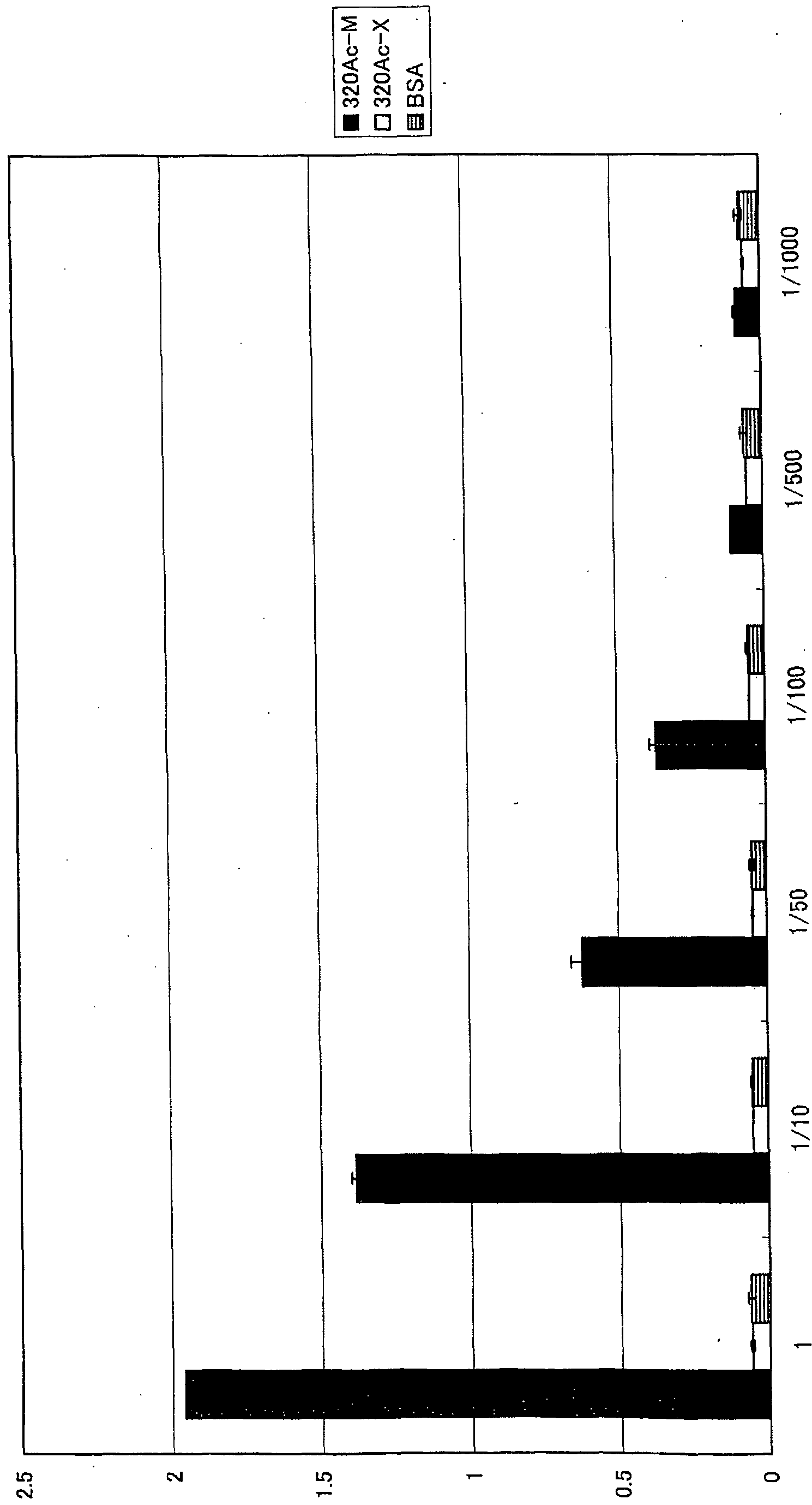
06 ao
差替え用紙 (規則 )
12/13
WO 2006/106957 PCT/JP2006/306921
[図 13]

06 αο
差替え用弒 (m )


差替え用紙 (細 U26)
Priority Applications (2)
| Application Number | Priority Date | Filing Date | Title |
|---|---|---|---|
| JP2007511193A JPWO2006106957A1 (ja) | 2005-04-01 | 2006-03-31 | p53翻訳後修飾部位を特異的に認識するモノクローナル抗体、及びその抗体を含む修飾部位測定キット |
| US11/910,313 US8062887B2 (en) | 2005-04-01 | 2006-03-31 | Monoclonal antibody specifically recognizing modification site after translation of p53 and kit for assaying modification site containing the same |
Applications Claiming Priority (2)
| Application Number | Priority Date | Filing Date | Title |
|---|---|---|---|
| JP2005106130 | 2005-04-01 | ||
| JP2005-106130 | 2005-04-01 |
Publications (1)
| Publication Number | Publication Date |
|---|---|
| WO2006106957A1 true WO2006106957A1 (ja) | 2006-10-12 |
Family
ID=37073507
Family Applications (1)
| Application Number | Title | Priority Date | Filing Date |
|---|---|---|---|
| PCT/JP2006/306921 Ceased WO2006106957A1 (ja) | 2005-04-01 | 2006-03-31 | p53翻訳後修飾部位を特異的に認識するモノクローナル抗体、及びその抗体を含む修飾部位測定キット |
Country Status (3)
| Country | Link |
|---|---|
| US (1) | US8062887B2 (ja) |
| JP (1) | JPWO2006106957A1 (ja) |
| WO (1) | WO2006106957A1 (ja) |
Cited By (2)
| Publication number | Priority date | Publication date | Assignee | Title |
|---|---|---|---|---|
| CN102924570A (zh) * | 2012-10-08 | 2013-02-13 | 辉源生物科技(上海)有限公司 | 荧光标记多肽及其制备方法和应用 |
| JP2023536162A (ja) * | 2020-07-30 | 2023-08-23 | ディアデム ソチエタ ペル アツィオーニ | 神経変性疾患の診断と予後推定におけるマーカーとしてのp53の翻訳後修飾 |
Families Citing this family (4)
| Publication number | Priority date | Publication date | Assignee | Title |
|---|---|---|---|---|
| CN103048471A (zh) * | 2013-01-09 | 2013-04-17 | 复旦大学 | 一种定量检测蛋白乙酰化水平的方法 |
| CN105112377B (zh) * | 2015-09-25 | 2018-10-30 | 福州迈新生物技术开发有限公司 | 一株分泌抗p53单克隆抗体的杂交瘤细胞株及其应用 |
| CN111138554A (zh) * | 2020-01-17 | 2020-05-12 | 新乡学院 | 一种重组蛋白g3p1-12及其制备方法和应用 |
| GB202012722D0 (en) * | 2020-08-14 | 2020-09-30 | Abcam Plc | Validation method |
Citations (3)
| Publication number | Priority date | Publication date | Assignee | Title |
|---|---|---|---|---|
| WO1999036532A1 (en) * | 1998-01-20 | 1999-07-22 | Medical & Biological Laboratories Co., Ltd. | Method for detecting acetyltransferase and deacetylase activities and method for screening inhibitors or accelerators of these enzymes |
| WO1999046574A2 (en) * | 1998-03-11 | 1999-09-16 | The Wistar Institute | Antibodies for assessing protein post-translational modification status and methods of making and using the same |
| JP2000325086A (ja) * | 1999-05-21 | 2000-11-28 | Igaku Seibutsugaku Kenkyusho:Kk | リン酸化酵素活性の測定方法 |
Family Cites Families (6)
| Publication number | Priority date | Publication date | Assignee | Title |
|---|---|---|---|---|
| US6884597B1 (en) | 1998-01-20 | 2005-04-26 | Medical & Biological Laboratories, Co., Ltd. | Method for detecting acetyltransferase and deacetylase activities and method for screening inhibitors or enhancers of these enzymes |
| US6441140B1 (en) | 1998-09-04 | 2002-08-27 | Cell Signaling Technology, Inc. | Production of motif-specific and context-independent antibodies using peptide libraries as antigens |
| JP2001161398A (ja) | 1999-12-06 | 2001-06-19 | Igaku Seibutsugaku Kenkyusho:Kk | タンパク質リン酸化酵素の活性測定方法およびタンパク質リン酸化酵素の活性測定キット |
| JP2003093056A (ja) | 2001-09-26 | 2003-04-02 | Japan Science & Technology Corp | p53のSer46をリン酸化する酵素 |
| EP1403639A1 (en) | 2002-09-30 | 2004-03-31 | G2M Cancer Drugs AG | Antibody tools for the diagnostic use in the medical therapy with inhibitors of histone deacetylases |
| US20050023912A1 (en) | 2003-07-28 | 2005-02-03 | A.O. Smith Corporation | Electric motor for hydromassage bathtubs |
-
2006
- 2006-03-31 WO PCT/JP2006/306921 patent/WO2006106957A1/ja not_active Ceased
- 2006-03-31 US US11/910,313 patent/US8062887B2/en not_active Expired - Fee Related
- 2006-03-31 JP JP2007511193A patent/JPWO2006106957A1/ja active Pending
Patent Citations (3)
| Publication number | Priority date | Publication date | Assignee | Title |
|---|---|---|---|---|
| WO1999036532A1 (en) * | 1998-01-20 | 1999-07-22 | Medical & Biological Laboratories Co., Ltd. | Method for detecting acetyltransferase and deacetylase activities and method for screening inhibitors or accelerators of these enzymes |
| WO1999046574A2 (en) * | 1998-03-11 | 1999-09-16 | The Wistar Institute | Antibodies for assessing protein post-translational modification status and methods of making and using the same |
| JP2000325086A (ja) * | 1999-05-21 | 2000-11-28 | Igaku Seibutsugaku Kenkyusho:Kk | リン酸化酵素活性の測定方法 |
Non-Patent Citations (6)
Cited By (3)
| Publication number | Priority date | Publication date | Assignee | Title |
|---|---|---|---|---|
| CN102924570A (zh) * | 2012-10-08 | 2013-02-13 | 辉源生物科技(上海)有限公司 | 荧光标记多肽及其制备方法和应用 |
| CN102924570B (zh) * | 2012-10-08 | 2014-07-16 | 辉源生物科技(上海)有限公司 | 荧光标记多肽及其制备方法和应用 |
| JP2023536162A (ja) * | 2020-07-30 | 2023-08-23 | ディアデム ソチエタ ペル アツィオーニ | 神経変性疾患の診断と予後推定におけるマーカーとしてのp53の翻訳後修飾 |
Also Published As
| Publication number | Publication date |
|---|---|
| US8062887B2 (en) | 2011-11-22 |
| JPWO2006106957A1 (ja) | 2008-09-11 |
| US20090298700A1 (en) | 2009-12-03 |
Similar Documents
| Publication | Publication Date | Title |
|---|---|---|
| Gembitsky et al. | A prototype antibody microarray platform to monitor changes in protein tyrosine phosphorylation | |
| US7816084B2 (en) | TLE3 as a marker for chemotherapy | |
| EP1718977B1 (en) | Protein phosphorylation in t-cell receptor signaling pathways | |
| EP3083698B1 (en) | Monoclonal anti-tk1 antibodies | |
| EP2105447A1 (en) | Avian-derived antibody capable of binding specifically to human hmgb1, immunological determination method for human hmgb1, and immunological determination reagent for human hmgb1 | |
| Seth et al. | Molecular portrait of high productivity in recombinant NS0 cells | |
| KR20050104045A (ko) | 종양 억제자로 작용하는 aim3의 신규 용도 | |
| WO2004081047A1 (ja) | モノクローナル抗体及びこれを産生するハイブリドーマ | |
| KR20030086345A (ko) | 신장세포암 종양 마커 | |
| Zhang et al. | Dissecting the axoneme interactome: the mammalian orthologue of Chlamydomonas PF6 interacts with sperm-associated antigen 6, the mammalian orthologue of Chlamydomonas PF16 | |
| US20100209427A1 (en) | Lysine acetylation sites | |
| US20050129678A1 (en) | Method for the prediction of an epitope | |
| US20100092992A1 (en) | Lysine acetylation sites | |
| US20140295462A1 (en) | RBM3 Protein in Colorectal Cancer Prognostics | |
| WO2011159959A2 (en) | Methods and systems for generating, validating and using monoclonal antibodies | |
| KR101524426B1 (ko) | 자가포식 특이적 억제제 발굴을 위한 스크리닝 방법 | |
| US8062887B2 (en) | Monoclonal antibody specifically recognizing modification site after translation of p53 and kit for assaying modification site containing the same | |
| US20090325312A1 (en) | Reagens for the detection of protein acetylation signaling pathways | |
| JPWO2014147873A1 (ja) | Hmgb1の分解産物と特異的に結合する抗体、並びにhmgb1の分解産物の測定方法及び測定試薬 | |
| EP2187216B1 (en) | Novel liver cancer marker | |
| JP2001161398A (ja) | タンパク質リン酸化酵素の活性測定方法およびタンパク質リン酸化酵素の活性測定キット | |
| Hattori et al. | Generation of monoclonal antibodies against mitocryptide‐2: toward a new strategy to investigate the biological roles of cryptides | |
| Ahn et al. | Production, characterization, and epitope mapping of monoclonal antibodies of ribosomal protein S3 (rpS3) | |
| JPWO1997045451A1 (ja) | ヒトlect2に対する抗体、それを産生する細胞、その測定法及び測定用キット | |
| Zorro Shahidian et al. | Development and Validation of Antibodies Targeting Site-Specific Histone Methylation |
Legal Events
| Date | Code | Title | Description |
|---|---|---|---|
| 121 | Ep: the epo has been informed by wipo that ep was designated in this application | ||
| WWE | Wipo information: entry into national phase |
Ref document number: 2007511193 Country of ref document: JP |
|
| NENP | Non-entry into the national phase |
Ref country code: DE |
|
| NENP | Non-entry into the national phase |
Ref country code: RU |
|
| 122 | Ep: pct application non-entry in european phase |
Ref document number: 06730869 Country of ref document: EP Kind code of ref document: A1 |
|
| WWE | Wipo information: entry into national phase |
Ref document number: 11910313 Country of ref document: US |