US9856462B2 - Method of treating eye disease using glycosylated VEGF decoy receptor fusion protein - Google Patents
Method of treating eye disease using glycosylated VEGF decoy receptor fusion protein Download PDFInfo
- Publication number
- US9856462B2 US9856462B2 US14/740,194 US201514740194A US9856462B2 US 9856462 B2 US9856462 B2 US 9856462B2 US 201514740194 A US201514740194 A US 201514740194A US 9856462 B2 US9856462 B2 US 9856462B2
- Authority
- US
- United States
- Prior art keywords
- vegf
- vegfr1
- domain
- residue
- val
- Prior art date
- Legal status (The legal status is an assumption and is not a legal conclusion. Google has not performed a legal analysis and makes no representation as to the accuracy of the status listed.)
- Active
Links
Images


Classifications
-
- C—CHEMISTRY; METALLURGY
- C12—BIOCHEMISTRY; BEER; SPIRITS; WINE; VINEGAR; MICROBIOLOGY; ENZYMOLOGY; MUTATION OR GENETIC ENGINEERING
- C12N—MICROORGANISMS OR ENZYMES; COMPOSITIONS THEREOF; PROPAGATING, PRESERVING, OR MAINTAINING MICROORGANISMS; MUTATION OR GENETIC ENGINEERING; CULTURE MEDIA
- C12N9/00—Enzymes; Proenzymes; Compositions thereof; Processes for preparing, activating, inhibiting, separating or purifying enzymes
- C12N9/10—Transferases (2.)
- C12N9/12—Transferases (2.) transferring phosphorus containing groups, e.g. kinases (2.7)
-
- A—HUMAN NECESSITIES
- A61—MEDICAL OR VETERINARY SCIENCE; HYGIENE
- A61K—PREPARATIONS FOR MEDICAL, DENTAL OR TOILETRY PURPOSES
- A61K33/00—Medicinal preparations containing inorganic active ingredients
- A61K33/24—Heavy metals; Compounds thereof
- A61K33/243—Platinum; Compounds thereof
-
- A—HUMAN NECESSITIES
- A61—MEDICAL OR VETERINARY SCIENCE; HYGIENE
- A61K—PREPARATIONS FOR MEDICAL, DENTAL OR TOILETRY PURPOSES
- A61K33/00—Medicinal preparations containing inorganic active ingredients
- A61K33/24—Heavy metals; Compounds thereof
-
- A—HUMAN NECESSITIES
- A61—MEDICAL OR VETERINARY SCIENCE; HYGIENE
- A61K—PREPARATIONS FOR MEDICAL, DENTAL OR TOILETRY PURPOSES
- A61K38/00—Medicinal preparations containing peptides
- A61K38/16—Peptides having more than 20 amino acids; Gastrins; Somatostatins; Melanotropins; Derivatives thereof
- A61K38/43—Enzymes; Proenzymes; Derivatives thereof
- A61K38/45—Transferases (2)
-
- A—HUMAN NECESSITIES
- A61—MEDICAL OR VETERINARY SCIENCE; HYGIENE
- A61K—PREPARATIONS FOR MEDICAL, DENTAL OR TOILETRY PURPOSES
- A61K39/00—Medicinal preparations containing antigens or antibodies
- A61K39/395—Antibodies; Immunoglobulins; Immune serum, e.g. antilymphocytic serum
-
- A—HUMAN NECESSITIES
- A61—MEDICAL OR VETERINARY SCIENCE; HYGIENE
- A61P—SPECIFIC THERAPEUTIC ACTIVITY OF CHEMICAL COMPOUNDS OR MEDICINAL PREPARATIONS
- A61P27/00—Drugs for disorders of the senses
- A61P27/02—Ophthalmic agents
-
- C—CHEMISTRY; METALLURGY
- C07—ORGANIC CHEMISTRY
- C07K—PEPTIDES
- C07K14/00—Peptides having more than 20 amino acids; Gastrins; Somatostatins; Melanotropins; Derivatives thereof
- C07K14/435—Peptides having more than 20 amino acids; Gastrins; Somatostatins; Melanotropins; Derivatives thereof from animals; from humans
- C07K14/705—Receptors; Cell surface antigens; Cell surface determinants
- C07K14/71—Receptors; Cell surface antigens; Cell surface determinants for growth factors; for growth regulators
-
- C—CHEMISTRY; METALLURGY
- C07—ORGANIC CHEMISTRY
- C07K—PEPTIDES
- C07K16/00—Immunoglobulins [IGs], e.g. monoclonal or polyclonal antibodies
-
- A—HUMAN NECESSITIES
- A61—MEDICAL OR VETERINARY SCIENCE; HYGIENE
- A61K—PREPARATIONS FOR MEDICAL, DENTAL OR TOILETRY PURPOSES
- A61K39/00—Medicinal preparations containing antigens or antibodies
- A61K2039/505—Medicinal preparations containing antigens or antibodies comprising antibodies
-
- A—HUMAN NECESSITIES
- A61—MEDICAL OR VETERINARY SCIENCE; HYGIENE
- A61K—PREPARATIONS FOR MEDICAL, DENTAL OR TOILETRY PURPOSES
- A61K38/00—Medicinal preparations containing peptides
-
- A—HUMAN NECESSITIES
- A61—MEDICAL OR VETERINARY SCIENCE; HYGIENE
- A61K—PREPARATIONS FOR MEDICAL, DENTAL OR TOILETRY PURPOSES
- A61K48/00—Medicinal preparations containing genetic material which is inserted into cells of the living body to treat genetic diseases; Gene therapy
-
- C—CHEMISTRY; METALLURGY
- C07—ORGANIC CHEMISTRY
- C07K—PEPTIDES
- C07K2319/00—Fusion polypeptide
- C07K2319/30—Non-immunoglobulin-derived peptide or protein having an immunoglobulin constant or Fc region, or a fragment thereof, attached thereto
-
- C—CHEMISTRY; METALLURGY
- C07—ORGANIC CHEMISTRY
- C07K—PEPTIDES
- C07K2319/00—Fusion polypeptide
- C07K2319/32—Fusion polypeptide fusions with soluble part of a cell surface receptor, "decoy receptors"
-
- C—CHEMISTRY; METALLURGY
- C07—ORGANIC CHEMISTRY
- C07K—PEPTIDES
- C07K2319/00—Fusion polypeptide
- C07K2319/70—Fusion polypeptide containing domain for protein-protein interaction
-
- C—CHEMISTRY; METALLURGY
- C07—ORGANIC CHEMISTRY
- C07K—PEPTIDES
- C07K2319/00—Fusion polypeptide
- C07K2319/90—Fusion polypeptide containing a motif for post-translational modification
- C07K2319/91—Fusion polypeptide containing a motif for post-translational modification containing a motif for glycosylation
-
- C—CHEMISTRY; METALLURGY
- C12—BIOCHEMISTRY; BEER; SPIRITS; WINE; VINEGAR; MICROBIOLOGY; ENZYMOLOGY; MUTATION OR GENETIC ENGINEERING
- C12Y—ENZYMES
- C12Y207/00—Transferases transferring phosphorus-containing groups (2.7)
- C12Y207/10—Protein-tyrosine kinases (2.7.10)
- C12Y207/10001—Receptor protein-tyrosine kinase (2.7.10.1)
Definitions
- the present application relates to making a chimeric molecule that is used to prevent blood vessel formation as a form of treating cancer or treating eye disease.
- VEGF-A Vascular endothelial growth factor-A
- VEGFR2 VEGF receptor 2
- VEGF-A is a critical regulator of tumor angiogenesis, mainly through the activation of its primary receptor, VEGF receptor 2 (VEGFR2).
- VEGF-A is expressed in most tumor cells and corresponding stromal cells throughout every stage of tumor progression, while VEGFR2 is highly expressed in growing tumor vessels, leading to the formation of structurally and functionally malformed tumor blood vessels.
- VEGF-A specifically binds to the second immunoglobulin (Ig) homology domain (D2) of the extracellular region of VEGFR2, resulting in activation of pro-angiogenic signalling.
- Ig immunoglobulin
- D2 second immunoglobulin homology domain
- VEGF-A/VEGFR2 signalling pathway For the past decade, much effort has been devoted to targeting this VEGF-A/VEGFR2 signalling pathway using monoclonal antibodies, soluble decoy receptor fusion proteins, and small molecular inhibitors in cancer patients. While the current therapeutic blockade of VEGF-A/VEGFR2 signalling provides clinical benefits, the anti-cancer effect is modest and transient, eventually giving rise to acquired resistance through the activation of alternative pro-angiogenic pathways and further recruitment of pro-angiogenic cells such as tumor-associated macrophages (TAM). These limitations highlight current unmet needs in anti-angiogenic cancer treatment strategies, which must be addressed for successful therapy development.
- TAM tumor-associated macrophages
- CNV choroidal neovascularization
- Angiogenesis is normally a compensatory mechanism of our body in pathologic situations such as coronary collateral formation and wound healing process. And this mechanism is also triggered by an oxygen insufficiency state as known as “hypoxia”. Hypoxic state stimulates hypoxic inducible factors (HIFs) including VEGF and it plays a crucial role in angiogenesis. In addition, eyes with high concentration of VEGF suffer from leakage from retinal vessels, and subsequently, macular edema develops.
- hypoxic inducible factors HIFs
- VEGF hypoxic inducible factors
- VEGF may be a therapeutic target for ocular diseases associated with abnormal angiogenesis and vascular leakage such as exudative AMD, diabetic retinopathy, retinopathy of prematurity, neovascular glaucoma, corneal neovascularization, retinal vein occlusion and macular edema due to diabetic retinopathy or retinal vein occlusion.
- VEGFR1 binds to both VEGFR1 and VEGFR2.
- the binding affinity of VEGFR1 to VEGF-A ( ⁇ 10 ⁇ 20 pM) is much higher than that of VEGFR2 ( ⁇ 100 ⁇ 125 pM).
- VEGFR1 is a receptor for other pro-angiogenic ligands, VEGF-B and placental growth factor (PlGF), which have recently been highlighted as alternative targets for anti-angiogenic therapy. Because of its ability to bind multiple pro-angiogenic ligands, VEGFR1 has been considered as a potential backbone for the development of a novel decoy receptor fusion protein for therapeutic purposes.
- VEGFR1 D3 By switching VEGFR1 D3 to VEGFR2 D3, the net pI of VEGF-Trap was decreased, resulting in less ECM binding and an improved pharmacokinetic (PK) profile compared to VEGFR1-Fc.
- PK pharmacokinetic
- Glycosylation is a post-translational modification that results in the addition of carbohydrate chains to specific asparagine (N-linked glycosylation) or serine/threonine (O-linked glycosylation) residues.
- Glycosylation of secreted and membrane proteins affects their biochemical and biological properties. It usually provides a negative charge and increases solubility, thus diminishing non-specific binding to the ECM. Moreover, glycosylation grants resistance to proteolysis and extended serum half-life, enhancing a protein's PK profile.
- Glyco-engineered therapeutic proteins such as Aranesp (erythropoietin) from Amgen and Gazyva (obinutuzumab) from Genentech are good examples that exploited these advantages.
- VEGF-Grab a novel VEGF decoy receptor fusion protein, VEGF-Grab.
- Parental VEGFR1-Fc VEGFR1 D2-D3 fused to Fc
- This engineered VEGF-Grab showed significantly improved decoy efficiency and a dramatic decrease in net pI, thus attenuating non-specific ECM binding and enhancing PK profiles.
- VEGF-Grab strongly suppressed tumor angiogenesis, progression, and metastasis via effective capturing of three VEGFR1 ligands, VEGF-A, VEGF-B, and PlGF.
- VEGF-A vascular endothelial growth factor A
- AMD age-related macular degeneration
- VEGF-A vascular endothelial growth factor A
- APD age-related macular degeneration
- VEGF-B vascular endothelial growth factor A
- PlGF placenta growth factor
- VEGF-Grab has the second and third immunoglobulin (Ig)-like domains of VEGF receptor 1 (VEGFR1) fused to IgG1 Fc, with three potential glycosylation sites introduced into the third Ig-like domain of VEGF-Grab by mutagenesis.
- VEGF-Grab showed more potent decoy activity against VEGF and PlGF, mainly attributed to the VEGFR1 backbone.
- the negatively charged O-glycans attached to the third Ig-like domain of VEGFR1 counterbalanced the originally positively charged VEGFR1 backbone, minimizing non-specific binding of VEGF-Grab to the extracellular matrix, and resulting in greatly improved pharmacokinetic profile.
- the present invention is drawn to an isolated nucleic acid molecule encoding a polypeptide capable of synchronously binding VEGF polypeptide and placenta growth factor (PIGF) polypeptide comprising a nucleotide sequence encoding a VEGFR1 component.
- the VEGFR1 component may include the second and third immunoglobulin (Ig)-like domains.
- the at least one encoded positive amino acid residue in at least one domain of VEGFR1 may be mutated to a negatively charged residue.
- the domain may be the third domain.
- amino acid residue may be on the ⁇ 1- ⁇ 2 loop, which may include nucleic acid positions 397 to 432 of SEQ ID NO:1, which corresponds to amino acid residues 133 to 144 of SEQ ID NO:2, or ⁇ 3- ⁇ 4 loop, which comprises nucleic acid positions 490 to 522 of SEQ ID NO:1, which corresponds to amino acid residues 164 to 174 of SEQ ID NO:2.
- the at least one encoded positive amino acid residue in at least one domain of VEGFR1 may be mutated so as to produce a glycosylation site.
- the at least one encoded positive amino acid residue in at least one domain of VEGFR1 may be mutated so as to produce a decrease in net pI of the encoded polypeptide.
- the residue to be mutated may be R135 residue on the ⁇ 1- ⁇ 2 loop, K138 residue on the ⁇ 1- ⁇ 2 loop, or R172 residue on the ⁇ 3- ⁇ 4 loop on the third domain.
- the VEGF may be VEGF-A or VEGF-B.
- the VEGFR1 component may be operatively linked to a nucleotide sequence encoding a multimerizing component.
- the invention is directed to an isolated nucleic acid molecule comprising a nucleotide sequence encoding: (a) VEGF-Grab1; (b) VEGF-Grab2; or (c) VEGF-Grab3.
- the invention is drawn to a nucleic acid vector, which includes any of the nucleic acid molecule described above.
- the vector may be viral vector.
- the vector may be an expression vector that includes any of above-described nucleic acid molecule, wherein the nucleic acid molecule may be operatively linked to an expression control sequence.
- the invention is directed to a method of generating a polypeptide capable of synchronously binding VEGF polypeptide and placenta growth factor (PIGF) polypeptide comprising a VEGFR1 component in a patient, comprising administering to the patient the nucleic acid vector described above.
- PIGF placenta growth factor
- the invention is drawn to a host-vector system for the production of a polypeptide, which includes the expression vector described above, in a suitable host cell.
- the suitable host cell may be a bacterial cell, yeast cell, insect cell, or mammalian cell.
- the invention is drawn to a method of generating a polypeptide capable of synchronously binding VEGF polypeptide and placenta growth factor (PIGF) polypeptide comprising a VEGFR1 component in a patient, comprising administering to the patient the cell described above.
- PIGF placenta growth factor
- the invention is drawn to a method of producing a polypeptide which steps include growing cells of the host-vector system described, under conditions permitting production of the polypeptide and recovering the polypeptide so produced.
- the invention is drawn to a polypeptide encoded by any of the isolated nucleic acid molecule described above.
- the polypeptide may be wherein the VEGFR1 component comprises the second and third immunoglobulin (Ig)-like domains.
- the polypeptide may be wherein at least one positive amino acid residue in at least one domain of VEGFR1 may be mutated to a negatively charged residue.
- the domain may be the third domain.
- the amino acid residue may be on the ⁇ 1- ⁇ 2 loop, which may include amino acid residues 133 to 144 of SEQ ID NO:2, or ⁇ 3- ⁇ 4 loop, which may include amino acid residues 164 to 174 of SEQ ID NO:2.
- the at least one positive amino acid residue in at least one domain of VEGFR1 may be mutated so as to include a glycosylation site.
- the polypeptide may be glycosylated. Further, the polypeptide may be sialylated.
- at least one positive amino acid residue in at least one domain of VEGFR1 may be mutated so as to produce a decrease in net pI of the polypeptide.
- the residue to be mutated may be R135 residue on the ⁇ 1- ⁇ 2 loop, K138 residue on the ⁇ 1- ⁇ 2 loop, or R172 residue on the ⁇ 3- ⁇ 4 loop on the third domain.
- the invention is drawn to a method of blocking blood vessel growth in a mammal comprising administering to the mammal in need thereof an effective amount of the polypeptide described above.
- the mammal may be human.
- the blood vessel growth may occur in the eye to cause a medical condition that affects sight.
- the medical condition may be age-related macular degeneration, exudative age-related macular degeneration, choroidal neovascularization, pathologic myopia, diabetic retinopathy, diabetic macular edema, retinal vein occlusion, retinopathy of prematurity or neovascular glaucoma.
- the choroidal neovascularization may be myopic choroidal neovascularization, traumatic choroidal neovascularization, uveitic choroidal neovascularization, ocular histoplasmosis, or idiopathic choroidal neovascularization.
- the invention is drawn to a method of inhibiting VEGF and/or PIGF activities in a mammal comprising administering to the mammal an effective amount of any of the polypeptides described above.
- the mammal may be human.
- the invention is drawn to a method of attenuating or preventing tumor growth in a mammal, comprising administering to a subject in need thereof a therapeutically effective amount of the polypeptides described above.
- the mammal may be human.
- the invention is drawn to a method of suppressing metastasis in a mammal, comprising administering to a subject in need thereof a therapeutically effective amount of the polypeptides described above.
- the mammal may be human.
- the invention is drawn to a method of attenuating or preventing tumor growth in a mammal, comprising administering to a subject in need thereof a therapeutically effective amount of any of the polypeptides described above and a cytotoxic therapeutic agent.
- the mammal may be human.
- the cytotoxic therapeutic agent may be without limitation cisplatin.
- FIGS. 1A-1G show generation and characterization of VEGF-Grab.
- FIG. 1A Schematic diagram of VEGF-Grabs (VEGF-Grab1, VEGF-Grab2 and VEGF-Grab3; hereinafter referred to as G1, G2, and G3) and VEGF-Trap (VT). Mutated residues in VEGF-Grabs are indicated by brown at the lower panel.
- FIG. 1B Electrostatic potential of D2-D3 domain in VEGFR1 (left) and VEGFR2 (right). Positively and negatively charged residues are colored in blue and red, respectively.
- FIG. 1C Model structure of VEGF-A/VEGFR1 D2-D3 complex. VEGF-A dimer is colored in yellow and green.
- VEGFR1 D2 and D3 are colored in tan and mustard. Residues indicated by blue stick are target sites for mutagenesis.
- FIG. 1D SDS-PAGE analysis of G1 and G3 in reduced (R) and non-reduced (NR) conditions.
- FIG. 2 shows secondary structure of original VEGFR1-Ig3. ⁇ strands are shown as arrows and cylinders (green). Residues on ⁇ 1- ⁇ 2 and ⁇ 3- ⁇ 4 loops of VEGFR1-D3 are displayed as bold.
- FIGS. 3A-3K show that VEGF-Grab3 exhibits low ECM binding and prolonged pharmacokinetic profile.
- FIG. 3A Isoelectric point analysis of G1, G3, and VT. Red lines on each band were used to analyse net pI.
- FIG. 3B Comparison of pI for each protein. Net pI of each protein was the mean pI of each isoform denoted as lines.
- FIG. 3C PNGase F digestion to remove N-linked glycan.
- FIG. 3D Analysis of O-linked glycosylation at Serine135 of G3.
- FIG. 3E Schematic diagram for glycosylated sites analysed by mass spectrometry.
- FIG. 3H Comparison of area under the curve (AUC) of VT and G3.
- I and J Tissue distribution of G3 and VT 48 hr after subcutaneous injection (4 mg/kg).
- FIG. 3I Accumulated VT and G3 in tumor.
- FIG. 3J Relative accumulated levels of VT and G3 in liver and kidney compared to tumor.
- FIGS. 4A-4H show that VEGF-grabs inhibit EC survival, migration, and tube formation via suppression of VEGF signalling pathway.
- FIGS. 4A-4C Inhibition of VEGF-A-induced phosphorylation of VEGFR2 and ERK in HUVECs by the treatment of G1, G3, and VT. Immunoblotting ( FIG. 4A ) and quantification ( FIG. 4B and FIG. 4C ).
- FIG. 4D Cell survival assay with HUVECs after G1, G3, and VT treatment (0.35, 0.7, 3.5, 7, 35, 70 nM) in the presence of VEGF-A (0.2 nM).
- FIGS. 5A-5U show that VEGF-Grab3 effectively suppresses tumor growth, angiogenesis, and metastasis in LLC tumors.
- FIGS. 5A-5N Mice were treated with proteins on the indicated days (arrows).
- FIG. 5A and FIG. 5C Comparison of tumor growth ( FIG. 5A ) and tumor weights ( FIG. 5C ).
- FIG. 5B and FIG. 5D Images ( FIG. 5B ) and quantification ( FIG. 5D ) of intratumoral necrotic area stained with H&E. Dotted line demarcates intratumoral necrosis.
- FIG. 5E and FIG. 5F Images ( FIG. 5E ) and quantification ( FIG.
- FIG. 5F Images showing cytokeratin + tumor cell metastasis (red) in inguinal LNs. Each indicated region (squares) is magnified in the lower panel.
- FIG. 5H and FIG. 5I Quantifications of lymphatic vascular densities (LVD) ( FIG. 5H ) and cytokeratin + tumor cell metastasis ( FIG. 5I ).
- FIG. 5J and FIG. 5K Images ( FIG. 5J ) and quantifications ( FIG. 5K ) of Hypoxyprobe + hypoxic areas (green) in tumors.
- FIG. 5L and FIG. 5M Images ( FIG.
- FIG. 5L Comparisons of mRNA expression levels of various genes in intratumoral tissue after treatment with G3 and VT. Values indicate fold changes over control tumors.
- FIGS. 5O-5R Comparative dose responses of VT and G3 on tumor growth and metastasis. LLC tumor-bearing mice were treated with either VT, or G3 at the indicated days (arrows), respectively.
- FIG. 5O and FIG. 5P Comparison of tumor growth after VT ( FIG. 5O ) or G3 ( FIG. 5P ) treatment.
- FIG. 5Q and FIG. 5R Images ( FIG. 5Q ) and quantification ( FIG.
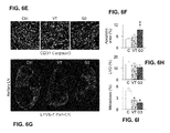
- FIGS. 6A-6I show that VEGF-Grab3 delays tumor growth and suppresses neovessel formation and metastasis in a spontaneous breast cancer model.
- FIGS. 6A-6I Female MMTV-PyMT mice (12-weeks old) received intraperitoneal-injections of VT, or G3 (25 mg/kg) twice per week for 3 weeks.
- FIG. 6A Tumor sections stained with H&E. Invasive tumor cells (Inv), early carcinoma lesions (Ea), and surrounding adipose tissue (Adi) are denoted by dotted lines.
- FIG. 6B Comparison of volumes of tumor nodules. Lines denote mean values.
- FIG. 6C and FIG. 6D Images ( FIG.
- FIG. 6C Images ( FIG. 6E ) and comparison ( FIG. 6F ) of Caspase3 + apoptotic cells (red) in tumor.
- FIG. 6G Images showing cytokeratin + tumor cell metastasis (red) in axillary LNs.
- FIGS. 7A-D show that VEGF-Grab3 exerts more durable suppression of tumor angiogenesis.
- FIGS. 7A-7D tumor vessel regrowth after VT or G3 treatment.
- FIG. 7A Experimental Scheme. Mice were treated with proteins on the indicated days (green arrows). Treatment was withdrawn, and analysed at D17 and D19 (blue arrows).
- FIGS. 7B-7D Changes in the vascularity after treatment with either VT, or G3.
- White arrowheads indicate representative new vascular sprouts. Images ( FIG. 7B ) and quantifications of CD31 + blood vessels ( FIG. 7C ) and sprouts numbers ( FIG. 7D ). *p ⁇ 0.05 VT D17 vs VT D19.
- FIGS. 8A-8B show that a single intravitreal injection of 50 ⁇ g VEGF-Grab3 suppresses choroidal neovascularization at sites of rupture of Bruch's membrane.
- FIG. 8B Representative confocal microscope images of the anti-PECAM-1/CD31 antibody-stained CNV lesions in the choroid flat mounts from laser-induced CNV in mice treated with PBS (Left), VEGF-Grab3 (middle) and Aflibercept (Right). CNVs were significantly abolished by
- FIGS. 9A-9B show that intravitreal injection of VEGF-Grab3 reduced the vascular density of OIR model mice.
- FIG. 9A Representative magnified confocal microscope images of OIR model mice. The density of vasculature was significantly reduced by the VEGF-Grab3 treatment compared to PBS-treatment.
- FIG. 9B Bar graph showing VEGF-Grab3 increases the area of avascular retina at P17 in OIR model mice. Data are expressed as means ⁇ standard error of the mean (SEM). The vascular intensity of VEGF-Grab3 treated mice significantly decreased compared with the PBS-injected control eyes. (P ⁇ 0.01).
- FIG. 10 shows sequence alignment of VEGF-Grab1, VEGF-Grab3, and VEGF-Trap.
- Amino acid sequences of G1, G3, and VT were aligned using CLUSTALW.
- Signal sequences, D2, D3, and Fc domains are shown above the sequence alignment.
- N-linked glycosylation sites detected by mass spectrometry in all proteins are highlighted by the red box.
- the mutated site occupied by O-linked glycosylation is highlighted by the blue box.
- the mutated sites for glycosylation that are unoccupied by glycans are highlighted by the grey box.
- the peptide fragments analysed by MS spectrometry after trypsin digestion are shown below the sequence as brown lines (See FIG. 11A for additional description).
- FIGS. 11A-11B show analysis of N-linked glycosylation for VEGF-Grabs and VEGF-Trap.
- FIG. 11A List of peptides possibly identified from PNGaseF/trypsin digestion of VEGF-Grabs and VT that encompass a potential site of N-glycosylation. If an Asn residue is N-glycosylated at a particular position, that residue will be converted to Asp after PNGase digestion, resulting in a mass gain of 0.984 Da. In contrast, Asn residues unoccupied by N-glycans are unaffected by deglycosylation.
- FIG. 13 shows that VEGF-Grab3 displays low binding to tumor ECM.
- Control tumor sections which were not treated with any protein therapeutics, were stained with 100 nM of VR1-Fc, VT, or G3 and subsequently visualized using the anti-human Fc-cy3 antibody to detect non-specific binding (red) of these molecules to tumor ECM.
- Collagen type IV green is a collagen primarily found in the basal lamina of the ECM. Scale bars, 100 ⁇ m.
- FIG. 14 shows pharmacokinetic profile of VEGF-Grab1 and VEGF-Grab3. Serum levels of G1 after subcutaneous injection of 4 mg/kg. Blood was sampled at 0, 1, 2, 4, 8, 12, 24, 48, and 96 hr, and analysed by ELISA.
- FIGS. 15A-15B show tissue accumulation of VEGF-Trap and VEGF-Grab3.
- FIGS. 15A and 15B VT and G3 levels in tissues after subcutaneous injections of VT (4 mg/kg) and G3 (4 mg/kg) into LLC tumor-bearing C57BL/6J mice. Tissues were harvested after 48 hr, lysed in lysis buffer (Lysis buffer 6, R&D), quantified by Bradford assay, and analysed by ELISA for liver ( FIG. 15A ) and kidney ( FIG. 15B ). *p ⁇ 0.05 G3 vs VT. Values are mean ⁇ SD.
- FIGS. 16A-16G show that VEGF-Grabs or VEGF-Trap have no effect on VEGFR2 signalling, EC survival, migration, and tube formation in the absence of VEGFA.
- FIGS. 16A-16C VEGFR2 and ERK phosphorylation in HUVECs after treatment with G1, G3, and VT (2 ⁇ g/ml, 14 nM, respectively) in the presence or absence of VEGFA (50 ng/ml, 1 nM).
- Immunoblotting FIG. 16A
- quantification FIG. 16B and FIG. 16C ). und. indicates undetected.
- FIG. 16E Cell migration assay with HUVECs in the presence or absence of VEGFA (50 ng/ml, 1 nM) and indicated proteins (2 ⁇ g/ml, 14 nM). The amount of wound healing was monitored after 12 hr. Wound healing areas are indicated in red. Images ( FIG. 16D ) and quantification ( FIG. 16E ) of migration area. FIG. 16F and FIG. 16G , Tube formation assay with HUVECs in the presence or absence of VEGFA (50 ng/ml, 1 nM) and indicated proteins (2 ⁇ g/ml, 14 nM). Images ( FIG. 16F ) and quantification ( FIG. 16G ) of tube formation. Scale bars, 100 ⁇ m.
- FIGS. 17A-17B show dose-dependent inhibition of VEGFR2 signalling with anti-VEGF therapy.
- FIGS. 17A-17B HUVECs were cultured, starved overnight, pre-treated with each protein (0.125 ⁇ g/ml (0.875 nM), 0.25 ⁇ g/ml (1.75 nM), 0.5 ⁇ g/ml (3.5 nM), and 1 ⁇ g/ml (7 nM)) for 15 min, and treated with VEGFA (50 ng/ml, 1 nM) for 10 min. Cells were lysed and indicated proteins were immunoblotted to detect the activation of VEGFR2 and ERK1/2 signalling.
- FIG. 17A Immunoblotting showing dose-dependent inhibition of VEGFR2 and ERK1/2 phosphorylation after treatment with VEGF-Grabs or VT.
- FIG. 17B Comparisons of VEGFR2 and ERK1/2 phosphorylation after treatment with VEGF-Grabs or VT.
- FIG. 18 shows histologic analyses of vital organs after anti-VEGF therapy. After a 2-week treatment with either control, VT, or VEGF-Grabs (25 mg/kg) in LLC tumor-bearing mice, indicated organs were sampled and sectioned for histologic analysis. Images show tissue sections of indicated organs stained with H&E. Scale bars, 100 ⁇ m.
- FIG. 19 shows that VEGF-Grab effectively suppresses the growth of established bulky macroscopic tumors. After the volume of tumor exceeded 500 mm 3 , either control, VT, or G3 were treated to LLC tumor bearing mice on the indicated days (arrows). Comparison of LLC tumor growth.
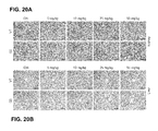
- FIGS. 20A-20B show histological analyses of the kidney and liver after dose dependent anti-VEGF therapy. After treatment with either control, VT (5, 10, 25, and 50 mg/kg), or G3 (5, 10, 25, and 50 mg/kg) in LLC tumor-bearing mice, kidney and liver were sampled and sectioned for histologic analysis. Images show tissue sections of kidney ( FIG. 20A ) and liver ( FIG. 20B ) stained with H&E. Scale bars, 100 ⁇ m.
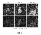
- FIG. 21 shows that combination therapy of VEGF-Grab3 and Cisplatin enhances intratumoral apoptosis.
- LLC tumor-bearing mice received injections of cisplatin (10 mg/kg) 9 days after tumor implantation in combination with either control, VT, or G3 (25 mg/kg) every 2 days.
- Caspase 3+ cells (Red) mostly overlap with Pan-cytokeratin+ cells (Green) which are epithelial cell derived LLC tumor, but not with CD31+ cells (Blue), suggesting apoptotic cells are more likely tumor cell.
- “about” or “substantially” generally provides a leeway from being limited to an exact number.
- “about” or “substantially” indicates that the polypeptide is not to be limited to the recited number of amino acids. A few amino acids add to or subtracted from the N-terminus or C-terminus may be included so long as the functional activity such as its binding activity is present.
- administration “in combination with” one or more further therapeutic agents includes simultaneous (concurrent) and consecutive administration in any order.
- amino acid and “amino acids” refer to all naturally occurring L- ⁇ -amino acids. This definition is meant to include norleucine, ornithine, and homocysteine.
- amino acid sequence variant refers to molecules with some differences in their amino acid sequences as compared to a reference (e.g. native sequence) polypeptide.
- the amino acid alterations may be substitutions, insertions, deletions or any desired combinations of such changes in a native amino acid sequence.
- Substitutional variants are those that have at least one amino acid residue in a native sequence removed and a different amino acid inserted in its place at the same position.
- the substitutions may be single, where only one amino acid in the molecule has been substituted, or they may be multiple, where two or more amino acids have been substituted in the same molecule.
- Substitutes for an amino acid within the sequence may be selected from other members of the class to which the amino acid belongs.
- the nonpolar (hydrophobic) amino acids include alanine, leucine, isoleucine, valine, proline, phenylalanine, tryptophan and methionine.
- the polar neutral amino acids include glycine, serine, threonine, cysteine, tyrosine, asparagine and glutamine.
- the positively charged (basic) amino acids include arginine, lysine and histidine.
- the negatively charged (acidic) amino acids include aspartic acid and glutamic acid.
- proteins or fragments or derivatives thereof which exhibit the same or similar biological activity and derivatives which are differentially modified during or after translation, e.g., by glycosylation, proteolytic cleavage, linkage to an antibody molecule or other cellular ligand, and so on.
- Insertional variants are those with one or more amino acids inserted immediately adjacent to an amino acid at a particular position in a native amino acid sequence. Immediately adjacent to an amino acid means connected to either the ⁇ -carboxy or ⁇ -amino functional group of the amino acid.
- Deletional variants are those with one or more amino acids in the native amino acid sequence removed. Ordinarily, deletional variants will have one or two amino acids deleted in a particular region of the molecule.
- antagonist refers to a ligand that tends to nullify the action of another ligand, as a ligand that binds to a cell receptor without eliciting a biological response.
- fusion proteins be labeled with a detectable label, such as radioisotope, fluorescent tag, enzymatic tag, or a chemiluminescent tag to determine ligand-receptor binding interaction.
- detectable label such as radioisotope, fluorescent tag, enzymatic tag, or a chemiluminescent tag to determine ligand-receptor binding interaction.
- assay systems employing the chimeric molecule is also contemplated.
- carriers include pharmaceutically acceptable carriers, excipients, or stabilizers which are nontoxic to the cell or mammal being exposed thereto at the dosages and concentrations employed.
- pharmaceutically acceptable carrier is an aqueous pH buffered solution.
- Examples of pharmaceutically acceptable carriers include without limitation buffers such as phosphate, citrate, and other organic acids; antioxidants including ascorbic acid; low molecular weight (less than about 10 residues) polypeptide; proteins, such as serum albumin, gelatin, or immunoglobulins; hydrophilic polymers such as polyvinylpyrrolidone; amino acids such as glycine, glutamine, asparagine, arginine or lysine; monosaccharides, disaccharides, and other carbohydrates including glucose, mannose, or dextrins; chelating agents such as EDTA; sugar alcohols such as mannitol or sorbitol; salt-forming counterions such as sodium; and/or nonionic surfactants such as TWEEN®, polyethylene glycol (PEG), and PLURONICS®.
- buffers such as phosphate, citrate, and other organic acids
- antioxidants including ascorbic acid
- proteins such as
- nucleic acid sequence refers to the sequence that is essential to carry out the intended function of the amino acid encoded by the nucleic acid.
- an effective amount is an amount sufficient to effect beneficial or desired clinical or biochemical results.
- An effective amount can be administered one or more times.
- an effective amount of an inhibitor compound is an amount that is sufficient to palliate, ameliorate, stabilize, reverse, slow or delay the progression of the disease state.
- fragments or “functional derivatives” refers to biologically active amino acid sequence variants and fragments of the native ligands or receptors of the present invention, as well as covalent modifications, including derivatives obtained by reaction with organic derivatizing agents, post-translational modifications, derivatives with nonproteinaceous polymers, and immunoadhesins.
- host cell includes an individual cell or cell culture which can be or has been a recipient of a vector of this invention.
- Host cells include progeny of a single host cell, and the progeny may not necessarily be completely identical (in morphology or in total DNA complement) to the original parent cell due to natural, accidental, or deliberate mutation and/or change.
- ligand refers to any molecule or agent, or compound that specifically binds covalently or transiently to a molecule such as a polypeptide.
- ligand may include antibody.
- ligand may refer to a molecule sought to be bound by another molecule with high affinity.
- mammal for purposes of treatment refers to any animal classified as a mammal, including humans, domestic and farm animals, and zoo, sports, or pet animals, such as dogs, cats, cattle, horses, sheep, pigs, and so on.
- the mammal is human.
- pharmaceutically acceptable carrier and/or diluent includes any and all solvents, dispersion media, coatings antibacterial and antifungal agents, isotonic and absorption delaying agents and the like.
- the use of such media and agents for pharmaceutical active substances is well known in the art. Except insofar as any conventional media or agent is incompatible with the active ingredient, use thereof in the therapeutic compositions is contemplated. Supplementary active ingredients can also be incorporated into the compositions.
- Dosage unit form refers to physically discrete units suited as unitary dosages for the mammalian subjects to be treated; each unit containing a predetermined quantity of active material calculated to produce the desired therapeutic effect in association with the required pharmaceutical carrier.
- the specification for the dosage unit forms of the invention are dictated by and directly dependent on (a) the unique characteristics of the active material and the particular therapeutic effect to be achieved, and (b) the limitations inherent in the art of compounding such an active material for the treatment of disease in living subjects having a diseased condition in which bodily health is impaired.
- the principal active ingredient is compounded for convenient and effective administration in effective amounts with a suitable pharmaceutically acceptable carrier in dosage unit form.
- a unit dosage form can, for example, contain the principal active compound in amounts ranging from 0.5 ⁇ g to about 2000 mg. Expressed in proportions, the active compound is generally present in from about 0.5 ⁇ g/ml of carrier. In the case of compositions containing supplementary active ingredients, the dosages are determined by reference to the usual dose and manner of administration of the said ingredients.
- sample or “biological sample” is referred to in its broadest sense, and includes any biological sample obtained from an individual, body fluid, cell line, tissue culture, or other source, which may contain any PIGF or VEGF-A binding peptides, depending on the type of assay that is to be performed.
- biological samples include body fluids, such as semen, lymph, sera, plasma, urine, synovial fluid, spinal fluid and so on. Methods for obtaining tissue biopsies and body fluids from mammals are well known in the art.
- subject is a vertebrate, preferably a mammal, more preferably a human.
- synchronous or “synchronously” binding refers to the binding of the protein to two or more designated proteins simultaneously if the proteins are available for binding.
- treatment is an approach for obtaining beneficial or desired clinical results.
- beneficial or desired clinical results include, but are not limited to, alleviation of symptoms, diminishment of extent of disease, stabilized (i.e., not worsening) state of disease, delay or slowing of disease progression, amelioration or palliation of the disease state, and remission (whether partial or total), whether detectable or undetectable.
- Treatment can also mean prolonging survival as compared to expected survival if not receiving treatment.
- Treatment refers to both therapeutic treatment and prophylactic or preventative measures. Those in need of treatment include those already with the disorder as well as those in which the disorder is to be prevented. “Palliating” a disease means that the extent and/or undesirable clinical manifestations of a disease state are lessened and/or the time course of the progression is slowed or lengthened, as compared to a situation without treatment.
- a polynucleotide vector of this invention may be in any of several forms, including, but not limited to, RNA, DNA, RNA encapsulated in a retroviral coat, DNA encapsulated in an adenovirus coat, DNA packaged in another viral or viral-like form (such as herpes simplex, and adeno-structures, such as polyamides.
- VEGFR1 glycosylated soluble decoy receptor fusion protein containing VEGFR1 D2-D3 and Fc, called VEGF-Grab, which demonstrates a prolonged PK profile and sequesters both VEGF and PlGF.
- VEGFR1 binds to VEGF-A and PlGF with higher affinity than does VEGFR2, the development of therapeutic decoy proteins with the VEGFR1 backbone has proven to be difficult thus far.
- the major reason behind this was the high pI value VEGFR1 due to the positively charged residues at the VEGFR1 D3 region, in particular, ⁇ 1- ⁇ 2 loop and/or ⁇ 3- ⁇ 4 loop.
- VEGFR1 D3 we mutated three positive residues within VEGFR1 D3 loop that were predicted to be irrelevant to ligand binding. These positive residues were altered to become potential glycosylation sites (Ser, Thr or Asn).
- Creating new decoy receptor fusion proteins using this glycosylation strategy results in several advantages.
- ECM binding of VEGF-Grab is dramatically decreased by introducing these glycosylation sites into the VEGFR1 D3 region. These sites counterbalance the positively charged residues with negatively charged residues or newly attached negatively charged glycans.
- G3 contains increased sialylation compared with G1 or VT. Terminal sialic acid is critical for in vivo half-life of proteins since the asialo-glycoprotein receptors in the liver bind to nonsialylated glycoproteins and remove them from the serum by endocytosis.
- VEGF-Grab As a single administration of VEGF-Grab3 demonstrated an equivalent outcome in vivo murine CNV and OIR model to aflibercept.
- VEGF-Trap AFD
- AMD age-related macular degeneration
- VEGF-Grab3 is a potent and effective recombinant decoy for both VEGF and PlGF.
- VEGF-Grab3 showed durable suppression of tumor angiogenesis, growth, and metastasis. Clinical applicability of this novel fusion protein should be explored through further preclinical and clinical studies.
- the inventive glycosylated VEGF decoy receptor fusion protein may be administered to a patient with an eye condition associated with abnormal angiogenesis or vascular leakage.
- the patient may be a person suffering from unwanted neovascularization in the eye or macular edema.
- Exudative age-related macular degeneration is one of the most important causes of blindness in developed countries and the most clinically critical subtype of AMD.
- AMD age-related macular degeneration
- CNV choroidal neovascularization
- RPE retinal pigment epithelium
- Bruch's membrane to subretinal space of macula.
- CNV is also a major complication that threatens the vision of patients with various retinal degenerative and inflammatory diseases, including pathologic myopia.
- Additional CNV conditions may include without limitation, myopic choroidal neovascularization, traumatic choroidal neovascularization, uveitic choroidal neovascularization such as ocular histoplasmosis, and idiopathic choroidal neovascularization.
- Myopic CNV is a disease of the retina where new, abnormal blood vessels grow into the retina in persons who are severely myopic (typically more than minus six diopters).
- the disease is characterized by an abnormally elongated eye with a physical stretching of the sclera, choroid, and retina resulting in degenerative and progressive changes. These degenerative changes can induce rupture in the Bruch's membrane and the development of choroidal neovascularization. Similar mechanism applies to traumatic CNV.
- Uveitis is an inflammation of the uvea.
- CNV is an uncommon complication of uveitis associated with visual impairment that occurs more commonly in forms affecting the outer retina-retinal pigment epithelium-choroid interface, during periods of inflammatory activity, in association with preretinal neovascularization, and in second eyes of patients with unilateral CNV.
- Ocular histoplasmosis and multifocal choroiditis and panuveitis (MCP) syndrome are examples of uveitis that are sometimes complicated by CNV.
- CNV is the main reason for vision deterioration in those ocular inflammatory diseases.
- a fungus is inhaled early in life and causes a usually asymptomatic and self-limited infection throughout the body, including the lungs and choroid (the vascular layer lining the retina). For unknown reasons, several decades after the initial infection, choroidal scars may develop abnormal blood vessels (choroidal neovascularization) which leak fluid and blood. Distorted central vision and loss of reading vision occurs when the leakage involves the macula.
- a goal of treatment is to prevent choroidal neovasculatization (CNV) from spreading into the macular center, or limit the size of and leakage from the CNV once it reaches the macular center.
- CNV choroidal neovasculatization
- idiopathic choroidal neovascularization When new blood vessels originating from the choroid appear to arise spontaneously, without a known cause, the condition is referred to as idiopathic choroidal neovascularization.
- the new blood vessels may proliferate beneath the retina's pigment epithelial layer, causing type 1 neovascularization. They may penetrate the pigment epithelial layer and occupy the sub-retinal space beneath the sensory retina, causing type 2 neovascularization. Regardless of underlying causes and locations of growth, neovascularization results in vision loss.
- Diabetic retinopathy develops in patients with diabetes mellitus and is the most common cause of blindness in working age population.
- retinal vascular obliteration and hypoxia induces VEGF upregulation and cause retinal neovascularization which is the hallmark of proliferative diabetic retinopathy.
- Retinal neovascularization usually causes vitreous hemorrhage and retinal detachment, which severely impairs vision. Suppression of VEGF can regress the retinal neovascularization in proliferative diabetic retinopathy.
- the high intraocular VEGF concentration can induce vascular leakage and macular edema.
- Macular edema can impair central vision in patients with diabetic retinopathy and other anti-VEGF agents such as bevacizumab, ranibizumab and aflibercept are known to be effective in the treatment of macular edema.
- retinal vein occlusion There are two types of retinal vein occlusion: branch retinal vein occlusion and central retinal vein occlusion.
- Retinal vein occlusion is affected by similar pathogenic mechanism as with diabetic retinopathy: retinal capillary occlusion, hypoxia, and VEGF up-regulation.
- retinal neovascularization and macular edema often develop in this condition.
- Other anti-VEGF agents are also known to be effective in treating macular edema from retinal vein occlusion.
- Retinopathy of prematurity is the most common cause of blindness in children.
- Retinopathy of prematurity is characterized with abnormal retinal neovascularization, which causes tractional detachment of the retina.
- a traditional treatment method is laser photocoagulation on the ischemic retina to suppress VEGF and abnormal neovascularization.
- Recently, down-regulation of VEGF using bevacizumab was shown to be effective in regressing neovascularization and in preventing blindness.
- Neovascular glaucoma is a blinding complication of ischemic retinopathy such as diabetic retinopathy, retinal vein occlusion and ocular ischemic syndrome.
- ischemic retinopathy such as diabetic retinopathy, retinal vein occlusion and ocular ischemic syndrome.
- new vessel develops in the iris and in the angle, which blocks the trabecular meshwork and ocular hypertension develops.
- High intraocular pressure compresses optic nerve head and permanent blindness may develop. This series of pathogenic events leads to neovascular glaucoma.
- Anti-VEGF agents have been known to be efficacious in the prevention and treatment of neovascular glaucoma.
- nucleotide symbols other than a, g, c, t they follow the convention set forth in WIPO Standard ST.25, Appendix 2, Table 1, wherein k represents t or g; n represents a, c, t or g; m represents a or c; r represents a or g; s represents c or g; w represents a or t and y represents c or t.
- Table 1 shows SEQ ID NO:1 nucleic acid sequence and its corresponding amino acid sequence (SEQ ID NO:2) for subdomain assemblies of VEGF-Grab backbone sequence composed of hVEGFR1 signal sequence, VEGFR1 domain 2, VEGFR1 domain 3, and hFC domain portion in order.
- Table 2 shows SEQ ID NO:3 nucleic acid sequence and its corresponding amino acid sequence (SEQ ID NO:4) for subdomain assemblies of VEGF-Grab1 composed of hVEGFR1 signal sequence, VEGFR1 domain 2, VEGFR1 domain 3, and hFC domain portion in order with the following mutations in VEGFR1 domain 3: at nucleic acid positions 403 ⁇ 405 (amino acid position 135Ser), and nucleic acid positions 412 ⁇ 414 (amino acid position 138Thr).
- Table 3 shows SEQ ID NO:5 nucleic acid sequence and its corresponding amino acid sequence (SEQ ID NO:6) for subdomain assemblies of VEGF-Grab2 composed of hVEGFR1 signal sequence, VEGFR1 domain 2, VEGFR1 domain 3, and hFC domain portion in order with the following mutations in VEGFR1 domain 3: at nucleic acid positions 514 ⁇ 516 (amino acid position 172Asn).
- Table 4 shows SEQ ID NO:7 nucleic acid sequence and its corresponding amino acid sequence (SEQ ID NO:8) for subdomain assemblies of VEGF-Grab3 composed of hVEGFR1 signal sequence, VEGFR1 domain 2, VEGFR1 domain 3, and hFC domain portion in order with the following mutations in VEGFR1 domain 3: at nucleic acid positions 403 ⁇ 405 (amino acid position 135Ser), nucleic acid positions 412 ⁇ 414 (amino acid position 138Thr), and nucleic acid positions 514 ⁇ 516 (amino acid position 172Asn).
- an expression vector comprising a nucleic acid molecule of the invention as described herein, wherein the nucleic acid molecule is operatively linked to an expression control sequence.
- a host-vector system for the production of a fusion polypeptide which comprises the expression vector of the invention which has been introduced into a host cell suitable for expression of the fusion polypeptide.
- the suitable host cell may be a bacterial cell such as E. coli , a yeast cell, such as Pichia pastoris , an insect cell, such as Spodoptera frugiperda , or a mammalian cell, such as a COS, HEK or CHO cell.
- the present invention also provides for methods of producing the fusion polypeptides of the invention by growing cells of the host-vector system described herein, under conditions permitting production of the fusion polypeptide and recovering the fusion polypeptide so produced.
- the fusion polypeptides useful for practicing the present invention may be prepared by expression in a prokaryotic or eukaryotic expression system.
- the recombinant gene may be expressed and the polypeptide purified utilizing any number of methods.
- the gene may be subcloned into a bacterial expression vector, such as for example, but not by way of limitation, pZErO.
- the fusion polypeptides may be purified by any technique which allows for the subsequent formation of a stable, biologically active protein.
- the factors may be recovered from cells either as soluble proteins or as inclusion bodies, from which they may be extracted quantitatively by 8M guanidinium hydrochloride and dialysis.
- any number of purification methods may be used, including but not limited to conventional ion exchange chromatography, affinity chromatography, different sugar chromatography, hydrophobic interaction chromatography, reverse phase chromatography or gel filtration.
- fusion polypeptide includes functionally equivalent molecules in which amino acid residues are substituted for residues within the sequence resulting in a silent or conservative change.
- one or more amino acid residues within the sequence can be substituted by another amino acid of a similar polarity, which acts as a functional equivalent, resulting in a silent or conservative alteration.
- Substitutes for an amino acid within the sequence may be selected from other members of the class to which the amino acid belongs.
- the nonpolar (hydrophobic) amino acids include alanine, leucine, isoleucine, valine, proline, phenylalanine, tryptophan and methionine.
- the polar neutral amino acids include glycine, serine, threonine, cysteine, tyrosine, asparagine and glutamine.
- the positively charged (basic) amino acids include arginine, lysine and histidine.
- the negatively charged (acidic) amino acids include aspartic acid and glutamic acid.
- the potential glycosylation amino acids include serine, threonine, and asparagine.
- proteins or fragments or derivatives thereof which exhibit the same or similar biological activity and derivatives which are differentially modified during or after translation, e.g., by glycosylation, proteolytic cleavage, linkage to an antibody molecule or other cellular ligand, etc.
- Cells that express the fusion polypeptides of the invention are genetically engineered to produce them by, for example, transfection, transduction, electroporation, or microinjection techniques.
- the present invention contemplates use of the fusion polypeptides described herein in tagged form.
- any of the methods known to one skilled in the art for the insertion of DNA fragments into a vector may be used to construct expression vectors encoding the fusion polypeptides of the invention using appropriate transcriptional/translational control signals and protein coding sequences. These methods may include in vitro recombinant DNA and synthetic techniques and in vivo recombinations (genetic recombination). Expression of nucleic acid sequence encoding the fusion polypeptides of the invention may be regulated by a second nucleic acid sequence so that the fusion polypeptide is expressed in a host transformed with the recombinant DNA molecule. For example, expression of the fusion polypeptides described herein may be controlled by any promoter/enhancer element known in the art.
- Promoters which may be used to control expression of the fusion polypeptide include, but are not limited to the long terminal repeat as described in Squinto et al., (1991, Cell 65:1-20); the SV40 early promoter region (Bernoist and Chambon, 1981, Nature 290:304-310), the CMV promoter, the M-MuLV 5′ terminal repeat the promoter contained in the 3′ long terminal repeat of Rous sarcoma virus (Yamamoto, et al., 1980, Cell 22:787-797), the herpes thymidine kinase promoter (Wagner et al., 1981, Proc. Natl. Acad. Sci. U.S.A.
- prokaryotic expression vectors such as the ⁇ -lactamase promoter (Villa-Kamaroff, et al., 1978, Proc. Natl. Acad. Sci. U.S.A. 75:3727-3731), or the tac promoter (DeBoer, et al., 1983, Proc. Natl. Acad. Sci. U.S.A.
- mouse mammary tumor virus control region which is active in testicular, breast, lymphoid and mast cells (Leder et al., 1986, Cell 45:485-495), albumin gene control region which is active in liver (Pinkert et al., 1987, Genes and Devel. 1:268-276), alpha-fetoprotein gene control region which is active in liver (Krumlauf et al., 1985, Mol. Cell. Biol. 5:1639-1648; Hammer et al., 1987, Science 235:53-58); alpha 1-antitrypsin gene control region which is active in the liver (Kelsey et al., 1987, Genes and Devel.
- beta-globin gene control region which is active in myeloid cells (Mogram et al., 1985, Nature 315:338-340; Kollias et al., 1986, Cell 46:89-94); myelin basic protein gene control region which is active in oligodendrocyte cells in the brain (Readhead et al., 1987, Cell 48:703-712); myosin light chain-2 gene control region which is active in skeletal muscle (Shani, 1985, Nature 314:283-286), and gonadotropic releasing hormone gene control region which is active in the hypothalamus (Mason et al., 1986, Science 234:1372-1378).
- expression vectors capable of being replicated in a bacterial or eukaryotic host comprising nucleic acids encoding a fusion polypeptide as described herein, are used to transfect the host and thereby direct expression of such nucleic acid to produce fusion polypeptides which may then be recovered in biologically active form.
- a biologically active form includes a form capable of binding to the relevant receptor and causing a differentiated function and/or influencing the phenotype of the cell expressing the receptor.
- Expression vectors containing the nucleic acid inserts can be identified by without limitation, at least three general approaches: (a) DNA-DNA hybridization, (b) presence or absence of “marker” gene functions, and (c) expression of inserted sequences.
- the presence of foreign nucleic acids inserted in an expression vector can be detected by DNA-DNA hybridization using probes comprising sequences that are homologous to an inserted nucleic acid sequences.
- the recombinant vector/host system can be identified and selected based upon the presence or absence of certain “marker” gene functions (e.g., thymidine kinase activity, resistance to antibiotics, transformation phenotype, occlusion body formation in baculovirus, etc.) caused by the insertion of foreign nucleic acid sequences in the vector. For example, if an ell nucleic acid sequence is inserted within the marker gene sequence of the vector, recombinants containing the insert can be identified by the absence of the marker gene function.
- recombinant expression vectors can be identified by assaying the foreign nucleic acid product expressed by the recombinant constructs.
- Such assays can be based, for example, on the physical or functional properties of the nucleic acid product of interest, for example, by binding of a ligand to a receptor or portion thereof which may be tagged with, for example, a detectable antibody or portion thereof or binding to antibodies produced against the protein of interest or a portion thereof.
- the fusion polypeptide in particular modified of the present invention, may be expressed in the host cells transiently, constitutively or permanently.
- the invention herein further provides for the development of a fusion polypeptide as a therapeutic agent for the treatment of patients suffering from disorders involving cells, tissues or organs which express the VEGF-A, VEGF-B and/or PIGF.
- a fusion polypeptide as a therapeutic agent for the treatment of patients suffering from disorders involving cells, tissues or organs which express the VEGF-A, VEGF-B and/or PIGF.
- Such molecules may be used in a method of treatment of the human or animal body, or in a method of diagnosis.
- compositions for use according to the invention include the fusion polypeptides described above in a pharmacologically acceptable liquid, solid or semi-solid carrier, linked to a carrier or targeting molecule (e.g., antibody, hormone, growth factor, etc.) and/or incorporated into liposomes, microcapsules, and controlled release preparation prior to administration in vivo.
- a carrier or targeting molecule e.g., antibody, hormone, growth factor, etc.
- the pharmaceutical composition may comprise a fusion polypeptide in an aqueous solution, such as sterile water, saline, phosphate buffer or dextrose solution.
- the active agents may be comprised in a solid (e.g. wax) or semi-solid (e.g. gelatinous) formulation that may be implanted into a patient in need of such treatment.
- the administration route may be any mode of administration known in the art, including but not limited to intravenously, intrathecally, subcutaneously, intrauterinely, by injection into involved tissue, intraarterially, intranasally, orally, or via an implanted device.
- Administration may result in the distribution of the active agent of the invention throughout the body or in a localized area.
- intravenous or intrathecal administration of agent may be desirable.
- an implant containing active agent may be placed in or near the lesioned area.
- Suitable implants include, but are not limited to, gelfoam, wax, spray, or microparticle-based implants.
- compositions comprising the fusion polypeptides described herein, in a pharmacologically acceptable vehicle.
- the compositions may be administered systemically or locally. Any appropriate mode of administration known in the art may be used, including, but not limited to, intravenous, intrathecal, intraarterial, intranasal, oral, subcutaneous, intraperitoneal, or by local injection or surgical implant. Sustained release formulations are also provided for.
- nucleic acids comprising sequences encoding the inventive chimeric molecule, by way of gene therapy to inhibit angiogenesis.
- Gene therapy refers to therapy performed by the administration to a subject of an expressed or expressible nucleic acid.
- the nucleic acids produce their encoded protein that mediates a therapeutic effect.
- nucleic acid sequences may encode VEGF-Grab, in which the nucleic acid sequences are part of expression vectors that express the polypeptides in a suitable host.
- such nucleic acid sequences have promoters operably linked to the polypeptide coding region, said promoter being inducible or constitutive, and, optionally, tissue-specific.
- nucleic acid molecules are used in which the polypeptide coding sequences and any other desired sequences are flanked by regions that promote homologous recombination at a desired site in the genome, thus providing for intrachromosomal expression of the antibody encoding nucleic acids (Koller and Smithies, Proc. Natl. Acad. Sci. USA 86:8932-8935 (1989); Zijlstra et al., Nature 342:435-438 (1989).
- Delivery of the nucleic acids into a patient may be either direct, in which case the patient is directly exposed to the nucleic acid or nucleic acid-carrying vectors, or indirect, in which case, cells are first transformed with the nucleic acids in vitro, then transplanted into the patient. These two approaches are known, respectively, as in vivo or ex vivo gene therapy.
- the nucleic acid sequences are directly administered in vivo, where it is expressed to produce the encoded product.
- This can be accomplished by any of numerous methods known in the art, e.g., by constructing them as part of an appropriate nucleic acid expression vector and administering it so that they become intracellular, e.g., by infection using defective or attenuated retrovirals or other viral vectors, or by direct injection of naked DNA, or coating with lipids or cell-surface receptors or transfecting agents, encapsulation in liposomes, microparticles, or microcapsules, or by administering them in linkage to a peptide which is known to enter the nucleus, by administering it in linkage to a ligand subject to receptor-mediated endocytosis (see, e.g., Wu and Wu, J.
- nucleic acid-ligand complexes can be formed in which the ligand comprises a fusogenic viral peptide to disrupt endosomes, allowing the nucleic acid to avoid lysosomal degradation.
- the nucleic acid can be targeted in vivo for cell specific uptake and expression, by targeting a specific receptor.
- the nucleic acid can be introduced intracellularly and incorporated within host cell DNA for expression, by homologous recombination (Koller and Smithies, Proc. Natl. Acad. Sci. USA 86:8932-8935 (1989); Zijlstra et al., Nature 342:435-438 (1989)).
- viral vectors that contain nucleic acid sequences encoding the polypeptide are used.
- the nucleic acid sequences encoding the polypeptide to be used in gene therapy are cloned into one or more vectors, which facilitates delivery of the gene into a patient.
- Retroviral vectors, adenoviral vectors and adeno-associated viruses are examples of viral vectors that may be used. Retroviral vectors contain the components necessary for the correct packaging of the viral genome and integration into the host cell DNA.
- Adenoviruses are especially attractive vehicles for delivering genes to respiratory epithelia because they naturally infect respiratory epithelia where they cause a mild disease.
- Other targets for adenovirus-based delivery systems are liver, the central nervous system, endothelial cells, and muscle.
- Adenoviruses have the advantage of being capable of infecting non-dividing cells.
- adeno-associated virus AAV has also been proposed for use in gene therapy.
- Another approach to gene therapy involves transferring a gene to cells in tissue culture by such methods as electroporation, lipofection, calcium phosphate mediated transfection, or viral infection.
- the method of transfer includes the transfer of a selectable marker to the cells. The cells are then placed under selection to isolate those cells that have taken up and are expressing the transferred gene. Those cells are then delivered to a patient.
- the nucleic acid is introduced into a cell prior to administration in vivo of the resulting recombinant cell.
- introduction can be carried out by any method known in the art, including but not limited to transfection, electroporation, microinjection, infection with a viral or bacteriophage vector containing the nucleic acid sequences, cell fusion, chromosome-mediated gene transfer, microcell-mediated gene transfer, spheroplast fusion and so on.
- Numerous techniques are known in the art for the introduction of foreign genes into cells and may be used in accordance with the present invention, provided that the necessary developmental and physiological functions of the recipient cells are not disrupted.
- the technique should provide for the stable transfer of the nucleic acid to the cell, so that the nucleic acid is expressible by the cell and preferably heritable and expressible by its cell progeny.
- Cells into which a nucleic acid can be introduced for purposes of gene therapy encompass any desired, available cell type, and include but are not limited to epithelial cells, endothelial cells, keratinocytes, fibroblasts, muscle cells, hepatocytes; blood cells such as T-lymphocytes, B-lymphocytes, monocytes, macrophages, neutrophils, eosinophils, megakaryocytes, granulocytes; various stem or progenitor cells, in particular hematopoietic stem or progenitor cells, e.g., as obtained from bone marrow, umbilical cord blood, peripheral blood, fetal liver, and so on.
- the cell used for gene therapy is autologous to the patient.
- nucleic acid sequences encoding the polypeptide are introduced into the cells such that they are expressible by the cells or their progeny, and the recombinant cells are then administered in vivo for therapeutic effect.
- stem or progenitor cells are used. Any stem and/or progenitor cells which can be isolated and maintained in vitro can potentially be used in accordance with this embodiment of the present invention.
- the nucleic acid to be introduced for purposes of gene therapy comprises an inducible promoter operably linked to the coding region, such that expression of the nucleic acid is controllable by controlling the presence or absence of the appropriate inducer of transcription.
- the present invention relates to treatment for various diseases that are characterized by unwanted blood vessel formation.
- the inventive therapeutic compound may be administered to human patients who are either suffering from, or prone to suffer from the disease by providing a molecule that bind to VEGF-A, VEGF-B and/or PIGF.
- the formulation of therapeutic compounds is generally known in the art and reference can conveniently be made to Remington's Pharmaceutical Sciences, 17th ed., Mack Publishing Co., Easton, Pa., USA. For example, from about 0.05 ng to about 20 mg per kilogram of body weight per day may be administered. Dosage regime may be adjusted to provide the optimum therapeutic response. For example, several divided doses may be administered daily or the dose may be proportionally reduced as indicated by the exigencies of the therapeutic situation.
- the active compound may be administered in a convenient manner such as by the oral, intravenous (where water soluble), intramuscular, subcutaneous, intra nasal, intra ocular, intradermal or suppository routes or implanting (eg using slow release molecules by the intraperitoneal route or by using cells e.g.
- the peptide may be required to be coated in a material to protect it from the action of enzymes, acids and other natural conditions which may inactivate said ingredients.
- the low lipophilicity of the peptides will allow them to be destroyed in the gastrointestinal tract by enzymes capable of cleaving peptide bonds and in the stomach by acid hydrolysis.
- they will be coated by, or administered with, a material to prevent its inactivation.
- peptides may be administered in an adjuvant, co-administered with enzyme inhibitors or in liposomes.
- Adjuvants contemplated herein include resorcinols, non-ionic surfactants such as polyoxyethylene oleyl ether and n-hexadecyl polyethylene ether.
- Enzyme inhibitors include pancreatic trypsin inhibitor, diisopropylfluorophosphate (DEP) and trasylol.
- Liposomes include water-in-oil-in-water CGF emulsions as well as conventional liposomes.
- the active compounds may also be administered parenterally or intraperitoneally.
- Dispersions can also be prepared in glycerol liquid polyethylene glycols, and mixtures thereof and in oils. Under ordinary conditions of storage and use, these preparations contain a preservative to prevent the growth of microorganisms.
- the pharmaceutical forms suitable for injectable use include sterile aqueous solutions (where water soluble) or dispersions and sterile powders for the extemporaneous preparation of sterile injectable solutions or dispersion.
- the form must be sterile and must be fluid to the extent that easy syringability exists. It must be stable under the conditions of manufacture and storage and must be preserved against the contaminating action of microorganisms such as bacteria and fungi.
- the carrier can be a solvent or dispersion medium containing, for example, water, ethanol, polyol (for example, glycerol, propylene glycol and liquid polyethylene glycol, and the like), suitable mixtures thereof, and vegetable oils.
- the proper fluidity can be maintained, for example, by the use of a coating such as lecithin, by the maintenance of the required particle size in the case of dispersion and by the use of superfactants.
- the prevention of the action of microorganisms can be brought about by various antibacterial and antifungal agents, for example, chlorobutanol, phenol, sorbic acid, theomersal and the like. In many cases, it will be preferable to include isotonic agents, for example, sugars or sodium chloride.
- Prolonged absorption of the injectable compositions can be brought about by the use in the composition of agents delaying absorption, for example, aluminium monostearate and gelatin.
- Sterile injectable solutions are prepared by incorporating the active compounds in the required amount in the appropriate solvent with various other ingredients enumerated above, as required, followed by filtered sterilization.
- dispersions are prepared by incorporating the various sterile active ingredient into a sterile vehicle which contains the basic dispersion medium and the required other ingredients from those enumerated above.
- the preferred methods of preparation are vacuum drying and the freeze-drying technique which yield a powder of the active ingredient plus any additional desired ingredient from a previously sterile-filtered solution thereof.
- the active compound may be orally administered, for example, with an inert diluent or with an assimilable edible carrier, or it may be enclosed in hard or soft shell gelatin capsule, or it may be compressed into tablets, or it may be incorporated directly with the food of the diet.
- the active compound may be incorporated with excipients and used in the form of ingestible tablets, buccal tablets, troches, capsules, elixirs, suspensions, syrups, wafers, and the like.
- Such compositions and preparations should contain at least 1% by weight of active compound.
- compositions and preparations may, of course, be varied and may conveniently be between about 5 to about 80% of the weight of the unit.
- the amount of active compound in such therapeutically useful compositions is such that a suitable dosage will be obtained.
- Preferred compositions or preparations according to the present invention are prepared so that an oral dosage unit form contains between about 0.1 ⁇ g and 2000 mg of active compound.
- the tablets, pills, capsules and the like may also contain the following: A binder such as gum tragacanth, acacia, corn starch or gelatin; excipients such as dicalcium phosphate; a disintegrating agent such as corn starch, potato starch, alginic acid and the like; a lubricant such as magnesium stearate; and a sweetening agent such as sucrose, lactose or saccharin may be added or a flavoring agent such as peppermint, oil of wintergreen, or cherry flavoring.
- a binder such as gum tragacanth, acacia, corn starch or gelatin
- excipients such as dicalcium phosphate
- a disintegrating agent such as corn starch, potato starch, alginic acid and the like
- a lubricant such as magnesium stearate
- a sweetening agent such as sucrose, lactose or saccharin may be added or a flavoring agent such as peppermint, oil of winter
- tablets, pills, or capsules may be coated with shellac, sugar or both.
- a syrup or elixir may contain the active compound, sucrose as a sweetening agent, methyl and propylparabens as preservatives, a dye and flavoring such as cherry or orange flavor.
- any material used in preparing any dosage unit form should be pharmaceutically pure and substantially non-toxic in the amounts employed.
- the active compound may be incorporated into sustained-release preparations and formulations.
- a compound of the invention e.g., encapsulation in liposomes, microparticles, microcapsules, recombinant cells capable of expressing the compound, receptor-mediated endocytosis, construction of a nucleic acid as part of a retroviral or other vector, etc.
- Methods of introduction include but are not limited to intradermal, intramuscular, intraperitoneal, intravenous, subcutaneous, intranasal, intra ocular, epidural, and oral routes.
- the compounds or compositions may be administered by any convenient route, for example by infusion or bolus injection, by absorption through epithelial or mucocutaneous linings (e.g., oral mucosa, rectal and intestinal mucosa, etc.) and may be administered together with other biologically active agents. Administration can be systemic or local.
- Pulmonary administration can also be employed, e.g., by use of an inhaler or nebulizer, and formulation with an aerosolizing agent.
- a protein including an antibody or a peptide of the invention
- care must be taken to use materials to which the protein does not absorb.
- the compound or composition can be delivered in a vesicle, in particular a liposome.
- the compound or composition can be delivered in a controlled release system.
- a pump may be used.
- polymeric materials can be used.
- a controlled release system can be placed in proximity of the therapeutic target, thus requiring only a fraction of the systemic dose.
- Suitable enzyme labels include, for example, those from the oxidase group, which catalyze the production of hydrogen peroxide by reacting with substrate.
- Glucose oxidase is particularly preferred as it has good stability and its substrate (glucose) is readily available.
- Activity of an oxidase label may be assayed by measuring the concentration of hydrogen peroxide formed by the enzyme-labeled antibody/substrate reaction.
- radioisotopes such as iodine ( 125 I, 121 I), carbon ( 14 C), sulphur ( 35 S), tritium ( 3 H), indium ( 112 In), and technetium ( 99m Tc), and fluorescent labels, such as fluorescein and rhodamine, and biotin.
- suitable enzyme labels include malate dehydrogenase, ⁇ -5-steroid isomerase, yeast-alcohol dehydrogenase, ⁇ -glycerol phosphate dehydrogenase, triose phosphate isomerase, peroxidase, alkaline phosphatase, asparaginase, glucose oxidase, ⁇ -galactosidase, ribonuclease, urease, catalase, glucose-6-phosphate dehydrogenase, glucoamylase, and acetylcholine esterase.
- radioisotopic labels examples include 3 H, 111 In, 125 I, 131 I, 32 P, 35 S, 14 C, 51 Cr, 57 To, 58 Co, 59 Fe, 75 Se, 152 Eu, 90 Y, 67 Cu, 217 Ci, 211 At, 212 Pb, 47 Sc, 109 Pd, etc.
- 111 In is preferred isotope where in vivo imaging is used since its avoids the problem of dehalogenation of the 125 I or 131 I-labeled polypeptide by the liver.
- this radionucleotide has a more favorable gamma emission energy for imaging.
- 111 In coupled to monoclonal antibodies with 1-(P-isothiocyanatobenzyl)-DPTA has shown little uptake in non-tumors tissues, particularly the liver, and therefore enhances specificity of tumor localization.
- non-radioactive isotopic labels examples include 157 Gd, 55 Mn, 162 Dy, 52 Tr, and 56 Fe.
- fluorescent labels examples include an 152 Eu label, a fluorescein label, an isothiocyanate label, a rhodamine label, a phycoerythrin label, a phycocyanin label, an allophycocyanin label, an o-phthaldehyde label, and a fluorescamine label.
- Suitable toxin labels include, Pseudomonas toxin, diphtheria toxin, ricin, and cholera toxin.
- chemiluminescent labels include a luminal label, an isoluminal label, an aromatic acridinium ester label, an imidazole label, an acridinium salt label, an oxalate ester label, a luciferin label, a luciferase label, and an aequorin label.
- nuclear magnetic resonance contrasting agents examples include heavy metal nuclei such as Gd, Mn, and iron. Deuterium may also be used. Other contrasting agents also exist for EPR, PET or other imaging mechanisms, which are known to persons of skill in the art.
- Coupling techniques include the glutaraldehyde method, the periodate method, the dimaleimide method, the m-maleimidobenzyl-N-hydroxy-succinimide ester method, all of which methods are incorporated by reference herein.
- polypeptides and antibodies of the present invention may be used to detect VEGF-Grab/ligand complex using biochip and biosensor technology.
- Biochip and biosensors of the present invention may also comprise antibodies, which specifically recognize the polypeptides of the present invention to detect VEGF-Grab/ligand complex.
- Human VEGFR1 (amino acid residues 132-331) fused to human Fc domain of IgG1 (referred to as Fc) was cloned into the pCMV-dhfr vector.
- Fc human Fc domain of IgG1
- a series of VEGF-Grabs were generated by site directed mutagenesis. VEGF-Grabs constructs were transfected into dhfr-deficient CHO cells. Transfected cells were selected by G418, and genes were amplified by gradually increasing methotrexate treatment (0.001-0.5 ⁇ M). The cells were then grown in HyQSFM4CHO (ThermoScientific) media containing 0.5 ⁇ M methotrexate.
- VEGF-Grabs and VEGF-Trap were purified by protein A-sepharose affinity chromatography. Purified proteins were quantitated using Bradford assay and confirmed by Coomassie blue staining after SDS-PAGE.
- VEGF grabs G1 and G3
- VEGF Trap VT
- 5 ⁇ g of proteins and IEF marker Novex
- IEF gels ranging pH 3-10 (Novex) and run at 100V for 1 hr, 200V for 1 hr and 500V for 30 min. After electrophoresis, gels were stained with silver staining kit (Elpis).
- VEGF-Grabs The binding affinities of VEGF-Grabs to ligands were measured by ELISA as described previously. Briefly, the MaxiSorp plates (Nunc) were coated with either hVEGF-A 165 (150 ng/ml), PlGF (62.5 ng/ml), or hVEGF-B (125 ng/ml) and serially increasing amounts (0.1 nM-10 ⁇ M) of VEGF-Grabs or VEGF-Trap were added. After washing, the plates were incubated with HRP-conjugated goat anti-human Fc antibody. Then, 3,3′,5,5′-tetramethylbenzidine (TMB) solution (Sigma-Aldrich) was added and absorbance was measured by ELISA reader (BioRad) at 450 nm.
- TMB 3,3′,5,5′-tetramethylbenzidine
- Glycoproteins were thermally denatured, followed by reduction and alkylation using dithiothreitol and iodoacetamide, respectively. Trypsin (Promega, Madison, Wis.) was added at an enzyme-to-protein ratio of 1:50 (w/w) and the mixture was incubated at 37° C. for 16 h. Peptides/glycopeptides were enriched by a C18 peptide trap (Michrom, Auburn, Calif.) and dried by speedvac. N-glycopeptides were deglycosylated by incubation with 2 ⁇ L, peptide N-glycosidase F (New England Biolabs, Ipswich, Mass.) at 37° C. for 16 h. Peptides were enriched by a C18 peptide trap (Michrom, Auburn, Calif.) and dried by speedvac.
- N-glycan release and associated processing steps were carried out as previously described. Briefly, glycoproteins were thermally denatured and reduced in an aqueous solution of ammonium bicarbonate and dithiothreitol prior to digestion by peptide N-glycosidase F at 37° C. for 16 h.
- glycans and glycopeptides were purified by graphitized carbon solid-phase extraction according to previously optimized procedures. Briefly, graphitized carbon cartridges (Grace Davison, Deerfield, Ill.) were conditioned with water; loaded with aqueous N-glycan solutions; and washed with water. N-glycans were eluted stepwise with 20% acetonitrile in water, followed by 40% acetonitrile and 0.05% trifluoroacetic acid (v/v) in water. Samples were dried by speedvac.
- VEGF-Grabs or VEGF-Trap (2 ⁇ g/ml, 14 nM) was then treated for 15 min followed by VEGF-A treatment (50 ng/ml, 1 nM) for 10 min. After the treatment, cells were washed with 1 ⁇ PBS and lysed in lysis buffer. Then, 50 ⁇ g of total proteins were loaded on 10% SDS-PAGE, transferred onto nitrocellulose membrane, and immunoblotted with anti-phospho-VEGFR2 and anti-phospho-ERK1/2 antibody (Cell signaling). After stripping, VEGFR2 and ERK1/2 were also immunoblotted.
- HUVECs were seeded on the culture-insert of ⁇ -dish (Ibidi). When cells became confluent, the culture-inserts were removed. Then migrated cells within the wound were monitored for 12 hr in the presence of VEGF-A (50 ng/ml, 1 nM) and indicated proteins (2 ⁇ g/ml, 14 nM) in EBM-2 (Lonza).
- HUVECs were seeded (5 ⁇ 10 4 cells per well) and treated with VEGF-Grabs or VEGF-Trap (2 ⁇ g/ml, 14 nM). After 15 min, VEGF-A (50 ng/ml, 1 nM) was added and incubated for 12 hrs. The images were taken under a microscope.
- HUVECs When the seeded HUVECs became confluent, cells were starved in OPTI-MEM (Invitrogen) concurrently treated with VEGF-Grabs or VEGF-Trap (0.35, 0.7, 3.5, 7, 35, and 70 nM) in the presence or absence of VEGFA (0.2 nM). After 36 hr, WST-1 (water-soluble tetrazolium salt, DOGEN) was added and absorbance was measured at 450 nm.
- OPTI-MEM Invitrogen
- VEGF-Grabs or VEGF-Trap 0.35, 0.7, 3.5, 7, 35, and 70 nM
- VEGFA 0.2 nM
- HUVECs Seventy ⁇ l of HUVECs at a density of 5 ⁇ 10 5 cells/ml were seeded on the culture-insert of ⁇ -dish (Ibidi). When cells became confluent, the culture-inserts were removed. Then migrated cells within the wound were monitored for 12 hr in the presence of VEGF-A (50 ng/ml) and indicated proteins (2 ⁇ g/ml) in EBM-2 (Lonza). The images were taken under a microscope.
- mice Specific pathogen-free (SPF) male C57BL/6J and female MMTV-PyMT transgenic mice (FVB/N) were purchased from Jackson Laboratory. All mice were anesthetized with 80 mg/kg of ketamine and 12 mg/kg of xylazine, before sacrifice. Particularly for ocular experiments, a total of 15 Male C57BL/6J mice, six-week-old, weighing 18-20 g were used. All mice were treated in accordance with the Association for Research in Vision and Ophthalmology (ARVO) Statement for the Use of Animals in Ophthalmic and Vision Research. All animal care and experimental procedures were performed under the approval (KA2013-42) from the Animal Care Committee of KAIST.
- PARVO Association for Research in Vision and Ophthalmology
- C57BL/6J mice ( ⁇ 25 g) were given subcutaneous injections of 4, 10, or 25 mg/kg VEGF-Grabs and VEGF-Trap. Blood samples were collected from tails at 1, 2, 4, 8, 12, 24, 48, 96 and 144 hr after injection. The protein levels in serum were measured with ELISA.
- Murine Lewis lung carcinoma (LLC) cells (1 ⁇ 10 6 cells in 100 ⁇ l) were subcutaneously injected into the dorsal flank of mice (8 to 10-weeks old). Tumor volume was calculated according to the formula: 0.5 ⁇ length ⁇ width 2 .
- VEGF-Grab3 or VEGF-trap (indicated dose) was intraperitoneally injected at given time points.
- cisplatin (10 mg/kg, Sigma-Aldrich) was intraperitoneally injected at day 9 into LLC tumor-bearing mice that were receiving injections of either VT, or G3.
- mice Female MMTV-PyMT mice (12-weeks old) received IP-injections of either VT, or G3 (25 mg/kg) twice per week for 3 weeks to test its anti-cancer effects in spontaneous breast cancer model. Mice were anesthetized and their primary tumors, LNs, and organs were harvested for histological analyses. Animal care and experimental procedures were performed under the approval (KA2013-42) from the Animal Care Committee of KAIST.
- Tumors were processed and stained as previously described. Frozen samples were sectioned and then stained with antibodies (See Table 6 for detailed information of antibodies).
- Hypoxyprobe-1TM 60 mg/kg, solid pimonidazole hydrochloride, Hypoxyprobe
- the tumors were harvested, sectioned, and stained with FITC-conjugated anti-Hypoxyprobe antibody.
- the CD11b + , cytokeratin + , and caspase3 + areas were calculated as a percentage per total sectional area.
- C57BL/6J mice subjected to diode 532 nm laser treatment (Lumenis Inc., Santa Clara, Calif.) after general anesthesia.
- a drop of 0.5% tropicamide and 0.5% phenylephrine (Mydrin-P®; Santen Pharmaceutical, Osaka, Japan) is instilled in each eye of each mouse to dilate the pupils, CNV was induced by rupturing the RPE and the underlying Bruch's membrane. Disruption of Bruch's membrane was induced using a power of 100 mW, a spot size of 75 ⁇ m and duration of 100 ms to 2.0 times of disc diameters apart from the optic disc.
- mice were then randomly divided into three groups: the VEGF-Trap-treated group that was intravitreally administered commercially available drug—Eylea® (Aflibercept) (concentration, 25 mg/ml; dose, 2 ⁇ l) Aflibercept is dissolved in an isosmotic liquid (10 mM sodium phosphate, 40 mM sodium chloride, 0.03% polysorbate 20, and 5% sucrose [pH 6.2]); the VEGF-Grab3-treated group that was intravitreally administered VEGF-Grab3 (concentration, 25 mg/ml; dose, 2 ⁇ l); and the control group that was treated with phosphate-buffered saline (PBS) (2 ⁇ l).
- PBS phosphate-buffered saline
- OIR model was established in the mouse by oxygen induction as described previously. Briefly, litters of 7-day-old [postnatal day 7 (P7)] C57BL/6J mice neonates and their mothers were placed in a closed high-oxygen chamber for 5 days. An oxygen concentration of 75% was maintained and the mice were fed a standard mouse diet and water ad libitum. Then, pups and their mothers were removed at P12 returned to room air (normoxia) conditions.
- P7 postnatal day 7
- mice were then randomly divided into three groups: the VEGF-Trap-treated group that was intravitreally administered commercially available drug—Eylea® (Aflibercept) (concentration, 25 mg/ml; dose, 1 ⁇ l); the VEGF-Grab3-treated group that was intravitreally administered VEGF-Grab3 (concentration, 25 mg/ml; dose, 1 ⁇ l); and the control group that was treated with phosphate-buffered saline (PBS) (1 ⁇ l).
- PBS phosphate-buffered saline
- mice Two weeks after laser photocoagulation for CNV model and 17 days after OIR pups were born, all mice were anesthetized and perfused through the left ventricle with 1 mL PBS containing 25 mg/mL of fluorescein isothiocyanate (FITC)-labeled dextran (2 ⁇ 10 6 average molecular weight, Sigma, St. Louis, Mo.).
- FITC fluorescein isothiocyanate
- the eyes were enucleated and fixed in 2% paraformaldehyde (PFA) in PBS for 5 minutes at room temperature. Under a dissecting microscope the cornea, lens and vitreous were removed. The open eyecup was inserted in a 1% PFA solution for 20 minutes.
- PFA paraformaldehyde
- RPE/choroid/sclera flat-mounts were incubated 2 days at 4° C. with hamster anti-PECAM-1/CD31 primary antibody in blocking buffer.
- Anti-PECAM-1/CD31 antibody binds to the surface of endothelial cells and selectively labels the murine vasculature. After washing with PBS, flat mounts incubated 2 days at 4° C.
- TRITC tetramethylrhodamine isothiocyanate
- goat anti-american hamster IgG Jackson ImmunoResearch Laboratories, Inc., 1:200
- DAPI 4′6-diamidino-2-phenylindole
- Antibodies used in this study are listed in Table 6.
- the flat mounts were then examined for CNV.
- Z-stack images were then captured using scanning laser confocal microscopy (Zeiss—LSM780, Zeiss, Jena, Germany).
- the vasculature filled with FITC-labeled dextran stained green and CNV complexes were identified using the red channel (anti-PECAM-1/CD31 antibody conjugated TRITC).
- Images of CNV in choroidal flat mounts were digitalized using an image capture program (LSM Image Browser, Zeiss).
- Anti-PECAM-1/CD31 antibody conjugated TRITC stained red color area confirms the CNV existence.
- the total area (in ⁇ m 2 ) of FITC-labeled dextran stained green color lesion was calculated and compared using an image analysis program (Image-J software, NIH, USA).
- Image-J software NIH, USA
- Whole retinal tile-scan images of OIR pups' retinal flat mounts were also obtained using scanning laser confocal microscopy. And then, the retinal vascular intensity was calculated using an image analysis program (Image-J software, NIH, USA).
- VEGFR1 and VEGFR2 have seven immunoglobulin (Ig)-like domains in the extracellular domain ( FIG. 1A ).
- VEGFR1 D2 is the primary contributor to VEGF-A and PlGF binding.
- residues in VEGFR1 D3 also participate in the high affinity binding of VEGF-A and PlGF. Therefore, the VEGFR1 D2-D3 is the minimal required domain to bind both VEGF-A and PlGF with high affinity.
- VEGFR1 D3 has abundant positively charged amino acids (shown in blue), responsible for the high pI of this domain compared to VEGFR2 D3 ( FIG. 1B ).
- VEGFR1 we designed VEGF decoy receptor fusion protein, called VEGF-Grab.
- VEGF-Grab includes hVEGFR1 signal sequence (amino acids from 1 to 26, nucleotides from 1 to 78 which is taken from amino acids from 1 to 26 (nucleotides from 1 to 78) of the original hVEGFR1), hVEGFR1 D2-D3 domain (amino acids from 27 to 229, nucleotide from 79 to 687 which is taken from amino acids from 132 to 332 (nucleotides from 394 to 996) of the original hVEGFR1), and Fc domain of human IgG (amino acids from 230 to 459, nucleotides from 688 to 1377).
- hVEGFR1 signal sequence amino acids from 1 to 26, nucleotides from 1 to 78 which is taken from amino acids from 1 to 26 (nucleotides from 1 to 78) of the original hVEGFR1
- hVEGFR1 D2-D3 domain amino acids from 27 to 229, nucleotide from
- the model structure of the VEGF-A/VEGFR1 D2-D3 complex shows that the ⁇ 1- ⁇ 2 loop of hVEGFR1 D3 domain (amino acids from 133 to 144 of SEQ ID NO:2, nucleotides from 397 to 432 of SEQ ID NO:1, which is taken from amino acids 236 to 247 (nucleotides 706 to 741) of the original hVEGFR1) and the ⁇ 3- ⁇ 4 loop of hVEGFR1 D3 domain (amino acids from 164 to 174 of SEQ ID NO:2, nucleotides 490 to 522 of SEQ ID NO:1, which is taken from amino acids from 267 to 277 (nucleotides from 799 to 831) of original hVEGFR1) are not involved in ligand binding but are located within the flexible loops as well as contain many positive residues.
- VEGFR1 variants VEGF-Grab1, VEGF-Grab2, and VEGF-Grab3 ( FIG. 1A ).
- All VEGF-Grab constructs consist of VEGFR1 D2-D3 variants fused to Fc ( FIG. 1A ).
- VEGF-Grab1 and VEGF-Grab3 All of the proteins—VEGF-Grab1, VEGF-Grab3, and VEGF-Trap (hereafter, abbreviated as G1, G3, and VT, respectively), were produced from CHO cell and purified by ProteinA affinity chromatography. Purified G1 and G3 showed diffuse band patterns in reduced SDS-PAGE condition ( FIG. 1D ), which is a typical characteristic of glycosylated proteins. Under non-reduced conditions, VEGF-Grabs displayed a dimeric form due to the disulfide bond in the Fc ( FIG.
- w e performed a mass spectrometry for glycosylation mapping. Calculated masses of both deglycosylated and unglycosylated tryptic peptides that encompass a potential site for N-glycosylation are listed in FIG. 11A .
- LC/MS data indicates that the mutated Asn172 on G3 was not glycosylated, whereas three original N-glycosylation sites, Asn61, Asn93, and Asn308 (Asn328 for VT), were all fully occupied ( FIG. 3E and FIG. 10 ).
- glyco-AMP Glyco-Analytical Multispecific Proteolysis
- N-glycan profile was quantified by both nano-LC/MS and MALDI-MS. This data indicated that G1 and G3 exhibited an increase in high mannose (HM) glycosylation, a decreased in fucosylated, and complex-undecorated glycans (C/H-Fuc) as compared to VT ( FIG. 11B ). Particularly, G3 displayed increased sialylation compared to G1 leading to G3's lower pI as compared to G1 ( FIG. 3A ).
- HM mannose
- C/H-Fuc complex-undecorated glycans
- Example 2.3 VEGF-Grab3 Exhibits Decreased ECM Binding and Enhanced PK Profiles
- VT showed an AUC of 37.57, 34.07, and 65.02 ⁇ g ⁇ days/ml
- G3 showed an AUC of 64.36, 65.68, and 117.5 ⁇ g ⁇ days/ml after 4, 10, and 25 mg/kg injections, respectively ( FIG. 3H ).
- This enhanced PK profile of G3 also supports low ECM binding of G3 in vivo.
- PK profiles demonstrated that VT was mostly eliminated by 6 days post-injection, while G3 levels at day 6 remained 2-5 fold higher than VT ( FIGS. 3 F and G), indicating that G3 has a prolonged half-life in serum.
- Example 2.4 VEGF-Grabs Inhibits EC Survival, Migration, and Tube Formation Via Suppression of the VEGF Signalling Pathway
- VEGF-Grabs and VT strongly suppressed VEGF-A-induced migration ( FIGS. 4E and F, and FIGS. 16D and E) and tube formation ( FIGS. 4G and H, and FIGS. 16F and G) of HUVECs. These results indicate that VEGF-Grabs and VT inhibit VEGF-A-induced endothelial cell activation at comparable levels, through VEGF-A sequestration.
- Anti-VEGF therapy induces hypoxia which in turn stimulates the recruitment of TAM into intratumoral hypoxic region.
- TAM usually express profound pro-angiogenic and angiogenesis-modulating factors to re-vascularize the tumor.
- FIGS. 5J and K the level of macrophage infiltration in the G3-treated group remained nearly the same as that in non-treated control tumor ( FIGS. 5L and M), which, we believe, is attributed to the increased affinity of G3 to PlGF compared with VT.
- FIGS. 5L and M we next compared the various gene expression profiles in tumors.
- G3 treatment down-regulated pro-angiogenic genes including VEGF-A, PlGF, VEGF-C, VEGFR1, and VEGFR2.
- G3 also reduced Bv8 and CCL2 expression compared with VT. These two genes are critical in myeloid cell recruitment to the tumor which can subsequently cause refractoriness to anti-angiogenic therapy ( FIG. 5N ).
- FIG. 18 Furthermore, no obvious differences were observed in H&E stained sections of vital organs including heart, lung, liver, and kidney of VT- or G3-treated mice ( FIG. 18 ).
- tumor-bearing mice received injections of varying doses (5, 10, 25, and 50 mg/kg) of VT, or G3 every 2 days.
- the tumor growth of G3-treated group was gradually reduced in a dose-dependent manner with the maximal effect at 50 mg/kg.
- no distinct differences on tumor growth were observed between 25 mg/kg and 50 mg/kg in VT-treated group, whose efficiency, strikingly, was comparable to that of 10 mg/kg treatment of G3 ( FIGS. 5O and P).
- VT and G3 both showed maximal anti-metastatic effects at 50 mg/kg ( FIGS. 5Q and R).
- No significant changes were found in kidney or liver stained with H&E for any dosage of VT or G3 ( FIG. 20 ).
- Example 2.6 VEGF-Grab3 Also Suppresses Tumor Growth, Angiogenesis, and Metastasis in a Spontaneous Breast Cancer Model
- G3 reduced tumor vascular densities by 45% and 53% in peri- and intratumoral regions versus control, respectively, as compared to the 27% and 28% decreases observed in VT-treated tumors ( FIGS. 6C and D).
- Tumor cell apoptosis was also significantly increased in G3-treated tumors compared to controls or VT-treated tumors ( FIGS. 6E and F).
- the metastatic tumor cells were 64% and 52% less abundant in the axillary LNs of G3 and VT-treated mice, respectively, compared to the control, whereas we could not identify any significant differences in the lymphatic vascular density ( FIG. 6G-I ).
- Example 2.7 VEGF-Grab3 Provides Durable Suppression of Tumor Angiogenesis
- VT-withdrawn tumor vessels at D19 showed 51% increase in vascular density and a 91% increase in vascular sprouts compared with D17, indicating a vigorous regrowth of tumor vessels upon cessation of conventional anti-VEGF treatment.
- G3-withdrawn tumor vessels showed only 20% and 22% increase in vascular density and vascular sprouts at D19 compared with D17, which are comparable to those of the control ( FIG. 7B-D ).
- Example 2.8 Intravitreal Injection of VEGF-Grab3 Inhibits Choroidal Neovascularization and OIR Non-Perfusion Area Regression
- CNVs size was significantly suppressed in the case of G3 treatment compared to PBS-treatment.
- Aflibercept-treated mice also demonstrated a significant CNV regression.
- VEGF-Grab increased the area of avascular retina at P17 compared with the PBS-injected control eyes.
- FIG. 9A There was a near 2-fold increase in the vessel intensity of PBS injected eyes at P17 compared with the G3 treated eyes (P ⁇ 0.01) ( FIG. 9B ).
- FIG. 9A As shown in representative magnified images of retinal vasculature ( FIG. 9A ), vascular density was significantly suppressed in the case of G3 treatment compared to PBS-treatment.
- Our results demonstrate that one administration of G3 could inhibit laser-induced CNV and OIR Non-perfusion Area Regression effectively, highlighting G3 as a potential therapeutic option for AMD and DME.
Priority Applications (1)
| Application Number | Priority Date | Filing Date | Title |
|---|---|---|---|
| US14/740,194 US9856462B2 (en) | 2014-03-18 | 2015-06-15 | Method of treating eye disease using glycosylated VEGF decoy receptor fusion protein |
Applications Claiming Priority (3)
| Application Number | Priority Date | Filing Date | Title |
|---|---|---|---|
| US201461954911P | 2014-03-18 | 2014-03-18 | |
| PCT/IB2015/000674 WO2015140638A1 (en) | 2014-03-18 | 2015-03-17 | Glycosylated vegf decoy receptor fusion protein |
| US14/740,194 US9856462B2 (en) | 2014-03-18 | 2015-06-15 | Method of treating eye disease using glycosylated VEGF decoy receptor fusion protein |
Related Parent Applications (1)
| Application Number | Title | Priority Date | Filing Date |
|---|---|---|---|
| PCT/IB2015/000674 Continuation WO2015140638A1 (en) | 2014-03-18 | 2015-03-17 | Glycosylated vegf decoy receptor fusion protein |
Publications (2)
| Publication Number | Publication Date |
|---|---|
| US20160024483A1 US20160024483A1 (en) | 2016-01-28 |
| US9856462B2 true US9856462B2 (en) | 2018-01-02 |
Family
ID=54143817
Family Applications (2)
| Application Number | Title | Priority Date | Filing Date |
|---|---|---|---|
| US14/740,194 Active US9856462B2 (en) | 2014-03-18 | 2015-06-15 | Method of treating eye disease using glycosylated VEGF decoy receptor fusion protein |
| US14/740,158 Active 2035-04-09 US9777261B2 (en) | 2014-03-18 | 2015-06-15 | Glycosylated VEGF decoy receptor fusion protein |
Family Applications After (1)
| Application Number | Title | Priority Date | Filing Date |
|---|---|---|---|
| US14/740,158 Active 2035-04-09 US9777261B2 (en) | 2014-03-18 | 2015-06-15 | Glycosylated VEGF decoy receptor fusion protein |
Country Status (3)
| Country | Link |
|---|---|
| US (2) | US9856462B2 (ko) |
| KR (2) | KR101844596B1 (ko) |
| WO (1) | WO2015140638A1 (ko) |
Cited By (2)
| Publication number | Priority date | Publication date | Assignee | Title |
|---|---|---|---|---|
| US11382955B2 (en) | 2019-11-25 | 2022-07-12 | The Regents Of The University Of California | Long-acting VEGF inhibitors for intraocular neovascularization |
| US11524053B2 (en) | 2018-01-26 | 2022-12-13 | The Regents Of The University Of California | Methods and compositions for treatment of angiogenic disorders using anti-VEGF agents |
Families Citing this family (9)
| Publication number | Priority date | Publication date | Assignee | Title |
|---|---|---|---|---|
| US11104730B2 (en) | 2013-11-20 | 2021-08-31 | Regeneren Pharmaceuticals, Inc. | Methods of treating eye disorders with APLNR antagonists and VEGF inhibitors |
| US9840553B2 (en) | 2014-06-28 | 2017-12-12 | Kodiak Sciences Inc. | Dual PDGF/VEGF antagonists |
| KR20180104635A (ko) | 2015-12-30 | 2018-09-21 | 코디악 사이언시스 인코포레이티드 | 항체 및 이의 접합체 |
| AU2018290633B9 (en) * | 2017-06-30 | 2021-04-01 | Korea Advanced Institute Of Science And Technology | Conjugate of VEGF-Grab protein and drug, and use thereof |
| WO2019087130A2 (en) * | 2017-11-03 | 2019-05-09 | Janssen Biotech, Inc. | Method of inhibiting angiogenesis |
| BR112020012336A2 (pt) * | 2017-12-19 | 2021-03-30 | Akouos, Inc. | Administração de anticorpos terapêuticos mediada por aav para o ouvido interno |
| CN114786731A (zh) | 2019-10-10 | 2022-07-22 | 科达制药股份有限公司 | 治疗眼部病症的方法 |
| KR20230131448A (ko) * | 2022-03-04 | 2023-09-13 | 주식회사 파노로스바이오사이언스 | VEGF-Grab 및 PD-1 또는 PD-L1 길항제를 포함하는 병용 투여용 조성물 |
| KR20240019034A (ko) * | 2022-08-02 | 2024-02-14 | 주식회사 파노로스바이오사이언스 | 변형된 융합 단백질 및 이의 용도 |
Family Cites Families (9)
| Publication number | Priority date | Publication date | Assignee | Title |
|---|---|---|---|---|
| ATE281469T1 (de) | 1993-03-25 | 2004-11-15 | Merck & Co Inc | Inhibitor des wachstumsfaktors für gefäss- endothelzellen |
| US7396664B2 (en) * | 1999-06-08 | 2008-07-08 | Regeneron Pharmaceuticals, Inc. | VEGF-binding fusion proteins and nucleic acids encoding the same |
| US6833349B2 (en) * | 1999-06-08 | 2004-12-21 | Regeneron Pharmaceuticals, Inc. | Methods of treating inflammatory skin diseases |
| WO2003079978A2 (en) * | 2002-03-18 | 2003-10-02 | Beth Israel Deaconess Medical Center | Protease activity of thrombin inhibits angiogenesis |
| WO2007041614A2 (en) * | 2005-10-03 | 2007-04-12 | Bolder Biotechnology, Inc. | Long acting vegf inhibitors and methods of use |
| US10259860B2 (en) * | 2007-02-27 | 2019-04-16 | Aprogen Inc. | Fusion proteins binding to VEGF and angiopoietin |
| KR20120022699A (ko) * | 2008-12-11 | 2012-03-12 | 한국과학기술원 | Vegf-a 및 tnf-알파에 결합할 수 있는 융합 단백질 |
| MA33122B1 (fr) * | 2009-02-17 | 2012-03-01 | Ucb Pharma Sa | Molecules d'anticorps ayant une specificite pour ox40 humain |
| HUE060464T2 (hu) | 2013-03-13 | 2023-03-28 | Genzyme Corp | PDGF- és VEGF-kötõrészeket tartalmazó fúziós fehérjék és eljárások azok alkalmazására |
-
2015
- 2015-03-17 KR KR1020157024734A patent/KR101844596B1/ko active IP Right Grant
- 2015-03-17 WO PCT/IB2015/000674 patent/WO2015140638A1/en active Application Filing
- 2015-03-17 KR KR1020157024294A patent/KR101819135B1/ko active IP Right Grant
- 2015-06-15 US US14/740,194 patent/US9856462B2/en active Active
- 2015-06-15 US US14/740,158 patent/US9777261B2/en active Active
Non-Patent Citations (52)
| Title |
|---|
| Adini A, Kornaga T, Firoozbakht F, Benjamin LE. Placental growth factor is a survival factor for tumor endothelial cells and macrophages. Cancer research 2002;62(10):2749-52. |
| Anisimov A, Leppanen VM, Tvorogov D, Zarkada G, Jeltsch M, Holopainen T, et al. The basis for the distinct biological activities of vascular endothelial growth factor receptor-1 ligands. Science signaling 2013;6(282):ra52. |
| Bergers G, Hanahan D. Modes of resistance to anti-angiogenic therapy. Nature reviews Cancer 2008;8(8):592-603. |
| Bork K, Horstkorte R, Weidemann W. Increasing the sialylation of therapeutic glycoproteins: the potential of the sialic acid biosynthetic pathway. Journal of pharmaceutical sciences 2009;98(10):3499-508. |
| Campochiaro PA, Soloway P, Ryan SJ, Miller JW. The pathogenesis of choroidal neovascularization in patients with age-related macular degeneration. Molecular vision 1999;5:34. |
| Davis-Smyth T, Chen H, Park J, Presta LG, Ferrara N. The second immunoglobulin-like domain of the VEGF tyrosine kinase receptor Flt-1 determines ligand binding and may initiate a signal transduction cascade. The EMBO journal 1996;15(18):4919-27. |
| des Guetz G, Uzzan B, Chouahnia K, Morere JF. Cardiovascular toxicity of anti-angiogenic drugs. Targeted oncology 2011;6(4):197-202. |
| Egeblad M, Nakasone ES, Werb Z. Tumors as organs: complex tissues that interface with the entire organism. Developmental cell 2010;18(6):884-901. |
| Egrie JC, Browne JK. Development and characterization of novel erythropoiesis stimulating protein (NESP). Nephrology, dialysis, transplantation : official publication of the European Dialysis and Transplant Association-European Renal Association 2001;16 Suppl 3:3-13. |
| Egrie JC, Browne JK. Development and characterization of novel erythropoiesis stimulating protein (NESP). Nephrology, dialysis, transplantation : official publication of the European Dialysis and Transplant Association—European Renal Association 2001;16 Suppl 3:3-13. |
| Elliott S, Lorenzini T, Asher S, Aoki K, Brankow D, Buck L, et al. Enhancement of therapeutic protein in vivo activities through glycoengineering. Nature biotechnology 2003;21(4):414-21. |
| Ferrara N, Alitalo K. Clinical applications of angiogenic growth factors and their inhibitors. Nature medicine 1999;5(12):1359-64. |
| Ferrara N, Hillan KJ, Gerber HP, Novotny W. Discovery and development of bevacizumab, an anti-VEGF antibody for treating cancer. Nature reviews Drug discovery 2004;3(5):391-400. |
| Fischer C, Jonckx B, Mazzone M, Zacchigna S, Loges S, Pattarini L, et al. Anti-PIGF inhibits growth of VEGF(R)-inhibitor-resistant tumors without affecting healthy vessels. Cell 2007;131(3):463-75. |
| Fischer C, Jonckx B, Mazzone M, Zacchigna S, Loges S, Pattarini L, et al. Anti-PIGF inhibits growth of VEGF(R)—inhibitor-resistant tumors without affecting healthy vessels. Cell 2007;131(3):463-75. |
| Fischer C, Mazzone M, Jonckx B, Carmeliet P. FLT1 and its ligands VEGFB and PIGF: drug targets for anti-angiogenic therapy? Nature reviews Cancer 2008;8(12):942-56. |
| Group CR, Martin DF, Maguire MG, Ying GS, Grunwald JE, Fine SL, et al. Ranibizumab and bevacizumab for neovascular age-related macular degeneration. The New England journal of medicine 2011;364(20):1897-908. |
| Heier JS, Brown DM, Chong V, Korobelnik JF, Kaiser PK, Nguyen QD, et al. Intravitreal aflibercept (VEGF trap-eye) in wet age-related macular degeneration. Ophthalmology 2012;119(12):2537-48. |
| Holash J, Davis S, Papadopoulos N, Croll SD, Ho L, Russell M, et al. VEGF-Trap: a VEGF blocker with potent antitumor effects. Proceedings of the National Academy of Sciences of the United States of America 2002;99(17):11393-8. |
| Hua S, Hu CY, Kim BJ, Totten SM, Oh MJ, Yun N, et al. Glyco-analytical multispecific proteolysis (Glyco-AMP): a simple method for detailed and quantitative Glycoproteomic characterization. Journal of proteome research 2013;12(10):4414-23. |
| Jager RD, Mieler WF, Miller JW. Age-related macular degeneration. The New England journal of medicine 2008;358(24):2606-17. |
| Jung K, Lee D, Lim HS, Lee SI, Kim YJ, Lee GM, et al. Double anti-angiogenic and anti-inflammatory protein Valpha targeting VEGF-A and TNF-alpha in retinopathy and psoriasis. The Journal of biological chemistry 2011;286 (16):14410-8. |
| Jung K, Lee JE, Kim HZ, Kim HM, Park BS, Hwang SI, et al. Toll-like receptor 4 decoy, TOY, attenuates gram-negative bacterial sepsis. PloS one 2009;4(10):e7403. |
| Kabbinavar F, Hurwitz HI, Fehrenbacher L, Meropol NJ, Novotny WF, Lieberman G, et al. Phase II, randomized trial comparing bevacizumab plus fluorouracil (FU)/leucovorin (LV) with FU/LV alone in patients with metastatic colorectal cancer. Journal of clinical oncology : official journal of the American Society of Clinical Oncology 2003;21(1):60-5. |
| Kim C, Yang H, Fukushima Y, Saw PE, Lee J, Park JS, et al. Vascular RhoJ is an effective and selective target for tumor angiogenesis and vascular disruption. Cancer cell 2014;25(1):102-17. |
| Kim JY, Kim YG, Lee GM. CHO cells in biotechnology for production of recombinant proteins: current state and further potential. Applied microbiology and biotechnology 2012;93(3):917-30. |
| Koh YJ, Kim HZ, Hwang SI, Lee JE, Oh N, Jung K, et al. Double antiangiogenic protein, DAAP, targeting VEGF-A and angiopoietins in tumor angiogenesis, metastasis, and vascular leakage. Cancer cell 2010;18(2):171-84. |
| Kronewitter SR, de Leoz ML, Peacock KS, McBride KR, An HJ, Miyamoto S, et al. Human serum processing and analysis methods for rapid and reproducible N-glycan mass profiling. Journal of proteome research 2010;9(10):4952-9. |
| Kuo CJ, Farnebo F, Yu EY, Christofferson R, Swearingen RA, Carter R, et al. Comparative evaluation of the antitumor activity of antiangiogenic proteins delivered by gene transfer. Proceedings of the National Academy of Sciences of the United States of America 2001;98(8):4605-10. |
| Laurent J, Hull EF, Touvrey C, Kuonen F, Lan Q, Lorusso G, et al. Proangiogenic factor PIGF programs CD11b(+) myelomonocytes in breast cancer during differentiation of their hematopoietic progenitors. Cancer research 2011;71(11):3781-91. |
| Lee et al., Mol Cancer Ther; 14(2);470-9 Dec. 22, 2014. * |
| Lin EY, Jones JG, Li P, Zhu L, Whitney KD, Muller WJ, et al. Progression to malignancy in the polyoma middle T oncoprotein mouse breast cancer model provides a reliable model for human diseases. The American journal of pathology 2003;163(5):2113-26. |
| Mancuso MR, Davis R, Norberg SM, O'Brien S, Sennino B, Nakahara T, et al. Rapid vascular regrowth in tumors after reversal of VEGF inhibition. The Journal of clinical investigation 2006;116(10):2610-21. |
| Michael IP, Westenskow PD, Hacibekiroglu S, Greenwald AC, Ballios BG, Kurihara T, et al. Local acting Sticky-trap inhibits vascular endothelial growth factor dependent pathological angiogenesis in the eye. EMBO molecular medicine 2014;6(5):604-23. |
| Murdoch C, Muthana M, Coffelt SB, Lewis CE. The role of myeloid cells in the promotion of tumour angiogenesis. Nature reviews Cancer 2008;8(8):618-31. |
| Olsson AK, Dimberg A, Kreuger J, Claesson-Welsh L. VEGF receptor signalling-in control of vascular function. Nature reviews Molecular cell biology 2006;7(5):359-71. |
| Olsson AK, Dimberg A, Kreuger J, Claesson-Welsh L. VEGF receptor signalling—in control of vascular function. Nature reviews Molecular cell biology 2006;7(5):359-71. |
| Pascolini D, Mariotti SP, Pokharel GP, Pararajasegaram R, Etya'ale D, Negrel AD, et al. 2002 global update of available data on visual impairment: a compilation of population-based prevalence studies. Ophthalmic epidemiology 2004;11(2):67-115. |
| Potente M, Gerhardt H, Carmeliet P. Basic and therapeutic aspects of angiogenesis. Cell 2011;146(6):873-87. |
| Qian BZ, Li J, Zhang H, Kitamura T, Zhang J, Campion LR, et al. CCL2 recruits inflammatory monocytes to facilitate breast-tumour metastasis. Nature 2011;475(7355):222-5. |
| Ratner M. Genentech's glyco-engineered antibody to succeed Rituxan. Nature biotechnology 2014;32(1):6-7. |
| Shibuya M. Vascular endothelial growth factor and its receptor system: physiological functions in angiogenesis and pathological roles in various diseases. Journal of biochemistry 2013;153(1):13-9. |
| Shojaei F, Wu X, Malik AK, Zhong C, Baldwin ME, Schanz S, et al. Tumor refractoriness to anti-VEGF treatment is mediated by CD11b+Gr1+ myeloid cells. Nature biotechnology 2007;25(8):911-20. |
| Shojaei F, Wu X, Zhong C, Yu L, Liang XH, Yao J, et al. Bv8 regulates myeloid-cell-dependent tumour angiogenesis. Nature 2007;450(7171):825-31. |
| Smith L, Wesolowski E, McLellan A, Kostyk SK, D'Amato R, Sullivan R, et al. Oxygen-induced retinopathy in the mouse. Investigative ophthalmology & visual science 1994;35(1):101-11. |
| Sola RJ, Griebenow K. Effects of glycosylation on the stability of protein pharmaceuticals. Journal of pharmaceutical sciences 2009;98(4):1223-45. |
| Stewart MW. Aflibercept (VEGF Trap-eye): the newest anti-VEGF drug. The British journal of ophthalmology 2012;96(9):1157-8. |
| Sullivan LA, Carbon JG, Roland CL, Toombs JE, Nyquist-Andersen M, Kavlie A, et al. r84, a novel therapeutic antibody against mouse and human VEGF with potent anti-tumor activity and limited toxicity induction. PloS one 2010;5(8):e12031. |
| Sung HK, Michael IP, Nagy A. Multifaceted role of vascular endothelial growth factor signaling in adult tissue physiology: an emerging concept with clinical implications. Current opinion in hematology 2010;17(3):206-12. |
| van Wijngaarden P, Coster DJ, Williams KA. Inhibitors of ocular neovascularization: promises and potential problems. Jama 2005;293(12):1509-13. |
| Wong et al., Brit. J. Opthamol 99:289-296, 2015, published online Jul. 2, 2014. * |
| Yang Z, Lasker K, Schneidman-Duhovny D, Webb B, Huang CC, Pettersen EF, et al. UCSF Chimera, Modeller, and IMP: an integrated modeling system. Journal of structural biology 2012;179(3):269-78. |
Cited By (4)
| Publication number | Priority date | Publication date | Assignee | Title |
|---|---|---|---|---|
| US11524053B2 (en) | 2018-01-26 | 2022-12-13 | The Regents Of The University Of California | Methods and compositions for treatment of angiogenic disorders using anti-VEGF agents |
| US11382955B2 (en) | 2019-11-25 | 2022-07-12 | The Regents Of The University Of California | Long-acting VEGF inhibitors for intraocular neovascularization |
| US11433118B2 (en) | 2019-11-25 | 2022-09-06 | The Regents Of The University Of California | Long-acting VEGF inhibitors for intraocular neovascularization |
| US11576948B2 (en) | 2019-11-25 | 2023-02-14 | The Regents Of The University Of California | Long-acting VEGF inhibitors for intraocular neovascularization |
Also Published As
| Publication number | Publication date |
|---|---|
| US20160032259A1 (en) | 2016-02-04 |
| WO2015140638A8 (en) | 2016-01-28 |
| US20160024483A1 (en) | 2016-01-28 |
| KR101844596B1 (ko) | 2018-04-05 |
| KR20160013516A (ko) | 2016-02-04 |
| KR101819135B1 (ko) | 2018-01-18 |
| KR20160022800A (ko) | 2016-03-02 |
| US9777261B2 (en) | 2017-10-03 |
| WO2015140638A1 (en) | 2015-09-24 |
Similar Documents
| Publication | Publication Date | Title |
|---|---|---|
| US9856462B2 (en) | Method of treating eye disease using glycosylated VEGF decoy receptor fusion protein | |
| JP5952847B2 (ja) | 成長因子と結合する融合タンパク質 | |
| EP1549679B1 (en) | Fusion protein comprising angiopoietin receptor-binding and a multimerization domain | |
| AU2016286432B2 (en) | Fusion proteins for inhibiting angiogenesis | |
| JP2007044044A (ja) | 線維芽細胞増殖因子様ポリペプチド | |
| JP7249060B2 (ja) | 抗血管新生融合タンパク質およびその使用 | |
| US20210079083A1 (en) | Anti-Angiopoietin-2 Antibodies and Uses Thereof | |
| US20150183864A1 (en) | Fusion protein capable of binding vegf-a and tnf-alpha | |
| JP2003529370A (ja) | 血管浸透性に対する有害な作用を有さないve−カドヘリンに対する拮抗薬抗体 | |
| US20090312249A1 (en) | TLR4 decoy receptor protein | |
| US20230287109A1 (en) | VEGF and TIE2-Binding Fusion Protein and Uses Thereof |
Legal Events
| Date | Code | Title | Description |
|---|---|---|---|
| AS | Assignment |
Owner name: KOREA ADVANCED INSTITUTE OF SCIENCE AND TECHNOLOGY Free format text: ASSIGNMENT OF ASSIGNORS INTEREST;ASSIGNORS:KIM, HO MIN;LIM, EUN-JIN;REEL/FRAME:037239/0801 Effective date: 20150622 Owner name: SEOUL NATIONAL UNIVERSITY BUNDANG HOSPITAL, KOREA, Free format text: ASSIGNMENT OF ASSIGNORS INTEREST;ASSIGNORS:HONG, HYE KYOUNG;PARK, KYU HYUNG;WOO, SE JOON;SIGNING DATES FROM 20150626 TO 20150629;REEL/FRAME:037239/0833 |
|
| STCF | Information on status: patent grant |
Free format text: PATENTED CASE |
|
| MAFP | Maintenance fee payment |
Free format text: PAYMENT OF MAINTENANCE FEE, 4TH YR, SMALL ENTITY (ORIGINAL EVENT CODE: M2551); ENTITY STATUS OF PATENT OWNER: SMALL ENTITY Year of fee payment: 4 |