US9243228B2 - Process for obtaining myofibroblasts - Google Patents
Process for obtaining myofibroblasts Download PDFInfo
- Publication number
- US9243228B2 US9243228B2 US13/386,522 US201013386522A US9243228B2 US 9243228 B2 US9243228 B2 US 9243228B2 US 201013386522 A US201013386522 A US 201013386522A US 9243228 B2 US9243228 B2 US 9243228B2
- Authority
- US
- United States
- Prior art keywords
- cells
- culture medium
- myofibroblasts
- serum
- sample
- Prior art date
- Legal status (The legal status is an assumption and is not a legal conclusion. Google has not performed a legal analysis and makes no representation as to the accuracy of the status listed.)
- Expired - Fee Related
Links
- 210000000651 myofibroblast Anatomy 0.000 title claims abstract description 59
- 238000000034 method Methods 0.000 title claims abstract description 30
- 230000008569 process Effects 0.000 title claims abstract description 24
- 210000004027 cell Anatomy 0.000 claims abstract description 75
- 210000002950 fibroblast Anatomy 0.000 claims abstract description 37
- 239000004017 serum-free culture medium Substances 0.000 claims abstract description 14
- 239000001963 growth medium Substances 0.000 claims description 31
- 239000002609 medium Substances 0.000 claims description 25
- 210000002966 serum Anatomy 0.000 claims description 24
- 239000000523 sample Substances 0.000 claims description 20
- JYGXADMDTFJGBT-VWUMJDOOSA-N hydrocortisone Chemical compound O=C1CC[C@]2(C)[C@H]3[C@@H](O)C[C@](C)([C@@](CC4)(O)C(=O)CO)[C@@H]4[C@@H]3CCC2=C1 JYGXADMDTFJGBT-VWUMJDOOSA-N 0.000 claims description 18
- NOESYZHRGYRDHS-UHFFFAOYSA-N insulin Chemical compound N1C(=O)C(NC(=O)C(CCC(N)=O)NC(=O)C(CCC(O)=O)NC(=O)C(C(C)C)NC(=O)C(NC(=O)CN)C(C)CC)CSSCC(C(NC(CO)C(=O)NC(CC(C)C)C(=O)NC(CC=2C=CC(O)=CC=2)C(=O)NC(CCC(N)=O)C(=O)NC(CC(C)C)C(=O)NC(CCC(O)=O)C(=O)NC(CC(N)=O)C(=O)NC(CC=2C=CC(O)=CC=2)C(=O)NC(CSSCC(NC(=O)C(C(C)C)NC(=O)C(CC(C)C)NC(=O)C(CC=2C=CC(O)=CC=2)NC(=O)C(CC(C)C)NC(=O)C(C)NC(=O)C(CCC(O)=O)NC(=O)C(C(C)C)NC(=O)C(CC(C)C)NC(=O)C(CC=2NC=NC=2)NC(=O)C(CO)NC(=O)CNC2=O)C(=O)NCC(=O)NC(CCC(O)=O)C(=O)NC(CCCNC(N)=N)C(=O)NCC(=O)NC(CC=3C=CC=CC=3)C(=O)NC(CC=3C=CC=CC=3)C(=O)NC(CC=3C=CC(O)=CC=3)C(=O)NC(C(C)O)C(=O)N3C(CCC3)C(=O)NC(CCCCN)C(=O)NC(C)C(O)=O)C(=O)NC(CC(N)=O)C(O)=O)=O)NC(=O)C(C(C)CC)NC(=O)C(CO)NC(=O)C(C(C)O)NC(=O)C1CSSCC2NC(=O)C(CC(C)C)NC(=O)C(NC(=O)C(CCC(N)=O)NC(=O)C(CC(N)=O)NC(=O)C(NC(=O)C(N)CC=1C=CC=CC=1)C(C)C)CC1=CN=CN1 NOESYZHRGYRDHS-UHFFFAOYSA-N 0.000 claims description 18
- 239000000203 mixture Substances 0.000 claims description 17
- 206010028980 Neoplasm Diseases 0.000 claims description 16
- 210000001519 tissue Anatomy 0.000 claims description 15
- 230000003115 biocidal effect Effects 0.000 claims description 13
- 238000012258 culturing Methods 0.000 claims description 13
- HZAXFHJVJLSVMW-UHFFFAOYSA-N 2-Aminoethan-1-ol Chemical compound NCCO HZAXFHJVJLSVMW-UHFFFAOYSA-N 0.000 claims description 12
- 239000003242 anti bacterial agent Substances 0.000 claims description 12
- 210000002919 epithelial cell Anatomy 0.000 claims description 11
- 238000004113 cell culture Methods 0.000 claims description 10
- 238000000746 purification Methods 0.000 claims description 10
- 102000009024 Epidermal Growth Factor Human genes 0.000 claims description 9
- 102000004877 Insulin Human genes 0.000 claims description 9
- 108090001061 Insulin Proteins 0.000 claims description 9
- 239000012472 biological sample Substances 0.000 claims description 9
- 229960000890 hydrocortisone Drugs 0.000 claims description 9
- 229940125396 insulin Drugs 0.000 claims description 9
- 241000283690 Bos taurus Species 0.000 claims description 8
- 238000009472 formulation Methods 0.000 claims description 8
- 230000012010 growth Effects 0.000 claims description 8
- 239000003550 marker Substances 0.000 claims description 8
- 230000001817 pituitary effect Effects 0.000 claims description 8
- 201000009030 Carcinoma Diseases 0.000 claims description 7
- SUHOOTKUPISOBE-UHFFFAOYSA-N O-phosphoethanolamine Chemical compound NCCOP(O)(O)=O SUHOOTKUPISOBE-UHFFFAOYSA-N 0.000 claims description 6
- VBEQCZHXXJYVRD-GACYYNSASA-N uroanthelone Chemical compound C([C@@H](C(=O)N[C@H](C(=O)N[C@@H](CS)C(=O)N[C@@H](CC(N)=O)C(=O)N[C@@H](CS)C(=O)N[C@H](C(=O)N[C@@H]([C@@H](C)CC)C(=O)NCC(=O)N[C@@H](CC=1C=CC(O)=CC=1)C(=O)N[C@@H](CO)C(=O)NCC(=O)N[C@@H](CC(O)=O)C(=O)N[C@@H](CCCNC(N)=N)C(=O)N[C@@H](CS)C(=O)N[C@@H](CCC(N)=O)C(=O)N[C@@H]([C@@H](C)O)C(=O)N[C@@H](CCCNC(N)=N)C(=O)N[C@@H](CC(O)=O)C(=O)N[C@@H](CC(C)C)C(=O)N[C@@H](CCCNC(N)=N)C(=O)N[C@@H](CC=1C2=CC=CC=C2NC=1)C(=O)N[C@@H](CC=1C2=CC=CC=C2NC=1)C(=O)N[C@@H](CCC(O)=O)C(=O)N[C@@H](CC(C)C)C(=O)N[C@@H](CCCNC(N)=N)C(O)=O)C(C)C)[C@@H](C)O)NC(=O)[C@H](CO)NC(=O)[C@H](CC(O)=O)NC(=O)[C@H](CC(C)C)NC(=O)[C@H](CO)NC(=O)[C@H](CCC(O)=O)NC(=O)[C@@H](NC(=O)[C@H](CC=1NC=NC=1)NC(=O)[C@H](CCSC)NC(=O)[C@H](CS)NC(=O)[C@@H](NC(=O)CNC(=O)CNC(=O)[C@H](CC(N)=O)NC(=O)[C@H](CC(C)C)NC(=O)[C@H](CS)NC(=O)[C@H](CC=1C=CC(O)=CC=1)NC(=O)CNC(=O)[C@H](CC(O)=O)NC(=O)[C@H](CC=1C=CC(O)=CC=1)NC(=O)[C@H](CO)NC(=O)[C@H](CO)NC(=O)[C@H]1N(CCC1)C(=O)[C@H](CS)NC(=O)CNC(=O)[C@H]1N(CCC1)C(=O)[C@H](CC=1C=CC(O)=CC=1)NC(=O)[C@H](CO)NC(=O)[C@@H](N)CC(N)=O)C(C)C)[C@@H](C)CC)C1=CC=C(O)C=C1 VBEQCZHXXJYVRD-GACYYNSASA-N 0.000 claims description 6
- 102000007469 Actins Human genes 0.000 claims description 5
- 108010085238 Actins Proteins 0.000 claims description 5
- 101800003838 Epidermal growth factor Proteins 0.000 claims description 5
- 229940116977 epidermal growth factor Drugs 0.000 claims description 5
- 238000003365 immunocytochemistry Methods 0.000 claims description 2
- 238000012546 transfer Methods 0.000 claims description 2
- 238000009630 liquid culture Methods 0.000 claims 2
- 238000003556 assay Methods 0.000 claims 1
- 239000006143 cell culture medium Substances 0.000 claims 1
- 210000003205 muscle Anatomy 0.000 claims 1
- 150000001875 compounds Chemical class 0.000 abstract description 5
- 230000001225 therapeutic effect Effects 0.000 abstract description 3
- 230000001093 anti-cancer Effects 0.000 abstract description 2
- 239000000090 biomarker Substances 0.000 abstract description 2
- 239000002537 cosmetic Substances 0.000 abstract description 2
- 238000010874 in vitro model Methods 0.000 abstract description 2
- 238000012216 screening Methods 0.000 abstract description 2
- 238000010200 validation analysis Methods 0.000 abstract description 2
- 239000006144 Dulbecco’s modified Eagle's medium Substances 0.000 description 9
- 239000007640 basal medium Substances 0.000 description 9
- 230000006862 enzymatic digestion Effects 0.000 description 9
- 102000005789 Vascular Endothelial Growth Factors Human genes 0.000 description 8
- 108010019530 Vascular Endothelial Growth Factors Proteins 0.000 description 8
- 239000013589 supplement Substances 0.000 description 5
- 206010016654 Fibrosis Diseases 0.000 description 4
- 230000008901 benefit Effects 0.000 description 4
- 239000006285 cell suspension Substances 0.000 description 4
- 230000000694 effects Effects 0.000 description 4
- 239000012679 serum free medium Substances 0.000 description 4
- 210000002460 smooth muscle Anatomy 0.000 description 4
- ZWQVYZXPYSYPJD-RYUDHWBXSA-N Glu-Gly-Phe Chemical compound OC(=O)CC[C@H](N)C(=O)NCC(=O)N[C@H](C(O)=O)CC1=CC=CC=C1 ZWQVYZXPYSYPJD-RYUDHWBXSA-N 0.000 description 3
- FAPWRFPIFSIZLT-UHFFFAOYSA-M Sodium chloride Chemical compound [Na+].[Cl-] FAPWRFPIFSIZLT-UHFFFAOYSA-M 0.000 description 3
- 229940088710 antibiotic agent Drugs 0.000 description 3
- 201000011510 cancer Diseases 0.000 description 3
- 229940088598 enzyme Drugs 0.000 description 3
- 238000005259 measurement Methods 0.000 description 3
- 239000008188 pellet Substances 0.000 description 3
- YBJHBAHKTGYVGT-ZKWXMUAHSA-N (+)-Biotin Chemical compound N1C(=O)N[C@@H]2[C@H](CCCCC(=O)O)SC[C@@H]21 YBJHBAHKTGYVGT-ZKWXMUAHSA-N 0.000 description 2
- MTCFGRXMJLQNBG-REOHCLBHSA-N (2S)-2-Amino-3-hydroxypropansäure Chemical compound OC[C@H](N)C(O)=O MTCFGRXMJLQNBG-REOHCLBHSA-N 0.000 description 2
- APKFDSVGJQXUKY-KKGHZKTASA-N Amphotericin-B Natural products O[C@H]1[C@@H](N)[C@H](O)[C@@H](C)O[C@H]1O[C@H]1C=CC=CC=CC=CC=CC=CC=C[C@H](C)[C@@H](O)[C@@H](C)[C@H](C)OC(=O)C[C@H](O)C[C@H](O)CC[C@@H](O)[C@H](O)C[C@H](O)C[C@](O)(C[C@H](O)[C@H]2C(O)=O)O[C@H]2C1 APKFDSVGJQXUKY-KKGHZKTASA-N 0.000 description 2
- 241000282465 Canis Species 0.000 description 2
- WSFSSNUMVMOOMR-UHFFFAOYSA-N Formaldehyde Chemical compound O=C WSFSSNUMVMOOMR-UHFFFAOYSA-N 0.000 description 2
- 229930182566 Gentamicin Natural products 0.000 description 2
- CEAZRRDELHUEMR-URQXQFDESA-N Gentamicin Chemical compound O1[C@H](C(C)NC)CC[C@@H](N)[C@H]1O[C@H]1[C@H](O)[C@@H](O[C@@H]2[C@@H]([C@@H](NC)[C@@](C)(O)CO2)O)[C@H](N)C[C@@H]1N CEAZRRDELHUEMR-URQXQFDESA-N 0.000 description 2
- DHMQDGOQFOQNFH-UHFFFAOYSA-N Glycine Chemical compound NCC(O)=O DHMQDGOQFOQNFH-UHFFFAOYSA-N 0.000 description 2
- XUJNEKJLAYXESH-REOHCLBHSA-N L-Cysteine Chemical compound SC[C@H](N)C(O)=O XUJNEKJLAYXESH-REOHCLBHSA-N 0.000 description 2
- DCXYFEDJOCDNAF-REOHCLBHSA-N L-asparagine Chemical compound OC(=O)[C@@H](N)CC(N)=O DCXYFEDJOCDNAF-REOHCLBHSA-N 0.000 description 2
- WHUUTDBJXJRKMK-VKHMYHEASA-N L-glutamic acid Chemical compound OC(=O)[C@@H](N)CCC(O)=O WHUUTDBJXJRKMK-VKHMYHEASA-N 0.000 description 2
- HNDVDQJCIGZPNO-YFKPBYRVSA-N L-histidine Chemical compound OC(=O)[C@@H](N)CC1=CN=CN1 HNDVDQJCIGZPNO-YFKPBYRVSA-N 0.000 description 2
- ROHFNLRQFUQHCH-YFKPBYRVSA-N L-leucine Chemical compound CC(C)C[C@H](N)C(O)=O ROHFNLRQFUQHCH-YFKPBYRVSA-N 0.000 description 2
- COLNVLDHVKWLRT-QMMMGPOBSA-N L-phenylalanine Chemical compound OC(=O)[C@@H](N)CC1=CC=CC=C1 COLNVLDHVKWLRT-QMMMGPOBSA-N 0.000 description 2
- AYFVYJQAPQTCCC-GBXIJSLDSA-N L-threonine Chemical compound C[C@@H](O)[C@H](N)C(O)=O AYFVYJQAPQTCCC-GBXIJSLDSA-N 0.000 description 2
- QIVBCDIJIAJPQS-VIFPVBQESA-N L-tryptophane Chemical compound C1=CC=C2C(C[C@H](N)C(O)=O)=CNC2=C1 QIVBCDIJIAJPQS-VIFPVBQESA-N 0.000 description 2
- OUYCCCASQSFEME-QMMMGPOBSA-N L-tyrosine Chemical compound OC(=O)[C@@H](N)CC1=CC=C(O)C=C1 OUYCCCASQSFEME-QMMMGPOBSA-N 0.000 description 2
- KZSNJWFQEVHDMF-BYPYZUCNSA-N L-valine Chemical compound CC(C)[C@H](N)C(O)=O KZSNJWFQEVHDMF-BYPYZUCNSA-N 0.000 description 2
- CSNNHWWHGAXBCP-UHFFFAOYSA-L Magnesium sulfate Chemical compound [Mg+2].[O-][S+2]([O-])([O-])[O-] CSNNHWWHGAXBCP-UHFFFAOYSA-L 0.000 description 2
- 241000124008 Mammalia Species 0.000 description 2
- DFPAKSUCGFBDDF-UHFFFAOYSA-N Nicotinamide Chemical compound NC(=O)C1=CC=CN=C1 DFPAKSUCGFBDDF-UHFFFAOYSA-N 0.000 description 2
- AUNGANRZJHBGPY-SCRDCRAPSA-N Riboflavin Chemical compound OC[C@@H](O)[C@@H](O)[C@@H](O)CN1C=2C=C(C)C(C)=CC=2N=C2C1=NC(=O)NC2=O AUNGANRZJHBGPY-SCRDCRAPSA-N 0.000 description 2
- 239000006146 Roswell Park Memorial Institute medium Substances 0.000 description 2
- UIIMBOGNXHQVGW-UHFFFAOYSA-M Sodium bicarbonate Chemical compound [Na+].OC([O-])=O UIIMBOGNXHQVGW-UHFFFAOYSA-M 0.000 description 2
- IQFYYKKMVGJFEH-XLPZGREQSA-N Thymidine Chemical compound O=C1NC(=O)C(C)=CN1[C@@H]1O[C@H](CO)[C@@H](O)C1 IQFYYKKMVGJFEH-XLPZGREQSA-N 0.000 description 2
- 206010052428 Wound Diseases 0.000 description 2
- 208000027418 Wounds and injury Diseases 0.000 description 2
- APKFDSVGJQXUKY-INPOYWNPSA-N amphotericin B Chemical compound O[C@H]1[C@@H](N)[C@H](O)[C@@H](C)O[C@H]1O[C@H]1/C=C/C=C/C=C/C=C/C=C/C=C/C=C/[C@H](C)[C@@H](O)[C@@H](C)[C@H](C)OC(=O)C[C@H](O)C[C@H](O)CC[C@@H](O)[C@H](O)C[C@H](O)C[C@](O)(C[C@H](O)[C@H]2C(O)=O)O[C@H]2C1 APKFDSVGJQXUKY-INPOYWNPSA-N 0.000 description 2
- 229960003942 amphotericin b Drugs 0.000 description 2
- 210000004102 animal cell Anatomy 0.000 description 2
- 230000031018 biological processes and functions Effects 0.000 description 2
- 210000004204 blood vessel Anatomy 0.000 description 2
- 230000007882 cirrhosis Effects 0.000 description 2
- 208000019425 cirrhosis of liver Diseases 0.000 description 2
- 230000004761 fibrosis Effects 0.000 description 2
- OVBPIULPVIDEAO-LBPRGKRZSA-N folic acid Chemical compound C=1N=C2NC(N)=NC(=O)C2=NC=1CNC1=CC=C(C(=O)N[C@@H](CCC(O)=O)C(O)=O)C=C1 OVBPIULPVIDEAO-LBPRGKRZSA-N 0.000 description 2
- 239000012634 fragment Substances 0.000 description 2
- 229960002518 gentamicin Drugs 0.000 description 2
- 239000003102 growth factor Substances 0.000 description 2
- FDGQSTZJBFJUBT-UHFFFAOYSA-N hypoxanthine Chemical compound O=C1NC=NC2=C1NC=N2 FDGQSTZJBFJUBT-UHFFFAOYSA-N 0.000 description 2
- 230000002055 immunohistochemical effect Effects 0.000 description 2
- 238000002372 labelling Methods 0.000 description 2
- 210000004185 liver Anatomy 0.000 description 2
- 230000001575 pathological effect Effects 0.000 description 2
- 230000003169 placental effect Effects 0.000 description 2
- 230000001023 pro-angiogenic effect Effects 0.000 description 2
- 239000000047 product Substances 0.000 description 2
- KIDHWZJUCRJVML-UHFFFAOYSA-N putrescine Chemical compound NCCCCN KIDHWZJUCRJVML-UHFFFAOYSA-N 0.000 description 2
- 230000009758 senescence Effects 0.000 description 2
- 239000011780 sodium chloride Substances 0.000 description 2
- DAEPDZWVDSPTHF-UHFFFAOYSA-M sodium pyruvate Chemical compound [Na+].CC(=O)C([O-])=O DAEPDZWVDSPTHF-UHFFFAOYSA-M 0.000 description 2
- UCSJYZPVAKXKNQ-HZYVHMACSA-N streptomycin Chemical compound CN[C@H]1[C@H](O)[C@@H](O)[C@H](CO)O[C@H]1O[C@@H]1[C@](C=O)(O)[C@H](C)O[C@H]1O[C@@H]1[C@@H](NC(N)=N)[C@H](O)[C@@H](NC(N)=N)[C@H](O)[C@H]1O UCSJYZPVAKXKNQ-HZYVHMACSA-N 0.000 description 2
- 230000017423 tissue regeneration Effects 0.000 description 2
- 230000007838 tissue remodeling Effects 0.000 description 2
- 239000001763 2-hydroxyethyl(trimethyl)azanium Substances 0.000 description 1
- DWRXFEITVBNRMK-UHFFFAOYSA-N Beta-D-1-Arabinofuranosylthymine Natural products O=C1NC(=O)C(C)=CN1C1C(O)C(O)C(CO)O1 DWRXFEITVBNRMK-UHFFFAOYSA-N 0.000 description 1
- 108091003079 Bovine Serum Albumin Proteins 0.000 description 1
- 206010006187 Breast cancer Diseases 0.000 description 1
- UXVMQQNJUSDDNG-UHFFFAOYSA-L Calcium chloride Chemical compound [Cl-].[Cl-].[Ca+2] UXVMQQNJUSDDNG-UHFFFAOYSA-L 0.000 description 1
- 235000019743 Choline chloride Nutrition 0.000 description 1
- 102000029816 Collagenase Human genes 0.000 description 1
- 108060005980 Collagenase Proteins 0.000 description 1
- AUNGANRZJHBGPY-UHFFFAOYSA-N D-Lyxoflavin Natural products OCC(O)C(O)C(O)CN1C=2C=C(C)C(C)=CC=2N=C2C1=NC(=O)NC2=O AUNGANRZJHBGPY-UHFFFAOYSA-N 0.000 description 1
- CKLJMWTZIZZHCS-UHFFFAOYSA-N D-OH-Asp Natural products OC(=O)C(N)CC(O)=O CKLJMWTZIZZHCS-UHFFFAOYSA-N 0.000 description 1
- QNAYBMKLOCPYGJ-UHFFFAOYSA-N D-alpha-Ala Natural products CC([NH3+])C([O-])=O QNAYBMKLOCPYGJ-UHFFFAOYSA-N 0.000 description 1
- 238000008157 ELISA kit Methods 0.000 description 1
- 102000004190 Enzymes Human genes 0.000 description 1
- 108090000790 Enzymes Proteins 0.000 description 1
- 229910016870 Fe(NO3)3-9H2O Inorganic materials 0.000 description 1
- WQZGKKKJIJFFOK-GASJEMHNSA-N Glucose Natural products OC[C@H]1OC(O)[C@H](O)[C@@H](O)[C@@H]1O WQZGKKKJIJFFOK-GASJEMHNSA-N 0.000 description 1
- 239000004471 Glycine Substances 0.000 description 1
- 108010003272 Hyaluronate lyase Proteins 0.000 description 1
- 102000001974 Hyaluronidases Human genes 0.000 description 1
- UGQMRVRMYYASKQ-UHFFFAOYSA-N Hypoxanthine nucleoside Natural products OC1C(O)C(CO)OC1N1C(NC=NC2=O)=C2N=C1 UGQMRVRMYYASKQ-UHFFFAOYSA-N 0.000 description 1
- 231100000750 In vitro toxicology Toxicity 0.000 description 1
- QNAYBMKLOCPYGJ-UWTATZPHSA-N L-Alanine Natural products C[C@@H](N)C(O)=O QNAYBMKLOCPYGJ-UWTATZPHSA-N 0.000 description 1
- CKLJMWTZIZZHCS-UWTATZPHSA-N L-Aspartic acid Natural products OC(=O)[C@H](N)CC(O)=O CKLJMWTZIZZHCS-UWTATZPHSA-N 0.000 description 1
- FFEARJCKVFRZRR-UHFFFAOYSA-N L-Methionine Natural products CSCCC(N)C(O)=O FFEARJCKVFRZRR-UHFFFAOYSA-N 0.000 description 1
- ONIBWKKTOPOVIA-BYPYZUCNSA-N L-Proline Chemical compound OC(=O)[C@@H]1CCCN1 ONIBWKKTOPOVIA-BYPYZUCNSA-N 0.000 description 1
- QNAYBMKLOCPYGJ-REOHCLBHSA-N L-alanine Chemical compound C[C@H](N)C(O)=O QNAYBMKLOCPYGJ-REOHCLBHSA-N 0.000 description 1
- ODKSFYDXXFIFQN-BYPYZUCNSA-N L-arginine Chemical compound OC(=O)[C@@H](N)CCCN=C(N)N ODKSFYDXXFIFQN-BYPYZUCNSA-N 0.000 description 1
- 229930064664 L-arginine Natural products 0.000 description 1
- 235000014852 L-arginine Nutrition 0.000 description 1
- CKLJMWTZIZZHCS-REOHCLBHSA-N L-aspartic acid Chemical compound OC(=O)[C@@H](N)CC(O)=O CKLJMWTZIZZHCS-REOHCLBHSA-N 0.000 description 1
- 235000013878 L-cysteine Nutrition 0.000 description 1
- 239000004201 L-cysteine Substances 0.000 description 1
- LEVWYRKDKASIDU-IMJSIDKUSA-N L-cystine Chemical compound [O-]C(=O)[C@@H]([NH3+])CSSC[C@H]([NH3+])C([O-])=O LEVWYRKDKASIDU-IMJSIDKUSA-N 0.000 description 1
- 235000019393 L-cystine Nutrition 0.000 description 1
- 239000004158 L-cystine Substances 0.000 description 1
- ZDXPYRJPNDTMRX-VKHMYHEASA-N L-glutamine Chemical compound OC(=O)[C@@H](N)CCC(N)=O ZDXPYRJPNDTMRX-VKHMYHEASA-N 0.000 description 1
- 229930182816 L-glutamine Natural products 0.000 description 1
- AGPKZVBTJJNPAG-WHFBIAKZSA-N L-isoleucine Chemical compound CC[C@H](C)[C@H](N)C(O)=O AGPKZVBTJJNPAG-WHFBIAKZSA-N 0.000 description 1
- 229930182844 L-isoleucine Natural products 0.000 description 1
- 239000004395 L-leucine Substances 0.000 description 1
- 235000019454 L-leucine Nutrition 0.000 description 1
- BVHLGVCQOALMSV-JEDNCBNOSA-N L-lysine hydrochloride Chemical compound Cl.NCCCC[C@H](N)C(O)=O BVHLGVCQOALMSV-JEDNCBNOSA-N 0.000 description 1
- FFEARJCKVFRZRR-BYPYZUCNSA-N L-methionine Chemical compound CSCC[C@H](N)C(O)=O FFEARJCKVFRZRR-BYPYZUCNSA-N 0.000 description 1
- 229930195722 L-methionine Natural products 0.000 description 1
- 229930182821 L-proline Natural products 0.000 description 1
- OYHQOLUKZRVURQ-HZJYTTRNSA-N Linoleic acid Chemical compound CCCCC\C=C/C\C=C/CCCCCCCC(O)=O OYHQOLUKZRVURQ-HZJYTTRNSA-N 0.000 description 1
- OVBPIULPVIDEAO-UHFFFAOYSA-N N-Pteroyl-L-glutaminsaeure Natural products C=1N=C2NC(N)=NC(=O)C2=NC=1CNC1=CC=C(C(=O)NC(CCC(O)=O)C(O)=O)C=C1 OVBPIULPVIDEAO-UHFFFAOYSA-N 0.000 description 1
- 239000004677 Nylon Substances 0.000 description 1
- 208000037273 Pathologic Processes Diseases 0.000 description 1
- 229930182555 Penicillin Natural products 0.000 description 1
- JGSARLDLIJGVTE-MBNYWOFBSA-N Penicillin G Chemical compound N([C@H]1[C@H]2SC([C@@H](N2C1=O)C(O)=O)(C)C)C(=O)CC1=CC=CC=C1 JGSARLDLIJGVTE-MBNYWOFBSA-N 0.000 description 1
- BELBBZDIHDAJOR-UHFFFAOYSA-N Phenolsulfonephthalein Chemical compound C1=CC(O)=CC=C1C1(C=2C=CC(O)=CC=2)C2=CC=CC=C2S(=O)(=O)O1 BELBBZDIHDAJOR-UHFFFAOYSA-N 0.000 description 1
- 239000005700 Putrescine Substances 0.000 description 1
- BUGBHKTXTAQXES-UHFFFAOYSA-N Selenium Chemical compound [Se] BUGBHKTXTAQXES-UHFFFAOYSA-N 0.000 description 1
- JZRWCGZRTZMZEH-UHFFFAOYSA-N Thiamine Natural products CC1=C(CCO)SC=[N+]1CC1=CN=C(C)N=C1N JZRWCGZRTZMZEH-UHFFFAOYSA-N 0.000 description 1
- 239000004473 Threonine Substances 0.000 description 1
- 102000004338 Transferrin Human genes 0.000 description 1
- 108090000901 Transferrin Proteins 0.000 description 1
- GLNADSQYFUSGOU-GPTZEZBUSA-J Trypan blue Chemical compound [Na+].[Na+].[Na+].[Na+].C1=C(S([O-])(=O)=O)C=C2C=C(S([O-])(=O)=O)C(/N=N/C3=CC=C(C=C3C)C=3C=C(C(=CC=3)\N=N\C=3C(=CC4=CC(=CC(N)=C4C=3O)S([O-])(=O)=O)S([O-])(=O)=O)C)=C(O)C2=C1N GLNADSQYFUSGOU-GPTZEZBUSA-J 0.000 description 1
- 108010073929 Vascular Endothelial Growth Factor A Proteins 0.000 description 1
- 229930003779 Vitamin B12 Natural products 0.000 description 1
- 230000004913 activation Effects 0.000 description 1
- 230000001464 adherent effect Effects 0.000 description 1
- 229960003767 alanine Drugs 0.000 description 1
- 229960000723 ampicillin Drugs 0.000 description 1
- AVKUERGKIZMTKX-NJBDSQKTSA-N ampicillin Chemical compound C1([C@@H](N)C(=O)N[C@H]2[C@H]3SC([C@@H](N3C2=O)C(O)=O)(C)C)=CC=CC=C1 AVKUERGKIZMTKX-NJBDSQKTSA-N 0.000 description 1
- 230000033115 angiogenesis Effects 0.000 description 1
- 239000005557 antagonist Substances 0.000 description 1
- 230000001772 anti-angiogenic effect Effects 0.000 description 1
- 229960001230 asparagine Drugs 0.000 description 1
- 229960005261 aspartic acid Drugs 0.000 description 1
- IQFYYKKMVGJFEH-UHFFFAOYSA-N beta-L-thymidine Natural products O=C1NC(=O)C(C)=CN1C1OC(CO)C(O)C1 IQFYYKKMVGJFEH-UHFFFAOYSA-N 0.000 description 1
- 229960002685 biotin Drugs 0.000 description 1
- 235000020958 biotin Nutrition 0.000 description 1
- 239000011616 biotin Substances 0.000 description 1
- FAPWYRCQGJNNSJ-UBKPKTQASA-L calcium D-pantothenic acid Chemical compound [Ca+2].OCC(C)(C)[C@@H](O)C(=O)NCCC([O-])=O.OCC(C)(C)[C@@H](O)C(=O)NCCC([O-])=O FAPWYRCQGJNNSJ-UBKPKTQASA-L 0.000 description 1
- 239000001110 calcium chloride Substances 0.000 description 1
- 229910001628 calcium chloride Inorganic materials 0.000 description 1
- 244000309466 calf Species 0.000 description 1
- 230000030833 cell death Effects 0.000 description 1
- 230000008859 change Effects 0.000 description 1
- 229960003178 choline chloride Drugs 0.000 description 1
- SGMZJAMFUVOLNK-UHFFFAOYSA-M choline chloride Chemical compound [Cl-].C[N+](C)(C)CCO SGMZJAMFUVOLNK-UHFFFAOYSA-M 0.000 description 1
- FDJOLVPMNUYSCM-WZHZPDAFSA-L cobalt(3+);[(2r,3s,4r,5s)-5-(5,6-dimethylbenzimidazol-1-yl)-4-hydroxy-2-(hydroxymethyl)oxolan-3-yl] [(2r)-1-[3-[(1r,2r,3r,4z,7s,9z,12s,13s,14z,17s,18s,19r)-2,13,18-tris(2-amino-2-oxoethyl)-7,12,17-tris(3-amino-3-oxopropyl)-3,5,8,8,13,15,18,19-octamethyl-2 Chemical compound [Co+3].N#[C-].N([C@@H]([C@]1(C)[N-]\C([C@H]([C@@]1(CC(N)=O)C)CCC(N)=O)=C(\C)/C1=N/C([C@H]([C@@]1(CC(N)=O)C)CCC(N)=O)=C\C1=N\C([C@H](C1(C)C)CCC(N)=O)=C/1C)[C@@H]2CC(N)=O)=C\1[C@]2(C)CCC(=O)NC[C@@H](C)OP([O-])(=O)O[C@H]1[C@@H](O)[C@@H](N2C3=CC(C)=C(C)C=C3N=C2)O[C@@H]1CO FDJOLVPMNUYSCM-WZHZPDAFSA-L 0.000 description 1
- 229960002424 collagenase Drugs 0.000 description 1
- 238000011109 contamination Methods 0.000 description 1
- 229910000366 copper(II) sulfate Inorganic materials 0.000 description 1
- 229960003067 cystine Drugs 0.000 description 1
- 230000009089 cytolysis Effects 0.000 description 1
- 230000006735 deficit Effects 0.000 description 1
- 230000000593 degrading effect Effects 0.000 description 1
- 238000010586 diagram Methods 0.000 description 1
- 230000004069 differentiation Effects 0.000 description 1
- BNIILDVGGAEEIG-UHFFFAOYSA-L disodium hydrogen phosphate Chemical compound [Na+].[Na+].OP([O-])([O-])=O BNIILDVGGAEEIG-UHFFFAOYSA-L 0.000 description 1
- 229910000397 disodium phosphate Inorganic materials 0.000 description 1
- 238000010494 dissociation reaction Methods 0.000 description 1
- 230000005593 dissociations Effects 0.000 description 1
- 230000008472 epithelial growth Effects 0.000 description 1
- 230000001747 exhibiting effect Effects 0.000 description 1
- 238000002474 experimental method Methods 0.000 description 1
- 239000012894 fetal calf serum Substances 0.000 description 1
- 230000001605 fetal effect Effects 0.000 description 1
- 229960000304 folic acid Drugs 0.000 description 1
- 235000019152 folic acid Nutrition 0.000 description 1
- 239000011724 folic acid Substances 0.000 description 1
- 229960002989 glutamic acid Drugs 0.000 description 1
- 230000002440 hepatic effect Effects 0.000 description 1
- 229960002885 histidine Drugs 0.000 description 1
- 229940088597 hormone Drugs 0.000 description 1
- 239000005556 hormone Substances 0.000 description 1
- 229960002773 hyaluronidase Drugs 0.000 description 1
- 238000000338 in vitro Methods 0.000 description 1
- 230000005764 inhibitory process Effects 0.000 description 1
- 229960000367 inositol Drugs 0.000 description 1
- CDAISMWEOUEBRE-GPIVLXJGSA-N inositol Chemical compound O[C@H]1[C@H](O)[C@@H](O)[C@H](O)[C@H](O)[C@@H]1O CDAISMWEOUEBRE-GPIVLXJGSA-N 0.000 description 1
- BAUYGSIQEAFULO-UHFFFAOYSA-L iron(2+) sulfate (anhydrous) Chemical compound [Fe+2].[O-]S([O-])(=O)=O BAUYGSIQEAFULO-UHFFFAOYSA-L 0.000 description 1
- 229910000359 iron(II) sulfate Inorganic materials 0.000 description 1
- 229960000310 isoleucine Drugs 0.000 description 1
- 229960000318 kanamycin Drugs 0.000 description 1
- 229930027917 kanamycin Natural products 0.000 description 1
- SBUJHOSQTJFQJX-NOAMYHISSA-N kanamycin Chemical compound O[C@@H]1[C@@H](O)[C@H](O)[C@@H](CN)O[C@@H]1O[C@H]1[C@H](O)[C@@H](O[C@@H]2[C@@H]([C@@H](N)[C@H](O)[C@@H](CO)O2)O)[C@H](N)C[C@@H]1N SBUJHOSQTJFQJX-NOAMYHISSA-N 0.000 description 1
- 229930182823 kanamycin A Natural products 0.000 description 1
- 229960003136 leucine Drugs 0.000 description 1
- OYHQOLUKZRVURQ-IXWMQOLASA-N linoleic acid Natural products CCCCC\C=C/C\C=C\CCCCCCCC(O)=O OYHQOLUKZRVURQ-IXWMQOLASA-N 0.000 description 1
- 235000020778 linoleic acid Nutrition 0.000 description 1
- 150000002632 lipids Chemical class 0.000 description 1
- AGBQKNBQESQNJD-UHFFFAOYSA-M lipoate Chemical compound [O-]C(=O)CCCCC1CCSS1 AGBQKNBQESQNJD-UHFFFAOYSA-M 0.000 description 1
- 235000019136 lipoic acid Nutrition 0.000 description 1
- 239000007788 liquid Substances 0.000 description 1
- 229910052943 magnesium sulfate Inorganic materials 0.000 description 1
- 238000004519 manufacturing process Methods 0.000 description 1
- 229960004452 methionine Drugs 0.000 description 1
- 230000000813 microbial effect Effects 0.000 description 1
- 239000003226 mitogen Substances 0.000 description 1
- 230000004048 modification Effects 0.000 description 1
- 238000012986 modification Methods 0.000 description 1
- 229960003966 nicotinamide Drugs 0.000 description 1
- 235000005152 nicotinamide Nutrition 0.000 description 1
- 239000011570 nicotinamide Substances 0.000 description 1
- -1 normocin (INVIVOGEN) Chemical compound 0.000 description 1
- 229920001778 nylon Polymers 0.000 description 1
- 210000000496 pancreas Anatomy 0.000 description 1
- 230000009054 pathological process Effects 0.000 description 1
- 229940049954 penicillin Drugs 0.000 description 1
- 229960003531 phenolsulfonphthalein Drugs 0.000 description 1
- 229960005190 phenylalanine Drugs 0.000 description 1
- 239000008363 phosphate buffer Substances 0.000 description 1
- 239000002243 precursor Substances 0.000 description 1
- 230000002028 premature Effects 0.000 description 1
- 229960002429 proline Drugs 0.000 description 1
- FCHXJFJNDJXENQ-UHFFFAOYSA-N pyridoxal hydrochloride Chemical compound Cl.CC1=NC=C(CO)C(C=O)=C1O FCHXJFJNDJXENQ-UHFFFAOYSA-N 0.000 description 1
- LXNHXLLTXMVWPM-UHFFFAOYSA-N pyridoxine Chemical compound CC1=NC=C(CO)C(CO)=C1O LXNHXLLTXMVWPM-UHFFFAOYSA-N 0.000 description 1
- 235000019171 pyridoxine hydrochloride Nutrition 0.000 description 1
- 239000011764 pyridoxine hydrochloride Substances 0.000 description 1
- 230000007115 recruitment Effects 0.000 description 1
- 238000007634 remodeling Methods 0.000 description 1
- 229960002477 riboflavin Drugs 0.000 description 1
- 235000019192 riboflavin Nutrition 0.000 description 1
- 239000002151 riboflavin Substances 0.000 description 1
- 231100000241 scar Toxicity 0.000 description 1
- CDAISMWEOUEBRE-UHFFFAOYSA-N scyllo-inosotol Natural products OC1C(O)C(O)C(O)C(O)C1O CDAISMWEOUEBRE-UHFFFAOYSA-N 0.000 description 1
- 229910052711 selenium Inorganic materials 0.000 description 1
- 239000011669 selenium Substances 0.000 description 1
- 229960001153 serine Drugs 0.000 description 1
- 229910000030 sodium bicarbonate Inorganic materials 0.000 description 1
- AJPJDKMHJJGVTQ-UHFFFAOYSA-M sodium dihydrogen phosphate Chemical compound [Na+].OP(O)([O-])=O AJPJDKMHJJGVTQ-UHFFFAOYSA-M 0.000 description 1
- 229910000162 sodium phosphate Inorganic materials 0.000 description 1
- 229940054269 sodium pyruvate Drugs 0.000 description 1
- 241000894007 species Species 0.000 description 1
- 230000002269 spontaneous effect Effects 0.000 description 1
- 238000010186 staining Methods 0.000 description 1
- 239000007858 starting material Substances 0.000 description 1
- 210000004500 stellate cell Anatomy 0.000 description 1
- 230000000638 stimulation Effects 0.000 description 1
- 229960005322 streptomycin Drugs 0.000 description 1
- 210000002536 stromal cell Anatomy 0.000 description 1
- 239000006228 supernatant Substances 0.000 description 1
- 239000000725 suspension Substances 0.000 description 1
- 238000012360 testing method Methods 0.000 description 1
- 235000019157 thiamine Nutrition 0.000 description 1
- KYMBYSLLVAOCFI-UHFFFAOYSA-N thiamine Chemical compound CC1=C(CCO)SCN1CC1=CN=C(C)N=C1N KYMBYSLLVAOCFI-UHFFFAOYSA-N 0.000 description 1
- 229960003495 thiamine Drugs 0.000 description 1
- 239000011721 thiamine Substances 0.000 description 1
- 229960002663 thioctic acid Drugs 0.000 description 1
- 229960002898 threonine Drugs 0.000 description 1
- 229940104230 thymidine Drugs 0.000 description 1
- 239000012581 transferrin Substances 0.000 description 1
- 239000006163 transport media Substances 0.000 description 1
- 229960004799 tryptophan Drugs 0.000 description 1
- 230000001173 tumoral effect Effects 0.000 description 1
- WBPYTXDJUQJLPQ-VMXQISHHSA-N tylosin Chemical compound O([C@@H]1[C@@H](C)O[C@H]([C@@H]([C@H]1N(C)C)O)O[C@@H]1[C@@H](C)[C@H](O)CC(=O)O[C@@H]([C@H](/C=C(\C)/C=C/C(=O)[C@H](C)C[C@@H]1CC=O)CO[C@H]1[C@@H]([C@H](OC)[C@H](O)[C@@H](C)O1)OC)CC)[C@H]1C[C@@](C)(O)[C@@H](O)[C@H](C)O1 WBPYTXDJUQJLPQ-VMXQISHHSA-N 0.000 description 1
- 229960004441 tyrosine Drugs 0.000 description 1
- 229960004295 valine Drugs 0.000 description 1
- 230000035899 viability Effects 0.000 description 1
- 238000011179 visual inspection Methods 0.000 description 1
- 235000019163 vitamin B12 Nutrition 0.000 description 1
- 239000011715 vitamin B12 Substances 0.000 description 1
- 229940011671 vitamin b6 Drugs 0.000 description 1
- NWONKYPBYAMBJT-UHFFFAOYSA-L zinc sulfate Chemical compound [Zn+2].[O-]S([O-])(=O)=O NWONKYPBYAMBJT-UHFFFAOYSA-L 0.000 description 1
- 229910000368 zinc sulfate Inorganic materials 0.000 description 1
- 239000011686 zinc sulphate Substances 0.000 description 1
Images
Classifications
-
- C—CHEMISTRY; METALLURGY
- C12—BIOCHEMISTRY; BEER; SPIRITS; WINE; VINEGAR; MICROBIOLOGY; ENZYMOLOGY; MUTATION OR GENETIC ENGINEERING
- C12N—MICROORGANISMS OR ENZYMES; COMPOSITIONS THEREOF; PROPAGATING, PRESERVING, OR MAINTAINING MICROORGANISMS; MUTATION OR GENETIC ENGINEERING; CULTURE MEDIA
- C12N5/00—Undifferentiated human, animal or plant cells, e.g. cell lines; Tissues; Cultivation or maintenance thereof; Culture media therefor
- C12N5/06—Animal cells or tissues; Human cells or tissues
- C12N5/0602—Vertebrate cells
- C12N5/0652—Cells of skeletal and connective tissues; Mesenchyme
- C12N5/0656—Adult fibroblasts
-
- C—CHEMISTRY; METALLURGY
- C12—BIOCHEMISTRY; BEER; SPIRITS; WINE; VINEGAR; MICROBIOLOGY; ENZYMOLOGY; MUTATION OR GENETIC ENGINEERING
- C12N—MICROORGANISMS OR ENZYMES; COMPOSITIONS THEREOF; PROPAGATING, PRESERVING, OR MAINTAINING MICROORGANISMS; MUTATION OR GENETIC ENGINEERING; CULTURE MEDIA
- C12N2500/00—Specific components of cell culture medium
- C12N2500/05—Inorganic components
- C12N2500/10—Metals; Metal chelators
- C12N2500/20—Transition metals
- C12N2500/24—Iron; Fe chelators; Transferrin
- C12N2500/25—Insulin-transferrin; Insulin-transferrin-selenium
-
- C—CHEMISTRY; METALLURGY
- C12—BIOCHEMISTRY; BEER; SPIRITS; WINE; VINEGAR; MICROBIOLOGY; ENZYMOLOGY; MUTATION OR GENETIC ENGINEERING
- C12N—MICROORGANISMS OR ENZYMES; COMPOSITIONS THEREOF; PROPAGATING, PRESERVING, OR MAINTAINING MICROORGANISMS; MUTATION OR GENETIC ENGINEERING; CULTURE MEDIA
- C12N2500/00—Specific components of cell culture medium
- C12N2500/90—Serum-free medium, which may still contain naturally-sourced components
-
- C—CHEMISTRY; METALLURGY
- C12—BIOCHEMISTRY; BEER; SPIRITS; WINE; VINEGAR; MICROBIOLOGY; ENZYMOLOGY; MUTATION OR GENETIC ENGINEERING
- C12N—MICROORGANISMS OR ENZYMES; COMPOSITIONS THEREOF; PROPAGATING, PRESERVING, OR MAINTAINING MICROORGANISMS; MUTATION OR GENETIC ENGINEERING; CULTURE MEDIA
- C12N2501/00—Active agents used in cell culture processes, e.g. differentation
- C12N2501/10—Growth factors
- C12N2501/11—Epidermal growth factor [EGF]
Definitions
- the present invention relates to a process for obtention of myofibroblasts.
- references in square brackets ([ ]) refer to the list of references given at the end of the text.
- the myofibroblasts represent a particular type of fibroblasts called CAFs, “carcinoma-associated fibroblasts”, in other words fibroblasts associated with carcinoma. They are also involved in a variety of biological processes such as for example tissue remodeling.
- fibroblast is used in the broad sense. It groups together not only the fibroblasts, here called fibroblasts “in the strict sense”, but also the derivatives of fibroblasts such as activated fibroblasts, CAFs and myofibroblasts. Thus, the fibroblasts involved in tumoral processes are included, and also those involved in any other biological process in which myofibroblasts are involved.
- CAFs in a medium containing 5% of serum which are determined as such by simple visual inspection, are available commercially.
- CAFs in culture medium containing 10% of serum.
- the phenotype of these cells is determined by measurement of the smooth muscle alpha-actin marker.
- these populations contain a variable percentage of CAFs, on average 30% at most.
- hepatic myofibroblasts Dominion Pharmakine
- SMA smooth muscle alpha-actin
- the cell from which the myofibroblast is derived is the stellate cell, which has the specific property of exhibiting spontaneous activation with expression of the SMA marker when it is cultured (Kinnman et al., Lab Invest 2001, 81:1709-1716[3]; Omary et al., JCI 2007, 117:50-59[4]).
- the main purpose addressed by the invention is to obtain a population of myofibroblasts, the characteristics whereof facilitate any study of these cells, and in particular as pure as possible a population of myofibroblasts.
- the invention relates to a process for obtention of myofibroblasts, characterized in that:
- the sample of cells essentially comprising fibroblasts signifies that at least 50% of the cells which it contains are fibroblasts, preferably at least 70%. In any case, those skilled in the art will know how to evaluate, by routine experiments, the percentage of fibroblasts which the sample of cells must contain for the implementation of the invention.
- the invention has the advantage of producing a population of myofibroblasts, purer than those of the prior art and which grow in a medium which does not contain serum.
- the myofibroblasts obtained by the process according to the invention make it possible for example to study the biology of these cells precisely and reproducibly, and to identify potential therapeutic targets.
- the population of myofibroblasts obtained with the process according to the invention can amount to more than 95% of myofibroblasts.
- the serum-free culture medium preferably comprises a serum-free basal medium which is a culture medium for epithelial cells.
- a serum-free basal medium which is a culture medium for epithelial cells.
- the culture medium obtained from such a basal medium makes it possible to obtain very good growth of the myofibroblasts.
- This serum-free basal medium can in particular be a culture medium for human mammary epithelial cells.
- this can be a serum-free culture medium known for the culturing of bronchial, placental or renal epithelial cells.
- This serum-free basal medium can be supplemented by means of one or more supplements typically used in cell culture.
- supplements hormones, growth factors, mitogens and antibiotics can be cited.
- It can thus for example contain at least one supplement selected from insulin, hydrocortisone, epidermal growth factor or EGF, a bovine pituitary extract or BPE, and an antibiotic.
- the antibiotic is not essential for good growth of the cells, but its presence prevents microbial contamination.
- the antibiotic can for example be GA-1000 comprising gentamicin and amphotericin B, or normocin. Any antibiotic routinely used in cell culture can be used in the invention, and, by way of example, penicillin, streptomycin, ampicillin, kanamycin and tylosine can be cited.
- the serum-free culture medium according to the invention can be a culture medium for human mammary
- epithelial cells such as the medium commercially available under the name MEGM (Mammary Epithelial Growth Medium) (CAMBREXTM).
- MEGM Micro Epithelial Growth Medium
- CAMBREXTM Micro Microthelial Growth Medium
- this can be a serum-free culture medium known for the culturing of bronchial, placental or renal epithelial cells.
- the starting formulation of MEGM is that of the medium MCDB 170 (Hammond et al., PNAS 1984, 81: 5435-5439 [5]). This medium MCDB 170, then MEGM, was developed and until now used specifically for the culturing of normal human mammary epithelial cells.
- this medium is particularly advantageous for the culturing of myofibroblasts.
- MEBN medium Mesmary Epithelial Basal Medium
- insulin hydrocortisone
- EGF bovine pituitary extract
- GA-1000 an antibiotic
- Ethanolamine and phosphoethanolamine are lipid precursors included in the basal formulation of MCDB 170 and in MEGM.
- FIG. 1 Another example of a serum-free basal medium which can be used in the invention is shown in table 1.
- the left-hand column lists the products contained in the composition of this medium.
- the right-hand column shows, for each product, its molar concentration in the medium, in moles per liter of medium.
- This serum-free basal medium in fact comprises a mixture of the DMEM and HamF12 (INVITROGEN) culture media in 1:1 proportions.
- This basal medium mixed with EGF (PEPROTECH®),hydro-cortisone (SIGMA®), BPE (INVITROGEN), ITS-X (INVITROGEN) which includes insulin, transferrin and selenium, and an antibiotic such as normocin (INVIVOGEN), gives a serum-free culture medium which can be used in the invention.
- the culture medium obtained is called “serum-free DMEM/HamF12 culture medium”.
- compositions of the DMEM and the HamF12 are those indicated for example in R. Ian Freshney, “ Culture of Animal Cells A Manual of Basic Technique”, 2005[6].
- stage (a) comprises obtention of a cell suspension from a biological sample such as a biological tissue, then an initial culturing of the cells obtained in a culture medium favoring the growth of fibroblasts, for example in a culture medium containing serum.
- stage (a) comprises obtention of a cell suspension from a biological sample such as a biological tissue, then a purification of cell subpopulations so as to obtain the sample of cells essentially comprising fibroblasts.
- the obtention of a cell suspension from a biological sample can be effected by enzymatic digestion or by any other method such as mechanical dissociation or cell strainers.
- Enzymatic digestion is preferred since this method is simple and effective.
- the biological sample can be of any origin enabling it to comprise essentially fibroblasts.
- the biological sample can be a tumor, preferably a carcinoma.
- it can be any other pathological tissue such as any tissue undergoing remodeling.
- the essential feature is that the biological sample contains fibroblasts and in particular myofibroblasts.
- the percentage of myofibroblasts in the biological sample is evaluated on a histological section with immuno-histochemical labeling for smooth muscle alpha-actin.
- fibroblasts it is preferable to start from a biological sample at least 30%, or more preferably at least 50%, of the fibroblasts whereof have the myofibroblast phenotype.
- the starting material for the culture can be any mammalian tissue which contains myofibroblasts, whether this be a tumor, for example a carcinoma, or a tissue being subjected to tissue remodeling or repair, such as in the case of fibrosis, cirrhosis, a scar or a wound.
- myofibroblasts in fact play a key part in all these processes.
- the culture medium favoring the growth of fibroblasts is more normally a medium containing serum. It can for example be a culture medium based on RPMI, DMEM or HAMF12.
- the invention also relates to a cell culture of myofibroblasts obtained by the process according to the invention, characterized in that at least 80%, or preferably at least 95%, of the cells which it contains are myofibroblasts.
- This cell culture according to the invention is advantageously free of serum.
- Such a cell culture makes it possible to study the effects of stimulation of the activity of the cells, such as for example in the case of wounds which do not heal, or of an angiogenesis deficit, or conversely the effects of an inhibition of the activity of the cells, such as in particular in the case of cancer, fibrosis, or cirrhosis.
- the invention also relates to the use of the process according to the invention, to obtain a cell culture in which the cells comprise at least 80%, or preferably at least 95%, of myofibroblasts.
- This cell culture is advantageously in a serum-free medium.
- the invention further relates to the use of a serum-free culture medium, developed for the culturing of human mammary epithelial cells, for the obtention of myofibroblasts from a sample of cells essentially comprising fibroblasts.
- This serum-free culture medium can contain at least one supplement selected for example from insulin, hydrocortisone, EGF, a bovine pituitary extract and an antibiotic.
- the cell culturing can be performed by the use of any appropriate means, in suspension or on a support, in a dish or in a flask, etc. Those skilled in the art are capable of selecting these means by drawing on their general knowledge.
- some examples of application of the invention are: identification of biomarkers of myofibroblasts, identific-ation of therapeutic targets, identification and validation of anticancer compounds, an in vitro model for the screening of pharmaceutical or cosmetic compounds, and in vitro toxicology.
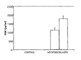
- FIG. 1 represents a bar diagram of the quantity of VEGF produced by the myofibroblasts obtained according to the invention, in picograms per milliliter of medium.
- the starting point is a tumor taken from a mammal, in the present case a carcinoma taken from a dog.
- one modification would be to start from a cirrhotic liver to obtain a population of myofibroblasts involved in this pathological process.
- a microscopic examination of the tumor can be performed to evaluate the percentage of myofibroblasts in the tumor, for example by means of immunohistochemical labeling of the tissue previously fixed in formalin.
- the starting point is a tumor which contains a high percentage of myofibroblasts, that is to say preferably at least 30% of the fibroblasts are myofibroblasts.
- An enzymatic digestion of the tumor is performed, which may or may not be followed by a purification of cell subpopulations.
- the tissue is firstly kept at 4° C. during the microscopic examination of the tumor, while awaiting the results of that examination; a disadvantage is that the tissue is capable of degrading.
- the culturing is started in a medium to favor the growth of the fibroblasts.
- the cells are transferred into the serum-free MEGM medium.
- a purification of cell subpopulations can be performed after the enzymatic digestion.
- the fraction containing the fibroblasts and derivatives is grown directly in the serum-free MEGM medium.
- immunocytochemistry can be used to determine the percentage of cells positive for the SMA marker.
- the MEGM medium was developed specifically for the culturing of normal human mammary epithelial cells.
- the cells thus obtained have in particular the advantages of maintaining their phenotype in culture and of growing actively over about 4 passages. Only at the fifth passage do they show signs of cell death or senescence.
- the starting point is a mammary tumor, for example stored in a suitable transport medium.
- the sample is rinsed in a saline phosphate buffer or PBS.
- Pieces of tissue are transferred into a culture dish containing an enzyme cocktail: collagenase and hyaluronidase in a DMEM culture medium.
- the tissue is torn into small fragments by means of two scalpels, and mixed thoroughly with a pipette.
- the fragments obtained are placed in an incubator at 37° C. for a minimum of 2 hours.
- DMEM tissue-enzyme cocktail mixture
- tissue-enzyme cocktail-DMEM mixture is passed through a 40 micron nylon filter.
- the mixture is centrifuged to give a cell pellet.
- the cells obtained are washed with PBS, and again centrifuged.
- the red corpuscles are removed, if necessary, with a red corpuscle lysis solution, and the cells are washed with PBS and again centrifuged to give a cell pellet.
- the cells are taken up from the pellet into a small volume of PBS.
- the viability of the cells can be estimated, for example by staining with trypan blue, and the cells can be counted.
- the culture medium is changed in order to remove the non-adherent cells.
- a transfer into serum-free medium can be effected from the first passage, when the cells are at 80% confluence.
- the antibiotic normally contained in the MEGM medium is GA-1000 which includes the antibiotics gentamicin and amphotericin-B. These antibiotics can in particular be replaced by normocin (Invivogen).
- the serum-free medium is then replaced approximately every 3 to 4 days.
- the invention makes it possible to obtain a cell population comprising a high percentage (up to more than 95%) of myofibroblasts which grow in a medium which does not contain serum.
- the cells thus obtained have in particular the advantages of maintaining their phenotype in culture and of growing actively over about 4 passages.
- VEGF vascular endothelial growth factor
- FIG. 1 illustrates the substantial production of VEGF by the myofibroblasts obtained with the invention.
- the measurements were performed by means of a canine VEGF ELISA kit (R&D SYSTEMS®).
- FIGURE firstly confirms that the myofibroblasts are a substantial source of VEGF, the principal proangiogenic molecule.
- a tumor in fact needs to recruit blood vessels in order to establish itself and to grow beyond a certain size. It has been shown that cells of the stroma are diverted by the tumor for this purpose. In addition, the recruitment of new blood vessels is a key stage in tissue repair.
- FIG. 1 further shows that the cells obtained by the invention can be used to screen pharmaceutical compounds with anti-angiogenic activity.
Landscapes
- Health & Medical Sciences (AREA)
- Engineering & Computer Science (AREA)
- Life Sciences & Earth Sciences (AREA)
- Biomedical Technology (AREA)
- Genetics & Genomics (AREA)
- Zoology (AREA)
- Organic Chemistry (AREA)
- Bioinformatics & Cheminformatics (AREA)
- Biotechnology (AREA)
- Chemical & Material Sciences (AREA)
- Wood Science & Technology (AREA)
- Rheumatology (AREA)
- Microbiology (AREA)
- Biochemistry (AREA)
- General Engineering & Computer Science (AREA)
- General Health & Medical Sciences (AREA)
- Cell Biology (AREA)
- Micro-Organisms Or Cultivation Processes Thereof (AREA)
- Measuring Or Testing Involving Enzymes Or Micro-Organisms (AREA)
Applications Claiming Priority (4)
| Application Number | Priority Date | Filing Date | Title |
|---|---|---|---|
| FR0922516 | 2009-07-24 | ||
| FR0955216A FR2948381B1 (fr) | 2009-07-24 | 2009-07-24 | Procede d'obtention de myofibroblastes. |
| FR0955216 | 2009-07-24 | ||
| PCT/EP2010/059478 WO2011009706A1 (fr) | 2009-07-24 | 2010-07-02 | Procede d'obtention de myofibroblastes |
Publications (2)
| Publication Number | Publication Date |
|---|---|
| US20120190109A1 US20120190109A1 (en) | 2012-07-26 |
| US9243228B2 true US9243228B2 (en) | 2016-01-26 |
Family
ID=41460947
Family Applications (1)
| Application Number | Title | Priority Date | Filing Date |
|---|---|---|---|
| US13/386,522 Expired - Fee Related US9243228B2 (en) | 2009-07-24 | 2010-07-02 | Process for obtaining myofibroblasts |
Country Status (12)
Families Citing this family (5)
| Publication number | Priority date | Publication date | Assignee | Title |
|---|---|---|---|---|
| CN104096265B (zh) * | 2014-07-01 | 2016-03-23 | 温州医科大学 | 一种用于构建人工血管模型的三维收缩模型的制备方法 |
| CN104630134A (zh) * | 2015-02-15 | 2015-05-20 | 叶中华 | 阴道上皮细胞培养基及阴道上皮细胞培养方法 |
| CN111500527A (zh) * | 2020-06-30 | 2020-08-07 | 北京昱龙盛世生物科技有限公司 | 一种表皮干细胞的分离培养方法 |
| WO2023039371A1 (en) * | 2021-09-07 | 2023-03-16 | Wisconsin Alumni Research Foundation | Cardiac fibroblast derived extracellular matrix |
| CN115197892A (zh) * | 2022-06-13 | 2022-10-18 | 浙江省人民医院 | 一种人甲状腺乳头状癌组织中成纤维细胞的分离培养方法 |
Citations (1)
| Publication number | Priority date | Publication date | Assignee | Title |
|---|---|---|---|---|
| EP1715033A1 (en) | 2004-02-13 | 2006-10-25 | ReproCELL Inc. | Medium for preparing feeder cells for embryonic stem cells and feeder cells |
Family Cites Families (2)
| Publication number | Priority date | Publication date | Assignee | Title |
|---|---|---|---|---|
| US5573937A (en) * | 1989-12-07 | 1996-11-12 | Snow Brand Milk Products Co., Ltd. | Serum free culture medium |
| JP5523674B2 (ja) * | 2005-02-11 | 2014-06-18 | ノボ ノルディスク ヘルス ケア アクチェンゲゼルシャフト | 植物タンパク質加水分解産物を含有する血清フリーの細胞培養液におけるポリペプチドの生産 |
-
2009
- 2009-07-24 FR FR0955216A patent/FR2948381B1/fr active Active
-
2010
- 2010-07-02 CN CN2010800324369A patent/CN102471761A/zh active Pending
- 2010-07-02 US US13/386,522 patent/US9243228B2/en not_active Expired - Fee Related
- 2010-07-02 EP EP10726999.5A patent/EP2456854B1/fr not_active Not-in-force
- 2010-07-02 NZ NZ597728A patent/NZ597728A/xx not_active IP Right Cessation
- 2010-07-02 CA CA2767929A patent/CA2767929A1/fr not_active Abandoned
- 2010-07-02 MX MX2012001004A patent/MX2012001004A/es active IP Right Grant
- 2010-07-02 JP JP2012520985A patent/JP5758891B2/ja not_active Expired - Fee Related
- 2010-07-02 ES ES10726999.5T patent/ES2650664T3/es active Active
- 2010-07-02 AU AU2010275678A patent/AU2010275678B2/en not_active Ceased
- 2010-07-02 WO PCT/EP2010/059478 patent/WO2011009706A1/fr active Application Filing
-
2012
- 2012-01-03 IL IL217352A patent/IL217352A/en not_active IP Right Cessation
Patent Citations (1)
| Publication number | Priority date | Publication date | Assignee | Title |
|---|---|---|---|---|
| EP1715033A1 (en) | 2004-02-13 | 2006-10-25 | ReproCELL Inc. | Medium for preparing feeder cells for embryonic stem cells and feeder cells |
Non-Patent Citations (41)
Also Published As
| Publication number | Publication date |
|---|---|
| EP2456854A1 (fr) | 2012-05-30 |
| US20120190109A1 (en) | 2012-07-26 |
| AU2010275678A1 (en) | 2012-02-09 |
| MX2012001004A (es) | 2012-05-29 |
| WO2011009706A1 (fr) | 2011-01-27 |
| IL217352A (en) | 2016-03-31 |
| CA2767929A1 (fr) | 2011-01-27 |
| IL217352A0 (en) | 2012-02-29 |
| FR2948381A1 (fr) | 2011-01-28 |
| EP2456854B1 (fr) | 2017-08-30 |
| FR2948381B1 (fr) | 2013-07-12 |
| JP2013500003A (ja) | 2013-01-07 |
| CN102471761A (zh) | 2012-05-23 |
| NZ597728A (en) | 2013-11-29 |
| ES2650664T3 (es) | 2018-01-19 |
| JP5758891B2 (ja) | 2015-08-05 |
| AU2010275678B2 (en) | 2016-06-16 |
Similar Documents
| Publication | Publication Date | Title |
|---|---|---|
| US11530385B2 (en) | Hormone responsive tissue culture system and uses thereof | |
| JP4532493B2 (ja) | 細胞培養培地 | |
| US8252591B2 (en) | Hormone responsive tissue culture system and uses thereof | |
| US20040067584A1 (en) | Defined systems for epithelial cell culture and use thereof | |
| JP7434359B2 (ja) | 胃がん及び胆嚢・胆管がん初代細胞を培養する方法及び補助試薬 | |
| KR101861171B1 (ko) | 투석된 혈청이 있는 심근세포 배지 | |
| CA2825070A1 (en) | Method for culturing human pluripotent stem cells | |
| US9243228B2 (en) | Process for obtaining myofibroblasts | |
| JP7660899B2 (ja) | ナイーブ型多能性幹細胞からの栄養外胚葉誘導方法 | |
| KR20190141168A (ko) | 동결보존 방법 | |
| JP2020092700A (ja) | 肝臓オルガノイドの製造方法、肝臓オルガノイド製造用培地、肝臓オルガノイド、細胞製剤、及び被験物質の評価方法 | |
| Talbot et al. | Establishment and characterization of feeder cell-dependent bovine fetal liver cell lines | |
| JP6265385B2 (ja) | アミノ酸製剤による細胞増幅法 | |
| JP6486619B2 (ja) | 薬物評価用細胞及び薬物評価方法 | |
| RU2631005C1 (ru) | Способ культивирования клеток слюнной железы человека | |
| Iriondo et al. | Protocols to co-culture human primary lung cells in the simple-flow device | |
| Wu | Growth of human lung tumor cells in culture | |
| EP1059352A1 (en) | Long term cell culture of human carcinoma | |
| WO2023076292A1 (en) | Culture media and conditions for in vitro expansion and/or maturation of hepatocytes | |
| KR20240056604A (ko) | 수임 심장 전구세포의 제조 방법 | |
| Echigoya et al. | Human amniotic epithelium as an unlimited source of Oct4-expressing totipotent stem cell subset |
Legal Events
| Date | Code | Title | Description |
|---|---|---|---|
| AS | Assignment |
Owner name: ONCOBIOTEK SA, FRANCE Free format text: ASSIGNMENT OF ASSIGNORS INTEREST;ASSIGNORS:ROUYER, NICOLAS JACQUES;BARTHEL, ROBERT;SIGNING DATES FROM 20120128 TO 20120202;REEL/FRAME:028062/0748 |
|
| STCF | Information on status: patent grant |
Free format text: PATENTED CASE |
|
| FEPP | Fee payment procedure |
Free format text: MAINTENANCE FEE REMINDER MAILED (ORIGINAL EVENT CODE: REM.); ENTITY STATUS OF PATENT OWNER: SMALL ENTITY |
|
| LAPS | Lapse for failure to pay maintenance fees |
Free format text: PATENT EXPIRED FOR FAILURE TO PAY MAINTENANCE FEES (ORIGINAL EVENT CODE: EXP.); ENTITY STATUS OF PATENT OWNER: SMALL ENTITY |
|
| STCH | Information on status: patent discontinuation |
Free format text: PATENT EXPIRED DUE TO NONPAYMENT OF MAINTENANCE FEES UNDER 37 CFR 1.362 |
|
| FP | Lapsed due to failure to pay maintenance fee |
Effective date: 20200126 |